Modeling APC mutagenesis and familial adenomatous ...
21
RESEARCH ARTICLE Modeling APC mutagenesis and familial adenomatous polyposis using human iPS cells Cesar A. Sommer 1,2‡ , Amalia Capilla 1,2‡ , Francisco J. Molina-Estevez 1,2‡ , Andreia Gianotti- Sommer 1,2 , Nicholas Skvir 2 , Ignacio Caballero 2 , Sanjib Chowdhury 1 , Gustavo Mostoslavsky 1,2 * 1 Section of Gastroenterology, Department of Medicine, Boston University School of Medicine, Boston, Massachusetts, United States of America, 2 Center for Regenerative Medicine (CReM), Boston University School of Medicine, Boston, Massachusetts, United States of America ‡ These authors are co-first authors on this work. * [email protected] Abstract Mutations in the gene Adenomatous Polyposis Coli or APC appear in most sporadic cases of colorectal cancer and it is the most frequent mutation causing hereditary Familial Adeno- matous Polyposis. The detailed molecular mechanism by which APC mutations predispose to the development of colorectal cancer is not completely understood. This is in part due to the lack of accessibility to appropriate models that recapitulate the early events associated with APC mediated intestinal transformation. We have established a novel platform utilizing human induced Pluripotent Stem cells or iPSC from normal or FAP-specific APC mutant individuals and evaluated the effect of the mutation in the cells before and after differentia- tion into intestinal organoids. In order to minimize genetic background effects, we also established an isogenic platform using TALEN-mediated gene editing. Comparison of nor- mal and APC mutant iPSC revealed a significant defect in cell identity and polarity due to the presence of APC in heterozygosity as well as chromosomal aberrations including abnormal anaphases and centrosome numbers. Importantly, upon specification into intestinal prog- eny, APC heterozygosity was responsible for a major change in the transcriptional identity of the cells with dysregulation of key signaling pathways, including metabolic reprogram- ming, abnormal lipid metabolism and intestinal-specific cadherin expression. In conclusion, we have developed a novel iPSC/intestinal model of APC mutagenesis and provide strong evidence that APC in heterozygosity imparts a clear phenotypic and molecular defect, affecting basic cellular functions and integrity, providing novel insights in the earlier events of APC-mediated tumorigenesis. Introduction Colorectal cancer (CRC) is the second leading cause of cancer-related death in America. Ten to 30% of CRC have a major hereditary component providing unique opportunities to study specific genes and pathways associated with intestinal tumorigenesis. Among them, one of the most important drivers of CRC is Adenomatous Polyposis Coli (APC) as more than 50% of hypermutated tumors and more than 80% of non-hypermutated tumors have mutations in PLOS ONE | https://doi.org/10.1371/journal.pone.0200657 July 19, 2018 1 / 21 a1111111111 a1111111111 a1111111111 a1111111111 a1111111111 OPEN ACCESS Citation: Sommer CA, Capilla A, Molina-Estevez FJ, Gianotti-Sommer A, Skvir N, Caballero I, et al. (2018) Modeling APC mutagenesis and familial adenomatous polyposis using human iPS cells. PLoS ONE 13(7): e0200657. https://doi.org/ 10.1371/journal.pone.0200657 Editor: Jozef Dulak, Faculty of Biochemistry, Biophysics and Biotechnology, Jagiellonian University, POLAND Received: March 22, 2018 Accepted: June 30, 2018 Published: July 19, 2018 Copyright: © 2018 Sommer et al. This is an open access article distributed under the terms of the Creative Commons Attribution License, which permits unrestricted use, distribution, and reproduction in any medium, provided the original author and source are credited. Data Availability Statement: RNA sequencing data has been deposited in the GEO database (GSE99821). Funding: These studies were funded in part by NIH 1R01CA175727-01 to GM. A.C. is a Postdoctoral Research Fellow under a T32 CTSI Award TL1TR001410. We are grateful to Brian R. Tilton of the BUSM Flow Cytometry Core for technical assistance, supported by NIH Grant 1UL1TR001430, and Drs. Greg Miller and
Transcript of Modeling APC mutagenesis and familial adenomatous ...

RESEARCH ARTICLE
Modeling APC mutagenesis and familial
adenomatous polyposis using human iPS cells
Cesar A. Sommer1,2‡, Amalia Capilla1,2‡, Francisco J. Molina-Estevez1,2‡, Andreia Gianotti-
Sommer1,2, Nicholas Skvir2, Ignacio Caballero2, Sanjib Chowdhury1,
Gustavo Mostoslavsky1,2*
1 Section of Gastroenterology, Department of Medicine, Boston University School of Medicine, Boston,
Massachusetts, United States of America, 2 Center for Regenerative Medicine (CReM), Boston University
School of Medicine, Boston, Massachusetts, United States of America
‡ These authors are co-first authors on this work.
Abstract
Mutations in the gene Adenomatous Polyposis Coli or APC appear in most sporadic cases
of colorectal cancer and it is the most frequent mutation causing hereditary Familial Adeno-
matous Polyposis. The detailed molecular mechanism by which APC mutations predispose
to the development of colorectal cancer is not completely understood. This is in part due to
the lack of accessibility to appropriate models that recapitulate the early events associated
with APC mediated intestinal transformation. We have established a novel platform utilizing
human induced Pluripotent Stem cells or iPSC from normal or FAP-specific APC mutant
individuals and evaluated the effect of the mutation in the cells before and after differentia-
tion into intestinal organoids. In order to minimize genetic background effects, we also
established an isogenic platform using TALEN-mediated gene editing. Comparison of nor-
mal and APC mutant iPSC revealed a significant defect in cell identity and polarity due to the
presence of APC in heterozygosity as well as chromosomal aberrations including abnormal
anaphases and centrosome numbers. Importantly, upon specification into intestinal prog-
eny, APC heterozygosity was responsible for a major change in the transcriptional identity
of the cells with dysregulation of key signaling pathways, including metabolic reprogram-
ming, abnormal lipid metabolism and intestinal-specific cadherin expression. In conclusion,
we have developed a novel iPSC/intestinal model of APC mutagenesis and provide strong
evidence that APC in heterozygosity imparts a clear phenotypic and molecular defect,
affecting basic cellular functions and integrity, providing novel insights in the earlier events
of APC-mediated tumorigenesis.
Introduction
Colorectal cancer (CRC) is the second leading cause of cancer-related death in America. Ten
to 30% of CRC have a major hereditary component providing unique opportunities to study
specific genes and pathways associated with intestinal tumorigenesis. Among them, one of the
most important drivers of CRC is Adenomatous Polyposis Coli (APC) as more than 50% of
hypermutated tumors and more than 80% of non-hypermutated tumors have mutations in
PLOS ONE | https://doi.org/10.1371/journal.pone.0200657 July 19, 2018 1 / 21
a1111111111
a1111111111
a1111111111
a1111111111
a1111111111
OPENACCESS
Citation: Sommer CA, Capilla A, Molina-Estevez FJ,
Gianotti-Sommer A, Skvir N, Caballero I, et al.
(2018) Modeling APC mutagenesis and familial
adenomatous polyposis using human iPS cells.
PLoS ONE 13(7): e0200657. https://doi.org/
10.1371/journal.pone.0200657
Editor: Jozef Dulak, Faculty of Biochemistry,
Biophysics and Biotechnology, Jagiellonian
University, POLAND
Received: March 22, 2018
Accepted: June 30, 2018
Published: July 19, 2018
Copyright: © 2018 Sommer et al. This is an open
access article distributed under the terms of the
Creative Commons Attribution License, which
permits unrestricted use, distribution, and
reproduction in any medium, provided the original
author and source are credited.
Data Availability Statement: RNA sequencing data
has been deposited in the GEO database
(GSE99821).
Funding: These studies were funded in part by NIH
1R01CA175727-01 to GM. A.C. is a Postdoctoral
Research Fellow under a T32 CTSI Award
TL1TR001410. We are grateful to Brian R. Tilton of
the BUSM Flow Cytometry Core for technical
assistance, supported by NIH Grant
1UL1TR001430, and Drs. Greg Miller and

APC [1, 2]. Furthermore, loss of heterozygosity (LOH) of APC has been shown to be one of
the earliest and rate-limiting events in the progression from normal epithelium to adenoma
formation and then cancer [3–5]. One of the first described functions of APC was to be part of
the destruction complex (together with GSK3 and AXIN) that mediates phosphorylation of β-
catenin and its ubiquitination and degradation. For years, the field accepted the causal rela-
tionship between mutated APC, dysregulated Wnt signaling, proliferation and CRC develop-
ment. However, improved diagnostic tools and better access to early lesions have found that
this is not the case, at least during the earliest stages of malignant transformation [6–8],
prompting investigators to study other known functions of APC and their potential role in the
initiation of intestinal cancer [9]. In this regard, it is still under debate whether APC in hetero-
zygosity can already alter the molecular and cellular phenotype of the affected cells providing a
fertile environment for the initiation of tumorigenesis [10, 11].
We sought to develop a novel approach based on the generation of induced pluripotent
stem cells (iPSC) from normal individuals or from patients with familial adenomatous polypo-
sis (FAP), the second most highly penetrant form of hereditary CRC, which is caused mainly
by APC mutations. The ability of these pluripotent cells to differentiate into intestinal orga-
noids provided us with a readily accessible platform in which to evaluate the effect of APC
mutations on the cellular and molecular phenotype of iPSC and their intestinal progeny.
Indeed, by comparing isogenic cells expressing either the wild-type or a truncated version of
APC, mimicking the germline mutation commonly found in FAP patients, we detected sur-
prising defects in cell identity and chromosomal integrity. Importantly, upon specification
into intestinal progeny, APC heterozygosity was responsible for a major change in the tran-
scriptional identity of the cells with dysregulation of key signaling pathways, including meta-
bolic reprogramming, abnormal lipid metabolism and cadherin overexpression, providing
novel insights in the earlier events of APC-mediated tumorigenesis.
Materials and methods
iPSC derivation and culture
Reprogramming of fibroblasts from normal and FAP patients was performed as previously
described using the excisable STEMCCA lentiviral reprogramming system [12]. Twenty-five
to thirty days post-transduction, iPSC colonies were mechanically isolated and expanded on
inactivated mouse embryonic fibroblasts (MEFs) in iPSC media, consisting of: DMEM/F12
containing 20% KnockOut Serum Replacement (Invitrogen), 1 mM L-glutamine (Invitrogen),
0.1 mM b-mercaptoethanol (Sigma-Aldrich), 1% nonessential amino acid solution (Invitro-
gen), and 10 ng/ml of FGF2 (Invitrogen). All human samples collected at the CReM were pro-
cessed and handled according to the procotol # H-29791 approved by the Institutional Review
Board (IRB) Committee of Boston University. The FAP fibroblasts were obtained from the
Coriell Institute for Medical Research (GM03948; GM03954; GM06888; GM06965)
Pluripotency marker expression of reprogrammed iPSC was tested using the ES Characteri-
zation Kit (Millipore # SCR001) by following manufacture’s recommendations for AP, TRA-
1-80, and SSEA4 staining. Colonies were analyzed with the inverted microscope Nikon Eclipse
TS100. Karyotype analysis was done by Cell Line Genetics.
Design and assembly of vectors encoding transcription activator-like
effector nucleases (TALENs)
Targeting of the human APC locus in iPSC was achieved by transient delivery of plasmid vec-
tors encoding TALENs. TALEN arrays specific for the APC gene were designed using the TAL
iPSC modeling of FAP
PLOS ONE | https://doi.org/10.1371/journal.pone.0200657 July 19, 2018 2 / 21
Marianne James of the CReM, supported by grants
R24HL123828 and U01TR001810.
Competing interests: The authors have declared
that no competing interests exist.

Effector Nucleotide Targeter 2.0 software, available at https://tale-nt.cac.cornell.edu. We
selected six combinations of TALEN pairs that bind close to the sequence encoding amino
acid 1309 of the APC protein. The APC1309 mutation is one of the most common APC germ-
line mutations leading to severe intestinal phenotypes. Custom TALEN arrays were assembled
as described[13] using the Golden Gate TALEN kit, a gift from Daniel Voytas and Adam Bog-
danove (Addgene kit # 1000000024). These TALEN arrays were then subcloned into the
pHAGE2 EF1α vector for high-level expression in mammalian cells. Different TALEN combi-
nations were first tested in HEK293 cells and the TALEN pair resulting in the highest fre-
quency of non-homologous end joining (NHEJ) events, as determined by Surveyor Nuclease
assay (Integrated DNA technologies), was selected for further experiments.
Targeting of the APC locus in human iPSC
In order to target the APC gene in iPSC, APC TALENs 1320-L and 1320-R3 were subcloned
into the pHAGE2 EF1α IRES Puro vector for co-expression of a TALEN monomer and the
Puro resistance gene. We reasoned that transient puromycin selection of iPSC expressing
high levels of the TALENs and the Puro gene would increase the probability to pick colonies
carrying frameshift mutations resulting in premature stop codons. To achieve this, we fol-
lowed a transfection/selection protocol that we had developed previously for the excision of
lentiviral cassettes from the genome of iPSC [12]. In brief, iPSC were cultured until 30% con-
fluent and transfected with the TALEN-Puro vectors using the Hela Monster transfection
reagent (Mirus) according to manufacturer’s instructions. The following day, the media was
removed and iPSC media containing 1.2 μg/mL puromycin was added. Selection was kept
for 48 hours. iPSC colonies re-emerged within 1 week and were picked and expanded as
described[12]. Clone screening was performed by PCR amplification of a 612 bp region of
genomic DNA surrounding codon 1309. Candidate clones were further interrogated by
DNA sequencing.
Western blot analysis
Protein extraction and Western blot was performed as described elsewhere[14]. For APC
detection, 100 μg of protein was loaded on each lane and blotted membranes were stained
with specific antibodies for the N-terminus or C-terminus of APC (S1 Table).
Fibroblast differentiation and scratch assay
In vitro differentiation of iPSC into fibroblasts was done as follows. iPSC were harvested and
allowed to form embryoid bodies (EBs) in DMEM media containing 20% KSR. Seven days
later, EBs were transferred to 10-cm gelatin-coated culture dishes containing DMEM media
with 10% fetal calf serum (FCS). 50–60% confluency cultures were maintained in absence of
bFGF and Matrigel and split weekly with trypsin-EDTA for at least 5 passages until achieving
uniform fibroblast-like morphology, loss of Tra-1-81, expression of CD90 and normalized
G0-G1 cell cycle accumulation. Confluent iPSC-derived fibroblasts were scratched with a
pipette tip and allowed to polarize and migrate into the wound space. Cells were fixed in 4%
PFA and stained with Phalloidin and pericentrin antibody followed by specific secondary
antibody detection. Finally, nuclei were counterstained with DAPI and cells photographed
with an inverted fluorescence microscope (See detailed antibody list and working dilutions in
S1 Table).
iPSC modeling of FAP
PLOS ONE | https://doi.org/10.1371/journal.pone.0200657 July 19, 2018 3 / 21

Chromosomal abnormalities
iPSC were expanded in feeder-free conditions using mTeSR™1 medium (Stemcell technolo-
gies) following the manufacturer’s recommendations. Upon reaching 80% confluence, iPSC
colonies were fixed in 4% PFA for 5 min, washed in PBS and stained with DAPI (1μg/mL) for
5 minutes. Stained cells were imaged with a fluorescence microscope and anaphase bridge
indexes (ABI) were scored as described[15]. For centrosome enumeration, cells were fixed and
stained with anti-human pericentrin antibody as described above.
Intestinal differentiation
Differentiation of control and mutant iPSC lines into intestinal tissue was performed as
described[16] with the following modifications. First, iPSC lines were cultured in feeder-free
conditions for at least 3 passages. iPSC colonies were then triturated into 1–2 mm fragments,
transferred onto Matrigel-coated 24-well dishes, and cultured in mTeSR™1 medium for three
to four days. Once cells reached 80% confluence, intestinal differentiation was begun by aspi-
rating mTeSR™1 media and adding endoderm differentiation media containing Activin A on
days 1, 2, and 3, as described [16]. Fresh mid/hindgut differentiation media was changed daily
from day 3 to day 8, as indicated in the text, and consisted in RPMI-1640 media supplemented
with human recombinant FGF4 (500 ng/ml) and the GSK3β inhibitor CHIR99021 (3 μM),
instead of Wnt3a protein. Starting as early as two days after exposure of cells to mid/hindgut
differentiation media, intestinal spheroids were collected and embedded into ice-cold intesti-
nal Matrigel (Matrigel1 Matrix. Corning: supplemented as the intestinal growth media
below). Intestinal matrigel drops containing the Spheroids were placed onto 24-well plates and
allowed to solidify by incubating them at 37˚C for 10 min. Fresh DMEM/F12 based intestinal
growth media containing Rspondin1 (500ng/mL), Noggin (100ng/mL), and EGF (100 ng/mL)
was added and replaced every 4 days. iPSC-derived “intestinal organoids” (Passage 1) were
split approximately every other week or used for experiments.
Flow cytometry
At day 3 of the differentiation protocol, cells were harvested with Gentle Cell Dissociation
Reagent (STEMCELL Technologies Inc.), washed with PBS containing 0.2% of Bovin Serum
Albumin (BSA) and stained using conjugated antibodies against markers CXCR4 and c-KIT
(S1 Table). The efficiency of definitive endoderm differentiation was measured through multi-
color cytometric analysis using a FACSCalibur flow cytometer (BD Biosciences).
In the same way, flow cytometry analysis was used to quantify differences on the prolifera-
tion rates between iPSC and HIOs samples from controls and FAP patients following the rec-
ommendations of the Click-iT EdU Alexa Fluor 488 Imaging Kit (Life Technologies).
Immunofluorescence and microscopy
At day 8 of differentiation, cultured cell layers containing three-dimensional (3D) emerging
structures were fixed with 4% paraformaldehyde (PFA) in PBS during 15 minutes and washed
3X for 5 minutes with TBST buffer. Cells were permeabilized with 1% Triton X-100 for 10
minutes. After washing with TBST, cells were blocked with 4% Normal Donkey Serum
(Sigma-Aldrich, D9663) during 30 minutes and stained with specific antibodies for CDX2 and
SOX17. After 1hour of incubation, cells were washed with TBST, followed by incubation with
appropriate antibodies conjugated to Alexa Fluor 488 and Cy3 during 45 minutes. DNA was
detected with DAPI. All the steps were performed at room temperature (RT). Well images
were taken with the Keyence BZ-X710 fluorescence microscope.
iPSC modeling of FAP
PLOS ONE | https://doi.org/10.1371/journal.pone.0200657 July 19, 2018 4 / 21

At day 21, 3D matrigel containing developed organoids was pipetted into a 15 ml Falcon
tube and fixed with fresh 4% PFA in PBS at 4˚C overnight. The next day, organoids were
recovered from the diluted matrigel, washed on PBS, and stained following the steps as previ-
ously indicated for cultured cells, with specific primary antibodies against the intestinal mark-
ers CDX2 and VILLIN, and Hoechst for DNA detection. Whole organoids were mounted on
cavity slides (Eisco) and imaged with a Zeiss LSM 700 confocal microscope.
After 40 days, organoids were fixed in 4% PFA in PBS at 4˚C overnight. Samples were
washed in cold PBS, rinsed six times in 7.5% sucrose in PBS (10 minutes each) and incubated
in 30% sucrose on the rotor at 4˚C overnight. Next day, samples were embedded in O.C.T and
frozen with liquid nitrogen. Frozen sections were permeabilized and blocked as described
before and stained with the intestinal markers CDX2, villin and lysozyme as well as antibodies
against β-catenin and EdU for proliferation analysis (Antibodies and dilutions are described
on S1 Table).
RNA isolation and quantitative real-time RT-PCR
Total RNA from 2D cultured cell and 3D organoids was isolated using the RNeasy Kit (Qia-
gen) following the manufacturer’s recommendations. Reversed transcription of 1μg of total
RNA from each sample was carried out with the SuperScript™ III First-Strand Synthesis System
(Invitrogen). The resulting cDNA was diluted to a final concentration of 50 ng/μl and gene
expression was detected with specific taqMan Probes (Thermo Fisher Scientific) (S2 Table)
with the StepOnePlus R-T PCR system (Applied Biosystems). The delta-delta Ct method was
used to quantify the fold change expression related to undifferentiated iPSC using GAPDH as
the housekeeping gene for sample normalization.
Digital genome sequencing (DGE)
Gene expression data was generated via a 3’ DGE protocol for high-throughput single cell
RNA barcoding and sequencing[17], run and aligned (GRCh37/hg19 assembly) at the Broad
Institute in Cambridge, MA. Normalization and analysis of expression data was performed
using the R statistical software environment (R v.3.1.2; https://cran.r-project.org/) and Biocon-
ductor (https://www.bioconductor.org/). Pairwise differential expression analysis between
samples was performed using the edgeR package (https://bioconductor.org/packages/release/
bioc/html/edgeR.html). Multiplicity correction was performed by applying the Benjamini-
Hochberg method on p-values to control the false discovery rate (FDR), with top genes sorted
by FDR and called as significantly differentially expressed when meeting thresholds of p
value<0.05. Heatmaps were generated using gplots(http://www.inside-r.org/packages/cran/
gplots/docs/heatmap.2) and RColorBrewer(https://cran.r-project.org/web/packages/
RColorBrewer/index.html). Expression data of significant genes was log2 adjusted and samples
were clustered via Pearson correlation and average linkage. All raw and processed data has
been deposited in GEO (GSE99821).
Ingenuity1 pathway analysis (IPA1)
IPA1, QIAGEN Redwood City, www.qiagen.com/ingenuity) software (build version:
389077M, content version: 27821452) was used to generate functional networks and analyze
signal transduction pathways. The functional networks and signal transduction pathways of
IPA are predicted on the basis of known gene–gene (protein–protein) and functional interac-
tions based on information contained in the Ingenuity Pathway Knowledge Base.
iPSC modeling of FAP
PLOS ONE | https://doi.org/10.1371/journal.pone.0200657 July 19, 2018 5 / 21

Statistical methods
Student’s t-tests were performed for statistical analysis. Statistically significant differences
from the wild-type control are indicated by asterisks: �p< 0.05; ��P< 0.01.
Results
A mini-library of FAP-specific iPSC
Fibroblasts from four previously diagnosed FAP individuals were obtained from Coriell and
reprogrammed using the STEMCCA system to generate three independent iPSC lines, as
described [12, 18] (Fig 1A). Two of the patient lines (FAP 3948–1 and FAP 3954–1) contain a
C>T mutation in the coding sequence of the APC gene at position 1621 and the other two
(FAP 6965–1 and FAP 6888–1) contain a AG deletion at position 4611 (Fig 1B), both inducing
production of truncated versions of APC protein. Overall, there were no major phenotypic dif-
ferences between the FAP-iPSC compared to normal cells in terms of proliferation rate. We did
observe an unusual growth pattern of the FAP-iPSC clones (“ring” morphology, S1 Fig) when
the cells were expanded on feeders, likely a consequence of spontaneous differentiation. This
morphology disappeared when the cells were passaged on Matrigel under feeder-free condi-
tions, however and as detailed below, the mutant cells showed increased spontaneous differenti-
ation. Clones FAP 3948–1 and FAP 6965–1 showed the more robust growing conditions (easier
to passage, less spontaneous differentiation, healthier morphology) and we focused on these
two clones for our experiments. In an attempt to see if mutations in APC affected the prolifera-
tion rate of the cells we quantified EdU uptake and incorporation into newly synthesized DNA.
As shown in Fig 1C, there was no difference between normal and FAP-specific clones.
If APC in heterozygosity is sufficient to induce changes in β-catenin signaling, FAP-specific
iPSC should display a spontaneous increase in expression of WNT target genes as well as sponta-
neous differentiation toward mesendodermal lineages [19]. However, when comparing normal vs
FAP cells we found gene expression of several WNT target genes (including FOXA2, AFP, MSX1,
GATA4 and BRACHYURY) to be variable between iPSC lines from different donors. This inter-
individual variability was confirmed by comparing iPSC lines derived from three healthy donors
(S2 Fig). We speculated that differences mediated by APC mutations may become more evident
upon differentiation into intestinal epithelium, as APC expression increases upon gut differentia-
tion (S3 Fig). For this purpose we utilized a protocol developed by Wells and colleagues that
employs sequential stimulation of NODAL and FGF4R /WNT3A to mimic early development
and drive endodermal specification first into mid- and hindgut prior to differentiation into intes-
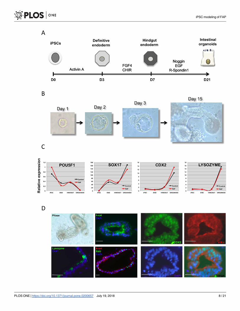
tinal epithelium (Fig 2A) [20]. Indeed, when exposed to these conditions we were able to obtain
definitive endoderm and intestinal specification, with formation of 3D human intestinal orga-
noids (HIOs) upon expansion in matrigel embedded cultures (Fig 2B) from both normal and
mutated APC iPSC. Expression of SOX17, CDX2 and LYSOZYME followed the expected develop-
mental pattern (Fig 2C) with upregulation of SOX17 in endodermal cells followed by upregulation
of the intestinal markers. Similar to our findings in undifferentiated iPSC, comparison between
the normal and the APC-mutant iPSC-derived HIOs did not reveal statistically significant differ-
ences in proliferation measured by EdU incorporation (Fig 1C) or expression of WNT target
genes (S3 Fig). However, we consistently observed that FAP-specific iPSC evidenced faster differ-
entiation towards endoderm than control lines, with expression of SOX17 already detectable after
short (3.0 days) NODAL activation (Fig 2C and S4 Fig). This finding appears to correlate with
increased expression of CDX2, more evident in the later stages of intestinal specification (Fig 2C).
Immunohistochemistry confirmed efficient expression of CDX2 (95% of cells were CDX2+), VIL-
LIN and β-CATENIN in intestinal organoids (Fig 2D).
iPSC modeling of FAP
PLOS ONE | https://doi.org/10.1371/journal.pone.0200657 July 19, 2018 6 / 21

In order to minimize the potential noise created by inter-individual genetic background
variability, we decided to create a gene editing-based APC-specific iPSC platform that would
allow the comparison of isogenic APC mutant vs normal iPSC.
Fig 1. Characterization of wild-type and FAP iPSC. (A) iPSC were generated with the constitutive EF1a-STEMCCA vector expressing the four reprogramming factors
Oct4, Klf4, Sox2 and c-Myc and containing a loxP site within the 3’ LTR that allows excision of the entire cassette following exposure to Cre recombinase. Transgene-
free iPSC clones generated with STEMCCA display the typical human ESC morphology and stain positive for pluripotency markers. (B) Sequence analysis of the APCgene in iPSC clones BU1 (WT), FAP 3948–1 and FAP 6965–1. Arrows indicate a C>T change resulting in the conversion of glutamine 541 of APC to a stop codon (FAP
3948–1), and a heterozygous 2 bp deletion in codon 1537 that results in a frameshift mutation that leads to a premature termination at proline 1542 (FAP 6965–1). G-
banding chromosome analysis reveals normal diploid number of chromosomes (2n = 46) in two representative FAP-iPSC lines. (C) iPSC and intestinal organoids
(HIOs) were pulsed with EdU, and then fixed, permeabilized, and stained with the Click-iT reaction. Percentages of cells in S + G2/M phase are indicated in the graphs.
Bar graph shows average +/- SD of two independent lines done in triplicate.
https://doi.org/10.1371/journal.pone.0200657.g001
iPSC modeling of FAP
PLOS ONE | https://doi.org/10.1371/journal.pone.0200657 July 19, 2018 7 / 21
iPSC modeling of FAP
PLOS ONE | https://doi.org/10.1371/journal.pone.0200657 July 19, 2018 8 / 21

Isogenic platform of normal and APC-mutant iPSC
In order to generate an isogenic system we designed TALENs targeting position 1320 of the
APC coding sequence (Fig 3A), a known hotspot commonly mutated in FAP patients [2]. We
chose BU1 iPSC as our targeting clone, which has been extensively characterized and used in
our lab at the CReM [21]. After four independent attempts and despite consistently observing
an efficiency of ~15–20% of TALEN-induced indels, the overall efficiency of a not-in-frame
heterozygous mutation was significantly low (1 out of 16), strongly suggesting that having del-
eterious mutations in APC induces a selection disadvantage to the survival, reprogramming, or
outgrowth of iPSC in culture. Nevertheless, we were able to obtain and expand a karyotypically
normal iPSC isogenic clone containing a 140 bp deletion that introduced a premature stop
codon at position 1246 with translation of a truncated APC protein lacking part of the WNT
signaling domain and the entire Basic Region and EB1 binding domain in the C-terminus end
(Fig 3B). The APC +/+ and APC+/- iPSC isogenic lines were then expanded and maintained in
identical culture conditions for the rest of the studies. The first obvious cellular effect of APCdysregulation was the spontaneous differentiation phenotype of the APC +/- clone in culture
(Fig 3C), which was confirmed molecularly by upregulation of several primitive streak, meso-
dermal and endodermal genes (Fig 3D) compared to the APC +/+ line. This was accompanied
by an increase in WNT target gene expression, including AXIN2 and CDX1 (Fig 3D).
APC haploinsufficiency affects the phenotype of isogenic iPSC and their
differentiated progeny
The known functions of APC in cellular motility and cytoskeletal integrity [22, 23] prompted us
to investigate whether expression of a defective copy of APC could affect cell polarity. For this
purpose we adapted a protocol for the differentiation of iPSC into fibroblasts (as demonstrated
by positive CD90/Thy-1 staining, not shown) and performed a classic “wound repair assay”.
First, iPSC-derived fibroblasts were grown to confluence and then a mechanical “scratch” was
induced with a pipette tip. Fibroblasts then tend to grow towards the “wound” by polarizing the
cells and then migrating to cover the defect. Staining the cells with phalloidin, pericentrin and
DAPI enables the quantification of the location of the microtubule-organizing center (MTOC)
relative to the nucleus as a measurement of cell polarity. As shown in Fig 3E, APC mutant cells
showed a significant decrease in the percentage of polarized MTOC. As APC has also been impli-
cated in the regulation of chromosomal segregation and the formation of the mitotic spindle,
with APC mutations causing chromosomal abnormalities [24], we tested two independent assays
to detect chromosomal aberrations. Indeed, APC mutant cells showed a significant increase in
abnormal anaphases (measured as Anaphase Bridge Index or ABI [15]) with almost double the
number of abnormal anaphases (Fig 3F). Similarly, while most normal APC+/+ cells contained 1
centrosome and rarely 2, APC mutant cells showed a significant increase in cells containing 2
centrosomes and even 4 centrosomes, which was never observed in the normal cells (Fig 3G).
Next, we induced the differentiation of the APC+/+ and APC+/- into intestinal organoids,
following the protocol described above. As shown in Fig 4, we were able to obtain efficient
Fig 2. Directed differentiation of iPSC into intestinal organoids. (A) Schematic of the protocol employed to induce intestinal differentiation from
iPSC. Cells are first induced to form endoderm followed by hindgut specification and formation of intestinal organoids in matrigel. (B) Bright field
images showing formation of a representative iPSC-derived intestinal organoid upon incubation in matrigel. (C) Real-time RT-PCR analyses of
differentiating cells at different stages demonstrate robust endoderm induction and hindgut specification followed by efficient generation of intestinal
organoids from both normal control and FAP-specific iPSC lines. (D) Representative micrographs and immunofluorescence images of the intestinal
organoids derived from an FAP-iPSC reveal formation of 3D epithelial structures with clear expression of intestinal markers CDX2, VILLIN and
LYSOZYME. Edu staining allows visualization of proliferating cells within the organoids. β-CATENIN staining shows mostly membrane and
cytoplasmic pattern.
https://doi.org/10.1371/journal.pone.0200657.g002
iPSC modeling of FAP
PLOS ONE | https://doi.org/10.1371/journal.pone.0200657 July 19, 2018 9 / 21

iPSC modeling of FAP
PLOS ONE | https://doi.org/10.1371/journal.pone.0200657 July 19, 2018 10 / 21

differentiation with the APC mutant line, first into endoderm (Fig 4A) with significant CDX2expression upon addition of FGF4 and WNT3A as observed with the WT isogenic line (Fig
4B) and robust formation of intestinal organoids upon culture in matrigel (Fig 4C–4E). Fur-
thermore, and in concordance with what we observed with the FAP lines, staining the orga-
noids on day 30 post differentiation with EdU/DAPI revealed similar proliferation rates in
mutant and WT cells (not shown).
APC haploinsuffciency is associated with a definable gene expression
profile, particularly associated with altered metabolic reprogramming and
dysregulation of lipid metabolism and cadherins
In order to study whether a truncated form of APC is associated with changes in the gene
expression landscape of both undifferentiated iPSC and their intestinal progeny, we sought to
perform RNA seq by digital gene expression (DGE). Similarly to SAGE, DGE allows sequenc-
ing of most RNA transcripts [17], including low abundant ones that can be missed when using
hybridization-based Affymetrix chip arrays. Not surprisingly, the first analysis done using
Multidimensional Scaling plot (MDS) showed a clear separation between the undifferentiated
cells and the HIOs (Fig 5A). More importantly, this analysis demonstrated that the presence of
one APC mutated allele changed the gene expression signature of the cells, separating APCWT from APC mutant cells, and that the effect of the mutation appeared to be more pro-
nounced in the intestinal organoids than in undifferentiated iPSC (Fig 5A). Indeed, pairwise
differential expression analysis between normal vs mutant samples at each time point (using
the edgeR package with an FDR cutoff of at least 0.05) showed 2124 differentially expressed
genes between normal and mutant HIOs, compared to only 322 genes differentially expressed
between normal and mutant undifferentiated cells (S3 Table). Furthermore, these expression
differences resulted in separate clustering of the normal vs wildtype cells at each time point
when analyzed by unsupervised hierarchical clustering (Fig 5B).
Upon analysis of each set of differentially expressed genes for potential differences in epi-
thelial or signaling programs, we observed that Cadherin 17 (CDH17), was the number 13
most highly dysregulated among the 2124 misexpressed genes in HIOs (with an FDR value of
1.54E-81). Cadherin 17, also known as IL Cadherin or Intestinal-Liver Cadherin is a member
of the cadherin superfamily that appears to be expressed primarily in the intestinal tract and its
protein may play a role in the morphological organization of the intestine and liver. Remark-
ably, abnormal CDH17 expression has been described as a diagnostic marker of adenocarcino-
mas of the digestive system [25], with higher sensitivity than CDX2 [26]. Another gene that
showed a striking increase in APC mutant HIO was Isocitrate dehydrogenase 1 (IDH1)
(logCPM 9.77956, p-value 4.42E-56 and FDR 2.94E-53). One of the top 5 canonical pathways
that was found by Gen Set Enrichment Analysis (See below) within the HIO gene set was oxi-
dative phosphorylation (p-value = 5.74E -11, S1 Table). In that regard, another gene of interest
found to be dysregulated was Lactate dehydrogenase A (LDHA) (logCPM 9.20493, p-value
Fig 3. Generation of isogenic APC+/- iPSC line and its effect on cell polarity and chromosomal instability. (A) We designed several pairs of site-specific TALENs
targeting the region surrounding codon 1309 of APC, a known mutational hotspot. The efficiency of gene modification by NHEJ was evaluated by the Surveyor
Nuclease Assay. (B) Schematic of the two APC proteins expressed by the heterozygous mutant iPSC line based on DNA sequencing and binding recognition of the
antibodies used for Western blot. Western blot confirms expression of both the WT and truncated versions of APC. (C) Bright field images comparing the APC+/+ vs the APC+/- human iPSC colonies showing spontaneous differentiation of the mutant cells. (D) Mutant iPSC show transcriptional changes reminiscent of
increased endogenous WNT signaling, as determined by TaqMan qPCR analysis of endoderm, mesoderm and WNT target genes.�p<0.05, ��p<0.01. (E) A scratch-
induced cell migration assay reveals impaired centrosome reorientation in mutant iPSC-derived fibroblasts compared with the control, with a significant reduction
in polarized MTOC. (F-G) Also consistent with the role of APC in microtubule dynamics, APC mutant iPSC display an increase in chromosomal instability, as
evidenced by increase in abnormal anaphases (quantitated by Anaphase Bridge Index or ABI) and higher number of centrosomes (cen).
https://doi.org/10.1371/journal.pone.0200657.g003
iPSC modeling of FAP
PLOS ONE | https://doi.org/10.1371/journal.pone.0200657 July 19, 2018 11 / 21

iPSC modeling of FAP
PLOS ONE | https://doi.org/10.1371/journal.pone.0200657 July 19, 2018 12 / 21

6.05E-13 and FDR 5.20E-11). LDHA is considered as critical for cancer cells since it catalyzes
the conversion of pyruvate to lactate. It is elevated in several cancers and has high potential as
a diagnostic, predictive as well as therapeutic target for new anticancer treatments [27–29].
Upregulation of IDH1 and LDHA in APC mutant HIO might indicate an early metabolic
reprogramming event associated with APC mutations that has not been studied yet. Finally,
VIMENTIN, which also appeared dysregulated in the APC mut HIO (logCPM 9.32429, p-
value 1.59E-30 and FDR 4.42E-28) is overexpressed in several cancers including gastrointesti-
nal cancers and correlates with poor prognosis and accelerated growth and invasion of cancer
cells. In addition, VIMENTIN is a marker for epithelial-mesenchymal transition (EMT) and is
currently being evaluated as a potential target for anti-cancer therapy [30, 31].
The top genes found upregulated in the DGE analyses of the isogenic APC heterozygous
iPSC line were confirmed to be overexpressed also in HIOs from FAP patient-specific iPSC
(S5 Fig). Real-time RT-PCR of representative genes reinforced the evidence pointing towards
a fatty acids altered metabolism, being FABP1, ALDH1A1 and APO1 overexpressed more than
100 fold (S5 Fig).
GO, GSEA and Ingenuity pathway analysis (IPA) of HIO gene sets
Analysis of the differentially expressed genes was done using three independent platforms,
Gene Ontology (GO), Gene Set Enrichment Analysis (GSEA) and Ingenuity Pathway Analysis
(IPA) and all showed similar results. Among the dysregulated KEGG pathways associated with
APC mutations, the PPAR signaling pathway appears at the top of the list (Fig 5C). Indeed,
within this pathway APC appears to modulate several genes that participate in adipocyte func-
tion and lipid metabolism, including FATP, FABP, PPARγ, FABP1, OLR1 and several lipid
transporters of the APO family (Fig 5D). Validating these findings, GSEA analysis revealed the
APC mutation was associated with abnormal lipid function (Fig 5E).
In order to further validate the pathways affected in APC+/- HIO, Ingenuity1 Pathway
Analysis (IPA) was used. A total of 2124 differentially expressed genes were uploaded to IPA
for core analysis. Out of those, 2113 genes IDs were successfully mapped. As shown in Fig 6,
we analyzed the top 5 diseases and bio functions and scored them as a percentage of the Top 5
hits. We observed cancer (p-value 2.61E-05–9.10E-33) and gastrointestinal diseases (p-value
2.43E-05–5.11E-17) [within Top Disease and Disorder subset], cellular growth and prolifera-
tion (p-value 3.13E-05–1.35E-27), cellular movement (p-value 3.19E-05–3.46E-23), cellular
survival (p-value 2.53E-05–9.24E-23) and lipid metabolism (p-value 2.76E-05–4.22E-14)
[within Top Molecular and Cellular Functions subset] and organismal development (p-value
3.12E-05–4.57E-21), tissue-, organ- and embryonic-development (all with similar p-value of
2.74E-05–1.35E-19) [within Top Physiological System Development and Function subset]
were the top hits within the Top Disease and Bio Functions categories of APC+/- HIO-associ-
ated genes. For each of the top 5 Disease and Bio Function, we also estimated the percentage of
genes that were involved in the process as compared to the total number of total genes used in
the core analysis dataset (Table 1). We observed that cancer (86.2%), cellular growth and pro-
liferation (39.4%) and organismal development (30.4%) had the highest number of common
genes within each subset of Top Disease and Bio Functions.
Fig 4. Intestinal differentiation of APC+/+ vs APC+/- mutant iPSC. (A) Efficient endoderm specification by the APC +/+ and APC +/- iPSC isogenic
lines as evidenced by cKit and CXCR4 expression. (B) Immunohistochemistry of cells at day 8 of differentiation shows similar patterns of SOX17 and CDX2
expression. (C) Bright field images show representative intestinal organoids upon incubation in matrigel. (D) Real time RT-PCR shows similar dynamics of
differentiation with upregulation of FOXA2 and SOX17 during endodermal specification (Day 3: D3) and robust upregulation of CDX2 in hindgut (Day 8:
D8). (E) Whole mount immunohistochemistry of a representative organoid from the APC +/- iPSC line showing CDX2 (in red) and VILLIN (in green)
expression counterstained with DAPI (blue).
https://doi.org/10.1371/journal.pone.0200657.g004
iPSC modeling of FAP
PLOS ONE | https://doi.org/10.1371/journal.pone.0200657 July 19, 2018 13 / 21

Discussion
The recent establishment of in vitro 3D culture approaches to model intestinal morphogenesis,
either from intestinal crypts [32] or from pluripotent stem cells [20] had opened new exciting
Fig 5. DGE analysis of APC+/+ vs APC+/- before and after differentiation into intestinal organoids. (A) Multidimensional Scaling plot (MDS) shows whole genome
transcriptome analysis of four independent APC+/+ (orange) vs four independent APC+/- (blue) iPSC subclones before (circles) and after differentiation (triangles) into
intestinal organoids. (B) Unbiased hierarchical clustering heatmaps of undifferentiated (hiPSC) or differentiated intestinal organoids (HIO) using pairwise differential
expression analysis between APC+/+ vs APC+/- samples, with an FDR cutoff of at least 0.05. (C) GSEA of KEGG pathways showing the top most affected pathway based
on the differentially expressed genes. (D) Schematic of the PPAR Signaling pathway. Red stars denote individual components of the pathway that appear differentially
expressed between APC+/+ vs APC+/-. (E) GSEA of metabolic pathways showing the lipid metabolism pathway is affected in APC mutant cells.
https://doi.org/10.1371/journal.pone.0200657.g005
iPSC modeling of FAP
PLOS ONE | https://doi.org/10.1371/journal.pone.0200657 July 19, 2018 14 / 21

opportunities to study in vitro intestinal tumorigenesis. Recent studies using in vitro organoids
grown from different intestinal tumors [33, 34] showed a strong correlation, functionally and
molecularly with their primary tissue counterparts, providing a strong argument in favor of
the potential for in vitro systems to model intestinal disease. In the case of iPSC, access to
intestinal epithelial cells offers a unique approach to study in a cell intrinsic manner the earliest
Fig 6. Ingenuity pathway analysis (IPA) of DGE data. Whole genome expression data was analyzed using IPA focusing on Diseases and Disorders (Red), Molecular
and Cellular Functions (Green) and Physiological System Development Function (Blue). Number of genes (Number of Molecules) within the top five categories are
shown in graphs.
https://doi.org/10.1371/journal.pone.0200657.g006
iPSC modeling of FAP
PLOS ONE | https://doi.org/10.1371/journal.pone.0200657 July 19, 2018 15 / 21

cellular and molecular events associated with APC mutagenesis and its potential role in CRC
development. Taking advantage of the fact that patient somatic cells are APC heterozygous, we
sought to recapitulate the primordial developmental state of gut epithelia and study the cellular
and molecular “milieu” that may provide a bona fide environment for the development of
intestinal tumors. We demonstrated that FAP patient iPSC were able to differentiate into gut
organoids, although many differences found in the intestinal-like tissues developed from nor-
mal or heterozygous APC iPSC were certainly blurred by the inter-patient variability. Once we
moved into the isogenic system, we were able to find robust and significant differences
between normal and APC mutant intestinal epithelial cells. It is relevant to emphasize that
studies with the FAP lines were also limited by the unstable nature of the APC mutant lines.
Spontaneous differentiation was always evident in our cultures requiring extra care in the
maintenance and passaging of the lines, and was further supported by the faster induction of
markers such as SOX17 and CDX2 in the APC mutant cells.
Another interesting finding relates to the gene editing approach to create the APC mutant
cells. As mentioned above, even though we had a relatively high efficiency of TALEN induced
DNA double strand breaks (~15–25%), the overall efficiency of obtaining non-conserved
mutations bearing truncated versions of APC was very low (~6%). As a matter of fact, after
screening more than 400 colonies we were never able to recover a line with homozygous muta-
tions in both APC alleles, strongly suggesting that APC is a key factor in the maintenance of
iPSC lines in their pluripotent state. Our findings that APC in heterozygosity was enough to
induce spontaneous differentiation of the iPSC could indicate that even if our TALENs were
able to create homozygous APC mutant lines, they were lost to spontaneous differentiation
before forming colonies. This is supported by the existence of a limited number of reports
describing APC mutant lines, which were all done on mouse ESC expressing either hypo-
morphic mutant variants of APC [35] or specifically overexpressing β-catenin APC mutations
[36]. These studies showed that abnormal catenin regulation induced dysregulated in vitro dif-
ferentiation (and not proliferation), findings in keeping with our APC heterozygous human
Table 1. Summary of ingenuity pathway analysis.
Name # Molecules # Molecules % of total 2125 genes p-value
Top 5 Diseases and Disorders
Cancer 1832 86.21176 2.61E-05–9.10E-33
Organismal Injury and Abnormalities 1861 87.57647 3.19E-05–9.10E-33
Reproductive System Disease 1023 48.14118 2.61E-05–2.36E-23
Gastrointestinal Disease 1590 74.82353 2.43E-05–5.11E-17
Respiratory Disease 372 17.50588 3.19E-05–9.00E-14
Top 5 Molecular and Cellular Functions
Cellular Growth and Proliferation 839 39.48235 3.13E-05–1.35E-27
Cellular Movement 481 22.63529 3.19E-05–3.46E-23
Cell Death and Survival 696 32.75294 2.53E-05–9.24E-23
Cell Morphology 506 23.81176 2.53E-05–1.66E-16
Lipid Metabolism 292 13.74118 2.76E-05–4.22E-14
Top 5 Physiological System Development and Function
Organismal Development 647 30.44706 3.12E-05–4.57E-21
Renal and Urological System Development and Function 130 6.117647 2.33E-05–4.57E-21
Embryonic Development 412 19.38824 2.74E-05–1.35E-19
Organ Development 348 16.37647 2.74E-05–1.35E-19
Tissue Development 682 32.09412 2.74E-05–1.35E-19
https://doi.org/10.1371/journal.pone.0200657.t001
iPSC modeling of FAP
PLOS ONE | https://doi.org/10.1371/journal.pone.0200657 July 19, 2018 16 / 21

iPSC line. Interestingly, recent studies describing a novel differentiation protocol to derive
colonic organoids from human iPSC found that FAP-specific iPSC had a significant increase
in proliferation as measured by percentage of ki67 positive cells [37]. Whether this difference
is due to their specific protocol or to other differences in culture conditions is unclear.
The finding that APC in heterozygosity has such an impact on the phenotypic and molecu-
lar characteristics of our iPSC and their intestinal progeny is not completely unexpected. Sev-
eral studies have previously shown that changes in APC dosage can affect cellular motility,
migration and polarity [24, 38, 39], and can induce the development of early intestinal neo-
plasms [40]. Our data strongly support the role of APC in cell motility and polarity as evi-
denced by the migratory defect evidenced in the APC mutant fibroblasts. In addition, the
presence of chromosomal aberrations (evidenced both by the higher number of abnormal ana-
phases as well as centrosomes) emphasizes a novel mechanism by which changes in APC func-
tion can initiate chromosomal instability and tumorigenesis.
There is a renewed interest in the area of cancer metabolism with metabolic reprogram-
ming coined as a hallmark of cancer based on the discovery of mutations and alterations in
IDH1 and IDH2 along with succinate dehydrogenase (SDH), fumarate hydratase (FH), and
pyruvate kinase M2 (PKM2) [41, 42]. IDH1/2 mutations have been observed in several solid
and blood cancers including colon cancer. The metabolic activity of IDH1/2 has been tied to
cancer epigenetic mechanisms. Several ongoing studies have started targeting these IDH1/2metabolic genes as selective targets for new anticancer therapeutics [43]. Metabolic switch
from oxidative phosphorylation to increased glycolysis even under hypoxic conditions (termed
Warburg effect) is a central biochemical feature of cancer cells.
Conclusions
Our studies show that a truncated form of APC induces a significant effect on the cellular and
molecular features of iPSC and their differentiated progeny. This effect is exacerbated upon
differentiation into intestinal organoids with a major impact on the gene expression profile of
the cells, and the emergence of a molecular signature resembling several features of intestinal
tumorigenesis and abnormal metabolism. Further studies using cells with homozygous muta-
tions mimicking the correspondent loss of heterozygosity (LOH) present in advanced intesti-
nal tumors is warranted.
Supporting information
S1 Fig. Isogenic WT and APC +/- iPSC colonies. Representative micrographs showing bright
field (top rows) and Alkaline Phosphatase staining (bottom rows) evidence abnormal colony
growth pattern in APC+/- colonies compared to the WT isogenic line (BU1).
(PDF)
S2 Fig. Spontaneous differentiation in human iPSC. Expression of endodermal genes mea-
sured by qRT-PCR comparing WT vs FAP iPSC lines (top graph) or three independent control
lines (bottom graph).
(PDF)
S3 Fig. Expression of APC and Wnt target genes. (A) Expression of APC measured by
qRT-PCR in WT (black) vs FAP (red) iPSC before (d0) and after differentiation into intestinal
organoids (d40). (B) Expression of Wnt target genes in intestinal organoids. Data are
mean ± SE from two independent WT and FAP lines.
(PDF)
iPSC modeling of FAP
PLOS ONE | https://doi.org/10.1371/journal.pone.0200657 July 19, 2018 17 / 21

S4 Fig. FAP iPSC differentiate into HIOs faster than WT cells. Expression of the endoder-
mal marker Sox17 overtime during differentiation of control (black) vs FAP (red) iPSC into
intestinal organoids. DE: definitive endoderm. Data are mean ± SE of two independent lines.
(PDF)
S5 Fig. Validation of DGE expression. qRT-PCR data of selected genes in WT vs FAP iPSC
upon intestinal differentiation. Data are mean ± SE of two independent lines.
(PDF)
S1 Table. List of antibodies used throughout these studies (See Materials and methods).
(DOCX)
S2 Table. List of TaqMan probes used in the real-time PCR experiments (See Materials
and methods).
(DOCX)
S3 Table. Complete list of genes that are dysregulated in APC+/+ vs APC +/- iPSC before
(blue) and after (red) differentiation into HIOs. Top genes sorted by FDR and called as sig-
nificantly differentially expressed when meeting thresholds of p value<0.05.
(XLSX)
Acknowledgments
The authors would like to thank all members of the CReM for helpful discussions and D. Kot-
ton for reading and editing of the manuscript.
Author Contributions
Conceptualization: Cesar A. Sommer, Gustavo Mostoslavsky.
Data curation: Cesar A. Sommer, Nicholas Skvir, Ignacio Caballero, Gustavo Mostoslavsky.
Formal analysis: Cesar A. Sommer, Nicholas Skvir, Ignacio Caballero, Sanjib Chowdhury.
Investigation: Cesar A. Sommer, Amalia Capilla, Francisco J. Molina-Estevez, Andreia Gia-
notti-Sommer, Gustavo Mostoslavsky.
Methodology: Cesar A. Sommer, Amalia Capilla, Francisco J. Molina-Estevez, Andreia Gia-
notti-Sommer, Gustavo Mostoslavsky.
Project administration: Gustavo Mostoslavsky.
Resources: Gustavo Mostoslavsky.
Software: Nicholas Skvir, Ignacio Caballero, Sanjib Chowdhury.
Supervision: Gustavo Mostoslavsky.
Writing – original draft: Cesar A. Sommer, Gustavo Mostoslavsky.
Writing – review & editing: Gustavo Mostoslavsky.
References1. Fearon ER, Vogelstein B. A genetic model for colorectal tumorigenesis. Cell. 1990; 61(5):759–67. Epub
1990/06/01. PMID: 2188735.
2. Cancer Genome Atlas N. Comprehensive molecular characterization of human colon and rectal cancer.
Nature. 2012; 487(7407):330–7. Epub 2012/07/20. https://doi.org/10.1038/nature11252 PMID:
22810696; PubMed Central PMCID: PMC3401966.
iPSC modeling of FAP
PLOS ONE | https://doi.org/10.1371/journal.pone.0200657 July 19, 2018 18 / 21

3. Kinzler KW, Vogelstein B. Lessons from hereditary colorectal cancer. Cell. 1996; 87(2):159–70. Epub
1996/10/18. doi: S0092-8674(00)81333-1 [pii]. PMID: 8861899.
4. Galiatsatos P, Foulkes WD. Familial adenomatous polyposis. Am J Gastroenterol. 2006; 101(2):385–
98. Epub 2006/02/04. doi: AJG375 [pii] https://doi.org/10.1111/j.1572-0241.2006.00375.x PMID:
16454848.
5. Segditsas S, Tomlinson I. Colorectal cancer and genetic alterations in the Wnt pathway. Oncogene.
2006; 25(57):7531–7. Epub 2006/12/05. doi: 1210059 [pii] https://doi.org/10.1038/sj.onc.1210059
PMID: 17143297.
6. Anderson CB, Neufeld KL, White RL. Subcellular distribution of Wnt pathway proteins in normal and
neoplastic colon. Proc Natl Acad Sci U S A. 2002; 99(13):8683–8. Epub 2002/06/20. https://doi.org/10.
1073/pnas.122235399 122235399 [pii]. PMID: 12072559.
7. Blaker H, Scholten M, Sutter C, Otto HF, Penzel R. Somatic mutations in familial adenomatous polyps.
Nuclear translocation of beta-catenin requires more than biallelic APC inactivation. Am J Clin Pathol.
2003; 120(3):418–23. Epub 2003/09/25. https://doi.org/10.1309/4E4W-G3AY-GJNC-D11P PMID:
14502807.
8. Phelps RA, Chidester S, Dehghanizadeh S, Phelps J, Sandoval IT, Rai K, et al. A two-step model for
colon adenoma initiation and progression caused by APC loss. Cell. 2009; 137(4):623–34. Epub 2009/
05/20. doi: S0092-8674(09)00251-7 [pii] https://doi.org/10.1016/j.cell.2009.02.037 PMID: 19450512.
9. Rajagopalan H, Nowak MA, Vogelstein B, Lengauer C. The significance of unstable chromosomes in
colorectal cancer. Nat Rev Cancer. 2003; 3(9):695–701. Epub 2003/09/03. https://doi.org/10.1038/
nrc1165 PMID: 12951588.
10. Patel BB, Li XM, Dixon MP, Blagoi EL, Nicolas E, Seeholzer SH, et al. APC +/- alters colonic fibroblast
proteome in FAP. Oncotarget. 2011; 2(3):197–208. Epub 2011/03/18. https://doi.org/10.18632/
oncotarget.241 PMID: 21411865; PubMed Central PMCID: PMC3195363.
11. Yeung AT, Patel BB, Li XM, Seeholzer SH, Coudry RA, Cooper HS, et al. One-hit effects in cancer:
altered proteome of morphologically normal colon crypts in familial adenomatous polyposis. Cancer
Res. 2008; 68(18):7579–86. Epub 2008/09/17. https://doi.org/10.1158/0008-5472.CAN-08-0856 PMID:
18794146; PubMed Central PMCID: PMC2562578.
12. Somers A, Jean JC, Sommer CA, Omari A, Ford CC, Mills JA, et al. Generation of transgene-free lung
disease-specific human induced pluripotent stem cells using a single excisable lentiviral stem cell cas-
sette. Stem Cells. 2010; 28(10):1728–40. Epub 2010/08/18. https://doi.org/10.1002/stem.495 PMID:
20715179.
13. Cermak T, Doyle EL, Christian M, Wang L, Zhang Y, Schmidt C, et al. Efficient design and assembly of
custom TALEN and other TAL effector-based constructs for DNA targeting. Nucleic Acids Res. 2011;
39(12):e82. Epub 2011/04/16. doi: gkr218 [pii] https://doi.org/10.1093/nar/gkr218 PMID: 21493687.
14. Fragola G, Germain PL, Laise P, Cuomo A, Blasimme A, Gross F, et al. Cell reprogramming requires
silencing of a core subset of polycomb targets. PLoS Genet. 2013; 9(2):e1003292. https://doi.org/10.
1371/journal.pgen.1003292 PMID: 23468641; PubMed Central PMCID: PMCPMC3585017.
15. Aoki K, Aoki M, Sugai M, Harada N, Miyoshi H, Tsukamoto T, et al. Chromosomal instability by beta-
catenin/TCF transcription in APC or beta-catenin mutant cells. Oncogene. 2007; 26(24):3511–20. Epub
2006/12/13. https://doi.org/10.1038/sj.onc.1210141 PMID: 17160019.
16. McCracken KW, Howell JC, Wells JM, Spence JR. Generating human intestinal tissue from pluripotent
stem cells in vitro. Nat Protoc. 2011; 6(12):1920–8. Epub 2011/11/16. doi: nprot.2011.410 [pii] https://
doi.org/10.1038/nprot.2011.410 PMID: 22082986.
17. Cacchiarelli D, Trapnell C, Ziller MJ, Soumillon M, Cesana M, Karnik R, et al. Integrative Analyses of
Human Reprogramming Reveal Dynamic Nature of Induced Pluripotency. Cell. 2015; 162(2):412–24.
Epub 2015/07/18. https://doi.org/10.1016/j.cell.2015.06.016 PMID: 26186193; PubMed Central
PMCID: PMC4511597.
18. Sommer CA, Stadtfeld M, Murphy GJ, Hochedlinger K, Kotton DN, Mostoslavsky G. Induced pluripotent
stem cell generation using a single lentiviral stem cell cassette. Stem Cells. 2009; 27(3):543–9. Epub
2008/12/20. doi: stemcells.2008-1075 [pii] https://doi.org/10.1634/stemcells.2008-1075 PMID:
19096035.
19. Davidson KC, Adams AM, Goodson JM, McDonald CE, Potter JC, Berndt JD, et al. Wnt/beta-catenin
signaling promotes differentiation, not self-renewal, of human embryonic stem cells and is repressed by
Oct4. Proc Natl Acad Sci U S A. 2012; 109(12):4485–90. Epub 2012/03/07. https://doi.org/10.1073/
pnas.1118777109 PMID: 22392999; PubMed Central PMCID: PMC3311359.
20. Spence JR, Mayhew CN, Rankin SA, Kuhar MF, Vallance JE, Tolle K, et al. Directed differentiation of
human pluripotent stem cells into intestinal tissue in vitro. Nature. 2010. Epub 2010/12/15. doi:
nature09691 [pii] https://doi.org/10.1038/nature09691 PMID: 21151107.
iPSC modeling of FAP
PLOS ONE | https://doi.org/10.1371/journal.pone.0200657 July 19, 2018 19 / 21

21. Park S, Gianotti-Sommer A, Molina-Estevez FJ, Vanuytsel K, Skvir N, Leung A, et al. A Comprehen-
sive, Ethnically Diverse Library of Sickle Cell Disease-Specific Induced Pluripotent Stem Cells. Stem
Cell Reports. 2017. https://doi.org/10.1016/j.stemcr.2016.12.017 PMID: 28111279.
22. Etienne-Manneville S, Hall A. Cdc42 regulates GSK-3beta and adenomatous polyposis coli to control
cell polarity. Nature. 2003; 421(6924):753–6. Epub 2003/03/01. https://doi.org/10.1038/nature01423
PMID: 12610628.
23. Watanabe T, Wang S, Noritake J, Sato K, Fukata M, Takefuji M, et al. Interaction with IQGAP1 links
APC to Rac1, Cdc42, and actin filaments during cell polarization and migration. Dev Cell. 2004; 7
(6):871–83. Epub 2004/12/02. https://doi.org/10.1016/j.devcel.2004.10.017 PMID: 15572129.
24. Caldwell CM, Green RA, Kaplan KB. APC mutations lead to cytokinetic failures in vitro and tetraploid
genotypes in Min mice. J Cell Biol. 2007; 178(7):1109–20. Epub 2007/09/26. https://doi.org/10.1083/
jcb.200703186 PMID: 17893240; PubMed Central PMCID: PMC2064647.
25. Su MC, Yuan RH, Lin CY, Jeng YM. Cadherin-17 is a useful diagnostic marker for adenocarcinomas of
the digestive system. Modern pathology: an official journal of the United States and Canadian Academy
of Pathology, Inc. 2008; 21(11):1379–86. Epub 2008/06/17. https://doi.org/10.1038/modpathol.2008.
107 PMID: 18552820.
26. Panarelli NC, Yantiss RK, Yeh MM, Liu Y, Chen YT. Tissue-specific cadherin CDH17 is a useful marker
of gastrointestinal adenocarcinomas with higher sensitivity than CDX2. Am J Clin Pathol. 2012; 138
(2):211–22. Epub 2012/08/21. https://doi.org/10.1309/AJCPKSHXI3XEHW1J PMID: 22904132.
27. Doherty JR, Cleveland JL. Targeting lactate metabolism for cancer therapeutics. The Journal of clinical
investigation. 2013; 123(9):3685–92. https://doi.org/10.1172/JCI69741 PMID: 23999443; PubMed Cen-
tral PMCID: PMC3754272.
28. Miao P, Sheng S, Sun X, Liu J, Huang G. Lactate dehydrogenase A in cancer: a promising target for
diagnosis and therapy. IUBMB life. 2013; 65(11):904–10. https://doi.org/10.1002/iub.1216 PMID:
24265197.
29. Wang J, Wang H, Liu A, Fang C, Hao J, Wang Z. Lactate dehydrogenase A negatively regulated by
miRNAs promotes aerobic glycolysis and is increased in colorectal cancer. Oncotarget. 2015; 6
(23):19456–68. https://doi.org/10.18632/oncotarget.3318 PMID: 26062441; PubMed Central PMCID:
PMC4637298.
30. Lahat G, Zhu QS, Huang KL, Wang S, Bolshakov S, Liu J, et al. Vimentin is a novel anti-cancer thera-
peutic target; insights from in vitro and in vivo mice xenograft studies. PLoS One. 2010; 5(4):e10105.
https://doi.org/10.1371/journal.pone.0010105 PMID: 20419128; PubMed Central PMCID:
PMC2855704.
31. Satelli A, Li S. Vimentin in cancer and its potential as a molecular target for cancer therapy. Cellular and
molecular life sciences: CMLS. 2011; 68(18):3033–46. https://doi.org/10.1007/s00018-011-0735-1
PMID: 21637948; PubMed Central PMCID: PMC3162105.
32. Sato T, Vries RG, Snippert HJ, van de Wetering M, Barker N, Stange DE, et al. Single Lgr5 stem cells
build crypt-villus structures in vitro without a mesenchymal niche. Nature. 2009; 459(7244):262–5.
Epub 2009/03/31. doi: nature07935 [pii] https://doi.org/10.1038/nature07935 PMID: 19329995.
33. Sato T, Stange DE, Ferrante M, Vries RG, Van Es JH, Van den Brink S, et al. Long-term expansion of
epithelial organoids from human colon, adenoma, adenocarcinoma, and Barrett’s epithelium. Gastroen-
terology. 2011; 141(5):1762–72. Epub 2011/09/06. https://doi.org/10.1053/j.gastro.2011.07.050 PMID:
21889923.
34. van de Wetering M, Francies HE, Francis JM, Bounova G, Iorio F, Pronk A, et al. Prospective derivation
of a living organoid biobank of colorectal cancer patients. Cell. 2015; 161(4):933–45. https://doi.org/10.
1016/j.cell.2015.03.053 PMID: 25957691.
35. Atlasi Y, Noori R, Gaspar C, Franken P, Sacchetti A, Rafati H, et al. Wnt signaling regulates the lineage
differentiation potential of mouse embryonic stem cells through Tcf3 down-regulation. PLoS Genet.
2013; 9(5):e1003424. https://doi.org/10.1371/journal.pgen.1003424 PMID: 23658527; PubMed Central
PMCID: PMCPMC3642041.
36. Kielman MF, Rindapaa M, Gaspar C, van Poppel N, Breukel C, van Leeuwen S, et al. Apc modulates
embryonic stem-cell differentiation by controlling the dosage of beta-catenin signaling. Nat Genet.
2002; 32(4):594–605. https://doi.org/10.1038/ng1045 PMID: 12426568.
37. Crespo M, Vilar E, Tsai SY, Chang K, Amin S, Srinivasan T, et al. Colonic organoids derived from
human induced pluripotent stem cells for modeling colorectal cancer and drug testing. Nat Med. 2017;
23(7):878–84. https://doi.org/10.1038/nm.4355 PMID: 28628110.
38. Nelson SA, Li Z, Newton IP, Fraser D, Milne RE, Martin DM, et al. Tumorigenic fragments of APC cause
dominant defects in directional cell migration in multiple model systems. Dis Model Mech. 2012; 5
(6):940–7. https://doi.org/10.1242/dmm.008607 PMID: 22563063; PubMed Central PMCID:
PMCPMC3484875.
iPSC modeling of FAP
PLOS ONE | https://doi.org/10.1371/journal.pone.0200657 July 19, 2018 20 / 21

39. Quyn AJ, Appleton PL, Carey FA, Steele RJ, Barker N, Clevers H, et al. Spindle orientation bias in gut
epithelial stem cell compartments is lost in precancerous tissue. Cell Stem Cell. 2010; 6(2):175–81.
https://doi.org/10.1016/j.stem.2009.12.007 PMID: 20144789.
40. Amos-Landgraf JM, Irving AA, Hartman C, Hunter A, Laube B, Chen X, et al. Monoallelic silencing and
haploinsufficiency in early murine intestinal neoplasms. Proc Natl Acad Sci U S A. 2012; 109(6):2060–
5. https://doi.org/10.1073/pnas.1120753109 PMID: 22308460; PubMed Central PMCID:
PMCPMC3277532.
41. Teicher BA, Linehan WM, Helman LJ. Targeting cancer metabolism. Clin Cancer Res. 2012; 18
(20):5537–45. https://doi.org/10.1158/1078-0432.CCR-12-2587 PMID: 23071355; PubMed Central
PMCID: PMC3475613.
42. Reitman ZJ, Yan H. Isocitrate dehydrogenase 1 and 2 mutations in cancer: alterations at a crossroads
of cellular metabolism. Journal of the National Cancer Institute. 2010; 102(13):932–41. https://doi.org/
10.1093/jnci/djq187 PMID: 20513808; PubMed Central PMCID: PMC2897878.
43. Mondesir J, Willekens C, Touat M, de Botton S. IDH1 and IDH2 mutations as novel therapeutic targets:
current perspectives. Journal of blood medicine. 2016; 7:171–80. https://doi.org/10.2147/JBM.S70716
PMID: 27621679; PubMed Central PMCID: PMC5015873.
iPSC modeling of FAP
PLOS ONE | https://doi.org/10.1371/journal.pone.0200657 July 19, 2018 21 / 21