Mobilized Hematopoietic Stem Cell Yield Depends on Species-Specific Circadian Timing
3
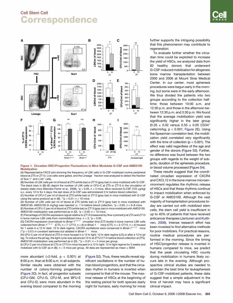
Cell Stem Cell Correspondence Mobilized Hematopoietic Stem Cell Yield Depends on Species-Specific Circadian Timing Daniel Lucas, 1,5 Michela Battista, 1,5 Patricia A. Shi, 1 Luis Isola, 1 and Paul S. Frenette 1,2,3,4, * 1 Department of Medicine 2 Department of Gene and Cell Medicine 3 Black Family Stem Cell Institute 4 Immunology Institute Mount Sinai School of Medicine, New York, NY 10029, USA 5 These authors contributed equally to this work *Correspondence: [email protected] DOI 10.1016/j.stem.2008.09.004 Endogenous rhythmicity likely evolved as a mechanism allowing organisms to antic- ipate predictable daily changes in the en- vironment (Rutter et al., 2002). Under ho- meostasis, murine hematopoietic stem cell (HSC) egress is orchestrated by rhyth- mic b3 adrenergic signals delivered by the sympathetic nervous system (SNS) that regulate Cxcl12 expression in stromal cells (Mendez-Ferrer et al., 2008). Here, we show that CXCR4 is also regulated un- der circadian control whose rhythm is synchronized with its ligand, CXCL12, to optimize HSC trafficking. These circadian oscillations are inverted in humans com- pared to the mouse and continue to influ- ence the yield even when stem cell mobi- lization is enforced. Our results suggest that the human HSC yield for clinical transplantation might be significantly greater if patients were harvested during the evening compared to the morning. The lowest Cxcl12 expression levels in murine bone marrow coincide with the peak of blood HSCs 5 hr after the onset of light (Zeitgeber Time [ZT5]), and the highest Cxcl12 levels 8 hr later (ZT13) match the lowest circulating HSC counts (Mendez-Ferrer et al., 2008). To assess whether the circadian time can affect en- forced mobilization by granulocyte-col- ony-stimulating factor (G-CSF), the most common stem cell mobilizer used in the clinic, we treated C57BL/6 mice for 4 days (Katayama et al., 2006) and assayed the number of circulating colony-forming progenitors and the stem-cell-enriched fraction Lineage Sca1 + c-kit + (LSK) cells at the circadian peak and trough (Fig- ure 1A). At both circadian times, G-CSF in- duced a clear increase in circulating pro- genitors (Figures 1B and 1C). In addition, significantly more progenitors and LSK cells were recovered when the blood col- lection was performed at ZT5 compared to ZT13 (Figures 1B and 1C). To test whether the circadian difference was sus- tained when using a rapid mobilizer, we treated C57BL/6 mice at ZT5 and ZT13 with the CXCR4 antagonist AMD3100 and assayed progenitors 1 hr later (Broxmeyer et al., 2005). We found that the LSK cell counts (Figure 1D) and progenitor yield (Figure 1E) elicited by AMD3100 adminis- tration were significantly higher at ZT5 than ZT13. These results thus suggest that the synchronization of blood collection at the peak circadian time can produce greater HSC recovery. In contrast to the mobilization by G- CSF that reduces CXCL12 synthesis in the bone marrow, the circadian time-de- pendent efficacy of AMD3100 cannot readily be explained by changes in the mi- croenvironment, since the drug targets the CXCR4 receptor on hematopoietic cells. Rhythmic circadian expression of certain signaling molecules and their cor- responding receptors have previously been described. For example, the recep- tors for melatonin (Gauer et al., 1993), cor- tisol (Schlaghecke and Kley, 1986), EGF (Scheving et al., 1989), and brain-derived neurotrophic factor receptor (Dolci et al., 2003) exhibit rhythmic oscillations. We reasoned that CXCR4 expression might also fluctuate on HSCs to modulate CXCL12 signaling. Flow cytometry analy- ses of CXCR4 expression on bone marrow LSK cells (Figure 1F) or CD150 + CD48 stem cells (Kiel et al., 2005) (see Figures S1A and S1B available online) revealed significantly higher CXCR4 expression at ZT13 than at ZT5. CXCR4 fluctuations depended on clock gene expression, since CXCR4 did not exhibit any circadian changes on LSK cells derived from Bmal-1 / mice housed in constant dark- ness (Figure 1G). In addition, AMD3100- induced mobilization was significantly al- tered in mice subjected to a jet lag (defined as a shift of 12 hr in the light cycle; Fig- ure 1H). Moreover, disruption of the light cycle by exposure to constant light re- duced the yield of G-CSF-induced mobili- zation (compare Figures 1C and 1I and see Mendez-Ferrer et al. [2008]) and abro- gated the circadian fluctuations in the yield (Figure 1I). Thus, these results dem- onstrate that, even in situations in which HSC/progenitor egress is pharmacologi- cally enforced, endogenous circadian rhythms controlled by the molecular clock can influence the yield through clock- controlled, synchronized fluctuations of CXCR4 and CXCL12. There are conflicting results about rhythms in circulating progenitors in healthy humans, with one study showing a peak at 9:00 a.m. (Ross et al., 1980) and another report with a peak at 3:00 p.m. (Verma et al., 1980). The internal phase relationships of clock gene expres- sion in the suprachiasmatic nucleus (SCN), relative to the light-dark cycle, are conserved across mammals whether the animals are nocturnal or diurnal (Bjar- nason et al., 2001; Lincoln et al., 2002; Mrosovsky et al., 2001). Clock gene ex- pression and SCN activity appear to be dictated by light input in all mammals. A classic example is melatonin levels in the pineal gland, which peak during the dark- ness period in both nocturnal rodents and diurnal humans (Buijs et al., 2003). To evaluate whether human circulating HSC rhythms, if present, differ from those in mice, we assayed CD34 + CD38 cells and colony-forming progenitor cells in nine healthy individuals (mean age 33.0 ± 1.4 years; six males, three females) at 8:00 a.m. and repeated the analyses on the same day at 8:00 p.m. As shown in Figures 2A–2C, both the CD34 + and CD34 + CD38 cell subsets were clearly 364 Cell Stem Cell 3, October 9, 2008 ª2008 Elsevier Inc.
-
Upload
daniel-lucas -
Category
Documents
-
view
214 -
download
2
Transcript of Mobilized Hematopoietic Stem Cell Yield Depends on Species-Specific Circadian Timing

Cell Stem Cell
Correspondence
Mobilized Hematopoietic Stem Cell YieldDepends on Species-Specific Circadian Timing
Daniel Lucas,1,5 Michela Battista,1,5 Patricia A. Shi,1 Luis Isola,1 and Paul S. Frenette1,2,3,4,*1Department of Medicine2Department of Gene and Cell Medicine3Black Family Stem Cell Institute4Immunology InstituteMount Sinai School of Medicine, New York, NY 10029, USA5These authors contributed equally to this work*Correspondence: [email protected] 10.1016/j.stem.2008.09.004
Endogenous rhythmicity likely evolved as
a mechanism allowing organisms to antic-
ipate predictable daily changes in the en-
vironment (Rutter et al., 2002). Under ho-
meostasis, murine hematopoietic stem
cell (HSC) egress is orchestrated by rhyth-
mic b3 adrenergic signals delivered by the
sympathetic nervous system (SNS) that
regulate Cxcl12 expression in stromal
cells (Mendez-Ferrer et al., 2008). Here,
we show that CXCR4 is also regulated un-
der circadian control whose rhythm is
synchronized with its ligand, CXCL12, to
optimize HSC trafficking. These circadian
oscillations are inverted in humans com-
pared to the mouse and continue to influ-
ence the yield even when stem cell mobi-
lization is enforced. Our results suggest
that the human HSC yield for clinical
transplantation might be significantly
greater if patients were harvested during
the evening compared to the morning.
The lowest Cxcl12 expression levels in
murine bone marrow coincide with the
peak of blood HSCs 5 hr after the onset
of light (Zeitgeber Time [ZT5]), and the
highest Cxcl12 levels 8 hr later (ZT13)
match the lowest circulating HSC counts
(Mendez-Ferrer et al., 2008). To assess
whether the circadian time can affect en-
forced mobilization by granulocyte-col-
ony-stimulating factor (G-CSF), the most
common stem cell mobilizer used in the
clinic, we treated C57BL/6 mice for
4 days (Katayamaetal., 2006) and assayed
the number of circulating colony-forming
progenitors and the stem-cell-enriched
fraction Lineage� Sca1+ c-kit+ (LSK) cells
at the circadian peak and trough (Fig-
ure 1A). At both circadian times, G-CSF in-
duced a clear increase in circulating pro-
genitors (Figures 1B and 1C). In addition,
significantly more progenitors and LSK
cells were recovered when the blood col-
lection was performed at ZT5 compared
364 Cell Stem Cell 3, October 9, 2008 ª2008
to ZT13 (Figures 1B and 1C). To test
whether the circadian difference was sus-
tained when using a rapid mobilizer, we
treated C57BL/6 mice at ZT5 and ZT13
with the CXCR4 antagonist AMD3100 and
assayed progenitors 1 hr later (Broxmeyer
et al., 2005). We found that the LSK cell
counts (Figure 1D) and progenitor yield
(Figure 1E) elicited by AMD3100 adminis-
tration were significantly higher at ZT5
than ZT13. These results thus suggest
that the synchronization of blood collection
at the peak circadian time can produce
greater HSC recovery.
In contrast to the mobilization by G-
CSF that reduces CXCL12 synthesis in
the bone marrow, the circadian time-de-
pendent efficacy of AMD3100 cannot
readily be explained by changes in the mi-
croenvironment, since the drug targets
the CXCR4 receptor on hematopoietic
cells. Rhythmic circadian expression of
certain signaling molecules and their cor-
responding receptors have previously
been described. For example, the recep-
tors for melatonin (Gauer et al., 1993), cor-
tisol (Schlaghecke and Kley, 1986), EGF
(Scheving et al., 1989), and brain-derived
neurotrophic factor receptor (Dolci et al.,
2003) exhibit rhythmic oscillations. We
reasoned that CXCR4 expression might
also fluctuate on HSCs to modulate
CXCL12 signaling. Flow cytometry analy-
ses of CXCR4 expression on bone marrow
LSK cells (Figure 1F) or CD150+CD48�
stem cells (Kiel et al., 2005) (see Figures
S1A and S1B available online) revealed
significantly higher CXCR4 expression at
ZT13 than at ZT5. CXCR4 fluctuations
depended on clock gene expression,
since CXCR4 did not exhibit any circadian
changes on LSK cells derived from
Bmal-1�/�mice housed in constant dark-
ness (Figure 1G). In addition, AMD3100-
induced mobilization was significantly al-
Elsevier Inc.
tered in mice subjected to a jet lag (defined
as a shift of 12 hr in the light cycle; Fig-
ure 1H). Moreover, disruption of the light
cycle by exposure to constant light re-
duced the yield of G-CSF-induced mobili-
zation (compare Figures 1C and 1I and see
Mendez-Ferrer et al. [2008]) and abro-
gated the circadian fluctuations in the
yield (Figure 1I). Thus, these results dem-
onstrate that, even in situations in which
HSC/progenitor egress is pharmacologi-
cally enforced, endogenous circadian
rhythms controlled by the molecular clock
can influence the yield through clock-
controlled, synchronized fluctuations of
CXCR4 and CXCL12.
There are conflicting results about
rhythms in circulating progenitors in
healthy humans, with one study showing
a peak at 9:00 a.m. (Ross et al., 1980)
and another report with a peak at
3:00 p.m. (Verma et al., 1980). The internal
phase relationships of clock gene expres-
sion in the suprachiasmatic nucleus
(SCN), relative to the light-dark cycle,
are conserved across mammals whether
the animals are nocturnal or diurnal (Bjar-
nason et al., 2001; Lincoln et al., 2002;
Mrosovsky et al., 2001). Clock gene ex-
pression and SCN activity appear to be
dictated by light input in all mammals. A
classic example is melatonin levels in the
pineal gland, which peak during the dark-
ness period in both nocturnal rodents and
diurnal humans (Buijs et al., 2003). To
evaluate whether human circulating HSC
rhythms, if present, differ from those in
mice, we assayed CD34+CD38� cells
and colony-forming progenitor cells in
nine healthy individuals (mean age 33.0 ±
1.4 years; six males, three females) at
8:00 a.m. and repeated the analyses on
the same day at 8:00 p.m. As shown in
Figures 2A–2C, both the CD34+ and
CD34+CD38� cell subsets were clearly
Cell Stem Cell
Correspondence
more abundant (>2-fold, p < 0.001) at
8:00 p.m. than at 8:00 a.m. in all subjects.
Similar results were obtained with the
number of colony-forming progenitors
(Figure 2D). In fact, all progenitor subsets
(CFU-GM, CFU-G, CFU-M, and BFU-E
and CFU-E) were more abundant in the
evening blood compared to the morning
(Figure S2). Thus, these results reveal sig-
nificant oscillations in the number of hu-
man blood progenitors, and that the circa-
dian rhythm in humans is inverted when
compared to that of the mouse. The max-
imal release of HSCs at the beginning of
the resting period for both species (early
night for humans, early morning for mice)
Figure 1. Circadian HSC/Progenitor Fluctuations in Mice Modulate G-CSF and AMD3100Mobilization(A) Representative FACS plot showing the frequency of LSK cells in G-CSF-mobilized murine peripheralblood at ZT5 or ZT13. Live cells were gated, and the Lineage� fraction was analyzed to detect the fractionof Sca-1+ and c-kit+ cells.(B) Number of LSK cells per ml of blood at ZT5 (white bar) or ZT13 (gray bar) in mice mobilized with G-CSF.The black bars in (B)–(E) depict the number of LSK cells or CFU-C at ZT5 or ZT13 in the circulation ofsteady-state mice (Mendez-Ferrer et al., 2008). *p < 0.05, n = 5 mice. Mice received G-CSF (125 mg/kg)s.c. every 12 hr for 4 days; the last dose of G-CSF was administered 3 hr before blood collection.(C) Number of CFU-C per ml of blood at ZT5 (white bar) or ZT13 (gray bar) in mice mobilized with G-CSFusing the same protocol as in (B). **p < 0.01; n = 10 mice.(D) Number of LSK cells per ml of blood at ZT5 (white bar) or ZT13 (gray bar) in mice mobilized withAMD3100. AMD3100 (5 mg/kg) was injected i.c. 1 hr before blood collection. *p < 0.05; n = 6–8 mice.(E) Number of CFU-C per ml of blood at ZT5 (white bar) or ZT13 (gray bar) in mice mobilized with AMD3100.AMD3100 mobilization was performed as in (D). *p < 0.05; n = 10 mice.(F) Percentage of CXCR4 expression signal relative to ZT13 (measured by flow cytometry) at ZT5 and ZT13in bone marrow LSK cells from nonmobilized mice. n = 3; *p < 0.05.(G) CXCR4 expression (normalized to Bmal-1+/+,+/� circadian time [CT] levels) in bone marrow LSK cellscollected from Bmal-1+/+,+/� (CT5, n = 7; CT13, n = 6) or Bmal-1�/�mice (CT5, n = 3; CT13, n = 4) housedfor 1 week in a 12 hr dark: 12 hr dark regime. CXCR4 oscillations were conserved in Bmal-1+/+,+/� mice(**p < 0.01) in constant darkness but ablated in Bmal-1�/� mice.(H) CFU-C per ml of blood at ZT5 in mice housed in a 12 hr light: 12 hr dark regime (LD) or after a 12 hr jetlag. To induce the jet lag, the light cycle was advanced 12 hr at ZT12 (17 hr before blood collection at ZT5).AMD3100 mobilization was performed as in (D). **p < 0.01; n = 5 mice per group.(I) CFU-C per ml of blood at CT5 or CT13 in mice housed in a 12 hr light: 12 hr light regime for 3 weeks andmobilized with G-CSF as in (B). n = 5 mice per group. All data are represented as mean ± SEM.
Cell Stem Cell
further supports the intriguing possibility
that this phenomenon may contribute to
regeneration.
To evaluate further whether the circa-
dian time could be exploited to increase
the yield of HSCs, we analyzed data from
82 healthy donors that underwent
G-CSF-induced mobilization for allogeneic
bone marrow transplantation between
2000 and 2006 at Mount Sinai Medical
Center. In our center, most apheresis
procedures were begun early in the morn-
ing, but some were in the early afternoon.
We thus divided the patients into two
groups according to the collection half-
time: those between 10:00 a.m. and
12:30 p.m. and those in the afternoon be-
tween 12:30 p.m. and 3:30 p.m. We found
that the average mobilization yield was
significantly higher in the later group
(0.35 ± 0.02 versus 0.55 ± 0.05 CD34+
cells/ml/kg; p < 0.001; Figure 2E). Using
the Spearman correlation test, the mobili-
zation yield correlated very significantly
with the time of collection (p < 0.001). The
effect was valid regardless of the age and
gender of the donors (Figure S3). Further,
no difference was found between the two
groups with regards to the weight of sub-
jects, duration of the apheresis procedure,
or blood volume processed (Figure S4).
These results suggest that the coordi-
nated circadian expression of CXCR4
and CXCL12 in the bone marrow microen-
vironment regulates the rhythmic release
of HSCs and that these rhythms continue
to impact mobilization when enforced by
G-CSF or AMD3100. Although the vast
majority of transplantation procedures to-
day are carried out with mobilized stem
cells, the stem cell yield is insufficient in
up to 40% of patients that have received
anticancer therapies (Jantunen and Kuitti-
nen, 2008). Intense research effort has
been invested to find alternative methods
for poor mobilizers. For practical reasons,
routine medical procedures are per-
formed in the morning. Since the timing
of HSC/progenitor release is inverted in
humans compared to mice, we predict
that the peak circulating HSC counts
during mobilization in humans likely oc-
curs late in the evening. Although pro-
spective clinical studies are needed to
ascertain the best time for leukapheresis
of G-CSF-mobilized patients, these data
suggest that a simple adjustment in the
time of harvest may have a significant
clinical impact.
3, October 9, 2008 ª2008 Elsevier Inc. 365

Cell Stem Cell
Correspondence
SUPPLEMENTAL DATA
The Supplemental Data include Supplemental Ex-perimental Procedures and four figures and canbe found with this article online at http://www.cellstemcell.com/cgi/content/full/3/4/364/DC1/.
ACKNOWLEDGMENTS
We thank Chris Bradfield for providing breedingpairs of Bmal-1+/� mice. This work was supportedby the National Institutes of Health (DK056638).P.S.F. is an Established Investigator of the Ameri-
Figure 2. Circadian Fluctuations of Human Progenitors in Peripheral Blood(A) Representative peripheral blood FACS analysis from healthy subjects to detect CD34+ CD38� cells inperipheral blood collected at 8:00 a.m. and 8:00 p.m. Cells were first gated on the CD34+ populations andthen analyzed for the presence of CD38� cells. Average number of CD34+ cells (B) and CD34+CD38� cells(C) per ml of blood at indicated times; n = 9; ***p < 0.001. (D) Total colony-forming units in culture (CFU-C)detected in the peripheral blood from same donors; ***p < 0.001. (E) Number of CD34+ cells recovered perblood volume processed (l) per weight (kg) of healthy donors that were mobilized using G-CSF (10 mg/kgevery morning for 5 days) for allogeneic bone marrow transplantation at Mount Sinai Medical Center be-tween 2001 and 2006. Subjects were grouped according to the apheresis half-time. ***p < 0.001.
366 Cell Stem Cell 3, October 9, 2008 ª2008 Elsevier Inc.
can Heart Association. D.L. and M.B. are sup-ported by fellowships from the Fondacion RamonAeceres and the Cooley’s Anemia Foundation,respectively.
REFERENCES
Bjarnason, G.A., Jordan, R.C., Wood, P.A., Li, Q.,Lincoln, D.W., Sothern, R.B., Hrushesky, W.J.,and Ben-David, Y. (2001). Am. J. Pathol. 158,1793–1801.
Broxmeyer, H.E., Orschell, C.M., Clapp, D.W.,Hangoc, G., Cooper, S., Plett, P.A., Liles, W.C.,Li, X., Graham-Evans, B., Campbell, T.B., et al.(2005). J. Exp. Med. 201, 1307–1318.
Buijs, R.M., van Eden, C.G., Goncharuk, V.D., andKalsbeek, A. (2003). J. Endocrinol. 177, 17–26.
Dolci, C., Montaruli, A., Roveda, E., Barajon, I., Viz-zotto, L., Grassi Zucconi, G., and Carandente, F.(2003). Brain Res. 994, 67–72.
Gauer, F., Masson-Pevet, M., Skene, D.J., Vivien-Roels, B., and Pevet, P. (1993). Neuroendocrinol-ogy 57, 120–126.
Jantunen, E., and Kuittinen, T. (2008). Eur. J. Hae-matol. 80, 287–295.
Katayama, Y., Battista, M., Kao, W.M., Hidalgo, A.,Peired, A.J., Thomas, S.A., and Frenette, P.S.(2006). Cell 124, 407–421.
Kiel, M.J., Yilmaz, O.H., Iwashita, T., Yilmaz, O.H.,Terhorst, C., and Morrison, S.J. (2005). Cell 121,1109–1121.
Lincoln, G., Messager, S., Andersson, H., and Ha-zlerigg, D. (2002). Proc. Natl. Acad. Sci. USA 99,13890–13895.
Mendez-Ferrer, S., Lucas, D., Battista, M., andFrenette, P.S. (2008). Nature 452, 442–447.
Mrosovsky, N., Edelstein, K., Hastings, M.H., andMaywood, E.S. (2001). J. Biol. Rhythms 16, 471–478.
Ross, D.D., Pollak, A., Akman, S.A., and Bachur,N.R. (1980). Exp. Hematol. 8, 954–960.
Rutter, J., Reick, M., and McKnight, S.L. (2002).Annu. Rev. Biochem. 71, 307–331.
Scheving, L.A., Tsai, T.H., Cornett, L.E., Feuers,R.J., and Scheving, L.E. (1989). Anat. Rec. 224,459–465.
Schlaghecke, R., and Kley, H.K. (1986). Steroids47, 287–294.
Verma, D.S., Fisher, R., Spitzer, G., Zander, A.R.,McCredie, K.B., and Dicke, K.A. (1980). Am. J.Hematol. 9, 185–192.