
MMIC Risk Management Department MMIC • MINIMIZING OBSTETRICAL RISK PAGE 89 With permission of...
90
2007 MMIC • M INIMIZING O BSTETRICAL R ISK With permission of Physicians Insurance, a Mutual Company, MMIC has reproduced this manual for all of its physicians practicing obstetrics. Review of the manual and minor edits have been made by an obstetrician practicing in Minnesota. MMIC Risk Management Department Partners in Patient Safety promoting safety – minimizing risk
Transcript of MMIC Risk Management Department MMIC • MINIMIZING OBSTETRICAL RISK PAGE 89 With permission of...

2 0 0 7 M M I C • M I N I M I Z I N G O B S T E T R I C A L R I S K PAGE 89
With permission of Physicians Insurance, a Mutual Company, MMIC has reproduced this manual for all of its physicians practicing obstetrics. Review of the manual and minor edits have been made by an obstetrician practicing in Minnesota.
MMIC Risk Management Department
Partners in Patient Safetypromoting safety – minimizing risk

2 0 0 7 M M I C • M I N I M I Z I N G O B S T E T R I C A L R I S K PAGE 89

2 0 0 7 M M I C • M I N I M I Z I N G O B S T E T R I C A L R I S K PAGE 88
Introduction ...............................................................................................................................1
Shoulder Dystocia ......................................................................................................................3
Delayed Performance of Cesarean Section .............................................................................13
Vaginal Birth After Cesarean Section ......................................................................................15
Postdating ................................................................................................................................23
Examination of the Placenta ....................................................................................................27
Neonatal Resuscitation ............................................................................................................35
The Morbidity & Mortality Conference .....................................................................................47
Communication ........................................................................................................................49
Preterm Labor ..........................................................................................................................55
Fetal Monitoring .......................................................................................................................61
Pregnancy-Induced Hypertension ...........................................................................................67
Prevention of Perinatal Group B Beta Strep ............................................................................71
The Neurologically Impaired Neonate ......................................................................................75
Neonatal Encephalopathy & Cerebral Palsy ............................................................................79
Perinatal Genetic Testing .........................................................................................................83
Contents

2 0 0 7 M M I C • M I N I M I Z I N G O B S T E T R I C A L R I S K PAGE 89

2 0 0 7 M M I C • M I N I M I Z I N G O B S T E T R I C A L R I S K PAGE 1
Introduction
It is a paradox of medicine that the greater the improvement, the greater the potential for er-ror. Advances in technology and clinical practice increase the chance that something vital will be overlooked. The combination of increased expec-tations and increased potential for error can be disastrous for obstetrical practitioners. Throughout the country, pregnancy and birth-related claims top the list in indemnity expense. It is not sur-prising that malpractice losses have driven many physicians from the practice of obstetrics. The personal toll on practitioners involved in a mal-practice suit, along with the financial risks, makes this field among the riskiest in medicine today.
While some untoward outcomes cannot be pre-vented even with exemplary care under the best of circumstances, a significant number of malpractice cases are indefensible, the result of poor judgment and systems problems. The majority of claims relate to:
◆ Physician-caused injury
◆ Weak documentation of complex deliveries
◆ Poor physician rapport with the patient
◆ Incomplete test reporting and follow-up
That’s the bad news.
The good news is that the above liability areas respond effectively to the simple tools of risk management. You can take steps today to improve your clinical skills, documentation, and rapport, and to close any gaps in your test reporting and follow-up systems.
Minimizing Obstetrical Risk is a preventive guide to situations that have caused physicians the most trouble. It includes tools and suggestions for re-ducing bad outcomes and malpractice claims.
The Obstetrical Task Force
As a local company owned and directed by its members, Physicians Insurance is dedicated to its mission of improving the quality of medical care and reducing adverse outcomes.
In response to the alarming number of obstetrical claims, the Risk Management Department con-vened a task force to identify and address issues that affect fetal outcome. Members of the task force represent a variety of practice disciplines, settings, and locations throughout Washington State. Individual members in turn consult with colleagues in their specialty organizations to present the most current approaches to optimal management of obstetrical patients.
With permission of Physicians Insurance, a Mutual Company, MMIC has reproduced this manual for all of its physicians practicing obstet-rics. Review of the manual and minor edits have been made by an obstetrician practicing in Minnesota.
Clinical Members, Obstetrical Task Force
Robert Jeffers, MD—ChairObstetrics and Gynecology, Edmonds
Donald Barford, MDPerinatology, Everett
Edith Cheng, MDObstetrics and GynecologyUniversity of Washington, Seattle
Jan Delli-Bovi, MDObstetrics and GynecologyMount Vernon
Thomas Easterling, MDObstetrics and GynecologyUniversity of Washington, Seattle
2 0 0 7 M M I C • M I N I M I Z I N G O B S T E T R I C A L R I S K PAGE 2
Corinne Fligner, MDPathology, University of Washington Seattle
Linda Gromko, MDFamily Practice, Seattle
Robert Hartman, MDObstetrics and Gynecology, Spokane
Rita Hsu, MDObstetrics and Gynecology, Wenatchee
Robert Justus, MDFamily Practice, Omak
David Luthy, MDPerinatology, Swedish Medical Center Seattle
Nancy O’Brien-Abel, RNC, MNPerinatology, Seattle
Gail Pinczower, MDAnesthesiology, Kirkland
Catherine Rogers, MDObstetrics and Gynecology, Edmonds
Theodore Rudd, MDObstetrics and Gynecology, Yakima
Susan Williams-Judge, RN, MNPerinatology, Edmonds
Cheryl Wright-Wilson, MDPediatrics, Kirkland
About This Book
The recommendations in this guidebook are not intended to establish a standard of care, nor are they a substitute for legal advice. The fact that a physician’s practice varies from these guidelines does not itself establish that the physician failed to meet the required standard of care. Compliance with these guidelines, however, may reduce the
risk of facing a lawsuit and the stress that accompa-nies even a successful defense in court. The guiding principle should be your best clinical judgment and a medical record that clearly reflects that judgment at each decision point.
We encourage you to add your own practice aids to this book, and to keep it in a place where you and your staff can refer to it often. The Dystocia Flash Cards and the HELPER flyer are intended to be placed in a prominent location. Keep the flash cards in a lounge where they can be reviewed over a cup of coffee. Copy the flyer for posting on the back of a door or on a tabletop. The more familiar you are with high-risk procedures, the more likely you will recall them in an emergency.
Finally, if you know or learn of a practice tool that would benefit your colleagues in one of the areas addressed by this guidebook, let us hear from you! Minimizing Obstetrical Risk is intended as a living reference that will draw from the collective experi-ence of the obstetrical community.
2 0 0 7 M M I C • M I N I M I Z I N G O B S T E T R I C A L R I S K PAGE 3
Introduction
Because shoulder dystocia can ambush even the most experienced practitioners, a well-conceived plan of action is essential to help prevent complica-tions that could lead to brain damage, Erb’s palsy, or death. The text that follows outlines strategies and guidelines in the event of a brachial plexus injury. Also included are:
• HELPER for Shoulder Dystocia Flyer
We recommend that you copy and share the enclosed 8 1/2" x 11" flyer with your colleagues and nurses on the Labor and Delivery unit. Place one in every deliv-ery room; affix one on the side of a monitor, inside a cabinet, or on a tabletop.
An 11" x 17" version of the flyer is available for physician and nurse lounges and restrooms (both good places for repetitive study). Please contact Physicians Insurance for copies.
Remember, even with a bad outcome such as a permanent brachial plexus injury, you will most likely have a defensible case if you can show that you went through the correct steps to manage it.
• Shoulder Dystocia Flash Cards
In the heat of the moment, you are more likely to recall the recommended steps for dealing with a shoulder dystocia if you have periodically reviewed the flash cards during breaks in your schedule.
Since this problem may present unexpectedly, it is wise to give yourself the benefit of ongoing review of the management techniques. Oc-casionally reviewing and practicing ALL of the steps, perhaps in conjunction with an easily resolved shoulder dystocia, or even during a routine delivery, should enable the practitioner to maintain a high level of readiness.
• Shoulder Dystocia Dictation Guide/Chart Form Sample
The most important step to take following a shoul-der dystocia is to adequately and accurately docu-ment what occurred, including which shoulder was impacted. Careful documentation frequently will dissuade even the most aggressive plaintiff’s attor-ney from pursuing legal action.
To ensure that your records are comprehensive, we’ve included a format for covering significant information in the event of a shoulder dystocia. Follow this guide in completing your dictated note or writing out your documentation. It may also be incorporated into the hospital chart.
• Forceps/Vacuum Dictation Guide/Preoperative Note
Use this form as a dictating guide, a guide for your written note, or a record for the chart.
Prevention
Shoulder dystocia does occur unexpectedly. The risk of incurring a permanent injury can be diminished by appropriate labor management and skilled maneuvers at the time of dystocia. However, the most prudent care and most careful delivery will not prevent all brachial plexus injuries. Even the best of practitioners may perform a delivery associated with permanent injury. A comprehensive program of risk reduction should help the practitioner reduce the risk of dysto-cia and injury for each delivery and simultaneously build a defensible case when an injury is experienced.
Risk Factors
Three clinical factors are associated with the risk of shoulder dystocia:
1. Macrosomia
2. Abnormal Labor
3. Operative Vaginal Delivery
Shoulder Dystocia
2 0 0 7 M M I C • M I N I M I Z I N G O B S T E T R I C A L R I S K PAGE 4
Macrosomia
The practitioner should routinely estimate fetal weight and record the estimated fetal weight for all women in labor. Diabetes and postdate preg-nancies are at particular risk for macrosomia. Documentation of clinical estimation of fetal weight is useful. While you may occasionally in-clude ultrasound to help estimate fetal weight, this method is not necessarily any more accurate than the clinical estimate.
Labor associated with an estimated fetal weight ≥4000 g should be considered at risk for shoul-der dystocia. The risk for permanent injury of a diabetic fetus ≥4000 g or nondiabetic fetus ≥4500 g may approach 1%. In these cases, discuss the op-tion of primary cesarean section with the patient, and document the discussion. In nondiabetic fetuses ≥4000 g but <4500 g, the risk of shoulder dystocia increases but the risk of permanent injury is less than 1%.
The first step in managing the risks of shoulder dystocia and permanent injury is to estimate fetal weight in all labors and document that estimation.
Abnormal Labor
Deviations from normal labor patterns and/or the presence of estimated fetal weight ≥4000 g should alert the clinician to the possibility of dystocia and permanent injury. The use of Friedman’s Labor Curve will help the practitioner prospectively iden-tify the abnormal labor.
When macrosomia is suspected, an abnormal labor should heighten the practitioner’s awareness of potential shoulder dystocia.
Operative Vaginal Delivery
Delivery by forceps or vacuum from +2 (ACOG criteria 0–+5) or higher station increases the risk
of shoulder dystocia. When macrosomia and an abnormal labor are associated with such deliver-ies, the risk of permanent injury approaches 1%.* Use particular care in assessing station. The prac-titioner should be certain that the skull, rather than just the caput, is at an adequate station.
Avoid the combination of macrosomia, abnor-mal labor, and operative vaginal delivery when possible. Except in emergent clinical circum-stances, the practitioner has discretion over addi-tion of the third risk, operative vaginal delivery.
Patient Involvement
As in all clinical situations, patients should be partners in decision making. When risks are iden-tified, inform patients of options and the nature and magnitude of the risks, as in the following example:
“Ms. Jones, I suspect you may be carrying a larger-than-average baby. Sometimes with larger babies, delivery of the shoulders after the head is deliv-ered can be difficult and, in rare circumstances, can result in permanent injury. If your labor is abnormal or if you are unable to push your baby out without my help, we may need to perform a cesarean section or try some other techniques to help deliver the baby.”
Whenever we are faced with difficult decisions, patients should be informed of the options. When risks are relatively balanced between two alternatives, a patient’s willingness to accept one type of risk rather than another is an important consideration in the final choice of action. The court case Villaneuva v. Harrington, 906 P.2d 374, 376 (Wash. 1995), established that under the law of informed consent, patients must be told of the risks and alternatives of an operative delivery. Documentation of discussions with patients is essential.

2 0 0 7 M M I C • M I N I M I Z I N G O B S T E T R I C A L R I S K PAGE 5
Example:
The risks of shoulder dystocia with a large baby were discussed with Ms. Jones and her partner. They would like to proceed with labor as long as the course of labor remains normal. Procedures, alternatives, benefits, and risks of operative delivery discussed with patient. Patient agrees to operative delivery as necessary.
or
The risks of shoulder dystocia associated with a large baby were discussed with Ms. Jones and her partner. Her sister had a baby with a permanent brachial plexus in-jury. Ms. Jones would rather accept the risks of cesarean section than a risk to her baby.
Reference
* Benedetti TJ. Shoulder dystocia. Contemp Ob Gyn. 1995;40:39–43.
The Obstetrical Task Force and Physicians Insurance wish to acknowledge the contribution of Thomas J. Benedetti, M.D., to this chapter.

2 0 0 7 M M I C • M I N I M I Z I N G O B S T E T R I C A L R I S K PAGE 6
A Guideline for Physicians
The impulse to deny the problem and avoid the family can be strong after a less than optimal out-come. Here are some guidelines for contact with the involved family after a brachial plexus injury.
In The Hospital
1. Sit on the bed or in a chair close to the bed when you are talking with the new mother and her family.
2. Maintain eye contact with the mother and family members.
3. Review the sequence of events at delivery: what was done, why it was done.
4. Provide facts:
◆ Of every 1,000 babies born, 15 to 60 deliveries may be complicated by a shoulder dystocia.
◆ Although shoulder dystocia is more com-monly associated with large babies, it can occur with babies of normal to small size.
◆ 80% of babies with a brachial plexus injury have complete resolution of the problem within one year.
5. Allow the family to know your concern and that you will be there for them.
6. Ask if there is a time the mother wants you to return to explain details to other family members.
7. Contact your malpractice insurer for advice.
After Hospital Discharge
1. Call the family in one week and again in one month (more often is fine).
2. If you sense there is a family member or friend who is angry with you, ask permission from the patient to call that individual. Invite him or her to your office. Let the family know you are not afraid.
3. If the baby is transferred and it is possible for you, visit the NICU when the baby’s parents are going to be there.
Remember to keep in touch with patients whose babies have a brachial plexus injury. Letting them know you care can make a big difference.
After A Brachial Plexus Injury Has Occurred

2 0 0 7 M M I C • M I N I M I Z I N G O B S T E T R I C A L R I S K PAGE 7
Shoulder Dystocia
Anterior Shoulder R LEpisiotomy? 2nd 3rd 4th
Time Head Delivered: __________________
Time Delivery Completed: _________________
Total Elapsed Time: ______________________
Maneuvers:
McRoberts ■■ Suprapubic Pressure ■■ Shoulder Rotation ■■ (Wood’s or Ruben’s) Posterior Arm ■■ Replacement of Head ■■ for C-section
Neonate
Weight:___________Apgars: _____1 min _____ 5 min ____ 10 min
CORD: Venous pH ______ Arterial pH ________
GASES: __________ pCO2 pCO
2 ____________
pO2 _____________ pO
2 ______________
HCO3 ___________ HCO
3 ____________
BE ______________ BE ______________
Moro Reflex: Symmetrical? Yes ■■ No ■■ R L Impaired
Required admission to special care nursery? Yes ■■ No ■■
Narrative:
Transfer? Yes ■■ No ■■
Shoulder Dystocia Dictation Guide/Chart Form Sample
Date: Time:
Attending Physician:
Assistant:
Pregnancy Complications: Labor Complications:
Diabetes (insulin) ■■ Protracted Active Phase ■■ Gestational Diabetes ■■ Secondary Arrest of First Stage ■■ Postdates ■■ Prolonged Second Stage ■■
Delivery Mode:
Spontaneous ■ ■ Vacuum ■ ■ Forceps ■■
OUTLET ■■ Position (please circle): ROA OA LOA ROT LOT ROP OP LOP
Station: Scalp Visible ■ ■ Skull at Pelvic Floor ■■ Present?: Labial Separation ■ ■ Dilating Rectum ■■ Bulging Perineum ■■
LOW ■ ■ Position (please circle): ROA OA LOA ROT LOT ROP OP LOP
Station ≥+2 (of 5) ■■
, M.D.

2 0 0 7 M M I C • M I N I M I Z I N G O B S T E T R I C A L R I S K PAGE 8
This page left intentionally blank.

2 0 0 7 M M I C • M I N I M I Z I N G O B S T E T R I C A L R I S K PAGE 9
Forceps/Vacuum Dictation Guide/Operative Note
Date: Time:
Delivery Attendants:
Pregnancy Complications Labor Complications
Diabetes (Insulin) ■■ Protracted Active Phase ■■
Gestational Diabetes ■■ Secondary Arrest of Labor ■■
Postdates ■■ Prolonged Second Stage ■■
Indication
Fetal Distress
Specify ____________________________________________________________
Prolonged Second Stage
Specify ____________________________________________________________
Maternal Disease
Specify ____________________________________________________________
Delivery Mode:
Vacuum ■■ Forceps ■■
Outlet ■■ Position (please circle): ROA OA LOA ROT LOT ROP OP LOP
Station: Scalp Visible ■ ■ Skull at Pelvic Floor ■■ Present?: Labial Separation ■■ Dilating Rectum ■■ Bulging Perineum ■■
Low ■■ Position (please circle): ROA OA LOA ROT LOT ROP OP LOP
Station ≥+2 (of 5) ■ ■
Apgars: ______________ 1 min _________________ 5 min _________________10 min
Required admission to special care nursery? Yes ■■ No ■■
Narrative:
, M.D.

2 0 0 7 M M I C • M I N I M I Z I N G O B S T E T R I C A L R I S K PAGE 10
This page left intentionally blank.

2 0 0 7 M M I C • M I N I M I Z I N G O B S T E T R I C A L R I S K PAGE 11
H E
L P
E R
For
Sho
ulde
r Dys
tocia
HE
L
P
ER
• H
ave
RN
pus
h di
gita
l clo
ck a
nd c
all o
ut ti
me
inte
rval
• Av
oid
exce
ssiv
e fo
rce
• D
ocum
ent w
hich
sho
ulde
r was
impa
cted

2 0 0 7 M M I C • M I N I M I Z I N G O B S T E T R I C A L R I S K PAGE 12
This page left intentionally blank.
2 0 0 7 M M I C • M I N I M I Z I N G O B S T E T R I C A L R I S K PAGE 13
Removing the Barriers to the Timely Performance of Cesarean Section
Our review of the company’s claims data revealed that the delayed performance of cesarean section is a recurrent theme. In some situations, it was obvious the delay contributed to an unfavorable outcome. In other cases, a causal relationship was questionable but the indefensibility of these cases was attributed to a deviation from The American College of Obstetrics and Gynecology’s established guidelines. Current guidelines require that a standard obstetric unit be able to initiate a cesar-ean section within 30 minutes from the time of decision to operate. It is well known, however, that catastrophic situations such as uterine rupture, hemorrhage from placenta previa, abruptio pla-centae, and prolapse of the umbilical cord require delivery in less than the 30-minute time frame. Physicians at facilities unable to implement a more expeditious cesarean section should advise patients of this limitation. A consent form, as well as docu-mentation of the discussion, should be included in the chart. Barriers to the timely performance of cesarean section are:
◆ Unavailability of physician
◆ Poor nurse-physician communication
◆ Reluctance to seek consultation
◆ Lack of staff preparedness
◆ External pressure to decrease the incidence of cesarean section
The highly reliable labor and delivery unit has established policies and procedures that substan-tially reduce or eliminate these barriers.
Physician Availability
Institutional evidence suggests that continuous, on-site physician supervision of the laboring patient decreases the incidence of unfavorable events. A team approach, with shared responsibility for having a physician present during active labor, is possible even in smaller communities comprised of solo practices.
Nurse-Physician Communication
In the highly charged environment of the obstetric unit, good communication between nurses and physicians is vital. Face-to-face contact is preferable to telephone consultation when there is any concern about the labor or delivery. Because the stakes are so high, ANY question of maternal or fetal well-be-ing should alert the physician on call to proceed to the hospital.
One effective vehicle for promoting a working-team approach is the physician-nurse liaison committee. We strongly recommend regular meetings of the two groups on all obstetric units. Meetings can provide a forum to air differences, resolve disputes, and dis-cuss cases. They should involve all unit members or designated representatives from each group, includ-ing a neonatal nurse or pediatrician.
An alternative to the physician-nurse liaison com-mittee is a morbidity and mortality conference in which physicians (including pediatricians/neona-tologists) and nurses participate regularly in case discussions and examination of common issues.
Increasing Consultation
The higher the quality of collaboration among physi-cians on difficult cases, the more likely the right decisions will be made. Healthy working relation-ships between physicians can set a positive tone for the entire department. Yet professional jealousies
Delayed Performance of Cesarean Section
2 0 0 7 M M I C • M I N I M I Z I N G O B S T E T R I C A L R I S K PAGE 14
and inter-specialty competition often impede a collaborative climate. The increasing focus on cost control creates an additional challenge to deliver optimal care. There is therefore a greater impera-tive for physicians to consciously resolve to com-municate with one another.
Staff Preparedness
Does your obstetric unit carry out a policy of regu-lar practice drills for the staff? In the highly reliable organization, personnel from labor and delivery, newborn nursery, and anesthesia services are familiar with the entire cesarean-section procedure. They keep the operating room in a constant state of readiness—with appropriate supplies, anesthesia equipment, and surgical instruments. Regular drills minimize confusion during emergencies, probably resulting in more favorable outcomes.
Obstetric services should anticipate the need for anesthesia services whenever the remotest pos-sibility of a cesarean section arises. Develop a high index of suspicion, and establish clear protocols for the early notification of the on-call anesthesi-ologist.
References
1. Standards for Obstetric-Gynecologic Services. 7th ed. Washington, DC: American College of Obstetrics and Gynecologists; 1989.
2. Shauberger CW, Rooney BL, Beguin EA, Schaper AM, Spindler J. Evaluating the thirty minute interval in emergency cesarean sec-tions. J Am Coll Surg. 1994;179(2):151–155.
3. Korhonen J, Kariniemi V. Emergency cesarean section: the effect of delay on umbilical arte-rial gas balance and Apgar scores. Acta Obstet Gynecol Scand. 1994;73(10):782–786.
4. Guidelines for Perinatal Care. 5th ed. Wash-ington, DC: American Academy of Pediat-rics/American College of Obstetricians and Gynecologists; 2002.
2 0 0 7 M M I C • M I N I M I Z I N G O B S T E T R I C A L R I S K PAGE 15
Guidelines for candidate selection and management
The old adage “once a cesarean section, always a cesarean section” has gradually changed, with increasing experience and support for vaginal delivery after prior cesarean section. With ris-ing cesarean-section rates in the late 1980s and repeat cesarean section comprising one-third of all cesarean deliveries, greater employment of vaginal birth after cesarean section (VBAC) was encour-aged and helped to stabilize cesarean-section rates in the 1990s.
VBAC, when successful, is associated with many maternal benefits: shorter hospital stays, fewer transfusions, and a decreased incidence of post-partum fever. However, recent data suggest the maternal safety of elective cesarean section is roughly equivalent to that of vaginal birth. Unsuccessful trial of labor (TOL), followed by unscheduled cesarean section, is associated with greater risks of morbidity, infection, and major complications than elective repeat cesarean sec-tion. Infant infection rates are higher after an unsuccessful trial of labor.
Uterine rupture is the most serious risk associ-ated with VBAC. Uterine-rupture risk may be as much as 10 times greater in a trial of labor than with elective repeat cesarean section. Though the published rate of uterine rupture is approximately 0.8%, this event may be catastrophic for mother and baby.
As the utilization of VBAC has become more widespread, catastrophic consequences have in-creased and, correspondingly, so have the number of malpractice claims. The keys to minimizing the risk are:
◆ Careful patient selection
◆ Well-documented informed consent
◆ Judicious labor management
Vaginal Birth After Cesarean Section
Patient Selection
The American College of Obstetrics and Gynecology practice bulletin lists the following selection criteria for VBAC candidates:
◆ 1 or 2 prior low transverse cesarean sections for non-labor-related indications (HSV, breech presentation, etc.)
◆ Clinically adequate pelvis
◆ No prior rupture
◆ MD immediately available within the hospital or adjacent office building throughout labor and capable of performing cesarean delivery
◆ Anesthesia and OR personnel available
Women whose prior cesarean section indication was non-labor-related (abruption, previa, fetal distress, breech) have VBAC success rates of approximately 75%. Women who underwent prior cesarean section for labor-related reasons (CPD, dysfunctional labor, failed induction, birth weight >4 kg) have success rates at 50% or less.
Spontaneous onset of labor and prior vaginal birth are associated with higher likelihood of successful VBAC, as is prior vaginal birth. Labor induction, however, increases the risk of uterine rupture nearly two-fold.
Contraindications for VBAC◆ Prior classical cesarean section—associated with
uterine-rupture risk of up to 12%
◆ Prior T-shaped incision or other transfundal uterine surgery
◆ Contracted pelvis
◆ Medical or surgical complication that contraindi-cates vaginal delivery
AND
◆ Inability to perform emergency cesarean delivery because of unavailable surgeon, unavailable anes-thesia, insufficient staff, or inadequate facility

2 0 0 7 M M I C • M I N I M I Z I N G O B S T E T R I C A L R I S K PAGE 16
Prior Uterine IncisionsBe sure to document prior uterine incisions. When documentation is unavailable, the patient’s history may offer some assistance in determining the prior uterine incision.
◆ Classical incisions are more likely when the in-dication for cesarean section is preterm breech presentation, transverse lie, multiple gestation, or placenta previa.
◆ Many Russian surgeons prefer the classical incision.
◆ Mexican surgeons commonly use the Kerr low transverse incision.
Informed Consent
The benefits, risks, and potential complications of VBAC should be thoroughly discussed, and the discussion carefully documented. When the pa-tient and her physician have jointly established a management plan, the plan should be document-ed in the prenatal record. A sample informed-con-sent form is included at the end of this chapter.
Labor Management
Early induction to avoid complications of macro-somia is not associated with a higher probability of a successful vaginal delivery. Induction of labor should be reserved for women with estab-lished obstetrical indications. The patient with an unfavorable cervix is not a good candidate for induction. Spontaneous labor is strongly encour-aged and is associated with a greater likelihood of a successful vaginal delivery.
Labor management in a woman undergoing a trial of labor after cesarean section should occur in a hospital setting—with the capabilities and resourc-es to mount an emergent response if needed. Fa-cilities that are unable to implement an emergency cesarean section in approximately 15 minutes should consider not performing VBAC in favor
of repeat cesarean delivery. Type and screen on admission is prudent. It is also important to notify anesthesia and the operating room staff of a VBAC in progress. Continuous fetal heart rate monitor-ing is recommended.
Epidural anesthesia, once considered a contrain-dication in a trial of labor, has not been associated with uterine rupture or a lower success rate of vagi-nal delivery. The availability of epidural anesthesia may encourage some women to choose trial of labor as an alternative. Epidural anesthesia can be associated with a delay in recognition of a rupture.
Oxytocin, used judiciously, is not contraindicated in a trial of labor. Greater risk of uterine rupture has been shown in one study to be associated with high infusion rates of oxytocin. Dysfunctional labor is associated with an eight-fold increase in the incidence of uterine rupture! Use of oxytocin for augmentation of dysfunctional labor requires careful monitoring.
Factors Associated With Increased Risk of Uterine Rupture:
◆ Latent phase oxytocin use—2.7 RR
◆ Two or more prior cesarean sections—3.8 RR
◆ Dysfunctional labor—8.1 RR
◆ Misoprostol use in labor induction—5.0 RR
◆ Labor induction—1.7 RR
◆ Failure to progress in labor—2.7 RR
Signs of Uterine Rupture:
◆ Fetal heart rate abnormalities—bradycardia or variable decelerations
◆ Loss of fetal station
◆ Cessation of contractions
◆ Vaginal bleeding
◆ Hypotension
◆ Decreasing amplitude of contractions (IUPC)
2 0 0 7 M M I C • M I N I M I Z I N G O B S T E T R I C A L R I S K PAGE 17
Cost considerations have garnered greater atten-tion in our efforts to more wisely allocate the health care dollar. The average total cost for a cesarean birth is currently about twice the cost of a normal vaginal birth. Hence, VBAC is a cost-ef-fective alternative to repeat cesarean section when appropriate candidate selection is made.
The safety and efficacy of VBAC have been well documented and, for most women, the benefits of a successful VBAC far outweigh the risks. The physician, as the patient’s caregiver and advocate, has the best opportunity to counsel and encourage the appropriate patient to undergo a trial of labor.
References
1. Leung AS, Farmer RM, Leung EK, Medearis AL, Paul RH. Risk factors associated with uter-ine rupture during trial of labor after cesarean delivery: a case-control study. Am J Obstet Gynecol. 1993;168(5):1358–63.
2. McMahon MJ, Luther ER, Bowes WA Jr, Ol-shan AF. Comparison of a trial of labor with an elective second cesarean section. N Engl J Med. 1996;335(10):689–695.
3. American College of Obstetricians and Gy-necologists. Vaginal delivery after a previous cesarean birth. ACOG Committee Opinion Number 143. Washington, DC: American College of Obstetricians and Gynecologists; October, 1994.
4. American College of Obstetricians and Gynecologists. Vaginal delivery after previous cesarean birth. ACOG Practice Patterns 1. Washington, DC: American College of Obste-tricians and Gynecologists; 1995.
5. American College of Obstetricians and Gyne-cologists. ACOG criteria set. Repeat cesarean delivery. Committee on Quality Assessment Number 13. Washington, DC: American College of Obstetricians and Gynecologists; December, 1995.
6. American College of Obstetricians and Gy-necologists. Informed refusal. ACOG Com-mittee Opinion Number 166. Washington, DC: American College of Obstetricians and Gynecologists; December, 1995.
7. American College of Obstetricians and Gyne-cologists. Vaginal Birth After Cesarean Delivery. ACOG Patient Education Pamphlet. Washing-ton, DC: American College of Obstetricians and Gynecologists; 1987.
8. American College of Obstetricians and Gynecologists. Induction of labor. ACOG Technical Bulletin Number 217. Washington, DC: American College of Obstetricians and Gynecologists; 1995.

2 0 0 7 M M I C • M I N I M I Z I N G O B S T E T R I C A L R I S K PAGE 18
This page left intentionally blank.
2 0 0 7 M M I C • M I N I M I Z I N G O B S T E T R I C A L R I S K PAGE 19
Risk Management Strategies For VBAC
Key Points
I. Patient Selection
A. Eliminate all those with:
1. Prior classical cesarean section
2. Prior T-shaped incision or other transfundal uterine surgery
3. Prior rupture
4. Contracted pelvis
B. Consider selecting out those with:
1. Age >35
2. No prior vaginal delivery
3. Labor-related prior cesarean section
5. Unfavorable cervix at term
II. Documentation and Communication
A. Document prior uterine incision.
B. Use a detailed informed-consent form.
C. Document all discussions and interventions.
D. Communicate the management plan with all caretakers: FPs, OBs, anesthesiologists, pediatricians, and nursing staff.
III. Labor Management
A. Alert anesthesia and operating room staff that a VBAC is in progress and emergent operative intervention may be necessary.
B. Allow spontaneous labor whenever possible.
C. Use oxytocin judiciously and preferably with intrauterine catheter monitoring.
D. Respond promptly to nursing staff concerns.
E. Be alert for signs of uterine rupture, including:
1. Non-reassuring fetal heart rate pattern with variable decelerations
2. Late decelerations
3. Bradycardia
4. Undetectable fetal heart rate
5. Decreasing amplitude of contractions
6. Loss of fetal station
7. Cessation of contractions
8. Vaginal bleeding
9. Hypotension
F. Err on the side of caution—be prepared for cesarean delivery at first severe deep variable decelera-tion (60 beats below baseline for 60 seconds).
G. If uterine rupture occurs, favorable outcome depends upon delivery within 10–15 minutes.

2 0 0 7 M M I C • M I N I M I Z I N G O B S T E T R I C A L R I S K PAGE 20
This page left intentionally blank.

2 0 0 7 M M I C • M I N I M I Z I N G O B S T E T R I C A L R I S K PAGE 21
Patient name: _______________________________________ Date of birth: ______________________
I request and authorize Dr. ____________________________ or his/her associates or assistants to perform a vaginal birth after cesarean section (VBAC) upon me.
Because I have had at least one cesarean (C-section) delivery, I understand that there are specific consider-ations which may affect my decisions in this pregnancy.
VBAC (vaginal birth after cesarean) has become a popular method of delivery for several reasons. It is my understanding that many women who have had a C-section delivery can have vaginal deliveries. Depending on the reason for the prior C-section, “success rates” for VBAC deliveries range from about fifty to seventy percent. In addition to having the experience of a vaginal delivery, recovery time and risks for surgical compli-cations (such as infection, bleeding, or injury to pelvic organs) are reduced.
However, in discussing this option with my health care provider, I understand that VBAC carries higher risks to me and my baby than were recognized in years past. I understand that during a VBAC, the uterine scar from an earlier C-section delivery may open, or “rupture,” sometimes causing heavy bleeding. I understand that such an event, while unlikely to happen, will likely result in injury or death—for my baby and/or myself.
My health care provider has explained that VBAC deliveries could be described as having “low individual risk, but high individual stakes.” In other words, a potentially catastrophic event such as uterine rupture is very unlikely to occur to me. But when such an event does happen, the results can be devastating for mother and/or baby. I have also been advised that a rupture of the uterus may occur with little or no warning to my health care team or me.
It is with this understanding that I acknowledge the following:
1. I understand that I have the following options:
• having an elective (planned) cesarean delivery, or:
• attempting a vaginal birth after cesarean (VBAC).
2. I understand that vaginal birth after cesarean (VBAC) carries higher risk to me and my baby than does elective (planned) C-section delivery.
3. I understand that certain factors may make VBAC success more likely. (For example, in women who had prior cesareans because of breech presentation or an active herpes infection, VBAC is more likely to succeed than in women who had cesarean deliveries because of a “poor fit” between mother’s pelvis and the size of the baby.)
My health care provider and I have discussed the reason(s) for my earlier C-section(s), and how those reasons could impact the chances for a successful VBAC in my current pregnancy.
4. I understand that all methods of delivery carry a small risk of harm and potential complications to both mother and baby.
5. I understand that the risk of a uterine rupture during a VBAC in someone such as myself, who has had a prior incision in the noncontracting part of the uterus, is about one percent.
6. I understand that if my uterus ruptures during my VBAC attempt, there may not be sufficient time to operate and to prevent the death of or permanent brain injury to my baby.

2 0 0 7 M M I C • M I N I M I Z I N G O B S T E T R I C A L R I S K PAGE 22
■ ■ I want to attempt a VBAC.
■ ■ I want a repeat C-section.
Patient’s name (printed) Patient’s signature Date
Witness (printed) Witness’s signature Date
7. I understand that if my uterus ruptures, I am at risk of hemorrhage (severe blood loss). While measures such as hysterectomy may be attempted, I understand that hemorrhage may be life-threatening to me. I recognize that there may not be sufficient time to intervene in such an emergency.
8. On the other hand, I recognize that if I deliver vaginally, I will most likely have fewer problems after de-livery and a shorter hospital stay than if I have a C-section. And I recognize that there are complications associated even with elective (planned) C-sections.
9. I understand that if I elect to have a VBAC, the use of oxytocin (Pitocin) hormone to make my uterus contract may be necessary to assist me in my vaginal delivery, and the risks of this drug have been thor-oughly explained to me.
10. I acknowledge that if I attempt a VBAC and end up having a C-section during labor, I have a slightly greater risk of complications than if I had had an elective (planned) C-section. In other words, I under-stand that a planned surgery carries less risk to me and my baby than an emergency surgery.
11. I acknowledge that the decision to have a VBAC is entirely my own, and the option of an elective (planned) C-section has been offered and discussed with me.
I consent to the administration of anesthesia or other medications before, during, or after the procedure by qualified medical personnel.
I understand that all anesthetics involve the rare potential of risks or complications such as damage to vital organs like the brain, heart, lungs, liver, and kidneys; paralysis; cardiac arrest; and/or death from both known and unknown causes.
I have chosen to undergo this procedure after considering the alternative forms of delivery including a cesarean section. Each of these alternative forms of delivery has its own potential benefits, risks, and complications.
I certify that I have read or had read to me the contents of this form. I have read or had read to me and will follow any patient instructions related to this procedure. I understand the potential risks, complications, and side effects involved with any medical or surgical treatment or procedure and have decided to proceed with this procedure after considering the possibility of both known and unknown risks, complications, side effects, and alternatives to the procedure. I declare that I have had the opportunity to ask questions, and all of my ques-tions have been answered to my satisfaction. After discussing the matter with my health care provider:

2 0 0 7 M M I C • M I N I M I Z I N G O B S T E T R I C A L R I S K PAGE 23
Postdating
Introduction
Up to 30% of obstetric malpractice claims involve pregnancies progressing past 42 weeks’ estimated gestational age. Clearly, postdates pregnancies require closer management by physicians and obstetrical staff.
The maximum normal duration of pregnancy is considered 42 weeks from a normal last menstrual period followed two weeks later by ovulation. The approximately 10% of pregnancies continuing past 42 weeks are at risk for continued growth resulting in macrosomia and shoulder dystocia, or conversely, in placental insufficiency with pos-sible growth restriction, meconium aspiration, and fetal distress. Efforts to counter the hazards of postdatism, such as antenatal testing, induction of labor, and cesarean delivery, may be associated with increased expense and the inherent risks of inappropriate interventions.
While no plan of assessment and management is universally accepted, accurate dates must be established and regularly reviewed. A definite clinical plan for management must be formulated, documented, and pursued.
Etiology
It is possible that a pregnancy designated as post-dates is in fact of mistaken or uncertain gestation-al age. As described below, expectant management may be indicated in this situation. For the remain-der of truly postdates pregnancies, the cause is id-iopathic except in rare cases of decreased placental estrogen production, such as that associated with anencephaly, fetal adrenal hypoplasia, and placen-tal sulfatase deficiency.
Risks
Normal placental function leads to the possible development of a macrosomic fetus with its atten-dant complications. Macrosomia is associated with
increased risk of cesarean delivery (up to 30%), difficult vaginal birth, and consequent increased maternal morbidity. Shoulder dystocia increases fetal risk for brachial plexus injuries, clavicular fracture, neonatal asphyxia, and death.
When placental insufficiency develops, oligohy-dramnios increases the risk of meconium aspira-tion, and predisposes to cord compression with fetal distress and occasionally fetal death. Fetal growth restriction (postmaturity syndrome) is char-acterized by the dysmature neonate with weight loss, subcutaneous fat and muscle loss, and cutane-ous desquamation.
Meconium-stained amniotic fluid is present in ap-proximately 10–15% of normal term pregnancies. This incidence increases to as high as 30% after 42 weeks. When associated with decreased amniotic fluid volume and fetal distress, the incidence of meconium aspiration with the attendant neonatal morbidity and mortality greatly increases.
Management
DatingThe best opportunity to reduce the risks of postda-tism occurs at the time of the patient’s first visit for prenatal care. The provider must obtain and criti-cally evaluate dating parameters. Accurate dating is established by:
◆ Careful menstrual history
◆ Date of conception, if known
◆ Date of last menstrual period
◆ Onset of symptoms
◆ Date(s) of pregnancy test(s) (positive and/or negative)
◆ Uterine size at the earliest pelvic exam
◆ Serial fundal height measurements
◆ The presence of fetal heart tones at 10–12 weeks with Doppler; at 17–20 weeks with a fetoscope
◆ Early ultrasound
2 0 0 7 M M I C • M I N I M I Z I N G O B S T E T R I C A L R I S K PAGE 24
Any discrepancy should prompt an ultrasound evaluation, which is best conducted in the first trimester, when studies are accurate +/– five days. Although later discrepancies in size and dates should prompt further evaluation, ultrasound studies conducted in the second and third trimes-ter are progressively less accurate (+/– 3 weeks at term). The estimated date of confinement (EDC) based on accurate early first-trimester information should not be changed based on later ultrasound dating.
Antepartum
A recent survey of institutions providing perinatol-ogy fellowships failed to show agreement concern-ing any particular plan of management. Nonethe-less, when pregnancy continues past 41–42 weeks, a definite plan of management should be estab-lished and communicated to all providers involved in the patient’s care. Choices include careful fetal surveillance, AFI and NST twice weekly or induc-tion of labor.
Expectant management may be appropriate if the cervix is unfavorable and the pregnancy is other-wise uncomplicated. Alternatively, cervical ripen-ing with prostaglandin may be a consideration. Although there is no perfect strategy for fetal surveillance that will prevent all fetal deaths, twice weekly non-stress testing with amniotic fluid de-terminations is the strategy recommended by most clinicians. If expectant management is pursued, an endpoint for delivery should be established. A review of perinatal mortality associated with postdates concluded that induction of labor at 41 weeks’ gestation would minimize the incidence of stillbirth, as well as neonatal and infant mor-tality, without significant increases in cesarean delivery rate.
Induction is appropriate if the cervix is favorable.
Expectant management with fetal surveillance is not appropriate in the presence of complica-tions such as pregnancy-induced hypertension or growth restriction. Active intervention is also indicated for a non-reassuring non-stress test, significant fetal heart rate decelerations, or oligohydramnios.
Have a plan for management of postdates preg-nancies. Discuss the plan with your patient and make certain it is accurately expressed to all potential care providers. Begin testing for fetal well-being by 41–42 weeks. Pregnancies complicat-ed by diabetes, pregnancy-induced hypertension, oligohydramnios, or other conditions known to compromise placental function may require earlier intervention.
Intrapartum
All potential risks and options should be dis-cussed in detail with the patient. Because of the increased possibility of macrosomia, an attempt should be made to estimate fetal weight, and this estimate should be charted on admission.
Due to the increased risk of fetal distress, continu-ous electronic fetal monitoring is advisable. In general, amniotomy should be carried out as early as possible to evaluate the amniotic fluid for me-conium, and to allow application of an internal electrode and intrauterine pressure catheter. Am-nioinfusion can be utilized in those cases where thick meconium is present or the fetal heart rate pattern is nonreassuring, suggesting possible cord compression.
Use of a labor curve will more readily identify dys-functional labor patterns and abnormal progress. Instrumental delivery should be avoided in the face of an abnormally protracted labor or if there are other indicators associated with shoulder dys-tocia. (See “Shoulder Dystocia” on page 3.) Antici-
2 0 0 7 M M I C • M I N I M I Z I N G O B S T E T R I C A L R I S K PAGE 25
pate possible consequences of dysfunctional labor and traumatic delivery, including cervical/vaginal lacerations, postpartum hemorrhage, and infec-tion. Anticipating the possible need for assistance from Anesthesiology/Neonatology/Pediatrics can save valuable time.
Risk Reduction in Postdates
Key Points◆ Date the pregnancy accurately as early
as possible. First-trimester information is the most accurate, although an ultrasound at 16–18 weeks provides excellent dating as well as more complete anatomical infor-mation.
◆ At 41–42 weeks:
• Antepartum fetal surveillance with non-stress testing and amniotic fluid index
• Favorable cervix: induction unless contraindicated
• Unfavorable cervix: consider cervical ripening
• Delivery if nonreassuring fetal tests
◆ Have a plan of management and communi-cate the plan clearly to all providers sharing in responsibility for the patient’s care (e.g., shared call).
◆ Monitor postdates carefully. Consider amnioinfusion for nonreassuring fetal heart rate patterns or thick meconium.
◆ Anticipate the possible need for Pediatric/Neonatology/Anesthesiology help.
References
1. Cunningham G, MacDonald P, Leveno K, Gant N, Gilstrap L. Williams Obstetrics. Nor-walk, CT: Appleton & Lange; 1993:871–875.
2. American College of Obstetricians and Gy-necologists. Diagnosis and management of postterm delivery. ACOG Technical Bulletin Number 130. Washington, DC: American Col-lege of Obstetricians and Gynecologists; 1989.
3. Benedetti T, Easterling T: Antepartum test-ing in postterm pregnancy. J Reprod Med. 1988;33(3):252.
4. American College of Obstetricians and Gy-necologists. ACOG criteria set. Postterm pregnancy. Committee on Quality Assessment Number 10. Washington, DC: American College of Obstetricians and Gynecologists; August, 1995.
5. Rand L, Robinson JN, Economy KE, Norwitz ER. Post-term induction of labor revisited. Obstet Gynecol. 2000;96:779–783.

2 0 0 7 M M I C • M I N I M I Z I N G O B S T E T R I C A L R I S K PAGE 26
This page left intentionally blank.

2 0 0 7 M M I C • M I N I M I Z I N G O B S T E T R I C A L R I S K PAGE 27
Examination of the Placenta
Guidelines and Indications
Careful examination of the placenta has several goals:
◆ Diagnostic (of problems during pregnancy and labor, and of abnormalities in the infant)
◆ Prognostic (for development of problems in subsequent pregnancies)
◆ Medicolegal (to provide objective information related to adjudication of malpractice suits for adverse neonatal outcome)
The College of American Pathologists (CAP) has formulated guidelines for examination of the placenta, including a listing of the indications for pathologic examination (1, 2, 3). These guidelines are only recommendations for patient manage-ment and should not be considered invariable standards of practice. The reader is referred to Reference 1 for complete details.
I. Gross Examination of All Placentas
All placentas should be examined grossly and triaged; those which fulfill specific indications should be further examined by a pathologist. Ide-ally, the triage examination should be performed in the delivery room, since the delivering obstetric practitioner is in the best position to determine which placentas should be sent for further patho-logic examination. An alternative triage site is the pathology laboratory.
A minimum gross examination should include:
◆ Measurement of total umbilical-cord length, including the portion attached to the new-born, and determination of the number of cord vessels and site of insertion
◆ Determination of placental weight and mea-surement of disc size in 3 dimensions
◆ Careful observation and palpation, with attention to:
– Integrity of disc and completeness of maternal surface
– Appearance and completeness of fetal membranes
– Any gross abnormalities or unusual appearance of fetal or maternal surface, membranes, or umbilical cord
◆ Distinctive identification of the umbilical cords of multiple gestations (clamps, for example)
The results of the gross examination should be recorded in the maternal chart in a standard fash-ion. One way to facilitate recording is to incorpo-rate a standardized checklist of placental findings into the delivery room record, to be filled out for each birth. These findings should also accompany the specimen to the pathology laboratory.
A more detailed pathologic examination (gross and microscopic) of the placenta may be indi-cated because of fetal or maternal complications of pregnancy, labor or delivery; grossly evident abnormalities of the placenta; or medicolegal/risk management concerns. The CAP guidelines include a compilation of recommended and other indications for placental examination by pathology (see III below). Each institution should establish its own list of indications for placental examination, using these guidelines as a basis. No list should be considered inclusive, however, and the obstetric practitioner should refer for patho-logic examination any placenta about which he or she has concerns, with respect to the pregnancy and outcome.

2 0 0 7 M M I C • M I N I M I Z I N G O B S T E T R I C A L R I S K PAGE 28
A request for pathologic examination of the placenta should accompany the placenta to the pathology laboratory and provide the following information:
◆ Obstetrical history
◆ Gestational age, route of delivery
◆ Infant weight and sex
◆ Apgar scores at 1 and 5 minutes
◆ Umbilical-cord length
◆ Data from the triage examination
◆ Reason for submission (checklist used for efficiency)
◆ Any specific questions for the pathologist to address
◆ Name of the delivering physician and, if known, the infant’s pediatrician
Some studies require that fresh tissue be obtained in a timely fashion. Ideally, cultures for bacteria or viruses, as well as tissue for cytogenetic study (for example, confined placental mosaicism) or for special metabolic studies (frozen), should be obtained in the delivery room as soon as possible after delivery. Cultures are obtained by separating amnion from chorion, and using a sterile swab beneath the amnion.
II. Handling, Transport, and
Storage of All Placentas
Placentas should remain unfixed (fresh) until after triage. Those that meet indications for further examination should have any clinically indicated studies performed using fresh tissue (culture, cytogenetics, etc.) prior to formalin fixation. It is optimal to obtain fresh tissue specimens in the delivery room.
All placentas not submitted initially for pathologic examination can be stored fresh for at least 3 days (3–7 day range), in labeled, individual containers,
refrigerated at 4°C. Refrigeration allows for later request for pathologic examination if clinical indi-cations emerge. Avoid the freezing of placentas.
III. Indications For Pathologic (histologic)
Examination of the Placenta (Reference 1)
These are general lists of indications for placental examination and should not be considered inclu-sive. Each institution should establish its own list of indications for placental examination, based on consultation with pathologists, obstetric practitioners, and neonatologists. Unless noted, these indications are based primarily on the CAP guidelines, which were formulated from both the medical literature and a survey of placental pathol-ogy experts. Expert opinion and scientific literature were in agreement for the recommended indica-tions; there was less agreement on the indications listed as “other.”
Maternal Indications
Recommended◆ Systemic disorders with clinical concerns for
mother or infant (e.g., severe diabetes, impaired glucose metabolism, hypertensive disorders, col-lagen vascular disease, seizures, severe anemia [<9 g])
◆ Premature delivery <34 weeks’ gestation
◆ Peripartum fever and/or infection
◆ Unexplained third-trimester bleeding or exces-sive bleeding >500 cm3
◆ Clinical concern for infection during this pregnancy (e.g., HIV, syphilis, CMV, primary herpes, toxoplasma, or rubella)
◆ Severe oligohydramnios
◆ Unexplained or recurrent pregnancy complica-tion (IUGR, stillbirth, spontaneous abortion, premature birth)
2 0 0 7 M M I C • M I N I M I Z I N G O B S T E T R I C A L R I S K PAGE 29
◆ Invasive procedures with suspected placental injury
◆ Abruption
◆ Nonelective pregnancy termination
◆ Thick/viscid meconium
Other◆ Premature delivery from >34 weeks’ to
<37 weeks’ gestation
◆ Severe unexplained polyhydramnios
◆ History of substance abuse
◆ Gestational age ≥42 weeks
◆ Severe maternal trauma
◆ Prolonged (>24 hours) rupture of membranes
NOTE: Delivery by cesarean section is not anindication for submission of the placenta for pathologic examination.
Fetal/Neonatal Indications
Recommended◆ Admission or transfer to other than Level I nursery
◆ Stillbirth/perinatal death
◆ Compromised clinical condition defined as any of the following:
– Cord blood pH <7.0
– Apgar score <6 at 5 minutes
– Ventilatory assistance >10 minutes
– Severe anemia (Hct <35%)
◆ Hydrops fetalis
◆ Birth weight <10th percentile (IUGR)
◆ Major congenital anomalies, dysmorphic phenotype, or abnormal karyotype
◆ Discordant twin growth (>20% weight difference)
◆ Multiple gestation with like-sex infants and fused placentas
Other◆ Birth weight >95th percentile
◆ Asymmetric growth
◆ Multiple gestation without other indication
◆ Vanishing twin beyond the first trimester
◆ Following therapeutic/diagnostic intervention in utero (not a CAP indication)
Placental Indications
Recommended◆ Any gross abnormality or unusual appearance
of the placenta, membranes, or umbilical cord. This includes, but is not limited to: in-farct, mass, vascular thrombosis, retroplacen-tal hematoma, amnion nodosum, abnormal coloration or opacification, bad odor, umbili-cal-cord thrombosis, torsion, true knot, single artery, absence of Wharton’s jelly.
◆ Small or large placental size or weight for gestational age (<350 g or >750 g at term)
◆ Total umbilical-cord length <32 cm at term
Other◆ Abnormalities of placental shape
◆ Long cord (>100 cm)
◆ Marginal or velamentous cord insertion
Although these indications will identify the ma-jority of placentas that should be sent for exami-nation, pathological examination of the placenta should be considered in any case in which there is reason to suspect an abnormal pregnancy, delivery, or infant, or if there is concern about potential litigation.

2 0 0 7 M M I C • M I N I M I Z I N G O B S T E T R I C A L R I S K PAGE 30
IV. Histologic Examination of the Placenta
Histologic examination should include the follow-ing procedures:
◆ For routine, normal-appearing placenta, use 3 blocks:
– Umbilical cord: 2 sections, near placenta and near fetus
– Membrane roll with peripheral placental attachment
– Two full-thickness villous tissue sections, including fetal and maternal surface, from separate areas (cotyledons) in the central placenta
◆ In addition, submit representative sections of grossly identified lesions (may use en face section for maternal vasculature).
◆ For placentas with unusual findings, use more blocks.
◆ For twin or higher multiple gestations, sample the component placentas as with singletons, with the addition of a section of each dividing membrane.
References
1. Langston C, Kaplan C, Macpherson T, et al. Practice guideline for examination of the placenta. Arch Pathol Lab Med. 1997;121:449–476.
2. Kaplan CG. Color Atlas of Gross Placental Pathology. New York: Igaku-Shoin; 1994.
3. Altshuler G, Deppisch LM: College of Ameri-can Pathologists Conference XIX on examina-tion of the placenta: report of the working group on indications for placental examina-tion. Arch Pathol Lab Med. 1991;115(7):701–703.
4. Driscoll SG, Langston C. College of Ameri-can Pathologists Conference XIX on exami-nation of the placenta: report of the working group on methods for placental examination. Arch Pathol Lab Med. 1991;115(7):704–708.
5. Driscoll SG. Placenta examination in a clinical setting. Arch Pathol Lab Med. 1991;115(7):668–671.
6. Altshuler G. Placenta within the medi-colegal imperative. Arch Pathol Lab Med. 1991;115(7):691–695.
7. Kaplan C, Lowell DM, Salafia C. College of American Pathologists Conference XIX on the examination of the placenta: report of the working group on the definition of structural changes associated with abnormal function in the maternal/fetal/placental unit in the second and third trimesters. Arch Pathol Lab Med. 1991;115(7):709–716.
8. Benirschke K. College of American Patholo-gists Conference XIX on the examination of the placenta: summary. Arch Pathol Lab Med. 1991;115(7):720–721.
9. Schindler NR: Importance of the placenta and cord in the defense of neurologically impaired infant claims. Arch Pathol Lab Med. 1991;115:685–687.
10. Salafia CM, Vintzileos AM. Why all placentas should be examined by a pathologist in 1990. Am J Obstet Gynecol. 1990;4:1282–1293.
11. Altshuler G. A conceptual approach to placental pathology and pregnancy outcome. Semin Diagn Pathol. 1993;10(3):204–221.
12. Benirschke K, Kaufmann P. Pathology of the Human Placenta. 4th ed. Springer-Verlag; 2000.
13. Fox H. Pathology of the Placenta: Manual of Principles and Practice, Vol. 7. W.B. Sanders; 1997.

2 0 0 7 M M I C • M I N I M I Z I N G O B S T E T R I C A L R I S K PAGE 31
14. Lewis SH, Perrin E. Pathology of the Placenta, 2nd ed. New York: Churchill Livingstone; 1999.
15. Altshuler G. The placenta In: Sternberg S. Diag-nostic Surgical Pathology, 2nd ed. New York: Raven Press; 1994:1993–2015.
16. Gersell DJ. ASCP survey on placental examina-tion. Am J Clin Pathol. 1997;109:127–143.
17. American College of Obstetricians and Gynecolo-gists. Placental pathology. ACOG Committee Opinion. Int J Gynaecol Obstet. 1993;42:318–319.
18. Lavery JP. The role of placental examination and its pathology in obstetric risk management. J Healthc Risk Manag. 1997;17(3):15–20.
The Obstetrical Task Force and Physicians Insurance wish to acknowledge the contribution of the following pathologists to this chapter:
Michael W. DeTar, M.D.
Raj P. Kapur, M.D., Ph.D.
Richard H. Knierim, M.D.
Selig Leyser, M.D.

2 0 0 7 M M I C • M I N I M I Z I N G O B S T E T R I C A L R I S K PAGE 32
This page left intentionally blank.
2 0 0 7 M M I C • M I N I M I Z I N G O B S T E T R I C A L R I S K PAGE 33
Indications For Pathologic Examination Of The Placenta
Based On Cap Practice Guidelines, 1997
Reference: Arch Pathol Lab Med. 1997;121:449-476
Maternal Indications
Recommended◆ Systemic disorders with clinical concerns
for mother or infant (e.g., severe diabetes, impaired glucose metabolism, hypertensive disorders, collagen vascular disease, seizures, severe anemia [<9 gm])
◆ Premature delivery <34 weeks’ gestation
◆ Peripartum fever and/or infection
◆ Unexplained third-trimester bleeding or excessive bleeding >500 cm3
◆ Clinical concern for infection during this pregnancy (e.g., HIV, syphilis, CMV, primary herpes, toxoplasma, or rubella)
◆ Severe oligohydramnios
◆ Unexplained or recurrent pregnancy complica-tion (IUGR, stillbirth, spontaneous abortion, premature birth)
◆ Invasive procedures with suspected placental injury
◆ Abruption
◆ Nonelective pregnancy termination
◆ Thick/viscid meconium
Other◆ Premature delivery from >34 weeks’ to <37
weeks’ gestation
◆ Severe unexplained polyhydramnios
◆ History of substance abuse
◆ Gestational age >42 weeks
◆ Severe maternal trauma
◆ Prolonged (>24 hours) rupture of membranes
NOTE: Delivery by cesarean section is not an indication for submission of the placenta for pathologic examination.
Fetal/Neonatal Indications
Recommended◆ Admission or transfer to other than
Level I nursery
◆ Stillbirth/perinatal death
◆ Compromised clinical condition defined as any of the following:
– Cord blood pH <7.0
– Apgar score <6 at 5 minutes
– Ventilatory assistance >10 minutes
– Severe anemia (Hct <35%)
◆ Hydrops fetalis
◆ Birth weight <10th percentile (IUGR)
◆ Major congenital anomalies, dysmorphic phe-notype, or abnormal karyotype
◆ Discordant twin growth (>20% weight difference)
◆ Multiple gestation with like-sex infants and fused placentas
Other◆ Birth weight >95th percentile
◆ Asymmetric growth
◆ Multiple gestation without other indication
◆ Vanishing twin beyond the first trimester
◆ Following therapeutic/diagnostic intervention in utero (not a CAP indication)
Placental Indications
Recommended◆ Any gross abnormality or unusual appearance
of the placenta, membranes, or umbilical cord. This includes, but is not limited to: infarct, mass, vascular thrombosis, retroplacental hema-toma, amnion nodosum, abnormal coloration or opacification, bad odor, umbilical-cord
2 0 0 7 M M I C • M I N I M I Z I N G O B S T E T R I C A L R I S K PAGE 34
thrombosis, torsion, true knot, single artery, and absence of Wharton’s jelly.
◆ Small or large placental size or weight for gesta-tional age (<350 g or >750 g at term)
◆ Short cord (total umbilical-cord length <32 cm at term)
Other◆ Abnormalities of placental shape
◆ Long cord (>100 cm)
◆ Marginal or velamentous cord insertion
Pathological examination of the placenta should be considered in any case in which there is reason to suspect an abnormal pregnancy, delivery, or infant, or concern about potential litigation.
2 0 0 7 M M I C • M I N I M I Z I N G O B S T E T R I C A L R I S K PAGE 35
Neonatal Resuscitation
Preparation for the Depressed Neonate
Inadequate resuscitation efforts have increasingly been cited as the cause of neonatal morbidity or mortality. Birth asphyxia accounts for nearly 19% of the approximately 5 million neonatal deaths that occur each year worldwide. This observation is sobering, given the fact that approximately 10% of newborns require initial life support at birth, and about 1% need extensive resuscitative measures to survive. Medical personnel attending deliver-ies must therefore anticipate resuscitation for an asphyxiated or depressed infant. Recognizing the need that “at least one person skilled in neonatal resuscitation should be in attendance at every delivery,” and “an additional skilled person should be readily available,” the American Heart Asso-ciation and the American Academy of Pediatrics established the Neonatal Resuscitation Program, which is endorsed by the American College of Ob-stetrics and Gynecology, the American Academy of Family Physicians, and the American Society of Anesthesiology. A recent study documented improved outcomes for high-risk neonates where providers have completed the Neonatal Resuscita-tion Program.
The Neonatal Resuscitation Program is required in many institutions in Washington State for those who deliver infants. The advantage of this training is obvious—techniques mastered in the program help caregivers provide for the best outcome to newborns. Keeping these skills sharp is an addi-tional challenge, especially in settings where they are infrequently required. The following strategies are recommended:
◆ Be sure that all delivering physicians, in addi-tion to anesthesiologists, nurses, and respira-tory therapists involved in newborn care, suc-cessfully complete the Neonatal Resuscitation Program. Provide mock resuscitation codes on a regular basis.
◆ Regularly inspect the equipment and medica-tions necessary for newborn resuscitation.
◆ Establish hospital policy that clearly identifies the personnel with primary responsibility for the actual resuscitation. Establish procedures for recruiting additional help if necessary. These policies should adhere to practice guidelines of other specialties. For example, an anesthesiologist providing maternal anesthesia who is present at a delivery may be asked to provide assistance in a resuscitation. However, the anesthesiologist’s primary responsibility is to provide care for the mother according to practice guidelines of the American Society of Anesthesiology.
◆ Place visual aids in the delivery area to remind care providers of the critical steps to follow in the assessment and management of the newborn. The enclosed poster provides an overview of resuscitation in the delivery room.
Resuscitation of the Newborn is
Divided into 4 Major Steps:
1. Stabilize the infant and minimize heat loss. Asphyxiated infants have unstable thermoregu-
latory systems, and recovery from acidosis is delayed with hypothermia. Dry off the infant under a radiant warmer, position the infant in a supine or lateral position with neutral head position, and suction the mouth and nose—a process which should take approximately 20 seconds. If meconium is present in the am-niotic fluid or on the infant’s skin, suction the mouth, nose, and posterior pharynx after delivery of the head, but before delivery of the shoulders. If meconium aspiration is suspect-ed, proceed with tracheal suctioning.
2. Assess neonatal respiration within 30 seconds of birth.
If the infant is gasping or apneic, begin positive pressure ventilation with 100% O2 bag-valve mask at a rate of 40-60 breaths per
2 0 0 7 M M I C • M I N I M I Z I N G O B S T E T R I C A L R I S K PAGE 36
minute. Initial lung inflation may require pressures of 30-40 cm H2O or greater. Observe chest-wall movement and auscultate over the axillae to confirm adequate ventilation. If positive pressure ventilation is not successful initially, reposition the face mask to improve the seal, administer further suctioning, and increase inflation pressures. If bag and mask ventilation is not adequate after these maneu-vers, proceed with immediate endotracheal intubation for ventilation.
3. Assess neonatal heart rate. It is very rare that neonatal resuscitation
requires chest compression if the neonate is adequately ventilated. Neonatal cardiac arrest is usually secondary to respiratory failure. Tis-sue hypoxia and acidosis from inadequate ven-tilation eventually result in bradycardia and cardiac arrest. Perform chest compressions if the neonate’s heart rate is less than 60, despite adequate ventilation with 100% O2 for 30 seconds. Guidelines recommend 90 compres-sions per minute at a 3:1 ratio of compressions interposed with ventilation. This provides 30 unobstructed breaths per minute.
4. Administer medications. Medications should be given if the heart rate
remains below 60 after giving 30 seconds of assisted ventilation, and another 30 seconds of coordinated chest compressions with ventila-tion. An overview of resuscitation, as well as medications, doses, and routes of administra-tion, are given in the Appendix.
RESUSCITATION REQUIRES A REPEATED, SIMULTANEOUS ASSESSMENT OF RESPIRATIONS, HEART RATE, AND COLOR.
While this information cannot substitute for the more extensive instruction contained in the Neonatal Resuscitation Program, it may serve as a refresher to those who have taken the program.
Care providers may find it useful to prepare for each delivery as though the anticipated newborn will require resuscitation. Check the warmer; the suction equipment; bag-valve mask device, oxygen source and manometer; laryngoscope with proper-sized blade and functional bulb; and appropriate endotracheal tube for gestational age or anticipated weight.
Anticipate A Depressed Infant In Circumstances
Such As Deliveries Complicated By But Not
Limited To:
1. Fetal distress noted during fetal monitoring
2. Chorioamnionitis
3. Meconium
4. Multiple birth
5. Forceps/instrument delivery
6. Breech presentation
7. Congenital anomalies
8. Prematurity
9. Recent narcotic use by mother
10. Exposure to magnesium sulfate
11. Vaginal bleeding, including placental abrup-tion and placenta previa

2 0 0 7 M M I C • M I N I M I Z I N G O B S T E T R I C A L R I S K PAGE 37
Mock Codes
Even in the best of hospitals, intensive resuscita-tion efforts for newborns are all too often unan-ticipated events. Any delay in establishing effective cardiorespiratory function increases the potential risk for hypoxic-ischemic cerebral injury, pulmo-nary arterial hypertension, and systemic organ dysfunction.
Since intensive resuscitation of neonates is usu-ally infrequent and often unanticipated, clinical expertise and working as part of a proficient team are critical skills to be learned or maintained.
Implementing mock codes as part of your hospi-tal’s regular routines is an easy and effective way to maintain these skills.
Practice Makes Perfect
Performing mock codes in your institution can pro-vide many benefits for staff and meet performance standards for continuous quality-improvement projects for JCAHO audits! Some of the benefits include the following:
◆ By using resuscitation scenarios, clinical skills can be learned or maintained and practiced in a fun way on a regular basis.
◆ Individuals from different disciplines learn to interact with each other through role-playing.
◆ Clinical decision making can be practiced in a relatively uncharged situation using various what-if scenarios, such as “What would you do if you were ruling out pneumothorax?” “What would the baby look like?” and “What clinical signs would you watch for?”
◆ Various systems and equipment problems can be identified and improved upon before a real code occurs.
The Nuts and Bolts of Setting Up Mock Codes
Following are suggestions from different institu-tions to facilitate the process of setting up mock codes, and to assist you with formulating a planof action.
Schedule mock codes often enough that different personnel can rotate through. Quarterly mock codes are probably enough to refresh your skills without becoming overdone. Whenever possible, make the mock codes interdisciplinary. If your institution utilizes a designated high-risk team or a group of neonatologists, involve them as experts in the planning, implementation, and evaluation phases as much as possible. Other disciplines that might be involved include respiratory therapy, pharmacy, and anesthesia.
Encourage all Birth Center personnel to partici-pate. Post memos ahead of time and send them to individual physicians to encourage participation. Additionally, send memos to staff scheduled for the day of the event to remind them they may be asked to participate.
Designate one person to lead the code. For ease of scheduling, consider designating the physician on call for that day.
Plan a scenario in advance to provide the team with some background information. For example:
This 30 y.o. G2 P1 female at 32 weeks’ gestation arrived at 1630 after being involved in an automobile accident. FHTs were found to be in the 90s on arrival and stat C/S was performed. Baby is now delivered at 1650 and is blue and limp. No respiratory effort is noted; an initial HR is 60. What do you do?
The person providing the scenario could be the medical director, neonatologist, or anyone having advanced neonatal skills. This person continues to provide information, and acts as an observer in order to assist with debriefing after the code is completed.

2 0 0 7 M M I C • M I N I M I Z I N G O B S T E T R I C A L R I S K PAGE 38
Use actual supplies as much as possible. This al-lows people to practice skills, such as drawing up and giving epinephrine via an endotracheal tube, as well as assessing, for instance, whether supplies are available, easy to find, and in correct dosages.
To take the anxiety out of role-playing, roles can be drawn out of a hat along with key points or directions. This allows for some spontaneity and prevents an “experts only” code from happening (see “Mock Code Roles” on p. 40).
Include charting as part of the code. Charting can be done on, or transcribed to, a code form and evaluated for completeness (see “Infant Code Record” on p. 43).
Debriefing after a mock code is critical to the overall success of the program. Questions to encourage participation include:
◆ What went well?
◆ What didn’t go as well?
◆ What needs to be worked on?
◆ Did we have the right personnel/supplies/equipment?
◆ Did everyone understand his or her role (and was it performed correctly)?
Use the debriefing session as a means to impart information and bolster interdepartmental com-munication. One word of caution: Overzealous-ness in focusing on “what went wrong” or placing blame could impart bad feelings among team members. The best rule of thumb is to provide “gentle direction for correction” and focus on systems changes or types of communication that work well.
Performing mock codes in your institution can only serve to enhance the performance of the resuscitation team and provide a template for actual skills review. It also serves as a way for people to get to know each other’s skill level and expertise, and to practice working as a team,
before that next unanticipated code occurs at your institution.
Key Points for Preparedness in
Neonatal Resuscitation
◆ Require delivering physicians and others at-tending deliveries to successfully complete the Neonatal Resuscitation Program (AHA, AAP) with regular — consider yearly—recertification.
◆ Ensure proper equipment and regular inspec-tion by a staff member who has successfully completed the Neonatal Resuscitation Program.
◆ Review equipment prior to delivery:
a. Warmer
b. Towels
c. Suction equipment
d. Bag-valve-mask device, manometer, and oxygen source
e. Laryngoscope with proper-sized blade, and functional bulb with stylette
f. Endotracheal tube of appropriate size
g. Stethoscope
◆ Review the Overview of Resuscitation schema in the delivery room.
◆ Practice mock resuscitation codes with person-nel on a regular basis.
Conclusion
In a sense, all infants require resuscitation. While most require only the first few steps of the pro-cess (e.g., placement under a radiant heat source, drying, positioning, suctioning, and stimulation), others require further steps in the scheme. Re-viewing the process of resuscitation and preparing for each delivery as suggested takes a few min-utes—minutes that will ensure the best possible outcome after delivery.

2 0 0 7 M M I C • M I N I M I Z I N G O B S T E T R I C A L R I S K PAGE 39
References
1. Braner D, Kattwinkel J, Denson S, Zaichkin J, eds. Textbook of Neonatal Resuscitation. 4th ed. Elk Grove Village, IL: American Academy of Pediatrics; 2000.
2. Patel D, Piotrowski ZH, Nelson MR, Sabich R. Effect of a statewide neonatal resuscita-tion training program on Apgar scores among high-risk neonates in Illinois. Pediatrics. 2001;107(4):648–655.
3. Perlman JM, Tack ED, Martin T, Shackelford G, Amon E. Acute systemic organ injury in term infants after asphyxia. Am J Dis Child. 1989;143(5):617–620.
Special thanks to Elsa Spicochi, R.N., for her mock-code expertise and assistance in writing this review.

2 0 0 7 M M I C • M I N I M I Z I N G O B S T E T R I C A L R I S K PAGE 40
Mock-Code Roles
The following mock-code roles, along with the key points associated with each role, may be cut up and drawn out of a hat. Two Helper and Observer roles are included for use when practical.
Roles Key Points
CPR—Compressions key points: rate, depth, position on chest, when to start or stop
CPR—Ventilation key points: rate, position of bag and mask, troubleshooting if inadequate chest rise, coor-dinate with compressions
Medications key points: calculate doses, label syringes, anticipate medication needs
Recorder key points: VS, meds given (drug, dose, route, and time), labs, lines placed, ?intubation (time, size of ETT, cm marking @ lip)
Helper key points: phone calls (supervisor, RT, phar-macy, lab), VS with BP, chems, opening/locat-ing equipment, pushing drugs, running labs
Helper key points: phone calls (supervisor, RT, phar-macy, lab), VS with BP, chems, opening/locat-ing equipment, pushing drugs, running labs
Observer key points: provide information, evaluate CPR compression and ventilation rates, medication administration, code sheet filled out by the recorder. Note where the code ran smoothly and areas for improvement.
Observer key points: provide information, evaluate CPR compression and ventilation rates, medication administration, code sheet filled out by the recorder. Note where the code ran smoothly and areas for improvement.
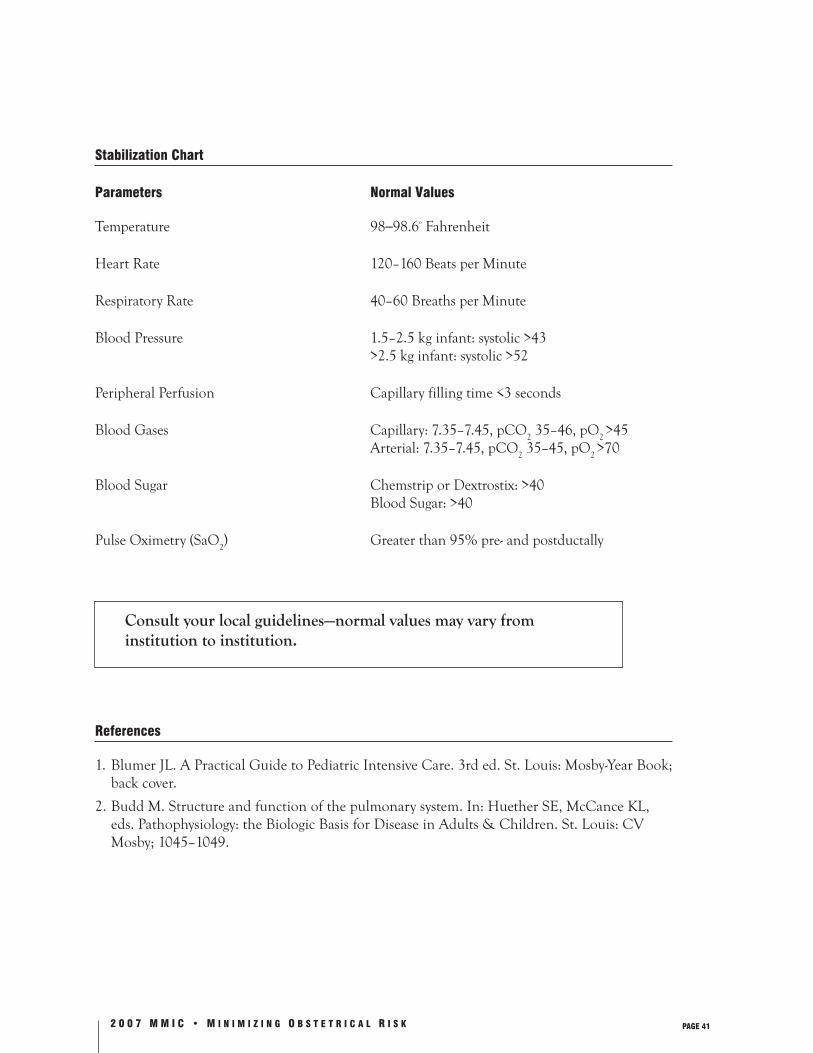
2 0 0 7 M M I C • M I N I M I Z I N G O B S T E T R I C A L R I S K PAGE 41
Stabilization Chart
Parameters Normal Values
Temperature 98–98.6º Fahrenheit
Heart Rate 120–160 Beats per Minute
Respiratory Rate 40–60 Breaths per Minute
Blood Pressure 1.5–2.5 kg infant: systolic >43 >2.5 kg infant: systolic >52
Peripheral Perfusion Capillary filling time <3 seconds
Blood Gases Capillary: 7.35–7.45, pCO2 35–46, pO
2 >45
Arterial: 7.35–7.45, pCO2 35–45, pO
2 >70
Blood Sugar Chemstrip or Dextrostix: >40 Blood Sugar: >40
Pulse Oximetry (SaO2) Greater than 95% pre- and postductally
Consult your local guidelines —normal values may vary frominstitution to institution.
References
1. Blumer JL. A Practical Guide to Pediatric Intensive Care. 3rd ed. St. Louis: Mosby-Year Book; back cover.
2. Budd M. Structure and function of the pulmonary system. In: Huether SE, McCance KL, eds. Pathophysiology: the Biologic Basis for Disease in Adults & Children. St. Louis: CV Mosby; 1045–1049.

2 0 0 7 M M I C • M I N I M I Z I N G O B S T E T R I C A L R I S K PAGE 42
This page left intentionally blank.

2 0 0 7 M M I C • M I N I M I Z I N G O B S T E T R I C A L R I S K PAGE 43

2 0 0 7 M M I C • M I N I M I Z I N G O B S T E T R I C A L R I S K PAGE 44
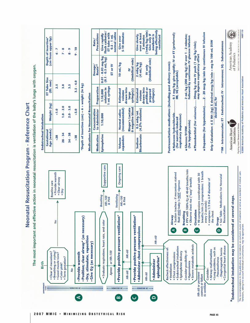
2 0 0 7 M M I C • M I N I M I Z I N G O B S T E T R I C A L R I S K PAGE 45

2 0 0 7 M M I C • M I N I M I Z I N G O B S T E T R I C A L R I S K PAGE 46
This page left intentionally blank.
2 0 0 7 M M I C • M I N I M I Z I N G O B S T E T R I C A L R I S K PAGE 47
The Morbidity and Mortality Conference
One of the most effective risk management tools is the learning that occurs when physicians gather to discuss their own difficult or challeng-ing cases. Cases occurring within a hospital setting hold a natural appeal for discussion, making the regularly scheduled Morbidity and Mortality (M & M) Conference an ideal opportunity for educational exchange. These conferences are particularly applicable to the obstetric service.
To Set Up an M & M Conference at your
Practice Site, We Recommend the Following:
◆ Schedule meetings on a regular basis, monthly or bimonthly.
◆ Encourage attendance through announcements and provision of refreshments.
◆ Ask fellow physicians and nurses to place their more challenging cases in a log kept on the delivery suite.
◆ Target cases from the preceding time interval.
◆ Invite participation from all providers on the maternity service.
◆ Ask the attending physician to present each case for discussion.
◆ Rotate responsibility for choosing cases and developing the agenda.
◆ Emphasize the educational focus.
During the first few meetings, accentuate the instructional nature of the conference. Physicians may initially feel reluctant to actively critique one another’s work, or to discuss their difficult cases in the presence of the nursing staff. Hesitation tends
to dissipate, however, in a nonpunitive atmosphere that focuses on the goal of improved patient care. Even professional jealousies can be overshadowed by the esprit de corps that develops from a mutu-ally cooperative working relationship.
The underlying objective of the conference should always be improving the quality of care. Because they are not intended as a peer review process, the meetings should not be an official function of the medical staff. To allay concern about peer review implications and the discoverability of the discus-sions, the meetings should not be burdened by documentation.
The Morbidity and Mortality Conference is a powerful vehicle for the enhancement of quality obstetric care. We strongly recommend regular M & M Conferences and the participation of all those involved in the care of the maternity patient.

2 0 0 7 M M I C • M I N I M I Z I N G O B S T E T R I C A L R I S K PAGE 48
This page left intentionally blank.

2 0 0 7 M M I C • M I N I M I Z I N G O B S T E T R I C A L R I S K PAGE 49
Communication
What do air traffic control operations, nuclear power plants, and the best labor and delivery units have in common? Besides being complex technological systems that carry enormous risk, all three are able to function over relatively long time periods without mistakes. In addition, all three attribute their success to an emphasis on com-munication. Technical skills notwithstanding, it is interpersonal skills that make the real difference in effectively managing risk.
While the number of high-risk situations inherent in obstetrics may imply the inevitability of poor outcomes and frequent lawsuits, some institutions are proving that it is possible to operate with a high degree of success and patient satisfaction. The key to their success is collaboration among members of the delivery team.
Collaboration is like world peace: everyone wants it, yet few people believe it’s possible. Most every-one involved in obstetrics can describe the typical conflicts among obstetricians, family physicians, pediatricians, anesthesiologists, nurses, and mid-wives—but they are hard-pressed to describe how effective collaboration might work.
Considering the barriers to collaboration, it is no surprise that implementation is difficult:
◆ Our health care system is hierarchical, operat-ing with superiors and subordinates rather than colleagues.
◆ Different methods of training for each medical discipline lead to misunderstandings of other practice styles or thought processes. A nurse, for instance, might judge a physician’s behav-ior by the standards of the nursing culture.
◆ Gender differences mirror differences in train-ing. Men are typically more task-oriented than relationship-oriented. Women tend to focus on rapport building at least as much as they focus on tasks. As in the various health profes-
sions, both genders are socialized to different values yet judge the other’s behavior by their own values. That most physicians are male and most nurses are female only reinforces these differences in practice patterns and communi-cation.
So how can the natural impasses between mem-bers of the delivery team be overcome?
While there is no one right way to communicate, collaborative skills can be learned. The following recommendations are intended to enhance the sense of teamwork in obstetrical departments:
◆ Increase your awareness of the communica-tion styles particular to different medical specialties, as well as ancillary personnel.
◆ Encourage meetings that include all mem-bers of the delivery team, including midwives and anesthesiologists. Discussions should fo-cus on areas needing refinements or improve-ment for the prevention of future problems. These reviews should be educational, blame-less, and supportive of the individual provid-ers. Note that labor and delivery units with the best track records recognize the potential danger of their successes—that is, the tendency of all working groups to take shortcuts when errors have not occurred for some time.
◆ Compromise. Adjust your communication style a bit to meet another practitioner half-way. Nurses can compromise by focusing on objective information in their reports to physicians, and making sure to state exactly what they need. Physicians can compromise by consciously building rapport with other staff members, as they would their patients.
◆ Respond to requests. Physicians who don’t hesitate to come to the hospital when called by a nurse at 2:00 a.m. will not only avoid potential disaster, but will increase collegial-
2 0 0 7 M M I C • M I N I M I Z I N G O B S T E T R I C A L R I S K PAGE 50
ity with obstetrical staff. They’ll also be better prepared to work with them during a crisis.
◆ Support patient management in progress. You can have a great effect on the confidence your patients feel in their care by demonstrat-ing respect for management already under-taken. Upon arriving at the patient’s bedside, you might say, “The midwife has called to let me know how your labor is progressing. She thinks you may need extra help with delivery and asked me to see what I think.” Statements such as this will lead to more trust and coop-eration from other staff members. In the event of a difficult delivery, having presented a uni-fied front may even prevent a lawsuit.
Collaboration may be hard work, and it may seem at times as elusive as world peace, but the effort you put into it tends to reinforce itself as benefits accumulate. You may find that, along with improvements in communication with others in labor and delivery, your own satisfaction with medicine increases.
Rapport
More collaboration on the obstetrical team may lead to better rapport with patients. Physicians often overlook the importance of rapport in their concentration on patient outcomes. Only when communication has come to a grinding halt may it be apparent that rapport with colleagues is as vital to obstetrical care as fetal monitoring.
Dissatisfaction with communication is the pri-mary reason patients sue after a delivery has gone badly. You may find it difficult to believe that for most patients, communication takes precedence over technical competence until you consider how communication actually imparts a degree of technical competence by relaying information. Pa-tients are generally far more eager for information than their physicians realize, and they want more detailed disclosures than their physicians usually
offer. It is easy to assume, especially with well-edu-cated patients, that they will ask for or otherwise obtain the information they seek, but patients generally don’t feel comfortable asking physicians for information.
A study published in 2001 found that patients want extensive discussions about risks, alterna-tives, and the adverse effects of prescribed medica-tion. You can do a great deal to instill confidence in your medical care by providing written materi-als during the visit, and checking to ensure that questions have been answered. How many of these additional rapport-building techniques do you use with your patients?
◆ Starting with a handshake: This universal greeting is your best opportunity to establish connection. If the patient is lying down, you might lay a hand briefly on her shoulder.
◆ Matching the patient’s posture and emotion-al state: In order to maintain eye contact at a similar level, sit when your patient is sitting, or pull up a stool to the exam table if she’s lying down. When the patient is in obvious physical or emotional pain, you can convey your caring by acknowledging her situation and mirroring her posture without being drawn into the turmoil.
◆ Active listening: The power of listening can-not be overstated. Study after study has shown that taking a few uninterrupted minutes to listen to the patient’s story saves far more time in the long run. Remember to adjust your listening style as patients move from physical descriptions of their condition to the ways it is affecting them. Listen to symptoms in a fact-focused way, but be ready to respond empathetically if the description shifts to an expression of fear or concern.
◆ Watching the jargon: Imagine how a woman without a medical background might hear the terms “failure to progress,” “unremark-able pregnancy,” “dysfunctional labor,” or

2 0 0 7 M M I C • M I N I M I Z I N G O B S T E T R I C A L R I S K PAGE 51
“inadequate pelvis.” Obstetrics has its own language that new practitioners, eager to adapt to the culture, often use to the dismay of their patients. More experienced providers are also subject to inadvertent insensitivity. Listen to common phrases with an ear towards formu-lating less threatening alternatives. You might say “developed toxemia” rather than “became toxic,” or “nonprogressive labor” instead of “failure to progress”—and then describe the condition in lay terms. People are sensitive enough to failure that an expression contain-ing the word “fail” or terms such as “trial of labor” or “trial of forceps” can have a judgmental ring.
Non-Adherence to Medical Advice
If a patient fails to follow your treatment plan and loses her baby as the result, can you be held ac-countable? Yes! Patients who don’t follow medical counsel are liability risks. Physicians are subject to blame for a patient’s disregard of their advice if the patient can convince a jury that the advice was unclear or never given. It is therefore in your own best interest to ensure that patients understand your recommendations, especially when the conse-quences of ignoring them are serious.
Protecting yourself from liability in this regard takes two forms. The first is to use every reason-able measure to encourage adherence, and the second is to document.
The following questions can serve as an adherence checklist:
◆ Did she understand? How easy it is to dimin-ish the importance of “bed rest” the hun-dredth time you’ve recommended it! How easy it is for the patient herself to disregard the advice when she feels fine. In addition, anything you say to minimize anxiety may be easily misconstrued. Telling the patient that
the mammogram is simply a reassurance might be heard as not being worth the effort and inconve-nience.
Explaining the significance of your instructions increases the likelihood they’ll be followed. Pro-viding written information is your best backup. It can help compensate for patients too shy to ask questions, or who may perceive you as too rushed to answer questions. Be aware of nonverbal signals you may be sending if you are feeling rushed or impatient, as well as technical language that might cloud understanding. Written recommendations can also convey important specifics such as what you mean by “light activity only” or symptoms that should prompt immediate action. You might ask your patient to summarize her understanding of the plan as a way to assure you both.
Finally, you might direct patients to your prac-tice Web site as a means to educate them with evidence-based articles or links to other respected Web sites. This is especially useful for patients who arrive at appointments with a pile of print-outs from less reputable sites.
◆ Did she agree? Many patients have their own opinions about what’s causing their symptoms or what treatment they should follow. Eliciting their beliefs is the best way to gain their cooperation. Consider asking them from the start whether they have anything in mind that might help with di-agnosis or treatment. Sharing your own thoughts during the history taking and exam is another technique. You might finish with, “How closely do my recommendations match what you were thinking?”
◆ Did she remember? Understanding and agree-ment will still lead to non-adherence if the patient forgets important elements in your recommenda-tion. Again, written materials will reinforce your message, supply details, and help patients explain their treatment to family and friends.

2 0 0 7 M M I C • M I N I M I Z I N G O B S T E T R I C A L R I S K PAGE 52
◆ Did she change her mind? A story on the evening news or a phone call from a friend can derail a treatment program to which your patient had previously agreed. Acknowledge to patients that it’s common to have second thoughts and ask them to call your office before changing any part of the plan.
◆ Is there a language barrier? You are obligated to provide interpretive services for patients with limited English proficiency. Hospitals often have qualified interpreters who can as-sist. The AT&T Language Line can connect you toll-free to interpreters in 140 languages. Document the name and relationship of any-one who interprets for your patients.
While chart notes detailing your advice are wise for any patient, documentation is crucial when your patient does not abide by your recommenda-tions. In that case, be sure to note the non-adher-ence in the medical record. If you have several conversations about the possible consequences of disregarding your plan, dictate a note that in-cludes details of exam findings, discussions, and recommendations. Be sure to include any missed appointments in your documentation. Obtaining an informed refusal will bolster your protection as well.
Patients may have every right to refuse medica-tions or referrals, but you also have the right to discharge non-adherent patients from your practice. Guidelines for dismissing patients can be found in the Physicians Risk Management Reference Manual. You are also encouraged to call the Risk Management Department for telephone advice.
After a Bad Outcome
Dr. Brown is a seasoned obstetrician who was shaken one day by a patient’s unexpected stillbirth. The baby’s death did not reflect badly on Dr. Brown’s care, yet he could not bring himself to approach the patient and her husband afterwards.
That he’d been certain of an easy delivery and had established strong rapport with the parents during their prenatal visits made it that much more pain-ful to talk with them about the tragedy. Instead he had a colleague explain the outcome, imagining that he was the last person they wanted to see during their grief.
Avoiding the parents was Dr. Brown’s only mis-take. They sued out of desperation to learn the truth, having assumed that their baby’s death was the result of a cover-up.
At about the same time, Dr. Jordan was called to the hospital in the middle of the night to deal with a difficult labor. After 20 minutes of struggle with a shoulder dystocia, he delivered the baby with a brachial plexus injury.
Dr. Jordan arranged for a private room in which he met with the patient and her partner, as well as his nurse, with whom the parents had established a close relationship throughout the pregnancy. Both the doctor and the nurse turned their cell phones off before entering the room. Meeting the gaze of both parents, Dr. Jordan congratulated them on their new baby boy, and then apologized for the unexpected problem during delivery. “Your baby was larger than I had anticipated,” he began, and then went on to review the maneuvers used during delivery, the extent of the injury, and what would be done to resolve it. He told them that he and his nurse would remain available to them on an ongo-ing basis for any questions that arose. Then he sat quietly as the two parents absorbed this informa-tion.
The father of the injured baby responded angrily. “You should have been able to predict and prevent this!”
Dr. Jordan acknowledged the father’s anger with steady eye contact while he and his nurse contin-ued to sit quietly. When tears began to fall onto the blanket where the mother sat in bed, Dr.

2 0 0 7 M M I C • M I N I M I Z I N G O B S T E T R I C A L R I S K PAGE 53
Jordan offered a box of tissues. After the initial emotions played out, the doctor again acknowl-edged their pain by saying, “We understand that this news is unexpected and difficult for you, and that you want to know what might have been done to prevent your baby’s injury.” Then the nurse ex-plained the risk factors for a shoulder dystocia, and Dr. Jordan admitted that because the mother was at low risk he didn’t discuss with them the possible emergency procedures beforehand. “Do either of you have any questions at this point?” he asked.
The father took the mother’s hand while they con-sulted each other silently. “Not right now, doctor,” the mother said. “We do appreciate your talking with us, but we’re overwhelmed at the moment.”
Dr. Jordan handed her his card and invited them to call if questions arose later. He went on to reiter-ate the treatment plan and gave them some written material about brachial plexus injury, including a reputable Web site through which they could obtain further information. Finally, he ended by saying that he would check in with them the same time tomorrow—and that it would be an honor for him to continue as their physician if they chose to stay with him.
Talking with patients and their families about sad news is for most physicians the most difficult aspect of communication. At the same time, the manner in which physicians approach patients about painful topics makes the biggest difference in whether or not the patients pursue a claim. In fact, the majority of claims are filed in an ef-fort to force open and honest discussion, since patients or their families often feel that doing so is the only way they will discover the facts related to an injury or death. In the scenario above, Dr. Jordan’s patient did not pursue a lawsuit and, in fact, did choose to keep him as their doctor. Of the following guidelines for delivering bad news, note those that Dr. Jordan followed:
◆ Hold the meeting in a private room, arrange the furniture beforehand, and have a box of tissues convenient to the patient.
◆ Turn off your cell phone. Ensure that the meeting isn’t rushed.
◆ Be sure that the staff member who knows the patient best is present during the meeting.
◆ Invite the patient to have a family member or friend present.
◆ Explain what happened.
◆ Use nonmedical terms.
◆ Let the patient express strong emotion.
◆ Show empathy both verbally and nonverbally.
◆ Check for understanding.
◆ Provide written materials, referrals, commu-nity resources, and follow-up consultation.
◆ Express hope. Even in the case of death, hope can be conveyed in terms of eliminating discomfort.
◆ Discuss your plan for future treatment.
◆ Document your objective description of the meeting, to include:
– Participant names – Discussion content – Recommendations – Action plan – Questions answered – Name of contact for any follow-up questions
◆ Arrange for a follow-up visit to establish that the patient retained important information.
◆ Be aware of any stress that might carry over into the next patient visit.
◆ If dealing with a patient from a culture with which you’re unfamiliar, consider consult-ing with the interpreter or a cultural broker employed by the hospital.
◆ Contact the facility risk manager and the Risk Management Department at Physicians Insur-ance for assistance.

2 0 0 7 M M I C • M I N I M I Z I N G O B S T E T R I C A L R I S K PAGE 54
Further information on documentation and com-munication skills is available in the Physicians Risk Management Manual. The Risk Management De-partment also offers telephone consultation at no charge to members, as well as professional support on an individual basis for physicians encountering a difficult situation.
2 0 0 7 M M I C • M I N I M I Z I N G O B S T E T R I C A L R I S K PAGE 55
Preterm Labor
Background
Despite recent technologic advances in obstetrical and neonatal care, the incidence of preterm birth has remained constant. Approximately 10–12% of infants are born prematurely (<37 completed weeks). Approximately 1% are very low birth weight (<1500 g). While birth weight-specific perinatal and neonatal mortality has declined, an infant with a very low birth weight is still much more likely to experience perinatal mortality or be neurologically impaired than those weighing ≥2500 g.
Etiology and Epidemiology
Historical factorsCertain epidemiologic and pregnancy characteris-tics have been associated with an increased risk for preterm delivery, including:
◆ Low socioeconomic status
◆ Nonwhite race
◆ Maternal age <18 or >40 years
◆ Previous 2nd-trimester loss
◆ Chemical dependency on cocaine or tobacco
◆ Multiple gestation
◆ Previous premature delivery (A woman with one previous preterm birth has a 15% recur-rence risk for preterm delivery in a subsequent pregnancy. With two previous preterm births, the recurrence risk is 40%.)
Reproductive-tract abnormalitiesCongenital uterine malformations, such as bicor-nuate or unicornuate uterus, and in some instanc-es, uterine myomata—especially submucosal—may increase the risk for preterm delivery.
Cervical incompetence may also predispose to premature labor and may be either:
◆ Congenital, as in association with uterine malformation, or
◆ As a result of a maternal in utero teratogen exposure from diethylstilbestrol, or
◆ Acquired from previous obstetrical trauma or conization of the cervix.
A dynamic cervix has been described as a predispos-ing risk for preterm delivery. (Dynamic cervix is a diagnosis describing dilation of the internal os and funneling of the lower uterine segment and membranes into the cervical canal. The diagnosis is best made in the second trimester by either a transabdominal or transvaginal ultrasound.)
Infectious etiologies and abnormal serum markersRisk factors for preterm delivery also include infec-tious factors. Very early labor (prior to 28 weeks) is more likely the result of infection than labor occurring after 28 weeks. In one study, 83% of preterm labor of less than 28 weeks had chorioam-nionitis. Patients with bacterial vaginosis are also at higher risk. The odds ratio for preterm birth in association with bacterial vaginosis in most studies ranges from 1.5 to 3.0. In high-risk patients with bacterial vaginosis, one study showed a decreased incidence of preterm labor with prophylactic metronidazole and ampicillin. Broad-spectrum antibiotics administered after premature rupture of membranes have also been shown to improve outcomes for the neonate.
Prevention
Because the prevalence of preterm delivery has not decreased, it is safe to say that an effective preven-tion program has not been established. According to studies, pregnancy risk-scoring systems, bed rest, home uterine-activity monitoring, and routine cervical examinations have not conclusively shown benefits. Additionally, the efficacy, timing, and du-ration of administration of antibiotics have yet to be determined. Of all preventive measures, early

2 0 0 7 M M I C • M I N I M I Z I N G O B S T E T R I C A L R I S K PAGE 56
access to prenatal care and frequent medical and nurse contact—along with active patient educa-tion—appear to be the best approach.
Diagnosis
The signs and symptoms of preterm labor may be subtle, including:
◆ Uterine contractions or menstrual-like cramps
◆ The feeling of pelvic pressure
◆ Increase in vaginal discharge
◆ Diarrhea
While the above symptoms frequently occur in the absence of preterm labor, patients with these complaints should warrant prompt evaluation. There is no question that preterm labor is over-diagnosed, most commonly in the presence of uterine contractions without cervical change.
Uterine contractions in the presence of signifi-cant cervical effacement of ≥75%, cervical dila-tion ≥2 cm, and low presenting part or balloon-ing of the lower uterine segment are associated with a high risk for preterm delivery and should be treated aggressively.
Fetal fibronectin, a basement membrane protein produced by the placenta and membranes, is a predictor of the likelihood of preterm birth. Its presence in cervical mucous at 24 weeks led to a 60-fold increase in delivery prior to 28 weeks. If negative with preterm contractions, there is less than a 1% chance of delivery within 2 weeks. A rapid screening assay for its detection is now avail-able.
Management
Patients with complaints consistent with preterm labor should be promptly evaluated, including evaluation of the cervix, initially with speculum
if there is concern about premature rupture of membranes.
If premature rupture of the membranes has oc-curred, digital examination of the cervix should NOT be done unless the patient is actively labor-ing, since the risk for infection is significantly increased.
Initial therapy for preterm labor usually consists of IV hydration and evaluation for urinary tract infec-tion; however, IV hydration has NO established benefit, and excessive hydration is best avoided.
After a diagnosis of premature labor has been made, consider the following issues:
◆ Tocolysis
Available data suggest that tocolysis is effective in most cases for 48–72 hours. Commonly used agents include:
– Magnesium sulfate
– Beta-adrenergic agents (terbutaline, rito-drine)
– Nonsteroidal anti-inflammatory drugs (indo-methacin)
– Calcium channel blockers (nifedipine)
Contraindications to tocolysis for preterm labor are listed in Table I.
◆ Group B streptococcus prophylaxis
Preterm infants are especially prone to neona-tal GBS infection. Cultures from the introitus and anus area should be sent for GBS culture. In the meantime, treat the patient with IV antibiotics until the results return. Acceptable coverage would include penicillin G, ampicil-lin, cephalosporins, or vancomycin.

2 0 0 7 M M I C • M I N I M I Z I N G O B S T E T R I C A L R I S K PAGE 57
◆ Fetal-lung maturity assessment
Amniocentesis may play a role in evaluating patients in preterm labor. A sample of am-niotic fluid may be sent for fetal pulmonary testing, including fluorescent polarization, which is commonly used in the Seattle area, with results available within two hours. The L/S ratio is an acceptable alternative. Since perinatal morbidity and mortality are closely linked to pulmonary status, knowing the fetal pulmonary maturity status may be helpful in guiding therapy with glucocorticoids and determining place of delivery. In addition, amniotic fluid may be sent for gram stain and culture and amniotic fluid glucose assessment, since as many as 15% of patients with idio-pathic preterm labor may have infection as an underlying cause. Amniotic fluid containing white cells, bacteria, or a low glucose level has a high association with chorioamnionitis. In this situation, labor may not be postponed, and should be treated aggressively with antibi-otics and delivery.
◆ Glucocorticoid treatment
A recent consensus conference on antenatal glucocorticoids concluded that antenatal treat-ment is effective in reducing the incidence of hyaline membrane disease in preterm deliv-eries and was significantly underutilized. If fetal pulmonary testing is performed and the fluorescent polarization is >270 or the L/S ratio is <2, glucocorticoids should be admin-istered. Although tocolytics are not likely to be effective for more than 48–72 hours, their administration may stave off delivery long enough to allow for the beneficial effects of glucocortocoids. Acceptable dosing includes betamethasone (one dose, 12 mg IM, followed by a second dose 24 hours later) or dexametha-sone (6mg IV q 6h X 4). Glucocorticoids may be used with ruptured membranes, but in these cases, amniocentesis may be performed
prior to administration, if possible, to rule out amniotic fluid infection. In pregnancies of ≤30 weeks’ gestation, only 10% will have mature fetal lungs. At 32 weeks, 25–30% will have reached pulmonary maturity, and 95% will have mature lungs at 36 weeks. The benefits of glucocorticoid administration after 34 weeks of gestation have not been established.
◆ Maternal transport
Survival rates for preterm/very low birth weight infants and premature infants with pulmonary immaturity are higher in Level III centers than those born in Level I and II facilities. If time is available for safe maternal transport in these cases, such transport should be strongly considered. Each hospital, in con-junction with obstetric, pediatric, and neona-tal consultation, should develop guidelines for maternal and neonatal transport.
◆ Continued tocolysis
After initial evaluation and a decision made to transport or keep a mother, tocolysis usually should be continued for a minimum of 48 hours. Tocolytic drugs may have major side effects to mother and fetus and should be administered only under careful supervision. The goal of tocolysis is to minimize uterine contractions with the least amount of drug possible. Potential complications of tocolytic agents are listed in Table II. The best outcomes result from a carefully constructed protocol for tocolysis. Each hospital, in conjunction with obstetric, neonatal, and pediatric staff, should develop a protocol for evaluation of preterm la-bor and safe administration of tocolytic agents.
◆ Consultation
As with any complicated case, consultation with obstetric, pediatric, neonatal, or perinatal colleagues should be encouraged.

2 0 0 7 M M I C • M I N I M I Z I N G O B S T E T R I C A L R I S K PAGE 58
◆ Risk management implications
Maintain a high index of suspicion for the patient with a previous history of preterm birth, as well as for the patient who reports subtle changes (e.g., cramps, backache, in-creased discharge). These patients need to be seen frequently and monitored carefully with screening for infection, cervical changes, and fetal fibronectin.
For those patients that deliver prematurely, ALL involved parties should be informed in anticipation, including neonatology and anesthesia. If possible, these babies should be transferred in utero to a Level III facility.
It is important to obtain the appropriate cultures from cervix and amniotic fluid and to send the placenta for pathological examination to document the presence of chorioamnionitis.
References
1. American College of Obstetricians and Gy-necologists. Preterm Labor. ACOG Technical Bulletin Number 31. Washington, DC: Ameri-can College of Obstetricians and Gynecolo-gists; October, 2001.
2. American College of Obstetricians and Gyne-cologists. Home uterine activity monitoring. ACOG Committee Opinion Number 172. Washington, DC: American College of Obste-tricians and Gynecologists; May, 1996.
3. American College of Obstetricians and Gy-necologists. Prevention of early-onset group B streptococcal disease in newborns. ACOG Committee Opinion 279. Washington, DC: American College of Obstetricians and Gyne-cologists; December, 2002.
4. American College of Obstetricians and Gy-necologists. Management of Preterm Labor. ACOG Practice Bulletin No. 43. Washington, DC: American College of Obstetricians and Gynecologists; May, 2003.
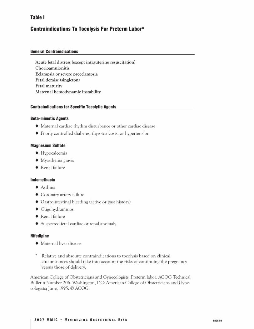
2 0 0 7 M M I C • M I N I M I Z I N G O B S T E T R I C A L R I S K PAGE 59
Table I
Contraindications To Tocolysis For Preterm Labor*
General Contraindications
Acute fetal distress (except intrauterine resuscitation) Chorioamnionitis Eclampsia or severe preeclampsia Fetal demise (singleton) Fetal maturity Maternal hemodynamic instability
Contraindications for Specific Tocolytic Agents
Beta-mimetic Agents
◆ Maternal cardiac rhythm disturbance or other cardiac disease
◆ Poorly controlled diabetes, thyrotoxicosis, or hypertension
Magnesium Sulfate
◆ Hypocalcemia
◆ Myasthenia gravis
◆ Renal failure Indomethacin
◆ Asthma
◆ Coronary artery failure
◆ Gastrointestinal bleeding (active or past history)
◆ Oligohydramnios
◆ Renal failure
◆ Suspected fetal cardiac or renal anomaly
Nifedipine
◆ Maternal liver disease
* Relative and absolute contraindications to tocolysis based on clinical circumstances should take into account the risks of continuing the pregnancy versus those of delivery.
American College of Obstetricians and Gynecologists. Preterm labor. ACOG Technical Bulletin Number 206. Washington, DC: American College of Obstetricians and Gyne-cologists; June, 1995. © ACOG

2 0 0 7 M M I C • M I N I M I Z I N G O B S T E T R I C A L R I S K PAGE 60
TABLE II
Potential Complications Of Tocolytic Agents
Beta-Adrenergic Agents
◆ Hyperglycemia
◆ Hypokalemia
◆ Hypotension
◆ Pulmonary edema
◆ Cardiac insufficiency
◆ Arrhythmias
◆ Myocardial ischemia
◆ Maternal death
Magnesium Sulfate
◆ Pulmonary edema
◆ Respiratory depression*
◆ Cardiac arrest*
◆ Maternal tetany*
◆ Profound muscular paralysis*
◆ Profound hypotension*
Indomethacin
◆ Hepatitis‡
◆ Renal failure‡
◆ Gastrointestinal bleeding‡
Nifedipine
◆ Transient hypotension
* Effect is rare; seen with toxic levels.
‡ Effect is rare; associated with chronic use.
American College of Obstetricians and Gynecologists. Preterm labor. ACOG Technical Bulletin Number 206. Washington, DC: American College of Obstetricians and Gyne-cologists; June, 1995. © ACOG
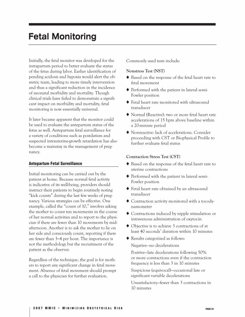
2 0 0 7 M M I C • M I N I M I Z I N G O B S T E T R I C A L R I S K PAGE 61
Fetal Monitoring
Initially, the fetal monitor was developed for the intrapartum period to better evaluate the status of the fetus during labor. Earlier identification of pending acidosis and hypoxia would alert the ob-stetric team, leading to more timely intervention and thus a significant reduction in the incidence of neonatal morbidity and mortality. Though clinical trials have failed to demonstrate a signifi-cant impact on morbidity and mortality, fetal monitoring is now essentially universal.
It later became apparent that the monitor could be used to evaluate the antepartum status of the fetus as well. Antepartum fetal surveillance for a variety of conditions such as postdatism and suspected intrauterine-growth retardation has also become a mainstay in the management of preg-nancy.
Antepartum Fetal Surveillance
Initial monitoring can be carried out by the patient at home. Because normal fetal activity is indicative of its well-being, providers should instruct their patients to begin routinely noting “kick counts” during the last few weeks of preg-nancy. Various strategies can be effective. One example, called the “count of 10,” involves asking the mother to count ten movements in the course of her normal activities and to report to the physi-cian if there are fewer than 10 movements by mid-afternoon. Another is to ask the mother to lie on her side and consciously count, reporting if there are fewer than 3–4 per hour. The importance is not the methodology but the recruitment of the patient as the observer.
Regardless of the technique, the goal is for moth-ers to report any significant change in fetal move-ment. Absence of fetal movement should prompt a call to the physician for further evaluation.
Commonly used tests include:
Nonstress Test (NST)◆ Based on the response of the fetal heart rate to
fetal movement
◆ Performed with the patient in lateral semi-Fowler position
◆ Fetal heart rate monitored with ultrasound transducer
◆ Normal (Reactive): two or more fetal heart rate accelerations of 15 bpm above baseline within a 20-minute period
◆ Nonreactive: lack of accelerations. Consider proceeding with CST or Biophysical Profile to further evaluate fetal status
Contraction Stress Test (CST)
◆ Based on the response of the fetal heart rate to uterine contractions
◆ Performed with the patient in lateral semi-Fowler position
◆ Fetal heart rate obtained by an ultrasound transducer
◆ Contraction activity monitored with a tocody-namometer
◆ Contractions induced by nipple stimulation or intravenous administration of oxytocin
◆ Objective is to achieve 3 contractions of at least 40 seconds’ duration within 10 minutes
◆ Results categorized as follows:
Negative—no decelerations
Positive—late decelerations following 50% or more contractions even if the contraction frequency is less than 3 in 10 minutes
Suspicious (equivocal)—occasional late or significant variable decelerations
Unsatisfactory—fewer than 3 contractions in 10 minutes

2 0 0 7 M M I C • M I N I M I Z I N G O B S T E T R I C A L R I S K PAGE 62
◆ Contraindicated in pregnancies with threat-ened preterm labor, at high risk for preterm labor, with placenta previa, or with a classic uterine scar
Biophysical Profile (BPP)
◆ Five components:
a. Reactive NST
b. Fetal breathing movements (1 or more episodes of rhythmic fetal breathing move-ments of 30 seconds or more within 30 minutes)
c. Fetal movement (3 or more distinct body or limb movements within 30 minutes)
d. Fetal tone (1 or more episodes of extension of a fetal extremity with return to flexion)
e. Amniotic fluid volume: a single vertical pocket of amniotic fluid exceeding 2 cm is considered evidence of adequate fluid
◆ Interpretation:
a. A score of either 0 or 2 assigned to each of the 5 observations.
b. A score of 8–10 is considered normal.
c. A score of 6 is considered equivocal (fetus should be retested in 12–24 hours).
d. A score of 4 or less is considered abnormal.
e. If oligohydramnios is present, further evaluation is warranted.
◆ Modified BPP:
a. Includes a reactive NST and a normal amniotic fluid index
b. Alternative to BPP with comparable predictive value
Ultrasound
◆ In situations with a high index of suspicion for fetal compromise, growth parameters should be evaluated for consideration of uterine growth restriction.
◆ Umbilical artery Doppler measurements reflect a placental resistance to blood flow, and may predict impending fetal distress.
Indications for antepartum testing include but are not limited to:
◆ Postdates (41–42 weeks)
◆ Decreased fetal movement
◆ Diabetes
◆ Maternal disease (cardiac, renal, pulmonary, neurologic, etc.)
◆ Hypertensive disorders (chronic or pregnancy-induced)
◆ Intrauterine growth restriction
◆ Maternal substance abuse
◆ Fetal isoimmunization
◆ Polyhydramnios
◆ Oligohydramnios
◆ Previous unexplained fetal demise
◆ Multiple pregnancy
Intrapartum Monitoring
Several studies suggest that monitoring by periodic auscultation is as effective as continuous electronic monitoring, with outcomes that are not signifi-cantly different. Although not considered standard, continuous monitoring for all laboring patients has become commonplace. It is considered standard, however, to continuously monitor all high-risk obstetric patients. We have seen some situations where the monitor was removed in preparation for cesarean section. It is imperative that the monitor

2 0 0 7 M M I C • M I N I M I Z I N G O B S T E T R I C A L R I S K PAGE 63
continuously record until the infant is delivered—even when cesarean section is required.
High-risk conditions which are indications for continuous electronic fetal heart rate monitoring include but are not limited to:
◆ Age >35 or <16
◆ Diabetes
◆ Hypertension
◆ Maternal disease (cardiac, pulmonary, renal, neurologic)
◆ Previous cesarean section
◆ Previous abruption
◆ Previous stillbirth
◆ Parity greater than five
◆ Substance abuse
◆ Dysfunctional labor
◆ Intrauterine growth restriction
◆ Polyhydramnios, oligohydramnios
◆ Vaginal bleeding
◆ Induction of labor
◆ Premature labor
◆ Premature rupture of membranes
◆ Multiple pregnancy
◆ Meconium-stained fluid
◆ Gestational age <37 or >42 weeks
◆ Estimated fetal weight <2500 g or >4000 g
◆ Prolonged labor
◆ Maternal temp >100ºF
◆ Abnormal presentation (e.g., breech, face)
◆ Isoimmunization
Medicolegal Implications
In those unfortunate situations where the outcome is suboptimal, the monitor tracing is almost always a major source of interest. Its record of the course
of intrapartum events may be helpful in defend-ing the provider’s care. It may also be the plain-tiff attorney’s best piece of evidence to prove an obstetric care team’s negligence.
Ironically, the tracing itself is frequently in dispute. Review of our claims files suggests that monitoring is far from an exact science. National experts have given contradictory opinions on the same tracing. In one situation, the jury was so confused by the expert testimony from both sides that the ultimate decision for the defense was reached indepen-dently of the monitor findings. Instead, the jury reasoned that the mother’s history of smoking had been the causative factor.
While monitoring can be ambiguous, certain persistent patterns on a tracing require a response on the part of the practitioner. These include but are not limited to:
◆ Severe variable decelerations
◆ Persistent late decelerations with a nonreactive tracing
◆ Persistent fetal tachycardia with a nonreactive tracing
◆ Prolonged bradycardia
Failure to execute an intervention or form a plan in these situations may make it difficult to defend against a malpractice allegation.
Despite differences of interpretive opinion, the electronic fetal monitor is a significant element in the current practice of obstetrics. We recommend, therefore, that all labor and delivery suites have specific monitoring policies to help eliminate the monitor as a potential problem in the defense of a malpractice claim.
Monitoring policies should cover thefollowing:
◆ Patients being observed in labor and delivery suite for any reason (e.g., labor check, non-stress testing, post-amniocentesis, prior to
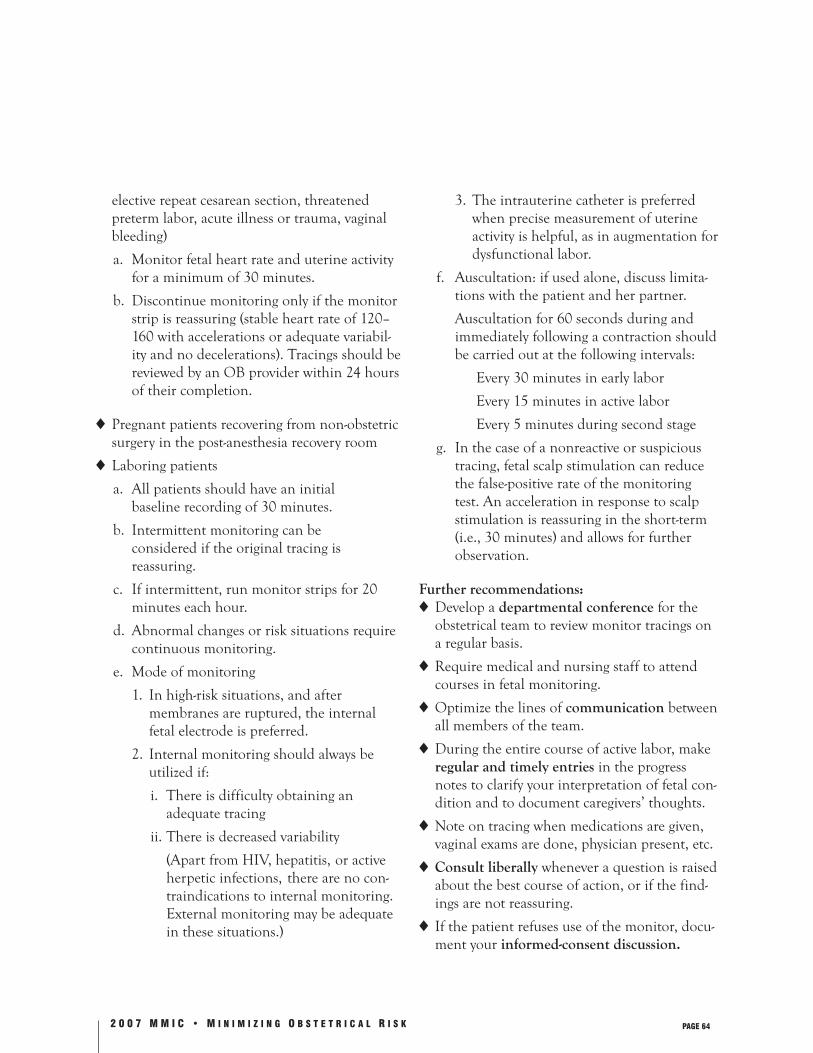
2 0 0 7 M M I C • M I N I M I Z I N G O B S T E T R I C A L R I S K PAGE 64
elective repeat cesarean section, threatened preterm labor, acute illness or trauma, vaginal bleeding)
a. Monitor fetal heart rate and uterine activity for a minimum of 30 minutes.
b. Discontinue monitoring only if the monitor strip is reassuring (stable heart rate of 120–160 with accelerations or adequate variabil-ity and no decelerations). Tracings should be reviewed by an OB provider within 24 hours of their completion.
◆ Pregnant patients recovering from non-obstetric surgery in the post-anesthesia recovery room
◆ Laboring patients
a. All patients should have an initial baseline recording of 30 minutes.
b. Intermittent monitoring can be considered if the original tracing is reassuring.
c. If intermittent, run monitor strips for 20 minutes each hour.
d. Abnormal changes or risk situations require continuous monitoring.
e. Mode of monitoring
1. In high-risk situations, and after membranes are ruptured, the internal fetal electrode is preferred.
2. Internal monitoring should always be utilized if:
i. There is difficulty obtaining an adequate tracing
ii. There is decreased variability
(Apart from HIV, hepatitis, or active herpetic infections, there are no con-traindications to internal monitoring. External monitoring may be adequate in these situations.)
3. The intrauterine catheter is preferred when precise measurement of uterine activity is helpful, as in augmentation for dysfunctional labor.
f. Auscultation: if used alone, discuss limita-tions with the patient and her partner.
Auscultation for 60 seconds during and immediately following a contraction should be carried out at the following intervals:
Every 30 minutes in early labor
Every 15 minutes in active labor
Every 5 minutes during second stage
g. In the case of a nonreactive or suspicious tracing, fetal scalp stimulation can reduce the false-positive rate of the monitoring test. An acceleration in response to scalp stimulation is reassuring in the short-term (i.e., 30 minutes) and allows for further observation.
Further recommendations:◆ Develop a departmental conference for the
obstetrical team to review monitor tracings on a regular basis.
◆ Require medical and nursing staff to attend courses in fetal monitoring.
◆ Optimize the lines of communication between all members of the team.
◆ During the entire course of active labor, make regular and timely entries in the progress notes to clarify your interpretation of fetal con-dition and to document caregivers’ thoughts.
◆ Note on tracing when medications are given, vaginal exams are done, physician present, etc.
◆ Consult liberally whenever a question is raised about the best course of action, or if the find-ings are not reassuring.
◆ If the patient refuses use of the monitor, docu-ment your informed-consent discussion.
2 0 0 7 M M I C • M I N I M I Z I N G O B S T E T R I C A L R I S K PAGE 65
◆ Given the current statute of limitations, store the tracings for 26 years. For added protection, indi-vidual practitioners may want to make personal office copies for safekeeping of files documenting bad outcomes. Be sure to copy thermal paper trac-ings onto standard paper.
◆ Computer disks are an acceptable alternative if they do not permit overwriting or revision.
References
1. American College of Obstetricians and Gyne-cologists. Fetal heart rate patterns: monitoring, in-terpretation and management. ACOG Technical Bulletin Number 207. Washington, DC: Ameri-can College of Obstetricians and Gynecologists; 1995.
2. American College of Obstetricians and Gyne-cologists. Fetal and neonatal neurologic injury. ACOG Technical Bulletin Number 163. Washing-ton, DC: American College of Obstetricians and Gynecologists; January, 1992.
3. American College of Obstetricians and Gynecolo-gists. Inappropriate use of the terms fetal distress and birth asphyxia. ACOG Committee Opinion Number 197. Washington, DC:American College of Obstetricians and Gynecologists; February, 1998.
4. American College of Obstetricians and Gyne-cologists. Antepartum fetal surveillance. ACOG Practice Bulletin Number 9. Washington, DC: American College of Obstetricians and Gynecolo-gists; 1999.
5. Guidelines for Perinatal Care. 5th ed. Washington, DC: American Academy of Pediatrics/American College of Obstetricians and Gynecologists; 2002.

2 0 0 7 M M I C • M I N I M I Z I N G O B S T E T R I C A L R I S K PAGE 66
This page left intentionally blank.

2 0 0 7 M M I C • M I N I M I Z I N G O B S T E T R I C A L R I S K PAGE 67
Pregnancy-Induced Hypertension
The purpose of including a chapter on hyperten-sive disorders in pregnancy is not to restate the well-known nature of this entity, but to increase awareness of potential trouble and discuss some of the related risk-management subtleties. The most common malpractice cases involve instances of:
◆ Missed diagnosis
◆ Lack of appreciation for potential severity
◆ Communication failure between patient and provider
◆ Presentation so atypical that proper manage-ment was never considered
The perplexing aspects of pregnancy-induced hy-pertension are the variety of presentations and the unpredictability of the clinical course, combined with the potential for adverse outcome. Elevated blood pressure (defined as a sustained blood pres-sure increase to levels of 140 mm Hg systolic or 90 mm Hg diastolic) may or may not be present in association with abnormalities of renal, hepatic, coagulation, cardiac, or CNS functioning. In ad-dition, each of these findings may occur in com-bination with one another or present as the sole symptom. Accordingly, the range of presentations varies from the common to the extremely bizarre. For example, we are aware of a patient who was normotensive at 23 weeks with 4+ proteinuria and who precipitously (within 24 hours) proceeded to hepatic rupture and death. Such cases raise the suspicion for other underlying and complicating disease processes such as acute glomerulonephri-tis, lupus nephritis, or one of the other collagen vascular diseases.
Fortunately, the vast majority of patients have minimal elevations in blood pressure, perhaps accompanied by mild proteinuria. They can be managed as outpatients and proceed to have un-eventful deliveries. Therefore, providers can easily become complacent or cavalier when confronted
with minimal findings. The following two cases from our files illustrate the need for caution even in the seemingly straightforward preeclamptic.
Case #1 A 19-year-old GIPO was seen for a regu-
lar prenatal visit at 41+ weeks on a Friday afternoon with BP of 130/90 and 120/85 (ten minutes later) and trace of proteinuria. She was scheduled for induction on Monday morning if labor had not ensued spontane-ously. Earlier prenatal BP measurements had been in the range of 110–120/70–80. On Saturday the patient called her mother to report that the baby was not moving very much. She did not, however, report this to her physician. On Monday morning she was admitted with BP of 140/90, 2+ proteinuria, and absent fetal heart tones. Fetal demise was confirmed by ultrasound, and after a 20-hour labor, a 9-pound stillborn male was delivered by cesarean section for cephalopelvic dispro-portion. Support for the clinical management could not be found since no fetal surveillance studies (specifically NST) were performed. The case was ultimately settled for $50,000.
Case #2 A 26-year-old GIPO who had been normo-
tensive throughout pregnancy was seen at 38 weeks for a routine prenatal visit with BP of 130/90 and 2+ proteinuria. She was admitted to the hospital antepartum ward in prepara-tion for induction the following morning. A mild sedative was given for sleep. The fol-lowing morning, when the fetal monitor was applied, fetal heart tones were absent. Fetal demise was confirmed by ultrasound and an 8-hour labor was productive of a 7-pound stillborn male. As in case #1, the liability problems could not be overcome and the case was settled out of court.
2 0 0 7 M M I C • M I N I M I Z I N G O B S T E T R I C A L R I S K PAGE 68
Both of these cases are marked by the low index of suspicion for the ultimate outcome. A more atten-tive and aggressive management by the provider in case #1 and by both provider and staff in case #2 would likely have prevented the adverse outcome. In both situations the simple performance of a reactive NST would have made these cases de-fensible. If the NST had been nonreactive, more aggressive intervention would have undoubtedly taken place. Other tools available for assessing fetal status, such as stress testing, amniotic fluid index, and the biophysical profile, might have fol-lowed a nonreactive NST.
Recommendations
Prenatal ScreeningMeasure and record each gravida’s weight, blood pressure, and urinary protein at each prenatal visit. Occasionally, the urinalysis may not be docu-mented because of collection problems.
Proteinuria is the most reliable and definitive diagnostic sign of the disease. If present, regard-less of the blood pressure, a 24-hour measure-ment is important to evaluate the severity of the process.
A confirmed dipstick value of 1+ or greater is significant. A value of 1+ protein should cause suspicion for preeclampsia, if not infection. A pregnant woman should not excrete more than 350 mg of protein in 24 hours.
Evaluation
Admission to the hospital for an initial period of observation and a comprehensive workup is recommended to fully evaluate maternal and fetal well-being. Initial lab should include:
◆ Urinalysis
◆ CBC
◆ Platelet count
◆ Liver and kidney function test
◆ NST
◆ Baseline ultrasound
◆ Serum uric acid
◆ 24-hour urine for protein and creatinine clearance
Follow-up should include:
◆ Daily weight
◆ Fluid intake/output
◆ Urine dipstick for protein
◆ Blood pressure four times daily
◆ Platelet count
◆ Liver function weekly
◆ Weekly or twice-weekly NST
◆ Weekly or twice-weekly biophysical profile for patients who remain undelivered
Specific criteria for monitoring the outpatient should be adopted and fully documented. These guidelines might include but not necessarily be limited to:
◆ A stable blood pressure not greater than 140/90
◆ Urine protein <350 mg per day
◆ Normal stable platelet count
◆ Normal liver function, e.g., ALT, AST
◆ Immature fetus with reactive NST
◆ Normal amniotic fluid volume
◆ Compliant patient who can remain at bed rest
◆ Available transportation
2 0 0 7 M M I C • M I N I M I Z I N G O B S T E T R I C A L R I S K PAGE 69
Consultation/Referral
While it may not always be necessary in the milder cases, another opinion and consultation with a maternal-fetal specialist—if only by phone—demon-strates a thorough management process. Referral to a subspecialist should be strongly considered in high-risk situations which include:
◆ Blood pressure >160 systolic or >100 diastolic
◆ Urine protein >5 g per 24 hours
◆ Oliguria (<30 cm3/hour x 6 hours)
◆ Abdominal pain suggesting liver involvement
◆ Visual disturbances
◆ HELLP syndrome (Hemolysis, Elevated Liver enzymes, Low Platelets)
◆ Intrauterine growth restriction
◆ Oligohydramnios
◆ Fetal status that is not reassuring by biophysi-cal profile testing
Management
If the fetus is mature (>37 weeks or by fetal lung maturity testing for those between 34 and 37 weeks), it is usually prudent to proceed to deliv-ery. Those that are not mature and need to be delivered for either maternal or fetal indications should be referred to a tertiary center. During labor, continual fetal monitoring is indicated. Magnesium sulphate is the drug of choice—it is extremely safe and should be used without hesita-tion for seizure prophylaxis. Apresoline or another antihypertensive may be used in situations where blood pressure is not well controlled, but it must be monitored carefully due to the potential for an unpredictable and sudden hypotensive response. These patients also need to be monitored in the postpartum period, and it is important to remember that preeclampsia and eclampsia may
occur initially in the postpartum period. Be sure to evaluate the patient who complains of first-time headache or abdominal pain after delivery.
Education and Informed Consent
Keep the patient and appropriate family fully informed of the reasons for performing each ele-ment of the evaluation. Outpatients should be instructed to immediately report changes such as:
◆ Severe headache
◆ Abdominal pain
◆ Persistent nausea and vomiting
◆ Visual disturbances
◆ Decreased fetal movement
◆ Cramping
◆ Leaking of fluid or vaginal bleeding
◆ Unusually rapid swelling or weight gain
Document discussions with the patient and family with a comprehensive note in the chart that in-cludes the benefits and risks of each management decision.
Communication
Since it is possible that a number of providers may be responsible for managing the patient at any given time, it is important that records be immediately available. It is essential that patients understand the importance of telling staff and providers unfamiliar with the case exactly what has transpired in the prenatal course and why added precaution has been recommended.

2 0 0 7 M M I C • M I N I M I Z I N G O B S T E T R I C A L R I S K PAGE 70
Key Points
Always keep in mind the following risk management principles. Their application provides the basis for quality patient care and the avoidance of adverse outcomes.
◆ A heightened awareness for potential problems
◆ A high index of suspicion for trouble
◆ Attention to detail
◆ Prompt evaluation
◆ Early intervention
◆ Careful monitoring
◆ Liberal consultation with colleagues
◆ Documentation with a defensible management plan
◆ Communication with all involved parties
◆ Patient education and informed consent
2 0 0 7 M M I C • M I N I M I Z I N G O B S T E T R I C A L R I S K PAGE 71
Prevention of Perinatal Group B Beta Strep
Background
Over the past several decades, Group B Strepto-coccus (GBS) has been implicated as a leading cause of serious perinatal infection. Yearly, more than 10,000 neonates in the U.S. are affected with a manifestation of GBS-associated morbidity, such as sepsis, pneumonia, or meningitis. In 1990, neonatal mortality in the U.S. was about 1.8 per 1,000, amounting to approximately 7,600 deaths. Neonatal complications of GBS can be categorized as early neonatal infection (i.e., occurring within 7 days of delivery) or late (more than 7 days after delivery). Manifestations of maternal morbidity include urinary tract infection, amnionitis, endo-metritis, and wound infection.
Neonatal complications remain the major concern of perinatal GBS infection. Although the timely use of intrapartum antibiotics significantly reduces the incidence of both early onset neonatal sepsis and neonatal colonization, it does not necessarily prevent late onset neonatal sepsis.
Approximately 30% of women are colonized with GBS. In uncomplicated pregnancies, GBS neona-tal sepsis occurs at a rate of 1 per 1000. This rate increases dramatically, however, in the prema-ture infant (i.e., less than 37 weeks’ gestational age). Perinatal management has concentrated on identifying the at-risk individuals and developing proper treatments to reduce both maternal and neonatal complications. In 1996, ACOG, AAP, and the CDC collaborated in the development of treatment strategies based on either antepartum culture identification or risk status. More recent evidence has revealed that approximately 40% of neonatal sepsis occurs in the low-risk group, which is therefore not identified by a risk-based prevention strategy. Accordingly, CDC guidelines released in 2001 recommend screening ALL patients with cultures at 35–37 weeks. Culture-positive patients, and those with high-risk charac-
teristics, should be given intrapartum prophylactic antibiotics. High-risk characteristics include:
◆ Premature infant (<37 weeks)
◆ Mothers with prolonged rupture of mem-branes (for example, PROM >18 hours prior to delivery)
◆ Maternal fever of >100.4oF (or >38oC)
◆ Clinical suspicion for amnionitis
◆ Mothers with a genetically inherited inability to develop antibodies against the capsid of the GBS bacterium. Because of this possibility, mothers who have previously delivered GBS-affected neonates have a much higher chance of delivering a similarly affected infant.
Because GBS primarily colonizes in the bowel, complete eradication is highly unlikely, and con-tamination from the bowel to the genitourinary system is common. For most women, the only symptom of GBS colonization is urinary tract infection; such infections tend to be found in patients with high colony counts of GBS as well as in patients with compromised GBS immu-nity. However, identification is difficult because, as Boyer’s work in 1986 revealed, the degree of vaginal colonization can vary. A negative first-tri-mester culture (which has an approximately 30% sensitivity) does not necessarily rule out significant GBS colonization at term. At present, a culture obtained in the last six weeks of pregnancy has a greater than 90% sensitivity and is the best indicator of GBS colonization. Cervical cultures, however, have the lowest sensitivity. Swabs should be taken from the outer third of the vagina and from the perianal area, and then plated on selec-tive broth/media (Todd-Hewitt) to maximize the sensitivity of the GBS culture.
Vaginal GBS colonization can be reduced tran-siently with IV antibiotics, but because eradication of the bacterium from the bowel is unlikely, the
2 0 0 7 M M I C • M I N I M I Z I N G O B S T E T R I C A L R I S K PAGE 72
chances are high for recolonization of the vaginal area. Thus, reduction of the colonization in the antepartum period may not persist to term. When administered at least four hours prior to delivery, IV antibiotic prophylaxis has been found to dra-matically reduce neonatal colonization and sepsis. This treatment appears to allow enough time to reduce the colony counts in the vaginal area and provide adequate therapeutic antibiotic levels in fetal blood. If delivery occurs sooner than four hours, antibiotics should be offered because they reduce neonatal colonization and sepsis.
Because GBS is highly sensitive to penicillin and there is minimal risk of developing resistant strains, penicillin is the drug of choice. Ampicillin is an acceptable alternative for GBS but carries a greater risk for the development of resistant strains of other bacteria. Patients in labor who have signs of amnionitis should receive a broad-spectrum antibiotic in addition to penicillin because of the multi-bacterial nature of this infection.
Penicillin-allergic patients should be treated with either vancomycin or a cephalosporin. Clindamy-cin and erythromycin are considered less likely to be effective because of the higher incidence of resistant strains.
Strategies
We recommend that the practicing clinician use the culture-based strategy to reduce perinatal GBS transmission and perinatal morbidity/mortality. Modification of this protocol is reasonable, pro-vided the basic premises are followed.
The CDC culture-based strategy is portrayed in Fig. 1.
Antibiotic Recommendations
◆ Penicillin G 5 mU IV load, then 2.5 mU IV every 4 hours until delivery
Alternatives:
◆ Ampicillin 2 g IV load, then 1 g IV every 4 hours until delivery
◆ Cefazolin 2 g IV load, then 1 g IV until delivery
◆ Vancomycin 500 mg IV every 6 hours in pa-tients with normal renal function
Risk Management Recommendations
◆ Make sure antibiotics are available on the labor and delivery suite for rapid administration to en-sure the appropriate treatment of patients prior to delivery.
◆ Start antibiotics as soon as possible in patients requiring prophylaxis.
◆ Have the results of the outpatient cultures read-ily available to the delivering physician.
◆ Obtain cultures for GBS at 35–37 weeks’ gesta-tion from the outer vaginal/perianal region and plate on selective media to maximize sensitivity.
◆ Encourage the use of penicillin instead of broad-spectrum antibiotics for GBS prophylaxis, if GBS-susceptible and not penicillin-allergic.
◆ Have a written protocol for implementation of these strategies on the labor and delivery suite.
References
1. American Academy of Pediatrics Committee on Infectious Diseases and Committee on Fetus and Newborn. Revised guidelines for prevention of early-onset group B streptococcal (GBS) infec-tion. Pediatrics. 1997;99(3):489–496.
2. American College of Obstetricians and Gyne-cologists. Group B streptococcal infections in pregnancy: ACOG’s recommendations. ACOG Newslett. 1993;37:1.
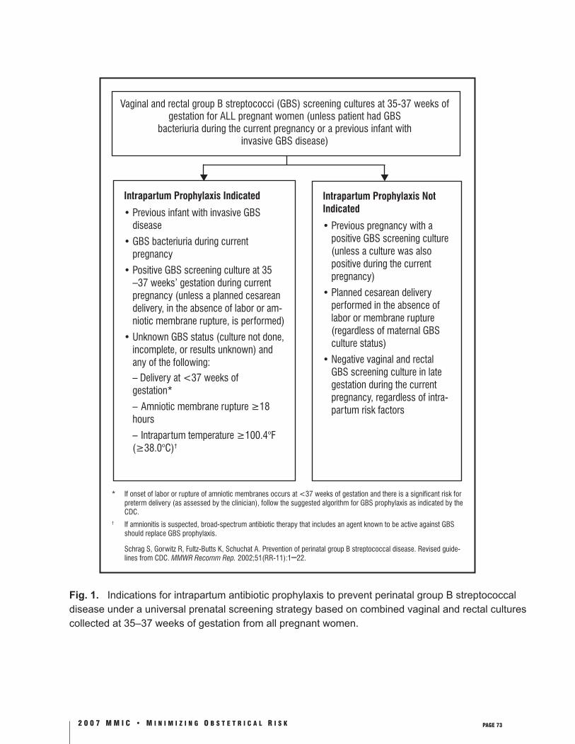
2 0 0 7 M M I C • M I N I M I Z I N G O B S T E T R I C A L R I S K PAGE 73
* If onset of labor or rupture of amniotic membranes occurs at <37 weeks of gestation and there is a significant risk for preterm delivery (as assessed by the clinician), follow the suggested algorithm for GBS prophylaxis as indicated by the CDC.
† If amnionitis is suspected, broad-spectrum antibiotic therapy that includes an agent known to be active against GBS should replace GBS prophylaxis.
Schrag S, Gorwitz R, Fultz-Butts K, Schuchat A. Prevention of perinatal group B streptococcal disease. Revised guide-lines from CDC. MMWR Recomm Rep. 2002;51(RR-11):1–22.
Fig. 1. Indications for intrapartum antibiotic prophylaxis to prevent perinatal group B streptococcal disease under a universal prenatal screening strategy based on combined vaginal and rectal cultures collected at 35–37 weeks of gestation from all pregnant women.
Intrapartum Prophylaxis Indicated
• Previous infant with invasive GBS disease
• GBS bacteriuria during current pregnancy
• Positive GBS screening culture at 35 –37 weeks’ gestation during current pregnancy (unless a planned cesarean delivery, in the absence of labor or am-niotic membrane rupture, is performed)
• Unknown GBS status (culture not done, incomplete, or results unknown) and any of the following:
– Delivery at <37 weeks of gestation*
– Amniotic membrane rupture ≥18 hours
– Intrapartum temperature ≥100.4°F (≥38.0°C)†
Intrapartum Prophylaxis Not Indicated
• Previous pregnancy with a positive GBS screening culture (unless a culture was also positive during the current pregnancy)
• Planned cesarean delivery performed in the absence of labor or membrane rupture (regardless of maternal GBS culture status)
• Negative vaginal and rectal GBS screening culture in late gestation during the current pregnancy, regardless of intra-partum risk factors
Vaginal and rectal group B streptococci (GBS) screening cultures at 35-37 weeks of gestation for ALL pregnant women (unless patient had GBS
bacteriuria during the current pregnancy or a previous infant withinvasive GBS disease)

2 0 0 7 M M I C • M I N I M I Z I N G O B S T E T R I C A L R I S K PAGE 74
3. Centers for Disease Control and Prevention. Prevention of perinatal group B streptococcal disease: a public health perspective. MMWR Morb Mortal Wkly Rep. 1996;45(RR-7):1–24.
4. American College of Obstetricians and Gyne-cologists, Committee on Obstetric Practice. Prevention of early-onset group B streptococ-cal disease in newborns. ACOG Committee Opinion Number 279. Washington, DC: American College of Obstetricians and Gyne-cologists; December, 2002.
5. Boyer KM, Gotoff SP. Prevention of early-on-set group B streptococcal disease with selective intrapartum chemoprophylaxis. N Engl J Med. 1986;314:1665–1669.
6. Boyer KM, Gadzala CA, Kelly PD, et al. Selec-tive intrapartum chemoprophylaxis of neona-tal group B streptococcal early-onset disease, II. Predictive value of prenatal cultures. J Infect Dis. 1983;148:802–809.
7. Amstey MS, Gibbs RS. Is penicillin G a better choice than ampicillin for prophylaxis of neo-natal group B streptococcal infections? Obstet Gynecol. 1994;84:1058–1059.
8. Carstensen H, Christensen KK, Grennert L, Persson K, Polberger S. Early-onset neonatal group B streptococcal septicaemia in siblings. J Infect. 1988;17:201–204.

2 0 0 7 M M I C • M I N I M I Z I N G O B S T E T R I C A L R I S K PAGE 75
The Neurologically Impaired Neonate
Historically, obstetrics leads other specialties in the total dollars spent in malpractice claims, largely due to the number of impaired neonates in which the enormous expense of long-term care is picked up by the malpractice insurer. A fac-tor in the expense of these cases is the tendency of plaintiff attorneys to exploit misconceptions regarding intrapartum events. For example, the plaintiff’s bar has been able to successfully argue that cerebral palsy is caused by intrapartum events that could have been prevented through effective predelivery monitoring and cesarean section. In actuality, studies show that at least 85% of cere-bral palsy cases were caused by undetected and untreatable events. An Oxford study puts the figure as high as 88%, reporting that only 12% of cerebral palsy cases related to intrapartum events were potentially preventable. The vast majority of cases were due to antenatal or postnatal causes with no evidence of intrapartum asphyxia.
These cases are hard to defend because the po-tential causes are infinite, given the multitude of obstetric, genetic, environmental, and infectious etiologic possibilities. Factors that determine the legal outcome of such cases include:
◆ Jury’s sympathy for the patient’s loss
◆ Manipulative strategies of plaintiff’s bar
◆ Credibility of expert witness testimony
◆ Failure to adhere to a standard of practice, whether or not causative of the child’s impairment
Progress has been made, however, in identifying causes of neonatal neurologic impairment which implicate antepartum rather than intrapartum events. For example, a recent study in San Fran-cisco found an increased association between cerebral palsy and the presence of maternal infec-tion at the time of admission for labor. The use of monitoring techniques, laboratory data, and
neuro-imaging studies will help provide ammuni-tion for defending claims without merit.
Clinical And Laboratory Indicators
Careful evaluation and documentation of data is critical during and after delivery of an affected child. While no single indicator can determine the exact time of neurologic damage, a combination of the following indicators, consistent with an unde-tectable and untreatable antepartum etiology, can provide helpful defense against an unwarranted claim.
Electronic Fetal MonitorThe fetal monitor is one of the best tools for esti-mating the time of the neurologic injury. A review of continuous monitor tracings started at the time of admission may make it possible to differentiate an antepartum event from an intrapartum event. For example, an initial flat tracing which lacks normal variability would strongly suggest an infant who was compromised at the time of admission or earlier. Conversely, the tracing of an infant with an initially normal baseline and variability which subsequently shows decelerations, decreased variability, tachycardia, and ultimately profound bradycardia would more likely suggest intrapartum asphyxia.
Apgar ScoreAn Apgar score of less than 3 at 10 minutes has been associated with birth asphyxia, whereas a score above 6 at 10 minutes is less consistent with acute intrapartum asphyxia.
Cord Blood GasesCord gases with normal pH, pO
2, pCO
2, and base
excess are strong evidence that an acute intrapar-tum hypoxic event did not occur. An infant with elevated pCO
2 who responds quickly to simple
resuscitation efforts also helps to differentiate a benign transient acute respiratory acidosis from a

2 0 0 7 M M I C • M I N I M I Z I N G O B S T E T R I C A L R I S K PAGE 76
metabolic acidosis related to a chronic or preexist-ing condition. For example, a metabolic acidosis with a base excess of –10.0 or greater is more likely to be associated with neurologic injury.
Hematological StudiesRecent literature suggests that nucleated red cell, platelet, and white cell counts are useful in dif-ferentiating acute hypoxic events from chronic and preexisting ones. For example, recent reports indicate that a preexisting hypoxic event will dem-onstrate higher nucleated red-cell counts which return to normal more slowly than those mea-sured for an intrapartum event. Controversy exists regarding the validity and significance of these tests in pinpointing the time of neurologic injury. This information may prove valuable to your defense, however, in the event of a future claim.
Bacterial CulturesAn expanding body of information suggests that intrauterine exposure to infection endangers the brains of normal-birth weight neonates. In one study, infants born from mothers with infection were ten times more likely to have unexplained cerebral palsy than controls. In the same study, half of the infants whose mothers had infection had five-minute Apgar scores of less than six. An-other study reveals that very low birth weights and cerebral palsy were four times more likely to have positive placental cultures for E. coli than those without cerebral palsy. Only one in ten mothers, however, had clinical signs of infection. These two studies confirm the hypothesis that infection-in-duced elevation of cytokines in fetal brains leads to infant brain damage.
The risk management recommendation is to obtain cultures and placental examination in ALL babies with low Apgar, very low birth weight, and abnormal neurological findings.
PlacentaHistological examination of the placenta may reveal etiological clues for unexplained cerebral palsy. For example, placental evidence may pro-vide the only clues for chorioamnionitis related to an antepartum occult infection, and old infarcts would be more suggestive of an antepartum event. Umbilical cord evaluations can also help identify etiologies for a compromised infant such as infec-tion (funisitis) or structural abnormalities (true knots, velementous cord insertions, vasa previa).
EncephalopathyThe acutely asphyxiated neonate usually demon-strates signs of encephalopathy, such as seizure activity or prolonged hypotonia. Neonates will generally exhibit signs of encephalopathy within 24 hours after delivery. This is helpful in signaling the practitioner to carefully review the intrapartum events and make sure all pertinent information is well documented.
End Organ DamageA detailed neonatal exam and laboratory evalu-ation, particularly of the kidneys and liver, is critical to rule out acute asphyxiation. Intrapartum asphyxial events that lead to neurologic damage will usually include kidney and liver involvement identified in the neonatal period.
Neuro-Imaging StudiesNeonatal ultrasound, CAT scan, and MRI find-ings may be useful for determining the etiology of a neurologic abnormality. For example, intra-cranial malformations or calcifications would be suggestive of antepartum infection present for at least one week. Periventricular leukomalacia seen on ultrasound suggests an injury at least 48 hours old or greater.
Samples of neonatal meconium for MecStat can assess prenatal drug exposure.

2 0 0 7 M M I C • M I N I M I Z I N G O B S T E T R I C A L R I S K PAGE 77
Summary
While none of these indicators alone can deter-mine an absolute time of neurologic injury, taken collectively they can be shown to be more consis-tent with chronic antepartum events, as opposed to acute intrapartum events. A persuasive causa-tion defense can therefore be provided. The bot-tom line is that collecting and documenting this information is of utmost importance for defending your management in such unfortunate outcomes.
Risk Management Recommendations
◆ A comprehensive history and physical should be performed at the patient’s initial antepar-tum visit to adequately assess and accurately document the level of risk. Particular attention should be given to past obstetrical history and any family history of abnormalities, as well as any history of smoking, drug abuse, and medi-cation exposure in the early prenatal period. It is important to notify the patient and discuss concerns about any potential increased risks in pregnancy. All tests, their results, and patient notification/discussion should be documented thoroughly.
◆ Any additional related information learned at subsequent prenatal visits should also be well documented.
◆ Obstetric departments should develop policies and protocols designating specific information to gather and explaining how this information should be stored. At the least, fetal monitor strips should be carefully preserved, cord blood gases obtained, and placentas saved for patho-logic evaluation. (When sending the placenta to the pathologist, specify particulars such as maternal fever, R/O amnionitis, etc.)
◆ Sometimes a provider may feel discomfort, fear, or guilt in the event of an adverse outcome. It is precisely at such times, however, that it is imperative that the patient not feel abandoned.
Do not minimize patient contact. Keeping the lines of communication with the patient and family open, making information readily avail-able, and engaging in honest and frank discus-sions may prevent the kind of anger which leads to litigious action.
◆ Neuro-reconstructive surgery should be con-sidered for Erb’s palsy from shoulder dystocia which does not improve by four weeks of age. Several centers report positive results when this is treated early.
◆ Communication may be facilitated if you let the pediatrician know that you would like follow-up on the infant’s progress. It is par-ticularly important to coordinate information given to the parents by all providers (obstetric, pediatric, neonatal, and nursing staff), as par-ents will focus on any discrepancies or seeming contradictions.
◆ As soon as neurologic impairment is evident, inform the liability carrier’s claims department for assistance and advice.
Reference
American College of Obstetricians and Gynecolo-gists/American Academy of Pediatrics. Neonatal encephalopathy and cerebral palsy: defining the pathogenesis and pathophysiology. ACOG/AAP Report on Infant Brain Damage. Washington, DC: American College of Obstetricians; 2003.

2 0 0 7 M M I C • M I N I M I Z I N G O B S T E T R I C A L R I S K PAGE 78
This page left intentionally blank.

2 0 0 7 M M I C • M I N I M I Z I N G O B S T E T R I C A L R I S K PAGE 79
Neonatal Encephalopathy & Cerebral Palsy
Knowledge about the causes of neonatal encepha-lopathy and cerebral palsy is rapidly developing. A report published in 2003 by a joint task force of the American College of Obstetricians and Gyne-cologists (ACOG) and the American Academy of Pediatrics (AAP) shed new light on the cause of neonatal encephalopathy and cerebral palsy.
Previously, evidence of birth asphyxia and hypoxic-ischemic neonatal encephalopathy was based upon four nonspecific clinical signs: (1) meco-nium-stained amniotic fluid, (2) nonreassuring fetal heart rate patterns, (3) low Apgar scores, and (4) neonatal encephalopathy. The evidence-based findings of the ACOG Task Force demonstrate that these peripartum signs are most often the secondary results of pathologic processes estab-lished before labor. New data show that less than a quarter of infants with neonatal encephalopathy show evidence of hypoxia or ischemia at birth. In most cases the events leading to cerebral palsy are caused by numerous and unpreventable factors during fetal development or after delivery. A large proportion of cerebral palsy cases are associated with conditions such as pre-term birth, intrauter-ine growth restriction, intrauterine infection, coagulation disorders, multiple pregnancies, antepartum hemorrhage, breech presentation, and chromosomal or congenital abnormalities. Physi-cians should therefore be aware that these four nonspecific signs should no longer be used as the main evidence for a diagnosis of birth asphyxia.
ACOG has issued new criteria to define an acute intrapartum hypoxic event. The nine criteria help to assess the likelihood that the pathology causing the cerebral palsy happened during labor. They focus on the analysis of peripartum blood gases as essential to demonstrate that hypoxia was present around the time of birth. All four of the follow-ing essential criteria must be present to define
an acute intrapartum hypoxic event sufficient enough to cause cerebral palsy:
1. Evidence of a metabolic acidosis in fetal umbili-cal cord arterial blood obtained at delivery (pH <7 and base deficit ≥12 mmol/L)
2. Early onset of severe or moderate neonatal encephalopathy in infants born at 34 or more weeks of gestation
3. Cerebral palsy of the spastic quadriplegic or dyskinetic type
4. Exclusion of other identifiable etiologies such as trauma, coagulation disorders, infectious conditions, or genetic disorders
If any one of the four essential criteria is not met, intrapartum hypoxia did not likely cause the cerebral palsy. If all four criteria are met, you must then establish whether the hypoxia is due to long-term hypoxia, or whether hypoxia occurred acutely during labor in a previously healthy fetus. Five criteria are reported to collectively suggest an intrapartum timing (within 48 hours of labor and delivery) but are nonspecific to asphyxial insults. Most or all of these five signs will be present as a group in severe cases of intrapartum hypoxia.
1. A sentinel hypoxic event (e.g., ruptured uterus) occurring immediately before or during labor
2. A sudden and sustained fetal bradycardia or the absence of fetal heart rate variability in the presence of persistent, late, or variable decelera-tions, after a hypoxic sentinel event when the pattern was previously normal
3. Apgar scores of 0–3 beyond 5 minutes
4. Onset of multisystem involvement within 72 hours of birth
5. Early imaging study showing evidence of acute nonfocal cerebral abnormality

2 0 0 7 M M I C • M I N I M I Z I N G O B S T E T R I C A L R I S K PAGE 80
Neonatal Evaluation
Assessing the depressed newborn, especially after an uncomplicated delivery, should include:
1. Consideration of maternal and family medical conditions, such as
a. Thyroid or other immune disorders
b. Deep vein and other thrombotic disorders
c. Intolerance to oral contraceptives
d. Early stroke or myocardial infarction
e. History of prior pregnancy loss
i. Death of twin fetus, even early in the pregnancy?
ii. Maternal history of chorioamnionitis or sexually transmitted disease?
iii. Evidence of intrapartum maternal fever?
2. Examination of the umbilical cord, membranes, and placenta
3. Immediate laboratory studies
a. Examination of the placenta, cord, and membranes
b. Cord gases
c. Placental cultures
d. Maternal and/or fetal blood cultures
4. Later laboratory studies
a. Imaging studies
b. Liver function tests and renal function tests
c. In the event of signs of thrombosis or stroke, examine indicators of thrombophilia
d. Consider submitting placenta for formal pathologic investigation
Conclusion
Much remains to be learned about the causes and prevention of cerebral palsy. The 2003 ACOG/AAP report reveals new information that might alleviate undue blame on physicians for cerebral palsy, especially when labor management was not ideal. Further research into the causes of neonatal encephalopathy and cerebral palsy will hopefully lead to interventions that will reduce the inci-dence of these conditions.
Key Points
Physicians are strongly advised to:
◆ Document whether or not the four criteria exist that define an acute intrapartum hy-poxic event for all newborns who may have or develop neonatal encephalopathy or cerebral palsy. The documentation should be done as close as possible to the time of delivery.
◆ Document whether or not the five criteria exist which suggest an intrapartum timing but are nonspecific to asphyxial insults.
◆ Unless you have determined so from the above-mentioned criteria, refrain from using terms such as “newborn asphyxia,” “birth trauma,” or “hypoxic/ischemic encephalopa-thy from birth.”
◆ Refrain from mentioning a diagnosis such as “newborn asphyxia” to parents unless you have confirmed the information in the chart or with the delivering physician. Ensure that all members of the delivery team are cautioned on this point.
◆ Read the entire ACOG/AAP report, Neonatal Encephalopathy and Cerebral Palsy: Defining the Pathogenesis and Pathophysiology.

2 0 0 7 M M I C • M I N I M I Z I N G O B S T E T R I C A L R I S K PAGE 81
Reference
American College of Obstetricians and Gynecolo-gists/American Academy of Pediatrics. Neonatal encephalopathy and cerebral palsy: defining the pathogenesis and pathophysiology. ACOG/AAP Report on Infant Brain Damage. Washington, DC: American College of Obstetricians; 2003.

2 0 0 7 M M I C • M I N I M I Z I N G O B S T E T R I C A L R I S K PAGE 82
This page left intentionally blank.

2 0 0 7 M M I C • M I N I M I Z I N G O B S T E T R I C A L R I S K PAGE 83
Perinatal Genetic Testing
Introduction
In the last decade, advances in genetics and genomics have provided physicians with more knowledge about human diseases than ever before. With this knowledge and ability to iden-tify at-risk individuals with “just a blood sample” comes the added pressure to provide a compre-hensive family history/genetic evaluation of each new patient. In obstetrics, the two leading causes of infant mortality are perinatal conditions (pre-term delivery) and congenital malformations. Two percent of fetuses have a chromosome abnormal-ity, 2% have a significant structural abnormality, and another 3% have a single gene disorder that will be evident by age 25. Thus, all conceptions have a more than 5% chance of an abnormal fetal outcome.
The current basic screening for couples or preg-nancies at risk utilizes a combination of family history, maternal serum screening, and ultrasound surveillance. The purpose of this section is to review the indications for genetic counseling and prenatal diagnosis, and the diagnostic modalities available in prenatal diagnosis.
Indications For Genetic Counseling/Genetic Testing
◆ Family History of Genetic ConditionThe family history continues to be one of the most important identifiers for genetic risk. Uhrich et al. discovered through a pilot study that about 20% of patients seen for routine prenatal care were found to have an increased genetic risk based on a family history evalua-tion. The evaluation included the patient and her partner, as well as their parents, siblings and their children, and aunts, uncles, and their children. Therefore, information in addition to the routine family history questions about medical conditions should include details about stillborns and members with birth
defects, mental retardation, or a known genetic disorder. Geographic or ethnic background and consanguinity should also be documented.
Most known genetic disorders are caused by single gene defects and have a recurrence risk of 25% or 50%. Prenatal diagnosis could be possible for the at-risk fetus if a biochemical abnormality or a DNA mutation is associated with the disease. These couples should be referred for genetic counseling as early in the pregnancy as possible because coordination of the testing can take many weeks. Also, some genetic conditions (e.g., osteogenesis imper-fecta) require samples obtained at chorionic villus sampling and not at amniocentesis for diagnosis.
In other cases in which there is a specific fam-ily history of an X-linked condition for which there is no biochemical or DNA diagnosis, prenatal diagnosis is possible for sex determina-tion only.
◆ Maternal Age Historically, a maternal age of 35 at the time
of delivery has been used to identify women for amniocentesis to exclude fetal trisomy. This age was selected because the risk at 35 years for a liveborn with trisomy approximates the risk of second-trimester amniocentesis. The following table summarizes the incidence of Down Syndrome at midtrimester and at term for different maternal ages. The inci-dence of fetal aneuploidy at term is less than the incidences at the time of chorionic villus sampling or amniocentesis because of the natural spontaneous loss of chromosomally abnormal fetuses.
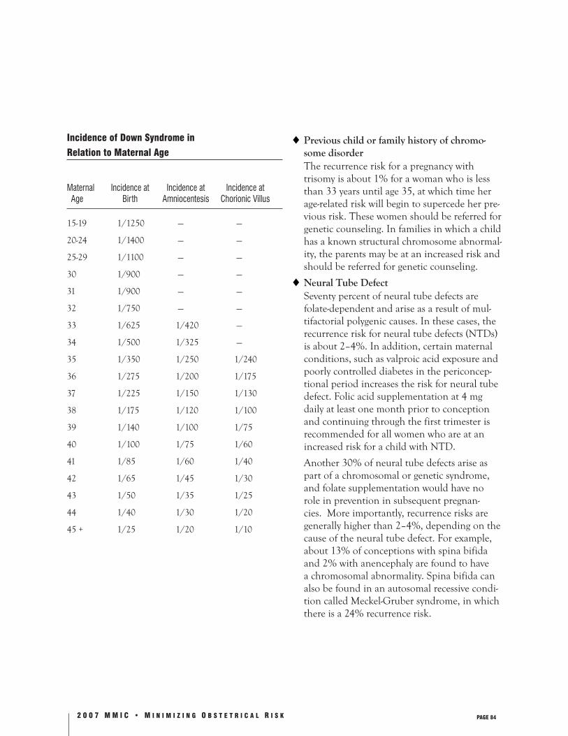
2 0 0 7 M M I C • M I N I M I Z I N G O B S T E T R I C A L R I S K PAGE 84
Incidence of Down Syndrome in
Relation to Maternal Age
Maternal Incidence at Incidence at Incidence at Age Birth Amniocentesis Chorionic Villus
15-19 1/1250 — —
20-24 1/1400 — —
25-29 1/1100 — —
30 1/900 — —
31 1/900 — —
32 1/750 — —
33 1/625 1/420 —
34 1/500 1/325 —
35 1/350 1/250 1/240
36 1/275 1/200 1/175
37 1/225 1/150 1/130
38 1/175 1/120 1/100
39 1/140 1/100 1/75
40 1/100 1/75 1/60
41 1/85 1/60 1/40
42 1/65 1/45 1/30
43 1/50 1/35 1/25
44 1/40 1/30 1/20
45 + 1/25 1/20 1/10
◆ Previous child or family history of chromo-some disorder
The recurrence risk for a pregnancy with trisomy is about 1% for a woman who is less than 33 years until age 35, at which time her age-related risk will begin to supercede her pre-vious risk. These women should be referred for genetic counseling. In families in which a child has a known structural chromosome abnormal-ity, the parents may be at an increased risk and should be referred for genetic counseling.
◆ Neural Tube Defect Seventy percent of neural tube defects are
folate-dependent and arise as a result of mul-tifactorial polygenic causes. In these cases, the recurrence risk for neural tube defects (NTDs) is about 2–4%. In addition, certain maternal conditions, such as valproic acid exposure and poorly controlled diabetes in the periconcep-tional period increases the risk for neural tube defect. Folic acid supplementation at 4 mg daily at least one month prior to conception and continuing through the first trimester is recommended for all women who are at an increased risk for a child with NTD.
Another 30% of neural tube defects arise as part of a chromosomal or genetic syndrome, and folate supplementation would have no role in prevention in subsequent pregnan-cies. More importantly, recurrence risks are generally higher than 2–4%, depending on the cause of the neural tube defect. For example, about 13% of conceptions with spina bifida and 2% with anencephaly are found to have a chromosomal abnormality. Spina bifida can also be found in an autosomal recessive condi-tion called Meckel-Gruber syndrome, in which there is a 24% recurrence risk.

2 0 0 7 M M I C • M I N I M I Z I N G O B S T E T R I C A L R I S K PAGE 85
◆ Abnormal Maternal Serum Screening/Abnor-mal Ultrasound
Genetic counseling and further assessment at a prenatal genetics center should be offered to all pregnancies in which fetal abnormalities are detected or suspected on ultrasound or when maternal serum screening is positive.
Noninvasive Prenatal Diagnosis Testing
◆ Maternal Serum Screening—Second Trimester
This term refers to the measurement of bio-chemical markers in maternal serum between the 15th and 20th weeks of pregnancy to iden-tify pregnancies that may be at an increased risk for Down Syndrome, neural tube defects or abdominal wall defect, or Trisomy 18. In the triple-marker screen, three analytes, alpha-fetoprotein (AFP), unconjugated estriol (uE3), and human chorionic gonadotropin (hCG) are measured. In the Quad Screen, the fourth analyte, dimeric inhibin A is added. The interpretation of each analyte is dependent on gestational age, maternal weight, ethnicity, and whether the patient has diabetes. The follow-ing table summarizes the pattern of analyte abnormality for Down Syndrome, Trisomy 18, and NTD/abdominal wall defect. All pregnant women who enter prenatal care prior to 20 weeks’ gestation should be informed of the availability of maternal serum screening.
◆ Serum Marker Screen Second-Trimester Screening
AFP uE3 hCG INH
Down Syndrome
Trisomy 18 NA
◆ Ultrasound Real-time ultrasonography has become an
integral part of prenatal care. Currently, 60–70% of pregnant women in the United States will undergo ultrasound at some time during the pregnancy. In low-risk pregnancies, a midtrimester ultrasound (around 18–20 weeks’ gestation) in conjunction with mater-nal serum screening could be completed if the patient presents for obstetrical care prior to 20 weeks and ultrasound is available in the com-munity.
◆ Preimplantation Genetic Diagnosis (PGD) This is a procedure that is used in conjunc-
tion with in vitro fertilization (IVF) in which a single blastomere is removed at the 6–8 cell stage and analyzed for chromosome abnor-mality or single gene defects. Situations in which PGD has been used include carriers of structural chromosome rearrangements in which there is a high percentage of chromo-somally abnormal conceptions. Preimplanta-tion genetic diagnosis allows the parents to transfer the chromosomally normal zygote to achieve a pregnancy rather than having to wait for prenatal diagnosis and termination of an affected pregnancy. Single gene defects in which a mutation has been identified can also undergo PGD and transfer of the unaf-fected zygote.
Carrier Screening
◆ Cystic Fibrosis In 2001, the American College of Obstetrics
and Gynecology (ACOG) and the American College of Medical Genetics formalized their recommendations for carrier screening for cys-tic fibrosis. Specifically, CF screening should be available to the following individuals:
• Those with a family of CF
• Reproductive partners of CF patients

2 0 0 7 M M I C • M I N I M I Z I N G O B S T E T R I C A L R I S K PAGE 86
• Caucasian couples who are pregnant or planning a pregnancy
• Individuals of other ethnic groups if pregnant or planning a pregnancy
Because there are now over 1,000 mutations associated with CF, and because of the com-plexity of the clinical interpretation of the mutations, patients who are found to be carri-ers of a mutation should be referred for genetic counseling.
◆ Hemoglobinopathies Individuals of African American, East Indian,
Asian, and Mediterranean background should be evaluated for hemoglobinopathies. Hemo-globin electrophoresis should be offered to individuals of African American descent to screen for sickle cell trait (and variants). In screening for thalassemia, the complete blood count (CBC) with indices is evaluated initially. Individuals with abnormal indices should then be evaluated for iron deficiency with the appro-priate studies and for thalassemia by hemoglo-bin electrophoresis. In rare cases, extremely severe iron deficiency could mask an underly-ing thalassemia which would not be evident until the patient’s iron stores were completely repleted.
◆ Tay Sachs Disease This is an autosomal recessive condition of
lysosomal storage in which the affected new-born cannot degrade a sphingolipid (GM2 ganglioside) due to a deficiency in the enzyme hexosaminidase A. Individuals of Ashkenazi Jewish ancestry have a risk of 1 in 27 to carry a mutation for this enzyme defect. Therefore, carrier screening should be offered.
Invasive Prenatal Diagnostic Testing
◆ Chorionic Villus Sampling (CVS) CVS is a placental biopsy of the chorionic
villi either transcervically or transabdominally
between the 10th and 12th week of pregnancy. Its major advantage over midtrimester amnio-centesis is that results are generally available earlier in pregnancy. In some cases, however, only biochemical testing can be completed on chorionic villi. Molecular testing may be so time-consuming that the period of uncertainty extends well into the second trimester, and in those cases CVS provides no advantage over amniocentesis. The disadvantages of CVS are a pregnancy loss rate of 1%, a 2% chance of yielding ambiguous results from chromosome mosaicism, and the inability to screen for NTD which is delayed until second-trimester maternal serum screening and ultrasound are completed.
◆ Midtrimester Amniocentesis This term refers to the transabdominal remov-
al of amniotic fluid from the uterus. In the midtrimester, analysis of the fluid is generally for genetic indications, whereas amniocente-sis in the third trimester is generally for fetal pulmonary procedure. The procedure is typi-cally performed under ultrasound guidance between the 15th and 18th week of pregnancy. The risk for pregnancy loss is about 1/250–1/400 among providers who routinely perform midtrimester genetic amniocentesis.
◆ Fetal Cordocentesis—Percutaneous Umbilical Cord Blood Sampling (PUBS)
This procedure is used to obtain a sample of fetal blood directly from the umbilical cord in utero under ultrasound guidance. Cordocen-tesis is used for diagnosis and treatment. In the former, genetic/chromosomal/biochemi-cal testing can be completed on extracted fetal cells and/or DNA. In the latter, fetal blood transfusion due to anemia from isoimmuniza-tion or infections (e.g., parvovirus) can be com-pleted. The risk of the procedure for fetal loss is about 1% among providers who routinely perform cordocentesis.