MKSAP for Students 5
362
MKSAP ® FOR STUDENTS 5 (2011) Front Matter Title Page MKSAP ® for Students 5 Medical Knowledge Self-Assessment Program Developed by American College of Physicians Clerkship Directors in Internal Medicine Copyright Page Editorial Production: Helen Kitzmiller Design: Michael E. Ripca Composition: ACP Graphic Services Copyright © 2011 by the American College of Physicians. All rights reserved. This publication is protected by copyright. No part of this publication may be reproduced, stored in a retrieval system, or transmitted in any form or by any means, electronic or mechanical, including photocopy, without the express consent of the American College of Physicians. Printed in the United States of America by Sheridan Books Important copyright information from the American College of Physicians: Unauthorized reproduction of the publication is unlawful. The American College of Physicians prohibits reproduction of this publication in its entirety in any form either for individual use or for distribution. The American College of Physicians will consider granting an individual permission to reproduce only limited portions of this publication for his or her own exclusive use. Send requests in writing to MKSAP ® for Students 5, American College of Physicians, 190 N. Independence Mall West, Philadelphia, PA 19106-1572. American College of Physicians 190 N. Independence Mall West Philadelphia, PA 19106-1572 215-351-2600 ISBN-13: 978-1-934465-54-7 ISBN-10: 1-934465-54-2 Acknowledgments MKSAP for Students 5 Editorial Board Eyad Al-Hihi, MD, MBA, FACP Associate Professor of Medicine Chief, Division of General Internal & Hospital Medicine Director, Internal Medicine Ambulatory Clerkship University of Missouri-Kansas City School of Medicine Medical Director, Medicine Clinics, Truman Med Center Kansas City, Missouri Irene Alexandraki, MD, MPH, FACP Assistant Professor, Department of Medicine Medicine Clerkship Director University of Florida College of Medicine Jacksonville, Florida Mark Allee, MD, FACP Associate Professor, Department of Medicine Medicine Clerkship Director University of Oklahoma School of Medicine Oklahoma City, Oklahoma Saad Alvi, MD, FACP Assistant Professor of Clinical Medicine M3 Clerkship Director University of Illinois College of Medicine at Peoria (UICOMP) Peoria, Illinois Alpesh Amin, MD, MBA, FACP Professor of Medicine Medicine Clerkship Director University of California, Irvine Orange, California
description
MKSAP for students
Transcript of MKSAP for Students 5

MKSAP® FO R STUDENTS 5 (2011)Front Matter
Title Page
MKSAP® for Students 5Medical Knowledge Self-Assessment Program
Developed byAmerican College of PhysiciansClerkship Directors in Internal Medicine
Copyright Page
Editorial Production: Helen KitzmillerDesign: Michael E. RipcaComposition: ACP Graphic Services
Copyright © 2011 by the American College of Physicians. All rights reserved. This publication is protected by copyright. No part of this publication may be reproduced,stored in a retrieval system, or transmitted in any form or by any means, electronic or mechanical, including photocopy, without the express consent of the AmericanCollege of Physicians.
Printed in the United States of America by Sheridan Books
Important copyright information from the American College of Physicians:Unauthorized reproduction of the publication is unlawful. The American College of Physicians prohibits reproduction of this publication in its entirety in any form either forindividual use or for distribution.
The American College of Physicians will consider granting an individual permission to reproduce only limited portions of this publication for his or her own exclusive use.Send requests in writing to MKSAP® for Students 5, American College of Physicians, 190 N. Independence Mall West, Philadelphia, PA 19106-1572.
American College of Physicians190 N. Independence Mall WestPhiladelphia, PA 19106-1572215-351-2600
ISBN-13: 978-1-934465-54-7ISBN-10: 1-934465-54-2
Acknowledgments
MKSAP for Students 5 Editorial Board
Eyad Al-Hihi, MD, MBA, FACPAssociate Professor of MedicineChief, Division of General Internal & Hospital MedicineDirector, Internal Medicine Ambulatory ClerkshipUniversity of Missouri-Kansas City School of MedicineMedical Director, Medicine Clinics, Truman Med CenterKansas City, Missouri
Irene Alexandraki, MD, MPH, FACPAssistant Professor, Department of MedicineMedicine Clerkship DirectorUniversity of Florida College of MedicineJacksonville, Florida
Mark Allee , MD, FACPAssociate Professor, Department of MedicineMedicine Clerkship DirectorUniversity of Oklahoma School of MedicineOklahoma City, Oklahoma
Saad Alvi, MD, FACPAssistant Professor of Clinical MedicineM3 Clerkship DirectorUniversity of Illinois College of Medicine at Peoria(UICOMP)Peoria, Illinois
Alpesh Amin, MD, MBA, FACPProfessor of MedicineMedicine Clerkship DirectorUniversity of California, IrvineOrange, California

Lisa M. Antes, MDAssociate ProfessorDepartment of Internal Medicine/Division of NephrologyInpatient Internal Medicine Clerkship Co-DirectorCarver College of Medicine/University of Iowa Hospitals and ClinicsIowa City, Iowa
Joel Appel, DODirector Ambulatory and Student ProgramsWayne State University School of MedicineChief Hematology/OncologySinai-Grace HospitalDetroit Medical CenterDetroit , Michigan
Jonathan S. Appelbaum, MD, FACPAssociate Professor, Clinical SciencesDirector, Internal Medicine EducationFlorida State University College of MedicineTallahassee, Florida
Scott Arnold, MD, FACPAssociate Professor, Department of MedicineMedicine Clerkship DirectorUniversity of Alabama School of MedicineTuscaloosa CampusTuscaloosa, Alabama
Emily Chism Barker, MDAssistant Professor of MedicineSenior Associate Program Director for Internal MedicineUniversity of Texas Houston Medical SchoolHouston, Texas
Jennifer Bierman, MD, FACPPrimary Care Clerkship DirectorNorthwestern University Feinberg School of MedicineChicago, Illinois
Susan Crouch Brewer, MD, FACPAssistant Dean for Clinical EducationAssociate Chair for Student ProgramsCollege of MedicineUniversity of Tennessee Health Science CenterMemphis, Tennessee
Cynthia A. Burns, MD, FACPAssistant ProfessorInternal Medicine Clerkship DirectorDepartment of Internal MedicineSection on Endocrinology & MetabolismWake Forest University School of MedicineWinston-Salem, North Carolina
Maria L. Cannarozzi, MD, FACP, FAAPAssociate Professor of Internal Medicine & PediatricsClerkship Director, Internal/Family MedicineUniversity of Central Florida College of MedicineOrlando, Florida
Danelle Cayea, MD, MSAssistant Professor of MedicineMedicine Clerkship DirectorJohns Hopkins University School of MedicineBaltimore, Maryland
J. Charles, MD, FACP, FHMAssistant Professor of MedicineDivision Education CoordinatorMayo Clinic HospitalPhoenix, Arizona
Brian J. Costello, DO

Co-Clerkship Director Ambulatory MedicineLehigh Valley Health NetworkAllentown, Pennsylvania
Camilla Curren, MDAssistant Clinical Professor of Internal MedicineOhio State University Medical CenterClinical Assistant Professor of PediatricsNationwide Childrens HospitalAssistant Clerkship Director, Ambulatory MedicineOhio State University College of MedicineColumbus, Ohio
Thomas M. DeFer, MD, FACPClerkship DirectorDivision of Medical EducationDepartment of Internal MedicineWashington University School of MedicineSt. Louis, Missouri
Stephanie A. Detterline, MD, FACPAssociate Program Director, Internal MedicineUnion Memorial HospitalMedicine Clerkship Site DirectorUniversity of Maryland School of MedicineBaltimore, Maryland
Gurpreet Dhaliwal, MDSite Director, Internal Medicine ClerkshipsSan Francisco VA Medical CenterAssociate Professor of Clinical MedicineUniversity of California San FranciscoSan Francisco, California
Gretchen Diemer, MD, FACPAssistant Professor of MedicineDirector of Undergraduate Medical EducationClerkship Director Internal MedicineAssistant Program Director Internal MedicineThomas Jefferson UniversityPhiladelphia, Pennsylvania
Anne Eacker, MD, FACPAssociate Professor, Department of MedicineMedical Director, General Internal Medicine CenterUniversity of WashingtonSeattle, Washington
Mark J Fagan, MD, FACPClerkship DirectorDepartment of MedicineAlpert Medical School of Brown UniversityProvidence, Rhode Island
Pamela J. Fall, MD, FACP, FASHProfessor of MedicineSection of Nephrology, Hypertension and TransplantationClerkship Director, Internal MedicineMedical College of GeorgiaAugusta, Georgia
L. Christine Faulk, MDMedicine Clerkship Co-DirectorUniversity of Kansas School of Medicine-WichitaWichita, Kansas
Sara B. Fazio, MD, FACPAssociate Professor, Harvard Medical SchoolDirector, Core I Medicine ClerkshipDivision of General Internal MedicineBeth Israel Deaconess Medical CenterBoston, Massachusetts
J. Michael Finley, DO , FACP, FACO IAssociate Professor of Medicine

Chief, Division of RheumatologyAssociate Dean for Graduate Medical EducationWestern University College of Osteopathic MedicinePomona, California
Jose Franco, MD, AGAFProfessor of Medicine and PediatricsDirector of HepatologyMedicine Clerkship DirectorMedical College of WisconsinMilwaukee, Wisconsin
Erica Friedman, MD, FACPAssociate DeanAssociate Professor of Medicine and Medical EducationMount Sinai School of MedicineNew York, New York
Peter Gliatto, MD, FACPAssistant Professor of Medicine and Medical EducationCo-Director, Medicine-Geriatrics ClerkshipDirector, Medicine SubinternshipMount Sinai School of MedicineNew York, New York
Susan A. Glod, MDAssistant Professor of MedicineAssociate Clerkship Director, Internal MedicinePenn State College of MedicineHershey, Pennsylvania
Gabrielle R. Goldberg, MDEducation DirectorThe Hertzberg Palliative Care InstituteAssistant ProfessorDepartment of Geriatrics and Palliative MedicineDepartment of MedicineMount Sinai School of MedicineNew York, New York
Eric Goren, MDMedicine Sub-Internship Course Co-DirectorAssistant Professor of Clinical MedicineUniversity of Pennsylvania School of MedicinePhiladelphia, Pennsylvania
Cyril M. Grum, MD, FACPProfessor and Senior Associate Chairfor Undergraduate Medical EducationDepartment of Internal MedicineInternal Medicine Clerkship DirectorUniversity of MichiganAnn Arbor, Michigan
Heather Harrell, MD, FACPAssociate Professor, Department of MedicineMedicine Clerkship DirectorDirector of Fourth Year ProgramsUniversity of Florida College of MedicineGainesville, Florida
Dan A. Henry, MD, FACPProfessor of MedicineDirector of Clinical EducationMedicine Clerkship DirectorUniversity of Connecticut School of MedicineFarmington, Connecticut
Susan Thompson Hingle , MD, FACPAssociate Professor of MedicineInternal Medicine Clerkship DirectorInternal Medicine Residency Associate Program DirectorSouthern Illinois University School of MedicineSpringfield, Illinois

William Howell, MBChBClinical Instructor of MedicineMedicine Clerkship Co-DirectorUniversity of Utah School of MedicineSalt Lake City, Utah
Eric Hsieh, MDDirector, Internal Medicine ClerkshipSenior Associate Director, Internal Medicine ResidencyDepartment of MedicineKeck School of MedicineUniversity of Southern CaliforniaLos Angeles, California
Hugh F. Huizenga, MD, MPHAssistant Professor of MedicineClerkship Director-Inpatient MedicineDartmouth Medical SchoolHanover, New Hampshire
T.J. Hundley, MD, FACPDirector, Internal Medicine ClerkshipAssistant Professor of MedicineDepartment of Internal MedicineUniversity of South Alabama College of MedicineMobile, Alabama
Nadia J. Ismail, MD, MPH, Med, FACPDirector, Internal Medicine Core ClerkshipAssistant Professor of MedicineBaylor College of MedicineHouston, Texas
Harish Iyer, MD, MRCP (UK)Chief Medical ResidentDepartment of MedicineAlbert Einstein Medical CenterPhiladelphia, Pennsylvania
Robert Jablonover, MDAssistant Professor in Internal MedicineClerkship Director in Internal MedicineGeorge Washington University School of MedicineWashington, District of Columbia
Janet A. Jokela, MD, MPH, FACPAssociate Professor of Clinical MedicineDepartment of MedicineUniversity of Illinois at Urbana-ChampaignUrbana, Illinois
Jason Kahn, MD, FACPInternal Medicine Clerkship Site DirectorSt. Joseph Mercy HospitalAnn Arbor, Michigan
Christopher A. Klipstein, MDAssociate Professor of MedicineDirector, Internal Medicine ClerkshipUniversity of North Carolina School of MedicineChapel Hill, North Carolina
Mary Ann Kuzma, MD, FACPAssociate Professor of MedicineMedicine Clerkship DirectorDrexel University College of MedicinePhiladelphia, Pennsylvania
Rosa Lee, MDMedicine Clerkship Site Leader,Montefiore Medical CenterAssistant Professor, Department of MedicineAlbert Einstein College of MedicineBronx, New York

Beth W. Liston, MD, PhD, FACPAssistant Professor of Clinical Medicine/PediatricsSub-internship Clerkship DirectorThe Ohio State University College of MedicineColumbus, Ohio
Mai A Mahmoud, MBBS, FACPAssistant Professor of MedicineMedicine Clerkship Co-DirectorWeill Cornell Medical College in QatarDoha, Qatar
Lianne Marks, MD, PhD, FACPDivision Director, Internal MedicineScott & White HealthcareAssistant Professor and Internal MedicineClerkship Director for 3rd Year Medical StudentsTexas A&M College of MedicineRound Rock, Texas
Kevin M. McKown, MDAssociate Professor and Head, Division of RheumatologyCo-Director M3 and M4 Student ProgramsDepartment of MedicineUniversity of WisconsinSchool of Medicine & Public HealthMadison, Wisconsin
Laura Meinke, MDAssistant Professor of MedicineMedicine Clerkship DirectorSection of Pulmonary & Critical Care MedicineUniversity of Arizona College of MedicineTucson, Arizona
James L. Meisel, MD, FACPClerkship Director/Evans Student EducatorAssistant Professor of MedicineBoston University School of MedicineBoston, Massachusetts
Chad S. Miller, MD, FACPInternal Medicine Clerkship DirectorAssociate Program Director, ResidencyTulane University Health Sciences CenterNew Orleans, Louisiana
Nina Mingioni, MD FACPAssociate Program Director, Internal MedicineClerkship Site Director, Internal MedicineAlbert Einstein Medical CenterPhiladelphia, Pennsylvania
Alita Mishra, MD, FACPClerkship Director, Department of MedicineHospitalist , Inova Fairfax HospitalClinical Assistant Professor of MedicineVirginia Commonwealth University School of MedicineInova CampusFalls Church, Virginia
Archana Mishra, MD, MS, FACP, FCCPAssociate Professor of MedicineAssociate Clerkship DirectorSUNY Buffalo School of Medicine and Biomedical SciencesBuffalo, New York
Lynda Misra, DO , FACP, MA EdDirector of Students - William Beaumont HospitalMedicine Clerkship DirectorOakland University William Beaumont School of MedicineRoyal Oak, Michigan
Neha Mittal, MDAssistant Professor

Year 4 Clerkship DirectorDepartment of Internal MedicineTexas Tech University Health Science CenterLubbock, Texas
Justin B. Moore, MDDivision Chief, EndocrinologyAssistant Professor, Department of MedicineMedicine Clerkship Co-DirectorUniversity of Kansas School of Medicine-WichitaWichita, Kansas
Mark T. Munekata, MD, MPH, FACPAssociate Clinical Professor of MedicineCo-Director, Ambulatory Internal Medicine ClerkshipDavid Geffen School of Medicine at UCLAMedical Director, Utilization ManagementDirector, Urgent Care ClinicHarbor-UCLA Medical CenterTorrance, California
Marty Muntz, MDAssistant Professor of MedicineDivision of General Internal MedicineAmbulatory Medicine Clerkship DirectorMedicine Subinternship DirectorMedical College of WisconsinMilwaukee, Wisconsin
David G. Naylor, MDAssistant Clerkship DirectorUniversity of Kansas Medical CenterKansas City, Kansas
Adesoji E. O derinde, MD, MSCR, FACPAssociate Program DirectorAssociate Director Student EducationDepartment of MedicineMorehouse School of MedicineAtlanta, Georgia
Thomas D. Painter MDInpatient Internal Medicine Clerkship DirectorUniversity of Pittsburgh School of MedicinePittsburgh, Pennsylvania
Carlos Palacio, MD, MPH, FACPAssociate Professor of Medicine, Department of MedicineClerkship DirectorUniversity of Florida College of Medicine-JacksonvilleJacksonville, Florida
Robert I. Pargament, MD, FACPInternal Medicine Clerkship DirectorYork HospitalYork, Pennsylvania
Alisa Peet, MDAssistant Professor of MedicineMedicine Clerkship DirectorTemple University School of MedicinePhiladelphia, Pennsylvania
Roshini C. Pinto-Powell, MDAssociate Professor of MedicineDartmouth Medical SchoolLebanon, New Hampshire
Suma Pokala, MD, FACPAssociate Professor, Department of MedicineTexas A&M Health Sciences CenterCentral Texas Veterans Health Care SystemTemple, Texas
Hari Raja, MD, FACP

Professor of MedicineClerkship DirectorDepartment of Internal MedicineUT Southwestern Medical CenterDallas, Texas
Temple A. Ratcliffe , MDAssistant Professor of MedicineAssistant Clerkship DirectorF. Edward Hebert School of MedicineUniformed Services University of the Health SciencesBethesda, Maryland
Emran Rouf, MD, FACPAssistant Professor, Department of MedicineScott & White HealthcareTexas A & M Health Science Center College of MedicineTemple, Texas
Gary M. Rull, MD, FACPAssociate Professor of Clinical Internal MedicineDoctoring DirectorDepartment of Internal MedicineSouthern Illinois University School of MedicineSpringfield, Illinois
James L. Sebastian, MD, FACPProfessor of MedicineDivision of General Internal MedicineMedical College of WisconsinMilwaukee, Wisconsin
Thomas K. Schulz , MDAssociate Program Director,Internal Medicine Residency ProgramAssociate ProfessorUniversity of Kansas School of Medicine-WichitaWichita, Kansas
Monica Ann Shaw, MD, FACPAssociate Professor of MedicineChief, Division of General Internal MedicineDirector, Internal Medicine ClerkshipsUniversity of Louisville School of MedicineLouisville, Kentucky
Leigh H. Simmons, MDAssistant in MedicineAssociate Medicine Clerkship DirectorMassachusetts General HospitalHarvard Medical SchoolBoston, Massachusetts
Harold M. Szerlip, MD, FACP, FCCP, FASNProfessor and Vice-Chair, Department of MedicineChief, Medical Service, UPH HospitalUniversity of Arizona College of MedicineTucson, Arizona
Heather Tarantino, MDAssistant ProfessorMedicine Clerkship DirectorWest Virginia University School of MedicineCharleston DivisionCharleston, West Virginia
Robert L. Trowbridge, MD, FACPAssistant Professor of MedicineTufts University School of MedicineDirector of Undergraduate Medical EducationDepartment of MedicineMaine Medical CenterPortland, Maine
John Varras, MD

Associate ProfessorInterim ChairClerkship DirectorDepartment of Internal MedicineUniversity of Nevada School of MedicineLas Vegas, Nevada
T. Robert Vu, MD, FACPAssociate Professor of Clinical MedicineDirector, Internal Medicine ClerkshipIndiana University School of MedicineIndianapolis, Indiana
Joseph T. Wayne, MD, MPH, FACPInternal Medicine Clerkship DirectorDepartment of Internal MedicineAlbany Medical CollegeAlbany, New York
Barry J. Wu, MD, FACPAssociate Program Director of Internal MedicineHospital of Saint RaphaelClinical Professor of MedicineYale University School of MedicineNew Haven, Connecticut
Parekha Yedla, MBBS, FACPAssistant Professor, Department of MedicineMedicine Clerkship DirectorUniversity of Alabama, Huntsville CampusHuntsville, Alabama
MKSAP for Students 5 Contributors
Arlina Ahluwalia, MD, FACPAssociate Professor of MedicineMedicine Clerkship Site Director, Palo Alto VAHCSStanford University School of MedicinePalo Alto, California
Erik K. Alexander, MD, FACPDirector, Medical Student EducationBrigham & Women's HospitalAssociate Professor of MedicineHarvard Medical SchoolBoston, Massachusetts
Irene Alexandraki, MD, MPH, FACPAssistant Professor, Department of MedicineMedicine Clerkship DirectorUniversity of Florida College of MedicineJacksonville, Florida
Mark Allee , MD, FACPAssociate Professor of MedicineClerkship DirectorDepartment of Internal MedicineUniversity of Oklahoma College of MedicineOklahoma City, Oklahoma
Alpesh Amin, MD, MBA, FACPProfessor of MedicineMedicine Clerkship DirectorUniversity of California, IrvineOrange, California
Joel Appel, DODirector, Ambulatory and Subinternship ProgramsWayne State University School of MedicineDetroit , Michigan
Jonathan S. Appelbaum, MD, FACPAssociate Professor, Clinical Sciences DepartmentDirector, Internal Medicine EducationFlorida State University College of Medicine

Tallahassee, Florida
MJ Barchman, MD, FACP, FASNProfessor of MedicineInternal Medicine Clerkship DirectorSection of Nephrology and HypertensionBrody School of Medicine at East Carolina UniversityGreenville, North Carolina
Gonzalo Bearman, MD, MPHAssociate Professor of MedicineAssociate Hospital EpidemiologistMedicine Clerkship DirectorVirginia Commonwealth UniversityRichmond, Virginia
Seth Mark Berney, MD, FACPProfessor of MedicineChief, Section of RheumatologyDirector, Center of Excellence for Arthritis and RheumatologyLouisiana State University Health Sciences Center School of Medicine in ShreveportShreveport, Louisiana
Jennifer Bierman, MD, FACPPrimary Care Clerkship DirectorNorthwestern University Feinberg School of MedicineChicago, Illinois
Cynthia A. Burns, MD, FACPAssistant ProfessorInternal Medicine Clerkship DirectorDepartment of Internal MedicineSection on Endocrinology & MetabolismWake Forest University School of MedicineWinston Salem, North Carolina
Danelle Cayea, MD, MSAssistant Professor of MedicineMedicine Clerkship DirectorJohns Hopkins University School of MedicineBaltimore, Maryland
J. Charles MD, FACP, FHMAssistant Professor of MedicineDivision Education CoordinatorMayo Clinic HospitalPhoenix, Arizona
Amanda Cooper, MDAssistant Professor of MedicineUniversity of Pittsburgh Medical CenterUniversity of Pittsburgh School of MedicinePittsburgh, Pennsylvania
Mark D. Corriere, MD, FACPAssociate Clerkship Director, Department of MedicineUniformed Services University of the Health SciencesBethesda, Maryland
Gretchen Diemer, MD, FACPAssistant Professor of Internal MedicineClerkship Director Internal MedicineDirector of Undergraduate Medical EducationAssociate Program Director Internal MedicineThomas Jefferson UniversityPhiladelphia, Pennsylvania
Reed E. Drews, MD, FACPAssociate ProfessorHarvard Medical SchoolProgram Director, Hematology-Oncology FellowshipBeth Israel Deaconess Medical CenterBoston, Massachusetts

D. Michael Elnicki, MD, FACPProfessor and Chief, Section of General Internal Medicine, UPMC ShadysideAmbulatory Clerkship DirectorUniversity of PittsburghPittsburgh, Pennsylvania
Mark J. Fagan, MD, FACPClerkship DirectorDepartment of MedicineAlpert Medical School of Brown UniversityProvidence, Rhode Island
Sara B. Fazio, MD, FACPAssociate Professor, Harvard Medical SchoolDirector, Core I Medicine ClerkshipDivision of General Internal MedicineBeth Israel Deaconess Medical CenterBoston, Massachusetts
J. Michael Finley, DO , FACP, FACO IAssociate Professor of MedicineChief, Division of RheumatologyAssociate Dean for Graduate Medical EducationWestern University College of Osteopathic MedicinePomona, California
Jane P. Gagliardi, MD, MHSAssistant Professor of Psychiatry & Behavioral SciencesAssistant Professor of MedicineDirector of UME, Department of MedicineMedicine Clerkship and Subinternship DirectorDuke University School of MedicineDurham, North Carolina
Peter Gliatto, MD, FACPAssociate Dean for Undergraduate Medical Education and Student AffairsMount Sinai School of MedicineNew York, New York
Eric H. Green MD, MSc, FACPClinical Associate Professor of MedicineDrexel University College of MedicineAssociate Program DirectorMercy Catholic Medical CenterDarby, Pennsylvania
Heather Harrell, MD, FACPAssociate Professor, Department of MedicineMedicine Clerkship DirectorDirector of Fourth Year ProgramsUniversity of Florida College of MedicineGainesville, Florida
Warren Hershman, MD, MPHDirector of Student EducationBoston University School of MedicineDepartment of MedicineBoston, Massachusetts
Susan Thompson Hingle , MD, FACPAssociate Professor of MedicineInternal Medicine Clerkship DirectorInternal Medicine Residency Associate Program DirectorSouthern Illinois University School of MedicineSpringfield, Illinois
Bryan Ho, MDAssistant Professor, Department of NeurologyNeurology Clerkship DirectorTufts University School of MedicineBoston, Massachusetts

Mark D. Holden, MD, FACPVice-Chair for Undergraduate EducationDepartment of Internal MedicineUniversity of Texas Medical BranchGalveston, Texas
Jeffrey Guy House, DO , FACPAssistant Professor of MedicineProgram Director Internal Medicine ResidencyUniversity of Florida Health Science Center-JacksonvilleJacksonville, Florida
Eric Hsieh, MDDirector, Internal Medicine ClerkshipSenior Associate Director, Internal Medicine ResidencyDepartment of MedicineKeck School of MedicineUniversity of Southern CaliforniaLos Angeles, California
Robert Jablonover, MDAssistant Professor in Internal MedicineClerkship Director in Internal MedicineGeorge Washington University School of MedicineWashington, District of Columbia
Saba Khan, MDFellow, Section of RheumatologyLouisiana State University Health Sciences CenterSchool of Medicine in ShreveportShreveport, Louisiana
Sarang Kim, MD, FACPAssistant Professor of MedicineDivision of General Internal MedicineUniversity of Medicine and Dentistry of New Jersey, Robert Wood Johnson Medical SchoolNew Brunswick, New Jersey
Valerie J. Lang, MD, FACPDirector, Inpatient Medicine ClerkshipUniversity of Rochester School of Medicine & DentistryHospital Medicine DivisionRochester, New York
Rosa Lee, MDClinical Assistant Professor, Department of MedicineAlbert Einstein College of MedicineSite Leader, Medicine ClerkshipMontefiore Medical CenterBronx, New York
Bruce Leff, MD, FACPProfessor of MedicineCo-Director, Basic Medicine ClerkshipJohns Hopkins University School of MedicineBaltimore, Maryland
Kyle L. Lokitz , MDFellow, Section of RheumatologyLouisiana State University Health Sciences CenterSchool of Medicine in ShreveportShreveport, Louisiana
Fred A. Lopez, MD, FACPRichard Vial Professor and Vice ChairDepartment of MedicineLouisiana State University Health Sciences CenterNew Orleans, Louisiana
Kevin M. McKown, MD, FACPAssociate Professor and Head, Division of RheumatologyCo-Director M3 and M4 Student ProgramsDepartment of MedicineUniversity of Wisconsin

School of Medicine & Public HealthMadison, Wisconsin
Chad S. Miller, MD, FACPDirector, Student ProgramsAssociate Program Director, ResidencyDepartment of Internal MedicineTulane University Health Sciences CenterNew Orleans, Louisiana
Katherine Nickerson, MDProfessor of Clinical MedicineVice Chair, Department of MedicineClerkship Director, Internal MedicineCollege of Physicians & SurgeonsColumbia UniversityNew York, New York
L. James Nixon, MDVice Chair for Education, Department of MedicineMedicine Clerkship DirectorUniversity of Minnesota Medical SchoolMinneapolis, Minnesota
Isaac O . O pole , MD, PhD, FACPAssistant Dean for Student AffairsInternal Medicine Clerkship DirectorKansas University Medical CenterKansas City, Kansas
Carlos Palacio, MD, MPH, FACPAssociate Professor of Medicine, Department of MedicineClerkship DirectorUniversity of Florida College of Medicine-JacksonvilleJacksonville, Florida
Suma Pokala, MD, FACPAssociate Professor, Department of MedicineTexas A&M Health Sciences CenterCentral Texas Veterans Health Care SystemTemple, Texas
Nora L. Porter, MD, MPH, FACPCo-director, Internal Medicine ClerkshipSaint Louis University School of MedicineSt. Louis, Missouri
Shalini Reddy, MDAssociate DeanStudent Programs and Professional DevelopmentThe University of Chicago Pritzker School of MedicineChicago, Illinois
Klara J. Rosenquist, MDClinical/Research FellowDivision of Endocrinology, Diabetes and HypertensionBrigham & Women's HospitalHarvard Medical SchoolBoston, Massachusetts
Kathleen F. Ryan, MD, FACPAssociate Professor of MedicineDepartment of MedicineDrexel University College of MedicinePhiladelphia, Pennsylvania
Mysti D. W. Schott, MD, FACPAssociate Professor of MedicineCourse Director, Advanced Clinical Evaluation SkillsDepartment of Medicine, Division of General Medicine& Office of Educational ProgramsUniversity of Texas Health Science Center San AntonioSan Antonio, Texas
Amy Wiegner Shaheen, MD

Ambulatory Medicine Clerkship DirectorUniversity of North Carolina School of MedicineChapel Hill, North Carolina
Monica Ann Shaw, MD, FACPAssociate Professor and ChiefDivision of General Internal Medicine, PalliativeMedicine and Medical EducationMedicine Clerkship DirectorUniversity of LouisvilleLouisville, Kentucky
Patricia Short, MD, FACPAssistant Professor of MedicineAssociate Clerkship DirectorUniformed Services University of the Health SciencesMadigan Army Medical CenterTacoma, Washington
Karen Szauter, MD, FACPProfessorDepartment of Internal Medicine and Office of Educational DevelopmentCo-Director, Internal Medicine ClerkshipUniversity of Texas Medical BranchGalveston, Texas
Harold M. Szerlip, MD, FACP, FCCP, FASNProfessor and Vice-Chair, Department of MedicineChief, Medical Service, UPH HospitalUniversity of Arizona College of MedicineTucson, Arizona
Gary Tabas, MD, FACPAssociate Professor of MedicineUniversity of Pittsburgh School of MedicinePittsburgh, Pennsylvania
Dario M. Torre, MD, MPH, PhD, FACPAssociate Professor of MedicineAssociate Program Director Internal Medicine University of Pittsburgh-ShadysideUniversity of Pittsburgh School of MedicinePittsburgh, Pennsylvania
John Varras, MDAssociate ProfessorInterim ChairClerkship DirectorDepartment of Internal MedicineUniversity of Nevada School of MedicineLas Vegas, Nevada
H. Douglas Walden, MD, MPH, FACPProfessor of MedicineCo-Director, Internal Medicine ClerkshipSaint Louis University School of MedicineSt. Louis, Missouri
John A. Walker, MD, FACPProfessor and Vice-Chair for EducationDepartment of MedicineMedicine Clerkship DirectorUniversity of Medicine and Dentistry of New JerseyRobert Wood Johnson Medical SchoolNew Brunswick, New Jersey
Joseph T. Wayne, MD, MPH, FACPInternal Medicine Clerkship DirectorDepartment of Internal MedicineAlbany Medical CollegeAlbany, New York
John Jason White , MD, FASNAssociate Professor of Medicine

Section of Nephrology, Hypertension & TransplantationGeorgia Health Sciences UniversityAugusta, Georgia
The American College of Physicians gratefully acknowledges the contributions to MKSAP for Students 5 of Scott Hurd (production systems), Lisa Rockey (editorialproduction support), Rosemarie Houton (editorial production support) and the Self-Assessment editorial staff. The College also wishes to acknowledge that many otherpersons, too numerous to mention, have contributed to the production of this product. Without their dedicated efforts, the publication would not have been possible.
Foreword
Dear Student:
As the national organization for internal medicine specialists and subspecialists, the American College of Physicians is committed to providing the highest quality educationalmaterials and resources throughout the continuum of training and practice in internal medicine. Early in that continuum are the clinical clerkships in internal medicine forstudents during their third and fourth years of medical school. Recognizing the critical importance of these clerkships for all students - whether or not they plan to enter thespecialty of internal medicine - the College has been collaborating with the Clerkship Directors in Internal Medicine to develop and produce two publications specificallytargeted to medical students on their internal medicine clerkships.
This publication, MKSAP for Students, now in its 5th edition, employs an interactive, case-based model of topic-based questions (with accompanying answers and critiques) toteach students about the major clinical problems in internal medicine. The companion publication, Internal Medicine Essentials for Medical Students, nicely complementsMKSAP for Students, providing a concise text that can be read from cover to cover during an internal medicine clerkship. Both MKSAP for Students and Internal MedicineEssentials are modeled after the much larger Medical Knowledge Self-Assessment Program (MKSAP), which has served for the past 43 years (and through 15 editions) as thegold standard for residents preparing for the certifying examination in internal medicine and for practicing physicians who wish to refresh, update, and assess their knowledge.
Internal medicine is an exciting and intellectually stimulating specialty. We hope that MKSAP for Students will reinforce that feeling for you, enriching your clinicalexperiences and serving as a useful companion as you learn the fundamental concepts of the specialty and apply them in clinical settings. Remember, the knowledge we gain isnot just abstract learning - it provides the foundation for the care of our patients, who deserve only the best!
Best of luck to you in your studies and in your future career.
Steven E. Weinberger, MD, FACPExecutive Vice President and Chief Operating OfficerAmerican College of Physicians
Preface
Welcome to the newest edition of MKSAP for Students. The fifth edition of this popular series contains over 450 completely new multiple-choice questions, updatedreferences, more color photographs and ECG tracings than ever before. MKSAP for Students is intended primarily for third-year students participating in their requiredinternal medicine clerkship. Other audiences include fourth-year students on an advanced medicine clerkship; second-year students involved in problem-based learning; andphysician assistant students.
MKSAP for Students 5 is supported by its companion textbook, Internal Medicine Essentials for Students. Authors who contributed to the Essentials textbook also wrotequestions for MKSAP for Students. Additional questions were written by internal medicine clerkship directors. Like Essentials, MKSAP for Students 5 is organized into 11chapters that correspond to the traditional subspecialty disciplines of internal medicine and general internal medicine. The editors and authors have made every effort toensure that all questions in MKSAP for Students are associated with relevant content in the textbook that can directly answer the question. This allows a one-to-onecorrespondence between the textbook and the self-assessment questions.
As in previous issues, the questions are formatted as clinical vignettes that resemble the types of questions you will encounter at the end of the clerkship subject examinationand the internal medicine component of the USMLE licensing examination. Each question has a detailed answer critique that identifies the correct answer and explains whythat answer is correct and the other options are incorrect, an educational objective, and a short bibliography. Succinct "Key Points" summarize the important "take-homemessages" for each question. We anticipate that reviewing the "Key Points" will be an efficient way to prepare for upcoming examinations. We recommend that studentsread the clinical vignette, select an answer, and then read the associated answer critique. Each question has been specifically reviewed by at least three clerkship directors toensure that it meets the learning needs of students participating in the medicine clerkship.
New to this edition is a categorization scheme for the self-assessment questions. All questions are categorized as either Advanced (A) or Basic (B) by the MKSAP for StudentsEditorial Board, which consists entirely of clerkship directors. Advanced questions are difficult and assess knowledge expected of an honors-level student or beginning first-year internal medicine resident. Basic questions assess knowledge expected of all third-year students completing the basic internal medicine clerkship. It is our expectationthat clerkship students will be able to answer most of the Basic questions and approximately half of the Advanced questions. We hope that this new system will providestudents with a better measure of their knowledge acquisition.
The fifth edition of MKSAP for Students would have been impossible without the valuable and entirely voluntary contributions of many people, some of whom are named inthe Acknowledgments section. Others, not specifically named, were representatives of a wide spectrum of constituencies and organizations, such as the Clerkship Directors inInternal Medicine and various committees within the American College of Physicians, including the Education and Publication Committee and the Council of StudentMembers.
As in the past, we hope to receive more excellent feedback from students to improve future editions. Thank you for making MKSAP for Students such a success!
Patrick C. Alguire, MD, FACPEditor-in-ChiefSenior Vice President for Medical EducationAmerican College of Physicians
Note: The drug dosage schedules contained in this publication are, we believe, accurate and in accordance with current standards. However, please ensure that therecommended dosages concur with information provided in the product information material, especially for new, infrequently used, or highly toxic drugs.

Contents
Acknowledgments…iiiForeword…xiPreface…xiii
Cardiovascular Medicine Questions…1Cardiovascular Medicine Answers and Critiques…17
Endocrinology and Metabolism Questions…39Endocrinology and Metabolism Answers and Critiques…46
Gastroenterology and Hepatology Questions…61Gastroenterology and Hepatology Answers and Critiques…73
General Internal Medicine Questions…91General Internal Medicine Answers and Critiques…107
Hematology Questions…139Hematology Answers and Critiques…147
Infectious Disease Medicine Questions…161Infectious Disease Medicine Answers and Critiques…172
Nephrology Questions…193Nephrology Answers and Critiques…201
Neurology Questions…215Neurology Answers and Critiques…222
Oncology Questions…235Oncology Answers and Critiques…241
Pulmonary Medicine Questions…253Pulmonary Medicine Answers and Critiques…262
Rheumatology Questions…279Rheumatology Answers and Critiques…288
Normal Laboratory Values…305
Color Plates
Errata…http://www.acponline.org/acp_press/essentials/errata.html

Section 1. Cardiovascular MedicineQ uestions
Item 1 [Basic]
A 66-year-old man is evaluated in the emergency department for left-sided chest pain that began at rest, lasted for 15 minutes, and has since resolved. A similar episodeoccurred at rest yesterday. Pertinent medical history includes hypertension and type 2 diabetes mellitus. Current medications are amlodipine, glyburide, and aspirin.
On physical examination, blood pressure is 125/65 mm Hg, heart rate is 70/min, and respiratory rate is 12/min. Estimated central venous pressure is 6 cm H2O, carotidupstroke is normal, there are no cardiac murmurs, and the lung fields are clear.
Laboratory findings include an elevated serum troponin I level. Electrocardiogram is shown. Chest radiograph is normal.
Which of the following is the most likely diagnosis?
(A) Chronic stable angina(B) Non-ST-elevation myocardial infarction(C) ST-elevation myocardial infarction(D) Unstable angina
Item 2 [Basic]
A 63-year-old woman is admitted to the hospital with pleuritic chest pain, diaphoresis, and dyspnea of 1 hour's duration. The pain is not affected by food, antacids, orexertion. It may be worse when supine and with deep breathing. She has a 10-year history of hypertension and hyperlipidemia. Her medications are chlorthalidone andlovastatin.
On physical examination, temperature is 37.8°C (100.0°F), blood pressure is 145/90 mm Hg (both arms), heart rate is 108/min, and respiration rate is 22/min. Cardiovascularexamination reveals a regular rhythm and a biphasic, scratchy sound best heard at the lower left sternal border. No murmur, S3, or S4 is heard. The lungs are clear toauscultation. The jugular venous pressure is normal and no peripheral edema is noted.
The electrocardiogram shows sinus tachycardia with diffuse ST elevation. Troponin level and chest radiograph findings are normal.
Which of the following is the most likely diagnosis?
(A) Acute myocardial infarction

(B) Acute pericarditis(C) Aortic dissection(D) Pulmonary embolism
Item 3 [Basic]
A 78-year-old man is evaluated in the emergency department for new-onset chest pain. He describes a crushing pain that is located in the left substernal area and has beenpresent for 10 hours. He has had no prior episodes of chest pain. His medical history is notable for hypertension and hyperlipidemia. Current medications are aspirin,hydrochlorothiazide, and atorvastatin.
On physical examination, blood pressure is 100/70 mm Hg in both arms, pulse is 100/min, and respiration rate is 16/min. There is no jugular venous distention and no cardiacmurmurs or rubs. The lungs are clear.
Laboratory results are notable for elevated levels of serum creatine kinase and troponin I. The initial electrocardiogram is shown. Chest radiograph is normal.
Which of the following is the best management for this patient?
(A) Chest CT with contrast(B) Echocardiogram(C) Percutaneous coronary intervention(D) Thrombolytic therapy
Item 4 [Basic]
A 50-year-old man is evaluated for a 2-hour episode of epigastric discomfort and dyspnea during exercise that is relieved by rest. He is now pain free. The patient states asimilar episode occurred on three previous occasions, but he did not seek medical advice. He has been using antacids for the past 6 weeks with partial relief. He reports nofever, chills, nausea, vomiting, diaphoresis, or postprandial abdominal pain. He has a 15-year history of hypertension and hyperlipidemia; his only medication ischlorthalidone.
On physical examination, he is afebrile, blood pressure is 150/85 mm Hg, pulse rate is 88/min, and respiration rate is 14/min. BMI is 28. Estimated central venous pressure isnormal. Cardiac examination reveals a regular rhythm. The S2 is normal, and an S4 is heard at the apex; no murmurs or other extracardiac sounds are heard. The lungs areclear to auscultation. The abdomen is not tender to palpation.
Complete blood count and troponin level are normal as are the electrocardiogram and chest radiograph.

Which of the following is the most likely diagnosis?
(A) Acute pericarditis(B) Aortic dissection(C) Ischemic heart disease(D) Peptic ulcer disease
Item 5 [Advanced]
A 52-year-old woman is evaluated in the emergency department for ongoing substernal chest pressure associated with nausea, diaphoresis, and lightheadedness. Her symptomsbegan 3 hours ago. She has hypertension and hypercholesterolemia. Her medications are hydrochlorothiazide, pravastatin, and aspirin.
On physical examination, her blood pressure is 84/62 mm Hg, pulse is 68/min, and respiration rate is 20/min. Cardiac auscultation reveals distant heart sounds with an S4. Thelungs are clear bilaterally; estimated central venous pressure is elevated at 11 cm H2O.
Electrocardiogram with right-sided precordial leads is shown. (Leads V1 through V6 are recorded from the right side of the chest.)
Which of the following should be given next in the treatment of this patient?
(A) Dobutamine intravenously(B) Metoprolol intravenously(C) Nitroglycerin sublingually(D) 0.9% saline intravenous bolus
Item 6 [Basic]
A 58-year-old woman is evaluated in the emergency department for substernal chest pain of 18 hours' duration. She describes the pain as a tightening that is not associatedwith eating or exertion and that radiates to the neck. The pain is not accompanied by dyspnea, nausea, or diaphoresis and is not associated with exertion. She also reportssymptoms of occasional heartburn and acid regurgitation. She had a similar episode of substernal chest pain 1 month ago, and an exercise stress test that achieved 90% herpredicted maximal heart rate showed no ischemia. The patient 's medical history is otherwise unremarkable.
On physical examination, temperature is 37.2°C (99.0°F), blood pressure is 130/74 mm Hg, pulse rate is 88/min, and respiration rate is 16/min; BMI is 32. Thecardiopulmonary examination is normal. Electrocardiography shows nonspecific ST-segment and T-wave abnormalities, which are unchanged from several previousexaminations.
Which of the following is the most appropriate management for this patient?
(A) Ambulatory esophageal pH monitoring

(B) Coronary angiography(C) Esophagogastroduodenoscopy(D) Oral proton pump inhibitor therapy(E) Repeat exercise stress test
Item 7 [Advanced]
A 22-year-old man is evaluated during the month of June in the emergency department for intermittent palpitations and dizziness for the past week. He has not experiencedchest pain, dyspnea, or orthopnea. He has no prior medical history and is healthy and active. He reports being ill 6 to 8 weeks ago with fever, fatigue, myalgia, and a graduallyexpanding, flat, erythematous rash on his abdomen measuring a minimum of 5 cm at widest point. He works as a forester in Massachusetts and has not traveled out of thearea recently.
On physical examination, his temperature is normal, blood pressure is 120/70 mm Hg, and pulse is 45/min. There are cannon waves in the jugular pulsation. There is no rash,and results of cardiac and pulmonary auscultation are normal.
The electrocardiogram is shown.
Which of the following is the most likely diagnosis?
(A) First-degree atrioventricular block(B) Mobitz type I atrioventricular block(C) Mobitz type II atrioventricular block(D) Third-degree atrioventricular heart block
Item 8 [Basic]
A 46-year-old man is evaluated for an 8-year history of episodic chest pain associated with dyspnea, tachycardia, diaphoresis, and dizziness that occurs several t imes eachweek. The symptoms develop suddenly, are often so severe that he feels that he is going to die, and improve significantly within 20 to 30 minutes. The patient does notknow what precipitates these episodes or whether anything makes the symptoms better or worse.
Previous medical evaluations have been unremarkable. Studies have included electrocardiographic exercise stress testing, 24-hour electrocardiographic monitoring,echocardiography, cardiac catheterization, and upper endoscopy. The patient takes no medications. Findings on physical examination are unremarkable. Medical recordsreveal that during these episodes, hypertension or tachycardia have never been documented.
Which of the following is the most likely diagnosis?

(A) Acute coronary syndrome(B) Panic disorder(C) Pheochromocytoma(D) Pneumothorax(E) Pulmonary embolism
Item 9 [Basic]
A 65-year-old woman is hospitalized for chest pain secondary to an acute coronary syndrome. Her immediate treatment consists of metoprolol, heparin, nitroglycerin, andaspirin, which results in immediate relief of her chest discomfort. A rhythm strip is shown.
Which of the following is the most likely e lectrocardiographic diagnosis?
(A) First-degree atrioventricular block(B) Mobitz type I second-degree atrioventricular block(C) Mobitz type II second-degree atrioventricular block(D) Third-degree atrioventricular block (complete heart block)
Item 10 [Advanced]
A 65-year-old man is evaluated during a routine follow-up examination for coronary artery disease. He was diagnosed with a myocardial infarction 5 years ago, and was startedon aspirin, metoprolol, atorvastatin, lisinopril, and sublingual nitroglycerin. He was asymptomatic until 3 months ago, when he noted exertional angina after walking twoblocks. He now uses sublingual nitroglycerin on a daily basis. He has not had any episodes of pain at rest or prolonged chest pain that were not relieved by sublingualnitroglycerin.
On physical examination, blood pressure is 146/94 mm Hg and heart rate is 87/min. Carotid upstrokes are normal with no bruits. Cardiac examination is normal. The lungsare clear.
His electrocardiogram is unchanged since the last visit , with no evidence of acute changes.
In addition to adding a long-acting nitrate , which of the following is the most appropriate management for this patient?
(A) Add ranolazine(B) Coronary angiography(C) Exercise treadmill stress testing(D) Increase metoprolol
Item 11 [Advanced]
A 48-year-old woman is evaluated in the emergency department 3 hours after the sudden onset of central anterior chest pain and dyspnea. There is constant chest pressure,tightness, and dyspnea. She is not on any medications.
On physical examination, the patient is afebrile. Blood pressure is 144/76 mm Hg bilaterally, pulse is 118/min, and respiration rate is 18/min. Estimated central venouspressure is 15 cm H2O. There are no murmurs, rubs, or gallops on cardiac auscultation. Her lungs are clear. There is mild pedal and lower leg edema that is more pronouncedon the right side.
The electrocardiogram shows ST-segment depression in the lateral leads. The chest radiograph is normal. Handheld echocardiography shows a small, hyperdynamic leftventricle with normal regional wall motion.
Which of the following tests should be performed next?
(A) CT pulmonary angiography(B) Coronary angiography(C) Radionuclide perfusion imaging(D) Transesophageal echocardiography
Item 12 [Basic]
A 68-year-old woman is evaluated for chest pain of 3 months' duration. She describes the pain as a left-sided burning that occurs both at rest and when she exercises. It lastsfor about 10 minutes, and is relieved by eating and by rest. She has hypertension, for which she takes hydrochlorothiazide. She has asthma, for which she takes inhaledcorticosteroids and inhaled albuterol as needed. If she pretreats herself with the inhaled bronchodilator, she can walk long distances at a brisk pace without dyspnea. Shecontinues to smoke cigarettes and has smoked 1 pack per day for 40 years.
On physical examination, she is afebrile. Blood pressure is 138/84 mm Hg, pulse is 64/min, and respiration rate is 18/min. Cardiopulmonary examination is normal. The

results of an electrocardiogram are normal.
Which of the following is the most appropriate diagnostic test for this patient?
(A) Adenosine nuclear perfusion stress test(B) Coronary angiography(C) Dobutamine stress echocardiography(D) Exercise stress test
Item 13 [Basic]
A 54-year-old man is evaluated for 2 days of fatigue and dyspnea on exertion. He denies chest pain and lightheadedness. He has no other medical problems, and his onlymedication is aspirin.
On physical examination, his blood pressure is 123/65 mm Hg and his pulse is 100/min. Cardiac examination reveals a normal S1 and S2 and no murmurs or gallops. Lungs areclear to auscultation.
The electrocardiogram is shown.
Which of the following is the most likely diagnosis?
(A) Atrial fibrillation(B) Atrial flutter(C) Sinoatrial node dysfunction(D) Ventricular tachycardia
Item 14 [Basic]
A 75-year-old man with chronic stable angina is evaluated during a routine appointment. He had a myocardial infarction 5 years ago treated medically and has had nocomplications. He only gets chest pain with significant exertion, typically occurring less than once a week. The pain is relieved by one sublingual nitroglycerin tablet orresting. He reports no shortness of breath or edema. Medications are lisinopril, carvedilol, simvastatin, aspirin, and nitroglycerin, as needed.
On examination, temperature is 37.0°C (98.6°F), blood pressure is 118/70 mm Hg, pulse rate is 60/min, and respiration rate is 14/min. BMI is 22. Cardiovascular examinationreveals normal heart sounds without murmurs, gallops, or rubs. Lungs are clear to auscultation. The remainder of the examination is normal.
Total cholesterol 140 mg/dL (3.6 mmol/L)
Triglycerides 100 mg/dL (1.1 mmol/L)HDL cholesterol 44 mg/dL (1.1 mmol/L)LDL cholesterol 76 mg/dL (2.0 mmol/L)
Which of the following is the best management for this patient?
(A) Add clopidogrel(B) Add ranolazine(C) Coronary angiography(D) No changes
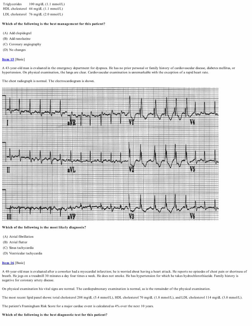
Item 15 [Basic]
A 43-year-old man is evaluated in the emergency department for dyspnea. He has no prior personal or family history of cardiovascular disease, diabetes mellitus, orhypertension. On physical examination, the lungs are clear. Cardiovascular examination is unremarkable with the exception of a rapid heart rate.
The chest radiograph is normal. The electrocardiogram is shown.
Which of the following is the most likely diagnosis?
(A) Atrial fibrillation(B) Atrial flutter(C) Sinus tachycardia(D) Ventricular tachycardia
Item 16 [Basic]
A 48-year-old man is evaluated after a coworker had a myocardial infarction; he is worried about having a heart attack. He reports no episodes of chest pain or shortness ofbreath. He jogs on a treadmill 30 minutes a day four times a week. He does not smoke. He has hypertension for which he takes hydrochlorothiazide. Family history isnegative for coronary artery disease.
On physical examination his vital signs are normal. The cardiopulmonary examination is normal, as is the remainder of the physical examination.
The most recent lipid panel shows: total cholesterol 208 mg/dL (5.4 mmol/L), HDL cholesterol 70 mg/dL (1.8 mmol/L), and LDL cholesterol 114 mg/dL (3.0 mmol/L).
The patient 's Framingham Risk Score for a major cardiac event is calculated as 4% over the next 10 years.
Which of the following is the best diagnostic test for this patient?
(A) Coronary angiography(B) Coronary calcium scoring(C) CT angiography(D) Exercise stress test(E) No additional testing
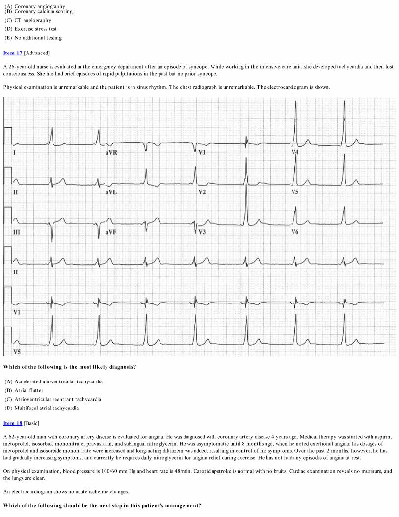
Item 17 [Advanced]
A 26-year-old nurse is evaluated in the emergency department after an episode of syncope. While working in the intensive care unit, she developed tachycardia and then lostconsciousness. She has had brief episodes of rapid palpitations in the past but no prior syncope.
Physical examination is unremarkable and the patient is in sinus rhythm. The chest radiograph is unremarkable. The electrocardiogram is shown.
Which of the following is the most likely diagnosis?
(A) Accelerated idioventricular tachycardia(B) Atrial flutter(C) Atrioventricular reentrant tachycardia(D) Multifocal atrial tachycardia
Item 18 [Basic]
A 62-year-old man with coronary artery disease is evaluated for angina. He was diagnosed with coronary artery disease 4 years ago. Medical therapy was started with aspirin,metoprolol, isosorbide mononitrate, pravastatin, and sublingual nitroglycerin. He was asymptomatic until 8 months ago, when he noted exertional angina; his dosages ofmetoprolol and isosorbide mononitrate were increased and long-acting diltiazem was added, resulting in control of his symptoms. Over the past 2 months, however, he hashad gradually increasing symptoms, and currently he requires daily nitroglycerin for angina relief during exercise. He has not had any episodes of angina at rest.
On physical examination, blood pressure is 100/60 mm Hg and heart rate is 48/min. Carotid upstroke is normal with no bruits. Cardiac examination reveals no murmurs, andthe lungs are clear.
An electrocardiogram shows no acute ischemic changes.
Which of the following should be the next step in this patient's management?

(A) Coronary angiography(B) Exercise treadmill stress testing(C) Increase metoprolol(D) Intravenous heparin and nitroglycerin
Item 19 [Basic]
A 62-year old man with a history of a myocardial infarction 1 year ago is evaluated in the emergency department for sudden episodes of dyspnea and weakness. He isdiaphoretic, cool, clammy, and pale; cannon waves are noted in the jugular pulsations. An electrocardiogram taken at the beginning of a typical episode is shown.
Which of the following is the most likely diagnosis?
(A) Atrial fibrillation with left bundle branch block(B) Atrial fibrillation with preexcitation (Wolf-Parkinson-White syndrome)(C) Supraventricular tachycardia with right bundle branch block(D) Ventricular tachycardia
Item 20 [Basic]
A 54-year-old woman is evaluated in the emergency department for jaw and shoulder pain that has occurred intermittently for the past week. The symptoms occur withactivity and are relieved by rest. Medical and family history is unremarkable. She is not taking any medications.
Physical examination shows a blood pressure of 150/68 mm Hg and a pulse of 90/min. There is no jugular venous distention and carotid upstrokes are normal. There are nocardiac murmurs and the lung fields are clear. Extremities show no edema and peripheral pulses are normal bilaterally. The troponin I level is elevated.
Electrocardiogram shows 1.0-mm ST-segment depression in leads V1 through V4 with T-wave inversions.
The patient is given aspirin, intravenous nitroglycerin, low-molecular-weight heparin, clopidogrel, and atorvastatin.
Which of the following is the most appropriate additional immediate treatment for this patient?
(A) Intra-aortic balloon pump(B) Metoprolol(C) Verapamil(D) Warfarin

Item 21 [Advanced]
A 72-year-old man is evaluated in the emergency department for dyspnea. One week ago, an episode of severe chest pain and dyspnea awoke him from sleep. Over the nextseveral days his dyspnea stabilized. On the morning of admission, the patient noted a sudden increase in dyspnea. His medical history is significant for hypertension andhyperlipidemia. He has no history of heart murmur. He currently takes simvastatin, aspirin, and lisinopril.
On physical examination, the patient is sitt ing up with labored breathing. Blood pressure is 86/52 mm Hg, pulse is regular at 110/min, and respiration rate is 24/min. Oxygensaturation is 92% on 6 L of oxygen. Jugular veins are distended to the angle of the jaw while sitt ing upright. Cardiac examination reveals a grade 3/6 holosystolic murmur atthe cardiac apex radiating toward the left axilla. Bibasilar crackles are present.
An electrocardiogram is shown. A chest radiograph shows pulmonary vascular congestion.
Which of the following is the most likely diagnosis?
(A) Acute mitral regurgitation(B) Left ventricular aneurysm(C) Pulmonary embolism(D) Ventricular free wall rupture
Item 22 [Advanced]
A 62-year-old woman is brought to the emergency department for chest pain that has been present for 5 hours. Medical history is notable for type 2 diabetes mellitus,hyperlipidemia, and hypertension. Medications are glyburide, lisinopril, atorvastatin, and aspirin.
On physical examination, blood pressure is 160/80 mm Hg, pulse rate is 88/min, and respiration rate is 16/min. Cardiac examination shows no murmurs, extra sounds, or rubs.The lungs are clear. Neurologic examination is normal.
The electrocardiogram shows 2-mm ST-segment elevation in leads II, III, and aVF.
A coronary catheterization laboratory is not available, and the nearest hospital with percutaneous intervention capability is 3 hours away.
Which of the following is the best management option for this patient?
(A) Aggressive medical therapy without reperfusion attempt(B) Immediate thrombolytic therapy(C) Transfer for coronary artery bypass graft surgery(D) Transfer for percutaneous coronary intervention

Item 23 [Basic]
A 65-year-old man is evaluated before beginning an exercise program. He is asymptomatic and his only medical problem is chronic hypertension that is well controlled onhydrochlorothiazide. He takes no other medications.
On physical examination, blood pressure is 138/76 mm Hg, and pulse is 80/min and regular. Physical examination is normal, except for a soft S1. His electrocardiogram isshown.
Which of the following best describes the e lectrocardiographic findings?
(A) First-degree atrioventricular block(B) Second-degree atrioventricular block(C) Third-degree (complete) atrioventricular block(D) Ventricular preexcitation (Wolff-Parkinson-White) syndrome
Item 24 [Advanced]
A 56-year-old man is evaluated in the emergency department for chest discomfort that began 3 hours ago. He describes the pain, which is well localized to the left chest, aspressure. He denies prior episodes. Medical history is notable for type 2 diabetes mellitus and hyperlipidemia. Medications are aspirin, metformin, and atorvastatin.
On physical examination, he is diaphoretic. Blood pressure is 95/60 mm Hg and heart rate is 110/min. There is jugular venous distention, with an estimated central venouspressure of 14 cm H2O. An S3 is heard on cardiac auscultation, but no murmurs are present. The lung fields are clear and there is no peripheral edema.
The electrocardiogram shows sinus tachycardia, 2-mm ST-segment elevation in leads II, III, and aVF, and 0.5-mm ST-segment elevation in lead V1. The chest radiograph isnormal.
Which of the following is the most likely cause of hypotension in this patient?
(A) Increased vagal tone(B) Pericardial tamponade(C) Right ventricular infarction(D) Ventricular septal defect
Item 25 [Basic]
An 85-year-old woman is admitted to the coronary care unit following successful thrombolytic therapy for an acute anteroseptal ST-elevation myocardial infarction. Bloodpressure is 120/70 mm Hg and heart rate is 90/min. There is no jugular venous distention and no cardiac murmurs. The lung fields are clear. Medications started in the hospitalare aspirin, low-molecular-weight heparin, intravenous nitroglycerin, and metoprolol.
On hospital day 3, the patient experiences acute onset of respiratory distress and her systolic blood pressure falls to 80 mm Hg. Her oxygen saturation remains at 80% despitethe administration of 100% oxygen by face mask. On physical examination, blood pressure is 96/40 mm Hg, pulse rate is 100/min, and respiration rate is 28/min. Findingsinclude jugular venous distention, crackles throughout both lung fields, and a grade 4/6 systolic murmur associated with a thrill.
Which of the following is the most likely diagnosis?
(A) Aortic dissection(B) Pericardial tamponade(C) Pulmonary embolism(D) Ventricular septal defect
Item 26 [Advanced]
A 77-year-old woman is admitted to the hospital for intermittent dizziness over the past few days. She has hypertension, hyperlipidemia, and paroxysmal atrial fibrillationwith a history of rapid ventricular response. Medications are metoprolol, hydrochlorothiazide, pravastatin, lisinopril, aspirin, and warfarin.
On physical examination, blood pressure is 137/88 mm Hg and pulse is 52/min. Estimated central venous pressure is 7 cm H2O. Cardiac auscultation reveals bradycardia withregular S1 and S2, as well as an S4. The lungs are clear to auscultation.
On telemetry, she has sinus bradycardia with rates between 40/min and 50/min, with two symptomatic sinus pauses of 3 to 5 seconds each.
Which of the following is the most likely diagnosis?

(A) Mobitz I atrioventricular block(B) Mobitz II atrioventricular block(C) Third-degree atrioventricular block(D) Sinoatrial node dysfunction
Item 27 [Advanced]
A 70-year-old man is evaluated in the emergency department for bradycardia that was detected in the nursing home and is found to have second-degree atrioventricular block.The patient has Alzheimer dementia. His medications are donepezil (dosage recently increased); memantine (recently started); vitamin E; and trazodone for agitation.
Which of the patient's medications is l ikely to explain the bradycardia?
(A) Donepezil(B) Memantine(C) Trazodone(D) Vitamin E
Item 28 [Basic]
A 67-year-old man is brought to the emergency department after he lost consciousness. His wife reports he had been experiencing palpitations and lightheadedness earlier inthe day. He has hypertension, dyslipidemia, and chronic obstructive pulmonary disease. His medications are lisinopril, hydrochlorothiazide, pravastatin, and a fluticasone-salmeterol inhaler.
On physical examination, the patient is awake but confused and in respiratory distress. He is afebrile, blood pressure is 80/45 mm Hg, pulse rate is 167/min, and respirationrate is 24/min and labored. Oxygen saturation is 86% on ambient air. The cardiac rhythm is irregular, and bibasilar crackles are present on pulmonary examination.
An electrocardiogram shows atrial fibrillation.
Which of the following is the most appropriate immediate management for this patient?
(A) Bedside echocardiography(B) CT pulmonary angiography(C) Coronary angiography(D) Electrical cardioversion
Item 29 [Basic]
A 62-year-old-woman is evaluated for a 6-month history of difficulty falling asleep and an unexplained 4.5-kg (10 lb) weight loss. She is active and rides her bicycle 5 miles aday. She does not drink alcohol, smoke cigarettes, or use recreational drugs. She has no other medical problems and takes no medications.
On physical examination, she is afebrile, blood pressure is 125/75 mm Hg, pulse rate is 108/min, and respiration rate is 14/min. On cardiac examination, a regular rhythmwithout murmurs or extra cardiac sounds is heard. The remainder of the physical examination is normal.
A metabolic profile and complete blood count are normal.
An electrocardiogram shows only sinus tachycardia.
Which of the following is the most appropriate management for this patient?
(A) Administer adenosine intravenously(B) Measure serum thyroid-stimulating hormone level (TSH)(C) Obtain an exercise stress test(D) Radiofrequency ablation of the sinoatrial node
Item 30 [Advanced]
A 79-year-old man is evaluated in the emergency department for a 1-week history of dyspnea and weakness. He has had several such episodes over the past 5 years but hasnever sought medical attention. He reports that 1 year ago, he had a 10-minute episode of left arm weakness that resolved spontaneously. He was never evaluated for thisproblem. He has hypertension treated with lisinopril and hydrochlorothiazide.
On physical examination, blood pressure is 135/80 mm Hg and heart rate is 143/min. Other than a rapid heart rate, the cardiopulmonary examination is normal, as is theremainder of the physical examination.
Electrocardiogram shows atrial fibrillation with a rapid ventricular rate without evidence of ischemic changes. Cardiac enzyme values are normal. Following the administrationof metoprolol, he converts to sinus rhythm, with a heart rate of 74/min.
Which of the following is the most appropriate long-term treatment for this patient?
(A) Amiodarone
(B) Low-molecular-weight heparin followed by warfarin(C) Metoprolol(D) Metoprolol and warfarin
Item 31 [Basic]
A 28-year-old man is evaluated for a pre-employment physical examination and electrocardiogram before entering the police academy. He has no medical problems, describesno worrisome symptoms, and does not take any medications. He does not smoke cigarettes, has one alcoholic drink or less each week, and rarely consumes caffeine. He runs4 miles a day 3 days a week and bikes 25 to 50 miles on the weekends. His parents are both alive and in good health as are his two older brothers.
On physical examination, vital signs are normal. The cardiopulmonary examination is normal as is the remainder of the examination.
The resting 12-lead electrocardiogram shows 3 unifocal premature ventricular contractions.
Which of the following is the best management plan for this patient?
(A) Begin amiodarone(B) Begin metoprolol(C) Exercise stress test(D) No further investigation or therapy(E) Order 24-hour ambulatory electrocardiography
Item 32 [Advanced]
A 55-year-old man is evaluated for fatigue, dyspnea with modest exertion, occasional lightheadedness, and palpitations. He has a history of ischemic cardiomyopathyfollowing a large anterolateral myocardial infarction 6 weeks ago. He does not have chest pain, and a postdischarge adenosine stress test with nuclear imaging demonstratedno inducible ischemia. His medications are lisinopril, carvedilol, furosemide, spironolactone, digoxin, and aspirin.
On physical examination, he is afebrile, blood pressure is 130/83 mm Hg, pulse rate is 50/min, and respiration rate is 12/min. He has no jugular venous distension. S1 and S2are soft, and S3 and S4 are present. A grade 2/6 holosystolic murmur at the cardiac apex is present. The lungs are clear. No peripheral edema is noted.
An electrocardiogram shows an episode of nonsustained ventricular tachycardia. Echocardiography shows diminished anterior wall motion with an ejection fraction of 25%.
Which of the following is the most appropriate treatment for this patient?
(A) Amiodarone(B) Flecainide(C) Procainamide(D) Implantable cardioverter-defibrillator (ICD)
Item 33 [Advanced]
A 67-year-old man presented to the emergency department 2 days ago with an acute ST-elevation myocardial infarction. During the initial evaluation, he becameunresponsive due to ventricular fibrillation. He was successfully resuscitated and taken to the cardiac catheterization lab, where a 100% occlusion of his proximal left anteriordescending artery was stented. His postinfarction course was notable for mild heart failure, which has now resolved. He is now stable on aspirin, metoprolol, atorvastatin,clopidogrel, and lisinopril.
On physical examination, blood pressure is 115/72 mm Hg, pulse is 65/min, and respiration rate is 12/min. There is no jugular venous distention, crackles, murmur, or S3. Theelectrocardiogram shows ST-segment changes consistent with a resolving anterior myocardial infarction but is otherwise unremarkable. Transthoracic echocardiogram revealsmild hypokinesis of the anterior wall and a left ventricular ejection fraction of 42%.
Which of the following is the best management option at this time?
(A) Add amiodarone(B) Continue medical management(C) Implantable cardioverter-defibrillator placement(D) Pacemaker placement
Item 34 [Advanced]
An 18-year-old woman is evaluated for recurrent syncope. She has experienced four syncopal episodes in her lifetime, all of which occurred during activity. Episodes have noprodrome, and she has had no dizziness. She is healthy and active, without cardiopulmonary complaints, and takes no medications. Her maternal cousin drowned at age 10years, and her mother has been evaluated for recurrent episodes of loss of consciousness.
On physical examination, blood pressure is 112/65 mm Hg, and pulse is 67/min and regular. The cardiopulmonary and general physical examinations are normal. Anechocardiogram examination is normal. An electrocardiogram is ordered.
Which of the following electrocardiographic findings is most likely to provide a diagnosis?
(A) Left bundle branch block

(B) Long PR interval(C) Long QT interval(D) Right bundle branch block
Item 35 [Basic]
A 50-year-man with a 6-month history of New York Heart Association class IV heart failure secondary to idiopathic dilated cardiomyopathy is evaluated following a recenthospitalization for worsening heart failure symptoms. The patient is adherent to his medications and his fluid and sodium restrictions. His medications are lisinopril,carvedilol, spironolactone, and furosemide.
On physical examination, vital signs are normal. He has no jugular venous distention. The cardiac rhythm is regular. Cardiac auscultation reveals an S3 and holosystolicmurmur at the cardiac apex. The chest is clear, and no peripheral edema is noted.
In the hospital, no evidence of ischemia, infection, arrhythmia, or thyroid disease was present. An echocardiogram demonstrated global hypokinesis of the left ventricle,moderate mitral regurgitation, and an ejection fraction of 25%
Which of the following medications should be initiated for this patient?
(A) Digoxin(B) Metolazone(C) Valsartan(D) Warfarin
Item 36 [Basic]
A 35-year-old woman is evaluated for progressive dyspnea 3 weeks after delivery of her first child. The pregnancy and delivery were uncomplicated. She has no history ofcardiovascular disease.
On physical examination, blood pressure is 110/70 mm Hg in both arms, heart rate is 105/min and regular, and respiratory rate is 28/min. The estimated central venouspressure is 10 cm H2O and there are no carotid bruits. The apical impulse is displaced and diffuse. There is a grade 2/6 holosystolic murmur at the apex. Third and fourth heartsounds are present. There is dullness to percussion at the posterior lung bases bilaterally, and there are crackles extending up half of the lung fields. Lower extremity pulses arenormal and without delay. Pedal edema is present.
The electrocardiogram demonstrates sinus tachycardia. There are no ST-segment or T-wave changes. The chest radiograph demonstrates bilateral pleural effusions andinterstit ial infiltrates. The aortic contour is unremarkable.
Which of the following is the most likely cause of the patient's current symptoms?
(A) Acute myocardial infarction(B) Aortic dissection(C) Coarctation of the aorta(D) Heart failure
Item 37 [Advanced]
A 65-year-old man is evaluated for 2 months of central chest pain with exertion and relief with rest, exertional dyspnea, orthopnea, and lower-extremity edema. The chestdiscomfort is increasing in frequency and severity. He has a 25-year history of hypertension and a 44-year history of smoking. His only medication is hydrochlorothiazide.
On physical examination, blood pressure is 118/80 mm Hg, pulse is 95/min, and respiration rate is 16/min. There is jugular venous distention. Cardiac examination reveals aregular rhythm. S1 and S2 are normal, and an S3 and S4 are present. Crackles are heard at both lung bases. There is edema at the ankles. Laboratory studies show a normalserum troponin T level. Electrocardiogram is normal. Echocardiogram shows an ejection fraction of 40%, global hypokinesis, and left ventricular hypertrophy.
Which of the following is the most appropriate diagnostic test?
(A) Cardiac angiography(B) Nuclear medicine stress test(C) Radionuclide ventriculography(D) Standard exercise stress test
Item 38 [Basic]
A 40-year-old woman is evaluated for 2 months of progressive dyspnea on exertion, orthopnea, and lower extremity edema. She denies chest discomfort and has no othermedical problems and takes no medications. She does not smoke cigarettes and rarely drinks alcohol. There is no family history of heart disease.
On physical examination, she is afebrile. Blood pressure is 120/80 mm Hg and pulse is 80/min. Estimated central venous pressure is 10 cm H2O. The lungs are clear. Cardiacexamination reveals a regular rhythm, an S3, and no murmurs. There is ankle edema. Chest radiograph shows vascular congestion. Electrocardiogram is normal. Initiallaboratory evaluation reveals a normal hemoglobin level and metabolic profile, including thyroid studies.
Which of the following is the most appropriate initial diagnostic test?

(A) B-type natriuretic peptide level(B) Echocardiography(C) Radionuclide ventriculography(D) Stress test
Item 39 [Basic]
A 70-year-old woman is evaluated for a 1-month history of dyspnea on exertion and fatigue. She can still perform activities of daily living, including vacuuming, groceryshopping, and ascending two flights of stairs carrying laundry. She has a history of hypertension. Her medications are lisinopril and hydrochlorothiazide.
On physical examination, blood pressure is 110/80 mm Hg and pulse is 70/min. Jugular veins are not distended. S1 and S2 are normal, and there is no S3 or murmur. Thepulmonary examination is normal and there is no edema. Laboratory studies show normal hemoglobin and thyroid-stimulating hormone levels. Electrocardiogram shows lowvoltage and left axis deviation. Echocardiogram shows a left ventricular ejection fraction of 45% and global hypokinesis. Chest radiograph is normal.
Which of the following is the most appropriate additional treatment?
(A) Amlodipine(B) Carvedilol(C) Digoxin(D) Losartan(E) Spironolactone
Item 40 [Basic]
A 60-year-old woman is diagnosed with heart failure due to nonischemic cardiomyopathy. Her ejection fraction is 40%. She currently has mild shortness of breath withmoderate exertion but no orthopnea or lightheadedness. She has a history of hypertension treated with hydrochlorothiazide and metoprolol.
On physical examination, she is afebrile. Blood pressure is 120/80 mm Hg and pulse is 65/min. The jugular veins are not distended, and the lungs are clear. Cardiacexamination discloses a regular rate and rhythm with no S3 or murmurs. There is no edema.
Which of the following agents should be added to her regimen?
(A) Digoxin(B) Eplerenone(C) Hydralazine and a nitrate(D) Lisinopril
Item 41 [Basic]
A 60-year-old woman is evaluated for dyspnea with mild activity (ascending less than one flight of stairs, walking less than one block on level ground) that has been stable forthe past year. She has a history of nonischemic cardiomyopathy (most recent left ventricular ejection fraction, 20%). Her current medications are lisinopril, carvedilol,digoxin, and furosemide. She had an implantable cardioverter-defibrillator placed 1 year ago.
On physical examination, blood pressure is 115/75 mm Hg and pulse rate is 70/min. Jugular veins are not distended, and the lungs are clear. Cardiac examination discloses aregular rhythm, no murmurs, normal S1 and S2, and no S3. There is no edema. Laboratory studies show normal serum creatinine and potassium levels.
Which of the following is the most appropriate addition to this patient's treatment?
(A) Losartan(B) Metolazone(C) Nifedipine(D) Spironolactone
Item 42 [Basic]
An 81-year-old woman with aortic stenosis is evaluated for increased shortness of breath and exercise intolerance. She was asymptomatic until 2 weeks ago when she notedincreased shortness of breath with exertion. She reports no chest pain, orthopnea, paroxysmal nocturnal dyspnea, or palpitations. She has had no fever, chills, or recentprocedures that might increase the risk for infective endocarditis. She has no other medical problems and takes no medications.
On physical examination, she is afebrile, blood pressure is 116/72 mm Hg, pulse rate is irregularly irregular at 112/min, and respiration rate is 12/min. No jugular venousdistention is present. Cardiac auscultation reveals an irregular rhythm with a grade 3/6 crescendo-decrescendo systolic murmur loudest at the second left intercostal space withradiation to the carotid arteries. Bibasilar pulmonary crackles are present, as is 1+ bilateral lower extremity edema.
Which of the following is most likely responsible for the new symptoms?
(A) Atrial fibrillation(B) Infective endocarditis

(C) Development of mitral stenosis(D) Development of mitral regurgitation
Item 43 [Basic]
A 54-year-old man is evaluated for increased shortness of breath of 3 weeks' duration. He has dyspnea on exertion, orthopnea, and occasional paroxysmal nocturnal dyspnea.He reports no chest pain, fever, or chills. He had an aortic bioprosthetic valve replacement for bicuspid valve aortic stenosis 15 years ago. He takes no medications.
On physical examination, he is afebrile, blood pressure is 140/50 mm Hg, pulse rate is 100/min, and respiration rate is 14/min. The carotid pulse is hyperdynamic. The S2 isdiminished. Cardiac auscultation reveals a grade 2/6 diastolic murmur, heard loudest at the third left intercostal space. A few scattered crackles can be heard at the lung bases.No peripheral edema is present.
Which of the following is the most likely diagnosis?
(A) Aortic regurgitation(B) Atrial septal defect(C) Coarctation of the aorta(D) Mitral stenosis(E) Ventricular septal defect
Item 44 [Basic]
A 65-year-old woman is evaluated as a new patient during a routine office visit . She is healthy and active and swims laps three or four times per week. She does not smoke andshe takes no medications.
On physical examination, blood pressure is 118/60 mm Hg. There are no carotid bruits. There is a normal S1 and a physiologically split S2. There is a grade 2/6 midsystolicmurmur that does not radiate and is heard best at the second right intercostal space. The rest of the physical examination is unrevealing.
Which of the following is the most appropriate management?
(A) Antibiotic endocarditis prophylaxis(B) Transthoracic echocardiography(C) Treadmill stress echocardiography(D) No further intervention
Item 45 [Advanced]
A 30-year-old woman is evaluated in the emergency department for shortness of breath, palpitations, and pedal edema. She is gravida 1, para 0, and she is 30 weeks pregnant.She has not received prenatal care until this point.
On physical examination, the patient is sitt ing upright to breathe. Blood pressure is 112/80 mm Hg, and pulse is 96/min. There is jugular venous distention to her jaw linewhile sitt ing upright. Cardiac auscultation demonstrates an irregularly irregular rhythm, a loud P2, and an opening snap followed by a low-pitched diastolic murmur heard bestat the cardiac apex. Bibasilar crackles are present.
Which of the following is the most likely diagnosis?
(A) Acute aortic regurgitation(B) Atrial septal defect(C) Mitral stenosis(D) Tricuspid regurgitation
Item 46 [Advanced]
An 18-year-old woman is evaluated during a routine examination prior to entering college. She has had no major medical problems and there is no family history ofcardiovascular disease.
On physical examination, blood pressure is 110/60 mm Hg and pulse is 70/min. S1 and S2 are normal and there is an S4 present. There is a harsh grade 2/6 midsystolic murmurheard best at the lower left sternal border. The murmur does not radiate to the carotid arteries. A Valsalva maneuver increases the intensity of the murmur; moving from astanding position to a squatting position decreases the intensity. Rapid upstrokes of the carotid pulses are present. Blood pressures in the upper and lower extremities areequal.
Which of the following is the most likely diagnosis?
(A) Aortic coarctation(B) Bicuspid aortic valve(C) Hypertrophic cardiomyopathy(D) Ventricular septal defect

Item 47 [Basic]
A 25-year-old asymptomatic man is evaluated during a routine examination. His blood pressure is 150/40 mm Hg and his heart rate is 90/min. Estimated central venouspressure is normal. The carotid upstroke is brisk and collapses quickly. The apical impulse is displaced. A grade 3/6 high-pitched decrescendo diastolic murmur is heard at thesecond right intercostal space with radiation down the left sternal border. The murmur is heard best with the patient leaning forward and in end-expiration. There is evidenceof nailbed pulsation. Femoral pulsations are full and collapse quickly. There is no change in the murmur with inspiration.
Which of the following is the most likely diagnosis?
(A) Aortic valve regurgitation(B) Mitral valve stenosis(C) Patent ductus arteriosus(D) Tricuspid regurgitation
Item 48 [Basic]
A 35-year-old woman is evaluated during a routine examination. She has no cardiovascular risk factors and no family history of cardiovascular disease. She is not currently onany medications.
On physical examination, vital signs are normal. There is a midsystolic click and a grade 2/6 late systolic murmur at the cardiac apex that radiates toward the left axilla.Following squat-to-stand maneuvers, the midsystolic click moves closer to the S1, and murmur duration and intensity are increased. Carotid upstrokes are normal. The rest ofthe examination is unremarkable.
Which of the following is the most likely diagnosis?
(A) Aortic stenosis(B) Coarctation of the aorta(C) Hypertrophic cardiomyopathy(D) Mitral valve prolapse
Item 49 [Advanced]
A 43-year-old man is evaluated during a routine examination. He has a history of a cardiac murmur diagnosed during childhood. He exercises regularly without restriction toactivity and has no history of syncope, palpitations, or edema. He takes no medications.
On physical examination, blood pressure is 120/64 mm Hg, pulse is 80/min and regular, and respiration rate is 16/min. Cardiac examination reveals a normal S1 and aphysiologically split S2. There is a grade 2/6 decrescendo diastolic murmur at the upper left sternal border. Distal pulses are brisk. There is no pedal edema.
A transthoracic echocardiogram demonstrates normal ventricular size and function, with ejection fraction of 60% to 65%. There is a bicuspid aortic valve with moderateregurgitation. Pulmonary pressure estimates are in the normal range.
Which of the following is the most appropriate management option for this patient?
(A) Antibiotic endocarditis prophylaxis(B) Aortic valve replacement(C) Begin metoprolol(D) Clinical follow-up in 1 year
Answers and Critiques
Item 1 Answer: B
Educational O bjective: Diagnose non-ST-elevation myocardial infarction.
The most likely diagnosis is a non-ST-elevation myocardial infarction (NSTEMI). This patient has had chest pain at rest, an elevated serum troponin I level, and anelectrocardiogram that shows ST-segment depression that is particularly prominent in leads V2 through V6. These features indicate a NSTEMI.
In an acute coronary syndrome, obstruction to coronary blood flow results in either transient or prolonged episodes of severe myocardial ischemia. ST-elevation myocardialinfarction (STEMI) is diagnosed in patients with a clinical presentation consistent with acute myocardial infarction together with electrocardiographic evidence of ST-segment elevation. Although most patients with STEMI ultimately develop Q waves on electrocardiogram, some exhibit diagnostic ST-segment elevation and cardiac enzymeelevations without Q waves.
Patients who present with ischemic chest pain but without diagnostic ST-segment elevation are categorized as having unstable angina or NSTEMI. These two conditions areclosely related and have similar pathophysiology and clinical presentations, but they differ in the severity of the myocardial ischemia. Patients with either condition typicallypresent with angina at rest; however, some patients describe a pattern of new-onset or increasing angina. In NSTEMI, ischemia is severe and results in a detectable release ofbiomarkers of myocardial injury hours after the onset of ischemic chest pain, most commonly cardiac troponin I, troponin T , and the MB isoenzyme of creatine kinase. Inunstable angina, there is no detectable increase in these enzymes.
Chronic stable angina refers to a coronary artery syndrome that is characterized by chest discomfort that occurs predictably and reproducibly at a certain level of exertionand is relieved with rest or nitroglycerin. Ischemic pain that occurs with rest is not compatible with the diagnosis of chronic stable angina, and the presence of positive serum

biomarkers for myocardial necrosis also excludes this diagnosis.
Key PointNon-ST-elevation myocardial infarction is characterized by chest pain at rest, absence of ST-elevation on the electrocardiogram, and elevated biomarkers of myocardialinjury.
BibliographyHillis LD, Lange RA. Optimal management of acute coronary syndromes. N Engl J Med. 2009;360(21):2237-2240. [PMID: 19458369]
Item 2 Answer: B
Educational O bjective: Diagnose acute pericarditis
The most likely diagnosis is acute pericarditis. Chest pain from acute pericarditis is sharp and pleuritic and worsened by assuming a supine position. Acute pericarditis isdiagnosed by the presence of at least two of the three classic features: (1) pleuritic chest pain; (2) friction rub; and (3) diffuse concordant ST-segment elevation onelectrocardiography, often with depression of the PR segment. The classic pericardial friction rub has three components (not all components may be heard) related to cardiacmotion and occurs during atrial systole, ventricular contraction, and rapid ventricular filling. The sound of the rub can be squeaky, scratchy, and high-pitched.
Patients with an acute aortic dissection present with the abrupt onset of severe pain in the thorax. Blood pressure is variable. Pain described as "ripping" or "tearing" occurs inonly about 50% of patients. The physical finding of a pulse deficit is uncommon. At least one third of patients have no electrocardiographic abnormality, and at least 40% ofpatients lack widening of the mediastinum on plain chest radiograph. However, pleuritic chest pain, pericardial friction rub, and diffuse ST elevations are not compatible withaortic dissection.
The pain of an acute myocardial infarction is usually retrosternal in location, may radiate to the shoulders or arms, and may be associated with nausea and vomiting,diaphoresis, or shortness of breath. Although the initial electrocardiogram is nondiagnostic in up to 50% of patients, compatible changes would include focal ST elevationswith reciprocal ST depression associated with elevation of cardiac biomarkers such as troponin.
Patients with pulmonary embolism may present with the classic symptoms of dyspnea, chest pain, hemoptysis, or syncope, but the presentation is often more subtle. Attimes, only nonspecific respiratory or hemodynamic manifestations may occur. Pulmonary embolism is not associated with a pericardial friction rub or diffuse ST-segmentchanges.
Key PointDiagnosis of acute pericarditis is made by the presence of at least two of the three classic features: (1) pleuritic chest pain; (2) friction rub; and (3) diffuse concordant ST-segment elevation on electrocardiography.
BibliographyLee TH, Goldman L. Evaluation of the patient with acute chest pain. N Engl J Med. 2000;20;342(16):1187-95. [PMID: 10770985]
Item 3 Answer: C
Educational O bjective: Treat a patient with ST-elevation myocardial infarction with percutaneous coronary intervention.
The best management for this patient is percutaneous coronary intervention (PCI). This patient is presenting with an ST-elevation inferior wall myocardial infarction(STEMI). The diagnosis is based upon the presence of chest pain, elevated cardiac biomarkers, and ST-segment elevations in the inferior leads (II, III, and aVF). Reperfusionstrategies for STEMI patients include either thrombolytic therapy or PCI. Percutaneous angioplasty and stent placement is the preferred therapy for most patients withSTEMI because it is associated with a lower 30-day mortality rate compared to thrombolytic therapy. PCI is also indicated in patients with a contraindication tothrombolytic therapy and in patients with cardiogenic shock. Contraindications to thrombolytic therapy include prior intracerebral hemorrhage, ischemic stroke within 3months, suspected aortic dissection, or active bleeding. PCI is most effective if completed within 12 hours of the onset of chest pain; the earlier the intervention, the betterthe outcome.
A chest CT can be a useful diagnostic tool in patients being evaluated for chest pain if there is a high clinical suspicion for an aortic dissection. This patient does not have anycharacteristics of ascending aortic dissection—back pain, unequal blood pressures or pulses, or a widened mediastinum on chest radiograph. Given these factors, the possibilityof an acute aortic dissection is extremely low, and further imaging with a chest CT would likely further delay treatment for the STEMI.
An echocardiogram is occasionally useful in the management of patients presenting with chest pain and a nondiagnostic electrocardiogram (ECG). A focal wall motionabnormality suggests a cardiac basis to the symptoms. In this patient presenting with a markedly abnormal ECG with ST-segment elevation and elevated cardiac biomarkers,echocardiography would not add to the management.
Key PointPercutaneous angioplasty and stent placement is the preferred therapy for most patients with ST-elevation myocardial infarction.
BibliographyHillis LD, Lange RA. Optimal management of acute coronary syndromes. N Engl J Med. 2009;360(21):2237-2240. [PMID: 19458369]
Item 4 Answer: C
Educational O bjective: Diagnose angina pectoris.

Symptoms of ischemic heart disease are fairly predictable for an individual patient, but great heterogeneity exists between patients. Patients may have a difficult t imedescribing the pain and may use words such as discomfort, pressure, or heaviness. Although the location of the pain is classically substernal, it may be difficult for patients tolocalize the pain; they may indicate the entire chest or upper abdomen is involved. Sharp pain, well-localized pain, and back pain are infrequently associated with ischemicheart disease. Ischemic cardiac pain has a predictable relation to exercise and relief with rest or nitroglycerin. In some patients, exertional dyspnea may be the onlymanifestation of cardiac ischemia (angina equivalent). A normal electrocardiogram at rest does not exclude ischemic heart disease nor does a normal troponin level.
The pain of acute pericarditis is often pleuritic, may radiate to the top of the shoulder, and is often worse when the patient is supine. Fever and a pericardial friction rub areusually evident. Characteristic electrocardiographic changes include diffuse ST elevations and PR depression.
The classic presentation of aortic dissection consists of sudden-onset severe chest pain radiating to the back. Other findings may include a blood pressure differential betweenthe right and left arms, murmur of aortic regurgitation, and a widened mediastinum on chest x-ray. Aortic dissection should always be considered in the differential diagnosisof acute chest pain, but this patient 's pain is not consistent with this diagnosis.
Most patients with peptic ulcer disease do not have pain at diagnosis; ulcers are usually detected during an evaluation for potential ulcer-related complications such as overt orobscure bleeding. When symptoms are present, they include dyspepsia or a nonspecific, gnawing epigastric pain. Other presentations include bleeding, perforation (sometimeswith penetration into adjacent organs), and gastric outlet obstruction. Finally, discomfort caused by a duodenal ulcer is not typically relieved by rest, worsened by exertion, oraccompanied by shortness of breath.
Key PointIschemic cardiac pain has a predicable relation to exercise and relief with rest or nitroglycerin.
BibliographyLee TH, Goldman L. Evaluation of the patient with acute chest pain. N Engl J Med. 2000;342(16):1187-95. [PMID: 10770985]
Item 5 Answer: D
Educational O bjective: Treat right ventricular myocardial infarction with normal saline infusion.
Volume expansion with normal saline is the primary supportive treatment for the hemodynamic abnormalities of a right ventricular myocardial infarction. The physicalexamination findings of hypotension, clear lung fields, and elevated estimated central venous pressure represent the classic triad of right ventricular myocardial infarction.However, the most predictive finding is ST-segment elevation on right-sided electrocardiographic lead V4R. Therefore, all patients with an inferior ST-elevation myocardialinfarction (STEMI) should have a right-sided electrocardiogram performed at the time of presentation. This patient 's electrocardiogram shows ST-segment elevation infrontal inferior leads II, III, and aVF, and 1-mm ST-segment elevation in right-sided precordial lead V4R. These findings indicate inferior and right ventricular injury in thesetting of an inferior STEMI, likely related to a right coronary artery occlusion.
In the setting of right ventricular myocardial infarction, right ventricular contractility is reduced, resulting in higher right ventricular diastolic pressure, lower right ventricularsystolic pressure, and reduced preload or filling of the left ventricle. Volume expansion improves the hemodynamic abnormalities of right ventricular myocardial infarctionbecause the gradient of pressure from the right atrium to the left atrium maintains filling of the left ventricle. In addition to reperfusion therapy for STEMI, the acutetreatment of right ventricular myocardial infarction is supportive.
Inotropic support, specifically using intravenous dobutamine, is appropriate treatment in patients with right ventricular myocardial infarction whose hypotension is notcorrected after 1 L of saline infusion. However, volume expansion should be tried before giving inotropic agents. By increasing cardiac contractility, inotropic agents increasemyocardial oxygen demand and potentially extend the infarction.
Bradycardia, potentially caused by increased vagal activity or sinoatrial node ischemia, exacerbates the hemodynamic abnormalities of right ventricular myocardial infarction,so β-blocker therapy with metoprolol is contraindicated in this patient.
Nitroglycerin is contraindicated in patients with hypotension and patients with the potential for hypotension, particularly right ventricular myocardial infarction. In rightventricular infarction, nitrate-induced venodilation impairs right ventricular filling and thereby cardiac output.
Key PointVolume expansion is the primary supportive treatment for the hemodynamic abnormalities of a right ventricular myocardial infarction.
BibliographyReynolds HR, Hochman JS. Cardiogenic shock: current concepts and improving outcomes. Circulation. 2008;117(5):686-697. [PMID: 18250279]
Item 6 Answer: D
Educational O bjective: Manage noncardiac chest pain caused by gastroesophageal reflux disease with an oral proton pump inhibitor.
The most appropriate management for this patient is empiric treatment with a proton pump inhibitor. Esophageal disease is the most common cause of noncardiac chestpain. Gastroesophageal reflux disease (GERD) is caused by reflux of gastric contents into the esophagus. Symptoms of GERD can mimic cardiac ischemia, with substernalsqueezing or burning that may radiate widely, including to the back, neck, jaw, or arms. Chest pain due to GERD may last minutes to hours and can resolve spontaneously orwith antacids. Most patients with GERD-induced chest pain also have typical reflux symptoms, including regurgitation and heartburn. The recommended approach in patientswith suspected esophageal chest pain is to rule out cardiac ischemia and then treat the patient empirically with a proton pump inhibitor.
Ambulatory esophageal pH monitoring can be used if the patient 's condition does not respond to empiric therapy or if the patient has atypical symptoms.Esophagogastroduodenoscopy is indicated in patients with long-standing reflux disease to screen for Barrett esophagus and in patients with such alarm symptoms as anemia,

weight loss, or dysphagia, but not as the initial screening test for GERD.
This patient 's previous normal stress test makes cardiac disease very unlikely and a repeat exercise stress test or coronary angiography is unlikely to provide additionaldiagnostic information. Because ischemic heart disease has been ruled out, it is reasonable in this patient to attempt empiric acid suppression therapy with a proton pumpinhibitor. A complete response to empiric therapy is considered diagnostic of GERD.
Key PointEmpiric proton pump inhibitor therapy is the first step in the management of esophageal noncardiac chest pain.
BibliographyFass R. Evaluation and diagnosis of noncardiac chest pain. Dis Mon. 2008;54(9):627-641. [PMID: 18725005]
Item 7 Answer: D
Educational O bjective: Diagnose third-degree atrioventricular heart block.
This patient most likely has third-degree (complete) atrioventricular heart block due to Lyme carditis. The presence of the characteristic skin rash (erythema migrans) withor without a history of tick bite in an endemic region, has a probability greater than 80% of being caused by Borrelia burgdorferi infection. Lyme carditis is manifested byacute-onset, high-grade atrioventricular conduction defects that may occasionally be associated with myocarditis.
Atrioventricular block is classified as first , second, or third degree. First-degree block is characterized by prolongation of the PR interval to greater than 0.2 sec and usually isnot associated with alterations in heart rate. There are two types of second-degree block, both recognized electrocardiographically by the presence of a P wave that is notfollowed by a ventricular complex. Mobitz type I block (Wenckebach block) manifests as progressive prolongation of the PR interval until there is a dropped beat, whereasMobitz type II block manifests as a dropped beat without progressive PR-interval prolongation. Mobitz type I block usually does not progress to complete heart block, butMobitz type II block, which is usually associated with a bundle branch block, typically progresses to third-degree block. Second-degree block may be associated withbradycardia, depending upon the frequency of blocked atrial impulses. Third-degree block is the complete absence of conduction of atrial impulses to the ventricle and is themost common cause of marked bradycardia; ventricular rates are usually 30-50/min. Patients with atrioventricular block may be asymptomatic or have severe bradycardia-related symptoms (weakness, presyncope, syncope) and ventricular arrhythmias.
Key PointThird-degree block is the complete absence of conduction of atrial impulses to the ventricle and is the most common cause of marked bradycardia.
BibliographyBarold SS. Atrioventricular block revisited. Compr Ther. 2002;28(1):74-78. [PMID: 11894446]
Item 8 Answer: B
Educational O bjective: Diagnose panic disorder.
This patient 's presentation is characteristic of panic disorder. Panic disorder is characterized by recurrent, unexpected panic attacks that feature the abrupt onset of numeroussomatic symptoms, such as palpitations, sweating, tremulousness, dyspnea, chest pain, nausea, dizziness, and numbness. Symptoms typically peak within 10 minutes of onset,and attacks usually last from 15 to 60 minutes. The most appropriate treatment for panic disorder is cognitive-behavioral therapy and a selective serotonin reuptakeinhibitor, such as paroxetine.
Because the symptoms of panic disorder are primarily physical, most patients with this disorder present to their primary care physicians or to emergency departments forevaluation. Patients with panic disorder are evaluated, on average, by 10 clinicians before the diagnosis is established. The alarming nature of their symptoms promptspatients with panic disorder to seek medical attention; consequently, they have very high health care utilization rates.
The clinical manifestations of pheochromocytomas are variable, with hypertension (episodic or sustained) observed in more than 90% of patients. Other major symptomsinclude diaphoresis, pallor, palpitations, and headaches; the classic triad of sudden severe headache, diaphoresis, and palpitations is highly suggestive of pheochromocytoma.Other manifestations include hyperglycemia, weight loss, arrhythmias (atrial and ventricular fibrillation), and catecholamine-induced cardiomyopathy. The absence ofhypertension and the long duration of symptoms make this diagnosis unlikely.
The diagnosis of acute coronary syndrome is unlikely in a patient with recurrent, self-limited attacks and previous normal results of coronary angiography. The currentnature of the symptoms and normal physical examination findings are not compatible with the diagnosis of pneumothorax. Symptoms that resolve after 20 minutes or recurfrequently over many years are not compatible with the diagnosis of pulmonary embolism.
Key PointPanic disorder is characterized by recurrent, unexpected panic attacks that feature the abrupt onset of numerous somatic symptoms such as palpitations, sweating,tremulousness, dyspnea, chest pain, nausea, dizziness, and numbness.
BibliographyCulpepper L. Identifying and treating panic disorder in primary care. J Clin Psychiatry. 2004;65(suppl 5):19-23. [PMID: 15078114]
Item 9 Answer: B
Educational O bjective: Diagnose Mobitz type I second-degree atrioventricular block.

This patient has evidence for Mobitz type I second-degree atrioventricular (AV) block. Second-degree heart block is characterized by intermittent nonconduction of P wavesand subsequent "dropped" ventricular beats. Second-degree heart block is divided into types, Mobitz I and Mobitz II. Mobitz type I second-degree heart block is characterizedby progressive prolongation of the PR interval until a dropped beat occurs. Mobitz type I block can occur in the absence of heart disease, including in athletes and olderadults; in patients with underlying heart disease, including acute ischemia; and in patients who are taking drugs that block the AV node, such as β-blockers (metoprolol),calcium channel blockers, and digoxin. This type of heart block is characteristically transient and usually requires no specific treatment; however, some patients may developexcessively slow heart rates and experience symptoms related to decreased cerebral or coronary perfusion. If treatment is necessary, it begins by identifying and correctingreversible causes of slowed conduction, such as myocardial ischemia, increased vagal tone (for example, from pain or vomiting), and discontinuation of drugs that depress AVconduction.
Mobitz type II second-degree AV block is characterized by a regularly dropped beat (for example, a nonconducted P wave every second or third beat) without progressiveprolongation of the PR interval. It is often associated with evidence of additional disease in the conduction system, such as bundle branch block or bifascicular or trifascicularblock. Mobitz type II second-degree block suddenly and unpredictably progresses to complete heart block and is usually treated with a pacemaker.
First-degree AV block is recognized, electrocardiographically, as a prolongation of the PR interval to greater than 0.2 sec. All P waves are conducted. First-degree blockrequires no specific treatment.
Third-degree AV block, or complete heart block, refers to a lack of AV conduction, characterized by lack of conduction of all atrial impulses to the ventricles.
Key PointMobitz type I second-degree atrioventricular block is characterized by progressive prolongation of the PR interval until a dropped beat occurs.
BibliographyDa Costa D, Brady WJ, Edhouse J. Bradycardias and atrioventricular conduction block. BMJ. 2002;324:535-538. [PMID: 11872557]
Item 10 Answer: D
Educational O bjective: Treat chronic stable angina with worsening symptoms with increased dosage of a β-blocker.
The most appropriate management at this point is to increase the patient 's dose of metoprolol. Medical therapy for chronic stable coronary artery disease (CAD) includesboth antianginal and vascular-protective agents. Antianginal therapy includes β-blockers, calcium channel blockers, and nitrates. Vascular-protective therapy includes aspirin,angiotensin-converting enzyme (ACE) inhibitors, and statins. This patient is already on a β-blocker, aspirin, a statin, and an ACE inhibitor. Switching to a long-acting nitratewill help relieve his angina symptoms. However, his resting heart rate of 87/min and high blood pressure suggest a suboptimal dose of β-blocker, and the patient 's dosage ofmetoprolol should be increased. The β-blocker dose should be titrated to achieve a resting heart rate of approximately 55 to 60/min and approximately 75% of the heart ratethat produces angina with exertion. The patient should be reevaluated in a few weeks to assess the response to therapy.
Ranolazine is a novel antianginal agent that is approved for the treatment of chronic stable angina. It should only be used, however, in addition to baseline therapy with a β-blocker, a calcium channel blocker, and a long-acting nitrate. Given that this patient was on suboptimal doses of metoprolol and is just being started on a long-acting nitrate,the addition of ranolazine would be premature.
Coronary angiography would not be indicated at this t ime because the patient is not receiving maximal medical therapy. In the setting of continued angina despite maximalmedical therapy, coronary angiography could be considered.
Exercise treadmill stress testing would not provide useful information in this setting. It would only confirm the high pretest probability that this patient has underlying CADas a cause for the current symptoms.
Key PointIn the treatment of chronic stable angina, the β-blocker dose should be titrated to achieve a resting heart rate of approximately 55 to 60/min and approximately 75% of theheart rate that produces angina with exertion.
BibliographyJawad E, Arora R. Chronic stable angina pectoris. Dis Mon. 2008;54(9):671-689. [PMID: 18725007]
Item 11 Answer: A
Educational O bjective: Diagnose pulmonary embolism with CT pulmonary angiography.
This patient 's symptoms of chest pain and dyspnea in combination with the physical findings of asymmetric leg edema, elevated central venous pressure, tachypnea, andtachycardia suggest the possibility of pulmonary embolism. The most appropriate diagnostic test to perform next is CT pulmonary angiography to look for pulmonaryemboli.
A normal echocardiogram between episodes of chest pain does not rule out unstable angina because wall motion returns to normal between ischemic episodes. However, thispatient had no wall motion abnormalities during her ongoing chest pain, making acute myocardial ischemia very unlikely. Because an acute coronary syndrome is highlyunlikely, coronary angiography is not indicated as the next diagnostic test.
A resting radionuclide perfusion study can be helpful in the diagnosis of coronary ischemia when the electrocardiogram is nondiagnostic but does not provide additiveinformation to that already obtained by echocardiography.
Transesophageal echocardiography is not sensitive for detection of pulmonary emboli but may be useful in acute chest pain if aortic dissection is suspected. Ascending aorticdissection is often associated with acute aortic regurgitation, myocardial ischemia, cardiac tamponade or hemopericardium, and hemothorax or exsanguination. Considerable

(>20 mm Hg) variation in systolic blood pressure in the arms may be present. Descending thoracic aortic aneurysm is more commonly associated with splanchnic ischemia,renal insufficiency, lower extremity ischemia, or focal neurologic deficit due to spinal cord ischemia. This woman has no physical findings to suggest this diagnosis.
Key PointNormal wall motion on echocardiography during chest pain excludes coronary ischemia or infarction.
BibliographyChunilal SD, Eikelboom JW, Attia J, et al. Does this patient have pulmonary embolism? JAMA. 2003;290(21):2849-2858. [PMID: 14657070]
Item 12 Answer: D
Educational O bjective: Evaluate suspected coronary artery disease using an exercise stress test.
The most appropriate diagnostic test for this patient is an exercise stress test. This 68-year-old woman has cardiac risk factors of smoking and hypertension, with anintermediate Framingham risk score (18% likelihood of a coronary event in 10 years). (A tool to calculate the Framingham risk score can be accessed atwww.acponline.org/acp_press/essentials/calculator.htm.) In addition, she has atypical chest pain, so further evaluation is appropriate. An exercise stress test is recommendedin patients with intermediate probability of coronary artery disease with a normal baseline electrocardiogram (ECG) who are able to exercise because it provides informationabout exercise tolerance and hemodynamic response to exercise.
An adenosine nuclear perfusion stress test is contraindicated in patients with significant bronchospastic disease and hence is not the correct choice for a patient with asthma.Furthermore, exercise stress testing is preferred over pharmacologic stress testing because of the additional physiologic information on exercise tolerance and the bloodpressure and heart rate response to exercise.
Coronary angiography would only be appropriate if the patient were presenting with an acute coronary syndrome or after an abnormal stress test to determine if there is anindication for revascularization.
Dobutamine stress echocardiography is an appropriate choice in patients who are unable to exercise and are not hypertensive at rest. This patient is able to exercise, sotreadmill testing is the more appropriate choice.
Key PointExercise electrocardiographic stress testing is the primary approach to the diagnosis of coronary artery disease in patients who can exercise and have a normal restingelectrocardiogram.
BibliographyWilson JM. Diagnosis and treatment of acquired coronary artery disease in adults. Postgrad Med J. 2009;85(1005):364-365. [PMID: 19581247]
Item 13 Answer: B
Educational O bjective: Diagnose atrial flutter.
The most likely diagnosis for this patient is atrial flutter. The presence on the electrocardiogram of multiple P waves in a "sawtooth" pattern, typically with 2:1 ventricularconduction, characterizes atrial flutter and eliminates atrial fibrillation as a diagnostic consideration. Electrocardiographically, atrial fibrillation is characterized by an absenceof discernible P waves, which are replaced by fibrillatory waves that vary in amplitude, shape, and timing. The ventricular response is grossly irregular and often rapid, exceptwhen there is concomitant atrioventricular block.
Sinoatrial node dysfunction (sick sinus syndrome) is a frequent cause of pacemaker implantation. It consists of symptomatic sinus bradycardia and the tachycardia-bradycardiasyndrome (alternating atrial tachyarrhythmias and bradycardia). In patients with "tachy-brady" syndrome, bradycardia usually occurs after termination of the tachycardia;atrial fibrillation is the most common tachyarrhythmia observed in this group of patients. The patient does not fulfill the diagnostic criteria for sinus node dysfunction.
Ventricular tachycardia is characterized by wide-complex QRS morphology (QRS >0.12 sec) and a ventricular rate that is greater than 100/min. In ventricular tachycardia, theventricular rate typically ranges from 140/min to 250/min. The patient does not have a wide-complex tachycardia and, therefore, does not have ventricular tachycardia.
Key PointAtrial flutter is typically a narrow-complex tachycardia characterized by multiple regular atrial contractions (flutter waves) creating a "sawtooth" baseline pattern prior to theQRS complex.
BibliographySawhney NS, Anousheh R, Chen WC, Feld GK. Diagnosis and management of typical atrial flutter. Cardiol Clin. 2009;27(1):55-67, viii. [PMID: 19111764]
Item 14 Answer: D
Educational O bjective: Treat chronic stable angina with appropriate medical therapy.
The most appropriate treatment of this patient would be to continue his current medical management. Medical therapy for chronic coronary artery disease (CAD) can beclassified as antianginal or vascular protective. Antianginal medications include β-blockers, calcium channel blockers, nitrates, and ranolazine. β-Blockers reduce mortality byapproximately 20%. Patients with chronic stable angina can be treated with calcium channel blockers if they are unable to tolerate β-blockers, or calcium channel blockerscan be added to β-blocker therapy for difficult-to-control symptoms. All patients with chronic stable angina should carry either a sublingual or a spray form of nitroglycerin

for emergency use. Ranolazine should only be considered in patients who remain symptomatic despite optimal doses of β-blockers, calcium channel blockers, and nitrates.
Vascular-protective medications include aspirin, clopidogrel, angiotensin-converting enzyme (ACE) inhibitors, and statins. Aspirin reduces the risk of stroke, myocardialinfarction, sudden death, and vascular death by 33%. Although clopidogrel is beneficial in patients with acute coronary syndromes, it has small clinical benefit in patients withchronic stable angina and is associated with an increased risk of bleeding. ACE inhibitors reduce cardiovascular mortality by 17% to 23%. Statins reduce future cardiovascularevents by approximately 25% to 30%. Current guidelines specify an LDL cholesterol target level of less than 100 mg/dL (2.6 mmol/L) for patients with coronary arterydisease. This patient is receiving standard therapy for chronic stable angina with aspirin, β-blocker (carvedilol), ACE inhibitor (lisinopril), a statin (simvastatin), andnitroglycerin. His blood pressure is well controlled, pulse rate is appropriately reduced, and the lipid profile is at target. His symptoms are infrequent and stable; no change inhis medical therapy is needed.
Coronary angiography is reserved for patients with acute coronary symptoms, angina increasing in severity, markedly abnormal stress tests, sudden cardiac death, ordiagnostic difficulties and is not indicated in this clinically stable patient. Because this patient 's symptoms are well controlled with the current therapy, it is not necessary toadd ranolazine.
Key PointMost patients with chronic stable angina are treated with aspirin, a β-blocker, an ACE inhibitor, nitroglycerin, and a statin.
BibliographyPfisterer ME, Zellweger MJ, Gersh BJ. Management of stable coronary artery disease. Lancet. 2010;375(9716):763-72. [PMID: 20189028]
Item 15 Answer: A
Educational O bjective: Diagnose atrial fibrillation.
The electrocardiogram is characteristic for atrial fibrillation, showing a rapid, irregularly irregular rhythm with no discernible P waves and atrial fibrillatory waves at a ratebetween 350 and 600 beats/min. The fibrillatory waves vary in amplitude, morphology, and intervals, creating a rough, irregular baseline between the QRS complexes. Thepatient 's evaluation should minimally include a transthoracic echocardiogram to exclude occult valve or other structural heart disease and also to assess the size of the leftatrial appendage; in addition, thyroid studies should be performed to exclude hyperthyroidism.
Atrial flutter is recognized by its saw-tooth pattern of flutter waves, most noticeable in the inferior leads II, III, and aVF; flutter waves are distinctly different from the small,chaotic fibrillation waves that are characteristic of atrial fibrillation.
Sinus tachycardia is a regular rhythm associated with P waves prior to each QRS complex. In each lead, the P-wave morphology and PR interval remain constant in shape andduration. The absence of well-defined P waves in this electrocardiogram rules out sinus tachycardia.
Ventricular tachycardia is characterized by wide-complex QRS morphology (QRS >0.12 sec) and a ventricular rate ranging from 140-250/min. This patient 's QRS complexesare narrow, excluding ventricular tachycardia as a diagnosis.
Key PointAtrial fibrillation is characterized electrocardiographically by an irregularly irregular rhythm with no discernible P waves and atrial fibrillation waves creating an irregularbaseline.
BibliographyZimetbaum P. Atrial fibrillation. Ann Intern Med. 2010;153(11):ITC61. [PMID: 21135291]
Item 16 Answer: E
Educational O bjective: Avoid screening for asymptomatic coronary artery disease.
No additional testing for coronary artery disease (CAD) is the best management option for this patient. He is at low risk for CAD, and he is asymptomatic. In addition, heonly has a 4% probability of a major coronary event over the next 10 years (www.acponline.org/acp_press/essentials/calculator.htm). The American College ofCardiology/American Heart Association, American College of Physicians, and U.S. Preventive Services Task Force all agree that there is lit t le evidence to support routinetesting for CAD in adults who are low risk and asymptomatic. Screening for CAD in adults who are asymptomatic is not recommended because the probability of a false-positive test is much greater than the probability of a true positive test. For patients with an intermediate probability of CAD, noninvasive stress testing with an exercisestress test provides the most useful information.
Coronary angiography should be reserved for patients with chronic CAD who have lifestyle-limiting angina despite medical therapy, markedly positive results on noninvasivestress testing, successful resuscitation from sudden cardiac death, or documented ventricular tachycardia. Coronary angiography can also be considered in patients withnonspecific chest pain to completely exclude CAD as a cause for the current symptoms if they have had recurrent hospitalizations. Coronary calcium testing may beconsidered in asymptomatic persons with a 10% to 20% Framingham 10-year risk category (intermediate risk) and in young persons with a strong family history ofpremature cardiovascular disease. The diagnostic accuracy of CT angiography to detect obstructive CAD is approximately 90%. Because CT angiography provides nofunctional information (that is, the extent of ischemia), a markedly abnormal study is followed by referral to coronary angiography or stress testing to determine theischemic burden. Recent consensus guidelines suggest that the benefits of CT angiography are greatest in symptomatic patients with an intermediate pretest probability ofCAD.
Key PointThere is lit t le evidence to support routine testing for coronary artery disease in adults who are low risk and asymptomatic.

BibliographySnow V, Barry P, Fihn SD, Gibbons RJ, Owens DK, Williams SV, Weiss KB, Mottur-Pilson C; ACP; ACC Chronic Stable Angina Panel. Evaluation of primary care patientswith chronic stable angina: guidelines from the American College of Physicians. Ann Intern Med. 2004;141 (1):57-64. [PMID: 15238371]
Item 17 Answer: C
Educational O bjective: Diagnose atrioventricular reentrant tachycardia (Wolff-Parkinson-White syndrome).
The most likely diagnosis is atrioventricular reentrant tachycardia. The patient presents with a history of tachycardia and recent syncope. The electrocardiogram shows ashort PR interval and the presence of a delta wave, which signifies preexcitation. These features, together with tachycardia, make the diagnosis of Wolff-Parkinson-Whitesyndrome, a type of atrioventricular reentrant tachycardia.
There are no clinical features to suggest idioventricular tachycardia or slow ventricular tachycardia, which is demonstrated electrocardiographically as a wide QRS complexand a heart rate between 60/min and 100/min. Atrial flutter is recognized by the characteristic negative sawtooth deflections in ECG leads II, III, and aVF, with a positivedeflection in V1, findings that are not present in this patient 's electrocardiogram. Multifocal atrial tachycardia characteristically occurs in the setting of chronic lung diseaseand is manifested by three or more P-wave configurations on the electrocardiogram with associated tachycardia.
Key PointThe combination of a short PR interval and a delta wave plus tachycardia confirms the diagnosis of Wolff-Parkinson-White syndrome, a type of atrioventricular tachycardia.
BibliographyLee KW, Badhwar N, Scheinman MM. Supraventricular tachycardia-part I. Curr Probl Cardiol. 2008;33(9):467-546. [PMID: 18707990]
Item 18 Answer: A
Educational O bjective: Evaluate progressive chronic angina pectoris with coronary angiography.
Coronary angiography is the most appropriate option in this patient with continued anginal symptoms despite optimal medical therapy. Compared with optimal medicaltherapy, a strategy of coronary angiography and revascularization provides no benefit in patients with chronic stable angina. This patient, however, remains highlysymptomatic despite optimal medical therapy, and therefore may benefit from coronary angiography and revascularization. Coronary revascularization is beneficial inpatients with chronic stable angina and the following conditions: angina pectoris refractory to medical therapy; a large area of ischemic myocardium and high-risk criteria onstress testing; high-risk coronary anatomy, including left main coronary artery stenosis or three-vessel disease; and significant coronary artery disease with reduced leftventricular systolic function. In appropriately selected patients, revascularization, either a percutaneous coronary intervention or coronary artery bypass grafting surgery, hasbeen shown to reduce angina, increase longevity, and improve left ventricular performance.
Exercise treadmill stress testing would not be useful for this patient 's management as it would only confirm the known diagnosis of coronary artery disease. Results of anexercise stress test would not influence therapeutic decisions.
β-Blockers, such as metoprolol, reduce myocardial oxygen demand by reducing heart rate and contractility, thereby reducing myocardial oxygen consumption. However, thispatient 's heart rate is already reduced to 48/min and his blood pressure is under excellent control. Increasing the dose of metoprolol in these circumstances is unlikely toproduce further benefit and may not be tolerated because of unacceptably low pulse rate and blood pressure.
For patients with unstable angina, admission to the coronary care unit and intravenous heparin and nitroglycerin would be beneficial. This patient has chronic stable angina,characterized by progressive exertional angina for 2 months without episodes of pain while at rest or prolonged episodes of pain.
Key PointCoronary angiography is indicated in patients with chronic stable angina who experience lifestyle-limiting angina despite optimal medical therapy.
BibliographyJawad E, Arora R. Chronic stable angina pectoris. Dis Mon. 2008;54(9):671-689. [PMID: 18725007]
Item 19 Answer: D
Educational O bjective: Diagnose ventricular tachycardia.
The most likely diagnosis is ventricular tachycardia. The electrocardiogram demonstrates sinus rhythm that is suddenly interrupted by the onset of ventricular tachycardia.Ventricular tachyarrhythmias consist of ventricular tachycardia, ventricular fibrillation, and torsades de pointes (a special subset of polymorphic ventricular tachycardia).Ventricular tachyarrhythmias are characterized by wide QRS complex morphology (QRS >0.12 sec) and ventricular rates greater than 100/min. In ventricular tachycardia, theventricular rate typically ranges from 140 to 250/min; in torsades de pointes, the ventricular rate ranges from 200 to 300/min; and in ventricular fibrillation, the rate istypically above 300/min.
Atrial fibrillation is characterized by an irregularly irregular rhythm, an irregular baseline (atrial fibrillatory waves), and the absence of P waves, which is inconsistent with thispatient 's electrocardiographic findings.
Supraventricular tachycardia with a wide QRS complex, usually due to coexisting bundle branch block or preexcitation (Wolff-Parkinson-White syndrome), can mimicventricular tachycardia. Differentiating ventricular tachycardia from supraventricular tachycardia with aberrant conduction is important because the treatment differsmarkedly. Ventricular tachycardia is more common than supraventricular tachycardia with aberrancy, particularly in patients with structural heart disease. A key point is thatany wide QRS tachycardia should be considered to be ventricular tachycardia until proven otherwise. The most important differentiating point is the history of ischemic heartdisease. In the presence of known structural heart disease, especially a prior myocardial infarction, the diagnosis of ventricular tachycardia is almost certain. The presence of

cannon waves (large a waves) in the jugular venous pulsations and varying intensity of the first heart sound support the diagnosis of atrioventricular dissociation caused byeither ventricular tachyarrhythmias or heart block.
Key PointA wide QRS tachycardia in the presence of known structural heart disease, especially a prior myocardial infarction, is almost certainly ventricular tachycardia.
BibliographySrivathsan K, Ng DW, Mookadam F. Ventricular tachycardia and ventricular fibrillation. Expert Rev Cardiovasc Ther. 2009;7(7):801-809. [PMID: 19589116]
Item 20 Answer: B
Educational O bjective: Treat non-ST-elevation myocardial infarction with a β-blocker.
The most appropriate additional treatment for this patient is metoprolol. This patient 's elevated troponin I level and ST-segment depression and T-wave inversions onelectrocardiogram are indicative of a non-ST-elevation myocardial infarction (NSTEMI). Early intravenous β-blocker therapy reduces infarct size, decreases the frequency ofrecurrent myocardial ischemia, and improves short- and long-term survival. β-Blockers diminish myocardial oxygen demand by reducing heart rate, systemic arterial pressure,and myocardial contractility; in addition, prolongation of diastole augments perfusion to the injured myocardium. β-Blocker therapy can be used in left ventriculardysfunction if heart failure status is stable.
An intra-aortic balloon pump is indicated for an acute coronary syndrome with cardiogenic shock that is unresponsive to medical therapy, acute mitral regurgitationsecondary to papillary muscle dysfunction, ventricular septal rupture, or refractory angina. The intra-aortic balloon pump reduces afterload during ventricular systole andincreases coronary perfusion during diastole. Patients with refractory cardiogenic shock who are treated with an intra-aortic balloon pump have a lower in-hospital mortalityrate than patients who are not treated. This patient has no indication for an intra-aortic balloon pump.
Calcium channel blockers, such as verapamil, are also effective antianginal medications, but data are conflicting as to whether calcium channel blockers reduce mortality inpatients with NSTEMI. Therefore, β-blockers are first-line therapy for unstable angina and NSTEMI unless contraindications are present. With ongoing ischemia despite β-blocker therapy, a calcium channel blocker can be added. However, there is no indication for starting verapamil rather than metoprolol at this time.
There is no role for the routine use of warfarin in the treatment of acute coronary syndrome, including NSTEMI. Warfarin is not associated with improved patient outcomeas compared to treatment without warfarin. Warfarin may be considered in patients at increased risk for thromboembolism, such as those with atrial fibrillation.
Key PointIn patients with myocardial infarction, early intravenous β-blocker therapy reduces infarct size, decreases the frequency of recurrent myocardial ischemia, and improvesshort- and long-term survival.
BibliographyHillis LD, Lange RA. Optimal management of acute coronary syndromes. N Engl J Med. 2009;360(21):2237-2240. [PMID: 19458369]
Item 21 Answer: A
Educational O bjective: Diagnose acute severe mitral regurgitation due to papillary muscle rupture.
The most likely diagnosis is acute mitral valve regurgitation due to papillary muscle rupture. The clinical history of chest pain and dyspnea a week ago and theelectrocardiographic findings of Q waves in leads II, III, and aVF strongly suggest an acute inferior wall myocardial infarction. The presence of a new systolic murmur andrespiratory distress several days after an acute myocardial infarction indicates the possibility of either a ventricular septal rupture or mitral regurgitation. Papillary musclerupture generally presents several days after the infarct event. Severe mitral regurgitation complicating an acute myocardial infarct is more common with inferior versusanterior infarcts and should be suspected in patients with pulmonary edema and respiratory distress in that setting. The murmur of mitral regurgitation may not be prominentbecause of the acutely elevated left atrial pressure and relatively lower transmitral systolic pressure gradient. Echocardiography is diagnostic, and early clinical recognitionwith aggressive support (intra-aortic balloon pump, afterload reduction if blood pressure allows) is essential, providing a bridge to surgical repair.
A left ventricular aneurysm may result in late-appearing complications of acute myocardial infarction. A ventricular aneurysm may be associated with intractable ventriculartachyarrhythmias, systemic emboli, or heart failure. It is not associated with a new holosystolic murmur.
Pulmonary embolism can complicate acute myocardial infarction and should be considered in any patient with new-onset pleuritic chest pain, dyspnea, and hypotension.However, pulmonary embolism is not associated with a new holosystolic murmur.
Ventricular free wall rupture typically leads to pericardial tamponade manifesting as sudden hypotension and death. Ventricular free wall rupture typically occurs 1 to 4 daysafter acute myocardial infarction. Patients usually present with cardiovascular collapse, tamponade, or pulseless electrical activity.
Key PointThe presence of a new systolic murmur and respiratory distress several days after an acute myocardial infarction indicates the possibility of either a ventricular septal ruptureor mitral regurgitation.
BibliographyTopalian S, Ginsberg F, Parrillo JE. Cardiogenic shock. Crit Care Med. 2008;36:S66-S74. [PMID: 18158480]
Item 22 Answer: B

Educational O bjective: Treat ST-elevation myocardial infarction with thrombolytic therapy.
The best management option for this patient is immediate thrombolytic therapy. The patient is experiencing an acute inferior wall ST-elevation myocardial infarction(STEMI). The diagnosis is established by the presence of chest pain and ST-segment elevations in leads II, III, and aVF. The treatment for an acute STEMI is eitherrevascularization or thrombolytic therapy. The patient has no contraindications for thrombolytic therapy. She is currently hemodynamically stable without cardiogenicshock. The treatment of choice is percutaneous coronary intervention (PCI) provided that it can be done immediately or in less than 60 minutes if transfer to anotherhospital is necessary. Since the nearest hospital with PCI capability is 3 hours away, immediate thrombolytic therapy is the most appropriate treatment for this patient.
Thrombolytic agents are an alternative to primary PCI in suitable candidates with STEMI. By lysing the clot that is limiting blood flow to the myocardium, thrombolyticsrestore perfusion to the ischemic area, reduce infarct size, and improve survival. Thrombolytics should be administered within 12 hours after the onset of chest pain; theearlier the administration, the better the outcome.
Aggressive medical therapy would not be the correct option for this patient given the electrocardiographic findings, her relatively young age, and the ability to treat her witha thrombolytic agent.
Transfer for coronary artery bypass graft surgery would not be the correct option given that the patient 's coronary anatomy has not yet been defined and there would be anunacceptable delay if she were transferred for coronary angiography. Most patients presenting with a STEMI can be treated effectively with percutaneous coronaryintervention or thrombolytic therapy. Bypass surgery in the setting of an acute infarction is therefore rarely performed. Bypass surgery may be preferred in patients whohave a large amount of myocardium at ischemic risk owing to proximal left main disease or multivessel disease, especially if the left ventricular ejection fraction is reduced.However, this is predicated by first performing coronary angiography and knowing the coronary anatomy.
Key PointThrombolytic agents are an alternative to primary percutaneous coronary intervention in suitable candidates with ST-elevation myocardial infarction and should beadministered within 12 hours after the onset of chest pain.
BibliographyHillis LD, Lange RA. Optimal management of acute coronary syndromes. N Engl J Med. 2009;360(21):2237-2240. [PMID: 19458369]
Item 23 Answer: A
Educational O bjective: Diagnose first-degree atrioventricular block.
This electrocardiogram shows first-degree atrioventricular block and is otherwise unremarkable. First-degree atrioventricular block is diagnosed when the PR interval is greaterthan 0.20 sec. It is often associated with a soft S1. The presence of first-degree heart block suggests atrioventricular nodal disease but rarely requires therapy. First-degreeatrioventricular block is also associated with acute reversible conditions, including inferior myocardial infarction, rheumatic fever, and digitalis intoxication. Additionally, anymedication that slows conduction through the atrioventricular node (for example, diltiazem) can potentially produce a first-degree atrioventricular block.
Second-degree atrioventricular block is established when not all beats are conducted from the atria to the ventricles ("dropped beats"). It is recognized in the tracing by thepresence of isolated P waves that are not followed by a QRS complex.
Third-degree (complete) atrioventricular heart block is characterized by complete absence of conduction from the atria to the ventricles; the P waves and the QRS complexesare completely independent of each other. Careful analysis will show that the P wave rate and the QRS rate are different and that the PR interval is different for every QRScomplex.
The electrocardiographic diagnosis of ventricular preexcitation is based on a short PR interval (<0.11 sec), prolonged QRS duration, and slurred onset of the QRS (delta wave)complex. None of these findings are present.
Key PointFirst-degree atrioventricular block is diagnosed when the PR interval is greater than 0.20 sec.
BibliographyUfberg JW, Clark JS. Bradydysrhythmias and atrioventricular conduction blocks. Emerg Med Clin North Am. 2006;24(1):1-9, v. [PMID: 16308110]
Item 24 Answer: C
Educational O bjective: Diagnose right ventricular infarction complicating an inferior wall ST-elevation myocardial infarction.
The most likely cause of hypotension in this patient is right ventricular infarction. Right ventricular infarction occurs in approximately 20% of patients with an inferior wallST-elevation myocardial infarction (STEMI). This diagnosis should be considered in patients with the clinical triad of hypotension, clear lung fields, and jugular venousdistention. The diagnosis can be made using a right-sided electrocardiogram, on which ST-segment elevation in leads V3R and V4R will be seen. Treatment for right ventricularinfarction consists of rapid restoration of blood flow to the right ventricle with either thrombolytic therapy or primary percutaneous coronary intervention, aggressivevolume loading with intravenous normal saline to increase filling of the right ventricle, and dopamine or dobutamine if hypotension persists.
Increased vagal tone can cause bradycardia and decreased right ventricular preload, resulting in hypotension early in the course of a myocardial infarction. Although increasedvagal tone is commonly associated with inferior wall myocardial infarction, the patient 's tachycardia suggests an alternative diagnosis.
Pericardial tamponade from rupture of the left ventricular free wall usually leads to sudden hypotension and death. Free wall rupture is second only to heart failure as the mostcommon cause of death for patients who die in hospitals after myocardial infarction. Ventricular free wall rupture typically occurs 1 to 4 days after acute myocardial

infarction. It would be unlikely for pericardial tamponade to be present upon initial presentation unless the patient had chest pain for several days prior to thehospitalization.
A ventricular septal defect manifests as a new systolic murmur, hypotension, and respiratory distress 1 to 3 days following the onset of a myocardial infarction. It would beexceedingly uncommon for a patient to present initially with a ventricular septal defect unless symptoms of chest pain were present for several days.
Key PointIn the setting of an inferior wall ST-elevation myocardial infarction, the clinical triad of hypotension, clear lung fields, and jugular venous distention suggests a rightventricular infarction.
BibliographyReynolds HR, Hochman JS. Cardiogenic shock: current concepts and improving outcomes. Circulation. 2008;117(5):686-697. [PMID: 18250279]
Item 25 Answer: D
Educational O bjective: Diagnose ventricular septal defect following ST-elevation myocardial infarction.
The most likely diagnosis is a ventricular septal defect. Mechanical complications occur in roughly 0.1% of patients with ST-elevation myocardial infarction (STEMI) andusually occur 2 to 7 days after infarction. Late complications following STEMI include cardiogenic shock, ventricular septal defect, mitral regurgitation, free wall rupture, andleft ventricular thrombus. This patient 's progressive hypotension, respiratory distress, and new systolic murmur and thrill suggest either ischemic mitral regurgitation or aventricular septal defect. Following echocardiography to confirm the diagnosis, this patient should undergo emergent surgery to repair the defect or valve.
Patients with aortic dissection typically present with severe, sharp, tearing chest pain. The pain may radiate widely and be associated with syncope, systemic ischemia (relatedto impaired blood flow to an organ or limb), or heart failure (aortic valve disruption, tamponade). Ascending aortic dissection is often associated with acute aorticregurgitation (diastolic murmur at the base of the heart), myocardial ischemia, cardiac tamponade or hemopericardium, and hemothorax or exsanguination. A new systolicmurmur and thrill are not compatible with aortic dissection.
Pericardial tamponade may occur following an STEMI from hemorrhagic pericarditis or free wall rupture. Pericardial tamponade from rupture of the left ventricular free wallusually leads to sudden hypotension and death. Ventricular free wall rupture typically occurs 1 to 4 days after acute myocardial infarction. Patients usually present withcardiovascular collapse, tamponade, or pulseless electrical activity. It is not associated with a new, loud systolic murmur or palpable thrill.
A massive pulmonary embolism may produce cardiovascular collapse and hypoxemia but cannot explain the new systolic murmur or left-sided heart failure.
Key PointA ventricular septal defect following an ST-elevation myocardial infarction results in respiratory distress, hypotension, a new systolic murmur, and a palpable thrill.
BibliographyPoulsen SH, Praestholm M, Munk K, Wierup P, Egeblad H, Nielsen-Kudsk JE. Ventricular septal rupture complicating acute myocardial infarction: clinical characteristics andcontemporary outcome. Ann Thorac Surg. 2008;85(5):1591-1596. [PMID: 18442545]
Item 26 Answer: D
Educational O bjective: Diagnose sinoatrial node dysfunction.
This patient has symptomatic sinoatrial node dysfunction (also called sick sinus syndrome). Sinoatrial node dysfunction comprises a collection of pathologic findings thatresult in bradycardia. These include sinus arrest, sinus exit block, and sinus bradycardia. This patient has sinus bradycardia and sinus arrest. Approximately 50% of patientswith sinoatrial node dysfunction also have associated supraventricular tachycardia, most often atrial fibrillation or atrial flutter. The tachycardia-bradycardia syndrome ischaracterized by rapid ventricular conduction during episodes of atrial fibrillation, but resting bradycardia between episodes. Symptomatic sinus node dysfunction is anindication for pacemaker placement, even if the bradycardia occurs as a consequence of drug therapy, if there is no acceptable alternative.
Atrioventricular nodal block is classified as first , second, or third degree. First-degree block is defined by prolongation of the PR interval to greater than 0.2 sec and usually isnot associated with alterations in heart rate. There are two types of second-degree block, both recognized electrocardiographically by the presence of a P wave that is notfollowed by a ventricular complex. Mobitz type I block (Wenckebach block) manifests as progressive prolongation of the PR interval until there is a dropped beat, whereasMobitz type II block manifests as a dropped beat without progressive PR-interval prolongation. Mobitz type I block usually does not progress to complete heart block, butMobitz type II block, which is usually associated with a bundle branch block, typically progresses to third-degree block. Second-degree block may be associated withbradycardia, depending upon the frequency of blocked atrial impulses. Third-degree block is the complete absence of conduction of atrial impulses to the ventricle and is themost common cause of marked bradycardia; ventricular rates are usually 30-50/min. This patient has no evidence of atrioventricular block.
Key PointSinoatrial node dysfunction comprises a collection of pathologic findings (sinus arrest, sinus exit block, and sinus bradycardia) that result in bradycardia.
BibliographyUfberg JW, Clark JS. Bradydysrhythmias and atrioventricular conduction blocks. Emerg Med Clin North Am. 2006;24(1):1-9, v. [PMID: 16308110]
Item 27 Answer: A
Educational O bjective: Diagnose heart block due to donepezil.

The medication most likely responsible for the patient 's heart block is donepezil. Donepezil inhibits acetylcholinesterase. Its activity occurs preferentially in the centralnervous system, but mild peripheral cholinergic side effects are common. These effects include increased vagal tone, bradycardia, and, occasionally, atrioventricular block. Inthis patient, this side effect became manifest when the dosage of donepezil was increased. In patients with preexisting heart block, cholinesterase inhibitors should be usedwith caution.
Memantine inhibits the glutamatergic N-methyl-D-aspartate receptor on central neurons. The side effects of memantine include hallucinations, confusion, restlessness,anxiety, dizziness, headache, fatigue, and constipation.
Trazodone does not commonly cause slowing of cardiac conduction, although it may be associated with palpitations and ventricular ectopy. Vitamin E at a high dose cancause loose stools and may inhibit vitamin K carboxylase, but it is not associated with cardiac side effects.
Key PointDonepezil, an acetylcholinesterase inhibitor, may cause mild peripheral cholinergic side effects, including increased vagal tone, bradycardia, and atrioventricular block.
BibliographyFeldman H, Gauthier S, Hecker J, et al. A 24-week, randomized, double-blind study of donepezil in moderate to severe Alzheimer's disease. Neurology. 2001;57:613-620.[PMID: 11524468]
Item 28 Answer: D
Educational O bjective: Treat an unstable patient with a cardiac arrhythmia with electrical cardioversion.
Electrical cardioversion is indicated for an unstable patient with any arrhythmia, other than sinus tachycardia, including atrial fibrillation with a rapid ventricular rate. Thispatient has persistent tachycardia, worsening mental status, hypoxia, and hypotension consistent with significant hemodynamic instability. Immediate cardioversion is oftenthe safest choice even in hemodynamically stable patients, particularly in those with wide complex tachycardia. After termination of the arrhythmia, the electrocardiogramcan provide clues regarding the presence of previous myocardial infarction, left ventricular hypertrophy, or long QT syndrome, and an echocardiogram provides evaluationfor structural heart disease and assessment of left ventricular function. Exercise testing can screen for significant coronary artery disease and provoke exercise-associatedtachycardias. In newly diagnosed cardiomyopathy, cardiac catheterization is often necessary to evaluate for coronary artery disease as the cause of myocardial dysfunction.CT pulmonary angiography can evaluate the patient for pulmonary emboli; however, none of these studies are indicated until the patient 's cardiac rhythm is stabilized withimmediate cardioversion.
Key PointElectrical cardioversion is indicated in hemodynamically unstable patients with an arrhythmia.
BibliographyZimetbaum P. Atrial fibrillation. Ann Intern Med. 2010;153(11):ITC61. [PMID: 21135291]
Item 29 Answer: B
Educational O bjective: Identify hyperthyroidism as the cause of sinus tachycardia.
Measuring the TSH level is an appropriate first step to determine the underlying cause of the sinus tachycardia. Sinus tachycardia originates from the sinoatrial node and isdefined as a rate of greater than 100/min. Common causes of sinus tachycardia include normal response to exercise and situations associated with increased catecholaminerelease (fear, pain, anxiety, alcohol withdrawal) as well as fever, hypovolemia, sepsis, heart failure, pulmonary embolism, and hypoxia. Hyperthyroidism is a relativelycommon cause of sinus tachycardia. Other clues suggesting hyperthyroidism in this patient include difficulty sleeping and unexplained weight loss. In patients withhyperthyroidism, treatment with a β-blocker may provide some symptomatic relief until the underlying thyroid disease is treated.
Adenosine bolus injection is highly effective at terminating atrioventricular (AV) nodal reentrant tachycardia and providing diagnostic information in unclear cases such asrevealing flutter waves during adenosine-induced AV block or revealing an underlying atrial tachycardia. Adenosine has no role in managing patients with sinus tachycardia.
An exercise stress test is used to diagnose coronary artery disease. This patient is asymptomatic, has good exercise tolerance, and has no other signs or symptoms of coronaryartery disease. Therefore, an exercise stress test is not warranted.
Sinoatrial ablation is indicated for patients with atrial tachycardia that uses part of the sinoatrial node as a reentry circuit . Sinoatrial node ablation is rarely used to treatinappropriate sinus tachycardia. This is a condition characterized by an elevated resting sinus rate in the absence of a recognized cause and an exaggerated rate response toexercise. Most patients with inappropriate sinus tachycardia respond to β-blockers or nondihydropyridine calcium channel blockers, but some refractory cases are treated withsinoatrial node ablation.
Key PointDetermining the underlying cause of sinus tachycardia is necessary to guide appropriate treatment.
BibliographyDeVoe JE, Judkins DZ, Woods L. Clinical inquiries. What is the best approach to the evaluation of resting tachycardia in an adult? J Fam Pract. 2007;56(1):59-61. [PMID:1721790]
Item 30 Answer: D

Educational O bjective: Treat atrial fibrillation with metoprolol and warfarin.
This patient is best treated with metoprolol and warfarin. There are two strategies in the treatment of persistent or paroxysmal atrial fibrillation: controlling the ventricularresponse rate to atrial fibrillation (rate control) and using antiarrhythmic drugs to maintain sinus rhythm (rhythm control). There is no survival advantage associated witheither of these strategies, but for older patients (age >70 years), rate control is associated with improved quality-of-life scores. More hospitalizations and adverse drugreactions occur in patients receiving rhythm control compared with rate control. This elderly patient would be at significant risk of drug side effects from antiarrhythmictherapy. Therefore, this patient should receive medication to control the ventricular rate, such as metoprolol, and not an antiarrhythmic agent, such as amiodarone. The useof anticoagulation for stroke prevention is not affected by choice of approach.
In patients with nonvalvular atrial fibrillation, warfarin with a target INR of 2.0 to 3.0 has been shown to decrease stroke risk by an average of 62%, compared with a 19%decrease with aspirin therapy. To determine whether the risk of stroke is high enough to warrant chronic anticoagulation, risk stratification scores have been developed. TheCHADS2 risk score for assessing the risk of stroke associated with atrial fibrillation has been validated in a large population. Points are scored for the presence of thefollowing specific risk factors for stroke: Congestive heart failure, Hypertension, Age >75 years, Diabetes, and Stroke or transient ischemic attack (TIA). Patients are given 2points for a history of stroke or TIA (the strongest risk factor) and 1 point for all other risk factors. The risk of stroke is lowest in patients with a CHADS2 score of 0(1.2%). The risk is 18% for a CHADS2 score of 6 (maximum score). Patients with a CHADS2 score of 3 or greater and patients with a history of TIA or stroke are at highrisk and should be considered for chronic anticoagulation with warfarin. This patient 's 10-minute episode of arm weakness is very suggestive of TIA, placing him in thehighest risk category for stroke. Patients with a CHADS2 score of 1 or 2 should be assessed on an individual basis for aspirin versus warfarin therapy.
Not all patients with newly diagnosed atrial fibrillation require acute anticoagulation with heparin. Asymptomatic patients with good rate control who require long-termanticoagulation with aspirin or warfarin can have this therapy initiated as an outpatient.
Key PointMost patients with atrial fibrillation are treated with a combination of rate control and long-term anticoagulation.
BibliographyZimetbaum P. Atrial fibrillation. Ann Intern Med. 2010;153(11):ITC61. [PMID: 21135291]
Item 31 Answer: D
Educational O bjective: Manage benign premature ventricular contractions.
The most appropriate management for this patient is no additional investigation or therapy. He is very active and has no symptoms associated with the prematureventricular contractions (PVCs). Physical examination is normal and no findings suggest structural heart disease. Finally, the family history includes no worrisome features tosuggest premature or sudden cardiac death syndromes. PVCs often are not associated with symptoms, although they can cause palpitations or a sensation that the heart hasstopped, owing to the post-PVC compensatory pause. PVCs at rest in the setting of a structurally normal heart appear to be associated with lit t le to no increased risk ofcardiovascular events, particularly in patients younger than 30 years. Repetitive or complex ectopy in the setting of heart disease is associated with increased mortality risk,although the risk is due to the underlying pathophysiologic substrate, and suppression of ambient ventricular arrhythmias does not reduce mortality. Some patients havebothersome symptoms associated with PVCs. If symptoms can be clearly correlated with PVCs, treatment may be appropriate, although many patients respond well to simplereassurance. First-line therapy is almost always a β-blocker such as metoprolol or a calcium channel blocker such as verapamil. However, antiarrhythmic drug therapy isassociated with side-effects, so treatment cannot be undertaken without a discussion of its risks and benefits. Additional investigation in this asymptomatic young and healthypatient is unlikely to change his management. Therefore, echocardiography, arrhythmia monitoring, and exercise stress testing is not indicated.
Key PointPremature ventricular contractions at rest in the setting of a structurally normal heart appear to be associated with lit t le to no increased risk of cardiovascular events,particularly in patients younger than 30 years.
BibliographyNg GA. Treating patients with ventricular ectopic beats. Heart. 2006;92 (11):1707-12. [PMID: 17041126]
Item 32 Answer: D
Educational O bjective: Treat patients at risk for sudden death with an implantable cardioverter-defibrillator.
The most appropriate treatment for this patient is an ICD. Sustained ventricular tachycardia (VT) occurs most commonly in patients with previous myocardial infarction,and it is the scar formed by the infarction that provides the anatomic substrate for reentry. Areas of fibrosis interspersed with viable myocardial t issue are present in theborder zone of dense scar tissue and impart the required conduction delay critical to the establishment of reentry circuits. Symptoms depend on the underlying state of thepatient, and some patients can be asymptomatic, particularly if the VT rate is slow. In general, medical therapy (amiodarone, procainamide, flecainide) does not improvesurvival in patients with VT and structural heart disease; thus, most patients are candidates for ICD placement. Large clinical trials have demonstrated ICD therapy improvessurvival rates in patients with hemodynamically unstable VT after cardiac arrest who have ischemic or nonischemic cardiomyopathy and ejection fractions less than 35%. Inpatients with left ventricular dysfunction in the absence of VT, ICD implantation has also been shown to improve survival. The primary eligibility criterion for ICDimplantation for primary prevention of sudden cardiac death in the setting of heart failure is left ventricular ejection fraction less than 35%, regardless of the presence orabsence of coronary disease or the occurrence of arrhythmia.
Key PointThe primary eligibility criterion for implantable cardioverter-defibrillator implantation for primary prevention of sudden cardiac death in the setting of heart failure is leftventricular ejection fraction less than 35%.

BibliographyCevik C, Nugent K, Perez-Verdia A, Fish RD. Prophylactic implantation of cardioverter defibrillators in idiopathic nonischemic cardiomyopathy for the primary preventionof death: a narrative review. Clin Cardiol. 2010;33(5):254-60. [PMID: 20513063]
Item 33 Answer: B
Educational O bjective: Manage a patient with ventricular fibrillation arrest in the setting of acute myocardial infarction.
The best option for the patient at this time is to continue medical management. Ventricular tachyarrhythmias are common in the setting of acute myocardial infarction,occurring in up to 20% of patients. Despite a sixfold increase in in-hospital mortality, the overall mortality at 1 year is not increased in patients with ventricular fibrillationthat occurs early in this setting. Therefore, unlike sudden cardiac death occurring in other settings, cardiac arrest occurring within the first 48 hours of transmural acutemyocardial infarction does not require defibrillator placement.
Primary ventricular fibrillation should be distinguished from ventricular fibrillation that occurs later in the course, usually as a result of heart failure. Before the advent of theimplantable cardioverter-defibrillator, ventricular fibrillation occurring late in the hospital course was associated with an 85% 1-year mortality rate. All patients, even thosewho have not suffered arrhythmia during myocardial infarction, should be reevaluated after myocardial infarction by transthoracic echocardiogram to further stratify risk. Ifthe ejection fraction is found to be reduced (<35%), the patient may be a candidate for defibrillator placement.
Amiodarone has not been shown to improve overall mortality following myocardial infarction. In the general population of cardiac arrest survivors, amiodarone does notimprove mortality.
Implantable cardioverter-defibrillator placement is not indicated for patients who experience ventricular arrhythmias less than 48 hours after an acute ST-elevationmyocardial infarction. Implantable cardioverter-defibrillators have demonstrated a mortality benefit for essentially all other groups of cardiac arrest survivors.
The typical indications for a pacemaker include symptomatic sinoatrial node dysfunction (sinus bradycardia, intra-atrial block, exit block) and symptomatic bradycardia dueto advanced second- or third-degree heart block. This asymptomatic man with no evidence of bradycardia or advanced heart block on his electrocardiogram has no indicationfor a pacemaker. Pacemaker placement does not prevent sudden death due to ventricular tachyarrhythmias.
Key PointCardiac arrest occurring within the first 48 hours of an acute, transmural myocardial infarction does not require secondary prevention therapy other than standard post-myocardial infarction care.
BibliographyKusumoto F. A comprehensive approach to management of ventricular arrhythmias. Cardiol Clin. 2008;26(3):481-496, vii. [PMID: 18538192]
Item 34 Answer: C
Educational O bjective: Diagnose familial long QT syndrome.
This patient most likely has long QT syndrome (LQTS). Cardiac events in patients with LQTS include syncope and cardiac arrest due to torsade de pointes ventriculartachycardia. LQTS may be either congenital or acquired. This patient probably has congenital LQTS, suggested by recurrent syncope triggered by activity and a family historyof early sudden death (cousin drowning). Her mother is probably affected as well. Risk factors for acquired LQTS include female sex, hypokalemia, hypomagnesemia,structural heart disease, previous QT-interval prolongation, and a history of drug-induced arrhythmia. (An extensive list of offending agents can be found atwww.azcert.org/medical-pros/drug-lists/drug-lists.cfm.) Other cardiac causes of syncope and sudden death in young patients include hypertrophic cardiomyopathy andarrhythmogenic right ventricular dysplasia.
Left bundle branch block (LBBB) and right bundle branch block (RBBB) are electrocardiographic patterns that increase in frequency with age. LBBB most often occurs inpatients with underlying heart disease. In older patients, LBBB is associated with increased mortality. In younger patients, however, LBBB is not associated with syncope orsudden death and the prognosis is generally excellent. RBBB is similarly associated with increased mortality in older patients with underlying heart disease. When RBBB is notassociated with underlying cardiac disease, patient outcomes are generally excellent, and RBBB is an unlikely cause of this patient 's symptoms.
First-degree atrioventricular block is characterized by prolongation of the PR interval to longer than 0.2 sec; it usually is not associated with alterations in heart rate and hasno association with syncope or sudden death.
Key PointCardiac events in patients with long QT syndrome include syncope and cardiac arrest due to torsade de pointes ventricular tachycardia.
BibliographyGoldenberg I, Zareba W, Moss AJ. Long QT syndrome. Curr Probl Cardiol. 2008;33(11):629-694. [PMID: 18835466]
Item 35 Answer: A
Educational O bjective: Treat New York Heart Association class IV heart failure with digoxin.
Digoxin is the most appropriate therapy to initiate in this patient. The role of digoxin in treating heart failure patients in sinus rhythm is primarily for symptom controlrather than improving survival. Treatment with digoxin has not been shown to affect mortality but has been shown to reduce hospitalizations. Digoxin can be added to othertherapy in patients with New York Heart Association class III or IV heart failure for symptom control. Maintaining lower serum concentrations of digoxin is as effective asmaintaining higher concentrations, and potential toxicities are avoided. Higher digoxin levels (≥1.2 ng/mL [1.5 nmol/L] versus 0.5-0.8 ng/mL [0.6-1.0 nmol/L]) appear to be

associated with higher mortality in patients with systolic heart failure. The primary reason to use an angiotensin receptor blocer (ARB) instead of an angiotensin-convertingenzyme (ACE) inhibitor, such as lisinopril, is to avoid the side effect of cough. Combined treatment with an ACE inhibitor and an ARB is generally not recommended,because concurrent therapy is significantly associated with increased risk of medication nonadherence and adverse effects, including worsening renal function andsymptomatic hypotension, whereas the additional benefit of using these two medications together is not well established. ACE inhibitors are currently preferred over ARBsfor most patients, because there is more clinical experience with these agents.
There is no role for the routine anticoagulation of heart failure patients with warfarin (or aspirin) because there is no apparent benefit and an increased risk of bleeding. Majorguidelines generally agree that warfarin should be considered in a patient with a heart failure and a previous thrombotic event (for example, stroke or pulmonary embolism)and in patients with an underlying atrial fibrillation heart rhythm.
Key PointDigoxin alleviates symptoms and decreases hospitalizations, but it provides no survival benefit in patients with heart failure.
BibliographyGoldberg LR. Heart failure. Ann Intern Med. 2010;152(11):ITC61-15; quiz ITC616. [PMID: 20513825]
Item 36 Answer: D
Educational O bjective: Diagnose heart failure due to peripartum cardiomyopathy.
This patient most likely has heart failure due to peripartum cardiomyopathy. Peripartum cardiomyopathy is defined as heart failure with a left ventricular ejection fractionless than 45% that is diagnosed between 3 months before and 6 months after delivery in the absence of an identifiable cause. It is usually diagnosed during the first monthpostpartum. This patient has clinical features consistent with heart failure (progressive dyspnea), evidence of left ventricular dysfunction (tachycardia, elevated centralvenous pressure, S3 and S4, displaced and diffuse apical impulse, mitral regurgitant murmur, pulmonary crackles), and confirmatory chest radiography (pleural effusions andinterstit ial infiltrates).
Acute myocardial infarction may occur during pregnancy and in the postpartum period. It may be related to atherosclerotic coronary artery disease or coronary arterydissection and vasculitis and carries a high risk of maternal mortality and morbidity. This patient 's presentation, however, does not suggest an acute coronary syndrome, giventhe absence of chest pain or electrocardiographic changes.
Aortic dissection may occur in the peripartum or postpartum period and is a particular concern in patients with aortopathy related to Marfan syndrome, familial thoracicaortic aneurysmal disease, or bicuspid aortic valve-related aortopathy. However, the current presentation does not suggest aortic dissection given the absence of chest pain,the presence of equal and normal blood pressures in the upper extremities, and the presence of normal lower extremity pulses. Additionally, aortic dissection is usuallyassociated with an abnormal chest radiograph (widened mediastinum, abnormal aortic contour), and the absence of these findings argues strongly against the diagnosis. Aorticdissection cannot explain the patient 's findings of a dilated left ventricle and signs of heart failure.
Coarctation of the aorta may rarely present initially as hypertension during pregnancy. However, the patient 's current physical examination findings of easily palpable lowerextremity pulses without delay and the absence of hypertension argue against coarctation. In addition, rib notching is not reported on the chest radiograph.
Key PointThe presence of elevated central venous pressure, pulmonary crackles, ventricular gallops (S3 or S4), any cardiac murmur, and lower extremity edema all increase thelikelihood of heart failure.
BibliographyGoldberg LR. Heart failure. Ann Intern Med. 2010;152(11):ITC61-15; quiz ITC616. [PMID: 20513825]
Item 37 Answer: A
Educational O bjective: Evaluate a patient with new-onset heart failure with cardiac angiography.
The most appropriate diagnostic test for this patient is a cardiac angiography. This patient has typical angina (substernal chest pain precipitated by exertion and relieved byrest) and new-onset heart failure, as evidenced by symptoms (exertional dyspnea and orthopnea), examination findings (elevated jugular venous pressure, pulmonary crackles,and an S3 and S4), and echocardiogram with a subnormal ejection fraction. Definitive testing for coronary artery disease (CAD) by cardiac catheterization is warranted. Theprimary aim of an evaluation for CAD is to identify possible targets for revascularization (percutaneous or surgical) with the goals of reducing angina, improving systolicfunction, reducing the risk of heart failure progression, and improving survival.
For patients with an intermediate likelihood of CAD who have no features of unstable angina, stress testing is the preferred approach when assessing for CAD. An exercisestress test is recommended in a patient with an intermediate probability of disease and a normal resting electrocardiogram (ECG). A nuclear medicine stress test is helpful ifthe resting ECG is abnormal. In this patient, however, the pretest probability for CAD is high based on his age, sex, and the presence of typical anginal symptoms. Because anegative result on stress testing in a patient with a high pretest probability of CAD would have a high likelihood of being a false negative, catheterization would still be neededfor a definitive diagnosis, as well as for planning therapy.
A radionuclide ventriculogram can be useful in confirming the ejection fraction if clarification is needed. In this patient, however, the ejection fraction value is provided bythe echocardiogram. Furthermore, a radionuclide ventriculogram would not assist in determining the cause of the new-onset heart failure.
Key PointPatients with new-onset heart failure and angina should be evaluated with cardiac catheterization and angiography if they are possible candidates for revascularization.

BibliographyGoldberg LR. Heart failure. Ann Intern Med. 2010;152(11):ITC61-15; quiz ITC616. [PMID: 20513825]
Item 38 Answer: B
Educational O bjective: Evaluate new-onset heart failure with echocardiography.
An echocardiogram should be obtained in all patients with newly diagnosed or suspected heart failure to determine whether the heart failure is systolic or diastolic and whetherthere are any structural or functional abnormalities that may be causing the heart failure (such as regional wall abnormalities, pericardial disease, or valvular abnormality). Allof these issues may have a significant impact on management and prognosis.
B-type natriuretic peptide (BNP) is a hormone synthesized by the cardiac ventricles in response to increased wall stress due to pressure or volume overload. BNP assays havebecome a useful tool in the diagnosis of acute heart failure and differentiating it from noncardiac causes of dyspnea. A patient with a BNP concentration below 100 pg/mL isunlikely to have acute heart failure, whereas a patient with a concentration higher than 500 pg/mL has a high likelihood of having heart failure. BNP level will likely beelevated in this patient, confirming the presence of volume overload. The physical examination, history, and chest radiograph concordantly suggest volume overload andheart failure, and a BNP level confirming this would not be helpful.
A radionuclide ventriculogram would accurately assess ventricular ejection fraction but would not provide other cardiac structural or functional information that may impactmanagement. A radionuclide ventriculogram, therefore, is not the most appropriate next test.
Ischemia is a major cause of left ventricular dysfunction. Given the potential significant benefits of revascularization in appropriate candidates, including improvedventricular function and reduced morbidity and mortality, diligent evaluation for coronary artery disease should be undertaken for most patients with heart failure. A stresstest would be appropriate in this patient if there were a higher clinical suspicion for coronary disease or ischemia causing new-onset heart failure. However, given the lack ofanginal symptoms, the patient 's relatively young age, and lack of risk factors, clinical suspicion at this point is low. Finally, and importantly, an exercise stress test iscontraindicated in a patient with decompensated heart failure, such as this patient.
Key PointAn echocardiogram should be obtained in all patients with newly diagnosed or suspected heart failure.
BibliographyAbraham J, Abraham TP. The role of echocardiography in hemodynamic assessment in heart failure. Heart Fail Clin. 2009;5(2):191-208. [PMID: 19249688]
Item 39 Answer: B
Educational O bjective: Treat New York Heart Association class I or II systolic heart failure with a β-blocker.
Treatment with an angiotensin-converting enzyme (ACE) inhibitor (such as lisinopril) and a β-blocker (such as carvedilol) is indicated for all patients with systolic heartfailure regardless of symptoms or functional status, including asymptomatic or very functional patients. The combination of these two classes of medications has additivebenefits with regard to morbidity and mortality in systolic heart failure. This patient is already taking an ACE inhibitor, so a β-blocker such as carvedilol should be added. β-Blockers should not be initiated or increased during decompensated states, such as volume overload or hypotension, because the transient decline in cardiac output may worsena decompensated state.
Amlodipine is the only calcium channel blocker demonstrated to have a neutral (rather than detrimental) effect on morbidity and mortality in heart failure. Thus, it is anacceptable agent to use for angina or hypertension that is not adequately controlled with ACE inhibitors or β-blockers. However, this patient has neither angina noruncontrolled hypertension, and amlodipine is not indicated.
Digoxin is indicated for patients with moderately to severely symptomatic heart failure (New York Heart Association [NYHA] class III-IV) or for rate control in patientswith atrial fibrillation. Digoxin improves symptoms and reduces hospitalizations, but it does not affect survival. Spironolactone is indicated only for treatment of NYHA classIII or IV heart failure; in this setting, its use is associated with a 30% reduction in mortality. This patient, however, is only minimally symptomatic (NYHA class II);treatment with either digoxin or spironolactone is not indicated.
There are currently no robust data to support addition of an angiotensin receptor blocker (ARB), such as losartan, to ACE inhibitor therapy for treatment of systolic heartfailure. No definitive improvement in survival has been demonstrated using the combination of these two agents. ARBs are currently recommended only for patients who areintolerant of ACE inhibitors, primarily owing to ACE inhibitor-induced cough.
Key PointTreatment with an angiotensin-converting enzyme inhibitor and a β-blocker is indicated for all patients with systolic heart failure regardless of symptoms or functionalstatus, including asymptomatic or very functional patients.
BibliographyGoldberg LR. Heart failure. Ann Intern Med. 2010;152(11):ITC61-15; quiz ITC616. [PMID: 20513825]
Item 40 Answer: D
Educational O bjective: Treat a patient with heart failure with a β-blocker and an angiotensin-converting enzyme inhibitor.
This patient should be started on lisinopril in addition to metoprolol. Angiotensin-converting enzyme (ACE) inhibitors, such as lisinopril, are indicated for treatment of allNew York Heart Association (NYHA) functional classes of systolic heart failure, including asymptomatic (NYHA class I) patients. ACE inhibitors reduce mortality and

morbidity in asymptomatic and symptomatic patients and delay the onset of clinical heart failure in patients with asymptomatic left ventricular dysfunction. Overall, ACEinhibitor therapy reduces mortality by about 20%, risk for myocardial infarction by about 20%, and hospitalization for heart failure by 30% to 40%.
For patients intolerant of ACE inhibitors owing to hyperkalemia or renal insufficiency, the combination of hydralazine and a nitrate is a suitable alternative, withhemodynamic effects of vasodilation and afterload reduction. Treatment with this combination is also associated with a reduction in mortality, although to a lesser degreethan is seen with ACE inhibitors, and this combination does not have the same positive impact on quality of life as an ACE inhibitor.
The role of digoxin in treating heart failure patients in sinus rhythm is primarily for symptom control rather than improving survival. Treatment with digoxin has not beenshown to affect mortality but has been shown to reduce hospitalizations. Digoxin can be added to other therapy in patients with NYHA class III or IV heart failure forsymptom control. Maintaining lower serum concentrations of digoxin is as effective as maintaining higher concentrations, and potential toxicities are avoided.
Eplerenone is a selective aldosterone blocker that is currently approved for treatment of hypertension and for left ventricular dysfunction after myocardial infarction.Eplerenone is a suitable alternative to spironolactone in treatment of severe heart failure (NYHA class III or IV) if gynecomastia develops as a side effect of spironolactonetreatment.
This patient has NYHA class II heart failure (symptoms with moderate exertion) and does not meet the criteria for treatment with digoxin or an aldosterone agonist.
Key PointAngiotensin-converting enzyme inhibitors are indicated for treatment of all New York Heart Association (NYHA) functional classes of systolic heart failure, includingasymptomatic (NYHA class I) patients.
BibliographyGoldberg LR. Heart failure. Ann Intern Med. 2010;152(11):ITC61-15; quiz, ITC616. [PMID: 20513825]
Item 41 Answer: D
Educational O bjective: Treat a patient with New York Heart Association functional class III or IV heart failure with spironolactone.
The most appropriate addition to this patient 's treatment is spironolactone. She has symptoms consistent with New York Heart Association (NYHA) class III functionalstatus (symptoms develop with mild activity), and she does not appear volume-overloaded on examination (normal central venous pressure and no S3, pulmonary crackles, oredema). Spironolactone is indicated for treatment of severe (NYHA functional class III or IV) systolic heart failure in addition to standard therapy with an angiotensin-converting enzyme (ACE) inhibitor, β-blocker, and diuretic as needed. Spironolactone further blocks the actions of aldosterone, which is not completely suppressed bychronic ACE inhibitor therapy; aldosterone has adverse effects of sodium retention, potassium wasting, and myocardial fibrosis. The addition of spironolactone is associatedwith a 30% relative reduction in mortality.
Serum creatinine and potassium levels should be serially monitored in patients taking spironolactone. Contraindications to spironolactone therapy include a serum creatininelevel greater than 2.5 mg/dL (221 μmol/L) in men or 2.0 mg/dL (176.8 μmol/L) in women or a potassium level greater than 5 meq/L (5 mmol/L).
It is generally not recommended to add angiotensin receptor blocker therapy, such as losartan, to ACE inhibitor therapy because the risk of adverse side effects, such ashyperkalemia and hypotension, is increased.
The addition of metolazone to loop diuretic therapy can be useful to increase diuretic effectiveness. However, this patient appears euvolemic on her current diuretic regimen,so enhanced diuresis is not needed.
First-generation calcium channel blockers (such as nifedipine) have been shown to increase the risk of heart failure decompensation and hospitalization. Amlodipine andfelodipine are the only calcium channel blockers with demonstrated neutral effects on mortality in patients with heart failure. These agents can be used in patients with heartfailure for the management of hypertension or angina not adequately controlled with other agents such as ACE inhibitors or β-blockers.
Key PointAngiotensin-converting enzyme inhibitors, β-blockers, and spironolactone reduce mortality in patients with New York Heart Association class III or IV heart failure.
BibliographyGoldberg LR. Heart failure. Ann Intern Med. 2010;152(11):ITC61-15; quiz ITC616. [PMID: 20513825]
Item 42 Answer: A
Educational O bjective: Diagnose atrial fibrillation as the cause of clinical deterioration in a patient with aortic stenosis.
New-onset atrial fibrillation is the most likely cause of the patient 's new symptoms. Aortic valve sclerosis, or valve thickening without outflow obstruction, is present inmore than 25% of persons older than 65 years. Patients are often diagnosed when an asymptomatic murmur is auscultated or following an incidental echocardiographicfinding. The progression from aortic sclerosis to stenosis is slow, and fewer than 20% of patients develop valve obstruction over the next 10 years. When mild stenosis ispresent, however, progressive valve stenosis proceeds more rapidly. Classic manifestations of aortic stenosis are angina, syncope, and heart failure. In early stages, aorticstenosis may present subtly with dyspnea or a decrease in exercise tolerance. Atrial fibrillation can be associated with rapid and severe clinical deterioration due to the morerapid rate and loss of atrial contribution to left ventricular filling. Angina occurs in more than 50% of patients with severe stenosis, due in part to maldistribution of coronaryflow in the hypertrophied myocardium. Patients with aortic stenosis have increased sensitivity to ischemic injury, and subsequently have higher mortality. Frank syncopeassociated with aortic stenosis is rare, with prospective studies documenting sudden cardiac death rates less than 1% annually.
Endocarditis should be suspected if an abnormal murmur is heard on examination, particularly in patients with a compelling history or concurrent fever. Incidence is higher inpatients with underlying valve abnormalities and prosthetic valves. Because of the absence of fever and the presence of atrial fibrillation as a more likely cause of clinicaldeterioration, infective endocarditis is unlikely.

Patients with a history of rheumatic fever may have involvement of multiple heart valves, but this is not the case in patients with degenerative aortic sclerosis. Furthermore,the physical examination findings of chronic mitral regurgitation include a holosystolic murmur, heard best at the apex, with radiation laterally or posteriorly. Theauscultatory findings for mitral stenosis include an opening snap with a low-pitched middiastolic murmur that accentuates presystole. These findings are not present, makingmitral stenosis or regurgitation unlikely.
Key PointIn patients with aortic stenosis, atrial fibrillation can be associated with rapid and severe clinical deterioration due to the more rapid rate and loss of atrial contribution to leftventricular filling.
BibliographyKappetein AP, van Geldorp M, Takkenberg JJ, Bogers AJ. Optimum management of elderly patients with calcified aortic stenosis. Expert Rev Cardiovasc Ther.2008;6(4):491-501. [PMID: 18402539]
Item 43 Answer: A
Educational O bjective: Diagnose failure of a prosthetic aortic heart valve.
Failure of a prosthetic aortic valve often leads to aortic insufficiency. Bioprosthetic valves are less durable than mechanical valves because of the progressive degenerativecalcification of the biologic material. In the first generation of bioprosthetic valves, only about 50% of aortic and 30% of mitral prostheses were still working after 15 years.Although newer valves are more durable and have larger valve areas and improved hemodynamics, they are still prone to progressive calcific degeneration. Physicalexamination in patients with chronic aortic regurgitation shows a widened pulse pressure with bounding peripheral and carotid pulses. Auscultatory findings include an early toholodiastolic murmur along the left upper sternal border, which is high pitched and may be better heard when the patient is at end-expiration, leaning forward.
The characteristic physical examination findings in atrial septal defect are fixed splitt ing of the S2 and a right ventricular heave. A pulmonary midsystolic flow murmur and atricuspid diastolic flow rumble caused by increased flow through the right-sided valves from a large left-to-right shunt may be heard.
Aortic coarctation is usually diagnosed in childhood by the association of a systolic murmur with systemic hypertension and reduced femoral pulse amplitude. More than 50%of patients with aortic coarctation also have a bicuspid aortic valve. When coarctation is severe, the murmur may be continuous and a murmur from collateral intercostalvessels may also be audible and palpable. An ejection click and aortic systolic murmur suggest the presence of a bicuspid aortic valve. An S4 is common.
The auscultatory findings for rheumatic mitral stenosis include an opening snap with a low-pitched middiastolic murmur that accentuates presystole and is best heard over themitral valve area. The S1 may be intensified owing to higher left atrial pressures.
New-onset ventricular septal defect in adults usually occurs 5 to 7 days following a myocardial infarction. It is characterized by the development of cardiogenic shock and anew systolic murmur. A thrill along the left sternal border may also be present. This patient does not have a history consistent with myocardial infarction and does not havethe typical findings.
Key PointFailure of a prosthetic aortic valve often leads to aortic insufficiency.
BibliographyChoudhry NK, Etchells EE. The rational clinical examination. Does this patient have aortic regurgitation? JAMA. 1999;281(23):2231-8. [PMID: 10376577]
Item 44 Answer: D
Educational O bjective: Manage an asymptomatic benign murmur.
This patient has a benign midsystolic murmur that is grade 2/6 in intensity and requires no further evaluation or intervention. Midsystolic murmurs grade 2/6 or less areconsidered innocent murmurs, especially when they are short in duration, associated with a physiologically split (normal) S2, and are not accompanied by any other abnormalcardiac sounds or murmurs. The most common etiology of this type of murmur in persons older than 65 years is minor valvular abnormalities due to aortic sclerosis. Aorticsclerosis is characterized by focal areas of valve thickening leading to mild valvular turbulence, producing the auscultated murmur. A hyperdynamic circulation (for example,from severe anemia, thyrotoxicosis, or pregnancy) also may produce an innocent midsystolic pulmonary or aortic flow murmur. A physiologically split S2 (apparent duringinspiration, absent during exhalation) excludes severe aortic stenosis.
Endocarditis prophylaxis is not indicated. The only patients who should receive endocarditis prophylaxis are those with prosthetic cardiac valves, those with a known historyof prior infective endocarditis, those with unrepaired cyanotic congenital heart disease, those with complex congenital heart disease with residual abnormalities, and cardiactransplant recipients with valve abnormalities.
Transthoracic echocardiography is indicated when a grade 3/6 or greater systolic murmur is heard on examination, in the presence of any diastolic or continuous murmur, orif a new murmur is diagnosed in the interval since a normal prior physical examination; none of these criteria are met by this patient.
A screening cardiac stress test is not warranted because she has no symptoms indicative of angina or risk factors for coronary artery disease. In a patient with a low pretestprobability of coronary artery disease, an exercise stress test would carry a high false-positive rate.
Key PointShort, soft, midsystolic murmurs in the elderly are usually benign and caused by minor, age-related changes of the aortic valve (aortic sclerosis).
BibliographyEtchells E, Bell C, Robb K. Does this patient have an abnormal systolic murmur? JAMA. 1997;277(7):564-571. [PMID: 9032164]
Item 45 Answer: C
Educational O bjective: Diagnose mitral stenosis.
This patient 's clinical history and physical examination findings are consistent with rheumatic mitral valve stenosis. Cardiac auscultation reveals the typical findings of mitralstenosis, including an accentuation of P2 (evidence of elevated pulmonary arterial pressure), an opening snap (a high-pitched apical sound best heard with the diaphragm ofthe stethoscope), and a low-pitched, rumbling diastolic murmur.
Mitral stenosis is usually caused by rheumatic valve disease. In the United States, clinical presentation tends to be 20 to 30 years after the initial episode of rheumatic fever,and most cases occur in women. Patients with mitral stenosis may be asymptomatic for some time, but become symptomatic with additional hemodynamic stress, such as theincreased volume load of pregnancy. This hemodynamic stress may precipitate an arrhythmia, such as atrial fibrillation, that can further exacerbate heart failure symptoms.
The diagnosis of acute aortic regurgitation is suggested in patients with rapid onset of dyspnea, exercise intolerance, or chest pain (aortic dissection). Physical findings includetachycardia, hypotension, a soft S1 (due to premature closure of the mitral valve), an S3 and/or S4 gallop, an accentuated pulmonic closure sound, and pulmonary crackles. Thetypical murmur of aortic regurgitation may not be prominent in acute disease as aortic and left ventricular diastolic pressures equilibrate quickly, resulting in a short and soft(sometimes inaudible) diastolic murmur at the base of the heart.
The characteristic physical examination finding in atrial septal defect is fixed splitt ing of the S2. A pulmonic midsystolic murmur at the base of the heart and a tricuspiddiastolic flow rumble may be heard at the lower left sternal border owing to increased flow through the valves from the left-to-right shunt.
The murmur of tricuspid regurgitation is a systolic murmur that is best heard at the lower left sternal border and characteristically increases in intensity with inspiration.Tricuspid regurgitation usually occurs as a secondary consequence of pulmonary hypertension, right ventricular chamber enlargement with annular dilatation, or endocarditis.
Key PointTypical findings of mitral stenosis include an opening snap and a low-pitched, rumbling diastolic murmur.
BibliographyChizner MA. The diagnosis of heart disease by clinical assessment alone. Dis Mon. 2002;48(1):7-98. [PMID: 11807426]
Item 46 Answer: C
Educational O bjective: Diagnose hypertrophic cardiomyopathy.
In this patient, the physical examination is most consistent with hypertrophic cardiomyopathy. The systolic murmur of hypertrophic cardiomyopathy is caused byobstruction in the left ventricular outflow tract from the thickened interventricular septum. In severe cases, systolic anterior motion of the mitral valve apparatus into theleft ventricular outflow tract contributes to the systolic murmur. If mitral valve leaflet coaptation is affected, there may be concurrent mitral regurgitation. The stand-to-squat maneuver and passive leg lift transiently increase venous return (preload), which increases left ventricular chamber size and volume. As a consequence, there is lessrelative obstruction and turbulence in the left ventricular outflow tract, decreasing murmur intensity. Handgrip exercise also diminishes murmur intensity, by increasingafterload and decreasing the relative pressure gradient across the left ventricular outflow tract. The Valsalva maneuver and the squat-to-stand maneuver transiently decreasevenous return, with the septum and anterior mitral leaflet brought closer together. Turbulent flow—and the murmur—are increased. Transthoracic echocardiography canconfirm a diagnosis of hypertrophic cardiomyopathy.
Aortic coarctation in an adult is characterized by hypertension and a continuous or late systolic murmur that may be heard over the back. Because pulses distal to the aorticobstruction are decreased, aortic coarctation is also associated with abnormal differences in upper and lower extremity blood pressures. The carotid upstroke is normal incoarctation.
A congenital bicuspid aortic valve is a common cause of calcific aortic stenosis. The murmur of aortic stenosis is an early systolic murmur that often radiates toward thecarotid arteries. Hypertrophic cardiomyopathy is associated with rapid upstrokes of the carotid arteries, helping to distinguish it from aortic stenosis, which is associated witha carotid artery pulsation that has a slow up-rise and is diminished in volume. In addition, the murmur of aortic stenosis decreases with the Valsalva maneuver. Finally,although the presence of a bicuspid aortic valve accelerates the process of aortic calcification, patients typically develop stenosis in their thirties or forties, not in the lateteens or early twenties.
The murmur associated with a ventricular septal defect is a harsh systolic murmur located parasternally that radiates to the right sternal edge and may be associated with apalpable thrill but no change in the carotid artery pulsation. Maneuvers that increase afterload, such as isometric handgrip exercise, increase the left-sided murmurs of mitralregurgitation and ventricular septal defect.
Key PointThe Valsalva maneuver and the squat-to-stand maneuver increase the murmur of hypertrophic cardiomyopathy.
BibliographyEtchells E, Bell C, Robb K. Does this patient have an abnormal systolic murmur? JAMA. 1997;277(7):564-571. [PMID:9032164]
Item 47 Answer: A
Educational O bjective: Diagnose aortic valve regurgitation.

This patient most likely has aortic regurgitation. Physical findings of chronic aortic regurgitation may include cardiomegaly, tachycardia, a widened pulse pressure, a thrill atthe base of the heart, a soft S1 and a sometimes absent aortic closure sound, and an S3 gallop. The characteristic high-pitched diastolic murmur begins immediately after S2 andis heard best at the second right or third left intercostal space with the patient leaning forward, and in end-expiration. Manifestations of the widened pulse pressure mayinclude the Traube sign (pistol-shot sounds over the peripheral arteries), the de Musset sign (head bobs with each heartbeat), the Duroziez sign (systolic and diastolic murmurheard over the femoral artery), and the Quincke sign (systolic plethora and diastolic blanching in the nail bed with nail compression).
Mitral stenosis is associated with accentuation of P2 (evidence of elevated pulmonary arterial pressure), an opening snap (a high-pitched apical diastolic sound best heard withthe diaphragm of the stethoscope) followed by a low-pitched, rumbling diastolic murmur best heard with the bell of the stethoscope at the apex with the patient in the leftlateral decubitus position. Presystolic accentuation of the murmur may be present. As the severity of the stenosis worsens, the opening snap moves closer to S2 as a result ofincreased left atrial pressure, and the murmur increases in duration.
A small patent ductus arteriosus in an adult produces a continuous murmur that envelopes the S2 and is characteristically heard beneath the left clavicle. Patients with amoderate-sized patent ductus arteriosus may present with symptoms of heart failure, a continuous "machinery-type" murmur best heard at the left infraclavicular area, andbounding pulses with a wide pulse pressure.
Tricuspid valve regurgitation usually occurs as a secondary consequence of pulmonary hypertension, right ventricular chamber enlargement with annular dilatation, orendocarditis. The murmur of tricuspid regurgitation occurs during systole and is loudest at the lower left sternal border and becomes louder with inspiration.
Key PointThe characteristic high-pitched diastolic murmur of chronic aortic regurgitation begins immediately after S2 and is heard best with the patient leaning forward, and in end-expiration at the second right or third left intercostal space.
BibliographyChoudhry NK, Etchells EE. The rational clinical examination. Does this patient have aortic regurgitation? JAMA. 1999;281(23):2231-2238. [PMID: 10376577]
Item 48 Answer: D
Educational O bjective: Diagnose mitral valve prolapse.
Mitral valve prolapse is the most common cause of mitral regurgitation. The prevalence in the United States is 2% to 3% with equal sex distribution. The classic auscultatoryfindings are a midsystolic click followed by a late apical systolic murmur. A squat-to-stand maneuver transiently decreases preload on the heart. This decreases left ventricularchamber size and increases systolic buckling of the redundant mitral valve into the left atrium, moving the midsystolic click earlier in systole and increasing mitralregurgitation.
Most patients with mitral valve prolapse have either minimal or no mitral regurgitation, and the prognosis is benign, with annual mortality below 1%. Serious complications,which are rare, include significant mitral regurgitation, infective endocarditis, and arrhythmia.
The murmur of aortic stenosis is an early systolic murmur that is heard best at the right second intercostal space but can be heard anywhere from the cardiac base to the apex.The murmur often radiates toward the carotid arteries. The murmur of aortic stenosis decreases with the Valsalva maneuver.
Aortic coarctation in an adult is characterized by hypertension and a continuous or late systolic murmur that may be heard over the back. Because pulses distal to the aorticobstruction are decreased, aortic coarctation is also associated with abnormal differences in upper and lower extremity blood pressures.
The murmur of hypertrophic cardiomyopathy is a harsh systolic murmur heard best near the lower left sternal border between the sternum and apex. The Valsalva maneuverand the squat-to-stand maneuver transiently increase the intensity of the murmur. Handgrip exercise increases afterload and decreases the relative pressure gradient across theleft ventricular outflow tract, so murmur intensity for hypertrophic cardiomyopathy is decreased. Carotid upstrokes are brisk. There is no midsystolic click.
Key PointThe classic auscultatory findings of mitral valve prolapse are a midsystolic click followed by a late apical systolic murmur.
BibliographyFoster E. Clinical practice. Mitral regurgitation due to degenerative mitral-valve disease. N Engl J Med. 2010;363(2):156-65. [PMID: 20647211]
Item 49 Answer: D
Educational O bjective: Follow a patient with an asymptomatic bicuspid aortic valve and preserved left ventricular function.
The most appropriate management for this patient is clinical follow-up in 1 year. This patient has a bicuspid aortic valve with moderate aortic regurgitation.Echocardiography demonstrates normal left ventricular size and systolic function. Pulmonary pressures are in the normal range, and there is no evidence of adversehemodynamic effects of valve regurgitation on the ventricle (ventricular size and function are normal). No specific treatment is needed at this t ime. However, becauseworsening of aortic regurgitation can be insidious, routine clinical follow-up is indicated in at least yearly intervals, typically with repeat transthoracic echocardiography tomonitor for disease progression. The presence of a bicuspid aortic valve is associated with ascending aorta dilatation, and transthoracic echocardiography can also monitor foraortic enlargement. Most patients with bicuspid valves will eventually develop aortic stenosis, regurgitation, or aortic root dilatation or dissection that will require surgery.
Antibiotic endocarditis prophylaxis is now recommended only in patients with prosthetic cardiac valves, those with a known history of prior infective endocarditis, cardiactransplant recipients with valve abnormalities, those with unrepaired cyanotic congenital heart disease, and those with complex congenital heart disease with residualabnormalities. This patient, therefore, should not receive antibiotic endocarditis prophylaxis.

Aortic valve replacement surgery is recommended in patients with severe aortic regurgitation and cardiopulmonary symptoms. In asymptomatic patients with severeregurgitation, surgery is recommended once there are signs of left ventricular enlargement or adverse hemodynamic effects on the left ventricle, or if the ejection fractionfalls below 50% to 55%. This patient is asymptomatic, and his left ventricular size and function are normal.
A β-blocker (such as metoprolol) is indicated for all stages of systolic heart failure, even asymptomatic patients with left ventricular ejection fractions less than 50%. Thispatient has no evidence of systolic heart failure either by symptoms or echocardiographic evidence. There is no evidence that treatment with a β-blocker delays the time toaortic valve replacement; therefore, metoprolol is not indicated.
Key PointAsymptomatic patients with chronic aortic regurgitation and normal left ventricular size and function have an excellent prognosis and do not require prophylactic surgery.
BibliographyMaurer G. Aortic regurgitation. Heart. 2006;92(7):994-1000. [PMID: 16775114]

Section 2. Endocrinology and MetabolismQ uestions
Item 1 [Basic]
A 48-year-old man comes to the office for a routine physical examination. The patient is asymptomatic but overweight. Although he has no pertinent personal medicalhistory, he has a strong family history of diabetes mellitus. He currently takes no medications.
Results of physical examination are normal, except for a BMI of 29.
Results of routine laboratory studies show a fasting plasma glucose level of 158 mg/dL (8.8 mmol/L). These results are confirmed 2 days later.
Which of the following terms best describes his current glycemic status?
(A) Impaired fasting glucose(B) Impaired glucose tolerance(C) Metabolic syndrome(D) Type 2 diabetes mellitus
Item 2 [Basic]
A 68-year-old woman is re-evaluated after laboratory studies show a fasting plasma glucose level of 113 mg/dL (6.3 mmol/L). She has a family history of type 2 diabetesmellitus.
On physical examination, blood pressure is 142/88 mm Hg and BMI is 29. Other vital signs and examination findings are normal.
She undergoes an oral glucose tolerance test, during which her 2-hour plasma glucose level increases to 135 mg/dL (7.5 mmol/L).
Which of the following is the most appropriate treatment recommendation?
(A) Acarbose(B) Metformin(C) Ramipril(D) Rosiglitazone(E) Diet and exercise
Item 3 [Advanced]
A 51-year-old man is evaluated for a 9-month history of chronic abdominal pain. He has a long-standing history of alcoholism and has been admitted to the hospital severaltimes in the past 8 years for acute pancreatitis. He reports a 5.5-kg (12.1-lb) weight loss over the past year. He currently takes no medications.
Vital signs are normal; BMI is 23. Physical examination reveals a scaphoid-appearing abdomen with normal bowel sounds and diffuse abdominal tenderness to palpationwithout guarding.
A fasting plasma glucose level is 175 mg/dL (9.7 mmol/L), and a repeat fasting plasma glucose level is 182 mg/dL (10.1 mmol/L). Urine ketones are negative.
A CT scan of the abdomen reveals diffuse pancreatic calcifications.
Which of the following is the best categorization of this patient's diabetes mellitus?
(A) Late-onset autoimmune diabetes of adulthood(B) Secondary diabetes(C) Type 1 diabetes(D) Type 2 diabetes
Item 4 [Basic]
An obese 44-year-old woman is evaluated for persistent hyperglycemia. For the past 3 months, she has followed a strict regimen of diet and exercise in an attempt to controlher hyperglycemia. Home blood glucose monitoring has shown preprandial levels between 120 and 160 mg/dL (6.7 and 8.9 mmol/L) and occasional postprandial levelsexceeding 200 mg/dL (11.1 mmol/L). She takes no medications.
Vital signs and physical examination findings are normal, except for a BMI of 30.
Laboratory studies show a serum creatinine level of 0.8 mg/dL (70.7 μmol/L); the urine is negative for microalbuminuria.
Which of the following is the most appropriate treatment?
(A) Begin exenatide(B) Begin glimepiride(C) Begin metformin

(D) Begin pioglitazone(E) Continue the diet and exercise for an additional 3 months
Item 5 [Advanced]
A 78-year-old man is evaluated in the hospital for poor glycemic control before undergoing femoral-popliteal bypass surgery. He has been on the vascular surgery ward for 3weeks with a nonhealing foot ulcer. The patient has an extensive history of arteriosclerotic cardiovascular disease, including peripheral vascular disease, and a 20-year-historyof type 2 diabetes mellitus. His most recent hemoglobin A1c value, obtained on admission, was 8.9%. His diabetes regimen consists of glipizide. While in the hospital, hisplasma glucose levels have generally been in the 200 to 250 mg/dL (11.1 to 13.9 mmol/L) range. He is eating well.
In addition to stopping glipizide, which of the following is most appropriate treatment for this patient?
(A) Basal insulin and rapid-acting insulin before meals(B) Continuous insulin infusion(C) Neutral protamine Hagedorn (NPH) insulin twice daily(D) Sliding scale regular insulin
Item 6 [Advanced]
A 48-year-old man is evaluated for mild blurring of his central vision bilaterally. He has had type 1 diabetes mellitus for 24 years. The patient is referred for an immediateretinal examination, which reveals macular edema and new neovascularization.
Which of the following is the most appropriate next management step?
(A) Addition of aspirin(B) Addition of atorvastatin(C) Decrease in the insulin dosage(D) Retinal photocoagulation
Item 7 [Basic]
A 67-year-old woman is seen for a follow-up visit . She has had several hypoglycemic episodes that have become increasingly frequent over the past 6 months. Her currentmedications are neutral protamine Hagedorn (NPH) insulin, 20 units, and regular insulin, 5 units, both injected before breakfast and supper. A review of her glucose log showsblood glucose readings ranging between 70 and 150 mg/dL (3.9 and 8.3 mmol/L) when fasting and 50 and 250 mg/dL (2.8 and 13.9 mmol/L) during the day. Her last measuredhemoglobin A1c value was 7.8%.
Which of the following changes is most appropriate?
(A) Change to insulin glargine and insulin lispro(B) Change to oral metformin and sitagliptin(C) Decrease the dosages of NPH and regular insulin by 10%(D) Increase her caloric intake
Item 8 [Basic]
A 20-year-old woman is brought to the emergency department by her college roommate. The patient is lethargic with rapid respirations. Her roommate reports that thepatient has had a cough, fever, and chills for the 3 days. She has a 12-year history of type 1 diabetes mellitus. During the previous 24 hours, the patient has had poor oralintake and has not taken her insulin. Today she developed abdominal pain, nausea, and vomiting.
On physical examination, the patient is lethargic but arousable. Temperature is 35.5°C (96.0°F), blood pressure is 90/68 mm Hg, pulse rate is 120/min, and respiration rate is28/min and deep. The cardiopulmonary examination is normal. Bowel sounds are diminished but present. Palpation elicits generalized tenderness, but no peritoneal signs arepresent. Other than lethargy, the neurologic examination is normal.
Which the following tests will establish the diagnosis?
(A) Serum glucose and electrolytes and urine ketones(B) Serum glucose and potassium, complete blood count, and urinalysis(C) Serum glucose, electrolytes, and ketones and arterial blood gases(D) Serum glucose, phosphate, and potassium and arterial blood gases(E) Serum ketones and carbon dioxide, complete blood count, and urine ketones
Item 9 [Advanced]
An 83-year-old obtunded woman is evaluated in the emergency department. She has a history of type 2 diabetes mellitus treated with insulin glargine. The patient developednausea, vomiting, and diarrhea 2 days ago. Her oral intake was limited, and she did not receive her insulin. Today, the patient was found minimally responsive.
On physical examination, the patient responds only to noxious stimuli with groaning. Temperature is 35.9°C (96.7°F), blood pressure is 90/50 mm Hg, pulse rate is 120/min,and respiration rate is 14/min. She has dry mucous membranes, and her skin demonstrates prolonged tenting. Other than obtundation, the neurologic examination is normal.

Glucose 976 mg/dL (54.2 mmol/L)Blood urea nitrogen 46 mg/dL (16.4 mmol/L)Creatinine 2.1 mg/dL (185.6 mmol/L)Electrolytes Sodium 132 meq/L (132 mmol/L) Potassium 4.8 meq/L (4.8 mmol/L) Chloride 98 meq/L (98 mmol/L) Carbon dioxide 22 meq/L (22 mmol/L)
Osmolality 335 mosm/kg H2O
Serum ketones NegativeArterial pH 7.33
Which of the following is the best next management step for this patient?
(A) Administer broad-spectrum antibiotics(B) Bicarbonate replacement(C) Insulin administration(D) Intravenous fluid administration(E) Potassium replacement
Item 10 [Basic]
A 23-year-old woman with type 1 diabetes mellitus is admitted to the hospital with a diagnosis of community-acquired pneumonia and lethargy. Before admission, her insulinpump therapy was discontinued because of confused mentation.
On physical examination, temperature is 37.5°C (99.5°F), blood pressure is 108/70 mm Hg, pulse rate is 100/min, and respiration rate is 24 min. There are decreased breathsounds in the posterior right lower lung. Neurologic examination reveals altered consciousness.
Blood urea nitrogen 38 mg/dL (13.6 mmol/L)Creatinine 1.4 mg/dL (123.8 μmol/L)Electrolytes Sodium 130 meq/L (130 mmol/L)Potassium 5.0 meq/L (5.0 mmol/L)Chloride 100 meq/L (100 mmol/L)Bicarbonate 14 meq/L (14 mmol/L)Glucose 262 mg/dL (14.5 mmol/L)Urine ketones Positive
Rapid infusion of normal saline is initiated.
Which of the following is the most appropriate next management step?
(A) Add insulin glargine(B) Add neutral protamine Hagedorn (NPH) insulin(C) Implement a sliding scale for regular insulin(D) Start an insulin drip
Item 11 [Basic]
A 50-year-old man is evaluated during a routine physical examination. He is asymptomatic, has no medical problems, and takes no medications. He is a nonsmoker and drinkstwo alcoholic beverages daily. His father, uncle, and a brother had myocardial infarctions between the ages of 55 and 60 years.
On physical examination, vital signs are normal. BMI is 28. On the skin examination, he has soft, nontender, yellow plaques measuring between 0.5 and 1 cm on his uppereyelids. The remainder of the physical examination results are normal.
Which of the diagnostic studies should be done next?
(A) Aminotransferase and alkaline phosphatase(B) Serum ferritin
(C) Serum glucose and hemoglobin A1c
(D) Serum lipids(E) Thyroid-stimulating hormone

Item 12 [Basic]
A 41-year-old man is evaluated for follow-up of a lipid profile obtained a month ago. He is a smoker with a 15 pack-year history. He works in an office and does not regularlyexercise. He does not have hypertension and does not have a family history of premature coronary heart disease.
On physical examination, vital signs are normal. BMI is 38. His waist circumference is 94 cm (43 in). The remainder of his physical examination results are normal.
Serum glucose (fasting) 98 mg/dL (5.4 mmol/L)Total cholesterol 188 mg/dL (4.8 mmol/L)HDL cholesterol 31 mg/dL (0.8 mmol/L)LDL cholesterol 128 mg/dL (3.3 mmol/L)Triglycerides 145 mg/dL (1.6 mmol/L)
Which of the following is the most appropriate next management step?
(A) Initiate fibrate therapy(B) Initiate lifestyle modifications(C) Initiate statin therapy(D) Ultrasonography to measure carotid artery intimal thickness
Item 13 [Basic]
A 38-year-old man is evaluated during a follow-up visit . A fasting lipid panel was performed 3 weeks ago. The patient does not use tobacco and has no history of heartdisease, stroke, transient ischemic attack, diabetes mellitus, or renal, liver, or thyroid disease. His father has hypertension. He takes no medications and has no allergies.
Vital signs are normal; BMI is 32. Physical examination is unremarkable. Fasting lipid levels are as follows: total cholesterol, 234 mg/dL (6.1 mmol/L); HDL-cholesterol, 48mg/dL (1.2 mmol/L); LDL-cholesterol, 158 mg/dL (4.1 mmol/L); triglycerides: 142 mg/dL (1.6 mmol/L). All other laboratory findings are within normal limits.
Which of the following is the most appropriate management option for this patient?
(A) Begin therapy with a fibrate(B) Begin therapy with a statin(C) Obtain lipoprotein(a) level(D) Repeat lipid screening in 1 to 2 years
Item 14 [Basic]
A 60-year-old man with type 2 diabetes mellitus and hypertension visits the office to establish medical care. His daily medications are metformin, lisinopril, amlodipine, andaspirin.
On physical examination, blood pressure is 128/65 mm Hg and pulse is 76/min; BMI is 26. The remaining physical examination findings are normal.
Cholesterol Total 215 mg/dL (5.6 mmol/L) HDL 39 mg/dL (1.0 mmol/L) LDL 145 mg/dL (3.8 mmol/L)Triglycerides 185 mg/dL (2.1 mmol/L)Hemoglobin A1c 6.5%
Which of the following drugs should be initiated?
(A) Colestipol(B) Ezetimibe(C) Niacin(D) Simvastatin
Item 15 [Basic]
A 63-year-old man is evaluated during a follow-up appointment. One month ago, he had a transient ischemic attack. A carotid ultrasound revealed a 60% left internal carotidartery stenosis, and a transthoracic echocardiogram revealed left ventricular hypertrophy. He is currently asymptomatic. He has hypertension and quit smoking 10 years ago.He has no history of coronary artery disease and no family history of premature coronary artery disease. Current medications are hydrochlorothiazide and aspirin. An LDL-cholesterol level 6 months ago was 138 mg/dL (3.6 mmol/L), and he has been compliant with recommended lifestyle modifications.
On physical examination, blood pressure is 122/78 mm Hg. There are no focal neurologic abnormalities. Fasting lipid levels are as follows: total cholesterol, 206 mg/dL (5.3mmol/L); HDL-cholesterol, 50 mg/dL (1.3 mmol/L); LDL-cholesterol, 128 mg/dL (3.3 mmol/L); triglycerides, 144 mg/dL (1.6 mmol/L).
Which of the following is the most appropriate management option to reduce the risk of stroke and coronary artery events in this patient?

(A) Add atorvastatin(B) Add nicotinic acid(C) Change hydrochlorothiazide to amlodipine(D) Change hydrochlorothiazide to carvedilol
Item 16 [Advanced]
A 55-year-old woman is evaluated for a 7-month history of worsening fatigue. She also reports that her hair is thinning and she has an unexplained weight gain of 4.1 kg (9lb) despite trying to limit food intake. She has no other medical problems and takes no medications.
On physical examination, temperature is 36.7°C (98.0°F), blood pressure is 120/70 mm Hg, pulse rate is 60/min, and respiration rate is 12/min. BMI is 27. The thyroid istwice its normal size. Her voice is normal, and deep tendon reflexes are 2+ throughout. The remainder of the physical examination is normal.
Laboratory evaluation reveals a serum thyroid-stimulating hormone (TSH) level of 14.1 μU/mL (14.1 mU/L) and a free thyroxine level of 0.9 ng/dL (12 pmol/L).
Which of the following tests are necessary before initiating therapy?
(A) Measurement of thyroid peroxidase (TPO) antibody(B) Radionuclide uptake scanning(C) Measurement of thyroglobulin level(D) No additional tests
Item 17 [Advanced]
A 42-year-old woman is evaluated for an asymmetric enlargement of her thyroid. She is otherwise asymptomatic, and she has no risk factors for thyroid cancer.
On physical examination, a possible thyroid nodule is palpated on the left side. A complete blood count, routine serum chemistry tests, and thyroid-stimulating hormone level(TSH) are normal. Ultrasound examination reveals a 2.2-cm left-sided solid nodule.
What is the appropriate next step in the evaluation of this patient?
(A) Fine-needle aspiration of the thyroid nodule
(B) Measurement of serum free thyroxine (T 4)
(C) Measurement of thyroglobulin level(D) Thyroid scan and radioactive iodine uptake test(E) Thyroidectomy
Item 18 [Advanced]
A 23-year-old woman comes to the office for follow-up. She has a 5-year history of hypothyroidism and has been on a stable dose of levothyroxine for the past 3 years. Sheis now 4 weeks pregnant with her first child.
Physical examination findings are noncontributory.
Results of laboratory studies 2 months ago showed a serum thyroid-stimulating hormone (TSH) level of 2.9 μU/mL (2.9 mU/L) and a free thyroxine level of 1.4 ng/dL (18.1pmol/L).
Which of the following is the most appropriate management?
(A) Add iodine therapy
(B) Measure her free triiodothyronine (T 3) level
(C) Recheck her serum TSH level(D) Continue current management
Item 19 [Advanced]
An 18-year-old woman is evaluated for tachycardia, nervousness, decreased exercise tolerance, and weight loss of 6 months' duration. She has otherwise been healthy. Hersister has Graves disease. She takes no medications.
On physical examination, blood pressure is 128/78 mm Hg, pulse rate is 124/min, respiration rate is 16/min, and BMI is 19. There is no proptosis. An examination of theneck reveals a smooth thyroid gland that is greater than 1.5 times the normal size. Cardiac examination reveals regular tachycardia. Her lungs are clear to auscultation.
Human chorionic gonadotropin NegativeThyroid-stimulating hormone <0.01 μU/mL (0.01 mU/L)Thyroxine (T 4), free 5.5 ng/dL (71.0 pmol/L)
Triiodothyronine (T 3), free 9.1 ng/L (14.0 pmol/L)

Which of the following is the most appropriate treatment for this patient?
(A) Atenolol(B) Atenolol and methimazole(C) Methimazole(D) Radioactive iodine and methimazole
Item 20 [Advanced]
A 26-year-old woman is evaluated for a 2-week history of constipation, fatigue, and weight gain. Three months ago, she began experiencing nervousness, heat intolerance,and weight loss but says these symptoms abated after 6 weeks. The patient delivered a healthy infant 14 weeks ago. After thyroid function tests performed 8 weekspostpartum revealed a thyroid-stimulating hormone (TSH) level of 0.02 μU/mL (0.02 mU/L) and a free thyroxine (T 4) level of 3.5 ng/dL (45.2 pmol/L), she was placed onatenolol, 25 mg/d.
On physical examination, blood pressure is 115/70 mm Hg, pulse rate is 50/min, respiration rate is 14/min, and BMI is 23.3. No proptosis or inflammatory changes are notedon ocular examination. Examination of the neck reveals no tenderness or bruits; the thyroid gland cannot be palpated.
Which of the following is the best next step in management?
(A) Methimazole
(B) Repeat measurement of TSH and free T 4 levels
(C) Thyroid scan and 24-hour radioactive iodine uptake test(D) Thyroid ultrasonography
Item 21 [Advanced]
A 75-year-old man is admitted to the intensive care unit with sepsis associated with pneumonia, hypoxemic respiratory failure requiring ventilator support, and hypotension.He is treated appropriately with volume resuscitation, vasopressors, and antibiotic therapy and is extubated 5 days later.
On physical examination, blood pressure is 110/75 mm Hg, pulse rate is 88/min, and respiration rate is 16/min. Examination of the neck reveals a thyroid gland of normalsize and without nodules. There are no tremors in the extremities.
Because results of admission laboratory studies showed mild hyponatremia, additional blood tests are performed to evaluate the hyponatremia.
Cortisol (8 AM) 30 μg/dL (828 nmol/L) (normal range, 5-25 μg/dL [138-690 nmol/L])Thyroid-stimulating hormone 0.23 μU/mL (0.23 mU/L)Thyroxine (T 4), free 0.9 ng/dL (11.6 pmol/L)
Triiodothyronine (T 3), free 0.4 ng/L (0.6 pmol/L)
Which of the following is the most appropriate next management step?
(A) Brain MRI(B) Levothyroxine administration(C) Repeat thyroid function tests in 6 weeks(D) Ultrasonography of the thyroid gland
Item 22 [Advanced]
A 51-year-old woman is evaluated in the office following an emergency department visit for abdominal pain. The pain spontaneously resolved. A CT scan in the emergencydepartment revealed an incidentally discovered 1.4-cm left adrenal nodule with smooth borders and low attenuation and vascularity. She is otherwise healthy and takes nomedications.
On physical examination, temperature is 36.5°C (97.7°F), blood pressure is 120/80 mm Hg, pulse rate is 60/min, and respiration rate is 14/min. The remainder of the physicalexamination is normal.
A comprehensive metabolic profile, including electrolytes, is normal.
Which of the following diagnostic tests should be done next?
(A) Aldosterone and renin levels and overnight suppression test(B) Aldosterone and renin levels and dehydroepiandrosterone sulfate (DHEA-S) and testosterone levels(C) Plasma metanephrine levels and overnight dexamethasone suppression test(D) Plasma metanephrine levels, DHEA-S, and testosterone levels(E) No additional tests
Item 23 [Advanced]

A 43-year-old man is evaluated for drug-resistant hypertension. Hypertension was diagnosed 1 year ago and has been difficult to control despite maximum dosages oflisinopril, metoprolol, and nifedipine. The patient reports feeling well.
On physical examination, temperature is 36.5°C (97.7°F), blood pressure is 146/92 mm Hg, pulse rate is 88/min, respiration rate is 17/min, and BMI is 27. Results of thegeneral physical examination and funduscopic examination are unremarkable.
Electrolytes Sodium 143 meq/L (143 mmol/L) Potassium 3.3 meq/L (3.3 mmol/L) Chloride 101 meq/L (101 mmol/L) Bicarbonate 33 meq/L (33 mmol/L)Creatinine 1.0 mg/dL (88.4 μmol/L)Spot urine potassium Inappropriately highUrinalysis Normal
Which of the following is the most appropriate next diagnostic test?
(A) CT of the adrenal glands(B) Determination of serum aldosterone to plasma renin activity ratio(C) Digital subtraction renal angiography(D) Measurement of plasma metanephrine and normetanephrine levels
Item 24 [Advanced]
A 34-year-old woman is seen for follow-up after results of laboratory studies confirm hypercortisolism.
Adrenocorticotropic hormone ElevatedUrine free cortisol ElevatedCortisol (8 AM) After 1 mg of dexamethasone the night before ElevatedAfter 8 mg of dexamethasone the night before Partial suppression
Which of the following is the most appropriate next diagnostic test?
(A) Adrenal CT(B) Adrenal MRI(C) Cosyntropin stimulation test(D) Pituitary MRI
Item 25 [Basic]
A 55-year-old woman is evaluated for a 6-month history of recurrent episodes of palpitations, sweating, and headaches. Medical history is otherwise unremarkable. She takesno medications.
On physical examination, the patient appears anxious. Temperature is 36.9°C (98.4°F), blood pressure is 158/96 mm Hg, pulse rate is 88/min, respiration rate is 18/min, andBMI is 30. Findings from a general physical examination, including examination of the thyroid gland, are otherwise unremarkable.
Laboratory studies show elevated plasma epinephrine and norepinephrine levels.
Which of the following is the most appropriate next management step?
(A) Abdominal CT scan(B) Adrenalectomy(C) Bilateral adrenal vein sampling(D) Metaiodobenzylguanidine (MIBG) scan
Item 26 [Advanced]
A 65-year-old woman is evaluated for a 3-week history of fatigue, nausea, and poor appetite. In the week before symptom onset, she had acute bronchitis with productivecough and fever. The patient has a 2-year history of osteoarthritis of the knees that requires intra-articular corticosteroid injections every 3 to 4 months; her last injectionwas 3 months ago. Her only other medication is acetaminophen.
On physical examination, the patient looks tired. Temperature is 37.5°C (99.5°F), blood pressure is 112/58 mm Hg, pulse rate is 92/min, respiration rate is 17/min, and BMIis 32. The patient has cushingoid features and central obesity. There are multiple ecchymoses on the upper and lower extremities. Decreased axillary and pubic hair is noted.There is bony hypertrophy and small effusions of the knees bilaterally but no evidence of warmth or erythema.

Adrenocorticotropic hormone (AM) Low normalCortisol (8 AM) Initial measurement LowAfter cosyntropin stimulation Low normal
Which of the following is the most likely cause of this patient's recent symptoms?
(A) Adrenal adenoma(B) Exogenous corticosteroids(C) Pituitary microadenoma(D) Primary adrenal insufficiency
Item 27 [Basic]
A 47-year-old woman is hospitalized for pyelonephritis. The patient has a 6-year history of primary adrenal insufficiency and has been doing well on therapy with oralhydrocortisone and fludrocortisone. She reports missing two doses of the hydrocortisone today because of nausea. She is started on rapid intravenous infusion of normal salineand ceftriaxone.
On physical examination, temperature is 38.9°C (102.0°F), blood pressure is 92/58 mm Hg supine and 76/50 mm Hg sitt ing, pulse rate is 98/min supine and 112/min sitt ing,and respiration rate is 19/min.
Results of laboratory studies reveal a serum sodium level of 125 meq/L (125 mmol/L) and a serum potassium level of 5.5 meq/L (5.5 mmol/L).
Which of the following should be administered next?
(A) Infusion of 3% saline(B) Stress dosage of intravenous hydrocortisone(C) Usual dosage of fludrocortisone alone(D) Usual dosage of oral hydrocortisone alone
Item 28 [Basic]
A 66-year-old woman is found to have a T-score of -2.7 at her hip following a routine screening dual energy x-ray absorptiometry (DEXA) scan. The patient is otherwisehealthy, has no obvious risk factors for osteoporosis, and takes no medicines.
Calcium 9.8 mg/dL (2.5 mmol/L)Phosphorus 3.8 mg/dL (1.2 mmol/L)25-Hydroxy vitamin D 22 ng/mL (54.9 nmol/L)Alkaline phosphatase 90 U/LParathyroid hormone 20 pg/mL (20 ng/L)
In addition to starting oral bisphosphonate therapy, which of the following should be done next?
(A) Add estrogen replacement therapy(B) Order a parathyroid scan(C) Recommend increased sun exposure(D) Start vitamin D and calcium supplementation
Item 29 [Advanced]
A 62-year-old woman is evaluated during a follow-up visit for hypertension. She has no symptoms and is monogamous with her husband of 35 years. Her only medication ishydrochlorothiazide. She received an influenza vaccination 3 months ago and a herpes zoster vaccination 1 year ago. Her last Pap smear was 14 months ago and results werenormal, as were results of the previous three annual Pap smears.
On physical examination, blood pressure is 136/72 mm Hg and weight is 62 kg (136 lb). Other general physical examination findings are normal.
The total cholesterol level is 188 mg/dL (4.9 mmol/L) and the HDL-cholesterol level is 54 mg/dL (1.4 mmol/L).
Which of the following is the most appropriate health maintenance intervention for this patient?
(A) Abdominal ultrasonography(B) Dual-energy x-ray absorptiometry(C) Pap smear(D) Pneumococcal vaccine

Item 30 [Basic]
A 69-year-old woman is evaluated for a loss of 2 inches in height over the past 5 years. She is currently taking adequate dosages of supplemental calcium and vitamin D.Results of dual-energy x-ray absorptiometry show a T-score of -2.5. An evaluation for secondary causes of osteoporosis is unrevealing.
Which of the following agents should be initiated in this patient?
(A) Alendronate(B) Calcitonin(C) Estrogen(D) Raloxifene(E) Teriparatide
Item 31 [Advanced]
A 70-year-old woman is evaluated for worsening gastroesophageal reflux disease with heartburn. She first noticed this symptom 1 month ago when she began takingalendronate, 70 mg orally once weekly, for osteoporosis. Current medications are alendronate, calcium, and ergocalciferol.
The alendronate is discontinued.
Which of the following is now the most appropriate treatment for this patient's osteoporosis?
(A) Calcitonin(B) Intravenous zoledronate(C) Raloxifene(D) Teriparatide
Item 32 [Basic]
A 72-year-old woman is evaluated for mid and low back pain. Her pain has been present for several months, but last week she experienced sharp and intense pain after pickingup a basket of wet laundry. Her medical history is unremarkable, and she takes no medications. She does not smoke cigarettes and does not drink alcohol.
On physical examination, BMI is 18. She has tenderness over the middle and lower thoracic regions. Spine radiographs show compression fractures at T10, L1, and L2.
Calcium 9.8 mg/dL (2.5 mmol/L)Phosphorus 3.9 mg/dL (1.3 mmol/L)Alkaline phosphatase 88 U/LBone mineral density Spine T-score: -2.6 Total hip T-score: -1.9
Which of the following is the most likely diagnosis?
(A) Normal bone density(B) Osteopenia(C) Osteoporosis(D) Secondary osteoporosis
Answers and Critiques
Item 1 Answer: D
Educational O bjective: Diagnose type 2 diabetes mellitus.
This patient has type 2 diabetes mellitus. The diagnosis of diabetes mellitus can be established by a fasting plasma glucose level of at least 126 mg/dL (7.0 mmol/L), a randomplasma glucose level of at least 200 mg/dL (11.1 mmol/L) and symptoms of hyperglycemia (for example, polyuria, polydipsia, or blurred vision), or a 2-hour oral glucosetolerance test (OGTT) result of at least 200 mg/dL (11.1 mmol/L). In 2010, the American Diabetes Association endorsed a hemoglobin A1c value of 6.5% of greater asdiagnostic of diabetes.
Impaired fasting glucose, impaired glucose tolerance, or both mark the transition from normal glucose tolerance to type 2 diabetes mellitus. Impaired fasting glucose isdiagnosed when the fasting plasma glucose level is in the range of 100 to 125 mg/dL (5.6 to 6.9 mmol/L), and impaired glucose tolerance—an analogous prediabetic state—isdiagnosed when the plasma glucose level at the 2-hour mark of an OGTT is 140 to 199 mg/dL (7.8 to 11.0 mmol/L).
For a diagnosis of the metabolic syndrome to be made, information about the patient 's blood pressure (≥130/85 mm Hg), lipid levels (triglyceride level ≥150 mg/dL [1.7mmol/L]; HDL-cholesterol <40 mg/dL in men [1.0 mmol/L]), fasting plasma glucose level (≥110 mg/dL [6.1 mmol/L]), and waist circumference (>40 in [>102 cm] in men) isnecessary. Insufficient data have been provided for this diagnosis.
Key Point

Type 2 diabetes mellitus is diagnosed when the fasting plasma glucose level is 126 mg/dL (7.0 mmol/L) or greater, the random plasma glucose level is 200 mg/dL (11.1mmol/L) or greater, the plasma glucose level is 200 mg/dL (11.1 mmol/L) or greater after a 2-hour oral glucose tolerance test, or the venous hemoglobin A1c value is 6.5% orgreater.
BibliographyAmerican Diabetes Association. Standards of medical care in diabetes—2010. Diabetes Care. 2010;33 Suppl 1:S11-61. [PMID: 20042772]
Item 2 Answer: E
Educational O bjective: Treat prediabetes with diet and exercise.
The most appropriate treatment for this patient is diet and exercise. She has impaired fasting glucose (IFG), defined as a fasting plasma glucose level in the range of 100 to125 mg/dL (5.6 to 6.9 mmol/L), and should begin a program of intensive lifestyle change, including 30 minutes of exercise most days of the week and a calorie-restricted diet,to achieve weight reduction on the order of 7% of body weight.
Diet and exercise is the recommended approach for patients with either IFG or impaired glucose tolerance (IGT), the prediabetic states. The relative risk reduction (RRR) inthe incidence of diabetes in patients with IGT associated with intensive lifestyle change is 58%.
Pharmacologic therapy with glucose-lowering drugs is not indicated for this patient with isolated IFG. In pharmacologic studies of diabetes prevention, acarbose therapyresulted in only a 25% RRR, which is inferior to that obtained with diet and exercise.
Metformin therapy is associated with an RRR of 31%, which is also inferior to the 58% RRR obtained with diet and exercise. Metformin therapy may be considered inpatients with both IFG and IGT, who constitute a higher risk group. This patient does not have IGT (fasting plasma glucose level of 140 to 199 mg/dL [7.7 to 11.0 mmol/L]at the 2-hour mark of an oral glucose tolerance test) and so should not receive metformin.
Modulators of the renin-angiotensin axis, such as ramipril and other angiotensin-converting enzyme inhibitors, do not contribute to diabetes prevention.
Rosiglitazone and pioglitazone have been associated with 62% and 81% RRRs, respectively, in the incidence of diabetes. These agents, however, are not endorsed for routinepharmacologic use in patients with prediabetes because of their costs and adverse effects, including edema, increased fracture risk in women, and possible increasedcardiovascular morbidity.
Key PointPatients with prediabetes should be advised to adopt a program of lifestyle change to prevent progression to type 2 diabetes mellitus.
BibliographyVijan S. Type 2 diabetes. Ann Intern Med. 2010;152(5):ITC31-15. [PMID: 20194231]
Item 3 Answer: B
Educational O bjective: Diagnose secondary diabetes related to chronic pancreatitis.
This patient has secondary diabetes related to chronic pancreatitis. Diabetes mellitus is generally categorized as type 1, type 2, gestational, and secondary diabetes. Thispatient 's diabetes is the last type, which consists of a group of unrelated conditions that are associated with hyperglycemia through effects on either insulin availability orinsulin sensitivity. These include various endocrine disorders, such as Cushing syndrome and acromegaly; several pancreatic conditions, such as acute and chronic pancreatitisand pancreatic cancer; drug-induced hyperglycemia; and several genetic syndromes. This patient has a history of alcohol abuse, a history of recurrent pancreatitis, andpancreatic calcifications on an abdominal CT scan, which collectively confirm the diagnosis of chronic pancreatitis.
Some older patients previously diagnosed with type 2 diabetes mellitus have autoimmune beta-cell destruction, albeit of a more gradually progressive nature (termed latentautoimmune diabetes of adulthood). Such patients develop absolute insulin deficiency over time. Late-onset autoimmune diabetes of adulthood is a possibility in this patient,given his age (51 years), his lean body habitus, and the insidious onset of diabetes. However, it would be much less common than secondary diabetes in this man withconfirmed chronic pancreatitis.
This patient 's clinical presentation is atypical for type 1 diabetes, which usually has an acute or subacute onset and is characterized by polyuria, polydipsia, polyphagia,ketonemia or ketonuria, and weight loss.
Most patients with type 2 diabetes are obese or at least have abdominal obesity (high waist-to-hip ratio). This patient is of normal body weight, and his scaphoid-appearingabdomen makes type 2 diabetes even less likely.
Key PointSecondary causes of diabetes mellitus may result from diseases of the exocrine pancreas, endocrinopathies, genetic syndromes, and drugs or chemicals.
BibliographyVijan S. Type 2 diabetes. Ann Intern Med. 2010;152(5):ITC31-15. [PMID: 20194231]
Item 4 Answer: C
Educational O bjective: Treat type 2 diabetes mellitus with metformin.

The most appropriate treatment for this patient is to begin metformin. Various oral and injectable agents are available for the initial management of type 2 diabetes, most ofwhich reduce hyperglycemia to a similar degree. Because of its low cost, effectiveness, good tolerability, relative safety, favorable effects on body weight, and absence ofhypoglycemia as a side effect, metformin remains the best first-line agent available. Metformin is contraindicated in patients with renal insufficiency (serum creatinine level>1.4 mg/dL [123.8 μmol/L] for women and >1.5 mg/dL [132.6 μmol/L] for men). For this patient, ongoing attempts at lifestyle change are unlikely to reduce her bloodglucose level further. Therefore, initiation of metformin therapy is most likely to improve her glycemic control.
Exenatide, an injectable agent, is only approved for use in combination regimens with oral agents and is inappropriate in most circumstances as monotherapy. Glimepiridecould be used but is associated with weight gain and the risk of hypoglycemia. Overall, it remains a less attractive choice than metformin in most patients, including this one.Pioglitazone is also available for monotherapy, but its side effects of weight gain, edema, increased peripheral bone fracture rates in women, and high cost make it lessattractive than metformin as a first-line therapy.
Key PointMetformin is recommended as the initial pharmacologic therapy for most patients with type 2 diabetes mellitus.
BibliographyNathan DM, Buse JB, Davidson MB, et al. Medical management of hyperglycemia in type 2 diabetes: a consensus algorithm for the initiation and adjustment of therapy: aconsensus statement of the American Diabetes Association and the European Association for the Study of Diabetes. Diabetes Care. 2009;32(1):193-203. [PMID: 18945920]
Item 5 Answer: A
Educational O bjective: Manage hyperglycemia in a hospitalized patient with basal and preprandial insulins.
The most appropriate treatment for this patient is basal and preprandial insulin administration. He has uncontrolled diabetes mellitus during hospitalization. Recent guidelinesrecommend attempting to improve glycemic control in all hospitalized patients (140 to 200 mg/dL [7.8 to 11.1 mmol/L). Thus, a basal-bolus insulin regimen consisting of along-acting insulin and a rapid-acting insulin analogue before meals is recommended for this hospitalized patient. Such an approach allows for a more easily titratable regimenand can conveniently be held during diagnostic testing or procedures when nutritional intake is interrupted.
Continuous insulin infusions are difficult to administer outside the intensive care unit in most hospitals. Therefore, initiating one is not the best treatment for this patient andmay not even be necessary to obtain good glycemic control.
A regimen of neutral protamine Hagedorn (NPH) insulin twice daily will likely improve glycemic control but is not as easily titratable as a basal-bolus correction and does notprevent postprandial glucose spikes.
Sliding scale regular insulin has been associated with increased hyperglycemic and hypoglycemic excursions and has been found to result in inferior glycemic control comparedwith a basal-bolus correction regimen in hospitalized patients. Initiating this approach is therefore inappropriate.
Key PointA basal-bolus insulin regimen consisting of a long-acting insulin and a rapid-acting insulin analogue before meals is recommended for hospitalized patients with uncontrolleddiabetes.
BibliographyUmpierrez GE, Smiley D, Zisman A, et al. Randomized study of basal-bolus insulin therapy in the inpatient management of patients with type 2 diabetes (RABBIT 2 trial).Diabetes Care. 2007;30(9):2181-2186. [PMID: 17513708]
Item 6 Answer: D
Educational O bjective: Manage diabetic retinopathy and macular edema with panretinal photocoagulation.
Panretinal photocoagulation is the most appropriate next step in management. Diabetic retinopathy is a well-recognized microvascular complication of type 1 diabetesmellitus and is one of the leading causes of visual loss in adults in the United States. Diabetic retinopathy is classified as nonproliferative (with hard exudates, microaneurysms,and minor hemorrhages), which is not associated with visual decline, and proliferative (with "cotton-wool spots" and neovascularization), which is associated with loss ofvision. Changes in retinal blood flow occur after several years of diabetes. These changes cause retinal ischemia, which in turn promotes growth factors that stimulateproliferation of new blood vessels. This process leads to scarring and fibrosis. Fibrous tissue can put traction on the retina, which can cause retinal detachment with resultantvision loss. New vessels can also become more permeable and leak serum, which causes macular edema. T ight glycemic control has been shown to decrease the incidence andprogression of retinopathy. Blood pressure reduction appears to exert as great a beneficial effect on retinopathy as glycemic control. Once proliferative retinopathy ormacular edema is established, vision can be preserved by appropriately timed laser photocoagulation.
Randomized clinical trials have detected no beneficial effect of aspirin on the incidence or progression of proliferative retinopathy or visual loss. At the same time, otherstudies have not demonstrated harm to the optic system of patients who must take aspirin for cardiovascular protection.
Although lipid-lowering drugs, such as atorvastatin, have been associated in some studies with reduced rates of retinopathy, they cannot alter the course of establishedretinopathy and are not indicated in this patient.
Abrupt rapid improvement in glycemic control has been associated with modest worsening of diabetic retinopathy in early studies, but there is no evidence that allowingcontrol to deteriorate by reducing the intake of insulin will improve retinopathy. This patient 's glycemic control has been stable, so his insulin regimen should not be changed.
Key PointLaser photocoagulation of the retina can help preserve vision in patients with proliferative diabetic retinopathy and/or macular edema.

BibliographyFante RJ, Durairaj VD, Oliver SC. Diabetic retinopathy: an update on treatment. Am J Med. 2010;123(3):213-216. [PMID: 20193825]
Item 7 Answer: A
Educational O bjective: Improve glucose control with the addition of basal and preprandial insulins.
The most appropriate therapeutic change is to substitute insulin glargine and insulin lispro for her current diabetes medications. Basal and rapid-acting insulin analogues, whendosed properly, reduce the risk of hypoglycemia. Current choices of long- or intermediate-acting basal insulins include insulin glargine, insulin detemir, and neutral protamineHagedorn (NPH) insulin. The optimal basal insulin should be peakless and have a 24-hour duration of action. Both insulin glargine and, to a lesser extent, insulin detemir meetthese requirements. NPH insulin, on the other hand, does not and is usually administered twice daily because its duration of action typically extends only 12 to 18 hours with apeak of activity at 4 to 8 hours after administration, which can precipitate hypoglycemic episodes at other times. In one study, the risk of hypoglycemia was significantlyhigher during the overnight hours in patients taking NPH insulin versus insulin glargine at bedtime. An ideal prandial insulin has a brisk peak and a short overall duration ofaction to properly cover postprandial glucose excursions. Such pharmacokinetics are found with the rapid-acting insulin analogues lispro, aspart, and glulisine. In contrast,regular insulin has a duration of action of 6 to 8 hours and so is not an optimal preprandial product.
She should be encouraged to switch to a regimen of four injections of insulin per day, with a once daily injection of a basal insulin, such as insulin glargine, and mealtimeinjections of a rapid-acting analogue, such as insulin lispro.
Patients with advanced type 2 diabetes mellitus who are on insulin should not be transferred to oral agents because the need for insulin suggests an already significant insulindeficiency that oral agents are unlikely to overcome. Glycemic control would inevitably deteriorate.
Decreasing the dosage of NPH and regular insulin may diminish her overnight hypoglycemic episodes but would also result in higher blood glucose levels. Therefore, thischange in the patient 's diabetes regimen is not appropriate.
Increasing caloric intake to combat hypoglycemia is rarely indicated. Ideally, the insulin regimen should be adjusted on the basis of the patient 's nutritional intake, not vice-versa.
Key PointThe combination of basal and rapid-acting insulin analogues, when dosed properly, reduces the risk of hypoglycemia.
BibliographyVijan S. Type 2 diabetes. Ann Intern Med. 2010;152(5):ITC31-15. [PMID: 20194231]
Item 8 Answer: C
Educational O bjective: Diagnose diabetic ketoacidosis.
This patient has diabetic ketoacidosis (DKA), and the tests to establish the diagnosis are serum glucose, electrolytes, and ketones and arterial blood gases. The most life-threatening acute complication of diabetes is DKA, which mostly affects patients with type 1 diabetes and is sometimes its presenting manifestation. At presentation,patients with DKA usually report a several-day history of polyuria, polydipsia, and blurred vision, culminating in nausea, vomiting, abdominal pain, dyspnea, and alteredmental status. Physical examination typically reveals deep, labored breathing (Kussmaul respirations), a fruity odor to the breath (from acetone), poor skin turgor,tachycardia, and hypotension. This complication can occur as a result of precipitating acute stresses such as infections (influenza, pneumonia, or gastroenteritis) or acutemyocardial infarction; in patients with insulin pumps, when a technical interruption of insulin infusion occurs; and in patients who are nonadherent to their medicationregimen. In almost all instances, DKA is entirely preventable if patients practice regular glucose monitoring and understand the need for increased insulin doses during acutestress events. The diagnosis of DKA is based on a blood glucose level less than 250 mg/dL (13.9 mmol/L), anion gap metabolic acidosis (arterial pH <7.30), a serum carbondioxide level less than 15 meq/L (15 mmol/L), and positive serum or urine ketone concentrations.
Key PointThe diagnosis of diabetic ketoacidosis is based on a blood glucose level less than 250 mg/dL (13.9 mmol/L), anion gap metabolic acidosis (arterial pH <7.30), a serum carbondioxide level less than 15 meq/L (15 mmol/L), and positive serum or urine ketone concentrations.
BibliographyWilson JF. In the clinic. Diabetic ketoacidosis. Ann Intern Med. 2010;152(1):ITC1-1-ITC1-15. [PMID: 20048266]
Item 9 Answer: D
Educational O bjective: Treat hyperglycemic hyperosmolar syndrome with intravenous fluids.
The next management step for this patient is rapid infusion of intravenous fluids. This patient has hyperglycemic hyperosmolar syndrome. Diagnostic criteria include plasmaglucose level greater than 600 mg/dL (33.3 mmol/L), arterial pH greater than 7.30, serum bicarbonate greater than 15 mg/dL (15 mmol/L), serum osmolality greater than320 mosm/kg H2O, and absent urine or serum ketones. Patients with this disorder usually have a precipitating factor, such as severe infection, myocardial infarction, or newkidney insufficiency. Management of hyperglycemic hyperosmolar syndrome mainly involves identifying the underlying precipitating illness and restoring a markedlycontracted plasma volume. Normal saline, which is already comparatively hypotonic in such patients, is usually chosen first to replenish the extracellular space. If the patienthas hypotension, fluids should be administered as rapidly as tolerated to restore plasma (intravascular) volume. When blood pressure is restored and urine output is established,administration rates should be slowed and hypotonic solutions should be administered. The total body water deficit can be calculated by using standard formulas, with the goalof replacing one-half the deficit during the first 24 hours and the remainder during the next 2 to 3 days.

Insulin reduces glucose levels but should be administered only after expansion of the intravascular space has begun. If given earlier, movement of glucose into cellstheoretically can reduce circulating volume further, which threatens cerebral, kidney, and coronary perfusion.
Electrolytes should be monitored, especially potassium, because the potassium level may fall as urine output is restored and kidney function improves with intravenous fluidtherapy; in addition, potassium is shifted intracellularly by the administration of insulin therapy. Potassium should not be administered until urine output is verified, becausethese patients are prone to acute kidney injury. Mild metabolic acidosis does not require bicarbonate therapy, because normalization of circulating volume will quickly correctthis defect. Antibiotics are not required unless a bacterial infection is identified.
Key PointManagement of hyperglycemic hyperosmolar syndrome involves identifying the underlying precipitating illness and restoring plasma volume with intravenous fluids.
BibliographyKitabchi AE, Nyenwe EA. Hyperglycemic crises in diabetes mellitus: diabetic ketoacidosis and hyperglycemic hyperosmolar state. Endocrinol Metab Clin North Am.2006;35(4):725-51. [PMID: 17127143]
Item 10 Answer: D
Educational O bjective: Treat diabetic ketoacidosis with an insulin drip.
This patient should be started on an insulin drip. Discontinuation of insulin pump therapy resulted in inadequate insulin coverage; as a result , the patient developed diabeticketoacidosis, as evidenced by the plasma glucose level of 262 mg/dL (14.5 mmol/L), the positive urine ketones, and an anion gap. It is imperative to recognize that patientswith insulin-deficient diabetes mellitus can develop ketoacidosis with only moderate glucose elevations. This patient should now be started on an insulin drip in a monitoredsetting. Intravenous insulin infusion is usually the preferred method of insulin delivery in an emergency because dehydration may be severe (which decreases subcutaneousabsorption) and rapid titration of insulin may be required. Her plasma glucose level should be measured every 1 to 2 hours and adjustments made to the insulin infusion, asrequired, to gradually normalize her glucose level and reverse the ketoacidosis. After the metabolic abnormalities have been corrected and the patient is ready to be transferredto subcutaneous administration of insulin (usually when the patient starts eating), intravenous and subcutaneous insulin administration need to be overlapped to avoid reboundketoacidosis. Short-acting or rapid-acting insulins should be given for 1 to 2 hours or intermediate or long-acting insulins for 2 to 3 hours before terminating the insulininfusion to ensure adequate overlap.
Insulin glargine and neutral protamine Hagedorn (NPH) insulin are long-acting preparations that do not provide the flexibility needed to aggressively treat diabeticketoacidosis.
The use of sliding scale insulin will not allow for adequate insulin coverage, and the ketoacidosis can be expected to progress.
Key PointKetoacidosis can develop in insulin-deficient patients with only moderate plasma glucose elevations; an insulin drip is the most effective treatment of diabetic ketoacidosis.
BibliographyWilson JF. In the clinic. Diabetic ketoacidosis. Ann Intern Med. 2010;152(1):ITC1-15. [PMID: 20048266]
Item 11 Answer: D
Educational O bjective: Diagnose xanthelasma.
The patient 's skin lesions are xanthelasmas, which are the most common type of xanthomas. Xanthomas are the characteristic skin conditions associated with primary (dueto genetic defects) or secondary hyperlipidemias. Xanthomas are yellow, orange, reddish, or yellow-brown papules, plaques, or nodules. If the infiltration is deep, thexanthoma may be nodular and have normal-appearing overlying skin. The type of xanthoma closely correlates with the type of lipoprotein that is elevated. Xanthelasma is atype of xanthoma characterized by soft, nontender, nonpruritic plaques localized to the eyelids. Xanthelasma can occur without hyperlipidemia, but is often associated withfamilial dyslipidemias.
Other types of xanthomas include eruptive xanthomas, which present as clusters of erythematous papules typically on the extensor surfaces. They are most often associatedwith extremely high (greater than 3000 mg/dL [33.9 mmol/L]) serum triglyceride levels. Eruptive xanthomas regress with treatment of hypertriglyceridemia. Planexanthomas are yellow-to-red plaques found in skin folds of the neck and trunk. They can be associated with familial dyslipidemias and a variety of hematologic malignancies.Tendon xanthomas are subcutaneous nodules occurring on the extensor tendons. They are associated with familial hypercholesterolemia.
Hypothyroidism is associated with elevated lipid levels and can be a cause of secondary hyperlipidemias. However, hypothyroidism is not directly associated with theformation of xanthomas and usually does not result in lipid levels high enough to cause xanthomas. An elevated serum ferritin suggests the diagnosis of hemochromatosis, buthemochromatosis is not associated with xanthomas. Although liver chemistry tests may be abnormal in patients with extremely elevated lipid levels and are important tomonitor during lipid therapy with statins, they are not associated with xanthoma formation. Type 2 diabetes is often seen in association with dyslipidemias, but abnormalglucose levels are not directly related to xanthoma formation.
Key PointXanthelasma is characterized by soft, nontender, nonpruritic plaques localized to the eyelids and may be associated with familial dyslipidemias.
BibliographyPitambe HV, Schulz EJ. Life-threatening dermatoses due to metabolic and endocrine disorders. Clin Dermatol. 2005;23(3):258-266. [PMID: 15896541]
Item 12 Answer: B
Educational O bjective: Manage isolated low HDL cholesterol with therapeutic lifestyle changes.
The most appropriate next management step is to recommend lifestyle modifications. In evaluating and managing low HDL cholesterol, it is important to remember theprimary target of therapy is LDL cholesterol. After LDL cholesterol has been evaluated and managed, non-HDL cholesterol is evaluated as a secondary target in patients withelevated triglycerides. This patient has isolated low HDL cholesterol. Because of insufficient evidence of risk reduction from controlled trials, ATP III has not set a specificgoal for raising HDL cholesterol. In patients in whom the HDL cholesterol remains low despite use of statins or fibrates to treat high LDL or non-HDL cholesterol, or inpatients with isolated low HDL cholesterol such as this patient, the first management step is institution of lifestyle interventions, including exercise, tobacco cessation, andweight management, because these interventions are capable of increasing the HDL cholesterol level.
The patient does not meet criteria for statin therapy because his LDL cholesterol goal is 130 mg/dL (3.4 mmol/L) and his measured LDL cholesterol is 128 mg/dL (3.3mmol/L). His LDL cholesterol goal is based on the presence of two cardiovascular risk factors: smoking and low HDL cholesterol. Fibrate therapy is not indicated to treat histriglycerides because his non-HDL cholesterol, measured as total cholesterol-HDL cholesterol, is 157 mg/dL (4.0 mmol/L) and is below his target of 160 mg/dL (4.1 mmol/L).The non-HDL cholesterol goal is calculated as 30 mg/dL (0.8 mmol/L) above the patient 's LDL cholesterol goal. Fibrate therapy would be indicated if the patient had acoronary heart disease equivalent such as diabetes or peripheral vascular disease, because fibrate therapy in this setting results in reduced mortality. Ultrasonography is notneeded to determine carotid intimal thickness, because such information will not modify therapeutic decisions.
Key PointIn patients with isolated low HDL cholesterol, the first management step is institution of lifestyle interventions, including exercise, tobacco cessation, and weightmanagement.
BibliographyKopin L, Lowenstein C. In the clinic. Dyslipidemia. Ann Intern Med. 2010;153(3):ITC21. [PMID: 20679557]
Item 13 Answer: D
Educational O bjective: Manage hyperlipidemia in a low-risk patient.
The best management for this patient is to repeat a fasting lipid level in the future. This patient has hyperlipidemia, defined by a total cholesterol level above 200 mg/dL (5.2mmol/L). The LDL-cholesterol goal varies depending on the presence or absence of five major cardiovascular risk factors: cigarette smoking, hypertension, older age (men≥45 years; women ≥55 years), low HDL-cholesterol level (<40 mg/dL [1.0 mmol/L]), and a family history of coronary artery disease (first degree male relative <55 years;female relative <65 years).
In patients with zero or one risk factor, the LDL-cholesterol goal is below 160 mg/dL (4.1 mmol/L). This patient has no major risk factors. Because his current LDL-cholesterol level is below 160 mg/dL (4.1 mmol/L), no therapy is indicated. The U.S. Preventive Services Task Force (USPSTF) concluded that the optimal interval forrepeat screening is uncertain. It would be reasonable to repeat screening every 5 years, as recommended by the National Cholesterol Education Program, or select a shorterinterval if the lipid levels are close to the threshold for treatment, as in this patient.
Fibrate therapy would be indicated for hypertriglyceridemia (>200 mg/dL [2.3 mmol/L]) in the setting of elevated non-HDL-cholesterol levels, which is not present in thispatient.
Statin therapy would be appropriate for this patient with no risk factors if his LDL-cholesterol level were above 190 mg/dL (5.0 mmol/L) and would be optional if the levelwere between 160 mg/dL and 190 mg/dL (4.1 and 5.0 mmol/L).
Lipoprotein(a) [Lp(a)] level determination is not recommended for routine practice. Lp(a) is associated with increased risk for CAD but does not appear to be an independentpredictor of risk of CAD.
Key PointIn patients with zero or one cardiovascular risk factor, the LDL cholesterol goal is below 160 mg/dL (4.1 mmol/L).
BibliographyKopin L, Lowenstein C. In the clinic. Dyslipidemia. Ann Intern Med. 2010;153(3):ITC21. [PMID: 20679557]
Item 14 Answer: D
Educational O bjective: Treat elevated LDL-cholesterol in a patient with diabetes mellitus.
The most appropriate therapy is initiation of a statin, such as simvastatin. This patient has multiple risk factors for coronary artery disease, including diabetes mellitus,hypertension, and hypercholesterolemia. Diabetes is a coronary artery disease equivalent risk factor, and patients with diabetes have the same LDL-cholesterol goal aspatients who have had a myocardial infarction, namely, below 100 mg/dL (2.6 mmol/L). A statin is the first-line treatment for cholesterol reduction. A 40-mg daily dose ofsimvastatin would likely reduce the LDL-cholesterol level by 30% and achieve the target goal.
Colestipol interrupts bile acid reabsorption and reduces LDL-cholesterol levels by 10% to 15%. It is often used as a second-line drug with statins because it acts synergisticallyto induce LDL receptors. However, colestipol can interfere with the absorption of this patient 's other medications and, for this reason, is not the best initial management ofhis hyperlipidemia.
Although ezetimibe reduces LDL-cholesterol levels by reducing cholesterol absorption from the intestine, there are presently no clinical trial results showing that thismedication reduces cardiovascular disease events, in contrast to statins. Therefore, ezetimibe should be reserved as an adjunct to other cholesterol-lowering medications if goallevel is not achieved or for patients intolerant or allergic to other proven medications.

Niacin is an effective medication for modestly lowering LDL-cholesterol levels and increasing HDL-cholesterol levels but is often not tolerated because of its adverse effects(nausea and flushing), particularly at the dosage needed to achieve adequate reduction of the LDL-cholesterol level. Niacin would be a poor choice for this patient because itcan cause glucose intolerance, potentially worsening his glucose control.
Key PointThe indication to initiate cholesterol-lowering medication and the goal level for treatment are dependent on the absolute level of LDL-cholesterol and the estimated risk fora coronary artery disease event.
BibliographyKopin L, Lowenstein C. In the clinic. Dyslipidemia. Ann Intern Med. 2010;153(3):ITC21. [PMID: 20679557]
Item 15 Answer: A
Educational O bjective: Treat hyperlipidemia in a patient with a history of transient ischemic attack.
The most appropriate treatment is to begin atorvastatin. This patient with carotid artery disease and a transient ischemic attack (TIA) is considered by the Adult TreatmentPanel III to have coronary artery-equivalent disease. In such patients, the LDL-cholesterol goal is lower than 100 mg/dL (2.6 mmol/L) to reduce the risk for future coronaryevents. Additionally, the American Heart Association/American Stroke Association and the National Stroke Association recommend aggressive risk factor reduction for thesecondary prevention of stroke following an ischemic stroke or TIA. There is also accumulating evidence that reduction of blood pressure and treatment with a statin mayprevent recurrent stroke even in patients with no evidence of hypertension or hyperlipidemia based upon current thresholds for treatment.
Changing antihypertensive medication to a β-blocker or calcium channel blocker in this patient is not indicated. The 2006 American Heart Association/American StrokeAssociation guidelines support the use of diuretics and an angiotensin-converting enzyme inhibitor.
Nicotinic acid is a lipid-lowering agent that, in addition to reducing LDL-cholesterol level, reduces triglyceride level and increases HDL-cholesterol level. However, statins arefirst-line therapy for lowering the LDL-cholesterol level in the absence of contraindications, and this patient has normal levels of triglycerides and HDL-cholesterol.
Key PointIn patients who have had a stroke or transient ischemic attack, the LDL-cholesterol goal is less than 100 mg/dL (2.6 mmol/L).
BibliographyO'Regan C, Wu P, Arora D, Perri D, Mills EJ. Statin therapy in stroke prevention: a meta-analysis involving 121,000 patients. Am J Med. 2008;121(1):24-33. [PMID:18187070]
Item 16 Answer: D
Educational O bjective: Diagnose hypothyroidism.
This patient requires no additional testing before levothyroxine therapy is initiated for her hypothyroidism. Hashimoto disease is the most common cause ofhypothyroidism, and confirmation of this diagnosis with measurement of TPO antibody is not necessary. Measurement of TPO antibody levels may be helpful in patientswith subclinical hypothyroidism (elevated thyroid-stimulating hormone [TSH] level but normal free thyroxine [T 4]). In these patients, increased titers of TPO antibodyconfer an increased risk of hypothyroidism (~4% per year), which escalates as TSH levels rise above the reference range.
The radioactive iodine uptake (RAIU) test measures thyroid gland iodine uptake over a timed period, usually 24 hours. Patients with thyrotoxicosis typically have an above-normal or high-normal RAIU, which is inappropriate in the context of a suppressed TSH level. In patients with thyroiditis or exposure to exogenous thyroid hormone, theRAIU will be below normal (<5% at 24 hours). Radionuclide uptake scanning has no role in the evaluation of hypothyroidism.
Thyroglobulin, a glycoprotein integral in follicular storage of thyroid hormone, can be detected in serum. Thyroglobulin levels can be elevated in hyperthyroidism anddestructive thyroiditis. Intake of exogenous thyroid hormone generally suppresses thyroglobulin levels, which makes its measurement useful in patients with thyrotoxicosisdue to surreptitious use of thyroid hormone. Thyroglobulin is also an effective tumor marker in patients with papillary or follicular thyroid cancer after thyroidectomy andradioactive iodine ablation therapy, because normal thyroid release of thyroglobulin should no longer be present. Measurement of thyroglobulin levels has no role in theevaluation of hypothyroidism.
Key PointHashimoto disease is the most common cause of hypothyroidism, and confirmation of this diagnosis with measurement of TPO antibody is not necessary.
BibliographyMcDermott MT. In the clinic. Hypothyroidism. Ann Intern Med. 2009;151(11):ITC61. [PMID: 19949140]
Item 17 Answer: A
Educational O bjective: Evaluate a thyroid nodule.
The appropriate next step in the evaluation of this patient is a fine-needle aspiration of the thyroid nodule. The prevalence of palpable thyroid nodules is 4% to 7%. Thecancer risk for a thyroid nodule is 5% to 10%. Factors associated with increased cancer risk include extremes of age (<20 or >60 years), male sex, a history of head or neckirradiation, a family history of thyroid cancer (especially medullary thyroid cancer), nodule size larger than 1 cm, rapid nodule growth, and hoarseness. Fine-needle aspirationis a simple method of determining the presence of malignancy. Sensitivity is approximately 90% to 95%, with a false-negative rate of 1% to 11%. Guidelines recommend

biopsy of any nodule greater than 1 cm in diameter, and biopsy of smaller nodules should be considered in patients with cancer risk factors.
Limited laboratory testing is typically required in the evaluation of a thyroid nodule. Beyond a routine complete blood count and serum chemistry panel, the serum thyroid-stimulating hormone (TSH) level should be measured, because the result will help guide the evaluation (autonomously functioning nodules and multinodular goiters thatsuppress TSH levels are rarely malignant). Concomitant measurement of the serum free thyroxine (T 4) level is also reasonable if patients have thyroid-related symptoms butunnecessary in an asymptomatic patient with a normal TSH level such as this patient.
Thyroglobulin, a glycoprotein integral in follicular storage of thyroid hormone, can be detected in serum of normal patients. Thyroglobulin levels can be elevated inhyperthyroidism and destructive thyroiditis and is an excellent thyroid cancer marker in patients who have undergone thyroidectomy or radioactive iodine ablation. In thispatient with an intact thyroid gland, a thyroglobulin level measurement will not be helpful.
A thyroid scan and radioactive iodine uptake test are appropriate in the context of a suppressed serum TSH level because a toxic nodule or multinodular goiter may bepresent. Because such hyperfunctional nodules rarely harbor cancer (<1%), their evaluation and management are far different. This patient does not have a suppressed TSH,and a thyroid scan and radioactive iodine uptake test is not indicated.
Although surgery is sometimes considered for nodules larger than 4 cm in diameter, surgery has no role in this asymptomatic patient with a smaller nodule.
Key PointGuidelines recommend biopsy of any nodule greater than 1 cm in diameter, and biopsy of smaller nodules should be considered in patients with cancer risk factors.
BibliographyMiller MC. The patient with a thyroid nodule. Med Clin North Am. 2010;94(5):1003-15. [PMID: 20736109]
Item 18 Answer: C
Educational O bjective: Manage hypothyroidism during pregnancy by monitoring TSH level.
The most appropriate next step is to recheck this patient 's serum thyroid-stimulating hormone (TSH) level. Because a fetus depends on maternal thyroid hormone for thefirst 10 to 12 weeks of gestation, the thyroid levels of pregnant women with hypothyroidism should be carefully monitored. Recent guidelines recommend that TSH and totalthyroxine (T 4) levels be monitored throughout pregnancy because standard free T 4 levels are not as accurate in pregnant patients. The total T 4 level should be kept stable atapproximately 1.5 times the normal range, and the TSH level should be kept in the lower range of normal. This may require an increase in their levothyroxine dosage ofapproximately 35% to 50% as early as the first trimester. Because of estrogen elevation during pregnancy, thyroid-binding globulin (TBG) levels increase. However, withoutan increase in the dosage of levothyroxine, free T 4 levels may decrease as more T 4 becomes bound by TBG. After delivery, TBG levels decrease, as do thyroid hormonerequirements.
Although maternal iodine replacement has been successfully used in countries with prevalent iodine deficiency, its use in patients who are iodine sufficient can be associatedwith catastrophic results, such as a fetal goiter (pharmacologic amounts of iodine blocks release of thyroid hormone). Because significant iodine deficiency in the UnitedStates is rare, iodine therapy in pregnant U.S. women is not indicated.
Measurement of the free triiodothyronine (T 3) level is not useful in the evaluation of hypothyroidism because T 3 levels typically remain within the reference range until thepoint of severe hypothyroidism. This pattern is unaltered by pregnancy.
Continuing the current management is inappropriate because undertreatment of maternal hypothyroidism can have a potentially negative effect on fetal neurocognitivedevelopment.
Key PointLevothyroxine requirements may increase 30% to 50% during the first trimester of pregnancy.
BibliographyMcDermott MT. In the clinic. Hypothyroidism. Ann Intern Med. 2009;151(11):ITC61. [PMID: 19949140]
Item 19 Answer: B
Educational O bjective: Treat Graves disease with atenolol and methimazole.
The most appropriate medical regimen for this patient with Graves disease is atenolol and methimazole. Graves disease can present with either subclinical or overtthyrotoxicosis. Physical examination may reveal tachycardia; an elevated systolic blood pressure with a widened pulse pressure; a palpable goiter, which is classically smooth;a thyrotoxic stare due to lid retraction; proptosis; and, infrequently, an infiltrative dermopathy. To control her tachycardia, a β-blocker, such as atenolol, is indicated. Giventhe clinical and laboratory findings, this patient is also moderately hyperthyroid. To treat her hyperthyroidism, either methimazole or propylthiouracil can be used.Methimazole, which generally has fewer side effects and results in quicker achievement of the euthyroid state than propylthiouracil, is preferred in most patients. Because ofa presumed immunomodulatory effect, antithyroidal drugs result in drug-free remission rates of between 30% and 50% in patients with Graves disease who are treated for 1year.
Atenolol alone would only address this patient 's adrenergic symptoms and not reduce her thyroid hormone levels, and methimazole alone would not immediately address hertachycardia.
Radioactive iodine therapy preceded or followed by adjunctive therapy with an antithyroidal drug is occasionally used to treat Graves disease. The drug is given in an attemptto decrease the risk of a transient worsening of the thyrotoxicosis after thyroid ablation. Because antithyroidal drugs render the thyroid radioresistant, they must be stoppedfor several days before and after giving the radioactive iodine. Although an occasional patient becomes euthyroid after radioactive iodine administration, the expected

outcome is hypothyroidism, which typically occurs within 2 to 3 months of therapy, at which time thyroid hormone replacement therapy is begun.
Key PointMethimazole has fewer side effects and results in quicker achievement of the euthyroid state than does propylthiouracil in patients with hyperthyroidism.
BibliographyNakamura H, Noh JY, Itoh K, Fukata S, Miyauchi A, Hamada N. Comparison of methimazole and propylthiouracil in patients with hyperthyroidism caused by Graves'disease. J Clin Endocrinol Metab. 2007;92(6):2157-2162. [PMID: 17389704]
Item 20 Answer: B
Educational O bjective: Diagnose the hypothyroid stage of postpartum thyroiditis.
The best next management step is repeat measurement of the thyroid-stimulating hormone (TSH) and free thyroxine (T 4) levels. Postpartum thyroiditis, which occurs inapproximately 5% of women in the United States within a few months of delivery, is a variant of painless thyroiditis. At presentation, patients may have transientthyrotoxicosis alone, transient hypothyroidism alone, or thyrotoxicosis that is followed by hypothyroidism and then by recovery. This patient most likely has postpartumthyroiditis that is now in the hypothyroid phase after a period of transient thyrotoxicosis. The hypothyroidism can be confirmed by remeasuring her TSH and free T 4 levels.
In this patient, the absence of a goiter and eye disease points away from Graves disease, as does the recent development of symptoms associated with hypothyroidism.
Methimazole therapy is inappropriate for this patient because she most likely has hypothyroidism, not hyperthyroidism. If transient hypothyroidism is confirmed by a highTSH level and low free T 4 level, thyroid hormone replacement, not methimazole, can be considered for bothersome symptoms.
With postpartum thyroiditis, results of thyroid scans and radioactive iodine uptake tests will be low during the thyrotoxic phase and then become elevated during thehypothyroid phase as the thyroid gland recovers and becomes very avid for iodine as stores are repleted. Before such testing can be advised, however, the results of currentthyroid function tests are required to assess the patient 's thyroid hormone status and determine if scan results suggest Graves disease or, what is more likely, recoverythyroiditis.
Ultrasound of the thyroid gland can be used to distinguish the high vascular flow of Graves disease from the low-flow pattern of autoimmune thyroiditis. A more direct test ofthis patient 's thyroid function, however, is measurement of the TSH and free T 4 levels, which can quantify thyroid function and provide a baseline with which to comparefuture thyroid function test results.
Key PointPostpartum thyroiditis can cause postpartum thyrotoxicosis, hypothyroidism, or a period of both.
BibliographyMcDermott MT. In the clinic. Hypothyroidism. Ann Intern Med. 2009;151(11):ITC61. [PMID: 19949140]
Item 21 Answer: C
Educational O bjective: Diagnose euthyroid sick syndrome.
This patient should have repeat thyroid function tests in 6 to 8 weeks. With his history of a recent severe illness, the results of his thyroid function tests (low thyroid-stimulating hormone [TSH] and free triiodothyronine [T 3] levels and a low-normal free thyroxine [T 4] level) are most consistent with changes from a nonthyroidal illness(collectively known as euthyroid sick syndrome). The classic pattern consists of low TSH and free T 3 levels with a free T 4 level in the normal to low-normal range (or evenfrankly low with a prolonged illness). Reverse T 3 levels are elevated (if measured), but because results of this measurement typically take several weeks to obtain, reverse T 3level results are seldom used clinically. The best next step is to allow the patient to recover for 4 to 8 weeks and then repeat the thyroid function tests. If results of thesetests are not normal after recovery, further workup can commence.
Brain MRI is not appropriate for this patient because no clinical finding suggests pituitary dysfunction. Furthermore, if evaluation of the pituitary gland were required, MRI ofthe sella turcica would be most appropriate.
There are no data showing that T 4 replacement therapy is beneficial for nonthyroidal illness. Therefore, initiation of levothyroxine is not appropriate.
Thyroid ultrasonography does not help determine changes in thyroid function and thus is not useful for this patient.
Key PointSevere illness can cause euthyroid sick syndrome, which is associated with abnormal results on thyroid function tests that often normalize after recovery.
BibliographyAdler SM, Wartofsky L. The nonthyroidal illness syndrome. Endocrinol Metab Clin North Am. 2007;36(3):657-672, vi. [PMID: 17673123]
Item 22 Answer: C
Educational O bjective: Evaluate an incidentally discovered adrenal adenoma.

Plasma-free metanephrine levels and overnight dexamethasone suppression test should be done next. The increasing use of imaging studies has revealed many previouslyunrecognized, often asymptomatic adrenal masses (adrenal incidentalomas). Initial assessment should include a careful history and physical examination to find any suggestionof malignant disease or clinical evidence of hormone hypersecretion. Most patients with metastatic cancer of the adrenal glands have clinical evidence of disease elsewhere.Imaging characteristics of the mass (size, CT attenuation, vascularity) can provide important clues. The risk of primary or metastatic cancer is nearly 2% for tumors lessthan 4 cm in diameter but increases to 25% for tumors 6 cm or larger. Metastatic lesions to the adrenal glands tend to have a high CT attenuation (>20 Hounsfield units) andare often bilateral. Primary adrenocortical carcinoma tends to be large with irregular borders and may include areas of necrosis. Pheochromocytoma, adrenal carcinoma, andmetastatic disease to the adrenal glands are often vascular, whereas benign adrenal adenomas are not highly vascular.
Because overt clinical manifestations are typically scant, screening tests are often necessary to identify potentially functioning adrenal incidentalomas secreting cortisol,aldosterone, or catecholamines. Subclinical Cushing syndrome is the most common abnormality associated with adrenal incidentalomas. Because these patients have nosymptoms or physical findings of Cushing syndrome, the possibility of autonomous hypersecretion of glucocorticoids should be evaluated with an overnight dexamethasonesuppression test. Additionally, measurements of plasma catecholamines are reasonable screening tests to rule out pheochromocytoma, which can be asymptomatic orassociated with intermittent symptoms.
Adrenal incidentalomas are unlikely to secrete aldosterone, but patients should be screened for that possibility if they have hypertension or hypokalemia, both of which areabsent in this patient. Similarly, excess adrenal androgen production is rare, except when the mass represents adrenal cancer, and screening is not routinely performed in theabsence of clinical signs or symptoms of feminization in men or hyperandrogenism in women, which is also absent in this patient.
Key PointHypersecretion of glucocorticoids and catecholamines should be evaluated in all patients, including asymptomatic patients, with incidentally discovered adrenal adenoma.
BibliographyNieman LK. Approach to the patient with an adrenal incidentaloma. J Clin Endocrinol Metab. 2010;95(9):4106-13. [PMID: 20823463]
Item 23 Answer: B
Educational O bjective: Diagnose hyperaldosteronism with measurement of the serum aldosterone to plasma renin activity ratio.
The most appropriate next diagnostic test is determination of the serum aldosterone to plasma renin activity ratio. This patient has drug-resistant hypertension(uncontrolled hypertension on three drugs, including a diuretic), unprovoked hypokalemia, and probable metabolic alkalosis; he also has an inappropriately high urinepotassium level. In this setting, primary hyperaldosteronism is a very likely cause of his hypertension and hypokalemia, especially given his age. The best screening test forprimary hyperaldosteronism is a determination of the ratio of serum aldosterone (in ng/dL) to plasma renin activity (in ng/mL/min). A ratio greater than 20, particularlywhen the serum aldosterone level is greater than 15 ng/dL (414 pmol/L), is consistent with the diagnosis of primary hyperaldosteronism.
After biochemical confirmation of hyperaldosteronism, localization procedures are appropriate to differentiate aldosterone-producing adenomas, which are amenable tosurgical resection, from bilateral hyperplasia, which is medically treated. Given the high incidence of incidental adrenal lesions, however, imaging studies, such as CT of theadrenal glands, should not be performed before biochemical testing that confirms the presence of hyperaldosteronism.
This patient does not fit the demographic or clinical profile of a patient with renovascular hypertension, and thus evaluating the renal arteries with digital subtraction renalangiography is not indicated. Renovascular hypertension due to fibromuscular disease of the renal arteries usually presents in patients younger than 35 years, and azotemia israrely present. Atherosclerotic renovascular hypertension is more common in patients older than 55 years and is frequently associated with vascular disease in other vessels;azotemia is often present.
Other than sustained hypertension, this patient did not have any of other symptoms or signs suggestive of a pheochromocytoma (palpitations, headache, tremor,diaphoresis). Therefore, screening for a pheochromocytoma with measurement of the plasma metanephrine and normetanephrine levels is less likely to be helpful than isscreening for hyperaldosteronism.
Key PointThe best screening test for primary hyperaldosteronism is a determination of the ratio of serum aldosterone to plasma renin activity.
BibliographyFunder JW, Carey RM, Fardella C, et al; Endocrine Society. Case detection, diagnosis, and treatment of patients with primary aldosteronism: an Endocrine Society clinicalpractice guideline. J Clin Endocrinol Metab. 2008;93(9):3266-3281. [PMID: 18552288]
Item 24 Answer: D
Educational O bjective: Evaluate a patient with suspected Cushing disease with pituitary MRI.
The most appropriate next diagnostic test for this patient is pituitary MRI. She has biochemical features of adrenocorticotropic hormone (ACTH)-dependent Cushingsyndrome (hypercortisolism and elevated ACTH). The cause of the ACTH hypersecretion is either a pituitary adenoma or an ectopic source, such as a carcinoid tumor. Inthis patient, partial suppression was achieved with high-dose dexamethasone, which suggests an ACTH-secreting pituitary microadenoma. High-dose dexamethasone is usuallynot successful in suppressing ACTH production from an ectopic source. However, there are exceptions, so caution must be exercised in interpretation. In such instances,expert consultation is highly recommended.
Adrenal imaging is indicated if the hypercortisolism is ACTH independent (hypercortisolism and normal or low ACTH level). In patients with hypercortisolism associatedwith suppressed ACTH secretion, a CT scan of the adrenal glands often shows a tumor (adenoma or carcinoma). However, this patient 's ACTH level was elevated and adrenalimaging is not indicated with either a CT or MRI scan.
The cosyntropin stimulation test is used to determine the adrenal reserve by measuring the response to a standard dose of synthetic adrenocorticotropic hormone. The testdoes not detect Cushing syndrome but, rather, adrenal insufficiency and is therefore not indicated for this patient.

Key PointAdrenocorticotropic hormone (ACTH)-dependent hypercortisolism is most commonly caused by a pituitary tumor or an ectopic ACTH source, such as a carcinoid tumor.
BibliographyFindling JW, Raff H. Cushing's syndrome: important issues in diagnosis and management. J Clin Endocrinol Metab. 2006;91(10):3746-3753. [PMID: 16868050]
Item 25 Answer: A
Educational O bjective: Diagnose pheochromocytoma with abdominal CT scan.
The most appropriate next management step for this patient is an abdominal CT scan. She has the classic symptoms of pheochromocytoma—palpitations, sweating,headaches, and hypertension. Additionally, biochemical testing revealed increased plasma levels of catecholamines. Most pheochromocytomas are located in the adrenalmedulla, although some are extra-adrenal in origin. CT has sensitivities of 93% to 100% in detecting adrenal pheochromocytoma and approximately 90% in detecting extra-adrenal catecholamine-secreting paragangliomas. MRI is as sensitive as CT in detecting adrenal pheochromocytomas and superior to CT in detecting extra-adrenalcatecholamine-secreting paragangliomas.
An adrenalectomy is appropriate only when a tumor is confirmed. An adrenalectomy would not be indicated if the source of the catecholamines were confirmed to be extra-adrenal.
If an abdominal CT shows no masses, the next best localizing study would be a metaiodobenzylguanidine (MIBG) scan. MIBG scintigraphy is highly specific (99%) but lesssensitive (80%) than CT techniques. MIBG scintigraphy is generally reserved for patients with equivocal CT results, extra-adrenal catecholamine-secreting tumors, orsuspected malignancy.
Adrenal vein sampling is a technically difficult and hazardous procedure, especially in a patient with a pheochromocytoma. The availability of the highly specific andsensitive MIBG scan should take precedence over this more hazardous procedure.
Key PointCT has sensitivities of 93% to 100% in detecting adrenal pheochromocytoma, and approximately 90% in detecting extra-adrenal catecholamine-secreting paragangliomas.
BibliographyYoung WF Jr. Adrenal causes of hypertension: pheochromocytoma and primary aldosteronism. Rev Endocr Metab Disord. 2007;8(4):309-320. [PMID: 17914676]
Item 26 Answer: B
Educational O bjective: Diagnose chronic corticosteroid therapy as the cause of adrenal insufficiency.
This patient has central adrenal insufficiency secondary to exogenous corticosteroid use. Systemic corticosteroids are the most common cause of central adrenalinsufficiency, with supraphysiologic dosages of exogenous corticosteroids causing disruption of hypothalamic/pituitary adrenocorticotropic hormone (ACTH) production.Consequently, the adrenal cortex atrophies. When subsequently challenged by stress, the hypothalamus and pituitary gland are unable to stimulate adequate adrenal productionof cortisol. This central effect of exogenous corticosteroids can occur after only 3 weeks of suppressive therapy. The patient appears to have developed Cushing syndrome asa result of chronic systemic exposure to the intra-articular injections of corticosteroids. Despite her cushingoid features, however, she has clinical and biochemical evidence ofadrenal insufficiency. Her low-normal serum ACTH level and her partial response to cosyntropin stimulation indicate that she has central (secondary) adrenal insufficiency.Patients with adrenal insufficiency often decompensate during concurrent illnesses.
An adrenal adenoma could cause a suppressed ACTH level, cushingoid features, and central obesity, but her symptoms also suggest glucocorticoid deficiency. Furthermore, anadrenal adenoma would cause an elevated, not suppressed, cortisol level.
A functioning pituitary adenoma might produce excessive ACTH, but in that case both the ACTH and cortisol levels would be elevated, not suppressed as they are in thispatient. A nonfunctioning pituitary adenoma might cause suppressed levels of ACTH and cortisol but there would be no signs of hypercortisolism, as seen in this patient.
Primary adrenal insufficiency (Addison disease) is typically associated with low cortisol production and elevated ACTH levels.
Key PointSecondary adrenal insufficiency due to exogenous corticosteroids may be associated with suppression of both adrenocorticotropic hormone and cortisol levels and with clinicalfindings of excess glucocorticoids.
BibliographyChakera AJ, Vaidya B. Addison disease in adults: diagnosis and management. Am J Med. 2010;123(5):409-413. [PMID: 20399314]
Item 27 Answer: B
Educational O bjective: Treat adrenal insufficiency during stress with intravenous stress doses of hydrocortisone.
Given her fever and hypotension, this patient with known primary adrenal insufficiency should receive a stress dosage of intravenous hydrocortisone. Patients with adrenalinsufficiency are educated to increase their corticosteroid dosage during stressful events, such as an infection or surgery. When they do not, symptoms of adrenal insufficiencyoccur. Some patients, such as this one, develop nausea and vomiting that limit the use of orally administered corticosteroids. In such patients, corticosteroids should be

administered parenterally. Cortisol replacement with corticosteroids and restoration of intravascular volume with normal saline are vital to treatment of acute adrenalinsufficiency. Stress-level dosages of corticosteroids are considered to be 10-times the normal daily replacement dosage. For most patients, this is equivalent to 100 mg ofhydrocortisone daily, administered intravenously in divided dosages three to four times per day. Once the dosage of hydrocortisone is over 60 mg per day, fludrocortisone isunnecessary because that dose of hydrocortisone has adequate mineralocorticoid activity.
Hyponatremia is a common feature of adrenal insufficiency and is easily corrected with hydrocortisone and normal saline to restore plasma volume. Administering 3% salineto correct the low sodium level is therefore inappropriate.
Fludrocortisone is a mineralocorticoid that is required in patients with primary adrenal insufficiency. Treatment with fludrocortisone is usually not necessary in a hospitalizedpatient receiving normal saline and high dosages of hydrocortisone, which has mineralocorticoid activity. This therapy will maintain vascular volume and suppressvasopressin, which is responsible for the hyponatremia. Fludrocortisone therapy alone is insufficient for a patient with primary adrenal insufficiency who is experiencingphysiologic stress.
Although this patient requires close observation, making no changes to her therapeutic regimen (maintaining baseline dosages) would be inappropriate, given the need for ahigher dosage of the corticosteroid during this stressful event.
Key PointStress-level dosages of corticosteroids are administered to patients with adrenal insufficiency during times of increased physiological stress.
BibliographyChakera AJ, Vaidya B. Addison disease in adults: diagnosis and management. Am J Med. 2010;123(5):409-413. [PMID: 20399314]
Item 28 Answer: D
Educational O bjective: Treat osteoporosis with bisphosphonate therapy and vitamin D and calcium.
Vitamin D supplementation is an important part of treatment for osteoporosis. Vitamin D levels are best measured by looking at stores of 25-Hydroxy vitamin D. A widerange of "optimal" levels are reported (15-80 ng/mL [37-200 nmol/L]). Generally, levels lower than 30 ng/mL (74.8 nmol/L) are defined as insufficient, and levels lower than20 ng/mL (49.9 nmol/L) are defined as deficient. Vitamin D is obtained through ultraviolet (sunlight) radiation and natural foods, fortified foods, and vitamin supplements.Many variables such as latitude, season, sunscreen use, and clothing prevent sunlight from being an effective method to maintain adequate vitamin D stores. Because thevitamin D content of most foods is relatively low, most patients will need vitamin D supplementation. A common supplementation strategy includes a loading dose followedby maintenance dosing. A loading dose of 50,000 IU of vitamin D is given orally once a week for 10 weeks and is then followed by a daily dose of 2000 IU. Vitamin Ddeficiency contributes to bone loss from decreased vitamin D-dependent intestinal calcium absorption and secondary hyperparathyroidism. Vitamin D supplements can alsoimprove muscle strength, which can lead to fewer falls in the osteoporotic patient.
The United States Preventive Services Task Force advises against using estrogen or estrogen plus progestin for the prevention of chronic diseases, including osteoporosis,after menopause, citing data from the Women's Health Initiative that showed at least a trend toward an increased risk of breast cancer, coronary heart disease, stroke, venousthromboembolism, dementia and cognitive decline, and urinary incontinence with such use.
In patients with hyperparathyroidism, localization of abnormal parathyroid glands preoperatively by means of ultrasonography, technetium Tc 99m sestamibi scintigraphy,or MRI offers the possibility of a less invasive surgical approach. However, the accuracy of these radiologic modalities is variable. This patient 's serum calcium andparathyroid hormone levels are normal, excluding hyperparathyroidism as the cause of this patient 's osteoporosis.
Key PointThe prevention and treatment of osteoporosis includes vitamin D and calcium supplementation.
BibliographyKennel KA, Drake MT, Hurley DL. Vitamin D deficiency in adults: when to test and how to treat. Mayo Clin Proc. 2010;85(8):752-7. [PMID: 20675513]
Item 29 Answer: B
Educational O bjective: Screen for osteoporosis with dual-energy x-ray absorptiometry.
Dual-energy x-ray absorptiometry is an appropriate screening test for this patient. Guidelines recommend that screening for osteoporosis begin at age 65 years for women.Women aged 60 to 64 years should be screened if they are at higher than average risk for osteoporosis. The most predictive risk factor for osteoporosis is weight below 70 kg(154 lb), as with this patient.
The U.S. Preventive Services Task Force (USPSTF) recommends against screening for abdominal aortic aneurysm in women because of the low prevalence of abdominalaortic aneurysm in this group. Abdominal ultrasonography, therefore, is not indicated.
After three consecutive negative annual cytology smears, the risk of cervical cancer is reduced to approximately 1/100,000 person-years. In this monogamous patient withthree consecutive normal Pap smears whose last Pap smear was 14 months ago, it would be reasonable to increase the screening interval to every 2 to 3 years, withconsideration of stopping screening at age 65 years if her Pap smears continue to be normal.
Pneumococcal vaccine is indicated for persons age 65 years and older or for those younger than 65 years who live in long-term care facilit ies, or who have chronic illnesses,or who are Alaskan natives or American Indians. This patient has no indication for pneumococcal vaccination at this t ime.
Key PointScreening for osteoporosis is recommended for women age 65 years and older and in women 60 to 64 years old who are at increased risk for osteoporosis.
BibliographyDavison KS, Kendler DL, Ammann P, et al. Assessing fracture risk and effects of osteoporosis drugs: bone mineral density and beyond. Am J Med. 2009;122(11):992-997.[PMID: 19854322]
Item 30 Answer: A
Educational O bjective: Treat osteoporosis with a bisphosphonate.
This patient should be started on a bisphosphonate, such as alendronate. These drugs are pyrophosphate derivatives that bind to the bone surface and inhibit osteoclastic boneresorption. They are poorly absorbed and must be taken in the fasting state to optimize absorption. Alendronate is effective in lowering fracture risk in patients withosteoporosis. Zoledronate is one of several newer bisphosphonates that can be administered intravenously and is associated with a long duration of action. A single injecteddose of zoledronate suppresses bone turnover markers for a full year and induces significant gains in bone mineral density over the same period. Zoledronate also reducesfracture risk.
Calcitonin nasal spray increases bone mass in the spine and decreases vertebral fractures but does not affect the incidence of hip fractures. This drug is indicated for womenwho are more than 5 years postmenopausal. It is a second-line drug to the bisphosphonates.
Hormone replacement therapy is no longer regarded as the mainstay of therapy for osteoporosis. In the Women's Health Initiative, the use of conjugated estrogens andmedroxyprogesterone in postmenopausal women increased bone mass but also increased the risk of cardiovascular disease, breast cancer, stroke, deep venous thrombosis, andpulmonary embolism.
Raloxifene is a selective estrogen receptor modulator that has estrogen-like effects on bone but inhibits the effects of estrogen in the breast and uterus. Raloxifene increasesbone mass and decreases the risk of vertebral fractures in postmenopausal women but does not affect the incidence of hip fractures. Raloxifene is not associated with adversecardiovascular events and decreases the risk of breast cancer in high-risk women. Side effects include hot flushes and an increase in the risk of thromboembolism. It is asecond-line drug to the bisphosphonates.
Teriparatide (recombinant human PTH [1-34]) is the only anabolic agent listed, whereas all the other medications are antiresorptive. When given intermittently, teriparatidestimulates osteoblastic bone formation. Given as a subcutaneous injection, teriparatide should not be used for more than 2 years. The drug significantly increases bone massand can decrease the incidence of both vertebral and nonvertebral fractures. Animal studies have shown an increased risk of osteosarcoma; therefore, this agent should beavoided in patients with Paget disease of bone, unexplained elevation of alkaline phosphatase level, previous radiation involving the skeleton, and a history of skeletalcancer. Teriparatide should be considered in patients who are intolerant of other medications and in those with the greatest fracture risk (T-score <-3.5 or <-3.0 with afragility fracture).
Key PointBisphosphonates are a class of drugs that can lower the fracture rate in patients with osteoporosis.
BibliographyBilezikian JP. Efficacy of bisphosphonates in reducing fracture risk in postmenopausal osteoporosis. Am J Med. 2009;122(2 Suppl):S14-21. [PMID: 19187808]
Item 31 Answer: B
Educational O bjective: Treat postmenopausal osteoporosis with intravenous zoledronate.
This patient should stop taking alendronate and instead receive intravenous zoledronate. Bisphosphonates are first-line drugs for treating postmenopausal women withosteoporosis. Alendronate and risedronate reduce the risk of both vertebral and nonvertebral fractures. Some patients with osteoporosis, however, may be intolerant of oralbisphosphonates because of aggravation of underlying gastroesophageal reflux disease. For these patients, once yearly intravenous infusion of zoledronate is a potent andeffective alternative. An injectable bisphosphonate, such as zoledronate, should also be considered when oral bisphosphonates are unsuccessful, contraindicated (as inesophageal stricture or achalasia), or likely to be poorly absorbed (as in uncontrolled celiac disease and inflammatory bowel disease) and when a patient is unable to remainupright for 30 to 60 minutes after dosing. An alternative to annual intravenous alendronate is intravenous ibandronate every 3 months.
Calcitonin is not a first-line drug for postmenopausal osteoporosis treatment. Its efficacy against fractures is not strong, and its effects on bone mineral density are less thanthose of other agents.
Although raloxifene, a selective estrogen receptor modulator, can prevent bone loss and reduces the risk of vertebral fractures, its effectiveness in reducing other fractures isuncertain. Extraskeletal risks (including risk of thromboembolism and fatal stroke) and benefits must be considered before starting postmenopausal women on raloxifenetherapy. For this patient, the safer alternative of intravenous zoledronate or ibandronate is available and is recommended as first-line therapy.
Teriparatide (recombinant human parathyroid hormone [1-34]) is reserved for treating patients at high risk of fracture, including those with very low bone mineral density(T-score below -3.0) with a previous vertebral fracture and contraindications to bisphosphonate use. This therapy improves bone mineral density, stimulates new boneformation, and reduces the risk of new vertebral and nonvertebral fractures. Dosage requirements, such as daily subcutaneous injections, may limit its use. Teriparatide is ananabolic agent, whereas the other osteoporosis drugs are antiresorptive agents. Since this patient does not have a previous fracture and is probably able to tolerate once yearlyintravenous zoledronate, teriparatide is not indicated.
Key PointOnce yearly intravenous infusion of zoledronate is a potent therapy for treating postmenopausal osteoporosis of the spine and hip.
BibliographyDavison KS, Kendler DL, Ammann P, et al. Assessing fracture risk and effects of osteoporosis drugs: bone mineral density and beyond. Am J Med. 2009;122(11):992-927.

[PMID: 19854322]
Item 32 Answer: C
Educational O bjective: Diagnose osteoporosis.
This patient has osteoporosis. She had recent vertebral compression fractures in response to minimal trauma (fragility fracture). Osteoporosis is diagnosed by the presence offragility fractures (fracture secondary to minor trauma, such as falling from a standing position), or by a bone mineral density (BMD) T-score less than -2.5 in patients whohave not experienced a fragility fracture. Bone density scan results are reported in terms of T-scores (the standard deviation from the mean BMD of a young healthypopulation) and Z-scores (the standard deviation from the BMD of an age- and sex-matched group). At the spine, a T-score of -1 represents approximately 10% bone loss.The T-score is used to diagnose osteoporosis.
Osteopenia is defined as a BMD T-score that is between -1 and -2.5. In all patients, the presence of a fragility fracture takes priority over BMD results in diagnosingosteoporosis. This patient has also had an evaluation for causes of secondary bone loss. The normal serum calcium, phosphorus, and alkaline phosphatase levels and normalurine calcium level are consistent with osteoporosis and exclude secondary osteoporosis.
Key PointOsteoporosis is diagnosed by the presence of fragility fractures or by a bone mineral density T-score less than -2.5 in patients who have not experienced a fragility fracture.
BibliographyDavison KS, Kendler DL, Ammann P, et al. Assessing fracture risk and effects of osteoporosis drugs: bone mineral density and beyond. Am J Med. 2009;122(11):992-997.[PMID: 19854322]

Section 3. Gastroenterology and HepatologyQ uestions
Item 1 [Basic]
A 42-year-old man is evaluated in the clinic for 1 day of acute onset, left-sided colicky flank pain. The pain radiates from his left upper quadrant to the left lower quadrantand upper thigh. With the onset of the pain, he has developed nausea but no vomiting. He has no other medical problems and takes no medications.
On physical examination, temperature is 37.4°C (99.3°F), blood pressure is 154/78 mm Hg, heart rate 85/min, and respiration rate is 18/min. He reports no tenderness toabdominal or costovertebral angle palpation. The remainder of the physical examination is normal.
Urinalysis shows many erythrocytes, but no leukocytes or bacteria.
Which of the following tests should be done next?
(A) Abdominal x-ray(B) Intravenous pyelography(C) Kidney ultrasound(D) Noncontrast helical abdominal CT scan
Item 2 [Basic]
A 67-year-old woman is evaluated in the hospital for acute abdominal pain. She was admitted for community-acquired pneumonia, and her condition was improving ontherapy with antibiotics and fluids. However, on day 3 she developed acute abdominal pain associated with nausea. Her medical history includes a 7-year history of peripheralarterial disease, type 2 diabetes mellitus, hyperlipidemia, and an episode of diverticulitis 2 years ago. Her medications are pravastatin, aspirin, dipyridamole, and metformin.
On physical examination, she is in distress; temperature is 38.3°C (101.0°F), blood pressure is 170/100 mm Hg, pulse rate is 120/min, and respiration rate is 25/min. She ratesher pain as 10 out of 10. She has diffuse abdominal tenderness, more pronounced in the left lower quadrant with rebound tenderness.
Which of the following is the most appropriate next diagnostic test?
(A) Colonoscopy(B) CT scan of the abdomen(C) Left pelvic ultrasonography(D) Supine and upright abdominal radiographs
Item 3 [Advanced]
A 70-year-old man is evaluated in the emergency department for severe lower abdominal and back pain that began suddenly 12 hours ago and was associated with a syncopalepisode. Since that t ime, he has had vague lower abdominal and back discomfort. There has been no change in bowel or urinary habits and no fever or chills. The patient has a40 pack-year history of smoking cigarettes. He also has hypertension and hyperlipidemia. His medications are atorvastatin, aspirin, lisinopril, and hydrochlorothiazide.
On physical examination, temperature is 37.7°C (99.8°F), blood pressure is 90/60 mm Hg, pulse rate is 110/min and regular, and respiration rate is 18/min. Results of thecardiac and neurologic examinations are normal. There is moderate tenderness to palpation in the infra-umbilical and suprapubic regions but no guarding or reboundtenderness. The remainder of the abdominal examination is normal.
Laboratory results include a hematocrit of 29% and a leukocyte count of 12,000/μL (12.0 × 10⁹/L). Results of liver chemistry studies and urinalysis are normal. A stoolsample tests negative for occult blood. Electrocardiogram shows normal sinus rhythm and left ventricular hypertrophy. A plain abdominal radiograph shows no free air or air-fluid levels.
Which of the following is the most likely diagnosis?
(A) Acute myocardial infarction(B) Diverticulitis(C) Nephrolithiasis (renal colic)(D) Ruptured abdominal aortic aneurysm
Item 4 [Advanced]
A 25-year-old woman is evaluated for a 2-year history of almost daily bloating and lower abdominal cramping; the symptoms are associated with constipation, relieved withbowel movements, and seem worse when she is under stress. She has one or two small bowel movements a week and often has a feeling of incomplete evacuation. She neverhas diarrhea and has not had blood in the stool, nocturnal awakening with pain or for bowel movements, or weight loss. She has taken a fiber supplement without relief. Thepatient is otherwise healthy, and her only medication is an oral contraceptive pill that she has been taking for 1 year.
On physical examination, vital signs are normal; there is mild lower abdominal tenderness with no rebound, guarding, or palpable abdominal masses. Hemoglobin level andserum biochemistry tests, including thyroid-stimulating hormone, are all normal.
Which of the following is the most appropriate next step in the management of this patient?
(A) Colonoscopy

(B) CT scan of the abdomen and pelvis(C) Discontinue the oral contraceptive(D) Reassurance and polyethylene glycol
Item 5 [Advanced]
A 75-year-old woman is evaluated in the emergency department for the acute onset of passage of bright red blood per rectum. This morning she had crampy abdominal painand had two episodes of diarrhea after which she passed bright red blood. The patient has a history of hypertension and coronary artery disease. Medications are aspirin,ramipril, metoprolol, and simvastatin. She had a colonoscopy 6 months ago, which was normal.
On physical examination, the patient is not in acute distress; temperature is 36.8°C (98.2°F), blood pressure is 130/80 mm Hg, pulse rate is 70/min, and respiration rate is14/min. The heart and lungs are normal. The abdomen is soft with tenderness in the left lower quadrant without rebound or guarding. Rectal examination shows the presenceof bright red blood. Laboratory studies reveal a hemoglobin level of 11.9 g/dL (119 g/L), a leukocyte count of 8400/μL (8.4 × 10⁹/L), and platelet count 246,000/μL (246 ×109/L). Serum electrolytes, glucose, creatinine, and urea nitrogen are normal. CT scan of the abdomen and pelvis shows segmental thickening in the sigmoid colon.
Which of the following is the most likely diagnosis?
(A) Crohn disease(B) Irritable bowel syndrome(C) Ischemic colitis(D) Peptic ulcer disease
Item 6 [Basic]
A 63-year-old man is evaluated for a 2-day history of left lower quadrant abdominal pain. The pain is constant and is not relieved by a bowel movement or by positionalchanges. The patient is slightly nauseated and has no appetite but is not vomiting. He has never had a similar episode. The patient is otherwise healthy.
On physical examination, temperature is 38.0°C (100.4°F), blood pressure is 125/85 mm Hg, pulse rate is 95/min, and respiration rate is 14/min. There is fullness andtenderness of the left lower quadrant with no rebound or guarding; bowel sounds are decreased. Rectal examination is normal; examination of stool for occult blood is negative.Leukocyte count is 14,000/μL (14 × 109/L); all other laboratory results are normal. A plain abdominal radiograph is unremarkable, and a chest radiograph shows no free airbeneath the diaphragms.
Which of the following is the most appropriate next step in the evaluation of this patient?
(A) Barium enema(B) Colonoscopy(C) Contrast-enhanced CT scan of the abdomen and pelvis(D) Small-bowel radiographic series
Item 7 [Advanced]
A 42-year-old man is evaluated in the hospital for a 1-year history of postprandial abdominal pain that radiates to the back, is worse after eating, and is associated withbloating and nausea. He has not lost weight. The patient has had at least five alcohol-containing drinks a day for 20 years.
On physical examination, vital signs are normal; BMI is 21. There is mild epigastric tenderness with no guarding or rebound and normal bowel sounds.
Laboratory studies reveal a normal complete blood count and normal glucose and liver chemistry tests; amylase is 221 U/L and lipase is 472 U/L. A plain film of the abdomenis shown.
Which of the following is the most likely diagnosis?

(A) Acute cholangitis(B) Chronic pancreatitis(C) Diverticulitis(D) Peptic ulcer disease
Item 8 [Advanced]
A 14-year-old boy is evaluated for a 1-week history of fever, abdominal pain, and bloody diarrhea. He reports no recent travel or medication use but did go to a barbeque 1week ago and ate hamburgers. His medical history is otherwise unremarkable.
On physical examination, temperature is 38.4°C (101.2°F), blood pressure is 150/96 mm Hg, pulse rate is 100/min, and respiration rate is 14/min. He has petechiae on hislegs. The abdomen is diffusely tender. The remainder of the examination is normal.
Hemoglobin 7.6 g/dL (76 g/L)Leukocyte count 15,000/μL (15 × 109/L)Platelet count 46,000/μL (24 × 109/L)Blood urea nitrogen 36 mg/dL (12.8 mmol/L)Creatinine 2.8 mg/dL (247.5 mmol/L)
Urinalysis is positive for many erythrocytes per high-power field.
Which of the following is the most appropriate next management step for this patient?
(A) Empiric antibiotics(B) Peripheral blood smear(C) Platelet transfusion(D) Routine stool culture (shigella, salmonella, campylobacter)(E) Stool for fecal leukocytes
Item 9 [Advanced]
A 64-year-old woman is evaluated for 2 weeks of nonbloody diarrhea. She was recently diagnosed with stage IV rectal cancer and underwent treatment with chemotherapy andradiation therapy 4 weeks ago. She has 10 small, loose bowel movements daily associated with tenesmus. She has no nausea, vomiting, abdominal pain, fever, or weight loss.
On physical examination, vital signs are normal. The abdomen is soft, and no evidence of rectal fissures or fistulas is seen.
Which is the following is the most appropriate diagnostic test?
(A) Abdominal/pelvic CT scan(B) Flexible sigmoidoscopy(C) Stool culture(D) Stool osmolality
Item 10 [Advanced]
A 41-year-old man is evaluated for an 8-month history of mid-epigastric pain that is worse after eating and six to eight oily bowel movements a day usually occurring after ameal. He has lost 6.8 kg (15 lb) over the past 6 months. The patient drinks six to eight cans of beer a day, and he has been admitted to the hospital twice with acutepancreatitis. He takes no medications.
On physical examination, BMI is 21. He has normal bowel sounds and mid-epigastric tenderness but no evidence of hepatosplenomegaly or masses. Rectal examinationreveals brown stool that is negative for occult blood. The remainder of the examination is normal. Plain radiograph of the abdomen shows a normal bowel gas pattern and isotherwise normal.
Fasting plasma glucose 124 mg/dL (6.9 mmol/L)Aspartate aminotransferase 191 U/LAlanine aminotransferase 82 U/LAmylase 132U/LLipase 289 U/L
Which of the following tests is most likely to establish the diagnosis in this patient?
(A) Colonoscopy(B) CT scan of the abdomen(C) Measurement of serum antiendomysial antibodies(D) Stool for leukocytes, culture, ova, and parasites
Item 11 [Basic]

A 30-year-old woman is evaluated for a 9-month history of cramping midepigastric discomfort that is relieved by defecation; the discomfort is sometimes accompanied bybloating. The stool is often watery. She has not had fever, chills, or weight loss. The patient is otherwise healthy and takes no medications; there is no family history ofgastrointestinal disease.
On physical examination, the patient is afebrile; blood pressure is 105/70 mm Hg, pulse rate is 72/min, and respiration rate is 14/min; BMI is 23. The abdomen is soft and nottender or distended; the stool is brown and negative for occult blood. Complete blood count and serum biochemistry studies, including liver studies, vitamin B12, vitamin D,and thyroid-stimulating hormone, are normal. A flexible sigmoidoscopy is normal.
Which of the following is the most appropriate management for this patient?
(A) Colonoscopy(B) CT enteroscopy(C) Gluten-free diet(D) Symptomatic management
Item 12 [Basic]
A 74-year-old woman is evaluated in the emergency department with a 2-day history of diarrhea characterized by ten bowel movements daily, with worsening abdominal painand fever. Five weeks ago, the patient was hospitalized with necrotizing fasciitis of the right thigh for which she underwent debridement, received nafcillin and clindamycintherapy, and was discharged after 2 weeks. On discharge, she was prescribed a 2-week course of nafcillin, which she completed 1 week ago.
On physical examination, temperature is 38.6°C (101.5°F), blood pressure is 90/55 mm Hg, pulse rate is 122/min, and respiration rate is 24/min. The abdomen is distendedand tender to palpation, and bowel sounds are absent.
Laboratory studies indicate a leukocyte count of 32,500/μL (32.5 × 10⁹/L). Stool, blood, and urine samples are obtained for culture.
Which of the following is the most likely diagnosis?
(A) Clostridium difficile infection(B) Crohn disease(C) Diverticulitis(D) Diverticulosis(E) Ischemic colitis
Item 13 [Advanced]
A 30-year-old man is evaluated for a 3-day history of fever and diarrhea. He has not recently traveled and he is otherwise healthy.
On physical examination, he appears fatigued but not severely ill. Temperature is 38.7°C (101.7°F); other vital signs are normal. There is mild diffuse abdominal tendernessto deep palpation. A stool specimen is submitted for diagnostic testing and is found to be positive for Salmonella species.
Which of the following is the most appropriate next step in management?
(A) Administer loperamide(B) Begin oral ciprofloxacin(C) Begin oral metronidazole(D) Reassure and set up follow-up phone call
Item 14 [Basic]
A 24-year-old man is evaluated for a 6-day history of malaise, fatigue, and jaundice following a camping trip in rural Mexico 3 weeks ago. His alcohol consumption isapproximately 6 beers per week, never exceeding more than 2 beers per occasion. Two weeks ago he participated in a marathon race and finished the race without incident.His sister was recently diagnosed with primary biliary cirrhosis. The remainder of his history is unremarkable.
On examination, temperature is 37.8°C (100.0°F), blood pressure is 132/72 mm Hg, pulse rate is 104/min, and the respiration rate is 16/min. BMI is 21. Examination showssclera icterus and hepatomegaly. The remainder of the examination findings are normal.
Bilirubin (total) 4.6 mg/dL (78.7 μmol/L)Bilirubin (direct) 3.5 mg/dL (59.9 μmol/L)Aspartate aminotransferase 1123 U/LAlanine aminotransferase 1350 U/LAlkaline phosphatase 185 U/L
Which of the following patterns of hepatic injury is present?
(A) Cholestatic injury(B) Hepatocellular injury

(C) Mixed hepatocellular and cholestatic injury(D) Nonhepatic injury pattern (muscle injury)
Item 15 [Advanced]
A 30-year-old woman is evaluated because of an abnormal serum total bilirubin level detected when she had a life insurance examination. Medical history is unremarkable. Heronly medication is an oral contraceptive agent. Physical examination is normal.
Hemoglobin 13.9 g/dL (139 g/L)Mean corpuscular volume 88 fLRed cell distribution width 10.8%Serum total bilirubin 2.4 mg/dL (41.0 μmol/L)Serum direct bilirubin 0.2 mg/dL (3.4 μmol/L)Serum aspartate aminotransferase 23 U/LSerum alanine aminotransferase 22 U/LSerum alkaline phosphatase 82 U/L
Which of the following is the most appropriate management at this time?
(A) Discontinue the oral contraceptive agent(B) No further intervention required(C) Obtain a reticulocyte count and haptoglobin level(D) Repeat the liver chemistry tests in 3 months(E) Schedule abdominal ultrasonography
Item 16 [Basic]
A 62-year-old man is evaluated in the hospital following an episode of acute cholecystitis 3 days ago. He had symptoms of right upper quadrant abdomin several t imes overthe past few months, but they resolved spontaneously. At the time of hospital admission, ultrasonography showed an enlarged gallbladder with a thickened wall and evidenceof stones in the gallbladder. He was treated with antibiotics and analgesics and his symptoms resolved. He currently feels well. He has no significant medical history.
On physical examination, temperature is normal, blood pressure is 112/64 mm Hg, pulse rate is 68/min, and respiration rate is 12/min. BMI is 28. He is not jaundiced. Onabdominal examination, normal bowel sounds are present without tenderness. Murphy sign is absent.
Which of the following is the most appropriate next step in the management of this patient?
(A) Cholecystectomy(B) Endoscopic retrograde cholangiopancreatography (ERCP) with sphincterotomy(C) Low-fat diet and weight loss(D) Ursodeoxycholic acid
Item 17 [Advanced]
A 42-year-old man is evaluated for a 1-month history of progressive jaundice, pruritus, and dark urine. The patient has a 15-year history of ulcerative colitis. He is treatedwith mesalamine and occasionally requires corticosteroid therapy. He takes no other medications.
On physical examination, vital signs are normal. There is jaundice and hepatomegaly but no splenomegaly, ascites, or abdominal tenderness. There is no asterixis.
Hemoglobin 14 g/dL (140 g/L)Aspartate aminotransferase 150 U/LAlanine aminotransferase 180 U/LBilirubin (total) 4.2 mg/dL (71.8 μmol/L)Alkaline phosphatase 450 U/L
Which of the following is the most likely diagnosis?
(A) Gilbert syndrome(B) Hepatitis A(C) Hepatitis C(D) Primary sclerosing cholangitis
Item 18 [Basic]
A 64-year-old woman is evaluated in the emergency department for a 3-week history of postprandial right upper quadrant abdominal pain that has become increasinglyintense. The pain is now constant, radiates to her right shoulder, and is accompanied by fever, nausea, and vomiting.

On physical examination, temperature is 38.3°C (101.0°F), blood pressure is 110/65 mm Hg, pulse rate is 110/min, and respiration rate is 20/min. There is pain on palpationin the right upper quadrant of the abdomen with a Murphy sign. There is no rebound, tenderness, or jaundice.
Bilirubin (total) 2.0 mg/dL (34.2 μmol/L)Aspartate aminotransferase 40 U/LAlanine aminotransferase 120 U/LAmylase 58 U/LLipase 36 U/L
Ultrasonography shows pericholecystic fluid with stones in the gallbladder. The wall of the gallbladder is thickened (6 mm), and there is no dilatation of the bile ducts.
Which of the following is the most likely diagnosis?
(A) Acute cholangitis(B) Acute cholecystitis(C) Acute pancreatitis(D) Gallbladder dyskinesia
Item 19 [Advanced]
A 63-year-old man comes to the emergency department because of significant epigastric pain, nausea, and fever of 24 hours' duration. On physical examination, the patientis jaundiced. Temperature is 38.5°C (101.3°F), blood pressure is 100/68 mm Hg, and pulse rate is 100/min. Abdominal examination discloses significant right upper quadranttenderness.
Leukocyte count 12,100/μL (12.1 × 10⁹/L)Serum alkaline phosphatase 315 U/LSerum aspartate aminotransferase 103 U/LSerum alanine aminotransferase 117 U/LSerum lipase 240 U/LSerum total bilirubin 2.9 mg/dL (49.6 μmol/L)
Abdominal ultrasonography shows a dilated 11-mm common bile duct and a gallbladder containing multiple stones.
Which of the following is the most likely diagnosis accounting for all the patient's findings?
(A) Acute cholangitis(B) Acute cholecystitis(C) Acute pancreatitis(D) Cholelithiasis
Item 20 [Advanced]
A 59-year-old woman is evaluated in the emergency department for a 9-hour history of epigastric pain, nausea, and vomiting. She had been previously healthy.
On physical examination, the patient appears ill. Temperature is 36.5°C (97.8°F), blood pressure is 148/84 mm Hg, pulse rate is 112/min, and respiration rate is 14/min. BMIis 30. Abdominal examination reveals diffuse tenderness.
Leukocyte count 28,200/μL (28.2 × 10⁹/L)Total bilirubin 3.4 mg/dL (58.1 μmol/L)Alkaline phosphatase 164 U/LAlanine aminotransferase 224 U/LAspartate aminotransferase 142 U/LAmylase 410 IU/LLipase 360 IU/L
Ultrasonography shows cholelithiasis and a dilated common bile duct.
Which of the following is the most appropriate next step in the management of this patient?
(A) Emergency laparoscopic cholecystectomy(B) Endoscopic retrograde cholangiopancreatography (ERCP)(C) Jejunal enteral feedings(D) Magnetic resonance cholangiopancreatography (MRCP)
Item 21 [Basic]

A 55-year-old woman is evaluated in the hospital for a 2-day history of severe epigastric abdominal pain, nausea and vomiting, and anorexia. The patient has no significantmedical history, takes no medications, and does not drink alcohol.
On physical examination, temperature is 38.0°C (100.4°F), blood pressure is 124/76 mm Hg, pulse rate is 99/min, and respiration rate is 16/min. There is scleral icterus andjaundice. There is mid-epigastric and right upper quadrant tenderness.
Aspartate aminotransferase 656 U/LAlanine aminotransferase 567 U/LBilirubin (total) 5.6 mg/dL (95.8 μmol/L)Amylase 1284 U/LLipase 6742 U/LTriglycerides 250 mg/dL (2.8 mmol/L)
Abdominal ultrasonography shows a biliary tree with a dilated common bile duct of 12 mm and cholelithiasis but no choledocholithiasis.
Which of the following is the most likely diagnosis?
(A) Alcoholic pancreatitis(B) Autoimmune pancreatitis(C) Gallstone pancreatitis(D) Hypertriglyceridemic pancreatitis
Item 22 [Advanced]
A 34-year-old woman is evaluated for continued severe mid-epigastric pain that radiates to the back, nausea, and vomiting 5 days after being hospitalized for alcohol-relatedpancreatitis. She has not been able eat or drink since being admitted.
On physical examination, temperature is 38.2°C (100.8°F), blood pressure is 132/84 mm Hg, pulse rate is 101/min, and respiration rate is 20/min. There is no scleral icterusor jaundice. The abdomen is distended and diffusely tender with hypoactive bowel sounds but no peritoneal signs.
Aspartate aminotransferase 189 U/LAlanine aminotransferase 151 U/LBilirubin (total) 1.1 mg/dL (18.8 μmol/L)Amylase 388 U/LLipase 924 U/L
CT scan of the abdomen shows a diffusely edematous pancreas with multiple peripancreatic fluid collections and no evidence of pancreatic necrosis.
Which of the following is the most appropriate next step in the management of this patient?
(A) Begin prednisone(B) Enteral nutrition with nasojejunal feeding tube(C) Intravenous imipenem(D) Pancreatic debridement
Item 23 [Basic]
A 68-year-old man is evaluated for a 15-year history of acid reflux symptoms and a 6-month history of worsening midsternal burning chest discomfort. The pain isnonexertional and exacerbated by lying down or bending over. He also notes occasional difficulty swallowing solids described as a sensation of food "sticking" at about thelevel of the lower sternum. The pain is partially alleviated by over-the-counter antacids, but they have no effect on the dysphagia. He does not smoke or drink, he has noother medical problems, and he takes no prescription medications.
On physical examination, vital signs are normal. BMI is 27. Cardiopulmonary and abdominal examinations are unremarkable.
Which of the following is the most appropriate initial diagnostic test for this patient?
(A) Ambulatory esophageal pH monitoring(B) Empiric treatment with omeprazole(C) Helicobacter pylori testing(D) Upper endoscopy
Item 24 [Advanced]
A 30-year-old woman is evaluated in the emergency department for abdominal pain and hematemesis. She undergoes upper endoscopy, which demonstrates a 1-cm duodenalulcer with a clean base. Helicobacter pylori is seen on biopsy, and the patient is discharged home with appropriate therapy for H. pylori. She is adherent to her therapy andher symptoms rapidly resolve.

The patient returns to the office 3 months later with 3 weeks of midepigastric abdominal discomfort; regurgitation; and burning chest discomfort that worsens with bendingover, lying down, or after eating large meals. Repeat endoscopy demonstrates complete healing of her duodenal ulcer; erosive esophagitis is present.
Which of the following is the most appropriate treatment for this patient?
(A) Metoclopramide(B) Omeprazole(C) Ranitidine(D) Sucralfate
Item 25 [Advanced]
A 61-year-old man is evaluated for a 3-week history of abdominal discomfort and early satiety. During this time, he has experienced a 1.4-kg (3-lb) weight loss. For the past10 days he has treated these symptoms with an over-the-counter proton pump inhibitor with partial relief of symptoms. He takes no other medications.
On physical examination, temperature is 37°C (98.6°F), blood pressure is 132/76 mm Hg, pulse rate is 68/min, and respiration rate is 12/min. His abdominal examinationreveals a rounded, soft abdomen with active bowel sounds. No masses are palpable. Deep epigastric palpation results in mild tenderness.
Upper endoscopy reveals a 9-mm ulcer in the gastric antrum proximal to the pylorus.
Which of the following is the most appropriate management for this patient's ulcer?
(A) Biopsy(B) Omeprazole, amoxicillin, and clarithromycin(C) Rapid urease test(D) Urea breath test
Item 26 [Basic]
A 65-year-old woman is evaluated 1 week after undergoing an esophagogastroduodenoscopy for persistent abdominal pain. The procedure showed a 1-cm, clean-based ulcer inthe duodenal bulb and scattered antral erosions. Biopsy specimens from the stomach showed nonspecific gastritis but no evidence of Helicobacter pylori infection. Serumantibody testing for H. pylori was also negative. Proton pump inhibitor therapy was started, and the patient 's symptoms were alleviated. The patient has a history of mildosteoarthritis and osteoporosis. Medications are over-the-counter ibuprofen for arthritis and a calcium supplement, vitamin D, and alendronate.
On physical examination, vital signs are normal. The abdominal examination reveals no tenderness, hepatomegaly, or palpable masses. Complete blood count is normal.
Which of the following is the most appropriate next step in the management of this patient?
(A) Measure serum gastrin(B) Perform fecal antigen test for Helicobacter pylori(C) Repeat endoscopy and ulcer biopsy(D) Stop the alendronate(E) Stop the ibuprofen
Item 27 [Basic]
A 46-year-old man is evaluated for a 5-month history of intermittent midabdominal discomfort that occurs after eating. Each episode lasts between 30 minutes and1 hour.The discomfort is described as fullness; he reports no symptoms characteristic of esophageal reflux or biliary colic. He has no vomiting, dysphagia, or changes in bowel habits.He has gained approximately 4.5 kg (10 lb) during the past 5 months. Medical history includes low back pain for which he takes ibuprofen. He has no family history ofmalignancy.
On physical examination, vital signs are normal. BMI is 28. The remainder of the physical examination is normal.
A metabolic panel and complete blood count are normal.
What is the most appropriate next management step for this patient?
(A) Start metoclopramide(B) Stop ibuprofen(C) Obtain an upper endoscopy(D) Test for Helicobacter pylori infection
Item 28 [Basic]
A 58-year-old woman is evaluated for a 3-month history of burning midepigastric pain after eating and early satiety. The pain feels the same as a gastric ulcer she had 10years ago. She reports no associated sour taste, belching, bloating, or worsening symptoms with recumbency or at night. She has no nausea, vomiting, painful swallowing,changes in bowel habits, or weight loss. She is otherwise in good health and has no symptoms of anxiety or depression.
On physical examination, vital signs are normal. Mild midepigastric tenderness is present, but her physical examination is otherwise normal.
A thyroid-stimulating hormone level, complete blood count, and metabolic panel are normal. An upper endoscopy is performed and it is normal. Testing for H. pylori isnegative.
Which of the following is the most appropriate management for this patient?
(A) Ambulatory esophageal pH monitoring(B) Psychiatric evaluation(C) Surgical evaluation(D) Trial of a proton pump inhibitor (PPI)
Item 29 [Advanced]
A 57-year-old woman is evaluated for recurrent gastrointestinal bleeding. She has a history of heartburn without dysphagia but no other medical problems. She has no obvioussource of bleeding. She reports no menstrual blood loss, hematochezia, hematemesis, easy bruising, or bleeding. She takes no medications other than therapeutic doses offerrous sulfate with which she is adherent. Three months ago, an upper endoscopy was normal except for a hiatal hernia. A colonoscopy was normal.
Her stool continues to test positive for fecal occult blood. Hemoglobin is 10.2 g/dL (102 g/L). Serum iron studies are compatible with iron deficiency.
Which of the following is the most appropriate next step in the management of this patient?
(A) Barium swallow(B) Double balloon enteroscopy(C) Repeat colonoscopy(D) Repeat upper endoscopy
Item 30 [Advanced]
A 78-year-old man hospitalized 3 days ago for heart failure is evaluated for 1 day of cramping left lower abdominal pain and the explosive passage of two bloody stools. Hismedical history is remarkable for hypertension and type 2 diabetes. Medications are lisinopril, metoprolol, furosemide, digoxin, insulin glargine, and premeal insulin lispro.
On physical examination, the patient is slightly uncomfortable. Temperature is 37.2°C (99.0°F), blood pressure is 130/68 mm Hg, pulse rate is 60/min, and respiration rate is12/min. Bowel sounds are present. An S3 is present and lung auscultation reveals clear lungs fields. The abdomen is tender in the left lower quadrant without rebound orguarding. No masses or tenderness is found on rectal examination.
Hemoglobin is 11.1 g/dL (111 g/L). Clostridium difficile toxin assay is negative.
Colonoscopy shows petechial hemorrhages interspersed with areas of pale, edematous mucosa in the sigmoid region.
Which of the following is the most likely diagnosis?
(A) Acute mesenteric ischemia(B) Diverticulitis(C) Diverticulosis(D) Ischemic colitis
Item 31 [Basic]
A 68-year-old man is evaluated in the emergency department for a 6-hour history of nausea and vomiting with some bright-red emesis. For the past 2 hours he has feltlightheaded and weak.
On physical examination, temperature is 37.0°C (98.6°F), blood pressure is 88/51 mm Hg, pulse rate is 114/min, and respiration rate is 18/min. Nasogastric aspiration shows amixture of coffee grounds and dark blood. The abdomen is not tender, and bowel sounds are normal. Laboratory studies reveal a hemoglobin level of 9.4g/dL (94 g/L); allother tests are normal. Intravenous omeprazole therapy is begun, and the patient is stabilized with infusion of normal saline and transfusion of two units of packederythrocytes.
Which of the following is the best management option for this patient?
(A) Esophagogastroduodenoscopy(B) Immediate surgical intervention(C) Observation(D) Octreotide infusion(E) Ranitidine infusion
Item 32 [Basic]
A 60-year-old man hospitalized for advanced cirrhosis complicated by ascites and encephalopathy is evaluated for massive hematemesis and hypotension. The patient 'smedications are spironolactone, propranolol, furosemide, and lactulose.

On physical examination, temperature is 35.6°C (96.1°F), blood pressure is 80/50 mm Hg, pulse rate is 146/min, and respiration rate is 20/min. The patient has just vomitedred blood. Laboratory studies show a hemoglobin level of 9g/dL (90 g/L), a platelet count of 60,000/μL (60 × 109/L), and an INR of 3.
Which of the following is the most appropriate initial management of this patient?
(A) Arteriography(B) Esophagogastroduodenoscopy(C) Intravenous nadolol(D) Rapid volume replacement
Item 33 [Basic]
A 76-year-old man is evaluated in the emergency department after having suddenly passed a large amount of red and maroon blood per rectum. He has not had abdominalpain, nausea, vomiting, fever, or weight loss. He had a colonoscopy 1 year ago that showed a benign polyp and diverticulosis. He is otherwise healthy and takes nomedications.
On physical examination, the patient is pale; blood pressure is 105/65 mm Hg and pulse rate is 100/min. Abdominal examination is normal; there is dried blood in the perianalarea, and rectal examination reveals fresh blood on the examination glove.
Laboratory studies reveal a hemoglobin level of 10.1 g/dL (101 g/L) and normal biochemical studies, including blood urea nitrogen. Leukocyte count is 5600/μL (5.6 × 10⁹/L)and platelet count is 348,000/μL (348 × 109/L); prothrombin time and activated partial thromboplastin time are normal.
Intravenous access is obtained and volume resuscitation with normal saline is begun. A nasogastric tube aspirate is positive for bile but negative for blood.
Which of the following is the most likely cause of the gastrointestinal bleeding?
(A) Colon cancer(B) Colonic ischemia(C) Diverticulosis or vascular ectasia(D) Inflammatory bowel disease
Item 34 [Basic]
A 51-year-old woman has a 3-month history of intermittent rectal bleeding and pain on defecation. Bloody streaks cover the stool, and the toilet paper is also bloody. She isotherwise well and takes no medications.
One month before the bleeding developed, she underwent an elective orthopedic surgical procedure and required narcotic drugs for several weeks postoperatively. Thenarcotics caused significant constipation. The patient had her first screening colonoscopy less than 1 year ago, and results were normal. At that t ime, a retroflexed view ofthe rectum revealed small internal hemorrhoids.
Visual inspection of the anal opening reveals small external hemorrhoids and several anal skin tags. Hemoglobin is 13.9 g/dL (139 g/L).
Which of the following is most likely causing this patient's rectal bleeding and pain?
(A) Anal fissure(B) Colon cancer(C) Colonic diverticula(D) Rectal cancer
Item 35 [Advanced]
A 45-year-old woman is evaluated during a routine office visit . She was diagnosed with chronic active hepatitis B infection 10 years ago. She has no symptoms and no othermedical problems. A liver biopsy performed 3 years ago revealed changes consistent with chronic active inflammation with no cirrhosis. She is taking interferon alfa.
On physical examination, vital signs are normal. No evidence of telangiectasias or other stigmata of chronic liver disease is present. The abdomen is unremarkable withoutevidence of hepatomegaly, liver tenderness, or ascites. The remainder of the examination findings are normal.
Serum aspartate aminotransferase is 200 U/L and alanine aminotransferase is 100 U/L. (unchanged from 6 months ago). Prothrombin time and activated partialthromboplastin time are normal. Hepatitis C antibody is negative.
Which of the following is the most appropriate screening strategy for hepatocellular carcinoma in this patient?
(A) α-Fetoprotein(B) Abdominal ultrasonography(C) CT of the liver with contrast(D) Screening is not indicated
Item 36 [Basic]

A 22-year-old college student has recently returned from a 3-month anthropology course in Thailand where she lived with a local village family. She has a 2-week history offatigue and nausea with occasional vomiting and a 2-day history of jaundice. She was previously well, takes no medications, and has no history of liver disease, injection druguse, blood transfusions, sexual exposures, or known exposure to anyone with hepatitis. She took malarial prophylaxis and her hepatitis B vaccination status is current. Shedoes not recall vaccination for hepatitis A.
Physical examination is significant only for jaundice and a slightly enlarged, nontender liver. There are no spider angiomata or signs of encephalopathy.
Aspartate aminotransferase 1586 U/LAlanine aminotransferase 1897 U/LTotal bilirubin 6.2 mg/dL (106.0 μmol/L)
Which of the following is the most likely diagnosis?
(A) Hepatitis A(B) Hepatitis B(C) Hepatitis C(D) Hepatitis D
Item 37 [Basic]
A 55-year-old man is hospitalized for a 2-week history of jaundice and altered mental status. The patient has a 10-year history of alcohol dependence. His family reports thathe had been drinking heavily every day until about 3 weeks ago.
On physical examination, the patient is confused and lethargic; temperature is 38.0°C (100.4°F), blood pressure is 90/60 mm Hg, pulse rate is 120/min, and respiration rate is30/min. Examination reveals scleral icterus. There is no guarding on palpation of the abdomen. The liver edge is tender and palpable. There is no ascites, edema, or evidenceof bleeding.
INR 4.0Bilirubin (total) 37.0 mg/dL (632.7 μmol/L)Aspartate aminotransferase 175 U/LAlanine aminotransferase 73 U/LHepatitis B surface antigen NegativeHepatitis B surface antigen antibody PositiveHepatitis C antibody NegativeHepatitis A antibody (IgM) NegativeHepatitis A antibody (IgG) PositiveAntinuclear antibody titer Negative
Ultrasonography shows an enlarged, fatty liver with no nodules, ascites, pericholecystic fluid, or bile duct dilatation.
Which of the following is the most likely diagnosis?
(A) Alcoholic hepatitis(B) Autoimmune hepatitis(C) Hepatitis A(D) Hepatitis B(E) Hepatitis C
Item 38 [Basic]
A 38-year-old woman is evaluated for abnormal liver chemistry tests detected in an evaluation for new-onset fatigue, joint pains, and jaundice. She has a history ofautoimmune hypothyroidism, and her only medications are levothyroxine and a multivitamin. She has never used illicit drugs and does not drink alcohol. Her mother hassystemic lupus erythematosus.
On physical examination, the patient is afebrile. Blood pressure is 130/75 mm Hg, pulse rate is 80/min, and respiration rate is 14/min. There is scleral icterus; the rest of theexamination is normal.
Bilirubin (total) 6.0 mg/dL (102.6 μmol/L)Bilirubin (direct) 3.6 mg/dL (61.6 μmol/L)Aspartate aminotransferase 890 U/LAlanine aminotransferase 765 U/LAlkaline phosphatase 100 U/LAntinuclear antibody Titer 1:40Anti-smooth muscle antibody Titer 1:640

Antimitochondrial antibody Negative
Viral serologic tests are negative.
Which of the following is the most likely diagnosis?
(A) Acute cholecystitis(B) Autoimmune hepatitis(C) Drug-induced liver injury(D) Primary biliary cirrhosis
Item 39 [Basic]
A 32-year-old man is evaluated for a 2-week history of nausea, malaise, low-grade fever, vomiting, and jaundice. Other than having multiple sex partners, he has no othersignificant medical history and takes only ibuprofen for headache and fever.
On physical examination, temperature is 37.6°C (99.7°F), blood pressure is 110/75 mm Hg, pulse rate is 90/min, and respiration rate is 22/min. Examination reveals scleralicterus, jaundice, hepatomegaly, asterixis, and somnolence. There are no stigmata of chronic liver disease.
Bilirubin (total) 17.5 mg/dL (299.2 μmol/L)Aspartate aminotransferase 8790 U/LAlanine aminotransferase 7650 U/LINR 2.3Hepatitis B surface antigen PositiveHepatitis B core antigen (IgM) PositiveHepatitis C virus RNA NegativeHepatitis A IgM antibody NegativeHepatitis A IgG antibody Positive
Ultrasonography shows hepatomegaly and increased echogenicity, a normal spleen, and perihepatic ascites. There is no ductal dilatation.
Which of the following is the most likely diagnosis?
(A) Acute hepatitis A(B) Acute hepatitis B(C) Chronic hepatitis B(D) Hepatitis C
Item 40 [Basic]
A 55-year-old woman recently had elevated liver chemistry tests detected on examination for life insurance. She has no symptoms of liver disease and no history of jaundice,ascites, lower extremity edema, or encephalopathy. While in college she received 3 units of blood following a major motor vehicle accident that resulted in a ruptured spleen.She has no other significant medical history and takes no medications. She has had only one sex partner in her lifetime.
On physical examination, vital signs are normal. There are spider angiomata on the upper body, and a nodular liver edge is noted.
Bilirubin (total) 1.1 mg/dL (18.8 μmol/L)Aspartate aminotransferase 48 U/LAlanine aminotransferase 96 U/LHepatitis C antibody PositiveHepatitis B surface antigen NegativeHepatitis A antibody (IgM) NegativeHepatitis A antibody (IgG) Positive
Abdominal CT scan shows changes in the liver consistent with cirrhosis.
Which of the following is the most likely diagnosis?
(A) Hepatitis A(B) Hepatitis B(C) Hepatitis C(D) Hepatitis D
Item 41 [Advanced]
A 60-year-old woman is evaluated for a 2-week history of jaundice, weight gain, and increased abdominal girth. She has no fever or abdominal pain. Her medical history is

significant for type 2 diabetes, hyperlipidemia, and obesity. She drinks 2 twelve-ounce bottles of beer per week and has never exceeded this amount. She was an intravenousdrug abuser for 10 years beginning at the age of 20 years but has not used any illicit drugs since then. Her medications are metformin, glyburide, pravastatin, and aspirin.
On physical examination, vital signs are normal. BMI is 32. She is jaundiced. Spider angiomata are present over the upper chest. Her abdomen is protuberant and nontenderwith shifting dullness. The liver and spleen are not palpable. The ankles show pitting edema.
Serum aspartate aminotransferase 165 U/LSerum alanine aminotransferase 160 U/LSerum alkaline phosphatase 123 U/LTotal bilirubin 5.6 mg/dL (95.8 μmol/L)Anti-hepatitis C antibody NegativeHepatitis B surface antigen NegativeAntibody to hepatitis B surface antigen Positive
Which of the following is the most likely cause of the patient's l iver disease?
(A) Alcohol(B) Chronic hepatitis B infection(C) Chronic hepatitis C infection(D) Nonalcoholic steatohepatitis(E) Primary biliary cirrhosis
Item 42 [Advanced]
A 40-year-man is evaluated for fatigue, yellow eyes, and increasingly severe pruritus over the past 8 weeks. He reports no fever, abdominal pain, recent travel, or risk factorsfor hepatitis. He was diagnosed with ulcerative colitis at age 32 years, and the disease has been well controlled with sulfasalazine. He does not drink alcohol, and he takes noother medications.
On examination, vital signs are normal. Scleral icterus is present. Spider angiomata are present over the upper chest, and gynecomastia is present. The abdomen is nontender,and the liver and spleen are not palpable. No ascites are present.
Serum aspartate aminotransferase 125 U/LSerum alanine aminotransferase 130 U/LSerum alkaline phosphatase 550 U/LTotal bilirubin 3.6 mg/dL (61.6 μmol/L)Hepatitis A IgG antibody PositiveHepatitis A IgM antibody NegativeAntimitochondrial antibody Negative
Which of the following is the most likely cause of the patient's l iver disease?
(A) Autoimmune hepatitis(B) Hepatitis A(C) Primary biliary cirrhosis(D) Primary sclerosing cholangitis
Item 43 [Basic]
A 43-year-old woman has a 3-month history of gradually increasing abdominal distention and jaundice. She has no other symptoms, and her medical history isnoncontributory.
On physical examination, the patient has jaundice, palmar erythema, and spider angiomata. Abdominal examination discloses hepatosplenomegaly and moderate ascites.
Aspartate aminotransferase 53 U/LAlanine aminotransferase 47 U/LAlkaline phosphatase 123 U/LTotal bilirubin 3.2 mg/dL (54.7 μmol/L)Albumin 2.9 g/dL (29 g/L)
Abdominal ultrasonography shows hepatomegaly, a coarse echotexture of the liver, patent portal and hepatic veins, mild splenomegaly, moderate ascites, and no bile ductdilatation. Paracentesis is performed. The ascitic fluid leukocyte count is 80/μL, and albumin is 0.7 g/dL (7 g/L). Gram stain and culture are pending.
Which of the following is the most likely cause of the ascites?
(A) Cirrhosis

(B) Nephrotic syndrome(C) Ovarian cancer(D) Tuberculosis
Item 44 [Advanced]
A 45-year-old man is evaluated in the emergency department for lethargy and disorientation. The patient has a history of alcoholic cirrhosis complicated by esophagealvariceal bleeding, ascites, and edema. His medications are furosemide, spironolactone, propranolol, and lactulose. He has been sober for 1 year.
On physical examination, the patient is somnolent but arousable. He is afebrile; blood pressure is 100/78 mm Hg, pulse rate is 65/min, and respiration rate is 12/min. Thereare no focal neurologic deficits; the pupils are equal and reactive to light. There is shifting abdominal dullness and 2+ lower extremity edema. The stool is negative for occultblood.
Leukocyte count 5600/μL (5.6 × 10⁹/L)Glucose (random) 112 mg/dL (6.2 mmol/L)Creatinine 1.8 mg/dL (159.1 μmol/L)Sodium 135 meq/L (135 mmol/L)Potassium 3.5 meq/L (3.5 mmol/L)Chloride 100 meq/L (100 mmol/L)Bicarbonate 28 meq/L (28 mmol/L)Bilirubin (total) 4.0 mg/dL (68.4 μmol/L)Aspartate aminotransferase 78 U/LAlanine aminotransferase 45 U/LAlbumin 2.7 g/dL (27 g/L)Ammonia 230 μg/dL (135 μmol/L)
Urinalysis is positive for leukocytes. Urine dipstick is positive for 3+ leukocyte esterase and nitrites. CT scan of the head is normal. A diagnostic peritoneal fluid tap excludesspontaneous bacterial peritonitis. Diuretics are discontinued and empiric antibiotic therapy is started.
Which of the following is the most appropriate management for this patient?
(A) Corticosteroids(B) Hemodialysis(C) Increase lactulose therapy(D) Transjugular intrahepatic portosystemic shunt
Item 45 [Advanced]
A 45-year-old man with alcoholic cirrhosis is admitted to the hospital for worsening ascites and abdominal pain. His medications are propranolol, lactulose, spironolactone,and furosemide.
On physical examination, temperature is 37.2°C (99.0°F), blood pressure is 110/60 mm Hg, pulse rate is 56/min, and respiration rate is 16/min. Tense ascites is present, andthe abdomen is tender to palpation. The remainder of the examination is noncontributory.
Admission serum creatinine is 0.8 mg/dL (70.7 μmol/L). A diagnostic paracentesis reveals 350 leukocytes/μL. Cefotaxime and albumin infusions are begun.
On hospital day 3 the patient is oliguric. Laboratory studies reveal a blood urea nitrogen level of 15 mg/dL (5.4 mmol/L) and a serum creatinine level of 2.0 mg/dL (176.8μmol/L). Urinalysis reveals a spot urine sodium of 10 meq/L (10 mmol/L). Furosemide and spironolactone are discontinued and infusions of normal saline and albumin areinitiated, but he remains oliguric.
Kidney ultrasound shows normal kidney size, and there is no hydronephrosis.
Which of the following is the most likely cause of his acute kidney injury?
(A) Hepatorenal syndrome(B) Obstructive nephropathy(C) Prerenal azotemia(D) Renal artery stenosis
Item 46 [Basic]
A 24-year-old woman with a 1-year history of Crohn disease is evaluated for tender bumps on her shins. She has been experiencing more abdominal pain and increased bowelmovements for the past 3 months. Starting 2 weeks ago, she developed low-grade fever, increased fatigue, and arthralgia. Her only medication is sulfasalazine.
On physical examination, vital signs are normal. On abdominal examination, bowel sounds are present and palpation produces slight tenderness in the right lower quadrant. Atypical skin lesion found on the lower extremity is shown (Plate 1).
Which of the following is the most likely diagnosis for the skin finding?

(A) Dermatitis herpetiformis(B) Erythema nodosum(C) Pyoderma gangrenosum(D) Rheumatoid nodule
Item 47 [Basic]
A 55-year-old man is evaluated for a 4-month history of frequent and urgent defecation with loose and bloody stool, mild abdominal cramping, and fatigue. He has up to eightbowel movements a day and often wakes at night with symptoms. He does not have fever, nausea, or vomiting, but he has lost 3 kg (7 lb). He has mild joint pain in his kneesand ankles that also began 4 months ago, is worse in the morning, and resolves somewhat during the day. The patient is a former cigarette smoker but quit smoking 2 yearsago. He has no other medical problems.
On physical examination, vital signs are normal. There is mild lower abdominal tenderness without rebound or guarding; there are no palpable abdominal masses. Examinationof the rectum shows gross blood.
Laboratory studies reveal a hemoglobin level of 12.3 g/dL (123 g/L) with a mean corpuscular volume of 76 fL. Fecal leukocytes are present, but stool culture is negative.Colonoscopy shows continuous erythema, friability, and loss of vascular pattern from the rectum to the splenic flexure; the rest of the colon and terminal ileum is normal.Histology shows cryptitis, crypt abscesses, and crypt architecture distortion.
Which of the following is the most likely diagnosis?
(A) Crohn colitis(B) Infectious colitis(C) Ischemic colitis(D) Microscopic colitis(E) Ulcerative colitis
Item 48 [Advanced]
A 52-year-old man is evaluated for a 5-month history of three to four loose, bloody stools a day with mild urgency, abdominal cramping, and fatigue. He has not lost weightduring this episode. He is otherwise healthy.
On physical examination, vital signs are normal. There is mild lower abdominal tenderness without rebound or guarding; there are no palpable abdominal masses. Examinationof the rectum shows gross blood. Colonoscopy shows continuous mild erythema and loss of vascular pattern from the rectum to the transverse sigmoid colon; the rest of thecolon and terminal ileum are normal. Biopsy specimens from the abnormal mucosa show cryptitis, crypt abscesses, and distortion of crypt architecture.
Which of the following is the most appropriate therapy for this patient?
(A) Azathioprine(B) Ciprofloxacin(C) Infliximab(D) Mesalamine(E) Metronidazole
Item 49 [Advanced]
A 65-year-old woman is evaluated for a 6-month history of watery, nonbloody diarrhea; she has from 3 to 20 bowel movements a day. She also has abdominal cramps andbloating and has lost 2.2 kg (5 lb) since the beginning of the episode. She had been previously healthy. She has not traveled recently, been hospitalized, or used antibiotics.
On physical examination, the vital signs are normal. The heart rate is regular and the chest is clear. The abdomen is soft with slight distention.
Colonoscopy is grossly normal. Multiple biopsy specimens are obtained.
Which of the following is the most likely diagnosis?
(A) Clostridium difficile colitis(B) Crohn disease(C) Microscopic colitis(D) Tropical sprue(E) Ulcerative colitis
Answers and Critiques
Item 1 Answer: D
Educational O bjective: Diagnose nephrolithiasis with helical CT scan.

A noncontrast helical abdominal CT scan should be the next diagnostic test. Acute renal colic is characterized by the sudden onset of unilateral flank pain. Acute renal colicalso may cause nausea and vomiting, and patients with stones located in the ureters or urethra may have irritative symptoms such as urinary urgency and frequency. Nearly90% of patients with nephrolithiasis have either gross or microscopic hematuria. Noncontrast helical abdominal CT has replaced intravenous pyelography as the goldstandard for diagnosing kidney stones. This test reveals urinary tract obstruction with hydronephrosis, detects stones as small as 1 mm in diameter, and helps evaluate otherpotential causes of abdominal pain and hematuria. However, noncontrast helical abdominal CT is expensive and has a higher radiation exposure than other imaging studies.
Most kidney stones are radiopaque and are easily visualized on plain radiographs of the abdomen, which are inexpensive, noninvasive, and widely available. However, false-negative results may occur in patients with small stones or radiolucent stones composed of uric acid or related to use of indinavir, and with interference of the overlyingbowel. Similarly, vascular calcification and phleboliths may cause false-positive results for kidney stones. As such, plain abdominal radiography has a low sensitivity andspecificity for the diagnosis of this condition.
Intravenous pyelography has a high sensitivity and specificity in the diagnosis of kidney stones. However, this study requires bowel preparation and the use of intravenousiodinated contrast agents, which are contraindicated in patients with acute kidney injury and chronic kidney disease.
Kidney ultrasonography has a low sensitivity for kidney stones but a higher specificity than plain abdominal radiography. This study is relatively inexpensive, widelyavailable, and provides a functional assessment of the severity of the stone disease. Kidney ultrasonography also can detect urinary tract obstruction with associatedhydronephrosis but may not reveal small stones or stones in the ureters and urethra.
Key PointNoncontrast helical abdominal CT scan is the imaging modality of choice for the diagnosis of nephrolithiasis.
BibliographyGoldfarb DS. In the clinic. Nephrolithiasis. Ann Intern Med. 2009;151(3):ITC2. [PMID: 19652185]
Item 2 Answer: D
Educational O bjective: Evaluate acute abdominal pain with supine and upright abdominal radiographs.
The most appropriate next diagnostic tests are supine and upright abdominal radiographs. The term acute abdomen refers to sudden and severe abdominal pain less than 24hours in duration. Rebound tenderness and severe diffuse abdominal pain are suggestive of an acute abdomen with peritonitis. Pain that is acute in onset generally points toacute inflammatory, infectious, or ischemic causes. Upper abdominal pain is usually of gastric, hepatobiliary, or pancreatic origin, whereas pain in the lower abdomenoriginates from the hindgut and genitourinary organs. All patients with abdominal pain should have measurements of serum amylase and lipase to evaluate for acutepancreatitis.
Chest radiograph and supine and upright abdominal radiographs should be obtained in every patient with significant acute abdominal pain to exclude bowel obstruction orperforation or intrathoracic processes (for example, pneumonia, pneumothorax, or aortic dissection) that can present as abdominal pain. This patient 's history ofdiverticulitis suggests possible diverticular rupture.
Colonoscopy is not indicated in a patient with acute peritoneal signs and has the potential to worsen the situation by causing a perforation of inflamed bowel wall.
Although an abdominal CT scan is usually necessary for a definitive diagnosis of acute abdominal pain, initial screening with supine and upright abdominal radiographs shouldbe done first to look for air-fluid levels, suggestive of a bowel obstruction, and free peritoneal air, suggestive of a perforated viscus.
Based on its relatively low cost, convenience, and noninvasive nature, ultrasonography has been utilized as a diagnostic tool for acute diverticulitis. However, the examinationremains operator-dependent and, in the absence of well-designed prospective comparative studies, it remains a second-line diagnostic tool.
Key PointIn patients with acute abdominal pain, initial screening should include supine and upright abdominal radiographs to look for air-fluid levels, suggestive of a bowel obstruction,and free peritoneal air, suggestive of a perforated viscus.
BibliographyCartwright SL, Knudson MP. Evaluation of acute abdominal pain in adults. Am Fam Physician. 2008;77(7):971-978. [PMID: 18441863]
Item 3 Answer: D
Educational O bjective: Diagnose ruptured abdominal aortic aneurysm.
The patient 's clinical presentation—severe abdominal or back pain with syncope followed by abdominal discomfort—is typical for a ruptured abdominal aortic aneurysm(AAA) that has been locally contained, preventing his immediate death. The sentinel event of severe sudden abdominal and back pain associated with loss of consciousnessmarks the occurrence of rupture. Leukocytosis and anemia are common. A CT scan should be performed for diagnosis, and the aneurysm should be repaired emergently.
Contained rupture of AAA, when misdiagnosed, is most often mistaken for renal colic, acute myocardial infarction, or diverticulitis. Renal colic may produce severe pain inthe lower back, flank, or groin. Typically, the pain waxes and wanes. It is unlikely that renal colic would present with syncope, and the normal urinalysis also makes thisdiagnosis unlikely.
Acute myocardial infarction can be associated with syncope, and the electrocardiogram is not always diagnostic, particularly if there are findings such as left ventricularhypertrophy, which may obscure subtle abnormalities. However, the presence of abdominal and lower back pain rather than chest pain makes this diagnosis less likely.
Diverticulitis would present with fever and crampy abdominal pain, most commonly in the left lower quadrant and often associated with a change in bowel habits.Leukocytosis may be present. Syncope associated with the onset of pain would be a very unusual presentation for this entity.

Key PointAbdominal pain, back pain, and syncope often herald an abdominal aortic aneurysm rupture.
BibliographyLederle FA. In the clinic. Abdominal aortic aneurysm. Ann Intern Med. 2009;150(9):ITC5-1-15; quiz ITC5-16. [PMID: 19414835]
Item 4 Answer: D
Educational O bjective: Diagnose and treat constipation-predominant irritable bowel syndrome.
The most appropriate next step in the management of this patient is reassurance and polyethylene glycol. This patient has irritable bowel syndrome. As a young woman, shefits the demographic profile, and she meets the Rome III criteria, with abdominal pain relieved by defecation and a change in bowel habits. The most recent diagnostic criteriarequire the presence of at least two of three symptoms occurring for 3 months (not necessarily consecutive) during a 12-month period. These symptoms include pain relievedwith defecation, onset associated with change in stool frequency, or onset associated with change in the consistency of the stool. In clinical practice, these criteria have apositive predictive value of 98%. Importantly, she has no alarm indicators, including older age, male sex, nocturnal awakening, rectal bleeding, weight loss, or family historyof colon cancer. In the absence of alarm symptoms, additional tests have a diagnostic yield of 2% or less. Furthermore, laboratory studies indicate no anemia or thyroiddeficiency. Irritable bowel syndrome is a clinical diagnosis, and there are no laboratory, radiographic, or endoscopic findings that aid in diagnosis. Additional evaluation is notonly unnecessary and expensive but also potentially harmful, especially when invasive procedures are ordered. The patient should be reassured that although this problem isannoying and inconvenient, it is not life-threatening. Because fiber supplementation has not been helpful, a nonabsorbed osmotic laxative such as polyethylene glycol willlikely provide her significant relief.
There is no indication for the patient to undergo a CT scan or colonoscopy. Oral contraceptives are not typically associated with the syndrome, and she began taking themedication after the onset of her symptoms.
Key PointIrritable bowel syndrome is a clinical diagnosis that can be made confidently when patients meet the Rome III criteria and do not have alarm indicators.
BibliographyWilson JF. In the clinic. Irritable bowel syndrome. Ann Intern Med. 2007;147(1):ITC7-1-ITC7-16. [PMID: 17606954]
Item 5 Answer: C
Educational O bjective: Diagnose ischemic colitis.
This patient likely has ischemic colitis, the most frequent form of ischemia of the gastrointestinal tract. This type of ischemia usually affects the elderly with atheroscleroticdisease, and in most cases is transient and resolves with conservative management. Patients with acute colonic ischemia usually present with rapid onset of abdominal painand tenderness over the affected bowel. Rectal bleeding or bloody diarrhea usually develops within 24 hours of the onset of abdominal pain. The typical finding on CT scan isthickening of the bowel wall in a segmental pattern, which is not specific for ischemia and can be seen in infectious colitis and Crohn disease. The finding of patchy segmentalulcerations on colonoscopy in a patient with a compatible history establishes the diagnosis. Colonic strictures are a rare complication.
The patient 's acute onset of symptoms with bloody diarrhea is not consistent with Crohn disease. Patients with Crohn disease commonly present with a chronic history ofabdominal pain, diarrhea, and weight loss. Peptic ulcer disease could present with bright red rectal bleeding but only in the setting of a large and rapid bleed and could notexplain the findings on the CT scan. Irritable bowel syndrome is a diagnosis of exclusion and does not present with rectal bleeding and the changes noted on the CT scan.
Key PointIschemic colitis presents most commonly in elderly patients with atherosclerotic vascular disease with crampy abdominal pain and bloody stool; in most cases it is self-limited.
BibliographySreenarasimhaiah J. Diagnosis and management of ischemic colitis. Curr Gastroenterol Rep. 2005;7(5):421-426. [PMID: 16168242]
Item 6 Answer: C
Educational O bjective: Evaluate diverticulitis with CT scan.
The most appropriate next step in the evaluation of this patient is a contrast-enhanced CT scan of the abdomen and pelvis. This patient 's left lower quadrant pain, fever, andelevated leukocyte count are classic symptoms and signs of diverticulitis. The most sensitive imaging modality to confirm this diagnosis as well as evaluate for anycomplications such as perforation, abscess, obstruction, and fistula is a contrast-enhanced CT scan of the abdomen and pelvis. A number of prospective investigations havereported a sensitivity of 69% to 95% and specificity of 75% to 100% for CT scan in acute diverticulitis. The presence of severe disease found on CT scan was prognosticallyvery useful by accurately predicting failure of medical treatment and risk of secondary complications.
Colonoscopy is generally avoided during an episode of acute diverticulitis for concern of increased risk of perforation with air insufflation; furthermore, colonoscopy wouldmiss the extraluminal complications such as abscess formation. A small-bowel series evaluates the small intestine, which is not affected in diverticulitis. Before the availabilityof CT scanning, barium enema was used to diagnose diverticulitis but, like colonoscopy, presents a risk for perforation and is not sensitive to the presence of extraluminalcomplications.

Key PointThe best imaging modality to confirm suspected diverticulitis and evaluate for extraluminal complications is a contrast-enhanced CT scan.
BibliographySheth AA, Longo W, Floch MH. Diverticular disease and diverticulitis. Am J Gastroenterol. 2008;103(6):1550-1556. [PMID: 18479497]
Item 7 Answer: B
Educational O bjective: Diagnose chronic pancreatitis confirmed by pancreatic calcification.
The diagnosis of chronic pancreatitis should be strongly considered in the appropriate clinical setting, such as a patient with a history of alcoholism who presents withchronic upper abdominal pain radiating to the back, diabetes, and steatorrhea. Such patients may not need additional testing. However, most patients have only nonspecificabdominal pain and elevated pancreatic enzyme levels and require diagnostic radiographic imaging studies. The presence of pancreatic calcifications on plain films or CT scanconfirms the diagnosis. Plain films of the abdomen will show pancreatic calcifications in some patients, as it did in this patient. Most patients, however, require abdominal CTscans to detect the calcifications and to exclude other causes of pain.
Acute cholangitis is associated with biliary obstruction and is characterized by the triad of pain, fever, and jaundice, which are absent in this patient. If biliary obstructioninvolves the pancreatic duct as well, pancreatic enzymes may be elevated. Acute cholangitis is not compatible with an illness lasting 1 year.
Diverticulitis is another acute illness characterized by left lower quadrant pain, fever, and localized abdominal tenderness. Neither acute cholangitis nor diverticulitis isassociated with pancreatic calcifications. Acute diverticulitis is not associated with pancreatic enzyme elevations.
Pain associated with peptic ulcer disease is likely to be epigastric, is usually described as burning or gnawing, and tends to occur during fasting or at night. It is not associatedwith elevated pancreatic enzyme levels or pancreatic calcifications.
Key PointThe diagnosis of chronic pancreatitis should be strongly considered in patients with a history of alcoholism presenting with chronic upper abdominal pain radiating to theback, diabetes, steatorrhea, and pancreatic calcifications on abdominal radiographs.
BibliographyConwell DL, Banks PA. Chronic pancreatitis. Curr Opin Gastroenterol. 2008;24(5):586-590. [PMID: 19122499]
Item 8 Answer: B
Educational O bjective: Diagnose hemolytic uremic syndrome.
The most appropriate next management step is a peripheral blood smear. This patient likely has hemolytic uremic syndrome (HUS), which is characterized bythrombocytopenia and thrombotic microangiopathy. Thrombotic microangiopathy is a clinical syndrome that affects multiple organ systems but is always characterized bythrombocytopenia and microangiopathic hemolytic anemia (schistocytes on the peripheral blood smear, elevated reticulocyte count, and elevated lactate dehydrogenaselevel). Thrombotic microangiopathy may manifest as thrombotic thrombocytopenic purpura or HUS. HUS is usually caused by infection with Shiga toxin-producingEscherichia coli (O157:H7), often related to ingestion of contaminated, under-cooked beef, or by complement dysregulation caused by genetic mutations. Additionalmanifestations of HUS may include acute kidney injury and neurologic findings (for example, headache, confusion) but the only diagnostic criteria are thrombocytopenia andmicroangiopathic hemolytic anemia in the absence of any other potential cause.
Antibiotics are not recommended in the treatment of HUS. Studies in children have shown either no benefit or increased complications when antibiotics are used. Themainstay of treatment is supportive, with adequate fluids and close monitoring of electrolytes and blood counts; dialysis may be required for acute renal failure. Transfusion ofpacked red blood cells may be indicated if anemia worsens, but platelet transfusion is controversial, because it may worsen the thrombotic process and is typically used onlywhen bleeding is significant.
Routine stool cultures only test for salmonella, shigella, and campylobacter. Therefore, in patients with bloody diarrhea, stool should also be sent specifically for E. coliO157:H7 testing. Fecal leukocyte testing has poor sensitivity and specificity in the diagnosis of infectious diarrhea, will not specifically diagnose HUS, and will not add usefuldiagnostic information.
Key PointHemolytic uremic syndrome diagnosis is based on the presence of microangiopathic hemolytic anemia and thrombocytopenia.
BibliographyRazzaq S. Hemolytic uremic syndrome: an emerging health risk. Am Fam Physician. 2006;74(6):991-6. [PMID: 17002034]
Item 9 Answer: B
Educational O bjective: Diagnose radiation proctitis.
Flexible sigmoidoscopy is the most appropriate next diagnostic test. The most likely cause of diarrhea in this patient is radiation proctitis, which occurs commonly inpatients receiving pelvic radiation. Acute radiation proctitis usually manifests within 6 weeks of therapy with symptoms of diarrhea and tenesmus. The proctitis is due todirect radiation injury to the rectal mucosal and usually resolves soon after radiation is discontinued. Chronic proctitis can occur months to years after treatment and isassociated with a worse prognosis. Diagnosis is established by endoscopic findings of mucosal telangiectasia, with biopsy showing submucosal fibrosis and arteriole endarteritis.

The temporal relationship between the patient 's radiation therapy and onset of her diarrheal symptoms within 6 weeks most strongly suggests radiation proctitis. Evaluationfor an infectious cause of diarrhea is unlikely to establish a diagnosis, because the pretest probability of an infectious cause of diarrhea is low. Abdominal/pelvic CT scan willnot be helpful, because no specific radiologic features define radiation proctitis; the diagnosis is established by direct visualization of the affected mucosa.
Measuring stool osmolality may occasionally be helpful in distinguishing osmotic diarrhea from secretory diarrhea and in those few patients suspected of having factitiousdiarrhea (low stool osmolality). This test will not be helpful in a patient with a history compatible with radiation proctitis.
Key PointAcute radiation proctitis can cause diarrhea and tenesmus within 6 weeks of therapy.
BibliographyLeiper K, Morris AI. Treatment of radiation proctitis. Clin Oncol (R Coll Radiol). 2007;19(9):724-9. [PMID: 17728120]
Item 10 Answer: B
Educational O bjective: Diagnose chronic pancreatitis with abdominal CT scan.
The test most likely to establish the diagnosis is CT scan of the abdomen. This patient has chronic pancreatitis secondary to alcohol abuse, which has resulted inmalabsorption. The three classic findings in chronic pancreatitis are abdominal pain that is usually mid-epigastric, postprandial diarrhea, and diabetes mellitus secondary topancreatic endocrine insufficiency. Malabsorption occurs in patients with chronic pancreatitis when approximately 80% of the pancreas is destroyed. Malabsorption presentswith diarrhea and steatorrhea, weight loss, and deficiencies of fat-soluble vitamins because the damaged pancreatic gland is no longer producing the pancreatic exocrineenzymes to absorb food. Additional clues to the diagnosis include elevated pancreatic enzyme levels and liver chemistry tests. Patients with a typical presentation may notneed additional testing. However, most patients with chronic pancreatitis have only nonspecific abdominal pain (and normal pancreatic enzyme concentrations) and requirediagnostic radiographic imaging studies. The presence of pancreatic calcifications on radiographs confirms the diagnosis. Plain films of the abdomen will show pancreaticcalcifications in approximately 30% of patients. Most patients, however, require abdominal CT scans, which are able to detect pancreatic calcification in up to 90% ofpatients. CT scanning can also exclude other causes of pain.
Antiendomysial antibodies are a marker for celiac disease, which is unlikely in this patient with an evident history of pancreatic malabsorption. Although colonoscopy isindicated as a screening tool for average risk asymptomatic patients beginning at the age of 50 years and for patients with a change in bowel habits and weight loss, thispatient 's history suggests pancreatic malabsorption, and colonoscopy is less likely than abdominal CT scan to confirm the diagnosis. Stool studies are appropriate fordetermining the cause of an acute infectious diarrhea, but this patient has had diarrhea for 8 months, and infectious diarrhea is not usually associated with such a degree ofweight loss or elevation of pancreatic enzymes.
Key PointPatients with chronic pancreatitis present with abdominal pain and, in more severe cases, malabsorption and endocrine insufficiency.
BibliographyWitt H, Apte MV, Keim V, Wilson JS. Chronic pancreatitis: challenges and advances in pathogenesis, genetics, diagnosis, and therapy. Gastroenterology. 2007;132(4):1557-1573. [PMID: 17466744]
Item 11 Answer: D
Educational O bjective: Diagnose and treat diarrhea-predominant irritable bowel syndrome.
The most appropriate management for this patient is symptom control. Irritable bowel syndrome (IBS) is the most common gastrointestinal condition diagnosed in theUnited States. This patient presents with symptoms that meet the Rome III criteria for IBS. The Rome criteria were developed to establish consensus guidelines for diagnosisof functional bowel disorders. Criteria for IBS are symptoms of recurrent abdominal pain or discomfort and a marked change in bowel habit for at least 6 months, withsymptoms experienced on at least 3 days a month for at least 3 months. Two or more of the following must also apply: (1) pain is relieved by a bowel movement; (2) onsetof pain is related to a change in frequency of stool; and/or (3) onset of pain is related to a change in the appearance of stool. In this otherwise healthy young woman,reassurance that she has a chronic but not a life-threatening disease with recommendation of a high-fiber diet should be the initial therapy.
CT enteroscopy or colonoscopy would be premature at this point given the absence of alarm symptoms: fever, weight loss, blood in stool, abnormal physical examination,family history of inflammatory bowel disease or colon cancer, or pain or diarrhea that awakens/interferes with sleep.
This patient does not have evidence of malabsorption, anemia, or weight loss to suggest a diagnosis of celiac disease; therefore, an empiric gluten-free diet would beinappropriate. An empiric gluten-free diet is never appropriate without first establishing the histological diagnosis of celiac disease with a small-bowel biopsy.
Key PointIrritable bowel syndrome is a clinical diagnosis of exclusion and, in the absence of alarm symptoms, invasive workup is not necessary.
BibliographyWilson JF. In the clinic. Irritable bowel syndrome. Ann Intern Med. 2007;147(1):ITC7-1-ITC7-16. [PMID: 17606954]
Item 12 Answer: A
Educational O bjective: Diagnose a patient with severe Clostridium difficile infection.

This patient most likely has severe Clostridium difficile infection (CDI). CDI typically presents with watery diarrhea, although the range of symptoms span an asymptomaticcarrier state to severe fulminant colitis with toxic megacolon. Patients with CDI and associated colitis typically have diarrhea up to 10 or 15 times daily, lower abdominalpain, cramping, fever, and leukocytosis that often exceeds 15,000/μL (15 × 109/L). CDI with colitis is most commonly associated with prior antibiotic administration. Thecolitis is produced by two toxins, A and B. These have different mechanisms of action, but both are highly potent and cause cytotoxicity at extremely low concentrations.The toxins can be detected in the clinical laboratory and the presence of either toxin confirms the diagnosis. Treatment of severe CDI with colitis consists of oralvancomycin and intravenous metronidazole.
The typical presentation of Crohn disease is abdominal pain, diarrhea, and weight loss that occurs over a period of months, if not years. This patient 's severe and rapidlyprogressive course is not consistent with Crohn disease.
Patients with uncomplicated diverticulitis present with abdominal pain and fever. Physical examination discloses left lower quadrant abdominal tenderness. Leukocytosis ispresent, and urinalysis may show sterile pyuria due to inflammation close to the bladder. The patient 's 2 day history of severe diarrhea is not consistent with diverticulitis.Diverticulosis consists of the presence of diverticula in the colon. Diverticulosis is common in aging Western populations and is not associated with pain or diarrhea.
Ischemic colitis symptoms include left lower quadrant abdominal pain and bloody diarrhea, which are often self-limited. Treatment is supportive and includes intravenousfluids and bowel rest. Most symptoms resolve within 48 hours. This patient 's progressive symptoms are not consistent with a diagnosis of ischemic colitis.
Key PointPatients with previous exposure to antibiotics may develop Clostridium difficile, infection and associated colitis, which is characterized by diarrhea up to 10 or 15 times daily,lower abdominal pain, cramping, fever, and leukocytosis.
BibliographyBartlett JG, Gerding DN. Clinical recognition and diagnosis of Clostridium difficile infection. Clin Infect Dis. 2008;46:S12-S18. [PMID: 18177217]
Item 13 Answer: D
Educational O bjective: Manage Salmonella gastroenteritis.
This patient has Salmonella gastroenteritis, but he does not require treatment at this t ime. Because Salmonella gastroenteritis is usually self-limited, antibiotic treatment isgenerally not required for most healthy persons. If the diarrhea worsens to the point that he cannot replace lost fluids or if he shows evidence of toxicity (persistent highfever, declining mentation, end-organ dysfunction) or likely bacteremia, he will need more aggressive intervention.
Treatment is recommended only for (1) immunocompetent patients younger than 2 years or older than 50 years to avoid the increased incidence of complications in theseage groups; (2) immunocompetent patients with severe illness requiring hospitalization; (3) immunocompetent patients with known or suspected atherosclerotic plaques orendovascular or bone prostheses because of seeding of salmonellae to these areas during a bloodstream infection; and (4) immunocompromised patients, such as patients withuncontrolled HIV infection or those requiring corticosteroids and other immunosuppressive agents.
There is a great temptation to treat patients with documented or suspected bacterial diarrhea. Although there is evidence that antibacterial treatment of Shigella or travelers'diarrhea (caused by certain toxin-producing strains of Escherichia coli) might hasten recovery, the benefits of early treatment are modest and diminish with time. Thetreatment of presumed or documented Salmonella diarrhea is even more problematic. For most patients with salmonellosis, recovery occurs equally fast with or withoutantibiotics. In addition, there may be a delay in clearing the salmonellae from the stool of antibiotic-treated patients, and the effects of the antibiotics can independentlycontribute to toxicity, including Clostridium difficile toxin-mediated diarrhea.
Of the antibiotics that might be useful in treating salmonellosis, ciprofloxacin would be reasonably likely to be effective in vitro, although resistance to the fluoroquinolonesis increasing in many parts of the world.
Metronidazole would not have any benefit for Salmonella or any other bacterial diarrhea. The use of antidiarrheal agents such as loperamide is probably safe for individualswith travelers' diarrhea but is not recommended for potentially invasive pathogens such as Salmonella. In addition, patients with fever, bloody stools, or signs of systemictoxicity should not be given bowel paralytics because these agents can worsen the acute disease course.
Key PointBecause Salmonella gastroenteritis is usually self-limited, antibiotic treatment is generally not required for most healthy persons.
BibliographyPawlowski SW, Warren CA, Guerrant R. Diagnosis and treatment of acute or persistent diarrhea. Gastroenterology. 2009;136(6):1874-1886. [PMID: 19457416]
Item 14 Answer: B
Educational O bjective: Diagnose hepatocellular injury pattern on laboratory testing.
This patient has acute hepatocellular damage associated with mild hyperbilirubinemia that could be caused by acute hepatitis. Hepatocellular injury most often results in anelevation of serum alanine aminotransferase (ALT) and aspartate aminotransferase (AST) concentrations, which reflect release of intracellular enzymes from injuredhepatocytes. AST is also released from other tissues, such as the heart and skeletal muscle. Therefore, elevations of ALT, which is minimally produced in nonhepatic tissues,are more specific for diagnosing liver disease. Hepatocyte dysfunction is often associated with conjugated hyperbilirubinemia, in which the direct bilirubin fraction is greaterthan 50%.
Cholestatic injury (cholestasis), which consists of a lack of or an abnormality in the flow of bile, is indicated primarily by an elevation of serum alkaline phosphatase andrelatively minimal elevations of AST and ALT. Cholestasis may occur without jaundice because of the capacity of the liver to continue to secrete bile sufficiently until theinjury to the bile ducts is significant. Profound disruption of the bile secretory mechanisms is likely to result in conjugated hyperbilirubinemia with elevation of the directfraction of serum bilirubin. The first evaluative step in a patient with a cholestatic pattern of injury is to obtain an ultrasound study to determine if intrahepatic or

extrahepatic biliary obstruction is present.
Liver disorders can also present with a mixed pattern of liver injury that is characterized by moderate to severe elevations of aminotransferase, alkaline phosphatase, andbilirubin levels. Hepatitis B and C are examples of conditions that can occasionally present with a mixed liver injury pattern.
This patient 's predominantly elevated aminotransferase levels with mildly elevated alkaline phosphatase concentration and direct bilirubin fraction clearly points to ahepatocellular injury pattern. A nonhepatic injury pattern, such as muscle injury, would be associated with striking elevations of AST, lesser elevations of ALT, and would notbe associated with elevations of conjugated bilirubin.
Key PointHepatocellular injury most often results in an elevation of serum alanine aminotransferase (ALT) and aspartate aminotransferase (AST) concentrations and often isassociated with direct hyperbilirubinemia.
BibliographyBurke MD. Liver function: test selection and interpretation of results. Clin Lab Med. 2002;22(2):377-90. [PMID: 12134466]
Item 15 Answer: B
Educational O bjective: Diagnose Gilbert syndrome.
This patient has indirect (unconjugated) hyperbilirubinemia, which in an asymptomatic patient with a normal hemoglobin level and otherwise normal liver tests is suggestiveof Gilbert syndrome. Bilirubin is measured as conjugated (direct) and unconjugated (indirect) fractions. In patients with cholestatic diseases leading to jaundice, approximatelyhalf of the bilirubin is measured as the conjugated fraction. Predominance of the unconjugated fraction indicates either the overproduction of bilirubin (as occurs in hemolysis)or impairment of bilirubin conjugation. The latter is relatively common, given the 5% prevalence of Gilbert syndrome in the general population. This benign syndrome, alsoknown as constitutional hepatic dysfunction and familial nonhemolytic jaundice, is characterized by total bilirubin concentrations up to 3.0 mg/dL (51.3 mmol/L) resultingfrom a reduced expression of the enzyme that conjugates bilirubin. Gilbert syndrome is the most common inherited disorder of bilirubin metabolism. In adults, it is a benigndisorder, and no additional diagnostic studies or therapy is required at this t ime.
Cholestasis due to an oral contraceptive agent will cause conjugated (direct) hyperbilirubinemia and an elevated serum alkaline phosphatase level, neither of which this patienthas. Patients with hemolysis significant enough to cause unconjugated hyperbilirubinemia generally have a low hemoglobin level and abnormal values for mean corpuscularvolume (low) and red cell distribution width (high). Abdominal ultrasonography may be a helpful study for patients with direct hyperbilirubinemia, which is usually associatedwith liver disease, but is not indicated in this patient who has indirect hyperbilirubinemia and no evidence of liver disease.
Key PointThe incidental finding of indirect (unconjugated) hyperbilirubinemia in an asymptomatic patient with a normal hemoglobin level and otherwise normal liver tests is indicativeof Gilbert syndrome.
BibliographyKrier M, Ahmed A. The asymptomatic outpatient with abnormal liver function tests. Clin Liver Dis. 2009;13(2):167-177. [PMID: 19442912]
Item 16 Answer: A
Educational O bjective: Manage symptomatic gallstone disease.
The best management for this patient is elective cholecystectomy before hospital discharge. Most patients with asymptomatic stones are managed conservatively. About70% of patients with gallstones and single or infrequent episodes of pain have a rate of biliary complications of 1% to 2% per year. Patients with an episode of complicatedbiliary disease (acute cholecystitis or gallstone pancreatitis) have a 30% chance of having a recurrence of complicated disease within 3 months. This patient has had recurrentepisodes of biliary colic and cholecystitis and is at risk for complicated gallbladder disease. In uncomplicated gallbladder disease, laparoscopic cholecystectomy is preferred toopen laparotomy, because it results in shorter hospital stays, less pain, and a more rapid recovery.
ERCP with sphincterotomy is indicated for patients with biliary obstruction due to choledocholithiasis. It has no role in removing nonobstructing stones from the gallbladder.
Although ursodeoxycholic acid may decrease the risk for future stone formation in some patients, it is less effective than cholecystectomy in patients with existing stones,and its use is limited to patients who are unable to undergo surgery. Dietary changes and weight loss may be useful for preventing development of gallstones, but do not treatstones that are already present.
Key PointCholecystectomy provides definitive therapy for patients with symptomatic gallstone disease.
BibliographySanders G, Kingsnorth AN. Gallstones. BMJ. 2007;335(7614):295-9. [PMID: 17690370]
Item 17 Answer: D
Educational O bjective: Diagnose primary sclerosing cholangitis.
This patient likely has primary sclerosing cholangitis, a chronic cholestatic liver disease associated with inflammatory bowel disease and characterized by fibrosis,

inflammation, and stricturing of the biliary tree. Up to 85% of affected patients have underlying inflammatory bowel disease, but less than 5% of patients with inflammatorybowel disease have primary sclerosing cholangitis. Cholestatic liver diseases primarily cause elevation of serum alkaline phosphatase values and minor elevations of theaminotransferase levels. The disorder is more common in patients with ulcerative colitis than with Crohn disease. Most patients are diagnosed while asymptomatic withabnormal results on liver biochemistry tests, but jaundice and pruritus can occur in patients with advanced disease. The diagnosis is usually made by endoscopic retrogradecholangiopancreatography, which is especially useful in advanced disease where histologic samples can be taken to rule out cholangiocarcinoma and stents can be placed ifthere is a dominant stricture. Magnetic resonance cholangiopancreatography can also be used.
Gilbert syndrome is a common disorder associated with indirect hyperbilirubinemia. Patients with this syndrome generally have a serum total bilirubin level of less than 3.0mg/dL (51.3 μmol/L), whereas the serum direct bilirubin level is less than or equal to 0.3 mg/dL (5.1 μmol/L). A presumptive diagnosis of Gilbert syndrome can be made in anotherwise healthy patient who has indirect hyperbilirubinemia, normal liver enzyme values, and a normal hemoglobin concentration (which excludes hemolysis). This patientdoes not fulfill the criteria for Gilbert syndrome.
Patients with acute hepatitis C are usually asymptomatic and therefore rarely present clinically, but 60% to 85% of persons who acquire acute hepatitis C develop chronicinfection. The cholestatic picture in the absence of other signs of advanced liver disease is inconsistent with chronic hepatitis C. Patients with acute hepatitis A often havefatigue, nausea, mild upper abdominal pain, and jaundice. Serum aspartate aminotransferase and alanine aminotransferase values are usually greater than 500 U/L. Hepatitis Adoes not present with a cholestatic biochemical profile as seen in this patient.
Key PointPatients with acute hepatitis have a marked elevation of aminotransferases, whereas patients with primary sclerosing cholangitis have a cholestatic pattern (primaryelevation of bilirubin and alkaline phosphatase levels).
BibliographyBroome U, Bergquist A. Primary sclerosing cholangitis, inflammatory bowel disease, and colon cancer. Semin Liver Dis. 2006;26(1):31-41. [PMID: 16496231]
Item 18 Answer: B
Educational O bjective: Diagnose acute cholecystitis.
The patient likely has acute cholecystitis; she has a history of biliary colic, including pain that radiates to the right shoulder, a Murphy sign elicited on examination, fever,leukocytosis, mild bilirubin and aminotransferase elevation, gallstones and pericholecystic fluid, and thickening of the gallbladder wall on ultrasonography. Whenultrasonography reveals gallstones and a positive ultrasonographic Murphy sign, the positive predictive value for acute cholecystitis is 92%. Murphy sign is interruption ofdeep inspiration when pressure is applied beneath the right costal arch. When the patient has the additional findings of gallstones and gallbladder wall thickening (>3 mm), thepositive predictive value is 95% for acute cholecystitis. Surgical cholecystectomy is advisable once gallstones lead to such complications as acute cholecystitis.
Acute cholangitis is associated with biliary obstruction and the subsequent development of a suppurative infection within the biliary tree. Obstruction is most often due togallstones. Charcot triad (pain, fever, and jaundice) occurs in most patients with cholangitis. The absence of biliary tract obstruction on ultrasonography rules out acutecholangitis.
Although the presentation of pancreatitis varies, the most common signs and symptoms are the sudden onset of constant, severe upper abdominal pain associated with nauseaand vomiting. The hallmark of diagnosis is the presence of markedly increased levels of circulating pancreatic enzymes, which are not found in this patient.
Gallbladder dyskinesia, characterized by a reduced rate of gallbladder emptying, is controversial as a cause of right upper quadrant pain. Some patients have features thatoverlap with those of functional bowel disease. It is not associated with fever, a positive Murphy sign, hyperbilirubinemia, or gallstones.
Key PointThe classic findings of acute cholecystitis are biliary colic, a Murphy sign, fever, leukocytosis, mild bilirubin and aminotransferase elevation, gallstones, pericholecystic fluid,and thickening of the gallbladder wall on ultrasonography.
BibliographyTrowbridge RL, Rutkowski NK, Shojania KG. Does this patient have acute cholecystitis? [erratum in JAMA. 2009;302(7):739]. JAMA. 2003;289(1):80-86. [PMID:12503981]
Item 19 Answer: A
Educational O bjective: Diagnose acute cholangitis.
This patient has classic acute cholangitis. The clinical diagnosis is based upon the presence of Charcot triad (fever, jaundice, and right upper quadrant abdominal pain). In thissetting, bile duct dilation, with stones in the gallbladder, suggests acute cholangitis due to choledocholithiasis. Broad-spectrum antibiotics to cover aerobic and anaerobic gram-negative bacilli and enterococci should be started immediately. Endoscopic retrograde cholangiopancreatography with sphincterotomy should then be performed to removeimpacted stones.
Patients with acute cholecystitis may have right upper quadrant pain and gallstones, but the bilirubin level is usually not greater than 2 mg/dL (34.2 μmol/L), andaminotransferase levels are normal. Uncomplicated cholecystitis is not associated with common bile duct obstruction. Patients with simple cholelithiasis are generallyasymptomatic. This patient may have pancreatitis that is related to obstruction of the common bile duct and is supported by the finding of elevated lipase level. However,pancreatitis alone cannot explain all of this patient 's symptoms and in particular cannot account for the right upper quadrant pain or dilated common bile duct.
Key PointThe clinical diagnosis of acute cholangitis is based upon the presence of fever, jaundice, and right upper quadrant abdominal pain and the finding of common bile ductobstruction.

BibliographyAttasaranya S, Fogel EL, Lehman GA. Choledocholithiasis, ascending cholangitis, and gallstone pancreatitis. Med Clin North Am. 2008;92:925-960, x. [PMID: 18570948]
Item 20 Answer: B
Educational O bjective: Manage gallstone pancreatitis with ERCP.
The most appropriate next step in management is ERCP. Patients with acute pancreatitis usually have the sudden onset of epigastric pain, often radiating to the back. Thesesymptoms are often accompanied by nausea, vomiting, fever, and tachycardia. The physical examination shows epigastric tenderness, abdominal distension, hypoactive bowelsounds, and occasional guarding. The diagnosis is confirmed by laboratory results showing serum concentrations of amylase and lipase at least three times the upper limit ofnormal. Abdominal ultrasonography should be used to detect cholelithiasis in patients with suspected gallstone pancreatitis. ERCP is recommended in patients with evidenceof gallstone pancreatitis and suspected biliary obstruction. Biliary obstruction is suspected if cholelithiasis or choledocholithiasis is present, bile ducts are dilated, and liverenzymes are elevated. Aminotransferase concentrations rise initially in gallstone pancreatitis, with subsequent rise of alkaline phosphatase and bilirubin if obstruction persists.ERCP with sphincterotomy has been shown to lower morbidity and mortality in these patients, significantly reducing rates of cholangitis and biliary sepsis.
Although elective cholecystectomy will be required in the future, ERCP is the preferred immediate intervention for removing obstructing stones in acute pancreatitis. MRCPmay be used to evaluate biliary obstruction if ultrasonography is nondiagnostic. In a patient with choledocholithiasis identified by ultrasonography, MRCP is unlikely toprovide additional diagnostic information and cannot be used therapeutically to remove the obstructing stone. Finally, definitive treatment should not be delayed to obtainfurther imaging studies. Jejunal enteral feedings are indicated in patients with severe pancreatitis when it is anticipated the patient will not be able to eat for a prolongedperiod of time. In this case, it is likely the patient will be able to resume oral intake after the obstructing stone is removed.
Key PointERCP with sphincterotomy and stone extraction is the initial treatment of choice for gallstone pancreatitis.
BibliographyGupta K, Wu B. In the clinic. Acute pancreatitis. Ann Intern Med. 2010;153(9):ITC51-5. [PMID: 21041574]
Item 21 Answer: C
Educational O bjective: Diagnose gallstone pancreatitis.
The most likely diagnosis is gallstone pancreatitis. This patient has a classic presentation of acute pancreatitis with the acute onset of epigastric abdominal pain, nausea, andvomiting associated with markedly elevated pancreatic enzymes. About 80% of all cases of acute pancreatitis are due to gallstones and alcohol abuse. About 10% of cases areclassified as idiopathic; obstruction, drugs, and metabolic, genetic, infectious, and vascular disorders cause the remaining 10% of cases. The presence of stones in thegallbladder, a dilated bile duct, and elevated aminotransferase levels highly suggest gallstones as the cause of pancreatitis. The scleral icterus, jaundice, and elevated bilirubinlevel suggest continuing bile duct obstruction. Abdominal ultrasonography has a sensitivity of only 50% to 75% for choledocholithiasis, and a common duct stone should besuspected in the correct clinical situation even when ultrasonography does not show a stone. Endoscopic retrograde cholangiopancreatography (ERCP) with sphincterotomyand stone removal is the most appropriate procedure in patients with acute gallstone pancreatitis.
The absence of alcohol consumption excludes alcoholic pancreatitis. Patients whose serum triglyceride level exceeds 1000 mg/dL (11.3 mmol/L) may develophypertriglyceridemic pancreatitis, but this patient 's triglyceride level is only high normal. Autoimmune pancreatitis is a type of chronic pancreatitis. Findings includehypergammaglobulinemia, diffuse pancreatic enlargement, a mass lesion in the pancreas, an irregular main pancreatic duct, and the presence of autoantibodies such asantinuclear antibody. Patients are usually asymptomatic or have only mild symptoms. This patient 's acute onset of pain and evidence of gallstone disease is not compatiblewith the diagnosis of autoimmune pancreatitis.
Key PointThe presence of stones in the gallbladder, a dilated bile duct, and elevated aminotransferase levels highly suggest gallstones as the cause of acute pancreatitis.
BibliographyWang GJ, Gao CF, Wei D, Wang C, Ding SQ. Acute pancreatitis: etiology and common pathogenesis. World J Gastroenterol. 2009;15(12):1427-1430. [PMID: 19322914]
Item 22 Answer: B
Educational O bjective: Manage severe acute pancreatitis with enteral nutrition.
The most appropriate management step is enteral nutrition with nasojejunal tube feeding. This patient has moderate to severe acute pancreatitis and after 5 days remainsfebrile, continues to be in pain, and cannot take in any oral nutrition. The patient will likely have an extended period before being able to take in oral nutrition. Two routesare available for providing nutrition in patients with severe acute pancreatitis: enteral nutrition and parenteral nutrition. Enteral nutrition is provided through a feeding tube,ideally placed past the ligament of Treitz so as not to stimulate the pancreas. Parenteral nutrition is provided through a large peripheral or central intravenous line. Enteralnutrition is preferred over parenteral nutrition because of its lower complication rate. Enteral nutrition is associated with a significantly lower incidence of infections, reducedsurgical interventions to control complications of pancreatitis, and a reduced length of hospital stay.
Imipenem therapy is only helpful in acute pancreatitis when there is evidence of pancreatic necrosis. Pancreatic necrosis is diagnosed by a contrast-enhanced CT scan thatshows nonenhancing pancreatic tissue. In patients with noninfected pancreatic necrosis, antibiotics may decrease the incidence of sepsis, systemic complications (forexample, respiratory failure), and local complications (for example, infected pancreatic necrosis or pancreatic abscess). There is no benefit from antibiotic use in acutepancreatitis without pancreatic necrosis, and such treatment may lead to development of nosocomial infections with resistant pathogens. Similarly, pancreatic debridement isrecommended only in patients with pancreatitis and infected pancreatic necrosis. There is no role for corticosteroid therapy in patients with acute pancreatitis of anyetiology. Corticosteroid use may increase the risk for nosocomial related infections and metabolic complications such as hyperglycemia.

Key PointEnteral feeding is the preferred route for providing nutrition in patients with severe acute pancreatitis.
BibliographyPetrov MS, van Santvoort HC, Besselink MG, van der Heijden GJ, Windsor JA, Gooszen HG. Enteral nutrition and the risk of mortality and infectious complications inpatients with severe acute pancreatitis: a meta-analysis of randomized trials. Arch Surg. 2008;143(11):1111-1117. [PMID: 19015471]
Item 23 Answer: D
Educational O bjective: Evaluate gastroesophageal reflux disease (GERD) alarm symptoms with upper endoscopy.
The initial diagnostic test for this patient is upper endoscopy. His presentation is typical for GERD: burning pain relieved by antacids and worsened by lying down and bendingforward. Response to empiric treatment with a proton pump inhibitor such as omeprazole would be sufficiently sensitive and specific to diagnose GERD; however, this patientalso has the alarm symptom of dysphagia. Upper endoscopy should be performed next to evaluate for acid-induced esophageal stricture and esophageal carcinoma.
Testing for H. pylori is not indicated for patients with GERD, because the presence or absence of H. pylori does not correlate with the presence or absence of GERD or guidetherapy. Ambulatory esophageal pH monitoring is the gold standard for diagnosing GERD and is typically used in patients in whom the diagnosis is uncertain or who areunresponsive to empiric therapy. In this patient who presents with symptoms typical for GERD with alarm symptoms, the primary goal of testing is to establish the presenceof a GERD complication such as acid stricture or esophageal carcinoma.
Key PointIn patients with gastroesophageal reflux disease, endoscopy is indicated for patients with alarm symptoms.
BibliographyWilson JF. In the clinic. Gastroesophageal reflux disease. Ann Intern Med. 2008;149(3):ITC2-1-15; quiz ITC2-16. [PMID: 18678841]
Item 24 Answer: B
Educational O bjective: Treat erosive esophagitis with a proton pump inhibitor.
The standard of care for the medical management of gastroesophageal reflux disease (GERD), including patients with erosive esophagitis, is proton pump inhibitor (PPI)therapy. Although histamine2 receptor antagonist therapy relieves symptoms and heals esophagitis in 50% to 60% of patients, PPI therapy provides results in the 80%range. Five PPIs are available in the United States: omeprazole, esomeprazole, lansoprazole, pantoprazole, and rabeprazole; they all have similar efficacy.
A promotility agent such as metoclopramide can theoretically be beneficial in the treatment of patients with GERD by increasing lower esophageal sphincter pressure,enhancing gastric emptying, or improving peristalsis. However, promotility agents have significant side effects and the FDA has imposed a black box warning onmetoclopramide, and specialty guidelines recommend against the use of metoclopramide because of questionable efficacy and numerous side effects.
Sucralfate (aluminum sucrose sulfate) is a topical therapy for peptic ulcer disease and GERD. Sucralfate adheres to the mucosal surface and promotes healing by an unknownmechanism. Sucralfate is approximately as effective as a histamine2 receptor antagonist for the treatment of GERD and nonerosive esophagitis but substantially less effectivethan a PPI and has no role in the treatment of erosive esophatitis.
Key PointProton pump inhibitor therapy is the treatment of choice for erosive or severe esophagitis.
BibliographyWang WH, Huang JQ, Zheng GF, Xia HH, Wong WM, Lam SK, Wong BC. Head-to-head comparison of H2-receptor antagonists and proton pump inhibitors in thetreatment of erosive esophagitis: a meta-analysis. World J Gastroenterol. 2005;11(26):4067-77. [PMID: 15996033]
Item 25 Answer: A
Educational O bjective: Biopsy a gastric ulcer.
The most appropriate management for this patient 's gastric ulcer is biopsy. Biopsies of all gastric ulcers should be performed, because even small, benign-appearing gastriculcers may harbor malignancy. In benign ulcers, biopsies can also provide evidence for the presence of Helicobacter pylori infection and guide appropriate therapy.
Testing for H. pylori is indicated in patients with active peptic ulcer disease (duodenal or gastric) and in patients with a history of peptic ulcer disease who have not beenpreviously treated for H. pylori infection. The most commonly used endoscopic tests include biopsy and histologic assessment and the rapid urease test. The sensitivity of therapid urease test can be reduced by as much as 25% in patients who have taken a proton pump inhibitor (PPI), such as omeprazole, within 2 weeks or bismuth or antibiotictherapy within 4 weeks of the endoscopy; therefore, biopsy followed by histologic evaluation for evidence of H. pylori infection is the endoscopic test of choice for thispatient. The sensitivity of urea breath testing, like that of the rapid urease test, is reduced by medications that affect urease production such as a PPI.
Treatment for peptic ulcer disease is guided by the biopsy and presence of H. pylori infection. In the presence of documented infection, triple therapy consisting of a PPI,amoxicillin, and clarithromycin is the most commonly used initial treatment. Triple therapy is not indicated in the absence of documented infection. In this patient, tripletherapy should be withheld pending documentation of infection.

Key PointBiopsies of all gastric ulcers should be performed, because even small, benign-appearing gastric ulcers may harbor malignancy.
BibliographyMcColl KE. Clinical practice. Helicobacter pylori infection. N Engl J Med. 2010:29;362(17):1597-604. [PMID: 20427808]
Item 26 Answer: E
Educational O bjective: Treat peptic ulcer disease by stopping an NSAID.
The most appropriate next step in the management of this patient is to stop the ibuprofen. The two most common causes of peptic ulcer disease are NSAIDs andHelicobacter pylori infection, which account for more than 90% of cases. This patient has a history of arthritis for which she takes over-the-counter ibuprofen; manypatients who take such nonprescription medications are unaware that they are taking an NSAID that can cause ulcer disease.
H. pylori infection has been ruled out in this patient by the negative histology for the organism as well as negative serum antibody testing; therefore, no further testing for H.pylori is needed.
Measuring serum gastrin should be considered in a patient in whom there is a suspicion of an acid hypersecretion state, such as a gastrinoma (Zollinger-Ellison syndrome),clinical features of which include multiple peptic ulcers, ulcers in unusual locations, severe esophagitis, or fat malabsorption, none of which this patient has.
Malignancy always needs to be considered in a patient with a gastric ulcer; therefore, biopsies of the ulcer and follow-up endoscopy to ensure ulcer healing would berecommended. However, this patient has a duodenal ulcer, which is much less likely to represent a malignancy, and biopsy of the ulcer or follow-up endoscopy to assess forhealing is not needed.
Alendronate therapy for osteoporosis has been associated with esophagitis and rare cases of gastric or duodenal ulcers; however, stopping alendronate without considering themore common causes of peptic ulcer disease would not be appropriate at this t ime.
Key PointThe two most common causes of peptic ulcer disease are NSAIDs and Helicobacter pylori infection, which account for more than 90% of cases.
BibliographyJones R, Rubin G, Berenbaum F, Scheiman J. Gastrointestinal and cardiovascular risks of nonsteroidal anti-inflammatory drugs. Am J Med. 2008;121(6):464-474. [PMID:18501223]
Item 27 Answer: B
Educational O bjective: Diagnose NSAID use as a cause of dyspepsia.
For a patient with dyspepsia who is taking nonsteroidal anti-inflammatory drugs (NSAIDs) and has no concerning alarm features, stopping the NSAID is the most appropriatenext step. NSAIDs are the drugs most frequently associated with dyspepsia. If stopping or changing the NSAID is not a viable option, initiation of a proton pump inhibitor iswarranted.
Metoclopramide is a treatment that may have efficacy in patients with dysmotility-like dyspepsia. However, this diagnosis is based on patient symptoms (early satiety,nausea), a normal upper endoscopy, and absence of other more common causes of dyspepsia such as NSAID use.
NSAID use is a much more common cause of dyspepsia than Helicobacter pylori infection. Testing for H. pylori before stopping the ibuprofen may lead to unnecessarytreatment or misdiagnosis in this patient. Simply stopping or changing the NSAID may obviate the need for further testing for H. pylori and unnecessary treatment if thepatient 's dyspepsia improves. If symptoms continue after stopping the NSAID, testing for H. pylori is warranted.
Alarm features such as unexplained iron deficiency anemia, unintentional weight loss, dysphagia, odynophagia, palpable abdominal masses, or jaundice would necessitate anurgent upper endoscopy. Because the incidence of malignancy is significantly greater in patients older than 55 years, upper endoscopy is indicated in any patient older than55 years with new-onset dyspepsia even without alarm features. This patient has no indication for upper endoscopy.
Key PointNSAIDs are potential causes of dyspepsia and should be stopped or changed in patients with dyspeptic symptoms.
BibliographyTalley NJ, Vakil NB, Moayyedi P. American Gastroenterological Association technical review on the evaluation of dyspepsia. Gastroenterology. 2005;129(5):1756-80.[PMID: 16285971]
Item 28 Answer: D
Educational O bjective: Empirically treat functional dyspepsia with a proton pump inhibitor.
The most appropriate management for this patient is an empiric trial with a proton pump inhibitor (PPI). Functional dyspepsia is defined as chronic or recurrent discomfortin the epigastrium with no organic cause determined. Upper endoscopy is necessary to rule out organic causes, and only after this is performed can the diagnosis of functionaldyspepsia be distinguished from organic dyspepsia (e.g., dyspepsia caused by peptic ulcer disease, reflux esophagitis, malignancy). Because this patient 's functional dyspepsiasymptoms are ulcer-like, an empiric trial of a PPI is the recommended treatment.

Ambulatory esophageal pH monitoring, which consists of inserting a pH monitor in the distal esophagus and recording the results over a period of 24 hours, is the mostaccurate means to confirm the diagnosis of gastroesophageal reflux disease (GERD). The technique also allows determination of an association between symptoms and theamount and pattern of esophageal acid exposure. This procedure may be helpful when the diagnosis of GERD is in doubt or appropriate GERD therapy is nonsuccessful. Thispatient 's symptoms are not compatible with GERD and ambulatory pH monitoring is not indicated.
Functional dyspepsia does not have an apparent organic cause that requires surgery. Therefore, surgical consultation is not indicated. With no other signs or symptoms tosuggest a psychiatric illness, psychiatric consultation is not warranted.
Key PointAn empiric trial of a proton pump inhibitor is indicated for ulcer-like functional dyspepsia.
BibliographyTack J, Lee KJ. Pathophysiology and treatment of functional dyspepsia. J Clin Gastroentrol. 2005:39(5 suppl 3):S211-S216. [PMID: 15798487]
Item 29 Answer: D
Educational O bjective: Diagnose gastrointestinal bleeding of obscure cause.
The next step in the management of this patient is to repeat the upper endoscopy. Initial endoscopy may miss lesions that are difficult to see or bleed intermittently.Between one third and two thirds of sources of gastrointestinal bleeding of obscure etiology are found within the reach of upper endoscopy, which is typically the nextprocedure performed following nondiagnostic upper and lower endoscopy. Repeat upper endoscopy is particularly appealing in this patient with a hiatal hernia because of thepossibility of Cameron lesions. Cameron lesions are linear gastric ulcers or erosions in the hiatal hernia sac. Cameron lesions are usually an incidental finding and are seen in5% of patients with hiatal hernias who undergo endoscopic examination, but they can cause chronic or, less often, acute blood loss.
If repeat upper endoscopy is negative, consideration can be given to repeat colonoscopy or capsule endoscopy. In wireless capsule endoscopy, a patient swallows a videocapsule that, by intestinal motility, passes through the stomach and into the small intestine. The video capsule transmits images to a recording device worn by the patient.This procedure has been shown to detect sources of bleeding in 70% of patients and is considered the test of choice following upper and lower endoscopy studies.
Barium swallow has a low sensitivity and specificity in the diagnosis of gastrointestinal bleeding compared with upper endoscopy and can interfere with subsequent endoscopicstudies. The double balloon endoscope is a system that uses two latex balloons, one mounted on the endoscope and a second balloon on the overtube, which are successivelyinflated and deflated to pleat the bowel over the endoscope and achieve deep intubation of the small intestine. Double balloon endoscopy may be performed through an oralor a transanal route and has replaced intraoperative enteroscopy in many cases. The role for double balloon endoscopy is to evaluate or treat findings seen on capsuleendoscopy, for evaluation of ongoing bleeding when bleeding is brisk enough that the need for endoscopic hemostasis is expected, and as a complementary test when a small-bowel source of bleeding remains a concern despite a nondiagnostic capsule endoscopy.
Key PointIn gastrointestinal bleeding of obscure origin, repeat upper endoscopy will identify a bleeding source in a significant proportion of patients.
BibliographyConcha R, Amaro R, Barkin JS. Obscure gastrointestinal bleeding: diagnostic and therapeutic approach. J Clin Gastroenterol. 2007;41(3):242-51. [PMID: 17426461]
Item 30 Answer: D
Educational O bjective: Diagnose ischemic colitis.
The most likely diagnosis is ischemic colitis. Although an underlying cause is often not identified, colonic ischemia can occur in association with colonic hypoperfusion in thesetting of aortic or cardiac bypass surgery, prolonged physical exertion (for example, long-distance running), and any cardiovascular event accompanied by hypotension.Medications such as oral contraceptives, drugs such as cocaine, vasculitides, and hypercoagulable states have also been identified as risk factors. Most patients with colonicischemia are older than 60 years and usually present with left lower quadrant pain, urgent defecation, and red or maroon rectal bleeding that does not require transfusion.Patients may have mild tenderness over the involved segment of colon; hypovolemia and peritonitis are rare. Colonoscopic findings are generally segmental and includehemorrhagic nodules, linear and circumferential ulceration, and gangrene. Therapy includes intravenous fluids and antibiotics to cover anaerobes and gram-negative bacteria,although data to support this latter practice are weak.
Most patients with acute mesenteric ischemia are older than 50 years. Severe abdominal pain is almost invariably present, but early physical examination findings areminimal, illustrating the classic teaching of "pain out of proportion to examination." Although occult blood-positive stool is common, overt bleeding is rare. Late signs andsymptoms include nausea, vomiting, fever, hematemesis, obstruction, back pain, and shock.
Diverticular disease encompasses both diverticular hemorrhage and diverticulitis. Diverticulosis is an important cause of massive painless lower gastrointestinal bleeding inolder patients. Diverticulitis results from obstruction at the diverticulum neck by fecal matter, leading to mucus and bacterial overgrowth. Because 85% of diverticulitis occursin the sigmoid or left colon, left lower quadrant pain is the most common clinical manifestation, often accompanied by fever and leukocytosis; overt rectal bleeding istypically not seen. In patients with suspected diverticulitis, colonoscopy is not performed because of the risk of colonic perforation.
Key PointMost patients with colonic ischemia are older than 60 years and usually present with left lower quadrant pain, urgent defecation, and red or maroon rectal bleeding that doesnot require transfusion.
Bibliography

Feuerstadt P, Brandt LJ. Colon ischemia: recent insights and advances. Curr Gastroenterol Rep. 2010;12(5):383-90. [PMID: 20690005]
Item 31 Answer: A
Educational O bjective: Recognize risks of rebleeding in a patient with upper gastrointestinal bleeding.
Upper endoscopy should be performed at the time of an upper gastrointestinal bleed after appropriate volume resuscitation to provide a diagnosis as to the cause of bleeding,provide a prognosis, and perform endoscopic guided therapy if required. For example, an ulcer with a visible vessel has an approximately 50% risk of rebleeding if not treatedendoscopically. These ulcers can be effectively treated with injection therapy, thermal coagulation via endoscopic probes, or mechanical modalities such as endoclips. Clean-based ulcers rebleed in less than 5% of cases and do not require endoscopic therapy.
There is a 5% to 10% rebleeding rate for endoscopic hemostasis. In these patients, endoscopic therapy may be repeated if the patient remains hemodynamically stable. Ifrepeat endoscopy is unsuccessful or the bleeding vessel is inaccessible or too large, surgical consultation should be obtained. However, endoscopic intervention is the firstmanagement choice for upper gastrointestinal bleeding.
Intravenous omeprazole has been shown to reduce the risk of recurrent upper gastrointestinal bleeding in peptic ulcers after endoscopic hemostasis. Oral omeprazole also maydecrease rebleeding. A meta-analysis showed that adjuvant high-dose proton pump inhibitor therapy following endoscopic hemostasis for ulcers at high risk of rebleedingreduces rebleeding, surgery, and mortality. Octreotide may have a marginal benefit by decreasing the rate of nonvariceal bleeding but is inferior to intravenous proton pumpinhibitors. Ranitidine, a histamine H2 blocker, is inferior to proton pump inhibitors as an adjunct to endoscopic therapy, and there is no benefit of adding a histamine H2blocker to proton pump inhibitor therapy.
Key PointUpper endoscopy should be performed at the time of an upper gastrointestinal bleed after appropriate volume resuscitation to provide a diagnosis as to the cause of bleeding,provide a prognosis, and perform endoscopic guided therapy if required.
BibliographyCappell MS, Friedel D. Initial management of acute upper gastrointestinal bleeding: from initial evaluation up to gastrointestinal endoscopy. Med Clin North Am.2008;92(3):491-509, xi. [PMID: 18387374]
Item 32 Answer: D
Educational O bjective: Manage upper gastrointestinal bleeding with restoration of the intravascular volume.
The first step in the management of acute variceal hemorrhage is the restoration of the intravascular volume using a large bore peripheral intravenous line or a central line.Packed erythrocytes are used as needed to replace blood loss and clotting factors are replaced as needed. Platelet transfusions may be indicated if values fall below 50,000/μL(50 × 109/L).
Following restoration of the intravascular volume, this patient should undergo urgent esophagogastroduodenoscopy and band ligation of esophageal varices. Band ligation hasbeen shown to be as effective as sclerotherapy for preventing early rebleeding. Therapy should also be started with intravenous octreotide, which reduces portal venous bloodinflow through inhibition of the release of vasodilatory hormones and is more effective for controlling bleeding than placebo; however, its ultimate effect on survival isunknown.
Arteriography is not first-line therapy in patients with a variceal bleed from venous portal hypertension, and no intervention should take precedence over restoration of theintravascular volume. Arteriography is reserved for patients with a presumed arterial source of bleeding as can be seen in peptic ulcer disease or tumors anywhere along thegastrointestinal tract. In such cases, arteriography can be used to identify and embolize the specific vessel involved. This method is usually reserved for cases in which thepatient is actively bleeding and either endoscopic therapy has failed to stop the bleeding or the presence of active bleeding interferes with identification of the bleeding siteand the patient is unstable.
Intravenous nadolol is not appropriate because this patient is hypotensive and needs volume replacement and endoscopic intervention rather than medical therapy. Anonselective β-blocker is useful in the primary and secondary prevention of variceal bleeding but not as acute therapy.
Key PointVolume restoration is a priority management intervention for gastrointestinal bleeding in hemodynamically unstable patients.
BibliographyCappell MS, Friedel D. Initial management of acute upper gastrointestinal bleeding: from initial evaluation up to gastrointestinal endoscopy. Med Clin North Am.2008;92(3):491-509, xi. [PMID: 18387374]
Item 33 Answer: C
Educational O bjective: Diagnose diverticulosis or vascular ectasia as the most likely cause of painless lower gastrointestinal bleeding.
The most likely sources of painless lower gastrointestinal bleeding are diverticulosis and vascular ectasia. After hemodynamic stabilization, the next step is to distinguishupper from lower gastrointestinal bleeding. The presence of blood or coffee-ground-like material on gastric lavage indicates ongoing or recent upper gastrointestinal bleedingand the need for upper endoscopy. Negative nasogastric tube lavage is reliable in ruling out upper gastrointestinal bleeding only if the aspirate contains bile (a yellow or greenfluid that tests positive for bile with a urine dipstick), indicating passage of the tube beyond the pylorus into the duodenum.
The most common causes of acute, severe lower gastrointestinal bleeding are colonic diverticula, angiectasia (also known as angiodysplasia), colitis (for example,inflammatory bowel disease, infection, ischemia, or radiation), and colonic neoplasia. Bleeding from a colonic diverticulum and vascular ectasia is typically acute and painless.

Ischemic colitis typically occurs in older individuals with significant cardiac and peripheral vascular disease who present with abdominal pain and only a small amount ofbleeding. Patients with inflammatory bowel disease and bleeding usually have an established history of inflammatory bowel disease and abdominal pain. The patient 's recentcolonoscopy and large-volume bleeding make a neoplasm unlikely.
Key PointThe most likely sources of painless lower gastrointestinal bleeding are diverticulosis and vascular ectasia.
BibliographyZuccaro G. Epidemiology of lower gastrointestinal bleeding. Best Pract Res Clin Gastroenterol. 2008;22(2):225-232. [PMID: 18346680]
Item 34 Answer: A
Educational O bjective: Diagnose anal fissure as a cause of painful hematochezia.
This patient most likely has an anal fissure that is causing rectal outlet bleeding and pain with defecation and that is probably due to her recent constipation. An anal fissure isa tear in the lining of the anal canal. Careful rectal examination by gently spreading the buttocks apart may reveal the fissure, but this finding may not always be present. Thepatient may have too much pain to allow digital rectal examination or anoscopy. Chronic fissures are often accompanied by external skin tags, as seen in this patient.Recurrent or nonhealing fissures should raise concern for underlying diseases, particularly Crohn disease.
Rectal cancer and colon cancer must always be considered in someone with new-onset rectal outlet bleeding. However, this patient underwent colonoscopy less than 1 yearago, and results were normal. Even if this patient had colonic diverticula, the fact that her bleeding occurs with painful defecation and has been present over a 3-month periodis not typical of diverticular bleeding, which tends to cause significant acute painless hematochezia that often stops spontaneously.
Key PointAnal fissures generally cause rectal outlet bleeding and pain with defecation.
BibliographyChong PS, Bartolo DC. Hemorrhoids and fissure in ano. Gastroenterol Clin North Am. 2008;37(3):627-644, ix. [PMID: 18794000]
Item 35 Answer: B
Educational O bjective: Screen for hepatocellular carcinoma in a patient with hepatitis B.
The most appropriate screening strategy for hepatocellular carcinoma in this patient is liver ultrasonography. Hepatocellular carcinoma is the most common primaryintrahepatic tumor and the fastest growing cause of cancer-related death in men in the United States. The cancer usually develops in patients with cirrhosis. The mostcommon causes of cirrhosis leading to hepatocellular carcinoma are chronic hepatitis B and hepatitis C viral infections and alcoholic liver disease; however, patients withchronic hepatitis B infection in the absence of cirrhosis may develop hepatocellular carcinoma. Patients with a compatible ultrasound imaging study and a subsequent serum α-fetoprotein level greater than 500 ng/mL (500 μg/L) can be diagnosed with hepatocellular carcinoma without a biopsy. The optimal time to initiate a screening program andits ideal frequency are unknown.
Combined use of α-fetoprotein measurement and ultrasonography increases the sensitivity of detection but at the expense of increased false-positive results. α-Fetoprotein isnot specific for hepatocellular carcinoma and should not be used alone as a screening test unless ultrasound is not available. Liver CT scanning exposes the patient tounnecessary radiation, particularly if screening is performed frequently.
Key PointPatients with chronic hepatitis B infection in the absence of cirrhosis may develop hepatocellular carcinoma and should undergo periodic screening.
BibliographyLim SG, Mohammed R, Yuen MF, Kao JH. Prevention of hepatocellular carcinoma in hepatitis B virus infection. J Gastroenterol Hepatol. 2009;24(8):1352-7. [PMID:19702903]
Item 36 Answer: A
Educational O bjective: Diagnose hepatitis A.
This patient most likely has acute hepatitis A. The patient has clinical symptoms and laboratory findings consistent with acute hepatitis (fatigue, jaundice, aminotransferaseconcentrations >1000 U/L). The major routes of transmission are ingestion of contaminated food or water and contact with an infected person. Groups at particularly highrisk include people living in or traveling to underdeveloped countries, children in day care centers, men who have sex with men, and perhaps people who ingest raw shellfish.Although any of the hepatitis viruses can cause symptomatic acute hepatitis, hepatitis A is the most likely infection in a traveler to an undeveloped country without otherrisk factors. Hepatitis A is almost always a self-limited infection, although acute hepatitis A may rarely present as fulminant hepatitis that may require liver transplantation.The clinical course may include a prolonged cholestatic phase characterized by persistence of jaundice for up to 6 months. Treatment of acute hepatitis A is supportive.Serum immune globulin should be administered to all household and intimate contacts within 2 weeks of exposure. Hepatitis A virus vaccine should be offered to travelers whogo to underdeveloped countries, men who have sex with men, injection drug users, and patients with chronic liver disease.
Hepatitis B, C, and D are less likely without a history of parenteral exposure. Hepatitis D virus (HDV or delta agent) depends upon the presence of HBsAg for its replicationand, therefore, cannot survive on its own. In a patient infected with hepatitis B, HDV infection may present as an acute hepatitis (in which case it is a coinfection) or anexacerbation of preexisting chronic hepatitis (in which case it is a superinfection). Patients with a history of injection drug use are at greatest risk for acquiring HDV

infection. Finally, acute hepatitis C rarely causes symptoms.
Key PointPatients with acute hepatitis generally have fatigue, nausea, vomiting, jaundice, and aminotransferase values greater than 1000 U/L.
BibliographyBrundage SC, Fitzpatrick AN. Hepatitis A. Am Fam Physician. 2006;73:2162-2168. [PMID: 16848078]
Item 37 Answer: A
Educational O bjective: Diagnose alcoholic hepatitis.
This patient has severe alcoholic hepatitis. Excessive alcohol intake may cause liver disease directly or may increase the risk of an unfavorable outcome in patients withpreexisting liver disease. This patient with chronic alcohol abuse has many of the characteristic findings of alcoholic hepatitis: a history of recent heavy alcohol use, elevatedserum aspartate aminotransferase (AST) and alanine aminotransferase (ALT) values (usually less than 500 U/L and frequently less than 300 U/L), AST to ALT ratio greaterthan 2 to 1, elevated alkaline phosphatase concentration, jaundice, coagulopathy, and encephalopathy. Moreover, other major causes of acute and chronic liver disease havebeen excluded.
The patient 's serology tests confirm past infection with hepatitis B virus and current immunity (positive hepatitis B surface antigen antibody). Similarly, the serologic testsfor hepatitis A are compatible with past infection and current immunity (positive IgG hepatitis A antibody). The negative hepatitis C antibody serology rules out chronichepatitis C virus infection.
Autoimmune hepatitis is an inflammatory condition of the liver of unknown cause. It primarily develops in persons 20 to 40 years of age, but all age groups and most ethnicgroups are affected. Women develop autoimmune hepatitis more often than men. Most patients present with features of chronic liver disease. Antinuclear antibody, anti-smooth-muscle antibody, or antibody to liver/kidney microsome type 1 (anti-LKM1) is present in 87% of patients and helps to support a diagnosis of autoimmune hepatitis.Finally, autoimmune hepatitis does not cause a fatty liver.
Key PointPatients with alcoholic hepatitis have a history of recent heavy alcohol use, elevated serum aspartate aminotransferase (AST) and alanine aminotransferase (ALT)concentrations, an AST:ALT ratio greater than 2 to 1, and elevated alkaline phosphatase concentration.
BibliographyLucey MR, Mathurin P, Morgan TR. Alcoholic hepatitis. N Engl J Med. 2009;360(26):2758-2769. [PMID: 19553649]
Item 38 Answer: B
Educational O bjective: Diagnose autoimmune hepatitis.
This patient has autoimmune hepatitis, a disorder that occurs most commonly in girls and young women. Like this patient with hypothyroidism, many affected patients haveother autoimmune disorders and a family history of autoimmunity. These patients usually present with vague symptoms. Fatigue, which occurs in 85% of patients, is the mostcommon presenting symptom, followed by jaundice (46%), anorexia (30%), myalgias (30%), and diarrhea. On physical examination, most patients have an enlarged liver.Patients can have aminotransferase concentrations into the thousands (but typically less than 500 IU at presentation), elevated bilirubin, often near-normal alkalinephosphatase, and hypergammaglobulinemia. Autoimmune serologic tests, specifically antinuclear antibodies, anti-smooth muscle antibodies, and antibody to liver/kidneymicrosome type 1 (anti-LKM1), may be positive but are not detected in up to 25% of patients.
Primary biliary cirrhosis is a chronic progressive cholestatic liver disease of unknown cause. It is an autoimmune disorder that occurs predominantly in women (80% to 90%of cases) between 40 and 60 years of age. The diagnostic triad associated with primary biliary cirrhosis includes a cholestatic liver profile, positive antimitochondrial antibodytiters, and compatible histologic findings on liver biopsy. Serum alkaline phosphatase level is usually elevated 10 times or more above normal. The patient 's near-normalalkaline phosphatase concentration and negative antimitochondrial antibody essentially rule out primary biliary cirrhosis.
Although drug-induced liver injury can cause similar liver test abnormalities, the patient has not taken any new medications recently, making this diagnosis unlikely, andlevothyroxine would be a very unusual cause of drug-induced hepatitis. Additionally, drug-induced hepatitis is not associated with positive anti-smooth muscle antibodyfindings. She has no pain to suggest acute cholecystitis.
Key PointLaboratory findings in patients with autoimmune hepatitis include elevated serum aminotransferase values, hypergammaglobulinemia, mild hyperbilirubinemia, mildly elevatedserum alkaline phosphatase values, and the presence of autoantibodies.
BibliographyKrawitt EL. Clinical features and management of autoimmune hepatitis. World J Gastroenterol. 2008;14(21):3301-3305. [PMID: 18528927]
Item 39 Answer: B
Educational O bjective: Diagnose acute hepatitis B.
The markedly elevated aminotransferase levels, positive hepatitis B surface antigen, and IgM antibody to hepatitis B core antigen establish the diagnosis of acute hepatitis Binfection. Patients at greatest risk for exposure to hepatitis B virus infection are those with a history of multiple sexual partners and injection drug users. Most adult patients

will clear their infection after a few months. However, about 5% patients develop acute progressive hepatitis B with hepatic decompensation and need urgent livertransplantation, as does this patient.
These patients tend to have an elevated INR and a rising bilirubin level and may develop encephalopathy, a marker of fulminant hepatic failure.
Patients with chronic hepatitis B have positive hepatitis B surface antigen and positive IgG antibody to hepatitis B core antigen; IgM antibody to hepatitis B core antigen isnegative. In addition, this patient 's fulminant course is not compatible with chronic hepatitis B infection.
Acute hepatitis A is diagnosed by the presence of IgM antibody to hepatitis A virus (IgM anti-HAV), which appears at the onset of the acute phase of the illness and becomesundetectable in 3 to 6 months. IgG anti-HAV also becomes positive during the acute phase but persists for decades and is a marker of immunity to further infection. A personwith a positive IgG anti-HAV titer but a negative titer for IgM anti-HAV has had hepatitis A in the remote past or has received hepatitis A vaccine.
Patients with acute hepatitis C are usually asymptomatic and therefore rarely present clinically, but 60% to 85% of persons who acquire acute hepatitis C develop chronicinfection. Although determination of antibody to hepatitis C virus (HCV) is a reliable and inexpensive test for diagnosing hepatitis C, the diagnostic "gold standard" is thepresence of HCV RNA in serum.
Key PointPositive hepatitis B surface antigen and IgM antibody to hepatitis B core antigen establish the diagnosis of acute hepatitis B infection.
BibliographyLiang TJ. Hepatitis B: the virus and disease. Hepatology. 2009;49(suppl 5):S13-S21. [PMID: 19399811]
Item 40 Answer: C
Educational O bjective: Diagnose chronic hepatitis C virus infection.
This patient most likely has chronic hepatitis C infection. Hepatitis C virus (HCV) is the most common bloodborne infection in the United States. Although screening ofblood products and reduced transmission among injection drug users have resulted in a decreasing number of new HCV infections, the number of deaths is increasing because ofthe "backlog" of chronic infections and the long duration of chronic infection before cirrhosis develops. Patients with acute hepatitis C are usually asymptomatic andtherefore rarely present clinically, but 60% to 85% of persons who acquire acute hepatitis C develop chronic infection. The anti-HCV antibody test is the screening test forat-risk persons; a positive test in a person with one of the risk factors confirms exposure to the virus. The HCV RNA test is required to determine active infection ratherthan just exposure to the virus.
Hepatitis A does not cause chronic liver disease. The patient 's serology is compatible with either a past infection with hepatitis A virus or immunization with hepatitis Avaccine.
Hepatitis B virus (HBV) causes 20% to 30% of cases of acute viral hepatitis and 15% of cases of chronic viral hepatitis in the United States. Multiple sex partners andinjection drug use are the major risk factors for disease acquisition in this country. This patient is negative for hepatitis B surface antigen and therefore does not have chronichepatitis B infection.
Hepatitis D virus (HDV or the delta agent) is a defective virus that requires the presence of HBsAg to replicate. In the United States, injection drug users with hepatitis B arethe group at highest risk for acquiring hepatitis D.
Key PointThe anti-hepatitis C virus antibody test is the screening test for at-risk persons; a positive test in a person with one of the risk factors confirms exposure to the virus.
BibliographyJou JH, Muir AJ. In the clinic. Hepatitis C. Ann Intern Med. 2008;148(11):ITC6-1-ITC6-16. [PMID: 18519925]
Item 41 Answer: D
Educational O bjective: Diagnose nonalcoholic steatohepatitis as the cause of cirrhosis.
The most likely cause of this patient 's liver disease is nonalcoholic steatohepatitis (NASH). Ascites and elevated aminotransferase and bilirubin levels suggest portalhypertension caused by cirrhosis. Nonalcoholic fatty liver disease (NAFLD) consists of variable degrees of fat accumulation, inflammation, and fibrosis in the absence ofsignificant alcohol intake. Fatty liver disease in the absence of inflammation is more common in women than in men and occurs in 60% of obese patients. NASH is asubcategory of NAFLD defined as the presence of inflammation occurring in about 20% of obese patients of which 2% to 3% will develop cirrhosis. NASH is most commonlyseen in patients with underlying consequences of obesity, including insulin resistance, hypertension, and hyperlipidemia (metabolic syndrome). NAFLD is usually diagnosedwhen patients with characteristic clinical risk factors are found to have mildly to moderately elevated serum aminotransferase concentrations. Imaging with ultrasonography,CT, or MRI can confirm the presence of steatosis. Liver biopsy is sometimes necessary to establish the diagnosis of NASH.
Alcohol and chronic hepatitis C infection are the most common causes of cirrhosis in the United States; however, this patient does not have evidence of hepatitis C infection(negative anti-hepatitis C antibody) nor does she consume alcohol in sufficient quantity to cause cirrhosis (6 alcoholic drinks per day for men and 3 alcoholic drinks per dayfor women for 10 years). Although chronic hepatitis B infection can lead to cirrhosis, this patient 's serologies indicate immunity to hepatitis B (negative hepatitis B surfaceantigen, positive anti-hepatitis B surface antibody), not chronic hepatitis B infection. Although primary biliary cirrhosis is more common in women than men, it ischaracterized by marked elevations of the alkaline phosphatase (cholestatic liver disease) not seen in this patient.
Key PointNonalcoholic steatohepatitis (NASH) is associated with obesity, type 2 diabetes, and hyperlipidemia and is a potential cause of cirrhosis.

BibliographyHashimoto E, Tokushige K. Prevalence, gender, ethnic variations, and prognosis of NASH. J Gastroenterol. 2011;46(suppl 1):63-9. [PMID: 20844903]
Item 42 Answer: D
Educational O bjective: Diagnose primary sclerosing cholangitis.
This patient has cholestatic liver disease characterized by striking elevations of the alkaline phosphatase level and only modest elevations of the aminotransferase levels. Themost likely diagnosis is primary sclerosing cholangitis. The most common symptoms of primary sclerosing cholangitis are pruritus and fatigue; as the disease progresses, mostpatients develop jaundice. Primary sclerosing cholangitis is a chronic condition that usually presents in the fourth or fifth decade of life; it is more common in men than inwomen and is characterized by progressive bile duct inflammation and destruction and, ultimately, fibrosis of the intrahepatic and extrahepatic bile ducts, leading to cirrhosis.The cause of the disorder is unknown, but a strong association exists with ulcerative colitis, which is present in more than 80% of patients with primary sclerosing cholangitis.However, the severity of ulcerative colitis does not correlate with the severity of primary sclerosing cholangitis, and treatment of ulcerative colitis does not significantlyaffect the prognosis of cholangitis.
Primary biliary cirrhosis, another cholestatic liver disease, is a slowly progressive autoimmune disease that mainly affects women older than 25 years. Pruritus usually predatesthe development of jaundice, and patients often have other immune disorders such as hypothyroidism, Sjogrenor sicca syndrome, and systemic sclerosis (scleroderma);antimitochondrial antibodies are found in 95% of cases.
Autoimmune hepatitis is more common in women and usually presents in adulthood. Approximately 50% of patients are asymptomatic and are diagnosed as a result ofscreening tests. In most cases, serum aminotransferase levels are elevated, ranging from mild increases in serum concentrations to values greater than 1000 U/L.Hyperbilirubinemia may occur with a normal or near-normal serum alkaline phosphatase level. Certain autoantibodies may be elevated, including anti-smooth-muscleantibody, antinuclear antibody, and rarely anti-liver-kidney-microsomal antibody type 1.
Many adults have IgG antibodies to hepatitis A, indicating previous exposure and immunity to the virus. Unlike hepatitis B and hepatitis C, hepatitis A does not cause chronicliver disease.
Key PointPrimary sclerosing cholangitis is strongly associated with ulcerative colitis and is associated with marked elevations of alkaline phosphatase.
BibliographyChapman R, Fevery J, Kalloo A, Nagorney DM, Boberg KM, Shneider B, Gores GJ; American Association for the Study of Liver Diseases. Diagnosis and management ofprimary sclerosing cholangitis. Hepatology. 2010;51(2):660-78. [PMID: 20101749]
Item 43 Answer: A
Educational O bjective: Diagnose cirrhosis as the cause of ascites.
This patient has cirrhosis with ascites. Ascites is the most common complication of portal hypertension secondary to cirrhosis. Any patient who develops new-onset ascitesshould undergo diagnostic paracentesis. Initial evaluation of ascitic fluid should include measurement of albumin and cell count with differential, Gram stain, and culture. Theserum-to-ascites albumin gradient (SAAG) is calculated by subtracting the ascitic fluid albumin level from the serum albumin level. A gradient greater than 1.1 g/dL (11 g/L)indicates that the patient has portal hypertension with a high degree of accuracy. In addition to cirrhosis, other causes of portal hypertension, such as constrictivepericarditis, right-sided heart failure, and the Budd-Chiari syndrome, should be considered. A gradient of less than 1.1 g/dL (11 g/L) is not associated with portal hypertensionbut with conditions that can cause ascites by other mechanisms, including infection, inflammation, or low serum oncotic pressure, such as the nephrotic syndrome,malignancy, or tuberculosis. Analysis of this patient 's ascitic fluid shows a SAAG of 2.2 g/dL (22 g/L), which is consistent with ascites due to sinusoidal hypertension from achronic liver disease such as cirrhosis.
Key PointAscitic fluid analysis showing a serum-to-ascites albumin gradient greater than 1.1 g/dL is consistent with ascites caused by chronic liver disease, such as cirrhosis, right-sidedheart failure, and the Budd-Chiari syndrome.
BibliographyWong CL, Holroyd-Leduc J, Thorpe KE, Straus SE. Does this patient have bacterial peritonitis or portal hypertension? How do I perform a paracentesis and analyze theresults? JAMA. 2008;299(10):1166-1178. [PMID: 18334692]
Item 44 Answer: C
Educational O bjective: Manage hepatic encephalopathy.
The most appropriate management for this patient is to increase the lactulose therapy. This patient has severe encephalopathy manifested by worsening somnolence.Encephalopathy progresses from subtle findings, such as reversal of the sleep-wake cycle or mild mental status changes, to irritability, confusion, slurred speech, andultimately coma if not recognized and treated. There can be multiple inciting causes of encephalopathy in patients with cirrhosis, including dehydration, infection (especiallyspontaneous bacterial peritonitis), diet indiscretions, gastrointestinal bleeding, and medications. This patient likely became worse with the development of the urinary tractinfection.
The best course of management is to treat the infection and to discontinue the diuretics and increase the lactulose to respond to the encephalopathy. The dose of lactuloseshould be titrated to achieve two to three soft stools per day with a pH below 6.0. Approximately 70% to 80% of patients with hepatic encephalopathy improve on lactulosetherapy, and treatment is usually well tolerated.

Corticosteroids have no role in the reversal of hepatic encephalopathy. Transjugular intrahepatic portosystemic shunt (TIPS) is not appropriate because placement of TIPSis likely to precipitate worsening hepatic encephalopathy as more blood is bypassed through the shunt rather than processed by the liver. There is no role for hemodialysis inthe treatment of hepatic encephalopathy and there appear to be no other indications for dialysis (severe acidosis, hyperkalemia, renal failure with hypervolemia).
Key PointFirst-line therapy for hepatic encephalopathy is lactulose.
BibliographyKalaitzakis E, Bjornsson E. Lactulose treatment for hepatic encephalopathy, gastrointestinal symptoms, and health-related quality of life. Hepatology. 2007;46(3):949-950.[PMID: 17879365]
Item 45 Answer: A
Educational O bjective: Diagnose hepatorenal syndrome.
This patient most likely has hepatorenal syndrome, which is defined as development of kidney failure in patients with portal hypertension and normal renal tubular function.Intense renal vasoconstriction leads to a syndrome of acute kidney dysfunction characterized by increased renal sodium avidity, a relatively normal urine sediment, andoliguria in some patients. This condition is diagnosed after other causes of acute kidney injury such as prerenal azotemia, renal parenchymal disease, or obstruction have beenexcluded. Spontaneous bacterial peritonitis, vigorous diuretic therapy, paracentesis without volume expansion, and gastrointestinal bleeding also may precipitate hepatorenalsyndrome. The most effective treatment for hepatorenal syndrome is liver transplantation.
Although patients with complete obstruction have significantly decreased urine output, those with partial obstruction may have polyuria caused by loss of tubular function orexcretion of excess retained solute. Kidney ultrasonography in most patients with obstruction reveals hydronephrosis and was absent in this patient.
This patient had no signs of hypovolemia such as hypotension or tachycardia, and his kidney dysfunction did not improve after discontinuation of diuretics andadministration of volume replacement with normal saline albumin. This makes prerenal azotemia an unlikely diagnosis.
The diagnosis of renal artery stenosis as the cause of this acute kidney injury is unlikely considering his end-stage cirrhosis, no evidence of hypertension, and no signs ofdiffuse vascular disease.
Key PointThe hepatorenal syndrome is defined as development of kidney dysfunction in patients with portal hypertension after exclusion of prerenal azotemia, renal parenchymaldisease, or obstruction.
BibliographyArroyo V, Fernandez J, Gines P. Pathogenesis and treatment of hepatorenal syndrome. Semin Liver Dis 2008;28:81-95. [PMID: 18293279]
Item 46 Answer: B
Educational O bjective: Diagnose erythema nodosum associated with inflammatory bowel disease.
The most likely diagnosis is erythema nodosum. Extraintestinal manifestations occur in approximately 10% to 20% of patients with inflammatory bowel disease at sometime in the course of their disease. Erythema nodosum, which manifests as small, exquisitely tender nodules on the anterior tibial surface, is the most common cutaneousmanifestation of inflammatory bowel disease and occurs more commonly in Crohn disease, whereas pyoderma gangrenosum is more common in ulcerative colitis. The typicalclinical presentation of erythema nodosum is the sudden onset of one or more tender, erythematous nodules on the anterior legs that are more easily palpated than visualized.The eruption is often preceded by a prodrome of fever, malaise, and arthralgia. A residual ecchymotic appearance is common as the lesions age. In patients withinflammatory bowel disease, treating the underlying bowel disease usually results in remission of erythema nodosum.
Dermatitis herpetiformis is characterized by grouped, pruritic, erythematous papulovesicles on the extensor surfaces of the arms, legs, central back, buttocks, and scalp. Agenetic predisposition is linked to the same genes associated with celiac disease. Virtually all patients with dermatitis herpetiformis have celiac disease, but gastrointestinalsymptoms occur in only about 25% of patients.
Pyoderma gangrenosum occurs in approximately 10% of patients with ulcerative colitis. Pyoderma gangrenosum is an uncommon, neutrophilic, ulcerative skin disease.Lesions tend to be multiple and to appear on the lower extremities. They begin as tender papules, pustules, or vesicles that spontaneously ulcerate and progress to painfululcers with a purulent base and undermined, ragged, violaceous borders.
Rheumatoid nodules are the most common cutaneous manifestation of rheumatoid arthritis. They may be asymptomatic or painful and interfere with function. Rheumatoidnodules are frequently found in the subcutaneous tissue just distal to the elbow on the extensor surface of the forearm. Nodules also may be found on the extensor surface ofthe hand and over the Achilles tendons. Rheumatoid nodules and inflammatory bowel disease are not linked.
Key PointErythema nodosum, which manifests as small, exquisitely tender nodules on the anterior tibial surface, is the most common cutaneous manifestation of inflammatory boweldisease.
BibliographyRequena L, Yus ES. Erythema nodosum. Dermatol Clin. 2008;26(4):425-38, v. [PMID: 18793974]

Item 47 Answer: E
Educational O bjective: Diagnose ulcerative colitis.
This patient has mild to moderate left-sided ulcerative colitis based on his clinical presentation and endoscopic and histologic findings. His ex-smoking status, microcyticanemia, and the presence of arthritis, which is the most common extraintestinal manifestation of inflammatory bowel disease, further support the diagnosis. Ulcerative colitistypically involves the rectum and extends proximally with contiguous inflammation that is generally limited to the mucosa of the colon and rectum. Patients usually presentwith bloody diarrhea associated with rectal discomfort, fecal urgency, and cramps. Although most patients have bloody diarrhea, those with proctitis can present withconstipation.
Although many colitides can have overlapping clinical, endoscopic, and even histologic features, there are important differences to consider. Microscopic colitis presentswith nonbloody diarrhea, and colonoscopy shows normal mucosa macroscopically and histology shows either increased intraepithelial lymphocytes (lymphocytic colitis) oran increased submucosal collagen layer (collagenous colitis). Bleeding is less often a feature of Crohn colitis, and endoscopic inflammatory changes are patchy and generallyspare the rectum but can extend throughout the entire gastrointestinal tract; histologic features, however, may be indistinguishable from those of ulcerative colitis. Infectiouscolitis usually presents with more acute symptoms, and chronic changes such as crypt architecture distortion are absent. Ischemic colitis also generally has a more acutecourse and spares the rectum because of the dual blood supply to this region and is often associated with other evidence of atherosclerotic vascular disease.
Key PointUlcerative colitis typically involves the rectum and extends proximally with contiguous inflammation that is generally limited to the mucosa of the colon and rectum.
BibliographyBaumgart DC, Sandborn WJ. Inflammatory bowel disease: clinical aspects and established and evolving therapies. Lancet. 2007; 369(9573):1641-1657. [PMID: 17499606]
Item 48 Answer: D
Educational O bjective: Treat ulcerative colitis.
The most appropriate treatment is mesalamine. This patient has mild left-sided ulcerative colitis based on his clinical presentation, endoscopic, and histologic findings.Topical therapy is appropriate for distal disease. Options include cortisone foam and mesalamine or corticosteroid suppositories for proctitis and hydrocortisone ormesalamine enemas for left-sided colitis. Oral 5-aminosalicylates, including sulfasalazine, mesalamine, balsalazide, and olsalazine, are appropriate for distal disease that doesnot respond to topical therapy or for mild to moderate pancolitis.
Oral prednisone is used when symptoms do not respond to 5-aminosalicylates. Because prednisone and other corticosteroids have many acute and chronic toxic effects thatare dose- and duration-dependent, the lowest effective dose should be given for the shortest t ime. Azathioprine (AZA) or 6-mercaptopurine (6-MP) may be used for patientswho have incomplete disease remission while on corticosteroids. However, because both agents have delayed onset of action, concomitant administration of either AZA or 6-MP together with a 3- to 4-month course of prednisone is often necessary.
Antibiotics, including both metronidazole and ciprofloxacin, have not been shown to be effective in ulcerative colitis.
Infliximab is a chimeric antibody against tumor necrosis factor α; in patients with severe disease or who do not respond to corticosteroid therapy for remission, infliximabmay be effective, but it would not be an appropriate first-line medication for mild ulcerative colitis.
Key PointFirst-line therapy for induction and maintenance of remission in mild to moderate ulcerative colitis is mesalamine or another 5-aminosalicylate agent.
BibliographyNg SC, Kamm MA. Therapeutic strategies for the management of ulcerative colitis. Inflamm Bowel Dis. 2009;15(6):935-950. [PMID: 18985710]
Item 49 Answer: C
Educational O bjective: Diagnose microscopic colitis.
This patient most likely has microscopic colitis, which is characterized by chronic watery diarrhea without bleeding. There are two types of microscopic colitis: collagenouscolitis and lymphocytic colitis. The average age of onset for collagenous colitis is in the sixth decade of life and it tends to affect more women than men. The average age ofonset for lymphocytic colitis is in the seventh decade of life, and women seem to be affected slightly more often than men. The cause of microscopic colitis is unknown. Onetheory is that the use of NSAIDs may contribute to the development of the disorder. Another theory is that it is caused by an autoimmune response. Colonoscopy in affectedpatients is grossly normal; to make a diagnosis, several biopsies must be taken from the colon. In collagenous colitis, biopsy specimens show more than normal amounts ofcollagen beneath the lining of the colon. In lymphocytic colitis, the specimen may also show an increased number of lymphocytes. Loperamide, diphenoxylate, and bismuthsubsalicylate, either alone or in combination, are effective and well tolerated when used as initial therapy.
Ulcerative colitis is characterized by bloody diarrhea associated with rectal discomfort, which this patient does not have. Fever, weight loss, tachycardia, dehydration, andsignificant abdominal tenderness or rebound indicates more severe disease. Endoscopic findings can be subtle in patients with mild ulcerative colitis, and examination mayshow only mucosal edema and erythema. Increased inflammation causes friability, ulceration, and bleeding. Patients with Crohn disease commonly present with abdominalpain, diarrhea, and weight loss. Disease involving the small intestine often causes nonbloody diarrhea, whereas hematochezia is more likely when the colon is involved.Endoscopic examination may show aphthous ulcers or large ulcers that can coalesce and cause a "cobblestone" appearance. The patient has not taken any antibiotics and doesnot have other established risk factors for Clostridium difficile colitis, including recent hospitalization, advanced age, and severe illness. Colonoscopy will showpseudomembranes appearing as raised yellow or off-white plaques scattered over the colorectal mucosa. She has not traveled out of the country recently, and therefore, is notat risk for tropical sprue.

Key PointMicroscopic colitis is characterized by chronic watery diarrhea without bleeding; the diagnosis must be made by histologic examination of colonoscopic biopsy specimens.
BibliographyTysk C, Bohr J, Nyhlin N, Wickbom A, Eriksson S. Diagnosis and management of microscopic colitis. World J Gastroenterol. 2008;14(48):7280-7288. [PMID: 19109861]

Section 4. General Internal MedicineQ uestions
Item 1 [Basic]
A new test to screen for prostate cancer has been developed and the results are compared with the results of needle biopsy of the prostate gland. A trial assessing the results ofthe new test against biopsy results is summarized below:
Biopsy + Biopsy + Biopsy - Biopsy - Test - Test + Test + Test -
Patients 5 20 15 60
What are the sensitivity and specificity for this new test?
(A) Sensitivity, 0.25; specificity, 0.75(B) Sensitivity, 0.33; specificity, 0.67(C) Sensitivity, 0.8; specificity, 0.8(D) Sensitivity, 0.96; specificity, 0.96
Item 2 [Basic]
A new diagnostic test is developed and tested in an experimental setting. The test 's sensitivity and specificity are 0.80. In the population of patients in which the test wasused, the positive and negative predictive values were 0.57 and 0.92, respectively. The prevalence of disease in the test population was 0.25. The usual prevalence of thisdisease in the community is 0.025.
When used in the community, which of the following measures is most likely to decrease?
(A) Sensitivity(B) Specificity(C) Positive predictive value(D) Negative predictive value(E) Positive likelihood ratio
Item 3 [Advanced]
A 54-year-old woman is evaluated during an insurance examination that requires human immunodeficiency virus (HIV) testing. She has no symptoms of HIV disease. She hashad a monogamous sexual relationship with her husband of 24 years, uses no recreational drugs, and has had one blood transfusion because of trauma from a motor vehicleaccident in 1970. Results of the physical examination are normal.
Results from an enzyme-linked immunosorbent assay (ELISA) are positive, with a test sensitivity and specificity of 98%. The prevalence of HIV positivity in similar patientsis approximately 1 in 10,000.
What is the probability that this patient has HIV infection?
(A) 98%(B) 90%(C) 10%(D) 0.5%
Item 4 [Advanced]
A 19-year-old woman is evaluated in the emergency department for right lower-quadrant abdominal pain and fever of 3 hours' duration. The patient is not sexually active.
On physical examination, the abdomen is tender to palpation in the right lower quadrant but without evidence of guarding or rebound tenderness. Pelvic examination isnormal without cervical motion tenderness or right adenexal pain. Following clinical examination, the probability of acute appendicitis is estimated to be 50%. An abdominal"appendiceal" CT scan is ordered. An appendiceal CT scan showing inflammation and a thickened appendiceal wall is associated with a likelihood ratio of 13.3 for thediagnosis of acute appendicitis.
If the CT scan is interpreted as positive for appendicitis, what is the posttest probability that the patient has appendicitis?
(A) 50%(B) 65%(C) 80%(D) 95%
Item 5 [Advanced]
A series of 4 new tests (A, B, C, and D) are developed to diagnose hemachromatosis. The operating characteristics for a variety of different cut points for each of the four

tests are plotted on a receiver operating characteristic (ROC) curve.
Which of the following tests has the best overall accuracy?
(A) A(B) B(C) C(D) D
Item 6 [Advanced]
A 67-year-old man is evaluated during a routine physical examination. His medical history is remarkable for hypertension and hyperlipidemia. He has a 20 pack-year historyof cigarette smoking but stopped smoking 18 months ago. He has no symptoms and no other medical problems. Medications are hydrochlorothiazide and simvastatin. Hereceived his influenza vaccine this year and the pneumococcal vaccine last year. Screening colonoscopy was performed 5 years ago and was normal.
On physical examination, temperature is normal, blood pressure is 120/70 mm Hg, pulse rate is 79/min, and respiration rate is 16/min. BMI is 27. The remainder of thephysical examination is normal.
Lipid panel reveals LDL and non-LDL cholesterol levels at target values.
Which of the following should be done next for this patient?
(A) Abdominal ultrasonography(B) Dual-energy x-ray absorptiometry(C) Pneumococcal vaccination(D) Resting electrocardiogram
Item 7 [Advanced]
A 51-year-old man is evaluated during a routine follow-up examination in November. He was diagnosed with GOLD stage I chronic obstructive pulmonary disease 2 years agoand also has hypertension. He has a 20-pack-year history of cigarette smoking but stopped smoking 18 months ago. Current medications are inhaled albuterol as needed andhydrochlorothiazide. He had influenza and pneumococcal vaccinations 1 year ago.
Which of the following is the most appropriate immunization strategy for this patient?
(A) Pneumococcal vaccination only(B) Trivalent intranasal live influenza and pneumococcal vaccinations(C) Trivalent intranasal live influenza vaccination only(D) Trivalent killed influenza and pneumococcal vaccinations(E) Trivalent killed influenza vaccination only
Item 8 [Advanced]
A randomized clinical trial is conducted to determine if screening for lung cancer with high-resolution CT will improve patient outcomes.
Which of the following cancer screening end points is least affected by bias?

(A) Case-fatality ratio(B) Incidence of early-stage cancer(C) Incidence of late-stage cancer(D) Lung cancer-specific mortality(E) Survival of patients diagnosed with cancer
Item 9 [Advanced]
A 68-year-old woman is evaluated during a routine examination. She states that last year she had a painful rash on the right side of her back that was self-limited. She takes nomedications and has no allergies.
Vital signs are normal, and the physical examination is unremarkable. Complete blood count, liver enzymes, and serum chemistry studies are all normal. She is scheduled toreceive her annual influenza vaccination today.
Which of the following is the most appropriate vaccination strategy for this patient?
(A) Zoster vaccination if negative for varicella antibodies(B) Zoster vaccination if positive for varicella antibodies(C) Zoster vaccination now(D) Zoster vaccination not indicated
Item 10 [Advanced]
A 30-year-old man is evaluated in the emergency department for a left foot injury that occurred 2 days ago during a camping trip. A small wood splinter became impaled inthe plantar surface of his left foot. The patient removed the splinter and hiked home yesterday. He then noted mild pain and erythema surrounding the puncture site. Hereports no fever or significant pain over the injury area. The patient 's electronic medical record indicates that he completed the tetanus vaccination series and received atetanus booster 8 years ago. He has never received the tetanus-diphtheria toxoid and acellular pertussis (Tdap) vaccine. He has no drug allergies.
On physical examination, vital signs are normal. There is tenderness and erythema around the wound site, and there is a small amount of purulent drainage. There is noforeign body in the wound.
The wound is cleaned, and appropriate antibiotic therapy is prescribed.
Which of the following is the most appropriate tetanus prevention strategy for the patient?
(A) Tdap vaccine(B) Tdap vaccine and tetanus immune globulin(C) Tetanus immune globulin(D) No tetanus booster is required
Item 11 [Basic]
A 19-year-old asymptomatic woman is evaluated at a routine annual physical examination. The patient states she is not sexually active and has not engaged in prior sexualactivity. She has a boyfriend whom she has dated for the past year. She has no pertinent family history or known drug allergies and takes no medications. She has not receiveda human papillomavirus vaccine.
Results of the physical examination, including a breast and pelvic examination, are normal.
Which of the following is the most appropriate option for prevention of human papillomavirus infection for this patient?
(A) Human papillomavirus (HPV) vaccine at age 21 years(B) HPV vaccine at onset of sexual activity(C) HPV vaccine at t ime of HPV seroconversion(D) HPV vaccine now
Item 12 [Basic]
A 52-year-old man is evaluated during a routine examination that includes a discussion of health maintenance issues. After discussing screening for colorectal cancer, herefuses colonoscopy. He is willing to consider other options for screening and states that if an abnormality is found, he would be willing to undergo colonoscopy. There is nofamily history of colorectal cancer and no previous colonoscopy. On physical examination, vital signs and the heart, lungs, and abdomen are normal.
Which of the following is the most appropriate colorectal cancer screening strategy for this patient?
(A) Annual home fecal occult blood testing(B) Annual office rectal examination and fecal occult blood testing(C) Double-contrast barium enema every 10 years(D) Flexible sigmoidoscopy every 10 years

Item 13 [Basic]
A 57-year-old woman is evaluated as a new patient. She is asymptomatic and has no significant medical history. Her father died of a myocardial infarction at age 71 years,and her 81-year-old mother is alive and healthy. There is no family history of cancer.
On physical examination, vital signs and general physical examination are normal. Laboratory results are all within normal limits.
Which of the following is an appropriate screening strategy for colon cancer in this patient?
(A) Annual digital rectal examination(B) Colonoscopy every 10 years(C) Double-contrast barium enema every 3 years(D) Flexible sigmoidoscopy every 10 years
Item 14 [Basic]
A 69-year-old man is evaluated for syncope that occurred this morning while walking with his wife. Before the event, he had no palpitations, light headedness, or diaphoresis.His wife reports that he suddenly fell to the ground, striking his face, and was very pale. He regained consciousness after about 10 seconds. On regaining consciousness, he wasnot confused and did not experience bowel or bladder incontinence, tongue biting, chest pain, or shortness of breath. His medical history is significant for hypertension andcoronary artery disease requiring percutaneous coronary intervention 1 year ago. His medications are carvedilol, simvastatin, aspirin, and lisinopril.
On physical examination, temperature is 36.6°C (97.8°F), blood pressure is 134/72 mm Hg sitt ing and 132/70 mm Hg after 3 minutes of standing, pulse rate is 75/min andregular, and respiration rate is 12/min. The cardiopulmonary and neurologic examinations are normal.
Electrocardiogram shows an old anterior wall myocardial infarction with evidence of new ischemic changes.
Which of the following is the most likely diagnosis?
(A) Orthostatic hypotension(B) Seizure(C) Situational syncope(D) Ventricular tachycardia
Item 15 [Basic]
A 53-year-old woman is evaluated for syncope. She became dizzy after getting out of bed this morning and then briefly lost consciousness without injury. She had nodiaphoresis, palpitations, or chest pain before the event. She had no incontinence, tongue biting, or postsyncopal confusion. She has recently experienced dizziness on risingtoo quickly from a chair. She has a 15-year history of diabetes for which she takes insulin glargine. She has a 3-year history of peripheral neuropathy and microalbuminuria.In addition to insulin, she takes lisinopril.
On physical examination, temperature is 37.1°C (98.9°F), blood pressure is 137/78 mm Hg, pulse rate is 82/min, and respiration rate is 14/min. Cardiopulmonary and vascularexaminations are normal. She has significantly decreased sensation in her lower extremities bilaterally to her midshins. The remainder of the neurologic examination isnormal.
Fingerstick blood glucose measurement is 140 mg/dL (7.8 mmol/L).
Which of the following diagnostic tests should be done next?
(A) 24-Hour ambulatory electrocardiography(B) Head CT(C) Orthostatic blood pressure measurements(D) Ultrasound of the carotid arteries
Item 16 [Basic]
A 36-year-old woman is evaluated in the emergency department after collapsing suddenly while waiting in line at a county fair on a hot summer day. The patient states shefelt nauseated and became diaphoretic and lightheaded. She sat on the ground and then lost consciousness. According to her son, she was unconscious for less than a minute,exhibited some twitching movements when she first lost consciousness, but had no incontinence, confusion, or further symptoms on regaining consciousness.
On physical examination, temperature is normal, blood pressure is 142/80 mm Hg supine and 138/78 mm Hg standing, pulse rate is 84/min supine and 92/min standing, andrespiration rate is 14/min. Cardiac and neurologic examination findings are normal. An electrocardiogram is normal.
Which of the following is the most appropriate management option for this patient?
(A) Echocardiogram(B) Electroencephalogram(C) Exercise stress test(D) Tilt-table testing

(E) No further testing
Item 17 [Advanced]
A 57-year-old man is evaluated in the emergency department after experiencing an episode of syncope while standing. The patient has had two other episodes of syncope inthe past 6 months, both under 10 seconds in duration and occurring when sitt ing. There is no prodrome to the syncopal episodes. He has no palpitations and no seizureactivity and denies confusion following the event. He has no cardiac history and takes no medications.
On physical examination, blood pressure is 128/75 mm Hg with no orthostatic changes, and pulse is 56/min. He has a forehead bruise. Results of the cardiac and neurologicexaminations are normal.
An electrocardiogram shows left axis deviation, first-degree atrioventricular block, and right bundle branch block (trifascicular block). An echocardiogram shows normalvalves, normal left ventricle size and function, and normal aortic root size.
Which of the following is the most likely diagnosis?
(A) Aortic stenosis(B) Intermittent complete heart block(C) Neurocardiogenic syncope(D) Pulmonary embolism
Item 18 [Advanced]
A 76-year-old man is evaluated for a syncope event that occurred a few weeks ago. He was standing in line at the grocery store and lost consciousness without any precedingsymptoms. He estimates that the duration of the episode was less than a minute, and he drove himself home. He has had two other syncopal events in the last 3 years, onewhile sitt ing, and one during a walk. He is otherwise asymptomatic with no chest pain, dyspnea, orthopnea, edema, or palpitations. He is healthy and active and he takes nomedications.
On physical examination, his blood pressure is 140/85 mm Hg without orthostatic changes and his pulse rate is 82/min. Cardiopulmonary examination is normal.
The electrocardiogram and echocardiogram are both normal.
Which of the following diagnostic tests is most likely to yie ld useful results for this patient?
(A) Electrophysiology study(B) Event monitoring(C) Implantable loop recorder(D) 24-Hour ambulatory monitoring
Item 19 [Advanced]
A 70-year-old woman is evaluated because of depressed mood, anhedonia, decreased appetite, impaired sleep, and decreased energy. Although the patient feels somewhathopeless about the future, she adamantly states that she would never take her own life. Her judgment appears intact. Medical history is unremarkable, and she has not hadprevious episodes of depression. She is taking no medications. Findings on physical examination and laboratory evaluation, including thyroid function testing and vitamin B12measurement, are unremarkable.
Sertraline, 50 mg/d, is begun. The patient returns for a follow-up visit 5 weeks later and reports that she is tolerating the medication well but has no significant change insymptoms, which is validated with a standardized symptom assessment tool. The sertraline is therefore increased to 100 mg/d. Six weeks later, she again reports no sideeffects and no improvement.
Which of the following is most appropriate at this time?
(A) Add methylphenidate(B) Discontinue sertraline and begin citalopram(C) Reassess in 4 weeks(D) Refer for electroconvulsive therapy
Item 20 [Basic]
A 25-year-old woman is evaluated for a 2-month history of feeling guilty, "down", and hopeless after her fiance ended their engagement. She is spending less time withfriends, restricting previously enjoyable social activities, and having difficulty concentrating. During the past week, she has been thinking about ending her life by cutting herwrists. She lives at home with her mother and two sisters, who have expressed feelings of support and willingness to help. She has no history of previous suicide attempts.Medical history is unremarkable, and she takes no medications.
Which of the following is the most appropriate initial care for this patient?
(A) Corroborate her account by contacting her former fiance(B) Reassurance and careful follow-up and observation(C) Start an antidepressant and follow up in 2 weeks(D) Urgent mental health referral

Item 21 [Advanced]
A 72-year-old woman is evaluated for a 4-month history of insomnia with difficulty falling asleep. The patient was the major caretaker for her husband, who had advancedheart failure and died suddenly 4 months ago. She has lost 3.6 kg (8 lb) and does not have much of an appetite. The patient used to volunteer at the hospital, but she does notenjoy going there any longer. She also does not have much energy. The patient is tearful and says that nearly everything reminds her of her husband. Medical history isotherwise unremarkable. Physical examination findings are unremarkable.
Which of the following is the most appropriate management option for this patient?
(A) Begin dextroamphetamine(B) Begin mirtazapine at bedtime(C) Begin zolpidem at bedtime(D) Reassure the patient
Item 22 [Basic]
A 22-year-old woman is evaluated because of decreased energy, increased sleep, weight gain, and feeling depressed. The patient always did very well academically, but since hergraduation from college several months ago, she has been unable to find a job so she had to move back in with her parents. She states that her life is not working out well butdenies thinking about suicide. She previously had periods of unlimited energy when she could stay up all night to do schoolwork or socialize without ever feeling tired. Duringsome of these periods, she had several sexual partners, sometimes with men she met for the first t ime in a bar, and occasionally did not use condoms.
Medical history is unremarkable. She has never been treated for depression and takes no medications, including oral contraceptive agents. Findings on physical examinationare unremarkable.
Which of the following is the most likely diagnosis?
(A) Attention-deficit/hyperactivity disorder(B) Bipolar disorder(C) Borderline personality disorder(D) Generalized anxiety disorder
Item 23 [Basic]
A 48-year-old man is brought by his wife for evaluation. He typically drinks 1 liter of vodka daily and has done so for more than 2 years, but is now trying to quit. His lastdrink was almost 24 hours ago. He feels anxious and nervous and admits to having auditory hallucinations. He had a seizure previously when he stopped drinking abruptly. Hehas no other medical problems and takes no medications.
On physical examination, temperature is 37.2°C (99.0°F), blood pressure is 170/100 mm Hg, pulse rate is 110/min, and respiration rate is 18/min. He is tremulous and hasdifficulty focusing. The remainder of the examination is normal. On the Clinical Institute Withdrawal Assessment Scale for Alcohol, Revised, the patient scores 16 points.Arrangements are made to admit the patient to the hospital.
Which of the following medications should be administered now?
(A) Atenolol(B) Clonidine(C) Haloperidol(D) Lorazepam(E) Phenytoin
Item 24 [Advanced]
A 28-year-old man who is known to use cocaine is found in a parking lot, throwing himself against cars. En route to the hospital, he has a generalized tonic-clonic seizurethat lasts approximately 3 minutes.
His temperature is 38.8°C (101.8°F), pulse rate is 120/min, respiration rate is 18/min, and blood pressure is 170/98 mm Hg. He is agitated, diaphoretic, and voicing paranoidthoughts. Restraints are applied, and intravenous access is obtained.
Which one of the following agents should be administered to this patient at this time?
(A) Haloperidol(B) Lorazepam(C) Phenytoin(D) Propranolol
Item 25 [Advanced]
A 48-year-old man with a long history of alcohol abuse is seen for ongoing care after an inpatient stay for alcohol withdrawal. This is his third episode of acutedetoxification, with the longest period of abstinence being 4 months. He does not have any current symptoms of depression and states that he is not using any illicit drugs or

prescription medications. His last alcohol intake was 2 weeks before his visit to the office, and he is now enrolled in an alcohol treatment program.
Which of the following pharmacologic agents is the best adjunct to this patient's treatment?
(A) Buspirone(B) Diazepam(C) Disulfiram(D) Naltrexone(E) Paroxetine
Item 26 [Advanced]
A 46-year-old man is evaluated after being found unresponsive in a city park. Needles and syringes are found in his possession.
The patient is difficult to arouse. Temperature is 35.5°C (96.0°F), blood pressure is 130/80 mm Hg, pulse rate is 100/min, and respiration rate is 8/min. Oxygen saturationwith the patient breathing ambient air is less than 80%. Fresh needle marks are found on his arms. He has decreased bibasilar breath sounds; heart sounds are normal. His pupilsare dilated. Neurologic examination reveals no focal abnormalities. The patient 's airway is protected.
Arterial blood gases (ambient air)pH 7.08PCO2 80 mm Hg (10.6 kPa)
PO2 40 mm Hg (5.3 kPa)
Bicarbonate 30 meq/L (30 mmol/L)Calculated alveolar-arterial difference Normal
Treatment is started with 100% oxygen by face mask.
Which of the following is the most appropriate intravenous antidote for this patient?
(A) Diazepam(B) Fomepizole(C) Naloxone(D) Sodium thiosulfate
Item 27 [Basic]
A 58-year-old man is evaluated as a new patient. He reports that he is healthy, he drinks one martini before dinner, and has wine with dinner. There is no family history ofalcohol problems. He has recently retired.
Which of the following is the best alcohol screening test for this patient?
(A) Alanine and aspartate aminotransferase concentrations(B) CAGE questionnaire(C) Complete blood count and mean corpuscular volume(D) Ethanol level
Item 28 [Advanced]
A 65-year old woman is evaluated for an 8-month history of low back pain due to lumbar spinal stenosis confirmed by MRI. The patient reports daily symptoms of pain andbilateral t ingling in her thighs with difficulty walking. She has no bowel or bladder incontinence. She has taken acetaminophen and ibuprofen with minimal relief ofsymptoms, and despite physical therapy, her symptoms have worsened. Previously she was a very active woman and her symptoms are now impairing her lifestyle. She isotherwise healthy and takes no medications.
On physical examination, vital signs are normal. Physical examination is significant for absent ankle reflexes and foot drop bilaterally. The remainder of the physicalexamination is normal.
Which of the following is the most appropriate next management step?
(A) Electromyography and nerve conduction study(B) Epidural steroid injection(C) Spinal traction(D) Surgery
Item 29 [Advanced]
A 58-year old man is evaluated in the emergency department for severe low back pain of 3 week's duration. The patient reports no recent trauma but has felt feverish. He hasno radicular symptoms and reports no bowel or bladder incontinence. He is a current smoker and active user of illicit intravenous drugs.

On physical examination, temperature is 38.9°C (102.0°F), blood pressure is 140/90 mm Hg, pulse rate is 100/min, and respiration rate is 14/min. Multiple needle marks arenoted on the arms and legs. He has tenderness over the first and second lumbar vertebrae. Upper and lower extremity motor strength and sensation are normal as are thetendon reflexes. Anal sphincter tone is normal. Lower extremity and perianal sensation is intact.
Leukocyte count is 8000/μL (8 × 10⁹/L). Erythrocyte sedimentation rate is 110 mm/h.
Which of the following is the most appropriate management?
(A) Empiric antibiotic therapy(B) Spine CT(C) Spine MRI(D) Spine x-ray
Item 30 [Basic]
A 42-year-old man is evaluated for low back pain that began after he lifted a box 5 days ago. The pain is moderately severe, and almost any movement makes it worse. Lyingdown results in some, but not complete, relief. He reports that he has had no trouble urinating. He has no other symptoms and is otherwise healthy.
Physical examination reveals tenderness over the L4 paravertebral musculature bilaterally. His gait is slow because of the pain. Results of a straight leg raising test are normal.There is no motor weakness or sensory loss, including perineal sensation. Deep tendon reflexes are normal bilaterally.
Which of the following is the best initial management option?
(A) Acetaminophen(B) MRI of the lumbar spine(C) Plain radiographs of the lumbar spine(D) Strict bed rest
Item 31 [Basic]
A 70-year-old man is evaluated because of increasing new-onset midback pain that is worse at night, interferes with his sleep, and does not improve with NSAIDs. The patientunderwent radical prostatectomy for prostate cancer 2 years ago. He has had urinary incontinence since surgery, which has significantly increased over the past few weeks.
Physical examination findings include tenderness over the midthoracic vertebrae, mild flexor weakness, and hyperreflexia of the lower extremities.
Which of the following diagnostic studies should be done next?
(A) Bone scan(B) MRI of the brain(C) MRI of the thoracolumbar spine(D) Plain radiographs of the thoracic spine
Item 32 [Advanced]
A 27-year-old man is evaluated for a 6-month history of cough, which is worse at night and after exposure to cold air. His cough often is brought on by his taking a deepbreath or laughing. He does not have postnasal drip, wheezing, or heartburn. He has a strong family history of allergies. He received no benefit from a previous 3-month trialof gastric acid suppression therapy, intranasal corticosteroids, and an antihistamine-decongestant combination.
Physical examination findings, a chest radiograph, and spirometry results are normal.
Which of the following is most likely to provide a diagnosis of this patient's chronic cough?
(A) Bronchoscopy(B) CT of the chest(C) CT of the sinuses(D) Esophageal pH monitoring over 24 hours(E) Trial of inhaled albuterol
Item 33 [Basic]
A 52-year-old man is evaluated for a daily cough for the past 6 months. It occurs throughout the day and occasionally at night, but he does not notice any specific triggers.There is occasional production of small amounts of white sputum but no hemoptysis. He does not have any known allergies, has no new pets or exposures, and does notsmoke. He does have nasal discharge. He has not noticed any wheezing and has no history of asthma. He has no symptoms of heartburn. He has had no fever, weight loss, orforeign travel, and takes no medications.
Vital signs are normal. There is a cobblestone appearance of the oropharyngeal mucosa but no mucus dripping down the oropharynx. Lungs are clear to auscultation. A chestradiograph is normal.

Which of the following is the most appropriate management for this patient?
(A) Antihistamine/decongestant combination(B) CT scan of chest(C) Inhaled fluticasone(D) Proton-pump inhibitor(E) Pulmonary function testing
Item 34 [Basic]
A 48-year-old woman is evaluated for a cough that has lasted for 3 months. She describes the cough as occurring daily, nonproductive, and without hemoptysis. She hasexperienced no associated dyspnea, wheezing, fever, weight loss, night sweats, or recent illness. She has not traveled recently or been exposed to anyone else who has been ill.She has never smoked. She was diagnosed with essential hypertension 6 months ago and has taken lisinopril daily since her diagnosis.
Physical examination is unremarkable. A chest radiograph is normal.
Which of the following is the most appropriate management option for this patient at this time?
(A) Discontinue the lisinopril(B) Order a chest CT(C) Order spirometry(D) Start an antihistamine/decongestant combination(E) Start a proton-pump inhibitor
Item 35 [Basic]
A 59-year-old man is evaluated because of a chronic morning cough productive of clear or yellow sputum that has been blood-tinged for the past 2 weeks. He has had no feveror weight loss. He has smoked 1.5 packs of cigarettes daily for the past 35 years.
On physical examination, blood pressure is 148/74 mm Hg, pulse rate is 88/min, and respiration rate is 20/min. Chest examination reveals decreased breath sounds throughoutthe thorax but no other symptoms. Other than evidence of hyperinflation, a chest radiograph is normal. The rest of the physical examination is unremarkable.
Which of the following is the most appropriate next step in management?
(A) Chest CT(B) Cytologic evaluation and immunostaining of sputum(C) Pulmonary function testing(D) Follow-up in 6 months
Item 36 [Basic]
A healthy 42-year-old man has a 15-day history of a cough that was initially associated with rhinorrhea, nasal congestion, and a sore throat. All symptoms have resolvedexcept the cough, which is productive of purulent sputum. He has not had fever, malaise, dyspnea, pleuritic chest pain, myalgia, paroxysms of coughing, or posttussivevomiting. The patient 's medical history is unremarkable, he has never smoked cigarettes, and he takes no medications.
On physical examination, vital signs, including temperature, are normal. There is no pharyngeal erythema or exudate and no lymphadenopathy. The lungs are clear toauscultation.
Which of the following is the best initial management?
(A) Albuterol inhaler(B) Azithromycin(C) Chest radiograph(D) Nasopharyngeal swab for influenza virus culture(E) Symptomatic treatment
Item 37 [Basic]
A 66-year-old woman with chronic obstructive pulmonary disease is evaluated because of chronic cough and dyspnea. She currently uses a long-acting bronchodilator twicedaily, an inhaled corticosteroid twice daily, ipratropium four times per day, and albuterol four to six times per day. She smokes 1 pack of cigarettes daily.
On physical examination, her vital signs are normal. Her oxygen saturation at rest and with exertion is 94%. She has diminished breath sounds, a prolonged expiratory phase,and no wheezes. Her cardiac examination is normal. Chest radiograph reveals hyperinflation, increased retrosternal airspace, and flattened hemidiaphragms bilaterally.
Which of the following should be done to manage this patient's cough and dyspnea?
(A) Help her quit smoking(B) Increase the dosage of the inhaled corticosteroid

(C) Increase the dosage of the long-acting bronchodilator(D) Start supplemental oxygen
Item 38 [Advanced]
A 54-year-old woman is evaluated because of a chronic cough and dyspnea on exertion of 6 years' duration. She has smoked 1.5 packs of cigarettes daily for 38 years. Onphysical examination, she has prolonged expiratory time and wheezes. Spirometry shows a FEV1 of 2.2 L (66% of predicted) and an FEV1/FVC ratio of 0.65.
If she is successful in stopping her cigarette smoking, which of the following changes in lung function can be expected?
(A) Improvement in lung function and a decrease in the rate of decline(B) Improvement in lung function, but no decrease in the rate of decline(C) No improvement in lung function, but a decrease in the rate of decline(D) No improvement in lung function or decrease in the rate of decline
Item 39 [Basic]
A 49-year-old woman is evaluated for a routine physical examination. She has a sedentary lifestyle and does not exercise. She has not experienced chest pain or dyspnea. Sheis a current smoker with a 32-pack-year history. She takes no medications. Her father had a myocardial infarction at age 58 years.
On physical examination, her blood pressure is 132/85 mm Hg and her pulse rate is 86/min. Her BMI is 31. The remainder of the physical examination is normal.
Which of the following lifestyle modifications will have the greatest impact on reducing this patient's risk of cardiovascular disease?
(A) Aerobic exercise 30 minutes 3 or 4 days weekly(B) Cessation of cigarette smoking(C) Sodium-restricted diet(D) Weight loss
Item 40 [Advanced]
A 52-year-old woman is evaluated at a routine appointment and seeks advice on smoking cessation. She smokes one and one half packs of cigarettes daily and wants help tostop. She has tried to stop smoking on three previous occasions, each time using nicotine replacement therapy, and she would like to try something different. She has aseizure disorder that is well controlled on valproate.
The patient is provided with a brief smoking cessation counseling intervention.
Which of the following is the most appropriate pharmacologic therapy?
(A) Bupropion(B) Nortriptyline(C) Sertraline(D) Varenicline
Item 41 [Advanced]
A 43-year-old woman is evaluated for obesity. She has been unsuccessful at multiple attempts at weight loss, including several diets, meeting with a dietician, an exerciseprogram, and a trial of orlistat. Medical history is remarkable for poorly controlled type 2 diabetes mellitus, hypertension, and hyperlipidemia. Medications are lisinopril,simvastatin, aspirin, metformin, and insulin glargine.
On physical examination, temperature is normal, blood pressure is 130/70 mm Hg, pulse rate is 79/min, and respiration rate is 16/min. BMI is 42. Other than changesassociated with obesity, her physical examination is normal.
Hemoglobin A1c is 9% and LDL cholesterol is 100 mg/dL (2.6 mmol/L).
Which of the following is the most appropriate management for her obesity?
(A) A regular walking program(B) Bariatric surgery(C) Low carbohydrate diet (Mediterranean style diet)(D) Low fat diet
Item 42 [Advanced]
A 41-year-old asymptomatic man is evaluated during a routine physical examination. His only medical problem is obesity for which he has never sought treatment. Medicalhistory is otherwise unremarkable, and he takes no medications.
On physical examination, temperature is normal, blood pressure is 127/79 mm Hg, pulse rate is 80/min, and respiration rate is 14/min. BMI is 39. Waist circumference is112.2 cm (44 in). The remainder of the physical examination is normal.

The patient is counseled regarding his obesity.
Which of the following is the most appropriate initial evaluation for this patient?
(A) Complete blood count and serum electrolytes(B) Electrocardiogram and chest x-ray(C) Fasting glucose, lipid profile, and serum creatinine(D) Sleep study and pulse oximetry
Item 43 [Advanced]
A 44-year-old woman is evaluated for obesity. She has been trying to lose weight over the past 3 years. The patient has tried several diets and has also attempted to increaseher physical activity. Medical history is remarkable for type 2 diabetes mellitus, hypertension, and hyperlipidemia. Current medications are lisinopril, simvastatin, aspirin,metformin, and glipizide.
On physical examination, temperature is normal, blood pressure is 138/90 mm Hg, pulse rate is 72/min, and respiration rate is 14/min. BMI is 30. Her hemoglobin A1c is6.9%.
Which of the following is the most appropriate management option?
(A) Add orlistat(B) Add sibutramine(C) Discontinue glipizide, initiate insulin glargine(D) Refer for bariatric surgery
Item 44 [Advanced]
A 41-year-old woman is evaluated for persistent nausea and vomiting after laparoscopic gastric bypass surgery 6 weeks ago for morbid obesity. The patient sometimes noticesdull epigastric discomfort after the vomiting. She also has early satiety. She has lost 4.5 kg (10 lb) since her surgery. She takes naproxen for osteoarthritis.
On physical examination, vital signs are normal. BMI is 43. There is no abdominal discomfort to deep palpation.
Complete blood count, hepatic enzyme levels, and serum chemistry studies are normal.
Which of the following management options is the best choice for this patient?
(A) Omeprazole(B) Right upper quadrant ultrasound(C) Surgical laparotomy(D) Upper endoscopy
Item 45 [Advanced]
A 60-year-old woman is evaluated for an unexplained weight loss during 1 year. The patient feels well and reports no fevers, night sweats, dysphagia, change in bowel habits,blood in her stool, joint pain, rash, or shortness of breath. She reports no depressed mood. She works as an accountant. She does not smoke or drink alcohol. Results of age-and sex-appropriate cancer screening tests performed 6 months ago were normal.
On physical examination, temperature is 37.0°C (98.6°F), blood pressure is 128/76 mm Hg, pulse rate is 84/min, and respiration rate is 16/min. BMI is 23. Her weight is 4.1kg (9 lb) less than it was 12 months ago. The remainder of the physical examination is normal.
Hemoglobin 12.8 g/dL (128 g/L)Creatinine 0.8 mg/dL (70.7 μmol/L)Albumin 3.8 g/dL (38 g/L)Aspartate aminotransferase 24 U/LAlanine aminotransferase 27 U/LC-reactive protein 0.1 mg/dL (1 mg/L)Alkaline phosphatase 90 U/LLactate dehydrogenase 130 U/LThyroid-stimulating hormone 3 μU/mL (3 mU/L)
Chest x-ray and abdominal ultrasound examinations are normal.
Which of the following is the most appropriate management step?
(A) Begin dietary supplements(B) Measure antinuclear antibodies

(C) Order chest CT(D) Order upper endoscopy(E) Re-evaluate in 6 months
Item 46 [Basic]
A 50-year-old man is evaluated for unintentional weight loss during a 1 year period. His appetite and energy level are reduced. He has lost interest in his usual activites, whichhe previously enjoyed. He also reports waking up several t imes per night. He reports no fevers, night sweats, fatigue, dysphagia, change in bowel habits, blood in his stool,joint pain, polydipsia, or shortness of breath. He works as a general contractor but has been out of work for several months. He does not smoke or drink alcohol. Results ofage- and sex-appropriate cancer screening tests performed 6 months ago were normal.
On physical examination, temperature is 37.0°C (98.6°F), blood pressure is 120/74 mm Hg, pulse rate is 74/min and regular, and respiration rate is 16/min. BMI is 25.7. Hisweight today is 7.3 kg (16 lb) less than it was 1 year ago. The remainder of the physical examination is normal.
Hemoglobin 13.3 g/dL (133 g/L)Fasting glucose 98 mg/dL (5.4 mmol/L)Creatinine 1.0 mg/dL (70.7 μmol/L)Albumin 3.7 g/dL (37 g/L)Aspartate aminotransferase 24 U/LAlanine aminotransferase 27 U/LC-reactive protein 0.1 mg/dL (1 mg/L)Alkaline phosphatase 90 U/LLactate dehydrogenase 130 U/L
Which of the following is the most likely cause of the patient's weight loss?
(A) Colon cancer(B) Depression(C) Prostate cancer(D) Type 2 diabetes
Item 47 [Advanced]
A 65-year-old woman is seen for evaluation of unintentional weight loss. Her medical history is significant for depression that began 7 months ago and was treated withbupropion, which resulted in a rapid remission of symptoms. This was her first episode of depression. The patient now reports loss of appetite for several months but cannotprecisely pinpoint when the symptom began. She has never smoked cigarettes, has no pulmonary or gastrointestinal symptoms, and has no other medical problems.Bupropion is her only medication, and she takes no supplements.
On examination, her weight has decreased by 4 kg (9 lb) from her baseline weight of 50 kg (110 lb) measured 6 months ago. No lymphadenopathy is present, and abdominalexamination findings are normal.
Results of laboratory studies are normal, including a complete blood count, erythrocyte sedimentation rate determination, liver chemistry tests, and measurements of thyroid-stimulating hormone level, electrolyte levels, and serum creatinine level. A Pap test, colonoscopy, and mammography performed 1 year ago were negative for cancer.
What is the most appropriate next management step?
(A) CT of the chest, abdomen, and pelvis(B) Discontinuation of the bupropion(C) Initiation of an appetite stimulant, such as megestrol acetate(D) Upper endoscopy
Item 48 [Advanced]
A 70-year-old woman is evaluated for a 6-month history of fatigue, an unintentional weight loss of 4.4 kg (10 lb), an increase in chronic cough with sputum production, and adecrease in exercise capacity. The patient has a 40-pack-year history of cigarette smoking but stopped smoking 10 years ago when chronic obstructive pulmonary disease wasdiagnosed. She has no other symptoms. Her medications are albuterol as needed, an inhaled corticosteroid, and salmeterol. She has been on stable dosages of these drugs for 18months. Results of age- and sex-appropriate cancer screening tests performed 6 months ago were normal.
On physical examination, temperature is 37.5°C (99.5°F), blood pressure is 128/76 mm Hg, pulse rate is 94/min and regular, respiration rate is 16/min, and BMI is 20. A pulseoximetry reading is 60% on ambient air. Heart sounds are distant, and breath sounds are diminished bilaterally. There are no abdominal masses or organomegaly and noperipheral edema.
Hemoglobin 15 g/dL (150 g/L)Creatinine 0.8 mg/dL (70.7 μmol/L)Albumin 3.0 g/dL (30 g/L)Thyroid-stimulating hormone 2.0 μU/mL (2.0 mU/L)

Spirometry shows an FEV1 of 40% of predicted and an FEV1 to FVC ratio of 45%. A chest radiograph shows only hyperinflation.
Which of the following is the most likely reason for this patient's weight loss?
(A) Breast cancer(B) Cervical cancer(C) Chronic obstructive pulmonary disease(D) Colon cancer
Item 49 [Advanced]
A 42-year-old woman is evaluated for a 6-month history of heavy menstrual bleeding. She has been menstruating for the last 8 days and is still going through 10 pads or morea day with frequent clots. She has fatigue but no dizziness. Previous evaluation for this problem has included normal thyroid function and prolactin testing. She has no othermedical problems and takes no medications. Pelvic ultrasonography has demonstrated a large posterior submucosal fibroid. A surgical treatment is planned in 2 weeks.
On examination, vital signs are normal. Her abdominal examination is benign, and the pelvic examination reveals a moderate amount of blood in the vaginal vault.
Hemoglobin level is 10.5 g/dL (105 g/L). Pregnancy test is negative.
Which of the following is the most appropriate next management step?
(A) Emergency surgery(B) Intravenous estrogen(C) Once daily oral contraceptives(D) Oral medroxyprogesterone acetate(E) Reevaluation in 1 week
Item 50 [Basic]
A 21-year-old woman is evaluated for a 7-year history of oligomenorrhea and slowly progressive hirsutism. Menses began at age 14 years and were always irregular. She hasgained weight at a rate of approximately 4.5 kg (10 lb) per year. Her facial hair has become progressively thicker since age 18 years, and she now menstruates only three tofour times per year. She is sexually active but does not want to become pregnant at this t ime. Family history is noncontributory, and she takes no medications.
On physical examination, vital signs are normal, and BMI is 28. Prominent terminal hairs are noted on the upper lip and chin, with some on the upper cheeks and chest;there is thick hair from the pubis to the umbilicus. Results of a pelvic examination and Pap smear are normal.
Dehydroepiandrosterone sulfate 4.3 μg/mL (11.6 μmol/L)Human chorionic gonadotropin Negative17-Hydroxyprogesterone 105 ng/dL (3.1 nmol/L) (normal, <400 ng/dL [12.0 nmol/L])Prolactin 11 ng/mL (11 μg/L)Testosterone, total 84 ng/dL (2.9 nmol/L)
A progestin withdrawal challenge with medroxyprogesterone acetate results in a temporary resumption of menses.
Which of the following is the most likely diagnosis?
(A) Adrenal tumor(B) Ovarian tumor(C) Polycystic ovary syndrome(D) Prolactinoma
Item 51 [Basic]
A 56-year-old woman is evaluated for hot flushes that have been interfering with her sleep and causing discomfort while at work. She wants some relief from her symptoms,which have been persistent since she experienced menopause 3 years ago. She is a nonsmoker and has no history of thromboembolic disease and no personal or family historyof cancer.
Which of the following is the most appropriate treatment?
(A) Black cohosh(B) Estrogen replacement therapy(C) Raloxifene(D) Red clover(E) Soy protein
Item 52 [Advanced]

A 26-year-old woman is evaluated for a 4-month history of amenorrhea. Menses began at age 13 years. At age 18 years, the patient was placed on an oral contraceptive pillto control heavy bleeding. She discontinued the oral contraceptive pill 4 months ago because she and her husband want to become pregnant, and she has had no menses sincethen. There is no family history of infertility or premature menopause.
On physical examination, vital signs are normal, and BMI is 24. There is no acne, hirsutism, or galactorrhea. Examination of the thyroid gland and visual field testing yieldnormal findings. Pelvic examination findings are also normal. An office pregnancy test is negative.
Follicle-stimulating hormone 2 mU/mL (2 U/L)Prolactin 17 ng/mL (17 μg/L)Thyroid-stimulating hormone 1.1 μU/mL (1.1 mU/L)Thyroxine (T 4), free 1.0 ng/dL (12.9 pmol/L)
Which of the following is the most appropriate next diagnostic test?
(A) Measurement of the plasma dehydroepiandrosterone sulfate level(B) Measurement of serum estradiol level(C) MRI of the pituitary gland(D) Progestin withdrawal challenge
Item 53 [Advanced]
A 23-year-old woman is evaluated after having no menses for 6 months. She began menstruating at age 12 years, and menses have always been regular. The patient reports norecent weight gain, voice change, or facial hair growth; she says she may even have lost some weight recently and tends to feel warm. She is not sexually active. There is nofamily history of infertility or premature menopause.
On physical examination, vital signs are normal; and BMI is 22. She has no acne, hirsutism, or galactorrhea. Her thyroid gland is slightly enlarged. Visual field testing yieldsnormal results.
Results of standard laboratory studies are normal, including thyroid-stimulating hormone and free thyroxine (T 4) levels; a human chorionic gonadotropin level is negative forpregnancy.
Which of the following is the most appropriate first step in evaluation?
(A) Brain MRI(B) Measurement of serum follicle-stimulating hormone and prolactin levels(C) Measurement of total serum testosterone level(D) Pelvic ultrasonography
Item 54 [Advanced]
A 48-year-old woman presents with a history of heavy painless menstrual bleeding for the past 4 days. Her last period was 20 days ago, but before that, her periods hadbecome more irregular over the previous 2 years, with lighter than usual bleeding.
On physical examination, the vital signs are normal. There is no evidence of hypovolemia or conjunctival pallor. The skin examination is negative for ecchymoses andpetechiae. The bimanual pelvic examination reveals a nontender, normal-sized, and regular uterus. Speculum examination reveals a normal-appearing cervix with dark bloodin the cervical os but no other abnormalities. A Pap smear is performed. A urine pregnancy test is negative.
Which of the following is the most appropriate next management step?
(A) Begin an oral contraceptive(B) Begin estrogen replacement therapy(C) Measure serum luteinizing hormone and follicle-stimulating hormone(D) Obtain an endometrial biopsy
Item 55 [Basic]
A 56-year-old man is evaluated for a fever and a 2-day history of an expanding, well-demarcated area of warmth, swelling, tenderness, and erythema on his right foot, leg, andupper thigh. He reports feeling feverish and slightly nauseated, but has not had chills, rigors, or itching. His medical history is significant for varicose veins and chronic ankleedema. He takes no medications.
On physical examination, temperature is 37.8°C (100.0°F), blood pressure is 140/88 mm Hg, and pulse rate is 100/min. His skin findings are shown (Plate 2). The remainderof the physical examination is normal.
Which of the following is the most likely diagnosis?
(A) Allergic contact dermatitis(B) Atopic dermatitis(C) Cellulitis

(D) Venous stasis dermatitis
Item 56 [Advanced]
A 65-year-old man is evaluated for a recurrent, itchy, erythematous rash that involves his eyebrows and cheeks. He has tried topical antibiotic ointment without effect. Hereports no fever, photosensitivity, arthralgia, muscle weakness, flushing, or any other symptoms. He has no other medical problems and takes no medications.
On physical examination, his vital signs are normal. The rash is shown (Plate 3). The remainder of the physical examination is normal.
Which of the following is the most likely diagnosis?
(A) Dermatomyositis(B) Rosacea(C) Seborrheic dermatitis(D) Systemic lupus erythematosus
Item 57 [Basic]
A 58-year-old man is evaluated because of a chronic skin rash involving the groin. The condition has been present for so long that he cannot remember when it started, but itprobably has been present for a year or more. It is minimally symptomatic but does itch. He has no other medical problems and takes no medications.
The skin rash in the groin is shown (Plate 4). He also has a scaling eruption on the sides of his feet.
Which of the following is the most likely diagnosis?
(A) Cutaneous candidiasis(B) Psoriasis(C) Seborrheic dermatitis(D) Tinea cruris
Item 58 [Basic]
A 28-year-old man is evaluated in the emergency department for an 8-hour history of fever, malaise, and a rash on his shoulders, lower back, arms, and palms. Ten days agohe was started on oral trimethoprim-sulfamethoxazole therapy for methicillin-resistant Staphylococcus aureus cellulitis. His last dose of trimethoprim-sulfamethoxazole wastaken this morning. He takes no other medications and has no allergies.
On physical examination, temperature is 38.6°C (101.4°F), blood pressure is 110/70 mm Hg, pulse rate is 110/min, and respiration rate is 20/min. There are erythematous,urticarial, targetoid plaques on the shoulders that are studded with small, tense blisters. Erythematous targetoid lesions are also noted on the lower back and palms. There aresmall vesicles and crusts on the upper and lower lips, as well as small areas of erosion on the soft palate. His eye examination is normal. All totaled, the rash involves lessthan 10% of the patient 's body surface area.
Which of the following is the most likely diagnosis?
(A) Allergic contact dermatitis(B) Cellulitis(C) "Red man syndrome"(D) Stevens-Johnson syndrome
Item 59 [Advanced]
A 33-year-old woman is evaluated for a 3-day history of a rash on her palms. The rash first appeared on her palms as small red macules that increased in size over 24 to 48hours, followed by the appearance of similar lesions on her arms and sores in her mouth. Three months ago she had a similar episode that resolved spontaneously. She takesno medications or supplements. She has not been exposed to anyone with similar skin lesions, but she did recently travel to North Carolina. She has a remote history ofgenital herpes but has had no outbreaks in several years.
On physical examination, she appears well. Temperature is 37.2°C (98.9°F), blood pressure is 118/70 mm Hg, and pulse rate is 68/min. Typical lesions are shown (Plate 5).
Which of the following is most likely diagnosis?
(A) Erythema multiforme(B) Lyme disease(C) Rocky Mountain spotted fever(D) Streptococcal infection
Item 60 [Basic]
A 70-year-old man has a 1-day history of a painful rash on the left side of his chest. Three days before the rash became apparent; he developed severe pain and paresthesiasin the same area.

Skin examination findings of the left side of the chest are shown (Plate 6).
The remainder of the examination findings are unremarkable.
In addition to analgesic agents, which of the following is the most appropriate treatment?
(A) Corticosteroids(B) Intravenous acyclovir(C) Oral famciclovir(D) Topical penciclovir
Item 61 [Advanced]
A 45-year-old woman is evaluated in the office for a facial rash of 6 months' duration that involves the cheeks and nose. She is unsure if sun exposure worsens the rash. Shedoes not have a rash elsewhere and does not have fatigue, ulcers, serositis, or joint pain.
Physical examination reveals a rash that is limited to the face as shown (Plate 7).
The remainder of the examination is unremarkable.
Which of the following is the most likely diagnosis?
(A) Dermatomyositis(B) Psoriasis(C) Rosacea(D) Seborrheic dermatitis(E) Systemic lupus erythematosus
Item 62 [Basic]
A 76-year-old man is seen in the office for a routine physical examination. He is healthy and has no complaints about his health, takes no medications, and does not smokeor drink alcohol. Until he retired 5 years ago, he was a farmer. On physical examination, several darkly pigmented lesions are noted on his trunk, as shown (Plate 8).
Which of the following is the most likely diagnosis?
(A) Basal cell carcinoma(B) Malignant melanoma(C) Seborrheic keratoses(D) Squamous cell carcinoma
Item 63 [Advanced]
A 17-year-old male high school student is evaluated for acne. The acne has been present for 4 years but seems to be getting worse rather than better as he gets older. He hastried to control the acne by showering and scrubbing with soap and water at least twice per day. He has never been on any medications for his acne. He has no other medicalproblems, takes no medications, and has no allergies.
On physical examination, the acne is extensively distributed on the face, shoulders, back, and chest. The back is shown (Plate 9).
Which of the following is the best initial treatment for this patient?
(A) Oral antibiotics(B) Oral isotretinoin(C) Salicylic acid(D) Topical retinoid
Item 64 [Basic]
A 34-year-old woman has a 1-week history of generalized, very pruritic, red plaques and papules that appear suddenly, last for less than 24 hours, then disappear only toreappear at a different body location. She has had these itchy bumps before, and they spontaneously resolved after 1 to 2 weeks. She has no idea what makes the rash appearor disappear. She typically treats the itching with over-the-counter antihistamines.
On physical examination, vital signs are normal. A representative skin lesion is shown (Plate 10).
What is the most likely diagnosis?
(A) Acute urticaria(B) Angioedema(C) Chronic urticaria

(D) Erythema multiforme
Item 65 [Basic]
A 25-year-old woman is evaluated for swollen glands located in her neck, under her arms, and in her groin. They are not tender. Two weeks ago, she had a head cold and asore throat and felt hot and sweaty for a few days. All symptoms resolved in about a week. She has no other symptoms, and her weight has been stable. She takes nomedications. She has traveled to Peru several t imes during the past 3 years but has never gotten ill and follows all the travel recommendations for vaccines and diseaseprevention. She does not smoke or use alcohol or other drugs. She has never had a sexually transmitted disease.
On physical examination, her vital signs are normal and she appears healthy. She has anterior and posterior cervical, axillary, and inguinal lymphadenopathy. The nodes areall nontender, rubbery, and mobile. Most are less than a centimeter, although the largest is 1.5 cm in the left groin.
Which of the following is the most appropriate next step in the management of this patient?
(A) Biopsy of the largest lymph node(B) Complete blood count and chest radiograph(C) Epstein-Barr early antigen antibody (anti-EA) test(D) Reassurance and watchful waiting
Item 66 [Advanced]
A 42-year-old woman is evaluated for a lump under her right arm present for 1 month. It is nontender and enlarging. She has never had a mammogram. She otherwise feelswell. She has no chronic medical conditions, takes no medications, and has no family history of malignancy. She does not smoke, drink alcohol, or use other drugs. She had apositive tuberculin skin test (PPD) 15 years ago that was treated with isoniazid for 9 months. HIV testing and chest radiograph were negative at the time.
On physical examination, her vital signs are normal. A 2.5-cm, firm, mobile, nontender lymph node is present in the right axilla. No dominant mass is palpated in eitherbreast. The remainder of a complete physical examination is normal.
Which of the following is the most appropriate initial step in the management of this patient?
(A) Chest radiograph(B) Lymph node aspirate(C) Lymph node biopsy(D) Repeat examination in 2 months
Item 67 [Basic]
A 66-year-old woman, who resides in a nursing home, is evaluated for urinary incontinence. Neither the nursing home staff nor family members report previous problemswith incontinence. Medical history is significant for a cerebrovascular accident with severe aphasia and left hemiparesis, hypertension, and type 2 diabetes mellitus. Currentmedications are aspirin, dipyridamole, lisinopril, pravastatin, and glipizide.
On physical examination, temperature is 36.8°C (98.2°F), blood pressure is 164/96 mm Hg, pulse rate is 92/min, and respiration rate is 18/min. Arterial oxygen saturation is98% on ambient air. Results of cardiopulmonary, abdominal, and rectal examinations are normal. On neurologic examination, the patient is confused. There is expressiveaphasia and moderate weakness of the left arm and leg.
Complete blood count NormalCalcium NormalCreatinine 1.2 mg/dL (106.1 μmol/L)Glucose 100 mg/dL (5.6 mmol/L)Electrolytes NormalUrinalysis 2+ glucose, moderate protein, 40-50 leukocytes and 3-5 erythrocytes/hpf
Which of the following is the best management for this patient's incontinence?
(A) Begin ciprofloxacin(B) Discontinue glipizide(C) Insert an indwelling urinary catheter(D) Schedule a CT scan of the head
Item 68 [Advanced]
A 78-year-old man comes for a routine annual physical examination. The patient feels well. He is accompanied by his wife, who is concerned about his hearing. The review ofsystems is normal, and the patient states that he does not have any difficulty hearing.
Which of the following is the best way to screen this patient for hearing impairment?
(A) Administer the Screening Hearing Handicap Inventory(B) Perform the Weber and Rinne tests

(C) Perform the whispered-voice test(D) Refer for audiometric testing(E) No further evaluation is needed
Item 69 [Advanced]
An 82-year-old woman is evaluated in the office after having fallen in her home 2 days ago. She states that she "tripped." She did not have prodromal symptoms, includingloss of consciousness, dizziness, lightheadedness, or imbalance. There is no history of falls. Medical history is significant for hypertension. Current medications arehydrochlorothiazide and lisinopril.
On physical examination, the patient is alert and oriented. Temperature is 36.9°C (98.5°F), blood pressure is 140/85 mm Hg without postural changes, pulse rate is 74/min,and respiration rate is 17/min. BMI is 36. Visual acuity is 20/40 in both eyes with glasses. The cardiopulmonary and neurologic examinations are normal.
Which of the following diagnostic studies should be done next?
(A) CT scan of the head(B) "Get up and go" test(C) 24-Hour electrocardiographic monitoring(D) Transthoracic echocardiography
Item 70 [Advanced]
A 68-year-old man has a 6-month history of urinary incontinence that occurs two to three times each week and results in loss of about one cup of urine each time. Beforemost episodes, he feels the need to urinate but often is unable to get to the bathroom in time. He started wearing adult diapers 1 month ago. The patient has not had dysuria,urinary frequency or hesitancy, nocturia, or postvoid dribbling. He has recently noted some memory loss. Although his wife has taken over managing their finances, thepatient continues to drive and perform all activities of daily living but has stopped playing golf because of embarrassment about his incontinence. Medical history issignificant for osteoarthritis of the right knee and hyperlipidemia. Current medications are acetaminophen and pravastatin.
Vital signs are normal. Abdominal examination shows no suprapubic mass or tenderness. On rectal examination, the prostate gland is normal. His score on the Mini-MentalState Examination is 23/30 (normal is ≥24/30). The remainder of the neurologic examination is normal. Pertinent laboratory results, including serum creatinine, electrolytes,and prostate-specific antigen levels, are normal. A urinalysis is normal, and a urine culture is negative.
Which of the following medications should be prescribed?
(A) Doxazosin(B) Imipramine(C) Oxybutynin(D) Phenylpropanolamine
Item 71 [Basic]
A 78-year-old asymptomatic man comes for an initial routine office visit . Medical history is significant for hypertension and hyperlipidemia, and current medications arehydrochlorothiazide, atenolol, and simvastatin. The patient has never smoked. His wife recently had a stroke, and the couple just moved from their home of many years tolive in an assisted living facility. The results of a routine screening examination and laboratory tests are normal.
Which of the following screening tests should be done next?
(A) Abdominal ultrasonography(B) Ankle-brachial index(C) Depression screening(D) Mini-Mental State Examination
Item 72 [Advanced]
An 89-year-old woman is evaluated for dizziness that she has had for the past year, mainly while standing and ambulating. The dizziness is described as a sense of unsteadiness.The symptoms can last for minutes to hours, and she has at least 4 to 5 episodes per day. She does not describe other motor or sensory symptoms. Medical history isremarkable for a 15-year history of type 2 diabetes mellitus, hypertension, and hyperlipidemia. Current medications are hydrochlorothiazide, ramipril, simvastatin,metformin, insulin glargine, and low-dose aspirin.
Vital signs are normal; there is no evidence of orthostasis. A cardiopulmonary examination is normal. The patient has a positive Romberg sign and is unsteady on tandemgait. Rapid alternating movements are slowed. The patient has a corrected visual acuity of 20/50 in the right eye and 20/70 in the left eye. Vibratory sense and light touch arediminished in a stocking pattern in the lower extremities. She has no motor abnormalities and no cranial nerve abnormalities. A Dix-Hallpike maneuver does not elicitvertigo or nystagmus.
A complete blood count, metabolic profile, and thyroid function studies are normal.
Which of the following management options is the best choice for this patient?
(A) Brain MRI(B) Meclizine

(C) Physical therapy(D) Replace aspirin with aspirin/extended-release dipyridamole
Item 73 [Basic]
A 22-year-old man comes for a routine evaluation. He feels well but has gained 6.8 kg (15 lb) during the past 4 years. He has a sedentary job as a software engineer, has a 3-pack-year history of cigarette smoking, and consumes two beers on most nights. His parents both have hypertension, and his mother has type 2 diabetes mellitus.
On physical examination, blood pressure is 140/95 mm Hg, pulse rate is 90/min, and respiration rate is 12/min; BMI is 29. There is no evidence of edema.
Laboratory studies, including a plasma fasting glucose level, fasting lipid panel, serum electrolyte level, serum creatinine level, and urinalysis results, are normal.
Which of the following is the most appropriate next management step?
(A) Atenolol(B) Diltiazem(C) Hydrochlorothiazide(D) Lifestyle modifications(E) Lisinopril
Item 74 [Advanced]
An 85-year-old woman comes for a follow-up evaluation of hypertension. She does not smoke and adheres to a Dietary Approaches to Stop Hypertension (DASH) diet. Atan office visit 1 month ago, her blood pressure was 170/70 mm Hg. She was diagnosed with hypertension and chronic stable angina 7 years ago and currently takesmetoprolol, sublingual nitroglycerin as needed, and aspirin.
On physical examination, blood pressure is 186/70 mm Hg, pulse rate is 60/min, and respiration rate is 12/min; BMI is 22. Cardiopulmonary examination reveals no jugularvenous distention, carotid bruits, murmur, extra cardiac sounds, or pulmonary crackles. The abdomen is soft without masses or bruits. Neurologic examination findings arenormal.
Electrolytes NormalBlood urea nitrogen 20 mg/dL (7.1 mmol/L)Creatinine 1.2 mg/dL (106.0 μmol/L)Urinalysis Normal
Which of the following is the most appropriate additional therapy?
(A) Chlorthalidone(B) Lisinopril(C) Losartan(D) Terazosin
Item 75 [Basic]
A 55-year-old man comes for a follow-up office visit after laboratory studies reveal a diagnosis of type 2 diabetes mellitus. He has no history of heart or kidney disease.
On physical examination, he is afebrile; blood pressure is 138/84 mm Hg, pulse rate is 78/min, and respiration rate is 16/min. BMI is 28. The remainder of the examination isnormal.
Laboratory studies show normal serum electrolyte, blood urea nitrogen, and serum creatinine levels and a urine albumin-creatinine ratio of 20 mg/g.
Which of the following is the maximal allowable target blood pressure for this patient?
(A) Less than 115/75 mm Hg(B) Less than 125/75 mm Hg(C) Less than 130/80 mm Hg(D) Less than 140/90 mm Hg
Item 76 [Advanced]
An asymptomatic 25-year-old man is evaluated for hypertension diagnosed during a recent pre-employment physical examination. He does not remember being told abouthypertension in the past, and he has no family history of hypertension. He is not taking any medications.
On physical examination, his blood pressure is 170/60 mm Hg in both arms. The heart rate is 65/min and regular. Carotid examination is normal; estimated central venouspressure is normal. The apical impulse is displaced and sustained. An ejection click is noted at the apex and left sternal border. There is a grade 2/6 early systolic murmurnoted at the second right intercostal space. No diastolic murmur is noted over the anterior precordium. Systolic and diastolic murmurs are noted over the patient 's back.There is no abdominal bruit . The lower extremity pulses are reduced and delayed.

A chest radiograph is shown.
Which of the following is the most likely diagnosis in this patient?
(A) Coarctation of the aorta(B) Essential hypertension(C) Pheochromocytoma(D) Renal artery stenosis
Item 77 [Advanced]
A 35-year-old man comes for a new patient evaluation. He takes no medications. His parents both have diabetes mellitus.
On physical examination, blood pressure is 165/104 mm Hg and BMI is 31. The remainder of the examination is unremarkable.
Laboratory studies, including serum electrolyte, blood urea nitrogen, and creatinine levels and urinalysis results, are normal.
Lifestyle modifications are recommended, but blood pressure findings are unchanged on a subsequent visit 2 weeks later.
Administration of which of the following is the most appropriate treatment?
(A) Hydrochlorothiazide(B) Lisinopril and hydrochlorothiazide(C) Metoprolol(D) Terazosin
Item 78 [Basic]
A 50-year-old woman is evaluated during a routine office visit . She is asymptomatic and takes no medications. Her father and sister have essential hypertension.
On physical examination, vital signs are normal except for a blood pressure of 136/86 mm Hg. BMI is 24. The remainder of the physical examination, includingcardiopulmonary and funduscopic examinations, is normal.
The blood pressure is confirmed on three other occasions, twice outside the office.
Which of the following is the most appropriate next step in management?
(A) Ambulatory blood pressure monitoring(B) Hydrochlorothiazide(C) Lifestyle modification(D) Lisinopril

Answers and Critiques
Item 1 Answer: C
Educational O bjective: Calculate sensitivity and specificity.
The sensitivity and specificity of the new screening test for prostate cancer is 0.8. All tests should be compared with a gold standard, which represents the current practicestandard; for prostate cancer, it is the prostate biopsy. (Figure) Sensitivity quantifies the percentage of patients with disease (in this case, patients with a positive prostatecancer biopsy) who have a positive screening test. Sensitivity is equal to a/(a + c). Specificity quantifies the percentage of normal patients (a negative biopsy for prostatecancer) with a negative screening test. Specificity is equal to d/(b + d).
A highly sensitive test reduces the probability of missing the diagnosis. Because a highly sensitive screening identifies most patients with the condition, a negative screeningtest helps to "rule out" the diagnosis. Highly sensitive tests often have a high false-positive rate (positive test results in patients without the disease). Therefore, a positivescreening test is usually followed by a highly specific (and sometimes more invasive) test, which often is the gold standard for the diagnosis. Because a highly specific test isnegative in patients without the disease, a positive test helps "rule in" the diagnosis. The concepts of sensitivity and specificity can be remembered by the mnemonics "SpIN"and "SnOUT," indicating that a very specific test, when positive, rules in a disease and a highly sensitive test, when negative, rules out disease.
Key PointA highly specific test, when positive, rules in a disease, and a highly sensitive test, when negative, rules out a disease.
BibliographyAkobeng AK. Understanding diagnostic tests 1: sensitivity, specificity and predictive values. Acta Paediatr. 2007;96(3):338-41. [PMID: 17407452]
Item 2 Answer: C
Educational O bjective: Understand the effect of disease prevalence on sensitivity, specificity, likelihood ratios, and predictive values.
The measure that is most likely to change is the positive predictive value. Sensitivity and specificity are test characteristics that do not change as populations vary.Likelihood ratios are based on sensitivity and specificity and do not change as populations vary. Predictive values address the chance of disease given a positive or negativetest result and are based on the prevalence of disease in the population being tested; therefore, predictive values do change as the populations vary. As the prevalence ofdisease decreases (for example, the prevalence in the community compared with that in an experiment), the positive predictive value decreases and the negative predictivevalue increases. The opposite is true if the disease prevalence increases. The example below demonstrates these concepts.

Key PointPredictive values change with disease prevalence whereas sensitivity, specificity and likelihood ratios do not.
BibliographyAkobeng AK. Understanding diagnostic tests 1: sensitivity, specificity and predictive values. Acta Paediatr. 2007;96(3):338-41. [PMID: 17407452]
Item 3 Answer: D
Educational O bjective: Calculate the positive predictive value of a diagnostic test.
The probability that this patient has HIV disease is approximately 0.5%. The reliability of screening tests is a function of the test 's sensitivity and specificity as well as theprevalence of the disease in the population under consideration. In this clinical scenario, disease prevalence of 1 in 10,000 translates to 10 patients with HIV infection in apopulation of 100,000 subjects. A test that is 98% sensitive will correctly identify 9.8 of these patients as having HIV infection. This is the true positive (TP) rate. Thisnumber is derived by multiplying the number of subjects with disease by the test 's sensitivity (10 × 0.98). In this same population of 100,000 subjects, 99,990 subjects do nothave disease (100,000 - 10). A test that is 98% specific will correctly identify 97,900 patients with a negative test result . This is the true negative (TN) rate. This number isderived by multiplying the number of subjects without disease by the test 's specificity (99,990 × 0.98). However, this also means that 2090 normal subjects will have a falsepositive (FP) test result (the difference between 99,990 subjects without HIV and 97,900 with a TN test result).
The positive predictive value (PPV) answers the question, "What is the probability that a positive test result is a true positive result?" PPV is calculated as the true positiverate divided by all positive test results. In this example, the PPV = TP/(TP + FP) = 9.8/(2090 + 9.8) = 0.0046. This phenomenon underscores the need for specificconfirmatory tests, such as Western blot analysis in this case, and the hazards of screening for disease in populations at low risk.
Key PointThe positive predictive value (PPV) is calculated as the true positive rate divided by all positive test results.
BibliographyAkobeng AK. Understanding diagnostic tests 1: sensitivity, specificity and predictive values. Acta Paediatr. 2007;96(3):338-41. [PMID: 17407452]
Item 4 Answer: D
Educational O bjective: Use a likelihood ratio to calculate posttest probability.
The likelihood ratio (LR) of a test is the proportion of patients with the disease who test positive divided by the proportion of patients without the disease who also testpositive. The numerator of this ratio is the test 's sensitivity; the denominator is the false-positive rate for the test. LRs can be used to approximate the probability of disease

after a test is performed, but to perform this function, three associations must be remembered: positive LRs of 2, 5, and 10 increase the probability of disease by 15%, 30%,and 45%, respectively. An appendiceal CT scan has a positive LR for appendicitis of 13.3; therefore, if the pretest probability of appendicitis was 50%, a positive scan resultincreases the probability for disease by roughly 45%, resulting in a posttest probability of 95% (45% added to the 50% pretest probability of disease).
Key PointPositive likelihood ratios of 2, 5, and 10 increase the probability of disease by 15%, 30%, and 45%, respectively.
BibliographyMcGee S. Simplifying likelihood ratios. J Gen Intern Med. 2002;17:646-9. [PMID: 12213147]
Item 5 Answer: A
Educational O bjective: Compare different tests using a receiver operating characteristic (ROC) curve.
Test A has the best overall performance. Many test outcomes are continuous variables, which are arbitrarily divided at some point into normal and abnormal values by using acut point. One way to visually compare the operating characteristics of different tests is to plot the cut points of each test on a receiver operating characteristic (ROC)curve. The ROC curve is a visual representation of the true positive rate (sensitivity) plotted as a function of the false positive rate (1.0-specificity) for different cut points.A test with the best sensitivity and specificity for each of its cut points will have a curve that "crowds" the upper left margins of the ROC curve. This concept is particularlyvaluable when comparing two or more tests. The test with the greatest overall accuracy will have the largest area under the ROC curve and will be located closest to the upperleft corner.
Key PointIn a comparison of two or more tests on a receiver operating characteristic (ROC) curve, the test with the best overall accuracy for each of its cut points will have the largestarea under the ROC curve.
BibliographyAkobeng AK. Understanding diagnostic tests 3: receiver operating characteristic curves. Acta Paediatr. 2007;96(5):644-647. Epub 2007 Mar 21. [PMID: 17376185]
Item 6 Answer: A
Educational O bjective: Screen for abdominal aortic aneurysm with abdominal ultrasonography.
One-time screening for abdominal aortic aneurysm (AAA) with ultrasonography is recommended for all men aged 65 to 75 years who have ever smoked. Data fromrandomized clinical trials indicate that ultrasound identification and repair of AAA larger than 5 cm in diameter reduces AAA-related mortality in older men. Death fromabdominal aortic aneurysm rupture is rare after a single normal screening test and repeat screening in these persons is not recommended.
The U.S. Preventive Services Task Force recommends that women aged 65 years and older be screened routinely for osteoporosis and that routine screening begin at age 60years for women at increased risk for osteoporotic fractures. The evidence is not as strong for men, but screening may be indicated for men with certain risk factors (such aslong-term corticosteroid use, androgen deprivation). This man has no high-risk factors to warrant screening.
The pneumococcal vaccine is associated with substantial reductions in morbidity and mortality among the elderly and high-risk adults and is, therefore, recommended for alladults aged 65 years and older or with other risk factors (diabetes, cirrhosis, asplenia). Patients who receive their initial vaccine at older than 65 years of age should receive asecond dose after 5 years. One-time revaccination is also recommended in 5 years for patients with chronic kidney disease, asplenia, or cancer or those who areimmunosuppressed. This patient has no indication for revaccination.
Routine screening for coronary artery disease in asymptomatic persons without cardiovascular risk factors is not recommended. Screening electrocardiograms are notrecommended because abnormalities of the resting electrocardiogram are rare, not specific for coronary artery disease, and do not predict subsequent mortality from coronarydisease. Exercise testing may identify persons with coronary artery disease, but two factors limit routine testing in asymptomatic adults. First, the prevalence of significantcoronary artery disease is low in this population, rendering the predictive value of a positive exercise test low (false-positive results are common). Second, abnormalities ofexercise testing do not accurately predict major coronary events in asymptomatic persons.
Key PointOne-time screening for abdominal aortic aneurysm with ultrasonography is recommended for all men aged 65 to 75 years who have ever smoked.
BibliographyLederle FA. In the clinic. Abdominal aortic aneurysm. Ann Intern Med. 2009;150(9):ITC5-1-ITC5-15. [PMID: 19414835]
Item 7 Answer: E
Educational O bjective: Immunize a patient with chronic obstructive pulmonary disease against influenza with trivalent killed vaccine.
The most appropriate immunization strategy for this patient is trivalent killed influenza vaccination. Annual influenza vaccination is recommended in patients with chronicobstructive pulmonary disease (COPD), regardless of their age. For the 2010-2011 influenza season, the Centers for Disease Control and Prevention has recommendeduniversal annual influenza vaccination for all persons age 6 months and older.
If this patient received influenza and pneumococcal vaccination 1 year ago, he needs only influenza vaccination. Pneumococcal vaccination is recommended for all adultsolder than 65 years and for younger patients who are active smokers or who have COPD, asthma, and other disorders that increase their risk for invasive pneumococcal

disease. Influenza vaccination is also recommended for pregnant women whose last two trimesters coincide with the influenza season (late December through mid-March).The main vaccine used in the United States is a trivalent inactivated virus, but an intranasally administered vaccine from a trivalent live attenuated virus is also available forpatients age 5 to 49 years who are not pregnant, immunosuppressed, or living with an immunosuppressed person. Because of the patient 's age, the most appropriatevaccination is the trivalent killed influenza vaccination.
A single revaccination with pneumococcal vaccine is recommended in adults older than 65 years if they were vaccinated more than 5 years previously at a time when theywere less then 65 years of age and in immunosuppressed patients 5 years or more after the first dose. Patients with COPD who received their first pneumococcal vaccinationafter age 65 years do not need revaccination.
Key PointInfluenza vaccination is recommended annually for all patients with chronic obstructive pulmonary disease, regardless of age.
BibliographyHessen MT. In the clinic. Influenza. Ann Intern Med. 2009;151(9):ICT5-1-ICT5-15. [PMID: 19894285]
Item 8 Answer: D
Educational O bjective: Recognize the relative importance of the most commonly reported end points in cancer screening studies.
Lung cancer-specific mortality is the best end point for measuring the effect of cancer screening on patient outcomes. The case-fatality ratio (the number of deaths from aspecific cancer divided by the number of patients with that cancer) is subject to serious artifacts from length bias, which is the preferential detection of asymptomatic, moreindolent cancers by any screening test. These cancers are more likely to be detected during screening because they are detectable for a greater length of time than fastergrowing, more aggressive cancers. Likewise, patient survival after cancer diagnosis is artificially prolonged by screening because of length bias as well as lead-time bias, whichis an advance in the date of cancer diagnosis without necessarily changing the patient 's outcome. Although a screening test that resulted in a higher incidence of early-stagecancer would suggest an improvement in life expectancy, the relative shift in the proportion of patients diagnosed at early stages is not a good assessment of screening-testefficacy due to selection of less aggressive tumors (length bias). An absolute decrease in the incidence of late-stage (metastatic) cancer is an early indicator that a test may beeffective because metastatic cancer frequently leads to death; however, lead-time bias and the resulting stage shift may still confound the absolute incidence of late-stagedisease. Cause-specific mortality is least subject to the above potentially confounding effects.
Key PointCancer-specific mortality is the best end point for measuring the effect of cancer screening on patient outcomes.
BibliographyGates TJ. Screening for cancer: evaluating the evidence. Am Fam Physician. 2001;63:513-22. [PMID: 11272300]
Item 9 Answer: C
Educational O bjective: Immunize a patient with a previous history of herpes zoster with the zoster vaccine.
The most appropriate strategy is zoster vaccination now. The zoster vaccine is indicated in immunocompetent patients older than 60 years for prevention of herpes zoster(shingles). Live attenuated zoster vaccine in adults 60 years or older reduces the incidence of herpes zoster by 51% and postherpetic neuralgia by 67%. The vaccine is moreefficacious in preventing herpes zoster among adults 60 to 69 years of age than among those 70 years or older. On the other hand, the vaccine prevents postherpeticneuralgia to a greater extent among adults aged 70 years or more. A reported history of possible herpes zoster is not a contraindication to vaccination. While recurrence ofherpes zoster is rare, there are no recognized safety concerns in giving the vaccine to a patient with a history of shingles. Excluding patients with a possible history would be abarrier to vaccination and impose a burden on physicians to assess the reliability of the prior diagnosis.
The zoster vaccine is a live vaccine and is contraindicated in people with active, untreated tuberculosis; in pregnant women; in immunocompromised patients; and in patientsreceiving chemotherapy, radiotherapy, or large doses of corticosteroids. Immunization should be avoided if an immunocompromised person is living in the household.
Zoster vaccine can be given concomitantly with all other live and inactivated vaccines, including influenza and pneumococcal vaccine. Zoster vaccine is given as a singlesubcutaneous dose. A booster dose is not recommended.
More than 99% of patients 40 years or older have serologic evidence of prior varicella infection. Therefore, routine serologic testing for varicella antibodies to determinewho should be vaccinated is not cost-effective or necessary.
Key PointZoster vaccine is indicated in all patients age 60 years and older without contraindications, regardless of history of prior varicella infection.
BibliographyAdvisory Committee on Immunization Practices. Recommended adult immunization schedule: United States, 2010. Ann Intern Med. 2010;152(1):36-39. [PMID: 20048270]
Item 10 Answer: A
Educational O bjective: Provide the appropriate tetanus prevention strategy in a patient with an infected wound.
This patient should receive tetanus-diphtheria toxoid and acellular pertussis (Tdap) vaccine. All patients evaluated for wounds should have their tetanus vaccination statusreviewed. Most patients who develop tetanus are not completely vaccinated. If there is any doubt about a patient 's vaccination history, the complete series should be

administered. The first dose and second dose should be separated by 4 weeks, and the third dose should be given 6 to 12 months later. Because susceptibility to tetanus anddiphtheria often co-exist, both tetanus and diphtheria toxoid (Td) should be administered, not just tetanus toxoid alone. It is also recommended that the Tdap vaccine be usedin place of one of the Td vaccinations in adults age 19 to 64 years who have not received their single recommended booster dose of this vaccine.
Tetanus immune globulin is indicated for patients who have not completed the primary series of tetanus immunizations or in patients who have an unclear immunizationhistory. As this patient 's immunization history is clear, tetanus immune globulin is not indicated.
Tetanus booster vaccination can be omitted in patients who received a tetanus booster within the past 5 years and in patients with clean minor wounds who have receivedvaccination within the past 10 years. However, this patient 's wound is infected, and his last booster was 8 years ago. Therefore, Tdap vaccine should be administered.
Key PointTetanus booster vaccination can be omitted in patients who received a tetanus booster within the past 5 years and in patients with clean minor wounds who have receivedvaccination within the past 10 years.
BibliographyAdvisory Committee on Immunization Practices. Recommended adult immunization schedule: United States, 2010. Ann Intern Med. 2010;152(1):36-39. [PMID: 20048270]
Item 11 Answer: D
Educational O bjective: Immunize against human papillomavirus.
The most appropriate option for this patient is human papillomavirus (HPV) vaccination now. The Advisory Committee on Immunization Practices of the Centers forDisease Control and Prevention recommends HPV vaccine for cervical cancer prevention. The vaccine is recommended for all females between ages 9 and 26 years regardlessof sexual activity. The vaccine has a high success rate in preventing infections with HPV strains which cause most cases of genital warts and cervical cancer.
HPV infection is predominantly spread by sexual contact. This patient states she is not sexually active. However, the vaccine should be recommended now as it is of low risk,and vaccine efficacy lasts for at least several years. The vaccine does not protect against all types of HPV, and roughly 30% of cervical cancers will not be prevented by thevaccine, so women should continue to get regular Pap smears even after completing the vaccination series.
The HPV vaccine is not effective in preventing HPV-related diseases in women who have an established infection at the time of vaccination; therefore, waiting for HPVseroconversion prior to vaccination is inappropriate.
Key PointA human papillomavirus quadrivalent vaccination series should be offered to all girls and women ages 9 through 26 years, and women should continue to get regular Papsmears even after completing the vaccination series.
BibliographyAdvisory Committee on Immunization Practices. Recommended adult immunization schedule: United States, 2010. Ann Intern Med. 2010;152(1):36-39. [PMID: 20048270]
Item 12 Answer: A
Educational O bjective: Screen for colorectal cancer with annual home fecal occult blood testing.
The most appropriate management option for this patient is annual home fecal occult blood testing. Starting at the age of 50 years, average-risk patients should be offeredseveral methods of screening, because personal preference and insurance coverage variations may render some methods more appropriate than others for individual patients.Even though sensitivity and specificity vary among the different screening methods, it is more important to choose and follow a screening program than it is to be concernedabout which method is used. Screening methods include structural tests (such as colonoscopy and sigmoidoscopy) that can accomplish both detection and prevention (byidentification and removal of precursor lesions) and stool-based tests (such as fecal occult blood testing [FOBT]), which detect existing cancers and, to a lesser degree, polyps.Annual home high-sensitivity FOBT, sampling two to three consecutive specimens, is a method recommended by the U.S. Preventive Services Task Force (USPSTF) forscreening if the patient is willing to undergo colonoscopy if results are positive. Other screening programs recommended by the USPSTF are colonoscopy every 10 years orflexible sigmoidoscopy every 5 years combined with annual high-sensitivity FOBT every 3 years.
Annual rectal examination with office FOBT is not considered adequate screening for colorectal cancer because of poor sensitivity (4.9% for advanced neoplasia and only 9%for cancer).
The American Cancer Society, U.S. Multi-Society Task Force on Colorectal Cancer, and American College of Radiology joint task force considers double-contrast bariumenema an acceptable screening method; however, it should be performed every 5 years. Double contrast barium enema is not endorsed by the USPSTF. The American CancerSociety, U.S. Multi-Society Task Force on Colorectal Cancer, and American College of Radiology joint task force also differs from the USPSTF by recommending flexiblesigmoidoscopy every 5 years together with an annual high-sensitivity FOBT.
Key PointUSPSTF endorsed colorectal cancer screening methods for average-risk patients include annual home stool testing, colonoscopy every 10 years, and flexible sigmoidoscopyevery 5 years together with high-sensitivity fecal occult blood testing every 3 years.
BibliographyWeinberg DS. In the clinic. Colorectal cancer screening. Ann Intern Med. 2008;148(3):ITC2-1-ITC2-16. [PMID: 18252680]
Item 13 Answer: B

Educational O bjective: Screen a patient at average risk for colon cancer with colonoscopy every 10 years.
Of the listed screening strategies, colonoscopy every 10 years is the most appropriate for this patient. She is at average risk for colorectal cancer; this population includespersons with no personal or family history of colon adenoma or cancer and who do not have a condition that predisposes them to cancer, such as inflammatory boweldisease. Screening for colorectal cancer is cost effective and well tolerated; it also saves lives. Screening is feasible because the 10 to 15 years needed for a polyp to developinto cancer are sufficient t ime to detect and remove an adenoma. Screening in the average-risk population should be started at age 50 years; there are various effectivescreening strategies in average-risk patients, with surveillance intervals depending upon chosen strategy.
Annual fecal occult blood testing is a screening option but requires that two samples be collected from each of three spontaneously passed stools. Digital examination toretrieve a sample is not an acceptable substitute because it is approximately five times less sensitive compared to the technique of obtaining two samples from each of threestools. Flexible sigmoidoscopy in combination with fecal occult blood testing in 5-year intervals is an acceptable option as well. Double-contrast barium enema is no longerconsidered to be a primary method of colorectal cancer screening by the U.S. Preventive Services Task Force. If double-contrast barium enema must be used for technical oranatomical reasons, the correct screening interval is 5 to 10 years.
Key PointScreening for colorectal cancer in the average-risk population should be started at age 50 years.
BibliographyWeinberg DS. In the clinic. Colorectal cancer screening. Ann Intern Med. 2008;148(3):ITC2-1-ITC2-16. [PMID: 18252680]
Item 14 Answer: D
Educational O bjective: Diagnose ventricular tachycardia as the cause of syncope.
The most likely cause of this patient 's syncope is ventricular tachycardia (VT). The patient 's cardiac history, lack of presyncopal prodrome, and head laceration all suggestcardiac arrhythmia as the cause of the syncope. VT is perhaps the most feared cause of syncope because of its tendency to recur and cause sudden cardiac death. VT is mostcommonly seen in patients with advanced systolic heart failure and underlying ischemic heart disease. Myocardial scarring from a previous myocardial infarction can serve asthe reentrant focus for ventricular arrhythmias.
Orthostatic hypotension is a frequent cause of syncope and presyncope. It is defined as a systolic blood pressure decrease of at least 20 mm Hg or a diastolic blood pressuredecrease of at least 10 mm Hg within 3 minutes of standing. Orthostatic hypotension may be classified into medication-induced, neurogenic, and nonneurogenic categories.Common medications associated with orthostatic hypotension include α-adrenergic blockers, nitrates, diuretics, phosphodiesterase inhibitors, and antidepressants. Examplesof diseases causing neurogenic orthostatic hypotension are diabetic or alcoholic polyneuropathy, multiple sclerosis, and multiple systems atrophy. Nonneurogenic orthostatichypotension may be caused by disorders such as adrenal insufficiency, venous pooling, or volume depletion from an acute medical illness. This patient has no risk factors orfindings suggestive of orthostatic hypotension.
The patient 's syncope was witnessed by his wife who did not report any seizure activity, postictal confusion, tongue biting, or incontinence, making seizure an unlikelydiagnosis.
Situational syncope is a neurocardiogenic reflex mediated in response to a particular stimulus, typically coughing, micturition, or defecation. No stimulus occurred before thesyncope and no prodrome of diaphoresis, nausea, or lightheadedness was reported, making situational syncope unlikely.
Key PointVentricular tachycardia is most commonly seen in patients with advanced systolic heart failure and underlying ischemic heart disease.
BibliographyOuyang H, Quinn J. Diagnosis and evaluation of syncope in the emergency department. Emerg Med Clin North Am. 2010;28(3):471-85. [PMID: 20709239]
Item 15 Answer: C
Educational O bjective: Diagnose orthostatic hypotension as a cause of syncope.
Determination of orthostatic blood pressure measurements is most likely to establish the cause of this patient 's syncope. She most likely has diabetic autonomic neuropathy.Patients with autonomic neuropathy experience symptoms of dizziness due to standing-induced hypotension. Orthostatic hypotension is a frequent cause of syncope andpresyncope. It is defined as a systolic blood pressure decrease of at least 20 mm Hg or a diastolic blood pressure decrease of at least 10 mm Hg within 3 minutes of standing.Orthostatic hypotension may be classified into medication-induced, neurogenic, and nonneurogenic categories. Examples of diseases causing neurogenic orthostatichypotension are diabetic or alcoholic polyneuropathy, multiple sclerosis, and multiple systems atrophy. Patients with orthostatic hypotension should be educated to avoidrising to standing positions quickly, to wear elastic support hose, and to avoid volume depletion and large meals.
If the cause of syncope is unexplained from the initial evaluation, continuous telemetry may be useful to screen for paroxysmal arrhythmias. If symptoms are frequent, 24-hour ambulatory monitoring may be useful to exclude arrhythmia as a cause of symptoms. If symptoms are infrequent, an event monitor or loop recorder should be used. Thispatient 's history is compatible with orthostatic hypotension, and determination of orthostatic blood pressure measurements will probably confirm the diagnosis, makingfurther investigation unnecessary.
Neuroimaging, such as CT scanning, is of limited use in evaluating syncope. It has the highest yield in patients who are older than 65 years and have neurologic symptomssuch as headache, neurologic examination abnormalities, head trauma, or are taking anticoagulants.
Bilateral carotid stenosis is a rare cause of syncope, and routine carotid duplex ultrasonography is not recommended. Typical symptoms of carotid artery stenosis includetransient ischemic attack, amaurosis fugax, and stroke.

Key PointOrthostatic hypotension is a frequent cause of syncope and presyncope and is defined as a systolic blood pressure decrease of at least 20 mm Hg or a diastolic blood pressuredecrease of at least 10 mm Hg within 3 minutes of standing.
BibliographyOuyang H, Quinn J. Diagnosis and evaluation of syncope in the emergency department. Emerg Med Clin North Am. 2010;28(3):471-85. [PMID: 20709239]
Item 16 Answer: E
Educational O bjective: Diagnose vasovagal syncope by history.
The best management option for this patient is no further testing. The patient 's history is consistent with vasovagal (neurocardiogenic) syncope on the basis of the historyof prolonged standing and prodromal symptoms of nausea, lightheadedness, and diaphoresis. These presyncopal warning symptoms are highly sensitive for the diagnosis ofvasovagal syncope if lasting for more than 10 seconds. Brief myoclonic jerking after losing consciousness is not unusual with syncope, especially vasovagal syncope. Inaddition, the normal physical examination findings, the normal electrocardiogram, and the lack of orthostasis on vital sign assessment all point toward vasovagal syncope.
Advanced cardiovascular diagnostic testing, such as an echocardiogram or exercise stress test, is not needed after a first episode of syncope when symptoms are characteristicfor vasovagal syncope.
An electroencephalogram might be indicated to evaluate a first , unprovoked seizure, but despite this patient 's few myoclonic jerks, there is no evidence of seizure activity,such as tongue biting, incontinence, or postictal confusion.
In suspected vasovagal syncope, a tilt-table test can be useful, providing a diagnosis in up to 60% of patients when performed with pharmacologic stimulation. This test isindicated in patients with recurrent syncope and in those with one episode who are at high risk on the basis of their occupation. However, this test has poor sensitivity,specificity, and reproducibility and is not indicated in most patients with suspected vasovagal syncope.
Key PointVasovagal syncope is typically associated with a prodrome of nausea, lightheadedness, and diaphoresis.
BibliographyHuff JS, Decker WW, Quinn JV, et al; American College of Emergency Physicians. Clinical policy: critical issues in the evaluation and management of adult patientspresenting to the emergency department with syncope. Ann Emerg Med. 2007;49(4):431-444. [PMID:17371707]
Item 17 Answer: B
Educational O bjective: Diagnose intermittent complete heart block as the cause of recurrent syncope.
This 57-year-old man has had three episodes of syncope in the past 6 months. His forehead bruise is a classic sign of syncope due to heart block, with the sudden loss ofconsciousness and lack of preceding symptoms resulting in the patient falling and injuring himself. Most patients with trifascicular block are asymptomatic and do not haveprogressive conduction system disease. Some studies suggest that only about 1% progress to complete heart block, but other studies report much higher rates.
In patients with trifascicular block, permanent pacer implantation is recommended for intermittent third-degree atrioventricular block, type II second-degree atrioventricularblock, and alternating bundle branch block. A pacer is not indicated for asymptomatic trifascicular block. The first principle of diagnosis of a suspected cardiac arrhythmia isto record the abnormal rhythm. The best approach in this patient is an implantable loop recorder that continuously records the electrocardiogram and allows the patient tosave the previous 30 seconds to 2 minutes (adjustable) after regaining consciousness.
Three major groups of disorders cause syncope of cardiac origin—cardiac outflow obstruction, arrhythmias, and cardiac ischemia. Causes of obstructed cardiac output leadingto syncope include severe aortic stenosis, hypertrophic obstructive cardiomyopathy, and pulmonary embolism. The patient 's normal examination and echocardiogrameliminates aortic stenosis and hypertrophic cardiomyopathy, and the absence of dyspnea, chest pain, and risk factors makes pulmonary embolism unlikely. Syncopeassociated with aortic stenosis is usually associated with exertion. The patient has no other symptoms to suggest cardiac ischemia.
Neurocardiogenic (vasovagal) syncope is one of the most common types of syncope. In neurocardiogenic syncope, triggers lead to increased parasympathetic tone, causing adrop in heart rate and blood pressure (cardioinhibitory response); decreased sympathetic tone, causing vasodilation and hypotension (vasodepressor response); or acombination of the two. The increased vagal tone seen in neurocardiogenic syncope typically causes a prodrome of nausea, diaphoresis, and pallor, which was not present inthis patient.
Key PointSudden loss of consciousness irrespective of body position and lack of preceding symptoms suggest the possibility of cardiac arrhythmia.
BibliographySud S, Klein GJ, Skanes AC, Gula LJ, Yee R, Krahn AD. Predicting the cause of syncope from clinical history in patients undergoing prolonged monitoring. Heart Rhythm.2009;6(2):238-43. Epub 2008 Oct 29. [PMID: 19187918]
Item 18 Answer: C
Educational O bjective: Evaluate recurrent syncope with an implantable loop recorder.

The test that is most likely to yield a diagnosis is the implantable loop recorder. Common causes of syncope include neurocardiogenic (vasovagal) syncope, bradyarrhythmia,tachyarrhythmia, outflow tract obstruction, and seizures. The next step in the evaluation of this patient is monitoring for an arrhythmia. The gold standard for diagnosis ofan arrhythmic cause of syncope is documentation of a rhythm disturbance at the time of symptom occurrence. The choice of monitoring test should be related to thefrequency of the symptoms. This patient has had recurrent, infrequent events; therefore, an implantable loop recorder would be the most likely test to result in a usefulfinding (either positive or negative). An implanted loop recorder records patient-activated events and automatically records bradycardic and tachycardic events; it is,therefore, significantly less prone to acquisition errors than an event monitor.
External event monitors are of two types: loop monitors, which are worn and record continuously but only save when the patient activates the monitor, and hand-held eventmonitors, which must be held to the chest to record. Loop monitors are useful for syncope because patient activation saves data from a short period of time (programmableand varying by company) before the monitor is activated by the patient. Hand-held event monitors are not useful for syncope, since the patient cannot activate the monitorwhen consciousness is lost.
This patient has had three syncopal events in 3 years. Neither a 24-hour ambulatory monitor nor an event monitor would be a good choice because of the infrequency of thepatient 's symptoms. He is highly unlikely to have an event during the standard 24 hours of ambulatory monitoring or the standard event monitor duration of 30 days. Eventmonitors are complex enough in their use that patient acquisition errors can occur, rendering the results less reliable. Since this patient has no prodromal symptoms, it isunlikely that the patient will be able to trigger the event monitor prior to the onset of syncope.
Electrophysiology study is incorrect because the diagnostic yield for syncope with an otherwise normal cardiac evaluation is extremely low.
Key PointThe implantable loop recorder has been shown to have the greatest diagnostic yield and cost-effectiveness for the evaluation of infrequent syncope.
BibliographyGould PA, Krahn AD, Klein GJ, Yee R, Skanes AC, Gula LJ. Investigating syncope: a review. Curr Opin Cardiol. 2006;21(1):34-41. [PMID: 16355027]
Item 19 Answer: B
Educational O bjective: Treat depression that does not respond to initial therapy by switching to another drug.
The most appropriate management for this patient is switching to citalopram. The goal of depression therapy should not be simply improvement of symptoms but ratherremission of depressive symptoms whenever possible. Patients with no response to full-dose therapy within 6 weeks should receive another medication or referral forpsychotherapy. The STAR*D trial found that 25% of patients with major depression who did not respond to an initial antidepressant achieved remission when another agentwas substituted for the initial drug. Because this patient has not responded to an appropriate dose of sertraline after a reasonable period of time, changing to anotherantidepressant such as citalopram is indicated. Although both citalopram and sertraline are selective serotonin reuptake inhibitors (SSRIs), the STAR*D trial reportedessentially identical responses when one SSRI was substituted for another or when an SSRI was changed to an antidepressant from a different class.
This patient has had a complete lack of response to sertraline after 3 months. Therefore, continuing this agent at the same dose for another 4 weeks is unlikely to be helpful.Similarly, augmentation with a second agent might be considered if a partial response had been achieved with sertraline, but that is not the case here. Although several casereports suggested methylphenidate might be an effective augmenter, a randomized, double-blind, placebo-controlled trial found no benefit for methylphenidate augmentationin treatment-resistant depression.
Electroconvulsive therapy is reserved for situations warranting immediate change and should be considered if profound suicidal ideation or psychotic features are present or ifthe patient fails to respond to multiple antidepressants. However, a trial of at least one other agent is warranted before considering electroconvulsive therapy in the absenceof compelling urgency to achieve a prompt response.
Key PointA patient with major depression who has not responded to one antidepressant at an appropriate dose for an adequate period should be given a different antidepressant.
BibliographyFancher TL, Kravitz RL. In the clinic. Depression. Ann Intern Med. 2010;152(9):ITC51-15; quiz ITC5-16. [PMID: 20439571]
Item 20 Answer: D
Educational O bjective: Manage a patient with depression with suicidal features with an urgent mental health referral.
This patient with major depression with suicidal ideation should be urgently referred to a mental health professional. Major depression is characterized by the presence of atleast five of the nine criteria for this disorder, including at least one of the two hallmark features of depressed mood and anhedonia. The nine depressive symptoms are asfollows: sleep disturbance, psychomotor agitation or retardation, appetite disturbance, concentration impairment, low energy level, depressed mood, lost interest in activities,guilt or worthlessness, and suicidal ideation. This patient describes five of these symptoms (poor concentration, depressed mood, lack of interest, guilt , suicidal ideation).Urgent referral to a psychiatrist is appropriate for patients with suicidal ideation and a plan. In evaluating a patient with major depression, previous suicide attempts should beconsidered the best predictor of completed suicide.
Patients with suicidal ideation can be further risk stratified by assessing the level of social support. Patients with good social support can likely be safely referred to apsychiatrist for management.
Reassurance and careful follow-up is an insufficient course of action for a patient with suicidal ideation and a plan, particularly if there is no planned therapeutic intervention.Speaking with her fiance is similarly inadequate.
Initiation of an antidepressant with follow-up in 2 weeks would be an acceptable approach for moderate depression, but not for a patient with significant suicidal or homicidal
ideation or psychotic features.
Key PointPatients with suicidal ideation and a plan should be either urgently referred to a psychiatrist or hospitalized for psychiatric assessment.
BibliographyFancher TL, Kravitz RL. In the clinic. Depression. Ann Intern Med. 2010;152(9):ITC51-15; quiz ITC5-16. [PMID: 20439571]
Item 21 Answer: B
Educational O bjective: Manage bereavement-associated depression.
The most appropriate management option for this patient is to initiate an antidepressant, such as mirtazapine. Between 20% and 30% of spouses experience depression orcomplicated grief after the loss of a loved one. Most negative symptoms of bereavement peak before 6 months, and most family members are able to resume social activitiesand other activities of daily living by 6 months after their loved one's loss. Patients who have symptoms of major depression for at least 2 consecutive weeks 8 or moreweeks after their loved one's death are candidates for pharmacologic therapy.
Major depression in the setting of bereavement cannot be diagnosed unless the symptoms persist for more than 2 months or include substantive functional impairment,morbid preoccupation with worthlessness, suicidal ideation, psychotic symptoms, or psychomotor retardation. In this patient, the symptoms have persisted for more than 2months, and therapy is indicated. Mirtazapine would be an appropriate initial choice in this patient because it is an effective antidepressant and has a side effect of sedation.Also, weight gain sometimes occurs with mirtazapine, which may be advantageous in a depressed patient with weight loss.
Psychostimulants such as dextroamphetamine have been studied as an initial treatment of depression but with inconsistent efficacy results. Treating the insomnia withzolpidem would not treat the underlying cause of the insomnia. Reassuring the patient does not adequately address the underlying problem, for which there is effectivetreatment.
Key PointPatients who meet symptoms of major depression for at least 2 consecutive weeks 8 or more weeks after their loved one's death are candidates for pharmacologic therapy.
BibliographyFancher TL, Kravitz RL. In the clinic. Depression. Ann Intern Med. 2010;152(9):ITC51-15. [PMID: 20439571]
Item 22 Answer: B
Educational O bjective: Diagnose bipolar disorder.
This patient most likely has bipolar disorder. Although she presents with symptoms of depression, her history includes episodes of mania or hypomania. Diagnostic criteriafor mania include a distinct period of abnormally and persistently elevated, expansive, or irritable mood lasting at least 1 week. Typical symptoms include inflated self-esteemor grandiosity, decreased need for sleep, distractibility, increased goal-directed behavior, and excessive involvement in pleasurable activities that have a high potential forconsequences (unrestrained buying sprees, sexual indiscretions). It is important to ask depressed patients about a personal and family history of manic symptoms in order toselect an appropriate therapy.
The diagnosis of attention-deficit/hyperactivity disorder (ADHD) requires documentation of multiple symptoms of inattention or hyperactivity and impulsivity dating backto at least age 7 years. There is no history provided to indicate either these symptoms or related impairment in two or more settings, such as school and home, so ADHDappears an unlikely diagnosis.
There is also no history provided of self-harm, dysfunctional relationships, or intense anger to suggest borderline personality disorder. Although the impulsive sexual behaviorcould be a manifestation of borderline personality disorder, it can also be characteristic of mania or hypomania, and the overall presentation is more consistent with bipolardisorder. While there is overlap between depression and generalized anxiety disorder (GAD), with impaired concentration, sleep disturbance, and fatigue being common toboth, this patient reports depression as a prominent symptom and has a history of depression as well as a history of prior manic or hypomanic periods. Thus, a diagnosis ofbipolar disorder is much more apt than GAD.
Key PointDiagnostic criteria for mania include a distinct period of abnormally and persistently elevated, expansive, or irritable mood lasting at least 1 week.
BibliographyFancher TL, Kravitz RL. In the clinic. Depression. Ann Intern Med. 2010;152(9):ITC51-15; quiz ITC5-16. [PMID: 20439571]
Item 23 Answer: D
Educational O bjective: Prevent seizures in acute alcohol withdrawal syndrome.
Lorazepam should be administered now. Clinicians should identify the severity of alcohol withdrawal and factors that may predict the onset of serious complications. The 10-item Clinical Institute Withdrawal Assessment Scale for Alcohol, Revised, can be used to measure symptom severity and to help provide guidance in the course of treatment.Patients scoring more than 10 points usually need additional medication for withdrawal, and patients scoring more than 15 points are typically hospitalized to managealcohol withdrawal symptoms. Benzodiazepines, such as lorazepam, are first-line therapy for patients who require pharmacologic prophylaxis or treatment for alcoholwithdrawal. Patients with the alcohol withdrawal syndrome who are treated with benzodiazepines have fewer complications, including delirium tremens and alcohol-withdrawal

seizures. Although shorter acting agents such as lorazepam are more commonly used, longer acting agents (chlordiazepoxide or diazepam) may be more effective inpreventing seizures but can pose a risk for excess sedation in older adults and in patients with liver disease. Patients with a history of seizures should receive a prophylacticbenzodiazepine on a fixed schedule, even if asymptomatic during the acute alcohol withdrawal period.
β-Blockers, such as atenolol, and clonidine can be used to control tachycardia and hypertension when needed but are adjunctive, not primary, treatments for alcoholwithdrawal. Haloperidol can be used to treat agitation and hallucinosis in patients exhibiting these signs. However, β-blockers are associated with a greater incidence ofdelirium, and neuroleptics like haloperidol are associated with a greater incidence of seizures during withdrawal. The use of antiepileptics, such as phenytoin, is ineffectivecompared with benzodiazepines in preventing alcohol-related seizures.
Key PointBenzodiazepines are the drug class of choice for prophylaxis of alcohol withdrawal seizures.
BibliographyBayard M, McIntyre J, Hill KR, Woodside J Jr. Alcohol withdrawal syndrome. Am Fam Physician. 2004;69(6):1443-50. [PMID: 15053409]
Item 24 Answer: B
Educational O bjective: Manage cocaine intoxication.
This patient 's sympathomimetic syndrome is consistent with cocaine intoxication. Clinical findings often include tachycardia, hypertension, hyperthermia, mydriasis,agitation, and psychosis. Other possible complications of cocaine abuse may be difficult to evaluate in the agitated patient. The initial treatment in this patient should includesedation with lorazepam, administered intravenously or intramuscularly. Control of agitation usually brings about a decrease in heart rate, blood pressure, and temperature.
Intravenous fluids should be administered to establish adequate urine output for possible rhabdomyolysis, and an electrocardiogram should be obtained to assess for myocardialischemia. Laboratory studies should include measurement of serum electrolytes and serum creatine kinase and evaluation of liver function. CT scan of the brain may beindicated to rule out intracranial injury.
Haloperidol is not initially indicated for control of agitation in a patient who abuses cocaine. Haloperidol has the potential to lower the seizure threshold and would not be aninitial treatment in this patient, who has already had a seizure. After agitation is controlled, haloperidol can be considered if the patient manifests psychotic features.
Drug-induced seizures, which are usually self-limited, do not respond well to phenytoin. The treatment of choice for drug-induced seizures is benzodiazepines.
Hypertension is rarely severe in cocaine abuse, and it usually responds to control of agitation. Propranolol and other β-blockers are not recommended because of thepotential concern about worsening of vasoconstriction due to unopposed α-blocking effects.
Key PointThe initial treatment of acute cocaine intoxication is sedation with a benzodiazepine.
BibliographyGlauser J, Queen JR. An overview of non-cardiac cocaine toxicity. J Emerg Med. 2007;32:181-6. Epub 2007 Jan 22. [PMID: 17307630]
Item 25 Answer: D
Educational O bjective: Manage alcohol dependence with naltrexone.
This patient presents with a history of alcohol dependence. Brief interventions work for patients with at-risk alcohol use, but more aggressive therapy, potentially includingpharmacotherapy, is indicated in both alcohol abuse and alcohol dependence. Naltrexone, an opioid receptor antagonist, has been shown to be effective in short-termtreatment as well as in decreasing the frequency of relapse. Benzodiazepines, such as diazepam, would be used in the acute detoxification setting. Antidepressants andanxiolytics may play a role if an underlying psychiatric disorder is present but is not a primary treatment of alcohol dependence. Disulfiram has been used for years and worksby leading to an accumulation of aldehyde if alcohol is consumed, resulting in vomiting, headache, and anxiety. However, studies have been inconclusive on its efficacy inenhancing abstinence.
Key PointNaltrexone, an opioid receptor antagonist, has been shown to be effective in short-term treatment of alcohol dependence as well as in decreasing the frequency of relapse.
BibliographyIn the clinic. Alcohol use. Ann Intern Med. 2009;150:ITC3-1-ITC3-15; quiz ITC3-16. Erratum in: Ann Intern Med. 2009;150:504. [PMID: 19258556]
Item 26 Answer: C
Educational O bjective: Manage hypoventilation caused by opioid overdose with naloxone.
The most appropriate next step is intravenous administration of naloxone. The patient 's acute ventilatory failure is most consistent with hypoventilation resulting fromopioid intoxication. In pure hypoventilation, the alveolar-arterial difference is normal. Improved alertness after administration of naloxone confirms opioid intoxication.Naloxone has a short half-life and is typically given as a continuous intravenous infusion. If the response to naloxone is inadequate, endotracheal intubation would beappropriate.
Benzodiazepines, such as diazepam, can be used to treat cocaine toxicity. Acute cocaine toxicity is characterized by hypertension, tachycardia, hyperthermia, mydriasis, andagitation. This patient 's presentation is not consistent with acute cocaine intoxication.
Intravenous fomepizole is an effective treatment for ethylene glycol and methyl alcohol poisoning. These alcohols cause an anion gap metabolic acidosis and an osmolar gap.This patient has an isolated acute respiratory acidosis that is not compatible with ethylene glycol and methyl alcohol poisoning, and thus fomepizole is not indicated.
Traditional therapy for cyanide poisoning includes inhalation of amyl nitrite followed by the administration of intravenous sodium nitrite or sodium thiosulfate. Cyanidepoisoning results from inhalation of gaseous hydrogen cyanide or the ingestion of potassium or sodium cyanide. Hydrogen cyanide poisoning is also common as a result ofsmoke inhalation from fires. Ingestion of cyanide is most often associated with suicides and homicides. Inhalation results in seizures, coma, and cardiopulmonary arrest.Chronic exposure to low levels results in weakness and paralysis. The patient 's findings are not compatible with cyanide poisoning.
Key PointContinuous naloxone infusion is used to treat opioid intoxication.
BibliographyStrang J, Kelleher M, Best D, Mayet S, Manning V. Emergency naloxone for heroin overdose. BMJ. 2006;333(7569):614-615. [PMID: 16990298]
Item 27 Answer: B
Educational O bjective: Screen a patient for alcohol problems.
The CAGE questionnaire is the best choice to screen for alcohol problems. The CAGE questionnaire is a commonly used instrument to identify alcohol problems:
C - Have you ever felt you should cut down on your drinking?
A - Have people annoyed you by criticizing your drinking?
G - Have you ever felt bad or guilty about your drinking?
E - Eye opener: Have you ever had a drink first thing in the morning to steady your nerves or to get rid of a hangover?
With a cutoff of two positive answers, the CAGE questionnaire is 77% to 94% sensitive and 79% to 97% specific for detecting alcohol abuse or dependence in primary caresettings and indicates that further assessment is warranted. Alcohol abuse may be difficult to diagnose. Patients often present with complaints that may be attributable toother medical conditions but actually are caused by alcohol consumption. These problems might include depression, insomnia, injuries, gastroesophageal reflux disease,uncontrolled hypertension, and important social problems. Other potential clues to alcohol misuse are recurrent legal or marital problems, absenteeism or loss ofemployment, and committing or being the victim of violence. The U.S. Preventive Services Task Force (USPSTF) recommends routine screening of adults with eitherdirected questioning or use of a standardized tool to identify persons whose alcohol use puts them at risk. More likely to be at risk are those with prior alcohol problems,young adults, and smokers. The USPSTF found good evidence that brief behavioral counseling interventions with follow-up produce small to moderate reductions in alcoholconsumption that are sustained over 6- to 12-month periods or longer.
Although the optimal interval for screening is not known, screening at the time of an initial visit is clearly important. There are multiple screening instruments; the CAGEquestionnaire is one of the most widely used. Two positive responses indicate that further assessment for alcohol misuse is warranted.
The CAGE questionnaire has a reported sensitivity ranging from 43% to 94% and specificity ranging from 70% to 97%.
Laboratory tests such as an elevated mean corpuscular volume (sensitivity 63%; specificity 48%) and an elevated aspartate aminotransferase/alanine aminotransferase ratio(sensitivity 12%; specificity 91%) can be suggestive but are not diagnostic of alcohol abuse and dependence. Their relatively low sensitivities make them unsuited forscreening.
Because of the short half-life of ethanol, a random ethanol level should not be used to screen for alcoholism.
Key PointScreening instruments such as the CAGE questionnaire are effective for alcohol misuse screening in the primary care setting.
BibliographySaitz R. Clinical practice. Unhealthy alcohol use. N Engl J Med. 2005;352(6):596-607. [PMID: 15703424]
Item 28 Answer: D
Educational O bjective: Treat lumbar spinal stenosis with surgery.
Back pain and neurologic impairment from spinal stenosis may be treated by surgery. In considering surgical treatment for patients with back pain from radiculopathy orspinal stenosis, guidelines recommend referring patients after a minimum of 3 months to 2 years of failed nonsurgical interventions. Failure is defined as progressiveneurologic deficits and severe pain that is not responsive to conservative treatment. This patient has tried conservative therapy with first-line medication treatment(NSAIDS and acetaminophen) and physical therapy, yet her symptoms have progressed. Therefore, if she is willing to consider surgical treatment, then the next appropriatestep is to refer the patient for surgical consideration. Surgical treatment of spinal stenosis is usually elective.
Electromyography and nerve conduction studies are not required for the diagnosis of lumbar spinal stenosis but may be helpful in patients with atypical symptoms and whenthe possibility of an alternative diagnosis such as peripheral neuropathy is considered. There is currently very lit t le evidence to support the use of corticosteroid injections inthe treatment of spinal stenosis. Spinal traction is the application of constant or intermittent pulling force applied to the spine to gradually stretch the spine. Continuous or

intermittent spinal traction has not been shown to be effective in patients with sciatica or spinal stenosis.
Key PointBack pain and neurologic impairment from spinal stenosis may be treated by surgery.
BibliographyWilson JF. In the clinic. Low back pain. Ann Intern Med. 2008;148(9):ITC5-1-ITC5-16. [PMID: 18458275]
Item 29 Answer: C
Educational O bjective: Diagnose vertebral osteomyelitis.
The most appropriate management is urgent spine MRI. "Red flags" in this patient 's presentation suggesting a systemic illness as the cause of back pain include localized pain,history of intravenous drug use, fever, and elevated erythrocyte sedimentation rate. These findings strongly suggest the possibility of vertebral osteomyelitis, infectiousdiskitis, or spinal epidural abscess. Most patients with vertebral osteomyelitis have back or neck pain that gradually worsens over weeks or months; fever is present in only50% of patients and leukocytosis is typically absent, but the erythrocyte sedimentation rate is often greater that 100 mm/h. Tenderness to palpation over the involvedportion of the spine is common. Vertebral osteomyelitis is most often disseminated hematogenously. Segmental arteries supply blood to the vertebrae, with bifurcatingarteries supplying blood to the inferior margin of one end plate and the superior margin of the adjacent end plate. Consequently, when infection occurs, it generally followsthis vascular pattern, involving bone in two adjacent vertebral bodies with invasion into their intervertebral disk (diskitis). Potential sources of hematogenous osteomyelitisinclude skin (injection drug users), urinary tract, or respiratory tract infection; endocarditis; or intravascular catheter-related infection. Patients with infectious diskitis are atrisk for spinal epidural abscess. Because blood cultures are positive in up to 75% of patients with vertebral osteomyelitis, they should be obtained for all patients in whom thiscondition is suspected. In patients with suspected vertebral osteomyelitis, diskitis, or spinal epidural abscess, urgent evaluation with imaging is warranted. MRI is the preferredimaging modality, because scans show changes of acute osteomyelitis within days of infection and are superior to and more sensitive (90%) and specific (80%) than plainfilms and CT scans; can detect soft t issue abscesses and epidural, paravertebral, or psoas abscesses possibly requiring surgical drainage; and can delineate anatomy beforesurgery. Nonetheless, false-positive MRI results may occur in patients with noninfectious conditions such as fractures, tumors, and healed osteomyelitis.
Antibiotic therapy, with or without surgery, is typically required to treat spine and related soft t issue infection. However, antibiotic therapy should be guided, wheneverpossible, by blood culture results; empiric antibiotic therapy without an attempt to confirm the underlying cause of the patient 's pain and source of infection is inappropriate.
Key PointMRI is the preferred imaging modality for suspected vertebral osteomyelitis, because scans show changes of acute osteomyelitis within days of infection and are superior toplain films and CT scans.
BibliographyWilson JF. In the clinic. Low back pain. Ann Intern Med. 2008;148:ITC5-1-ITC5-16. [PMID: 18458275]
Item 30 Answer: A
Educational O bjective: Manage acute nonspecific low back pain with acetaminophen.
The best initial management option for this patient is acetaminophen. This patient has acute low back pain resulting from a recent injury. He has no signs of neurologiccompromise or potentially serious underlying conditions. Guidelines published by the American College of Physicians and the American Pain Society recommendacetaminophen or NSAIDs for first-line treatment of acute nonspecific low back pain. An opioid analgesic or tramadol is an option when used judiciously in patients withsevere, disabling acute low back pain that is not controlled (or is unlikely to be controlled) with acetaminophen or NSAIDs. Patients should be informed that the prognosis foracute low back pain is generally good and that most patients improve within 1 month.
The patient has no signs of neurologic compromise or features suggesting a potentially serious condition; therefore, MRI would not be necessary. Even if there were signs ofdisk herniation (a positive straight leg raising test), an MRI would not be necessary unless the patient had evidence of motor impairment, had not responded to therapy, orsymptoms were increasing.
Lumbar plain radiographs are generally not recommended in the evaluation of low back pain because there is no evidence that routine plain radiography in patients withnonspecific low back pain is associated with a greater improvement in patient outcomes than selective imaging. Plain radiographs would be appropriate if a vertebralcompression fracture is suspected.
Studies have not demonstrated that bed rest is helpful in acute low back pain, and it may impair recovery time.
Key PointAcetaminophen or NSAIDs are first-line therapy for acute nonspecific low back pain.
BibliographyWilson JF. In the clinic. Low back pain. Ann Intern Med. 2008;148(9):ITC5-1-ITC5-16. [PMID: 18458275]
Item 31 Answer: C
Educational O bjective: Diagnose spinal cord compression due to bone metastases with MRI scan.
The next diagnostic study should be an MRI scan of the thoracolumbar spine. This patient most likely has spinal cord compression due to bone metastases from recurrent
prostate cancer. The three most common malignancies responsible for spinal cord compression are prostate, breast, and lung cancer. His signs and symptoms are consistentwith spinal cord compression. The initial symptom in patients with epidural spinal cord compression due to tumor usually is spinal or radicular pain that may precede theonset of neurologic symptoms, which may include weakness, numbness, or sphincter disturbances. Spinal cord compression is an oncologic emergency, and MRI of thethoracolumbar spine is needed to confirm the diagnosis and assist in treatment planning. Clinicians should have a low threshold of suspicion for spinal cord compression in apatient with known cancer or a risk for recurrent cancer. The absence of neurologic findings should not alter that strategy. Patients whose cord compression is discoveredafter developing neurologic deficits are more likely to remain functionally impaired after intervention.
Radionuclide bone scanning is very sensitive for detecting bone metastases and has the advantage of visualizing the entire skeleton, but it has a high false-positive rate andprovides no information about thecal sac compression. Because a bone scan cannot confirm a diagnosis of spinal cord compression, it is an inappropriate diagnostic test inthis emergent setting.
The clinical feature of bilateral lower extremity weakness accompanied by hyperreflexia localizes the process to the spinal cord. An MRI of the brain would therefore be aninappropriate diagnostic test.
Plain radiographs are used to diagnose compression fractures of the thoracic spine but cannot directly visualize spinal cord compression. In a patient such as this, majorvertebral body collapse or pedicle erosion is approximately 80% predictive of spinal cord compression, but a false-negative result occurs in nearly 20% of patients with spinalcord compression. Therefore, plain radiography is not the best diagnostic test for this patient.
Key PointThe triad of back pain, muscle weakness, and loss of bowel or bladder control is suggestive of spinal cord compression.
BibliographyCole JS, Patchell RA. Metastatic epidural spinal cord compression. Lancet Neurol. 2008;7(5):459-66. [PMID: 18420159]
Item 32 Answer: E
Educational O bjective: Diagnose cough-variant asthma with a trial of inhaled albuterol.
A trial of inhaled albuterol could help control the patient 's symptoms and confirm the diagnosis of cough-variant asthma. The most common causes of chronic cough areasthma, postnasal drip syndrome (chronic sinusitis-rhinitis), and gastroesophageal reflux disease (GERD). Bronchoscopy and chest CT play no role in diagnosing cough due tothese three causes. The diagnosis of cough-variant asthma is suggested by the presence of airway hyperresponsiveness and confirmed when cough resolves with asthmatherapy.
The patient does not have postnasal drip, purulent nasal secretions, sinus congestion, or other symptoms suggestive of chronic or recurrent sinusitis and has not responded totreatment. Therefore, CT of the sinuses is not necessary. If the patient does not respond to albuterol, eosinophilic bronchitis should be considered as the cause of chroniccough, and bronchoscopy with biopsy should be performed to confirm that diagnosis. Otherwise, bronchoscopy is not a first-line diagnostic test in this patient.
There is lit t le about the character and timing of chronic cough due to GERD that distinguishes it from other conditions; in addition, it often can be "silent" from agastrointestinal standpoint. However, the patient did not benefit from 3 months of empiric gastric acid suppression therapy for GERD. Therefore, it is reasonable to rule outcough-variant asthma before pursuing 24-hour esophageal pH monitoring.
Key PointThe diagnosis of cough-variant asthma is suggested by the presence of airway hyperresponsiveness and confirmed when cough resolves with a trial of inhaled albuterol.
BibliographyAbouzgheib W, Pratter MR, Bartter T . Cough and asthma. Curr Opin Pulm Med. 2007;13(1):44-48. [PMID: 17133124]
Item 33 Answer: A
Educational O bjective: Initiate empiric management for chronic cough with antihistamine/decongestant combination.
The most appropriate treatment for this patient is a trial of an antihistamine/decongestant combination. The initial approach in patients with chronic cough (>8 weeks induration) is to conduct a history and physical examination looking for identifiable causes, determine whether the patient is taking an angiotensin-converting enzyme (ACE)inhibitor, and obtain a chest radiograph. In the population of patients who do not smoke, do not take an ACE inhibitor, and have a normal chest radiograph, upper airwaycough syndrome (UACS) (previously termed postnasal drip), asthma, and gastroesophageal reflux disease (GERD) are responsible for approximately 99% of cases of chroniccough. When the cause of a chronic cough is unclear, the American College of Chest Physicians recommends initial treatment with a first-generationantihistamine/decongestant combination to treat UACS. This is true even in the absence of evidence of a postnasal drip. The diagnosis of chronic cough is often based uponthe patient 's response to empiric therapy, and it may take weeks or even months for the cough to resolve with appropriate therapy.
In a nonsmoking patient with a normal chest radiograph and no systemic symptoms, CT scan of the chest is not indicated.
Asthma is a common cause for a chronic cough and may present only with a cough (cough-variant asthma). However, pursuing pulmonary function testing or initiatingempiric β-agonist therapy or an inhaled corticosteroid such as fluticasone for asthma is premature unless the patient fails to respond to empiric treatment of UACS.
In the absence of GERD symptoms, proton-pump inhibitors should be reserved for patients with chronic cough who have a normal chest radiograph, are not taking an ACEinhibitor, do not smoke, and who have failed to improve with treatment for UACS, asthma, and nonallergic eosinophilic bronchitis.
In patients with chronic cough who have normal chest radiograph findings, normal spirometry, and a negative methacholine challenge test, the diagnosis of nonasthmaticeosinophilic bronchitis (NAEB) should be considered. This diagnosis is often considered after patients fail to respond to treatments directed at UACS, GERD, and asthma.Although confirmation of NAEB requires a bronchial biopsy, the response to empirically administered inhaled corticosteroids is often used to establish the diagnosis.

Key PointEmpiric treatment of chronic cough in a nonsmoking patient not taking an angiotensin-converting enzyme inhibitor who has a normal chest radiograph begins with anantihistamine/decongestant combination.
BibliographyPavord ID, Chung KF. Management of chronic cough. Lancet. 2008;371(9621):1375-1384. [PMID: 18424326]
Item 34 Answer: A
Educational O bjective: Diagnose angiotensin-converting enzyme inhibitor chronic cough.
The most appropriate management option for this patient is to discontinue the angiotensin-converting enzyme (ACE) inhibitor, lisinopril. This patient presents with acough of longer than 8 weeks' duration and thus meets the definition for chronic cough. According to American College of Chest Physicians guidelines, the initial evaluationincludes a history and physical examination to determine likely etiologies, followed by a chest radiograph to identify obvious abnormalities. If the chest radiograph is normal,one should recommend discontinuing ACE inhibitors and smoking, if these factors are identified in the history, or pursue empiric management of chronic cough if the patientis a nonsmoker and is not taking an ACE inhibitor. In some estimates, up to 20% of patients taking an ACE inhibitor develop a chronic cough. There may be no obvioustemporal relationship between the initiation of ACE inhibitor therapy and the onset of cough. The median time to resolution is 26 days from withdrawal of the ACEinhibitor.
In patients with chronic cough and a normal chest radiograph, a chest CT is only indicated for those at high risk for lung cancer.
Upper airway cough syndrome (UACS) is a common cause of chronic cough. A trial of a first-generation antihistamine/decongestant combination for several weeks isappropriate treatment for UACS. In a nonsmoking patient who is taking an ACE inhibitor, however, the ACE inhibitor should be discontinued for several weeks beforetreating for UACS.
Asthma and nonallergic eosinophilic bronchitis may present without any symptoms other than cough. Spirometry would be indicated in the evaluation of chronic cough thathas not resolved after the initial management measures (history, physical examination, chest radiograph, cessation of ACE inhibitor, treatment for upper airway coughsyndrome).
Although empiric therapy for gastroesophageal reflux disease (GERD) is appropriate if prominent symptoms of GERD accompany the cough or if initial managementmeasures fail, discontinuing the ACE inhibitor always should precede empiric therapy for GERD.
Key PointIn patients taking an angiotensin-converting enzyme inhibitor who present with a chronic cough and a normal chest radiograph, discontinuing the angiotensin-convertingenzyme inhibitor may be both diagnostic and therapeutic.
BibliographyIrwin RS, Baumann MH, Bolser DC, et al; American College of Chest Physicians (ACCP). Diagnosis and management of cough executive summary: ACCP evidence-basedclinical practice guidelines. Chest. 2006;129(1 suppl):1S-23S. [PMID: 16428686]
Item 35 Answer: A
Educational O bjective: Use chest CT to evaluate a patient with hemoptysis for lung cancer.
The most appropriate next step in the management of this patient is chest CT. The most commonly encountered causes of hemoptysis in ambulatory patients are infection(bronchitis or pneumonia) and malignancy. All patients with hemoptysis should have a chest radiograph. Risk factors that increase the risk of malignancy include male sex,age older than 40 years, a smoking history of more than 40 pack-years, and symptoms lasting for more than 1 week. These patients should be referred for chest CT andfiberoptic bronchoscopy even if the chest radiograph is normal.
Sputum cytology examination alone is not effective in early diagnosis of lung cancer because of low sensitivity. Although pulmonary function testing can determine if thepatient has chronic obstructive pulmonary disease and whether the patient is a surgical candidate, it is not indicated to establish a diagnosis of lung cancer. Follow up in 6months is not an advisable strategy for a patient at high risk for lung cancer such as this patient; delayed diagnosis may result in suboptimal outcome.
Key PointAll patients with hemoptysis should have a chest radiograph; patients at high risk for lung cancer should be referred for chest CT and fiberoptic bronchoscopy even if thechest radiograph is normal.
BibliographyDudha M, Lehrman S, Aronow WS, Rosa J. Hemoptysis: diagnosis and treatment. Compr Ther. 2009;35(3-4):139-49. [PMID: 20043609]
Item 36 Answer: E
Educational O bjective: Treat acute bronchitis with symptomatic measures.
The best management for this patient is symptomatic treatment. Treatment of acute bronchitis is usually symptomatic. There is no evidence to support the use of mostover-the-counter and prescription antitussive medications. However, some studies have shown that NSAIDs, with or without an antihistamine, decrease cough severity. A trial

of ibuprofen may be reasonable for this patient, provided there are no contraindications.
Albuterol in a metered-dose inhaler may help to decrease cough severity and duration in adults with acute bronchitis when there is evidence of wheezing. However, β-agonistinhalers have not been shown to be helpful in the absence of wheezing and, therefore, probably would not benefit this patient.
Approximately 50% of patients with acute bronchitis have purulent sputum, but this is not a reliable predictor of bacterial infection. Most studies fail to show thatadministration of antibiotics, such as azithromycin, significantly improves outcomes, including symptom resolution and early return to work, in patients with acutebronchitis. In patients with an acute exacerbation of chronic obstructive pulmonary disease, antibiotic therapy is most likely to be helpful in those with at least two of thefollowing: increased sputum purulence (change in sputum color), increased sputum volume, or increased dyspnea. Antibiotics are appropriate for patients with pertussis inorder to decrease disease transmission, although these agents have a limited effect on symptoms. Pertussis should be suspected when a community outbreak has been reported.Symptoms may include coughing paroxysms and posttussive vomiting but are not reliable indicators of infection.
A chest radiograph is not indicated in a patient with acute bronchitis who does not have signs or symptoms of pneumonia, such as fever, dyspnea, and pleuritic chest pain.Cough that persists for more than 3 weeks is atypical for acute bronchitis, and a chest radiograph is generally indicated as the initial diagnostic test.
Influenza virus culture is not needed in a patient without fever, myalgia, or malaise because the probability of influenza virus infection is very low.
Key PointAntibiotics are not beneficial for patients with acute bronchitis.
BibliographyWenzel RP, Fowler AA 3rd. Clinical Practice. Acute bronchitis. N Engl J Med. 2006;355(20):2125-2130. [PMID: 17108344]
Item 37 Answer: A
Educational O bjective: Treat chronic obstructive pulmonary disease with smoking cessation.
Smoking cessation is the single most effective intervention to reduce the risk of developing chronic obstructive pulmonary disease and to stop its progression. Short-termtobacco dependence treatment is effective, and every tobacco user should be offered counseling and nicotine replacement (patch, gum, inhaler, and nasal spray) at every visit .Counseling should focus on establishing a quit date, emphasizing abstinence, using other family members, and avoiding alcohol and other drugs. Several effectivepharmacotherapies for tobacco dependency are available and at least one of these medications should be added to counseling unless contraindicated. Data from the NationalInstitutes of Health Lung Health Study revealed that more participants in the smoking intervention group quit compared with usual and customary therapy (year 1: 34.4% vs.9.0%; year 5: 37.4% vs. 21.9%). The annual rate of decline in FEV1 over 4 years for quitters was half that for continuing smokers.
This patient is already on therapeutic doses of a long-acting bronchodilator, an inhaled corticosteroid, ipratropium, and a short-acting β-agonist, and she does not exhibitoxyhemoglobin desaturation with exertion. Therefore, she does not need any changes in her inhaled medications or require supplemental oxygen therapy.
Key PointSmoking cessation is the single most effective intervention to reduce the risk of developing chronic obstructive pulmonary disease and to stop its progression.
BibliographyWilson JF. In the clinic. Smoking cessation. Ann Intern Med. 2007;146:ITC2-1-ICT2-16. [PMID: 17283345]
Item 38 Answer: A
Educational O bjective: Know the effects of smoking cessation on lung function.
This patient likely has chronic obstructive pulmonary disease (COPD), based on her clinical findings and pulmonary function results (FEV1 <80% and FEV1/FCV <0.7). In theLung Health Study, 6000 smokers with mild to moderate COPD were randomized to receive smoking intervention plus ipratropium bromide, smoking intervention plusplacebo, or no intervention. Lung function was monitored for 5 years. The smoking intervention arms were both associated with a decreased rate of decline in lung function.The use of a bronchodilator had a small beneficial effect on lung function. The benefit did not persist after patients stopped using the bronchodilator. In a subsequent study,the Lung Health Study II, inhaled corticosteroids did not improve the rate of decline in lung function.
Among those who quit smoking, lung function improved during the first year by an average of 2%. Women who successfully quit smoking improved by 3.7%, compared withmen, who improved by 1.6%. In the smoking intervention group, among those who quit smoking, the subsequent rate of decline in lung function was 31 mL per year, anormal value, compared with continuing smokers, whose lung function declined at a rate of 63 mL per year.
Key PointSmoking cessation is associated with a decreased rate of decline in lung function.
BibliographyWilson JF. In the clinic. Smoking cessation. Ann Intern Med. 2007;146(3):ITC2-1-ICT2-16. [PMID: 17283345]
Item 39 Answer: B
Educational O bjective: Counsel the patient on smoking cessation to reduce the risk of cardiovascular disease.

Smoking cessation will have the greatest impact on reducing this patient 's risk of future cardiovascular disease. This woman has several risk factors for cardiovascular disease,including obesity (BMI >30), her family history, her sedentary lifestyle, and her smoking status. When faced with the possibility of multiple interventions for cardiovasculardisease risk factor management, it is often important to counsel the patient regarding the relative benefit of each intervention and to address the risk factors andinterventions sequentially. However, there are also data to suggest that addressing multiple risk factors sequentially is not superior to a simultaneous approach. Women whoare current smokers have a threefold higher risk of cardiovascular disease compared with women who have stopped smoking or have never smoked. Surprisingly, the durationof smoking does not correlate with risk of future cardiovascular disease. (It does, however, correlate with risk of smoking-related cancers and lung disease.) In addition, therisk of cardiovascular disease drops rapidly after smoking cessation, with the greatest relative reduction occurring in the first 5 years after stopping. Therefore, although thispatient has a 32-pack-year history of smoking, smoking cessation now will immediately and significantly reduce her risk of cardiovascular disease. With regard to her risk ofhaving a cardiovascular event, the Framingham risk estimate is calculated to be 3% over the next 10 years (low risk) compared with less than 1% if she were a nonsmoker.
Other important lifestyle considerations include regular exercise, a healthy diet, and maintaining a healthy weight. While these interventions have been shown to reduce therisk of hypertension and diabetes, they have not conclusively been shown to reduce the risk of cardiovascular events.
Key PointThe risk of a cardiovascular event is reduced rapidly (within 5 years) of smoking cessation in women.
BibliographyKenfield SA, Stampfer MJ, Rosner BA, Colditz GA. Smoking and smoking cessation in relation to mortality in women. JAMA. 2008;299(17):2037-2047. [PMID: 18460664]
Item 40 Answer: D
Educational O bjective: Treat smoking addiction with varenicline.
The most appropriate pharmacologic therapy for this patient is varenicline. The U.S. Public Health Service has recommended brief smoking cessation counselinginterventions for smokers interested in quitting. Potential quitters should be warned of withdrawal symptoms (which will improve in several weeks), plan a coping strategy forcravings (such as chewing gum), avoid high-risk smoking situations, and anticipate some weight gain.
For this patient who would like to try an alternative to nicotine replacement therapy, varenicline would be the best option. Systematic reviews have addressed a number ofpharmacologic approaches to smoking cessation treatment. Varenicline for 12 weeks increased the odds of long-term smoking cessation approximately threefold comparedwith placebo. When compared directly with bupropion, varenicline was the more effective drug. The main side effect was nausea, which usually subsided over time. Two trialstested varenicline for an additional 12 weeks without adverse effects. One randomized, open label trial found a modest effect of varenicline compared to nicotine replacementtherapy. Generally, nicotine replacement therapy should not be combined with varenicline, since the combination may increase the risk of nausea, vomiting, headache,dizziness, and other adverse effects. In 2009, the FDA required boxed warnings on varenicline and bupropion noting the risk for serious neuropsychiatric symptoms includingchanges in behavior, hostility, agitation, depressed mood, suicidal thoughts and behaviors, and attempted suicide.
When used as sole pharmacotherapy, bupropion and nortriptyline doubled the odds of cessation compared with placebo. However, although bupropion and nortriptylineappear to be equally effective and of similar efficacy to nicotine replacement therapy, they appear to be less effective than varenicline. There is a risk of 1 in 1000 ofseizures associated with bupropion use, making bupropion a poor choice for this patient. Adverse effects of bupropion include insomnia, dry mouth, nausea and seriouspsychiatric symptoms (as noted above); those of nortriptyline include dry mouth, constipation, nausea, and sedation.
Trials of selective serotonin reuptake inhibitors, including sertraline, have shown no evidence of significant benefit for smoking cessation.
Key PointFor smoking cessation, bupropion and nortriptyline appear to be equally effective and of similar efficacy to nicotine replacement therapy but less effective than varenicline.
BibliographyWilson JF. In the clinic. Smoking cessation. Ann Intern Med. 2007;146(3):ITC2-1-ICT2-16. [PMID: 17283345]
Item 41 Answer: B
Educational O bjective: Treat obesity with bariatric surgery.
Surgical treatment should be considered for patients with BMI of 35 or greater and serious obesity-related medical comorbidities (such as hypertension, diabetes, dyslipidemia,coronary artery disease, or sleep apnea) or BMI of 40 or greater without comorbidities in whom attempts at weight loss, including drug therapy, were unsuccessful. Surgeryproduces long-term weight loss that can be more than 25% of body weight at 1 year, and lost weight is regained slowly, if at all. Surgery may also be recommended to personswith progressive obesity, such as continuing weight increases of more than 5 kg/year before age 30 years. After bariatric surgery, many patients have significant improvementor resolution of obesity-related diseases, including diabetes, hypertension, sleep apnea, and hyperlipidemia, and recent studies suggest decreased overall mortality.
Low carbohydrate diets produce slightly more weight loss than other diets, and may have a slightly more favorable effect on lipid panel and blood pressure. Unfortunately,this patient has had continued weight gain despite her diet attempts and a trial of a different diet is unlikely to produce different results.
Increasing physical activity increases energy expenditure. Recommending exercise for 30 to 60 minutes 5 or more days a week by increasing walking or other comparableactivities is useful. Exercise is particularly helpful in maintaining a lower weight once achieved. Reducing calories, however, is essential for weight loss. Exercise alone is rarelya successful weight loss program.
Key PointSurgical treatment should be considered for patients with BMI of 35 or greater and serious obesity-related medical comorbidities or BMI of 40 or greater without comorbiditiesin whom attempts at weight loss, including drug therapy, were unsuccessful.

BibliographyBray GA, Wilson JF. In the clinic. Obesity. Ann Intern Med. 2008;149(7):ITC4-1-ITC4-15. [PMID: 18838723]
Item 42 Answer: C
Educational O bjective: Screen for obesity-related complications.
The most appropriate evaluation for this patient includes fasting blood glucose, lipid panel, and serum creatinine evaluation. Abnormal waist circumference (>102 cm [40 in]for males or >88 cm [35 in] for females) is a measure for central obesity, a surrogate estimate for visceral fat. Visceral fat is a more metabolically active fat that releases freefatty acids into the portal system, which contributes to hyperlipidemia, hyperinsulinemia, and atherogenesis. BMI has a good correlation with risks associated with obesityand body fat, such as diabetes mellitus, heart disease, osteoarthritis, gallbladder disease, gastroesophageal reflux, and certain types of cancer (e.g., breast, endometrium,prostate, colon, kidney, and gallbladder). In adults with a BMI of 25 to 34.9, an abnormal waist circumference is associated with a greater risk than that determined by BMIalone. Patients identified as overweight (BMI, 25-29.9), obese (BMI, ≥30), or having an abnormal waist circumference are assessed for obesity-associated conditions such ashypertension, metabolic syndrome, endocrinopathies (i.e., hypothyroidism, diabetes, and Cushing syndrome), and reproductive disorders such as polycystic ovary disease. Inall patients with a BMI greater than 25, obtain a blood glucose level, serum creatinine level, and fasting lipid profile (HDL cholesterol, triglycerides, and LDL cholesterol) toassess for comorbidities.
Screening asymptomatic patients with complete blood count and serum electrolytes (in the absence of conditions known to alter electrolytes) does not provide measureablehealth care benefits. Screening electrocardiograms are not recommended in asymptomatic patients because abnormalities of the resting electrocardiogram are rare, are notspecific for coronary artery disease, and do not predict subsequent mortality from coronary disease. Similarly, screening chest x-rays are not helpful in asymptomatic persons.A sleep study may be indicated to confirm sleep apnea in patients with daytime fatigue, somnolence, hypertension, or history of snoring, but this patient is asymptomatic anddoes not require a sleep study or pulse oximetry to determine oxygen saturation.
Key PointIn all patients with a BMI greater than 25, obtain a blood glucose level, serum creatinine level, and fasting lipid profile (HDL cholesterol, triglycerides, and LDL cholesterol)to assess for comorbidities.
BibliographyBray GA, Wilson JF. In the clinic. Obesity. Ann Intern Med. 2008;149(7):ITC4-1-ITC4-15. [PMID: 18838723]
Item 43 Answer: A
Educational O bjective: Treat obesity with orlistat.
The best management option for this patient is to add a weight loss medication such as orlistat with meals, with continued encouragement of diet and exercise. A meta-analysis of orlistat trials demonstrated an average weight loss of 2.9 kg (6.4 lb) compared with placebo. Repeated attempts at dietary modification and exercise have failed totreat this patient 's obesity, which is likely a contributing factor to her hyperlipidemia, hypertension, and type 2 diabetes mellitus. According to the American College ofPhysicians clinical guideline, drug therapy can be offered to obese patients who have failed to achieve their weight loss goals through diet and exercise alone. Before initiatingdrug therapy, it is important to have a frank discussion with the patient regarding the drugs' side effects, safety data, and the temporary nature of the weight loss achievedwith medications.
Sibutramine is a sympathomimetic agent that suppresses appetite and food intake. Like orlistat, it is an effective drug for weight loss. However, sibutramine increases systolicblood pressure and pulse. It is also associated with in increase in nonfatal myocardial infarction and stroke in patients with preexisting cardiac disease. In October 2010,sibutramine was voluntarily withdrawn from the U.S. and Canadian markets for safety concerns.
Medications are a common contributing factor to obesity, and sulfonylureas such as glipizide have been associated with weight gain. However, insulin is also associated withweight gain, and switching the patient 's medication from glipizide to insulin is unlikely to be of benefit .
Referring the patient for bariatric surgery is not indicated, as the patient does not meet commonly established referral guidelines (BMI ≥40 or BMI ≥35 with medicalcomorbidities such as sleep apnea, obesity-related cardiomyopathy, severe arthritis, hyperlipidemia, diabetes, or glucose intolerance).
Key PointPharmacologic treatment may be considered when obese patients fail to lose weight after an adequate trial of diet and exercise and treatment of any contributingcomorbidities.
BibliographyBray GA, Wilson JF. In the clinic. Obesity. Ann Intern Med. 2008;149(7):ITC4-1-15; quiz ITC4-16. [PMID: 18838723]
Item 44 Answer: D
Educational O bjective: Diagnose a stomal stenosis after gastric bypass surgery.
The patient should undergo upper endoscopy to rule out a complication from her recent laparoscopic gastric bypass surgery, especially stomal stenosis (a stricture at theanastomosis of the gastric pouch and jejunum) or marginal ulcerations or erosions. If a stricture is diagnosed at the time of endoscopy, dilation of the stricture endoscopicallyoften results in relief of symptoms without the need for repeat surgery.
Stomal stenosis typically presents with symptoms of nausea, vomiting, and inability to eat. Either barium swallow or upper endoscopy can establish the diagnosis. Endoscopymay be more appropriate than barium swallow in this patient because of the possibility of stomal erosion, which is more difficult to diagnose with barium radiography.

Because the patient has not had other signs of upper gastrointestinal bleeding or anemia with her persistent symptoms, stricture is more likely. However, the patient takesnaproxen, and therefore, ulceration at the anastomotic site should also be excluded.
Marginal erosions or ulcerations can be treated with proton pump inhibitors, such as omeprazole, and cessation of the NSAID in most patients. However, this therapy is notindicated until a diagnosis is established.
A right upper quadrant ultrasound would be useful for the diagnosis of biliary colic. Gallstones are very common after gastric bypass surgery with rapid weight loss. However,the close proximity to the surgery, normal hepatic enzyme levels, and the prominent recurrent vomiting (rather than pain) associated with this patient 's symptoms argueagainst this diagnosis.
Surgical laparotomy should not be performed unless endoscopy fails to diagnose a treatable cause of her symptoms that can be managed less invasively.
Key PointStomal stenosis is a cause of persistent nausea and vomiting occurring within the first few months after gastric bypass surgery.
BibliographySchneider BE, Villegas L, Blackburn GL, Mun EC, Critchlow JF, Jones DB. Laparoscopic gastric bypass surgery: outcomes. J Laparoendosc Adv Surg Tech A. 2003;13(4):247-255. [PMID: 14561253]
Item 45 Answer: E
Educational O bjective: Manage involuntary weight loss.
The most appropriate management step is to re-evaluate the patient in 6 months. Among patients with involuntary weight loss, approximately 50% will have a physicalcause for the weight loss, and a significant proportion of those with a physical cause have a malignancy. However, among patients who have an initial normal laboratoryevaluation, normal chest x-ray, normal abdominal ultrasound, and no focal symptoms to guide further testing, the proportion who are ultimately diagnosed with an occultmalignancy is extremely low, perhaps less than 1%. In these patients, continued extensive testing is unlikely to yield a diagnosis, leads to significant patient inconvenience,and is not cost effective. In particular, CT scanning has particularly low yield after an initial negative evaluation. A watchful waiting strategy is the preferred approach.
This patient does not have focal symptoms, abnormal laboratory studies, or radiographic screening to suggest upper endoscopy, chest CT, or measurement of antinuclearantibodies are indicated. No evidence indicates dietary supplements improve quality of life or mortality in this population.
Key PointAmong patients with involuntary weight loss, lack of focal symptoms and a negative baseline evaluation predict the absence of malignancy.
BibliographyMetalidis C, Knockaert DC, Bobbaers H, Vanderschueren S. Involuntary weight loss. Does a negative baseline evaluation provide adequate reassurance? Eur J Intern Med.2008;19(5):345-9. [PMID: 18549937]
Item 46 Answer: B
Educational O bjective: Diagnose depression as a cause of weight loss.
The most likely cause of this patient 's weight loss is depression. Among community-dwelling patients with involuntary weight loss, approximately 10% to 20% will have apsychiatric disorder as the cause. This proportion may be as high as 60% among nursing home residents. This patient 's loss of interest in his previous activities representsanhedonia; this paired with his sleep difficulty, in the setting of a major life stressor (unemployment), may be a sign of major depression and warrant further evaluation.Unexplained weight loss of 5% or more in 1 month is one of the diagnostic criteria for major depression, although some patients present with weight gain due to a decline inphysical activity and increase in appetite.
Although prostate cancer can rarely cause weight loss in the absence of other symptoms, it is a much less likely cause than is depression, particularly in a patient withanhedonia and sleep disturbance. This patient does not have polyuria or polydipsia to suggest a diagnosis of type 2 diabetes, and the fasting blood glucose level is normal. Age-appropriate cancer screening, which would include a colonoscopy in this age group, was normal 6 months ago, making colon cancer unlikely as well.
Key PointPsychiatric disorders are common causes of involuntary weight loss among patients who do not have a physical cause for weight loss.
BibliographyVanderschueren S, Geens E, Knockaert D, Bobbaers H. The diagnostic spectrum of unintentional weight loss. Eur J Intern Med. 2005;16:160-164. [PMID: 15967329]
Item 47 Answer: B
Educational O bjective: Manage unintentional weight loss by stopping bupropion.
The most appropriate next step in the management of this patient 's unintended weight loss is to stop bupropion. Many medicines can cause involuntary weight loss byinducing anorexia, dysgeusia, gastrointestinal symptoms, dry mouth, confusion or inattention, or a movement disorder. The patient 's medication list should be reviewed withspecial attention to the presence of anticholinergic agents, antiparkinsonian agents, digoxin, iron and potassium supplements, aspirin and NSAIDs, opiates, certainantidepressants (bupropion and fluoxetine), thyroid hormone supplementation, and certain hypoglycemic agents (metformin and exenatide). In this patient, there is a

possible temporal relationship between the initiation of bupropion and the onset of anorexia and weight loss. Because she has no other findings to suggest another cause ofweight loss, stopping the bupropion and carefully observing the patient is the most reasonable option. Most experts recommend maintenance with an antidepressant drug for4 to 9 months after remission of symptoms. This patient has been treated for 7 months, so it is not unreasonable to discontinue therapy with careful follow up.
Body imaging of the thorax and abdomen with CT or MRI in the absence of historical information or physical examination findings pointing to the thorax or abdomen hasnot been shown to help determine the cause of involuntary weight loss. In studies in which a cause of involuntary weight loss was aggressively pursued, a thorough history andphysical examination and basic laboratory testing, rather than advanced imaging, provided the diagnosis in nearly all patients. Similarly, in the absence of symptoms, upperendoscopy is unlikely to reveal a cause of unintentional weight loss and is not indicated.
Appetite stimulant therapy for involuntary weight loss with megestrol acetate or similar agents has been studied mainly in patients with AIDS or cancer cachexia. In thesepatients, certain agents have been shown to promote weight gain; however, a survival advantage has not been shown. Furthermore, the use of an appetite stimulate does notaddress the underlying cause of weight loss.
Key PointMedications can be a cause of involuntary weight loss in elderly patients.
BibliographyAlibhai SM, Greenwood C, Payette H. An approach to the management of unintentional weight loss in elderly people. CMAJ. 2005;172(6):773-780. [PMID: 15767612]
Item 48 Answer: C
Educational O bjective: Diagnose chronic obstructive pulmonary disease as a cause of weight loss.
Severe chronic obstructive pulmonary disease (COPD) can cause systemic effects, including unexplained weight loss, skeletal muscle dysfunction, increased cardiovascularmorbidity and mortality, increased risk for type 2 diabetes mellitus, osteoporosis, fractures, and depression. Unexplained weight loss occurs in approximately half of thepatients with severe COPD, mostly because of the loss of skeletal muscle mass. Unexplained weight loss carries a poor prognosis in COPD independent of other indicators,such as FEV1 or PCO2. The mechanisms underlying these systemic effects are unclear but are probably interrelated and multifactorial, including inactivity, systemicinflammation, t issue hypoxia, and oxidative stress. Increases in concentrations of inflammatory mediators indicating peripheral blood cell activation also have been foundthroughout the body and may mediate some of these systemic effects.
Although the weight loss of malignancy is a possibility in this patient, the absence of gastrointestinal symptoms or other localizing symptoms, the normal cancer screeningtest results within the last year, and the patient 's history of severe COPD make the cachexia of COPD, not cancer, the most likely cause in this patient. The patient 'sspirometry indicates severe COPD, and aggressive management of COPD is necessary. Evaluation for depression is also indicated.
Key PointSevere chronic obstructive pulmonary disease can cause systemic effects, including unintentional weight loss, skeletal muscle dysfunction, and increased risk of cardiovasculardisease, osteoporosis, and depression.
BibliographyAgusti A, Soriano JB. COPD as a systemic disease. COPD. 2008;5(2):133-138. [PMID: 18415812]
Item 49 Answer: D
Educational O bjective: Treat heavy menstrual bleeding with oral medroxyprogesterone.
The most appropriate next management step is oral medroxyprogesterone acetate. In patients who present with menorrhagia (heavy menstrual bleeding) with a knownetiology, several therapeutic agents can decrease bleeding. For moderate bleeding that can be managed on an outpatient basis, a progestational agent such asmedroxyprogesterone acetate can be given for 10 to 21 days. The progesterone will typically act to stabilize the endometrium and stop uterine blood flow. Alternatively, amonophasic oral contraceptive may be dosed four times a day for 5 to 7 days, and subsequently reduced to daily dosing for 3 weeks, followed by withdrawal bleeding.
Nonsteroidal anti-inflammatory drugs act by inhibiting prostaglandin synthesis and may decrease mild bleeding by approximately 30%. Once daily oral contraceptives areeffective in decreasing menstrual blood loss by 50%; however, in bleeding that is as heavy as this case, neither of these medications would be as effective asmedroxyprogesterone.
If the patient were orthostatic or dizzy from blood loss, intravenous estrogen would be appropriate. Parenteral conjugated estrogens are approximately 70% effective instopping the bleeding entirely. Pulmonary embolism and venous thrombosis are complications of intravenous estrogen therapy.
Surgical options are reserved for cases when medical treatment fails, but it is likely in this case that medical treatment can provide a bridge until her scheduled surgicalprocedure. Monitoring her for a week will not be helpful, because it is likely she will sustain a great deal of additional blood loss during this time.
Key PointMedroxyprogesterone acetate for 10 to 21 days is effective treatment for moderate menstrual bleeding.
BibliographyFazio SB, Ship AN. Abnormal uterine bleeding. South Med J. 2007;100(4):376-82; quiz 383, 402. [PMID: 17458397]
Item 50 Answer: C

Educational O bjective: Diagnose polycystic ovary syndrome.
This patient has classic polycystic ovary syndrome (PCOS). PCOS affects 6% of women of child-bearing age and typically presents with oligomenorrhea and signs ofandrogen excess (hirsutism, acne, and occasionally alopecia). Insulin resistance is a major feature of the disorder, as are overweight and obesity, although only 50% of affectedwomen are obese. Typically, there is a mild elevation in testosterone and dehydroepiandrosterone sulfate levels and a luteinizing hormone to follicle-stimulating hormoneratio of greater than 2:1. Diagnosis requires two of the three following features: (1) ovulatory dysfunction, (2) laboratory or clinical evidence of hyperandrogenism, and (3)ultrasonographic evidence of polycystic ovaries. This patient has ovulatory dysfunction and clinical evidence of hyperandrogenism.
This patient 's total testosterone level (84 ng/dL [2.9 nmol/L]), although somewhat high, is not high enough to raise concerns about a tumor. Typically, the serumtestosterone level in patients with PCOS rarely exceeds 150 ng/dL (5.2 nmol/L); higher levels warrant a search for an adrenal or ovarian tumor. In addition the patient lacksany signs of virilism that is commonly associated with androgen secreting tumors such as sudden onset of menstrual irregularity, hirsutism, acne, deepening of the voice,frontal (or crown) balding, increased muscle mass, or clitoromegaly.
The absence of galactorrhea and normal prolactin level eliminates a pituitary prolactinoma as the cause of the patient 's oligomenorrhea. Additionally, hyperprolactinemiadoes not cause hirsutism.
Key PointPolycystic ovary syndrome typically presents with oligomenorrhea and signs of androgen excess including hirsutism, acne, and occasionally alopecia.
BibliographyWilson JF. The polycystic ovary syndrome. Ann Intern Med. 2011;154(3):ITC21. [PMID: 21282692]
Item 51 Answer: B
Educational O bjective: Treat perimenopausal symptoms with estrogen replacement therapy.
The most appropriate treatment for this patient is estrogen replacement therapy (ERT). ERT provides significant relief for hot flushes associated with menopause in 50% to90% of patients. There is no clear benefit of one estrogen-containing product over another. Relief of hot flushes is the primary (arguably the only) indication for ERT,although it also reduces the rate of postmenopausal bone density loss. However, the benefits of ERT must be weighed against the risks, which include potential increased ratesof breast cancer, thromboembolic events, and cardiac events.
Contraindications to estrogen use include undiagnosed vaginal bleeding, breast cancer, other estrogen-sensitive cancers, current or previous history of venous or arterialthrombosis, and liver dysfunction or disease. The U.S. Food and Drug Administration recommends use of the smallest effective dose of hormone replacement therapy for theshortest duration possible to treat menopausal symptoms.
Although some studies have reported positive results with black cohosh, reports have been inconsistent, and the methodologically strongest studies have found no evidence ofbenefit . Conclusive evidence is similarly lacking for other alternative medicines such as soy proteins and red clover.
Prescription treatments for which there is some evidence of benefit in patients with hot flushes include the selective serotonin and norepinephrine reuptake inhibitorsvenlafaxine and selective serotonin reuptake inhibitors such as citalopram, paroxetine, fluvoxamine, and fluoxetine. These can be considered as second-line agents, especiallyin women who also have some symptoms of mood or anxiety disorders. It is hypothesized that hot flushes are pathophysiologically associated with increased noradrenergicactivity and decreased serotonergic activity, so it is likely that the blockage of serotonin reuptake is responsible for the benefits with these agents. Other agents that mayrelieve hot flushes include mirtazapine and gabapentin.
Raloxifene is a selective estrogen receptor modulator that is approved for the prevention of postmenopausal bone mass loss, but it does not help with hot flushes or otherpostmenopausal symptoms, and may even worsen them.
Key PointEstrogen replacement therapy provides effective relief of hot flushes, but its use must be weighed against the potential adverse effects.
BibliographyCol NF, Fairfield KM, Ewan-Whyte C, Miller H. In the clinic. Menopause. Ann Intern Med. 2009;150(7):ITC4-1-15; quiz ITC4-16. [PMID: 19349628]
Item 52 Answer: D
Educational O bjective: Evaluate secondary amenorrhea with a progestin withdrawal challenge.
The next step in the evaluation of this patient with secondary amenorrhea after stopping her oral contraceptive pill is a progestin withdrawal challenge. This patient has anunremarkable personal and family medical history and no evidence of androgen excess. Results of her screening laboratory studies are negative for thyroid disorders, ovariandysfunction, and hyperprolactinemia. Given these data, the differential diagnosis of this patient 's secondary amenorrhea includes anatomic defects and chronic anovulation,with or without estrogen. The differential diagnosis can be narrowed most effectively with a progestin withdrawal challenge. Menses after challenge excludes anatomic defectsand chronic anovulation without estrogen. Therefore, a progestin withdrawal challenge is the most appropriate next step.
Polycystic ovary syndrome (PCOS) affects 6% of women of childbearing age and typically presents with oligomenorrhea and signs of androgen excess (hirsutism, acne, andoccasionally alopecia). Insulin resistance is a major feature of the disorder, as is overweight and obesity (although only 50% of women with PCOS are obese). Typically,testosterone and dehydroepiandrosterone sulfate levels are mildly elevated, and the luteinizing hormone to follicle-stimulating hormone ratio is greater than 2:1.Measurement of dehydroepiandrosterone sulfate is rarely clinically useful.
Positive withdrawal bleeding after the progestin withdrawal challenge suggests an estradiol level of greater than 40 pg/mL (146.8 pmol/L) and thus obviates the need for

measurement of serum estradiol levels.
An MRI of the pituitary gland is unnecessary at this point because her follicle-stimulating hormone, prolactin, and thyroid levels are all normal.
Key PointMenstrual flow on progestin withdrawal indicates relatively normal estrogen production and a patent outflow tract, which limits the differential diagnosis of secondaryamenorrhea to chronic anovulation with estrogen present.
BibliographyPractice Committee of the American Society for Reproductive Medicine. Current evaluation of amenorrhea. Fertil Steril. 2006;86(5 Suppl 1):S148-S155. [PMID: 17055812]
Item 53 Answer: B
Educational O bjective: Evaluate secondary amenorrhea with measurement of serum follicle stimulating hormone and prolactin levels.
This patient 's serum follicle-stimulating hormone (FSH) and prolactin levels should be measured. Secondary amenorrhea is defined by the absence of menses for 3 or moreconsecutive months in a woman who has menstruated previously. Menstrual failure can be complete amenorrhea or varying degrees of oligomenorrhea, the latter being muchmore common. Pregnancy should be excluded in all patients prior to other evaluations. Polycystic ovary syndrome is the most common cause of secondary amenorrhea, andhypogonadotropic hypogonadism (low FSH and low estrogen) is most commonly caused by hyperprolactinemia (elevated serum prolactin). In young women, secondaryamenorrhea may be associated with hypergonatrophic hypogonadism. This group includes primary ovarian failure (often due to Turner syndrome mosaicism and autoimmunedisorders) and in cancer survivors can be traced to chemotherapy or radiation treatments.
Laboratory evaluation is first directed toward ovarian failure, hyperprolactinemia, and thyroid disease. Therefore, FSH, prolactin, thyroid-stimulating hormone, and freethyroxine (T 4) levels are generally measured. An FSH greater than 20 mU/mL (20 U/L) suggests ovarian failure.
If serum FSH and prolactin levels are normal on laboratory studies, the next step in the evaluation is a progestin withdrawal challenge. If the progestin challenge does notresult in withdrawal bleeding, then assessment of the pelvic anatomy with ultrasonography or MRI would be appropriate. A high serum prolactin level requires additionalpituitary evaluation, including MRI. Obtaining an MRI before this patient 's serum prolactin level has been determined, however, is premature.
This patient has no symptoms of hyperandrogenemia. Therefore, measurement of her total serum testosterone level is of lit t le value.
Key PointAfter pregnancy is excluded, the initial evaluation of secondary amenorrhea includes measurement of follicle-stimulating hormone, thyroid-stimulating hormone, andprolactin levels.
BibliographyPractice Committee of the American Society for Reproductive Medicine. Current evaluation of amenorrhea. Fertil Steril. 2006;86(5 suppl 1):S148-S155. [PMID: 17055812]
Item 54 Answer: D
Educational O bjective: Diagnose abnormal uterine bleeding with an endometrial biopsy.
The most appropriate next step in the management of this patient with abnormal uterine bleeding is to obtain an endometrial biopsy. Abnormal uterine bleeding can takemany forms, including infrequent menses, excessive flow, prolonged duration of menses, intermenstrual bleeding, and postmenopausal bleeding. In all patients with abnormalbleeding, physical examination should include a pelvic examination and Pap smear. In pre- or perimenopausal patients, a urine pregnancy test is also appropriate. Furtherlaboratory testing depends on the findings of the history and physical examination and may include cultures for gonorrhea and Chlamydia trachomatis, complete blood count,thyroid function tests, plasma glucose measurement, prolactin levels, and coagulation studies. After performing appropriate laboratory studies, an assessment of theendometrial lining with an endometrial biopsy is appropriate to rule out endometrial cancer or hyperplasia in patients older than 35 years of age with abnormal uterinebleeding.
Luteinizing hormone and follicle-stimulating hormone levels may be able to confirm the menopausal state, but these tests cannot exclude the possibility of endometrialcarcinoma.
In patients with anovulatory bleeding, initiation of oral contraceptives or cyclic progestins can help to maintain regular cycles. However, this intervention would beinappropriate without first eliminating the possibility of endometrial cancer as the cause of the abnormal uterine bleeding in this patient.
Estrogen is the most effective treatment for the relief of hot flushes, with a 50% to 90% response rate, and evidence shows that even low doses provide effective symptomrelief. Relief of hot flushes is now considered the primary reason for initiating estrogen replacement therapy. Estrogen replacement therapy is not indicated in themanagement of abnormal uterine bleeding and would be harmful if the bleeding is caused by an endometrial cancer.
Key PointAn assessment of the endometrial lining with an endometrial biopsy is appropriate to rule out endometrial cancer or hyperplasia in patients older than 35 years withabnormal uterine bleeding.
BibliographyFazio SB, Ship AN. Abnormal uterine bleeding. South Med J. 2007;100(4):376-382. [PMID: 17458397]
Item 55 Answer: C

Educational O bjective: Diagnose cellulitis.
This patient has cellulitis. Cellulitis is a rapidly spreading, deep (dermis), subcutaneous-based infection most frequently caused by Staphylococcus aureus or group Astreptococci. It is characterized by a well-demarcated area of warmth, swelling, tenderness, and erythema that may be accompanied by lymphatic streaking and/or fever andchills. Risk factors for lower-extremity cellulitis include inflammation (eczema), t inea pedis, onychomycosis, skin trauma, chronic leg ulcerations, type 2 diabetes mellitus,and edema. Cellulitis is a clinical diagnosis; cultures are usually not necessary and are seldom positive. Treatment is based on the risk of methicillin-resistant S. aureus (MRSA)infection and the severity of illness and generally consists of oral antibiotics and analgesics; intravenous antibiotics may be necessary for unsuccessful outpatient treatment, insome patients with diabetes, or if signs of systemic toxicity are present.
Allergic contact dermatitis (ACD) is a delayed-type hypersensitivity reaction in which the skin is itchy, red, edematous, weepy, and crusted, sometimes with vesicles or bullae.Contact dermatitis can usually be differentiated from cellulitis by the presence of pruritus and the absence of fever. Atopic dermatitis, when acute, results in poorlydemarcated, eczematous, crusted, erythematous papulovesicular plaques and excoriations that characteristically are pruritic and involve the antecubital and popliteal fossaeand flexural wrists. Venous stasis dermatitis affects the skin on the lower legs, particularly around the medial malleoli, and results from venous hypertension, edema, chronicinflammation, and microangiopathy. Bilateral involvement, absence of fever or leukocytosis, hyperpigmentation due to hemosiderin deposition, and minimal pain helpdistinguish venous stasis dermatitis from cellulitis.
Key PointThe hallmark of cellulitis is a well-demarcated, rapidly spreading area of warmth, swelling, tenderness, and erythema that may be accompanied by fever.
BibliographyDaum RS. Clinical practice. Skin and soft-tissue infections caused by methicillin-resistant Staphylococcus aureus. N Engl J Med. 2007;357(4):380-90. [PMID: 17652653]
Item 56 Answer: C
Educational O bjective: Diagnose seborrheic dermatitis
This patient has seborrheic dermatitis. Seborrheic dermatitis affects areas of the scalp (dandruff) and face that are rich in sebaceous glands and is distinguished from otherdermatoses primarily by its distribution. Lesions are erythematous, with dry or greasy scales and crusts, and may be pruritic. Common areas of involvement include thenasolabial folds, cheeks, eyebrows, eyelids, and the external auditory canals. Frequent remissions and exacerbations are common. Treatment consists of low-potencycorticosteroids (face), ketoconazole cream (face), and medicated shampoos that contain tar, ketoconazole, or selenium sulfide (scalp).
Patients with systemic lupus erythematosus (SLE) almost exclusively develop acute cutaneous lupus erythematosus (LE), which is typically precipitated by sunlight. Acutecutaneous LE can present as the classic "butterfly rash," characterized by confluent malar erythema, or as generalized, red, papular or urticarial lesions on the sun-exposedskin. Other symptoms of SLE may be present.
Rosacea is a chronic inflammatory skin disorder of unknown etiology affecting the face, typically the cheeks and nose, and usually occurring after the age of 30 years.Erythema with telangiectasias, pustules, and papules without comedones are found on physical examination. Rosacea can be differentiated from seborrheic dermatitis by thepresence of pustules. In early stages, rosacea can present with only facial erythema and resemble the butterfly rash of SLE; however, acute cutaneous LE typically spares thenasal labial folds and areas under the nose and lower lip.
Dermatomyositis is a condition with characteristic cutaneous manifestations combined with proximal inflammatory muscle weakness; cutaneous disease may sometimes bethe only manifestation. The distinctive cutaneous features are the heliotrope rash characterized by a violaceous to dusky erythematous periorbital rash and Gottron papulesappearing as slightly elevated, scaly, violaceous papules and plaques over bony prominences, particularly the small joints of the hands.
Key PointSeborrheic dermatitis affects the scalp and face and is recognized by erythematous, dry, or greasy scales and crusts.
BibliographyNaldi L, Rebora A. Clinical practice. Seborrheic dermatitis. N Engl J Med. 2009;360(4):387-96. [PMID: 19164189]
Item 57 Answer: D
Educational O bjective: Diagnose tinea cruris.
This patient has tinea cruris. Superficial fungal infections, or t inea, are classified by body part. T inea cruris is a subacute and chronic dermatophyte infection of the skin,involving the groin, pubic region, and inner thighs; in contrast to candidiasis, the scrotum is rarely involved. The condition is recognized as light pink to red papules and thinplaques with scaling borders. Occasionally the lesions may have an arciform or polycyclic pattern. The lesion has an "active border," meaning that the border has moreredness and scaling than the inner portion of the lesion, which may have central clearing. The presence of fungi can be confirmed with a potassium hydroxide (KOH) slidepreparation.
Cutaneous candidiasis is a superficial infection that occurs most frequently in warm, moist skin areas. Many patients with this infection have altered local immunity, such asincreased moisture at the site of infection, diabetes, or altered systemic immunity. The infection begins with pustules on a red base that become eroded and confluent.Eventually, the rash evolves into a sharply demarcated, bright red patch (or patches), with small, pustular lesions at the periphery (satellite lesions).
Psoriasis can present on the trunk with red to salmon-colored papules and plaques that are covered with a heavy silver-white scale.
The lesions of seborrheic dermatitis are ill defined (lack a distinct border), are yellowish-red, vary in size, and are usually associated with a greasy or dandruff-like scale.Seborrheic dermatitis most commonly occurs on the scalp, central face, upper mid-chest, and other oily areas of the body.

Key PointTinea is recognized as light pink to red papules and thin plaques with scaling, "active" borders and central clearing.
BibliographySchwartz RA. Superficial fungal infections. Lancet. 2004;364:1173-82. [PMID: 15451228]
Item 58 Answer: D
Educational O bjective: Diagnose Stevens-Johnson syndrome.
This patient has Stevens-Johnson syndrome, which is characterized by fever followed by the onset of erythematous macules and plaques that progress to epidermal necrosisand sloughing. Involvement is limited to less than 10% of the body surface area. Mucous membranes are affected in most patients, and ocular, oral, and genital surfaces maybe involved. Toxic epidermal necrolysis is the more severe variant of this condition, and is defined as epidermal necrosis and sloughing involving more than 30% of the bodysurface area. A sulfonamide is the most likely causative drug, but Stevens-Johnson syndrome can be caused by other antibiotics, anti-epileptic drugs, and allopurinol, as well ascertain systemic diseases.
Allergic contact dermatitis is a delayed-type hypersensitivity reaction. The first reaction to an antigen may occur several weeks after exposure, but subsequent reactionsusually develop within 24 to 48 hours of reexposure. Allergic contact dermatitis is usually intensely itchy. In acute reactions, the skin is red, edematous, weepy, and crusted,and there may be vesicles or bullae. This patient 's eruption, which involves many parts of his body (including the soft palate) and includes the presence of urticarial targetoidlesions, is not consistent with the diagnosis of allergic contact dermatitis.
Cellulitis is a rapidly spreading, deep, subcutaneous-based infection characterized by a well-demarcated area of warmth, swelling, tenderness, and erythema that may beaccompanied by lymphatic streaking and/or fever and chills. It is not characterized by urticarial targetoid lesions or involvement of the oral mucosa.
The "red man syndrome" is the most common adverse reaction to vancomycin. This reaction does not appear to be antibody related and is characterized by flushing,erythema, and pruritus involving primarily the upper body, neck, and face. This patient has none of the clinical findings characteristic of red man syndrome and was notexposed to vancomycin, making this an unlikely diagnosis.
Key PointStevens-Johnson syndrome is an acute severe cutaneous reaction characterized by fever followed by the onset of erythematous macules and plaques that progress to epidermalnecrosis and sloughing; involvement is limited to less than 10% of the body surface area.
BibliographyGreenberger PA. 8. Drug allergy. J Allergy Clin Immunol. 2006;117(2 Suppl Mini-Primer):S464-S470. [PMID: 16455348]
Item 59 Answer: A
Educational O bjective: Diagnose erythema multiforme.
The most likely diagnosis is erythema multiforme. Erythema multiforme is a mucocutaneous reaction characterized by targetoid lesions and, in most cases, both skin andmucosal involvement. The majority (up to 90%) of recurrent cases of erythema multiforme have been associated with infections, the most common of which is herpessimplex virus (both HSV-1 and HSV-2). It may also be idiopathic or drug related. No virus is routinely recovered with culture, and treatment with antiviral agents does notaffect the outcome of an acute outbreak. Suppressive antiviral therapy, however, may minimize the number of erythema multiforme recurrences. It is important to recognizethat recurrences of erythema multiforme can occur in the absence of apparent clinical reactivation of HSV; patients may not be aware that they are infected with HSV.
Erythema migrans (also called erythema chronicum migrans) is the hallmark cutaneous lesion of early Lyme disease. A centrifugally spreading ring of erythema thatresembles a bull's eye usually develops at the site of infection 3 to 30 days after a tick bite. Erythema migrans lesions are most typically found near the axilla, inguinal region,popliteal fossa, or at the belt line, and palmar involvement is rare, if it occurs at all. Lesions slowly expand over days or weeks, with central clearing producing a target orbull's-eye appearance, and increase in size to 20 cm or more. Erythema migrans is distinguished from erythema multiforme by the lesion size, its location, and lack ofassociated mucosal involvement.
Rocky Mountain spotted fever (RMSF) is a tick-borne disease caused by Rickettsia rickettsii. RMSF may present with subtle, fine, pink, blanching macules and papules on thewrists and ankles that then spread centripetally and to the palms and soles. As the rash spreads, the characteristic petechial and purpuric "spots" appear. Most patients havefever, severe headache, and myalgia.
Streptococcal infections have been associated with erythema nodosum, flares of psoriasis, and several skin infections, including perianal cellulitis and blistering distaldactylitis; however, they are not commonly associated with erythema multiforme.
Key PointErythema multiforme is a mucocutaneous reaction characterized by targetoid lesions and, in most cases, both skin and mucosal involvement.
BibliographyAurelian L, Ono F, Burnett J. Herpes simplex virus (HSV)-associated erythema multiforme (HAEM): a viral disease with an autoimmune component. Dermatol Online J.2003;9(1):1. [PMID: 12639459]
Item 60 Answer: C

Educational O bjective: Treat acute herpes zoster infection with oral famciclovir.
The most appropriate treatment for this patient is oral famciclovir. This patient has acute herpes zoster. When given within 72 hours of the onset of the herpetic rash,antiviral therapy with oral acyclovir, valacyclovir, or famciclovir decreases acute pain severity and duration, promotes more rapid healing of the lesions, and possiblydecreases postherpetic neuralgia incidence and severity. These benefits appear to be greatest in patients older than 50 years. This patient 's pain began more than 72 hoursago, but the rash has been present for just 24 hours. Therefore, antiviral therapy will likely be beneficial. Because of their improved bioavailability, valacyclovir andfamciclovir are preferred to acyclovir, which is poorly absorbed and requires more pills daily.
Adding corticosteroids may help accelerate healing of lesions, decrease the time to acute pain resolution, decrease insomnia incidence, help patients return to normal dailyactivities sooner, and decrease analgesic pain medication needs. However, corticosteroids do not appear to decrease postherpetic neuralgia incidence. Therefore, ifcorticosteroids are given, they should be used only as an adjunct to antiviral agents, never as the sole therapy.
The bioavailability of oral valacyclovir and famciclovir is excellent, so treatment of cutaneous herpes zoster infection with intravenous acyclovir is not necessary. It isreasonable to consider beginning therapy with intravenous acyclovir for patients with severe herpes zoster ophthalmicus or for those who develop central nervous systemiccomplications of herpes zoster, but this patient does not meet any of these criteria.
There is no role for antiviral topical creams or ointments, including topical acyclovir or penciclovir, in the management of herpes zoster because they are not as effective assystemic antiviral treatment, and their addition to systemic antiviral treatment does not enhance healing compared with systemic treatment alone.
Key PointIn patients with herpes zoster, administration of oral acyclovir, valacyclovir, or famciclovir within 72 hours of the development of the rash decreases acute pain severity andduration, promotes more rapid healing of the lesions, and possibly decreases postherpetic neuralgia incidence and severity.
BibliographyWilson JF. Herpes zoster. Ann Intern Med. 2011;154(5):ITC31. [PMID:21357905]
Item 61 Answer: C
Educational O bjective: Diagnose rosacea.
This patient has rosacea, which is an inflammatory dermatitis characterized by erythema, telangiectasias, papules, pustules, and sebaceous hyperplasia that develops on thecentral face, including the nasolabial folds. Rhinophyma, or the presence of a bulbous, red nose, is a variant of this condition. Recurrent flushing in response to stimuli such asspicy food or alcohol is a common manifestation.
Dermatomyositis may be associated with various skin manifestations. Periungual erythema and malar erythema, consisting of a light purple (heliotrope) edematousdiscoloration of the upper eyelids and periorbital t issues, are the most common presentations. Dermatomyositis also may cause an erythematous, papular eruption thatdevelops in a V-shaped pattern along the neck and upper torso; in a shawl-shaped pattern along the upper arms; and on the elbows, knees, ankles, and other sun-exposedareas. Involvement of the hands may include scaly, slightly raised, purplish papules and plaques that develop in periarticular areas of the metacarpal and interphalangeal jointsand other bony prominences (Gottron sign or Gottron papules) and scaly, rough, dry, darkened, cracked, horizontal lines on the palmar and lateral aspects of the fingers(mechanic's hands).
Psoriasis usually involves the scalp, elbows, or other extensor areas but does manifest as an isolated facial rash. Characteristic findings of psoriasis include an erythematousplaque with an adherent, variably thick, silvery scale.
Seborrheic dermatitis causes white, scaling macules and papules on yellowish-red skin and may be greasy or dry. Sticky crusts and fissures often develop behind the ears, andsignificant dandruff or scaling of the scalp frequently occurs. Seborrheic dermatitis may develop in a "butterfly"-shaped pattern but also may involve the nasolabial folds,eyebrows, and forehead. This condition usually improves during the summer and worsens in the fall and winter.
Distinguishing rosacea from systemic lupus erythematosus can be difficult and is frequently a reason that patients are referred to a dermatologist. Systemic lupuserythematosus is unlikely in this patient because the malar rash associated with this condition is usually photosensitive and often spares the nasolabial folds and the areasbelow the nares and lower lip (areas relatively protected from the sun). Finally, the patient has no other supporting symptoms or signs of systemic lupus erythematosus.
Key PointRosacea is an inflammatory dermatitis characterized by erythema, telangiectasias, papules, pustules, and sebaceous hyperplasia that affects the central face, including thenasolabial folds.
BibliographyPowell FC. Clinical practice. Rosacea. N Engl J Med. 2005;352(8):793-803. [PMID: 15728812]
Item 62 Answer: C
Educational O bjective: Diagnose seborrheic keratoses.
This patient has seborrheic keratoses, a benign skin condition. These lesions are common in adults and increase in number with age. They are characterized by sharplydemarcated, tan to dark brown, warty papules, plaques, and nodules that have a waxy texture and appear to be "stuck on" the skin. While they can arise on any area of theskin, they are frequently located in the scalp and on the back and chest.
Skin cancers tend to occur on the sun-exposed parts of the body. Basal cell carcinoma is a pearly or translucent papule or nodule with associated telangiectasias. Melanomas,like seborrheic keratoses, are pigmented, but do not classically have a waxy, warty surface. Melanomas often have irregular borders, whereas seborrheic keratoses are usually
well demarcated. Distinguishing between the two can be difficult , however, and a biopsy may be necessary if the diagnosis is in question. Squamous cell carcinoma presents as ascaly, hyperkeratotic, red or pink papule, patch, or plaque. It is not brown, tan, or black and does not have a warty appearance like seborrheic keratoses.
Key PointSeborrheic keratoses are common, benign neoplasms that present as brown to black, well-demarcated, "stuck-on"-appearing papules with waxy surfaces.
BibliographyLuba MC, Bangs SA, Mohler AM, Stulberg DL. Common benign skin tumors. Am Fam Physician. 2003;67(4):729-738. [PMID: 12613727]
Item 63 Answer: A
Educational O bjective: Treat inflammatory acne with oral antibiotics.
This patient has cystic and pustular acne and should be treated with oral antibiotics. Acne is classified by severity and type as noninflammatory and inflammatory acne.Noninflammatory acne consists of open comedos ("blackheads") or closed comedones ("whiteheads"). Subsequent inflammatory papules, pustules, or nodules may develop.Acne lesions most commonly develop in areas that have a high concentration of sebaceous glands, including the face, neck, chest, upper arms, and back. Exacerbating factorsare mechanical obstructions (such as clothing) and medications (anabolic steroids such as danazol and testosterone, corticosteroids, isoniazid, lithium, and phenytoin).Noninflammatory acne can be treated with topical comedolytic agents such as benzoyl peroxide, salicylic acid, azelaic acid, and retinoids. Mild inflammatory acne consistingof comedones and a few papules and pustules can be treated with topical comedolytic agents combined with a topical antibiotic. If topical therapy is ineffective, oralantibiotic therapy is indicated. Oral antibiotic therapy may be the first line of therapy in cases where the cystic and pustular acne lesions are extensive and topical applicationwould be impractical or in cases where the disease is so severe that there would be a high likelihood of failure with topical treatment alone, as in this patient. Isotretinoin isthe only medication that alters the natural history of acne, and it is indicated for cystic and pustular acne that is unresponsive to antibiotics. Isotretinoin is highly teratogenicand has potentially severe side effects, including hypertriglyceridemia, pseudotumor cerebri, decreased bone mineral density, and possibly depression and psychosis; strictattention to informed consent and careful monitoring are mandatory if this medication is used.
Key PointOral antibiotic therapy is first line in cases where the cystic and pustular acne lesions are extensive and topical application would be impractical or in cases where the disease isso severe that there would be a high likelihood of failure with topical treatment alone.
BibliographyBershad SV. In the clinic. Acne. Ann Intern Med. 2008;149:ITC1-1-ITC1-16. [PMID: 18591631]
Item 64 Answer: A
Educational O bjective: Diagnose acute urticaria.
This patient has acute urticaria. Urticaria, also known as hives, is a common skin finding that arises from a recurrent, but transient, cutaneous swelling with sudden erythemacaused by vascular extravasation. This condition can signify a completely benign, almost evanescent nuisance, or a severe, life-threatening form of urticaria calledangioedema. The hallmark of urticaria is the rapid appearance of the wheal, a superficial, itchy, sometimes painful, discrete swelling of the skin. Wheals can be multiple orisolated and usually involve the trunk and extremities, sparing the palms and soles. The hallmark of angioedema is self-limited, localized swelling of the skin or mucosa,usually the lips, face, hands, feet, penis, or scrotum. The skin is either normal or red in color and itching is absent unless associated with urticarial lesions. Concomitantangioedema and urticaria occur in 40% of patients; 40% have urticaria alone; and 20% have angioedema but no urticaria.
The clinical classification of urticaria depends on symptom duration and precipitating factors. By definition, the individual lesions of acute urticaria last less than 24 hours.Lesions can be observed carefully by drawing circles around them and observing their duration. Acute urticaria is generally related to environmental allergens, including drugs,foods, and occasionally inhalants. Penicillin, aspirin, NSAIDs, contrast dyes, and sulfonamides are the most common drug-related causes of acute urticaria. Other commonexposures that precipitate acute urticaria are latex, nuts, fish, eggs, and chocolate. When chronic urticaria occurs, patient diaries are often helpful in determining the cause.An individual wheal in chronic urticaria lasts more than 24 hours and may occur several t imes per week for up to six weeks.
Erythema multiforme is recognized by the appearance of red papules, vesicles, and bullae distributed in round, often target-shaped patterns. They often have a bullouscomponent in the center of the target.
Key PointThe hallmark of urticaria is the rapid appearance of the wheal, a superficial, itchy, sometimes painful, discrete swelling of the skin.
BibliographyFrigas E, Park MA. Acute urticaria and angioedema: diagnostic and treatment considerations. Am J Clin Dermatol. 2009;10(4):239-50. [PMID: 19489657]
Item 65 Answer: D
Educational O bjective: Diagnose benign lymphadenopathy.
The most appropriate management for this patient is reassurance and watchful waiting. This patient has none of the features to suggest a serious cause of her generalizedlymphadenopathy. She is younger than 40 years of age, and the lymphadenopathy is less than 2 cm, mobile and rubbery in consistency, and is located in regions typical forbenign lymphadenopathy. Her recent viral-like illness suggests that it is reactive lymphadenopathy. Usually, an evaluation is initiated in patients with systemic symptoms,progressively enlarging lymph nodes, or persistently enlarged nodes for more than 2 weeks. Therefore, this is a situation in which watchful waiting is the correct course ofaction.
If she had any signs or symptoms to suggest a pathological cause of lymphadenopathy (systemic symptoms, progressive enlargement, persistence beyond 3 weeks), then acomplete blood count with a differential and a chest radiograph would be reasonable tests in addition to targeting the evaluation of localized signs or symptoms. Her illness 2weeks ago could have been Epstein-Barr virus (EBV) infection or another infectious mononucleosis-like illness, such as cytomegalovirus infection. The early antigen antibodytest for EBV may not become positive until a month after the illness. The capsid (anti-VCA) IgM antibody becomes positive earlier, as does the heterophile agglutination test(monospot), and these are the usual early tests ordered for EBV infection. No features in the history or examination suggest a pathological cause of the lymphadenopathywarranting biopsy. Although the inguinal lymph node was the largest, it was less than 2 cm, and furthermore, inguinal lymph nodes are frequently reactive and thus the leastpreferred for biopsy when other enlarged lymph nodes are present.
Key PointLymphadenopathy is usually evaluated in patients with systemic symptoms, progressively enlarging lymph nodes, or persistently enlarged nodes for greater than 2 weeks.
BibliographyHabermann TM, Steensma DP. Lymphadenopathy. Mayo Clin Proc. 2000;75(7):723-32. [PMID: 10907389]
Item 66 Answer: C
Educational O bjective: Diagnose metastatic breast cancer in a woman with isolated axillary lymphadenopathy.
The most appropriate initial step in the evaluation of this patient is lymph node biopsy. Although she lacks obvious risk factors for breast cancer, the size, location, andgrowth of the lymph node are all worrisome for malignancy. Breast cancer would also be the most likely malignancy given the location, lack of other symptoms, and her ageand gender; therefore, she should have a lymph node biopsy as part of her initial evaluation. Pathologic confirmation of malignancy should be performed early in thediagnostic evaluation. Optimal pathologic evaluation, including special stains that might reveal t issue of origin, help to distinguish carcinoma from other cancer types,determine histologic type, and identify specific treatment target characteristics.
Because the lymph node is increasing in size and it is already larger than 2 cm, it does not make sense to follow it conservatively with a repeat examination. She does have ahistory of latent tuberculosis, and she received the appropriate course of treatment. This, in addition to the absence of any systemic symptoms, makes tuberculouslymphadenitis very unlikely. If suspicion were greater for tuberculous lymphadenitis, then a lymph node aspirate and subsequent stains and culture for Mycobacteriumtuberculosis would be a reasonable initial test. Chest x-ray can evaluate for the presence of intrathoracic lymphadenopathy caused by conditions like sarcoidosis, lung cancer,and lymphoma, but she does not have any symptoms to suggest these diagnoses such as fever, night sweats, weight loss, or cough.
Key PointEnlarging, firm axillary lymphadenopathy in a woman older than 40 years suggests the possibility of metastatic breast cancer.
BibliographyHabermann TM, Steensma DP. Lymphadenopathy. Mayo Clin Proc. 2000;75(7):723-32. [PMID: 10907389]
Item 67 Answer: A
Educational O bjective: Diagnose and treat cystitis as the cause of urinary incontinence.
The best management of the urinary incontinence is the initiation of ciprofloxacin. Patients with new-onset urinary incontinence should first be evaluated for transient,reversible causes, for which the mnemonic DIAPERS may be useful: Drugs, Infection, Atrophic vaginitis, Psychological (depression, delirium, dementia), Endocrine(hyperglycemia, hypercalcemia), Restricted mobility, and Stool impaction. Urinary tract infection is a very common cause of transient incontinence in the elderly,particularly if other contributing factors such as cognitive impairment or impaired mobility are present. The presence of significant pyuria in this setting generally justifiesadministration of empiric antibiotic therapy pending urine culture results.
Although some medications may induce transient incontinence, causative agents are most often diuretics or drugs that affect autonomic nervous system or bladder function.Oral hypoglycemic agents do not typically cause incontinence, and discontinuing these agents in a patient with diabetes mellitus could precipitate hyperglycemia andincreased incontinence.
Indwelling catheterization is a treatment of last resort for patients who have chronic incontinence that is unresponsive to other therapy and in whom intermittentcatheterization is not feasible.
This patient 's confusion is more consistent with delirium in an elderly patient as a generalized response to an acute illness rather than a focal neurologic event. CT scan of thehead is typically not helpful in such patients and is unlikely to provide an explanation for this patient 's incontinence.
Key PointPatients with new-onset urinary incontinence should first be evaluated for transient, reversible causes, for which the mnemonic DIAPERS may be useful: Drugs, Infection,Atrophic vaginitis, Psychological (depression, delirium, dementia), Endocrine (hyperglycemia, hypercalcemia), Restricted mobility, and Stool impaction.
BibliographyGoode PS, Burgio KL, Richter HE, Markland AD. Incontinence in older women. JAMA. 2010;303(21):2172-2181. [PMID: 20516418]
Item 68 Answer: C
Educational O bjective: Screen for hearing impairment with the whispered voice test.

The best way to screen for hearing loss is the whispered voice test. Screening for hearing loss is important in elderly persons because hearing impairment is prevalent butfrequently underdiagnosed in this population. In addition, significant hearing loss is still possible despite a patient 's denial of having trouble hearing. A recent systematicreview evaluated the accuracy and precision of office clinical maneuvers for diagnosing hearing impairment. The whispered-voice test is a quick and easy assessment tool thathas the best test characteristics among the office maneuvers. This test assesses the ability to hear a whispered voice with the examiner standing behind the patient 2 feet fromthe patient 's ear while occluding and simultaneously rubbing the opposite external auditory canal and whispering three numbers or letters. Using a battery-powered handheldaudioscope is an acceptable alternative screening modality.
The systematic review also found that the Screening Hearing Handicap Inventory and the Weber and Rinne tests did not perform as well as the whispered-voice test indetecting hearing impairment.
Referring patients for formal audiometry, although the gold standard for evaluating hearing loss, is expensive and time consuming. It is also unnecessary to do routinely, sincea normal result on the whispered-voice test effectively rules out significant hearing loss.
Key PointElderly persons should be screened for hearing impairment with the whispered-voice test or the handheld audioscopy, even if they deny having a hearing problem.
BibliographyBagai A, Thavendiranathan P, Detsky AS. Does this patient have hearing impairment? JAMA. 2006;295(4):416-428. [PMID: 16434632]
Item 69 Answer: B
Educational O bjective: Evaluate a fall in an elderly patient with the "get up and go" test.
The next diagnostic study for this patient is the "get up and go test." Risk factors for falling include lower extremity weakness, gait deficit , arthritis, impaired activities ofdaily living, female sex, and age over 80 years. Other risk factors for falls include balance deficits, impaired vision, depression, cognitive impairment, psychotropic drug use,and use of an assistive device. Because falls often have multiple causes and more than one predisposing risk factor, there is no standard diagnostic evaluation for patients whofall or are at risk for falling. However, evaluations should begin with balance and gait screening, such as the "get up and go" test. The "get up and go" test is appropriate forscreening because it is a quantitative evaluation of general functional mobility. A strong association exists between performance on this test and a person's functionalindependence in activities of daily living. Persons are timed in their ability to rise from a chair, walk 10 feet, turn, and then return to the chair. Most adults can complete thistask in 10 seconds, and most frail elderly persons, in 11 to 20 seconds. Those requiring more than 20 seconds should undergo a fall evaluation. Typically, this consists of afocused history and physical examination, much of which has already been performed in this patient. Further evaluation, including measurement of 25-hydroxyvitamin Dlevels, should be directed according to findings of the evaluation. Interventions to prevent falls should be tailored to the patient 's needs.
A CT scan of the head, 24-hour electrocardiographic monitoring, and echocardiography are not routine studies for fall evaluation and should not be done before balance andgait screening. A CT scan of the head is unlikely to be helpful in the absence of focal neurologic findings. The diagnostic value of echocardiography in the evaluation of fallsis low in the absence of heart failure or murmurs. Electrocardiographic monitoring is unlikely to be helpful in a patient who falls in the absence of syncope or other cardiacsymptoms such as palpitations.
Key PointFor elderly persons the "get up and go" test is a good screening test for gait and balance problems that may warrant further evaluation.
BibliographyTinetti ME, Kumar C. The patient who falls: "It 's always a trade-off". JAMA. 2010;303(3):258-266. [PMID: 20085954]
Item 70 Answer: C
Educational O bjective: Treat urge urinary incontinence with oxybutynin.
The most appropriate medication for this patient is oxybutynin. This patient 's symptoms are most consistent with urge urinary incontinence (overactive bladder), which ismanifested by involuntary leakage of large amounts of urine. The incontinence is frequently preceded by a sense of urgency but an inability to get to the bathroom in time.This patient 's memory loss and findings on the Mini-Mental State Examination could indicate early dementia, which is a risk factor for urge incontinence.
An anticholinergic agent such as oxybutynin is effective in reducing episodes of urge incontinence. Tolterodine is an alternative agent in the same drug class with similarefficacy. Although this patient has no signs or symptoms of benign prostatic hyperplasia, he should be monitored for difficulty urinating or urinary retention followinginitiation of any anticholinergic agent. Oxybutynin appears to be safe in patients with mild to severe dementia.
Doxazosin, like other α-adrenergic blockers, is effective for the urinary symptoms associated with benign prostatic hyperplasia, such as slow urinary stream, urinaryhesitancy, and nocturia. However, doxazosin is not indicated for the treatment of urge incontinence.
Although tricyclic antidepressants such as imipramine have been used to treat urge incontinence, there is no strong evidence from clinical trials supporting their effectivenessin this setting.
Adrenergic drugs (phenylpropanolamine, norepinephrine, clenbuterol) have been principally studied for treatment of stress urinary incontinence in women, but have notproved superior to placebo or pelvic floor muscle training.
Key PointTolterodine and oxybutynin are anticholinergic agents that are effective for treating urge urinary incontinence.

BibliographyGoode PS, Burgio KL, Richter HE, Markland AD. Incontinence in older women. JAMA. 2010;303(21):2172-2181. [PMID: 20516418]
Item 71 Answer: C
Educational O bjective: Screen an older patient for depression.
This asymptomatic elderly man should be screened for depression. Depression is common in later life. Risk factors include older age, neurologic conditions including strokeand Parkinson disease, stressful life events, a personal or family history of depression, and other medical illnesses. The U.S. Preventive Services Task Force (USPSTF) hasdocumented that screening adults in primary care settings leads to accurate identification of depression, a disorder for which treatment is often effective. The USPSTFrecommends that screening be restricted to primary care settings in which an accurate diagnosis of depression can be made, effective treatment can be provided, and follow-upcare is available. Screening should be considered in patients with the risk factors listed above (such as the stressful life events in the patient discussed here) and in those withunexplained or unrelated somatic symptoms; other psychological conditions, such as anxiety or substance abuse; chronic pain; or lack of response to usually effectivetreatment of other medical conditions.
A two-item screening instrument has a sensitivity of 96% and specificity of 57% for diagnosing depression. A "yes" response to either of the following questions constitutes apositive screen: "Over the past 2 weeks have you felt down, depressed, or hopeless?" and "Over the past 2 weeks have you felt little interest or pleasure in doing things?" Apositive result on either of these screening measures should be followed by a full diagnostic interview to determine the presence of a depressive disorder.
Although the USPSTF recommends one-time ultrasonographic screening for abdominal aortic aneurysm in men 65 to 75 years of age who are current or former smokers, itdoes not extend this recommendation to never-smokers because of the lower risk of large aneurysms in this population.
The USPSTF does not recommend for or against screening for the diagnosis of peripheral arterial disease (such as determination of the ankle-brachial index) because there islit t le evidence that treatment, other than therapy based on standard cardiovascular risk factor assessment, is beneficial during the asymptomatic phase of this disease.
The USPSTF does not recommend for or against screening for dementia with instruments such as the Mini-Mental State Examination because of the potential harm ofinaccurate diagnosis and the modest benefits of drug therapy for this disorder.
Key PointScreening adults for depressive disorders in the primary care setting is recommended by the U.S. Preventive Services Task Force.
BibliographyWilliams JW Jr, No'l PH, Cordes JA, Ramirez G, Pignone M. Is this patient clinically depressed? JAMA. 2002;287(9):1160-1170. [PMID: 11879114]
Item 72 Answer: C
Educational O bjective: Manage multifactorial dizziness in a geriatric patient with physical therapy.
Physical therapy is the best management option for this patient. Disequilibrium in the elderly is often described as a vague sense of unsteadiness, most often occurring whilestanding or walking. It is different from orthostatic hypotension in that symptoms are not always temporally related to moving from a seated to a standing position and arenot associated with a drop in blood pressure. Disequilibrium in the elderly is often multifactorial, with contributors including peripheral neuropathy, visual loss, decline inbilateral vestibular function, deconditioning, autonomic neuropathy, and medication side effects. Treatment of disequilibrium involves reducing polypharmacy, installingsafety features in patients' homes, providing assistive devices such as walkers and canes, correcting eyesight and hearing if possible, and instituting physical therapy toimprove muscle strength.
Neuroimaging should usually be reserved for patients with signs suggesting cerebellar or focal neurologic symptoms or vertical nystagmus. There is no evidence that thispatient has a new neurologic lesion.
Meclizine can be of use in patients with prolonged or sustained vertigo such as in acute viral labyrinthitis; however, for intermittent episodes of unsteadiness, it is not likely tobe of benefit and will add to her polypharmacy.
The combination of aspirin and dipyridamole is an effective strategy for the secondary prevention of ischemic stroke. There is no evidence, however, that such treatmentimproves disequilibrium in the elderly.
Key PointDizziness in geriatric patients is often multifactorial and caused by deficits in multiple sensory systems and medication side effects.
BibliographyEaton DA, Roland PS. Dizziness in the older adult, Part 2. Treatments for causes of the four most common symptoms. Geriatrics. 2003;58(4):46,49-52. [PMID: 12708155]
Item 73 Answer: D
Educational O bjective: Manage hypertension in a young patient with lifestyle modifications.
Lifestyle modifications are recommended for all patients with hypertension, including prehypertension. The Dietary Approaches to Stop Hypertension (DASH) study showedthat 8 weeks of a diet of fruits, vegetables, low-fat dairy products, whole grains, poultry, fish, and nuts, along with a reduction in intake of fats, red meat, and sweets, caused an11.4-mm Hg decrease in systolic pressure and a 5.5-mm Hg decrease in diastolic pressure. In addition, patients using the DASH diet who consumed less than 100 mmol/d ofsodium had a systolic pressure 3 mm Hg and a diastolic pressure 1.6 mm Hg less than those who consumed high amounts of sodium.

Weight reduction in a patient whose weight is 10% above ideal body weight lowers blood pressure by an average of 5 to 7 mm Hg. Alcohol consumption should be limited totwo drinks daily for men and one for women because excess amounts of alcohol may contribute to hypertension and resistance to antihypertensive medications. Regularaerobic exercise also modestly decreases blood pressure. In addition, this patient should be counseled about smoking cessation.
In patients with stage 1 hypertension (systolic pressure between 140 and 159 mm Hg or diastolic pressure between 90 and 99 mm Hg), lifestyle modifications should be triedbefore antihypertensive medication is initiated. In patients with stage 1 hypertension who do not have evidence of cardiovascular disease or target organ damage, therapeuticlifestyle changes can be tried for 6 to 12 months before initiating drug therapy.
Key PointIn young patients with stage 1 hypertension, lifestyle modifications should be tried before antihypertensive medication is initiated.
BibliographyAmerican College of Physicians. In the clinic. Hypertension. Ann Intern Med. 2008;149(11):ITC6(1-15). [PMID: 19047024]
Item 74 Answer: A
Educational O bjective: Treat hypertension in an elderly patient with the addition of hydrochlorothiazide.
The most appropriate next step in this patient 's management is the addition of low-dose chlorthalidone and follow-up in 1 week. Antihypertensive therapy has been shownto benefit patients age 60 to 80 years. Furthermore, antihypertensive therapy in patients older than 80 years is associated with a decrease in stroke and cardiovascularmortality.
Diuretics enhance the antihypertensive efficacy of multidrug regimens and are inexpensive. According to the Seventh Report of the Joint National Committee onPrevention, Detection, Evaluation, and Treatment of High Blood Pressure (JNC 7), thiazide diuretics should be used as initial therapy for most patients with hypertension,either alone or in combination with one of the other classes of antihypertensive agents. JNC 7 also recommends that older patients with hypertension should follow the sameprinciples outlined for the general care of hypertension in younger patients. Because older patients with hypertension are more likely to be salt sensitive and responsive to adiuretic, low-dose chlorthalidone is appropriate for this patient. Follow-up evaluation in 1 week also is indicated to assess for electrolyte abnormalities or azotemia.
Adding an angiotensin-converting enzyme inhibitor (such as lisinopril), an angiotensin receptor blocker (such as losartan), or an α-blocker (such as terazosin) would be lesslikely than a diuretic to benefit an elderly patient.
Key PointLow-dose diuretic therapy is appropriate in older patients with hypertension because these patients are more likely to be salt sensitive.
BibliographyAmerican College of Physicians. In the clinic. Hypertension. Ann Intern Med. 2008;149(11):ITC6(1-15). [PMID: 19047024]
Item 75 Answer: C
Educational O bjective: Identify the maximum blood pressure in a patient with diabetes as less than 130/80 mm Hg.
The maximum target blood pressure for this patient is less than 130/80 mm Hg. The Seventh Report of the Joint National Committee on Prevention, Detection, Evaluation,and Treatment of High Blood Pressure (JNC 7) defines normal blood pressure as less than 120/80 mm Hg. Cardiovascular risk correlates directly with blood pressure stage,beginning at 115/75 mm Hg and doubling with each 20/10 mm Hg increment.
The goal of antihypertensive treatment in patients with essential hypertension is to reduce blood pressure to less than 140/90 mm Hg. However, patients with diabetesmellitus have an increased risk for cardiovascular morbidity and mortality. Therefore, the target blood pressure goal for these patients is less than 130/80 mm Hg, which isassociated with a lower rate of cardiovascular outcomes.
A similar blood pressure target is appropriate for patients with chronic nondiabetic kidney disease not associated with significant proteinuria. A target blood pressure of lessthan 125/75 mm Hg is recommended for patients with kidney disease accompanied by a urine protein-creatinine ratio above 1 mg/mg. However, this level of blood pressurecontrol is not indicated in this patient, because he does not meet the proteinuria criteria on the basis of his urine albumin-creatinine ratio.
Key PointThe target blood pressure in patients with type 2 diabetes mellitus and nondiabetic chronic kidney disease in the absence of proteinuria is less than 130/80 mm Hg.
BibliographyKidney Disease Outcomes Quality Initiative (K/DOQI). K/DOQI clinical practice guidelines on hypertension and antihypertensive agents in chronic kidney disease. Am JKidney Dis. 2004;43(5 Suppl 1):S1-S290. [PMID: 15114537]
Item 76 Answer: A
Educational O bjective: Diagnose coarctation of the aorta.
This patient has classic features of aortic coarctation. He has a pulse delay between the upper and lower extremities (radial artery to femoral artery delay). The blood pressurein the lower extremities, when measured, will be lower than the blood pressure noted in the upper extremities. The patient also has an ejection click and an early systolic

murmur consistent with a bicuspid aortic valve, which is present in more than 50% of patients with aortic coarctation. The systolic and diastolic murmurs noted over theback are related to collateral vessels, which also cause the sign of rib notching, seen on this patient 's chest radiograph on the inferior surface of the posterior upper thoracicribs bilaterally. Also, indentation of the aortic wall at the site of coarctation combined with pre- and post-coarctation dilatation produces the "3" sign.
Essential hypertension is the most common cause of systemic hypertension, but the physical examination features of this patient are not explained by this diagnosis. Inaddition, a family history of hypertension is common in patients with essential hypertension.
Pheochromocytoma causes paroxysmal hypertension in about half of affected patients; other pheochromocytomas present similarly to essential hypertension. The signs andsymptoms of pheochromocytoma are variable. The classic triad of sudden severe headaches, diaphoresis, and palpitations carries a high degree of specificity (94%) andsensitivity (91%) for pheochromocytoma in hypertensive patients. The absence of all three symptoms reliably excludes the condition. Finally, the physical examinationfeatures in this patient do not reflect this diagnosis.
Renovascular hypertension due to fibromuscular disease of the renal arteries usually presents in patients younger than 35 years of age. Atherosclerotic renovascular disease ismore common in older patients and is frequently associated with vascular disease in other vessels (carotid or coronary arteries and peripheral vessels). Azotemia is oftenobserved in patients with atherosclerotic renovascular hypertension. Renal artery stenosis cannot explain this patient 's cardiac and peripheral vascular examination findings.
Key PointCoarctation of the aorta should be suspected in a young patient presenting with systemic hypertension, radial to femoral artery delay, and rib notching on chest radiography.
BibliographyTanous D, Benson LN, Horlick EM. Coarctation of the aorta: evaluation and management. Curr Opin Cardiol. 2009. [Epub ahead of print] [PMID: 19667980]
Item 77 Answer: B
Educational O bjective: Treat stage 2 hypertension with two-drug therapy.
The most appropriate next step in management for this patient is initiation of lisinopril and hydrochlorothiazide. This patient has stage 2 hypertension (systolic bloodpressure ≥160 mm Hg or diastolic blood pressure ≥100 mm Hg), and both lifestyle modifications and antihypertensive therapy are indicated. The guidelines proposed by theSeventh Report of the Joint National Committee on Prevention, Detection, Evaluation, and Treatment of High Blood Pressure (JNC 7) recommend initiating treatment withtwo medications in patients with stage 2 hypertension or those whose blood pressure is greater than 20 mm Hg systolic or 10 mm Hg diastolic above target. Low-dosehydrochlorothiazide and an angiotensin-converting enzyme (ACE) inhibitor, such as lisinopril, would be reasonable in this patient to ensure adequate blood pressure control.Careful follow-up and monitoring for signs of impaired fasting glucose or glucose intolerance also are recommended.
Monotherapy with hydrochlorothiazide, metoprolol, or terazosin would not be appropriate in a patient with stage 2 hypertension. Furthermore, evidence suggests that β-blockers do not perform as well as comparator drugs, particularly in preventing stroke, and thus they are no longer universally recommended as first-line single agents in theabsence of a compelling indication, which may include a history of myocardial infarction and heart failure. Finally, thiazide diuretics appear to be superior to α-blockers (suchas terazosin), ACE inhibitors, and calcium channel blockers as initial therapy for reducing cardiovascular and kidney risk in patients with hypertension.
Key PointCurrent guidelines recommend initiating treatment with two medications in patients with stage 2 hypertension or those whose blood pressure is greater than 20 mm Hgsystolic or 10 mm Hg diastolic above target.
BibliographyAmerican College of Physicians. In the clinic. Hypertension. Ann Intern Med. 2008;149(11):ITC6(1-15). [PMID: 19047024]
Item 78 Answer: C
Educational O bjective: Treat prehypertension with lifestyle modification.
The most appropriate next step in management is lifestyle modification. This patient has prehypertension, defined by the Seventh Report of the Joint National Committeeon Prevention, Detection, Evaluation, and Treatment of High Blood Pressure (JNC 7) guidelines as a blood pressure of 120 to 139/80 to 89 mm Hg. Lifestyle modificationconsists of adhering to a Dietary Approaches to Stop Hypertension (DASH) diet, reducing sodium intake, regular aerobic exercise, and moderating alcohol intake. Lifestylemodifications can lower blood pressure, modify additional cardiovascular risk factors, and decrease the incidence of overt hypertension in patients with prehypertension.
Ambulatory blood pressure monitoring is primarily indicated for patients with white coat hypertension. Although no overall agreement exists about the diagnostic criteria,some experts define white coat hypertension as at least three separate office blood pressure measurements above 140/90 mm Hg with at least two sets of measurements below140/90 mm Hg obtained in non-office settings.
Current data do not support pharmacologic treatment in patients with prehypertension with no other major risk factors for hypertension, such as diabetes mellitus, kidneydisease, or evidence of target organ damage. Therefore, administration of hydrochlorothiazide or lisinopril is inappropriate.
Key PointLifestyle modifications, such as maintaining a normal body weight, regular aerobic physical activity, adhering to a Dietary Approaches to Stop Hypertension (DASH) diet,reducing sodium intake, and moderating alcohol intake, are indicated for patients with prehypertension who do not have other major risk factors for hypertension.
BibliographyAmerican College of Physicians. In the clinic. Hypertension. Ann Intern Med. 2008;149(11):ITC6(1-15). [PMID: 19047024]

Section 5. HematologyQ uestions
Item 1 [Advanced]
A 50-year-old woman is evaluated for methotrexate therapy for her new-onset rheumatoid arthritis. She had a mild anemia attributed to rheumatoid arthritis. Low-dosemethotrexate and folic acid therapy are initiated.
Five months later, her arthritis symptoms are improved, but her anemia has worsened. She feels well without fatigue. She continues to have menses every 28 days, with flowlasting 5 days. She has no other source of blood loss.
5 Months Ago TodayHemoglobin 10.8 g/dL (108 g/L) 9.7 g/dL (97 g/L)Reticulocyte count 0.7% 0.8%Mean corpuscular volume 92 fL 93 fLSerum iron 36 μg/dL (6.4 μmol/L) 15 μg/dL (2.7 μmol/L)Total iron-binding capacity (calculated) 394 μg/dL (70.5 μmol/L) 394 μg/dL (70.5 μmol/L)Serum ferritin — 36 ng/mL (36 μg/L)Serum creatinine — NormalSerum haptoglobin — NormalSerum lactate dehydrogenase — NormalVitamin B12 — Normal
Which of the following is the most likely diagnosis for her anemia?
(A) Iron deficiency(B) Anemia of inflammation(C) Anemia of inflammation plus iron deficiency(D) Megaloblastic anemia
Item 2 [Basic]
A 28-year-old woman has a 3-month history of easy bruising and bleeding gums. She feels otherwise well. Medical and family histories are unremarkable, and she takes nomedications.
On physical examination, vital signs are normal. Petechiae are present on the buccal mucosa and pretibial areas, and ecchymoses are noted on the upper thighs. There is nolymphadenopathy or splenomegaly.
Hemoglobin 10.4 g/dL (104 g/L)Leukocyte count 2800/μL (2.8 × 10⁹/L)Absolute neutrophil count 1200/μL (1.2 × 10⁹/L) (normal >1500/μL [1.5 × 10⁹/L])Platelet count 18,000/μL (18 × 109/L)Reticulocyte count 0.9% of erythrocytesDirect antiglobulin (Coombs) test Negative
A peripheral blood smear shows no circulating blasts. The platelets are decreased in number but otherwise normal. Bone marrow examination shows hypoplastic marrow(<20% cellularity) with normal maturation of all cellular elements and normal iron stores. There are no findings suggesting an infiltrative disease and no fibrosis.
Which of the following is the most likely diagnosis?
(A) Aplastic anemia(B) Autoimmune hemolytic anemia(C) Immune thrombocytopenic purpura(D) Iron deficiency anemia
Item 3 [Basic]
A 64-year-old man is evaluated in the office for a gradual decrease in exercise tolerance over the past 2 months. He has ostearthritis of the right knee but no other medicalproblems. His only medication is over-the-counter ibuprofen. Results of routine screening colonoscopy 4 months ago were normal.
On physical examination, heart rate is 90/min, respiration rate is 20/min, and blood pressure is 140/80 mm Hg.
Laboratory study findings include a hemoglobin level of 9.6 g/dL (96 g/L) and mean corpuscular volume of 76 fL. Results of routine serum chemistry analysis are normal.Serum iron, serum ferritin, and transferrin saturation levels are all low, and the total iron-binding capacity is elevated. Stool is positive for occult blood.
Upper endoscopy reveals chronic gastritis, and the ibuprofen is stopped.

Which of the following is the most appropriate treatment for this patient's anemia?
(A) Blood transfusion(B) Erythropoietin(C) Intravenous iron(D) Oral iron
Item 4 [Advanced]
A 27-year-old man is evaluated in the office for dark urine. Four days ago, the patient began taking trimethoprim-sulfamethoxazole for bacterial sinusitis. He has a brotherwho developed hemolytic anemia when exposed to a sulfa-containing drug.
On physical examination, temperature is 37.8°C (100.2°F), blood pressure is 127/66 mm Hg, pulse rate is 112/min, and respiration rate is 25/min. Scleral icterus is noted.Tachycardia is heard on cardiac auscultation, but cardiac examination is otherwise unremarkable. Abdominal examination discloses no hepatosplenomegaly.
Hemoglobin 10.2 g/dL (102 g/L)Reticulocyte count 11% of erythrocytesBilirubin Total 5.1 mg/dL (87.2 μmol/L)Indirect 4.6 mg/dL (78.7 μmol/L)Lactate dehydrogenase 1145 U/LUrinalysis 3+ bilirubin
A peripheral blood smear is shown (Plate 11).
Which of the following is the most likely diagnosis?
(A) Glucose 6-phosphate dehydrogenase deficiency(B) Hereditary spherocytosis(C) Microangiopathic hemolytic anemia(D) Warm antibody-mediated hemolytic anemia
Item 5 [Advanced]
A 14-year-old boy undergoing a routine evaluation is found to have mild microcytic anemia. His history and physical examination findings are normal.
Hemoglobin level is 10 g/dL (100 g/L), erythrocyte count is 5.9 × 10⁶/μL (5.9 × 10¹²/L), and mean corpuscular volume is 76 fL. The serum iron and ferritin levels arenormal.
The peripheral blood smear is shown (Plate 12).
Which of the following is the most likely diagnosis?
(A) Hereditary spherocytosis(B) Iron deficiency anemia(C) Sickle cell anemia(D) Thalassemia minor
Item 6 [Advanced]
An 86-year-old woman is evaluated for a 6-month history of increasing fatigue and paresthesias of the toes. Medical history is otherwise noncontributory. Her onlymedication is a daily aspirin.
On physical examination, vital signs are normal. Neurologic examination shows decreased vibratory sense and proprioception in the toes and fingers. Findings from theremainder of the neurologic and general physical examination are normal.
Hemoglobin 11.8 g/dL (118 g/L)Mean corpuscular volume 106 fLPlatelet count 102,000/μL (102 × 109/L)Reticulocyte count 0.8% of erythrocytesVitamin B12 220 pg/mL (162.4 pmol/L)
Folate (serum) 22 ng/mL (49.8 nmol/L)Lactate dehydrogenase 470 U/L
A peripheral blood smear shows macro-ovalocytes but no other abnormality.

Which of the following is the most appropriate next diagnostic test?
(A) Bone marrow biopsy(B) Erythrocyte folate measurement(C) Methylmalonic acid and homocysteine measurements(D) Parietal cell antibody assay
Item 7 [Basic]
A 56-year-old woman is evaluated during a routine follow-up examination. Six months ago, she underwent aortic valve replacement with a mechanical prosthesis. Her initialpostoperative course has been uneventful, and she has no symptoms. Her hematocrit at discharge was 40%. Her only medication is warfarin.
On physical examination, vital signs are normal. Her cardiac examination reveals a normal S1 and mechanical S2 without an S3 or S4. There is a nonradiating grade 2/6midpeaking systolic murmur heard best at the lower left sternal border. Other physical examination findings are normal.
Haptoglobin 8 mg/dL (80 mg/L)Hematocrit 31%INR 2.6Platelet count 144,000/μL (144 × 109/L)Reticulocyte count 6% of erythrocytesCreatinine 1.0 mg/dL (88.4 μmol/L)Lactate dehydrogenase 440 U/L
The blood smear shows normocytic erythrocytes with schistocytes.
Which of the following is the most likely cause of her anemia?
(A) Autoimmune hemolytic anemia(B) Disseminated intravascular coagulation(C) Hemolytic anemia due to mechanical heart valve(D) Thrombotic thrombocytopenic purpura
Item 8 [Basic]
A previously healthy 65-year-old man is evaluated for easy fatigability. He has no significant medical history, has never had a screening colonoscopy or other colon cancerscreening, and does not drink alcohol. The patient takes no medications. His hemoglobin level 1 year ago was 14.3 g/dL (143 g/L).
Results of physical examination are normal except for the finding of pallor. Laboratory examination reveals a hemoglobin level of 8.6 g/dL (86 g/L). Testing of stool foroccult blood is negative. A peripheral blood smear is shown (Plate 13).
Which of the following is the most likely diagnosis?
(A) Hereditary spherocytosis(B) Iron deficiency anemia(C) Sickle cell anemia(D) Thalassemia minor
Item 9 [Basic]
A 57-year-old woman with chronic lymphocytic leukemia (CLL) is evaluated in the emergency department because of a 2-week history of increasing malaise, decreasedexercise tolerance, and darkened urine. Her CLL was last treated 2 months ago with chemotherapy.
On physical examination, the patient has scleral icterus. Temperature is 37.3°C (99.2°F), blood pressure is 142/82 mm Hg, pulse rate is 117/min, and respiration rate is18/min. Cardiopulmonary examination discloses a regular tachycardia. Splenomegaly is present.
Hemoglobin 6.9 g/dL (69 g/L)Leukocyte count 6500/μL (6.5 × 10⁹/L)Platelet count 250,000/μL (250 × 109/L)Reticulocyte count 10% of erythrocytesBilirubin Total 6.3 mg/dL (107.7 μmol/L)Direct 0.5 mg/dL (8.6 μmol/L)Lactate dehydrogenase 357 U/LDirect antiglobulin (Coombs) test Positive for IgG

A peripheral blood smear is shown (Plate 14).
Which of the following is the most likely diagnosis?
(A) Autoimmune hemolytic anemia(B) Hereditary spherocytosis(C) Microangiopathic hemolytic anemia(D) α-Thalassemia
Item 10 [Advanced]
A 60-year-old man is evaluated in the hospital for esophageal variceal bleeding and easy bruising. He was admitted 2 hours ago and was initially stabilized with intravenousfluids and endoscopic band ligation, but he continues to bleed. He has a long history of alcohol abuse, is taking no medications, and has not recently consumed aspirin ornonsteroidal anti-inflammatory (NSAIDs) medications. There is no family history of bleeding. One year ago, he successfully underwent a partial colectomy for recurrentdiverticulitis without excessive bleeding.
The patient is alert but tremulous. On physical examination, blood pressure is 90/60 mm Hg, pulse rate is 110/min, and respiration rate is 14/min. Cardiopulmonaryexamination is normal. Abdominal palpation reveals minimal tenderness. The spleen is palpable. Large ecchymoses are present at previous phlebotomy sites. No petechiaeare present.
Hemoglobin 8.6 g/dL (86 g/L)Serum creatinine 1.5 mg/dL (132.6 mmol/L)Platelet count 105,000/μL (105 × 109/L)Prothrombin time 16.1 sActivated partial thromboplastin time 40 sMixing study Corrects to near normal
Which of the following is the most likely cause of his bleeding disorder?
(A) Acquired factor deficiency(B) Acquired factor inhibitor(C) Acquired platelet dysfunction(D) Thrombocytopenia
Item 11 [Basic]
A 79-year-old woman is evaluated in the hospital for sepsis secondary to pyelonephritis. The patient was well before this illness and has no other medical problems.
On physical examination, temperature is 38.9°C (102.0°F), blood pressure is 90/50 mm Hg, pulse rate is 110/min, and respiration rate is 18/min. She has bleeding atphlebotomy sites and around her intravenous access insertion and many ecchymoses on her arms and legs.
Hemoglobin 9 mg/dL (90 g/L)Platelet count 60,000/μL (60 × 109/L)Prothrombin time 15 sActivated partial thromboplastin time 30 sD-dimer ElevatedFibrinogen Reduced
Examination of the peripheral blood smear shows many fragmented erythrocytes and diminished platelets.
Which of the following is the most likely cause of her bleeding disorder?
(A) Disseminated intravascular coagulation(B) Hemolytic uremic syndrome(C) Immune thrombocytopenic purpura(D) Thrombotic thrombocytopenic purpura
Item 12 [Basic]
An 18-year-old man is evaluated for excessive bleeding. After a routine tooth extraction, he had 5 hours of bleeding that the dentist was able to control. The patient ishealthy and takes no medications, including aspirin or NSAIDs. Medical history includes easy bruisability and occasional nose bleeds that are easily controllable. The patientwas circumcised at birth, and his mother recalls that he had more bleeding than expected from the circumcision site. His father also has easy bruisability.
Physical examination is normal, with no evidence of petechiae, ecchymoses, or abnormal vasculature.

Hemoglobin 14.2 g/dL (142 g/L)Platelet count 195,000/μL (195 × 109/L)INR 1.1Activated partial thromboplastin time (aPTT) 41 saPTT mixing study Corrects to normalThrombin time 16 s (control, 15 s)Fibrinogen NormalD-dimer assay NegativeBleeding time 10 min (prolonged)Factor VIII activity 60% (normal, 65%-120%)
Which of the following is the most likely diagnosis?
(A) Hemophilia A (factor VIII deficiency)(B) Presence of a lupus inhibitor(C) Vitamin K deficiency(D) von Willebrand disease
Item 13 [Basic]
A 55-year-old woman undergoes preoperative evaluation before elective laparoscopic cholecystectomy. Medical history includes three pregnancies and full-term deliverieswith no complications or health problems. She has had no previous surgeries. Physical examination findings are normal.
Which of the following is the best screening approach to detect any bleeding disorders in this patient?
(A) Clinical history(B) INR(C) INR, prothrombin time (PT), and partial thromboplastin time (PTT)(D) INR, PT, PTT, and bleeding test
Item 14 [Advanced]
A 19-year-old man with sickle cell disease develops severe weakness and dyspnea. He has not had rash, fever, new joint or abdominal symptoms, bleeding from his mucousmembranes, hemoptysis, hematemesis, or hematuria.
On physical examination, vital signs are normal except for a pulse rate of 148/min. His conjunctivae are very pale. There is no thrush or lymphadenopathy. A stoolspecimen is negative for occult blood. Hematocrit is 16% with no reticulocytes compared with his usual hematocrit of approximately 25% with 15% reticulocytes. Theleukocyte and platelet counts are normal.
Which of the following is the most likely diagnosis?
(A) Acute myeloblastic leukemia(B) Bleeding peptic ulcer(C) Epstein-Barr virus infection(D) Parvovirus B19 infection
Item 15 [Advanced]
A 46-year-old man is evaluated in the emergency department for swelling of the feet and ankles and a 2-month history of worsening dyspnea. The patient has homozygoussickle cell disease and a history of acute chest syndrome. Current medications are hydroxyurea and folic acid.
On physical examination, temperature is normal, blood pressure is 145/85 mm Hg, pulse rate is 98/min, and respiration rate is 28/min. Jugular venous pressure is elevated andis associated with large a and v waves. On cardiac examination, there is a heave, fixed splitt ing of S2 with a palpable P2, and a systolic murmur at the lower left sternal borderthat increases with respiration. The lungs are clear. Examination of the lower extremities shows 2+ edema bilaterally.
A chest radiograph shows enlargement of the central pulmonary arteries with clear lung fields. Echocardiographic findings include a normal left ventricular ejection fraction,right ventricular enlargement and hypertrophy, right atrial enlargement, and tricuspid regurgitation.
Which of the following is the most likely cause of this patient's findings?
(A) Aortic stenosis(B) Hypertrophic cardiomyopathy(C) Ischemic heart disease(D) Pulmonary hypertension
Item 16 [Advanced]

A 31-year-old man with sickle cell disease is hospitalized because of right-sided pleuritic chest pain, a nonproductive cough, fever, and pain in his upper legs and pelvis.
On physical examination, temperature is 38.6°C (101.5°F), blood pressure is 130/85 mm Hg, pulse rate is 95/min, and respiratory rate is 20/min. Crackles and rhonchi areheard over the right lower lung field. There is no tenderness over the joints or bones. Hemoglobin is 6.7 g/dL (67 g/L), reticulocyte count is 18%, and leukocyte count is17,000/μL (17 × 109/L) with 65% neutrophils. No neutrophils are seen on a sputum smear. A chest radiograph shows a large infiltrate in the right lower lobe. His arterial PO2is 60 mm Hg (8 kPa) on ambient air. The patient is started on antibiotics and supplemental oxygen.
Which of the following should be performed next?
(A) Erythropoietin initiation(B) Hydroxyurea initiation(C) Red blood cell exchange transfusion(D) Red blood cell transfusion
Item 17 [Advanced]
A 34-year-old woman with sickle cell disease is evaluated for a 2-week history of increasingly severe left groin pain. The pain awakens her at night and causes substantialdifficulty in walking. There is no history of trauma, but she typically has 1 to 2 hospitalizations per year for treatment of sickle cell painful crisis. Her only medication is afolic acid supplement.
On physical examination, she has an obvious left leg limp. Vital signs are normal. There is restricted flexion and internal rotation of the left hip due to pain located in thegroin. No tenderness to palpation over the lateral hip, sacroiliac joints, or sciatic notch is noted. There is no evidence of other joint involvement.
Plain radiographs of the pelvic region and hips are normal.
Which of the following is the best test to evaluate the hip pain?
(A) Arthrocentesis(B) Bone densitometry(C) MRI(D) Radionuclide bone scan
Item 18 [Basic]
A 13-year-old girl is hospitalized because of the sudden development of left hemiparesis and aphasia. A peripheral blood smear is shown (Plate 15).
Which of the following is the most likely diagnosis?
(A) Hereditary spherocytosis(B) Iron deficiency anemia(C) Sickle cell disease(D) Thalassemia minor
Item 19 [Advanced]
A 67-year-old woman is admitted to the hospital because of a deep venous thrombosis involving the left leg that developed during a 12-hour car trip. Medical history issignificant for a 2-day hospitalization 14 weeks ago for a non-ST elevation myocardial infarction; she underwent cardiac catheterization at that t ime and was treated withlow-molecular-weight heparin. Her current medications include aspirin, clopidogrel, pravastatin, and lisinopril.
On physical examination, vital signs are normal. The left leg is swollen and slightly tender to palpation. A complete blood count, electrolyte levels, and liver chemistry testfindings are normal on laboratory studies; the platelet count is 150,000/μL (150 × 109/L).
Unfractionated heparin is administered. Twelve hours later, the patient 's platelet count is 87,000/μL (87 × 109/L).
Which of the following is the most appropriate next step in treatment?
(A) Continue heparin and administer a platelet transfusion(B) Continue heparin and initiate high-dose corticosteroid therapy(C) Stop heparin and initiate argatroban(D) Stop heparin and initiate warfarin
Item 20 [Advanced]
A 27-year-old woman is evaluated in the emergency department for a 2-day history of diffuse headache, fatigue, and gingival bleeding on brushing her teeth. She is otherwisehealthy and takes no mediations.
On physical examination, she is alert and oriented but in considerable distress from the headache. Funduscopic examination is normal. A few scleral hemorrhages and mildicterus are noted. Petechiae are visible on the lower extremities. Cardiopulmonary and abdominal examination findings are normal.

Hemoglobin 8 g/dL (80 g/L)Platelet count 34,000/μL (34 × 109/L)Reticulocyte count 12% of erythrocytesProthrombin time NormalActivated partial thromboplastin time NormalThrombin time NormalLactate dehydrogenase 2000 U/LSerum creatinine Normal
A peripheral blood smear is shown (Plate 16).
Which of the following is the most likely diagnosis?
(A) Autoimmune hemolytic anemia and thrombocytopenia(B) Heparin-induced thrombocytopenia(C) Immune thrombocytopenia(D) Thrombotic thrombocytopenia purpura
Item 21 [Advanced]
A 28-year-old woman in the third trimester of an uncomplicated pregnancy has a complete blood count performed during a routine visit . She feels well and has no evidence ofbleeding.
On physical examination, all findings are consistent with a normal seven month gestation.
Hemoglobin 13.5 g/dL (135 g/L)Leukocyte count 5800/μL (5.8 × 10⁹/L)Platelet count 12,000/μL (12 × 109/L)
A peripheral blood smear is shown (Plate 17).
Which of the following is the most likely diagnosis?
(A) Gestational thrombocytopenia(B) Immune thrombocytopenic purpura(C) Pseudothrombocytopenia(D) Thrombotic thrombocytopenic purpura
Item 22 [Advanced]
A 29-year-old woman is evaluated for a petechial rash of the lower extremities of 3 weeks' duration. The patient reports no bleeding problems except for recent, occasionalbleeding from her gums after brushing her teeth. Medical history is otherwise unremarkable, and she takes no medications.
Physical examination reveals petechiae limited mainly to both lower extremities, with a few similar spots noted on her forearms and abdomen. Other examination findingsare normal.
Hemoglobin 12.5 g/dL (125 g/L)Leukocyte count 8500/μL (8.5 × 10⁹/L)Platelet count 14,000/μL (14 × 109/L)Reticulocyte count 2.0% of erythrocytesAntinuclear antibody assay NegativeHIV antibody Negative
A peripheral blood smear is shown (Plate 18).
Which of the following is the most appropriate next step in management?
(A) Admit for urgent splenectomy(B) Initiate corticosteroids(C) Initiate platelet transfusion(D) Perform a bone marrow biopsy
Item 23 [Advanced]
A 27-year-old woman undergoes follow-up evaluation 5 months after diagnosis of an idiopathic pulmonary embolism for which she was prescribed a 6-month course of

warfarin. Family history includes a maternal grandmother, a mother, and an older brother with a history of deep venous thrombosis, which was diagnosed in all three relativesbefore they were aged 50 years. The patient takes no oral contraceptives or other medications and is otherwise healthy. The complete blood count is normal, and the INR is3.0.
Which of the following is the most appropriate next step in the evaluation of this patient?
(A) Immediate thrombophilic screening(B) JAK2 mutation analysis(C) Thrombophilic screening 2 weeks after stopping warfarin(D) No further evaluation needed
Item 24 [Advanced]
A 42-year-old woman is evaluated for swelling and discomfort of the right leg without an obvious precipitating event. She has no other medical problems.
On physical examination, vital signs are normal. Examination of the right lower extremity shows mild erythema, swelling, warmth, and tenderness to deep palpation of thecalf. Cardiopulmonary and abdominal examination findings are normal.
Laboratory studies indicate a moderately elevated IgG anticardiolipin antibody level and the presence of a lupus inhibitor on coagulation testing.
An ultrasound shows a right proximal lower extremity deep venous thrombosis.
The patient is treated with anticoagulation therapy. Repeat anticardiolipin antibody testing 12 weeks later confirms the previous result .
Which of the following is the most appropriate anticoagulation management for this patient?
(A) Anticoagulation therapy indefinitely(B) Anticoagulation therapy for a total of 12 months(C) Anticoagulation therapy for a total of 6 months(D) Cessation of anticoagulation therapy at 3 months
Item 25 [Advanced]
A 33-year-old woman who has been trying to become pregnant for 8 years is evaluated after receiving positive pregnancy test results. Medical history is significant for threemiscarriages occurring 6 years ago, 3 years ago, and 1 year ago, each of which occurred early in her pregnancy. She had an unprovoked venous thromboembolism 18 monthsago. Her last menstrual period was approximately 5 weeks ago.
Results of physical examination, including vital signs and abdominal examination, are normal.
Results of laboratory studies show a prolonged activated partial thromboplastin time (aPTT). A mixing study does not correct the aPTT
Testing for which of the following is the most appropriate next step?
(A) Factor V Leiden mutation(B) Homocysteine level(C) Lupus inhibitor and antiphospholipid antibody(D) Prothrombin G20210A mutation
Item 26 [Basic]
A 70-year-old woman is evaluated because of malaise and anorexia for 1 week. She has hypertension treated with hydrochlorothiazide.
On physical examination, the supine blood pressure is 150/95 mm Hg, pulse rate is 80/min, respiration rate is 20/min, and temperature is 37.4°C (99.3°F). The blood pressureis 125/80 mm Hg and the pulse rate 96/min while standing. The remainder of the examination is unremarkable.
Hematocrit 29%Blood urea nitrogen 62 mg/dL (22.1 mmol/L)Serum creatinine 4.6 mg/dL (406.6 μmol/L)Serum sodium 134 meq/L (134 mmol/L)Serum potassium 5.0 meq/L (5.0 mmol/L)Serum chloride 114 meq/L (114 mmol/L)Serum bicarbonate 15 meq/L (15 mmol/L)Serum calcium 12.5 mg/dL (3.1 mmol/L)Serum phosphate 8.5 mg/dL (2.7 mmol/L)Urinalysis Specific gravity 1.007; trace proteinuria; no glucosuria or ketonuria
Which of the following is the most likely diagnosis?

(A) Hypercalcemia secondary to hydrochlorothiazide therapy(B) Milk-alkali syndrome(C) Multiple myeloma(D) Primary hyperparathyroidism
Item 27 [Basic]
A 59-year-old man is evaluated for a 4-day history of progressively worsening fatigue, forgetfulness, constipation, excessive thirst , and increased urination. He has no pain.His only significant medical history is a diagnosis of right lower lobe pneumonia due to Streptococcus pneumoniae 3 months ago.
On physical examination, he appears somnolent but is arousable. Temperature is 37.1°C (98.8°F), blood pressure is 110/70 mm Hg, pulse rate is 120/min, and respiration rateis 17/min. The oral mucosa is dry, and the conjunctivae are pale. The lungs are clear.
Hemoglobin 8.9 g/dL (89 g/L)Leukocyte count 2500/μL (2.5 × 10⁹/L)Platelet count 150,000/μL (150 × 109/L)Calcium 13.6 mg/dL (3.4 mmol/L)Creatinine 2.9 mg/dL (256.4 μmol/L)Protein Total 7.6 g/dL (76 g/L)Albumin 3.3 g/dL (33 g/L)Urinalysis Negative for protein
A peripheral blood smear shows normochromic, normocytic erythrocytes with rouleaux formation and no evidence of teardrop erythrocytes or immature myeloid anderythroid cells.
A chest radiograph shows osteopenia of all ribs. No pulmonary parenchymal infiltrates are seen.
The patient is hospitalized and responds to intravenous hydration with normal saline. The results of a bone marrow aspiration are shown (Plate 19).
Which of the following is the most likely diagnosis?
(A) Acute myeloid leukemia(B) Chronic lymphocytic leukemia(C) Metastatic small cell lung cancer(D) Multiple myeloma
Item 28 [Advanced]
A 74-year-old woman is evaluated after a high serum total protein level was found during routine laboratory testing. Medical history is noncontributory.
On physical examination, vital signs are normal and examination findings are unremarkable, with no organomegaly or lymphadenopathy.
Hemoglobin 13.5 g/dL (135 g/L)Leukocyte count 5500/μL (5.5 × 10⁹/L)Platelet count 230,000/μL (230 × 109/L)Calcium 9.0 mg/dL (2.3 mmol/L)Creatinine 1.0 mg/dL (88.4 μmol/L)Protein Total 10.1 g/dL (101 g/L)Albumin 4.0 g/dL (40 g/L)
Serum protein electrophoresis shows a monoclonal spike of 1.8 g/dL (18 g/L), which is further identified as IgG-K by serum immunofixation. A bone marrow aspirate reveals6% plasma cells. A skeletal survey does not show any lytic lesions.
Which of the following is the most likely diagnosis?
(A) AL (light chain) amyloidosis(B) Lymphoplasmacytic lymphoma (Waldenstrom macroglobulinemia)(C) Monoclonal gammopathy of undetermined significance (MGUS)(D) Multiple myeloma
Item 29 [Basic]
A 60-year-old man comes to the office for follow-up evaluation 1 month after being seen for symptoms of an upper respiratory tract infection. Physical examination

findings at his initial visit were normal; laboratory study results showed a leukocyte count of 18,000/μL (18 × 109/L), with 60% lymphocytes. The patient 's upper respiratorytract infection symptoms have since resolved completely, and he notes no other medical problems or symptoms.
Physical examination findings during this visit are again normal, with no evidence of lymphadenopathy or splenomegaly. The repeated leukocyte count remains the same,and the comprehensive metabolic profile and lactate dehydrogenase concentration are normal. Peripheral blood smear reveals morphologically mature-appearinglymphocytes.
Which of the following is the most likely diagnosis?
(A) Acute lymphocytic leukemia(B) Acute myeloid leukemia(C) Chronic lymphocytic leukemia(D) Chronic myeloid leukemia
Item 30 [Basic]
A 57-year-old woman is evaluated in the emergency department for fever and shaking chills of 8 hours' duration. The patient has a 1-year history of myelodysplasticsyndrome treated with azacitidine.
On physical examination, temperature is 39.2°C (102.6°F), blood pressure is 100/70 mm Hg, pulse rate is 110/min, and respiration rate is 20/min. Physical examinationfindings are otherwise unremarkable, with no rash, lymphadenopathy, costovertebral angle tenderness, abdominal tenderness, or splenomegaly.
Hemoglobin 10.6 g/dL (106 g/L)Leukocyte count 33,600/μL (33.6 × 10⁹/L)Platelet count 88,000/μL (88 × 109/L)Urinalysis Normal
A chest radiograph is normal.
A peripheral blood smear is shown (Plate 20).
Which of the following is the most likely diagnosis?
(A) Acute lymphoblastic leukemia(B) Acute myeloid leukemia(C) Acute promyelocytic leukemia(D) Chronic myeloid leukemia
Item 31 [Advanced]
A 58-year-old man is evaluated for increasing fatigue of 2 months' duration. He has no other medical problems and is not taking any medications.
On physical examination, vital signs are normal. There is no lymphadenopathy or peripheral edema. The spleen is palpable 4 cm below the left costal margin.
Hemoglobin 12.1 g/dL (121 g/L)Leukocyte count 55,200/μL (55.2 × 10⁹/L)Platelet count 105,000/μL (105 × 109/L)
A peripheral blood smear shows an increased number of granulocytic cells in all phases of development but with a marked left shift and no Auer rods in the blasts. Bonemarrow examination shows hypercellular marrow (80% cellularity) with marked granulocytic hyperplasia, a left shift in the granulocytes, and 3% myeloblasts. Cytogenetictesting reveals a BCR/ABL translocation.
Which of the following is the most likely diagnosis?
(A) Acute lymphoblastic leukemia(B) Acute myeloid leukemia(C) Acute promyelocytic leukemia(D) Chronic myeloid leukemia
Answers and Critiques
Item 1 Answer: C
Educational O bjective: Diagnose iron deficiency in the setting of inflammatory anemia.
This patient has anemia of inflammation and iron deficiency. She has rheumatoid arthritis, a cause of inflammatory anemia. Inflammatory cytokines block iron utilization,decrease transferrin (and total iron-binding capacity [TIBC]) levels, and increase ferritin levels. In contrast, the physiologic response to iron deficiency is to increasetransferrin (and TIBC) levels and decrease ferritin levels. When inflammation accompanies iron deficiency, inflammatory cytokines always confound the expected pattern of

serum iron chemistries for iron deficiency alone.
This patient 's initial serum iron chemistries demonstrate a serum iron-to-TIBC ratio of 12%. Repeat serum iron chemistries 5 months later show an even lower iron-to-TIBCratio of 4.7%, and her ferritin level is low-normal. Virtually all patients with serum ferritin levels lower than 10 to 15 ng/mL (10-15 μg/L) are iron deficient; however, 25%of menstruating women with absent stainable bone marrow iron have ferritin levels higher than 15 ng/mL (15 μg/L). Assuming absence of inflammation, higher ferritin cutofflimits of 30 to 41 ng/mL (30-41 μg/L) improve the efficiency of diagnosing iron deficiency in women during their reproductive years. Because this patient also hasrheumatoid arthritis, serum ferritin levels are expected to rise (by as much as threefold) owing to the effects of inflammatory cytokines. Therefore, this patient 's serumferritin level of 36 ng/mL (36 μg/L) supports a diagnosis of iron deficiency, particularly in the setting of her inflammatory illness. As a rule of thumb, serum ferritin levelslower than 100 to 120 ng/mL (100-120 μg/L) may reflect iron deficiency in patients with inflammatory states.
Although monthly physiologic blood loss with menstruation is the most likely cause of iron deficiency in this patient, she should have age-appropriate cancer screening withcolonoscopy (at a minimum) to exclude gastrointestinal causes of occult blood loss.
Methotrexate is an antimetabolite that inhibits dihydrofolate reductase and causes megaloblastic maturation; however, low-dose methotrexate is unlikely to cause significantmegaloblastic anemia, whereas higher doses may do so with a significant rise in mean corpuscular volume (MCV). This patient 's MCV is unchanged with low-dosemethotrexate. Hence, megaloblastic anemia due to methotrexate is not a likely contributor to this patient 's anemia.
Key PointSerum ferritin levels lower than 100 to 120 ng/mL (100-120 μg/L) may reflect iron deficiency in patients with inflammatory states.
BibliographyWeiss G, Goodnough LT. Anemia of chronic disease. N Engl J Med. 2005;352(10):1011-23. [PMID: 15758012]
Item 2 Answer: A
Educational O bjective: Diagnose aplastic anemia.
This patient has aplastic anemia. Patients with this disorder have pancytopenia, a low reticulocyte count, and a hypoplastic bone marrow (<20% cellularity) with normalmaturation of all cell lines. Aplastic anemia is a fatal disorder in which myeloid progenitor cells and stem cells are severely diminished or absent in the bone marrow becauseof either an intrinsic defect of the stem cells or immune-mediated stem cell destruction, which leads to transfusion-dependent anemia, thrombocytopenia, and severeneutropenia. In approximately 50% of cases of aplastic anemia, there is no obvious cause; chemicals, drugs, viral infections, collagen vascular diseases, and thymoma can beimplicated in the remaining cases. Interferon-activated T lymphocytes are involved in autoimmune destruction of stem cells in a significant proportion of patients with theidiopathic or the acquired form of the disease; this fact explains why immunosuppressive therapy is effective in some patients. Initial management involves withdrawal of anypotentially causative agents and a CT scan of the chest to rule out an associated thymoma.
Patients with immune thrombocytopenic purpura (ITP) have petechiae and ecchymoses but do not have a decreased leukocyte count. Most patients with ITP do not haveanemia, but some of them may have an associated autoimmune hemolytic anemia or iron deficiency anemia due to bleeding. However, this patient 's direct antiglobulin(Coombs) test was negative, and she had a low reticulocyte count, which together rule out hemolytic anemia. Finally, this patient does not have a clinical history of bleeding,and her iron stores are normal, which suggests that blood loss and iron deficiency is not the cause of her anemia.
Key PointPatients with aplastic anemia have pancytopenia, a low reticulocyte count, and hypoplastic bone marrow.
BibliographyYoung NS, Scheinberg P, Calado RT. Aplastic anemia. Curr Opin Hematol. 2008;15(3):162-168. [PMID: 18391779]
Item 3 Answer: D
Educational O bjective: Treat iron deficiency anemia with oral iron.
The most appropriate treatment for this patient is oral iron. This patient has iron deficiency anemia, as confirmed by the low mean corpuscular volume, low serum ironlevel, elevated total iron-binding capacity (TIBC), and low transferrin saturation (iron/TIBC). Iron deficiency is best treated by using oral iron salts; ferrous sulfate, 325 mgthree times daily, is the least expensive preparation. Although there are alternative preparations of oral iron, none has conclusively been shown to increase oral tolerabilityand they deliver less elemental iron. Iron therapy is typically continued several months after normalization of the hemoglobin level. Patients who are unable to absorb ironorally (for example, patients with Crohn disease, celiac disease, or small bowel resection) instead may receive parenteral iron.
Because the patient is hemodynamically stable, there is no indication for a blood transfusion. This patient 's signs, symptoms, and laboratory findings are not suggestive ofrenal disease, which may cause a low erythropoietin level, or of a bone marrow disorder, which would require administration of supraphysiologic levels of erythropoietin.Moreover, erythropoietin therapy would be ineffective in this patient until iron has become available for erythrocyte production. This patient has no evidence ofmalabsorption that would require parenteral administration of iron.
Key PointPatients with iron deficiency anemia require oral iron replacement therapy.
BibliographyKillip S, Bennett JM, Chambers MD. Iron deficiency anemia. Am Fam Physician. 2007;75(5):671-678. [PMID: 17375513]
Item 4 Answer: A

Educational O bjective: Diagnose glucose 6-phosphate dehydrogenase deficiency.
This patient has glucose 6-phosphate dehydrogenase (G6PD) deficiency. G6PD deficiency is the most common disorder of erythrocyte metabolism. G6PD is necessary forgenerating adequate nicotinamide adenine dinucleotide phosphate-oxidase (NADPH) to prevent oxidant stress. G6PD is found on the X-chromosome, and, therefore,deficiency rarely occurs in women. The acute onset of symptoms and findings in patients with this deficiency can be precipitated by drugs, infection, or diabetic ketoacidosis.In this patient, hemolysis was likely precipitated by trimethoprim-sulfamethoxazole.
At 2 to 4 days after introduction of the oxidative stress in patients with G6PD deficiency, onset of jaundice and dark urine occurs, with or without abdominal and back pain.The hemoglobin level decreases by 3 to 4 g/dL (30 to 40 g/L), and there is an appropriate increase in reticulocytes. The hemolysis spontaneously resolves in approximately 1week as the older enzyme-depleted cells are replaced by new cells with sufficient G6PD to prevent further hemolysis. Additional laboratory findings in patients with G6PDdeficiency include a negative direct and indirect antiglobulin (Coombs) test and the presence of "bite" or "blister" cells, produced when accumulated oxidized hemoglobinremains adherent to the erythrocyte membrane with an adjacent membrane-bound clear zone. Such cells are visible on this patient 's peripheral blood smear. Becausereticulocytes can have normal G6PD levels, measuring G6PD levels during an acute episode may produce a false-negative result .
Patients with hereditary spherocytosis have spherocytes on the peripheral blood smear, as do patients with warm antibody-mediated hemolysis. The direct antiglobulin test ispositive in patients with warm antibody-mediated hemolysis and negative in patients with hereditary spherocytosis. Patients with a microangiopathy have schistocytes on theperipheral blood smear and, typically, a low platelet count. Neither of these findings is present in the peripheral blood smear shown.
Key PointIn glucose-6-phosphate dehydrogenase deficiency, "bite" or "blister" cells are produced when accumulated oxidized hemoglobin remains adherent to the erythrocytemembrane, which creates an adjacent membrane-bound clear zone.
BibliographyCappellini MD, Fiorelli G. Glucose-6-phosphate dehydrogenase deficiency. Lancet. 2008;371:64-74. [PMID: 18177777]
Item 5 Answer: D
Educational O bjective: Diagnose thalassemia minor on the basis of a peripheral blood smear.
The most likely diagnosis is thalassemia minor. Patients with thalassemia have a low mean cellular volume and target cells on the peripheral smear. Target cells arecharacterized by a central dense deposit of hemoglobin surrounded by a halo of pallor. This gives the erythrocytes a "bull's-eye" appearance. In normal erythrocytes, there isonly central pallor. In thalassemia, the erythrocyte count is usually normal, or even slightly elevated. Iron studies are typically normal unless there is a coexisting irondeficiency anemia. The degree of poikilocytosis and anisocytosis is only modest but the hypochromia is striking and helps differentiate thalassemia from iron deficiencyanemia.
Hereditary spherocytosis is characterized by uniformly small spherocytes that lack the typical central pallor of normal erythrocytes, and sickle cell anemia is characterizedby sickled cells (crescent- or spindle-shaped cells) on the peripheral blood smear. These findings are absent in this patient 's peripheral blood smear. Iron deficiency anemia isunlikely given the normal results of the iron studies. Additionally, a more striking variation in red blood cell size and shape would be expected with this degree ofhypochromia in iron deficiency anemia.
Key PointPatients with thalassemia have a low mean cellular volume and target cells on the peripheral blood smear and have normal results on iron studies.
BibliographyRund D, Rachmilewitz E. Beta-thalassemia. N Engl J Med. 2005;353(11):1135-1146. [PMID: 16162884]
Item 6 Answer: C
Educational O bjective: Diagnose cobalamin deficiency in a patient with a low-normal vitamin B12 level with methylmalonic acid and homocysteine measurements.
The most appropriate next diagnostic test is measurement of serum methylmalonic acid and homocysteine. This patient 's findings of macrocytic anemia, thrombocytopenia,elevated lactate dehydrogenase level, and neurologic findings are very suggestive of vitamin B12 deficiency despite the low-normal B12 level. Levels of methylmalonic acid andhomocysteine become elevated in patients with vitamin B12 deficiency before serum vitamin B12 levels decrease below the normal range. In contrast, only homocysteine iselevated in folate deficiency. Subclinical vitamin B12 deficiency in patients with subtle signs and symptoms of vitamin B12 deficiency can be detected by identification ofelevated methylmalonic acid levels. This is particularly true for patients whose vitamin B12 level is in the "low-normal" range, as this patient 's is.
Although bone marrow biopsy can be suggestive of vitamin B12 deficiency, it is not a specific test for confirming this disorder because there are other potential causes ofmegaloblastic marrow, including myelodysplasia.
Erythrocyte folate levels have been touted as a better indication of folate stores than serum folate levels are, but they are subject to problems of defining normal values andmay not be that helpful clinically. Erythrocyte folate levels can also be depressed in patients with vitamin B12 deficiency. Additionally, folate deficiency would not cause theneurologic symptoms present in this patient.
Parietal cell antibodies are elevated in patients with pernicious anemia but may not be elevated in patients whose vitamin B12 deficiency is due to malabsorption, which is avery common cause of deficiency in the elderly.

Key PointMethylmalonic acid levels become elevated before vitamin B12 levels decrease below the normal range in patients with vitamin B12 deficiency.
BibliographyCravens DD, Nashelsky J, Oh RC. Clinical inquiries. How do we evaluate a marginally low B12 level? J Fam Pract. 2007;56(1):62-63. [PMID: 17217902]
Item 7 Answer: C
Educational O bjective: Diagnose prosthetic valve hemolytic anemia.
The most likely cause of this patient 's anemia is hemolytic anemia due to mechanical destruction by the prosthetic heart valve. All patients with hemolytic anemia havecommon findings, which include an increased serum lactate dehydrogenase level, a decreased serum haptoglobin level, and reticulocytosis. Examination of the peripheral bloodsmear is often helpful in identifying the cause of the hemolytic anemia.
This patient 's blood smear shows schistocytes (fragmented blood cells), which are found in patients with microangiopathic hemolytic anemia (for example, thromboticthrombocytopenic purpura, hemolytic uremic syndrome, and disseminated intravascular coagulation) and with mechanical destruction due to prosthetic heart valves.Microangiopathic hemolytic anemia is an unlikely diagnosis for a patient who appears well, has no evidence of organ dysfunction, and has a normal platelet count. Patientswith autoimmune hemolytic anemia have microspherocytes on their peripheral smear in addition to an increased reticulocyte count; schistocytes are notably absent.
Mild hemolytic anemia is common in patients with prosthetic heart valves, but the anemia can be more severe in up to 20% of patients. Symptomatic hemolytic anemia canusually be treated with oral iron and folate replacement, although more severe cases may warrant blood transfusion or recombinant human erythropoietin. Rarely, patientswill require valve reoperation if hemolysis is due to significant valve dysfunction or progressive paravalvular regurgitation.
Key PointSchistocytes (fragmented blood cells) are associated with microangiopathic hemolytic anemia (thrombotic thrombocytopenic purpura, hemolytic uremic syndrome, anddisseminated intravascular coagulation) and with mechanical destruction due to prosthetic heart valves.
BibliographyShapira Y, Vaturi M, Sagie A. Hemolysis associated with prosthetic heart valves: a review. Cardiol Rev. 2009;17(3):121-124. [PMID: 19384085]
Item 8 Answer: B
Educational O bjective: Diagnose iron deficiency anemia on the basis of a peripheral blood smear.
The findings on the peripheral blood smear are suggestive of iron deficiency anemia. The erythrocytes show hypochromia, anisocytosis (changes in size), and poikilocytosis(changes in shape) and are also likely to be microcytic. If a confirmatory test (for example, serum ferritin determination) supports the diagnosis of iron deficiency anemia, asource of gastrointestinal blood loss should be sought, regardless of whether the stool is positive or negative for occult blood. A gastrointestinal lesion, such as colon cancer orgastritis, is far more likely than dietary inadequacies or malabsorption in an otherwise healthy adult.
Hereditary spherocytosis, another cause of microcytosis, is associated with spherocytes (small cells with loss of the normal central pallor) and an enlarged spleen, and wouldbe unlikely to present in a 65-year-old man. Sickle cell anemia would be associated with sickled cells (spindle- or crescent-shaped cells) on the peripheral blood smear, whichare not present. Although the thalassemias are associated with hypochromic, microcytic erythrocytes, the diagnosis is unlikely in a patient who had a normal hemoglobinlevel 1 year ago. In addition, erythrocytes in thalassemia show less hypochromia, anisocytosis, and poikilocytosis and more target cells. Target cells are characterized by aconcentration of dense hemoglobin in the center of the cell surrounded by a rim of pallor creating a "bull's-eye" appearance. Normal erythrocytes have an area of centralpallor.
Key PointPeripheral blood smear findings suggestive of iron deficiency anemia include microcytosis, hypochromia, anisocytosis (changes in size), and poikilocytosis (changes in shape).
BibliographyClark SF. Iron deficiency anemia: diagnosis and management. Curr Opin Gastroenterol. 2009;25(2):122-128. [PMID: 19262200]
Item 9 Answer: A
Educational O bjective: Diagnose autoimmune hemolytic anemia.
This patient has autoimmune hemolytic anemia (AIHA). The hemolytic anemias are characterized by increased destruction of erythrocytes associated with reticulocytosis.Elevated levels of unconjugated bilirubin, lactate dehydrogenase, and uric acid and depressed levels of haptoglobin are characteristic of hemolysis. AIHA may be idiopathic orresult from drugs, lymphoproliferative disorders, collagen vascular diseases, or malignancies. Warm antibody-mediated hemolytic anemia, the most common type of AIHA, isdiagnosed by the direct antiglobulin (Coombs) test, which detects IgG or complement on the cell surface, and the presence of spherocytes on the peripheral blood smear. Inthis condition, IgG antibodies bind to Rh-type antigens on the erythrocyte surface at 37.0°C (98.6°F).
Hereditary spherocytosis is a congenital hemolytic anemia caused by abnormalities in erythrocyte membrane proteins and is characterized by spherocytic erythrocytes withincreased osmotic fragility due to their large volume/surface area ratio. It would be very unusual for a congential hemolytic anemia to be first diagnosed at this patient 's age.Additionally, hereditary spherocytosis is not associated with positive results on a Coombs test. Spherocytes are small round cells that lack the typical central pallor of normalerythrocytes.
Microangiopathic hemolytic anemia is a nonimmune hemolytic anemia. As with other hemolytic anemias, it is characterized by reticulocytosis, elevated levels of

unconjugated bilirubin, lactate dehydrogenase, and depressed levels of haptoglobin. Microangiopathic hemolysis may be associated with thrombocytopenia. Anotherdistinguishing characteristic of microangiopathic hemolytic anemia is the presence of schistocytes on the peripheral blood smear. This finding was lacking on this patient 'speripheral blood smear.
α-Thalassemia is a congenital hemolytic anemia. Patients with a two-α-gene defect have target cells and an absence of spherocytes on the peripheral blood smear and do nothave positive results on a direct antiglobulin test.
Key PointWarm antibody-mediated hemolytic anemia, a common complication of lymphoid malignancies, is characterized by spherocytes on the peripheral blood smear.
BibliographyHauswirth AW, Skrabs C, Schutzinger C, Gaiger A, Lechner K, Jager U. Autoimmune hemolytic anemias, Evans' syndromes, and pure red cell aplasia in non-Hodgkinlymphomas. Leuk Lymphoma. 2007;48(6):1139-1149. [PMID: 17577777]
Item 10 Answer: A
Educational O bjective: Diagnose an acquired coagulation factor deficiency.
The patient most likely has an acquired coagulation factor deficiency. All coagulation factors are synthesized in the liver, and severe hepatic impairment leads to variousfactor deficiencies, and vitamin K deficiency may further increase the risk for deficient coagulation factors. Patients with liver disease, especially cirrhosis, have an enlargedspleen and a moderate reduction in the platelet count (50,000 to 100,000/μL [50-100 × 109/L]) caused by splenic sequestration (hypersplenism), which may increase the riskfor bleeding. In addition, cirrhosis is often associated with esophageal varices, which also cause gastrointestinal bleeding. Coagulation usually does not become impaired untilthe cirrhosis is advanced, and the prothrombin time, activated partial thromboplastin time, and thrombin time are all prolonged (as in this patient). If results of any of theseassays are prolonged and the diagnosis remains in doubt, an inhibitor mixing study should be done. This involves repeating the abnormal assay with a 1:1 mixture of thepatient 's plasma and normal plasma to detect either a factor deficiency or the presence of an inhibitor (that is, an antibody directed against a factor). The results of themixing study will normalize in a patient with a factor deficiency (as they did in this patient) but will remain abnormal if an inhibitor is present.
The patient 's platelet count is not low enough to cause significant bleeding. Acquired qualitative platelet disorders are most commonly caused by drugs, especially aspirin andNSAIDs. Other antiplatelet agents used to treat cardiovascular disease may also cause platelet disorders (for example, abciximab, eptifibatide, clopidogrel). Uremia is anothercommon cause of a qualitative platelet disorder. The platelet defect in uremia has been attributed to impaired platelet-vessel wall adhesion. The patient is taking nomedications and the serum creatinine is 1.5 mg/dL (132.6 mmol/L), making an acquired platelet function disorder unlikely.
Key PointAll coagulation factors are synthesized in the liver, and severe hepatic impairment leads to various factor deficiencies.
BibliographyCaldwell SH, Sanyal AJ. Coagulation disorders and bleeding in liver disease: future directions. Clin Liver Dis. 2009;13(1):155-7. [PMID: 19150319]
Item 11 Answer: A
Educational O bjective: Diagnose disseminated intravascular coagulation.
The most likely diagnosis is disseminated intravascular coagulation (DIC). DIC is the result of widespread activation of coagulation that leads to formation of fibrin clots.Some patients have a thrombotic disorder, but in most patients, secondary fibrinolysis dissolves the fibrin clot and consumption of platelets and coagulation factors causesthrombocytopenia, clotting factor deficiencies, bleeding, and vascular injuries. Erythrocyte consumption is manifested by a microangiopathic hemolytic anemia withfragmented erythrocytes seen on a peripheral blood smear. DIC most commonly occurs in patients with infections, cancer, and obstetrical complications. Gram-negativesepsis is the most common infection associated with DIC. The diagnosis of DIC is based on a prolonged prothrombin time, activated partial thromboplastin time, andthrombin time; a high D-dimer titer; a reduced serum fibrinogen level and platelet count; and microangiopathic hemolytic anemia. The degree of these abnormalities dependson the extent of consumption of platelets and coagulation factors and the ability of the patient to compensate for these findings.
Two thrombotic microangiopathies in the differential diagnosis of DIC are thrombotic thrombocytopenic purpura (TTP) and hemolytic uremic syndrome (HUS). The twosyndromes overlap, and it is often difficult to distinguish between them. The pentad of TTP includes thrombocytopenia, microangiopathic hemolytic anemia, neurologicdeficits, renal impairment, and fever. All five findings do not need to be present for the diagnosis to be established. HUS is a condition primarily of children and mainlyaffects the kidneys as a result of intrarenal platelet-fibrin thrombi. Neither TTP nor HUS is associated with elevations of the prothrombin or partial thromboplastin time orthe D-dimer or depression of the fibrinogen level.
Immune thrombocytopenic purpura (ITP) may be autoimmune mediated or drug induced. The diagnosis is based on excluding other causes of thrombocytopenia, othersystemic illnesses, and medications. The only laboratory disorder associated with ITP is thrombocytopenia.
Key PointThe diagnosis of disseminated intravascular coagulation is based on a prolonged prothrombin time, activated partial thromboplastin time, and thrombintime; a high D-dimer titer; a reduced serum fibrinogen level and platelet count; and microangiopathic hemolytic anemia.
BibliographyLevi M, Toh CH, Thachil J, Watson HG. Guidelines for the diagnosis and management of disseminated intravascular coagulation. British Committee for Standards inHaematology. Br J Haematol. 2009;145(1):24-33. [PMID: 19222477]
Item 12 Answer: D

Educational O bjective: Diagnose von Willebrand disease.
The most likely diagnosis is von Willebrand disease. This patient has abnormal bleeding on tooth extraction, a history suggestive of a bleeding tendency, and a family historyof a potential bleeding problem. His laboratory studies support both a qualitative platelet defect (prolonged bleeding time) and a mild coagulopathy (borderline elevatedactivated partial thromboplastin time [aPTT] and low factor VIII level). These findings are suggestive of the most common inherited hemostatic disorder, namely, vonWillebrand disease. von Willebrand disease is one of the few hemostatic disorders characterized by both a platelet and coagulation defect due to a reduction or defect in vonWillebrand factor (vWF), which supports platelet adhesion and also serves as a carrier protein for factor VIII. The diagnosis is confirmed by measuring the vWF antigen leveland activity.
Hemophilia A (factor VIII deficiency) is not associated with a prolonged bleeding time, nor is it transmitted from father to son (X-linked inheritance). The presence of alupus inhibitor is generally associated with a thrombotic, not a bleeding, disorder; is an acquired disorder; does not prolong the bleeding time; and is not associated withdecreased levels of factor VIII. Vitamin K deficiency can occur in patients receiving total parenteral nutrition or long-term antibiotics and in those who are malnourished,particularly in the setting of warfarin administration. This condition is characterized by a progressively prolonged prothrombin time (PT) and aPTT (with the PTproportionately more prolonged than the aPTT) and a normal thrombin time, findings not consistent with those in this patient.
Key Pointvon Willebrand disease is an autosomal dominant disorder characterized by a personal and family history of a bleeding tendency, a prolonged bleeding time, a borderline-elevated activated partial thromboplastin time, and a low factor VIII level.
BibliographyKessler CM. Diagnosis and treatment of von Willebrand disease: new perspectives and nuances [erratum in Haemophilia. 2008;14(3):669]. Haemophilia. 2007;13 Suppl 5:3-14. [PMID: 18078392]
Item 13 Answer: A
Educational O bjective: Screen for bleeding disorders with a clinical history.
The best screening approach to detect bleeding disorders is by taking a thorough clinical history. The clinical history should focus on the presence of any systemic illnessesand previous bleeding. If bleeding is reported, its severity should be determined, as should whether the bleeding is spontaneous or is an excessive response to normal bleedingafter injury, surgery, or dental procedures; whether the bleeding pattern is lifelong or recently acquired; and whether the bleeding suggests a platelet or a coagulation defect.Platelet-related bleeding tends to occur immediately after injury and often affects the mucous membranes or the skin in the form of petechiae. Coagulation-related bleedingmay be delayed in onset, is manifested more by deep tissue bruises (ecchymoses), and may produce hemarthroses in patients with congenital deficiencies. Women should beasked about the pattern of menstrual bleeding or, if postmenopausal, about whether any abnormal bleeding has occurred. Obtaining a detailed medication history and a familyhistory of any bleeding disorders is imperative.
In the absence of a personal or family history of abnormal bleeding, liver disease, significant alcohol use, malabsorption, or anticoagulation therapy, the likelihood of ableeding disorder is low, and no further preoperative testing is required. Patients with any of these risk factors should be screened further by obtaining a prothrombin time(PT/INR), an activated partial thromboplastin time, and a platelet count. In addition, plasma fibrinogen measurement and von Willebrand factor testing should be consideredin patients with a history of bleeding problems.
Key PointIn the absence of a personal or family history of abnormal bleeding, liver disease, significant alcohol use, malabsorption, or anticoagulation therapy, the likelihood of ableeding disorder is low, and no further preoperative testing is required.
BibliographySmetana GW, Macpherson DS. The case against routine preoperative laboratory testing. Med Clin North Am. 2003;87(1):7-40. [PMID: 12575882]
Item 14 Answer: D
Educational O bjective: Diagnose parvovirus B19 infection as the cause of aplastic crisis in sickle cell disease.
Transient aplastic crisis in a patient with chronic hemolytic anemia is usually due to acute infection with parvovirus B19, a single-stranded DNA virus. The propensity of thisvirus to infect bone marrow erythroid progenitor cells can cause profound anemia in someone who is dependent on rapid erythrocyte production. Acute infection can bediagnosed by finding serum IgM antibodies against parvovirus B19. Recovery usually occurs spontaneously in days to weeks.
Parvovirus B19 infection also causes erythema infectiosum (fifth disease), a common childhood illness characterized by a "slapped-cheek" appearance of the face followed bya lacy red rash on the trunk and limbs. This virus can also cause polyarthritis in adults (often after exposure to a child with erythema infectiosum), hydrops fetalis if infectionoccurs early during pregnancy, and chronic infection in immunocompromised persons, including patients infected with HIV.
Although leukemia may cause severe anemia, other hematopoietic lineages are also often involved, which was not evident in this patient. A bleeding ulcer should stimulateerythrocyte production and not depress reticulocyte production and most likely would be associated with melena or stool samples positive for occult blood. Epstein-Barr virusinfection is unlikely to cause red cell aplasia. This viral infection may be associated with hemolytic anemia and reticulocytosis, lymphadenopathy, splenomegaly, hepatitis,and a variety of lymphoproliferative disorders.
Key PointTransient aplastic crisis in patients with chronic hemolytic anemia is usually due to acute infection with parvovirus B19.

BibliographyServey JT , Reamy BV, Hodge J. Clinical presentations of parvovirus B19 infection. Am Fam Physician. 2007;75(3):373-376. [PMID: 17304869]
Item 15 Answer: D
Educational O bjective: Diagnose pulmonary hypertension in a patient with sickle cell anemia.
The most likely diagnosis is pulmonary hypertension. Pulmonary hypertension is a newly recognized cause of morbidity and mortality in patients with homozygous sicklecell disease. Various studies suggest a prevalence of 20% to 60%. The presentation of pulmonary hypertension is characterized by right-sided heart failure with peripheraledema, abnormal venous waveforms, fixed splitt ing of S2, loud or palpable pulmonic valve closure, tricuspid regurgitation, a right ventricular heave, and clear lungs. Thispatient 's echocardiographic results are consistent with pulmonary hypertension.
The cardinal symptoms of aortic stenosis are angina, syncope, and dyspnea. The murmur of aortic stenosis is a midsystolic crescendo-decrescendo murmur heard best at thesecond right intercostal space; the murmur radiates toward the carotid arteries. The murmur does not vary with respiration and is not associated with fixed splitt ing of S2; inaortic stenosis, splitt ing is often absent or reversed (heard during exhalation, not inspiration).
Most patients with hypertrophic cardiomyopathy are relatively asymptomatic; however, some may develop symptoms of pulmonary congestion (exertional dyspnea,orthopnea, and paroxysmal nocturnal dyspnea), chest pain, fatigue, palpitations, dizziness, and syncope. In affected patients, physical examination in the presence of leftventricular outflow obstruction shows a variable and dynamic systolic murmur that is increased by the Valsalva maneuver. There is no change in the murmur with respiration.Echocardiography in patients with hypertrophic cardiomyopathy delineates a pattern of left ventricular or septal hypertrophy, which is absent in this patient.
Ischemic heart disease can cause left ventricular dysfunction and signs of heart failure without chest pain. However, this patient 's findings are most compatible with right-sidedheart failure (peripheral edema and clear lungs) due to pulmonary hypertension, not left ventricular dysfunction (elevated central venous pressure, an S3, and pulmonarycrackles). Finally, the echocardiogram shows normal left ventricular function.
Key PointPulmonary hypertension is a common cause of morbidity and mortality in patients with sickle cell disease.
BibliographyGladwin MT, Vichinsky E. Pulmonary complications of sickle cell disease. N Engl J Med. 2008;359(21):2254-2265. [PMID: 19020327]
Item 16 Answer: C
Educational O bjective: Diagnose and treat acute chest syndrome.
Acute chest syndrome in patients with sickle cell anemia should be managed by exchange transfusion. Red blood cell exchange transfusions are performed to increase thehemoglobin A level to at least 50% and thereby decrease the percentage of abnormal sickle cells and prevent hemoglobin S polymerization and sickling. In adolescents andadults, pulmonary crises usually start with infarctions that may become secondarily infected. With time, multiple infarctions predominate, and pulmonary congestion andintrapulmonary shunting develop and lead to more hypoxia and sickling.
Erythropoietin administration has a limited role in patients with sickle cell disease. Erythropoietin has been used to accelerate recovery from aplastic crises in some patients.However, this patient has a brisk reticulocyte response, which indicates that erythropoiesis is likely already under intense erythropoietin stimulation; the addition of extraexogenous erythropoietin is unlikely to be helpful. Hydroxyurea may reduce the frequency of painful crises and acute chest syndrome but is not used in the acute treatment ofthe sickling process. The drug works by increasing hemoglobin F production, which helps prevent hemoglobin S polymerization and sickling. Because of the increased bloodvolume resulting from red blood cell transfusions, it is not possible to increase the hemoglobin A to more than 50% without inducing volume overload; therefore, exchangetransfusions are required.
Key PointIn patients with sickle cell disease, acute chest syndrome should be managed by exchange transfusion.
BibliographySwerdlow PS. Red cell exchange in sickle cell disease. Hematology Am Soc Hematol Educ Program. 2006:48-53. [PMID: 17124039]
Item 17 Answer: C
Educational O bjective: Diagnose osteonecrosis of the hip with MRI.
The best test to evaluate the patient 's hip pain is MRI. This patient most likely has osteonecrosis of the hip. Osteonecrosis (previously called avascular necrosis) of thefemoral head in adults is often associated with trauma, sickle cell disease, alcohol abuse, gout, corticosteroid use, and hypercoagulable states; it can also be idiopathic. Pain isthe most common symptom and is usually located in the groin; thigh and buttock pain is also common. Plain film radiography is often the initial diagnostic test, and earlyfindings may include increased density, reflecting marrow infarction and calcification. However, changes on plain film radiography may take weeks to months to appear, andradiography is insensitive in the diagnosis of early osteonecrosis. MRI has a reported sensitivity for osteonecrosis that exceeds 90% and is positive when other studies arenegative. It is the preferred imaging modality, particularly if initial plain radiographs are normal.
Septic arthritis should always be considered in a patient with acute monoarticular arthritis. However, in the absence of previous hip disease or prosthesis, septic arthritis of thehip is relatively rare. An arthrocentesis is thus not indicated at this t ime.
Localized osteoporosis may occur in patients with injuries and is a prominent feature of complex regional pain syndrome (reflex sympathetic dystrophy), which ischaracterized by pain in the extremities associated with swelling, limited range of motion, vasomotor instability, and skin changes. Bone densitometry is not indicated in this

patient because she has no history of injury and none of the symptoms characteristic of complex regional pain syndrome.
A radionuclide bone scan is more sensitive than plain radiographs but not as sensitive as MRI in the diagnosis of osteonecrosis. A radionuclide bone scan is typically reservedfor patients who have a contraindication for MRI (for example, metal implants).
Key PointMRI is more than 90% sensitive in the diagnosis of osteonecrosis and is the preferred imaging procedure when plain radiographs are normal.
BibliographyJones LC, Hungerford DS. Osteonecrosis: etiology, diagnosis, and treatment. Curr Opin Rheumatol. 2004;16(4):443-449. [PMID: 15201609]
Item 18 Answer: C
Educational O bjective: Diagnose sickle cell disease on the basis of a peripheral blood smear.
It is highly likely that this adolescent has homozygous sickle cell disease and has had occlusion of a major vessel in the distribution of the left middle cerebral artery causingright hemiparesis and aphasia. The peripheral blood smear shows characteristic sickle cells (elongated crescent- and spindle-shaped cells). Patients with sickle cell diseasecommonly have target cells characterized by a central dense area of hemoglobin surrounded by a rim of pallor, giving it a "bull's-eye" appearance. Normal erythrocytes havean area of central pallor. Strokes due to occlusion of a large vessel are not uncommon in patients with sickle cell disease and are an indication for chronic blood transfusiontherapy to maintain the peripheral blood hemoglobin S level below 50%.
Hereditary spherocytosis, iron deficiency anemia, and thalassemia all cause microcytosis but are not associated with elongated sickled cells. Hereditary spherocytosis isassociated with uniformly small erythrocytes that lack the normal central pallor. Iron deficiency anemia usually is associated with erythrocytes that have increased centralpallor and variation in size (anisocytosis) and shape (poikilocytosis). Thalassemia minor is associated with many target cells on the peripheral blood smear.
Key PointSickle cell disease is characterized by a peripheral blood smear showing elongated crescent- and spindle-shaped cells (sickle cells) and target cells.
BibliographyInati A, Koussa S, Taher A, Perrine S. Sickle cell disease: new insights into pathophysiology and treatment. Pediatr Ann. 2008;37(5):311-321. [PMID: 18543542]
Item 19 Answer: C
Educational O bjective: Treat a patient with heparin-induced thrombocytopenia.
The most appropriate next step is to stop heparin and administer argatroban. Up to 2% of patients treated with heparin (either unfractionated or low molecular weightheparin) develop heparin-induced thrombocytopenia (HIT), and approximately 30% of patients with HIT also develop thrombosis (HIT/T). HIT/T should be considered inany patient with an otherwise unexplained decrease in the platelet count and/or a new thrombotic event 5 to 10 days after initiation of heparin therapy. However, in patientswith recent exposure to heparin (such as this patient), the onset of HIT may be rapid (median time, 10.5 hours), and a syndrome of delayed-onset HIT that develops as lateas 3 weeks after discontinuation of heparin has also been recognized. Almost all patients with HIT/T have circulating antibodies to complexes containing platelet factor 4(PF4) and heparin. Laboratory testing for HIT/T is considered confirmatory to clinical evaluation findings. Tests for the diagnosis of HIT/T rely on platelet activation ormeasure binding of antibodies to PF4/heparin complexes. The 14C-serotonin release assay (SRA) is considered the "gold standard" for diagnosis, with a positive predictivevalue approaching 100% and a negative predictive value of approximately 20%; therefore, a negative SRA does not exclude HIT/T . Heparin must be discontinued promptlyafter the diagnosis of HIT is suspected and alternative anticoagulation initiated. Lepirudin and argatroban are direct thrombin inhibitors that have emerged as the agents ofchoice for treatment of HIT or HIT/T in the United States. Fondaparinux and bivalirudin have also been used.
Platelet transfusions are contraindicated in patients in whom thrombocytopenia is caused by a consumptive process, unless there is life-threatening bleeding. Corticosteroidswould only be appropriate if the thrombocytopenia were thought to be typical for immune thrombocytopenic purpura, which is not likely in this patient given therelationship between the reexposure to heparin and the rapid development of thrombocytopenia. Warfarin should not be initiated in the absence of a concurrentlyadministered parenteral anticoagulant because warfarin alone can potentiate a hypercoagulable state early in treatment.
Key PointHeparin-induced thrombocytopenia and thrombosis should be considered in any patient with an otherwise unexplained decrease in the platelet count and/or a new thromboticevent 5 to 10 days after initiation of heparin therapy.
BibliographyPrechel M, Walenga JM. The laboratory diagnosis and clinical management of patients with heparin-induced thrombocytopenia: an update. Semin Thromb Hemost.2008;34(1):86-96. [PMID: 18393145]
Item 20 Answer: D
Educational O bjective: Diagnose thrombotic thrombocytopenic purpura.
The most likely diagnosis is thrombotic thrombocytopenic purpura (TTP). This patient has the principal triad of TTP: microangiopathic hemolytic anemia (schistocytes, orerythrocyte fragments, on the peripheral blood smear); thrombocytopenia with normal coagulation; and central nervous system symptoms. The presence of renal failure andfever compose the pentad of TTP findings. Classic TTP occurs mainly in adults, and its pathogenesis appears to be related to deficient von Willebrand factor (vWF)cleavage. Patients with TTP often have increased levels of ultralarge vWF multimers (ULvWF), which are particularly active in binding to platelets and inducing platelet
agglutination. Under normal conditions, ULvWF are not present in the circulation because of cleavage of vWF monomers by ADAMTS13, the vWF cleaving protease. Asevere deficiency of ADAMTS13 has been shown in most patients with TTP. Plasma exchange is the principal treatment modality for TTP and should be initiated as soon aspossible to decrease patient morbidity.
Idiopathic thrombocytopenic purpura is an immune-mediated disorder of platelet destruction. At presentation, affected patients typically have isolated thrombocytopenia,generally without splenomegaly or adenopathy. The peripheral blood smear in idiopathic thrombocytopenic purpura shows only decreased numbers of platelets with normalerythrocyte and leukocyte morphology. The presence of severe microangiopathic hemolytic anemia seen in this patient 's blood smear is inconsistent with immunethrombocytopenic purpura.
Evans syndrome refers to the combination of Coombs-positive warm autoimmune hemolytic anemia and immune thrombocytopenic purpura. However, patients withautoimmune hemolytic anemia have microspherocytes on their peripheral smear (in addition to an increased reticulocyte count) rather than schistocytes, such as this patienthas.
Heparin-induced thrombocytopenia (HIT) should be considered in any patient with an otherwise unexplained decrease in the platelet count of at least 50% and/or a newthrombotic event 5 to 10 days after initiation of heparin therapy. However, this patient has no history of heparin exposure, and HIT is not associated with microangiopathichemolytic anemia.
Key PointMicroangiopathic hemolytic anemia, thrombocytopenia with normal coagulation, and central nervous system symptoms are the principal triad of thromboticthrombocytopenic purpura; renal failure and fever compose the pentad of symptoms.
BibliographyGeorge JN. Clinical practice. Thrombotic thrombocytopenic purpura. N Engl J Med. 2006;354(18):1927-1935. [PMID: 16672704]
Item 21 Answer: C
Educational O bjective: Diagnose pseudothrombocytopenia.
This patient most likely has pseudothrombocytopenia, a condition in which platelets agglutinate and the clumped platelets are not recognized as such by automated bloodcounters. The diagnosis is suspected by finding large platelet clumps on the stained blood film. These clumps occasionally adhere to neutrophils but may also be unassociatedwith any other cell types. If platelet clumping is observed, the platelet count is repeated using an alternative anticoagulant to EDTA, such as heparin or sodium citrate.
Gestational thrombocytopenia is the most common cause of pregnancy-associated thrombocytopenia. Most pregnant women who do have a mild thrombocytopenia haveplatelet counts ranging between 70,000/μL (70 × 109/L) and 150,000/μL (150 × 109/L). This occurs in approximately 5% of pregnancies and appears in late gestation. Thecause of gestational thrombocytopenia is unknown, although it is not believed to have an immune basis. Specific therapy is not required. The diagnosis of gestationalthrombocytopenia cannot be confirmed or excluded until a reliable platelet count is obtained.
Immune thrombocytopenic purpura is a disorder caused by antibodies reactive with platelet glycoproteins (particularly glycoprotein IIb-IIIa), the platelet fibrinogen receptor,and glycoprotein Ib. At presentation, affected patients have isolated thrombocytopenia, generally without splenomegaly or adenopathy. The peripheral blood smear showsonly decreased numbers of platelets with normal erythrocyte and leukocyte morphology; occasionally, large (immature) platelets are seen. In this patient the diagnosis ofITP cannot be confirmed or excluded until the platelet count is repeated with an alternative anticoagulant to eliminate platelet clumping.
Microangiopathic hemolytic anemia, thrombocytopenia with normal coagulation, and central nervous system symptoms are the principal triad of thromboticthrombocytopenic purpura; renal failure and fever compose the pentad of symptoms. Microangiopathic hemolytic anemia is suggested by the presence of schistocytes(erythrocyte fragments) on the peripheral blood smear, reticulocytosis, and an elevated lactate dehydrogenase level. These findings are not present.
Key PointPseudothrombocytopenia, in which platelets agglutinate and the clumped platelets are not counted by automated blood counters, is an artifactual cause of thrombocytopenia.
BibliographySekhon SS, Roy V. Thrombocytopenia in adults: a practical approach to evaluation and management. South Med J. 2006;99(5):491-498. [PMID: 16711312]
Item 22 Answer: B
Educational O bjective: Initiate corticosteroids to manage immune thrombocytopenic purpura.
The most appropriate next step in management is the initiation of corticosteroids. This healthy young woman has petechiae caused by her very low platelet count. Althoughmost of her remaining laboratory studies are normal, her peripheral blood smear showing few, but large, platelets supports the presence of a young population of platelets,consistent with increased turnover. These findings are suggestive of immune thrombocytopenic purpura (ITP), and a bone marrow examination is not essential. Instead, apresumptive diagnosis of ITP should be established, and the patient should receive high-dose corticosteroids. Corticosteroids are generally indicated in patients with ITP whohave symptomatic bleeding and platelet counts below 50,000/μL (50 × 109/L) or in those with severe thrombocytopenia and platelet counts below 15,000/μL (15 × 109/L).
Splenectomy is not indicated as first-line therapy for ITP but may be considered when other less-invasive therapies have failed. Platelet transfusions would not be indicated inthis patient unless serious, life-threatening bleeding was present. A bone marrow biopsy also is not necessary in this setting given the absence of any other signs of a bonemarrow stem cell disorder, such as leukopenia or the presence of nucleated erythrocytes; early myeloid forms suggest a myelophthisic process, which occurs in neoplastic andmetastatic disease, lymphoma, granulomatous disease, and infections of the bone marrow.
Key PointCorticosteroids are the first-line treatment for patients with immune thrombocytopenic purpura.

BibliographyStasi R, Evangelista ML, Stipa E, Buccisano F, Venditti A, Amadori S. Idiopathic thrombocytopenic purpura: current concepts in pathophysiology and management. ThrombHaemost. 2008;99(1):4-13. [PMID: 18217129]
Item 23 Answer: C
Educational O bjective: Evaluate a patient for thrombophilia after completion of a course of warfarin.
The most appropriate next step in the evaluation of this patient is thrombophilic screening at least 2 weeks after she completes the 6-month course of warfarin. This patientwith an idiopathic pulmonary embolism and a strong family history of venous thromboembolism has a high likelihood of having an underlying thrombophilic condition.Therefore, she should undergo testing for activated protein C resistance, the prothrombin gene mutation, antiphospholipid antibodies, factor V Leiden, antithrombindeficiency, protein C deficiency, protein S deficiency, and the lupus inhibitor. Screening for thrombophilia should not be performed during anticoagulant therapy because bothheparin and warfarin will influence the results of certain tests. Neither should it be done during the acute presenting episode before anticoagulants are initiated because thethrombosis itself may influence the results of certain tests. Thus, thrombophilia testing is best done at least a few weeks after a course of therapy is completed.
Polycythemia vera and essential thrombocythemia predispose patients to venous and arterial thrombotic events, particularly when the red blood cell mass or platelet count isnot controlled. Although the JAK2 mutation is found in almost all patients who have polycythemia vera and in approximately 50% of patients with essentialthrombocythemia, routine screening for this mutation is not recommended, except in patients in whom a myeloproliferative disease is suspected. Because this patient has anormal complete blood count, screening for the JAK2 mutation would be inappropriate.
Performing no further testing in this patient, given her recent idiopathic pulmonary embolism and family history of thrombotic issues, would be inappropriate because therisk of recurrent venous thromboembolism and need for continued anticoagulation cannot be estimated without screening for inherited thrombophilia.
Key PointThrombophilic screening should be performed not at the onset of a thrombotic event or during anticoagulant therapy but rather a few weeks after completion of therapy.
BibliographyDalen JE. Should patients with venous thromboembolism be screened for thrombophilia? Am J Med. 2008;121(6):458-463. [PMID: 18501222]
Item 24 Answer: A
Educational O bjective: Treat a patient with antiphospholipid antibodies after a first deep venous thrombosis with anticoagulation indefinitely.
The most appropriate anticoagulation management for this patient is anticoagulation therapy indefinitely. The absolute risk of new venous thromboembolism (VTE) inpatients with antiphospholipid antibodies is low (less than 1% per year). However, this risk may be increased to up to 10% per year in women with antiphospholipidantibodies or antiphospholipid antibody syndrome and recurrent fetal loss and to more than 10% per year in patients with antiphospholipid antibodies and previous VTE whohave discontinued anticoagulants within 6 months. Current recommendations are to treat these patients with anticoagulation indefinitely. The benefit of VTE preventionwith long-term anticoagulation in these high-risk patients may outweigh the risk for bleeding complications.
Positive results for anticardiolipin antibody or lupus inhibitor assay should be confirmed over time to ensure that they are not transient, which can occur after viralinfections. Ideally, at least two positive laboratory tests (anticardiolipin antibody or lupus inhibitor assay) at least 12 weeks apart should be documented to confirm thepresence of an antiphospholipid antibody syndrome before a patient is committed to lifelong anticoagulation.
Key PointPatients with an antiphospholipid antibody syndrome and deep venous thrombosis have a high risk for recurrence once anticoagulation therapy is discontinued and thusrequire anticoagulation therapy indefinitely.
BibliographyGiannakopoulos B, Krilis SA. How I treat the antiphospholipid syndrome. Blood. 2009;114(10):2020-2030. Epub 2009 Jul 8. [PMID: 19587374]
Item 25 Answer: C
Educational O bjective: Diagnose lupus inhibitor and antiphospholipid antibody syndrome.
The most appropriate next diagnostic step is testing for the lupus inhibitor and antiphospholipid antibody. The antiphospholipid antibody sometimes interferes with thecoagulation cascade as measured by the activated partial thromboplastin time or prothrombin time and causes a prolongation that is not corrected with a corresponding mixthat includes normal plasma. These antibodies, although they prolong in vitro coagulation tests, are associated with an increased risk of venous (approximately two thirds)and arterial thromboembolism. There also is a strong correlation between these antibodies and pregnancy loss, presumably due to placental insufficiency in affected patientssecondary to thrombosis. The diagnosis of antiphospholipid antibody syndrome requires a history of a thrombotic event (including recurrent fetal loss) in association with apersistent lupus anticoagulant or persistently elevated levels of IgG anticardiolipin or β2-glycoprotein I antibodies. Lupus anticoagulants or elevated levels of antiphospholipidantibodies are frequently present in patients with systemic lupus erythematosus; they also occur in patients with cancer or infections (such as HIV) and in association with theuse of certain drugs (for example, hydralazine, procainamide, or phenothiazines); the latter cases are often associated with IgM antibodies and do not result in ahypercoagulable state.
The factor V Leiden mutation results in resistance to activated protein C. In heterozygotes with the prothrombin G20210A mutation, prothrombin antigen and activitymeasurements are elevated by approximately 30% over those of normal persons. Among unselected white patients presenting with an initial symptomatic episode of deepvenous thrombosis, 12% to 20% are heterozygous for the factor V Leiden mutation and 6% heterozygous for the prothrombin G20210A mutation, compared with 6% and2%, respectively, in asymptomatic control populations. Neither the factor V Leiden mutation nor the prothrombin G20210A mutation is associated with increased fetal loss.

Hyperhomocysteinemia is associated with an increased risk of venous and arterial thrombosis. Plasma homocysteine levels are determined by genetic and environmentalfactors, the latter including primarily dietary intake of folic acid, vitamin B12, and vitamin B6. Administration of these B vitamins can lower plasma homocysteine levels, butthis intervention has not yet been shown to reduce the risk of recurrent vascular events. Hyperhomocysteinemia is not associated with recurrent fetal loss.
Key PointAntiphospholipid antibody syndrome consists of a history of a thrombotic event (including recurrent fetal loss) in association with a persistent lupus anticoagulant orpersistently elevated levels of anticardiolipin or β2-glycoprotein I antibodies.
BibliographyGeorge D, Erkan D. Antiphospholipid syndrome. Prog Cardiovasc Dis. 2009;52(2):115-125. [PMID: 19732604]
Item 26 Answer: C
Educational O bjective: Diagnose multiple myeloma as the cause of renal failure, anemia, low-anion gap, and hypercalcemia.
The decreased anion gap in the presence of anemia, proteinuria, hypercalcemia, and renal failure suggests multiple myeloma. Acute kidney injury is the initial presentation inas many as one half of patients with multiple myeloma. Except in multiple myeloma, hypercalcemia in the presence of acute kidney injury is relatively unusual becausehyperphosphatemia and a decrease in renal 1-α hydroxylation of 25-hydroxycholecalciferol both act to predispose to hypocalcemia. Hypercalcemia may cause renalinsufficiency through several mechanisms, including hemodynamic effects of vasoconstriction that mediate renal sodium and water retention, and direct effects on renaltubular sodium and water handling, resulting in prerenal azotemia secondary to volume depletion. Normally, the anion gap is approximately 12 ± 2 meq/L (12 ± 2 mmol/L).Most unmeasured anions consist of albumin. Therefore, the presence of either a low albumin level or an unmeasured cationic light chain, which occurs in multiple myeloma,results in a low anion gap.
Although hydrochlorothiazide toxicity can present with volume depletion and prerenal azotemia, the presence of hematologic and metabolic complications makes this lesslikely as a unifying diagnosis. The hypercalcemia that characterizes the milk-alkali syndrome is not associated with anemia or proteinuria and is usually associated withmetabolic alkalosis. Primary hyperparathyroidism should be associated with hypophosphatemia and not anemia or proteinuria.
Key PointA decreased anion gap in the presence of anemia, proteinuria, hypercalcemia, and renal failure suggests multiple myeloma.
BibliographyRaab MS, Podar K, Breitkreutz I, Richardson PG, Anderson KC. Multiple myeloma. Lancet. 2009;374:324-339. [PMID: 19541364]
Item 27 Answer: D
Educational O bjective: Diagnose multiple myeloma in a patient with hypercalcemia.
This patient has hypercalcemia, diffuse osteopenia, anemia, leukopenia, renal insufficiency, and a history of encapsulated organism-related pneumonia, which is acharacteristic presentation of multiple myeloma. The diagnosis is supported by the bone marrow aspirate, which shows clusters of plasma cells. These cells can easily bedistinguished from megaloblastoid erythrocytes by their dispersed chromatin pattern and perinuclear halo (Golgi apparatus).
Acute myeloid leukemia rarely causes hypercalcemia. In addition, more severe bone marrow failure and a decreased platelet count would be likely, and the bone marrowaspirate would show leukemic blasts. Chronic lymphocytic leukemia (CLL) only rarely causes hypercalcemia and renal insufficiency; an elevated leukocyte count is typical.Moreover, the increased number of plasma cells in the bone marrow aspirate in this patient rules out a diagnosis of CLL. Metastatic small cell lung cancer in the bone marrowmay cause cytopenia and hypercalcemia. However, the peripheral blood smear would show leukoerythroblastic features with teardrop erythrocytes and immature myeloid anderythroid cells. Furthermore, the morphologic findings of the bone marrow aspirate in this patient, which shows a proliferation of plasma cells, is inconsistent with metastasisfrom any other cancer.
Key PointMultiple myeloma is suggested by the presence of hypercalcemia, osteopenia, anemia, leukopenia, and renal insufficiency.
BibliographyRaab MS, Podar K, Breitkreutz I, Richardson PG, Anderson KC. Multiple myeloma. Lancet. 2009;374(9686):324-339. Epub 2009 Jun 21. [PMID: 19541364]
Item 28 Answer: C
Educational O bjective: Diagnose monoclonal gammopathy of undetermined significance.
This patient has monoclonal gammopathy of undetermined significance (MGUS), which is characterized by the presence of a low serum monoclonal protein (M-protein)level (<3.0 g/dL [30 g/L]), less than 10% plasma cells in the bone marrow, and the absence of lytic bone lesions, anemia, hypercalcemia, or renal insufficiency associated witha plasma cell proliferative process or related B-cell lymphoproliferative disorder. The incidence of MGUS increases with age, and more than 5% of persons older than 80years may be affected. No specific treatment is required, except for close follow-up to identify progression to myeloma or other disorders and periodic measurement of serumM-protein levels. The risk of progression correlates best with the M protein level: the higher the level, the greater the risk.
AL (light-chain) amyloidosis is a monoclonal plasma cell dyscrasia in which secreted immunoglobulin is deposited as fibrils in kidneys, heart, and peripheral nerves, therebyproducing progressive organ dysfunction. Common symptoms include fatigue, weight loss, and easy bruising. Kidney involvement produces nephrotic syndrome with large

amounts of non-light-chain proteinuria; azotemia develops late in the disease course. Cardiac involvement can be detected as thickening of the septum and leads to heartfailure and arrhythmias. Sensorimotor neuropathy is the manifestation of peripheral nerve involvement. Detection of monoclonal immunoglobulin in serum, blood, or tissuesdifferentiates AL amyloidosis from other forms of amyloidosis.
Lymphoplasmacytic lymphoma is usually associated with a monoclonal serum paraprotein of immunoglobulin M type (Waldenstrom's macroglobulinemia), not IgG as seen inthis patient. Most patients have bone marrow, lymph node, and splenic involvement, and some may develop hyperviscosity syndrome.
Major diagnostic criteria for multiple myeloma include the following: the finding of a plasmacytoma on tissue biopsy; greater than 30% clonal plasma cells in the bonemarrow; high M-protein levels (IgG >3.5 g/dL [35 g/L] and IgA >2.0 g/dL [20 g/L]); and Bence Jones proteinuria (urine protein excretion >1.0 g/24h). Minor criteria include10% to 30% plasma cells in the bone marrow; M-protein level less than 3.5 g/dL (35 g/L); lytic bone lesions; and diminished levels of non-monoclonal proteins. A diagnosisis established with one major and one minor criterion or three minor criteria.
Key PointMonoclonal gammopathy of undetermined significance is characterized by a serum monoclonal protein level less than 3.0 g/dL (30 g/L) without the overt clinical features ofmyeloma and less than 10% plasma cells in the bone marrow.
BibliographyBlade J, Rosinol L, Cibeira MT, de Larrea CF. Pathogenesis and progression of monoclonal gammopathy of undetermined significance. Leukemia. 2008;22(9):1651-1657.Epub 2008 Jul 31. [PMID: 18668131]
Item 29 Answer: C
Educational O bjective: Diagnose chronic lymphocytic leukemia.
The most likely diagnosis for this patient is chronic lymphocytic leukemia (CLL), which is characterized by abnormal accumulation of morphologically mature-appearinglymphocytes with a characteristic immunophenotype (CD5+, CD20+, and CD23+ B cells) in the blood, bone marrow, or lymphatic tissues. The diagnosis often is establishedby flow cytometry to avoid the need for bone marrow aspiration or biopsy. CLL occurs in patients after age 40 years, with increasing frequency in successive decades of life.The disease is often found incidentally on routine blood workup as a lymphocytosis without other evident disease. Long periods of stability or very slow progression of diseasemay occur over many years.
Acute lymphoblastic leukemia (ALL) is an extremely aggressive disease of precursor T or B cells that is usually of explosive onset. Rapidly rising levels blast cells in the bloodand bone marrow, bulky lymphadenopathy (especially in the mediastinum), a younger age at onset, and cytopenia secondary to bone marrow involvement are the usualpresenting clinical features of ALL.
Acute myeloid leukemia (AML) typically presents with severe pancytopenia and circulating myeloid blasts. Infection and bleeding are common presenting problems ofpatients with AML.
Chronic myeloid leukemia (CML) is recognized by an elevated leukocyte count and increased numbers of granulocytic cells in all phases of development on the peripheralblood smear. Very immature cells or blasts represent 1% to 5% of the granulocytes, with increasing numbers of promyelocytes, myelocytes, and metamyelocytes. CML isdiscovered incidentally in many patients.
Key PointChronic lymphocytic leukemia is characterized by abnormal accumulation of morphologically mature-appearing lymphocytes with a characteristic immunophenotype in theblood, bone marrow, or lymphatic tissues.
BibliographyYee KW, O'Brien SM. Chronic lymphocytic leukemia: diagnosis and treatment. Mayo Clin Proc. 2006;81(8):1105-1129. [PMID: 16901035]
Item 30 Answer: B
Educational O bjective: Diagnose acute myeloid leukemia on the basis of a peripheral blood smear.
The most likely diagnosis is acute myeloid leukemia (AML). Myelodysplastic syndromes are clonal disorders of the hematopoietic stem cells in patients older than 50 yearsand are characterized by ineffective hematopoiesis and peripheral cytopenia. Although the natural history of distinct subtypes of myelodysplasia ranges from indolentchronic anemia to rapid death from progression to acute leukemia, most patients eventually progress to leukemic syndromes or die from complications of bone marrowfailure. Patients with myelodysplastic syndrome treated with azacitidine have significantly delayed transformation to leukemia and improved quality of life. Despitetreatment with azacitidine, this patient has AML, as indicated by the peripheral blood smear showing a myeloblast with Auer rods. Auer rods are clumps of azurophilic,needle-shaped crystals made from primary cytoplasmic granules. They occur most often in patients with AML and rarely in patients with myelodysplasia. Fever in patientswith AML is almost always related to infection; therefore, this patient must be quickly and thoroughly evaluated for a source of infection and treated empirically with broad-spectrum antibiotics.
Acute lymphoblastic leukemia (ALL) is an extremely aggressive disease of precursor T or B cells, usually of explosive onset. Rapidly rising blast cells in the blood and bonemarrow, bulky lymphadenopathy (especially in the mediastinum), and cytopenia secondary to bone marrow involvement are the usual presenting clinical features. Auer rodsdo not occur in patients with ALL.
Acute promyelocytic leukemia (APL) is a subtype of AML that accounts for 10% of patients with AML. The disorder is exquisitely sensitive to anthracycline cytotoxictherapy. The addition of all-trans-retinoic-acid and arsenic trioxide to the therapy has resulted in high cure and salvage rates. Patients with APL may have circulating blasts,but the predominant cell is a large immature granulocyte with multiple granules overlying the cytoplasm and nucleus.
Typically, chronic myeloid leukemia (CML) is diagnosed as a result of a routine blood count that shows leukocytosis with circulating myeloid precursors in all stages of

development. Patients with CML may have circulating blasts but also will have more mature granulocytes and will not have Auer rods.
Key PointA peripheral blood smear showing myeloblasts that contain Auer rods is diagnostic of acute myeloid leukemia.
BibliographyO'Donnell MR, Appelbaum FR, Coutre SE, et al. Acute myeloid leukemia. J Natl Compr Canc Netw. 2008;6(10):962-993. [PMID: 19176196]
Item 31 Answer: D
Educational O bjective: Diagnose chronic myeloid leukemia.
This patient has chronic myeloid leukemia (CML). The prototype of the myeloproliferative syndromes, CML results from a balanced translocation between chromosomes 9and 22 [t(9;22), the Philadelphia chromosome], which creates a unique gene designated BCR-ABL; this gene codes a 210-kDa protein (p210) that functions as tyrosinekinase. A t(9;22) is not only diagnostic of CML but is also the causative genetic event and a therapeutic target. The diagnosis of CML in this patient is based on the presenceof the BCR/ABL oncogene, peripheral blood smear findings showing increased granulocytes with a marked left shift , and hypercellular bone marrow with marked myeloidproliferation.
Patients with acute lymphoblastic leukemia (ALL) typically have lymphocytosis, neutropenia, anemia, thrombocytopenia, lymphadenopathy, and hepatosplenomegaly atpresentation. An increased number of lymphoblasts found on bone marrow examination are suspicious for the diagnosis. Immunophenotyping is necessary to confirm thediagnosis of AML and determine if the lymphocytes are B cells, T cells, or biphenotypic cells with markers of both lymphoid and myeloid cells.
Acute myeloid leukemia (AML) should be considered when circulating blasts are present in the peripheral blood smear. The diagnosis of AML is confirmed by a bone marrowaspirate showing hypercellular marrow containing greater than 20% to 30% myeloblasts. Once the diagnosis of acute leukemia is established, the classification is based on themorphology of the immature cells. The presence of Auer rods confirms the myeloid nature of the leukemia.
Acute promyelocytic leukemia (APL) is a subtype of AML that accounts for 10% of cases. Patients with APL may have circulating blasts, but the predominant cell is a largeimmature granulocyte with multiple granules overlying the cytoplasm and nucleus.
Key PointThe diagnosis of chronic myeloid leukemia is based on the presence of the BCR/ABL oncogene, peripheral blood smear findings showing increased granulocytes with a markedleft shift , and hypercellular bone marrow with marked myeloid proliferation.
BibliographyVardiman JW. Chronic myelogenous leukemia, BCR-ABL1+. Am J Clin Pathol. 2009;132(2):250-260. [PMID: 19605820]

Section 6. Infectious Disease MedicineQ uestions
Item 1 [Advanced]
A 62-year-old man who lives in Atlanta is evaluated in July for a 24-hour history of fever, myalgia, and a frontal headache. He is otherwise healthy and takes no medications.
Recent travel includes a 2-week camping trip to the Blue Ridge Mountains of Virginia 11 days ago. The patient does not recall a specific insect or tick bite.
On physical examination, the patient appears mildly ill. Temperature is 38.7°C (101.6°F), blood pressure is 125/65 mm Hg, pulse rate is 90/min, and respiration rate is18/min. He has blanching erythematous macules located around the wrists. There is no lymphadenopathy. Cardiopulmonary and abdominal examinations are normal.
Laboratory tests, blood cultures, and chest radiograph are pending.
Which of the following is the most likely diagnosis?
(A) Babesiosis(B) Influenza(C) Lyme disease(D) Rocky Mountain spotted fever
Item 2 [Advanced]
A 33-year-old woman is evaluated for fever, fatigue, myalgia, headaches, dyspnea, and abdominal pain of 1 month's duration. She has no history of travel, camping, or animalexposure. She takes no medications.
On physical examination, she appears healthy. The vital signs and complete physical examination are normal.
She is re-evaluated for the same symptoms 10 days later and no changes from her initial examination are evident.
Laboratory evaluation is normal, including complete blood count, erythrocyte sedimentation rate, comprehensive metabolic profile, and urinalysis. Serologic testing forantinuclear antibody and rheumatoid factor are negative. Testing for Epstein-Barr, cytomegalovirus, and HIV infection is negative. Urine culture and three sets of bloodcultures are negative.
CT of the chest, abdomen, and pelvis is normal.
Review of her fever diary shows nondaily, random spikes to 40.3°C (104.5°F) lasting less than 1 hour. No diurnal temperature variation is noted, and no associated chills orsweats are reported.
Which of the following is the most likely diagnosis?
(A) Factitious fever(B) Familial Mediterranean fever(C) Infective endocarditis(D) Systemic lupus erythematosus
Item 3 [Advanced]
A 65-year-old man is admitted to the intensive care unit for gram-negative sepsis. The patient 's medical history is significant only for hyperthyroidism, for which he takesmethimazole. On day 2 in the intensive care unit, he undergoes intubation following medication with propofol and succinylcholine for acute respiratory distress syndrome.The patient also receives intermittent lorazepam and fentanyl boluses intravenously for sedation. Several hours later, the patient becomes febrile (temperature 40°C[104°F]), hypertensive, and tachycardic. On examination, he is diaphoretic and has muscular rigidity. Arterial blood gas analysis shows a metabolic and respiratory acidosis,and laboratory results are significant for an elevated serum creatine kinase level.
Which of the following is the most likely cause of the fever?
(A) Malignant hyperthermia(B) Neuroleptic malignant syndrome(C) Serotonin syndrome(D) Thyroid storm
Item 4 [Advanced]
A 23-year-old woman is admitted to the intensive care unit with sepsis. She reports sustaining a cut on her left leg 4 days ago while hiking. She takes no medications and hasbeen otherwise healthy.
On physical examination, temperature is 38.5°C (101.3°F), blood pressure is 95/55 mm Hg, pulse rate is 115/min, and respiration rate is 22/min. On the lower left leg is a 3-cm purulent wound surrounded by 10 cm of marked erythema, induration, and crepitus under the skin. Skin mottling is present up to midthigh.
Leukocyte count is 22,000/μL (22 × 109/L) and serum creatinine is 1.9 mg/dL (168 mmol/L). Radiograph of the leg shows subcutaneous air.

Blood and wound culture specimens are taken, and high-volume intravenous fluid administration and empiric broad spectrum antibiotics are initiated.
Which of the following is the most appropriate immediate next step in management?
(A) Begin drotrecogin alfa (activated protein C)(B) Begin renal (low-dose) dopamine(C) Begin norepinephrine(D) Surgical wound debridement
Item 5 [Basic]
A 71-year-old woman is brought to the emergency department from a nursing home because of confusion, fever, and flank pain. Her temperature is 38.5°C (101.3°F), bloodpressure is 82/48 mm Hg, pulse rate is 123/min, and respiration rate is 27/min. Mucous membranes are dry, and there is costovertebral angle tenderness, poor skin turgor, andno edema. Hemoglobin concentration is 10.5 g/dL (105 g/L) and leukocyte count is 15,600/μL (15.6 × 10⁹/L); urinalysis reveals 50 to 100 leukocytes/hpf and manybacteria/hpf. The patient has an anion gap metabolic acidosis. A central venous catheter is placed, and antibiotic therapy is started.
Which of the following is most likely to improve survival for this patient?
(A) Aggressive fluid resuscitation(B) Hemodynamic monitoring with a pulmonary artery catheter(C) Maintaining hemoglobin concentration above 12 g/dL (120 g/L)
(D) Maintaining PCO2 below 50 mm Hg (6.7 kPa)
Item 6 [Advanced]
A 67-year-old man is evaluated in the surgical intensive care unit. He underwent laparotomy and diverting colostomy for a ruptured diverticulum 72 hours ago, and now has atemperature of 40.0°C (104.0°F) and a heart rate of 135/min. In the past 3 hours his mean arterial blood pressure has dropped to 58 mm Hg despite three 1-L boluses ofnormal saline; urine output was only 15 mL in the past hour. The patient 's oxygen saturation is 85% on 100% oxygen by non-rebreather mask. Platelet count is 42,000/μL(42 × 109/L) and random glucose is 148 mg/dL (8.2 mmol/L).
A portable chest radiograph shows bilateral alveolar infiltrates. A central venous catheter is placed; invasive mechanical ventilation and broad-spectrum antibiotic therapy arebegun.
Which of the following is most likely to improve survival of this patient?
(A) Activated protein C(B) Colloid fluid infusion(C) Insulin drip(D) Low-dose dopamine
Item 7 [Advanced]
A 60-year-old woman is admitted to the intensive care unit because of upper abdominal pain, fever, nausea, and vomiting of 6 hours' duration. She has no other pertinentmedical history and takes no medications.
On physical examination, her temperature is 39.1°C (102.4°F), blood pressure is 70/30 mm Hg, and heart rate is 120/min. She has right upper quadrant tenderness withoutrebound.
Abdominal ultrasonography shows a gallstone obstructing the common bile duct that is extracted during emergency endoscopic retrograde cholangiopancreatography. Post-procedure, her systolic blood pressure remains below 90 mm Hg despite two intravenous boluses of 500 mL of normal saline. Ampicillin, gentamicin, and metronidazole arebegun.
Which of the following is the most appropriate next step in management?
(A) Central venous catheter placement and aggressive fluid resuscitation(B) Methylprednisolone, intravenously(C) Pulmonary artery catheter placement(D) Recombinant human activated protein C (drotrecogin α), intravenously
Item 8 [Advanced]
A 75-year-old woman is brought to the emergency department because of a 2-day history of generalized weakness and fever. The patient is a nursing home resident andrequires a chronic indwelling urinary catheter.
On physical examination, temperature is 38.4°C (101.1°F), blood pressure is 132/72 mm Hg, pulse rate is 95/min, and respiration rate is 22/min. Examination findings areotherwise normal.
The leukocyte count is 13,000/μL (13 × 109/L) with 11% immature band forms. Urinalysis shows 20-25 leukocytes/hpf. A complete metabolic profile is normal. Arterialoxygen saturation is 95% by pulse oximetry with the patient breathing ambient air. Urine culture and two sets of blood cultures obtained in the emergency department are

growing Escherichia coli.
Which of the following terms best describes this patient's i l lness?
(A) Sepsis(B) Septic shock(C) Severe sepsis(D) Systemic inflammatory response syndrome
Item 9 [Advanced]
A 21-year-old man is evaluated for 5 days of sore throat, anterior neck pain and fever. Four days ago he was treated for streptoccocal pharyngitis with penicillin. Despiteantibiotic therapy, his symptoms progressed and now he has difficulty swallowing and managing his secretions. Medical history is otherwise unremarkable and his onlymedication is oral penicillin.
On physical examination his temperature is 38.9°C (102°F), blood pressure is 140/86 mm Hg, pulse rate is 110/min and respiration rate is 20/min. He spits frequently anddrools. His oral pharynx is notable for enlarged tonsils, right significantly greater than the left . The right tonsil enlargement crowds the uvula to the left . He has tender rightcervical lymphadenopathy.
Which of the following is the most appropriate management?
(A) Add azithromycin(B) Change to clindamycin(C) Change to intramuscular penicillin(D) Emergency ENT consultation
Item 10 [Basic]
A 32-year-old woman is evaluated for an upper respiratory infection. She was well until 5 days ago when she developed a fever, sore throat, nonproductive cough, and runnynose. She has no ear pain or nasal congestion. Her medical history is otherwise unremarkable. She has no allergies and takes no medications. She lives at home with herhusband and 8-year-old twin boys.
On physical examination, her vital signs are normal. Her ears, nose, and oropharynx appear normal. She has no lymphadenopathy and her lungs are clear to auscultation.
Which of the following is the best prevention strategy for her family?
(A) Echinacea(B) Frequent hand washing(C) Penicillin G(D) Surgical facemask(E) Vitamin C
Item 11 [Basic]
A 45-year-old man is evaluated because of the acute onset of right ear pain. The patient was well until 10 days ago, when he developed symptoms of an upper respiratorytract infection, including nasal congestion and a nonproductive cough. Although these symptoms are resolving, pain and some loss of hearing in the right ear first occurredlast night. He does not have fever, sore throat, or drainage from the ear. Medical history is unremarkable. The patient has no allergies and takes no medications.
On physical examination, vital signs, including temperature, are normal. The right tympanic membrane is erythematous, opacified, and immobile, but the external auditorycanal is normal. The left ear and posterior pharynx are normal. Examination of the chest is unremarkable.
Which of the following is the best initial antibiotic choice in this patient?
(A) Amoxicillin(B) Amoxicillin-clavulanate(C) Azithromycin(D) Ceftriaxone
Item 12 [Basic]
A 28-year-old woman has a 3-day history of a sore throat, malaise, and fatigue without cough or fever. Medical history is unremarkable. She has no known drug allergies andtakes no medications.
On physical examination, vital signs, including temperature, are normal. Bilateral tonsillar exudates are present. There is no cervical lymphadenopathy.
Which of the following is the most appropriate next management step?
(A) Begin erythromycin(B) Begin penicillin

(C) Obtain a rapid streptococcal antigen test(D) Obtain a throat culture
Item 13 [Basic]
A 32-year-old man has a 5-day history of persistent nasal congestion and pain in the right forehead area associated with a clear nasal discharge and mild cough. The patientreports that he has had similar episodes in the past that were helped in 2 to 3 days by antibiotics. Medical history is otherwise unremarkable, and he currently takes nomedications.
On physical examination, vital signs, including temperature, are normal. Mild right suborbital ridge tenderness is present. The nares are patent with a clear mucoid discharge.There is no pharyngeal erythema or exudate. The lungs are clear to auscultation.
Which of the following is the best initial management?
(A) Amoxicillin(B) CT scan of the sinuses(C) Plain films of sinuses(D) Symptomatic treatment(E) Trimethoprim-sulfamethoxazole
Item 14 [Basic]
A 25-year-old woman who is 28 weeks pregnant has asymptomatic bacteriuria detected during a routine prenatal visit . She has not had fever, urinary frequency, or dysuria andis not taking any medications other than prenatal vitamins. She has never had a urinary tract infection before and has no medical problems.
On physical examination, vital signs, including temperature, are normal. There is no costovertebral angle tenderness.
Which of the following is the most appropriate management?
(A) Ampicillin(B) Ciprofloxacin(C) Trimethoprim(D) Observation
Item 15 [Advanced]
A 69-year-old man is evaluated in the emergency department for a 2-day history of fever, confusion, vomiting, dysuria, and lower abdominal and perineal pain. He has ahistory of chronic kidney disease, and his last serum creatinine level was 2.5 mg/dL (221 mmol/L).
On physical examination, temperature is 39.9°C (103.8°F), blood pressure is 88/50 mm Hg, heat rate is 130/min. Examination shows poor skin turgor and dry mucousmembranes. The abdomen is soft, but suprapubic tenderness is present. Rectal examination reveals an enlarged and tender prostate.
Leukocyte count is 22,000/μL (22 × 109/L) with 80% segmented neutrophils and 10% band forms. Blood urea nitrogen is 35 mg/dL (12.5 mmol/L) and creatinine is 3.8mg/dL (336 mmol/L). Urinalysis shows 150 leukocytes per high-power field and many bacteria.
Intravenous fluids and parenteral ciprofloxacin are started. Urine culture grows Proteus mirabilis sensitive to fluoroquinolones. After 3 days, the patient continues to havefever, abdominal and perineal pain, and persistent leukocytosis.
Which of the following is the most appropriate management for this patient?
(A) Discontinue ciprofloxacin and start gentamicin(B) Add gentamicin(C) Insert a catheter for bladder drainage(D) Obtain a transrectal ultrasound
Item 16 [Advanced]
A 77-year-old woman is evaluated during a routine physical examination. She is asymptomatic and has a history of hypertension, type 2 diabetes mellitus, andhyperlipidemia. Medications are aspirin, lisinopril, hydrochlorothiazide, a glyburide, and lovastatin. She has no allergies.
Her physical examination is unremarkable. Laboratory studies including serum creatinine, blood urea nitrogen, electrolytes, liver function tests, and a fasting lipid panel arenormal. Her hemoglobin A1c is 6.3%.
Urinalysis shows 15 leukocytes per high-power field and bacteria; no erythrocytes, protein, or glucose are noted. Urine culture grows Escherichia coli of at least 105 colony-forming units (cfu)/mL.
Repeat urinalysis and urine culture confirm these results.
Which of the following is the most appropriate next step in management of this patient?

(A) Ciprofloxacin for 3 days(B) Ciprofloxacin for 7 days(C) Trimethoprim-sulfamethoxazole for 3 days(D) No treatment
Item 17 [Advanced]
A 32-year-old woman is evaluated for a 2-day history of fever, urinary frequency, left-sided flank pain, and nausea without vomiting. She also has diet-controlled type 2diabetes mellitus.
On physical examination, temperature is 38.3°C (100.9°F), blood pressure is 122/82 mm Hg, pulse rate is 102/min, and respiration rate is 18/min. Left-sided costovertebralangle tenderness is present. Results of abdominal and pelvic examinations are normal.
Leukocyte count 14,000/μL (14 × 109/L)Creatinine 1.1 mg/dL (97.2 μmol/L)Urinalysis Positive for leukocyte esterase and nitritesPregnancy test NegativeUrine culture Pending
Which of the following is the most appropriate treatment?
(A) Ampicillin(B) Ciprofloxacin(C) Nitrofurantoin(D) Trimethoprim-sulfamethoxazole
Item 18 [Advanced]
An otherwise healthy 28-year-old woman has had two episodes of acute cystitis within the past 6 months. The patient is sexually active and has intercourse with her husbandon average 2 times per week and says her cystitis does not seem to be intercourse related. Each time, symptoms remit after a single course of trimethoprim-sulfamethoxazole. The patient is currently asymptomatic but will be traveling abroad for the next 2 months and is concerned about recurrent infections. Her only medicationis an oral contraceptive for birth control. She reports no allergies.
Which of the following is the most appropriate management?
(A) Ciprofloxacin after intercourse(B) Ciprofloxacin for 10 days when symptoms develop(C) Trimethoprim chronic suppressive therapy(D) Trimethoprim-sulfamethoxazole for 3 days when symptoms develop
Item 19 [Basic]
A 24-year-old woman is evaluated during a new patient visit . She is not currently sexually active, but has been in the past; she has had two lifetime partners, and has alwaysused condoms. She has no medical problems and takes no medications. She was last seen by a physician 3 years ago.
In addition to obtaining a Pap smear, screening for which of the following sexually transmitted diseases should be preformed?
(A) Chlamydia and gonorrhea(B) Chlamydia, gonorrhea, and HIV(C) Chlamydia, gonorrhea, and syphilis(D) Chlamydia, gonorrhea, HIV, and syphilis(E) HIV and syphilis
Item 20 [Basic]
A 34-year-old woman is evaluated for vaginal discharge and intermenstrual bleeding. She is gravida 4, para 2, with one spontaneous pregnancy loss and one termination. Shehad been living intermittently with the father of her children, though recently the two have been apart, and she is in the process of moving out of town. Her last menstrualperiod was 3 weeks ago. She takes no medications and has no allergies.
Vital signs are normal. On pelvic examination, a yellow-tinged cervical discharge is noted, and bleeding is easily induced when obtaining a culture from the os. Bimanualexamination is unremarkable, with no adnexal or cervical motion tenderness.
Examination of the discharge under microscopy reveals no pseudohyphae or clue cells, and Gram stain is unremarkable. Pregnancy test is negative.
Which of the following conditions should receive empiric antibiotic treatment at this time?

(A) Bacterial vaginosis(B) Chlamydia(C) Gonorrhea and chlamydia(D) Pelvic inflammatory disease (PID)
Item 21 [Basic]
An 18-year-old woman is evaluated in the emergency department for a 3-day history of fever and rash accompanied by joint pain and swelling that initially involved only theleft elbow before progressing to the left wrist. Medical history is unremarkable. She takes a depot medroxyprogesterone acetate injection every 12 weeks for contraception.
On physical examination, temperature is 38.1°C (100.6°C); other vital signs are normal. The left wrist is erythematous and swollen, and pain is induced with active range ofmotion. The left elbow is also swollen and painful. Scattered lesions are present on the left hand and both feet. Skin examination findings of the left hand are shown (Plate21).
Appropriate cultures are taken.
Which of the following is the most appropriate treatment?
(A) Acyclovir(B) Ceftriaxone(C) Ciprofloxacin(D) Gentamicin
Item 22 [Basic]
An 18-year-old woman has a 3-day history of fever, headache, and painful sores in the genital area. The patient has no previous history of genital lesions. Medical history isunremarkable, and her only medication is an oral contraceptive agent. She does not use condoms.
On physical examination, temperature is 38.1°C (100.6°F); other vital signs are normal. There are no signs of meningismus. Tender ulcerative lesions with a yellow crustedroof cover the labia bilaterally and the vaginal introitus.
Which of the following is the most likely diagnosis?
(A) Chancroid(B) Herpes simplex virus infection(C) Primary syphilis(D) Vulvovaginal candidiasis
Item 23 [Advanced]
An 18-year-old woman is evaluated in the emergency department because of a 3-day history of lower abdominal pain. She does not have urinary frequency, dysuria, flankpain, nausea, or vomiting. Her only medication is an oral contraceptive agent.
On physical examination, temperature is 38.3°C (101.0°F), blood pressure is 118/68 mm Hg, pulse rate is 104/min, and respiration rate is 16/min. Abdominal examination isnormal. There is no flank tenderness. Pelvic examination shows cervical motion tenderness, fundal tenderness, and bilateral adnexal tenderness on bimanual examination.
The leukocyte count and urinalysis results are normal. Urine and serum pregnancy tests are negative.
Which of the following is the most appropriate treatment?
(A) Ampicillin and gentamicin, intravenously(B) Azithromycin, orally(C) Cefoxitin, intramuscularly(D) Ceftriaxone, intramuscularly, and doxycycline, orally
Item 24 [Advanced]
A 38-year-old man is admitted to the hospital with a 1-week history of progressive dyspnea, cough, and low-grade fever. He has a history AIDS and has found it difficult totake all of his medications. His CD4 count measured 6 months ago was 80/μL. He has no allergies.
On examination, he appears dyspneic. Temperature is 39.2°C (102.5°F), blood pressure is 110/70 mm Hg, pulse rate is 110/min, and respiration rate is 32/min. Oxygensaturation by pulse oximetry is 82% on ambient air. Cardiopulmonary examination is normal.
Arterial blood gas results are: pH, 7.41; PO2, 58 mm Hg (7.7 kPa); PCO2, 32 mm Hg (4.3 kPa); HCO3, 20 meq/L (22 mmol/L). Chest x-ray revealed bilateral interstit ialinfiltrates. A silver stain of induced sputum is positive for Pneumocystis jirovecii.
Which of the following is the best initial treatment regimen for this patient?
(A) Dapsone

(B) Dapsone plus corticosteroids(C) Trimethoprim-sulfamethoxazole(D) Trimethoprim-sulfamethoxazole plus corticosteroids
Item 25 [Advanced]
A 28-year-old woman is evaluated following the diagnosis of HIV infection discovered during a routine screening examination.
On examination, the patient appears well. Temperature is 37.1°C (98.8°F), blood pressure is 105/70 mm Hg; pulse rate is 88/min, and respiration rate is 10/min. Theremainder of her examination is normal.
Her CD4 cell count is 77/μL and her HIV-1 RNA level is 200,000/mL. Her toxoplasma antibody is positive, and her tuberculin skin test is negative. All of her immunizationsare up to date.
The patient agrees to begin antiretroviral drug therapy.
Which of the following treatments is also indicated at this time?
(A) Trimethoprim-sulfamethoxazole(B) Trimethoprim-sulfamethoxazole plus azithromycin(C) Trimethoprim-sulfamethoxazole plus fluconazole(D) Trimethoprim-sulfamethoxazole plus isoniazid(E) Trimethoprim-sulfamethoxazole plus valganciclovir
Item 26 [Advanced]
A 37-year-old man is evaluated for a 6-day history of a painful eruption on his left posterior thorax. He had an episode of shingles 1 year ago that was treated withfamciclovir. He has a history of alcoholism and intermittent injection drug use. He has lost approximately 3.0 kg (6.6 lb) over the past 3 months. He has had increasedfatigue but denies fever, chills, lymphadenopathy, jaundice, or change in his bowel habits. He takes no medications.
On physical examination, temperature is 37.0°C (98.6°F), and BMI is 28. The skin findings are shown (Plate 22).
Which of the following is the most likely underlying disease?
(A) Cirrhosis(B) Diabetes mellitus(C) Hepatitis B(D) Hepatitis C(E) HIV infection
Item 27 [Advanced]
A 28-year-old man is evaluated at a community health center for a 10-day history of sore throat, headache, fever, anorexia, and muscle aches. Two days ago, a rashdeveloped on his trunk and abdomen. He had been previously healthy and has not had any contact with ill persons. He has had multiple male and female sexual partners andinfrequently uses condoms. He has been tested for HIV infection several t imes, most recently 8 months ago; all results were negative.
On physical examination, temperature is 38.6°C (101.4°F). There are several small ulcers on the tongue and buccal mucosa and cervical and supraclavicularlymphadenopathy. A faint maculopapular rash is present on the trunk and abdomen.
A rapid plasma reagin test is ordered.
Which of the following diagnostic studies should also be done at this time?
(A) CD4 cell count measurement(B) Epstein-Barr virus IgG measurement(C) HIV RNA viral load and HIV antibody measurements(D) Skin biopsy
Item 28 [Advanced]
A 38-year-old man is evaluated for a 3-week history of progressive right-sided weakness of the upper and lower extremities, difficulty with balance, and slurred speech.Medical history includes syphilis 12 years ago that was treated with benzathine penicillin G.
On physical examination, vital signs are normal. The Mini-Mental State Examination score is 22 (normal >24/30). Speech is dysarthric. There is right-sided hemiparesis withincreased muscle tone on the right. Serologic testing for HIV antibodies is positive. CD4 cell count is 80/μL.
MRI of the brain reveals numerous ring-enhancing lesions in the basal ganglia and the corticomedullary junction, predominantly on the left side, with mass effect.
Which of the following is the most likely diagnosis?

(A) Cryptococcal meningitis(B) Mycobacterium avium complex infection(C) Progressive multifocal leukoencephalopathy(D) Toxoplasmosis encephalitis
Item 29 [Basic]
A 69-year-old man is admitted to the cardiac intensive care unit with an exacerbation of heart failure.
Notable clinical findings include elevated central venous pressure, an S3, pulmonary crackles, and pedal edema. Intravenous furosemide and lisinopril are initiated, and aurinary catheter is placed to measure urine output.
In addition to meticulous hand hygiene, which of the following measures is most effective in preventing a catheter-associated urinary tract infection?
(A) Antibiotic prophylaxis(B) Changing the urinary catheter every 72 hours(C) Minimizing manipulation and irrigation of the catheter(D) Promptly discontinuing urinary catheter use(E) Using silver impregnated urinary catheters
Item 30 [Advanced]
A 36-year-old woman is admitted to the intensive care unit from the emergency department after intentionally ingesting an overdose of a long-acting barbiturate.Endotracheal intubation was performed in the emergency department and the patient was placed on mechanical ventilation. Other than being minimally arousable andmechanically ventilated, the patient is stable.
Which of the following measures will reduce the risk of ventilator-associated pneumonia?
(A) Changing the endotracheal tube every 2 days(B) Nasal placement of the endotracheal tube(C) Prophylactic antibiotics(D) Semi-erect (45°) positioning in bed
Item 31 [Advanced]
A 68-year-old man is diagnosed with Clostridium difficile infection 5 days after elective hip replacement surgery. This hospital has recently reported a high incidence of C.difficile infections. The patient was in a two-bed hospital room.
In addition to bleach for enhanced room cleaning, which of the following "bundled" measures would be most effective in preventing the spread of C.difficile in this hospital setting?
(A) Airborne precautions and alcohol hand sanitizer(B) Airborne precautions and soap and water for hand hygiene(C) Barrier precautions and alcohol hand sanitizer(D) Barrier precautions and soap and water for hand hygiene(E) Droplet precautions and soap and water for hand hygiene
Item 32 [Advanced]
A 19-year-old female college freshman is evaluated for possible meningitis. Cerebrospinal fluid analysis shows a leukocyte count of 13,259/μL (13,259 × 106/L) with 85%neutrophils, a glucose concentration of 40 mg/dL (2.2 mmol/L) and a protein level of 230 mg/dL (2300 mg/L). Gram stain shows many neutrophils and gram-negativediplococci.
The patient is placed in a private room and intravenous antibiotics are initiated.
Which of the following is the most appropriate next step in infection-control management?
(A) Face mask(B) High-filter mask(C) Nonsterile gloves and gown(D) Sterile gloves and gown
Item 33 [Basic]
A 65-year-old man recently immigrated to the United States from Africa. He is evaluated in the emergency department for a 3-week history of cough and dyspnea, now withhemoptysis. He has also had fevers, night sweats, and a 13.6-kg (30-lb) weight loss over the past 3 months.
On physical examination, he is thin and coughs frequently. Temperature is 38.3°C (101.0°F), blood pressure is 100/60 mm Hg, pulse rate is 101/min, and respiration rate is

30/min. Pulmonary examination reveals crackles over the right upper lung field.
Which of the following is the most important initial infection-control option in this setting?
(A) Chest radiograph(B) Institution of airborne precautions(C) Sputum for acid-fast bacilli stain and culture(D) Tuberculin skin testing
Item 34 [Advanced]
A 45-year-old woman is evaluated because of a tuberculin skin test result of 11-mm induration discovered yesterday following a hospital pre-employment examination. She isotherwise healthy. Her HIV test result is negative. She relocated 3 months ago to the United States from El Salvador where she worked as a bank teller. She feels well andreports no fever, cough, or weight loss. She takes no medications.
On physical examination, the vital signs and cardiopulmonary examinations are normal.
Which of the following is the most appropriate next management step?
(A) Chest radiograph(B) Four-drug antituberculous therapy(C) Isoniazid therapy(D) Clearance for employment
Item 35 [Advanced]
A 32-year-old healthy female physician is beginning a postgraduate fellowship at a university hospital and must undergo tuberculin skin testing. She grew up in Africa andcompleted medical school and residency training in London. She received the bacille Calmette-Guerin (BCG) vaccine at age 6 years.
Tuberculin skin testing indicates a 16-mm area of induration at the tuberculin skin testing site.
Physical examination is normal.
Which of the following is the most appropriate next step in the management of this patient?
(A) Chest radiograph(B) Isoniazid, rifampin, pyrazinamide, and ethambutol(C) Repeat tuberculin skin testing in 2 weeks(D) No additional therapy or evaluation
Item 36 [Basic]
A 32-year-old man is evaluated in the office for a 2-month history of fever, night sweats, weight loss, and cough. He works as a pharmacy technician in an extended carefacility for veterans.
On physical examination, he has a prominent cough and appears ill. Temperature is 38°C (100.4°F); other vital signs are normal. Fine crackles are auscultated over the rightposterior thorax. The remainder of the physical examination is normal.
A chest radiograph shows a right upper lobe infiltrate with a small cavity. A stained sputum specimen is positive for acid-fast organisms; sputum culture results are pending.HIV testing is negative.
Which of the following is the most appropriate initial therapy for this patient?
(A) Isoniazid(B) Isoniazid, pyrazinamide, and ethambutol(C) Isoniazid, rifampin, pyrazinamide, and ethambutol(D) No therapy until cultures confirm Mycobacterium tuberculosis
Item 37 [Advanced]
A 39-year-old woman is recently diagnosed with systemic lupus erythematosus after investigation of a fever, fatigue, arthralgia, a Coombs-positive hemolytic anemia, andleukopenia. She requires treatment with prednisone at an initial dosage of 1 mg/kg/day. She weighs 60 kg (132 lbs).
Hemoglobin is 7.5 g/dL (750 g/L) and leukocyte count is 1900/μL (1.9 × 10⁹/L). Chest radiograph is normal. Tuberculin skin testing reveals 8 mm of induration.
Which of the following is the most appropriate next step in this patient's management?
(A) Isoniazid for 9 months(B) Isoniazid, pyrazinamide, rifampin, and ethambutol for 12 months

(C) Rifampin for 1 month(D) No antituberculous therapy
Item 38 [Advanced]
A 35-year-old woman is evaluated in the office before the initiation of infliximab for rheumatoid arthritis. She was diagnosed with rheumatoid arthritis 5 years ago, and herdisease is inadequately controlled on methotrexate and naproxen. She has no other complaints or medical problems and has no risk factors for tuberculosis. She has neverbeen screened for tuberculosis.
Her physical examination is unremarkable except for changes compatible with active rheumatoid arthritis involving her hands and feet. A chest radiograph is normal. Forty-eight hours after administering the tuberculin skin test, there is 7 mm of induration at the injection site.
Initiation of which of the following is the most appropriate next step in this patient's treatment?
(A) Infliximab(B) Isoniazid(C) Isoniazid and infliximab(D) Isoniazid, rifampin, pyrazinamide, and ethambutol
Item 39 [Advanced]
A 65-year-old man is evaluated during a routine examination. Medical history is significant for hypertension, asthma, and type 2 diabetes mellitus; three years ago, he wasadmitted to the hospital with respiratory failure and was treated for pneumonia. He received pneumococcal immunization upon his hospital discharge. He is a current smokerwith a 50 pack-year history. His medications are lisinopril, metformin, aspirin, and an albuterol inhaler that he uses as needed. He has no allergies.
Vital signs are normal. The results of the physical examination are normal.
Which of the following pneumococcal immunization strategies is the most appropriate for this patient?
(A) Administer one dose of pneumococcal vaccine now(B) Administer one dose of pneumococcal vaccine in 2 years(C) Administer a dose of pneumococcal vaccine now and every 5 years thereafter(D) No need for additional pneumococcal vaccination
Item 40 [Advanced]
A 35-year-old woman is evaluated in the emergency department in December because of fever, confusion, and shortness of breath. Three days ago, she became ill with fever,sore throat, myalgias, and cough. Her symptoms rapidly worsened. Her medical history is unremarkable. Influenza infection has been reported in the community.
On physical examination, temperature is 40.0°C (104.0°F), blood pressure is 82/48 mm Hg, heart rate is 130/min, respiration rate is 36/min, and pulse oximetry is 86% onambient air. Pulmonary examination reveals bilateral diffuse crackles. The patient is intubated and receives mechanical ventilation and is admitted to the intensive care unit.
The leukocyte count is 2200/μL (2.2 × 10⁹/L) with 82% segmented neutrophils and 10% band forms. The chest radiograph shows multilobar pneumonia. Sputum and bloodcultures are sent, and intravenous fluids are started.
Which of the following is the most appropriate empiric antibiotic therapy for this patient?
(A) Ceftriaxone and azithromycin(B) Clindamycin(C) Cefotaxime, levofloxacin, and vancomycin(D) Piperacillin-tazobactam
Item 41 [Basic]
A 60-year-old woman is evaluated for the acute onset of fever, chills, nonproductive cough, diarrhea, and altered mental status. Medical history is significant for a 10-yearhistory of type 2 diabetes mellitus controlled with diet and metformin therapy.
On physical examination, temperature is 39.4 °C (103.0 °F), blood pressure is 100/56 mm Hg, pulse rate is 100/min, and respiration rate is 32/min. Crackles are heard at theleft lung base. On neurologic examination, the patient is oriented only to person.
Laboratory studies indicate a hematocrit of 34%, a leukocyte count of 18,000/μL (18 × 109/L), a platelet count of 149,000/μL (149 × 109/L), and a serum sodiumconcentration of 125 meq/L (125 mmol/L). Chest radiograph reveals an alveolar infiltrate in the left lower lobe and a small left pleural effusion.
Which of the following studies is most likely to be helpful in determining the cause of pneumonia in this patient?
(A) Acid-fast bacilli sputum smear(B) Blood culture(C) Legionella urinary antigen test(D) Thoracentesis

Item 42 [Basic]
A 22-year-old woman is evaluated in September for the acute onset of fever, myalgia, arthralgia, and nonproductive cough. Medical history is noncontributory.
On physical examination, the patient is not ill-appearing. Temperature is 38.0°C (100.5°F), blood pressure is 114/62 mm Hg, pulse rate is 90/min, and respiration rate is18/min. A few crackles are heard at the right lung base.
The leukocyte count is 12,000/μL (12 × 109/L), and the remaining laboratory studies are normal. Chest radiograph reveals a right middle lobe infiltrate.
Which of the following oral antimicrobial agents should be initiated?
(A) Azithromycin(B) Moxifloxacin(C) Penicillin(D) Zanamivir
Item 43 [Advanced]
A 45-year-old woman is evaluated for fever, diminished appetite, weight loss, and cough productive of foul-smelling sputum of 2 weeks' duration. She has a history of chronicalcoholism and frequent hospital admissions for alcohol-withdrawal seizures, with the most recent episode occurring 3 weeks ago.
On physical examination, temperature is 38.3 °C (101.0 °F), blood pressure is 130/84 mm Hg, pulse rate is 80/min, and respiration rate is 18/min. Her breath is foul smellingand dentition is poor. Pulmonary examination reveals crackles and rhonchi in the right anterior chest.
Laboratory studies indicate a leukocyte count of 12,500/μL (12.5 × 10⁹/L) with 8% band forms. The chest radiograph is shown.
Sputum Gram stain results indicate gram-positive cocci in chains, gram-negative bacilli, and gram-positive bacilli.
Which of the following empiric antimicrobial regimens should be initiated?
(A) Ampicillin-sulbactam(B) Aztreonam(C) Ceftriaxone(D) Levofloxacin(E) Metronidazole
Item 44 [Advanced]
A 38-year-old man is scheduled to have a root canal. He has a history of a heart murmur. On physical examination, there is a normal S1, an ejection sound, and aphysiologically split S2. There is a grade 2/6 midsystolic murmur heard best at the second right intercostal space that radiates to the right carotid artery. He has no otherpertinent medical history.
A previous transthoracic echocardiogram demonstrated a bicuspid aortic valve with normal left ventricular function.
Which of the following is the most appropriate antibiotic prophylaxis for this patient before his dental procedure?
(A) Amoxicillin, orally(B) Ampicillin, orally(C) Clindamycin, orally(D) Vancomycin, intravenously(E) No antibiotic prophylaxis

Item 45 [Advanced]
A 56-year-old woman is scheduled to undergo a root canal procedure. Medical history is significant for mitral valve infective endocarditis treated 10 years ago. She has noother known medical problems. She is allergic to penicillin and developed hypotension and respiratory failure the last t ime she was given this antibiotic.
Which of the following is the most appropriate endocarditis prophylaxis for this patient?
(A) Amoxicillin, orally(B) Ampicillin, intravenously(C) Cefazolin, intravenously(D) Clindamycin, orally(E) No antibiotic prophylaxis
Item 46 [Basic]
A 26-year-old man is evaluated in the emergency department for the acute onset of a nonproductive cough and right-sided pleuritic chest pain of 2 days' duration. Thepatient is an injection drug user, with his last use approximately 4 days ago. Results of his most recent HIV test 2 months ago were negative. The remainder of the medicalhistory is noncontributory, and he takes no medications.
On physical examination, temperature is 39.4°C (103.0 °F), blood pressure is 120/80 mm Hg, pulse rate is 100/min, and respiration rate is 20/min. Cardiopulmonaryexamination reveals clear lungs and a grade 3/6 holosystolic murmur heard best at the right sternal border that increases on inspiration.
Laboratory studies indicate a hematocrit of 39%, a leukocyte count of 17,000/μL (17 × 109/L) with 15% band forms, and a platelet count of 160,000/μL (160 × 109/L).Chest radiograph reveals small infiltrates in the left upper lobe, right upper lobe, and right lower lobe. Blood cultures are obtained.
Which of the following empiric antimicrobial regimens should be initiated?
(A) Azithromycin plus ceftriaxone(B) Levofloxacin plus clindamycin(C) Piperacillin/tazobactam plus aztreonam(D) Trimethoprim-sulfamethoxazole plus prednisone(E) Vancomycin plus cefepime
Item 47 [Basic]
A 54-year-old man is evaluated because of fatigue, backache, and intermittent fever of 3 months' duration. He has no history of cardiac disease or drug allergies.
On physical examination, there are no abnormalities of his skin. Ophthalmologic examination reveals a right conjunctival hemorrhage. Funduscopic examination is normal.The lungs are clear. Cardiac examination discloses a new murmur of aortic insufficiency. The remainder of the examination is normal.
A transthoracic echocardiogram shows a thickened bicuspid aortic valve, with evidence of mild aortic insufficiency. A transesophageal echocardiogram confirms thesefindings and also shows an oscillating mass on the aortic valve. Four sets of blood cultures grow a microorganism of the viridans streptococci group that is sensitive topenicillin.
Which of the following is the most appropriate initial antibiotic therapy for this patient?
(A) Penicillin G(B) Vancomycin(C) Vancomycin plus ceftriaxone(D) Vancomycin plus gentamicin
Item 48 [Basic]
A 29-year-old woman is hospitalized because of a 4-day history of fever, chills, myalgia, and a nonproductive cough. The patient has a 10-year history of heroin use. Medicalhistory is otherwise unremarkable, including no allergies.
Temperature is 39.0°C (102.0°F); other vital signs are normal. Cardiac examination discloses an early grade 2/6 systolic murmur at the base. The remainder of theexamination is normal.
An electrocardiogram is normal, and a transthoracic echocardiogram shows changes compatible with a tricuspid valve vegetation.
After blood culture specimens are obtained, vancomycin, 1 g intravenously every 12 hours, is begun. Within 48 hours, all the initial blood cultures are growing Staphylococcusaureus that is susceptible to oxacillin, the cephalosporins, tetracycline, clindamycin, and the fluoroquinolones and is resistant to penicillin G, ampicillin, and erythromycin.
Which of the following is most appropriate at this time?
(A) Continue vancomycin(B) Switch to clindamycin

(C) Switch to linezolid(D) Switch to oxacillin
Item 49 [Advanced]
A 75-year-old man with type 2 diabetes mellitus is evaluated in the emergency department for a draining chronic ulcer on the left foot, erythema, and fever. Drainageinitially began 3 weeks ago. Current medications include metformin and glyburide.
On physical examination, he does not appear ill. Temperature is 37.9°C (100.2°F); other vital signs are normal. The left foot is slightly warm and erythematous. A plantarulcer that is draining purulent material is present over the fourth metatarsal joint. A metal probe makes contact with bone. The remainder of the examination is normal.
The leukocyte count is normal, and an erythrocyte sedimentation rate is 70 mm/h. A plain radiograph of the foot is normal.
Gram stain of the purulent drainage at the ulcer base shows numerous leukocytes, gram-positive cocci in clusters, and gram-negative rods.
Which of the following is the most appropriate management now?
(A) Begin imipenem(B) Begin vancomycin and ceftazidime(C) Begin vancomycin and metronidazole(D) Perform bone biopsy
Item 50 [Advanced]
A 75-year-old man has a 2-month history of gradually increasing severe low back pain that is not related to trauma. The patient often feels warm and diaphoretic. He has nourine or stool incontinence. Ten weeks ago, he was discharged from the hospital after a prolonged intensive care unit stay for the treatment of community-acquiredpneumonia-associated sepsis. During his stay, he required mechanical ventilation, enteral nutrition, and prolonged central venous access.
On physical examination, temperature is 38.1°C (100.5°F); other vital signs are normal. There is mild tenderness to palpation over the lower back with no definitive point ofmaximum tenderness. Neurologic examination is normal.
Complete blood count and urinalysis are normal. Erythrocyte sedimentation rate is 90 mm/h. Blood cultures are drawn.
Which of the following is the optimal diagnostic test?
(A) CT scan of the lumbar spine(B) MRI of the lumbar spine(C) Plain radiograph of the lumbar spine(D) Three-phase bone scintigraphy
Item 51 [Advanced]
A 64-year-old woman is hospitalized because of a nonpainful draining ulcer on the plantar aspect of the left foot. She has a 10-year history of type 2 diabetes mellitus. Theulcer is chronic and nonhealing but over the past 3 days has begun draining foul-smelling material. Yesterday, the patient developed fever, and the area around the ulcerbecame erythematous. Medications include metformin and pioglitazone.
On physical examination, she does not appear ill. Temperature is 38.3°C (101.0°F); other vital signs are normal. A 3-cm by 2-cm deep plantar ulcer that is draining apurulent green exudate is present at the base of the fourth metatarsal. The entire foot is warm, erythematous, and edematous. Pulses in the foot are palpable. No bone isvisible or detected with a metal probe. A plain radiograph of the foot shows only soft t issue swelling.
Which of the following imaging studies of the foot should be performed next?
(A) CT scan(B) Indium-labeled leukocyte scan(C) MRI(D) Triple-phase technetium bone scan
Item 52 [Advanced]
A 65-year-old man is evaluated in the emergency department for a 3-day history of gradually worsening low back pain and fever.
On physical examination, temperature is 38.0°C (100.4°F); other vital signs are normal. General examination, including neurologic examination, is normal. MRI of the spineshows enhancement of the L3-L4 end plates and inflammation of the disk space. No epidural enhancement or paravertebral collections are seen. The patient is hospitalized.
Which of the following is the most appropriate next management step?
(A) Begin ceftriaxone(B) Begin nafcillin(C) Obtain blood cultures(D) Obtain bone biopsy

Answers and Critiques
Item 1 Answer: D
Educational O bjective: Diagnose Rocky Mountain spotted fever.
This patient most likely has Rocky Mountain spotted fever (RMSF). He presents with a flu-like illness during the summer. Because of the nonspecific nature of its symptoms,RMSF should be strongly considered in patients such as this one with a nonspecific febrile illness within 3 weeks of potential t ick exposure, and immediate treatment withdoxycycline should be given pending results of diagnostic studies. Many people with tick-borne infection do not recall a specific tick bite. Up to 90% of patients eventuallydevelop the characteristic blanching erythematous macules located around the wrists and ankles that spread centripetally. The most commonly available diagnostic test forRMSF is a convalescent serology.
Babesiosis is caused by Babesia microti, an intracellular protozoan parasite. Babesiosis is transmitted to humans by ticks and occurs primarily in the northeastern UnitedStates with an epicenter in Cape Cod, Massachusetts, and the associated islands. Most infections are subclinical, but a nonspecific febrile illness can occur. Babesiosis should beconsidered in patients who have traveled to endemic areas and now have a nonfocal febrile illness with chills, sweats, myalgia, arthralgia, nausea, vomiting, or fatigue. Onphysical examination, fever, splenomegaly, hepatomegaly, and jaundice may be present.
Although this patient 's presentation is compatible with influenza, this disease does not generally occur in temperate regions of the world during the summer.
Most early localized Lyme disease occurs in the summer and fall and is characterized by the erythema migrans rash, an expanding erythematous patch appearing 5 to 14 daysafter inoculation by an infected tick. Early Lyme disease is usually diagnosed clinically because serologic test results are often negative at presentation.
Key PointRocky Mountain spotted fever should be strongly considered in patients with a nonspecific febrile illness within 3 weeks of potential t ick exposure and blanchingerythematous macules located around the wrists and ankles.
BibliographySexton DJ, Kaye KS. Rocky Mountain spotted fever. Med Clin North Am. 2002;86(2):351-60, vii-viii. [PMID: 11982306]
Item 2 Answer: A
Educational O bjective: Diagnose factitious fever.
This patient most likely has factitious fever. Factitious fever was the diagnosis in 9% of a National Institute of Health cohort of 343 referrals for evaluation of fever ofunknown origin (a highly selected population). Factitious fever usually is diagnosed in young women, generally shows unusual fever patterns such as very high or brief spikesand rapid defervescence without chills, and diaphoresis. A fever diary will typically demonstrate a lack of normal diurnal temperature variation. Like this patient, physical andlaboratory findings of infection or inflammation are lacking during the febrile illness.
Familial Mediterranean fever is an autosomal recessive disorder prevalent in people of Jewish, Turkish, Arabic, and Armenian heritage. Most patients have the onset of illnessbefore age 10 years, 95% before the age of 20 years. The key feature is short periods of fever (1-3 days) associated with serositis; 90% of patients have abdominal pain, andpleuritis and synovitis are also common. Episodes of fever are accompanied by elevated markers of inflammation such as leukocytosis and erythrocyte sedimentation rate.
Endocarditis should be suspected if an abnormal murmur is heard on examination, particularly in patients with a compelling history or concurrent fever. Endocarditis isunlikely in this patient with normal physical findings and negative blood cultures.
Approximately 80% of patients with systemic lupus erythematosus (SLE) have cutaneous involvement at some point in their disease course. Most rashes associated with SLEoccur in areas exposed to the sun. More than 90% of patients with SLE develop joint involvement that can manifest as arthralgia or true arthritis. Joint pain is oftenmigratory and can be oligoarticular or polyarticular, or asymmetric or symmetric. More than 99% of untreated patients with SLE have high titers of antinuclear antibodies; anegative antinuclear antibody test effectively rules out the diagnosis of SLE.
Key PointFactitious fever usually is diagnosed in young women; generally shows unusual fever patterns such as very high or brief spikes, absent diurnal variation, and rapid defervescencewithout chills; and diaphoresis.
BibliographyCunha BA. Fever of unknown origin: focused diagnostic approach based on clinical clues from the history, physical examination, and laboratory tests. Infect Dis Clin NorthAm. 2007;21(4):1137-87, xi. [PMID: 18061092]
Item 3 Answer: A
Educational O bjective: Diagnose malignant hyperthermia.
This patient most likely has malignant hyperthermia, which is an inherited skeletal muscle disorder characterized by a hypermetabolic state precipitated by exposure tovolatile inhalational anesthetics (halothane, isoflurane, enflurane, desflurane, sevoflurane) and the depolarizing muscle relaxants succinylcholine and decamethonium. Itusually occurs on exposure to the drug but can occur several hours after the initial exposure and can develop in patients who were previously exposed to the drug withouteffect. Increased intracellular calcium leads to sustained muscle contractions, with skeletal muscle rigidity and masseter muscle spasm, tachycardia, hypercarbia, hypertension,hyperthermia, tachypnea, and cardiac arrhythmias. Rhabdomyolysis (elevated creatine kinase) and acute renal failure can develop. Malignant hyperthermia should besuspected in patients with a family history of problems during anesthesia.

The neuroleptic malignant syndrome is a life-threatening disorder caused by an idiosyncratic reaction to neuroleptic tranquilizers (dopamine D2-receptor antagonists) andsome antipsychotic drugs. The most common offending neuroleptic agents are haloperidol and fluphenazine. The syndrome occurs with all drugs that cause central dopaminereceptor blockade, usually soon after starting a new drug or with dose escalation. It has been reported in patients with Parkinson disease who abruptly discontinue levodopa oranticholinergic therapy. Most patients with the syndrome develop muscle rigidity, hyperthermia, cognitive changes, autonomic instability, diaphoresis, sialorrhea, seizures,arrhythmias, and rhabdomyolysis within 2 weeks after initiating the drug. Because this patient did not receive a neuroleptic agent, neuroleptic malignant syndrome is unlikely.
Like the neuroleptic malignant syndrome, the serotonin syndrome presents with high fever, muscle rigidity, and cognitive changes. Findings unique to the serotonin syndromeare shivering, hyperreflexia, myoclonus, and ataxia. The serotonin syndrome is caused by the use of selective serotonin reuptake inhibitors, a category of drug that thispatient has not been exposed to.
Thyroid storm is a potential cause of hyperthermia in hospitalized patients, but thyroid storm does not cause muscle rigidity or elevations of the creatine kinase level and isunlikely in a patient receiving adequate treatment for hyperthyroidism.
Key PointMalignant hyperthermia is an inherited skeletal muscle disorder characterized by a hypermetabolic state precipitated by exposure to volatile inhalational anesthetics anddepolarizing muscle relaxants.
BibliographyChamorro C, Romera MA, Balandin B. Fever in critically ill patients. Crit Care Med. 2008;36(11):3129-3130. [PMID: 18941337]
Item 4 Answer: D
Educational O bjective: Treat sepsis with infection source identification and control.
In septic patients with identifiable or potential sources of infection, source control, including the removal of sources of infection such as indwelling catheters, drainage ofabscesses, and surgical debridement of wounds should be done promptly upon diagnosis. In this patient with evidence of tissue destruction and infection with gas-formingorganisms, early surgical debridement is an urgent necessity.
Therapy with drotrecogin alfa (activated protein C) is not indicated, because the patient does not meet the high-risk mortality threshold determined by a severity-of-illnessscoring system such as APACHE (Acute Physiology and Chronic Health Evaluation) II with a score of greater than 25 or the presence of septic shock requiring vasopressors,sepsis-induced acute respiratory distress syndrome (ARDS) requiring mechanical ventilation, or two or more sepsis-induced organ dysfunctions. Drotrecogin alfa has not beenshown to directly neutralize inflammatory mediators.
Low-dose dopamine is not indicated. A randomized controlled trial showed no benefit from "renal doses" of dopamine on renal or other clinical outcomes in early renaldysfunction. In sepsis, vasopressors can be added as part of early goal-directed therapy if a fluid challenge fails to achieve a mean arterial pressure (diastolic pressure plus one-third the pulse pressure) greater than 65 mm Hg despite adequate fluid resuscitation. In this patient, vasopressors may be considered after a fluid challenge of at least 4 to 6liters but is premature at this t ime.
Key PointIn septic patients with identified sources of infection, efforts should be made to remove the source as early as possible
BibliographyJimenez MF, Marshall JC; International Sepsis Forum. Source control in the management of sepsis. Intensive Care Med. 2001;27(suppl 1):S49-62. [PMID: 11307370]
Item 5 Answer: A
Educational O bjective: Treat severe sepsis with aggressive fluid resuscitation.
The additional intervention that is most likely to improve survival in this patient is aggressive fluid resuscitation. The patient has severe urosepsis. Aggressive fluidresuscitation with resolution of lactic acidosis within 6 hours will have a beneficial effect on this patient 's survival. Fluid resuscitation should target central venous oxygensaturation (SCVO2) or mixed venous oxygen saturation (SVO2) of at least 70%. Other reasonable goals include a central venous pressure of 8 to 12 mm Hg, a mean arterialpressure of at least 65 mm Hg, and a urine output of at least 0.5 mL/kg/h. This often translates into administration of 5 to 6 L of fluid over 6 hours. T iming of resuscitationmatters to survival. Early goal-directed therapy that includes interventions within the first 6 hours to maintain a SCVO2 of greater than 70% and to resolve lactic acidosisresults in higher survival rates than more delayed resuscitation attempts.
Blood transfusion may be part of resuscitation for anemic patients in shock, but maintaining hemoglobin levels above 12 g/dL (120 g/L) is not supported by evidence. Instable patients who are not in shock, a transfusion threshold of 7 g/dL (70 g/L) is an acceptable conservative approach. There are no data to support that maintaining a lowerPCO2 or using a pulmonary artery catheter would help to increase survival in this patient.
Key PointIn patients with severe sepsis, aggressive fluid resuscitation with resolution of lactic acidosis within 6 hours has a beneficial effect on survival.
BibliographyRivers E, Nguyen B, Havstad S, et al; Early Goal-Directed Therapy Collaborative Group. Early goal-directed therapy in the treatment of severe sepsis and septic shock. NEngl J Med. 2001;345(19):1368-1377. [PMID: 11794169]
Item 6 Answer: A
Educational O bjective: Treat a patient with severe sepsis with activated protein C.
The administration of activated protein C is the most appropriate next step. Activated protein C (drotrecogin alfa activated) is a time-sensitive intervention that canimprove survival in patients with severe sepsis at high risk of death. Improved survival has been demonstrated in patients with severe sepsis who have an APACHE score of25 or greater. Patients with either a single failing organ system or an APACHE score less than 25 do not appear to benefit and are at risk of bleeding complications. Althoughactivated protein C is an anticoagulant, when administered to patients with a platelet count between 30,000/μL (30 × 109/L) and 50,000/μL (50 × 109/L), there was a relativerisk reduction in mortality of more than 30%. Platelet counts below 30,000/μL (30 × 109/L) are considered a relative contraindication. The patient is more than 12 hours outof surgery, with no ongoing active bleeding, a platelet count of 42,000/μL (42 × 109/L), and a high risk of death; therefore, activated protein C is an excellent consideration.
The goals of fluid resuscitation are a central venous pressure of 8 to 12 mm Hg, mean arterial pressure greater than 65 mm Hg, urine output greater than 0.5 mL/kg/h, andcentral venous oxygen saturation greater than 70%. Randomized controlled trials have shown no benefit to the use of colloid compared with crystalloid fluids.
Hyperglycemia is associated with poor clinical outcomes in critically ill patients. However, the benefit of t ight glycemic control (≤110 mg/dL [6.1 mmol/L]) is controversialin critically ill postsurgical patients, and no benefit has been shown in critically ill medical patients.
Vasopressors are part of early goal-directed therapy if the mean arterial pressure is less than 65 mm Hg after initial adequate fluid resuscitation. The most commonly usedvasopressor for septic shock is norepinephrine, a potent peripheral vasoconstrictor that reverses the endotoxin-induced vasodilation that is the hallmark of septic shock.Dopamine is also acceptable but is associated with more tachycardia and arrhythmia. Low-dose dopamine, however, is not indicated. A randomized controlled trial showedthat there is no benefit from low-dose dopamine on renal or other clinical outcomes in early renal dysfunction.
Key PointActivated protein C has been shown to improve survival in patients with severe sepsis with an APACHE score of 25 or greater.
BibliographyToussaint S, Gerlach H. Activated protein C for sepsis. N Engl J Med. 2009;361(27):2646-52. [PMID: 20042756]
Item 7 Answer: A
Educational O bjective: Treat sepsis with fluid resuscitation.
The placement of a central venous line and aggressive fluid resuscitation will have the greatest impact on improving this patient 's chances of survival. Sepsis is known toresult in tissue hypoperfusion. Most patients need at least 4 to 6 L of intravascular volume replacement within the first 6 hours, and one of the biggest pitfalls ofmanagement is underestimating the intravascular volume deficit . Vasopressor therapy with norepinephrine, vasopressin, or dopamine may be necessary when appropriatefluid challenge fails to restore adequate tissue perfusion or during life-threatening hypotension, but no trials have established a single superior approach to handling initialvasopressor choice.
Replacement-dose hydrocortisone is no longer recommended routinely for patients with septic shock who achieve a systolic blood pressure of at least 90 mm Hg with fluidsand vasopressors, although corticosteroids may be useful for patients with more profound, refractory shock.
Placement of a pulmonary artery catheter in a patient with clinical evidence of sepsis and hypoperfusion has not been shown to improve outcomes.
Drotrecogin alfa (activated protein C) is approved by the U.S. Food and Drug Administration and international regulatory authorities for patients with severe sepsis who areat high risk for death. Drotrecogin alfa therapy increases bleeding risk at rates similar to those of heparin. Serious bleeding increases by 1.5% to 2.5% above that expectedwith placebo, and intracerebral hemorrhage occurs in 0.6% of treated patients. Drotrecogin alfa is contraindicated in the presence of active bleeding, concurrent therapy withother anticoagulant drugs, and platelet counts less than 30,000/μL (30 × 109/L), and in patients with risks of uncontrollable or central nervous system bleeding. Drotrecoginalfa therapy should be considered in patients with all of the following criteria: septic shock requiring vasopressors despite fluid resuscitation, sepsis-induced acute respiratorydistress syndrome (ARDS) requiring mechanical ventilation; and any two sepsis-induced dysfunctional organs. This patient does not meet the criteria for treatment withactivated protein C.
Key PointMost patients with sepsis need at least 4 to 6 L of intravascular volume replacement within the first 6 hours.
BibliographyRivers E, Nguyen B, Havstad S, Ressler J, Muzzin A, Knoblich B, et al. Early goal-directed therapy in the treatment of severe sepsis and septic shock. N Engl J Med.2001;345:1368-77. [PMID: 11794169]
Item 8 Answer: A
Educational O bjective: Diagnose sepsis.
The term that best describes this patient 's illness is sepsis. The diagnostic criteria for sepsis include either a culture-proven infection or visual identification of an infection(for example, a wound with purulent drainage) and at least two criteria fulfilling the definition of a systemic response to infection (fever, tachycardia, tachypnea, and anelevated leukocyte count with immature band forms). She does not have organ dysfunction or perfusion abnormalities (hypotension and lactic acidosis), which occur inpatients with severe sepsis or septic shock. Therefore, the term that best defines her illness is sepsis.
Definitions of systemic inflammatory response syndrome, sepsis, severe sepsis, septic shock, and multiple organ dysfunction syndrome are as follows:
Systemic inflammatory response syndrome (SIRS): The systemic inflammatory response to a wide variety of severe clinical insults, manifested by at least two of the

following conditions: temperature greater than 38.0°C (100.4°F) or less than 36.0°C (96.8°F), heart rate greater than 90/min, respiration rate greater than 20/min or arterialblood PCO2 less than 32 mm Hg (4.3 kPa), leukocyte count greater than 12,000/μL (12 × 109/L) or less than 4000/μL (4 × 10⁹/L) or with greater than 10% immature bandforms.
Sepsis: The systemic inflammatory response to a documented infection. In association with infection, manifestations of sepsis are the same as those described for SIRS.
Severe sepsis: Sepsis associated with organ dysfunction, hypoperfusion, or hypotension.
Septic shock: A subset of severe sepsis, defined as sepsis-induced hypotension despite adequate fluid resuscitation plus the presence of perfusion abnormalities. Patientsreceiving inotropic or vasopressor agents may no longer be hypotensive by the time they develop hypoperfusion abnormalities or organ dysfunction; however, they wouldstill be considered to have septic shock.
Key PointThe diagnostic criteria for sepsis include a culture-proven infection or visual identification of an infection and evidence of a systemic response to infection (fever,tachycardia, tachypnea, and an elevated leukocyte count with immature band forms).
BibliographyLevy MM, Fink MP, Marshall JC, Abraham E, Angus D, Cook D, Cohen J, Opal SM, Vincent JL, Ramsay G; SCCM/ESICM/ACCP/ATS/SIS. 2001SCCM/ESICM/ACCP/ATS/SIS International Sepsis Definitions Conference. Crit Care Med. 2003;31(4):1250-6. [PMID: 12682500]
Item 9 Answer: D
Educational O bjective: Diagnose and treat peritonsillar abscess.
The most appropriate management for this patient is emergent ENT consultation. The patient is not responding to antibiotics. He most likely has a peritonsillar abscess.Complications of untreated group A β-hemolytic streptococcal (GABHS) infection include peritonsillar abscess ("quinsy"), poststreptococcal glomerulonephritis, andrheumatic fever. About half of patients with peritonsillar abscess present first with this complication rather than with pharyngitis. Among those who present first with sorethroat and then develop peritonsillar abscess, only one quarter have GABHS pharyngitis. Patients who present first with sore throat, such as this patient, are distinguished byworsening sore throat despite antibiotic therapy, fever dysphagia, pooling of saliva, possible drooling, and muffled voice. On physical examination, the patient is ill-appearingand often has enlarged tonsils with deviation of the uvula to the unaffected side. The more serous complications include airway obstruction, dissection of the infection to theparapharyngeal space, spontaneous abscess drainage and aspiration of pus (usually while sleeping) and sepsis. Treatment consists of needle drainage or surgical incision anddrainage of the abscess.
Antibiotic selection is informed by culture results. However, cultures are not frequently obtained in this setting and are difficult to interpret because of the presence of mouthflora. Penicillin alone, by any route, is unlikely to be successful due to the emergence of penicillin-resistant oral anaerobes associated with this infection. Recommendedtreatment is ampicillin-sulbactam or the combination of parenteral penicillin G and metronidazole. Surgical drainage may be necessary. Clindamycin is reserved for penicillinallergic patients. Azithromycin does not provide anaerobic coverage and is therefore not indicated in this infection.
Key PointA serious complication of untreated group A β-hemolytic streptococcal (GABHS) infection is peritonsillar abscess.
BibliographySteyer TE. Peritonsillar abscess: diagnosis and treatment. Am Fam Physician. 2002;65(1):93-6. Erratum in: Am Fam Physician 2002;66(1):30. [PMID: 11804446]
Item 10 Answer: B
Educational O bjective: Prevent upper respiratory tract infection with hand washing.
Handwashing with soap and water has proven efficacy in removing viruses from the hands and helps prevent the spread of infections. Contact with secretions is probably theprinciple mode of upper respiratory viral infection transmission. A meta-analysis of 30 studies assessing hand hygiene showed a reduction in respiratory illness of 21%.
Although commonly used to prevent upper respiratory tract infections, prevention studies using echinacea have failed to show consistent benefit and it cannot berecommended. Antibiotics, such as penicillin G, are ineffective in the treatment or prevention of viral diseases. In addition, the inappropriate use of antibiotics increases therisk of bacterial resistance and exposes the patient to unnecessary risks, such as allergic reactions, and complications, such as vaginal yeast infection and gastrointestinalupset, including the possibility of Clostridium difficile infection. Commonly used in Asia to prevent respiratory infections, the benefit of face masks is undocumented. In onetrial in Japan, use of surgical face masks did not result in a lower incidence of upper respiratory tract infections but was associated with a greater incidence of headache.Prevention studies using vitamin C have failed to show consistent benefit in the general community; however, a subgroup of studies that included persons exposed tosignificant cold or physical stress showed a reduction in the incidence and duration of colds. Multivitamin and multimineral supplementation are commonly used in men andwomen aged 65 years or older; however, prevention studies using daily multivitamin and multimineral supplementation in this population have failed to show consistentbenefit on the incidence of upper respiratory tract infections.
Key PointHandwashing with soap and water has proven efficacy in removing viruses from the hands and helps prevent the spread of infections.
BibliographyPratter MR. Cough and the common cold: ACCP evidence-based clinical practice guidelines. Chest. 2006;129(1 suppl):72S-74S. [PMID: 16428695]

Item 11 Answer: AEducational O bjective: Treat otitis media with amoxicillin.
The best initial antibiotic for this patient is amoxicillin. Although otitis media is the most frequent bacterial infection in children, it is much less common in adults. In mostcases of acute otitis media, a viral upper respiratory tract infection precedes the ear infection. Eustachian tube obstruction occurs secondary to inflammation. Bacteriasubsequently enter the middle ear by means of a compliant eustachian tube, aided by other factors, including nose blowing, sniffing, and negative middle ear pressure. Themicrobiology of otitis media in adults is similar to that of children: Streptococcus pneumoniae, 21% to 63%; Haemophilus influenzae, 11% to 26%; Staphylococcus aureus,3% to 12%; and Moraxella catarrhalis, 3%. Thirty percent of bacterial cultures of the middle ear show no growth.
Antibiotic therapy should be reserved for patients in whom evidence of purulent otitis exists. Guidelines for antibiotic use are the same in children and adults. Amoxicillin isthe recommended initial antibiotic because of its proven efficacy, safety, relatively low cost, and narrow spectrum of activity. If symptoms do not improve after 48 to 72hours of amoxicillin therapy, initiation of amoxicillin-clavulanate, cefuroxime, or ceftriaxone is recommended. Alternative agents for patients with penicillin allergy are oralmacrolides (azithromycin, clarithromycin). Follow-up of these patients is not necessary unless symptoms persist or progress.
Key PointAmoxicillin is the recommended antibiotic for treating acute otitis media in adults because of its proven efficacy, safety, relatively low cost, and narrow spectrum of activity.
BibliographyRamakrishnan K, Sparks RA, Berryhill WE. Diagnosis and treatment of otitis media [erratum in Am Fam Physician. 2008;78(1):30]. Am Fam Physician. 2007;76(11):1650-1658. [PMID: 18092706]
Item 12 Answer: C
Educational O bjective: Manage acute pharyngitis using the Centor criteria.
The best management step is rapid streptococcal antigen testing. Managing patients with pharyngitis includes estimating the probability of the presence of group A β-hemolytic streptococcal (GABHS) infection. The four-point Centor criteria (fever, tonsillar exudates, tender anterior cervical lymphadenopathy, and absence of cough) areoften used as a prediction rule in patients with suspected GABHS infection. Patients with two Centor criteria, such as the patient described here, have an intermediateprobability for GABHS infection, and rapid streptococcal antigen testing (sensitivity of 88% and specificity of 94%) is a reasonable strategy for these patients. Patients with0 or 1 criterion have a low (<3%) probability of GABHS, and neither testing nor antibiotic treatment is recommended. Empiric antibiotic therapy is recommended forpatients who meet all four Centor criteria because the probability of GABHS is 40% or greater. Opinion differs regarding the management of patients with three criteria, andeither empiric antibiotic treatment or testing and then treating only if test results are positive is acceptable. Patients with Centor scores of 3 or 4 who have negative rapidantigen testing should then undergo throat cultures to guide treatment decisions. An initial throat culture rather than a rapid antigen test would be unnecessarily expensive.
Antibiotics are not indicated for this patient before rapid streptococcal antigen testing is done to determine whether they are needed. If treatment is indicated, the antibioticof choice is penicillin. Macrolide antibiotics and first- and second-generation cephalosporins are alternative choices for penicillin-allergic patients.
Key PointRapid streptococcal antigen testing is a reasonable strategy for patients with pharyngitis who have two of the four Centor criteria (fever, tonsillar exudates, tender anteriorcervical lymphadenopathy, and absence of cough).
BibliographyWessels MR. Clinical practice. Streptococcal pharyngitis. N Engl J Med. 2011;364(7):648-55. [PMID: 21323542]
Item 13 Answer: D
Educational O bjective: Treat acute sinusitis with symptomatic measures.
Symptomatic management is best for this patient. He most likely has acute sinusitis. Most cases of acute sinusitis are caused by a virus; only 0.5% to 2% are caused bybacteria. Signs and symptoms are not reliable for diagnostic purposes. A meta-analysis found that no symptoms or signs, including unilateral facial pain, pain in the teeth,pain on bending, or purulent nasal discharge, were precise enough to establish the diagnosis. In most patients, symptoms last up to 2 weeks and resolve without additionaldiagnostic studies or administration of antibiotics.
Antibiotics are not indicated for this patient. Even though most patients with suspected acute sinusitis do receive antibiotics, there is lit t le evidence to support theeffectiveness of this practice. A randomized trial found that the duration of symptoms did not differ between patients who did and did not receive antibiotics. A meta-analysisfound that although patients with more severe symptoms had a longer period of illness, antibiotics did not decrease symptom severity or duration of infection. However,some guidelines still recommend administration of antibiotics. If these agents are used, they should be limited to patients with at least two of the following findings: symptomslasting longer than 7 days, facial pain, and purulent nasal discharge. The data as a whole suggest that if antibiotics are to be used, amoxicillin or doxycycline are adequate first-line agents. Trimethoprim-sulfamethoxazole is acceptable for β-lactam-allergic adults. The patient described here does not meet the criteria for antibiotic administration bymost guidelines.
Imaging studies, including CT scans or plain films of the sinuses and sinus aspiration, should be considered only in patients with predisposing factors for atypical microbialcauses (for example, pseudomonal or fungal infection) and in patients with AIDS or who are otherwise immunocompromised.
Key PointAntibiotics are unlikely to be effective for most patients with suspected acute sinusitis.
Bibliography

Wilson JF. Acute Sinusitis. Ann Intern Med. 2010;153(5):ITC31. [PMID: 20820036]
Item 14 Answer: A
Educational O bjective: Treat asymptomatic bacteriuria in a pregnant patient with ampicillin.
The most appropriate management is to begin ampicillin. Pregnant women are screened for asymptomatic bacteriuria, which is associated with low birth weight, prematurity,and an increased risk for pyelonephritis. This pregnant woman has asymptomatic bacteriuria that now requires treatment. An appropriate antibiotic for this patient isampicillin, amoxicillin, or nitrofurantoin. These antibiotics are Food and Drug Administration pregnancy risk category B drugs. Ciprofloxacin and trimethoprim are bothpregnancy risk category C drugs and are therefore not indicated.
Urine cultures should be obtained after treatment in pregnant women with asymptomatic bacteriuria to confirm eradication of bacteria. Confirming the sterility of the urinecan be done by repeating urine cultures at intervals until delivery.
Key PointAsymptomatic bacteriuria during pregnancy should be treated with ampicillin, amoxicillin, or nitrofurantoin.
BibliographyDrekonja DM, Johnson JR. Urinary tract infections. Prim Care. 2008;35(2):345-367. [PMID: 18486719]
Item 15 Answer: D
Educational O bjective: Diagnose prostatic abscess.
The most appropriate management for this patient is a transrectal ultrasound. The patient presents with acute prostatit is and appropriately has intravenous ciprofloxacininitiated. The failure of clinical improvement after 36 to 72 hours is most likely due to a complication such as a prostatic abscess and further evaluation with a transrectalultrasound (TRUS) or abdominal/pelvic CT is indicated. The TRUS might be the preferred diagnostic modality in this patient because of his chronic kidney disease. Contrast-enhanced CT should be avoided in patients with reduced kidney function.
Parenteral administration of empiric antibiotics is appropriate for a patient who presents with systemic signs of illness and requires hospital admission. Intravenous antibiotictherapy may be changed to oral when the patient shows clinical improvement and can tolerate oral intake. Adding or starting an aminoglycoside should be avoided in thispatient with reduced kidney function. Also, substituting aminoglycoside for ciprofloxacin would not be indicated. The urine culture shows an organism that is sensitive tofluoroquinolones. Furthermore, fluoroquinolones have excellent prostate penetration, favorable pharmacokinetic properties, a good safety profile, and a broad spectrum ofantibacterial activity against gram-negative pathogens, including Proteus mirabilis.
T ransurethral catheterization should be avoided in acute prostatit is. If bladder drainage is necessary, it should be suprapubic to reduce the risk of prostatic abscess andsepticemia. Furthermore, there is no indication for placement of a bladder catheter such as outflow obstruction.
Key PointPatients with acute prostatit is who fail to respond to appropriate antibiotic therapy within 36 to 72 hours may have a complication such as a prostatic abscess.
BibliographyRamakrishnan K, Salinas RC. Prostatit is: acute and chronic. Prim Care. 2010;37(3):547-63, viii-ix. [PMID: 20705198]
Item 16 Answer: D
Educational O bjective: Recognize that asymptomatic bacteriuria does not require treatment.
The most appropriate management for this patient 's asymptomatic bacteriuria is no treatment. For asymptomatic women, bacteriuria is defined as 2 consecutive voided urinespecimens with isolation of the same bacterial strain in quantitative counts of at least 105 cfu/mL. Escherichia coli remains the single most common organism isolated fromwomen, but other organisms, such as Proteus mirabilis, are more common in men. Treatment of asymptomatic bacteriuria in women with diabetes is not indicated. Arandomized, controlled trial of antibiotic therapy or no therapy for women with diabetes and asymptomatic bacteriuria showed antibiotic therapy did not delay or decreasethe frequency of symptomatic urinary tract infection, nor did it decrease the number of hospitalizations for urinary infection or other causes. However, women who receivedantibiotic therapy had significantly more adverse antimicrobial effects.
Screening for asymptomatic bacteriuria is recommended only for pregnant women and before transurethral resection of the prostate, urinary tract instrumentation involvingbiopsy, or other tissue trauma resulting in mucosal bleeding. Women with diabetes, premenopausal nonpregnant women, older persons living in the community, elderlyinstitutionalized persons, persons with spinal cord injury, and patients with catheters while the catheter remains in situ should not be screened or treated for asymptomaticbacteriuria. Screening is also not recommended for simple catheter placement or cystoscopy without biopsy. Unless indicated, screening and treatment for asymptomaticbacteriuria should be discouraged.
Key PointScreening or treatment of asymptomatic bacteriuria is not recommended for most nonpregnant women.
BibliographyNicolle LE, Bradley S, Colgan R, Rice JC, Schaeffer A, Hooton TM; Infectious Diseases Society of America; American Society of Nephrology; American Geriatric Society.Infectious Diseases Society of America guidelines for the diagnosis and treatment of asymptomatic bacteriuria in adults [erratum in Clin Infect Dis. 2005;40(10):1556]. Clin

Infect Dis. 2005;40(5):643-54. [PMID: 15714408]
Item 17 Answer: B
Educational O bjective: Treat pyelonephritis with a fluoroquinolone antibiotic.
This patient has pyelonephritis and should be treated with a fluoroquinolone antibiotic, such as ciprofloxacin or levofloxacin. Pyelonephritis is associated with the abruptonset of fever, chills, sweats, nausea, vomiting, diarrhea, and flank or abdominal pain; hypotension and septic shock may occur in severe cases. The presence of bacteriuriaand pyuria is the gold standard for diagnosing pyelonephritis if these findings are associated with a suggestive history and physical examination findings. Leukocyte casts inthe urine are suggestive of pyelonephritis but are uncommonly detected. Blood cultures should be obtained in patients who appear ill. Hypotensive patients withpyelonephritis should receive intravenous fluids.
Treatment of pyelonephritis consists of antibiotics for 7 to 14 days. Patients who are acutely ill, nauseated, or vomiting should receive parenteral therapy initially and canbegin receiving oral therapy once oral intake is tolerated. The standard therapy in nonpregnant women is a fluoroquinolone. Alternatives to fluoroquinolone antibioticsinclude extended-spectrum cephalosporins or penicillins, but oral options may be more limited for patients with a contraindication to fluoroquinolones. Eradication ofbacteriuria in patients treated for pyelonephritis can be confirmed through repeat urinalysis and urine culture. Imaging studies should be used only if an alternative diagnosis ora urologic complication is suspected.
Ampicillin and amoxicillin are not used as initial therapy of acute pyelonephritis because of the high rates of resistance to these agents. Nitrofurantoin does not achievelevels sufficiently high for pyelonephritis treatment.
Until recently, trimethoprim or the combination of trimethoprim-sulfamethoxazole was highly effective for treating acute pyelonephritis. The increased frequency ofresistant strains of Escherichia coli and other gram-negative bacteria to these antimicrobial agents has led to a preference for initial therapy with fluoroquinolones except inpregnant women, because fluoroquinolone antibiotics are Food and Drug Administration pregnancy risk category C drugs.
Key PointStandard outpatient management for pyelonephritis in women who are not pregnant is an oral fluoroquinolone.
BibliographyDrekonja DM, Johnson JR. Urinary tract infections. Prim Care. 2008;35(2):345-367. [PMID: 18486719]
Item 18 Answer: D
Educational O bjective: Manage recurrent cystitis with patient-initiated trimethoprim-sulfamethoxazole.
The most appropriate management is trimethoprim-sulfamethoxazole for 3 days when symptoms develop. This patient most likely has a recurrent urinary tract infection(UTI). Self-treatment with trimethoprim-sulfamethoxazole on development of symptoms is appropriate. Recurrent UTIs are common in women and are believed torepresent new infection rather than a relapse of a previous episode. Although evaluation for subtle predisposing factors such as anatomic urinary tract abnormalities is seldomuseful, inquiring about behavioral practices can be helpful. Sexual intercourse is a risk factor for acute and recurrent UTIs, as is the use of spermicides or spermicides plus adiaphragm.
Most women are able to diagnose a UTI accurately and begin antimicrobial treatment without being seen by a physician. Self-treatment is highly effective in compliantwomen. One study found that women correctly diagnosed more than 90% of recurrent infections, and that self-treatment was effective in more than 95% of patients—numbers that rival those of physician-initiated therapy. Self-management at first onset of symptoms is a feasible, safe, and convenient option for this young, otherwisehealthy woman with recurrent cystitis. T rimethoprim-sulfamethoxazole, 160 mg/800 mg twice daily for 3 days, is effective for treating uncomplicated cystitis. In addition,the reported 12% resistance of Escherichia coli to this agent is low enough that other antibiotics do not need to be considered.
This patient 's recurrent infections do not seem to be related to sexual intercourse; therefore, postcoital prophylactic ciprofloxacin therapy would not be indicated. A 10-daycourse of ciprofloxacin is indicated only if pyelonephritis is documented. Some sources may recommend chronic suppressive therapy for patients with more than two UTIsper year; however, this approach may increase the risk of infection with antibiotic-resistant bacteria.
Key PointShort-course antibiotic self-treatment is appropriate for young, otherwise healthy women with recurrent cystitis.
BibliographyDrekonja DM, Johnson JR. Urinary tract infections. Prim Care. 2008;35(2):345-367. [PMID: 18486719]
Item 19 Answer: B
Educational O bjective: Screen for HIV, chlamydia, and gonorrhea.
According to Centers for Disease Control and Prevention (CDC) guidelines, all sexually active women aged 24 years and younger should be screened for chlamydial infection,either with nucleic acid amplification of a cervical swab if a pelvic examination is being performed or of a urine sample, which has a very similar sensitivity. The UnitedStates Preventative Services Task Force recommends screening for gonorrhea in high-risk persons, including women who have a history of sexually transmitted disease (STD)infection, have multiple sexual partners, are pregnant, or are under the age of 25 years, because this is the population with the highest prevalence. Gonorrhea testing can onlybe performed on a cervical swab. The American College of Physicians and the CDC recommend HIV screening for patients in all health care settings. They recommend "opt-out screening," in which the patient is notified that testing will be performed unless the patient declines. In addition, pregnant women at increased risk or in areas of highprevalence should have an additional HIV test during their third trimester.

Persons at risk and all pregnant women should be screened for syphilis. Pregnant women are screened during the first prenatal visit and during the third trimester and, forwomen at high risk, at the time of delivery. Other high-risk persons include commercial sex workers, prisoners, any person diagnosed with another STD, men who have sexwith men, and those who engage in other high-risk behaviors. Screening is not recommended in the general population because a positive test result will most likely be a false-positive test result .
Key PointRoutinely screen sexually active women under the age of 25 years for chlamydia, gonorrhea, and HIV.
BibliographyCenters for Disease Control and Prevention, Workowski KA, Berman SM. Sexually transmitted diseases treatment guidelines, 2006 [erratum in MMWR Recomm Rep.2006;55(36):997]. MMWR Recomm Rep. 2006;55(RR-11):1-94. [PMID: 16888612]
Item 20 Answer: C
Educational O bjective: Diagnose and empirically treat cervicitis.
This patient has cervicitis and should be treated empirically for gonorrhea and chlamydia. Cervicitis is the presence of a mucopurulent cervical discharge or endocervicalbleeding easily induced by gentle passage of a cotton swab through the cervical os. Cervicitis is commonly caused by either gonorrhea or chlamydial infection, and althoughgonorrhea infection is often symptomatic, either may be asymptomatic or only mildly symptomatic. In patients with cervicitis, an abnormal vaginal discharge orintermenstrual bleeding is not uncommon. The absence of gram-negative intracellular diplococci on Gram stain does not rule out gonorrhea, because it is observed in only50% of women with this infection.
In a woman with a high pretest probability for cervicitis who may easily be lost to follow-up, it is best to treat empirically for gonorrhea and chlamydia rather than wait forspecific testing to return. Ceftriaxone with either doxycycline or azithromycin is an appropriate regimen. In the presence of a documented gonorrhea infection by nucleicacid or culture, chlamydia is always treated even in the absence of positive test results for chlamydia infection.
(PID) includes a spectrum of disorders of the female upper genital tract, including endometritis, salpingitis, tubo-ovarian abscess, and pelvic peritonitis. Diagnosis isestablished clinically with suggestive findings, including cervical motion tenderness, uterine tenderness, or adnexal tenderness on pelvic examination.
Bacterial vaginosis is not a sexually transmitted disease and does not cause cervicitis. On vaginal examination, inflammation is not evident, but a homogeneous, white,noninflammatory discharge coats the vaginal walls. The vaginal pH is higher than 4.5, the "whiff test" is positive (a fishy odor is present when potassium hydroxide is addedto vaginal secretions), and "clue cells" (squamous epithelial cells covered with bacteria obscuring the edges of the epithelial cells) are found on wet mount.
Key PointCervicitis is defined by either the presence of a mucopurulent cervical discharge or endocervical bleeding and is caused by chlamydial and/or gonococcal infection.
BibliographyWilson JF. In the clinic. Vaginitis and cervicitis. Ann Intern Med. 2009;151(5):ITC3-1-ITC3-15; Quiz ITC3-16. [PMID: 19721016]
Item 21 Answer: B
Educational O bjective: Treat disseminated gonococcal infection.
This patient most likely has disseminated gonococcal infection (DGI) and initial treatment should include parenteral therapy with ceftriaxone or a comparable third-generation cephalosporin.
DGI may cause septic or sterile immune-mediated arthritis and tenosynovitis and frequently involves the knees, hips, and wrists but not the spine. Dermatitis associated withsparse peripheral necrotic pustules also is common. A characteristic prodrome of migratory arthralgia and tenosynovitis may precede the settling of the synovitis in one orseveral joints.
Genitourinary symptoms associated with DGI usually are absent in women, and genital infection in women may have occurred long before systemic dissemination. Patientswith rectal and pharyngeal colonization of Neisseria gonorrhoeae in the setting of DGI are commonly asymptomatic. In all patients in whom DGI is clinically suspected,routine culture of the rectum and pharynx, as well as the blood and the joints, is indicated.
On diagnosis of DGI, prompt evaluation for additional sexually transmitted diseases, including syphilis and HIV, is indicated. Empiric treatment for Chlamydia trachomatisinfection with doxycycline also should be considered, because coinfection with N. gonorrhoeae and C. trachomatis is common. Patients with DGI are frequentlyasymptomatic, and this condition can cause infertility if untreated. Sexual partners of patients with DGI also should be treated.
Acyclovir is an effective treatment for herpes simplex infection. However, herpes simplex is most likely to cause painful vesicular and erosive disease of the genitalia, notpapules and necrotic pustules on the cutaneous surfaces, and herpes simplex is not associated with a migratory arthritis.
Fluoroquinolones such as ciprofloxacin are no longer recommended by the Centers for Disease Control and Prevention for the treatment of gonorrhea because of the highresistance rate. Gentamicin is not indicated because N. gonorrhoeae is not consistently susceptible to this agent.
Key PointDisseminated gonococcal infection may cause arthritis and tenosynovitis and is often associated with sparse peripheral necrotic pustules.
Bibliography

Centers for Disease Control and Prevention, Workowski KA, Berman SM. Sexually transmitted diseases treatment guidelines, 2006. MMWR Recomm Rep. 2006;55(RR-11).[PMID: 16888612]
Item 22 Answer: B
Educational O bjective: Diagnose genital herpes simplex virus infection.
This patient has the classic findings of primary genital herpes simplex virus (HSV) infection. HSV-1 or HSV-2 may cause the infection, but HSV-2 is the more commonpathogen. Genital herpes lesions typically begin as vesicles that ulcerate and are quite painful. The initial infection is often the most severe and can be accompanied by locallymphadenopathy and systemic symptoms. Recurrences vary in frequency and are typically less severe than the initial episode. Many recurrences are subclinical but arenonetheless contagious. The diagnosis of genital herpes is often suspected on clinical grounds but may be confirmed by viral culture or serologic testing if the diagnosis is indoubt. Viral culture for HSV-1 and HSV-2 is a rapid test, with results often available by the next day. The specificity of viral culture approaches 100%, but the sensitivityvaries with the quality of specimen handling and the age of the lesion (older, crusted lesions have lower yield).
Chancroid is a relatively uncommon sexually transmitted disease caused by Haemophilus ducreyi. Infection is characterized by the presences of ragged, purulent, painful ulcersassociated with tender lymph nodes that may suppurate. The superficial vesicles and erosions of herpes simplex virus infection are not easily mistaken for the deep, raggedulcers of chancroid.
The primary ulcerative lesion (chancre) in patients with syphilis develops approximately 3 weeks after infection occurs, has a clean appearance with heaped-up borders, andis usually painless and often unrecognized, particularly in women. Multiple small painful vesicles and erosions argue strongly against this diagnosis.
Symptoms of vulvovaginal candidiasis include pruritus, external and internal erythema, and nonodorous, white, curd-like discharge. Lack of pruritus makes vulvovaginalcandidiasis less likely, and candidal infection does not cause painful genital ulcers.
Key PointPrimary genital herpes simplex virus infection is characterized by fever, headache, and painful, ulcerated, vesicular lesions.
BibliographyCernik C, Gallina K, Brodell RT. The treatment of herpes simplex infections: an evidence-based review. Arch Intern Med. 2008;168(11):1137-1144. [PMID: 18541820]
Item 23 Answer: D
Educational O bjective: Treat a patient with pelvic inflammatory disease with ceftriaxone and doxycycline.
This patient 's clinical findings are compatible with pelvic inflammatory disease (PID), and she should receive intramuscularly delivered ceftriaxone and oral doxycycline. PIDis a polymicrobial infection of the endometrium, fallopian tubes, and ovaries; diagnosis is based on the presence of abdominal discomfort, uterine or adnexal tenderness, orcervical motion tenderness. Other diagnostic criteria include temperature higher than 38.3°C (101.0°F), cervical or vaginal mucopurulent discharge, leukocytes in vaginalsecretions, and documentation of gonorrheal or chlamydial infection. PID is most likely to occur within 7 days of the onset of menses. All women with suspected PID shouldbe tested for gonorrhea, chlamydia, and HIV infection, and undergo pregnancy testing. In severe cases, imaging should be performed to exclude a tubo-ovarian abscess.Ambulatory patients are treated with ceftriaxone and doxycycline with or without metronidazole. Duration of treatment is 14 days. Patients with PID should be hospitalizedif there is (1) no clinical improvement after 48 to 72 hours of antibiotics; (2) an inability to tolerate oral antibiotics; (3) severe illness with nausea, vomiting, or high fever;(4) suspected intra-abdominal abscess; (5) pregnancy; or (6) noncompliance with outpatient therapy.
Ampicillin and gentamicin do not reliably treat gonorrhea and chlamydial infection and therefore are not adequate antibiotic therapy for a patient with PID. Azithromycinalone is sufficient treatment for chlamydial infection but is no longer recommended as initial treatment for gonorrheal infection owing to the high prevalence of gonorrheastrains with decreased susceptibility. Cefoxitin alone is insufficient treatment for PID and combination with oral doxycycline is recommended.
Key PointThe treatment for ambulatory patients with pelvic inflammatory disease is intramuscular ceftriaxone and oral doxycycline.
BibliographyCenters for Disease Control and Prevention (CDC). Update to CDC's sexually transmitted diseases treatment guidelines, 2006: fluoroquinolones no longer recommended fortreatment of gonococcal infections. MMWR Morb Mortal Wkly Rep. 2007;56(14):332-336. [PMID: 17431378]
Item 24 Answer: D
Educational O bjective: Treat Pneumocystis jirovecii pneumonia with trimethoprim-sulfamethoxazole plus corticosteroids.
The best initial treatment regimen for this patient is trimethoprim-sulfamethoxazole plus corticosteroids. Pneumocystis jirovecii pneumonia remains the most commonAIDS-defining illness and cause of death in patients with AIDS. The diagnosis should be considered in any patient with a CD4 cell count of less than 200/μL who presents withfever, dry cough, and dyspnea developing over several days or weeks. The chest radiograph typically shows bilateral interstit ial infiltrates, but findings can vary from anormal film to consolidation or a pneumothorax. The diagnosis is established by silver stain examination of induced sputum or a bronchoscopic sample showing characteristiccysts. A 3-week course of trimethoprim-sulfamethoxazole is the standard treatment. Corticosteroids are required for patients with evidence of hypoxia (arterial PO2 <70 mmHg [9.3 kPa] or an alveolar-arterial gradient >35 mm Hg [4.7 kPa]) and should be continued for the entire course of treatment.
Dapsone can be an adjunctive treatment to trimethoprim in acute Pneumocystis jirovecii and can be used alone as a prophylactic agent for patients with a CD4 count lessthan 200/μL in patients who are intolerant of trimethoprim-sulfamethoxazole, but it is not recommended as single drug therapy for Pneumocystis jirovecii pneumonia.

Key PointTrimethoprim-sulfamethoxazole plus corticosteroids is the preferred initial treatment for Pneumocystis jirovecii pneumonia and hypoxia (arterial PO2 <70 mm Hg [9.3 kPa]or an alveolar-arterial gradient >35 mm Hg [4.7 kPa]).
BibliographyCatherinot E, Lanternier F, Bougnoux ME, Lecuit M, Couderc LJ, Lortholary O. Pneumocystis jirovecii pneumonia. Infect Dis Clin North Am. 2010;24(1):107-38. [PMID:20171548]
Item 25 Answer: A
Educational O bjective: Provide appropriate prophylactic therapy for a patient with HIV infection.
This patient should receive trimethoprim-sulfamethoxazole. Several drugs have been shown to provide effective prophylaxis against opportunistic infections in patients withHIV infection and to prolong life in some patients. The CD4 cell count is an indicator of immune competence. Recommendations regarding when to initiate prophylaxis arebased on CD4 cell count levels. The threshold for Pneumocystis and toxoplasmosis prophylaxis is 200/μL and 100/μL, respectively. The patient 's CD4 cell count is 77/μL,and she should receive prophylaxis for Pneumocystis and for toxoplasmosis if her antibody titer is positive (demonstrating previous infection but not immunity).Trimethoprim-sulfamethoxazole is the first-line agent for both.
Azithromycin is used for prophylaxis against Mycobacterium avium complex in patients with a CD4 cell count less than 50/μL. This patient 's CD4 cell count is not at thisthreshold and, therefore, prophylactic azithromycin therapy is not recommended. Fluconazole is not recommended for the primary prophylaxis of Candida infectionsdespite its effectiveness in this role. The potential for drug resistance, numerous potential drug-drug interactions, the ease and effectiveness of treating infection when it doesoccur, and lack of survival benefit argue against prophylactic use. Isoniazid would be indicated if the patient were found to have a positive tuberculin skin test greater than 5mm and a negative chest x-ray excluding active tuberculosis. There is no reason to provide prophylactic isoniazid therapy for patients who have not been exposed toMycobacterium tuberculosis. Although valganciclovir is effective in preventing cytomegalovirus (CMV), infection, decisions regarding prophylaxis are complex.Valganciclovir is expensive, a theoretical concern about the development of drug resistance exists, it is toxic to the bone marrow, and treatment of early infection is veryeffective. Finally, no proven survival benefit is associated with CMV prophylaxis.
Key PointIn patients with HIV infection, prophylactic therapy for Pneumocystis, toxoplasmosis, and Mycobacterium avium complex are determined by the CD4 cell count.
BibliographyAberg JA, Kaplan JE, Libman H, Emmanuel P, Anderson JR, Stone VE, Oleske JM, Currier JS, Gallant JE; HIV Medicine Association of the Infectious Diseases Society ofAmerica. Primary care guidelines for the management of persons infected with human immunodeficiency virus: 2009 update by the HIV medicine Association of theInfectious Diseases Society of America. Clin Infect Dis. 2009;49(5):651-81. [PMID: 19640227]
Item 26 Answer: E
Educational O bjective: Understand the association between recurrent herpes zoster infection and HIV infection.
This patient most likely has HIV infection. Herpes zoster infection is the reactivation of the varicella virus in a single cutaneous nerve. Recurrence of herpes zoster infectionin the immunocompetent host is uncommon but does occur. When recurrent disease is present, the underlying cause is overwhelmingly HIV infection. In this patient, there isa band of crusts and blisters on an erythematous base along a dermatomal distribution on the left thorax. There is evidence of scarring in a dermatome several centimetersabove the currently involved site, representing a previous herpes zoster infection. Almost half of all herpes zoster episodes diagnosed in patients with HIV are recurrences.The advent of highly active antiretroviral therapy has not lessened the incidence of recurrent herpes zoster infection in patients with HIV infection. Patients onchemotherapy and patients who have undergone organ transplant may also develop recurrent herpes zoster.
All patients with HIV infection and herpes zoster infection are treated with antiviral therapy regardless of the age of the zoster lesions. Most patients with HIV infection canbe treated with an oral antiviral drug with good bioavailability, such as valacyclovir or famciclovir, but patients with severe disease, evidence of dissemination, orophthalmologic involvement may have better outcomes if treated with intravenous acyclovir.
This patient 's alcoholism is a risk factor for cirrhosis but not for recurrent herpes zoster infection. A patient with unexplained weight loss and fatigue may have an underlyingmetabolic disease such as diabetes mellitus, but diabetes is not associated with recurrent herpes zoster. Because of this patient 's injection drug use, he is at risk for hepatitis Band hepatitis C, and screening for these infections is recommended. However, neither of these infections is associated with recurrent herpes zoster.
Key PointRecurrent herpes zoster infection should trigger testing for possible associated HIV infection.
BibliographyGebo KA, Kalyani R, Moore RD, Polydefkis MJ. The incidence of, risk factors for, and sequelae of herpes zoster among HIV patients in the highly active antiretroviraltherapy era. J Acquir Immune Defic Syndr. 2005;40(2):169-174. [PMID: 16186734]
Item 27 Answer: C
Educational O bjective: Diagnose acute HIV infection with HIV RNA viral load measurement.
The most appropriate additional test is an HIV RNA viral load and HIV antibody measurement. This patient 's prolonged febrile syndrome in the setting of HIV risk factorsshould raise concerns for recent infection with HIV. Detection of HIV RNA is the most sensitive test for detecting HIV infection during the acute symptomatic phase. Testsfor HIV-specific antigens, such as p24, can also detect the presence of virus in the acute setting. Antibodies to HIV do not commonly occur until about 6 weeks after

infection and may therefore be negative during the acute symptomatic phase. Patients diagnosed with acute HIV infection on the basis of an HIV viral load measurementshould have confirmatory serologic antibody testing performed at a subsequent point in time.
In addition to the acute retroviral syndrome, this patient must be evaluated for secondary syphilis using the rapid plasma reagin test. Secondary syphilis and acute retroviralsyndrome should always be considered in sexually active patients with rash, fever, and generalized lymphadenopathy. Other causes of a mononucleosis syndrome (forexample, Ebstein-Barr virus and cytomegalovirus infections) should also be considered if these tests are inconclusive.
The CD4 cell count may be profoundly depressed in patients with acute HIV infection, but the CD4 cell count can be both insensitive in the diagnosis of acute infection anddepressed by various other, non-HIV-1 infectious agents.
Although acute HIV infection can mimic the signs and symptoms of mononucleosis, testing for Epstein-Barr virus infection is a less immediate concern in this patient whohas multiple risk factors for HIV infection. Furthermore, acute infection would be detected with an elevated IgM antibody titer, not IgG.
The histopathology of the rash in acute HIV infection is nonspecific and is not useful in diagnosis; therefore, a skin biopsy is not indicated.
Key PointThe measurement of HIV RNA viral load is the most sensitive test for infection during the acute stage.
BibliographyMiles K. Primary HIV infection. Community Pract. 2005;78(9):331-3. [PMID: 16187668]
Item 28 Answer: D
Educational O bjective: Diagnose toxoplasmosis encephalitis.
The most likely diagnosis is toxoplasmosis encephalitis. Toxoplasmosis almost always presents as reactivation disease in patients with HIV infection and typically occurswhen the CD4 cell count is less than 100/μL. Additional findings are fever, neurologic deficits, and an MRI showing ring-enhancing lesions, often with associated edema.Sulfadiazine plus pyrimethamine and folinic acid are given initially. Follow-up MRI is critical to assess treatment response. If there is no therapeutic response after 14 days,stereotactic brain biopsy is recommended to rule out other causes, especially a primary central nervous system lymphoma.
Cryptococcal meningitis is the most common form of meningitis in patients with AIDS, who typically present with symptoms such as headache, irritability, and nausea thatcan mimic other disorders. Most patients have a CD4 cell count of less than 100/μL. The diagnosis is based on detection of cryptococcal antigen in the cerebrospinal fluid orculture of Cryptococcus neoformans in the cerebrospinal fluid. Cryptococcal meningitis is not usually associated with ring-enhancing lesions on brain MRI.
Disseminated Mycobacterium avium complex (MAC) infection is common in patients with advanced-stage HIV infection and a CD4 cell count of less than 50/μL. Symptomsinclude fever, weight loss, hepatosplenomegaly, lymphadenopathy, malaise, and abdominal pain. The diagnosis is generally confirmed by recovering the pathogen from steriletissue (usually blood). MAC infection is not associated with ring-enhancing lesions on MRI. This patient 's CD4 count also helps exclude this diagnosis.
Progressive multifocal leukoencephalopathy is a demyelinating disease of the central nervous system caused by the polyomavirus JC virus. It occurs almost exclusively inseverely immunocompromised patients, including those with advanced HIV-1 infection. Clinical findings of progressive multifocal leukoencephalopathy include dementia,hemiparesis or paralysis of one extremity, ataxia, hemianopia, and diplopia. The characteristic MRI appearance of these lesions is hyperintense (white) areas on T2-weightedimages and fluid-attenuated inversion recovery (FLAIR) sequences and hypointense (dark) areas on T1-weighted images. There is usually no mass effect.
Key PointToxoplasmosis encephalitis typically occurs in HIV-infected patients when the CD4 cell count is less than 100/μL and is associated with ring-enhancing lesions on brain MRI,often with mass effect.
BibliographyGuidelines for prevention and treatment of opportunistic infections in HIV-infected adults and adolescents. Available at[http://aidsinfo.nih.gov/contentfiles/Adult_OI_041009.pdf]. Published April 10, 2009. Accessed on April 26, 2011.
Item 29 Answer: D
Educational O bjective: Prevent catheter-associated urinary tract infection.
The most effective way to prevent catheter-associated urinary tract infections (UTIs) is to decrease catheter use. Devices should be used for specific indications, not forconvenience and should be removed as soon as possible. Examples of appropriate use include diagnosing pathologic findings in the lower urinary tract or the cause of urinaryretention, monitoring fluid status in acutely ill patients when this directly impacts medical treatment, and managing patients with stage 3 or 4 pressure ulcers on the buttocks.However, urinary catheters often are used for convenience, which significantly increases the risk for UTIs. If the catheter is needed, measures are required to decrease the riskof bacteriuria and subsequent infection. These include handwashing, using an aseptic technique and sterile equipment for catheter insertion and care, securing the catheterproperly, maintaining unobstructed urine flow and closed sterile drainage, and considering use of antibacterial-coated catheters. Manipulation and irrigation should beminimized. Specimens should be collected using the drainage bag valve. Antibiotics should not be used as prophylaxis for the prevention of UTIs because such use promotesantibiotic resistance.
Key PointThe most effective way to prevent catheter-associated urinary tract infections is to decrease catheter use.
Bibliography
Lo E, Nicolle L, Classen D, et al. Strategies to prevent catheter-associated urinary tract infections in acute care hospitals. Infect Control Hosp Epidemiol. 2008;29(suppl1):S41-50. [PMID: 18840088]
Item 30 Answer: D
Educational O bjective: Prevent ventilator-associated pneumonia with semi-erect positioning.
Semi-erect position will most likely reduce the risk of ventilator-associated pneumonia (VAP). Even when a cuffed tube is in place, bacteria from the stomach can reach thelungs and cause pneumonia. Semi-erect positioning in bed at 45° is useful because it reduces the risk of excursion of bacteria from the stomach into the upper airways.
Changing endotracheal tubes seems logical, but reintubation is associated with certain risks (for example, intubating the esophagus or precipitating hypoxia during theprocedure). Reintubation may also increase the risk of nosocomial pneumonia. Careful inspection and management of the tubing can help reduce infections slightly. Becausethe tubing has a tendency to collect water, careful drainage of accumulated condensate into patient-specific drainage containers is advocated.
Oral placement of endotracheal tubes is currently believed to be superior to nasal placement because nasogastric and nasotracheal tubes cause some degree of obstruction ofthe ostia in the nose, which can predispose to nosocomial sinusitis. Whether all nasal tubes should be replaced by oral tubes is unclear. However, no benefit will be gained bychanging from an orotracheal tube to a nasotracheal tube to prevent a case of health-care associated pneumonia.
Reducing the density of gastric bacteria by use of prophylactic antibiotics is tempting. However, this approach is ineffective and serves to select for resistant strains.
Key PointSemi-erect positioning reduces the risk of ventilator-associated pneumonia.
BibliographyBarsanti MC, Woeltje KF. Infection prevention in the intensive care unit. Infect Dis Clin North Am. 2009;23(3):703-25. [PMID: 19665091]
Item 31 Answer: D
Educational O bjective: Use barrier precautions and soap and water for hand hygiene to prevent the spread of Clostridium difficile infection.
The most effective bundled measures to prevent the spread of Clostridium difficile infection are barrier precautions and soap and water for hand cleaning. Environmentalcontamination with vegetative C. difficile and C. difficile spores frequently occurs. C. difficile is transmitted to other patients through the hands and clothes of health careworkers and from common equipment that is used on patients without cleaning. A combination of interventions "bundled" together have been shown to be effective atreducing many hospital-acquired infections. The use of a C. difficile bundle consisting of barrier precautions, enhanced cleaning with bleach, and traditional soap-and-waterhand hygiene is useful in preventing the spread of C. difficile. Soap and water are not sporicidal, but the mechanics of hand washing effectively removes spores. Alcohol-basedhand hygiene products do not kill spores and are ineffective at removing them from hands.
Barrier precautions such as wearing nonsterile gloves and a gown and using dedicated equipment have been recommended for C. difficile control by the Centers for DiseaseControl and Prevention and have been shown to be effective.
Airborne precautions are recommended for patients with known or suspected illnesses transmitted by airborne droplet nuclei, such as tuberculosis, measles, varicella, ordisseminated varicella zoster virus infection. Patients must be isolated in a private room with negative air pressure, the door must remain closed, and all entering persons mustwear masks with a filtering capacity of 95%. Transported patients must wear masks.
Droplet precautions are recommended for patients with known or suspected illnesses transmitted by large-particle droplets, such as Neisseria meningitidis infections andinfluenza. Patients are isolated in private rooms, and hospital personnel wear face masks when within 3 feet of the patient.
Key PointA Clostridium difficile "bundle" consisting of barrier precautions, enhanced cleaning with bleach, and traditional soap-and-water hand hygiene is useful in preventing thespread of C. difficile.
BibliographyHessen MT. In the clinic. Clostridium difficile Infection. Ann Intern Med. 2010;153(7):ITC41-15; quiz ITC416. [PMID: 20921540]
Item 32 Answer: A
Educational O bjective: Institute infection-control measures in a patient with Neisseria meningitidis meningitis.
The most appropriate next step is the use of a face mask. This patient has Neisseria meningitidis meningitis. Droplet precautions should be initiated when this diagnosis issuspected and require that health care workers within 6 to 10 feet of the index patient wear a face mask. Appropriate infection control measures for all patients include handhygiene and standard precautions.
The human nasopharynx is the only known reservoir for meningococcal meningitis. Meningococci are spread from person to person by respiratory droplets of infectednasopharyngeal secretions. Persons with significant exposure to the index patient (same household, day-care center, or anyone with direct contact with a patient 's oralsecretions) should receive chemoprophylaxis with appropriate antibiotics. Significant health care exposure includes personnel with potential for intimate contact (within 3feet) of the patient 's respiratory secretions.
For infection spread by direct contact with the patient (for example, vancomycin-resistant enterococci), additional infection control measures include patient placement intoa private room or with those who have a similar infection and the use of nonsterile gloves and gowns for direct contact with the patient or any infective material. Airborne

infection precautions are appropriate for illnesses transmitted by airborne droplet nuclei (for example, tuberculosis, measles, and varicella). Additional infection controlmeasures include placement into a private room, typically in a pressure-negative room, and special masks with a filtering capacity of 95% of particulates (N-95 respirator ora powered air purifying respirator [PAPR]).
Key PointDroplet precautions, which require that health care workers wear a face mask for close contact with infectious patients and don the mask on room entry, are indicated whenmeningococcal meningitis is suspected.
BibliographySiegel JD, Rinehart E, Jackson M, Chiarello L; The Healthcare Infection Control Practices Advisory Committee. Guideline for Isolation Precautions: PreventingTransmission of Infectious Agents in Healthcare Settings. www.cdc.gov/ncidod/dhqp/pdf/guidelines/Isolation2007.pdf. Published 2007. Accessed on April 28, 2011.
Item 33 Answer: B
Educational O bjective: Institute airborne precautions in the setting of suspected tuberculosis.
The most important infection control measure is the institution of airborne precautions. This foreign-born patient most likely has reactivation of pulmonary tuberculosis.Tuberculosis is a communicable disease and is inhaled into the respiratory system via airborne droplets. A diagnosis of pulmonary tuberculosis should be considered in anypatient with cough for longer than 3 weeks, loss of appetite, unexplained weight loss, night sweats, hoarseness, fever, fatigue, or chest pain. The index of suspicion should besubstantially high for patients who have spent time in developing countries, geographic areas of the United States such as Miami or New York City, or in a correctionalfacility.
Initially, the most important management is protection from potential tuberculosis exposure with airborne precautions, including placement of the patient into a negative-pressure room and use of respiratory protection by health care workers. Acceptable protection includes a "respirator," which refers to an N95 or higher filtering facepiecerespirator or a powered air-purifying respirator. Within health care settings, tuberculosis airborne precautions should be immediately initiated in patients with symptoms orsigns consistent with tuberculosis or in those with documented infectious tuberculosis who have not completed antituberculosis treatment. Such patients should continue to bemanaged with airborne precautions until they are determined to be noninfectious (clinical response to a standard multidrug antituberculosis treatment regimen or until analternative diagnosis is made).
Although chest radiograph and sputum acid-fast bacilli stain and culture would be performed in a setting such as this, they would not be done before implementation ofeffective airborne precautions to reduce the risk for transmission of infection to health care workers and other patients. Performing tuberculin skin testing can help toestablish a diagnosis of tuberculosis; however, such testing would not differentiate active from latent tuberculosis and should not be performed before airborne precautions areinstituted.
Key PointAirborne precautions should be immediately initiated for any patient with suspected tuberculosis to reduce risk of transmission to health care workers and other patients.
BibliographyJensen PA, Lambert LA, Iademarco MF, Ridzon R; CDC. Guidelines for preventing the transmission of Mycobacterium tuberculosis in healthcare settings, 2005. MMWRRecomm Rep. 2005;54(RR-17):1-141. [PMID: 16382216]
Item 34 Answer: A
Educational O bjective: Manage latent tuberculosis with a chest radiograph.
The most appropriate next management step is to obtain a chest radiograph. The most common tuberculosis testing procedure is the tuberculin skin test (TST). Theprocedure involves injecting purified protein derivative (PPD) intradermally and assessing the skin response to the antigen load. The induration—not the erythema—resulting within 48 to 72 hours is then measured. Various cutoff values are used, based on the patient 's risk status, to increase the specificity of the test results. Because of herrecent arrival from a high-prevalence area, an induration greater than10 mm is considered a positive test result and is indicative of latent or active tuberculosis infection.Consequently, this patient should receive a chest radiograph to exclude the presence of active tuberculous disease. If radiographic results are negative, treatment for latenttuberculosis infection consisting of isoniazid therapy with vitamin B6 (pyridoxine) supplementation should be offered. A tuberculin skin test with an induration greater than10mm is also considered positive in the following high-risk groups: injection drug users, residents or employees of high-risk congregate settings (such as prisons and jails, nursingand homeless shelters), mycobacteriology laboratory personnel, persons with clinical conditions that put them at high risk for active disease, children younger than 4 years orchildren exposed to adults in high-risk categories.
Standard therapy for active tuberculosis includes at least 6 months of three- to four-drug therapy (isoniazid, rifampin, pyrazinamide, and ethambutol). This therapy would beinappropriate if the patient has a normal chest radiograph and no symptoms of active infection.
Treatment of latent tuberculosis infection substantially reduces the risk that tuberculosis infection will progress to active disease; therefore, providing no additional evaluationor therapy and clearing the patient for employment would not be appropriate.
Key PointA positive tuberculin skin test is defined as an induration greater than 10 mm for patients who are recent arrivals from high-prevalence countries.
BibliographyEscalante P. In the clinic. Tuberculosis [erratum in Ann Intern Med. 2009;151(4):292]. Ann Intern Med. 2009;150(11):ITC61-614. [PMID: 19487708]
Item 35 Answer: A

Educational O bjective: Diagnose latent tuberculosis in a patient who received bacille Calmette-Guerin (BCG) vaccination.
This patient should receive a chest radiograph. She received bacille Calmette-Guerin (BCG) vaccination in Africa more than 20 years ago. Receipt of this vaccination shouldnot influence interpretation of the tuberculin skin test. BCG vaccination is used in many countries with a high prevalence of tuberculosis to prevent childhood tuberculousmeningitis and miliary disease. Tuberculin reactivity caused by BCG vaccination wanes with the passage of time and is unlikely to persist more than 10 years after vaccinationin the absence of Mycobacterium tuberculosis infection; therefore, tuberculin skin testing reactions in persons vaccinated with BCG should be interpreted using the samecriteria as used in those who have not received the vaccine. This patient 's tuberculin skin testing result should be interpreted as positive (16 mm) and is indicative of latent oractive tuberculosis infection. Consequently, she should receive a chest radiograph to exclude the presence of active tuberculous disease. If radiographic results are negative,treatment for latent tuberculosis infection consisting of isoniazid therapy should be initiated. Vitamin B6 (pyridoxine) supplementation should also be considered as 2% ofpatients treated with isoniazid will develop peripheral neuropathy that is preventable with supplemental pyridoxine therapy. Supplemental pyridoxine is especially importantin patients at high risk for neuropathy (those with diabetes, uremia, alcoholism, malnutrition, HIV infection, pregnancy, or seizure disorders).
Four-drug therapy (isoniazid, rifampin, pyrazinamide, and ethambutol), which is appropriate in patients with active tuberculosis, is not indicated in this patient.
Treatment of latent tuberculosis infection substantially reduces the risk that tuberculosis infection will progress to active disease; therefore, providing no additional evaluationor therapy would not be appropriate. Patients exposed to tuberculosis in the distant past may have an initial negative skin test; performing a second test 7 to 21 days afterthe first may be helpful in reducing the false-negative response rate. Such two-step testing often "boosts" a negative test result to positive as the immune system recalls itsprevious exposure, thus divulging a true-positive result . Two-step testing may be particularly helpful in older persons and in distinguishing new from old exposures in annualemployee-testing programs. Because this patient has a positive tuberculin skin test, repeating the tuberculin skin test is unnecessary and will not alter management.
Key PointTuberculin skin testing reactions in persons who received the bacille Calmette-Guerin (BCG) vaccine should be interpreted using the same criteria as for those who have notreceived the vaccine.
BibliographyEscalante P. In the clinic. Tuberculosis. Ann Intern Med. 2009;150(11):ITC61-614; quiz ITV616. Erratum in: Ann Intern Med. 2009;151(4):292. [PMID: 19487708]
Item 36 Answer: C
Educational O bjective: Treat Mycobacterium tuberculosis infection with four-drug therapy.
The initial treatment of this patient with active tuberculosis must include four antituberculous drugs. Because of increasing concerns of drug resistance, all patients withsuspected or confirmed tuberculosis are treated with four-drug therapy with the first-line agents isoniazid, rifampin, pyrazinamide, and ethambutol for 2 months, followed byde-escalation of antimicrobial therapy once drug susceptibility of isoniazid and rifampin is established. These agents are then continued for 7 months, totaling a 9-monthtreatment course.
Latent tuberculosis (positive tuberculin skin test result but no evidence of active disease) can be treated with a 9-month course of isoniazid. However, this patient hassymptoms, an abnormal chest radiograph, and a positive sputum stain for acid-fast organisms, excluding latent tuberculosis. Three-drug therapy with isoniazid, pyrazinamide,and ethambutol is insufficient therapy for potentially drug-resistant tuberculosis. Therapy cannot be delayed until a definitive diagnosis of tuberculosis is made because thismay take weeks, and the infection could worsen or spread to other persons in the interim.
Key PointWhen initiating antituberculous therapy, a four-drug regimen is used initially.
BibliographyEscalante P. In the clinic. Tuberculosis. Ann Intern Med. 2009;150:ITC61-614; quiz ITV616. Erratum in: Ann Intern Med. 2009;151:292. [PMID: 19487708]
Item 37 Answer: A
Educational O bjective: Treat latent tuberculosis in an immunosuppressed patient.
Isoniazid therapy for 9 months is recommended for this patient and pyridoxine (vitamin B6) is typically added to prevent isoniazid-induced peripheral neuropathy. Thispatient was screened for tuberculosis because of concerns regarding immunosuppression due to her disease and her impending therapy with prednisone. Malnutrition,immunosuppressed states, and stress are risk factors for primary progression or reactivation of quiescent tuberculosis. Various cutoff values are used, based on the patient 's riskstatus, to increase the specificity of tuberculin skin test results. Three cut-points have been defined for a positive tuberculin reaction: 5 mm, 10 mm, and 15 mm ofinduration. An induration of 5 mm or more is considered positive in persons at highest risk of developing active tuberculosis (HIV-infected patients, immunosuppressedpatients, persons with close contact with anyone with active tuberculosis, or those with a chest radiograph consistent with prior tuberculosis). This patient isimmunosuppressed, and the 5-mm threshold for initiating therapy for latent tuberculosis applies to her. In addition, the American Thoracic Society recommends that patientswho use prednisone (≥15 mg/d) or any other immunosuppressive agent and who have a 5 mm or larger area of induration on tuberculin skin testing begin prophylactic therapywith isoniazid.
An induration of 10 mm or more is considered positive in persons who have immigrated to the United States from high-risk countries within the past 5 years; injection drugusers; prisoners; health care workers; and patients with silicosis, diabetes mellitus, chronic renal failure, leukemia or lymphoma, carcinoma of the head or neck or lung, recentsignificant weight loss, or a history of gastrectomy or jejunoileal bypass. Healthy adolescents who are exposed to adults in high-risk categories should also be screened usingthis 10-mm cut-off. A cut-off point of 15 mm is used for all low-risk persons.
Because this patient has no evidence of active tuberculosis, four-drug treatment for 1 year is not needed. The use of rifampin for the treatment of latent tuberculosis has notbeen extensively studied. Rifampin for 3 months seems to be as effective as longer treatment with isoniazid; one month of rifampin therapy is insufficient. No

antituberculous therapy places the patient at risk for reactivation tuberculosis because of her immunosuppressed state and would not be appropriate.
Key PointProphylactic isoniazid therapy is beneficial in patients who use prednisone (≥15 mg/d) or any other immunosuppressive agent and who have 5 mm or more of induration ontuberculin skin testing.
BibliographyEscalante P. In the clinic. Tuberculosis. Ann Intern Med. 2009;150:ITC61-614; quiz ITV616. Erratum in: Ann Intern Med. 2009;151:292. [PMID: 19487708]
Item 38 Answer: B
Educational O bjective: Treat latent tuberculosis with isoniazid prior to the administration of a tumor necrosis factor α (TNF-α) inhibitor.
The most appropriate next step in this patient 's management is to initiate isoniazid. Screening for latent tuberculosis is indicated in patients prior to solid organ transplant,initiation of chemotherapy or tumor necrosis factor α (TNF-α) inhibitors, or in the presence of other major immunocompromising conditions. Adverse effects of TNF-αinhibitors include the risk for serious infection. Infliximab, adalimumab, and etanercept are associated with an increased incidence of reactivation tuberculosis, particularlyextrapulmonary tuberculosis. Therefore, all patients being considered for such therapy should undergo screening for latent tuberculosis infection, which includes a full medicalhistory, physical examination, and tuberculin skin testing with purified protein derivative (PPD) or an interferon-γ release assay. If screening is positive, appropriatetreatment for latent tuberculosis is indicated before beginning therapy with a TNF-α inhibitor. The Centers for Disease Control and Prevention recommend treatment oflatent tuberculosis infection for all patients planning to take a TNF-α inhibitor who have a PPD result of 5 mm or more of induration or a positive interferon-γ release assay.Therefore, the most appropriate treatment for this patient is isoniazid for 9 months. Although the most appropriate duration of treatment with isoniazid before beginninginfliximab is unknown, most experts recommend at least 2 months of isoniazid therapy before initiating a TNF-α inhibitor.
Four-drug antituberculous therapy is indicated for active tuberculosis when a patient 's drug resistance status is unknown but is not appropriate for this patient, who has noevidence of active tuberculosis.
Key PointThe Centers for Disease Control and Prevention recommend treatment of latent tuberculosis infection for all patients planning to take a tumor necrosis factor α inhibitorwho have a tuberculin skin test result of 5 mm or more of induration or a positive interferon-γ release assay.
BibliographyBellofiore B, Matarese A, Balato N, Gaudiello F, Scarpa R, Atteno M, Bocchino M, Sanduzzi A. Prevention of tuberculosis in patients taking tumor necrosis factor-alphablockers. J Rheumatol Suppl. 2009;83:76-7. [PMID: 19661550]
Item 39 Answer: B
Educational O bjective: Immunize a patient to prevent invasive pneumococcal disease.
This patient should receive a repeat pneumococcal vaccination in 2 years. A one-time revaccination 5 years after the first dose is recommended for adults age 65 years andolder who received their first dose for any indication when they were younger than age 65 years. This patient received his first pneumococcal vaccination when he was 62years old after being hospitalized with respiratory failure due to severe pneumonia. The Centers for Disease Control and Prevention (CDC) also recommends one-timerevaccination 5 years after the first dose for adults younger than age 65 years at highest risk for serious pneumococcal infection or who are likely to have a rapid decline inantibody levels. Persons at highest risk include adults with anatomic or functional asplenia (including sickle cell disease), HIV infection, leukemia, lymphoma, Hodgkindisease, multiple myeloma, generalized malignancy, chronic kidney disease, nephrotic syndrome, or other conditions associated with immunosuppression (such as organ orbone marrow transplantation), and those receiving immunosuppressive chemotherapy, including long-term corticosteroids.
Adults who receive pneumococcal vaccine at or after age 65 years should receive only a single dose. The pneumococcal vaccine is a polysaccharide vaccine that does notboost well, and data do not indicate that more than two doses are beneficial. Neither the CDC nor the Advisory Committee on Immunization Practices recommends an every-5-year schedule.
Key PointPersons who received pneumococcal vaccine before age 65 years for any indication should receive another dose of the vaccine at age 65 years or later if at least 5 years havepassed since their previous dose.
BibliographyCenters for Disease Control and Prevention (CDC); Advisory Committee on Immunization Practices. Updated recommendations for prevention of invasive pneumococcaldisease among adults using the 23-valent pneumococcal polysaccharide vaccine (PPSV23). MMWR Morb Mortal Wkly Rep. 2010;59(34):1102-6. [PMID: 20814406]
Item 40 Answer: C
Educational O bjective: Treat severe community-acquired pneumonia.
The most appropriate empiric antibiotic therapy for this patient is cefotaxime, levofloxacin, and vancomycin. Methicillin-resistant Staphylococcus aureus (MRSA) shouldbe suspected in persons with severe, rapidly progressive pneumonia, especially during influenza season; in those with cavitary infiltrates on the chest radiograph; or in thosewith a history of MRSA infection. Administering cefotaxime and levofloxacin for a patient who is admitted to the intensive care unit (ICU) with pneumonia and no riskfactors for Pseudomonas aeruginosa infection (for example, bronchiectasis, corticosteroid or broad-spectrum antibiotic use in the previous month, malnutrition) isappropriate. However, if MRSA pneumonia is suspected, vancomycin or linezolid should be added to this empiric antibiotic regimen. Community-acquired MRSA pneumonia

often affects young, otherwise healthy persons and can be rapidly fatal.
Empiric treatment with ceftriaxone and azithromycin or piperacillin-tazobactam for a patient with suspected MRSA pneumonia is not appropriate. The majority ofcommunity-acquired MRSA infections typically are resistant to beta-lactams and macrolides. Little information is available about the use of clindamycin for community-acquired MRSA pneumonia and vancomycin is preferred.
Key PointMethicillin-resistant Staphylococcus aureus community-acquired pneumonia should be suspected in persons with severe, progressive pneumonia, especially during influenzaseason.
BibliographyCenters for Disease Control and Prevention (CDC). Severe methicillin-resistant Staphylococcus aureus community-acquired pneumonia associated with influenza—Louisianaand Georgia, December 2006-January 2007. MMWR Morb Mortal Wkly Rep. 2007;56(14):325-9. [PMID: 17431376]
Item 41 Answer: C
Educational O bjective: Diagnose Legionella pneumophila pneumonia with Legionella urinary antigen.
The test most likely to establish a diagnosis is the Legionella urinary antigen test. Risk factors for legionnaires disease include smoking, diabetes mellitus, hematologicmalignancy, other types of cancer, chronic kidney disease, and HIV infection. Symptoms of Legionella pneumonia may include cough with nonproductive, mildly productive,or blood-streaked sputum and chest pain. Gastrointestinal symptoms are prominent and often include diarrhea, abdominal pain, nausea, and vomiting. Most patients arelethargic and have headache, and some may be obtunded. High fever is common, and an oral temperature greater than 40.0°C (104.0°F) is suggestive of legionnaires disease.Hyponatremia is found more often in patients with legionnaires disease than it is in patients with pneumonia from other causes.
The urinary antigen test has a sensitivity of 70% to 90% and a specificity of nearly 99% for detection of L. pneumophila serogroup 1; the urine is positive on day 1 ofillness and continues to be positive for weeks. However, urinary antigen tests do not detect other Legionella species. Therefore, a negative test result cannot be used toexclude the diagnosis of Legionella pneumonia.
Legionella species would not be isolated from blood cultures or thoracentesis fluid. While blood cultures are generally warranted in patients hospitalized with pneumonia, ablood culture is unlikely to be diagnostic in this patient.
Most patients with pulmonary tuberculosis present with a subacute or chronic presentation of cough, weight loss, low-grade fever, mild systemic symptoms, and, possibly,blood-tinged sputum. The radiographic changes of reactivation tuberculosis include upper pulmonary lobe infiltrates, cavitation, and volume loss, and pleural effusions may beindicative of pleural tuberculosis. This patient 's clinical presentation and radiographic findings are not consistent with tuberculosis; therefore acid-fast bacilli testing fortuberculosis would not be appropriate.
Key PointUrinary antigen tests for detection of Legionella pneumophila serogroup 1 have a sensitivity of 70% to 90% and specificity of nearly 99%, but these tests do not detectother Legionella species; therefore, a negative test cannot be used to exclude the diagnosis of Legionella pneumonia.
BibliographyMurdoch DR. Diagnosis of Legionella infection. Clin Infect Dis. 2003;36(1):64-69. [PMID: 12491204]
Item 42 Answer: A
Educational O bjective: Treat community-acquired pneumonia in a patient with no comorbidities with azithromycin.
Oral azithromycin should be initiated. The most common pathogens identified from recent studies of patients with mild community-acquired pneumonia (CAP) wereStreptococcus pneumoniae, Mycoplasma pneumoniae, and Chlamydophila pneumoniae. Macrolides have long been commonly prescribed for treatment of outpatients withCAP, and numerous randomized clinical trials have demonstrated the efficacy of clarithromycin or azithromycin as monotherapy. Erythromycin is a less expensivemacrolide but not generally recommended owing to the need for more frequent dosing, more gastrointestinal upset, and lack of coverage for Haemophilus influenzae.
The use of fluoroquinolones to treat ambulatory patients with CAP without comorbidities is discouraged because of the concern that widespread use would lead to thedevelopment of resistance.
Penicillin would not be effective in treating M. pneumoniae or C. pneumoniae and is therefore not appropriate in this patient.
Influenza virus symptoms typically consist of fever (usually high), headache, extreme fatigue, nonproductive cough, sore throat, nasal congestion, rhinorrhea, myalgia, and,occasionally, gastrointestinal symptoms, with most cases of influenza occurring from November through April in the Northern Hemisphere. Because the patient 's symptomsand findings are more consistent with CAP than influenza virus infection, antiviral therapy with zanamivir is not indicated.
Key PointClarithromycin or azithromycin are recommended for treatment of mild community-acquired pneumonia.
BibliographyNiederman M. In the clinic. Community-acquired pneumonia. Ann Intern Med. 2009;151(7):ITC4-2-ITC4-14; quiz ITC4-16. [PMID: 19805767]
Item 43 Answer: A

Educational O bjective: Treat a patient with lung abscess following aspiration with ampicillin-sulbactam.
This patient should receive ampicillin-sulbactam. She has a history of alcohol abuse and alcohol-withdrawal seizures, which puts her at risk for aspiration pneumonia. She nowpresents with a lung abscess, characterized radiologically by a cavity with an air-fluid level, which probably occurred as a complication of aspiration pneumonia. Lungabscesses are polymicrobial infections caused by anaerobic bacteria that are normally present in the mouth; micro-aerophilic streptococci, viridans streptococci, and gram-negative enteric pathogens have also been implicated. In studies using sample techniques that avoid oral contamination, anaerobes are found in about 90% of patients withlung abscess and are the only organisms isolated in about half. Possible anaerobes in patients with lung abscess as a complication of aspiration pneumonia includePeptostreptococcus species, Fusobacterium nucleatum , Prevotella melaninogenica, and Bacteroides species (including B. fragilis). Of the choices listed, only ampicillin-sulbactam would have a broad enough spectrum to cover the likely pathogens.
Of the other antimicrobial choices, levofloxacin and aztreonam would not be effective in treating oral anaerobes, and ceftriaxone would be effective in treating some oralanaerobic species but not β-lactamase-producing strains.
Although metronidazole is highly active in vitro against most anaerobes, it is not active against micro-aerophilic streptococci and some anaerobic cocci.
Key PointPatients with lung abscess as a complication of aspiration pneumonia require treatment with an antimicrobial agent effective against β-lactamase-producing strains of oralanaerobes, such as ampicillin-sulbactam.
BibliographyNiederman M. In the clinic. Community-acquired pneumonia. Ann Intern Med. 2009;151(7):ITC4-2-ITC4-14; quiz ITC4-16. [PMID: 19805767]
Item 44 Answer: E
Educational O bjective: Appropriately withhold infective endocarditis prophylaxis in a patient with a heart murmur associated with a native valve abnormality.
This patient does not require antibiotic prophylaxis prior to his dental procedure. Evidence is clear that bacteremia resulting from normal, daily activities is much more likelyto cause infective endocarditis than bacteremia associated with dental procedures, and that only an extremely small number of cases of infective endocarditis are prevented byprophylaxis. Therefore, antibiotic prophylaxis is now recommended only for patients with underlying conditions associated with the highest risk of adverse outcome frominfective endocarditis, including patients with:
• Prosthetic cardiac valves
• History of prior infective endocarditis
• Unrepaired cyanotic congenital heart disease
• Completely repaired congenital heart disease for 6 months following repair
• Repaired congenital heart disease with residual defects or abnormalities
• Cardiac transplantation recipients with cardiac valvulopathy
This patient meets none of the criteria for antibiotic prophylaxis.
Key PointAntibiotic prophylaxis to prevent infective endocarditis is now recommended only for patients with underlying conditions associated with the highest risk of adverse outcomefrom infective endocarditis; this does not include heart murmurs associated with native valve abnormalities.
BibliographyDuval X, Leport C. Prophylaxis of infective endocarditis: current tendencies, continuing controversies. Lancet Infect Dis. 2008;8(4):225-32. [PMID: 18353264]
Item 45 Answer: D
Educational O bjective: Provide infective endocarditis prophylaxis in a patient who is allergic to penicillin.
The most appropriate endocarditis prophylaxis for this patient is oral clindamycin. Antibiotic prophylaxis is recommended before certain dental procedures (i.e., thoseinvolving perforation of the oral mucosa or manipulation of the periapical region of the teeth or gingival t issue) for patients with underlying conditions associated with thehighest risk of adverse outcome from infective endocarditis. These patients include those with:
• Prosthetic cardiac valves
• History of prior infective endocarditis
• Unrepaired cyanotic congenital heart disease
• Completely repaired congenital heart disease for 6 months following repair
• Repaired congenital heart disease with residual defects or abnormalities

• Cardiac transplantation recipients with cardiac valvulopathy
The organisms associated with the development of infective endocarditis in this setting are viridans streptococci. For the non-penicillin-allergic patient, oral amoxicillin isthe recommended prophylactic regimen administered 30 to 60 minutes before the procedure. In patients unable to take oral medications, ampicillin, cefazolin, or ceftriaxoneintramuscularly or intravenously is recommended. This patient, however, has a history of anaphylaxis to penicillin; therefore, oral clindamycin, azithromycin, orclarithromycin is recommended.
Key PointOral clindamycin, azithromycin, or clarithromycin is recommended for infective endocarditis prophylaxis in penicillin-allergic patients.
BibliographyNishimura RA, Carabello BA, Faxon DP, et al; American College of Cardiology/American Heart Association Task Force. ACC/AHA 2008 guideline update on valvular heartdisease: focused update on infective endocarditis: a report of the American College of Cardiology/American Heart Association Task Force on Practice Guidelines: endorsed bythe Society of Cardiovascular Anesthesiologists, Society for Cardiovascular Angiography and Interventions, and Society of Thoracic Surgeons. Circulation. 2008;118(8):887-96. [PMID: 18663090]
Item 46 Answer: E
Educational O bjective: Treat an injection drug user with tricuspid valve endocarditis and multilobar pneumonia with vancomycin plus cefepime.
The best initial treatment for this patient is vancomycin plus cefepime. He most likely has tricuspid valve endocarditis as suggested by the right sternal border systolicmurmur that increases with inspiration. He has developed septic pulmonary emboli to both lung fields leading to multilobar pneumonia. Septic pulmonary emboli are commonin tricuspid endocarditis, occurring in up to 75% of patients. In this instance, the most likely infecting organism is Staphylococcus aureus, and treatment of a possiblemethicillin-resistant strain must be initiated pending culture and in vitro susceptibility results. Vancomycin plus cefepime provides appropriate coverage for endocarditiscaused by S. aureus, gram-negative bacilli (for example, Pseudomonas aeruginosa), and other likely infectious causes of pneumonia, especially Streptococcus pneumoniae.
Azithromycin plus ceftriaxone, levofloxacin plus clindamycin, and piperacillin/tazobactam plus aztreonam do not provide appropriate coverage for methicillin-resistant S.aureus and are therefore not appropriate for this patient. Although trimethoprim-sulfamethoxazole plus prednisone would treat Pneumocystis jirovecii pneumonia, such adiagnosis is unlikely in this patient given his clinical presentation. In HIV-infected patients, Pneumocystis pneumonia typically has a subacute onset of cough, fever, anddyspnea. The most common radiographic abnormalities are diffuse, bilateral, interstit ial infiltrates. This patient 's acute onset of symptoms, multilobar pneumonia, andpresence of a heart murmur are not compatible with Pneumocystis pneumonia.
Key PointTreatment for septic pulmonary emboli from an infected tricuspid valve in an injection drug user should include empiric therapy for methicillin-resistant Staphylococcusaureus, such as vancomycin plus cefepime.
BibliographyChambers HF, Korzeniowski OM, Sande MA. Staphylococcus aureus endocarditis: clinical manifestations in addicts and nonaddicts. Medicine (Baltimore). 1983;62(3):170-177. [PMID: 6843356]
Item 47 Answer: A
Educational O bjective: Diagnose and treat native valve endocarditis.
The patient meets all three major clinical criteria for definite endocarditis. He has a typical microorganism grown on two blood cultures, echocardiographic evidence ofendocardial involvement (an oscillating intracardiac mass), and a new valvular regurgitation murmur. His history also suggests endocarditis (several months of fever, fatigueand muscle aches). Also supporting the diagnosis are the presence of a bicuspid aortic valve, fever, and conjunctival hemorrhage, which fulfill three of the minor clinicalcriteria for endocarditis.
Endocarditis due to sensitive viridans streptococci on native valves can be treated for 4 weeks with penicillin or ceftriaxone or for 2 weeks when either agent is combinedwith synergistic low-dose gentamicin. In a patient with uncomplicated endocarditis, the addition of gentamicin decreases the total treatment course from 4 weeks to 2 weeks.
In the absence of penicillin allergy or penicillin resistance, vancomycin is inappropriate.
Key PointEndocarditis due to penicillin-sensitive viridans streptococci on native valves can be treated for 4 weeks with penicillin or ceftriaxone or for 2 weeks when either agent iscombined with synergistic low-dose gentamicin.
BibliographyBeynon RP, Bahl VK, Prendergast BD. Infective endocarditis. BMJ. 2006;333:334-9. [PMID: 16902214]
Item 48 Answer: D
Educational O bjective: Treat methicillin-susceptible Staphylococcus aureus right-sided endocarditis with oxacillin.
The most appropriate treatment as this t ime is oxacillin. She has bacteremia most likely resulting from right-sided endocarditis. T reatment requires using the most effective

drug with the fewest side effects, and vancomycin should therefore be changed to oxacillin. Penicillins have never been shown to be less effective than other antibiotics fortreating susceptible strains of Staphylococcus aureus, their safety profile is well documented, and they are reasonably inexpensive. In the rare setting in which an isolate of S.aureus is susceptible to penicillin G, this would be the drug of choice.
Both vancomycin and daptomycin have been used to treat S. aureus bacteremia and endocarditis. Although both are effective for treating methicillin-resistant S. aureus(MRSA) strains, they show no clinical superiority for treating methicillin-susceptible S. aureus (MSSA). Patients with MSSA infections appear to have a slower response tovancomycin than to the semisynthetic penicillins such as oxacillin or to the cephalosporins, and this accounts for the preference of β-lactam versus vancomycin therapy inpatients with MSSA.
Clindamycin may be effective in vitro; however, the failure rate is higher for clindamycin than for other drugs used to treat endocarditis, presumably owing to itsbacteriostatic, rather than bactericidal, activity for many strains of susceptible S. aureus.
Linezolid has been used with some success and some failure in patients with staphylococcal bacteremia. Because of concerns about its efficacy, it is usually reserved forpatients who do not respond to first-line therapy.
Key PointSynthetic penicillins, such as oxacillin and nafcillin, are appropriate for treating patients with methicillin-susceptible Staphylococcus aureus bacteremia and endocarditis.
BibliographyFowler VG Jr., Boucher HW, Corey GR, et al; The S. aureus Endocarditis and Bacteremia Study Group. Daptomycin versus standard therapy for bacteremia and endocarditiscaused by Staphylococcus aureus. N Engl J Med. 2006;355(7):653-665. [PMID: 16914701]
Item 49 Answer: D
Educational O bjective: Diagnose osteomyelitis of the foot in a patient with diabetes with a bone biopsy.
The most appropriate management is to perform a bone biopsy. Contact with bone (when using a sterile, blunt, stainless steel probe) in the depth of an infected pedal ulcer inpatients with diabetes mellitus is strongly correlated with the presence of underlying osteomyelitis, with a positive predictive value of 90%. Patients with diabetes require bonebiopsy to obtain deep pathogens, identification of which is the only way to establish a definitive diagnosis and guide therapy. Although it may seem intuitive that drainagefrom a superficial site such as an ulcer or a sinus tract would contain the causative pathogens, superficial cultures usually do not include the deep organisms responsible for theinfection. Failure to identify the causative deep-bone pathogens may lead to spread of infection to adjacent bones or soft t issues and the need for extensive debridement oramputation. The one exception is Staphylococcus aureus, which, if found in superficial cultures, correlates well with findings on deep cultures.
This patient appears well enough to wait for the bone biopsy to be completed before starting empiric antibiotic therapy and adjusting the antibiotics based on culture results.Empiric therapy should include activity against streptococci, methicillin-resistant S. aureus (MRSA), aerobic gram-negative bacilli, and anaerobes. Therapy with imipenemalone will not adequately cover MRSA, vancomycin and ceftazidime will not adequately cover anaerobic bacteria, and vancomycin and metronidazole will not adequatelycover gram-negative organisms.
Key PointCultures obtained from a sinus tract or ulcer base often do not reflect the bacterial etiology of an underlying osteomyelitis; bone biopsy is indicated to identify the causativepathogens and guide antibiotic therapy.
BibliographyButalia S, Palda VA, Sargeant RJ, Detsky AS, Mourad O. Does this patient with diabetes have osteomyelitis of the lower extremity? JAMA. 2008;299(7):806-13. [PMID:18285592]
Item 50 Answer: B
Educational O bjective: Evaluate a patient with vertebral osteomyelitis with a spine MRI.
The next management step is MRI of the lumbar spine. Vertebral osteomyelitis is an infection of the spine that must be considered in any patient with new-onset back painand fever. Patients with acute hematogenous osteomyelitis are more likely to present with acute pain and fever than are patients with chronic contiguous osteomyelitis (forexample, foot ulcer-associated osteomyelitis). In adults, hematogenous osteomyelitis most often involves the intervertebral disk space and two adjacent vertebrae. Potentialsources of hematogenous infection include the genitourinary tract (particularly following instrumentation), skin (injection drug use), infected intravascular devices (forexample, a central venous catheter), and endocarditis, but often, the source of the infection cannot be identified. In patients with hematogenous osteomyelitis, the leukocytecount is typically normal, but the erythrocyte sedimentation rate is elevated in 80% to 90% of patients and is often greater than 100 mm/h.
MRI is the most appropriate imaging study for patients with suspected vertebral osteomyelitis and is a more sensitive study than CT scans or plain radiographs. In addition,MRI can detect an epidural abscess or a paravertebral or psoas abscess that may require surgical drainage. If MRI cannot be performed (for example, in patients withpacemakers or metal prosthetic devices) or if results are inconclusive, a gallium nuclear study is very sensitive and specific in this setting.
Three-phase bone scintigraphy using labeled technetium can occasionally be helpful in diagnosing osteomyelitis, but it is associated with false-positive results in patients withother causes of back pain, including fracture, as well as false-negative results if the infection is early. Three-phase bone scintigraphy is an inferior diagnostic test compared toMRI scanning but may be appropriate when the initial MRI imaging result is indeterminate.
Key PointThe diagnosis of vertebral osteomyelitis must be considered in any patient who presents with new-onset back pain and fever.

BibliographyZimmerli W. Clinical practice. Vertebral osteomyelitis. N Engl J Med. 2010;362(11):1022-9. [PMID: 20237348]
Item 51 Answer: C
Educational O bjective: Evaluate possible osteomyelitis with MRI.
MRI is the preferred imaging study for patients such as this one with foot infection. Foot infections are a significant cause of morbidity in patients with diabetes mellitus and,if untreated, can progress to osteomyelitis that may require amputation for cure. Appropriate assessment of diabetic foot infections is therefore essential. Unless bone isvisible, physical examination findings are often inconclusive for diagnosing osteomyelitis. Plain radiographs are insensitive and may show soft t issue swelling but no bonyabnormalities for 2 or more weeks after infection has developed. In addition, this patient had a negative metal-probe test (a positive test has a predictive value of 90% fordiagnosing osteomyelitis). Although her ulcer is limited to the plantar surface, the cellulitis is more diffuse, which implies a more extensive process requiring rapid assessmentand treatment.
A CT scan is neither as sensitive nor specific as MRI and is indicated only when MRI cannot be performed (for example, in patients with pacemakers or metal prostheticdevices). Indium-labeled leukocyte scan and triple-phase technetium bone scan are very sensitive but not specific for diagnosing osteomyelitis and are associated with highfalse-positive rates, especially when an overlying cellulitis or soft t issue infection is present.
Key PointMRI is the most sensitive and specific study for diagnosing foot infection-associated osteomyelitis.
BibliographyButalia S, Palda VA, Sargeant RJ, Detsky AS, Mourad O. Does this patient with diabetes have osteomyelitis of the lower extremity? JAMA. 2008;299(7):806-13. [PMID:18285592]
Item 52 Answer: C
Educational O bjective: Diagnose the cause of vertebral osteomyelitis with blood cultures.
Blood cultures are the most appropriate next step. In a patient with suspected vertebral osteomyelitis, a microbiologic diagnosis must be established to guide antibiotictherapy. Because the infection is often hematogenous, blood cultures should be obtained initially in all patients. Cultures are positive in up to 75% of patients, andidentification of Staphylococcus aureus, which is the most frequent cause of vertebral osteomyelitis, may obviate the need for a bone biopsy. However, if the imaging studiessuggest vertebral osteomyelitis but the blood cultures are negative, CT-guided percutaneous needle biopsy should be performed. Because this procedure is only about 50%sensitive, antibiotics should be withheld until a microbiologic diagnosis is made.
Empiric ceftriaxone will provide adequate coverage for most gram-negative and streptococcal organisms responsible for vertebral osteomyelitis but will provide no coveragefor methicillin-resistant S. aureus (MRSA). Starting empiric nafcillin would be a good choice if the infective organism were a susceptible staphylococcus; however, nafcillinwill not adequately cover infections caused by MRSA or gram-negative organisms.
Key PointBlood cultures are positive in 75% of patients with vertebral osteomyelitis.
BibliographyZimmerli W. Clinical practice. Vertebral osteomyelitis. N Engl J Med. 2010;362(11):1022-9. [PMID: 20237348]

Section 7. NephrologyQ uestions
Item 1 [Advanced]
A 19-year-old man is evaluated for proteinuria discovered during a routine sports preparticipation evaluation. He has been asymptomatic, has no other medical problems, andtakes no medications.
On physical examination, temperature is 36.6°C (97.8°F) blood pressure is 110/72 mm Hg, pulse rate is 60/min, and respiration rate is 12/min. BMI is 18. The funduscopic,cardiopulmonary, and skin findings are normal. No peripheral edema is present.
Hemoglobin 15 mg/dL (150 g/L)Albumin 4.1 g/dL (41 g/L)Total cholesterol 130 mg/dL (3.4 mmol/L)Creatinine 0.7 mg/dL (61.9 μmol/L)Glucose 92 mg/dL (5.1 mmol/L)Total protein 6.5 g/dL (65 g/L)Spot urine protein-creatinine ratio 0.8 mg/mgUrinalysis Urine dipstick: protein, 2+; blood, negative; glucose, negative. Microscopic: rare hyaline cast, no cells or crystals.
Which of the following is the most appropriate next diagnostic test?
(A) 24-Hour urine protein excretion(B) Evaluate for orthostatic proteinuria(C) Kidney biopsy(D) Protein electrophoresis of the serum and urine
Item 2 [Advanced]
A 62-year-old woman is evaluated for acute oliguric renal failure. She was admitted to the hospital 7 days ago for sepsis due to methicillin-sensitive Staphylococcus aureus;intravenous cefazolin was begun and she quickly improved. Today, her urine output is 10 mL/h.
On physical examination, temperature is 37.5°C (99.5°F), blood pressure is 138/88 mm Hg, pulse rate is 78/min, and respiration rate is 12/min. A macular erythematous rashis present over her anterior chest and abdomen. The remainder of the physical examination is normal.
Day 1 Day 7Blood urea nitrogen 8 mg/dL (2.9 mmol/L) 20 mg/dL (7.1 mmol/L)Creatinine 0.6 mg/dL (53.0 μmol/L) 1.8 mg/dL (159.1 μmol/L)Urinalysis Normal Dipstick: pH, 5; protein, 1+; blood, negative. Microscopic: 15-20 white blood cells per high power field; many leukocyte castsUrine culture No growth
Renal ultrasonography shows normal kidney size without hydronephrosis.
Which of the following is the most likely diagnosis?
(A) Acute interstit ial nephritis(B) Acute tubular necrosis(C) Cholesterol crystal embolization(D) Prerenal azotemia
Item 3 [Advanced]
A 65-year-old man comes for a follow-up office visit . Three weeks ago, he was admitted to the hospital for deep venous thrombosis of the left leg. He was treated with low-molecular-weight heparin followed by warfarin. He takes no other medications. He has a 30-pack-year history of cigarette smoking and currently smokes two packs ofcigarettes daily.
Vital signs and physical examination are normal.
Urinalysis 3 weeks ago revealed trace protein and 1+ blood.
Laboratory studies obtained today:
INR 3.0Serum creatinine 1.4 mg/dL (123.8 μmol/L)Urinalysis Specific gravity 1.015; no protein; 1+ blood; 5-10 non-dysmorphic erythrocytes/hpf; no casts
On kidney ultrasound, the right kidney is 10.4 cm and the left kidney is 9.0 cm. No masses, cysts, or stones are identified.

Which of the following is the most appropriate next management step?
(A) Cystoscopy(B) Discontinuation of warfarin(C) Kidney biopsy(D) Repeat urinalysis in 3 months
Item 4 [Basic]
An 18-year-old man is evaluated in the emergency department after his mother found him unconscious in his bed at home. She reported that her son had gone to a party 24hours ago, but she was not sure when he returned home. When she checked on him, he was unarousable. He has no significant medical history and takes no medications.
In the emergency department, he is afebrile, blood pressure is 110/70 mm Hg, pulse rate is 50/min, and respiration rate is 6/min; he is intubated.
Creatinine 3.2 mg/dL (282.9 μmol/L)Aspartate aminotransferase 80 U/LAlanine aminotransferase 46 U/LCreatine kinase 18,400 U/LINR 1.2
Complete blood count, alkaline phosphatase, bilirubin, and albumin are normal. Urine dipstick is 4+ positive for occult blood, negative for erythrocytes. Blood alcohol level is0.8 g/dL (174 mmol/L). Toxicology testing is positive for opiates and cocaine. Bladder catheterization reveals only 30 mL of brown urine.
Which of the following is the most likely cause of the acute kidney injury?
(A) Hemolytic anemia(B) Hemolytic-uremic syndrome(C) Hepatorenal syndrome(D) Rhabdomyolysis
Item 5 [Advanced]
A 72-year-old man is evaluated in the emergency department for a 2-day history of suprapubic abdominal pain. He has had difficulty urinating for the last 6 months, includingurinary frequency and difficulty starting his urinary stream. He has nocturia four to five times per night. Medical history is otherwise nonsignificant, and he takes nomedications.
On physical examination, the patient is uncomfortable. Temperature is 37.0°C (98.6°F), blood pressure is 162/90 mm Hg, pulse rate is 92/min, and respiration rate is 12/min.Suprapubic fullness to palpation is noted.
Blood urea nitrogen 30 mg/dL (10.7 mmol/L)Creatinine 2.2 mg/dL (194.5 μmol/L)Electrolytes Sodium 136 meq/L (136 mmol/L) Potassium 5.2 meq/L (5.2 mmol/L) Chloride 100 meq/L (100 mmol/L)Carbon dioxide 20 meq/L (20 mmol/L)
Which of the following is the best diagnostic test for this patient?
(A) Kidney arteriography(B) Kidney biopsy(C) Kidney ultrasound(D) Urine dipstick for protein
Item 6 [Advanced]
A 21-year-old woman is evaluated in the emergency department for a 2-day history of abdominal pain and bloody diarrhea. Before this episode she was healthy, and she takesno medications.
On physical examination, she appears ill and pale; temperature is 38.5°C (101.3°F), blood pressure is 97/70 mm Hg, pulse rate is 120/min, and respiration rate is 16/min.Bowel sounds are hyperactive, and the abdomen is tender without guarding. The remainder of the examination is normal.
Hematocrit 27%Platelet count 42,000/μL (42 × 109/L)Blood urea nitrogen 42 mg/dL (15.0 mmol/L)

Creatinine 3.2 mg/dL (283 μmol/L)Lactate dehydrogenase 1124 U/LUrinalysis (microscopic and dipstick) Normal
The peripheral blood smear reveals schistocytes.
Which of the following is the most likely diagnosis?
(A) Acute interstit ial nephritis(B) Acute tubular necrosis(C) Hemolytic uremic syndrome(D) Rhabdomyolysis
Item 7 [Basic]
A 29-year-old woman comes for a follow-up office visit . Six months ago, she underwent double-lung transplantation for cystic fibrosis. She was diagnosed with Pseudomonasbronchitis 14 days ago and began oral ciprofloxacin and intravenous tobramycin at that t ime. Today, she states that her cough has resolved, she has not had fever, and shefeels well. Additional medications are acyclovir, mycophenolate mofetil, prednisone, tacrolimus, and trimethoprim-sulfamethoxazole.
On physical examination, temperature is 36.6°C (97.8°F), blood pressure is 132/80 mm Hg, pulse rate is 90/min, and respiration rate is 18/min. Cardiopulmonaryexamination is normal. Cutaneous examination is normal. There is no asterixis. There is no edema.
Hemoglobin 12.0 g/dL (120 g/L)Leukocyte count 8400/μL (8.4 × 10⁹/L)Platelet count 335,000/μL (335 × 109/L)Serum creatinine 2.3 mg/dL (203.3 μmol/L) (1.2 mg/dL [106.1 μmol/L] 6 weeks ago)Urinalysis Specific gravity 1.011; pH 5.5; 1+ protein; no blood; 2-5 erythrocytes/hpf; no leukocyte esterase
Urine sediment findings are shown.
Kidney ultrasound shows normal-sized kidneys and no hydronephrosis.
Which of the following is the most likely cause of this patient's findings?
(A) Acute interstit ial nephritis(B) Acute tubular necrosis(C) Thrombotic thrombocytopenic purpura(D) Urinary tract obstruction
Item 8 [Basic]
A 56-year-old woman is evaluated for a 1-week history of right upper-quadrant abdominal pain, anorexia, nausea, and vomiting and a 3-day history of increasing lethargy andweakness. She also has dark-colored urine and a decreased urine output. One year ago, she was diagnosed with stage IV breast cancer treated with mastectomy and hormonaland chemotherapy. Current medications are tamoxifen and trastuzumab.
On physical examination, temperature is normal, blood pressure is 90/50 mm Hg, pulse rate is 100/min, and respiration rate is 18/min. Cardiopulmonary examination isnormal. The mucous membranes are dry. Abdominal examination reveals hepatomegaly. There is no edema.
Sodium 124 meq/L (124 mmol/L)Potassium 5.7 meq/L (5.7 mmol/L)Chloride 94 meq/L (94 mmol/L)Bicarbonate 12 meq/L (12 mmol/L)Uric acid 9.2 mg/dL (0.54 mmol/L)Phosphorus 5.8 mg/dL (1.9 mmol/L)Calcium 10.1 mg/dL (2.5 mmol/L)Blood urea nitrogen 105 mg/dL (37.5 mmol/L)Serum creatinine 5 mg/dL (442 μmol/L) (1 mg/dL [88.4 μmol/L] 1 month ago)Urinalysis Specific gravity 1.022; pH 5; trace protein; rare amorphous crystalsUrine sodium excretion 4 meq/L (4 mmol/L)
On abdominal ultrasound, the right kidney is 9.6 cm and the left kidney is 9.1 cm. There is no hydronephrosis, and no renal calculi or focal solid masses are seen. There ishepatomegaly with multiple liver metastases.
Which of the following is the most appropriate next management step?
(A) Dialysis

(B) Isotonic saline(C) Midodrine and octreotide(D) Rasburicase
Item 9 [Basic]
A 65-year-old man with a history of stage 4 chronic kidney disease and hypertension comes for a follow-up examination. Two days ago, he was discharged from the hospitalafter a 4-day stay for pneumonia. During his hospitalization, his blood pressure averaged 130/70 mm Hg and he was not exposed to radiocontrast agents. He was treated withceftriaxone and azithromycin; on discharge, these agents were discontinued and he began oral levofloxacin. Since his discharge, he has had nausea, vomiting, and anorexia. Hebelieves that his urine output over the past day has been less than 500 mL. Additional medications are lisinopril, calcium carbonate, and low-dose aspirin.
On physical examination, temperature is 35.8°C (96.4°F), blood pressure is 110/50 mm Hg standing and 100/80 mm Hg supine, pulse rate is 108/min standing and 96/minsupine, and respiration rate is 16/min. The remainder of the examination is normal except for crackles heard at the base of the lungs bilaterally.
Serum creatinine 6.0 mg/dL (530.4 μmol/L) (2.5 mg/dL [221.0 μmol/L] in the hospital)Urinalysis Specific gravity 1.016; no protein or blood; occasional hyaline casts
Which of the following is the most likely cause of this patient's acute kidney injury?
(A) Acute interstit ial nephritis(B) Acute tubular necrosis(C) Prerenal azotemia(D) Renal vein thrombosis
Item 10 [Basic]
A 55-year-old man with a 15-year history of type 2 diabetes mellitus and hypertension is evaluated during a new-patient visit . He reports no symptoms other than ankleedema. A review of his medical records documents the presence of microalbuminuria 5 years ago. His medications are amlodipine, chlorthalidone, simvastatin, metformin, andglargine insulin.
On physical examination, temperature is 37.1°C (97.8°F), blood pressure is 150/95 mm Hg, pulse is 80/min, and respiration rate is 12/min. Cardiopulmonary examination isnormal. He has trace pretibial edema.
Potassium 4.2 meq/L (4.2 mmol/L)Creatinine 1.8 mg/dL (186 μmol/L)Urine protein-creatinine ratio 1 mg/mg
The patient is prescribed losartan 25 mg/d. He returns in 3 weeks for a repeat blood pressure check. The average of two blood pressure recordings is 145/88 mm Hg. Theserum potassium level is 4.4 meq/L (4.4 mmol/L) and the serum creatinine level is 2.0 mg/dL (177 μmol/L).
Which of the following is the most appropriate next step in this patient's management?
(A) Add lisinopril(B) Discontinue losartan(C) Increase the dose of losartan(D) Schedule a renal biopsy
Item 11 [Advanced]
A 60-year-old man is evaluated during follow-up for chronic kidney disease secondary to autosomal dominant polycystic kidney disease. He reports no chest pain, dyspnea,changes in his mental status, or excessive sleepiness. He has no anorexia, nausea, vomiting, weight loss, or itching. His medications are lisinopril, furosemide, low-dose aspirin,calcitriol, sevelamer, and ferrous sulfate. He has several family members who are being evaluated as potential kidney donors. Estimated glomerular filtration rate (GFR) 2months ago was 18 mL/min/1.73 m2
On physical examination, he is mentally alert . Temperature is 37.0°C (98.7°F), blood pressure is 125/75 mm Hg, pulse rate is 75/min, and respiration rate is 14/min. Cardiacrhythm is normal without murmurs, extra sounds, or rubs. The estimated central venous pressure is 8 cm H2O. The lungs are clear to auscultation. His abdominal examinationis significant for large, nontender bilateral flank masses. No bleeding, ecchymosis, or petechiae is evident. He scores 29/30 on the Mini-Mental State Examination and noasterixis is evident. He has 1+ pretibial edema.
Hemoglobin 13.0 g/dL (130 g/L)Albumin 3.9 g/dL (39 g/L)Blood urea nitrogen 60 mg/dL (21.4 mmol/L)Calcium 8.4 mg/dL (2.1 mmol/L)Creatinine 4.9 mg/dL (433.1 micromol/L)Electrolytes Sodium 140 meq/L (140 mmol/L) Potassium 5.3 meq/L (5.3 mmol/L)

Chloride 100 meq/L (100 mmol/L)Carbon dioxide 21 meq/L (21 mmol/L)Phosphorus 5.2 mg/dL (1.7 mmol/L)Estimated GFR 13 mL/min/1.73 m2
Which of the following is the most appropriate next step in the management of this patient's disease?
(A) Initiation of dialysis(B) Increase the dose of lisinopril(C) Ultrasonography of the abdomen(D) No change in management
Item 12 [Advanced]
A 35-year-old woman is evaluated for a 1-month history of progressive bilateral lower-extremity edema. She was diagnosed with type 1 diabetes mellitus 10 years ago. At herlast office visit 4 months ago, the urine albumin-creatinine ratio was 40 mg/g. Medications are enalapril, insulin glargine, insulin aspart, and low-dose aspirin.
On physical examination, vital signs are normal except for a blood pressure of 162/90 mm Hg (baseline 130/70 mm Hg). Cardiopulmonary and funduscopic examinations arenormal. There is 3+ pitting edema of the lower extremities to the level of the thighs bilaterally.
Hemoglobin A1c 6.8%
Albumin 3 g/dL (30 g/L)Serum creatinine 1.1 mg/dL (97.2 μmol/L)Urinalysis 3+ protein; 2+ blood; 8-10 dysmorphic erythrocytes/hpf; 2-5 leukocytes/hpf; few erythrocyte castsUrine protein-creatinine ratio 5.2 mg/mg
On kidney ultrasound, the right kidney is 12.2 cm and the left kidney is 12.7 cm. There is no hydronephrosis, and no kidney masses are seen.
Which of the following is the most appropriate next step in this patient's management?
(A) Cystoscopy(B) Kidney biopsy(C) Spiral CT of the abdomen and pelvis(D) Observation
Item 13 [Advanced]
A 33-year-old woman comes for follow-up examination for a left fibula fracture due to a fall 1 week ago. She has hypertension and stage 5 chronic kidney disease treated withhome hemodialysis. Medications are lisinopril, sevelamer, epoetin alfa, paricalcitol, and multivitamins.
On physical examination, temperature is normal, blood pressure is 130/70 mm Hg, pulse rate is 88/min, and respiration rate is 12/min. BMI is 29. Cardiopulmonaryexamination is normal. An arteriovenous fistula is present in the left forearm. Except for a cast on her left leg, musculoskeletal examination is normal and reveals no bonepain.
Hemoglobin 10.3 g/dL (103 g/L)Albumin 3.5 g/dL (35 g/L)Phosphorus 5.8 mg/dL (1.9 mmol/L)Calcium 8.4 mg/dL (2.1 mmol/L)Parathyroid hormone 700 pg/mL (700 ng/L)Alkaline phosphatase 330 U/L
Which of the following is the most likely cause of this patient's bone disease?
(A) Adynamic bone disease(B) Osteonecrosis(C) Osteoporosis(D) Secondary hyperparathyroidism
Item 14 [Advanced]
A 55-year-old woman is found in an alleyway by paramedics. She is obtunded, hypotensive and tachycardic. Her breath smells of alcohol.
Electrolytes Sodium 135 meq/L (135 mmol/L) Chloride 92 meq/L (92 mmol/L)

Potassium 3.8 meq/L (3.8 mmol/L)Carbon dioxide 12 meq/L (12 mmol/L)Blood gases pH 7.08
PCO2 42 mm Hg (5.6 kPa)
Which of the following acid-base disorders is most likely present?
(A) Metabolic acidosis, metabolic alkalosis, and respiratory acidosis(B) Metabolic acidosis and respiratory alkalosis(C) Metabolic alkalosis and respiratory acidosis(D) Respiratory acidosis and metabolic acidosis(E) Simple metabolic acidosis
Item 15 [Advanced]
A 40-year-old woman is evaluated in the hospital for metabolic acidosis.
Arterial blood gases pH 7.30
PCO2 36 mm Hg (4.8 kPa)
Electrolytes Sodium 140 meq/L (140 mmol/L) Potassium 3.0 meq/L (3 mmol/L) Chloride 113 meq/L (113 mmol/L)Carbon dioxide 17 meq/L (17 mmol/L)Urine electrolytes Sodium 40 meq/L (40 mmol/L) Potassium 10 meq/L (10 mmol/L) Chloride 30 meq/L (30 mmol/L)
Which of the following is the most likely cause of this patient's acid-base disorder?
(A) Diabetic ketoacidosis(B) Renal tubular acidosis(C) Laxative abuse(D) Viral gastroenteritis
Item 16 [Basic]
A 61-year-old man is evaluated in the emergency department because of a 3-day history of cough productive of yellow sputum. He has chronic obstructive pulmonary diseaseand he routinely uses supplemental oxygen, 2 L/min. He states that he is now short of breath at rest.
Physical examination shows that he is using accessory muscles of respiration and pursed-lipped breathing. He has prolonged expiratory-to-inspiratory phase on exhalationand scattered wheezes. He has tachycardia and bilateral pitt ing edema of the extremities.
His oxygen saturation is 91% on supplemental oxygen. Chest radiograph shows changes consistent with emphysema, but is otherwise unchanged from baseline. His arterialblood gas values are PO2, 59 mm Hg (7.8 kPa); PCO2, 64 mm Hg (8.5 kPa); and pH, 7.32. Other pertinent laboratory values include sodium, 140 meq/L (140 mmol/L);chloride, 100 meq/L (100 mmol/L); potassium, 3.5 meq/L (3.5 mmol/L); and bicarbonate, 32 meq/L (32 mmol/L).
Which of the following acid-base disorders is most likely present?
(A) Metabolic acidosis(B) Respiratory acidosis(C) Respiratory acidosis and metabolic acidosis(D) Respiratory alkalosis
Item 17 [Basic]
A 28-year-old man is brought to the emergency department with the sudden onset of dyspnea following a stressful interview at work.
On physical examination, temperature is 36.7°C (98°F), heart rate is 99/min, respiration rate is 32/min, and blood pressure is 156/80 mm Hg. He is weak and in moderaterespiratory distress. Cardiovascular and pulmonary examinations are normal.
Sodium 140 meq/L (140 mmol/L)

Potassium 4.9 meq/L (4.9 mmol/L)Chloride 110 meq/L (110 mmol/L)Bicarbonate 22 meq/L (22 mmol/L)Arterial blood gas studies (on ambient air): pH 7.49
PCO2 30 mm Hg (4.0 kPa)
Po2 99 mm Hg (13.2 kPa)
Which of the following best characterizes the patient's acid-base disorder?
(A) Mixed anion gap metabolic acidosis and respiratory alkalosis(B) Mixed metabolic alkalosis and respiratory alkalosis(C) Respiratory acidosis(D) Respiratory alkalosis
Item 18 [Basic]
A 56-year old man with a history of alcoholism is found lying on the street. On arrival at the emergency department, he is confused.
On physical examination, temperature is 36.1°C (97.0°F), blood pressure is 126/80 mm Hg, and pulse rate is 70/min. Funduscopic examination shows no papilledema. Cardiac,pulmonary, and abdominal examinations are normal.
Glucose (fasting) 86 mg/dL (4.8 mmol/L)Blood urea nitrogen 45 mg/dL (16.1 mmol/L)Serum creatinine 2.8 mg/dL (247.5 μmol/L)Sodium 138 meq/L (138 mmol/L)Potassium 5.4 meq/L (5.4 mmol/L)Chloride 98 meq/L (98 mmol/L)Bicarbonate 14 meq/L (14 mmol/L)Plasma osmolality 336 mosm/kg (336 μmol/kg)Urinalysis Calcium oxalate crystalsArterial blood gas studies (with the patient breathing ambient air) pH 7.32
PCO2 29 mm Hg (3.9 kPa)
PO2 80 mm Hg (10.6 kPa)
Which of the following is the most likely diagnosis?
(A) Alcoholic ketoacidosis(B) Diabetic ketoacidosis(C) Ethylene glycol poisoning(D) Lactic acidosis
Item 19 [Advanced]
A 39-year-old man is evaluated in the emergency department because of severe left flank pain and hematuria after playing softball. The pain is sharp and radiates to thegroin. He vomited eight times before presentation. He has a nonobstructing, calcium-containing kidney stone at the ureteropelvic junction on the left side.
On initial evaluation, his blood pressure was 130/90 mm Hg, respiratory rate was 30/min, and pulse rate was 110/min.
Serum sodium 141 meq/L (141 mmol/L)Serum potassium 4.0 meq/L (4.0 mmol/L)Serum chloride 100 meq/L (100 mmol/L)Serum bicarbonate 34 meq/L (34 mmol/L)
Arterial blood gases pH, 7.60; PCO2, 36 mm Hg (4.8 kPa)
Which of the following best describes this patient's acid-base disorder?
(A) Metabolic acidosis and respiratory alkalosis(B) Metabolic alkalosis(C) Metabolic alkalosis and respiratory acidosis(D) Metabolic and respiratory alkalosis

(E) Respiratory alkalosis
Item 20 [Basic]
A 66-year old man is evaluated in the emergency department for a 6-week history of dyspnea on exertion and a 3-day history of orthopnea. He reports no chest pain orpalpitations. He has a 45-pack-year history of cigarette smoking. He takes no medications.
On physical examination, temperatures is 37.2°C (99.0°F), blood pressure is 150/90 mm Hg, pulse rate is 108/min, and respiration rate is 26/min. Oxygen saturation by pulseoximetry is 88% on ambient air. The patient has jugular venous distention to the angle of the jaw when sitt ing upright. A prominent S3 and bibasilar crackles are heard onauscultation of the lungs, and pitting edema to the knees is present.
Blood urea nitrogen 56 mg/dL (20 mmol/L)Creatinine 1.7 mg/dL (150.3 μmol/L)Electrolytes Sodium 120 meq/L (120 mmol/L) Potassium 5.3 meq/L (5.3 mmol/L) Chloride 100 meq/L (100 mmol/L)Carbon dioxide 22 meq/L (22 mmol/L)Glucose 180 mg/dL (6.5 mmol/L)
Which of the following best describes this patient's plasma tonicity and serum sodium status?
(A) Hyperosmolar hyponatremia(B) Hyposmolar hyponatremia(C) Normal osmolar hypernatremia(D) Normal osmolar hyponatremia(E) Normal osmolar pseudohyponatremia
Item 21 [Advanced]
A 73-year-old woman is brought to the emergency department after falling at home. Her family states that she has been very confused and disoriented over the past 2 daysand that she began therapy with a new medication 4 days ago. She has type 2 diabetes mellitus, hypertension, and glaucoma. A bag containing the patient 's medicationsincludes glyburide, metformin, hydrochlorothiazide, acetazolamide, and enalapril.
On physical examination, temperature is 37°C (98.6°F), heart rate is 68/min, respiration rate is 12/min, and blood pressure is 115/65 mm Hg. She is confused and unable toanswer questions appropriately. Cardiac examination is normal. The lungs are clear. There is no edema.
Blood urea nitrogen 17 mg/dL (6.1 mmol/L)Creatinine 1.1 mg/dL (97.2 μmol/L)Sodium 107 meq/L (107 mmol/L)Potassium 3.9 meq/L (3.9 mmol/L)Chloride 76 meq/L (76 mmol/L)Bicarbonate 24 meq/L (24 mmol/L)
Which of the following drugs is most likely responsible for the patient's findings?
(A) Acetazolamide(B) Glyburide(C) Hydrochlorothiazide(D) Metformin
Item 22 [Advanced]
A 55-year-old man is seen during a routine evaluation. He was diagnosed with type 2 diabetes mellitus 15 years ago. He also has hypertension and a 1-year history of rightknee osteoarthritis that is well controlled with maximal-dose ibuprofen. His other medications are losartan, metformin, and pravastatin.
On physical examination, temperature is 37.2°C (98.9°F), blood pressure is 146/92 mm Hg, pulse rate is 70/min, and respiration rate is 14/min. Cardiopulmonaryexamination is normal. There is bilateral lower-extremity edema to the mid shin.
Glucose (nonfasting) 230 mg/dL (12.8 mmol/L)Sodium 142 meq/L (142 mmol/L)Potassium 5.9 meq/L (5.9 mmol/L)Chloride 108 meq/L (108 mmol/L)Bicarbonate 18 meq/L (18 mmol/L)Serum creatinine 2.5 mg/dL (221 μmol/L)

Urine protein-creatinine ratio 0.46 mg/mgUrinalysis Specific gravity 1.015; 3+ protein; 2+ glucose; no casts
Which of the following is the most appropriate initial management step?
(A) Begin hydrochlorothiazide(B) Begin spironolactone(C) Discontinue ibuprofen and begin furosemide(D) Substitute lisinopril for losartan
Item 23 [Basic]
A 30-year-old man with type 1 diabetes mellitus and chronic kidney disease (serum creatinine, 5.3 mg/dL [468.5 μmol/L]) is evaluated in the emergency department for a 2-day history of muscle weakness and recent onset of light headedness. An electrocardiogram taken in the emergency department is shown.
Which of the following is the best immediate treatment option?
(A) Calcium gluconate, intravenously(B) 50% glucose, intravenously(C) Hemodialysis(D) Sodium polystyrene sulfonate in sorbitol, rectally
Item 24 [Advanced]
A 17-year-old girl is evaluated for weakness. On physical examination, the blood pressure is 124/74 mm Hg with no orthostatic changes, pulse rate 72/min, and respirationrate 15/min. BMI is 18. The rest of the physical examination is unremarkable.
Sodium 140 meq/L (140 mmol/L)Potassium 2.8 meq/L (2.8 mmol/L)Chloride 110 meq/L (110 mmol/L)Bicarbonate 21 meq/L (21 mmol/L)
Blood urea nitrogen and serum creatinine are normal. A spot urine potassium concentration is 8 meq/L (8 mmol/L).
Which of the following is the most likely diagnosis?
(A) Laxative abuse(B) Primary hyperaldosteronism(C) Primary hypoaldosteronism(D) Surreptitious diuretic use
Item 25 [Basic]
A 44-year-old man comes to the emergency department with polyuria and polydipsia. Over the past 3 days, he has noted increased urination with nearly constant thirst .

Physical examination is normal. Admission laboratory results included serum sodium of 155 meq/L (155 mmol/L), plasma glucose of 150 mg/dL (8.3 mmol/L), and urineosmolality of 117 mosm/kg (117 μmol/kg). He has significant increase in urine osmolality (greater than 50%) within 1 to 2 hours after injection of arginine vasopressin.
What is the most likely cause of the hypernatremia?
(A) Central diabetes insipidus(B) Diabetes mellitus(C) Nephrogenic diabetes insipidus(D) Primary polydipsia
Item 26 [Advanced]
A 55-year-old woman is evaluated in the emergency department for a 2-day history of severe midepigastric pain radiating through to her back and of nausea with vomiting.Before this, she was well and had no medical problems; she does not take any medications or drink alcohol.
On examination, she is in distress from pain. Temperature is 37.8°C (98.9°F), blood pressure is 155/97 mm Hg, and pulse rate is 104/min. BMI is 29. Midepigastric tendernessto palpation is marked.
An abdominal ultrasound reveals a dilated common bile duct and multiple gallstones. Laboratory evaluation reveals elevated alkaline phosphatase, amylase, and lipase levels;total calcium level is 6.8 mg/dL (1.7 mmol/L).
What of the following is the most likely cause of the hypocalcemia?
(A) 1,25-dihydroxy vitamin D deficiency(B) Autoimmune hypoparathyroidism(C) Calcium chelation with free fatty acids(D) Parathyroid gland injury
Item 27 [Advanced]
A 45-year-old man with stage 1 pulmonary sarcoidosis (hilar lymphadenopathy) is evaluated for abdominal pain and distention. He has chronic constipation, and his lastbowel movement was 6 days ago.
On physical examination, temperature is 36.8°C (98.2°F), blood pressure is 152/80 mm Hg, pulse rate is 78/min, and respiration rate is 12/min. Bowel sounds are present butdiminished. Tenderness to palpation is present and most prominent in the left lower quadrant without guarding. Rectal examination reveals firm stool but no masses.
Serum calcium level is 11.6 mg/dL (2.9 mmol/L); the reminder of the laboratory evaluation is normal, including thyroid-stimulating hormone level and free thyroxine. Aplain film of the abdomen shows marked colonic distention with stool but no free air under the diaphragm or evidence of mechanical bowel obstruction. Enemas and laxativesare prescribed.
Which of the following is the most appropriate additional treatment for this patient?
(A) Cinacalcet(B) Hydrochlorothiazide(C) Intravenous normal saline(D) Prednisone
Item 28 [Basic]
A 45-year-old man is evaluated for a 3-month history of fatigue, constipation, and polyuria. He also has a 5-year history of hypertension. Current medications are losartanand diltiazem.
Physical examination findings, including vital signs, are normal.
Calcium 11.4 mg/dL (2.9 mmol/L)Creatinine 1.1 mg/dL (97.2 μmol/L)Glucose, fasting 88 mg/dL (4.9 mmol/L)Phosphorus 2.2 mg/dL (0.71 mmol/L)Thyroid-stimulating hormone 1.2 μU/mL (1.2 mU/L)
Measurement of which of the following levels should be done next?
(A) Calcitonin(B) 25-Hydroxy vitamin D(C) Parathyroid hormone(D) Parathyroid hormone-related protein

Item 29 [Advanced]
A 47-year-old man with a long-standing history of alcoholism is hospitalized for acute pancreatitis. His last drink was 6 days ago. He has lost approximately 10% of his bodyweight over the past 4 months due to drinking alcohol and not eating.
On physical examination, he is cachectic. Vital signs are normal. BMI is 17. He is not confused or tremulous. There is midepigastric tenderness without rebound. Neurologicexamination is normal.
Sodium 130 meq/L (130 mmol/L)Potassium 3.4 meq/L (3.4 mmol/L)Chloride 90 meq/L (90 mmol/L)Bicarbonate 20 meq/L (20 mmol/L)Phosphorus 3.5 mg/dL (1.1 mmol/L)Calcium 9.0 mg/dL (2.2 mmol/L)
The patient receives thiamine replacement, folic acid supplementation, and a multivitamin followed by vigorous intravenous fluid replacement with 5% dextrose and normalsaline and potassium chloride. Morphine is used to control pain.
Eighteen hours later, the patient 's abdominal pain has improved but he becomes restless, agitated, and extremely weak and is barely able to raise his extremities againstgravity.
Which of the following is the most likely diagnosis?
(A) Hypercalcemia(B) Hypokalemia(C) Hyponatremia(D) Hypophosphatemia
Answers and Critiques
Item 1 Answer: B
Educational O bjective: Diagnose orthostatic proteinuria.
The most appropriate next diagnostic test is to evaluate the patient for orthostatic proteinuria. This is done by obtaining separate upright (daytime) and supine (overnight)urine collections for protein quantitation. Orthostatic proteinuria is defined by an increase in urinary protein excretion only in the upright position; when supine, the urinaryprotein excretion rate is normal (<50 mg/8 h). The condition is seen most commonly in children or young adults. The total urine protein excretion rate is usually less than 1g/24 h, and the urinalysis is otherwise normal. The etiology of the condition is uncertain; in some cases, mild alterations in glomerular histology have been reported. Anassociation of orthostatic proteinuria with entrapment of the left renal vein between the aorta and the superior mesenteric artery ("nutcracker syndrome") also has beenreported. The condition is benign; it often resolves spontaneously, and long-term follow-up studies in affected patients have shown renal function remains normal.
A kidney biopsy is not the most appropriate next step in this patient 's management. His medical history provides no evidence of significant past or present illness, and hisphysical examination and initial laboratory studies do not suggest serious kidney disease. Specifically, he is normotensive and not edematous; the serum creatinine, albumin,and cholesterol are normal; and, the urinalysis is normal.
A single 24-hour urine collection would most likely only confirm the subnephrotic rate of urinary protein excretion, which is already evident from the spot urine protein-creatinine ratio of 0.8 mg/mg. More importantly, a single 24-hour urine collection would fail to differentiate the rates of upright versus supine proteinuria, which is essentialin distinguishing orthostatic from persistent proteinuria.
Given the patient 's age, normal hemoglobin, and serum protein, a dysproteinemia is very unlikely, and serum and urine protein electrophoresis studies are not needed. Shouldthe patient prove to have persistent proteinuria, urine protein electrophoresis and immunofixation studies may be useful in determining whether the proteinuria reflectsglomerular or tubular disease.
Key PointO rthostatic proteinuria is defined by an increase in urinary protein excretion only in the upright position.
BibliographySebestyen JF, Alon US. The teenager with asymptomatic proteinuria: think orthostatic first . Clin Pediatr. 2011;50(3):179-182. [PMID: 20837623]
Item 2 Answer: A
Educational O bjective: Diagnose acute interstitial nephritis.
The most likely diagnosis is acute interstit ial nephritis. This patient has acute kidney injury (AKI), as evidenced by the sudden onset of oliguria and an increase in her bloodurea nitrogen (BUN) and serum creatinine values. Her urine (normal upon admission) is positive for leukocytes and leukocyte casts, but the culture is negative (sterile pyuria).This clinical picture is most consistent with tubulointerstit ial inflammation caused by acute interstit ial nephritis. Drugs, particularly β-lactam antibiotics, are the mostcommon etiology of acute interstit ial nephritis. Patients may also have fever, rash, and eosinophilia, although only a minority of patients will have all three features.
Acute tubular necrosis (ATN) is the most common form of intrarenal disease that causes acute kidney injury in hospitalized patients. Onset of this condition usually occurs

after a sustained period of ischemia or exposure to nephrotoxic agents. Urinalysis in approximately 75% of patients with acute tubular necrosis reveals muddy brown casts;leukocytes and leukocyte casts are not associated with ATN.
Cholesterol crystal embolization may cause AKI in patients with aortic atherosclerotic plaques. This condition may occur spontaneously but most often develops aftercoronary or kidney angiography or aortic surgery. Kidney injury in patients with cholesterol crystal embolization usually has a subacute onset with a stuttering course overseveral weeks. Cutaneous manifestations develop in approximately 10% to 15% of patients and may include livedo reticularis, skin ulceration, and nodules. Patients withcholesterol crystal embolization typically have a bland urine sediment but may have dysmorphic hematuria and erythrocyte casts.
The absence of orthostatic hypotension, tachycardia, and the normal BUN-creatinine ratio of approximately 10:1, argue against prerenal azotemia as the cause of AKI.
Key PointAcute interstitial nephritis is characterized by acute kidney injury, sterile pyuria, and leukocyte casts.
BibliographyPerazella MA, Markowitz GS. Drug-induced acute interstit ial nephritis. Nat Rev Nephrol. 2010;6(8):461-70. [PMID: 20517290]
Item 3 Answer: A
Educational O bjective: Evaluate persistent hematuria with cystoscopy.
The most appropriate next step is cystoscopy. This patient has persistent hematuria, defined as the presence of three or more erythrocytes/hpf in the urine detected on twoor more samples. Bleeding in patients with persistent hematuria may originate anywhere along the genitourinary tract, and differentiating between glomerular andnonglomerular hematuria helps to guide management. This patient 's normal-appearing erythrocytes revealed on urine microscopy and absence of erythrocyte casts andprotein in the urine are consistent with nonglomerular hematuria.
One possible cause of persistent nonglomerular hematuria is genitourinary tract malignancy. Risk factors for these malignancies include male sex, age greater than 50 years,tobacco use, and exposure to drugs such as cyclophosphamide and benzene and radiation. Because this patient has several risk factors, cystoscopy is indicated to exclude amalignancy.
Hematuria may develop in patients taking NSAIDs or anticoagulants but should not automatically be attributed to these agents; appropriate evaluation for glomerular ornonglomerular disorders should still be performed in this setting. In addition, discontinuation of warfarin may place him at risk for further thromboembolic disorders.
This patient 's slightly increased serum creatinine level is suggestive of glomerular disease, which often manifests as a decrease in kidney function. However, glomerularhematuria is commonly associated with dysmorphic erythrocytes and erythrocyte casts. Glomerular disease also is unlikely in the absence of protein on urinalysis. Therefore,kidney biopsy, which is often used to evaluate patients with glomerular disease, would not be appropriate for this patient.
In a patient with persistent hematuria and at high risk for genitourinary tract malignancy, repeating the urinalysis in 3 months puts the patient at risk for progressive spreadof cancer and is not an appropriate management option.
Key PointIn patients with nonglomerular hematuria, kidney ultrasonography and cystoscopy are indicated to exclude a genitourinary tract malignancy inindividuals with risk factors for this condition.
BibliographyCohen R, Brown S. Microscopic hematuria. N Engl J Med. 2003;348(23):2330-2338. [PMID: 12788998]
Item 4 Answer: D
Educational O bjective: Diagnose rhabdomyolysis secondary to narcotic overdose.
This patient most likely has rhabdomyolysis, which is caused by skeletal muscle damage that leads to release of intracellular components into the circulation. The syndromewas first identified in patients with traumatic crush injuries, but there are nontraumatic causes, such as alcohol (due to hypophosphatemia), drug use, metabolic disorders, andinfections. The classic triad of findings includes muscle pain, weakness, and dark urine. The diagnosis is based on clinical findings and a history of predisposing factors (such asprolonged immobilization or drug toxicity) and confirmed by the presence of myoglobinuria, an increased serum creatine kinase level, and, in some cases, hyperkalemia. Apositive urine dipstick for blood in the absence of erythrocytes also suggests rhabdomyolysis. The disorder usually resolves within days to weeks. Treatment consists ofaggressive fluid resuscitation; fluids should be adjusted to maintain the hourly urine output at least 300 mL until the urine is negative for myoglobin. Acute kidney injuryresulting from acute tubular necrosis occurs in approximately one third of patients. Dialysis is sometimes necessary.
Although fulminant hepatic failure may result in coma, dark urine, and renal failure, other tests of synthetic liver function in this patient are normal. Hemolytic anemiawould not explain the patient 's elevated creatine kinase level and usually does not cause renal failure. Hemolytic uremic syndrome is not consistent with the normal completeblood count, clinical findings of polysubstance overdose or the laboratory finding of the elevated serum creatine kinase level.
Key PointRhabdomyolysis is associated with muscle pain, weakness, and dark urine; laboratory findings include an elevated serum creatine kinase level and positive urine dipstick forblood in the absence of erythrocytes.
BibliographyTalaie H, Pajouhmand A, Abdollahi M, et al. Rhabdomyolysis among acute human poisoning cases. Hum Exp Toxicol. 2007:26(7):557-561. [PMID: 17884958]

Item 5 Answer: C
Educational O bjective: Diagnose obstructive nephropathy as a cause of acute kidney injury.
The best diagnostic test for this patient is kidney ultrasound to evaluate for urinary obstruction. The patient has lower urinary tract symptoms with difficulty voiding andsuprapubic fullness. This is consistent with bladder outlet obstruction from prostatic hypertrophy. Obstruction can cause intrarenal vasoconstriction, ischemic tubular injury,and interstit ial fibrosis that may lead to end-stage kidney disease if uncorrected. Although patients with complete obstruction have significantly decreased urine output, thosewith partial obstruction may have polyuria caused by loss of tubular function or excretion of excess retained solute. Kidney ultrasound in most patients with obstructionreveals hydronephrosis. Patients with acute kidney injury (AKI) caused by urinary tract obstruction have a favorable prognosis when obstruction is relieved within 1 week ofonset. A kidney ultrasound would reveal a distended bladder and possible hydronephrosis. Insertion of a Foley catheter is initial treatment.
Kidney biopsy should be considered when the diagnosis of AKI remains unclear after excluding prerenal and postrenal disease. Biopsy is warranted to help guide therapy orprovide prognostic information. Ultrasound duplex arteriography, CT arteriography, MRI, and angiotensin-converting enzyme inhibitor renography are used to evaluaterenal vasculature in the presence of disrupted arterial or venous blood flow. In this patient with voiding symptoms and suprapubic fullness, noninvasive kidney ultrasoundy isthe diagnostic test of choice.
Albumin is the only protein detected on dipstick urinalysis, and nothing in the patient 's presentation suggests he has a primary glomerular disease causing AKI; dipstickevaluation would add lit t le to this case. Quantitative measurements, rather than dipstick methodology, are recommended to detect albumin excretion less than 300 mg/24 h.
Key PointKidney ultrasound is indicated for all patients with acute kidney injury to define kidney anatomy and to exclude hydronephrosis.
BibliographySharfuddin AA, Sandoval RM, Molitoris BA. Imaging techniques in acute kidney injury. Nephron Clin Pract. 2008;109(4):c198-c204. [PMID: 18802368]
Item 6 Answer: C
Educational O bjective: Diagnose hemolytic uremic syndrome.
The most likely diagnosis is hemolytic uremic syndrome (HUS). HUS commonly manifests as acute kidney injury (AKI) accompanied by thrombocytopenia andmicroangiopathic hemolytic anemia (schistocytes on peripheral blood smear). The two most common causes are infection by Shiga toxin-producing Escherichia coli (E. coliO157:H7 and other serotypes) and familial deficiency of factor H. The toxin causes bloody diarrhea and enters the circulation and binds to platelets, glomerular capillaryendothelial cells, mesangial cells, and glomerular and tubular epithelial cells. Shiga toxin binds to platelets by means of globotriaosylceramide receptors, which leads to plateletaggregation. Shiga toxin may also stimulate endothelial cells to release large von Willebrand factor multimers, which can further enhance platelet aggregation. Factor H, aprotein in the complement pathway, normally protects cells from damage by the alternative complement pathway. A deficiency of factor H allows C3 to potentiateautoantibody-mediated or immune complex-mediated injury to glomerular cells, leading to exposure of subendothelium and activation of both platelets and coagulation.
Acute interstit ial nephritis (AIN) is most commonly caused by a hypersensitivity reaction to a medication or by certain infections or autoimmune conditions. Urinalysisfindings in patients may include leukocyte casts and eosinophils. AIN does not cause hemolytic anemia or thrombocytopenia.
Acute tubular necrosis (ATN) usually occurs after a sustained period of ischemia or exposure to nephrotoxic agents. ATN may resolve in 1 to 3 weeks or result in permanentend-stage kidney disease, depending on the duration and severity of the ischemic or nephrotoxic insult . Urinalysis usually reveals muddy brown casts and is not associated withmicroangiopathic hemolytic anemia or thrombocytopenia.
Rhabdomyolysis develops when muscle injury leads to the release of myoglobin and other intracellular muscle contents into the circulation. Myoglobin is known to causenephrotoxicity by induction of kidney ischemia and tubular obstruction. Rhabdomyolysis most commonly develops after exposure to myotoxic drugs, infection, excessiveexertion, or prolonged immobilization. A diagnosis of rhabdomyolysis should be considered in patients with a serum creatine kinase level greater than 5000 U/L whodemonstrate heme positivity on urine dipstick testing in the absence of hematuria.
Key PointHemolytic uremic syndrome commonly manifests as acute kidney injury accompanied by thrombocytopenia and microangiopathic hemolytic anemia.
BibliographyAmirlak I, Amirlak B. Haemolytic uraemic syndrome: an overview. Nephrology (Carlton). 2006;11(3):213-8. [PMID: 16756634]
Item 7 Answer: B
Educational O bjective: Diagnose acute tubular necrosis-associated acute kidney injury.
This patient 's elevated serum creatinine level, minimal proteinuria, and muddy brown casts on urinalysis are most consistent with acute tubular necrosis. This condition usuallydevelops after a sustained period of ischemia or exposure to nephrotoxic agents such as cisplatin, intravenous aminoglycosides, or radiocontrast.
Acute interstit ial nephritis most commonly develops after exposure to certain medications, including trimethoprim. Manifestations of this condition may include rash,pruritus, eosinophilia, and fever. Urine sediment findings include pyuria, leukocyte casts, microscopic hematuria, and tubular-range proteinuria. These features are absent inthis patient.
Manifestations of the thrombotic microangiopathies, including thrombotic thrombocytopenic purpura, may include acute kidney injury that is usually accompanied bymicroangiopathic hemolytic anemia and thrombocytopenia. Approximately 50% of patients have low C3 levels. The urine sediment usually shows minimal or no

abnormalities and is nondiagnostic; rarely, erythrocyte or muddy brown casts may be seen. This patient 's normal hemoglobin concentration and platelet count exclude athrombotic microangiopathy as the cause of the acute kidney injury.
Kidney ultrasonography in most patients with obstruction reveals hydronephrosis, which is absent in this patient. Furthermore, the urinalysis in patients with obstruction isbenign and is not associated with the muddy brown casts found in this patient 's urine.
Key PointAcute tubular necrosis usually develops after a sustained period of ischemia or exposure to nephrotoxic agents and is associated with muddy brown casts on urinalysis.
BibliographyChoudhury D, Ahmed Z. Drug-associated renal dysfunction and injury. Nat Clin Pract Nephrol. 2006;2(2):80-91. [PMID:16932399]
Item 8 Answer: B
Educational O bjective: Diagnose and treat prerenal azotemia with volume replacement.
This patient most likely has prerenal azotemia, and the most appropriate next step in management is isotonic saline. Acute kidney injury in patients with malignancy isoften due to prerenal disease, obstruction, or use of nephrotoxic agents. The presence of hypotension, hyponatremia, and a decreased urine sodium excretion accompanied bya bland urine sediment raises suspicion for prerenal azotemia.
Dialysis would be indicated if the azotemia persisted or worsened after correction of the hypovolemia, particularly if other uremic complications such as encephalopathy orrefractory hyperkalemia were present. Dialysis is not indicated before this patient has undergone a trial of adequate volume replacement.
Therapy with midodrine and octreotide may be effective and safe for the treatment of hepatorenal syndrome in some patients. However, the absence of ascites or other signsof portal hypertension are not consistent with hepatorenal syndrome.
Tumor lysis syndrome may manifest as hyperkalemia, hyperphosphatemia, and hyperuricemia. Tumor lysis syndrome most likely occurs in patients with extremely rapidlyprogressive lymphoid neoplasms and in those who have bulky lymphoid neoplasms that respond rapidly to treatment. Rasburicase can be used to treat malignancy-relatedhyperuricemia in order to prevent tumor lysis syndrome in high-risk patients or as a component of therapy for established tumor lysis syndrome and associatedhyperuricemia. However, this patient 's serum electrolyte, phosphorus, and uric acid level abnormalities are most likely a result of hypovolemia and associated kidneydysfunction and should improve with volume repletion; therefore, rasburicase therapy would not be warranted.
Key PointThe presence of hypotension, hyponatremia, and a decreased urine sodium excretion accompanied by a bland urine sediment should raise suspicion for prerenal azotemia.
BibliographyDarmon M, Ciroldi M, Thiery G, Schlemmer B, Azoulay E. Clinical review: specific aspects of acute renal failure in cancer patients. Crit Care 2006;10:211.[PMID:16677413]
Item 9 Answer: C
Educational O bjective: Diagnose prerenal azotemia in a patient with chronic kidney disease.
The most likely cause of this patient 's acute kidney injury is prerenal azotemia. Prerenal azotemia develops when autoregulation of kidney blood flow can no longer maintainglomerular filtration rate (GFR). This condition generally occurs in patients with a mean arterial pressure below 60 mm Hg but may occur at higher pressures in individualswith chronic kidney disease (CKD) or in those who take medications that can alter local glomerular hemodynamics, such as NSAIDs. Patients with prerenal azotemia mayhave a history of fluid losses and decreased fluid intake accompanied by physical examination findings consistent with extracellular fluid volume depletion, such as posturalhypotension. However, these findings are absent in up to 50% of patients with this condition. Nausea, vomiting, and anorexia accompanied by relatively low blood pressure inthe absence of edema or urine sediment abnormalities strongly suggest prerenal azotemia.
Acute interstit ial nephritis may be caused by use of certain drugs, including antibiotics and NSAIDs, and classically manifests as pyuria, leukocyte casts, or eosinophils onurinalysis. Fever, rash, and blood eosinophilia also may be present. The urine sediment in acute tubular necrosis usually shows muddy brown casts or tubular epithelial cellcasts. Renal vein thrombosis is an uncommon cause of acute kidney injury associated with hematuria and nephrotic-range proteinuria. This condition is most often associatedwith membranous nephropathy, malignancy, trauma, or hypercoagulable states. The normal urinalysis helps exclude acute interstit ial nephritis, acute tubular necrosis, andrenal vein thrombosis as the cause of this patient 's renal decompensation.
Key PointPrerenal disease is usually associated with relative low blood pressure, oliguria, and normal urinalysis.
BibliographyKellum JA. Acute kidney injury. Crit Care Med. 2008;36(suppl 4):S141-S145. [PMID: 18382185]
Item 10 Answer: C
Educational O bjective: Treat diabetic nephropathy with an angiotensin receptor blocker.
The most appropriate next management step is to increase the dose of the losartan. Uncontrolled hypertension and proteinuria are important modifiable risk factors for

progressive kidney disease. Lowering blood pressure is critical regardless of the underlying disease. For patients with chronic kidney disease, guidelines recommend bloodpressure targets of less than 130/80 mm Hg or less than 125/75 mm Hg when significant proteinuria is present. Angiotensin-converting enzyme inhibitors, such as lisinopril,and angiotensin receptor blockers (ARBs), such as losartan, are the preferred agents in chronic kidney disease and slow progression of kidney disease in patients with diabetes.These agents reduce efferent arteriolar resistance and lower intraglomerular pressure and, therefore, may be associated with increases in serum creatinine in patients with areduced glomerular filtration rate. An increase in creatinine of up to 30% is acceptable. In this patient, blood pressure remains elevated and he has significant proteinuria. Themost logical next step would be to increase losartan. It is not necessary to discontinue losartan, because the increase in creatinine is not unexpected and his potassium remainsat an acceptable level.
Results from a recent study, which involved elderly patients at high risk for cardiovascular events, indicate the use of combination ACE inhibitor and ARB therapy does notreduce morbidity and mortality and furthermore increases adverse side effects compared with the use of ACE inhibitors alone. Further studies are warranted beforecombination therapy can be recommended.
The clinical course and long-standing diabetes progressing to microalbuminuria and then to overt proteinuria and loss of kidney function over a period of years stronglysuggests diabetic nephropathy. Kidney biopsy would be unlikely to change the long-term management in this patient.
Key PointIn patients with proteinuria and chronic kidney disease, angiotensin-converting enzyme inhibitors or angiotensin receptor blockers should be used to lower blood pressure,decrease proteinuria, and slow disease progression.
BibliographyDefarrari G, Ravera M, Berruti V, Leoncini G, Deferrari L. Optimizing therapy in the diabetic patient with renal disease: antihypertensive treatment. J Am Soc Nephrol.2004;15(suppl 1):S6-S11. [PMID: 14684664]
Item 11 Answer: D
Educational O bjective: Know the indications for initiating dialysis in a patient with chronic kidney disease.
At this time, no change in the management of this patient 's disease is required. Patients with stage 5 chronic kidney disease (glomerular filtration rate [GRF] <15mL/min/1.73 m2 or receiving dialysis) often develop signs of uremia and require kidney replacement therapy. Absolute indications include uncontrollable hyperkalemia,uncontrollable hypervolemia, altered mental status or excess somnolence, pericarditis, or bleeding-diathesis secondary to uremic platelet dysfunction. Relative indicationsinclude nausea, vomiting, and poor nutrition caused by decreased appetite; severe metabolic acidosis; mild changes in mental status such as lethargy and malaise; asterixis; andworsening of kidney function with GFR less than 15 mL/min/1.73 m2. However, the timing of hemodialysis in patients without fluid overload, hyperkalemia, metabolicacidosis, or uremic symptoms, such as this patient, is unclear. A recent study suggests early initiation of hemodialysis does not improve patient outcomes. Kidneytransplantation is the treatment of choice for uremia. Transplantation in patients who have not yet been treated with hemodialysis is associated with better patient andallograft outcomes. This patient has several family members who are willing kidney donors, and it is possible that he could receive a transplant in the near future; therefore,the best course of action would be to follow the patient closely to ensure he does not develop uremic signs or symptoms or other indications for dialysis and strive fortransplantation rather than dialysis.
No indication exists for increasing the lisinopril, especially with controlled blood pressure and a borderline high serum potassium level. Likewise, no reason exists to performan abdominal ultrasound, because the bilateral flank masses are expected physical findings in a patient with enlarged kidneys secondary to polycystic kidney disease and arecurrently asymptomatic.
Key PointKidney replacement therapy for patients with stage 5 chronic kidney disease who are not hypervolemic, hyperkalemic, acidotic, or uremic may be delayed.
BibliographyCooper BA, Branley P, Bulfone L, et al; IDEAL Study. A randomized, controlled trial of early versus late initiation of dialysis. N Engl J Med. 2010;363(7):609-19. [PMID:20581422]
Item 12 Answer: B
Educational O bjective: Evaluate a patient with nondiabetic kidney disease with a kidney biopsy.
Kidney biopsy would be appropriate for this patient. This study is recommended in patients with features of nondiabetic kidney disease in order to establish a diagnosis anddetermine the most appropriate treatment. Diabetic nephropathy is characterized by proteinuria, hypertension, and a decline in the glomerular filtration rate in patients witha long-standing history of type 1 diabetes or a 5- to 10-year history of type 2 diabetes. This condition usually progresses from microalbuminuria to macroalbuminuria to anelevated serum creatinine level over a number of years. Although this patient 's long-standing history of diabetes and proteinuria is suggestive of diabetic nephropathy, thepresence of glomerular hematuria (dysmorphic erythrocytes and erythrocyte casts) and the rapid onset of symptomatic nephrotic syndrome are not consistent with diabeticnephropathy. These findings raise suspicion for primary glomerular disease. Furthermore, patients with diabetic nephropathy often have diabetic retinopathy, which is absentin this patient.
Cystoscopy would be considered in an adult with hematuria of uncertain origin in order to exclude bladder cancer. Similarly, imaging studies may help to evaluate urinary tractobstruction, kidney stones, kidney cysts or masses, renal vascular diseases, and vesicoureteral reflux. However, cystoscopy or a spiral CT would not be warranted in a patientwith erythrocyte casts seen on urinalysis and dysmorphic erythrocytes, which suggests glomerular hematuria.
Observation alone would place this patient at risk for progressive kidney injury if her condition remains undiagnosed and untreated.
Key PointKidney biopsy is recommended in patients with diabetes mellitus who have features of nondiabetic kidney disease.

BibliographyLin YL, Peng SJ, Ferng SH, Tzen CY, Yang CS. Clinical indicators which necessitate renal biopsy in type 2 diabetes mellitus patients with renal disease. Int J Clin Pract.2009;63(8):1167-1176. [PMID:18422591]
Item 13 Answer: D
Educational O bjective: Diagnose bone disease due to secondary hyperparathyroidism in a patient with end-stage kidney disease.
The most likely cause of this patient 's bone disease is secondary hyperparathyroidism. Chronic kidney disease (CKD) is associated with progressive alterations in mineral andbone metabolism that can cause bone disease. In patients with end-stage kidney disease (ESKD), the kidney's inability to excrete phosphorus leads to hyperphosphatemia.Loss of kidney function also is associated with 1,25-dihydroxyvitamin D deficiency. Hyperphosphatemia along with decreased 1,25 dihydroxyvitamin D levels result inhypocalcemia, which leads to direct stimulation of parathyroid hormone secretion. Furthermore, decreased 1,25 dihydroxyvitamin D levels cause increased production ofparathyroid hormone. Therefore, bone disease due to secondary hyperparathyroidism, the most common bone pathologic finding seen in patients with ESKD, develops. Thispatient 's hyperphosphatemia, hypocalcemia, and elevated serum parathyroid hormone and alkaline phosphatase levels are consistent with secondary hyperparathyroidism.
Adynamic bone disease commonly occurs in patients with ESKD and may cause fractures. However, unlike bone disease associated with secondary hyperparathyroidism,adynamic bone disease is often associated with hypoparathyroidism caused by excess vitamin D intake and/or calcium loading. This condition usually manifests as bone painaccompanied by a serum parathyroid hormone level below 100 pg/mL (100 ng/L) and a normal alkaline phosphatase level.
Osteoporosis is defined by low bone mass, which is associated with reduced bone strength and an increased risk of fractures. Osteoporosis occurs most commonly inpostmenopausal women but may develop secondary to drugs such as corticosteroids and anticonvulsants. Osteoporosis does not affect the concentrations of serum calcium,phosphorus, parathyroid hormone, or alkaline phosphatase.
Osteonecrosis is caused by transient or permanent lack of blood supply to bone, which causes death of bone and bone marrow infarction that results in mechanical failure.Patients typically present with chronic bone pain, not fracture, and normal concentrations of calcium, phosphorus, and parathyroid hormone.
Key PointBone disease due to secondary hyperparathyroidism commonly occurs in patients with end-stage kidney disease and may be associated with elevated serum parathyroidhormone and alkaline phosphatase levels, hyperphosphatemia, and hypocalcemia.
BibliographyAbboud H, Henrich WL. Clinical practice. Stage IV chronic kidney disease. N Engl J Med. 2010;362(1):56-65. [PMID: 20054047]
Item 14 Answer: A
Educational O bjective: Diagnose a mixed acid-base disorder.
The most likely acid-base disorder is metabolic acidosis, metabolic alkalosis, and respiratory acidosis. The low pH defines acidosis; the finding of a low carbon dioxide levelfurther defines the acidosis as metabolic acidosis. The increased anion gap categorizes the metabolic acidosis as an increased anion-gap acidosis. The PCO2 measurementdetermines if respiratory compensation is appropriate for the degree of metabolic acidosis. The adequacy of respiratory compensation can be checked using Winter formula:
Expected PCO2 = (1.5 × [HCO3-] + 8) ± 2 = 24 ± 2
This formula confirms the measured PCO2 is elevated for the degree of metabolic acidosis, establishing the diagnosis of respiratory acidosis. Finally, the corrected carbondioxide level is calculated to determine if a complicating metabolic disturbance is present:
Corrected [HCO3-] = measured [HCO3
-] + (measured anion gap - 12)
Using this formula, the corrected carbon dioxide level (the expected carbon dioxide concentration if no other acid-base disturbances were present) is 31 meq/L (31 mmol/L),establishing the diagnosis of a complicating metabolic alkalosis.
Key PointTo diagnose a mixed acid-base disorder, it is necessary to evaluate the pH, anion gap, expected PCO2, bicarbonate and corrected bicarbonate levels.
BibliographyKraut JA, Madias NE. Approach to patients with acid-base disorders. Respir Care. 2001;46(4):392-403. [PMID: 11262558]
Item 15 Answer: B
Educational O bjective: Diagnose renal tubular acidosis.
The patient 's low pH indicates an acidosis. The low carbon dioxide level confirms that it is a metabolic acidosis. The normal anion-gap calculation further classifies theacidosis as normal anion-gap acidosis. Normal anion-gap metabolic acidosis can be of kidney or extrarenal origin. Metabolic acidosis of kidney origin, such as renal tubularacidosis (RTA), is caused by abnormalities in tubular hydrogen transport. Metabolic acidosis of extrarenal origin is most commonly caused by gastrointestinal losses of carbondioxide; other extrarenal causes include the external loss of biliary and pancreatic secretions and ureteral diversion procedures. The clinical history usually helps to distinguishbetween kidney and extrarenal causes of metabolic acidosis, but measuring the urine ammonium excretion can confirm the cause of this condition. Extrarenal causes ofmetabolic acidosis are associated with an appropriate increase in net acid excretion primarily reflected by high levels of urine ammonium excretion, whereas kidney causes of

this condition are associated with low net acid excretion and decreased urine ammonium levels.
Urine ammonium measurement is not a commonly available study, but this value can be indirectly assessed by calculating the urine anion gap (UAG) using the followingformula:
Urine anion gap = ([urine sodium] + [urine potassium]) - [urine chloride]
The UAG is normally between 30 and 50 meq/L (30 to 50 mmol/L). Metabolic acidosis of extrarenal origin is suggested by a large, negative UAG caused by significantlyincreased urine ammonium excretion. Conversely, metabolic acidosis of kidney origin is suggested by a positive UAG related to minimal urine ammonium excretion. Thispatient 's UAG is 20 meq/L (20 mmol/L); although it is within the normal range, it is inappropriately low for the degree of the patient 's acidosis. This type of acidosis iscompatible with renal tubular acidosis. It is not compatible with gastroenteritis or laxative abuse, both causes of extrarenal normal anion-gap acidosis. Diabetic ketoacidosiscauses an increased anion-gap acidosis and is not compatible with this patient 's laboratory findings.
Key PointNormal anion-gap metabolic acidosis of extrarenal origin is suggested by a large, negative urine anion gap, whereas a positive urine anion gap suggests metabolic acidosis ofkidney origin.
BibliographyKraut JA, Madias NE. Approach to patients with acid-base disorders. Respir Care. 2001;46(4):392-403. [PMID: 11262558]
Item 16 Answer: B
Educational O bjective: Diagnose respiratory acidosis due to chronic obstructive pulmonary disease.
This patient 's acid-base disorder is respiratory acidosis. Respiratory acidosis is produced by any process associated with primary retention of carbon dioxide. In this patient,the pH is less than 7.38 and the PCO2 is >40 mm Hg (5.3 kPa), indicating the presence of a respiratory acidosis. Renal compensatory response occurs in respiratory acidosis.Persistent hypercapnia stimulates the secretion of protons at the level of the distal nephron. The urinary pH decreases, and excretion of urinary ammonium, titratable acid,and chloride is enhanced. Consequently, the reabsorption of bicarbonate throughout the nephron is enhanced. The predicted increase in serum bicarbonate is calculated as 1meq/L (1 mmol/L) for each 10 mm Hg (1.3 kPa) increase in PCO2 (acute) or 4 meq/L (4 mmol/L) for each 10 mm Hg (1.3 kPa) increase in PCO2 (chronic). Because thispatient with chronic obstructive pulmonary disease probably has chronic retention of carbon dioxide, an increase in the serum bicarbonate by at least 8 meq/L (8 mmol/L) isexpected. This is consistent with the measured serum bicarbonate level. Therefore, there is appropriate compensation for the respiratory acidosis and no evidence for acoexisting metabolic acidosis. Respiratory alkalosis is not consistent with the observed decrease in the serum pH.
Key PointIn respiratory acidosis, the predicted increase in serum bicarbonate is calculated as 1 meq/L (1 mmol/L) for each 10 mm Hg (1.3 kPa) increase in PCO2 (acute) or 4 meq/L (4mmol/L) for each 10 mm Hg (1.3 kPa) increase in PCO2 (chronic).
BibliographyPalmer BF. Approach to fluid and electrolyte disorders and acid-base problems. Prim Care. 2008;35:195-213, v. [PMID: 18486713]
Item 17 Answer: D
Educational O bjective: Diagnose respiratory alkalosis.
This patient has a pure respiratory alkalosis. The presence of an alkaline pH with a low PCO2 is compatible with respiratory alkalosis. Furthermore, there is appropriatemetabolic compensation for the respiratory alkalosis. In acute respiratory alkalosis, for each 10 mm Hg (1.3 kPa) decline in PCO2 the expected decline in serum bicarbonateis 2 meq/L (2 mmol/L). Since his PCO2 declined by 10 mm Hg (1.3 kPa) to 30 mm Hg (4.0 kPa), the expected decline in the serum bicarbonate is 2 meq/L (2 mmol/L); thismatches the measured serum bicarbonate concentration exactly. Because the decline in the serum bicarbonate level is appropriate for the degree of respiratory alkalosis thepatient cannot have a metabolic acidosis or metabolic alkalosis. And since his anion gap is normal, there is no possibility that the acid-base disturbance is an anion-gapmetabolic acidosis. The anion gap is 8, calculated as [Na+] - ([Cl-] + [HCO3
-]). Normal anion gap is 12 ± 2.
Because the PCO2 is depressed rather than elevated, the diagnosis cannot be respiratory acidosis.
There are many potential causes of respiratory alkalosis, and the physical examination is often helpful in identifying the correct diagnosis. Common causes of respiratoryalkalosis include psychogenic (for example, hyperventilation associated with anxiety), normal pregnancy, pulmonary vascular disease (for example, pulmonary hypertensionor pulmonary embolism), pulmonary parenchymal disease (for example, pneumonia and pulmonary fibrosis), heart failure, sepsis, and cirrhosis.
Key PointIn acute respiratory alkalosis, for each 10 mm Hg (1.33 kPa) decline in PCO2, the expected decline in serum bicarbonate is 2 meq/L (2 mmol/L).
BibliographyPalmer BF. Approach to fluid and electrolyte disorders and acid-base problems. Prim Care. 2008;35:195-213, v. [PMID: 18486713]
Item 18 Answer: C
Educational O bjective: Diagnose ethylene glycol poisoning.

This patient has ethylene glycol poisoning, which may manifest as acute kidney injury associated with an increased anion gap metabolic acidosis and an increased osmolal gap.The osmolal gap is the difference between the calculated plasma osmolality and measured plasma osmolality. In this patient, the osmolality is calculated using the followingformula:
2 × [Sodium] + [Glucose]/18 + [Blood Urea Nitrogen]/2.8 = 296 mosm/kg (296 μmol/kg) Where sodium is meq/L and glucose and blood urea nitrogen are mg/dL.
The difference between the measured and calculated osmolality is 40 mosm/kg (40 μmol/kg). The normal osmolal gap is approximately 10 mosm/kg (10 μmol/kg). Anelevated osmolal gap suggests the presence of an unmeasured osmole that is most commonly ethanol but can be ethylene glycol or methanol. However, only ethylene glycolis associated with kidney injury and calcium oxalate crystals in the urine.
Although alcoholic and diabetic ketoacidosis and lactic acidosis can cause an anion gap metabolic acidosis, none of these conditions is associated with an osmolal gap.
Key PointEthylene glycol poisoning is associated with an anion gap metabolic acidosis, an increased osmolal gap, kidney injury, and calcium oxalate crystals in the urine.
BibliographyPalmer BF. Approach to fluid and electrolyte disorders and acid-base problems. Prim Care. 2008;35:195-213, v. [PMID: 18486713]
Item 19 Answer: D
Educational O bjective: Diagnose a mixed metabolic and respiratory alkalosis disorder.
Arterial blood gas values demonstrate a mixed metabolic and respiratory alkalosis. Metabolic alkalosis is indicated by the high serum bicarbonate level and a pH greater than7.4. Respiratory compensation for the metabolic alkalosis is not appropriate; the PCO2 would be expected to increase in compensation for the elevated serum bicarbonatelevel, but instead, the PCO2 has decreased to 36 mm Hg (4.8 kPa), indicating the presence of a respiratory alkalosis. In most patients, for each 1 meq/L (1 mmol/L) increasein serum bicarbonate, the PCO2 can be expected to increase by 0.7 mm Hg (0.09 kPa).
The anion gap is 7, calculated as (141 - [100 + 34]); thus, there is no hidden anion-gap metabolic acidosis (normal anion gap < 12 ± 2). The respiratory alkalosis is mostlikely due to pain-induced hyperventilation from the kidney stone, and metabolic alkalosis is probably a result of vomiting.
Key PointA mixed metabolic and respiratory alkalosis is suggested by an elevated pH and serum bicarbonate concentration and a PCO2 concentration that is lower than expected for thedegree of alkalosis.
BibliographyPalmer BF. Approach to fluid and electrolyte disorders and acid-base problems. Prim Care. 2008;35:195-213, v. [PMID: 18486713]
Item 20 Answer: B
Educational O bjective: Diagnose hyposmolar hyponatremia in a patient with heart failure.
This patient has hyposmolar hyponatremia. Osmolality is defined as the number of solute particles per kilogram of solution. Plasma osmolality can be directly measured byan osmometer or calculated using the following equation:
Plasma osmolality (mosm/kg = 2 serum [Na+] (meq/L) + blood urea nitrogen (mg/dL)/2.8 + plasma glucose (mg/dL)/18
Using this formula, the calculated plasma osmolality is 252 mosm/kg (normal, 275-295 mosm/kg [275-295 μmol/kg]). Therefore, the patient is hyposmolar andhyponatremic.
Hyponatremia can be caused by a decrease in effective arterial blood volume, which results in baroreceptor stimulation of antidiuretic hormone (ADH) secretion, whichimpairs water excretion. Consequently, distal delivery of filtrate to the tip of the loop of Henle decreases. A decrease in effective arterial blood volume may be associatedwith low extracellular fluid volume (hypovolemic hyponatremia) or high extracellular fluid volume in edematous patients (hypervolemic hyponatremia), including heartfailure, cirrhosis, and nephrotic syndrome.
True hyponatremia may be associated with an elevation in the plasma concentration of an effective osmole, such as glucose. This elevation results in an increase in plasmaosmolality (hyperosmolar hyponatremia), which causes water to leave the cells and results in a diluted serum sodium concentration. Hyponatremia that occurs in the absenceof a hyposmolar state (pseudohyponatremia) is generally caused by an increased serum concentration of an effective osmole. Common causes of pseudohyponatremia includehyperglobulinemia and hypertriglyceridemia. Because these conditions are associated with a decrease of plasma water relative to plasma solids in the blood, the amount ofsodium in a given volume of blood also decreases.
Key PointHyponatremia can be caused by a decrease in effective arterial blood volume, which results in baroreceptor stimulation of antidiuretic hormone (ADH) secretion.
BibliographyKazory A. Hyponatremia in heart failure: revisiting pathophysiology and therapeutic strategies. Clin Cardiol. 2010;33(6):322-9. [PMID:20556801]

Item 21 Answer: C
Educational O bjective: Diagnose hydrochlorothiazide-induced hyponatremia.
Hydrochlorothiazide is a common cause of hyponatremia in the outpatient setting. It is especially common in the elderly. Early signs of symptomatic hyposmolality may bevery nonspecific, such as nausea, vomiting, and headaches (hyponatremic encephalopathy). Worsening of brain swelling then causes decreased mental status and seizures.Diuretic-induced hyponatremia most commonly occurs in patients taking thiazide diuretics. Elderly women with low body mass indices who tend to increase fluid intake afterinitiation of therapy with these agents are often affected. Thiazide diuretics work at the level of the convoluted tubule and collecting segment. Therefore, these agentsmaintain urinary concentrating capacity but not diluting capacity, which makes them prone to cause hyponatremic encephalopathy. By inducing relative volume depletion,antidiuretic hormone secretion is stimulated, which leads to urine concentration and water retention. Treatment includes stopping the diuretic and infusing normal saline (formildly symptomatic patients) or 3% saline (for significantly symptomatic patients).
Acetazolamide acts in the proximal tubule as a carbonic anhydrase IV inhibitor. Blocking this enzyme in the proximal tubule impairs bicarbonate reabsorption but not dilutingcapacity and is most often associated with hypokalemia and metabolic acidosis. Acetazolamide is not associated with the development of hyponatremia. Metformin andglyburide do not affect fluid and electrolyte balance.
Key PointHydrochlorothiazide can cause severe hyponatremia.
BibliographyMann SJ. The silent epidemic of thiazide-induced hyponatremia. J Clin Hypertens (Greenwich). 2008;10:477-484. [PMID: 18550938]
Item 22 Answer: C
Educational O bjective: Manage hyperkalemia in a patient with chronic kidney disease.
Discontinuation of ibuprofen and initiation of furosemide are the most appropriate next steps in the initial management of this patient 's chronic kidney disease. Thispatient 's long-standing history of diabetes mellitus, hypertension, proteinuria, and elevated serum creatinine level are consistent with diabetic nephropathy. Aggressive bloodpressure control, particularly with pharmacologic modulators of the renin-angiotensin-aldosterone system, would help to slow the progression of this patient 's disease but willlikely worsen his hyperkalemia.
Until the glomerular filtration rate decreases to less than 15 mL/min/1.73 m2, chronic kidney disease usually does not cause hyperkalemia without other mitigating factors.These factors include use of medications that interfere with the renin-angiotensin-aldosterone system and NSAIDs. Use of the NSAID ibuprofen is most likely contributing tothis patient 's hyperkalemia and reduced glomerular filtration rate and should be discontinued.
However, discontinuing ibuprofen alone would most likely not help to lower this patient 's blood pressure, control volume overload, or fully correct his hyperkalemia; theaddition of a loop diuretic is therefore warranted. If needed, additional interventions to help decrease the risk of hyperkalemia include adherence to a low-potassium diet anduse of sodium bicarbonate.
Thiazide diuretics are largely ineffective in individuals with an estimated glomerular filtration rate below 30 mL/min/1.73 m2.
The addition of losartan would worsen this patient 's hyperkalemia and would not be recommended.
Spironolactone has been shown to further decrease urine protein excretion when added to either angiotensin-converting enzyme inhibitors or angiotensin receptor blockers inpatients with diabetic nephropathy. However, this agent impairs kidney potassium excretion and also would further exacerbate this patient 's hyperkalemia.
Key PointDiscontinuation of medications that interfere with the renin-angiotensin-aldosterone system, including NSAIDs and, if needed, angiotensin-converting enzyme inhibitors andangiotensin receptor blockers, is warranted to help correct significant hyperkalemia in the setting of chronic kidney disease.
BibliographyPalmer BF. Managing hyperkalemia caused by inhibitors of the renin-angiotensin-aldosterone system. N Engl J Med. 2004;351(6):585-592. [PMID:15295051]
Item 23 Answer: A
Educational O bjective: Treat hyperkalemia with intravenous calcium gluconate.
The best immediate treatment option is intravenous calcium gluconate. The electrocardiogram shows spiking of the T waves and widening of the QRS complexes, findingsthat indicate hyperkalemic cardiotoxicity in this patient with chronic kidney disease. The choice of treatment for hyperkalemia and the aggressiveness of its implementationdepend largely on the severity of the hyperkalemia as well as on electrocardiographic and neuromuscular manifestations. The approximate relationship betweenelectrocardiographic changes and the serum potassium concentration is substantially modified by changes of other cations in the serum and the acid-base status. (For example,with the simultaneous presence of hyponatremia, hypocalcemia, and acidemia, even modest degrees of hyperkalemia may result in serious and potentially fatal electricaldisturbances.)
Urgent therapy of hyperkalemia consists of antagonism of the membrane effects of hyperkalemia and induction of intracellular potassium shift . Removing potassium fromthe body (for example, by sodium polystyrene sulfonate, hemodialysis, peritoneal dialysis) is important, but the effects cannot be accomplished with the necessary urgency.Therefore, the first step in treating urgent hyperkalemia is to administer calcium gluconate to antagonize hyperkalemic cardiac toxicity, an effect that usually begins within 2to 3 minutes of administration of intravenous calcium gluconate. Sodium bicarbonate and β-antagonists such as albuterol and glucose (with or without insulin) would facilitateintracellular potassium shift . However, their effect is slower (10 minutes for sodium bicarbonate, 15 to 30 minutes for albuterol, and 30 minutes for glucose and insulin).Hypertonic glucose should not be administered without insulin when treating a patient with diabetes.

Dialysis, 50% glucose, and sodium polystyrene sulfonate are all helpful therapeutic steps in managing urgent hyperkalemia, but none is the ideal first step.
Key PointThe first step in treating urgent hyperkalemia is to administer calcium gluconate to antagonize hyperkalemic cardiac toxicity.
BibliographyAlfonzo AV, Isles C, Geddes C, Deighan C. Potassium disorders: clinical spectrum and emergency management. Resuscitation. 2006;70(1):10-25. [PMID: 16600469]
Item 24 Answer: A
Educational O bjective: Diagnose laxative abuse in a patient with hypokalemia.
This patient likely has been abusing laxatives. This is supported by the serum electrolyte pattern suggesting hypokalemia and a metabolic acidosis. In the absence of a cellularshift , a low serum potassium concentration can be caused by losses via the gastrointestinal tract, skin or kidney, or due to inadequate dietary intake of potassium. A urinepotassium concentration of less than 20 meq/L (20 mmol/L) is suggestive of extrarenal losses, whereas a concentration higher than this value is suggestive of kidney losses.Therefore, this patient has a hypokalemic disorder associated nonrenal potassium loss. Gastrointestinal disorders are the most common clinical cause of extrarenal potassiumlosses. Diarrhea leads to fecal potassium wastage and is associated with a normal anion gap acidosis due to increased gastrointestinal loss of bicarbonate. Her low serumbicarbonate is consistent with metabolic acidosis but without an arterial blood gas, the acid-base disorder cannot be determined. Villous adenoma and laxative abuse are twosuch conditions that can cause gastrointestinal potassium losses. The fact that the patient is an underweight adolescent female, in whom eating disorders are common, suggeststhe possibility of surreptitious laxative abuse in an effort to control weight.
Although primary hyperaldosteronism may result hypokalemia, it is typically associated with hypertension and high urinary potassium concentration, both of which is absentin this patient. Hypoaldosteronism is associated with hyponatremia and hyperkalemia, which are not compatible with this patient 's findings. Surreptitious diuretic abuse cancause of hypokalemia; however, it is associated with metabolic alkalosis and high urinary potassium concentration, findings not seen in this patient.
Key PointIn a patient with hypokalemia, a urine potassium concentration of less than 20 meq/L (20 mmol/L) is suggestive of extrarenal losses, whereas a concentration higher thanthis value is suggestive of kidney losses.
BibliographyAlfonzo AV, Isles C, Geddes C, Deighan C. Potassium disorders: clinical spectrum and emergency management. Resuscitation. 2006;70(1):10-25. [PMID: 16600469]
Item 25 Answer: A
Educational O bjective: Diagnose central diabetes insipidus.
The most likely cause of this patient 's hypernatremia is central diabetes insipidus. This patient is clearly hyperosmolar, as estimated by multiplying the serum sodium level by2 (310 mosm/kg [310 μmol/kg]; normal, 275-295 mosm/kg [275-295 μmol/kg]). The appropriate renal response to hyperosmolality is to maximally concentrate the urine(generally to greater than 800 mosm/kg [800 μmol/kg]). This response is not seen in this patient. Thus, he has either diabetes insipidus or a solute diuresis. A solute diuresis ismost often caused by hyperglycemia. This patient does have a plasma glucose level of 150 mg/dL (8.3 mmol/L); however, this degree of elevation is unlikely to causesignificant solute diuresis because the renal threshold for glucose reabsorption in most persons is 200 to 225 mg/dL (11.1 to 12.5 mmol/L). Furthermore, solute diuresis isusually characterized by isotonicity of the urine, whereas this patient has a markedly hypotonic urine. Consequently, diabetes mellitus is unlikely.
Hyperosmolar patients without glucosuria who have submaximally concentrated urine have diabetes insipidus by definition. Distinguishing between central and nephrogenicdiabetes insipidus in a patient who is already hyperosmolar can be done by measuring plasma arginine vasopressin (AVP) (patients with central diabetes insipidus have aninappropriately low level, whereas patients with nephrogenic diabetes insipidus have a normal to elevated level); or by evaluating the response to administered AVP (5 Usubcutaneously) or, preferably, the selective AVP V2 receptor agonist desmopressin (arginine vasopressin, 1 to 2 μg subcutaneously or intravenously). A significant increase inurine osmolality (greater than 50%) within 1 to 2 hours after injection indicates insufficient endogenous AVP secretion, and, therefore, central diabetes insipidus, whereas alack of response indicates renal resistance to the effects of AVP and, therefore, nephrogenic diabetes insipidus.
Patients with primary polydipsia also manifest polyuria and polydipsia but do not become hypernatremic and hyperosmolar. These patients may develop hyponatremia andtypically have clearly identifiable psychiatric illness.
Key PointHyperosmolar patients without glucosuria who have submaximally concentrated urine have diabetes insipidus by definition.
BibliographyLoh JA, Verbalis JG. Disorders of water and salt metabolism associated with pituitary disease. Endocrinol Metab Clin North Am. 2008;37:213-234, x. [PMID: 18226738]
Item 26 Answer: C
Educational O bjective: Diagnose acute pancreatitis as a cause for hypocalcemia.
The most likely cause of the patient 's hypocalcemia is calcium chelation with free fatty acids liberated by pancreatic enzymes during an episode of acute gallstonepancreatitis. When the pancreas is injured, the secretion of pancreatic enzymes is blocked, which leads to an autodigestive injury to the gland. The pancreatic enzymes arethen released within the peritoneum and digest fat; the generated free fatty acids avidly chelate insoluble calcium salts, resulting in hypocalcemia and deposition of calcium

salts in the pancreatic bed. This process is known as saponification and can lead to symptomatic hypocalcemia and calcium deposits identifiable on plain films of theabdomen. Pancreatic calcification identified by imaging studies is a diagnostic sign of chronic pancreatitis.
1,25-hydroxy vitamin D deficiency is most commonly seen in chronic kidney disease and is due to decreased activity of the 1α-hydroxylase enzyme responsible forconverting 25-hydroxy vitamin D to the active form. This patient does not have a history of chronic kidney disease.
This patient has no history of previous thyroid surgery, which is the most common reason for parathyroid injury and hypoparathyroidism and is related to incidental removalor vascular injury to the parathyroid glands. Autoimmune destruction of the parathyroid gland usually occurs in the setting of other autoimmune disorders (polyglandularautoimmune syndrome) including adrenal insufficiency and mucocutaneous candidiasis, which are not present in this patient.
Key PointAcute pancreatitis can generate free fatty acids that avidly chelate insoluble calcium salts in the pancreatic bed, resulting in hypocalcemia.
BibliographyJuan D. Hypocalcemia. Differential diagnosis and mechanisms. Arch Intern Med. 1979;139(10):1166-71. [PMID: 226022]
Item 27 Answer: D
Educational O bjective: Treat hypercalcemia due to sarcoidosis with corticosteroids.
This man presents with severe constipation due to hypercalcemia in the setting of sarcoidosis. Sarcoidosis is a multisystem, granulomatous, inflammatory condition ofunknown cause that occurs in young adults of both sexes. The temporal pattern of disease progression ranges from asymptomatic to acute systemic presentations with fever,erythema nodosum, polyarthralgia, and hilar lymphadenopathy (Lofgren syndrome). Approximately 90% of patients have pulmonary involvement at the time ofpresentation. Hypercalcemia and hypercalciuria in sarcoidosis are caused by unregulated production of 1α-hydroxylase by activated macrophages in the granuloma tissue.Increased 1α-hydroxylase activity increases the production of 1,25 [OH]2 vitamin D. Increased amounts of vitamin D3 result in increased gastrointestinal absorption ofcalcium, resulting in hypercalcemia. Corticosteroid therapy decreases vitamin D3 production by decreasing the number of activated macrophages.
Cinacalcet binds to the parathyroid calcium-sensing receptor, leading to decreased release of parathyroid hormone. This therapy is indicated only in refractory secondaryhyperparathyroidism of chronic kidney disease (low serum calcium and elevated parathyroid hormone levels) or tertiary hyperparathyroidism (elevated serum calcium andelevated serum parathyroid hormone levels), neither of which applies to this patient.
Hydrochlorothiazide indirectly inhibits calcium excretion by the kidney, leading to calcium retention, and may cause hypercalcemia. Although this patient 's blood pressure iselevated, it should be remeasured after his constipation is relieved and hypercalcemia is resolved; regardless, hydrochlorothiazide would be an inappropriate medication becauseof its propensity to cause hypercalcemia.
Measures taken to treat acute symptomatic hypercalcemia include increasing urinary excretion of calcium. Urine calcium excretion can be attained by inhibition of proximaltubular and loop sodium resorption, which is best achieved by volume expansion using intravenous normal saline infusion (1-2 L for 1 hour). This therapy is generallyreserved for symptomatic patients with moderate calcium elevation (>12 mg/dL [3.0 mmol/L]) and is unnecessary in this patient.
Key PointSarcoidosis causes hypercalcemia through increased production of 1α-hydroxylase and can be treated with prednisone.
BibliographyIannuzzi MC, Fontana JR. Sarcoidosis: clinical presentation, immunopathogenesis, and therapeutics. JAMA. 2011;305(4):391-9. [PMID:21266686]
Item 28 Answer: C
Educational O bjective: Diagnose hyperparathyroidism by measuring the parathyroid hormone level.
This patient 's parathyroid hormone (PTH) level should be determined next. Primary hyperparathyroidism is the most common cause of hypercalcemia in the outpatientsetting. The first step in the diagnosis of hypercalcemia is determination of the PTH level with an assay for intact PTH. If the PTH level is high or "inappropriately"normal, primary hyperparathyroidism is the diagnosis. If the PTH level is suppressed, a search for other entities that cause hypercalcemia must be conducted.
Calcitonin is secreted by thyroid parafollicular C cells. This serum level is elevated in patients with medullary thyroid cancer or C-cell hyperplasia. Calcitonin tends to lowerthe calcium level by enhancing cellular uptake, renal excretion, and bone formation. The effect of calcitonin on bone metabolism is weak and only relevant in pharmacologicamounts. Measurement of serum calcitonin is not indicated in a patient with hypercalcemia.
One of the ways in which PTH increases the serum calcium level is by up-regulation of the 1α-hydroxylase enzyme, which stimulates conversion of vitamin D to its mostactive form, 1,25-dihydroxy vitamin D. This form of vitamin D increases the percentage of dietary calcium absorbed by the intestine. Body stores of vitamin D are assessedby measuring the 25-hydroxy vitamin D level, which has a long half-life. Measurement of this patient 's 25-hydroxy vitamin D and 1,25-dihydroxy vitamin D levels may beappropriate if the parathyroid hormone level is suppressed. At this t ime, however, such measurement is not indicated.
Humoral hypercalcemia of malignancy results from the systemic effect of a circulating factor produced by neoplastic cells. The hormone most commonly responsible for thissyndrome is parathyroid hormone-related protein (PTHrP). This peptide's N-terminal shares many homologic features with PTH and most, if not all, of the metaboliceffects of PTH. Tumors that elaborate PTHrP are most commonly squamous cell carcinomas, such as those of the lung, esophagus, and head and neck. This patient has noevidence of cancer. The diagnosis of humoral hypercalcemia of malignancy can often be made in the absence of PTHrP measurements if a compatible malignancy,hypercalcemia, and suppressed PTH level are present.

Key PointThe most common cause of hypercalcemia in the outpatient setting is hyperparathyroidism.
BibliographyMoe SM. Disorders involving calcium, phosphorus, and magnesium. Prim Care. 2008;35(2):215-237, v-vi. [PMID: 18486714]
Item 29 Answer: D
Educational O bjective: Diagnose hypophosphatemia in a patient with chronic alcoholism.
This patient most likely has hypophosphatemia. Severe hypophosphatemia most often develops in patients with chronic alcoholism who have poor oral intake, decreasedintestinal absorption due to frequent vomiting and diarrhea, and increased kidney excretion due to the direct effect of ethanol on the tubule. Despite total body phosphorusdepletion, these patients may have normal serum phosphorus levels on admission to the hospital. Severe hypophosphatemia often develops over the first 12 to 24 hoursafter admission, usually because of intravenous glucose administration, which stimulates insulin release and causes phosphate to shift into cells. The sudden development ofhypophosphatemia may cause confusion, rhabdomyolysis, hemolytic anemia, and severe muscle weakness that can lead to respiratory failure. Oral phosphate is the preferredtreatment in this setting, but intravenous administration may be needed if oral therapy cannot be tolerated.
Hypercalcemia may manifest as decreased neuromuscular excitability that causes decreased muscular tone. Hypercalcemia is most commonly caused by alterations in calciumabsorption from the gut and bone resorption due to primary hyperparathyroidism, malignancy, and granulomatous diseases. Primary hyperparathyroidism and thiazidediuretic use also may cause this condition. The sudden development of hypercalcemia in this patient is unlikely.
Hypokalemia can cause diffuse muscle weakness, gastrointestinal tract atony, respiratory failure, and cardiac arrhythmias. In chronic hypokalemia, muscle weakness is unusualin patients with a serum potassium level above 2.5 meq/L (2.5 mmol/L) and the risk of profound hypokalemia is low in a patient receiving potassium supplementation.
Early signs of hyponatremia typically include nausea, vomiting, and headaches; progressive manifestations include impaired mental status and seizures. These symptoms arenot compatible with this patient 's presentation. Finally, the development of acute hyponatremia would be unlikely in a patient receiving intravenous normal saline.
Key PointPatients with chronic alcoholism may have normal serum phosphorus levels on admission to the hospital but may develop severe hypophosphatemia over the first 12 to 24hours.
BibliographyMoe S. Disorders involving calcium, phosphorus, and magnesium. Prim Care. 2008;35(2):215-237. [PMID: 18486714]

Section 8. NeurologyQ uestions
Item 1 [Basic]
An 80-year old woman living in a nursing home with history of dementia is admitted to the hospital with pneumonia. In the emergency department, a peripheral intravenousline was inserted, appropriate antibiotics were initiated, she was given oxygen by nasal cannula, and a urinary catheter was placed.
On physical examination, temperature is 38.3°C (101.0°F), blood pressure is 140/88 mm Hg, pulse rate is 100/min, and respiration rate is 16/min. Pulmonary auscultationreveals left lower lobe crackles. Cardiac examination is normal. Moderate cognitive impairment is noted but no inattention or focal neurologic deficits.
She is provided access to her glasses and hearing aid, and a large clock and night light are in place in her room.
Which of the following additional steps should be taken to prevent delirium in this patient?
(A) Administer benzodiazepine, as needed(B) Administer diphenhydramine for sleep(C) Administer haloperidol twice daily(D) Check vital signs every 4 hours through the night(E) Remove her urinary catheter
Item 2 [Basic]
A 79-year-old woman was hospitalized 4 days ago after sustaining a right hip fracture in a fall. She underwent surgical repair with right hip replacement 3 days ago and did notfully awake from general anesthesia until 12 hours after extubation. As her alertness has increased, she has become increasingly agitated. The patient has a 4-year history ofAlzheimer dementia. She has no other pertinent personal or family medical history. Current medications are donepezil, memantine, and low-molecular-weight heparin.
On physical examination today, temperature is 37.2°C (99.0°F), blood pressure is 100/68 mm Hg, pulse rate is 100/min and regular, and respiration rate is 18/min. Thepatient can move all four limbs with guarding of the right lower limb. She is inattentive and disoriented to time and place and exhibits combativeness alternating withhypersomnolence. The remainder of the neurologic examination is unremarkable, without evidence of focal findings or meningismus.
Which of the following is the most likely diagnosis?
(A) Acute stroke(B) Acute worsening of Alzheimer dementia(C) Meningitis(D) Postoperative delirium
Item 3 [Advanced]
A 75-year-old woman with a history of chronic obstructive pulmonary disease is evaluated in the intensive care unit for delirium. She had a median sternotomy and repair ofan aortic dissection and was extubated uneventfully on postoperative day 4. Two days later she developed fluctuations in her mental status and inattention. While still in theintensive care unit, she became agitated, pulling at her lines, attempting to climb out of bed, and asking to leave the hospital. Her arterial blood gas values are normal. Thepatient has no history of alcohol abuse or other substance abuse. The use of frequent orientation cues, calm reassurance, and presence of family members has done lit t le toreduce the patient 's agitated behavior. Medical evaluation identifies no focal neurologic deficits and no evidence of infection or metabolic abnormality.
Which of the following is the most appropriate therapy?
(A) Diphenhydramine(B) Haloperidol(C) Lorazepam(D) Propofol
Item 4 [Basic]
A 36-year-old woman is evaluated in the emergency department for a 3-day history of confusion and falls. The patient lives alone and is accompanied by her neighbor whosays that the patient 's symptoms seem to be getting worse. The patient has a 10-year history of chronic alcoholism and has had recent weight loss due to diarrhea. She takesno medications.
On physical examination, temperature is 35.6°C (96.0°F), blood pressure is 142/76 mm Hg, pulse rate is 90/min, respiration rate is 14/min; BMI is 17. Temporal musclewasting, sunken supraclavicular fossae, and absent adipose stores are noted. Abdominal examination findings are normal. On neurologic examination, the patient is confused;she is unable to state the date and does not know the name of the hospital. Marked horizontal nystagmus is noted. There is no nuchal rigidity or obvious motor weakness.Deep tendon reflexes are reduced, and plantar responses are flexor. The patient has a markedly ataxic gait .
Which of the following is the best initial management?
(A) Electroencephalography(B) Haloperidol(C) Thiamine

(D) Vancomycin, ampicillin, and ceftriaxone
Item 5 [Advanced]
A 73-year-old man is evaluated for confusion that began 2 weeks ago. He wanders aimlessly in the house, sometimes not recognizing his wife and mistaking the newspaper forhis hat. He has visual hallucinations and believes he sees mice in the refrigerator. His medical history includes type 2 diabetes mellitus with painful peripheral neuropathy,coronary artery disease, depression, and heart failure. Medications are glyburide, nortriptyline, digoxin, lorazepam, metoprolol, lisinopril, aspirin, and pravastatin. He doesnot remember how long he has been taking these medications and if there have been any recent dosage changes. The patient drinks alcohol only occasionally, usually winewith a weekend meal.
On physical examination, the patient has asterixis. Vital signs are normal; oxygen saturation is normal with the patient breathing ambient air. He is inattentive and notoriented to time or place. His score on the Mini-Mental State Examination is 13/30 (28/30 6 months ago). Results of laboratory studies, including electrolyte levels and liverchemistry and renal function studies, are normal. An MRI of the brain is normal.
Which of the following is the most likely diagnosis?
(A) Alcohol hallucinosis(B) Alzheimer dementia(C) Depression(D) Toxic encephalopathy (delirium)
Item 6 [Basic]
A 33-year-old woman is evaluated in the emergency department for paresthesia that began in the left face and spread over 30 minutes to the left arm and leg, clumsiness ofthe left hand that began 30 minutes ago, and a subsequent right-sided throbbing headache and nausea. This is the first t ime she has ever had such symptoms. She is otherwisehealthy but has a family history of migraine. Her only medication is a daily oral contraceptive pill.
On physical examination, temperature is normal, blood pressure is 140/82 mm Hg, pulse rate is 110/min, and respiration rate is 20/min. All other examination findings arenormal.
Results of laboratory studies and a CT scan of the head are also normal.
Which of the following is the most likely diagnosis?
(A) Migraine with aura(B) Multiple sclerosis(C) Partial complex seizure(D) Transient ischemic attack(E) Trigeminal neuralgia
Item 7 [Basic]
A 24-year-old woman is evaluated in the office for headache that occurs once or twice a week. The pain is a constant pressure in the back of the head. She has no nausea andcan continue to work during the headaches. She has had these headaches since age 13 years and notes that they now seem to be more frequent and associated with less sleepand increased stress. When treated with ibuprofen or acetaminophen, the headaches abate in 30 minutes; untreated, they last several hours. She typically uses headachemedication four to six times per month.
On physical examination, vital signs are normal. All other findings from the general physical examination findings, including those from a neurologic evaluation, are normal.
Which of the following is the most likely diagnosis?
(A) Chronic daily headache(B) Cluster headache(C) Migraine headache without aura(D) Tension-type headache
Item 8 [Advanced]
A previously healthy 42-year-old woman is evaluated in the emergency department for the sudden onset of a severe occipital headache during defecation 8 hours ago,followed by two episodes of vomiting. The headache reached maximum intensity within seconds. She has never had a headache like this before. She reports no neck stiffnessor neurologic symptoms. Her mother and two sisters have a history of migraine.
On physical examination, temperature is 36.8°C (98.2°F), blood pressure is 148/88 mm Hg, pulse rate is 90/min, and respiration rate is 20/min. The patient is in significantdistress as a result of the pain. There is no evidence of meningismus, papilledema, or focal neurologic signs.
Which of the following is the most appropriate next step in management?
(A) CT angiography of the head and neck(B) Lumbar puncture(C) Noncontrast CT of the head

(D) Subcutaneous administration of sumatriptan
Item 9 [Basic]
A 32-year-old woman is evaluated for a gradual increase in migraine frequency and severity over the past 6 months. Migraine attacks, which formerly occurred two or threetimes each month, are now occurring approximately three times each week, with each attack lasting at least 12 hours. She has no other medial problems and takes onlyalmotriptan as needed for acute migraine.
On physical examination, vital signs and results of a general physical examination, including a neurologic examination, are normal.
An MRI of the brain shows no abnormalities.
Which of the following is the most appropriate treatment for this patient?
(A) Botulinum toxin(B) Propranolol(C) Nortriptyline(D) Sertraline
Item 10 [Basic]
A 75-year-old woman is evaluated in the emergency department after she was witnessed driving erratically on a city street. Initially, the patient was unable to answer anyquestions and had difficulty with her speech. Twenty minutes later, her speech was fluid, and, although she did not have any recollection of the past few hours' events, she wasable to provide some details of her life, including her husband's name. When her husband arrived, the patient was able to recognize him, but 10 minutes later she did notrecognize him. No evidence of hallucinations or delusions exists.
The husband reports that the patient has had a gradual and progressive cognitive impairment over the previous 5 years for which she takes donepezil. She often awakens atnight and roams about the house. She has chronic problems with her memory and managing activities of daily living.
Which of the following is the most likely diagnosis?
(A) Delirium(B) Delirium superimposed on dementia(C) Dementia(D) Psychosis
Item 11 [Basic]
A 68-year-old man is evaluated for memory difficulty that, according to his wife, began insidiously 3 or 4 years earlier. He has difficulty remembering recent events. Forexample, he forgets appointments and recent conversations and forgot that a close relative had recently died. He is no longer able to manage his own checkbook or operatehis car without getting lost. Medical history is otherwise unremarkable.
Physical examination findings, including vital signs, are normal. Mental status examination shows prominent memory loss and difficulty drawing a complex figure.
Laboratory studies show that a complete blood count and routine chemistries are normal. An MRI of the brain shows only mild cerebral atrophy.
Which of the following is the most likely diagnosis?
(A) Alzheimer dementia(B) Creutzfeldt-Jakob disease(C) Dementia with Lewy bodies(D) Frontotemporal dementia
Item 12 [Advanced]
An 84-year-old man is evaluated for the gradual onset of progressive memory loss over the past 2 years. In the past 4 months, he has twice been unable to find his way homeafter going to the local supermarket. His wife has assumed responsibility for the household finances after the patient overdrew their checking account for the third time. Hismother had onset of Alzheimer dementia at age 79 years and died at age 86 years. His only medication is a daily multivitamin.
On physical examination, vital signs are normal. His level of alertness, speech, and gait are normal. His score on the Folstein Mini-Mental State Examination is 24/30,including 0/3 on the recall portion, which corresponds with a diagnosis of mild dementia.
Results of laboratory studies, including a complete blood count, serum vitamin B12 measurement, thyroid function tests, and a basic metabolic panel, are normal.
An unenhanced MRI of the brain shows no abnormalities.
Which of the following is the most appropriate treatment at this time?
(A) Donepezil(B) Ginkgo biloba

(C) Quetiapine(D) Sertraline
Item 13 [Advanced]
An 81-year-old woman is evaluated in the office for increasing difficulty with activities of daily living, including dressing and feeding herself, over the past 6 months. She hashad gradually progressive cognitive decline for the past 5 years and now needs 24-hour help from a caregiver; Alzheimer dementia was previously diagnosed. Currentmedications are donepezil and a daily multivitamin.
On physical examination, vital signs are normal. Her level of alertness, speech, and gait are normal. The patient scores only 12/30 on the Folstein Mini-Mental StateExamination.
Results of a complete blood count, a basic metabolic panel, a serum vitamin B12 measurement, and thyroid function tests are normal.
A CT scan of the head without contrast shows no evidence of tumor, hemorrhage, or infarction.
Which of the following is the most appropriate next step in treatment?
(A) Add memantine(B) Add quetiapine(C) Add sertraline(D) Stop donepezil
Item 14 [Basic]
A 23-year-old man is evaluated in the emergency department because of the acute onset of uncontrolled head turning to one side and tongue protrusion. He was recentlydiagnosed with schizophrenia and had haloperidol treatment started 3 days ago. He has no other medical problems and takes no additional medications.
On examination, he appears anxious; temperature is normal, blood pressure is 140/80 mm Hg, pulse rate is 100/min, and respiration rate is 14/min. His head is turned 40degrees to the right, and he has sustained tongue protrusion. He is unable to turn his head back to midline or retract his tongue back into his mouth. The remainder of hisneurologic examination is normal.
Which of the following is the most likely diagnosis?
(A) Drug-induced dystonia(B) Huntington disease(C) Idiopathic cervical dystonia(D) Neuroleptic malignant syndrome
Item 15 [Advanced]
A 66-year-old man is evaluated in the office for a 6-month history of a resting right arm tremor. He says that his writing has gotten smaller during this time and that he hashad difficulty buttoning his dress shirts. The patient reports no prior medical problems and is not aware of any neurologic problems in his family. He takes no medications.
Results of a general medical examination are normal. Neurologic examination shows a paucity of facial expression (hypomimia). Cranial nerve function is normal. Motorexamination shows normal strength but mild left upper limb rigidity and a 5-Hz resting tremor of the right upper limb. Deep tendon reflexes are normal, as are results ofsensory examination. There is no truncal or appendicular ataxia. Diminished arm swing is noted bilaterally but is worse on the right. A tremor in the right upper limb is notedduring ambulation. Left upper limb alternating motion rates are diminished.
Which of the following is the most likely diagnosis?
(A) Cervical dystonia(B) Essential tremor(C) Huntington disease(D) Parkinson disease
Item 16 [Basic]
A 62-year-old woman is evaluated for a 1-year history of tremor that affects both upper extremities. She says that her handwriting has become sloppier since she first noticedthe tremor and that she occasionally spills her morning coffee because of it . The patient is otherwise healthy. Her mother, who died at age 79 years, had a similar tremor. Heronly medication is a daily multivitamin.
On examination, she has a mild tremor in the upper extremities that is present with the arms extended and during finger-to-nose testing. No resting tremor is apparent.Muscle tone and gait and limb coordination are normal.
Administration of which of the following drugs is the most appropriate treatment of this patient?
(A) Carbidopa-levodopa(B) Pramipexole

(C) Propranolol(D) Ropinirole
Item 17 [Advanced]
A 45-year-old woman with a history of heavy alcohol use is evaluated in the emergency department for a headache and altered mental status of 2-day's duration. One weekearlier she had a diarrheal illness that quickly resolved.
On physical examination, she is lethargic and unable to follow commands. Temperature is 38.8°C (101.9°F), blood pressure is 110/70 mm Hg, pulse rate is 105/min, andrespiration rate is 22/min. Jolt accentuation of her headache is present.
The leukocyte count is 14,500/μL (14.5 × 10⁹/L) with 44% neutrophils, 42% bands, and 13% lymphocytes; platelet count is 146,000/μL (146 × 109/L). The serum albumin is2.6 mg/dL (26 g/L), the INR is 1.5, and the partial thromboplastin time is 44.1 seconds.
A noncontrast head CT scan is normal. Cerebrospinal fluid (CSF) leukocyte count is 1500/μL (1500 × 106/L), with 60% neutrophils and 40% lymphocytes; glucose level is 5mg/dL (0.3 mmol/L); and protein level is 328 mg/dL (3280 mg/L). The CSF Gram stain reveals intracellular gram-positive bacilli.
Which of the following is the most likely diagnosis?
(A) Listeria monocytogenes meningitis(B) Neisseria meningitidis meningitis(C) Streptococcus pneumoniae meningitis(D) Viral meningitis
Item 18 [Advanced]
A 19-year-old-woman who is a sophomore in college is evaluated in December for a 24-hour history of fever and headache. She lives in a dormitory on campus. Her medicalhistory is unremarkable. She takes no medications and is up to date with all of her immunizations, including the meningococcal vaccine, which she received before enteringcollege. Two cases of meningococcal serogroup B-associated meningitis have been reported on campus.
On physical examination, the patient appears ill. Temperature is 39.1°C (102.4°F), blood pressure is 95/50 mm Hg, pulse rate is 125/min, and respiration rate is 25/min. Apurpuric rash is appreciated over both lower extremities. Neck stiffness is present and jolt accentuation of the headache is elicited.
A noncontrast CT scan of the head is normal. The leukocyte count is 19,500/μL (19.5 × 10⁹/L) with 87% neutrophils and 13% lymphocytes; platelet count is 110,000/μL(110 × 109/L). Cerebrospinal fluid (CSF) leukocyte count is 2000/μL (2000 × 106/L), with 95% neutrophils and 5% lymphocytes; glucose level is 20 mg/dL (1.1 mmol/L); andprotein level is 100 mg/dL (1000 mg/L). The CSF Gram stain reveals gram-negative diplococci.
Which of the following is the most likely diagnosis?
(A) Neisseria meningitidis meningitis(B) Rocky Mountain spotted fever(C) Streptococcus pneumoniae meningitis(D) Vibrio vulnificus meningitis
Item 19 [Advanced]
A 20-year-old female college student is evaluated in December because of a 12-hour history of fever, myalgia, headache, and a rash. She takes no medications.
On physical examination, the patient appears ill. Temperature is 38.8°C (101.8°F), blood pressure is 90/45 mm Hg, pulse rate is 112/min, and respiration rate is 24/min. Apetechial rash, most prominent on the lower extremities, is present. Passive neck flexion causes discomfort.
Leukocyte count 10,500/μL (10.5 × 10⁹/L)Platelet count 105,000/μL (105 × 109/L)Blood urea nitrogen 30 mg/dL (10.7 mmol/L)Creatinine 2.5 mg/dL (221 μmol/L)Bicarbonate 15 meq/L (15 mmol/L)
Lumbar puncture is performed. Opening pressure is 300 mm H2O. Cerebrospinal fluid leukocyte count is 1250/μL (1250 × 106/L) with 95% polymorphonuclear cells. Proteinlevel is 100 mg/dL (1000 mg/L) and glucose level is 40 mg/dL (2.2 mmol/L). No organisms are seen Gram stain.
Which of the following is the most likely diagnosis?
(A) Listeria monocytogenes meningitis(B) Neisseria meningitidis meningitis(C) Rocky Mountain spotted fever(D) Viral meningitis
Item 20 [Advanced]

A 65-year-old woman is evaluated for a 1-day history of fever, headache, and altered mental status. Medical history includes type 2 diabetes mellitus and hypertension treatedwith glipizide and hydrochlorothiazide. She has no allergies.
On physical examination, the patient is confused. Temperature is 38.9°C (102.0°F), blood pressure is 104/66 mm Hg, pulse rate is 100/min, and respiration rate is 20/min.Her neck is supple, and she has no rashes.
The leukocyte count is 19,000/μL (19 × 109/L); platelet count, 90,000/μL (90 × 109/L); and plasma glucose level, 120 mg/dL (6.7 mmol/L). A non-contrast-enhanced CTscan of the head is normal. Cerebrospinal fluid (CSF) analysis shows a leukocyte count of 1300/μL (1300 × 106/L) with 98% neutrophils, a glucose level of 20 mg/dL (1.1mmol/L), and a protein level of 200 mg/dL (2000 mg/L). CSF Gram stain results are negative for any organisms.
Dexamethasone is begun.
Which of the following antimicrobial regimens should now be initiated?
(A) Ceftriaxone(B) Penicillin G(C) Vancomycin, ampicillin, and ceftriaxone(D) Vancomycin plus ceftriaxone(E) Vancomycin plus trimethoprim-sulfamethoxazole
Item 21 [Advanced]
A 75-year-old woman with an 8-hour history of aphasia and right-sided weakness is admitted to the hospital. She has a 35-year history of hypertension treated withchlorthalidone and a 10-year history of hyperlipidemia treated with simvastatin. Her medical history is otherwise unremarkable, and she takes no other medications.
On physical examination she is afebrile, blood pressure is 150/90 mm Hg, pulse rate is 88/min, and respiration rate is 12/min. Oxygen saturation on pulse oximetry is 96%.She is alert but has expressive aphasia and dense right hemiplegia. No carotid bruits are heard, and the cardiopulmonary examination is normal.
Coagulation studies, serum electrolytes, and comprehensive metabolic panel are normal. The electrocardiogram shows sinus rhythm without evidence of ischemia. The CTscan shows no signs of hemorrhage. Echocardiography is normal with an estimated ejection fraction of 54%.
Which of the following is the most appropriate next step in this patient's hospital management?
(A) Bed rest for the next 48 hours(B) Bed rest for the next week(C) Begin a mechanical soft diet(D) Begin physical and occupational therapy
Item 22 [Basic]
A 74-year-old man is brought to the emergency department by ambulance 1 hour after he had an acute witnessed onset of aphasia and right hemiparesis. He has a history ofhypertension. His current medications are hydrochlorothiazide and metoprolol.
On physical examination, blood pressure is 178/94 mm Hg and pulse rate is 80/min and regular. Neurologic examination confirms nonfluent aphasia, a right pronator drift , aright leg drift , and an extensor plantar response on the right.
An electrocardiogram obtained on the patient 's arrival at the emergency department documents sinus rhythm. A CT scan of the head obtained within 1 hour of his arrivalreveals early ischemic changes.
Which of the following is the best treatment?
(A) Aspirin(B) Continuous intravenous heparin(C) Intravenous labetalol(D) Intravenous recombinant tissue plasminogen activator
Item 23 [Advanced]
A 74-year-old woman is admitted to the hospital after sustaining a severe left hemispheric ischemic stroke while alone at home. She was last known to be normal 8 hours ago.The patient has hypertension for which she takes enalapril but no history of ischemic heart disease or heart failure.
On physical examination, blood pressure is 190/105 mm Hg, pulse rate is 80/min, and respiration rate is 16/min. The patient has right hemiparesis, right facial droop,aphasia, and dysarthria. The remainder of the physical examination, including the cardiovascular examination, is normal.
Results of laboratory studies, including serum creatinine level, are normal.
A CT scan shows ischemic changes that occupy most of the left middle cerebral artery territory. An electrocardiogram and chest radiograph show normal findings.
Which of the following is the most appropriate treatment of her hypertension at this time?

(A) Intravenous labetalol(B) Intravenous nicardipine(C) Oral nifedipine(D) No treatment at this t ime
Item 24 [Basic]
A 73-year-old retired woman is evaluated in the emergency department 6 hours after experiencing the sudden, explosive onset of a severe headache. The patient hashypertension controlled by diet and exercise. There is no relevant family history. She has no allergies and takes no over-the-counter medications.
On physical examination, she is in obvious distress from the headache. Temperature is normal, blood pressure is 179/108 mm Hg, pulse rate is 119/min, and respiration rate is14/min. There is no meningismus. Neurologic examination shows a normal level of consciousness and no focal abnormalities.
Results of laboratory studies and a CT scan of the head without contrast are normal.
Which of the following is the most appropriate next management step?
(A) Lumbar puncture(B) MRI of the brain(C) Observation(D) Sumatriptan, orally
Item 25 [Basic]
A 34-year-old woman is evaluated in the office for right-sided facial paralysis that she noticed on awakening 1 hour ago. She has a 10-pack-year smoking history. Personaland family medical history is noncontributory. Her only medication is a daily oral contraceptive.
On physical examination, vital signs are normal. Limb strength, reflexes, and tone are normal bilaterally. Findings from a sensory examination, which included her face, arealso normal. When asked to raise her eyebrows, the patient does not elevate the right side. When asked to shut her eyes, she cannot close the right one but the globe rotatesupward, partially covering the iris. When asked to smile, the patient does not move the right side of her face.
Which of the following is the most likely diagnosis?
(A) Graves ophthalmopathy(B) Left cerebral infarction(C) Right facial nerve (Bell) palsy(D) Right trigeminal neuralgia
Item 26 [Advanced]
A 53-year-old woman is evaluated in the office for a 1-week history of paresthesias that began symmetrically in the feet and progressed to involve the distal legs and, morerecently, the hands. She is unsteady when walking, has lower limb weakness, and has difficulty going upstairs. The patient has no history of pain or bowel or bladderimpairment. Personal and family medical history is noncontributory, and she takes no medications.
On physical examination, vital signs are normal. Weakness of distal lower extremity muscles is noted, with stocking-glove sensory loss and areflexia. Deep tendon reflexesare absent. Plantar responses are normal, and gait is unsteady. No sensory level is present across the thorax. Mental status, language, and cranial nerve function are normal.
Complete blood count results, erythrocyte sedimentation rate, serum creatinine and creatine kinase levels, and liver chemistry test results are normal.
A chest radiograph shows no abnormalities.
Which of the following is the most likely diagnosis?
(A) Amyotrophic lateral sclerosis(B) Diabetic neuropathy(C) Guillain-Barre syndrome(D) Myelopathy
Item 27 [Basic]
A 35-year-old woman is evaluated in the office for a 5-month history of right-hand numbness and tingling. She says that these symptoms involve the entire hand, seem to beworse when she drives or holds a book or newspaper, and have been awakening her at night. She reports no history of neck pain or hand weakness. Personal and familymedical histories are noncontributory, and she takes no medication.
General physical examination reveals no abnormalities. Neurologic examination shows normal strength but sensory loss in the first three digits and the radial half of thefourth digit in the right hand.
Which of the following is the most likely diagnosis?

(A) Carpal tunnel syndrome(B) de Quervain tenosynovitis(C) Ganglion cysts(D) Ulnar nerve compression (Guyon tunnel syndrome)
Item 28 [Basic]
An obese 66-year-old man has had increasing pain and tingling in his feet for more than 8 months. The patient has not seen a physician in more than 20 years. His onlyother symptoms are fatigue, blurry vision, and nocturia. He takes no medications.
On examination, vital signs are normal; BMI is 30. Results of skin, ophthalmoscopic, cardiopulmonary, and abdominal examinations are normal. On neurologic examination,he has sensory loss in the feet and distal legs. Muscle strength and reflexes are normal.
Which of the following tests will most likely diagnose the cause of the neurologic findings?
(A) Creatine kinase level(B) Fasting blood glucose level(C) Lumbar puncture and cerebrospinal fluid analysis(D) Sural nerve biopsy
Answers and Critiques
Item 1 Answer: E
Educational O bjective: Prevent delirium in patients at high risk.
Elderly patients with a history of dementia are at very high risk for developing delirium during a hospitalization. Delirium is an acute state of confusion that may manifest asa reduced level of consciousness, cognitive abnormalities, perceptual disturbances, or emotional disturbances. Prevention involves addressing medical and environmentalissues. Urinary catheters are associated with increased risk of delirium. In the absence of a medical indication for a catheter (e.g., relieve urinary retention, monitor fluid statusin acutely ill patients when this directly impacts medical treatment, manage patients with stage 3 or 4 pressure ulcers on the buttocks), it should be removed.
Benzodiazepines and diphenhydramine have sedating effects but can cause delirium in the elderly. They should generally be avoided, unless a specific indication is presented,such as benzodiazepines for alcohol withdrawal or diphenhydramine for an allergic reaction. Alternative nonpharmacologic methods for relaxation include music, massage,and meditation.
In appropriate selected patients with severe delirium, low-dose haloperidol may lessen the severity and duration of delirium, but it is not indicated for the prevention ofdelirium. The use of antipsychotic medications in elderly patients with dementia is associated with an increased risk of death, primarily due to infection, such as pneumonia. Anormal sleep-wake cycle should be maintained as much as possible, minimizing interruptions or unnecessary testing during the night, and keeping a light on and increasingstimulation during the day.
Key PointAccess to hearing aids, glasses, and canes and removal of unnecessary restraints and urinary catheters are basic procedures to reduce the risk of delirium in persons at risk.
BibliographyInouye SK. Delirium in older persons. N Engl J Med. 2006;354(11):1157-65. [PMID: 16540616]
Item 2 Answer: D
Educational O bjective: Diagnose postoperative delirium in a patient with dementia.
The most likely diagnosis is postoperative delirium. Patients with delirium have acute, fluctuating mental status changes, with difficulty in focusing or maintaining attentionand disorganized thinking. Based on psychomotor activity, there are four types of delirium: 1) hypoactive, 2) hyperactive, 3) mixed delirium with hypo- and hyperactivity,and 4) delirium without changes in psychomotor activity. Delirium in elderly patients with chronic dementia usually results from an acute medical problem. In addition,patients with chronic dementia from almost any cause are at greater risk for delirium after surgery with general anesthesia. This patient with a hip fracture who underwentright hip surgery with general anesthesia and did not recover from the anesthesia until 12 hours after extubation most likely has postoperative delirium. Such delirium ishighly predictable and often easily managed by identification and correction of any underlying disorders and the removal or reduction of contributing factors.
The possibility of acute stroke must be considered in a patient with a change in mental status. However, this patient has no clinical evidence of such an event, which makesthis diagnosis extremely unlikely.
Surgery does not exacerbate Alzheimer dementia (or dementia of any other cause) but rather produces a superimposed delirium. Finally, dementia does not acutely worsenover several hours; the decline is steadily progressive.
This patient has had dementia for 4 years that has abruptly gotten worse after surgery. Although not impossible, meningitis is highly unlikely in this setting, especially giventhe absence of any supporting physical examination findings, including meningeal irritation.
Key Point
Patients with chronic dementia are at greater risk for delirium after surgery with general anesthesia.
BibliographyRudolph JL, Jones RN, Rasmussen LS, Silverstein JH, Inouye SK, Marcantonio ER. Independent vascular and cognitive risk factors for postoperative delirium. Am J Med.2007;120(9):807-813. [PMID: 17765051]
Item 3 Answer: B
Educational O bjective: Treat delirium in the intensive care unit with an antipsychotic agent (haloperidol).
The appropriate treatment for this patient is haloperidol. When supportive care is insufficient for prevention or treatment of delirium, symptom control with medication isoccasionally necessary to prevent harm or to allow evaluation and treatment in the intensive care unit. The recommended therapy for delirium is antipsychotic agents,although no drugs are approved by the U.S. Food and Drug Administration for this indication. Ongoing randomized, placebo-controlled trials are investigating differentmanagement strategies for intensive care unit delirium. A recent systematic evidence review found no evidence of superiority for second-generation antipsychotics comparedwith haloperidol for delirium. Haloperidol does not cause respiratory suppression, which is one reason that it is often used in patients with hypoventilatory respiratory failurewho require sedation. All antipsychotic agents, and especially "typical" agents such as haloperidol, pose a risk of torsades de pointes, extrapyramidal side effects, and theneuroleptic malignant syndrome.
Diphenhydramine and other antihistamines are a major risk factor for delirium, especially in older patients. Lorazepam is actually deliriogenic, and its use in a deliriouspatient should be carefully re-evaluated, except perhaps in patients experiencing benzodiazepine withdrawal or delirium tremens. There is no evidence that propofol has anyrole in treating delirium.
Key PointNo drug is approved by the U.S. Food and Drug Administration for the treatment of delirium, but clinical practice guidelines recommend antipsychotic agents, such ashaloperidol.
BibliographyCampbell N, Boustani MA, Ayub A, et al. Pharmacological management of delirium in hospitalized adults—a systematic evidence review. J Gen Intern Med. 2009;24(7):848-853. [PMID: 19424763]
Item 4 Answer: C
Educational O bjective: Diagnose and treat Wernicke encephalopathy due to thiamine deficiency.
This patient should receive thiamine now as the best initial management. She has Wernicke encephalopathy, a syndrome that results from deficiency of vitamin B1, animportant coenzyme in several biochemical pathways of the brain. Typical clinical manifestations of the disorder include mental status changes, nystagmus, ophthalmoplegia,and unsteady gait , all varying in intensity from minor to severe. When there is additional loss of memory with a confabulatory psychosis, the condition is described asWernicke-Korsakoff syndrome. The classical clinical triad of gait ataxia, encephalopathy, and ophthalmoplegia is seen in only 19% of affected patients. Conditionsassociated with Wernicke encephalopathy include AIDS, alcohol abuse, cancer, hyperemesis gravidarum, prolonged total parenteral nutrition, postsurgical status (particularlybariatric surgeries), and glucose loading (in a predisposed patient).
Because Wernicke encephalopathy remains a clinical diagnosis, other neurologic disorders should be considered in this patient after thiamine has been administered.Electroencephalography can help exclude a seizure disorder, such as nonconvulsive status epilepticus. Infections, including encephalitis and meningitis, for which intravenousadministration of broad-spectrum antibiotic drugs (such as vancomycin, ampicillin, and ceftriaxone) may be appropriate also should be part of the differential diagnosis andcan be excluded with cerebrospinal fluid analysis.
Haloperidol is not indicated in this patient, who is confused but has no apparent history of psychosis or agitation. Some patients with Wernicke encephalopathy do haveagitation, hallucinations, and behavioral disturbances that can mimic an acute psychosis.
Key PointWernicke encephalopathy is caused by thiamine deficiency and may result in mental status changes, ophthalmoplegia, nystagmus, and unsteady gait; it is best treated withthiamine.
BibliographySechi G, Serra A. Wernicke's encephalopathy: new clinical settings and recent advances in diagnosis and management. Lancet Neurol. 2007;6(5):442-455. [PMID: 17434099]
Item 5 Answer: D
Educational O bjective: Diagnose medication-induced delirium.
The most likely diagnosis is toxic encephalopathy presenting as delirium. Delirium is an acute state of confusion that may manifest as a reduced level of consciousness,cognitive abnormalities, perceptual disturbances, or emotional disturbances. The presence of asterixis suggests a toxic/metabolic cause of this patient 's symptoms. The patientis taking several medications that might impair cognition. A prime suspect is nortriptyline; this drug has anticholinergic properties and is likely to cause impairment inpatients with latent cholinergic deficiency (the elderly or patients with mild cognitive impairment, early dementia, or Parkinson disease). Digoxin and the sedative-hypnoticlorazepam may also contribute to cognitive impairment.
Symptoms of alcohol withdrawal most typically occur after cessation of prolonged, sustained alcohol intake. However, most people drink in an episodic fashion, as illustratedby this patient, and this pattern of drinking is not associated with sustained high blood alcohol levels that are requisite for withdrawal symptoms on abrupt cessation.

Alcoholic hallucinosis develops 12 to 24 hours after the last drink and resolve within 24 to 48 hours, a symptomatic period much shorter than that experienced by thispatient. Hallucinations are usually visual and are not associated with clouding of the sensorium and are not associated with asterixis.
In patients with early Alzheimer dementia, delirium is produced more readily by anticholinergic medications. Alzheimer dementia cannot be ruled out in this patient, butestablishing the diagnosis would require removal of the causative agent and re-evaluation after recovery. However, asterixis, a sign of metabolic encephalopathy, would beunusual in this setting and points strongly to a metabolic encephalopathy and not dementia.
Depression may cause chronic cognitive impairment (pseudodementia) and difficulty concentrating, but not asterixis and an altered level of consciousness.
Key PointCognitive impairment accompanied by fluctuating lethargy and inattention, hallucinations, and asterixis most likely results from a toxic encephalopathy.
BibliographyInouye SK. Delirium in older persons [erratum in N Engl J Med. 2006;354:1655]. N Engl J Med. 2006;354(11):1157-1165. [PMID: 16540616]
Item 6 Answer: A
Educational O bjective: Diagnose migraine headache with aura.
This patient is most likely experiencing a migraine with aura. Approximately 15% to 20% of patients with migraine experience aura within 1 hour of or during headache.Aura constitutes neurologic abnormalities, including visual loss, hallucinations, numbness, t ingling, weakness, or confusion. Aura is caused by spreading cortical depression—awave of abnormal electrical discharges that travel slowly across the brain's surface and essentially short-circuit the brain. Typically, aura lasts a few minutes but may last up to1 hour per symptom. Additional clinical clues supporting a diagnosis of migraine are the patient 's young age, the absence of vascular risk factors, and the family history ofmigraine. In addition, the diffuseness of this patient 's symptoms and their progression are more compatible with migraine than a focal vascular process, such as a transientischemic attack.
Although multiple sclerosis (MS) should be in the differential diagnosis of neurological symptoms in a young woman, this patient is less likely to have MS than a migraine orstroke because her presentation was more acute than would be typical in MS, and MS is not typically associated with a throbbing headache.
Partial seizures in which the patient maintains full awareness are classified as simple partial, whereas those involving an alteration of consciousness are classified as partialcomplex. Partial seizures that originate in the temporal lobe often begin with an aura, which may consist of a feeling of deja vu, a rising epigastric sensation, or autonomicdisturbances. Automatisms, such as lip smacking, are also suggestive of partial complex seizures, but a throbbing headache with nausea is not.
Trigeminal neuralgia is associated with pain occurring in paroxysms that involves one or more divisions of the trigeminal nerve. Each episode may persist between a fewseconds and 2 minutes, and pain may be intensely sharp or stabbing. Behavior such as face washing or touching, tooth brushing, or chewing may trigger an event. Thepatient 's symptoms are not consistent with trigeminal neuralgia.
Key PointBetween 15% and 20% of patients with migraine experience aura within 1 hour of or during headache characterized by a variety of neurologic symptoms, including visual loss,hallucinations, numbness, t ingling, weakness, or confusion.
BibliographyWilson JF. In the clinic. Migraine [erratum in Ann Intern Med. 2008;148(5):408]. Ann Intern Med. 2007;147(9):ITC11-1-ITC11-16. [PMID: 17975180]
Item 7 Answer: D
Educational O bjective: Diagnose tension-type headache.
This patient has tension-type headache, the most prevalent of all headache types. Tension-type headache is a dull, bilateral, or diffuse headache, often described as a pressureor squeezing sensation of mild to moderate intensity. There are no accompanying migraine features (nausea, emesis, photophobia, phonophobia), and the pain neitherworsens with movement nor prohibits activity. The key feature that establishes a diagnosis of tension-type headache is the lack of disabling pain.
Chronic daily headache is a nonspecific term that refers to both primary (including migraine) and secondary headache disorders in which headache is present on more than 15days per month for at least 3 months. Risk factors for chronic daily headache include obesity, a history of frequent headache (more than 1 per week), caffeine consumption,and overuse (>10 days per month) of acute headache medications, including analgesics, ergots, triptans, and opioids. In addition, more than half of all patients with chronicdaily headache have sleep disturbance and mood disorders, such as depression and anxiety. Chronic migraine and medication overuse headache overwhelmingly represent themost common and challenging of the chronic daily headache disorders in clinical practice.
Cluster headache is a painful, disabling headache that may be associated with autonomic symptoms such as tearing or rhinorrhea. Cluster headaches are typically unilateral andperiorbital/temporal and are associated with at least one of the following features on the same side as the headache: conjunctival irritation/lacrimation, rhinorrhea/nasalcongestion, eyelid edema, facial/forehead sweating, and miosis/ptosis. Cluster episodes usually last 6 to 8 weeks and remission periods usually last 2 to 6 months.
Migraine headache is a recurrent headache disorder that manifests in attacks lasting 4 to 72 hours. Typical characteristics of migraine are its unilateral location, pulsatingquality, moderate or severe intensity, aggravation by routine physical activity, and association with nausea and/or photophobia and phonophobia. This patient does notdescribe disability or any other features of migraine headache. A neurologic aura occurs in only one third of patients with migraine. The aura consists of such visual symptomsas perceptions of flashes of light, arcs of flashing light that often form a zigzag pattern, and an area of loss of vision surrounded by a normal field of vision.
Key PointTension-type headache is distinguished from migraine by the fact that patients with tension-type headache are not disabled and can carry out activities of daily living in a

normal, expedient manner.
BibliographySacco S. Diagnostic issues in tension-type headache. Curr Pain Headache Rep. 2008;12(6):437-441. [PMID: 18973737]
Item 8 Answer: C
Educational O bjective: Manage a thunderclap headache.
This patient should undergo noncontrast CT of the head. She has experienced a thunderclap headache, which is a severe and explosive headache that is maximal in intensityat or within 60 seconds of onset. Every thunderclap headache must be immediately evaluated to detect potentially catastrophic conditions, especially subarachnoidhemorrhage. A negative CT scan of the head should be followed by a lumbar puncture to assess for blood in the cerebrospinal fluid not detected on the CT scan.
If both the CT scan of the head and lumbar puncture are negative, most of the other causes of thunderclap headache, such as an unruptured cerebral aneurysm, a carotid orvertebral artery dissection, cerebral venous sinus thrombosis, and reversible cerebral vasoconstriction syndrome, can be excluded by noninvasive angiography. CT angiographyof the head and neck can detect unruptured aneurysms as small as 3 mm in diameter and thus is adequate to exclude this diagnosis. Magnetic resonance angiography (MRA)would also be appropriate in this setting. Both CT angiography and MRA can be performed with a venous phase to exclude cerebral venous sinus thrombosis. Given that mostcauses of thunderclap headache can be excluded by such noninvasive angiography, if prior cerebrospinal fluid analysis has shown no evidence of a subarachnoid hemorrhage,conventional cerebral angiography, in which a catheter is inserted into a large artery and advanced through the carotid artery, is unnecessary.
Because the patient may have intracerebral bleeding with mass effect, the performance of a lumbar puncture could result in brainstem herniation. This is why the lumbarpuncture is performed only after a CT scan is performed. If the CT scan reveals intracerebral bleeding, a lumbar puncture is unnecessary.
Treatment with a vasoconstrictive drug, such as sumatriptan, would not be appropriate until the other causes of thunderclap headache have been excluded. Drugs with thepotential to constrict extracranial and intracranial cerebral vessels can precipitate or exacerbate the cerebral ischemia that may be associated with arterial dissection andreversible cerebral vasoconstriction syndromes.
Key PointA thunderclap headache is a potential neurologic emergency that must be immediately evaluated to detect potentially catastrophic conditions, especially subarachnoidhemorrhage.
BibliographySchwedt TJ, Matharu MS, Dodick DW. Thunderclap headache. Lancet Neurol. 2006;5(7):621-631. [PMID: 16781992]
Item 9 Answer: B
Educational O bjective: Treat a patient prophylactically for migraine with propranolol.
The most appropriate treatment for this patient is propranolol. Prophylactic treatment should be considered in patients who experience 2 or more days of headache perweek. Nearly 40% of patients with migraine need preventive treatment. There is evidence from at least two randomized, double-blind, placebo-controlled studies to supportthe use of eight drugs available in the United States (topiramate, valproic acid, amitriptyline, metoprolol, propranolol, t imolol, and extract of the plant Butterbur rootPetasites hybridus) and two nonpharmacologic approaches (relaxation therapy and biofeedback).
Several randomized placebo-controlled trials have found no consistent, statistically significant benefits for botulinum toxin injection in the treatment of episodic migraineheadache. Similarly, there is no evidence of efficacy for nortriptyline or sertraline in the preventive treatment of migraine.
Key PointProphylactic medication should be initiated in patients with two or more migraine attacks per week.
BibliographyWilson JF. In the clinic. Migraine [erratum in Ann Intern Med. 2008;148(5):408]. Ann Intern Med. 2007;147(9):ITC11-1-ITC11-16. [PMID: 17975180]
Item 10 Answer: B
Educational O bjective: Diagnose delirium superimposed on dementia.
This patient has delirium superimposed on dementia. Delirium is an altered level of alertness, often in connection with globally impaired cognition. It is typicallycharacterized by abrupt onset and may be associated with rapid fluctuations of alertness, attention, memory, and psychomotor activity (for example, lethargy or agitation).Dementia is an acquired and persistent impairment of intellectual ability that compromises at least three areas of mental functioning: language, memory, visuospatial skills,emotion or personality, or cognition. Dementia typically has an insidious onset and is usually stable from day to day. Over the protracted course of dementia, many patientsmay experience an acute delirium, with confused and slurred speech, somnolence, agitation, tremulousness, unsteadiness, falls, and worsened incontinence. Often, the deliriumis from a superimposed illness (most commonly, a urinary tract infection or pneumonia), a medication error, an injury, or some other cause that must be sought and managed.
Psychosis encompasses delusions, hallucinations, disorganized speech, and disorganized or catatonic behavior. Impaired cognition, including decrements in short-term memoryand attention, is also characteristic. This patient 's sudden decline in the setting of dementia and absence of hallucinations and delusions is more likely to represent an acutedelirium rather than an acute psychosis.

Key PointOver the course of dementia, many patients may experience an acute delirium, with confused and slurred speech, somnolence, agitation, tremulousness, unsteadiness, falls, andworsened incontinence.
BibliographyHan JH, Wilson A, Ely EW. Delirium in the older emergency department patient: a quiet epidemic. Emerg Med Clin North Am. 2010;28(3):611-31. [PMID: 20709246]
Item 11 Answer: A
Educational O bjective: Diagnose Alzheimer dementia.
The most likely diagnosis is Alzheimer dementia. Dementia is a clinical syndrome in which multiple cognitive domains—including memory, language, spatial skills, judgment,and problem solving—are impaired to a disabling degree. Some dementing illnesses can also affect noncognitive neurologic functions, such as gait . Diseases that causedementia often produce characteristic patterns of cognitive (and sometimes noncognitive) impairment that can aid diagnosis. Alzheimer dementia is characterized byprominent memory loss, anomia, constructional apraxia, anosognosia (impaired recognition of illness), and variable degrees of personality change.
Creutzfeldt-Jakob disease (CJD) is the most common of the human prion diseases, with an annual incidence of less than 1 in 1,000,000 persons. The main clinical features ofCJD are dementia that progresses over months (rather than years, as in this case) and startle myoclonus, although the latter may not be present early in the illness. Otherprominent features include visual or cerebellar disturbance, pyramidal/extrapyramidal dysfunction, and akinetic mutism.
Dementia with Lewy bodies is accompanied by parkinsonism, visual hallucinations, and fluctuating symptoms, none of which this patient has. The characteristic cognitiveprofile of dementia in patients with dementia with Lewy bodies includes impaired learning and attention, psychomotor slowing, constructional apraxia, and more profoundvisuospatial impairment but less memory impairment than in similarly staged patients with Alzheimer dementia.
Frontotemporal dementia is a progressive neuropsychiatric condition. Patients initially have behavioral and personality changes that range from apathy to socialdisinhibition. They fail to change their clothes, brush their teeth, pursue their former interests, or initiate many of their previous activities that constituted a normal day.They may fixate, in a seemingly idiosyncratic fashion, on a particular activity, such as going to the bathroom, sorting through a wallet, hoarding magazines, or watchingtelevision. Some patients have greater disinhibition and emotional lability (crying or laughing inappropriately).
Key PointAlzheimer dementia is characterized by prominent memory loss, anomia, constructional apraxia, anosognosia (impaired recognition of illness), and variable degrees ofpersonality change.
BibliographyBlass DM, Rabins PV. In the clinic. Dementia. Ann Intern Med. 2008;148(7):ITC4-1-ITC4-16. [PMID: 18378944]
Item 12 Answer: A
Educational O bjective: Treat Alzheimer dementia with an acetylcholinesterase inhibitor (donepezil).
This patient should receive donepezil. The Folstein Mini-Mental State Examination (MMSE) discriminates well between the major stages of dementia used for prognosis andmanagement purposes. The MMSE score range of 21 to 25 corresponds to mild dementia, 11 to 20 to moderate dementia, and 0 to 10 to severe dementia. This patient hasAlzheimer dementia and is at a mild stage of impairment. The most appropriate medication with which to begin treatment is an acetylcholinesterase inhibitor of which thereare currently three: donepezil, rivastigmine, and galantamine. In patients with mild, moderate, or severe Alzheimer dementia the use of acetylcholinesterase inhibitors areassociated with a small but statistically significant improvement in performance of instrumental and functional activities of daily living and caregiver stress and may beassociated with improved cognitive function compared with patients treated with placebo. Treatment effects are small and not always apparent in practice. Cholinesteraseinhibitors are generally safe but have significantly more side effects than placebo, including diarrhea, nausea, vomiting, and symptomatic bradycardia. The gastrointestinal sideeffects are usually transient and mild.
Ginkgo biloba, although safe, has inconsistent and unconvincing evidence of benefit in the treatment of Alzheimer dementia. Also, there is no regulation regarding thecontents of herbal extracts, which allows for variability in dose strength and quality.
Quetiapine is an antipsychotic drug, and sertraline is an antidepressant agent. Although both can be used in patients with Alzheimer dementia, their use is limited to treatmentof behavioral symptoms of psychosis and depression, respectively, neither of which this patient has exhibited. However, if these medications are to be used in such patients,the risks must first be carefully weighed against the benefits. Antipsychotics have limited effectiveness in treating behavioral problems and are associated with increased riskof death in patients with dementia.
Key PointFirst-line pharmacotherapy for mild Alzheimer dementia is an acetylcholinesterase inhibitor.
BibliographyMayeux R. Early Alzheimer's disease. N Engl J Med. 2010;362(23):2194-2201. [PMID: 20558370]
Item 13 Answer: A
Educational O bjective: Treat functional decline in a patient with advanced Alzheimer dementia with memantine.
The most appropriate next step in treatment is the addition of memantine. This patient has Alzheimer dementia that is moderately advanced and now has difficulties with

basic activities of daily living. The N-methyl-D-aspartate receptor antagonist memantine is the only drug approved by the U.S. Food and Drug Administration as first-linetreatment of moderate to advanced Alzheimer dementia. Memantine has been shown to improve cognition and global assessment of dementia, but while the changes havebeen statistically significant, the clinical effect is not always evident. Memantine may improve quality-of-life measures, but these findings are not robust. Although evidenceis limited, there is some suggestion that the stepped approach of adding memantine to a regimen that includes a cholinesterase inhibitor (such as donepezil) results in a modestadditional benefit over substituting memantine for the cholinesterase inhibitor.
Quetiapine is an antipsychotic medication and sertraline is an antidepressant agent. Although both drugs can be used in patients with Alzheimer dementia, their use is limitedto the treatment of the behavioral symptoms of psychosis and depression, respectively, neither of which this patient has at this t ime. The effectiveness of antipsychoticmedications is on behavioral problems is limited and their use is associated with an increased risk of death in patients with dementia.
Discontinuing the donepezil taken by this patient without substituting another drug to manage her functional decline would not help slow or otherwise improve the course ofher disease.
Key PointMemantine is a first-line agent for treatment of moderate to advanced Alzheimer dementia.
BibliographyMayeux R. Early Alzheimer's disease. N Engl J Med. 2010;362(23):2194-201. [PMID: 20558370]
Item 14 Answer: A
Educational O bjective: Diagnose drug-induced dystonia.
The most likely diagnosis is drug-induced dystonia. All medications that block D2 dopamine receptors can cause acute dystonic reactions. Dystonic movements are due tosustained contraction of agonist and antagonist muscles, which results in twisting and repetitive movements or sustained abnormal postures. These movements mostfrequently affect the ocular muscles (oculogyric crisis) and the face, jaw, tongue, neck, and trunk. The limbs are rarely affected. Neuroleptic, antiemetic, and serotoninergicagents have been implicated, and symptoms usually occur within 5 days of initiation of the drug. Treatment consists of parenteral diphenhydramine, benztropine mesylate, orbiperiden.
Cervical dystonia, formerly known as torticollis, is a focal dystonia that involves the cervical musculature and causes abnormal postures of the head, neck, and shoulders.Quick, nonrhythmic, repetitive movements can also occur and can be mistaken for tremor. Cervical dystonia generally does not present so acutely and does not explain thesustained tongue protrusion.
Huntington disease is a hereditary, progressive, neurodegenerative disorder characterized by increasingly severe motor impairment, cognitive decline, and psychiatricsymptoms. The associated movement disorder is chorea. Chorea refers to brief, irregular, nonstereotypical, nonrhythmic movements and can involve the extremities, head,trunk, and face. In addition to chorea, other motor symptoms include ataxia, dystonia, slurred speech, swallowing impairment, and myoclonus. Symptoms typically begin inthe fourth and fifth decade. Huntington disease does not present acutely, as occurred in this patient.
The neuroleptic malignant syndrome is a life-threatening disorder caused by an idiosyncratic reaction to neuroleptic tranquilizers (dopamine D2-receptor antagonists) andsome antipsychotic drugs, of which haloperidol is the most common. Most patients with the syndrome develop muscle rigidity, hyperthermia, cognitive changes, autonomicinstability, diaphoresis, sialorrhea, seizures, arrhythmias, and rhabdomyolysis within 2 weeks of initiating the drug. The lack of fever and generalized muscle rigidity arguestrongly against this diagnosis.
Key PointNeuroleptic, antiemetic, and serotoninergic agents have been implicated in acute dystonic reactions, which usually occur within 5 days of initiating the offending drug.
BibliographyTarsy D, Simon DK. Dystonia. N Engl J Med. 2006;355(8):818-29. [PMID: 16928997]
Item 15 Answer: D
Educational O bjective: Diagnose Parkinson disease.
The most likely diagnosis is Parkinson disease. Parkinson disease remains a clinical diagnosis that is based on a cardinal set of clinical features, including resting tremor,bradykinesia, rigidity, and postural instability; the tremor, bradykinesia, and rigidity are asymmetric. Sustained levodopa responsiveness is expected in Parkinson disease andhelps confirm the clinical diagnosis. Signs suggesting an alternative condition include symmetric symptoms or signs, early falls, rapid progression, poor or waning response tolevodopa, dementia, early autonomic failure, and ataxia.
The patient 's findings are not compatible with cervical dystonia, essential tremor, or Huntington disease. Cervical dystonia, formerly known as torticollis, is a focal dystoniathat involves the cervical musculature and causes abnormal postures of the head, neck, and shoulders. Quick, nonrhythmic, repetitive movements can also occur and can bemistaken for tremor.
Essential tremor is characterized by an upper extremity high-frequency tremor, which is present with both limb movement and sustained posture of the involved extremitiesand is absent at rest. The tremor is characteristically bilateral, but there can be mild to moderate asymmetry. Essential tremors typically improve with alcohol and worsenwith stress. Tremor amplitude over time generally increases and can be so severe as to interfere with writing, drinking, and other activities requiring smooth, coordinatedupper limb movements.
Huntington disease is a hereditary, progressive, neurodegenerative disorder characterized by increasingly severe motor impairment, cognitive decline, and psychiatricsymptoms. In addition to chorea, other motor symptoms include ataxia, dystonia, slurred speech, swallowing impairment, and myoclonus. Various psychiatric symptoms,

such as dysphoria, agitation, irritability, anxiety, apathy, disinhibition, delusions, and hallucinations, are commonly seen.
Key PointThe diagnosis of Parkinson disease is based on a cardinal set of clinical features, including resting tremor, bradykinesia, rigidity, and postural instability.
BibliographyNutt JG, Wooten GF. Clinical practice. Diagnosis and initial management of Parkinson's disease. N Engl J Med. 2005;353(10):1021-1027. [PMID: 16148287]
Item 16 Answer: C
Educational O bjective: Treat essential tremor with propranolol.
This patient should be treated with propranolol. She has a history and examination findings consistent with the presence of essential tremor. Essential tremor primarilyoccurs when a patient maintains a posture, such as when the hands are outstretched. Essential tremor also may be present during movement, particularly postural adjustments.Autosomal dominant transmission occurs in approximately half of patients with this condition. Essential tremor most commonly affects the upper extremities; however, thelegs, head, trunk, face, and vocal cords may be involved. Up to 15% of patients with essential tremor have major disability associated with this condition. Progression ofessential tremor is typically slow, with intermittent lengthy periods of stable symptoms. Features that may be predictive of a more severe essential tremor include a positivefamily history of tremor, longer tremor duration, voice tremor, and unilateral tremor onset. Alcoholic beverage consumption suppresses symptoms in most patients with thiscondition.
Treatment options for essential tremor are often limited and frequently only partially effective. It has been estimated that 50% of patients with essential tremor have noresponse to medical treatment. First-line medications used to treat essential tremor include propranolol, primidone, gabapentin, and topiramate. Propranolol is typically thedrug of choice in most patients with essential tremor because of its effectiveness, which has been established in multiple well-designed randomized clinical trials.
Essential tremor is distinguished from Parkinson disease by its lack of parkinsonian features, such as rigidity, bradykinesia, postural instability, and resting tremor. Carbidopa-levodopa can be an appropriate choice to treat Parkinson disease but is not useful in the treatment of essential tremor. Parkinsonian tremor is typically postural and kinetic(occurring with movement). Ropinirole and pramipexole are dopamine agonist medications used to treat Parkinson disease. Given the absence of any other signs of Parkinsondisease, these medications are not indicated in this patient.
Key PointPropranolol is a first-line medication for essential tremor.
BibliographyLorenz D, Deuschl G. Update on pathogenesis and treatment of essential tremor. Curr Opin Neurol. 2007;20(4):447-452. [PMID: 17620881]
Item 17 Answer: A
Educational O bjective: Diagnose Listeria monocytogenes meningitis.
The most likely diagnosis is Listeria monocytogenes meningitis. Listeria monocytogenes is a gram-positive bacillus that can cause invasive disease in immunocompromisedstates, including alcoholism, extremes of age (neonates and those >50 years of age), malignancy, immunosuppression, diabetes mellitus, pregnancy, hepatic failure, chronickidney disease, iron overload, collagen vascular disorders, use of antitumor necrosis factor-α agents, and HIV infection. The gastrointestinal tract is the usual portal of entrywith symptoms typically developing after consumption of contaminated cole slaw, raw vegetables, milk, cheese, processed meats, smoked seafood, and hot dogs, potentiallyresulting in a febrile gastroenteritis syndrome including diarrhea. The organism can continue to multiply at refrigerator-level temperatures.
Neisseria meningitidis meningitis occurs primarily in children and young adults. This illness is characterized by abrupt onset of flu-like illness including fever, headache, neckstiffness, altered mental status, intense myalgias, and rash. The rash is often petechial, purpuric, or maculopapular. The evolution of this infection can be rapid and fulminant,potentially resulting in septic shock and death. Invasive infections can be established by the growth of these gram-negative diplococci from CSF and blood cultures. Biopsyand culture of skin lesions may also reveal the organism.
Streptococcus pneumoniae is the most common cause of bacterial meningitis in adults. The clinical presentation of pneumococcal meningitis is not specific, but CSF andpossibly blood cultures will reveal growth of gram-positive diplococci.
Patients with viral meningitis can present with a syndrome similar to bacterial meningitis. However, CSF findings typically reveal a lymphocytic pleocytosis, a glucose levelgreater than 45 mg/dL (2.5 mmol/L), protein level less than 200 mg/dL (2000 mg/L), and a negative Gram stain. In the evaluation of patients with acute bacterial or viralmeningitis, a CSF protein concentration greater than 220 mg/dL (2200 mg/L), CSF glucose less than 34 mg/dL (1.9 mmol/L), CSF blood-glucose ratio less than 0.2, CSFleukocytes greater than 2000/μL (2000 × 106/L), or CSF neutrophils greater than 1180/μL (1180 × 106/L) are individual predictors of bacterial etiology with a 99% or greatercertainty.
Key PointListeria monocytogenes is a gram-positive bacillus that can cause invasive disease in immunocompromised patients, particularly those with cell-mediated immunodeficiency.
BibliographyNudelman Y, Tunkel AR. Bacterial meningitis: epidemiology, pathogenesis and management update. Drugs. 2009;69(18):2577-96. [PMID: 19943708]
Item 18 Answer: A

Educational O bjective: Diagnose N. meningitidis serogroup B meningitis.
This patient 's illness, physical examination, and CSF profile are consistent with N. meningitidis meningitis. This infection most commonly occurs in children and youngadults. The Centers for Disease Control and Prevention (CDC) recommends routine immunization with the meningococcal vaccine, which protects against serogroups A, C,Y, and W-135, but not serogroup B, the causative agent in as many as one third of U.S. cases and the recent cause of other cases of meningitis on campus.
Rocky Mountain spotted fever (RMSF) can manifest as headache, fever, myalgia, abdominal pain, and rash. The purpuric rash typically develops 3 to 4 days after the onsetof constitutional symptoms and begins on the wrists and ankles before spreading centripetally. Thrombocytopenia, a relative leukopenia, and elevated transaminases mayprovide clues to the diagnosis, particularly if the patient resides or has traveled to areas where RMSF-associated American dog ticks are present. These ticks transmitinfection in spring and early summer but not in December, which is when this patient became ill.
Streptococcus pneumoniae is the most common cause of bacterial meningitis in adults. The clinical presentation of pneumococcal meningitis is not specific, but CSF and,possibly, blood cultures will reveal gram-positive (not gram-negative) diplococci.
Vibrio vulnificus is a gram-negative bacillus that can cause septicemia, wound infection, and, rarely, gastroenteritis. Wound infection typically occurs by inoculation throughthe skin, and septicemia and gastroenteritis occurs after ingestion of raw or undercooked shellfish. Invasive disease typically occurs in immunocompromised hosts,particularly those with liver disease. These infections are more common in summer months when warmer sea water temperatures support the growth of this organism.Meningitis is not characteristic of infection with this organism.
Key PointThe N. meningitidis vaccine does not provide protection against N. meningitidis serogroup B meningitis.
BibliographyNudelman Y, Tunkel AR. Bacterial meningitis: epidemiology, pathogenesis and management update. Drugs. 2009;69(18):2577-2596. [PMID: 19943708]
Item 19 Answer: B
Educational O bjective: Diagnose meningococcal meningitis.
This patient 's illness is most consistent with Neisseria meningitidis (meningococcal) infection, which is characterized by the sudden onset of fever, myalgia, headache, andrash in a previously healthy patient. Early in its course, meningococcal disease may be indistinguishable from other common viral illnesses; however, the rapidity with whichthe disease worsens (often over hours) and progresses to septic shock differentiates it from these other illnesses. A petechial rash is most common and may coalesce to formpurpuric lesions. The diagnosis is established on the basis of clinical presentation and confirmed with blood and cerebrospinal fluid (CSF) cultures.
Meningitis caused by Listeria monocytogenes is associated with extremes of age (neonates and persons age >50 years), alcoholism, malignancy, immunosuppression, diabetesmellitus, hepatic failure, renal failure, iron overload, collagen vascular disorders, and HIV infection. The clinical presentation of Listeria meningoencephalitis ranges from amild illness with fever and mental status changes to a fulminant course with coma. It is not associated with a rash.
The classic presentation of Rocky Mountain spotted fever is a severe headache, fever, myalgia, and arthralgia. Thrombocytopenia and acute kidney injury can occur. Amaculopapular rash develops 3 to 5 days later, beginning on the wrists and ankles and potentially involving the palms and soles. Rocky Mountain spotted fever is transmittedby the American dog tick in the spring and early summer, which is inconsistent with the timing of this patient 's presentation.
Viral meningitis can present with fever, headache, stiff neck, and photophobia and may be associated with a maculopapular eruption. However, acute viral meningitis is rarelyassociated with thrombocytopenia, metabolic acidosis, and acute kidney injury. Finally, CSF findings typically show a lymphocyte-predominant leukocytosis (leukocyte count<1000/μL [1000 × 106/L]), a glucose level greater than 45 mg/dL (2.5 mmol/L), a protein level less than 200 mg/dL (2000 mg/L), and a Gram stain negative for organisms.
Key PointMeningococcal infection should be considered in the differential diagnosis of any previously healthy patient who presents with acute-onset fever, headache, rash, and myalgia.
Bibliographyvan de Beek D, de Gans J, Tunkel AR, Wijdicks EF. Community-acquired bacterial meningitis in adults. N Engl J Med. 2006;354(1):44-53. [PMID: 16394301]
Item 20 Answer: C
Educational O bjective: Treat bacterial meningitis empirically with vancomycin, ampicillin, and ceftriaxone.
This patient most likely has bacterial meningitis and requires therapy with vancomycin, ampicillin, and ceftriaxone. Bacterial meningitis in adults is characterized by fever,headache, nuchal rigidity, and signs of cerebral dysfunction. In elderly patients, such as this one, insidious onset with lethargy or obtundation and variable signs of meningealirritation may be present, particularly in the setting of diabetes mellitus. This patient 's symptoms and cerebrospinal fluid results are consistent with acute bacterial meningitis.The most likely etiologic agents are Streptococcus pneumoniae, Listeria monocytogenes, Neisseria meningitidis, and aerobic gram-negative bacilli. Pending culture results andresults of in vitro susceptibility testing, empiric treatment with antimicrobial therapy consisting of vancomycin, ampicillin, and ceftriaxone for infection caused by penicillin-resistant pneumococci and L. monocytogenes is necessary. Administration of adjunctive dexamethasone should be strongly considered in patients with acute bacterialmeningitis because clinical trials have established the benefit of adjunctive dexamethasone on adverse outcomes and death in adults with suspected or proven S. pneumoniaemeningitis.
Intravenous ceftriaxone or intravenous penicillin G alone might not provide adequate cerebrospinal fluid levels for treatment of penicillin-resistant S. pneumoniae. Mostinfectious disease experts would recommend vancomycin plus ceftriaxone for the treatment of penicillin-resistant S. pneumoniae; however, this combination would notadequately treat meningitis caused by L. monocytogenes, which requires the addition of ampicillin. Trimethoprim-sulfamethoxazole does treat Listeria meningitis, but thecombination of vancomycin plus trimethoprim-sulfamethoxazole would be potentially inadequate treatment for S. pneumoniae meningitis.

Key PointEmpiric therapy of acute bacterial meningitis in an older adult should include a third-generation cephalosporin, vancomycin, and ampicillin.
BibliographyTunkel AR, Hartman BJ, Kaplan SL, et al. Practice guidelines for the management of bacterial meningitis. Clin Infect Dis. 2004;39(9):1267-1284. [PMID: 15494903]
Item 21 Answer: D
Educational O bjective: Treat acute stroke with early mobilization.
The patient should begin physical and occupational therapy. A total of 40% of patients who have a stroke retain moderate functional impairment, and 15% to 30% havesevere disability. Current evidence points to the benefits of early mobilization and graded exercise in promoting more complete recovery from stroke and in preventingcomplications such as decubitus ulcers, deconditioning, and loss of function. For this reason, early mobilization with physical and occupational therapy and a variety ofapproaches is recommended. Confining the patient to bed for 48 hours or longer is associated with a greater incidence of preventable complications and less than optimalfunctional recovery.
On admission to a hospital ward, a patient with stroke should be given nothing by mouth until a swallowing assessment is conducted. In a patient with significant languagedisturbance, there is concern for the possibility of aspiration and complications from it; the patient should not be prescribed a diet until a swallowing assessment has beenconducted to determine safety in swallowing. The American Heart Association/American Stroke Association recommends a water swallow test performed at the bedside by atrained observer as the best bedside predictor of aspiration. A prospective study of the water swallow test demonstrated a significantly decreased risk of aspiration pneumoniaof 2.4% versus 5.4% in patients who were not screened.
Key PointEarly mobilization of stroke patients is recommended as a strategy for reducing complications.
BibliographySmith LN, James R, Barber M, Ramsay S, Gillespie D, Chung C; Guideline Development Group. Rehabilitation of patients with stroke: summary of SIGN guidance. BMJ.2010;340:c2845. [PMID: 20551122]
Item 22 Answer: D
Educational O bjective: Treat an acute hemispheric stroke with thrombolytic therapy (intravenous recombinant tissue plasminogen activator).
This patient should receive intravenous recombinant tissue plasminogen activator (rtPA). He has clinical symptoms and signs and radiologic evidence of an acute lefthemispheric stroke. The probable mechanism of stroke is ischemic infarction, given the results of the head CT scan. He was brought to the emergency department within 1hour of the witnessed onset of stroke symptoms, and his evaluation is completed 1 hour later. He does not appear to have any clinical, radiologic, or laboratorycontraindication to receiving the preferred treatment of intravenous rtPA, and he can receive it within the recommended window of 3 hours from stroke onset.
Aspirin is indicated for acute ischemic stroke in patients who are not eligible for rtPA. For patients with acute stroke who are eligible for thrombolysis, aspirin should bewithheld in the emergency department and for 24 hours after rtPA administration.
Although long-term anticoagulation is an effective treatment for prevention of cardioembolic stroke in patients with atrial fibrillation, acute anticoagulation with heparin hasnot been shown to be beneficial in patients with acute ischemic stroke.
Elevated blood pressure is common at the time of initial stroke presentation, even among patients without chronic hypertension. Rapid lowering of blood pressure mayfurther impair cerebral blood flow and worsen the ischemic injury. Elevated blood pressure often will resolve spontaneously or improve gradually during the first few days aftera stroke. The threshold for acute blood pressure lowering in patients with acute stroke who are eligible for thrombolysis is 185/110 mm Hg. In such a setting, preferred agentsinclude intravenous infusions of labetalol or nicardipine. Because this patient 's blood pressure is already below that threshold, there is no indication for intravenous use oflabetalol at this t ime.
Key PointThe preferred treatment of ischemic stroke is intravenous recombinant tissue plasminogen activator if it can be administered within 3 hours from stroke onset.
Bibliographyvan der Worp HB, van Gijn J. Clinical practice. Acute ischemic stroke. N Engl J Med. 2007;357(6):572-579. [PMID: 17687132]
Item 23 Answer: D
Educational O bjective: Manage hypertension in a patient with an acute ischemic stroke.
There is no urgent need to treat hypertension in an uncomplicated ischemic stroke. For uncomplicated ischemic strokes in patients without concurrent acute coronary arterydisease or heart failure, consensus exists that antihypertensive medications, such as intravenous labetalol or nicardipine, should be withheld if the systolic blood pressure is lessthan 220 mm Hg or the diastolic blood pressure is less than 120 mm Hg, unless there are other manifestations of end-organ damage. This patient 's systolic and diastolic bloodpressure levels are below these limits and there is no urgent need to treat hypertension, such as aortic dissection, myocardial infarction, or heart failure. Many such patientshave spontaneous declines in blood pressure during the first 24 hours after stroke onset.
Oral nifedipine is an inappropriate treatment for this patient not only because of its antihypertensive qualities, but also because of its route of administration. Given theseverity of her stroke deficits, in particular the dysarthria, she should receive nothing by mouth until a swallowing evaluation is carried out because of the high risk of

aspiration.
Notably, the patient is not eligible for recombinant tissue plasminogen activator therapy because the time interval between now and her previous symptom-free state is morethan 3 hours. Aspirin (160 to 325 mg/d) administered within 48 hours of stroke onset results in a small but significant reduction in the risk for recurrent stroke during the first2 weeks after the stroke and improves outcome at 6 months. Therefore, aspirin is recommended as initial therapy for most patients with acute stroke. However, aspirinshould not be administered for at least 24 hours after administration of thrombolytics.
Key PointFor uncomplicated ischemic strokes in patients without concurrent acute coronary artery disease, aortic dissection, or heart failure, antihypertensive medications should bewithheld if the systolic blood pressure is less than 220 mm Hg or the diastolic blood pressure is less than 120 mm Hg.
BibliographyUrrutia VC, Wityk RJ. Blood pressure management in acute stroke. Neurol Clin. 2008;26(2):565-583, x-xi. [PMID: 18514827]
Item 24 Answer: A
Educational O bjective: Evaluate a subarachnoid hemorrhage with a lumbar puncture and cerebrospinal fluid analysis.
This patient should have a lumbar puncture. A thunderclap headache is a severe and explosive headache that is maximal in intensity at or within 60 seconds of onset. CTscanning is the first test to be conducted in a patient with thunderclap headache in whom a subarachnoid hemorrhage is suspected; a ruptured intracranial aneurysm is the mostserious cause of such headaches. The ability to detect subarachnoid hemorrhage is dependent on the amount of subarachnoid blood, the interval after symptom onset, theresolution of the scanner, and the skills of the radiologist. On the day of the hemorrhage, extravasated blood will be present in more than 95% of patients, but in thefollowing days, this proportion falls sharply. If an initial CT scan of the head reveals nothing, a lumbar puncture should be performed next in patients with this presentation.The finding of xanthochromia or gross hemorrhage is diagnostic for subarachnoid hemorrhage. Subsequent angiography (CT or MRI) can confirm the presence of a rupturedaneurysm in patients with a positive lumbar puncture.
Early in the diagnosis of subarachnoid hemorrhage, brain MRI is no more accurate than head CT. There is nothing to be gained by performing brain MRI in this patient withnegative findings on a head CT scan.
In patients with an initial subarachnoid hemorrhage, there is substantial risk of rebleeding (2% per day for the first month). Rebleeding is associated with high mortality. Thetreatment of subarachnoid hemorrhage involves localizing the aneurysm with cerebral angiography and securing it to prevent subsequent bleeding. Traditionally, surgicalclipping within 72 hours of onset has been recommended. Aneurysms also may be treated endovascularly by filling them with metallic coils and promoting localizedthrombosis within the aneurysm to obliterate it from the cerebral circulation. Hospital observation as the sole management option for this patient places her at increased riskfor rebleeding and death.
Treatment with sumatriptan is indicated for migraine headache. All of the triptans promote vasoconstriction and block pain pathways in the brainstem. They arecontraindicated in patients with stroke and uncontrolled hypertension. The use of sumatriptan in this patient who has a high likelihood of subarachnoid hemorrhage iscontraindicated.
Key PointA lumbar puncture with subsequent cerebrospinal fluid analysis is necessary in any patient with thunderclap headache and normal findings on a CT scan to fully evaluate apossible subarachnoid hemorrhage.
Bibliographyvan Gijn J, Kerr RS, Rinkel GJ. Subarachnoid haemorrhage. Lancet. 2007;369(9558):306-318. [PMID: 17258671]
Item 25 Answer: C
Educational O bjective: Diagnose Bell palsy.
This patient 's physical examination findings most strongly suggest right facial nerve (Bell) palsy. The precise cause of Bell palsy is not known, and it is still considered anidiopathic disorder. Research strongly suggests it may be the result of herpes simplex virus infection of the facial nerve. Bell palsy is not considered contagious. The seventhcranial nerve innervates all muscles of facial expression (the mimetic muscles). Any cause of a complete facial neuropathy will therefore impair the entire hemiface, includingthe forehead corrugators typically spared by cerebral lesions. Bell phenomenon describes the reflexive rolling upwards of the globe during eye closure. When a normal patientis asked to close the eyes, forced eyelid opening will reveal this phenomenon, as will the selective paralysis of the orbicularis oculi due to a facial neuropathy. Facialneuropathies will otherwise spare the extraocular muscles that govern globe movement. Because Bell palsy is a diagnosis of exclusion, clinicians need to make every effort toexclude other identifiable causes of facial paralysis, such as Lyme disease, HIV disease, acute and chronic otitis media, cholesteatoma, and multiple sclerosis. Other commoncauses of acute peripheral facial paralysis will often have findings on history or physical examination that suggest the correct diagnosis.
Graves ophthalmopathy can cause proptosis or extraocular muscle edema with consequent eye movement abnormalities but is not associated with the facial hemiparalysistypical of facial nerve (Bell) palsy.
Cerebral infarction, brain hemorrhage, or any structural brain lesion can cause weakness of the lower face but not of the forehead because the bilateral cortical representationof the midline forehead spares the forehead corrugators. Some limb or sensory abnormality is also often, but not universally, observed in the setting of cerebral infarction; nosuch abnormality was observed in this patient. Therefore, despite her cerebrovascular risk factors of oral contraception and cigarette smoking, this patient is unlikely to havehad a cerebral infarction.
The trigeminal nerve provides sensation, not movement, to the muscles of facial expression, so trigeminal neuralgia is not a likely diagnosis in this patient with normalsensation.

Key PointAny cause of a complete facial neuropathy will impair the entire hemiface, including the forehead muscles.
BibliographyTiemstra JD, Khatkhate N. Bell's palsy: diagnosis and management. Am Fam Physician. 2007;76(7):997-1002. [PMID: 17956069]
Item 26 Answer: C
Educational O bjective: Diagnose Guillain-Barre syndrome.
This patient has a rapidly progressive disorder affecting the peripheral nervous system, most compatible with a clinical diagnosis of Guillain-Barre syndrome. Patients withGuillain-Barre syndrome typically develop paresthesias distally in the lower extremities that are followed by limb weakness and gait unsteadiness. In addition to sensory lossand limb weakness, deep tendon reflexes are characteristically absent or markedly reduced. The diagnosis is confirmed by electromyography, which usually shows ademyelinating polyradiculoneuropathy. Cerebrospinal fluid (CSF) analysis characteristically shows albuminocytologic dissociation, whereby the spinal fluid cell count isnormal but the spinal fluid protein level is elevated. CSF analysis may also yield normal results early in the course of the disease. However, a normal CSF cell count is useful inexcluding other infectious conditions, such as polyradiculoneuropathies associated with HIV and cytomegalovirus infection, infection due to West Nile virus, andcarcinomatous or lymphomatous nerve root infiltration. By definition, symptoms in patients with Guillain-Barre syndrome peak within 4 weeks of onset. Intravenousimmune globulin and plasma exchange are equally efficacious in the treatment of Guillain-Barre syndrome.
Amyotrophic lateral sclerosis (ALS) is a degenerative disease of the anterior horn cells of the spinal cord and presents with both upper and lower motor neuron signs,including hyperreflexia, spasticity, and an extensor plantar response (upper motor neuron signs) and weakness, muscle atrophy, and fasciculations (lower motor neuron signs).The patient 's findings are not compatible with ALS.
Diabetes mellitus most commonly causes a slowly progressive, distal, symmetric sensorimotor polyneuropathy. Autonomic dysfunction frequently is associated with diabeticneuropathy and is characterized by symptoms of impotence, orthostatic hypotension, and gastroparesis. The symptoms of a distal symmetric sensorimotor neuropathy maybe the first clinical manifestation, but the rapidly progressive course of this patient 's neuropathy rules out diabetic neuropathy.
A spinal cord lesion (myelopathy) would be an unlikely cause of the symptoms noted on clinical examination. The absence of bowel or bladder impairment, the lack of asensory level across the thorax, and the upper and lower limb areflexia argue against a central nervous system disorder affecting the spinal cord.
Key PointGuillain-Barre syndrome is a disorder associated with rapidly progressive extremity weakness, paresthesias, and areflexia.
BibliographyBurns TM. Guillain-Barre syndrome. Semin Neurol. 2008;28(2):152-167. [PMID: 18351518]
Item 27 Answer: A
Educational O bjective: Diagnose carpal tunnel syndrome.
This patient most likely has carpal tunnel syndrome. Carpal tunnel syndrome refers to median nerve compression at the wrist in the carpal tunnel. Symptoms include achingwrist pain with sparing of the palm, numbness and tingling in the median nerve sensory distribution of the fingers, and weakness of the thenar muscles. The paresthesias areoften worse at night or when holding a book or steering a car.
de Quervain tenosynovitis is an exercise-related injury associated with knitting and sports involving extensive wrist action. Tenderness may be elicited in the anatomicsnuffbox (the extensor pollicis brevis and abductor pollicis longus tendons). Pain elicited by flexing the thumb into the palm, closing the fingers over the thumb, and thenbending the wrist in the ulnar direction (Finkelstein test) is confirmatory.
Ganglion cysts are synovia-filled cysts arising from joints or tendon sheaths that typically appear on the dorsal hand or ventral wrist. They can cause pain and compressother structures. The absence of cystic structures on the dorsal and ventral wrist and the distribution of the patient 's pain eliminate this diagnosis.
Ulnar nerve compression at the wrist is also called Guyon tunnel syndrome, because the entrapment occurs where the ulnar nerve transverses the Guyon tunnel between thepisiform and hamate bones on the anterolateral side of the wrist, and cyclist 's palsy, because the compression of the ulnar nerve often occurs as the hand rests on thehandlebars. However, the ulnar nerve can be compressed by muscles, tumors (lipomas), scar tissue, synovial cysts, or any other internal structure that passes close to thetunnel. The presentation is similar to that of carpal tunnel syndrome, but with symptoms and signs on the ulnar distribution of the hand.
Key PointSymptoms of carpal tunnel syndrome include aching wrist pain with sparing of the palm, numbness and tingling in the median nerve sensory distribution of the fingers, andweakness of the thenar muscles.
BibliographyKeith MW, Masear V, Chung K, et al. Diagnosis of carpal tunnel syndrome. J Am Acad Orthop Surg. 2009;17(6):389-396. [PMID: 19474448]
Item 28 Answer: B
Educational O bjective: Diagnose diabetic polyneuropathy.
The fasting blood glucose level will most likely provide a diagnosis for the patient 's neurologic findings. Diabetic neuropathy involves injury to sensory, motor, and

autonomic nerves. Loss of sensation in a "stocking-glove" distribution that is associated with paresthesias or painful dysesthesias is the most common presentation of thiscondition. Loss of sensation in the lower extremities is typical and plays a major part in the development of foot ulcerations, which can lead to limb loss. No direct treatmentfor diabetic neuropathy exists, other than to improve glycemic control. Pharmacologic therapy, however, may help symptoms. Partial serotonin and norepinephrinereuptake inhibitors (duloxetine), tricyclic antidepressants (amitriptyline), and various antiseizure medications (gabapentin, phenytoin, carbamazepine) are frequently used totreat the pain associated with this condition.
Obtaining a creatine kinase level would be appropriate in someone with suspected primary muscle disease, but this diagnosis is not likely in this patient, given the presence ofneuropathic pain and sensory loss without muscle weakness. Lumbar puncture and cerebrospinal fluid examination should be considered in patients with acute, severe, orrapidly progressive neuropathy and in those with a demyelinating neuropathy; in these situations, lumbar puncture may help to confirm the presence of an inflammatoryprocess in the cerebrospinal fluid but would not result in a specific diagnosis. Multiple sclerosis, the most common example of a demyelinating disorder, is characterized bydiscrete subacute episodes of neurologic dysfunction that progress over days to weeks, plateau, and then improve partially or completely over subsequent days to months.Sural nerve biopsy is most typically performed in patients with suspected vasculitis or amyloidosis. Patients with vasculitic neuropathy typically have a systemic illness withmanifestations in other organs, including the skin, lungs, and kidneys; vasculitic neuropathy as the sole presenting feature of a systemic illness would be very unusual. Likevasculitis, amyloidosis is a systemic disease with manifestations involving many organ systems and would not likely present with symptoms confined to the peripheralnervous system of the lower extremities.
Key PointLoss of sensation in a "stocking-glove" distribution that is associated with paresthesias or painful dysesthesias is the most common presentation of diabetic neuropathy.
BibliographyKanji JN, Anglin RE, Hunt DL, Panju A. Does this patient with diabetes have large-fiber peripheral neuropathy? JAMA. 2010;303(15):1526-1532. [PMID: 20407062]

Section 9. O ncologyQ uestions
Item 1 [Advanced]
A 45-year-old woman is evaluated for a lump in the right breast that has been present for 3 months. The patient is gravida 1, para 1. She has menses every 28 days. She isotherwise healthy. No family history of breast cancer exists. Her mother died of endometrial cancer at the age of 63 years.
On physical examination, vital signs are normal. A firm and nontender 2-cm mass in the upper outer quadrant of the right breast is palpated. No other masses or axillaryadenopathy are noted.
A mammogram is BI-RADS 2 (benign findings).
Which of the following is the most appropriate next step in the management of this patient?
(A) Obtain a fine needle aspirate(B) Obtain an ultrasound of the right breast(C) Repeat mammogram in 1 year(D) Test for BRCA-1 and BRCA-2 genes
Item 2 [Advanced]
A 55-year-old woman undergoes follow-up evaluation for a mildly pruritic eruption on and surrounding her right nipple, which developed about 5 months ago. Triamcinoloneacetonide cream was begun 1 month ago with no improvement in the lesion. She has no personal or family history of eczema or psoriasis, and she is otherwise healthy. Heronly medication is triamcinolone.
Breast findings are shown (Plate 23).
Which of the following is the most likely diagnosis?
(A) Chronic cutaneous lupus erythematosus(B) Lichen simplex chronicus(C) Paget disease of the breast(D) Psoriasis
Item 3 [Basic]
A 49-year-old woman is evaluated after noticing a small lump in her right breast 3 weeks ago. It is painless and has not changed in size. She has no other pertinent medicalhistory. Her last menstrual period was 2 weeks ago. Her mother had breast cancer at age 55 years; there is no other family history of cancer.
On physical examination, vital signs are normal. There is a 1.0 × 1.5-cm firm, discrete, mobile mass in the upper outer quadrant of the right breast. There is nolymphadenopathy or other abnormalities on examination.
A bilateral mammogram does not reveal any suspicious lesion in either breast.
Which of the following is the most appropriate management option for this patient?
(A) Aspiration or biopsy(B) Clinical reevaluation in 1 month(C) MRI of both breasts(D) Repeat mammography in 6 months
Item 4 [Basic]
A 52-year-old woman undergoes evaluation after a recent diagnosis of invasive ductal adenocarcinoma of the right breast. Her aunt died of breast cancer at age 85 years, butthere is no other family history of breast or ovarian cancer. The patient is otherwise healthy. Her physical examination is normal.
The patient undergoes tumor resection and a sentinel lymph node biopsy of the right axilla. On pathologic examination, a 1.2-cm invasive ductal adenocarcinoma with freemargins is confirmed, and the lymph node reveals no metastases. Complete blood count, metabolic profile, and liver chemistry tests are normal as are diagnosticmammography and chest radiography.
Which of the following will be most helpful in directing the approach to management of this patient?
(A) Full right axillary lymph node dissection(B) Genetic testing for the BRCA1/2 mutation(C) Tumor estrogen and progesterone receptor assay(D) Whole-body positron emission tomography
Item 5 [Basic]

A 48-year-old postmenopausal woman is evaluated after a recent diagnosis of breast cancer. Her annual screening mammogram revealed a new 1.5-cm area ofmicrocalcification in the left breast without any associated mass. Stereotactic biopsy revealed grade 2, estrogen receptor-progesterone receptor-negative and HER2-negativeinfiltrating ductal carcinoma. She is otherwise healthy.
Her physical examination is normal except for ecchymosis at the biopsy site.
Which of the following is the most appropriate next step in management?
(A) Left lumpectomy and tamoxifen(B) Left lumpectomy followed by breast irradiation(C) Left lumpectomy with sentinel lymph node biopsy and tamoxifen(D) Left lumpectomy with sentinel lymph node biopsy followed by breast irradiation
Item 6 [Advanced]
A 51-year-old woman is evaluated during a routine health maintenance examination. She is healthy and takes no medications. She does not smoke or drink alcohol. She is upto date on all of her immunizations and cancer screening tests. Her most recent screening colonoscopy was performed at age 50 years. The only new addition to her history isa recent diagnosis of colorectal cancer in her 55-year-old brother.
What is the best colorectal cancer screening strategy for this patient?
(A) APC gene mutations screening(B) Colonoscopy at age 55 years and then every 3 to 5 years(C) Colonoscopy at age 60 years and then every 10 years(D) Colonoscopy now and then every 2 years
Item 7 [Advanced]
A 59-year-old man is evaluated for a change in his bowel habits over the past 2 months. Typically, he has a soft, formed bowel movement every other day, but he now passeshard, pellet-like stools alternating with loose stools associated with a sense of bloating and abdominal fullness; there is no blood. He has not lost weight. He has neverundergone colon cancer screening.
On physical examination, vital signs are normal. Abdominal examination reveals normal bowel sounds and no evidence of tenderness or masses. Rectal examination isnormal, and the stool is negative for occult blood. Routine screening chemistry tests are normal.
Which of the following is the most appropriate next step in the management of this patient?
(A) Colonoscopy(B) Contrast-enhanced abdominal CT scan(C) Flexible sigmoidoscopy(D) Testing three stools for occult blood
Item 8 [Basic]
A 58-year-old woman undergoes a general physical examination. She is asymptomatic and takes no medications or over-the-counter drugs. Family history is unremarkable.Her preferred method of colorectal cancer screening has been annual fecal occult blood testing (FOBT). Annual testing for the past 8 years has been negative for fecal occultblood.
Physical examination is normal. Results of routine laboratory studies are also normal, including a hemoglobin level of 14.8 g/dL (148 g/L). One of three stool samplessubmitted by the patient for FOBT is positive.
Which of the following is the most appropriate next step in evaluating this patient?
(A) Colonoscopy(B) Flexible sigmoidoscopy(C) Repeat FOBT now(D) Repeat FOBT in 1 year
Item 9 [Advanced]
A 27-year-old woman with an 8-year history of ulcerative colitis is evaluated during a follow-up examination. The initial colonoscopy after diagnosis showed pancolitis. Shehas been treated with mesalamine since diagnosis and has had episodes of bloody diarrhea two or three times a year but has otherwise been well. Her most recent colonoscopy1 year ago when she had increased diarrhea and bleeding showed no progression of disease. Since then, she has been clinically stable. The patient 's medical history is otherwiseunremarkable, and her only medications are low-dose mesalamine and a multivitamin. There is no family history of colorectal cancer.
On physical examination, vital signs are normal. There is mild abdominal tenderness in the right lower quadrant without rebound or guarding. The rest of the physicalexamination is normal.

Laboratory studies reveal a normal complete blood count, including leukocyte differential, and a normal serum C-reactive protein level.
Which of the following is the most appropriate management for this patient?
(A) Annual colonoscopy(B) Annual fecal occult blood testing(C) Annual flexible sigmoidoscopy(D) Annual wireless capsule endoscopy
Item 10 [Basic]
A 66-year-old man is evaluated during a routine examination. The patient has a 25-pack-year cigarette-smoking history. He has tried to quit smoking with nicotine gum andvarenicline with no success. He has mild airway obstruction but good exercise tolerance and no cough, sputum production, or hemoptysis. The importance of smokingcessation is reviewed with the patient.
Which of the following is the recommended screening strategy for lung cancer in this patient?
(A) 18F-fluorodeoxyglucose and positron emission tomography scan (FDG-PET)(B) Chest radiography(C) Spiral CT scan of the chest(D) Sputum cytology(E) No screening
Item 11 [Advanced]
A 52-year-old woman is evaluated after an abdominal CT scan detected a 3-mm nodule in the right lower pulmonary lobe. The CT scan was obtained to evaluate abdominalpain, which has since completely resolved. The patient has never smoked. She works in the home and has not been exposed to potential carcinogens. She has not had a chestradiograph or other imaging procedure, except mammography. Her medical history is unremarkable, and she takes no medication. Her family history is unremarkable.
The physical examination is normal.
Which of the following is the most appropriate next step in the management of this patient?
(A) Chest radiograph in 3 months(B) CT scan of the chest in 3 months(C) CT scan of the chest in 6 months(D) No follow-up
Item 12 [Basic]
A 78-year-old woman is evaluated for a 6-month history of fatigue and an unintentional 9.0-kg (20-lb) weight loss.
On physical examination, vital signs are normal. There is a palpable 1.5-cm left supraclavicular lymph node. The skin is normal, the lungs are clear, and the abdomen is softand without organomegaly.
Complete blood count, liver enzymes, and serum calcium level are normal. Chest radiograph shows a 5-cm left lung mass with left hilar lymphadenopathy. CT scan shows a 5-cm left upper lobe mass with left hilar lymphadenopathy and normal-sized mediastinal lymph nodes.
Which of the following is the most appropriate management?
(A) Bronchoscopy with biopsy of the mass(B) CT-guided transthoracic needle biopsy of the mass(C) Positron emission tomography-CT (PET-CT)(D) Supraclavicular lymph node biopsy
Item 13 [Advanced]
A 62-year-old man is evaluated for a recent diagnosis of biopsy-proven, limited-stage small cell lung cancer involving the right upper lobe. The patient has excellentperformance status and has recently stopped smoking cigarettes. He has no other medical problems and takes no medications.
On physical examination, vital signs are normal; BMI is 24. Pulmonary examination discloses decreased breath sounds throughout all lung fields and a few early wheezes in theright upper chest. Neurologic examination results are normal.
Which of the following is the most appropriate treatment for this patient?
(A) Chemotherapy and radiation therapy(B) Chemotherapy followed by autologous stem cell transplantation(C) Radiation therapy

(D) Right upper lobectomy(E) Small cell lung cancer vaccination
Item 14 [Basic]
A 60-year-old man is evaluated during a routine visit . He has no chronic medical problems or genitourinary symptoms. His physical examination is normal, including a digitalrectal examination that reveals a slightly enlarged, nontender, smooth prostate gland.
He has previously decided to undergo annual prostate-specific antigen (PSA) cancer screening. One year ago, his PSA was 2.5 ng/mL (2.5 μg/L). A PSA measured yesterdaywas 5.4 ng/mL (5.4 μg/L).
Which of the following is the most appropriate next management step?
(A) CT scan of the pelvis(B) Transrectal prostate biopsy(C) Radical prostatectomy(D) Repeat PSA measurement in 6 months
Item 15 [Advanced]
A 57-year-old man is evaluated during a routine examination. He asks if he should undergo prostate-specific antigen (PSA) testing. Medical history is otherwise unremarkable.There is no family history of cancer. Results of the physical examination are unremarkable.
Which of the following is the best prostate cancer screening option for this patient?
(A) Discuss the risks and benefits of screening for prostate cancer(B) Order PSA testing(C) Order PSA testing and perform digital rectal examination (DRE)(D) Perform DRE
Item 16 [Basic]
An 80-year-old man is evaluated after an elevated serum prostate-specific antigen (PSA) level of 5.7 ng/mL (5.7 μg/L) was noted during a community screening program. Hehas no symptoms related to the genitourinary system and denies bone pain, weight loss, or any change in his health status. The patient has hypertension andhypercholesterolemia. He also underwent four-vessel coronary artery bypass graft surgery 5 years ago and has chronic stable angina. His current medications arehydrochlorothiazide, atenolol, lisinopril, pravastatin, nitroglycerin, and low-dose aspirin.
On physical examination, the patient is afebrile, blood pressure is 140/80 mm Hg, and the pulse rate is 72/min and regular. The lungs are clear, and the abdomen is soft andnontender. There is trace pedal edema in the lower extremities.
Which of the following is the most appropriate next step in management?
(A) Bone scan(B) Repeat PSA(C) Transrectal prostate biopsy(D) Observation
Item 17 [Advanced]
A 66-year-old man is evaluated because of an increasingly elevated serum prostate-specific antigen (PSA) level. The patient received a prostate cancer diagnosis 4 years agoand underwent definitive radiation therapy, following which his PSA level became undetectable. He is currently asymptomatic.
A bone scan now shows multiple metastatic lesions.
Which of the following is the most appropriate management?
(A) Docetaxel plus prednisone(B) Hospice care(C) Leuprolide(D) Samarium-153(E) Observation
Item 18 [Basic]
A 27-year-old woman with a history of multiple sex partners is evaluated during a routine office visit . She feels well and has no symptoms. She has never had an abnormalPap smear. She has been sexually active for 10 years and has not had any sexually transmitted diseases, although she has not usually used condoms. Safe sex counseling isprovided.
Results of a pelvic examination are normal, and a Pap smear is obtained. HIV testing is performed. Results of the Pap smear are positive for atypical squamous cells of

undetermined significance (ASCUS). Subsequent human papillomavirus testing is positive for subtype 16.
Which of the following is the best next management step?
(A) Colposcopy(B) Repeat human papillomavirus testing in 1 year(C) Repeat Pap testing in 1 year(D) Treatment with interferon
Item 19 [Basic]
A 19-year-old asymptomatic woman is evaluated during a routine annual physical examination. The patient states she is not sexually active and has not engaged in priorsexual activity. She has a boyfriend whom she has dated for the past year. The patient completed her menstrual cycle 3 days ago. She has no pertinent family history orknown drug allergies and takes no medications. She has not received a human papillomavirus vaccine.
Results of the physical examination, including a breast and pelvic examination, are normal.
Which of the following is the most appropriate management option for this patient?
(A) Human papillomavirus (HPV) vaccine at age 21 years(B) HPV vaccine at onset of sexual activity(C) HPV vaccine at t ime of HPV seroconversion(D) HPV vaccine now
Item 20 [Basic]
A 38-year-old woman comes to the office for a routine physical examination and Pap smear. Her husband has been her only sexual partner for the past 14 years, and they areboth human immunodeficiency virus-negative. She has received annual Pap tests since the onset of sexual activity at age 21 years and has never had an abnormal Pap smear.
If her current Pap smear is normal, when does she need to have her next Pap test?
(A) Today(B) In 6 months(C) In 3 years(D) In 5 years(E) Never, unless she has a new sexual partner
Item 21 [Basic]
A 47-year-old woman undergoes her annual physical examination. She has no medical problems and takes no medications. She had a complete vaginal hysterectomy forabnormal uterine bleeding caused by leiomyoma 2 years ago. She has had a monogamous sexual relationship with her husband since her marriage 24 years ago. She has hadcervical Pap tests every 3 years since the age of 30 years until 2 years ago, all of which have been normal.
Which of the following is the most appropriate frequency of Pap tests for this patient?
(A) Annually, indefinitely(B) Every 3 years, indefinitely(C) Every 3 years until age 65 years(D) Discontinue Pap smear
Item 22 [Advanced]
A 72-year-old man is evaluated for an asymptomatic lesion on his forearm. It is firm to palpation and has been gradually enlarging over the past year. It does not itch. Noother similar skin lesions are present.
Skin findings are shown (Plate 24).
Which of the following is the most likely diagnosis?
(A) Nummular eczema(B) Psoriasis(C) Squamous cell carcinoma in situ(D) Superficial basal cell carcinoma(E) Superficial spreading melanoma
Item 23 [Basic]

A 62-year-old man is evaluated for a dark blue "berry-like" lesion that has been enlarging over the past 6 months. It is bothersome, because it now tends to catch on hisclothing.
Skin findings are shown (Plate 25).
Which of the following is the most likely diagnosis?
(A) Basal cell carcinoma(B) Keratoacanthoma(C) Nodular melanoma(D) Seborrheic keratosis(E) Spitz nevus
Item 24 [Advanced]
A 70-year-old man comes to the office to ask about the skin changes on his hands, as shown (Plate 26).
The skin changes have been present for years, but he is concerned now about the possibility of skin cancer.
Which of the following is the most likely diagnosis?
(A) Actinic keratoses(B) Basal cell carcinoma(C) Malignant melanoma(D) Seborrheic keratoses
Item 25 [Basic]
A 62-year-old man is evaluated for an asymptomatic nodule on his shoulder that has been present for more than 1 year.
Skin findings are shown (Plate 27).
Which of the following is the most likely diagnosis?
(A) Basal cell carcinoma(B) Pyogenic granuloma(C) Seborrheic keratosis(D) Squamous cell carcinoma
Item 26 [Advanced]
A 62-year-old man is evaluated for a rapidly growing nodule on his face. The lesion has arisen within the past 4 to 6 weeks and is painless. He does not recall a history oftrauma.
The lesion is shown (Plate 28). It is not tender, warm to the touch, or fluctuant. There is no associated lymphadenopathy.
Which of the following is the most likely diagnosis?
(A) Abscess(B) Keloid(C) Keratoacanthoma(D) Nodular basal cell carcinoma
Item 27 [Basic]
A 69-year-old man is evaluated for low back discomfort. He has a history of metastatic prostate cancer to the spine without evidence of spinal cord compression. He isambulatory and functional in all activities of daily living. He recently received palliative radiation therapy to treat metastatic disease in L1 and L3. The patient takes at leasttwo naproxen 250-mg tablets daily. The pain medication reduces, but does not eliminate, his back discomfort.
On physical examination, vital signs are normal as are results of neurologic and mental status examinations. There is no point tenderness over the lumbar vertebrae.
Which of the following is the most appropriate next therapeutic step?
(A) Add a fentanyl patch(B) Add an extended-release opioid(C) Add a short-acting opioid(D) Discontinue naproxen and substitute ibuprofen

Item 28 [Basic]
A 74-year-old man with metastatic lung cancer to the liver and pelvis is evaluated for low back pain. The pain is localized to the right ischial region and has progressivelyworsened over the past month despite the use of immediate-release morphine (15 mg) every 6 hours. He states the pain returns about 4 hours after taking his medication. Hehas no symptoms of spinal cord compression. He has declined radiation therapy for his bony lesions.
On physical examination, vital signs are normal as are results of neurologic and mental status examinations.
Which of the following is the most appropriate maintenance pain management strategy?
(A) Add gabapentin three times daily(B) Increase frequency of immediate-release morphine to every 4 hours(C) Switch to immediate-release oxycodone every 4 hours(D) Switch to sustained-release morphine twice daily
Item 29 [Advanced]
A 67-year-old man with newly diagnosed, widely metastatic prostate cancer is hospitalized for severe hip, chest wall, and shoulder pain. Adequate dosages of acetaminophen,ibuprofen, and hydrocodone have not relieved his pain. Several boluses of intravenous morphine sulfate are administered followed by a continuous infusion. After 24 hours,his pain is adequately controlled, and he is not experiencing any adverse effects related to the morphine administration.
Which of the following is the most appropriate oral drug regimen after hospital discharge?
(A) Hydrocodone as needed(B) Long-acting morphine sulfate twice daily and immediate-release morphine sulfate as needed(C) Long-acting morphine sulfate twice daily and oxycodone as needed(D) Oxycodone as needed
Item 30 [Advanced]
A 79-year-old woman is evaluated at home by a visiting hospice nurse for dyspnea that began 4 days ago and has worsened in the past 24 hours. The patient has metastaticbreast cancer. She has executed a do-not-resuscitate order and has discontinued cancer treatment. Over the past 6 weeks, her oral intake has worsened, and she cannot walkwithout assistance because of weakness. Her pain is well controlled with extended-release morphine and immediate-release morphine as needed for breakthrough pain.
The hospice nurse reports that the patient is alert and oriented. Vital signs are normal. Oxygen saturation is 97% with the patient breathing ambient air. She has crackles andearly inspiratory wheezing over both lung fields and a reduced cough effort. She has mild dullness to percussion and reduced breath sounds over the right base. There is no S3,jugular venous distention, or peripheral edema. Her last dose of extended-release morphine was 6 hours ago, and her last dose of immediate-release morphine was yesterday.
Which of the following is the best management option for her dyspnea?
(A) A supplemental dose of immediate-release morphine(B) Emergency department evaluation(C) Furosemide(D) Home oxygen therapy
Answers and Critiques
Item 1 Answer: B
Educational O bjective: Evaluate a breast mass.
The next management step for this patient is to obtain an ultrasound of the right breast. The Breast Imaging Reporting and Data System (BI-RADS) is a standardizedreporting system for mammography findings and a source of recommendations for further management. Category assignments are either incomplete (category 0) or finalassessment (categories 1 through 6). Category 2 findings correspond to findings compatible with benign nodules or cysts or benign calcifications. But because this patient 'smass has persisted through several menstrual cycles, she needs further evaluation despite the normal mammogram. Because she is older than 30 years and the mammogram isclassified as BI-RADS 2, the ultrasound is the next appropriate test. This is true for mammograms rated BI-RADS 1-3. Ultrasound serves to distinguish cystic from solidmasses. A cystic mass should be aspirated and the fluid sent for cytologic evaluation if bloody or recurrent. A solid mass requires biopsy by fine-needle aspiration, core needle,or excision. If the mammogram is classified as BI-RADS 4 or 5, malignancy is much more likely and a tissue diagnosis with fine needle aspirate or biopsy is the mostappropriate management.
Inherited germline abnormalities in BRCA-1 and BRCA-2 genes confer very high risk for breast and ovarian cancer (absolute risk of breast cancer greater than 60% by age 50years). Fewer than 5% of all cases of breast cancer are attributed to germline abnormalities in these genes, however, and the prevalence of these abnormalities in the generalpopulation is approximately 1/800. Testing for BRCA-1 and BRCA-2 genes should be performed only in women who appear to have a genetic risk (multiple relatives withbreast or ovarian cancer, especially with early-onset of disease). These women and their families should be referred to a genetic counselor for discussion and consideration ofthese complex issues. This patient does not have an increased genetic risk for breast cancer and testing for BRCA-1 and BRCA-2 genes is not indicated.
Key PointBreast ultrasonography serves to distinguish cystic from solid masses.

BibliographyStein L, Chellman-Jeffers M. The radiologic workup of a palpable breast mass. Cleve Clin J Med. 2009;76(3):175-80. [PMID: 19258464]
Item 2 Answer: C
Educational O bjective: Diagnose Paget disease of the breast.
This patient has Paget disease of the breast, defined as a persistent, scaling, eczematous, or ulcerated lesion involving the nipple/areolar complex. Histologically, the hallmarkof Paget disease of the breast is the presence of malignant, intraepithelial adenocarcinoma cells (Paget cells) within the epidermis of the nipple associated with an underlyinginvasive or intraductal cancer. It is often misdiagnosed on the first presentation as either eczema or psoriasis, but when there is a lack of response to appropriate therapy, abiopsy should be performed.
The most characteristic lesions of chronic cutaneous lupus erythematosus are discoid lesions appearing as erythematous, infiltrated plaques that are covered with scale and areassociated with follicular plugging. These lesions are most often found on the face, neck, and scalp. As they expand, they develop depressed central scars. This patient 'slesion, location, and appearance are not clinically compatible with chronic cutaneous lupus.
Lichen simplex chronicus is a localized disorder characterized by intense pruritus, which leads to a localized area of lichenified skin (thickened skin with increased andexaggerated skin markings due to scratching). This patient has no evidence of lichenification.
The most common form of psoriasis is plaque psoriasis. The skin lesions of this disorder are sharply demarcated, erythematous plaques covered by silvery-white scales thataffect the scalp and extensor surfaces (elbows and knees) as well as the nails. A single patch of psoriasis located on the nipple would be a very rare presentation.
Key PointPaget disease of the breast is a ductal carcinoma that presents as a persistent, scaling, eczematous, or ulcerated lesion involving the nipple/areolar complex and may bemistaken for more benign conditions such as eczema.
BibliographyCaliskan M, Gatti G, Sosnovskikh I, et al. Paget 's disease of the breast: the experience of the European Institute of Oncology and review of the literature. Breast Cancer ResTreat. 2008;112(3):513-521. [PMID: 18240020]
Item 3 Answer: A
Educational O bjective: Manage a breast mass with aspiration or biopsy.
The most appropriate management option is fine-needle aspiration or biopsy of the breast mass. A patient with a breast mass requires triple assessment: palpation,mammography with or without ultrasonography, and surgical evaluation for biopsy. Mammograms may be normal in 10% to 15% of patients with breast lumps, some ofwhich may be cancerous. After performance of bilateral diagnostic mammography, the initial focus of the workup of a dominant breast mass is to distinguish a simple cystfrom a solid mass by fine-needle aspiration (FNA) or ultrasonography. If the fluid from FNA is bloody, the fluid should undergo cytologic evaluation. Women with simplecysts should undergo a breast examination 4 to 6 weeks after cyst aspiration to evaluate for cyst recurrence or a residual lump. A solid mass requires a tissue diagnosis by fine-needle aspiration biopsy (FNAB), core-needle biopsy, or excisional biopsy. Patients with benign FNAB or core-needle biopsy results and negative mammogram require closeclinical follow-up of the breast abnormality.
It is inappropriate to observe the patient without further workup or to just repeat mammography later, because she has a discrete mass that may represent breast cancerdespite the normal mammographic findings. Even if an ultrasound is negative, the patient requires tissue sampling.
The role of breast MRI for screening high-risk patients is currently being evaluated. In patients with an established diagnosis of breast cancer, it may be of value indetermining the extent of disease. In this patient, a breast MRI would not obviate the need for fine-needle aspiration or biopsy and is much more expensive thanultrasonography.
Key PointA patient with a breast mass requires aspiration or biopsy regardless of mammography results.
BibliographyKerlikowske K, Smith-Bindman R, Ljung BM, Grady D. Evaluation of abnormal mammography results and palpable breast abnormalities. Ann Intern Med 2003;139(4):274-284. [PMID: 12965983]
Item 4 Answer: C
Educational O bjective: Evaluate a patient with early-stage breast cancer for tumor estrogen- and progesterone-receptor status.
The next step is a tumor estrogen-receptor (ER) and progesterone-receptor (PR) assay. This patient has early-stage breast cancer (stage I) based on the tumor size (<2 cm);absent lymph node involvement; and no apparent metastases based on her symptoms, physical examination findings, and routine blood tests. The step that would be mosthelpful in directing the approach to management of this patient is to perform an assay for expression of ER and PR to determine the optimal systemic treatment, and thisevaluative step should be performed in all cases of primary breast cancer. Endocrine therapy (for example, tamoxifen, aromatase inhibitors, fulvestrant, and megestrolacetate) is beneficial only in patients with ER-positive or PR-positive tumors. Patients whose tumors are hormone receptor-negative are refractory to endocrine treatmentand should receive chemotherapy instead.
In patients with early-stage breast cancer, the routine evaluation is limited to a thorough history and physical examination, diagnostic mammography, chest radiography, and
routine blood tests (including liver chemistry tests). The use of additional imaging studies or blood tests is not warranted in the absence of specific symptoms because theymay lead to the detection of abnormalities of no significance (a false-positive test result).
In early-stage disease, the positive predictive value of abnormal findings of whole-body positron emission tomography is approximately 1%; therefore, this type of imagingmodality is not recommended. The absence of metastases in a sentinel axillary lymph node has a high negative predictive value, obviating the need for a complete axillarylymph node dissection with its associated morbidity.
About 5% of breast cancer cases are attributable to rare, high-penetrance mutations in a few specific genes; mutations in BRCA1 and BRCA2 account for up to 50% of allcases of hereditary and familial breast cancer. This proportion is higher in patients with breast cancer at a younger age of onset and in families with multiple breast or ovariancancer cases. Testing for the BRCA1 or BRCA2 gene in this patient would not be indicated without a more compelling family history of disease.
Key PointAssay for expression of estrogen and progesterone receptors is crucial in determining the optimal systemic treatment for breast cancer and should be performed in all patientswith primary breast cancer.
BibliographyCarlson RW, Allred DC, Anderson BO, Burstein HJ, Carter WB, Edge SB, Erban JK, Farrar WB, Goldstein LJ, Gradishar WJ, Hayes DF, Hudis CA, Jahanzeb M, Kiel K, LjungBM, Marcom PK, Mayer IA, McCormick B, Nabell LM, Pierce LJ, Reed EC, Smith ML, Somlo G, Theriault RL, Topham NS, Ward JH, Winer EP, Wolff AC; NCCN BreastCancer Clinical Practice Guidelines Panel. Breast cancer. Clinical practice guidelines in oncology. J Natl Compr Canc Netw. 2009;7(2):122-92. [PMID: 19200416]
Item 5 Answer: D
Educational O bjective: Treat a patient with breast cancer and a small focal tumor with lumpectomy, sentinel node dissection, and radiation.
This patient should undergo breast lumpectomy plus sentinel lymph node biopsy followed by radiation therapy. Breast lumpectomy plus radiation therapy is known as"breast-conserving therapy." Breast-conserving therapy consists of excision of the primary tumor followed by radiation to the remaining ipsilateral breast t issue and isgenerally indicated for patients with focal disease and small tumors for which conservation will offer a good cosmetic result . However, patient preferences must be consideredin the surgical decision-making process. The survival rate for women undergoing breast-conserving therapy is equivalent to that of those who undergo mastectomy, withbreast-conserving therapy resulting in improved cosmetic outcomes and less morbidity than mastectomy. Most patients treated with lumpectomy without radiation therapyhave a high risk for local recurrence; therefore, this treatment modality cannot be recommended. In addition, sentinel lymph node biopsy is a safe and accurate method forscreening the axillary lymph nodes for metastases in women with small breast tumors. Sentinel lymph node biopsy has replaced full axillary lymph node dissection for thestaging of disease in many women with early-stage, clinically lymph node-negative breast cancer. The first draining (or sentinel) lymph node is identified by injecting blue dyeand radioactive colloid into the tumor site. If the sentinel lymph node does not contain metastases, it is unlikely that more distal axillary lymph nodes will containmetastases; consequently, no further surgery is indicated in this setting, and the toxicity from a full axillary lymph node dissection is avoided. However, if the sentinel lymphnode shows metastatic involvement, then axillary lymph node dissection is performed to determine the number of involved lymph nodes.
Selective estrogen receptor modulators such as tamoxifen are not indicated in patients with estrogen receptor-negative tumors.
Key PointLumpectomy with sentinel lymph node biopsy followed by breast irradiation is the appropriate management of women with small, focal breast cancer.
BibliographyBuchholz TA. Radiation therapy for early-stage breast cancer after breast-conserving surgery. N Engl J Med. 2009;360(1):63-70. [PMID: 19118305]
Item 6 Answer: B
Educational O bjective: Screen a patient at high risk for colorectal cancer.
The best colorectal cancer screening strategy for this patient is colonoscopy at age 55 years and then every 3 to 5 years. A family history of colorectal cancer oradenomatous polyps significantly increases a person's risk for colorectal cancer. The presence of colorectal cancer in a first-degree relative carries a twofold to threefoldincreased lifetime risk over the general population; that risk is doubled again if the affected relative was diagnosed before age 45 years. If two first-degree relatives havecolorectal cancer, the colorectal cancer risk approaches 20%. For persons with a family history of colorectal cancer in a first-degree relative, screening is initiated either atage 40 years or beginning 10 years earlier than the diagnosis of the youngest affected family member. If normal, colonoscopy is repeated every 3 to 5 years. Because thispatient had colonoscopy screening at the age of 50 years, colonoscopy should be offered at some time between ages 53 and 55 years.
Familial adenomatous polyposis and attenuated familial adenomatous polyposis are most commonly diagnosed after polyposis is detected on endoscopy. In these patients,testing for mutations in the APC gene is reasonable. However, this patient does not have a personal history of polyposis.
Performing colonoscopy at age 50 years and then every 10 years is the recommendation for patients at average risk for colorectal cancer. Persons at average risk forcolorectal cancer include those with no personal or family history of colon adenoma or cancer and who do not have a condition that predisposes them to cancer.
Patients with inflammatory bowel disease have an increased risk for colorectal cancer. The most reliable estimates suggest an annual colorectal cancer rate in extensive colitisof at least 0.5% per year after the first decade of colitis. Screening recommendations include colonoscopy every 1 to 2 years beginning 8 years after diagnosis. This screeninginterval is not appropriate for this patient who does not have inflammatory bowel disease.
Key PointFor persons with a family history of colorectal cancer in a first-degree relative, screening is initiated either at age 40 years or beginning 10 years earlier than the diagnosis ofthe youngest affected family member.

BibliographyGeiger TM, Ricciardi R. Screening options and recommendations for colorectal cancer. Clin Colon Rectal Surg. 2009;22(4):209-17. [PMID: 21037811]
Item 7 Answer: A
Educational O bjective: Evaluate a symptomatic patient for colon cancer with colonoscopy.
This patient requires a colonoscopy. Common signs and symptoms of colorectal cancer are influenced by the site of the primary tumor and may include a change in bowelhabits, diarrhea, constipation, a feeling that the bowel does not empty completely, bright red blood in the stool or melanotic stools, and stools that are narrower in caliberthan usual. Other signs include general abdominal discomfort (frequent gas pains, bloating, fullness, or cramps), weight loss for no known reason, fatigue, and vomiting.Findings that should prompt investigation for colon cancer include a rectal or abdominal mass, hepatomegaly, abdominal tenderness, or iron deficiency anemia. If one ormore such findings are present, a full colorectal examination with colonoscopy should be done. However, the examination may be limited to sigmoidoscopy for rectalbleeding in most persons younger than 40 years of age because colorectal cancer is uncommon in such patients (except those with hereditary colorectal cancer syndromes),and in most young patients with hematochezia, a rectosigmoid lesion, usually hemorrhoids, is the cause of rectal bleeding. This patient is older than 40 years, has concerningsymptoms, and therefore requires an immediate colonoscopy.
A contrast-enhanced CT scan of the abdomen is not sensitive for diagnosing colon cancer. Options for colorectal cancer screening include colonoscopy, fecal occult bloodtesting, flexible sigmoidoscopy, or barium enema used alone or in combination for screening. However, these screening modalities are reserved for asymptomatic patients.This patient is not asymptomatic and screening modalities, such as fecal occult blood testing, are not adequate owing to lack of sensitivity in a patient with a higher thanaverage probability of disease. Even if the test results were positive, additional diagnostic studies would be needed to clarify the diagnosis. Direct visualization of the colonicmucosa with colonoscopy is needed for this patient with symptoms suggestive of colon cancer.
Key PointPatients with suspected colon cancer should be evaluated with colonoscopy.
BibliographyKahi CJ, Rex DK, Imperiale TF. Screening, surveillance, and primary prevention for colorectal cancer: a review of the recent literature. Gastroenterology. 2008;135(2):380-99. Epub 2008 Jun 26. [PMID: 18582467]
Item 8 Answer: A
Educational O bjective: Evaluate a patient with a positive fecal occult blood test result with colonoscopy.
This patient has a positive result on a screening test for colorectal neoplasia and should be evaluated next with colonoscopy. Fecal occult blood testing (FOBT) is associatedwith a 15% to 33% reduction in mortality rates from colorectal cancer when annual or biennial testing is done. Six-window FOBT is performed by taking two separatesamples from each of three spontaneously passed stools (six samples). Even though only one of three of this patient 's samples submitted for fecal occult blood testing waspositive, she requires appropriate follow-up with a diagnostic test such as colonoscopy.
Repeating the FOBT is inappropriate because one test result was already positive. Since bleeding from colorectal neoplasia can occur intermittently, a subsequent negativeFOBT would not rule out colon cancer or an adenomatous polyp. Flexible sigmoidoscopy, because of its limited ability to examine the entire colon, is inadequate fordetermining whether this patient has colorectal neoplasia. Waiting a year to repeat the FOBT potentially places this patient at risk for missing a curable colon cancer andcannot be recommended.
Key PointAn asymptomatic patient with a single positive fecal occult blood test on routine screening requires follow-up with colonoscopy.
BibliographyWeinberg DS. In the clinic. Colorectal cancer screening. Ann Intern Med. 2008;148:ITC2-1-ITC2-16. [PMID: 18252680]
Item 9 Answer: A
Educational O bjective: Screen for colon cancer in a patient with inflammatory bowel disease.
The most appropriate management for this patient is annual colonoscopy beginning now. This patient has pancolitis of 8 years' duration. The inflammation involves theileum and proximal colon. The colon cancer risk in patients with ulcerative colitis or Crohn disease reaches a significant level (estimate annual cancer risk of 1% to 2% peryear) after 8 years of inflammation. The cancer risk is slightly delayed for patients with inflammation limited to the distal colon. The recommendation is to initiate asurveillance program with colonoscopy 8 years after onset of disease, with follow-up colonoscopy every 1 to 2 years thereafter. Random biopsies are performed in four-quadrant fashion throughout the entire colon. Colectomy is recommended for patients with dysplastic findings on biopsy.
In wireless capsule endoscopy, a patient swallows a video capsule that by intestinal motility passes through the stomach and into the small intestine. The video capsuletransmits images to a recording device worn by the patient. The images are downloaded onto a computer where they can be reviewed. With capsule endoscopy, the smallbowel can be visualized in its entirety. There is no recommendation for standard screening for small-bowel carcinoma in the setting of ulcerative colitis or Crohn disease, andtherefore, capsule endoscopy is not indicated. Furthermore, capsule endoscopy has no ability to biopsy the bowel wall and assess for dysplasia. Flexible sigmoidoscopy wouldnot reach the at-risk colonic mucosa in the proximal colon beyond the reach of the sigmoidoscope, and annual fecal occult blood testing is insensitive to the diagnosis ofcolonic dysplasia, the earliest precursor of colon cancer.
Key Point

Patients with inflammatory bowel disease should initiate screening for colorectal cancer after 8 years' disease duration.
BibliographyWeinberg DS. In the clinic. Colorectal cancer screening. Ann Intern Med. 2008;148:ITC2-1-ITC2-16. [PMID: 18252680]
Item 10 Answer: E
Educational O bjective: Do not screen for lung cancer.
Screening for early-stage lung cancer is not now recommended with the use of any methodology, including spiral CT scan, chest radiography, sputum cytology, or 18F-fluorodeoxyglucose (FDG)-PET scan. No method has been shown to reduce death from lung cancer. Lung cancer screening with spiral CT scanning detects 60% or more ofincident Stage I cancers. However, the false-positive rate (number of benign nodules detected) with spiral CT lung cancer screening is high and may result in patient anxietyand unnecessary invasive testing. Recent large, single-arm observational studies of spiral CT screening show that about 60% to 85% of incident cancers (detected after thebaseline scan) are stage I and that the survival rate is better than that of historical unscreened cohorts. However, this finding alone does not prove that lung cancer screeningis effective. Proof of efficacy for a lung cancer screening test would be a reduction in the mortality rate among those screened compared with a comparable group at risk whowere not screened. Although survival results may be provocative in an observational study, they are subject to bias. The survival rate may be dramatically improved withoutactually resulting in a reduction in deaths from lung cancer because of lead-time bias, length-time bias, and overdiagnosis. A randomized, controlled trial is generally acceptedas the definitive means of establishing efficacy for a screening test. Various screening studies now under way are randomized trials that might answer the question of theefficacy of screening for lung cancer.
Key PointScreening for early-stage lung cancer is not now recommended with the use of any methodology.
BibliographySmith RA, Cokkinides V, Brawley OW. Cancer screening in the United States, 2008: a review of current American Cancer Society guidelines and cancer screening issues. CACancer J Clin. 2008;58(3):161-179. [PMID: 18443206]
Item 11 Answer: D
Educational O bjective: Evaluate a patient at low-risk for lung cancer with a very small pulmonary nodule.
This patient requires no further follow up. Studies of chest CT screening have shown that 25% to 50% of patients have one or more pulmonary nodules detected on theinitial CT scan. Even in patients at relatively high risk for lung cancer, the likelihood that a small nodule is malignant is low. For example, the risk of malignancy is about0.2% for nodules smaller than 3 mm in diameter and 0.9% for nodules measuring 4 to 7 mm in diameter. The Fleischner Society recommendations include no follow-up forlow-risk patients with nodules 4 mm or smaller and follow-up CT at 12 months for patients with such nodules who are at risk for lung cancer. This small nodule is not likelyto be visible on chest radiograph, and, therefore, such imaging would not be helpful.
Key PointIn a patient at low risk for malignancy, no follow-up is required for an incidentally noted pulmonary nodule of 4 mm or smaller in diameter.
BibliographyMacMahon H, Austin JH, Gamsu G, et al; Fleischner Society. Guidelines for management of small pulmonary nodules detected on CT scans: a statement from the FleischnerSociety. Radiology. 2005;237(2):395-400. [PMID: 16244247]
Item 12 Answer: D
Educational O bjective: Diagnose and stage advanced lung cancer with a peripheral lymph node biopsy.
The most appropriate management for this patient is supraclavicular lymph node biopsy. In the evaluation of a patient with suspected lung cancer, obtaining a tissuediagnosis is critical for treatment planning and determining prognosis. Staging the cancer and determining whether the patient is a candidate for resection are also importantparts of the evaluation. In this patient, determining whether the supraclavicular lymph node contains non-small cell cancer should be done, and the next step in theevaluation would be to sample the lymph node; this would likely establish both a diagnosis and that the patient has advanced unresectable disease.
Because the clinical stage is already suggesting an advanced stage of disease, positron emission tomography (PET)-CT would not be needed for staging if the supraclavicularlymph node is positive, and a positive PET-CT result of the supraclavicular lymph nodes would not obviate the need for lymph node sampling. Biopsy of the mass or hilarlymph nodes by CT guidance or bronchoscopy would establish the diagnosis but not the stage and would not determine resectability.
Key PointIn the evaluation of possible lung cancer, obtaining a tissue diagnosis and staging for lung cancer should be done simultaneously whenever possible.
BibliographySilvestri GA, Gould MK, Margolis ML, et al; American College of Chest Physicians. Noninvasive staging of non-small cell lung cancer: ACCP evidenced-based clinicalpractice guidelines (2nd edition). Chest. 2007;132(3 Suppl):178S-201S. [PMID: 17873168]
Item 13 Answer: A

Educational O bjective: Manage a patient with limited-stage, small cell lung cancer.
This patient has limited-stage small-cell lung cancer (SCLC) and should receive chemotherapy and radiation therapy. SCLC is considered a systemic disease; patients whopresent with seemingly localized disease almost always have concurrent micrometastases and consequently the more complicated staging system of non-small cell lung cancerdoes not apply. Patients with visibly localized disease that can be encompassed within a radiation therapy port are designated as having limited-stage disease. Patients withtumor beyond the confines of a radiation port are considered to have extensive-stage disease. Chemotherapy plus radiation therapy is considered first-line treatment forpatients with limited-stage SCLC. Typical regimens consist of a combination of a platinum agent (carboplatin or cisplatin) and etoposide or irinotecan. Combinationchemotherapy and radiation therapy is associated with a substantially improved median survival compared with chemotherapy, surgery, or radiotherapy alone, although cureremains rare.
High-dose chemotherapy with autologous hematopoietic stem cell transplantation is not recommended for patients with SCLC because the substantial morbidity associatedwith this therapy negates any benefit . Local treatment consisting of surgery or radiation as single-modality therapies does not have the potential for cure. SCLC vaccinationis not an effective therapy for SCLC.
Key PointChemotherapy and radiation therapy is considered the first-line treatment for patients with limited-stage small cell lung cancer.
BibliographySimon GR, Turrisi A; American College of Chest Physicians. Management of small cell lung cancer: ACCP evidence-based clinical practice guidelines (2nd edition). Chest.2007;132(3 Suppl):324S-339S. [PMID: 17873178]
Item 14 Answer: B
Educational O bjective: Manage a rising PSA level with prostate biopsy.
Prostate cancer is most often diagnosed following prostate-specific antigen (PSA) screening in asymptomatic men. Patients with an elevated or rising serum PSA level notedduring routine screening should also undergo prostate biopsy, even if they are asymptomatic. Specifically, these patients need ultrasound-guided biopsies of their prostate(typically in 6 random areas) to assess for the presence of prostate cancer. After prostate cancer is diagnosed, additional studies such as a bone scan or CT scan of theabdomen and pelvis should be considered in patients with signs or symptoms suggestive of distant spread of the malignancy.
Pathologic proof of prostate cancer is needed before embarking on definitive therapy such as radical prostatectomy. A PSA greater than 4.0 ng/mL (4.0 μg/L) is only 25%sensitive for prostate cancer. In the absence of a histologic diagnosis for prostate cancer, surgical intervention is not appropriate.
Patients with "borderline" PSA measurements might be appropriately managed by repeating the measurement in 6 months; however, this patient has had a rapid rise in thePSA level. Any rise greater than 0.75 ng/mL/year (0.75 μg/L/year) is considered abnormal and should be evaluated.
Key PointPatients with a prostate-specific antigen (PSA) level greater than 4.0 ng/dL (4.0 μg/L) should have further evaluation for prostate cancer.
BibliographyLin K, Lipsitz R, Miller T , Janakiraman S; U.S. Preventive Services Task Force. Benefits and harms of prostate-specific antigen screening for prostate cancer: an evidenceupdate for the U.S. Preventive Services Task Force. Ann Intern Med. 2008;149(3):192-9. [PMID: 18678846]
Item 15 Answer: A
Educational O bjective: Manage prostate cancer screening by discussing risks and benefits.
This 57-year-old asymptomatic man should participate in a discussion with his doctor about the risks and benefits of prostate cancer screening. The U.S. Preventive ServicesTask Force (USPSTF) concluded that the evidence was insufficient to recommend for or against prostate cancer screening using prostate-specific antigen (PSA) testing ordigital rectal examination (DRE) and recommended that physicians discuss potential, but uncertain, benefits and possible harms (complications of future diagnostic testing andtherapies, including incontinence; erectile dysfunction; and bowel dysfunction) before ordering PSA testing. The USPSTF also recommended against prostate cancer screeningin men aged 75 years or older. They noted that if screening were to be performed, men ages 50 to 70 years would benefit most. The USPSTF stated that men should beinformed of the gaps and conflicting results in the evidence and should be assisted in considering their personal preferences before deciding whether to be tested. TheAmerican Cancer Society recommends that PSA testing be offered to men at age 50 years (age 45 years for men at high risk owing to a positive family history of prostatecancer or who are black) and that information about limitations and benefits be provided.
Two large randomized screening trials studied the effect of screening on rate of death from prostate cancer. There was no difference in the rate of death from prostate cancerbetween screened and control groups in one study. In the second study, the prostate cancer death rate per 1000 person-years was 0.35 in the screened group versus 0.41 in thecontrol group (P = 0.04). The number needed to screen was 1410 and the number needed to treat to prevent one prostate cancer-related death over a 10-year period was 48.About 50% to 75% of those 48 men would be expected to have some complication of treatment. In evaluating the limited benefit demonstrated in this trial, one mustconsider the increased number of false diagnoses (false-positive rate of 75.9% of those undergoing biopsy) and unnecessary treatments.
Key PointPhysicians should discuss potential, but uncertain, benefits and possible harms before ordering prostate-specific antigen testing.
BibliographyU.S. Preventive Services Task Force. Screening for prostate cancer: U.S. Preventive Services Task Force recommendation statement. Ann Intern Med. 2008;149(3):185-91.[PMID: 18678845]

Item 16 Answer: D
Educational O bjective: Manage an elderly patient with prostate cancer and medical comorbidities.
The most appropriate next step in the management of this patient is observation. Although screening by prostate-specific antigen (PSA) measurement does result indetection of some cases of cancer, no definitive benefits resulting from such detection have been reported. In this older patient, observation or watchful waiting is the mostappropriate management.
According to the U.S. Preventive Services Task Force (USPSTF), there is inadequate evidence to suggest that treatment of patients younger than 75 years with screening-detected prostate cancer results in improved outcomes compared with treatment of patients with clinically detected (symptomatic) prostate cancer. Adequate evidenceindicates that the incremental benefits of treatment of patients 75 years of age or older with screening-detected prostate cancer are small to none. Moderate to substantialharms, including erectile dysfunction, urinary incontinence, bowel dysfunction, and death, in addition to small harms, including prostate biopsy-induced pain and discomfortand psychological effects of false-positive test results, are associated with prostate cancer screening. Because prostate cancer symptoms may not develop in many olderpatients who receive treatment for screening-detected prostate cancer during their lifetime, consideration of these harms is important.
In view of the recommendations from the USPSTF and the evidence supporting those recommendations, repeating the PSA, bone scan, and transrectal prostate biopsy are allinappropriate choices for this 80-year-old man with significant comorbidities.
Key PointIn men age 75 years or older there is lit t le to no benefit associated with prostate cancer screening.
BibliographyU.S. Preventive Services Task Force. Screening for prostate cancer: U.S. Preventive Services Task Force recommendation statement. Ann Intern Med. 2008;149(3):I85-I91.[PMID: 18678845]
Item 17 Answer: C
Educational O bjective: Treat asymptomatic metastatic prostate cancer with androgen deprivation therapy.
The most appropriate management of this patient is androgen deprivation therapy with leuprolide. This patient has metastatic prostate cancer involving the bones. Prostatecancer is a hormone-responsive tumor, and his disease will most likely respond to hormone deprivation therapy with surgical castration or gonadotropin hormone-releasinghormone (GnRH) agonists such as leuprolide. GnRH therapy causes impotence, hot flushes, gynecomastia, and loss of libido, as does orchiectomy. Patients may experiencetumor-flare reactions with the use of GnRH agonists, which initially cause an increase in luteinizing and follicle-stimulating hormones. These hormones lead to a transientincrease in testosterone, which can exacerbate prostate cancer symptoms. This reaction can be prevented by a brief course of concomitant antiandrogen therapy with agentssuch as bicalutamide, nilutamide, or flutamide.
Although docetaxel-based chemotherapy has been shown to improve survival, this agent is generally indicated only for patients with hormone-refractory cancer. Severalstudies have demonstrated an improvement in overall survival with docetaxel-based chemotherapy in this setting.
Hospice care is premature because multiple sequential medical interventions are likely to improve this patient 's overall and progression-free survival. Observation isinappropriate because his disease is likely to progress without any intervention.
Samarium-153 is a radionuclide that is taken up by bone. It may be useful in treating prostate cancer with painful bone metastases but may cause bone marrow suppression andshould be used only in patients whose cancer is no longer responsive to other therapies.
Key PointAndrogen deprivation therapy with surgical castration or gonadotropin hormone-releasing hormone agonists is first-line therapy for asymptomatic patients with metastaticprostate cancer.
BibliographyRamirez ML, Keane TE, Evans CP. Managing prostate cancer: the role of hormone therapy. Can J Urol. 2007;14 Suppl 1:10-8. [PMID: 18163939]
Item 18 Answer: A
Educational O bjective: Manage an adult patient with an abnormal Pap smear and high-risk human papillomavirus serology with colposcopy.
The most appropriate next management step is colposcopy. The most common abnormal finding following cervical cancer screening is atypical squamous cells (ASC), whichis reported in approximately 5% of test results. Most ASC abnormalities resolve spontaneously, but approximately 15% of patients harbor a precancerous lesion discoveredon biopsy. Adult women with ASC should be tested for human papillomavirus infection. If the patient with ASC tests positive for high-risk human papillomavirus subtypes(for example, 16 or 18), colposcopy with biopsy is performed. Colposcopy provides an illuminated, magnified view of the cervix, vagina, and vulva and enhances the abilityof the operator to detect premalignant and malignant lesions that can biopsied.
In a patient with ASC and high-risk human papillomavirus infection, delaying investigation for the presence of possible premalignant or malignant lesions for 1 year isexcessively long and inappropriate. There is no effective treatment for cervical human papillomavirus infection, including interferon.
Key PointColposcopy with biopsy is indicated for patients with atypical squamous cells on cervical cytologic screening and who test positive for high-risk human papillomavirus

subtypes.
BibliographyApgar BS, Kittendorf AL, Bettcher CM, Wong J, Kaufman AJ. Update on ASCCP consensus guidelines for abnormal cervical screening tests and cervical histology. Am FamPhysician. 2009;80(2):147-55. [PMID: 19621855]
Item 19 Answer: D
Educational O bjective: Prevent human papillomavirus infection with human papillomavirus immunization.
The Advisory Committee on Immunization Practices of the Centers for Disease Control and Prevention recommends the quadrivalent human papillomavirus (HPV) vaccinefor cervical cancer prevention for all girls and women between the ages of 9 and 26 years regardless of sexual activity. The vaccine has a high success rate in preventinginfections with HPV strains 6, 11, 16, and 18, which cause most cases of genital warts and cervical cancer.
HPV infection is predominantly spread by sexual contact. This patient states she is not sexually active; however, the vaccine should be recommended now because it is of lowrisk, and vaccine efficacy lasts for at least several years. The vaccine does not protect against all types of HPV, and roughly 30% of cervical cancers will not be prevented bythe vaccine; therefore, women should continue to receive regular Pap smears even after completing the vaccination series.
The HPV quadrivalent vaccine is not effective in preventing HPV-related diseases in women who have an established infection at the time of vaccination; therefore, waitingfor HPV seroconversion prior to vaccination is inappropriate.
Although the HPV vaccine is well tolerated in men, and high rates of seroconversion against pathologic HPV have been demonstrated, efficacy of male vaccination inpreventing clinical disease in men and women remains uncertain, and cost-effectiveness is unproved. The Centers for Disease Control and Prevention does not recommendroutine HPV immunization for boys.
Key PointA human papillomavirus quadrivalent vaccination series should be offered to all girls and women ages 9 through 26 years, and women should continue to get regular Papsmears even after completing the vaccination series.
BibliographyBarr E, Sings HL. Prophylactic HPV vaccines: new interventions for cancer control. Vaccine. 2008;26:6244-57. Epub 2008 Aug 9. [PMID: 18694795]
Item 20 Answer: C
Educational O bjective: Screen for cervical cancer every 3 years in low-risk women.
This patient is at low risk for cervical cancer. She is in a long-term, monogamous relationship and has had many normal Pap smears, so it is appropriate to lengthen thescreening interval for Pap tests to every 3 years. The United States Preventive Services Task Force recommends cervical cancer screening for women who are sexually activeand have a cervix. Screening should begin within 3 years of onset of sexual activity but no later than age 21 years. After age 65 years, the effectiveness of screening is low inwomen who have had recent negative Pap smears. Women at normal risk can be screened every 3 years, although the American Cancer Society recommends waiting until age30 years before lengthening the screening interval from an annual basis. Annual Pap tests do not identify more invasive cancer than tests performed every 2 or 3 years inlow-risk women who have had several normal tests.
Women who have human immunodeficiency virus infection should be screened for cervical cancer more frequently, because human papillomavirus can grow faster and lesionscan progress more quickly in significantly immunosuppressed patients. Women who have multiple sex partners, have a history of abnormal Pap smears, or have recently beendiagnosed with a sexually transmitted disease are at higher risk for cervical cancer, and current guidelines suggest screening such women annually.
Key PointIn women older than 30 years with three previous normal annual Pap smears, the screening interval can be lengthened to every 3 years.
BibliographySafaeian M, Solomon D, Castle PE. Cervical cancer prevention—cervical screening: science in evolution. Obstet Gynecol Clin North Am. 2007;34:739-60, ix. [PMID:18061867]
Item 21 Answer: D
Educational O bjective: Discontinue screening for cervical cancer in patients who have had a hysterectomy for benign disease.
In asymptomatic women who have had a vaginal hysterectomy for benign disease, there is no proven benefit to routine Pap testing to detect cancer. The United StatesPreventive Service Task Force recommends cervical cancer screening only for women who are sexually active and have a cervix. Vaginal hysterectomy for benign disease isnot associated with an increased incidence of vaginal malignancy. Estimates of the positive predictive value of an abnormal vaginal smear in this setting approach zero.
Key PointIn asymptomatic women who have had a vaginal hysterectomy for benign disease, there is no proven benefit to routine Pap testing to detect cancer.
BibliographyStokes-Lampard H, Wilson S, Waddell C, Ryan A, Holder R, Kehoe S. Vaginal vault smears after hysterectomy for reasons other than malignancy: a systematic review of the

literature. BJOG. 2006;113:1354-65. [PMID: 17081187]
Item 22 Answer: C
Educational O bjective: Diagnose squamous cell carcinoma in situ (Bowen disease).
This lesion is most consistent with squamous cell carcinoma in situ (Bowen disease). It is a form of intraepidermal carcinoma, a malignant tumor of keratinocytes. It presentsas a single lesion in two thirds of cases. The head, neck, and extremities are the most commonly affected sites in men. The cheeks and lower extremities are the mostcommonly affected sites in women. Lesions vary from a few millimeters to several centimeters in diameter. Lesions can arise de novo or from a preexisting actinic keratosis.Etiology is most likely multifactorial and includes chronic ultraviolet radiation, arsenic exposure, human papillomavirus, immunosuppression, genetic factors, trauma, x-rayradiation, and chemical carcinogens.
Nummular eczema presents as circular, erythematous, well-demarcated, intensely pruritic, patches 2 to 10 cm in diameter, usually found on the trunk and lower extremities.Onset is usually spontaneous with no inciting event.
Psoriasis is a chronic skin condition that usually presents in young adults. Lesions are characterized as 1 to 10 cm in diameter erythematous papules and plaques with silverscales, having sharply defined margins raised above the normal surrounding skin. These plaques are symmetrically distributed and usually involve the scalp, extensor elbows,knees, and back.
Superficial basal cell carcinoma is recognized as a solitary, well-defined, pink, pearly translucent, dome-shaped papule with telangiectasias. With time, the center mayumbilicate and ulcerate to produce the characteristic rolled borders. Eighty percent of these occur on the head and neck.
Superficial spreading melanoma presents as a variably pigmented plaque with an irregular border, ranging from a few to several centimeters. It is not scaly. It can occuranywhere, but is commonly seen on the back in men and the legs in women.
Key PointBowen disease is recognized as a gradually enlarging, well-demarcated, erythematous scaly plaque that can resemble superficial basal cell carcinoma, psoriasis, or eczema.
BibliographyMadan V, Lear JT , Szeimies RM. Non-melanoma skin cancer. Lancet. 2010;375(9715):673-85. [PMID: 20171403]
Item 23 Answer: C
Educational O bjective: Diagnose nodular melanoma.
The most likely diagnosis is nodular melanoma. Nodular melanoma often presents as uniformly dark blue or black "berry-like" lesions that most commonly originate fromnormal skin. It is most often found in people aged 60 years or older. Nodular melanomas often do not fulfill the ABCDE (asymmetry, irregular borders, color variegation,expanding diameter, evolution over time) criteria for melanoma and tend to expand vertically rather than horizontally. Nodular melanomas are mostly symmetric, elevated,and one color.
Basal cell carcinoma presents classically as a solitary, well-defined, pink, pearly translucent, dome-shaped papule with telangiectasias. With time, the center may umbilicateand ulcerate to produce the characteristic rolled borders. Eighty percent of these occur on the head and neck.
Keratocanthoma is a rapidly growing skin cancer thought to be a form of squamous cell cancer. Early lesions present as solitary, round nodules that grow rapidly. As thelesions mature, a central keratotic plug becomes visible and the lesion becomes crater-like. It rarely progresses to invasive or metastatic cancer and often involutes withinmonths.
Seborrheic keratosis is a benign common epidermal tumor, usually tan, brown, or black in color with discrete borders most commonly found on the trunk, face, and upperextremities. It is elevated from the surface of the skin and has a "stuck-on" warty or waxy appearance. It is most commonly found in persons older than 50 years. Thenumber of lesions can vary from one to hundreds in a person.
Spitz nevus is a clinically benign mole usually found in children or young adults that presents as a dome-shaped uniformly pink, red, or pigmented nodule. Its surface can besmooth or verrucous and is most commonly found on the face and lower extremities. It can have histopathologic features that overlap with those of melanoma.
Key PointNodular melanomas often present as uniformly dark blue or black "berry-like" lesions that are mostly symmetric, elevated, and one color.
BibliographyChamberlain AJ, Fritschi L, Kelly JW. Nodular melanoma: patients' perceptions of presenting features and implications for earlier detection. J Am Acad Dermatol.2003:48(5):694-701. [PMID: 12734497]
Item 24 Answer: A
Educational O bjective: Diagnose actinic keratoses.
This patient has actinic keratoses, common lesions that occur on sun-exposed skin of older white-skinned persons. Actinic keratoses are believed to be the earliest clinicallyrecognized step in a biologic continuum that may result in invasive squamous cell carcinoma. Actinic keratoses are 1- to 3-mm, elevated, flesh-colored or red papules,surrounded by a whitish scale. They are often easier to feel as "rough spots" on the skin than they are to see. Most patients will have, on average, 6 to 8 lesions. Most remainstable and some regress, but others enlarge to become invasive squamous cell carcinomas. The appearance of the patient 's lesion is not consistent with basal cell carcinoma,

melanoma, or seborrheic keratosis.
A basal cell carcinoma classically presents as a pink, pearly or translucent, dome-shaped papule with telangiectasias. The papule may have central umbilication.
A melanoma is classically a pigmented macule or plaque that is asymmetric and has irregular, scalloped, notched, or indistinct borders. It is black or dark brown or hasvariegated (multiple) coloration, including shades of black, red, and blue. Melanomas may also have depigmented or white areas, which represent regression of the lesion.Rarely, melanomas are not pigmented and can resemble basal cell carcinomas.
Seborrheic keratoses can be brown or black but have discrete borders, are elevated above the surface of the skin, and have a "stuck-on" warty or waxy appearance.
Key PointActinic keratoses are precancerous lesions that can develop into invasive squamous cell carcinoma and that typically appear as erythematous lesions with overlyinghyperkeratosis.
BibliographySchwartz RA, Bridges TM, Butani AK, Ehrlich A. Actinic keratosis: an occupational and environmental disorder. J Eur Acad Dermatol Venereol. 2008;22(5):606-615.[PMID: 18410618]
Item 25 Answer: A
Educational O bjective: Diagnose basal cell carcinoma.
The most likely diagnosis is basal cell carcinoma. Basal cell carcinoma (BCC) typically presents as a pearly, pink papule or nodule with telangiectatic vessels. As BCC grows,the central area often ulcerates, resulting in its characteristic rolled edge. Flecks of melanin pigment are commonly present. A biopsy is necessary, as amelanotic melanomamay have a similar appearance. Common biopsy techniques include shave or punch. Most nodular BCCs are treated with excision, whereas ill-defined lesions, high-riskhistologic types, and tumors on the face and hands are treated with Mohs micrographic surgery. Selected superficial lesions can be treated with curettage, imiquimod,cryotherapy, or excision.
Pyogenic granulomas are typically bright red and friable, are commonly crusted, and develop over a few days to weeks. Removal is necessary only if the lesion is cosmeticallyunacceptable, painful, causes unwanted bleeding, or is otherwise bothersome.
Seborrheic keratosis is a painless, nonmalignant growth appearing as a waxy brownish patch or plaque. Seborrheic keratoses lack a pearly appearance and typically exhibithorn cysts (epidermal cysts filled with keratin) on the surface that can best be visualized with a magnifying lens. Treatment is necessary only if lesions are symptomatic orinterfere with function.
Squamous cell carcinomas are rapidly growing, hyperkeratotic, flesh-colored, pink or red ulcerated macules, papules, or nodules that commonly appear on the scalp, neck, andpinnae. A shave or punch biopsy is used to confirm the diagnosis of suspicious lesions.
Key PointBasal cell carcinomas present as pink, pearly nodules with telangiectases and, commonly, flecks of melanin pigment.
BibliographyMogensen M, Jemec GB. Diagnosis of nonmelanoma skin cancer/keratinocyte carcinoma: a review of diagnostic accuracy of nonmelanoma skin cancer diagnostic tests andtechnologies. Dermatol Surg. 2007;33(10):1158-1174. [PMID: 17903149]
Item 26 Answer: C
Educational O bjective: Diagnose keratoacanthoma.
The most likely diagnosis is keratoacanthoma. Keratoacanthoma is an epithelial neoplasm that is characterized by rapid growth over 2 to 6 weeks and by a crater-likeconfiguration. Early lesions are frequently misdiagnosed as skin infections. The typical early lesion is a hard, erythematous nodule with a keratotic (horny) center.Keratoacanthomas typically occur on heavily sun-damaged skin, usually in older persons, with a peak age of 60 years. As the lesion enlarges, the center of the crater becomesmore prominent. Unlike typical squamous cell carcinomas, keratoacanthomas are capable of spontaneous resolution by terminal differentiation, in which the tumor"keratinizes itself to death." The clinical presentation and characteristic histologic features establish the diagnosis.
Because keratoacanthomas may cause significant local t issue destruction, simple observation is generally not recommended despite the tendency for spontaneous involution.Prompt surgical excision is recommended for solitary lesions on the trunk or extremities. Intralesional 5-fluorouracil or methotrexate, topical imiquimod, and radiationtherapy have also been used to treat large lesions or those in areas where surgical excision would be anatomically difficult .
An abscess is warm, red, and tender and may be fluctuant if palpated. This is not consistent with the patient 's findings.
Keloids present as slow-growing, hard nodules, often with a dumbbell shape. They occur at sites of prior trauma.
Nodular basal cell carcinoma is often found on the face and is characterized by slow growth and the presence of a skin-toned to pink, pearly, translucent papule withtelangiectasia, rolled borders, and central depression, often with ulceration. The rapid growth and appearance of the patient 's lesion is not consistent with nodular basal cellcarcinoma.
Key PointKeratoacanthomas are rapidly growing, nontender, firm nodules with depressed keratotic centers.

BibliographySarabi K, Selim A, Khachemoune A. Sporadic and syndromic keratoacanthomas: diagnosis and management. Dermatol Nurs. 2007;19(2):166-170. [PMID: 17526304]
Item 27 Answer: C
Educational O bjective: Use short-acting opioid medications for mild to moderate cancer-related pain.
The most appropriate next therapeutic step is to add a short-acting opioid. The pain in patients with advanced malignancy often outpaces the ability of non-narcoticanalgesics to control the pain. In patients with cancer who have mild to moderate pain such as this one, an effective strategy is moving to step 2 on the World HealthOrganization three-step pain relief ladder by prescribing an intermittent low-dose narcotic in addition to adjuvant, non-narcotic pain medicine. Appropriate choices includeimmediate-release formulations of oxycodone, morphine, or oxymorphone.
Initiating a long-acting narcotic such as a fentanyl transdermal patch or extended-release oxycodone would not be indicated until the patient 's pain is adequately controlledwith short-acting narcotics, which can be rapidly titrated to achieve adequate pain control. Once pain control is established, the cumulative dose of the short-acting opioidcan be used to calculate an effective dose of a long-acting opioid, with the dose reduced by 30% to 50% and access to a short-acting opioid maintained for break-through pain.If the short-acting opioid is needed more than three times daily, the amount of long-acting opioid is increased.
Changing to a different NSAID is much less likely to control this patient 's pain than is adding a short-acting opioid analgesic.
Key PointFor mild to moderate cancer-associated pain, a short-acting opioid is indicated when non-opioid drugs fail to adequately control pain.
BibliographyBruera E, Kim HN. Cancer Pain. JAMA. 2003;290(18):2476-2479. [PMID: 14612485]
Item 28 Answer: D
Educational O bjective: Use sustained-release morphine to treat moderate to severe cancer-related pain.
The most appropriate pain management strategy for this patient is switching to sustained-release morphine. Pain control is a common management issue in terminally illpatients. The use of as-needed doses of opioid analgesics combined with non-opioid adjunctive therapy is an effective management strategy for mild to moderate cancer pain.When this strategy no longer suffices, however, additional pain relief must be given. This patient has discomfort from pain that returns before he is scheduled to take his nextdose of analgesic therapy, and he is taking the medication on a continual basis. This patient would benefit most from the addition of a longer-acting narcotic to treat his pain.An appropriate solution would be to give him sustained-release morphine twice daily. A breakthrough pain strategy should be continued so the patient has options if thelonger-acting pain medication does not provide complete relief. When adding a long-acting opioid, a strategy to avoid overmedication is to give a starting dose of 30% to50% of the patient 's average 24-hour dosage of narcotic. The dose of the opioid for breakthrough pain is calculated as 10% of the total daily opioid dose given as animmediate-release opioid.
Gabapentin can be useful in treating neuropathic pain but would likely not be useful for reducing pain from bony metastases. Increasing the frequency of the immediate-releasemedication is not the best answer because of the patient 's need to re-dose frequently owing to multiple recurrent bouts of pain. The goal of palliative therapy for pain is torelieve the patient of significant pain for most of the day, which will result in the infrequent need for additional doses of immediate-release pain medication. Switching themedication from one immediate-release formulation to another is also unlikely to make the patient more comfortable.
Key PointLong-acting narcotics are useful in pain control for patients with recurrent pain while on short-acting, as-needed narcotic therapy.
BibliographyBruera E, Kim HN. Cancer Pain. JAMA. 2003;290(18):2476-2479. [PMID: 14612485]
Item 29 Answer: B
Educational O bjective: Treat cancer pain with long-acting and immediate-release morphine sulfate.
Because of the severity of this patient 's pain, long-acting morphine sulfate (or other strong opioids such as hydromorphone or fentanyl) and immediate-acting morphinesulfate for breakthrough pain is the therapy of choice. The transition from parenteral to oral morphine is straightforward: 10 mg of intravenous morphine is equivalent to 30mg of oral morphine. In this case, the patient 's total daily dose of morphine sulfate should be calculated (including total doses of bolus and continuous infusion) and ismultiplied by 3 to obtain the equivalent oral dose. Because long-acting morphine is dosed every 12 hours, half of the calculated oral dose is given twice daily. Ten percent ofthe total oral dose is made available as immediate-acting morphine to use as needed for breakthrough pain (the "rescue dose"). If the immediate-acting opioid is needed morethan three times per day, the dose of long-acting opioid can be increased.
Hydrocodone is a weak opiate and is not indicated in the treatment of severe pain. Furthermore, hydrocodone has proven ineffective for this patient in the past. Althoughlong-acting morphine sulfate and oxycodone as needed may provide adequate pain relief, it is appropriate to use the same drug whenever possible for both breakthrough andbasal dosing in order to simplify future dose titrations as well as minimize drug-related side effects. In addition, in treating severe pain, analgesics provided on an as-neededbasis are less effective than regularly dosed, long-acting opioids supplemented with immediate-acting agents for breakthrough pain.
Key PointModerate to severe pain requires treatment with a strong opioid analgesic, usually in both long-acting and immediate-acting formulations.

BibliographyJost L, Roila F; ESMO Guidelines Working Group. Management of cancer pain: ESMO clinical recommendations. Ann Oncol. 2009;20 Suppl 4:170-3. [PMID: 19454446]
Item 30 Answer: A
Educational O bjective: Treat dyspnea in a palliative care setting with an opioid.
The home hospice nurse of this patient should be advised to administer the immediate-release morphine to treat the dyspnea. In terminally ill patients with malignancy orcardiopulmonary disease, narcotics can be an effective treatment for dyspnea. In a randomized trial that evaluated narcotics for dyspnea in patients already on thesemedications for pain, the intensity of dyspnea and the respiration rate improved with administration of a supplemental dose of opioid. In a study of patients with cancer whowere not oxygen dependent, cautious titration of parenteral opioids was not associated with respiratory depression. Similar studies have not been performed with oral agents.
An emergency department visit would be unlikely to provide long-term improvements in comfort or prognosis and would be generally inappropriate for a hospice patient.
The results of the patient 's physical examination are consistent with a right pleural effusion, which could be a sign of heart failure. However, the patient has no other signssuggestive of heart failure, and a diuretic such as furosemide is therefore not indicated.
Although supplemental oxygen is often used in the palliative care setting to relieve dyspnea in nonhypoxemic patients with malignancy, this approach is not effective. Ameta-analysis demonstrated no improvement in patients' perception of dyspnea at activity or at rest after receiving 4 to 10 L/min of oxygen.
Key PointMorphine is effective in treating cancer-related dyspnea as well as dyspnea related to end-stage cardiopulmonary disorders.
BibliographyEstfan B, Mahmoud F, Shaheen P, et al. Respiratory function during parenteral opioid titration for cancer pain. Palliat Med. 2007;21(2):81-86. [PMID: 17344255]

Section 10. Pulmonary MedicineQ uestions
Item 1 [Basic]
A 60-year-old man is evaluated for progressive exertional dyspnea. For the past year, he has been unable to walk three blocks without stopping twice because of shortness ofbreath. He has a daily cough productive of a small amount of white sputum. He has smoked 1 pack per day since the age of 20 years.
On physical examination, he is comfortable at rest. Vital signs are normal. Oxygen saturation by pulse oximetry is 90% on ambient air. Estimated central venous pressure isnormal and no murmurs or extra cardiac sounds are heard. His lung examination reveals decreased air movement without wheezes or crackles. The remainder of theexamination is normal.
Chest x-ray shows increased radiolucency and low-lying diaphragms.
Pulmonary function studies following administration of a bronchodilator:
FEV1 65% of predicted
FVC 75% of predictedFEV1/FVC ratio 0.60
Total lung capacity (TLC) 105% of predictedDiffusing capacity of lung for carbon dioxide (DLCO) 44% of predicted
Which of the following is the most likely diagnosis?
(A) Asthma(B) Chronic obstructive pulmonary disease(C) Heart failure(D) Interstit ial lung disease
Item 2 [Basic]
A 25-year-old woman is evaluated for a 3-month history of progressive breathlessness and decreased exercise capacity. She reports no recent illness, fever, cough, or previoushistory of breathing problems. She has a 1-year history of pain and stiffness in the joints of her hands and wrists.
On physical examination, her vital signs are normal. She has a normal cardiopulmonary examination. She has active synovitis involving her wrists and second and thirdmetacarpophalangeal joints bilaterally.
The chest x-ray is normal.
Pulmonary function studies following administration of a bronchodilator:
FEV1 60% of predicted
FVC 63% of predictedFEV1/FVC ratio 0.85
Total lung capacity(TLC) 65% of predictedDiffusing capacity of lung for carbon dioxide (DLCO) 45% of predicted
Which of the following is the most likely diagnosis in this patient?
(A) Chronic obstructive lung disease(B) Asthma(C) Pulmonary embolism(D) Interstit ial lung disease
Item 3 [Basic]
A 56-year-old woman is evaluated for a 2-year history of episodic cough and chest t ightness. Her symptoms began after a severe respiratory tract infection. Since then, shehas had cough and chest discomfort after similar infections, typically lasting several weeks before resolving. She feels well between episodes. She is otherwise healthy andtakes no medications. Physical examination reveals no abnormalities. Chest radiograph and spirometry are normal.
Which of the following is the most appropriate next diagnostic test?
(A) Bronchoscopy(B) CT scan of the sinuses(C) Exercise echocardiography(D) Methacholine challenge test

Item 4 [Advanced]
A 60-year-old man is hospitalized because of progressive dyspnea during the past month. He has a 45-pack-year smoking history, but he has no other medical problems.
On physical examination, he is cyanotic and has paradoxical respiratory movements of his rib cage and abdomen. Blood pressure is 140/78 mm Hg, pulse rate is 105/min, andrespiration rate is 28/min. Jugular venous distention is present. The lungs are clear. The remainder of the examination is normal.
Pulmonary function studies:
Total lung capacity 85% of predictedResidual volume 72% of predictedFVC 60% of predictedFEV1 63% of predicted
FEV1/FVC ratio 105%
Residual volume/total lung capacity 163%Maximum inspiratory pressure 36% of predictedMaximum expiratory pressure 45% of predictedDLCO 82% of predicted
Arterial blood gases studies show a pH of 7.3, a PO2 of 42 mm Hg (5.6 kPa), a PCO2 of 55 mm Hg (7.3 kPa), and a bicarbonate level of 27 meq/L (27 mmol/L).
Which of the following is the most likely cause of his respiratory failure?
(A) Chronic obstructive pulmonary disease(B) Idiopathic pulmonary fibrosis(C) Neuromuscular weakness(D) Pulmonary arterial hypertension
Item 5 [Advanced]
A 63-year-old man is evaluated for a 1-year history of dyspnea that has gradually worsened during the past 6 months. He has dyspnea at rest, but has no orthopnea. Thepatient reports his breathing is better when lying flat and is worse when sitt ing upright. He previously abused alcohol and intravenous drugs. Medical history is significant forcirrhosis and portal hypertension.
On physical examination, the patient is afebrile. Blood pressure is 110/68 mm Hg, pulse rate is 80/min, and respiration rate is 24/min. Oxygen saturation by pulse oximetry is85% on ambient air. The cardiac examination is normal. Lungs are clear. Ascites is present as are clubbing, peripheral cyanosis, and spider nevi. Lower extremities show 1+pitting edema.
The chest x-ray is normal.
Which of the following is the most likely cause of this patient's dyspnea?
(A) Bronchogenic carcinoma(B) Constrictive pericarditis(C) Emphysema(D) Hepatopulmonary syndrome
Item 6 [Basic]
A 60-year-old woman is evaluated for an 18-month history of progressive dyspnea on exertion and a 3-month history of orthopnea. She is otherwise well and takes nomedications.
On examination, she is afebrile, blood pressure is 110/85 mm Hg, pulse rate is 100/min, and respiration rate is 24/min. The carotid upstroke is diminished and delayedcompared with the apical pulsation. Cardiac examination shows a sustained cardiac apex impulse, grade 3/6 late-peaking systolic ejection murmur at the right upper sternalborder radiating to the carotid arteries, and an S4. Lungs are clear to auscultation. The remainder of the examination is normal.
Which of the following is the most likely diagnosis?
(A) Aortic stenosis(B) Atrial septal defect(C) Pulmonary arterial hypertension(D) Pulmonary valvular stenosis
Item 7 [Basic]
A previously healthy 30-year-old man who is a lifelong nonsmoker is evaluated in the emergency department for sudden onset of right-sided chest pain and shortness ofbreath.

On physical examination, vital signs are normal. There are decreased breath sounds over the posterior right thorax. The cardiac examination is normal.
A chest radiograph is obtained and is shown.
Which of the following is the most likely diagnosis?
(A) Heart failure(B) Hydropneumothorax(C) Pneumonia(D) Pulmonary embolism
Item 8 [Advanced]
A 20-year-old woman is evaluated in the emergency department for an acute episode of wheezing and dyspnea without cough or sputum production. She has had frequentevaluations in emergency departments for similar episodes. In between these episodes, findings on physical examination and pulmonary function testing, includingmethacholine challenge, have been normal. She is otherwise healthy and takes no medications.
On physical examination, the patient has inspiratory and expiratory wheezing and is in moderate discomfort. The temperature is 37.1°C (98.8°F), pulse rate is 100/min, andrespiration rate is 24/min; oxygen saturation on ambient air is 96%. After receiving albuterol and intravenous corticosteroids, she continues to wheeze and is in moderaterespiratory distress. Oxygen saturation on ambient air remains at 96%. Chest radiograph shows decreased lung volumes.
Which of the following is the most appropriate management for this patient?
(A) Chest CT scan(B) Intravenous aminophylline(C) Intravenous azithromycin(D) Laryngoscopy
Item 9 [Advanced]
A 60-year-old woman is evaluated for a 4-month history of progressive fatigue and dyspnea on exertion. She does not smoke cigarettes and denies chest pain, palpitations,dizziness, or syncope. She has a 12-year history of limited cutaneous systemic sclerosis. A screening cardiopulmonary evaluation 3 years ago was normal. She also hasgastroesophageal reflux disease and Raynaud phenomenon. She intermittently develops ulcers on the fingertips. Current medications are amlodipine, omeprazole, andnitroglycerin ointment.
On physical examination, temperature is 37.0°F (98.6°F), blood pressure is 120/80 mm Hg, pulse rate is 84/min, and respiration rate is 16/min. Cardiac examination reveals aloud pulmonic component of S2 with fixed splitt ing and a 2/6 early systolic murmur at the lower left sternal border that increases with inspiration. The lungs are clear toauscultation. The abdominal examination is unremarkable. Sclerodactyly is present, and pitting scars are visible over several fingertips. There is no peripheral edema.
Complete blood count and erythrocyte sedimentation rate are normal. Electrocardiogram shows evidence of right ventricular hypertrophy. Chest radiograph shows noinfiltrates.
Pulmonary function studies:

FVC 84% of predictedFEV1/FVC 80%
DLCO 44% of predicted
Which of the following is the most likely diagnosis?
(A) Chronic obstructive pulmonary disease(B) Interstit ial lung disease(C) Left ventricular failure(D) Pulmonary arterial hypertension
Item 10 [Advanced]
A 55-year-old man is found to have a pleural effusion after a 2-week history of cough, sputum production, dyspnea, chills, and pleuritic chest pain. Upright and lateraldecubitus chest x-rays confirm that the effusion is free flowing (without evidence of loculation). He has otherwise been in good health and takes no medications.
A thoracentesis is performed and 1.2 L of fluid is removed. Analysis of the pleural fluid is performed.
Leukocytes 3000/μL (3 × 10⁹/L) with 82% neutrophilsGlucose 25 mg/dL (1.4 mmol/L)Lactate dehydrogenase 2500 U/LpH 6.95
Gram stain and culture are pending. Blood cultures are obtained. Empiric broad-spectrum antibiotics are begun.
Which of the following is the most appropriate next step in the management of this patient?
(A) Chest CT(B) Chest tube drainage of the effusion(C) Video-assisted thorascopic surgery (VATS)(D) No additional treatment
Item 11 [Basic]
A 24-year-old man is evaluated in the emergency department for a 10-day history of increasing shortness of breath and dry cough. Before this t ime, the patient was healthyand took no medications.
On physical examination, temperature is 37.9°C (100.3°F), blood pressure is 105/70 mm Hg, pulse is 106/min, and respirations are 32/min. A lung examination revealsdullness to percussion, decreased tactile fremitus, and decreased breath sounds at the right base. The remainder of his physical examination is unremarkable.
Which of the following is the most likely diagnosis?
(A) Heart failure(B) Lobar consolidation(C) Pleural effusion(D) Pneumothorax
Item 12 [Advanced]
A 30-year-old medical resident is evaluated for cough, right-sided chest pain, and fever of 3 weeks' duration. He has no significant medical history, and he takes nomedications.
Hemoglobin is 14 g/dL (140 g/L), and the leukocyte count is 8000/μL (8.0 × 10⁹/L). Chest radiograph shows a right pleural effusion occupying approximately 50% of thehemithorax without other abnormalities. Thoracentesis yields turbid, yellow fluid, and analysis shows the following:
Erythrocyte count 500/μL (500 × 106/L)Nucleated cell count 3500/μL (3.5 × 10⁹/L) with 20% neutrophils, 60% lymphocytes, 10% macrophages, 4% mesothelial cells, and 6% eosinophilsTotal protein 4.2 g/dL (42 g/L)Lactate dehydrogenase 240 U/LpH 7.35Glucose 68 mg/dL (3.8 mmol/L)
Serum total protein is 7.0 g/dL (70 g/L) and serum lactate dehydrogenase is 100 U/L. Gram stain shows no organisms and culture is pending.
Which of the following is the most appropriate next step in management?

(A) Azithromycin for 5 days(B) Chest CT scan(C) Flexible bronchoscopy(D) Pleural biopsy
Item 13 [Advanced]
A 40-year-old man is evaluated for shortness of breath and left-sided chest discomfort without cough, fever, or hemoptysis. The patient has a history of lymphoma that isnow in remission.
Examination of the chest shows dullness to percussion and decreased breath sounds on the left side. Chest radiograph shows a moderate-sized, left-sided pleural effusionwithout a pneumothorax. Serum protein is 5.8 g/dL (58 g/L), cholesterol is 200 mg/dL (5.2 mmol/L), and triglycerides are 100 mg/dL (1.1 mmol/L). Thoracentesis yields 500mL of milky-appearing pleural fluid, and analysis shows the following:
Cell count Erythrocytes 300/μL (300 × 106/L); leukocytes 890/μL (890 × 109/L) with 65% lymphocytes, 22% neutrophils, 8% mesothelial cells, and 4%eosinophils
Total protein 3.5 g/dL (35 g/L)Lactatedehydrogenase 250 U/L
pH 7.50Amylase 25 U/LTriglycerides 145 mg/dL (1.6 mmol/L)Cholesterol 38 mg/dL (1.0 mmol/L)
Cytology, Gram stain, acid-fast bacilli stain, and bacterial culture are negative.
Which of the following is the most likely diagnosis?
(A) Chylothorax(B) Heart failure(C) Parapneumonic effusion(D) Tuberculous pleural effusion
Item 14 [Advanced]
A 33-year-old woman is evaluated in the emergency department for a 6-hour history of worsening asthma symptoms. Her medications include albuterol and an inhaledcorticosteroid. She is treated with intravenous corticosteroids and albuterol. After 4 hours of treatment, she remains symptomatic and can speak only one to two wordsbetween breaths.
On physical examination, she appears uncomfortable and tired. Temperature is 36.8°C (98.2°F), blood pressure is 150/90 mm Hg, heart rate is 124/min, and respiration rateis 32/min. Oxygen saturation by pulse oximetry is 92% with oxygen 2 L/min by nasal cannula. Lung examination reveals poor air movement and diffuse expiratory wheezes.
Results of arterial blood gas studies: pH, 7.2; PCO2, 45 mm Hg (6.0 kPa); PO2, 70 mm Hg (9.3 kPa). Peak flow is 30% of best performance.
Which of the following the most appropriate management for this patient?
(A) Admission to the hospital ward(B) Intubation and mechanical ventilation(C) Continued therapy in the emergency department(D) Discharge home
Item 15 [Basic]
A 40-year-old woman is evaluated for worsening asthma symptoms after resolution of an acute respiratory tract infection that was treated with supportive measures. Thepatient has a 15-year history of asthma that has been well controlled on moderate-dose inhaled corticosteroids plus as-needed inhaled albuterol. Since her respiratory tractinfection 10 days ago, her asthma symptoms have worsened; she has had frequent nighttime episodes of wheezing and has used her albuterol inhaler six to eight times a day.
On physical examination, the patient is afebrile and has no chest pain or significant sputum production. Her peak flow is more than 40% below her baseline value.
Which of the following is the most appropriate management for this patient?
(A) 7-Day course of a fluoroquinolone(B) Leukotriene-modifying agent(C) Long-acting β-agonist(D) Nebulized albuterol at home(E) Short course of an oral corticosteroid

Item 16 [Basic]
A 24-year-old woman with persistent asthma, which is well controlled on low-dose fluticasone and albuterol as needed, became pregnant 2 months ago and asks for adviceabout asthma therapy during her pregnancy. Before she started fluticasone therapy, she had frequent asthma symptoms and occasional exacerbations requiring emergencydepartment treatment. Since she became pregnant, her asthma has remained under good control. The physical examination is unremarkable, and spirometry is normal.
Which of the following is the most appropriate management for this patient?
(A) Continue the current regimen(B) Stop fluticasone; add inhaled cromolyn(C) Stop fluticasone; add salmeterol(D) Stop fluticasone; add theophylline
Item 17 [Basic]
A 75-year-old woman with long-standing asthma is evaluated for a 1-month history of nocturnal asthma symptoms at least weekly and the need to use an albuterol inhalerdaily. Her asthma therapy is a moderate-dose inhaled corticosteroid. The patient is otherwise healthy.
On physical examination, she has occasional wheezing, but the rest of the examination is unremarkable. On office spirometry, the FEV1 is 70% of predicted and FVC is 85%of predicted.
Which of the following is the most appropriate management?
(A) Adding a leukotriene-modifying agent(B) Adding a long-acting anticholinergic agent(C) Adding a long-acting β-agonist(D) Adding theophylline(E) Doubling the corticosteroid dose
Item 18 [Advanced]
A 37-year-old man with asthma is evaluated for frequent episodes of wheezing and dyspnea unrelieved by short-acting β-agonist therapy. He uses his controller medicationsregularly, including an inhaled long-acting β-agonist and inhaled high-dose corticosteroids. He has symptoms daily and frequent nocturnal symptoms.
On physical examination, the patient is in mild respiratory distress. The temperature is 37.0°C (98.6°F), blood pressure is 140/85 mm Hg, pulse rate is 90/min, andrespiration rate is 18/min. He has bilateral wheezing. Spirometry shows an FEV1 of 65% of predicted. After the supervised use of a bronchodilator in the office, there wasrelief of symptoms, and repeat spirometry 10 minutes after the administration of the bronchodilator showed that the FEV1 increased to 85% of predicted.
Which of the following is the most appropriate next step in this patient's management?
(A) Add a leukotriene-modifying drug(B) Have the patient demonstrate his inhaler technique(C) Have the patient keep a symptom and treatment log(D) Start oral prednisone therapy
Item 19 [Advanced]
A 65-year-old man with chronic obstructive pulmonary disease is evaluated in the emergency department for a 4-day history of worsening dyspnea, cough, and increasedproduction of purulent sputum. His albuterol inhaler has been ineffective in relieving his symptoms.
On physical examination, the patient is in respiratory distress using pursed-lip breathing. Temperature is 36.7°C (98.0°F), blood pressure is 145/84 mm Hg, pulse rate is102/min, and respiration rate is 20/min. He has audible polyphonic wheezes but no crackles. Heart sounds are distant but otherwise normal. The remainder of his physicalexamination is normal.
Arterial blood gases performed on 2 L/min nasal cannula: pH, 7.31; PCO2, 50 mm Hg (6.7 kPa); PO2, 65 mm Hg (8.6 kPa). Chest radiograph displays hyperinflation but noinfiltrates.
Intravenous corticosteroids and inhaled albuterol are begun.
Which of the following treatments should also be initiated?
(A) Amoxicillin(B) Inhaled corticosteroids(C) Levofloxacin(D) Theophylline
Item 20 [Basic]

A 58-year-old man with chronic obstructive pulmonary disease (COPD) is evaluated for slowly progressive dyspnea beginning 6 months ago. He now has dyspnea withminimal exertion, such as walking two blocks, and he can no longer climb a flight of stairs. He has a 42-pack-year smoking history, quitting 2 years ago. His medications arealbuterol and tiotropium inhalers.
On physical examination, temperature is 36.6°C (97.8°F), blood pressure is 140/86 mm Hg, pulse rate is 90/min, and respiration rate is 16/min. Oxygen saturation by pulseoximetry is 87% on ambient air. Breath sounds are decreased, but no audible wheezes are present.
Arterial blood gas analysis, on ambient air, reveals: pH, 7.38; PO2, 54 mm Hg (7.2 kPa); PCO2, 45 mm Hg (6.0 kPa). Chest radiograph shows hyperinflation. Spirometryshows an FEV1 of 30% of predicted and an FEV1/FVC ratio of 50%.
Which of the following interventions is most likely to improve this patient's survival?
(A) Continuous oxygen therapy(B) Inhaled corticosteroid(C) Inhaled salmeterol(D) Theophylline
Item 21 [Basic]
A 55-year-old man with a 7-year history of severe chronic obstructive pulmonary disease is evaluated after being discharged from the hospital following an acuteexacerbation; he has had three exacerbations over the previous 18 months. He is a long-term smoker who stopped smoking 1 year ago. His medications are albuterol asneeded and inhaled tiotropium and salmeterol.
On physical examination, vital signs are normal. Breath sounds are decreased bilaterally; there is no edema or cyanosis. Oxygen saturation after exertion is 92% on ambientair. Spirometry shows an FEV1 of 32% of predicted and an FEV1/FVC ratio of 40%. Chest radiograph done in the hospital 3 weeks ago showed no active disease.
Which of the following medications should now be initiated?
(A) An inhaled corticosteroid(B) Ipratropium(C) N-acetylcysteine(D) Oral prednisone
Item 22 [Advanced]
A 64-year-old man with a history of chronic obstructive pulmonary disease is evaluated in the emergency department for increased dyspnea over the past 48 hours. There isno change in his baseline production of white sputum, but he has increased nasal congestion and sore throat. His medications are inhaled tiotropium, fluticasone, salmeterol,and albuterol. Therapy with methylprednisolone, inhaled albuterol, and ipratropium bromide is started.
The patient is alert but in mild respiratory distress. The temperature is 38.6°C (101.5°F), the blood pressure is 150/90 mm Hg, the pulse rate is 108/min, and the respirationrate is 30/min. Breath sounds are diffusely decreased with bilateral expiratory wheezes; he is using accessory muscles to breathe. With the patient breathing oxygen, 2 L/minby nasal cannula, arterial blood gases are pH 7.27, PCO2 60 mm Hg (8.0 kPa), and PO2 62 mm Hg (8.2 kPa); oxygen saturation is 91%.
Which of the following is the most appropriate next step?
(A) Increase oxygen to 5 L/min(B) Intubation and mechanical ventilation(C) Start aminophylline infusion(D) Start noninvasive positive-pressure ventilation
Item 23 [Advanced]
A 72-year-old woman is evaluated for fatigue and decreased exercise capacity. She has severe chronic obstructive pulmonary disease, which was first diagnosed 10 years ago.She was hospitalized 1 month ago for her second exacerbation this year. She stopped smoking 5 years ago. She has no other significant medical problems. Her medications arealbuterol as needed, an inhaled corticosteroid, a long-acting bronchodilator, and oxygen, 2 L/min by nasal cannula.
On physical examination, vital signs are normal. Breath sounds are decreased. Spirometry done 1 month ago showed an FEV1 of 28% of predicted, and blood gases measuredat that t ime (on supplemental oxygen) showed a pH of 7.41, PCO2 of 43 mm Hg (5.7 kPa), and PO2 of 64 mm Hg (8.4 kPa); DLCO is 30% of predicted. There is nonocturnal oxygen desaturation. Chest radiograph at this t ime shows hyperinflation. CT scan of the chest shows homogeneous distribution of emphysema.
Which of the following is the most appropriate management for this patient?
(A) Lung transplantation(B) Lung volume reduction surgery(C) Nocturnal assisted ventilation(D) Pulmonary rehabilitation

Item 24 [Basic]
A 40-year-old man who is a new patient is evaluated for a 6-month history of mild shortness of breath, which occurs primarily with exertion, and also occasional wheezing.He has smoked a half pack of cigarettes daily since the age of 18 years. He is otherwise healthy and takes no medications. He works in an automobile repair shop. His father,a cigarette smoker, died of emphysema at the age of 55 years.
On physical examination, vital signs are normal. Breath sounds are diminished bilaterally, and there is occasional wheezing posteriorly. Spirometry shows an FEV1 of 58% ofpredicted and an FEV1/FVC ratio of 65%. Chest radiograph shows bilateral basilar lucency (lung bullae).
Which of the following is the most appropriate next step in management?
(A) Measure plasma α1-antitrypsin
(B) Measure sweat chloride(C) Obtain a flow-volume loop(D) Obtain high-resolution CT scan of the chest
Item 25 [Basic]
A 45-year-old man is evaluated at the insistence of his wife because of his loud snoring. On questioning, the patient admits to morning headaches, nasal congestion, and fallingasleep once while driving. He does not smoke cigarettes. He has no other medical problems and takes no medications.
On physical examination, temperature is 36.6°C (97.8°F), blood pressure is 148/90 mm Hg, pulse rate is 80/min, and respiration rate is 12/min. BMI is 34. Oxygen saturationby pulse oximetry is 90% on ambient air. The cardiopulmonary examination is normal. No peripheral edema is present.
Which of the following should be done next to evaluate this patient's symptoms?
(A) Brain natriuretic peptide (BNP) measurement(B) Coronary artery calcium (CAC) score(C) CT pulmonary angiography(D) Polysomnography and arterial blood gas measurement
Item 26 [Advanced]
A 55-year-old man is evaluated for fatigue that has been present for more than 1 year. He feels tired on awakening and requires at least one nap daily to accomplish his worktasks. He reports no other changes in his sleep pattern or symptoms of depression. He does not smoke cigarettes, drink alcohol, or take illicit drugs. His only other medicalproblem is a 2-year history of difficult-to-control hypertension. Medications are lisinopril, hydrochlorothiazide, and amlodipine.
On physical examination, temperature is normal, blood pressure is 140/92 mm Hg, pulse rate is 78/min, and respiration rate is 14/min. BMI is 36. The cardiopulmonaryexamination is normal. The vascular examination is normal without evidence of carotid, abdominal, or femoral bruits. The remainder of the physical examination isunremarkable.
Routine laboratory testing, including fasting blood glucose, serum electrolytes, and kidney function tests are normal.
Which of the following diagnostic tests should be done next?
(A) 24-Hour urine free cortisol measurement(B) Percutaneous renal artery angiography(C) Plasma aldosterone-renin activity ratio(D) Polysomnography
Item 27 [Advanced]
A 64-year-old woman is evaluated for a 6-week history of dyspnea, dry cough, fever, chills, night sweats, and fatigue, which have not responded to treatment withazithromycin and levofloxacin; she has lost 2.2 kg (5 lb) during that t ime. The patient had an examination 6 months ago while she was asymptomatic that included routinelaboratory studies, age- and sex-appropriate cancer screening, and a chest radiograph; all results were normal. The patient has never smoked, has had no knownenvironmental exposures, and has not traveled recently or been exposed to anyone with a similar illness. Her only medications are aspirin and a multivitamin.
On physical examination, temperature is 37.8°C (100.0°F); other vital signs are normal. Cardiac examination is normal. There are scattered crackles in the mid-lung zoneswith associated rare expiratory wheezes. There is no digital clubbing. Musculoskeletal and skin examinations are normal. Chest radiograph is shown.

Which of the following is the most likely diagnosis?
(A) Asbestosis(B) Community-acquired pneumonia(C) Cryptogenic organizing pneumonia(D) Idiopathic pulmonary fibrosis
Item 28 [Advanced]
A 74-year-old man is evaluated for a 5-year history of gradually progressive dyspnea and dry cough without wheezing or hemoptysis. He has not had fever or lost weight. Hesmoked one pack of cigarettes per day between the ages of 18 and 60 years. He worked as an insulator for 40 years.
Physical examination shows no digital clubbing or cyanosis. Auscultation of the lungs reveals bilateral end-inspiratory crackles. Pulmonary function testing shows thefollowing:
Total lung capacity 67% of predictedResidual volume 72% of predictedFVC 65% of predictedFEV1 75% of predicted
FEV1/FVC ratio 89%
DLCO 52% of predicted
His chest radiograph is shown.
Which of the following is the most likely diagnosis?

(A) Asbestosis(B) Idiopathic pulmonary fibrosis(C) Pulmonary sarcoidosis(D) Rheumatoid interstit ial lung disease
Item 29 [Basic]
A 55-year-old man has a 1-year history of fatigue, chronic morning headache, daytime hypersomnolence, and frequent nighttime awakenings. The patient has hypertensionand a 48-pack-year smoking history. His only medication is lisinopril. His family history is noncontributory.
On physical examination, temperature is normal, blood pressure is 135/85 mm Hg, pulse rate is 88/min, and respiration rate is 28/min. BMI is 35. The examination isotherwise unremarkable.
Hematocrit 53% (normal 5 years ago)Leukocyte count 5700/μL (5.7 × 10⁹/L)Platelet count 345,000/μL (345 × 109/L)Erythropoietin ElevatedArterial oxygen saturation (on ambient air) 98%
Cytogenetic studies are negative for the JAK2 gene mutation.
Which of the following is the most likely cause of the patient's e levated hematocrit?
(A) High-oxygen-affinity hemoglobin(B) Polycythemia vera(C) Sleep apnea(D) Volume contraction
Item 30 [Advanced]
A 38-year-old woman with a 4-year history of systemic sclerosis is evaluated for a 6-month history of dry cough and shortness of breath. She has no fever, sputumproduction, or orthopnea. The clinical manifestations of her systemic sclerosis include arthralgia, gastroesophageal reflux disease, and Raynaud phenomenon.
On physical examination, temperature is 36.9°C (98.5°F), blood pressure is 120/76 mm Hg, pulse rate is 88/min, and respiration rate is 18/min. Oxygen saturation by pulseoximetry is 90% on ambient air. Fine bibasilar late inspiratory crackles are heard. Cardiac examination is normal without murmurs or extra sounds.
Complete blood count, serum electrolytes, and metabolic panel are normal. Chest x-ray is normal. Results of pulmonary function testing: FEV1, 75% of predicted; FVC, 71%of predicted; FEV1/FVC ratio, 100% of predicted; and diffusing capacity of lung for carbon monoxide (DLCO), 64% of predicted. Antitopoisomerase I antibody testing ispositive.
Which of the following is the most appropriate next diagnostic test?
(A) Bronchoalveolar lavage(B) High resolution chest CT(C) Lung biopsy(D) Pulmonary artery angiogram
Item 31 [Advanced]
A 60-year-old woman is evaluated for a 2-month history of progressive exertional dyspnea, low-grade fever, and cough. She has never smoked and has worked all her life as ahomemaker. Medical history includes 10-year history of hypertension and a 3-month history of atrial fibrillation. Her medications are hydrochlorothiazide, atenolol,amiodarone, and warfarin.
On physical examination, temperature is 37.8°C (100.0°F), blood pressure is 138/78 mm Hg, pulse rate is 92/min, respiration rate is 24/min. Oxygen saturation on pulseoximetry is 94% on ambient air. No evidence of jugular venous distention is seen. Heart sounds are normal without extra cardiac sounds or murmur. Bilateral crackles at thelung bases are noted. No clubbing is noted.
Hemoglobin is 11 mg/dL (110 g/L), leukocyte count is 12,800/μL (12.8 × 10⁹/L) with 9% eosinophils. Chest x-ray shows diffuse interstit ial infiltrates with basilarpredominance.
Which of the following is the most likely diagnosis?
(A) Acute eosinophilic pneumonitis(B) Asbestosis(C) Drug-induced lung toxicity(D) Heart failure

Item 32 [Basic]
A 75-year-old woman is admitted to the hospital from her home for treatment of community-acquired pneumonia because of extreme weakness and nausea. She has a historyof hypertension and compensated heart failure. Medications are metoprolol, lisinopril, and hydrochlorothiazide.
On physical examination, temperature is 38.9°C (102.0°F), blood pressure is 110/74 mm Hg, pulse rate is 100/min, and respiration rate is 20/min. Oxygen saturation by pulseoximetry is 92% on ambient air. Crackles are heard at the left lower lung base. Cardiopulmonary examination is otherwise normal.
Hemoglobin 15 g/dL (150 g/L)Leukocyte count 18,500/μL (18.5 × 10⁹/L)Platelet count 150,000/μL (150 × 109/L)Creatinine 1.2 mg/dL (106.1 mmol/L)Electrolytes Normal
Which of the following venous thrombosis prophylactic interventions is most appropriate for this patient?
(A) Aspirin(B) Knee-high compression stockings(C) Lepirudin(D) Unfractionated heparin(E) Prophylaxis not indicated
Item 33 [Advanced]
A 54-year-old man is evaluated in the emergency department for a 1-hour history of chest pain and dyspnea. The patient had been hospitalized 1 week ago for a colectomyfor colon cancer. His medical history also includes hypertension and nephrotic syndrome secondary to membranous glomerulonephritis. His medications are furosemide,ramipril, and pravastatin.
On physical examination, the temperature is 37.5°C (99.5°F), the blood pressure is 110/60 mm Hg, the pulse rate is 120/min, the respiration rate is 24/min, and the BMI is30. Oxygen saturation is 89% with the patient breathing ambient air and 97% on oxygen, 4 L/min. Cardiac examination shows tachycardia and an S4. Breath sounds arenormal. Serum creatinine concentration is 2.1 mg/dL (185.6 μmol/L). Chest radiograph is normal. Empiric unfractionated heparin therapy is begun.
Which of the following tests should be done next?
(A) Assay for plasma D-dimer(B) CT pulmonary angiography(C) Lower extremity ultrasonography(D) Measurement of antithrombin III(E) Ventilation/perfusion scan
Item 34 [Advanced]
A 50-year-old woman is evaluated in the emergency department for a 4-day history of pain, swelling, and erythema of the left leg. There is no history of recentimmobilization, cancer, surgery, or deep venous thrombosis.
On physical examination, temperature is 37.7°C (100.0°F), blood pressure is 132/82 mm Hg, pulse rate is 65/min, and respiration rate is 16/min. Examination of the left legdiscloses warmth and circumscribed erythema and tenderness limited to the posterior tibial portion of the leg. The circumference of the left leg is 1 cm greater than the rightwhen measured 10 cm below the tibial tuberosity. Localized tenderness along the distribution of the deep venous system and pitting edema are absent, as are venousvaricosities.
Which of the following is the most appropriate next step in diagnosis?
(A) CT of the leg(B) D-dimer assay(C) MRI of the leg(D) Venography
Item 35 [Advanced]
A 57-year-old woman is evaluated in the emergency department for a 1-week history of swelling and pain in the left leg. She has had two normal pregnancies and nomiscarriages. There is no family or personal history of thromboembolic disease. The patient is otherwise healthy.
A proximal deep venous thrombosis is confirmed on ultrasound. Unfractionated heparin is given as an initial bolus followed by a continuous infusion at a dose to prolong theactivated partial thromboplastin time to two times the control value. Warfarin, 5 mg/d, is also initiated.
Which of the following is the most appropriate duration of heparin therapy for this patient?

(A) Minimum of 3 days(B) Minimum of 3 days, with one INR measurement of >2(C) Minimum of 5 days(D) Minimum of 5 days, with two INR measurements of >2, 24 h apart
Answers and Critiques
Item 1 Answer: B
Educational O bjective: Diagnose chronic obstructive pulmonary disease.
The most likely diagnosis is chronic obstructive pulmonary disease (COPD). A clinical diagnosis of COPD should be considered in any patient who has dyspnea, chronic coughor sputum production, or a history of risk factors for the disease. The diagnosis of COPD is confirmed and staged by spirometry. Spirometry should be performed after theadministration of an adequate dose of an inhaled bronchodilator (for example, salbutamol 400 μg) to minimize variability. Although measurements of postbronchodilatorFEV1/FVC ratio and FEV1 are recommended for the diagnosis and assessment of severity of COPD, respectively, determining the degree of reversibility of airflow limitation(change in FEV1 after administration of bronchodilators or corticosteroids) is no longer recommended for diagnosis, for distinguishing COPD from asthma, or for predictingthe response to long-term treatment with bronchodilators or corticosteroids. A postbronchodilator FEV1 less than 80% of predicted and FEV1/FVC ratio less than 0.70confirm the presence of airflow limitation that is not fully reversible, establishes the diagnosis of COPD, and excludes the diagnosis of asthma. Finally, this patient has a lowDLCO, which is not compatible with asthma. DLCO measures the ability of the lungs to transfer gas from alveoli to the red blood cells in pulmonary capillaries. It is low inconditions characterized by barriers to diffusion (interstit ial edema, interstit ial infiltrates, t issue fibrosis) or loss of lung tissue (for example, emphysema).
Heart failure is usually associated with increased central venous pressure, an S3 on cardiac examination, and crackles on lung auscultation. Patients with heart failure typicallyhave normal pulmonary function testing, except for the possibility of decreased DLCO due to interstit ial edema. In interstit ial lung disease, the patient may have dry crackleson examination. Additionally, pulmonary function testing typically shows a proportionate decrease in FEV1 and FVC resulting in a normal FEV1/FVC ratio, a decreased TLC,and decreased DLCO.
Key PointA postbronchodilator FEV1 less than 80% of predicted and FEV1/FVC ratio less than 0.70 confirm the presence of airflow limitation that is not fully reversible and establishesthe diagnosis of chronic obstructive pulmonary disease.
BibliographySweitzer BJ, Smetana GW. Identification and evaluation of the patient with lung disease. Med Clin North Am. 2009;93(5):1017-30. [PMID: 19665617]
Item 2 Answer: D
Educational O bjective: Diagnose interstitial lung disease.
The most likely diagnosis is rheumatoid arthritis-interstit ial lung disease. The diagnosis of rheumatoid arthritis is suggested by the symmetrical synovitis of the wrists andmetacarpophalangeal joints. The chest x-ray is often normal in patients with rheumatoid arthritis-interstit ial lung disease, particularly in the early course of the disease.However, the pulmonary function tests show proportionate reduction in FEV1 and FVC resulting in a normal FEV1/FVC ratio. This finding is consistent with a restrictivepattern, which is supported by the finding of reduced TLC. Additionally, the decreased DLCO is compatible with interstit ial lung disease. DLCO measures the ability of thelungs to transfer gas from alveoli to the red blood cells in pulmonary capillaries. It is low in conditions characterized by loss of lung tissue or barriers to gas diffusion (forexample, interstit ial edema, interstit ial infiltrates, t issue fibrosis) or loss of lung tissue (emphysema).
Chronic obstructive lung disease and asthma would have shown an obstructive pattern, with reduced FEV1/FVC ratio; the TLC may be normal or increased. In COPD, theDLCO is often low but it is normal in patients with asthma. A pulmonary embolism would not affect the spirometry or lung volumes, but may show a decrease in DLCO.
Key PointInterstit ial lung disease is characterized by pulmonary function tests showing proportionate reduction in FEV1 and FVC, resulting in a normal FEV1/FVC ratio, reduced TLCand decreased DLCO.
BibliographyDempsey OJ, Kerr KM, Remmen H, Denison AR. How to investigate a patient with suspected interstit ial lung disease. BMJ. 2010;340:c2843. [PMID: 20534676]
Item 3 Answer: D
Educational O bjective: Diagnose asthma with a methacholine challenge test.
The most appropriate next diagnostic test is a methacholine challenge. This patient 's history is consistent with, but not typical of, asthma. This presentation is sometimesreferred to as cough-variant asthma. Asthma is often an episodic disease, with normal examination findings and spirometry between episodes. In such cases, a bronchialchallenge test, such as with methacholine, can induce bronchoconstriction even when the patient is asymptomatic and spirometry is normal. Methacholine challenge testingis done by giving the patient increasing concentrations of methacholine by nebulization and performing spirometry after each dose until there is a greater than 20% decreasein FEV1 from baseline. The methacholine dose that leads to a 20% decrease in the FEV1 is known as the provocative concentration 20 (PC20) and is calculated from a dose-response curve. In general, a PC20 of less than 4 mg/mL is consistent with asthma. A PC20 between 4 and 16 mg/mL suggests some bronchial hyperreactivity and is lessspecific for asthma. A PC20 above 16 mg/mL is considered normal. The sensitivity of a positive methacholine challenge test in asthma is in the range of 85% to 95%. False-positive results can occur in patients with allergic rhinitis, chronic obstructive pulmonary disease, heart failure, cystic fibrosis, or bronchitis.

Bronchoscopy to evaluate the trachea could be helpful if an anatomic lesion is suspected. However, the symptoms in patients with such lesions are persistent or progressiverather than intermittent. Since this patient has intermittent symptoms, bronchoscopy is not indicated. Exercise echocardiography could help determine the presence ofcardiac ischemia or myocardial dysfunction, the typical symptoms of which are dyspnea on exertion, chest t ightness, or pain. Cough and wheezing can occur in coronaryartery disease, particularly when associated with acute decompensation of the left ventricle, but this patient 's intermittent episodes of cough and wheezing are provoked by anupper respiratory tract infection, making the diagnosis of coronary artery disease unlikely. Patients with rhinosinusitis have symptoms consisting of nasal congestion,purulent nasal secretions, sinus tenderness, and facial pain. Radiography, including sinus CT scan, is not indicated in the initial evaluation of acute sinusitis.
Key PointMethacholine challenge testing is most useful in evaluating patients with suspected asthma who have episodic symptoms and normal baseline spirometry.
BibliographyPanettieri RA Jr. In the clinic. Asthma. Ann Intern Med. 2007;146(11):ITC6-1-ITC6-16. [PMID: 17548407]
Item 4 Answer: C
Educational O bjective: Diagnose respiratory failure caused by neuromuscular weakness.
This patient most likely has severe muscle weakness due to a subacute or chronic neuromuscular disorder such as amyotrophic lateral sclerosis or myasthenia gravis. Eithercondition can present with respiratory failure. An increased residual volume/total lung capacity (RV/TLC) ratio is commonly seen in obstructive disorders, but it may also becaused by a neuromuscular restrictive disorder. In such cases, the normal FEV1/FVC ratio and the low maximum respiratory pressures indicate neuromuscular weakness ratherthan an obstructive lung disease.
Interstit ial lung diseases, such as idiopathic pulmonary fibrosis (IPF), cause restriction but would not explain the increased RV/TLC ratio, normal DLCO, and reducedmaximum respiratory pressures. When respiratory failure develops with IPF, it is usually characterized by hypoxemia. Hypercapnic respiratory failure is rare in IPF.
Similarly, pulmonary hypertension presents with hypoxia and hypocapnia; hypercapnia would be unusual. In some patients, pulmonary arterial hypertension may beassociated with a mild decrease FEV1 or FVC but the RV/TLC and maximal inspiratory pressure are normal. Finally, the DLCO is usually decreased in pulmonary arterialhypertension, and this is not compatible with this patient 's findings.
Key PointIn patients with neuromuscular respiratory failure, an increased residual volume/total lung capacity ratio, normal FEV1/FVC ratio, low maximum respiratory pressures, andnormal DLCO are typical.
BibliographyVazquez-Sandoval A, Huang EJ, Jones SF. Hypoventilation in neuromuscular disease. Semin Respir Crit Care Med. 2009;30(3):348-358. [PMID: 19452395]
Item 5 Answer: D
Educational O bjective: Diagnose hepatopulmonary syndrome.
This patient most likely has hepatopulmonary syndrome, which manifests as dyspnea at rest or on exertion, platypnea, and hypoxemia in the setting of chronic liver disease.In the context of chronic liver disease, clubbing, cyanosis, and hypoxemia are characteristic of hepatopulmonary syndrome. The hypoxemia results from pulmonary vasculardilatation with intrapulmonary shunt and ventilation-perfusion mismatch, which may worsen when the individual is in an upright position. These can cause orthodeoxia (fallin partial pressure of oxygen with upright posture) and platypnea (dyspnea worse when sitt ing upright). The chest x-ray in patients with hepatopulmonary syndrome istypically normal; occasionally, subtle bibasilar infiltrates may be seen.
Bronchogenic carcinoma may be associated with clubbing, but the patient has no cough, hemoptysis, or focal pulmonary findings on physical examination to suggest thisdiagnosis, and the chest x-ray is normal.
Emphysema can cause dyspnea, hypoxemia, and diminished breath sounds. However, no features, such as cough, or physical examination findings, such as hyperresonance topercussion, wheezes, or prolonged expiration, are present. Finally, emphysema would not explain the platypnea.
Constrictive pericarditis may present with two types of symptom complexes: fluid overload causing ascites, jugular venous distention, hepatic congestion, and lowerextremity edema; and diminished cardiac output causing fatigue and dyspnea on exertion. The patient has no suggestive findings of constrictive pericarditis such as elevatedjugular venous pressure, pulsus paradoxus, or pericardial knock.
Key PointThe main features of hepatopulmonary syndrome include signs of portal hypertension, dyspnea, platypnea, hypoxemia with orthodeoxia, cyanosis, and clubbing.
BibliographyRodriguez-Roisin R, Krowka MJ. Hepatopulmonary syndrome—a liver-induced lung vascular disorder. N Engl J Med. 2008;358(22):2378-87. [PMID: 18509123]
Item 6 Answer: A
Educational O bjective: Diagnose severe aortic stenosis.
Heart failure due to aortic stenosis is the most likely cause of this patient 's dyspnea on exertion and orthopnea. Classic manifestations of severe aortic stenosis are angina,

syncope, and heart failure. In early stages, aortic stenosis may present subtly, with dyspnea or a decrease in exercise tolerance. About one half of patients with aortic stenosisare diagnosed when heart failure develops. This patient has characteristic findings of severe aortic stenosis, including narrow pulse pressure; delayed, diminished carotidupstroke; sustained apical impulse; late-peaking systolic ejection murmur radiating to the carotids; and S4.
Adults with unrepaired atrial septal defects may be asymptomatic or may present with symptoms related to excess pulmonary blood flow, including fatigue, dyspnea,palpitations, or right-sided heart failure. Atrial arrhythmias, including atrial fibrillation or flutter and sick sinus syndrome, are common. The characteristic physicalexamination findings in atrial septal defect are fixed splitt ing of the S2 and a right ventricular heave. A pulmonary midsystolic flow murmur and a tricuspid diastolic flowrumble caused by increased flow through the right-sided valves from a large left-to-right shunt may be heard. The patient is quite old to be diagnosed with congenital heartdisease and she has no findings to support this diagnosis.
The symptoms of pulmonary arterial hypertension tend to be nonspecific, including progressively worsening dyspnea and dizziness, although these symptoms usually occur inthe absence of (or out of proportion to) pulmonary disease or left-sided heart disease. Physical examination may disclose signs of elevated pulmonary artery pressure andright ventricular strain (loud P2, fixed split S2, tricuspid regurgitation, elevated jugular venous distention), which are not present in this patient.
Valvular pulmonary stenosis is usually an isolated congenital abnormality and causes obstruction to the right ventricular outflow. Severe valvular obstruction may be toleratedfor many years before development of symptoms. In severe pulmonary stenosis, the jugular venous pressure demonstrates a prominent a wave. A right ventricular lift iscommon. An ejection click is common, and a systolic murmur is present, with the pulmonic component of the S2 delayed.
Key PointClassic manifestations of severe aortic stenosis are angina, syncope, and heart failure.
BibliographyCheitlin MD. Pathophysiology of valvular aortic stenosis in the elderly. Am J Geriatr Cardiol. 2003;12(3):173-7. [PMID: 12732812]
Item 7 Answer: B
Educational O bjective: Diagnose hydropneumothorax on a chest radiograph.
This patient has a hydropneumothorax. Spontaneous pneumothorax is a relatively common event in healthy young persons. The radiographic abnormality is characterizedby the loss of normal lung markings in the periphery of the hemithorax and the presence of a well-defined, visceral pleural line at some point between the chest wall and thehilum. Spontaneous pneumothorax occurs when a subpleural bleb ruptures into the pleural space, an event that commonly occurs during exertion. The presence of air withinthe pleural space allows the lung to collapse toward the hilum. Frequently, a small amount of bleeding accompanies rupture of the bleb and produces the characteristicappearance of a flat-line junction between the air and the fluid that collects at the base of the hemithorax; this is known as a hydropneumothorax. Large pneumothoracesrequire insertion of a chest tube to drain the pleural space and reexpand the lung. An initial chest radiograph showing shift of the mediastinum away from the side of thepneumothorax indicates the development of a tension pneumothorax and requires immediate chest tube insertion.
Typical radiographic findings of heart failure include cardiomegaly, pulmonary vascular congestion, Kerley B-lines, and pleural effusions; pulmonary edema may be recognizedas perihilar interstit ial infiltrates. Pneumonia may have various radiographic presentations, including lobar consolidation, interstit ial infiltrates, and cavitation. Radiographicabnormalities are commonly associated with pulmonary embolism but are not specific. Findings such as atelectasis, infiltrates, and pleural effusions are found as frequently inpatients with pulmonary embolism as in patients without pulmonary embolism. Slightly over 10% of patients with pulmonary embolism will have a normal chest radiograph.
Key PointThe radiographic abnormality that defines pneumothorax is the loss of normal lung markings in the periphery of the hemithorax and the presence of a well-defined, visceralpleural line at some point between the chest wall and the hilum.
BibliographyCurrie GP, Alluri R, Christie GL, Legge JS. Pneumothorax: an update. Postgrad Med J. 2007;83:461-465. Erratum in: Postgrad Med J. 2007;83:722. [PMID: 17621614]
Item 8 Answer: D
Educational O bjective: Diagnose vocal cord dysfunction with laryngoscopy.
The most appropriate management for this patient is laryngoscopy. She likely has vocal cord dysfunction (VCD). Patients with VCD can have throat or neck discomfort,wheezing, stridor, and anxiety. The disorder can be difficult to differentiate from asthma; however, affected patients do not respond to the usual asthma therapy. DiagnosingVCD is made more difficult by the fact that many of these patients also have asthma. The chest radiograph in this patient showed decreased lung volumes, which is incontrast to hyperinflation that would be expected in acute asthma. Oxygen saturation is typically normal in patients with VCD.
Laryngoscopy, especially when done while the patient is symptomatic, can reveal characteristic adduction of the vocal cords during inspiration. Another test that helps makethe diagnosis is flow volume loops, in which the inspiratory and expiratory flow rates are recorded while a patient is asked to take a deep breath and then to exhale. Inpatients with VCD, the inspiratory limb of the flow volume loop is "flattened" owing to narrowing of the extrathoracic airway (at the level of the vocal cords) duringinspiration. Recognition of VCD is essential to prevent lengthy courses of corticosteroids and to initiate therapies targeted at VCD, which include speech therapy, relaxationtechniques, and treating underlying causes such as anxiety.
The chest CT scan can be used to exclude parenchymal lung disease or evaluate the possibility of a pulmonary embolism; however, these disorders are unlikely in this patientwith previous normal pulmonary examinations and radiographs and excellent oxygenation.
Intravenous aminophylline is not recommended for treating either acute asthma or VCD.
Azithromycin is a reasonable choice for acute bronchitis in patients with underlying lung disease, but there is lit t le evidence that this patient has acute bronchitis, which would

manifest with cough, sputum production, and fever.
Key PointLaryngoscopy during an exacerbation of vocal cord dysfunction shows adduction of the vocal cords during inspiration.
BibliographyKing CS, Moores LK. Clinical asthma syndromes and important asthma mimics. Respir Care. 2008;53(5):568-580. [PMID: 18426611]
Item 9 Answer: D
Educational O bjective: Diagnose pulmonary arterial hypertension associated with systemic sclerosis.
This patient most likely has pulmonary arterial hypertension (PAH) associated with collagen vascular disease related to systemic sclerosis. Pulmonary disease is the primarycause of morbidity in patients with systemic sclerosis; PAH is among the most common manifestations of lung involvement in these patients, particularly in those withlimited cutaneous disease. This patient 's worsening fatigue and dyspnea on exertion in the presence of clear lung fields are consistent with PAH.
Physical signs of elevated pulmonary artery pressure include a loud P2, fixed split S2, pulmonic flow murmur, and tricuspid regurgitation. Chest radiographs are usually normalin early disease but may show enlargement of the pulmonary arteries, the right atrium, and the right ventricle. Electrocardiograms in these patients may show rightventricular strain or hypertrophy.
Pulmonary function studies in patients with PAH usually reveal an isolated decreased DLCO in the setting of normal airflow and lung volumes (excluding restrictive lungdisease). Echocardiography is an early diagnostic test for patients with signs and symptoms of PAH. This study is used to exclude congenital heart disease, such as atrial septaldefect, and valvular heart disease, such as mitral stenosis, that may manifest as pulmonary hypertension. Echocardiography may be used to estimate peak right ventricularsystolic pressure if tricuspid regurgitation is noted on examination.
A clinical diagnosis of chronic obstructive pulmonary disease (COPD) should be considered in any patient who has dyspnea, chronic cough or sputum production, and/or ahistory of risk factors for the disease. The diagnosis of COPD is confirmed and staged by spirometry. This patient has no risk factors, symptoms, or physical examinationfindings (barrel chest, decreased breath sounds, wheezing) to support the diagnosis of COPD. Patients with COPD have FEV1/FVC <70%.
Interstit ial lung disease (ILD) is a common pulmonary manifestation in patients with systemic sclerosis who also have dyspnea and fatigue, and patients with ILD also usuallyhave dry cough. The absence of late inspiratory crackles on pulmonary examination and the presence of normal lung volumes on pulmonary function testing further argueagainst this condition. In patients with ILD, the lung volumes are less than 80% of predicted.
Left ventricular failure may manifest as dyspnea and fatigue and may be associated with cardiac murmurs. However, this condition is unlikely in the absence of additionalabnormal cardiopulmonary examination findings, such as an S3 or S4 gallop and pulmonary crackles. In addition, chest radiographs in patients with left ventricular heartfailure usually demonstrate pulmonary vascular congestion.
Key PointPhysical signs of elevated pulmonary artery pressure include a loud P2, fixed split S2, pulmonic flow murmur, and tricuspid regurgitation.
BibliographyHighland KB, Garin MC, Brown KK. The spectrum of scleroderma lung disease. Semin Respir Crit Care Med. 2007;28(4):418-429. [PMID: 17764059]
Item 10 Answer: B
Educational O bjective: Treat a complicated pleural effusion with chest tube drainage.
The next step in the management of this patient is chest tube drainage of the pleural effusion. In pleural effusions associated with pneumonia, the presence of loculatedpleural fluid, pleural fluid with a pH less than 7.20, pleural fluid with a glucose level less than 60 mg/dL (3.3 mmol/L), lactate dehydrogenase level greater than 1000 U/L,positive pleural fluid Gram stain or culture, or the presence of gross pus in the pleural space predicts a poor response to antibiotics alone; such effusions are treated withdrainage of the fluid through a catheter or chest tube.
This patient 's history is compatible with community-acquired pneumonia (cough, sputum, fever, chills), and the radiographic findings are consistent with a free-flowingpleural effusion. Because this patient 's pleural fluid findings predict a poor response to antibiotics alone, his effusion is called a complicated parapneumonic effusion. A CTscan may be helpful to detect very small effusions, to determine thickness of the pleural lining, to distinguish empyema (pus in the pleural space) from a lung abscess, or todetect an underlying malignancy obscured by the pleural fluid; however, none of these indications apply to this patient and a CT scan is not needed.
Most pleural effusions resolve with treatment of the underlying disease. The only effusions that usually require invasive treatment are complicated parapneumonic effusions(such as this one), empyema, and malignancy. In patients with pneumonia, thoracic empyema develops when antibiotics are not given (or delayed) and the pleural space isnot drained in a timely manner. In this case, video-assisted thorascopic surgery (VATS) is indicated to break down loculations and drain pus from the pleural cavity.Therefore, not intervening with chest tube placement is inappropriate for this patient, and VATS surgery is overly aggressive therapy at this point in time.
Key PointIn parapneumonic effusion, the presence of loculated pleural fluid, pleural fluid with a pH less than 7.20, pleural fluid with a glucose level less than 60 mg/dL (3.3 mmol/L),lactate dehydrogenase greater than 1000 U//L, positive pleural fluid Gram stain or culture, or the presence of gross pus in the pleural space predicts the need for chest tubedrainage.

BibliographySahn SA. Diagnosis and management of parapneumonic effusions and empyema. Clin Infect Dis. 2007;45(11):1480-6. [PMID: 17990232]
Item 11 Answer: C
Educational O bjective: Diagnose pleural effusion.
The most likely diagnosis is parapneumonic pleural effusion. Large fluid accumulation in the pleural space blocks transmission of sound between the lung and the chest wall;percussion over an effusion is dull, and tactile (vocal) fremitus is diminished or absent. On auscultation, the most common findings are decreased to absent breath sounds overthe effusion. Fever and pleural effusion suggests an underlying infection, malignancy, or associated collagen vascular disease. The patient 's history of recent onset of fever andcough makes a pneumonia-related parapneumonic effusion most likely.
Heart failure can be associated with a pleural effusion, but other symptoms and signs of heart failure such as orthopnea, elevated central venous pressure, S3, and peripheraledema are likely to be present. Furthermore, heart failure in a 24-year-old person who was previously well is very unusual.
Patients with lobar pneumonia typically have tachypnea, fever, crackles, bronchial breath sounds, and dullness to percussion with reduced breath sounds. Because consolidatedlung tissue is an excellent transmitter of sound and vibration, tactile fremitus is increased, not decreased as in pleural effusion
Pneumothorax should be considered in any patient with sudden onset of pleuritic chest pain and dyspnea. The physical examination may show decreased breath sounds andhyperresonance to percussion on the affected side rather than dullness to percussion.
Key PointLarge pleural effusion is associated with dullness to percussion and absent or decreased tactile (vocal) fremitus and breath sounds over the affected area.
BibliographyWong CL, Holroyd-Leduc J, Straus SE. Does this patient have a pleural effusion? JAMA. 2009;301(3):309-17. [PMID: 19155458]
Item 12 Answer: D
Educational O bjective: Evaluate a tuberculous pleural effusion with a pleural biopsy.
The most appropriate next step is a pleural biopsy. He likely has a tuberculous pleural effusion based on the subacute (3-week) duration of symptoms and the characteristicsof the pleural effusion. Because of the patient 's age and the presentation with an isolated pleural effusion, primary tuberculosis is most likely. A tuberculous effusion istypically exudative by both protein (pleural fluid to serum protein ratio greater than 0.5) and lactate dehydrogenase (LDH) criteria (pleural fluid to serum LDH ratio greaterthan 0.6 and pleural fluid to serum upper limits of normal LDH ratio greater than 0.6). The cellular response in the pleural fluid is classically lymphocytic (greater than 80%mature lymphocytes). However, it can be neutrophilic within the first 2 weeks, after which it typically evolves into the classic lymphocyte-predominant exudate. Whereaspleural fluid cultures for Mycobacterium are positive in less than one third of cases, the combination of pleural biopsy for histologic evaluation and culture is typicallypositive in more than two thirds of cases.
The 3-week history of symptoms is too long for a typical bacterial pneumonia, no definite infiltrate was present on the chest radiograph, and the cellular response in thepleural fluid was primarily lymphocytic rather than neutrophilic. Therefore, a bacterial pneumonia with a parapneumonic effusion is unlikely, and an empiric course ofazithromycin would not be appropriate.
Chest CT scan might be helpful to assess whether there is an underlying parenchymal infiltrate that was not visible on plain chest radiograph, but it would not help indetermining the underlying cause of the pleural effusion.
Flexible bronchoscopy, with collection of samples for histology and culture, is useful for diagnosing pulmonary tuberculosis in the setting of pulmonary parenchymal disease.However, the yield from culture of bronchopulmonary secretions (obtained either as sputum or bronchoscopic samples) is low, especially in the absence of pulmonaryparenchymal abnormalities on chest radiograph.
Key PointA patient with tuberculous pleural effusion typically presents with a lymphocyte-predominant exudative effusion.
BibliographyEscalante P. In the clinic. Tuberculosis [erratum in Ann Intern Med. 2009;151(4):292.]. Ann Intern Med. 2009;150(11):ITC61-614; quiz ITV616. [PMID: 19487708]
Item 13 Answer: A
Educational O bjective: Diagnose chylothorax.
The most likely diagnosis is chylothorax. Chylothorax is drainage of lymphatic fluid into the pleural space secondary to disruption or blockage of the thoracic duct or one ofits lymphatic tributaries. Malignancy is the most common cause of chylothorax, but trauma is the second most common cause. Chylothorax can also occur in associationwith pulmonary tuberculosis and chronic mediastinal infections, sarcoidosis, lymphangioleiomyomatosis, and radiation fibrosis. The pleural fluid in chylothorax is usuallymilky but may also be serous or serosanguineous in malnourished patients with lit t le fat intake. The pleural fluid triglyceride concentration in a chylothorax is typicallygreater than 110 mg/dL (1.24 mmol/L) and occurs in association with a low pleural fluid cholesterol concentration. If the pleural fluid triglyceride level is less than 50 mg/dL(0.6 mmol/L), chylothorax is unlikely.
Heart failure is associated with a transudative pleural effusion. The pleural fluid protein to serum protein ratio is >0.5 and milky appearance of the effusion excludes heartfailure as a possible diagnosis. Parapneumonic effusion is usually associated with a neutrophilic pleocytosis. Tuberculosis is the most common cause of lymphocyte-

predominant exudate worldwide, typically as high as 90% to 95% lymphocytes. Patients with tuberculous pleural effusion usually present with a nonproductive cough, chestpain, and fever, and the effusion is usually pale yellow in color. This patient 's presentation and history of lymphoma do not support tuberculosis as the cause of the effusion.
Key PointThe most common causes of chylothorax are cancer and trauma; other causes are pulmonary tuberculosis, chronic mediastinal infections, sarcoidosis,lymphangioleiomyomatosis, and radiation fibrosis.
BibliographyAgrawal V, Doelken P, Sahn SA. Pleural fluid analysis in chylothorax. Chest. 2008;133(6):1436-1441. [PMID: 18339791]
Item 14 Answer: B
Educational O bjective: Treat respiratory failure with mechanical ventilation.
The most appropriate management for this patient is intubation and mechanical ventilation and admission to the intensive care unit. The cause of acute ventilatory failure inpatients with exacerbations of asthma is increased airway resistance and also dynamic hyperinflation that reduces chest-wall compliance. Both contribute to excessive work ofbreathing. Bronchospasm, airway edema, and secretions, as well as excessive expiratory airway collapse, can severely reduce airway diameter, resulting in markedly prolongedexpiration. Increased respiratory drive and high metabolic demands increase minute ventilation, and expiration between breaths is incomplete. Progressive stacking of breathsleads to an equilibration at a higher lung volume with higher positive end-expiratory alveolar pressure (auto-PEEP or intrinsic PEEP), associated with dynamic air trappingand hyperinflation. The associated flattening of the diaphragm decreases its function and forces greater reliance on accessory muscles, further increasing carbon dioxideproduction and oxygen consumption as a result of the inefficiency of these muscles compared with a properly functioning diaphragm. Severe air trapping can also causealveolar rupture and marked reductions in venous return to the right heart, resulting in pneumothorax and hypotension, respectively. Typically, patients with an asthmaexacerbation initially present with respiratory alkalosis. Slightly elevated or even normal PaCO2 levels often indicate impending respiratory failure rather than recovery, andclinical correlation is critical for interpreting arterial blood gas findings in this setting. Additional features that suggest respiratory failure in this patient include pulseoximetry less than 95%, PO2 less than 75 mm Hg (10.0 kPa), respiration rate greater than 30/min, and heart rate greater than 120/min.
Key PointRespiratory acidosis, hypoxemia, and fatigue are indications for intubation and mechanical ventilation in patients with an acute exacerbation of asthma.
BibliographyLazarus SC. Clinical practice. Emergency treatment of asthma. N Engl J Med. 2010;363(8):755-64. [PMID: 20818877]
Item 15 Answer: E
Educational O bjective: Begin step-up therapy for asthma with systemic corticosteroids.
The most appropriate management for this patient is a short course of oral corticosteroids. This patient with previously well-controlled asthma has had "loss of control"after a respiratory tract infection. A short course of an oral corticosteroid (for example, prednisone, 0.5 mg/kg daily, for 5 to 7 days) can resolve the asthma symptoms andenable the patient to regain control of her disease. It is unclear whether doubling (or even quadrupling) the dose of inhaled corticosteroids is an effective strategy in place oforal corticosteroids.
Antibiotics are generally not recommended for acute respiratory infections in asthma because most of these infections are viral and the routine use of antibiotics in patientswith an asthma exacerbation is not recommended.
Nebulized therapy at home should be reserved for patients who cannot use a metered-dose inhaler appropriately. Although nebulized bronchodilator therapy can be moreeffective in reversing bronchoconstriction than metered-dose inhaled bronchodilators, nebulized therapy should not be used as a substitute for oral corticosteroid therapy inpatients with asthma exacerbations. Adding a leukotriene-modifying agent can be considered in patients who cannot or will not take oral corticosteroids; however, leukotrienereceptor antagonists are less potent anti-inflammatory agents than corticosteroids and are not effective in patients with significant exacerbations. Adding a long-acting β-agonist would be reasonable in this patient if her symptoms persist after the oral corticosteroid therapy, but the persistence and severity of the patient 's current symptomssuggest that there is ongoing airway inflammation and that a systemic corticosteroid is warranted.
Key PointA short course of oral corticosteroids may help restore asthma control in previously well-controlled patients who have developed unstable disease as a result of a respiratorytract infection.
BibliographyPanettieri RA Jr. In the clinic. Asthma. Ann Intern Med. 2007;146(11):ITC6-1-ITC6-16. [PMID: 17548407]
Item 16 Answer: A
Educational O bjective: Manage persistent asthma during pregnancy with inhaled corticosteroids.
The best management for this patient is to continue her current asthma regimen. Asthma during pregnancy follows the rule of thirds: the condition improves in one third ofpatients, worsens in one third, and remains unchanged in one third. Uncontrolled asthma has a significantly worse impact on pregnancy outcome than the potential risk ofmedications during pregnancy. Short-acting β-agonists are regarded as safe during pregnancy. Budesonide has been studied in pregnancy and has been shown to be safe. Thereare fewer data on other inhaled corticosteroids, such as fluticasone, which is a U.S. Food and Drug Administration pregnancy risk category C drug (studies of safety inpregnancy are lacking but the potential benefit of the drug may justify the potential risk). The inhaled corticosteroids are, however, believed from clinical experience to be

safe during pregnancy; therefore, it is generally recommended to keep the patient on the regimen that has been effective for control of asthma.
Theophylline and aminophylline are pregnancy risk category C drugs also, but extensive clinical experience suggests that they are safe during pregnancy. However, themetabolism of these agents may be altered in pregnancy, requiring increased drug level monitoring. Also, inhaled corticosteroids are as effective as theophylline with fewerside effects in pregnant patients.
The National Asthma Education and Prevention Program expert panel guidelines in 2007 affirmed the recommendation of adding long-acting β-agonists to patients whoseasthma is not controlled with an inhaled corticosteroid but advised against using long-acting β-agonists as a single controller therapy. There is no need to add a long-acting β-agonist to this patient 's asthma regimen because her symptoms are well controlled, and substituting the long-acting β-agonist for inhaled fluticasone may result in loss ofsymptom control and possible increased risk of asthma-related death. Cromolyn is also considered safe in pregnancy but no safer than inhaled corticosteroids and lesseffective in persistent asthma.
Key PointClinical experience has shown that inhaled corticosteroids are safe and effective in pregnant patients with asthma.
BibliographySchatz M, Dombrowski MP. Clinical practice. Asthma in pregnancy. N Engl J Med. 2009;360(18):1862-1869. [PMID: 19403904]
Item 17 Answer: C
Educational O bjective: Treat inadequately controlled persistent asthma by adding a long-acting β-agonist.
The most appropriate management for this patient is the addition of a long-acting β-agonist. She has persistent asthma, which is defined as asthma symptoms occurring 2 ormore days per week or 2 or more nights per month. Patients with persistent asthma should be treated with daily inhaled corticosteroid therapy. When asthma is notadequately controlled on low- or moderate-dose inhaled corticosteroid therapy, adding a long-acting β-agonist (salmeterol or formoterol) has been shown to be superior todoubling the dose of the corticosteroid for improving asthma control and quality of life. The concerns about increased asthma-related deaths in patients using a long-acting β-agonist led the U.S. Food and Drug Administration to include a black box warning in the package insert for these drugs. The National Asthma Education and PreventionProgram expert panel guidelines in 2007 affirmed the recommendation of adding a long-acting β-agonist in patients whose disease is not controlled with an inhaledcorticosteroid but advised against using a long-acting β-agonist as a single controller therapy.
Theophylline and leukotriene-modifying drugs are third-line agents that should be considered in patients who remain symptomatic despite the addition of a long-acting β-agonist to the corticosteroid therapy. Long-acting anticholinergic drugs are beneficial in patients with chronic obstructive pulmonary disease; however, their role inmanagement of asthma is not defined.
Key PointIn patients with persistent asthma not adequately controlled with daily low- or moderate-dose inhaled corticosteroids, adding a long-acting β-agonist improves asthma controland quality of life.
BibliographyPanettieri RA Jr. In the clinic. Asthma. Ann Intern Med. 2007;146(11):ITC6-1-ITC6-16. [PMID: 17548407]
Item 18 Answer: B
Educational O bjective: Recognize poor inhaler technique as a possible cause of medication failure in asthma.
The best initial management approach for this patient is to have him demonstrate his inhaler technique. Patient education is a key component in asthma care. Studies haveshown that patient education by the physician decreases the number of visits to the emergency department and improves asthma control. Improper technique in the use ofinhalers is a major reason that patients do not respond well to medications. A clue suggesting poor inhaler technique is the patient 's rapid improvement in FEV1 after thesupervised use of a bronchodilator. Although there used to be one type of inhalation device (the metered-dose inhaler) with one technique that could be taught to the patient,there are now several new and different devices with significant differences in the technique needed for their use. Physicians should learn the proper technique for use of theseinhalers before prescribing them to patients in order to ensure proper technique to optimize drug delivery and effectiveness and to reduce side effects.
Adding a leukotriene-modifying agent would be appropriate if the patient is effectively using the current medications. Oral prednisone would be appropriate for anexacerbation of poorly controlled severe persistent asthma. It would improve asthma control, but without proper education in the use of the inhaler, symptoms would mostlikely return when the corticosteroid dosage is tapered. Furthermore, oral corticosteroids have increased adverse effects. Simply having the patient return with a symptom andtreatment log would not be expected to identify poor inhaler technique, although it would be helpful to assess compliance and symptom pattern.
Key PointPoor inhaler technique is a major reason why patients with asthma do not respond well to specific asthma therapy.
BibliographyPanettieri RA Jr. In the clinic. Asthma. Ann Intern Med. 2007;146(11):ITC6-1-ITC6-16. [PMID: 17548407]
Item 19 Answer: C
Educational O bjective: Treat an exacerbation of chronic obstructive pulmonary disease (COPD) with antibiotics.
Levofloxacin should be initiated at this t ime. Oral or intravenous corticosteroids, short-acting bronchodilators (such as albuterol or ipratropium), and supplemental oxygenare the principle treatments for acute exacerbations of COPD; however, many patients will also benefit from the addition antibiotics. In select populations, antibiotics haveimproved several clinical outcomes, including resolution of symptoms, shorter hospital stay, and mortality. Antibiotics are recommended for patients with severe COPDexacerbations and those on mechanical ventilation. Patients with moderate to severe exacerbations characterized by increased dyspnea, increased sputum volume, increasedsputum purulence, or need for hospitalization also benefit from antibiotics.
The optimal antibiotic regimen for the treatment of exacerbations is based on the most commonly isolated bacterial pathogens, including Haemophilus influenzae,Streptococcus pneumoniae, and Moraxella catarrhalis. Generally, antibiotic regimens for community-acquired infection include coverage with a third-generationcephalosporin in combination with a macrolide or monotherapy with a fluoroquinolone. Because of the high incidence of H. influenzae and M. catarrhalis resistance,amoxicillin is no longer considered a first-line agent for patients with moderate to severe COPD exacerbations.
The addition of inhaled corticosteroids would not likely add any benefit to a patient already receiving intravenous (or oral) corticosteroids. Theophylline is not recommendedfor the treatment of acute exacerbations of COPD because it provides no additional benefit beyond that of inhaled bronchodilators and oral or inhaled corticosteroids but isassociated with significant side effects, including nausea, vomiting, palpitations, and arrhythmias.
Key PointRecommended antibiotics for moderate to severe exacerbations of COPD include a third-generation cephalosporin combined with a macrolide or monotherapy with afluoroquinolone.
BibliographyLittner MR. Chronic obstructive pulmonary disease. Ann Intern Med. 2011;154(7):ITC41. [PMID: 21464346]
Item 20 Answer: A
Educational O bjective: Treat hypoxic COPD with oxygen.
The use of long-term oxygen therapy in patients with chronic respiratory failure improves survival and has a beneficial effect on hemodynamics, exercise capacity, andmental status. Oxygen is usually prescribed for patients who have arterial PO2 less than 55 mm Hg (7.3 kPa) or oxygen saturation less than 88% with or without hypercapniaor who exhibit arterial PO2 of 56 to 59 mm Hg (7.4 to 7.8 kPa) or oxygen saturation less than 89% with one or more of the following: pulmonary hypertension, evidence ofcor pulmonale or edema as a result of right heart failure, or hematocrit greater than 56%. The duration of treatment should be at least 15 hours a day. Oxygen as needed oroxygen with activity has no proven mortality benefit .
Inhaled corticosteroids and a long-acting β-agonist, such as salmeterol, may be indicated in this patient and likely would reduce the frequency of exacerbations, reducehospitalizations, and improve lung function, but these medications do not increase survival. Methylxanthines, such as theophylline, are usually used only after other long-acting bronchodilators have been tried. They have a narrow therapeutic window, and most patients are effectively treated with plasma levels of 5 to 12 μg/mL (27.8 to 66.6μmol/L). Toxicity is dose-related, and common side effects include headache, insomnia, nausea, and heartburn, as well as a potential for development of arrhythmias andtremor. Methylxanthines are metabolized by cytochrome P450, and drug interactions are common. Methylxanthines decrease dyspnea and improve lung function, but do notimpact survival.
Key PointContinuous oxygen improves mortality in patients with hypoxic COPD.
BibliographyLittner MR. Chronic obstructive pulmonary disease. Ann Intern Med. 2011;154(7):ITC41. [PMID: 21464346]
Item 21 Answer: A
Educational O bjective: Treat severe chronic obstructive pulmonary disease by adding an inhaled corticosteroid.
This patient should be started on an inhaled corticosteroid. Regular use of inhaled corticosteroids in patients with chronic obstructive pulmonary disease (COPD) is associatedwith a reduction in the rate of exacerbations, and patients who have frequent exacerbations benefit most. The Global Initiative for Chronic Obstructive Lung Diseaseguidelines recommend consideration of inhaled corticosteroids in patients whose lung function is less than 50% and those who have exacerbations. When inhaledcorticosteroids are combined with a long-acting β2-agonist, the rate of decline in quality of life and health status is significantly reduced; lung function is also improved anddyspnea is alleviated. The effects of combination therapy on mortality are uncertain.
Anticholinergic agents in COPD are especially useful when combined with short-acting or long-acting β2-agonists. T iotropium is effective in patients with stable COPD for upto 24 hours and should not be combined with short-acting anticholinergic agents, such as ipratropium. Mucolytic agents have lit t le effect on lung function. The antioxidantN-acetylcysteine, a drug with both mucolytic and antioxidant action, did not reduce the number of exacerbations of COPD in a large prospective 3-year trial. Oralcorticosteroids are not recommended for regular use in a long-term maintenance program because their use is not associated with superior outcomes compared with standardtherapy and is associated with increased side effects.
Key PointInhaled corticosteroids may offer significant benefit in patients with severe chronic obstructive pulmonary disease.
BibliographyLittner MR. In the clinic. Chronic obstructive pulmonary disease. Ann Intern Med. 2008;148(5):ITC3-1-ITC3-16. [PMID: 18316750]

Item 22 Answer: D
Educational O bjective: Manage chronic obstructive pulmonary disease exacerbation with noninvasive positive-pressure ventilation.
The patient is having a moderate to severe exacerbation of chronic obstructive pulmonary disease (COPD) and should be placed on noninvasive positive-pressure ventilation(NPPV). A landmark study found that NPPV reduced the need for intubation, the length of hospital stay, and the mortality rate in such patients. Suitable candidates forNPPV include patients with moderate to severe dyspnea, use of accessory respiratory muscles, respiration rate greater than 25/min, and pH less than 7.35 with PCO2 greaterthan 45 mm Hg (6.0 kPa). Contraindications to NPPV include impending respiratory arrest, cardiovascular instability, altered mental status, high aspiration risk, productionof copious secretions, and extreme obesity, as well as surgery, trauma, or deformity of the face or upper airway.
Intubation is inappropriate because the patient is not in respiratory arrest and is a suitable candidate for NPPV. However, if the patient 's condition deteriorates or does notimprove after 1 to 2 hours of NPPV, intubation should be considered. Most patients with exacerbations of COPD are usually easily oxygenated on low levels of inspiredoxygen, as was the patient in this case. Excessive oxygen supplementation can worsen carbon dioxide retention during a COPD exacerbation. Therefore, oxygen should betitrated to maintain a saturation of approximately 90%; increasing the nasal oxygen to 5 L/min is not indicated at this t ime.
Methylxanthines are generally not recommended for the treatment of acute exacerbations of COPD because they are not more effective than inhaled bronchodilators andcorticosteroid therapy but can cause nausea and vomiting.
Key PointNoninvasive positive-pressure ventilation should be initiated early in the course of moderate or severe exacerbations of chronic obstructive pulmonary disease.
BibliographyLittner MR. In the clinic. Chronic obstructive pulmonary disease. Ann Intern Med. 2008;148(5):ITC3-1-ITC3-16. [PMID: 18316750]
Item 23 Answer: D
Educational O bjective: Prescribe pulmonary rehabilitation for a patient with severe chronic obstructive pulmonary disease.
This patient, who is on maximum medical treatment for chronic obstructive pulmonary disease (COPD) and is still symptomatic, would benefit from pulmonaryrehabilitation. Comprehensive pulmonary rehabilitation includes patient education, exercise training, psychosocial support, and nutritional intervention as well as theevaluation for oxygen supplementation. Referral should be considered for any patient with chronic respiratory disease who remains symptomatic or has decreased functionalstatus despite otherwise optimal medical therapy. Pulmonary rehabilitation increases exercise capacity, reduces dyspnea, improves quality of life, and decreases health careutilization.
Lung transplantation should be considered in patients who are hospitalized with COPD exacerbation complicated by hypercapnia (PCO2 greater than 50 mm Hg [6.7 kPa])and patients with FEV1 not exceeding 20% of predicted and either homogeneous disease on high-resolution CT scan or DLCO less than 20% of predicted who are at high riskof death after lung volume reduction surgery. Lung transplantation is, therefore, not an option for this patient.
The effect of lung volume reduction surgery is larger in patients with predominantly upper-lobe disease and limited exercise performance after rehabilitation. The idealcandidate should have an FEV1 between 20% and 35% of predicted, a DLCO no lower than 20% of predicted, hyperinflation, and limited comorbidities.
There is no indication for nocturnal assisted ventilation in this patient because she does not have daytime hypercapnia and worsening oxygen desaturation during sleep.
Key PointPulmonary rehabilitation in patients with advanced lung disease can increase exercise capacity, decrease dyspnea, improve quality of life, and decrease health care utilization.
BibliographyZuWallack R, Hedges H. Primary care of the patient with chronic obstructive pulmonary disease - part 3: pulmonary rehabilitation and comprehensive care for the patientwith chronic obstructive pulmonary disease. Am J Med. 2008;121 (suppl 7):S25-S32. [PMID: 18558104]
Item 24 Answer: A
Educational O bjective: Diagnose α1-antitrypsin deficiency.
This patient may have α1-antitrypsin (AAT) deficiency, a clinically underdiagnosed disorder that primarily affects the lungs but also the liver and, rarely, the skin. AATprotects against proteolytic degradation of elastin, a protein that promotes elasticity of connective tissue. The normal plasma concentration of AAT is 150 to 350 mg/dL(1.5 to 3.5 g/L). Patients with plasma levels lower than 50 to 80 mg/dL (0.5 to 0.8 g/L) have severe deficiency. In the lungs, severe deficiency of AAT predisposes to early-onset chronic obstructive pulmonary disease, especially panacinar emphysema, which involves the lung bases. This patient is younger than 45 years and has bilateral basilaremphysema, and, therefore, AAT deficiency must be ruled out.
The sweat chloride test is a screening test for cystic fibrosis. Nearly 10% of patients diagnosed with cystic fibrosis are older than 18 years. Of these patients, gastrointestinalsymptoms and infertility are the most common presenting problems. In cystic fibrosis lung disease, chest radiography typically shows hyperinflation and accentuatedbronchovascular markings, appearing first in the upper lobes, followed by bronchiectasis and cyst formation. This patient 's age, presenting symptoms, and chest radiographfindings make cystic fibrosis unlikely.
A flow-volume loop, which includes forced inspiratory and expiratory maneuvers, is indicated for patients with unexplained dyspnea and can detect upper airway obstructionthat cannot be diagnosed with spirometry. However, this patient has no physical findings suggestive of upper airway obstruction (for example, stridor), and even if suchfindings were present, they would not explain the patient 's findings on chest radiography.

High-resolution CT scan is not helpful in the diagnosis of AAT deficiency, although it may be useful in evaluating the extent of the disease.
Key PointPatients with severe α1-antitryspin deficiency are predisposed to early-onset chronic obstructive pulmonary disease, especially panacinar emphysema, which involves the lungbases.
BibliographyAmerican Thoracic Society; European Respiratory Society. American Thoracic Society/European Respiratory Society Standards for the diagnosis and management ofindividuals with alpha 1 antitryspin deficiency. Am J Respir Crit Care Med. 2003;168(7):818-900. [PMID: 14522813]
Item 25 Answer: D
Educational O bjective: Evaluate a patient with probable obstructive sleep apnea with polysomnography and arterial blood gases.
Polysomnography and arterial blood gas measurement should be performed next to evaluate this patient. Although snoring, morning headaches, and daytime sleepiness arecommon symptoms of obstructive sleep apnea (OSA), clinical and physical examination features are neither sensitive nor specific enough for the diagnosis.Polysomnography is required to determine the presence and severity of OSA. However, this patient also has low oxygen saturation while awake as measured by pulseoximetry, suggesting the presence of obesity-hypoventilation syndrome. The symptoms of obesity-hypoventilation syndrome are the same as obstructive sleep apnea, andmost patients with obesity-hypoventilation syndrome also have obstructive sleep apnea. The diagnosis is established by documenting alveolar hypoventilation (PCO2 >45mm Hg [6.0 kPa]) in the absence of other known causes. Additional studies to evaluate other causes of alveolar hypoventilation include chest x-ray and pulmonary functiontesting.
B-type natriuretic peptide (BNP) may be helpful for differentiating heart failure from noncardiac causes of shortness of breath in the acute setting. Patients presenting to theemergency department with acute dyspnea with a serum BNP concentration less than 100 pg/mL are unlikely to have acute heart failure. However, this patient does not haveacute dyspnea or any signs of heart failure, and BNP testing is not indicated. Additionally, factors other than ventricular wall stress that influence BNP levels include renalfailure, older age, and female sex, all of which increase BNP, and obesity, which reduces BNP. Interpretation of BNP results should take these factors into account.
The CAC score correlates with cardiovascular risk but is not a direct measure of the severity of luminal coronary disease, and CAC scores are not indicated for routinescreening. CAC measurement may be considered in asymptomatic patients with an intermediate risk of coronary artery disease (10%-20% 10-year risk), because a high CACscore is an indication for more intensive preventive medical treatment. This patient may have an increased risk of coronary artery disease, and a lipid profile and use of theFramingham risk calculator will estimate the patient 's 10-year risk for a major cardiovascular event. However, an abnormal CAC cannot explain the patient 's symptoms orwaking hypoxemia and is, therefore, not the most appropriate next diagnostic test.
A CT pulmonary angiogram is a reasonable diagnostic test if pulmonary embolism is a consideration. However, the patient has no symptoms referable to possible pulmonaryembolism such as dyspnea and chest pain. Pulmonary embolism should be considered in all patients with obstructive sleep apnea and compatible symptoms, because it is afrequent cause of death in this group of patients.
Key PointThe symptoms of obesity-hypoventilation syndrome are the same as obstructive sleep apnea, and most patients with obesity-hypoventilation syndrome also have obstructivesleep apnea.
BibliographyPiper AJ, Grunstein RR. Obesity hypoventilation syndrome: mechanisms and management. Am J Respir Crit Care Med. 2011;183(3):292-98. [PMID: 21037018]
Item 26 Answer: D
Educational O bjective: Diagnose obstructive sleep apnea as a secondary cause of hypertension.
The next diagnostic test should be polysomnography. Risk factors for obstructive sleep apnea (OSA) include excessive body weight, abnormalities of craniofacial anatomy,male sex, underlying medical or neurologic disorders (myxedema, acromegaly, and stroke), alcohol use, certain medications (muscle relaxants, sedatives, opioids, andanesthetics), and aging. Patients with untreated OSA have a greater likelihood of developing systemic and pulmonary arterial hypertension, coronary artery disease, acutemyocardial infarction during sleep, heart failure, recurrent atrial fibrillation, stroke, insulin resistance, mood disorders, and parasomnias. OSA may also negatively affectquality of life and academic and occupational performance, may increase the risk of vehicular and work-related accidents, and may increase the overall mortality rate.Polysomnography is required to determine the presence and severity of OSA. Therapy is recommended for all patients with OSA and excessive daytime sleepiness, insomnia,impaired cognition, mood disorder, hypertension, ischemic heart disease, or stroke. Treatment of OSA modestly reduces blood pressure in many, but not all, patients withhypertension.
A 24-hour urine free cortisol measurement is a screening test for Cushing syndrome. Although patients with hypercortisolism may develop obesity and hypertension, thispatient has few other findings compatible with this disorder such as muscle weakness, ecchymosis, hypokalemia, unexplained osteoporosis, and diabetes mellitus. Percutaneousrenal artery angiography is used to diagnose renal artery stenosis. Atherosclerotic renovascular disease is usually associated with widespread atherosclerosis, peripheral vasculardisease, cardiovascular disease, and ischemic target organ damage, which is not evident in this patient. The ratio of plasma aldosterone to plasma renin activity is thepreferred screening test for hyperaldosteronism. Hyperaldosteronism causes hypertension, hypokalemia, and metabolic alkalosis. Because these findings are not present inthis patient, a plasma aldosterone-renin activity ratio is not indicated.
Key PointPatients with untreated obstructive sleep apnea have a greater likelihood of developing systemic hypertension.
Bibliography

Bagai K. Obstructive sleep apnea, stroke, and cardiovascular diseases. Neurologist. 2010;16(6):329-39. [PMID: 21150380]
Item 27 Answer: C
Educational O bjective: Diagnose cryptogenic organizing pneumonia.
The most likely diagnosis is cryptogenic organizing pneumonia (COP). This nonsmoker without any exposure history has acute to subacute development of nonspecificsystemic and respiratory symptoms with a dominant alveolar (opacification) process on chest radiograph. The tempo of the disease process is the key to differentiating COPfrom other interstit ial lung diseases. COP is often acute or subacute, with symptom onset occurring within 2 months of presentation in the majority of patients. Thepresentation is so suggestive of an acute or subacute lower respiratory tract infection that patients have almost always been treated with and failed to respond to one or morecourses of antibiotics before diagnosis.
The diagnosis of asbestosis is based on a convincing history of asbestos exposure and definite evidence of interstit ial fibrosis. The most specific finding on chest radiograph isbilateral partially calcified pleural plaques. This patient lacks an exposure history, an interstit ial infiltrate and evidence of pleural disease, making asbestosis an unlikelydiagnosis.
Community-acquired pneumonia is an acute infectious process that progresses over days, not weeks, and would have responded to levofloxacin.
Idiopathic pulmonary fibrosis (IPF) typically follows a prolonged course with evidence of respiratory symptoms and radiographic findings that progress slowly over monthsor years. Radiographic findings in COP are also distinct from those in IPF. A dominant alveolar opacification process is typically present in patients with COP. The opacitiesare almost always bilateral with varied distribution. One of the key radiographic features of COP is the tendency for COP opacities to "migrate" or involve different areas ofthe lung on serial examinations. Although the radiographic findings of IPF are varied, it has a dominant interstit ial (reticular) pattern with or without opacities.
Key PointCryptogenic organizing pneumonia most often presents with subacute disease progression and bilateral alveolar-filling opacities on chest radiograph.
BibliographyRyu JH, Daniels CE, Hartman TE, et al. Diagnosis of interstit ial lung diseases. Mayo Clin Proc. 2007;82(8):976-986. [PMID: 17673067]
Item 28 Answer: A
Educational O bjective: Diagnose asbestosis.
The diagnosis of asbestosis is based on a convincing history of asbestos exposure with an appropriately long latent period (10 to 15 years) and definite evidence of interstit ialfibrosis without other likely causes. This patient worked as an insulator when asbestos exposure was still widespread and is at risk for asbestos-related lung disease. The mostspecific finding on chest radiograph is bilateral partially calcified pleural plaques. Pleural plaques are focal, often partially calcified, fibrous tissue collections on the parietalpleura and are considered a marker of asbestos exposure.
Idiopathic pulmonary fibrosis presents with slowly progressive dyspnea and a chronic, nonproductive cough. The chest radiograph is almost always abnormal at the time ofpresentation, with decreased lung volumes and basal reticular opacities. Almost all patients have a physiologic restrictive process (decreased FVC, total lung capacity,functional residual capacity) as well as impaired gas exchange with a decreased DLCO. However, asbestosis is a much more likely diagnosis in a patient with a positiveexposure history and radiographic evidence of pleural plaques.
Sarcoidosis occurs most commonly in young and middle-aged adults, with a peak incidence in the third decade. More than 90% of patients with sarcoidosis have lunginvolvement. The chest radiograph may show hilar lymphadenopathy alone, hilar lymphadenopathy and reticular opacities predominantly in the upper lung zone, or reticularopacities without hilar lymphadenopathy. Pulmonary function tests may reveal a restrictive pattern and reduction in DLCO or may be normal. The patient 's age,predominantly lower lobe involvement, occupational history, and pleural plaques argue against pulmonary sarcoidosis.
Rheumatoid lung disease has many manifestations, including an interstit ial lung disease, which is most common in patients with severe rheumatoid arthritis. This patient doesnot have evidence of rheumatoid arthritis.
Key PointPleural plaques are focal, often partially calcified, fibrous tissue collections on the parietal pleura and are a marker of asbestos exposure.
BibliographyAberle DR, Balmes JR. Computed tomography of asbestos-related pulmonary parenchymal and pleural disease. Clin Chest Med. 1991;12(1):115-131. [PMID: 2009740]
Item 29 Answer: C
Educational O bjective: Diagnose secondary polycythemia due to sleep apnea.
This obese man most likely has sleep apnea with secondary polycythemia. Excessive daytime sleepiness is the hallmark of sleep apnea. Other clinical manifestations thatshould alert the clinician to the presence of sleep apnea include morning headaches, nocturia, and alterations in mood. Hypoxia is the main inducer of erythropoietinproduction by the proximal nephrons, and an elevated erythropoietin level strongly supports a diagnosis of secondary polycythemia. Some patients with cardiopulmonarydisease or sleep apnea will not show reduced oxygen saturation at rest or during the daytime. In these cases, pulse oximetry should be obtained after exertion, and, dependingon the clinical history, sleep studies may be indicated to confirm the diagnosis of sleep apnea and to detect nocturnal oxygen desaturation. In patients with sleep apnea, thesecondary polycythemia will most likely resolve once the sleep apnea is corrected.
Congenital polycythemia due to a high-oxygen-affinity hemoglobin is suspected when polycythemia is discovered during childhood or when it is associated with a positive
family history of polycythemia. In these cases, the serum erythropoietin level is normal. This patient lacks an appropriate childhood or family history, and his normalhematocrit 5 years ago and elevated erythropoietin level argue against a high-oxygen-affinity hemoglobin.
Polycythemia vera is characterized by a low serum erythropoietin level and an increased erythrocyte mass and may be accompanied by mild elevation in leukocyte andplatelet counts. Hematocrit values greater than 60% for men and 56% for women in the absence of secondary causes of erythrocytosis and the presence of splenomegalyestablish the diagnosis of polycythemia vera. A JAK2 mutation is detected in 95% of patients with polycythemia vera, and a polymerase chain reaction assay for thismutation can aid in establishing the differential from secondary causes of erythrocytosis. This patient 's history, which is strongly suggestive of sleep apnea, argues stronglyagainst the diagnosis of polycythemia vera.
Relative polycythemia often accompanies plasma volume contraction as a result of excessive sweating, diarrhea, vomiting, capillary leak syndrome, and, occasionally,diuretic use. The hematocrit or hemoglobin appears increased because of a reduction in plasma volume. There is nothing in the history to suggest the presence of relativepolycythemia, and relative polycythemia is not associated with an increase in the erythropoietin level as seen in this case.
Key PointSleep apnea with nocturnal hypoxemia may be associated with secondary polycythemia.
BibliographyLengfelder E, Merx K, Hehlmann R. Diagnosis and therapy of polycythemia vera. Semin Thromb Hemost. 2006;32(3):267-275. [PMID: 16673281]
Item 30 Answer: B
Educational O bjective: Diagnose scleroderma-related diffuse parenchymal lung disease.
The most appropriate next diagnostic test is high resolution chest CT. Connective tissue diseases, along with drugs and environmental causes, are the most common knowncauses of diffuse parenchymal lung disease (DPLD). DPLD is most likely in patients with systemic sclerosis who develop antitopoisomerase I (anti-Scl-70) antibodypositivity. DPLD associated with systemic sclerosis usually manifests as dyspnea, dry cough, and decreased exercise tolerance. Fine bibasilar crackles that extend into lateinspiration are heard on physical examination. On pulmonary function testing, these patients have a restrictive pattern with a decreased FVC and DLCO (and normalFEV1/FVC ratio). High-resolution CT (HRCT) is more sensitive than chest x-ray for DPLD and reveals ground-glass and reticular linear opacities, subpleural cysts, andhoneycombing in patients with advanced disease. Together, the clinical findings and HRCT can establish the diagnosis in this patient.
If the clinical context, temporal pattern of disease, and HRCT findings do not yield a diagnosis, it may be reasonable to obtain a bronchoscopic or surgical lung biopsy. Thediagnostic yield of surgical lung biopsy is approximately 90%. However, a limited number of histopathologic patterns are recognized for a large number of DPLDs, and thespecificity of lung biopsy depends on the pattern. Bronchoalveolar lavage can provide additional diagnostic information, including culture, cytology, and cell differential.Bronchoalveolar lavage is safe and simple to perform and may be helpful to diagnose infections and carcinoma, as well as eosinophilic pneumonia. Neither test isrecommended before an HRCT.
In patients with systemic sclerosis, pulmonary vascular disease may manifest as isolated pulmonary arterial hypertension (PAH) or as a complication of vascular obliterationin patients with DPLD. Patients with PAH may present with fatigue, decreased exercise tolerance, dyspnea, or syncope. Physical examination findings include an increased P2and a persistently split S2. Chest radiographs are usually normal. A decrease in DLCO in the setting of normal lung volumes is consistent with PAH. This patient has arestrictive physiology on pulmonary function testing and no physical examination findings to support PAH.
Key PointConnective tissue diseases, along with drugs and environmental causes, are the most common known causes of diffuse parenchymal lung disease
BibliographyEickelberg O, Selman M. Update in diffuse parenchymal lung disease 2009. Am J Respir Crit Care Med. 2010;181(9):883-8. [PMID: 20430925]
Item 31 Answer: C
Educational O bjective: Diagnose drug-induced lung toxicity.
The most likely diagnosis is drug-induced lung toxicity. A high index of suspicion for drug-induced lung disease is essential, because early identification and drug withdrawal canprevent morbidity and mortality. Establishment of a definitive diagnosis of drug-induced lung disease requires exclusion of other known causes and symptom improvementwith drug withdrawal. Most offending drugs cause a hypersensitivity-type reaction, with presenting symptoms of fatigue, low-grade fever, and cough. Peripheral bloodeosinophilia may be present. Amiodarone is a well-known cause of drug-induced lung toxicity, and this diagnosis is supported by the temporal relationship between startingamiodarone for atrial fibrillation and onset of symptoms.
The diagnosis of heart failure is unlikely in the absence of orthopnea, jugular venous distention, or an S3. Additionally, heart failure cannot account for the patient 's low-gradefever and eosinophilia.
Acute eosinophilic pneumonitis is a rapidly progressive illness occurring over days to 3 weeks associated with fever, sputum production, eosinophilia, and a peripherallydistributed infiltrate. The patient 's subacute illness that began 2 months ago is not consistent with this diagnosis nor is the pattern of infiltrates on her chest x-ray.
The term asbestosis refers to bilateral interstit ial fibrosis of the lung parenchyma caused by inhalation of asbestos fibers. An exposure history of appropriate duration, latency(typically 20-30 years), and intensity and radiographic evidence of interstit ial fibrosis on chest radiograph or chest CT scan are usually sufficient for diagnosis. Symptomsinclude breathlessness, bibasilar inspiratory crackles, and digital clubbing and pulmonary function testing showing a restrictive pattern. This patient has a subacute process andno history of asbestos exposure, making asbestosis an unlikely diagnosis.

Key PointDrug-induced lung toxicity typically presents as a hypersensitivity-type reaction, with symptoms of fatigue, low-grade fever, cough, and peripheral eosinophilia.
BibliographyDempsey OJ, Kerr KM, Remmen H, Denison AR. How to investigate a patient with suspected interstit ial lung disease. BMJ. 2010;340:c2843. [PMID: 20534676]
Item 32 Answer: D
Educational O bjective: Prevent deep venous thrombosis with prophylactic unfractionated heparin.
The most appropriate venous thrombosis prophylactic intervention for this patient is unfractionated heparin. Preventing venous thromboembolism (VTE) was the highestranked intervention for patient safety in a recent Agency for Healthcare Research and Quality report. Appropriate prophylaxis can reduce the rate of VTE by approximatelytwo thirds; however, various studies have shown suboptimal use of prophylaxis in medical and surgical patients. The American College of Chest Physicians (ACCP) guidelinesrecommends the use of unfractionated heparin, low-molecular-weight heparin (LMWH), and fondaparinux for prevention of venous thromboembolism in hospitalized,medically ill patients. In patients with renal impairment (glomerular filtration rate <30 mL/min/1.73 m2), dosing of LMWH must be adjusted and fondaparinux iscontraindicated.
ACCP guidelines state that aspirin should not be used as the sole prophylaxis in any high-risk group because it is not as effective as equally safe alternatives. The evidence forgraduated compression stockings in the prevention of venous thromboembolism is weak, and compression stockings are not recommended as primary prophylaxis inhospitalized patients.
Three direct thrombin inhibitors are in clinical use: lepirudin, the recombinant form of the leech enzyme hirudin; bivalirudin, an engineered form of hirudin that alters itsthrombin-binding capacity and half-life; and argatroban, a small molecule that binds irreversibly to the active site of thrombin. Each of these is a parenterally administereddrug with limited Food and Drug Administration-approved indications, and all require therapeutic monitoring. Lepirudin should be considered when a patient has heparin-induced thrombocytopenia, which is not present in this patient. Additionally, lepirudin is very expensive. Lepirudin is not indicated for routine prevention of venousthromboembolism in the hospitalized, medically-ill patient.
Key PointUnfractionated heparin, low-molecular-weight heparin (LMWH), and fondaparinux can be used for prevention of venous thromboembolism in hospitalized, medically illpatients.
BibliographyGoodacre S. In the clinic. Deep venous thrombosis. Ann Intern Med. 2008;149(5):ITC3-1. [PMID: 18765697]
Item 33 Answer: E
Educational O bjective: Diagnose acute pulmonary embolism with a ventilation/perfusion scan.
A ventilation/perfusion lung scan should be done next. This patient is at high risk for pulmonary embolism (PE) because of his recent hospitalization, cancer, and nephroticsyndrome. A positive ventilation/perfusion scan would confirm the diagnosis of PE in this patient with a high pretest probability for the condition, especially in the absenceof parenchymal lung defects on chest radiograph.
The probability of PE is very high based on this patient 's presentation that included chest pain, dyspnea, recent hospitalization and surgery, active cancer, and protein-losingnephropathy. A negative D-dimer test would not be sufficient evidence to rule out a PE under these circumstances, and a high D-dimer level would add lit t le to the diagnosticevaluation.
CT angiography is an acceptable modality to diagnose acute PE but requires a significant amount of contrast infusion, which would be contraindicated in a patient with anelevated serum creatinine level.
Lower extremity ultrasonography can disclose asymptomatic deep venous thrombosis in a small percentage of patients presenting with symptoms of PE. However, the yieldis relatively low and ventilation/perfusion scanning would have a much higher degree of accuracy.
Decreased antithrombin III levels may result from nephrotic syndrome, and levels are lowered during acute thrombosis, especially during treatment with heparin. Therefore,measuring antithrombin III would add lit t le to the accuracy of the diagnosis of PE or have any implication for immediate management decisions.
Key PointVentilation/perfusion scanning is an appropriate noninvasive test to diagnose acute pulmonary embolism, especially in the presence of chronic kidney disease.
BibliographyAgnelli G, Becattini C. Acute pulmonary embolism. N Engl J Med. 2010;363(3):266-74. [PMID: 20592294]
Item 34 Answer: B
Educational O bjective: Evaluate low-probability venous thrombosis with a D-dimer test.
The most appropriate next diagnostic test is a D-dimer assay. Several imaging procedures can exclude deep venous thrombosis (DVT), but the diagnostic goal is to use themost efficient, least invasive, and least expensive method with the fewest side effects. A D-dimer assay is a simple, relatively noninvasive test that has been shown to have ahigh negative predictive value, especially if the suspicion for DVT is low. The Wells criteria have been established to help the clinician assess the likelihood of DVT, and

studies have shown that with a low clinical suspicion (as in this patient) and a negative D-dimer assay, the presence of DVT can be reliably excluded without the need for moreinvasive or complex imaging.
In the Wells criteria, the following clinical variables each earn 1 point: active cancer; paralysis or recent plaster cast; recent immobilization or major surgery; tendernessalong the deep veins; swelling of the entire leg; greater than a 3-cm difference in calf circumference compared with the other leg; pitting edema; and collateral superficialveins. The clinical suspicion that an alternative diagnosis is likely earns -2 points. Based on this system, the pretest probability of DVT is considered high in patients withscores of greater than or equal to 3, moderate in patients with scores of 1 to 2, and low in patients with scores less than or equal to 0. This patient 's Wells score is -2, and thelikelihood for DVT is therefore low. This patient 's fever, circumscribed area of warmth, and tenderness localized to the posterior calf could represent cellulitis, a reasonablealternative to the diagnosis of venous thrombosis.
Venography, the traditional gold standard for diagnosis of DVT, is rarely performed today because of its invasiveness, discomfort, costs, and complexity. Neither an MRI norCT of the leg has been substantially validated as a reliable diagnostic test for DVT.
Key PointNegative D-dimer assay results and a low Wells criteria probability score reliably exclude a diagnosis of deep venous thrombosis.
BibliographyHargett CW, Tapson VF. Clinical probability and D-dimer testing: how should we use them in clinical practice? Semin Respir Crit Care Med. 2008;29(1):15-24. [PMID:18302083]
Item 35 Answer: D
Educational O bjective: Treat a patient with an idiopathic deep venous thrombosis with heparin.
The appropriate treatment for a patient with deep venous thrombosis that is either idiopathic or associated with a transient risk factor is an initial short course of animmediate-acting anticoagulant such as unfractionated heparin, low-molecular-weight heparin, or fondaparinux for at least 5 days. Warfarin should be started atapproximately the same time that heparin is administered, and the two drugs should be overlapped until the INR reaches a therapeutic range (>2) measured on two occasionsapproximately 24 hours apart. This timing allows for further reduction of prothrombin, the vitamin K-dependent factor with the longest half-life (approximately 60 h),which is responsible for much of the antithrombotic effect of warfarin. Usually 5 to 7 days of therapy are required to achieve this therapeutic level. The initial recommendeddaily warfarin dose is 5 mg, but occasionally 7.5 to 10 mg may be used. Lower doses (2.5 mg) are recommended in the elderly, especially in the setting of malnourishment,liver disease, or recent major surgery.
Key PointTreatment of deep venous thrombosis consists of an immediate-acting anticoagulant such as unfractionated heparin, low-molecular-weight heparin, or fondaparinux for atleast 5 days.
BibliographyGage BF, Fihn SD, White RH. Management and dosing of warfarin therapy. Am J Med. 2000;109(6):481-488. [PMID: 11042238]

Section 11. RheumatologyQ uestions
Item 1 [Basic]
A 60-year-old woman has noted recurrent, right elbow pain. The pain occurs mainly with the use of the arm and hand, not with bending the elbow. She has noted no pain ortenderness in other joints.
Examination of the right arm shows marked tenderness over the lateral epicondyle. The elbow range of motion is normal, and there is no redness or swelling. Resisted wristextension exacerbates the elbow pain.
Which one of the following is the most likely diagnosis?
(A) Cubital tunnel syndrome(B) Lateral epicondylitis(C) Olecranon bursitis(D) Septic arthritis
Item 2 [Advanced]
A 45-year-old woman is evaluated in the office for a 3-month history of pain, stiffness, and swelling of the small joints of the hands and feet. She also has increasing fatiguethat has caused her to miss work at least 1 day per week. She has no other medical problems.
On physical examination, the vital signs and general physical examination, including skin examination, are normal. A photograph of one of her hands is shown (Plate 29).
Complete blood count, serum chemistries, and urinalysis are all normal. Erythrocyte sedimentation rate is 44 mm/h.
Which of the following is the most likely diagnosis?
(A) Osteoarthritis(B) Psoriatic arthritis(C) Rheumatoid arthritis(D) Systemic lupus erythematosus
Item 3 [Basic]
A 78-year-old obese woman is evaluated because right hip pain that worsens when she goes up and down stairs. She says that she cannot sleep on her right side because of painover the hip and can localize the pain by pointing the lateral aspect of her hip. She has been active and in good health.
On physical examination, moving the hip through rotation, flexion, and extension does not elicit pain, but abduction reproduces the pain minimally. There is full,unrestricted range of motion. There is tenderness to pressure over the lateral aspect of the right hip.
Which of the following is the most likely diagnosis?
(A) Avascular necrosis of the femoral head(B) Osteoarthritis(C) Rheumatoid arthritis(D) Trochanteric bursitis
Item 4 [Basic]
A 35-year-old woman is evaluated for a 5-day history of acute right knee pain that began when she hopped down from the bed of a truck, twisting her knee. She experienced apopping sensation and a gradual onset of knee joint swelling over the next several hours. Since then, she has continued to have moderate pain, particularly when walking upor down stairs. She reports no locking or giving way of the knee or any previous knee injury.
On physical examination, the right knee has a minimal effusion with full range of motion. The medial aspect of the joint line is tender to palpation. Maximally flexing thehip and knee and applying abduction (valgus) force to the knee while externally rotating the foot and passively extending the knee (McMurray test) result in a palpable snapbut no crepitus.
Which of the following is the most likely diagnosis?
(A) Anserine bursitis(B) Anterior cruciate ligament tear(C) Meniscal tear(D) Patellofemoral pain syndrome
Item 5 [Advanced]
A 66-year-old woman is evaluated because of right knee pain of 4 weeks' duration. Although the knee is stiff for 20 to 30 minutes in the morning, she does not have much

pain at work. Walking up the stairs in her house, however, causes a good deal of pain, which is not relieved by ibuprofen or acetaminophen. Kneeling also causes pain. Kneeradiographs done 6 weeks ago show mild medial compartment osteoarthritis bilaterally.
On physical examination, she is overweight. There is coarse crepitus with flexion and extension of the right knee. Both knees are in slight varus angulation ("bow-legged").On palpation, there is tenderness along the joint margins of both knees and exquisite tenderness to digital pressure at the medial upper tibia on the right that reproduces herpain. In addition, with the patient 's right knee semiflexed, palpation along the medial semimembranous tendinous (hamstring) edge of the thigh elicits pain when theexamining fingers meet the tibia.
Which of the following is most likely responsible for the exacerbation of the right knee pain?
(A) Anserine bursitis(B) Gout(C) Osteoarthritis(D) Rheumatoid arthritis
Item 6 [Basic]
A 47-year-old man is evaluated for right lateral shoulder pain. He has been pitching during batting practice for his son's baseball team for the past 2 months. He has shoulderpain when lifting his right arm overhead and also when lying on the shoulder while sleeping. Acetaminophen does not relieve the pain.
On physical examination, he has no shoulder deformities or swelling. Range of motion is normal. He has subacromial tenderness to palpation, with shoulder pain elicited at 60degrees of passive abduction. He also has pain with resisted mid-arc abduction but no pain with resisted elbow flexion or forearm supination. He is able to lower his right armsmoothly from a fully abducted position, and his arm strength for abduction and external rotation against resistance is normal.
Which of the following is the most likely diagnosis?
(A) Adhesive capsulitis(B) Bicipital tendinitis(C) Glenohumeral arthritis(D) Rotator cuff tear (complete)(E) Rotator cuff tendinitis
Item 7 [Advanced]
A 59-year-old woman is evaluated for a 3-week history of pain in the right upper scapula and trapezius areas. There are no paresthesias.
On physical examination, the shoulder has full range of motion without eliciting worse pain or altering the character of the pain. There is no sign of rotator cuff pain orweakness with testing against resistance. There are no signs of impingement. The shoulder apprehension test is negative. Strength, deep tendon reflexes, and sensation arenormal bilaterally.
What is the most appropriate next step in this patient's management?
(A) Chest radiograph(B) Intra-articular corticosteroid injection(C) Physical therapy(D) Radiograph of the shoulder(E) Skeletal muscle relaxant
Item 8 [Advanced]
An 82-year-old woman is evaluated for a flare of polymyalgia rheumatica manifested by aching in the shoulders and hips that began 2 weeks ago. She also has fatigue andmalaise. She was diagnosed with polymyalgia rheumatica 8 months ago. At that t ime, she was prescribed prednisone, 20 mg/d; her symptoms promptly resolved; and herprednisone dosage was gradually tapered. Four months ago, her prednisone dosage was decreased from 7.5 mg/d to 5 mg/d, and her symptoms returned. Her prednisone dosagewas then increased to 10 mg/d followed by a slow taper of this agent. Her prednisone dosage was most recently decreased from 7 mg/d to 6 mg/d, which is her current dosage.She also takes calcium and vitamin D supplements and a bisphosphonate.
On physical examination, vital signs are normal. Range of motion of the shoulders, neck, and hips elicits mild pain. There is no temporal artery tenderness.
Which of the following is the most appropriate treatment for this patient?
(A) Increase prednisone to 20 mg/d(B) Increase prednisone to 7.5 mg/d; add methotrexate(C) Increase prednisone to 20 mg/d; add methotrexate(D) Increase prednisone to 7.5 mg/d; add infliximab
Item 9 [Advanced]
A 62-year-old woman is evaluated for a 3-day history of fever and left knee pain and swelling. She has a 30-year history of rheumatoid arthritis treated with methotrexateand nonsteroidal anti-inflammatory drugs. She has no other medical problems.

On physical examination, temperature is 37.8°C (100.0°F), blood pressure is 140/78 mm Hg, pulse rate is 86/min, and respiration rate is 14/min. The left knee is swollen, red,warm, and tender to palpation. Range of motion is limited because of pain.
Arthrocentesis is performed, and the following results are reported: synovial fluid leukocyte count 75,000/μL (75 × 109/L) with 69% neutrophils. Gram stain is positive forgram-positive cocci.
Which would be the most appropriate initial antibiotic therapy pending culture results?
(A) Cefazolin(B) Ceftriaxone(C) Nafcillin(D) Vancomycin
Item 10 [Basic]
A 32-year-old woman is evaluated in the emergency department for a 4-day history of pain and swelling of the right wrist and low-grade fever. She has a 7-year history ofsevere rheumatoid arthritis. She does not recall any specific trauma involving the wrist but has recently been very physically active. Medications are methotrexate, a folicacid supplement, etanercept, prednisone, and ibuprofen.
On physical examination, temperature is 37.8°C (100.0°F), blood pressure is 118/68 mm Hg, pulse rate is 90/min, and respiration rate is 18/min. BMI is 22. Cardiopulmonaryexamination is normal. There is no rash. The right wrist is swollen and tender and has a decreased range of motion. There are a subcutaneous nodule and small flexiondeformity on the left elbow but no active synovitis. Mild synovitis is present on the second metacarpophalangeal joints bilaterally. The hips, knees, and feet are not tender orswollen and have full range of motion.
Which of the following diagnostic studies of the wrist will be most helpful in establishing this patient's diagnosis?
(A) Arthrocentesis(B) Arthroscopy(C) Bone scan(D) MRI(E) Radiography
Item 11 [Advanced]
A 67-year-old man is evaluated in the emergency department for a 2-week history of pain involving the left hip. He has had no fever. Four years ago, he underwent totalarthroplasty of the left hip joint to treat osteoarthritis. One month ago, he underwent tooth extraction for an abscessed tooth.
On physical examination, temperature is 36.6°C (98.0°F), blood pressure is normal, and pulse rate is 90/min. Cardiopulmonary examination is normal. A well-healed surgicalscar is present over the left hip, and there is no warmth or tenderness. External rotation of the left hip joint is markedly painful.
Laboratory studies reveal an erythrocyte sedimentation rate of 88 mm/h.
Radiograph of the left hip shows a normally seated left hip prosthesis. Fluoroscopic-guided arthrocentesis is performed. The synovial fluid leukocyte count is 38,000/μL (38 ×109/L) (90% neutrophils). Polarized light microscopy of the fluid shows no crystals, and Gram stain is negative. Culture results are pending.
Which of the following is the most likely diagnosis?
(A) Aseptic loosening(B) Gout(C) Pigmented villonodular synovitis(D) Prosthetic joint infection
Item 12 [Basic]
A 36-year-old man is evaluated for the acute onset of a warm swollen right ankle of 3 days' duration. He had a similar episode 2 years ago involving his left great toe thatresolved in 5 days. He is otherwise healthy and takes no medications.
On physical examination, temperature is 36.7°C (98.0°F), blood pressure is 140/90 mm Hg, pulse rate is 80/min, and respiration rate is 12/min. Abnormal findings are limitedto a warm swollen right ankle with painful range of motion.
An arthrocentesis is performed. Synovial fluid cell count is 30,000/μL (30 × 109/L) with 95% polymorphonuclear cells and 5% lymphocytes. Gram stain is negative forbacteria.
Polarized microscopy demonstrates intracellular monosodium urate crystals.
Which of the following is the most appropriate treatment?
(A) Allopurinol(B) Colchicine

(C) Febuxostat(D) Indomethacin
Item 13 [Basic]
A 68-year-old man is evaluated for hyperuricemia. He has had multiple attacks of acute gout typically affecting the great toe, but 2 weeks ago he had an attack involving hisright great toe and both ankles. Arthrocentesis confirmed the presence of monosodium urate crystals. He was successfully treated with ibuprofen.
Physical examination is normal except for a tophaceous deposit on the right forefoot.
Laboratory studies are normal except for a serum uric acid level of 11.2 mg/dL (0.7 mmol/L).
Which of the following is the most appropriate therapy for this patient?
(A) Allopurinol(B) Low-dose colchicine(C) Low-dose colchicine and allopurinol(D) Low-dose indomethacin
Item 14 [Basic]
An 82-year-old woman with a 2-year history of osteoarthritis of the knees is evaluated for persistent swelling and pain in the right knee of 3 months' duration. She now uses acane for ambulation and is unable to go grocery shopping. Medications are naproxen and hydrocodone-acetaminophen as needed.
On physical examination, vital signs are normal. The right knee has a large effusion and a valgus deformity. There is decreased flexion of the right knee secondary to painand stiffness, and she is unable to fully extend this joint. Range of motion of both knees elicits coarse crepitus.
Laboratory studies reveal a serum creatinine level of 1.1 mg/dL (97.2 μmol/L) and a serum uric acid level of 8.2 mg/dL (0.5 mmol/L).
Radiograph of the right knee reveals a large effusion and changes consistent with end-stage osteoarthritis. Aspiration of the right knee is performed. Synovial fluid leukocytecount is 3200/μL (3.2 × 10⁹/L). Polarized light microscopy of the fluid demonstrates rhomboid-shaped weakly positively birefringent crystals. Results of Gram stain andcultures are pending.
Which of the following is the most likely diagnosis?
(A) Calcium pyrophosphate dihydrate deposition disease(B) Chronic apatite deposition disease(C) Gout(D) Septic arthritis
Item 15 [Advanced]
A 70-year-old male dairy farmer is evaluated for a 1-year history of pain in the left knee that worsens with activity and is relieved with rest. On physical examination, vitalsigns are normal. A small effusion is present on the left knee, but there is no erythema or warmth. Range of motion of the left knee elicits pain and is slightly limited.Extension of this joint is limited to approximately 10 degrees, but flexion is nearly full. The remainder of the musculoskeletal examination is normal.
The erythrocyte sedimentation rate is 15 mm/h. A standing radiograph of the left knee is shown.

Which of the following is the most likely diagnosis?
(A) Avascular necrosis(B) Osteoarthritis(C) Rheumatoid arthritis(D) Torn medial meniscus
Item 16 [Basic]
A 72-year-old woman is evaluated for a 1-year history of progressive pain in the right knee. The pain is most acute along the medial aspect of the joint, worsens withactivity, and is relieved with rest. She has no stiffness in the morning and has had no swelling. She also has not experienced locking or giving away of this joint.
On physical examination, vital signs are normal. There is bony enlargement of the proximal and distal interphalangeal joints. There is no evidence of a right knee effusion.Passive flexion and extension of the right knee are painful.
Laboratory studies, including complete blood count, erythrocyte sedimentation rate, and C-reactive protein, are normal. Radiograph of the right knee also is normal.
In addition to acetaminophen as needed, which of the following is the most appropriate next step in this patient's management?
(A) Arthroscopy(B) Aspiration of the knee(C) MRI of the knee(D) Physical therapy
Item 17 [Basic]
A 60-year-old woman is evaluated for wrist pain of 6 months' duration. The pain is located at the base of the right thumb at the wrist. She is a watercolor artist and a graphicdesigner and is right handed. The pain is described as a persistent ache for which she takes acetaminophen, which provides moderate relief. She has 20 minutes of morningstiffness in the right thumb that improves with a hot shower.
On physical examination, vital signs are normal. Examination of her hands shows no soft t issue swelling, warmth, or redness of any hand or wrist joints. Bilateral boneyhypertrophy of the proximal interphalangeal joints is present bilaterally. Range of motion of the right thumb is limited by pain. Point tenderness is elicited at the base of theright thumb at the first carpometacarpal joint. Circular movement of her right thumb exacerbates the pain. Tapping the flexor retinaculum does not reproduce or aggravatethe pain nor does passively stretching the tendons over the radial styloid.
Which of the following is the most likely diagnosis?

(A) Carpal tunnel syndrome(B) Crystal-induced arthritis(C) de Quervain tenosynovitis(D) Osteoarthritis(E) Rheumatoid arthritis
Item 18 [Basic]
A 67-year-old man is evaluated in the office for acute right knee pain that developed 6 days ago after working on his car. The pain is worse when he walks or climbs stairs andimproves when he rests. He has no fever and denies knee stiffness. He has minimal pain relief with maximal dosage of acetaminophen and the application of ice. He has noother medical illness and takes no medications.
On examination, his temperature is 36.8°C (98.2°F), blood pressure is 132/75 mm Hg, heart rate is 92/min and respiratory rate is 14/min. BMI is 27. Moderate valgusdeformity of both knees is present. The right knee has a moderate effusion without warmth or erythema. Pain is elicited with passive and active range of motion. Theremainder of his examination is normal.
Plain radiographs of his left knee show severe medial joint space narrowing with subchondral sclerosis and osteophyte formation. Analysis of the synovial fluid reveals 1100leukocytes/μL (1.1 × 10⁹/L) (79% lymphocytes, 10 % macrophages).
Which of the following is the best management for this patient's knee pain?
(A) Begin a nonsteroidal anti-inflammatory drug(B) Begin empiric antibiotics(C) Inject intra-articular corticosteroids(D) Obtain right knee magnetic resonance imaging (MRI)(E) Refer for total knee arthroplasty
Item 19 [Advanced]
A 60-year-old woman is evaluated for bilateral joint pain and swelling in her hands. She has 2 hours of morning stiffness that improves slightly with activity. Acetaminophenand nonsteroidal anti-inflammatory drugs provide only minimal symptom relief. She has no other medical problems and takes no additional medications.
On physical exam, vital signs are normal. She has symmetric joint swelling and tenderness the second and third metacarpophalangeal joints and tenderness over the left fifthmetatarsophalangeal joint.
Which of the following test results would most likely support a diagnosis of rheumatoid arthritis in this patient?
(A) Elevated erythrocyte sedimentation rate(B) Normocytic, normochromic anemia(C) Positive rheumatoid factor(D) Radiographs showing marginal joint erosions
Item 20 [Advanced]
A 19-year-old woman with a 4-year history of rheumatoid arthritis is evaluated because of a 3-month history of worsening symptoms. She had previously good diseasecontrol with methotrexate. She is otherwise well and denies fevers, night sweats and weight loss.
On physical examination, vital signs are normal. There is active synovitis involving the left first and second metacarpophalangeal joints and right wrist. The remainder ofthe physical examination is normal.
Hand x-rays reveal a new erosion on the right ulnar styloid and the left second MCP joint.
Initiation of the tumor necrosis factor (TNF)-α inhibitor, adalimumab, is recommended.
Which of the following tests should be performed prior to initiating adalimumab therapy?
(A) Brain MRI(B) Chest CT(C) Thyroid stimulating hormone measurement(D) Tuberculin skin test
Item 21 [Basic]
A 26-year-old woman is evaluated for a 2-month history of pain and swelling in the hands and daily morning stiffness that lasts for 3 to 4 hours. She is 4 months postpartum,and her pregnancy was without complications. She has no history of rash and is otherwise well. Her only medication is ibuprofen, which has not sufficiently relieved hersymptoms.
On physical examination, temperature is normal, blood pressure is 110/68 mm Hg, pulse rate is 82/min, and respiration rate is 16/min. The second and third proximalinterphalangeal and metacarpophalangeal joints and the wrists are tender and swollen bilaterally.

Laboratory studies show an erythrocyte sedimentation rate of 67 mm/h, and titers of IgM antibodies against parvovirus B19 are negative.
Which of the following is the most likely diagnosis?
(A) Gout(B) Osteoarthritis(C) Parvovirus B19 infection(D) Rheumatoid arthritis
Item 22 [Basic]
A 55-year-old woman is evaluated for a 3-month history of fatigue, morning stiffness lasting for 1 hour, and decreased grip strength. She drinks two glasses of wine daily andis unwilling to stop. Her only medication is over-the-counter ibuprofen, 400 mg three times daily, which has helped to relieve her joint stiffness.
On physical examination, vital signs are normal. Musculoskeletal examination reveals swelling of the metacarpophalangeal and proximal interphalangeal joints of the handsand decreased grip strength. There are effusions on both knees. The remainder of the physical examination is normal.
Erythrocyte sedimentation rate 35 mm/hC-reactive protein NormalRheumatoid factor PositiveAntinuclear antibodies PositiveAnti-cyclic citrullinated peptide antibodies PositiveAlanine aminotransferase 25 U/LAspartate aminotransferase 28 U/L
Radiographs of the hands show soft-tissue swelling but no erosions or joint-space narrowing. Radiographs of the feet are normal.
Which of the following is the most appropriate treatment for this patient?
(A) Add hydroxychloroquine(B) Add methotrexate(C) Add subcutaneous etanercept(D) Increase ibuprofen dosage
Item 23 [Advanced]
A 23-year-old man is evaluated for arthritis of 6 weeks' duration involving his hands and right knee. He also reports a history of psoriasis that began abruptly 3 months agoand has spread rapidly. He has no history of trauma or preceding illness, including diarrhea, urethritis, or conjunctivitis. He has no other medical problems and takes nomedications.
On physical examination, temperature is 37.2°C (99.0°F), blood pressure is 120/76 mm Hg, pulse rate is 78/min, and respiration rate is 12/min. A right knee effusion ispresent. The right knee is erythematous, warm, and tender to palpation with limited range of motion because of pain. The third right toe is swollen and red. Evidence ofsynovitis is present involving the distal interphalangeal joints of both hands. Typical psoriatic lesions are noted involving the elbows, knees, periumbilical area, sacrum, soles,and palms.
A right knee arthrocentesis is performed and results are compatible with an inflammatory arthritis. Synovial fluid Gram stain is negative and fluid is sent for culture.
Which of the following additional tests should be performed?
(A) Antinuclear antibody(B) HIV testing(C) HLA-B27 antigen(D) Rheumatoid factor
Item 24 [Advanced]
A 28-year-old woman is evaluated for a 3-week history of pain and swelling of the right knee and ankle. For the past 6 weeks, she has had diffuse, crampy abdominal pain.For the past week, the pain has been accompanied by four to six daily episodes of bloody diarrhea and fecal urgency. She has lost approximately 1.5 kg (3.3 lb) since theonset of her symptoms. She has not noticed a rash or other joint or soft-tissue involvement. She has not traveled outside of her hometown and has a monogamous sexualrelationship with her husband. She has no other medical problems and does not take any medications.
On physical examination, temperature is 37.7°C (99.9°F), blood pressure is 128/72 mm Hg, pulse rate is 98/min, and respiration rate is 18/min. The abdomen is soft anddiffusely tender to palpation. Bowel sounds are normal, and there is no organomegaly. Rectal examination reveals tenderness of the rectal canal and stool associated withbright red blood. The right ankle and knee are swollen and slightly warm to the touch, and range of motion of these joints elicits pain. The remainder of the physicalexamination is normal.
Plain radiographs of the ankle and knee are normal. Arthrocentesis is performed. Synovial fluid analysis reveals a leukocyte count of 14,000/μL (14 × 109/L) (92%

polymorphonuclear cells, 8% macrophages).
Which of the following is the most likely cause of this patient's joint symptoms?
(A) Crystal-induced arthritis(B) Enteropathic arthritis(C) Gonococcal arthritis(D) Whipple disease
Item 25 [Basic]
A 23-year-old man is evaluated in the emergency department for a 5-day history of headache, blurred vision, and right eye pain. His eye pain increases when he attempts toread or when exposed to light. He also has a 3-year history of back stiffness that is worse in the morning and tends to improve as he becomes more active. He does not havearthralgia, arthritis, or rash. He takes no medications and is monogamous.
On physical examination, temperature is 36.8°C (98.2°F), blood pressure is 130/76 mm Hg, pulse rate is 85/min, and respiration rate is 14/min. There are no skin lesions.The appearance of the right eye is shown (Plate 30).
Photophobia is present during the penlight examination of the pupil. Both pupils react to light. An emergency referral is made to an ophthalmologist.
Following resolution of the eye problem, this patient should be evaluated for which of the following systemic diseases?
(A) Ankylosing spondylitis(B) Sarcoidosis(C) Sjogren syndrome(D) Systemic lupus erythematosus
Item 26 [Advanced]
A 26-year-old female electrical engineer is evaluated for a 2-year history of persistent pain and stiffness involving the low back. These symptoms are worse in the morningand are alleviated with exercise and hot showers. There are no radicular symptoms. Her only medication is ibuprofen, which has helped to relieve her symptoms. She has noother medical problems and takes no additional medications.
On physical examination, vital signs are normal. Cutaneous examination is normal. Palpation of the pelvis and low back elicits pain. There is loss of normal lumbar lordosis,and forward flexion of the lumbar spine is decreased. Reflexes and strength are intact.
Radiographs of the lumbar spine and pelvis are normal.
Which of the following studies is most likely to establish the diagnosis in this patient?
(A) Anti-cyclic citrullinated peptide antibodies(B) Erythrocyte sedimentation rate(C) HLA-B27(D) MRI of the sacroiliac joints
Item 27 [Basic]
A 36-year-old woman is evaluated for a 5-month history of fever, joint swelling, and pleuritic chest pain. Her symptoms have not responded to daily naproxen.
On physical examination, temperature is 36.6°C (97.8°F), blood pressure is 140/85 mm Hg, pulse rate is 102/min, and respiration rate is 14/min. She has a shallow, nontenderhard palate ulcer and central facial redness sparing the nasolabial folds. She has bilateral synovitis of her wrist and metacarpophalangeal joints.
Hemoglobin 10.7 mg/dL (107 g/L)Leukocyte count 3000/μL (3 × 10⁹/L)Antinuclear antibody (ANA) Positive, 1:640Urine protein 1.5 g/24 hours
Which of the following serologic tests is most likely to confirm the diagnosis?
(A) Anti-double-stranded DNA antibody(B) Antiribonucleoprotein antibody(C) Anti-SS-A (Ro) and anti-SS-B (La) antibodies(D) Anti-topoisomerase I (anti-Scl-70) antibody(E) Rheumatoid factor
Item 28 [Advanced]
A 55-year-old woman is evaluated for progressive polyarthralgia, photosensitive rash, and lower-extremity purpura of 7 weeks' duration. She also has daily low-grade fever

and intermittent pleuritic chest pain. She has an 11-year history of rheumatoid arthritis treated with oral methotrexate and intravenous infliximab. Her disease has beenmostly stable except for occasional flares treated with prednisone.
On physical examination, vital signs are normal except for a temperature of 38.0°C (100.4°F). Malar rash is present. Cardiopulmonary examination reveals normal breathsounds, and no rubs are heard. She is unable to take a deep breath because of pain. Several small 1-cm maculopapular eruptions are visible on the lower extremities bilaterally.Musculoskeletal examination reveals synovitis of the metacarpophalangeal and proximal interphalangeal joints and the wrists bilaterally. The left elbow has a nodule. Rangeof motion of the right wrist is decreased.
Rheumatoid factor PositiveAntinuclear antibodies Titer of 1:640Anti-double-stranded DNA antibodies Positive
Chest radiograph reveals small bilateral pleural effusions.
Which of the following is the most appropriate next step in this patient's treatment?
(A) Add sulfasalazine(B) Discontinue infliximab; begin prednisone(C) Discontinue methotrexate; begin hydroxychloroquine(D) Discontinue methotrexate; begin sulfasalazine
Item 29 [Advanced]
A 25-year-old woman is evaluated during a routine follow-up visit . Four months ago, she was diagnosed with systemic lupus erythematosus that manifested as fatigue, malarrash, oral ulcers, pleuritis, and arthralgia. At that t ime, she began treatment with hydroxychloroquine and a 1-month course of low-dose prednisone.
On physical examination today, she states that her symptoms have resolved somewhat but that she still has slight fatigue and mild arthralgia in her hands, feet, and knees.Temperature is 36.4°C (97.6°F), blood pressure is 130/92 mm Hg, pulse rate is 84/min, and respiration rate is 18/min. She has a mild malar flush, a painless ulcer on the hardpalate, and trace bilateral ankle edema. The remainder of the examination is normal.
Hemoglobin 10 g/dL (100 g/L)Leukocyte count 2300/μL (2.3 × 10⁹/L)Platelet count 132,000/μL (132 × 109/L)Erythrocyte sedimentation rate 45 mm/hSerum creatinine 1.0 mg/dL (88.4 μmol/L)Albumin 3.1 g/dL (31 g/L)Serum complement (C3 and C4) DecreasedUrinalysis 2+ protein; 3+ blood; 5-10 leukocytes, 15-20 erythrocytes, and 1 erythrocyte cast/hpf
Which of the following is the next best step in this patient's treatment?
(A) Amlodipine(B) High-dose prednisone(C) Ibuprofen(D) Low-dose prednisone
Item 30 [Basic]
A 22-year-old woman is evaluated because of a 3-week history of pain in her joints and a rash. Both the skin rash and arthralgia began after a 2-week sailing vacation in Julyon Lake Michigan. The joint pain involves primarily the wrists and hands bilaterally, and tends to be worse in the morning and improve as the day progresses. She originallythought that the rash was the result of sunburn, but it has not gone away, and is shown (Plate 31). It is not painful and does not itch.
Which of the following conditions is most likely responsible for this patient's rash?
(A) Dermatomyositis(B) Rosacea(C) Seborrheic dermatitis(D) Systemic lupus erythematosus
Item 31 [Advanced]
A 63-year-old woman with dermatomyositis is evaluated for cough and dyspnea. She was diagnosed with polymyositis 6 months ago and has been treated with prednisone andmethotrexate. She was doing very well until 6 weeks ago when she developed a dry cough and progressive dyspnea. She has no history of pulmonary disease and does notsmoke.
On physical examination, temperature is 37.0°C (98.6°F), blood pressure is 110/60 mm Hg, pulse rate is 88/min, and respiration rate is 20/min. The cardiac examination isnormal. No jugular venous distention or peripheral edema is evident. On pulmonary auscultation, bibasilar crackles are heard. Muscle strength is normal, and no skin rash is
evident.
Laboratory evaluation shows a normal complete blood count, comprehensive chemistry panel, and serum creatine kinase level. Chest x-ray shows increased interstit ialmarkings in both lung bases.
Which of the following is the most likely diagnosis?
(A) Community-acquired pneumonia(B) Heart failure(C) Interstit ial lung disease(D) Pneumocystis pneumonia
Item 32 [Advanced]
A 41-year-old woman is evaluated for intermittent pain and cyanosis of the fingers that is usually associated with exposure to cold temperatures or stress. She does notsmoke, and her efforts to keep room temperatures warm and to wear gloves and layers of clothing to maintain her core temperature have not been successful in managing hersymptoms.
She was diagnosed with limited cutaneous systemic sclerosis 1 year ago. She also has gastroesophageal reflux disease. Her only medication is omeprazole.
On physical examination, temperature is 37.0°C (98.6°F), blood pressure is 128/72 mm Hg, and pulse rate is 88/min. Cutaneous examination of the hands showssclerodactyly. Radial and ulnar pulses are 2+ and equal bilaterally.
Which of the following is the most appropriate additional treatment for this patient?
(A) Amlodipine(B) Isosorbide dinitrate(C) Prednisone(D) Propranolol
Item 33 [Basic]
A 25-year-old woman is evaluated during a follow-up visit for a 6-month history of diffuse muscle and joint pain above and below the waist, fatigue, and difficulty sleeping.She has a 2-year history of hypothyroidism treated with levothyroxine. Her only other medication is hydrocodone-acetaminophen, which has not relieved her pain.
On physical examination, temperature is 37.0°C (98.6°F), blood pressure is 125/78 mm Hg, pulse rate is 85/min, and respiration rate is 12/min. Cardiopulmonaryexamination is normal. Musculoskeletal examination reveals diffuse periarticular tenderness, including bilateral tenderness in the biceps brachii, thighs, and calves. Musclestrength testing cannot be completed because of pain. The joints are not swollen, and she does not have lower-extremity edema.
Complete blood count NormalComplete metabolic panel NormalErythrocyte sedimentation rate 10 mm/hCreatine kinase 100 U/LAntinuclear antibodies Titer of 1:640Thyroid-stimulating hormone 1.5 μU/mL (1.5 mU/L)Urinalysis Normal
Which of following is the most likely diagnosis?
(A) Fibromyalgia(B) Polymyositis(C) Sjogren syndrome(D) Systemic lupus erythematosus
Item 34 [Basic]
A 68-year-old woman is evaluated because of recent soft t issue swelling posterior to the mandible, and dryness of both eyes, dry mouth. Her medical history is otherwiseunremarkable.
Physical examination reveals normal vital signs, bilateral parotid gland swelling, diffuse lymphadenopathy, and trace joint effusion in both knees. Schirmer's test shows 8 mm,right eye; 5 mm, left eye (normal, greater than 15 mm/5 min), consistent with decreased tear production.
Which of the following is the most likely diagnosis?
(A) Hodgkin disease(B) Sarcoidosis(C) Sjogren syndrome

(D) Systemic lupus erythematosus
Item 35 [Advanced]
A 69-year-old woman is evaluated for a progressive 6-month history of fatigue and weakness. She has had painful paresthesias on the dorsum of the right foot for the past 10days and weakness in the right wrist for the past 2 days. She also has had fever and night sweats accompanied by arthralgia and myalgia. She has had a 4.1-kg (9-lb) weight lossduring this time and over the past month has developed anorexia and nausea. Colonoscopy, mammography, and Pap smear performed 2 months ago were normal. Hermedical history is otherwise unremarkable, and she takes no medications.
On physical examination, temperature is normal, blood pressure is 155/115 mm Hg, pulse rate is 102/min, and respiration rate is 18/min. BMI is 28. There is jugular venousdistention. Cardiac examination reveals a summation gallop. Left and right renal bruits are heard during systole and diastole. The lungs are clear to auscultation. She is unableto dorsiflex the right foot, and extension of the right wrist is weak. There is 2+ ankle edema bilaterally. There are no skin lesions and no evidence of synovitis.
Hemoglobin 10 g/dL (100 g/L)Erythrocyte sedimentation rate 98 mm/hBlood urea nitrogen 30 mg/dL (10.7 mmol/L)Serum creatinine 1.2 mg/dL (106.0 μmol/L)Antinuclear antibodies NegativeRheumatoid factor Negativec-ANCA Negativep-ANCA NegativeC3 and C4 NormalUrinalysis 2+ protein; no castsUrine protein-creatinine ratio 0.36 mg/mg
Serum and urine electrophoreses are negative. Blood cultures are pending. Echocardiography is negative for valvular disease, valvular vegetation, or tumor. On kidneyultrasound, the kidneys are 11 cm bilaterally. There is no hydronephrosis.
Which of the following studies is most likely to yie ld a definitive diagnosis?
(A) Abdominal fat pad aspiration(B) Angiography of the renal arteries(C) Kidney biopsy(D) Skin biopsy
Item 36 [Advanced]
A 57-year-old man is evaluated in the emergency department for the acute onset of rapidly worsening dyspnea. For the past 10 weeks, he has had pain and swelling in thesmall joints of the hands and in the knees; he was diagnosed with seronegative symmetric inflammatory polyarthritis 2 weeks ago and was started on low-dose methotrexate, afolic acid supplement, low-dose prednisone, and naproxen at that t ime. He also has a history of refractory otitis media and underwent bilateral tympanostomy tube placement6 months ago.
He is in respiratory failure and is intubated, mechanically ventilated, and admitted to the hospital. Blood is noted when he is intubated. On physical examination on admission,temperature is 38.5°C (101.3°F), blood pressure is 135/95 mm Hg, pulse rate is 125/min, and respiration rate is 24/min. There is no bleeding from the gums. Pulmonaryexamination reveals diffuse crackles throughout all lung fields. The metacarpophalangeal and proximal interphalangeal joints are swollen, and both knees have medium-sizedeffusions. Palpable purpura is present on the calves.
Hemoglobin 10 g/dL (100 g/L)Leukocyte count 12,500/μL (12.5 × 10⁹/L) (80% neutrophils)Serum creatinine 2.6 mg/dL (229.8 μmol/L)Rheumatoid factor NegativeAntinuclear antibodies Negativec-ANCA PositiveAnti-cyclic citrullinated peptide antibodies NegativeAntiproteinase-3 antibodies PositiveSerologic test for HIV antibodies NegativeUrinalysis 2+ protein; 1+ blood; 15 erythrocytes/hpf
A chest radiograph shows normal heart size and diffuse alveolar infiltrates in both lung fields.
Ceftriaxone, azithromycin, and hydrocortisone are started. His previous medications are discontinued.
Which of the following is the most likely diagnosis?
(A) Interstit ial pneumonitis(B) Methotrexate-induced pneumonitis
(C) Pneumocystis pneumonia(D) Wegener granulomatosis
Item 37 [Basic]
A 75-year-old woman is evaluated for a sudden loss of vision in the left eye that began 30 minutes ago. She has a 2-week history of fatigue; malaise; and pain in the shoulders,neck, hips, and lower back. She also has a 5-day history of mild bitemporal headache.
On physical examination, temperature is 37.3°C (99.1°F), blood pressure is 140/85 mm Hg, pulse rate is 72/min, and respiration rate is 16/min. BMI is 31. The left temporalartery is tender. Funduscopic examination reveals a pale, swollen optic disc. Range of motion of the shoulders and hips elicits moderate pain.
Hemoglobin 9.9 g/dL (99 g/L)Leukocyte count 7300/μL (7.3 × 10⁹/L)Platelet count 456,000/μL (456 × 109/L)Erythrocyte sedimentation rate 116 mm/h
Which of the following is the most appropriate next step in this patient's management?
(A) Brain MRI(B) High-dose intravenous methylprednisolone(C) Low-dose oral prednisone(D) Temporal artery biopsy
Answers and Critiques
Item 1 Answer: B
Educational O bjective: Diagnose lateral epicondylitis of the elbow ("tennis elbow").
This woman has lateral epicondylitis of the elbow, commonly referred to as "tennis elbow." Epicondylitis is caused by microtearing of the tendons resulting from repetitivemotions. Lateral epicondylitis is the most common cause of elbow pain. Symptoms are tenderness of the lateral epicondyle and pain on resisted wrist extension and handgripping. Medial epicondylitis, or golfer's elbow, is less common. There is tenderness in the medial epicondyle and pain with wrist flexion. Only a minority of cases of lateralepicondylitis can be attributed to playing tennis. Treatment of the disorder consists of application of ice, nonsteroidal anti-inflammatory drugs, local steroid injection, and aforearm brace or isometric exercises to strengthen the forearm.
Cubital tunnel syndrome, or ulnar nerve entrapment, is a common cause for pain and sensory and motor loss in the ulnar region and for paresthesias in the ulnar aspect of thearm and hand. Systemic diseases such as end-stage renal disease may be involved; extrinsic causes such as ganglion cysts or external pressure are common as well.
Olecranon bursitis, or carpet-layers elbow, occurs when the olecranon bursa develops an effusion, either from trauma, an inflammatory process, or infection. On examination,an inflamed bursa does not cause restriction or pain with range of motion of the elbow, providing evidence that the joint is not involved. However, the bursa can be extremelytender to palpation.
A history of joint pain, joint swelling, and fever are the only findings associated with septic arthritis that occur in more than 50% of affected patients. Approximately 85%to 90% of patients have involvement of only one joint. Common sites of infection include the knee, wrists, ankles, and hips. The hallmark of a septic joint is pain onpassive range of motion in the absence of trauma, and an infected joint typically appears swollen and warm with overlying erythema.
Key PointLateral epicondylitis is a clinical diagnosis based upon the presence of localized pain made worse by wrist extension, point tenderness, and an absence of signs of limitation ofmotion or inflammation of the elbow joint.
BibliographyCalfee RP, Patel A, DaSilva MF, Akelman E. Management of lateral epicondylitis: current concepts. J Am Acad Orthop Surg. 2008;16(1):19-29. [PMID: 18180389]
Item 2 Answer: C
Educational O bjective: Diagnose rheumatoid arthritis.
This patient has symptoms and signs consistent with rheumatoid arthritis. Different joints are variably affected by different disorders. Rheumatoid arthritis and osteoarthritiscan both involve the proximal interphalangeal joints of the hands, but metacarpophalangeal joint involvement occurs in rheumatoid arthritis but not typically inosteoarthritis. Distal interphalangeal joint involvement is characteristic of osteoarthritis but not rheumatoid arthritis. Unless a secondary condition, such as trauma,metabolic disorder, or inflammatory arthritis, has already affected the joint, osteoarthritis does not occur in the metacarpophalangeal, wrist, elbow, shoulder, and ankle joints.This patient has erythema and swelling of the metacarpophalangeal joints and loss of function leading to absenteeism from work; these findings are most consistent withrheumatoid arthritis.
Psoriasis is associated with an underlying inflammatory arthritis in up to 30% of patients with skin disease; nail pitt ing suggests psoriatic arthritis, even in the absence ofpsoriatic skin lesions. These changes are not present in this patient.
More than 90% of patients with SLE develop joint involvement that can manifest as arthralgia or true arthritis. Joint pain is often migratory and can be oligoarticular orpolyarticular and asymmetric or symmetric. Pain typically involves the large and small joints; the wrists and metacarpophalangeal and proximal interphalangeal joints in

particular are most commonly affected. The absence of other manifestations of SLE (serositis, cytopenias, kidney disease, rash, photosensitivity) make this diagnosisunlikely.
Key PointRheumatoid arthritis and osteoarthritis can both involve the proximal interphalangeal joints of the hands, but metacarpophalangeal joint involvement occurs in rheumatoidarthritis and not osteoarthritis.
BibliographyMajithia V, Geraci SA. Rheumatoid arthritis: diagnosis and management. Am J Med. 2007;120:936-9. [PMID: 17976416]
Item 3 Answer: D
Educational O bjective: Diagnose trochanteric bursitis.
Many patients with pain over the greater trochanter describe it as hip pain. Often patients can point with one finger to the source of the pain on the lateral hip. Activelyresisted abduction of the hip worsens the pain. The treatment of choice is a corticosteroid injection. Formal instruction in exercises to stretch the iliotibial band andstrengthen the gluteus medius and minimus muscles may be helpful, as will nonsteroidal anti-inflammatory drugs or hot packs.
Patients with hip joint pathology typically have pain that is localized to the groin and have painful, often restricted range of hip motion. The patient 's ability to preciselylocalize the pain to the lateral aspect of the hip is not consistent with hip joint pathology. Finally, findings on physical examination indicate that the source of pain is notthe hip joint itself; therefore, osteoarthritis, rheumatoid arthritis, and avascular necrosis of the femoral head are unlikely causes of the pain.
Key PointPatients with trochanteric bursitis can point with one finger to the source of the pain on the lateral hip and actively resisted abduction of the hip worsens the pain.
BibliographyShbeeb MI, Matteson EL. Trochanteric bursitis (greater trochanter pain syndrome). Mayo Clin Proc. 1996;71:565-9. [PMID: 8642885]
Item 4 Answer: C
Educational O bjective: Diagnose meniscal tear.
The patient 's history is suspicious for a meniscal tear. Patients typically describe a twisting injury with the foot in a weight-bearing position, in which a popping or tearingsensation is often felt , followed by severe pain. Swelling occurs over several hours, in contrast to ligamentous injuries, in which swelling is immediate. Patients with meniscaltears may report a clicking or locking of the knee secondary to loose cartilage in the knee but often have pain only on walking, particularly going up or down stairs. Painalong the joint line is 76% sensitive for a meniscal tear, and an audible pop or snap on the McMurray test is 97% specific for a meniscal tear.
Anserine bursitis is characterized by pain and tenderness over the anteromedial aspect of the lower leg below the joint line of the knee. The location of the patient 's pain andher abnormal physical examination findings do not support the diagnosis of anserine bursitis.
Ligamentous damage usually occurs as a result of forceful stress or direct blows to the knee while the extremity is bearing weight. Excessive medial rotation with a plantedfoot stresses the anterior cruciate ligament. A popping or tearing sensation is frequently reported in patients with ligamentous damage. This patient 's physical examinationfindings, particularly the result of the McMurray test, support a diagnosis of meniscal, rather than ligamentous, injury.
Patellofemoral pain syndrome is the most common cause of chronic knee pain in active adults, particularly women, younger than 45 years. The exacerbation of the pain bygoing down steps and the development of knee stiffness and pain at rest when the knee is flexed for an extended period of time are clues to the diagnosis. Reproducing thepain by firmly moving the patella along the femur confirms the diagnosis. This patient 's history and physical examination findings are consistent with acute injury to themeniscus rather than the patellofemoral pain syndrome.
Key PointPain along the joint line is 76% sensitive for a meniscal tear, and a pop or snap on the McMurray test is 97% specific.
BibliographyJackson JL, O'Malley PG, Kroenke K. Evaluation of acute knee pain in primary care. Ann Intern Med. 2003;139(7):575-588. [PMID: 14530229]
Item 5 Answer: A
Educational O bjective: Diagnose anserine bursitis.
The patient has anserine bursitis; the maneuver with the knee semiflexed helps to confirm the diagnosis. In anserine bursitis, the diagnosis rests on the finding of focaltenderness on the upper, inner tibia, about 5 cm distal to the medial articular line of the knee. Patients are usually middle-aged or older and often have knee osteoarthritis, butthe problem can occur in active young people also. Usually, there is no redness, swelling, or increased warmth at the painful site. It may be that the underlying problem isstrain of the pes anserinus tendon rather than true bursitis. Corticosteroid injection at the bursal site almost always provides relief of pain. Often, knee pain attributed to evensevere osteoarthritis of the knee disappears after treatment of the anserine bursitis. Because the corticosteroid is injected into soft t issue, the risk of tendon rupture isminimal. In addition, patients should adhere to a regimen of isometric quadriceps exercises and, if applicable, weight reduction.
Osteoarthritis and gout would produce findings limited to the knee joint, and are not associated with focal tenderness of the upper, inner tibia. Rheumatoid arthritis of the

knee is uncommon, particularly if it is asymmetrical, and would not produce focal tenderness along the upper tibia.
Key PointIn anserine bursitis, the diagnosis rests on the finding of focal tenderness on the upper, inner tibia, about 5 cm distal to the medial articular line of the knee.
BibliographyLarsson LG, Baum J. The syndrome of anserine bursitis: An overlooked diagnosis. Arthritis Rheum. 1985;28:1062-5. [PMID: 4038358]
Item 6 Answer: E
Educational O bjective: Diagnose rotator cuff tendinitis.
Rotator cuff tendinitis, an inflammation of the supraspinatus and/or infraspinatus tendon that can also involve the subacromial bursa, is a common overuse injury. This injuryis characterized by subacromial tenderness and impingement—painful compression of the rotator cuff tendons and subacromial bursa between the humeral head and theacromion with arm elevation. Pain in patients with rotator cuff tendinitis often occurs with reaching overhead and when lying on the shoulder. The passive painful-arcmaneuver assesses the degree of impingement. The examiner places one hand on the acromion and the other on the forearm and abducts the arm while preventing the patientfrom shrugging. Subacromial pain at 60 to 70 degrees of abduction suggests moderate impingement, while pain at 45 degrees or less suggests severe impingement. Pain withresisted mid-arc abduction is a specific finding for rotator cuff tendinitis. Appropriate treatments for acute tendinitis include NSAIDs, ice, and exercises; overhead reachingand lifting should be limited.
Adhesive capsulitis (frozen shoulder) is characterized by a decreased range of shoulder motion predominantly resulting from stiffness rather than from pain or weakness.
Bicipital tendinitis is also an overuse injury in which the bicipital groove may be tender, and anterior shoulder pain is elicited with resisted forearm supination or elbowflexion.
Glenohumeral arthritis is often related to trauma and the gradual onset of pain and stiffness over months to years.
A torn rotator cuff usually results in arm weakness, particularly with abduction and/or external rotation. A positive drop-arm test (inability to smoothly lower the affectedarm from full abduction) is a very specific but relatively insensitive method for diagnosing rotator cuff tear.
Key PointRotator cuff tendinitis is characterized by subacromial tenderness and impingement; pain often occurs with reaching overhead and when lying on the shoulder.
BibliographyKoester MC, George MS, Kuhn JE. Shoulder impingement syndrome. Am J Med. 2005;118(5):452-455. [PMID: 15866244]
Item 7 Answer: A
Educational O bjective: Diagnose referred shoulder pain.
The most appropriate next step in this patient 's management is a chest radiograph to evaluate for a cause of referred pain to the shoulder. Referred shoulder pain, in contrastto intrinsic shoulder problems, is always associated with a normal shoulder examination that does not alter the severity or the character of the pain. This patient 'sexamination is not specific for a process involving the shoulder apparatus. There are no physical examination signs that localize the pain to any shoulder structure. Radicularsymptoms due to nerve entrapment at the level of the cervical spine might be poorly localized to the trapezius or arm; however, these symptoms are usually accompanied byparesthesias, muscle weakness, and abnormal reflexes, which are absent in this patient. Referred shoulder pain is often the result of an underlying intrathoracic process. Achest radiograph may help to identify an underlying intrathoracic process, such as an apical lung tumor, effusion, or pneumothorax.
Physical therapy would be a reasonable option if the shoulder pain were found to be musculoskeletal in nature; however, given her full range of motion and no localizing signson examination, physical therapy is unlikely to be helpful and further evaluation for the cause of the shoulder pain is warranted. Likewise, without evidence of either amusculoskeletal or an intra-articular etiology, neither a skeletal muscle relaxant nor intra-articular corticosteroid injection would be appropriate.
Plain radiography has limited utility in the initial evaluation of shoulder pain in the absence of trauma or shoulder deformity. Imaging may be necessary in patients withsevere or persistent pain and/or functional loss. In this patient, a radiograph would be unlikely to demonstrate changes, given that she has had no trauma and her physicalexamination shows no findings of shoulder pathology.
Key PointReferred shoulder pain, in contrast to intrinsic shoulder problems, is always associated with a normal shoulder examination that does not alter the severity or the character ofthe pain.
BibliographyMitchell C, Adebajo A, Hay E, Carr A. Shoulder pain: diagnosis and management in primary care. BMJ. 2005;331(7525):1124-8. [PMID: 16282408]
Item 8 Answer: B
Educational O bjective: Treat polymyalgia rheumatica.
The most appropriate management in this patient is to increase the prednisone dosage to 7.5 mg/d and add methotrexate, 10 mg weekly. Typically, patients with

polymyalgia achieve resolution of their symptoms with low-dose prednisone (10 to 20 mg/d); once these symptoms are controlled, the prednisone dosage can then betapered. However, polymyalgia rheumatica commonly recurs when the prednisone dosage is being tapered. During flares, the prednisone dosage should be increased to theminimum amount needed to provide symptomatic relief; once symptoms subside, slower tapering of the dosage is warranted.
Because two previous attempts to taper this patient 's prednisone dosage below 7.5 mg/d have been unsuccessful, the addition of a steroid-sparing agent as well as an increase inher prednisone dosage is warranted. Methotrexate in particular has been shown to be an effective steroid-sparing agent in patients with polymyalgia rheumatica.
This patient 's prednisone dosage should be increased to the minimum dosage needed to control her symptoms, which in this individual has been shown to be between 7 and 7.5mg/d. Increasing this patient 's prednisone dosage to 20 mg/d would unnecessarily place her at greater risk for corticosteroid toxicity. Infliximab has not been shown to be aneffective steroid-sparing agent in patients with polymyalgia rheumatica and therefore would not be indicated for this patient.
Key PointMethotrexate is an effective steroid-sparing agent in the treatment of polymyalgia rheumatica.
BibliographyCaporali R, Cimmino MA, Feraccioli G, et al; Systemic Vasculitis Study Group of the Italian Society for Rheumatology. Prednisone plus methotrexate for polymyalgiarheumatica: a randomized, double-blind, placebo-controlled trial. Ann Intern Med. 2004;141 (7):493-500. [PMID: 15466766]
Item 9 Answer: D
Educational O bjective: Treat presumed methicillin-resistant Staphylococcus aureus septic arthritis with vancomycin.
The most appropriate initial antibiotic selection for this patient is vancomycin. Septic arthritis is a medical emergency. Hematogenous spread is the most commonmechanism of joint infection, because the synovium has no basement membrane and is, therefore, particularly vulnerable to infection. Staphylococcus is the most commongram-positive organism affecting native and prosthetic joints, and infection with the methicillin-resistant strain is becoming increasingly common. Joints that have beenpreviously damaged are more likely to become infected than structurally normal joints. In particular, patients with rheumatoid arthritis have usually used intra-articularcorticosteroids or immunosuppressive agents at some point in their disease course and are, therefore, particularly susceptible to infection. Septic arthritis also is more likelyto have a polyarticular presentation in patients with pre-existing rheumatoid arthritis than in other patients. In general, vancomycin is the empiric therapy of choice forcommunity-acquired septic arthritis and synovial fluid positive for gram-positive cocci or in patients at low risk for gram-negative infection and with a negative synovial fluidGram stain. Because this patient 's synovial fluid is positive for gram-positive cocci and because of increasing concern about methicillin-resistant Staphylococcus aureus(MRSA) infection in the community, vancomycin is the initial treatment of choice pending culture results. Modifications of the initial antibiotic regimen can be made tonarrow coverage with cefazolin or nafcillin when culture results are available but are not generally recommended as initial empiric therapy because of the prevalence of MRSAin the community.
Ceftriaxone is the initial antibiotic of choice in patients at risk for gonococcal infection. Gonococcal arthritis is the most common form of bacterial arthritis in young,sexually active persons and should be considered in patients who present with migratory tenosynovitis and arthralgia.
Key PointIn general, vancomycin is the empiric therapy of choice for community-acquired septic arthritis with synovial fluid positive for gram-positive cocci and in patients at lowrisk for gram-negative infections and with a negative Gram stain result .
BibliographyMathews CJ, Weston VC, Jones A, Field M, Coakley G. Bacterial septic arthritis in adults. Lancet. 2010;375(9717):846-55. [PMID: 20206778]
Item 10 Answer: A
Educational O bjective: Diagnose septic arthritis in a patient with rheumatoid arthritis.
This patient most likely has septic arthritis, which usually manifests as acute monoarthritis and is characterized by pain on passive range of motion in the absence of knowntrauma. Arthrocentesis of the wrist will most likely help to establish a diagnosis in this patient.
Septic arthritis should particularly be suspected in patients with underlying rheumatologic disorders such as rheumatoid arthritis who present with a sudden single joint flarethat is not accompanied by other features of the pre-existing disorder. However, all patients who present with acute monoarthritis should be presumed to have septic arthritisuntil synovial fluid analysis via arthrocentesis excludes this condition. Synovial fluid analysis is the only definitive way to diagnose septic arthritis and is critical to guideantibiotic treatment. Patients with suspicion for this condition should begin empiric systemic antibiotic therapy until culture results are available.
Surgical drainage or debridement via arthroscopy may be warranted in patients with septic arthritis who do not respond to repeated percutaneous drainage and appropriateantibiotic therapy but would not be an appropriate initial intervention.
Joint and bone damage due to infection are relatively late radiographic findings. In acute septic arthritis, nonspecific soft-tissue fullness and joint effusions are often the onlyinitial radiographic findings and do not establish the diagnosis of infection. Bone scans are more sensitive in detecting inflammatory lesions in bones and joints but also arenot specific for infection.
MRI of the affected joint is especially useful in detecting avascular necrosis, soft-tissue masses, and collections of fluid not visualized by other imaging modalities but wouldnot establish the diagnosis of infection.
Key PointAll patients who present with acute monoarthritis should be presumed to have septic arthritis until synovial fluid analysis via arthrocentesis excludes this condition.

BibliographyKherani RB, Shojania K. Septic arthritis in patients with pre-existing inflammatory arthritis. CMAJ. 2007;176(11):1605-1608. [PMID: 17515588]
Item 11 Answer: D
Educational O bjective: Diagnose prosthetic joint infection.
This patient most likely has prosthetic joint infection, which may occur at any time in the postoperative period. Prosthetic joint infections that occur after the firstpostoperative year are most frequently caused by hematogenous spread of organisms to the prosthetic joint. The source of infection in this setting is often obvious andincludes skin or genitourinary tract infection or, as in this patient, an abscessed tooth. Pain is the predominant or only symptom in patients with prosthetic joint infection,and fever and leukocytosis are frequently absent. Patients with prosthetic joint infection usually have an elevated erythrocyte sedimentation rate. Radiography may revealprosthetic loosening, but hardware loosening may occur in patients without infection, as well.
The gold standard for diagnosing prosthetic joint infection is arthrocentesis or intraoperative tissue sampling with culture before antibiotic therapy is initiated. The synovialfluid leukocyte count in patients with prosthetic joint infection is usually lower compared with that in patients with other forms of septic arthritis.
Aseptic loosening refers to loss of fixation of the arthroplasty components, which is a major long-term complication of hip arthroplasty. The most striking manifestation ofthis condition is pain in the proximal and medial aspect of the thigh that is worse with weight bearing. Osteolysis is typically seen on radiographs of affected patients, whichthis patient does not have. Aseptic loosening also would not explain this patient 's inflammatory synovial fluid.
This patient 's elevated synovial fluid leukocyte count with a predominance of neutrophils is suggestive of gout, but this condition does not have a subacute onset and does notcommonly affect the hips. An acute attack of gout also would be associated with crystals visible on polarized light microscopy of the synovial fluid.
Pigmented villonodular synovitis is a rare proliferative synovitis that most commonly involves the hip or knee. Radiographs in patients with this condition may reveal boneerosions or may be normal. Pigmented villonodular synovitis typically develops in young patients and is not associated with prosthetic joint placement.
Key PointIn patients with prosthetic joint infection, pain is the predominant or only symptom, and fever and leukocytosis are frequently absent.
BibliographyTrampuz A, Zimmerli W. Diagnosis and treatment of implant-associated septic arthritis and osteomyelitis. Curr Infect Dis Rep. 2008;10(5):394-403. [PMID: 18687204]
Item 12 Answer: D
Educational O bjective: Treat acute gout with a nonsteroidal anti-inflammatory drug (NSAID).
The most appropriate treatment for this patient is administration of an NSAID such as indomethacin. Definitive diagnosis of gout requires the identification of monosodiumurate crystals on arthrocentesis or aspiration of a tophus. During an attack of gout, needle-shaped monosodium urate crystals that typically appear engulfed by theneutrophils are visible on compensated polarized light microscopy. NSAIDs, corticosteroids, and colchicine are options in the treatment of an acute attack of gout. NSAIDsare highly effective when administered during an acute attack, but they should be used with caution in patients at risk for renal impairment, bleeding, or ulcer disorders,especially in the elderly. Oral, intra-articular, or intravenous corticosteroid therapy is also effective in acute gouty attacks. However, oral and intravenous therapy may beproblematic in patients with diabetes mellitus. Colchicine is most effective in patients with monoarticular involvement and, when used within the first 24 hours of symptoms,can abort a severe attack. At the first sign of an attack in patients with normal renal function, this agent is usually administered two or three times daily until the patientexperiences symptomatic relief, develops gastrointestinal toxicity, or reaches a maximum dose of 6 mg per attack.
Allopurinol and febuxostat are xanthine oxidase inhibitors useful in reducing uric acid levels in patients with recurrent attacks of acute gout and patients with uric acid tophi orrenal stones. Rapid control of serum uric acid levels generally is not necessary during an acute attack, and acute increases and decreases in the uric acid level alter the steadystate and may prolong the current attack or precipitate new attacks. Prophylactic colchicine, low-dose corticosteroids, or NSAIDs initiated at least 1 week before beginningor adjusting the dose of uric acid-lowering therapy help to prevent disease flares associated with changes in uric acid levels and may need to be continued until therapeuticserum uric acid levels have been achieved (<6 mg/dL [0.4 mmol/L]). Prolonged use of these agents may be indicated in patients with chronic tophaceous gout until the diseaseis controlled.
Key PointNSAIDs, corticosteroids, and colchicine are options in the treatment of an acute attack of gout.
BibliographyWilson JF. In the clinic. Gout. Ann Intern Med. 2010;152(3):ITC2-1-ITC2-16. [PMID: 20124228]
Item 13 Answer: C
Educational O bjective: Treat hyperuricemia with uric acid-lowering therapy.
The most appropriate therapy for this patient is low-dose colchicine and allopurinol. Criteria for initiating treatment of hyperuricemia in patients with symptomatic goutinclude the presence of tophi or renal stones, multiple attacks of acute gout, or a history of a decreasing period between attacks. Uric acid-lowering therapy typically is notinitiated until a patient experiences two documented acute attacks. Dietary purine restriction, weight loss, and discontinuation of alcohol may help to decrease uric acid levelsin patients with mild hyperuricemia and symptomatic gout. Medications that raise serum uric acid levels, such as thiazide diuretics and low-dose salicylates, should bediscontinued if alternative therapy is available. However, most patients with recurrent gouty attacks, particularly those with tophaceous deposits, require pharmacologictherapy to lower serum levels of uric acid. The goal in uric acid-lowering therapy is to achieve a serum uric acid level less than 6.0 mg/dL (0.4 mmol/L), not just levels within

the normal range. When the uric acid levels are below 6.0 mg/dL (0.4 mmol/L), monosodium urate crystals from within the joint and from soft-tissue tophaceous deposits arereabsorbed. Prophylactic colchicine, low-dose corticosteroids (10 mg/d or less), or nonsteroidal anti-inflammatory drugs (NSAIDs) initiated at least 1 week before beginningor adjusting the dose of uric acid-lowering therapy help to prevent disease flares associated with changes in uric acid levels and may need to be continued until therapeuticserum uric acid levels have been achieved.
Low-dose NSAIDs, such as indomethacin, or low-dose colchicine may prevent attacks of gout but do not lower uric acid levels and, therefore, cannot prevent the continuedaccumulation of uric acid in soft t issues (tophi), uric acid kidney stones, or destructive arthritis.
Key PointProphylactic colchicine, low-dose corticosteroids, or NSAIDs are initiated at least 1 week before beginning or adjusting the dose of uric acid-lowering therapy to preventdisease flares associated with changes in uric acid levels.
BibliographyWilson JF. In the clinic. Gout. Ann Intern Med. 2010;152(3):ITC2-1-ITC2-16. [PMID: 20124228]
Item 14 Answer: A
Educational O bjective: Diagnose calcium pyrophosphate dihydrate deposition disease.
This patient has calcium pyrophosphate dihydrate (CPPD) deposition disease presenting as pseudogout. Pseudogout manifests as acute or subacute attacks of warmth andswelling in one to two joints that resemble acute gouty arthropathy. Pseudogout is associated with inflammatory synovial fluid and the presence of CPPD crystals that areweakly positively birefringent and rhomboid in shape seen on polarized light microscopy. Treatment of an acute pseudogout attack primarily involves NSAIDs, but acorticosteroid or colchicine would be appropriate alternative choices.
This patient 's radiographic and physical examination findings also are suggestive of osteoarthritis. Osteoarthritis that manifests in patients with CPPD deposition or thepresence of chondrocalcinosis on radiography is known as pseudo-osteoarthritis. This degenerative condition mimics osteoarthritis except that it may affect joints nottypically involved in osteoarthritis, such as the wrists, metacarpophalangeal joints, shoulders, and ankles. The synovial fluid in patients with pseudo-osteoarthritis isnoninflammatory. Both pseudo-osteoarthritis and pseudogout may be present in the same patient. The treatment of pseudo-osteoarthritis is no different than the treatmentof osteoarthritis and includes adequate analgesia, physical and occupational therapy, and arthroplasty for symptomatic disease unresponsive to more conservative therapy.
Characteristic features of chronic apatite deposition disease include large, minimally inflammatory effusions that usually develop in the shoulder or knee, destruction ofassociated tendon structures, and chronic pain. Calcium apatite crystals may only appear as amorphous nonbirefringent crystalline clumps on synovial fluid analysis andtherefore are not identified on routine examination. Identification of these crystals requires special staining or crystal analysis that is not routinely available. The absence ofthese crystals in this patient excludes this condition.
Gout is caused by the deposition of monosodium urate crystals in the tissues of and around the joints. Early attacks of gout are monoarticular and most commonly involve thefirst metatarsophalangeal joint, whereas chronic gout may manifest as symmetric involvement of the small joints of the hands and feet accompanied by tophi and subcorticalerosions on radiography. Definitive diagnosis of gout is established by the presence of strongly negatively birefringent needle-shaped crystals on polarized light microscopy ofsynovial fluid or fluid from a tophus, which is not consistent with this patient 's findings.
The diagnosis of septic arthritis should be considered in all patients with acute monoarthritis and a sudden increase in pain in a chronically damaged joint. This patient 's jointfluid is inflammatory, but the leukocyte count is not sufficiently elevated to suggest septic arthritis.
Key PointPseudogout is associated with acute or subacute attacks of warmth and swelling in one to two joints that resemble acute gouty arthropathy and weakly positive birefringentcrystals that are rhomboid in shape seen on polarized light microscopy of synovial fluid.
BibliographyRosenthal AK. Update in calcium deposition diseases. Curr Opin Rheumatol. 2007;19(2):158-162. [PMID: 17278931]
Item 15 Answer: B
Educational O bjective: Diagnose osteoarthritis of the knee.
This patient most likely has osteoarthritis of the knee. He has two risk factors for this condition, advanced age and an occupation involving repetitive bending and physicallabor. Osteoarthritis commonly affects weight-bearing joints such as the knees and is characterized by pain on activity that is relieved with rest. Swelling in patients with thiscondition is usually minimal, and range of motion may be limited. According to the American College of Rheumatology, osteoarthritis of the knee can be diagnosed if kneepain is accompanied by at least three of the following features: age greater than 50 years, morning stiffness lasting less than 30 minutes, crepitus, bony tenderness, bonyenlargement, and an absence of palpable warmth. This patient 's radiographic findings of osteophytes, joint-space narrowing, sclerosis, and cyst formation are typical of thiscondition. Arthrocentesis is not necessary to establish a diagnosis of osteoarthritis.
Patients with avascular necrosis of the knee typically experience pain on weight bearing and may have a painful, limited range of motion. However, this condition also isassociated with pain on rest and most commonly occurs in patients who use corticosteroids, have systemic lupus erythematosus, or consume excessive amounts of alcoholicbeverages. Radiographs in patients with avascular necrosis usually reveal density changes; subchondral radiolucency; cysts; sclerosis; and, eventually, joint-space narrowing.
Rheumatoid arthritis may be associated with a limited range of motion and joint-space narrowing visible on radiography. Patients with rheumatoid arthritis usually havesymmetric arthritis that affects at least three joints as well as an elevated erythrocyte sedimentation rate and is associated with morning stiffness that persists for more than30 minutes. In addition, rheumatoid arthritis also would not explain the presence of subchondral sclerosis and osteophytes on radiography.
A torn medial meniscus would cause pain in the knee and can occur in the elderly in association with osteoarthritis. Patients with acute meniscal damage often describe a

twisting injury with the foot in a weight-bearing position in which a popping or tearing sensation is felt , followed by severe pain; in addition, this condition is characterized bythe sensation that the knee "locks" or "gives out."
Key PointPhysical examination findings consistent with osteoarthritis of the knee include crepitus, bony tenderness, bony enlargement, and an absence of palpable warmth.
BibliographyFelson DT. Clinical practice. Osteoarthritis of the knee [erratum in N Engl J Med. 2006;354(23):2520]. N Engl J Med. 2006;354(8):841-848. [PMID: 16495396]
Item 16 Answer: D
Educational O bjective: Manage osteoarthritis of the knee.
This patient has osteoarthritis of the knee. The most appropriate next step in her management is referral for physical therapy, which is an appropriate first-linemanagement option for patients with this condition. Quadriceps muscle training in particular has been shown to reduce pain in this population group. Use of over-the-counteracetaminophen or an NSAID on an as-needed basis also may benefit this patient.
Arthroscopy and MRI of the knee would most likely reveal abnormalities of the articular cartilage not visible on plain radiography but are not needed to establish thediagnosis of osteoarthritis. Similarly, aspiration of the knee joint would be warranted in patients with an effusion to obtain a synovial fluid leukocyte count but is not neededto establish a diagnosis; furthermore, this patient does not have an effusion.
Key PointPhysical therapy is an appropriate first-line management option for patients with osteoarthritis of the knee, and quadriceps muscle training in particular has been shown toreduce pain in this setting.
BibliographyBennell KL, Hinman RS, Metcalf BR, et al. Efficacy of physiotherapy management of knee joint osteoarthritis: a randomised, double blind, placebo controlled trial. AnnRheum Dis. 2005;64(6):906-912. [PMID: 15897310]
Item 17 Answer: D
Educational O bjective: Diagnose osteoarthritis of the first carpometacarpal joint.
This patient most likely has osteoarthritis of the first carpometacarpal joint. Osteoarthritis in this location often presents with well localized tenderness to palpation.Movement of the thumb in a circular motion ("grind test") will often elicit the pain. Predisposing factors include repetitive use of the wrist or thumb. Patients may presentwith pain, swelling, or enlargement of the carpometacarpal joint recognized as squaring or boxing at the base of the thumb. Associated findings of osteoarthritis are commonand may include boney enlargement of the distal interphalangeal joints (Heberden nodes) or the proximal interphalangeal joints (Bouchard nodes).
Rheumatoid arthritis is an inflammatory arthritis most often affecting the small joints of the hands (wrist, metacarpophalangeal and proximal interphalangeal joints) and feet(metatarsophalangeal joints) in a symmetric pattern. Stiffness in rheumatoid arthritis usually lasts more than 1 hour, and synovial swelling, tenderness, and warmth areapparent on examination.
de Quervain tenosynovitis is an inflammation of the abductor pollicis longus and extensor pollicis brevis tendons. Pain is present on palpation of the distal aspect of theradial styloid. This point is more proximal than the first carpometacarpal joint. Pain elicited by flexing the thumb into the palm, closing the fingers over the thumb, and thenbending the wrist in the ulnar direction (Finkelstein test) is confirmatory.
Carpal tunnel syndrome will often present with pain, numbness, and tingling in the thumb and first two fingers of the hand. Tapping the flexor retinaculum (T inel sign) orflexing the wrists against each other (Phalen sign) may exacerbate symptoms. Symptoms are often worse at night or with repetitive movements or continuous pressure onthe wrist such as typing on a keyboard.
Crystal-induced arthritis in the hands is usually associated with evidence of joint inflammation such as redness and warmth. Additionally, the chronicity of this patient 'ssymptoms argues against crystal-induced arthropathy. Secondary osteoarthritis, however, can develop in the setting of chronic inflammation from crystal-induced arthritis.
Key PointChronic pain at the base of the thumb is suggestive of osteoarthritis.
BibliographyHunter DJ. In the clinic. Osteoarthritis. Ann Intern Med. 2007;147(3):ITC8-1-ITC8-16. [PMID: 17679702]
Item 18 Answer: A
Educational O bjective: Manage acute osteoarthritis of the knee with nonsteroidal anti-inflammatory drugs.
The best management for this patient is to begin a nonsteroidal anti-inflammatory drug (NSAID). This patient most likely has acute osteoarthritis of the knee. Classicfindings of osteoarthritis include pain with activity that is relieved with rest. The patient 's radiographic findings of joint space narrowing, subchondral sclerosis andosteophyte formation are consistent with osteoarthritis, and his valgus deformity predisposes him to medial compartment osteoarthritis due to uneven loading forces whenambulating. He has failed treatment with acetaminophen and therefore an oral NSAID such as ibuprofen is the appropriate next management step. Physical therapy,

temporary use of a cane and bracing or taping are also reasonable initial interventions. NSAIDs are associated with an increased risk of gastrointestinal bleeding andcardiovascular disease. The American College of Rheumatology guidelines recommend that physicians and patients weigh the potential risks and benefits of treatment withNSAIDs, but no evidence-based guidelines yet exist to indicate which patients can safely use these agents.
Intra-articular corticosteroid or hyaluronan injections may be considered in patients with mono- or pauciarticular osteoarthritis in whom NSAIDs are either contraindicatedor do not provide adequate pain relief. Referral to an orthopedic surgeon for consideration of total joint arthroplasty (replacement) of the knee is warranted only when nofurther medical therapy is available and the patient decides that the impairment caused by his or her condition warrants this intervention. This patient has not had a sufficienttrial of more conservative measures to warrant either of these therapies.
Magnetic resonance imaging would be indicated if there were features on history or physical exam that suggested another etiology for the pain. Meniscal tears are seen almostuniversally in patients with osteoarthritis of the knee and are not necessarily a cause of increased symptoms. Removal of menisci should be avoided unless symptoms of kneelocking or the inability to extend the knee are present.
Antibiotic treatment is not indicated because arthrocentesis does not suggest septic arthritis. In noninflammatory arthritis, the synovial fluid leukocyte count is usually lessthan 2000/μL (2.0 × 10⁹/L). Septic arthritis usually has leukocyte counts greater than 50,000/μL (50 × 109/L) and a predominance of polymorphonuclear cells.
Key PointIn patients with osteoarthritis not adequately controlled with acetaminophen, the next pharmacological intervention is usually a nonsteroidal anti-inflammatory drug.
BibliographyHunter DJ. In the clinic. Osteoarthritis. Ann Intern Med. 2007;147:ITC8-1-ITC8-16. [PMID: 17679702]
Item 19 Answer: D
Educational O bjective: Diagnose rheumatoid arthritis with characteristic radiographic findings.
A radiograph showing marginal joint erosions would most likely support a diagnosis of rheumatoid arthritis (RA). Plain radiographs of the hands and feet should be performedat the time of diagnosis in patients with rheumatoid arthritis to detect erosions and joint-space narrowing. Erosions of cartilage and bone are cardinal features of RA. Erosionsand joint-space narrowing may develop as early as 2 to 3 months, and are often present 6 months after the onset of rheumatoid arthritis. Progression is more rapid early inthe disease. In a patient with symmetrical synovitis of the small joints of the hand and prolonged morning stiffness, x-rays showing joint erosions is most supportive ofrheumatoid arthritis.
The results of initial laboratory studies in patients with rheumatoid arthritis may be normal but can reveal thrombocytosis, leukocytosis, mild anemia (normochromic,normocytic, or microcytic), an elevated erythrocyte sedimentation rate, or an elevated C-reactive protein level. Normochromic, normocytic anemia is a nonspecificresponse to chronic inflammation and is not specific for RA. An elevated erythrocyte sedimentation rate does not always indicate inflammation, just as a normal result doesnot exclude it . Serologic markers for rheumatoid arthritis, including rheumatoid factor and anti-cyclic citrullinated peptide (CCP) antibodies, have been found in the serum ofpatients years before the onset of clinically apparent disease. Approximately 75% of patients with rheumatoid arthritis are rheumatoid factor positive, but the prevalencerate may be as low as 50% in early disease. Rheumatoid factor positivity is not specific for rheumatoid arthritis and frequently occurs in other autoimmune disorders andchronic infections, most notably chronic active hepatitis C virus infection.
Key PointErosions of cartilage and bone are cardinal x-ray features of rheumatoid arthritis.
BibliographyHuizinga TW, Pincus T . In the clinic. Rheumatoid arthritis. Ann Intern Med. 2010;153(1):ITC1-1-ITC1-15. [PMID: 20621898]
Item 20 Answer: D
Educational O bjective: Screen for latent tuberculosis prior to initiating tumor necrosis factor-α inhibitor therapy.
A tuberculin skin test should be performed prior to initiating therapy with adalimumab. When adequate disease control is not achieved with oral disease modifyingantirheumatic drugs (DMARDs) such as methotrexate, biologic therapy should be initiated. The initial biologic therapy should be a TNF-α inhibitor. This agent generallyshould be added to the baseline methotrexate therapy, because the rate of radiographic progression has been shown to decrease with combination therapy. Rarely, seriousinfections have occurred in patients treated with these agents; among these infectious complications, reactivation tuberculosis is the most common. Tuberculin skin testing isindicated before beginning treatment with these agents, and positive results on this test warrant treatment for latent tuberculosis. Furthermore, periodic tuberculin skin testingfor tuberculosis is now recommended during treatment with a TNF-α inhibitor. A chest CT scan is not necessary prior to initiating therapy with a TNF-α inhibitor. If thepatient has a positive tuberculin skin test (>5 mm induration), a chest x-ray will be required to exclude active pulmonary tuberculosis but neither this test nor a chest CT scanis indicated in an asymptomatic person with a negative tuberculin skin test.
Rare cases of multiple sclerosis or demyelinating conditions such as optic neuritis have been reported as a potential complication of TNF-α inhibitor therapy but usually remitupon discontinuation of therapy. There is no value in screening asymptomatic patients for multiple sclerosis with a brain MRI. Numerous other conditions, such as migraine,cerebrovascular disease, hypertension, smoking, diabetes mellitus, hyperlipidemia, and head trauma, are also associated with white matter abnormalities on brain MRI.Misinterpretation of white matter abnormalities discovered incidentally in a patient with nonspecific symptoms is a leading cause of multiple sclerosis misdiagnosis
Many drugs can interfere with thyroid hormone production, release, transport and activity; however drugs to treat rheumatoid arthritis do not usually fall into this category.Corticosteroids, for example, can interfere with TSH release and decrease thyroid binding globulin, but TNF-α inhibitors do not interfere with thyroid function and there is noneed to obtain a TSH measurement in this patient prior to initiating therapy.
Key Point

The most common infectious complication of TNF-α inhibitors is reactivation tuberculosis.
BibliographyHuizinga TW, Pincus T . In the clinic. Rheumatoid arthritis. Ann Intern Med. 2010;153(1):ITC1-1-ITC1-15; quiz ITC1-16. [PMID: 20621898]
Item 21 Answer: D
Educational O bjective: Diagnose rheumatoid arthritis.
This patient most likely has rheumatoid arthritis, which is the most common cause of chronic, inflammatory polyarthritis in premenopausal women. Rheumatoid arthritiscommonly affects the metacarpophalangeal, proximal interphalangeal, and wrist joints. This patient 's swelling, prolonged morning stiffness, and elevated erythrocytesedimentation rate are consistent with this diagnosis. Furthermore, women are three times more likely to develop rheumatoid arthritis than men and have a slightly increasedrisk of developing this condition during the first 3 months postpartum.
Gout may involve the hand and wrist and is associated with inflammatory features. However, gout usually has an asymmetric presentation and is unlikely to develop in apremenopausal woman.
Osteoarthritis may manifest as chronic arthritis involving the proximal interphalangeal joints but would not affect the metacarpophalangeal joints or the wrists. Secondaryosteoarthritis related to trauma or a metabolic condition such as hemochromatosis may explain this patient 's pattern of joint involvement, but this condition would beunlikely in a 26-year-old woman. Osteoarthritis also would not have an inflammatory presentation.
Viral arthritis usually is self-limited except when associated with hepatitis B and C virus infection. Parvovirus B19 infection in adults may induce an acute rheumatoid factor-positive oligo- or polyarthritis. Most adult patients with parvovirus B19 infection also develop rash, but only rarely in adults does rash manifest as the classic rash seen inchildhood erythema infectiosum, the "slapped cheek" rash. Diagnosis of acute parvovirus B19 infection may be established by detecting circulating IgM antibodies againstparvovirus B19.
Viral arthritis usually resolves within 3 weeks, although a minority of patients may develop persistent arthritis. The arthritis associated with acute parvovirus B19 infectiondoes not cause joint destruction, and supportive analgesic therapy with NSAIDs is appropriate as tolerated. Parvovirus B19 infection is unlikely in this patient considering theduration of her symptoms, absence of rash, and negative titers of IgM antibodies against parvovirus B19.
Key PointRheumatoid arthritis is the most common cause of chronic, inflammatory polyarthritis in premenopausal women.
BibliographyHuizinga TW, Pincus T . In the clinic. Rheumatoid arthritis. Ann Intern Med. 2010;153(1):ITC1-1-ITC1-15; quiz ITC1-16. [PMID: 20621898]
Item 22 Answer: A
Educational O bjective: Treat early rheumatoid arthritis.
This patient has rheumatoid arthritis, and the most appropriate treatment patient is the addition of hydroxychloroquine, 400 mg/d. Prominent morning stiffness that usuallylasts for more than 1 hour and fatigue are consistent with early presentations of rheumatoid arthritis. This condition most often involves the small joints of the hands andfeet in a symmetric pattern, but involvement of the large joints also may occur. The presence of both rheumatoid factor and anti-cyclic citrullinated peptide antibodies ishighly specific for rheumatoid arthritis, and radiographic manifestations of affected patients include periarticular osteopenia and, eventually, articular erosions.
In patients with rheumatoid arthritis, early, aggressive disease control is critical and should be instituted as soon as the diagnosis is established. Experts recommend thataffected patients begin disease-modifying antirheumatic drug (DMARD) therapy within 3 months of the onset of this condition. Hydroxychloroquine is warranted in a patientwith early, mild, and nonerosive rheumatoid arthritis and is well tolerated.
Methotrexate is often used as an initial DMARD in the treatment of rheumatoid arthritis. However, this agent is associated with hepatotoxicity, and risk for this condition isincreased in patients who regularly consume alcoholic beverages; therefore, methotrexate is not indicated for these patients. The amount of alcohol that can safely beconsumed in patients who use methotrexate has not yet been determined, but daily consumption of alcoholic beverages while using this agent is not recommended and mostexperts advise against the use of methotrexate for patients who regularly consume alcohol.
The biologic DMARD etanercept would be an appropriate adjunct medication in a patient with rheumatoid arthritis in whom oral DMARD therapy has not provided adequatedisease control. Etanercept and other tumor necrosis factor α inhibitors have greater efficacy when used in combination with methotrexate. However, there is currentlyinsufficient evidence showing that single-agent use of a biologic DMARD is an appropriate initial treatment for this condition.
Combination therapy with an NSAID and a DMARD has been shown to reduce joint pain and swelling in patients with rheumatoid arthritis. However, increasing this patient 'sibuprofen dosage in the absence of DMARD therapy would not help to control her disease progression or prevent radiographic damage.
Key PointIn patients with rheumatoid arthritis, disease-modifying antirheumatic drug therapy should be initiated as soon as the diagnosis is established.
BibliographyHuizinga TW, Pincus T . In the clinic. Rheumatoid arthritis. Ann Intern Med. 2010;153(1):ITC1-1-ITC1-15; quiz ITC1-16. [PMID: 20621898]
Item 23 Answer: B
Educational O bjective: Diagnose HIV-related psoriatic arthritis.
The most appropriate diagnostic approach is testing for the presence of HIV infection. HIV-infected patients with a CD4 cell count less than 200/μL who are not takingantiretroviral therapy commonly have psoriasis or other skin conditions, including photodermatitis, prurigo nodularis, molluscum contagiosum, and drug reactions. Psoriasisin patients with a low CD4 cell count can be severe, affect more than 50% of the body surface area, and present in an atypical fashion (more severe, explosive onset). Inpatients with psoriasis, 20% to 40% develop arthritis. A diagnosis of HIV-related psoriatic arthritis should be suspected in patients with explosive onset, widespread psoriasisand the occurrence of dactylitis; marked distal interphalangeal (DIP) joint involvement; asymmetric joint involvement; symptoms of enthesitis; or joint ankylosis.
No confirmatory laboratory tests for psoriatic arthritis are available. In patients with psoriatic arthritis, low titer antinuclear antibody tests and rheumatoid factor are foundin less 50% and 10% of patients, respectively. HLA-B27 antigen testing is neither sensitive nor specific for psoriatic arthritis and is not helpful in establishing the diagnosis.
Key PointUntreated HIV infection is associated with the occurrence of explosive-onset, widely distributed psoriasis and 20% to 40% of patients with psoriasis may go on to developpsoriatic arthritis.
BibliographyCantini F, Niccoli L, Nannini C, Kaloudi O, Bertoni M, Cassara E. Psoriatic arthritis: a systematic review. Int J Rheum Dis. 2010;13(4):300-17. [PMID:21199465]
Item 24 Answer: B
Educational O bjective: Diagnose enteropathic arthritis.
This patient 's joint symptoms are most likely caused by enteropathic arthritis. She has a 6-week history of crampy abdominal pain and the recent onset of bloody diarrheaand rectal urgency. She also has had weight loss. This clinical presentation raises suspicion for inflammatory bowel disease.
For the past 3 weeks, this patient also has had acute arthritis of the right knee and ankle accompanied by inflammatory features such as tenderness and swelling; her synovialfluid findings confirm the presence of an inflammatory process. The presence of acute oligoarticular arthritis involving the lower extremities in a patient with aninflammatory diarrheal illness is suggestive of enteropathic arthritis; enteropathic arthritis also may manifest as axial arthritis, such as a spondyloarthropathy.
Crystal-induced arthritis typically manifests as acute monoarticular arthritis and would be unlikely in a premenopausal woman.
Gonococcal arthritis may be associated with oligoarticular arthritis, and joint manifestations in this condition may be migratory. However, patients with gonococcal arthritiscommonly have tenosynovitis and cutaneous involvement, which are not present in this patient. Furthermore, neither gonococcal nor crystal-induced arthritis would explainthis patient 's diarrhea and abdominal pain.
Whipple disease is an extremely rare infectious syndrome caused by Tropheryma whippelii. The most common presenting symptom in affected patients is arthritis; othersymptoms include diarrhea, malabsorption, and central nervous system and constitutional symptoms. Joint involvement is usually migratory and follows a chronic course.
Key PointThe presence of acute oligoarticular arthritis involving the lower extremities in a patient with inflammatory bowel disease is suggestive of enteropathic arthritis.
BibliographyHolden W, Orchard T , Wordsworth P. Enteropathic arthritis. Rheum Dis Clin North Am. 2003;29(3):513-530, viii. [PMID: 12951865]
Item 25 Answer: A
Educational O bjective: Diagnose ankylosing spondylitis in a patient with anterior uveitis.
The patient has anterior uveitis with a hypopyon, and the associated systemic disease is most likely ankylosing spondylitis. The classic triad for acute anterior uveitis is pain,sensitivity to light, and blurred vision; headache, tenderness, and tearing may also occur. Photophobia during penlight examination has a positive predictive value of 60% forsevere eye disease and a negative predictive value of 90%.
Prospective studies have documented systemic illness in 53% of patients with anterior uveitis. Patients with uveitis associated with systemic disease usually have a history orphysical examination findings that suggest an underlying disorder. The most commonly diagnosed systemic illnesses in this setting are reactive arthritis, ankylosingspondylitis, and sarcoidosis.
Acute anterior uveitis, particularly unilateral presentations that fluctuate between both eyes over time, is strongly associated with the HLA-B27-related arthropathies,including ankylosing spondylitis. In addition, this patient 's chronic back stiffness is highly suggestive of ankylosing spondylitis. Furthermore, in up to 65% of patients withuveitis, spondyloarthropathy remains undiagnosed until these patients present with uveitis.
Posterior uveitis may be related to sarcoidosis or vasculitis but is not typically associated with pain or redness of the eye. Patients with posterior uveitis also often havedecreased visual acuity and floaters, which is not consistent with this patient 's presentation. Furthermore, sarcoidosis is an unlikely cause of this patient 's chronic low backpain.
Sicca syndrome manifests as dryness of the mouth, eyes, and vagina and variable enlargement of the parotid glands in association with concomitant redness and grittyirritation of the eyes. This condition is suggestive of primary or secondary Sjogren syndrome. However, Sjogren syndrome would not cause anterior uveitis and also would notexplain the presence of chronic low back pain in a young man.
Anterior uveitis is associated with psoriasis and, in rare cases, Whipple disease, systemic lupus erythematosus, and the systemic vasculitides. However, the patient 's long

history of back pain in the absence of cutaneous and other manifestations of systemic lupus erythematosus makes this diagnosis unlikely.
Key PointThe most commonly diagnosed systemic illnesses in patients with anterior uveitis are reactive arthritis, ankylosing spondylitis, and sarcoidosis.
BibliographySampaio-Barros PD, Conde RA, Bonfiglioli R, Bertolo MB, Samara AM. Characterization and outcome of uveitis in 350 patients with spondyloarthropathies. Rheumatol Int.2006;26(12):1143-1146. [PMID: 16957887]
Item 26 Answer: D
Educational O bjective: Diagnose ankylosing spondylitis with an MRI of the sacroiliac joints.
This patient most likely has ankylosing spondylitis, and MRI of the sacroiliac joints is most likely to establish a diagnosis. Radiographic evidence of sacroiliit is is required fordefinitive diagnosis and is the most consistent finding associated with this condition. Onset of ankylosing spondylitis usually occurs in the teenage years or 20s and manifestsas persistent pain and morning stiffness involving the low back that are alleviated with activity. This condition also may be associated with tenderness of the pelvis.Typically, the earliest radiographic changes in affected patients involve the sacroiliac joints, but these changes may not be visible for several years; therefore, this patient 'snormal radiographs of the pelvis do not exclude sacroiliit is. MRI, especially with gadolinium enhancement, is considered a sensitive method for detecting early erosiveinflammatory changes in the sacroiliac joints and spine and can assess sites of active disease and response to effective therapy.
Anti-cyclic citrullinated peptide antibodies are highly specific for rheumatoid arthritis. However, rheumatoid arthritis does not involve the sacroiliac joints or lumbar spine,and testing for this condition in this patient is therefore not indicated.
An elevated erythrocyte sedimentation rate would raise suspicion for an inflammatory process but would not help to establish a specific diagnosis. In addition, the erythrocytesedimentation rate does not correlate with disease activity in patients with ankylosing spondylitis, and measurement of this value is therefore not useful in diagnosing ormonitoring patients with this condition.
HLA-B27 positivity is a strong risk factor for ankylosing spondylitis. However, less than 5% of patients who have this allele develop this condition. In addition, not allpatients who have ankylosing spondylitis have this allele. Therefore, it is neither 100% sensitive nor 100% specific for the diagnosis of ankylosing spondylitis.
Key PointMRI, especially with gadolinium enhancement, is a sensitive method for detecting early erosive inflammatory changes in the sacroiliac joints and spine.
BibliographyMaksymowych WP. MRI in ankylosing spondylitis. Curr Opin Rheumatol. 2009;21(4):313-7. [PMID: 19496307]
Item 27 Answer: A
Educational O bjective: Diagnose systemic lupus erythematosus with anti-double-stranded DNA antibody.
The next, most helpful serologic test for this patient is the anti-double-stranded DNA antibody. This patient with pleuritic chest pain, symmetric synovitis of the hand andwrist joints, leukopenia, proteinuria, and a positive ANA likely has systemic lupus erythematosus (SLE). Patients with a high pretest probability of SLE and ANAs (titer≥1:160) should undergo confirmatory testing, such as measurement of compliment levels C3, C4, and total hemolytic compliment (CH50) and more specific autoantibodytesting, such as anti-double-stranded DNA antibody testing (specificity, 75%-100%).
The antiribonucleoprotein antibody is strongly associated with mixed connective tissue disease but also can be seen in patients with SLE and myositis, but is neither verysensitive nor specific for SLE. Anti-SS-A and anti-SS-B antibodies (sometimes referred to as anti-Ro and anti-La, respectively) are neither sensitive nor specific for SLE; theyare seen in Sjogren syndrome. In patients with SLE, a positive anti-SS-A antibody is often associated with subacute cutaneous lupus erythematosus. Two antibodies areassociated with systemic sclerosis: anti-topoisomerase I (anti-Scl-70) and anticentromere. The anti-Scl-70 antibody is seen in approximately half of the patients with diffusesystemic sclerosis and is associated with the development of interstit ial lung disease. The anticentromere antibody is associated with limited cutaneous systemic sclerosis.Approximately 75% of patients with rheumatoid arthritis are rheumatoid factor positive, but the prevalence rate of rheumatoid arthritis may be as low as 50% in earlydisease. Rheumatoid factor positivity is not specific for rheumatoid arthritis and frequently occurs in other autoimmune disorders, including SLE, and chronic infections, mostnotably chronic active hepatitis C virus infection.
Key PointThe anti-double-stranded DNA antibody is very specific for systemic lupus erythematosus.
BibliographyCabral AR, Alarcon-Segovia D. Autoantibodies in systemic lupus erythematosus. Curr Opin Rheumatol. 1997;9(5):387-92. [PMID: 9309193]
Item 28 Answer: B
Educational O bjective: Treat drug-induced lupus.
This patient most likely has drug-induced lupus caused by the tumor necrosis factor α inhibitor infliximab. The most appropriate next step in this patient 's management is todiscontinue infliximab and begin prednisone.

Many patients who use tumor necrosis factor α inhibitors develop autoantibodies, including antinuclear, anti-double-stranded DNA, and anti-Smith antibodies; rarely, thesepatients develop drug-induced lupus. Patients with this condition may present with typical manifestations of systemic lupus erythematosus but are particularly likely to havecutaneous and pleuropericardial involvement. Renal and neurologic manifestations are extremely rare.
The most appropriate management of a patient with drug-induced lupus caused by a tumor necrosis factor α inhibitor is discontinuation of the offending agent, which usuallyresolves this condition. Prednisone also should be added to this patient 's medication regimen to control pleuritis and synovitis associated with drug-induced lupus.
Although this patient 's worsening joint symptoms may be related to her underlying rheumatoid arthritis, her rheumatoid arthritis had been well controlled on her currentmedication regimen. If her flare were related to active rheumatoid arthritis, her symptoms would most likely be alleviated by initiation of sulfasalazine or an increase in herinfliximab dosage. However, her musculoskeletal features, fever, malar rash, photosensitivity, purpura, symptoms of pleuritis, antinuclear and anti-double-stranded DNAantibody positivity, and findings on chest radiography also raise strong suspicion for drug-induced lupus. Therefore, progressive rheumatoid arthritis is a less likelyexplanation for this patient 's current symptoms than is drug-induced lupus, and initiation of sulfasalazine or an increase in her infliximab dosage would not be indicated.
Hydroxychloroquine may be useful for the treatment of systemic lupus erythematosus and drug-induced lupus and could be added to this patient 's existing medication regimen,but discontinuing methotrexate would not be appropriate.
Key PointThe most appropriate management of a patient with drug-induced lupus caused by a tumor necrosis factor α inhibitor is discontinuation of the offending agent.
BibliographyRamos-Casals M, Brito-Zeron P, Munoz S, et al. Autoimmune diseases induced by TNF-targeted therapies: analysis of 233 cases. Medicine (Baltimore). 2007;86(4):242-251.[PMID: 17632266]
Item 29 Answer: B
Educational O bjective: Treat suspected lupus glomerulonephritis.
This patient 's hypertension, ankle edema, hematuria, proteinuria, hypoalbuminemia, and erythrocyte casts on urinalysis are highly suggestive of lupus nephritis despite theabsence of renal insufficiency. To prevent irreversible renal damage, early treatment with a high-dose corticosteroid such as prednisone is indicated for patients whosecondition raises strong suspicion for lupus nephritis. Whether renal biopsy is necessary in this clinical situation in order to establish a diagnosis remains uncertain, andtreatment with high-dose corticosteroids would not significantly alter subsequent biopsy results.
Initiation of antihypertensive therapy would benefit this patient but is not the most appropriate next step in the management of her condition; treatment of her nephritistakes precedence and may itself help to control her hypertension. Instead of a calcium channel blocker such as amlodipine, angiotensin-converting enzyme inhibitors are theantihypertensive drugs of choice in patients with lupus nephritis because these agents help to control proteinuria.
Ibuprofen may help to control this patient 's arthralgia. However, NSAIDs can significantly worsen renal function in patients with lupus nephritis and are thereforecontraindicated in this patient population.
Low-dose prednisone may help to alleviate this patient 's arthralgia and rash but would not treat her lupus nephritis.
Key PointEarly treatment with high-dose corticosteroids is indicated in patients whose condition raises strong suspicion for lupus nephritis.
BibliographyBuhaescu I, Covic A, Deray G. Treatment of proliferative lupus nephritis—a critical approach. Semin Arthritis Rheum. 2007;36(4):224-237. [PMID: 17067659]
Item 30 Answer: D
Educational O bjective: Diagnose the malar skin rash of systemic lupus erythematosus.
This woman most likely has systemic lupus erythematosus (SLE). Patients with SLE have sun sensitivity, and their disease is triggered or exacerbated by light in theultraviolet A and ultraviolet B spectrums. The facial rash, seen in the figure, is a classic presentation, involving the bridge of the nose, malar areas, and forehead (not seen),with erythematosus plaques and a fine scale. The nasolabial folds are relatively protected from the sun, and the absence of the rash in this area helps to distinguish it fromother common rashes of the face, including rosacea and seborrheic dermatitis. The rash may last for hours or days and has a tendency to recur.
Rosacea is a chronic inflammatory skin disorder that begins in early to middle adulthood and is characterized by central telangiectasis, flushing, and acneiform papules andpustules. Many patients with rosacea are misdiagnosed as having SLE. However, the nasolabial folds are not typically spared in rosacea, acne-like pustules are moreprominent, and rosacea is not associated with other systemic symptoms, such as arthritis.
Patients with dermatomyositis have pronounced proximal muscle weakness and elevated serum concentrations of muscle enzymes such as creatine kinase. Patients withdermatomyositis may have a facial rash that extends up to the eyelids, giving them a purplish (heliotrope) hue. Another characteristic finding is red to purplish plaques on thedorsal hands, more prominent over the joints, and known as Gottron papules.
The lesions of seborrheic dermatitis are ill defined (lack a distinct border), yellowish-red, and of varying size, and are usually associated with a greasy or dandruff-like scale. Itoccurs most commonly on the scalp, central face, upper mid-chest, and other oily areas of the body, and is not related to sun exposure or associated with systemic symptoms.
Key PointThe characteristic facial rash of systemic lupus erythematosus involves the bridge of the nose, malar areas, and forehead with erythematosus plaques and a fine scale.

BibliographyRothfield N, Sontheimer RD, Bernstein M. Lupus erythematosus: systemic and cutaneous manifestations. Clin Dermatol. 2006;24:348-62. [PMID: 16966017]
Item 31 Answer: C
Educational O bjective: Diagnose dermatomyositis-related interstitial lung disease.
The most likely diagnosis is interstit ial lung disease (ILD). ILD with progressive pulmonary fibrosis and secondary pulmonary arterial hypertension is one of the leadingcauses of death in patients with polymyositis and dermatomyositis. ILD may be prominent at the onset of myopathy or develop over the course of the disease. The presenceof anti-Jo-1 antibodies is associated with an increased risk for ILD. Patients with ILD have progressive dyspnea, basilar crackles, bibasilar infiltrates on chest radiographs, andrestrictive changes on pulmonary function studies, including a decreased forced vital capacity, total lung capacity, and diffusing capacity of the lungs for carbon monoxide.Chest radiographs demonstrate an interstit ial pattern, and high-resolution CT scans of the chest most commonly suggest a diagnosis of nonspecific interstit ial pneumonia.Lung biopsy is generally not needed for diagnosis, but bronchoscopy may be needed to exclude infection.
Typical community-acquired pneumonia is characterized by rapid onset of high fever, productive cough, and pleuritic chest pain, all of which are absent in this patient.Cardiac involvement in patients with an inflammatory myopathy is rare and includes arrhythmias and cardiomyopathy. Furthermore, this patient has no findings to supportheart failure, including an S3, jugular venous distention, or peripheral edema. Approximately 1% to 2% of patients with rheumatic diseases can develop pneumocystispneumonia, usually in patients taking combination immunosuppressant therapy that includes corticosteroids. The risk may be higher in patients with dermatomyositis orpolymyositis compared with other rheumatic diseases. Most patients with rheumatic disease and pneumocystis pneumonia have an abrupt onset of acute respiratory failureand fever. This patient 's 6-week course of progressive dyspnea and absence of fever make pneumocystis pneumonia an unlikely diagnosis.
Key PointInterstit ial lung disease with progressive pulmonary fibrosis and secondary pulmonary arterial hypertension is one of the leading causes of death in patients with polymyositisand dermatomyositis.
BibliographyFathi M, Lundberg IE, Tornling G. Pulmonary complications of polymyositis and dermatomyositis. Semin Respir Crit Care Med. 2007;28(4):451-8. [PMID: 17764062]
Item 32 Answer: A
Educational O bjective: Treat Raynaud phenomenon associated with systemic sclerosis.
This patient has Raynaud phenomenon, which is present in more than 95% of patients with systemic sclerosis and is particularly likely to develop in patients with limitedcutaneous disease. The most appropriate treatment for this patient is amlodipine.
Systemic sclerosis is classified according to the degree of skin involvement. Systemic sclerosis with limited cutaneous involvement, or CREST syndrome (calcinosis, Raynaudphenomenon, esophageal dysmotility, sclerodactyly, and telangiectasia), manifests as skin thickening distal to the elbows and knees. Conversely, systemic sclerosis withdiffuse cutaneous involvement is associated with skin thickening proximal to the elbows and knees. Diffuse and limited cutaneous systemic sclerosis may affect the face.
Episodes of Raynaud phenomenon are often precipitated by cold exposure or stress and usually involve the extremities. In patients with Raynaud phenomenon, cigarettesmoking is contraindicated and avoidance of cold is recommended; pharmacologic therapy is warranted for patients in whom these interventions do not provide sufficientrelief. Dihydropyridine calcium channel blockers such as amlodipine have been shown to reduce the frequency and severity of attacks in patients with both primary andsecondary Raynaud phenomenon, and these agents are frequently used as first-line treatment in this condition. Other agents used to manage Raynaud phenomenon includeperipherally acting α-1 blockers, phosphodiesterase inhibitors, and endothelin receptor antagonists.
Topical nitrates applied to the finger webs are often used in the treatment of Raynaud phenomenon but are usually used as second-line therapy. Oral therapy withnitroglycerin is less effective and less well tolerated than amlodipine and is not indicated as a first-line drug for this condition.
Raynaud phenomenon is caused by microvascular involvement in patients with systemic sclerosis and is characterized by intimal proliferation and progressive luminalobliteration, as well as digital spasm. This process does not respond to anti-inflammatory agents; therefore, prednisone is not indicated in the treatment of Raynaudphenomenon.
β-Blockers such as propranolol are not indicated in the treatment of Raynaud phenomenon and may actually worsen symptoms by preventing β-adrenergic-mediatedvasodilation.
Key PointUse of a dihydropyridine calcium channel blocker is warranted in patients with Raynaud phenomenon in whom cold avoidance does not provide sufficient relief.
BibliographyHenness S, Wigley FM. Current drug therapy for scleroderma and secondary Raynaud's phenomenon: evidence-based review. Curr Opin Rheumatol. 2007;19(6):611-618.[PMID: 17917543]
Item 33 Answer: A
Educational O bjective: Diagnose fibromyalgia.
This patient most likely has fibromyalgia. This condition is characterized by diffuse pain on both sides of the body and above and below the waist as well as axial skeletal pain,or, according to the original American College of Rheumatology criteria, the presence of pain in at least 11 of 18 specified potential tender points. However, expert opinion

now states that these tender points are arbitrary and not essential in the diagnosis of fibromyalgia.
Most patients with this condition have fatigue and sleep disturbance. Fibromyalgia also may be associated with dry eyes and mouth. Studies that have assessed the comorbidityof fibromyalgia with other symptom-defined syndromes have found high rates of chronic fatigue syndrome, migraine, irritable bowel syndrome, pelvic pain, andtemporomandibular joint pain in patients with fibromyalgia.
Polymyositis may manifest as muscle pain and fatigue but is unlikely in the absence of significant proximal muscle weakness or an elevated creatine kinase level.
Up to 25% of patients with systemic inflammatory conditions, such as systemic lupus erythematosus (SLE) and rheumatoid arthritis, have symptoms consistent withfibromyalgia in the initial stages of their illness. This patient 's fatigue, polyarthralgia, dry eyes and mouth, and strongly positive titers of antinuclear antibodies are consistentwith SLE and Sjogren syndrome. However, patients with SLE usually have anemia, leukopenia, or lymphopenia. Similarly, joint involvement in Sjogren syndrome typicallymanifests as inflammatory arthritis. Furthermore, patients with SLE and Sjogren syndrome may have systemic manifestations, including cutaneous, neurologic, and renalinvolvement, which are absent in this patient.
The presence of antinuclear antibodies is not diagnostic of SLE or Sjogren syndrome. These antibodies are often present in the general population and particularly in patientswith autoimmune thyroid disease or in first-degree relatives of patients with SLE. In addition, high titers of antinuclear antibodies do not necessarily indicate the presence ofautoimmune disease.
Key PointFibromyalgia is characterized by diffuse pain on both sides of the body and above and below the waist as well as axial skeletal pain.
BibliographyChakrabarty S, Zoorob R. Fibromyalgia. Am Fam Physician. 2007;76(2):247-254. [PMID: 17695569]
Item 34 Answer: C
Educational O bjective: Diagnose Sjogren syndrome.
This patient has Sjogren's syndrome. Sjogren's syndrome is an autoimmune disease characterized by keratoconjunctivitis sicca, xerostomia, and the presence of multipleautoantibodies. This condition may occur as a primary disease process or may be associated with another autoimmune disease. Primary Sjogren's syndrome usually is diagnosedin patients between 40 and 60 years of age, and this condition has a 9:1 female predominance. The characteristic manifestations of Sjogren's syndrome are symptomatic oraland ocular dryness. Lymphocytic inflammation of the lacrimal glands causes an aqueous tear deficiency with resultant keratoconjunctivitis sicca, whereas lymphocyticinflammation of the major and minor salivary glands is associated with salivary gland enlargement and xerostomia. A cardinal feature of Sjogren's syndrome is the presence ofautoantibodies, which may include antibodies to Ro/SSA and La/SSB. These autoantibodies are not specific for Sjogren's syndrome; they may also occur in subsets of patientswith systemic lupus erythematosus and in asymptomatic women. Antinuclear antibodies and rheumatoid factor also frequently are present in patients with this condition, as ishypergammaglobulinemia.
Hodgkin disease is an aggressive lymphoid malignancy that typically presents with rapidly progressive, symptomatic disease, often initially localized to one organ orcompartment (for example, bone marrow).
Sarcoidosis is a multisystem, granulomatous inflammatory disease of unknown cause. It occurs most commonly in young and middle-aged adults, with a peak incidence in thethird decade. The most common presenting manifestations involve the lymphatic and pulmonary systems, along with the eyes and skin.
Systemic lupus erythematosus (SLE) is a chronic multisystem autoimmune disease of unknown cause. Manifestations of this heterogeneous syndrome range from mild tosevere and life threatening and most commonly involve the skin and joints. Other manifestations include cytopenias, hemolytic anemia, serositis, aphthous ulcers, and kidneydisease.
The syndrome of dry eyes, parotid gland enlargement and arthritis is not found in systemic lupus erythematosus, lymphoma, or sarcoidosis.
Key PointSjogren syndrome is an autoimmune disease characterized by keratoconjunctivitis sicca, xerostomia, and the presence of multiple autoantibodies.
BibliographyTzioufas AG, Voulgarelis M. Update on Sjogren's syndrome autoimmune epithelitis: from classification to increased neoplasias. Best Pract Res Clin Rheumatol. 2007;21:989-1010. [PMID: 18068857]
Item 35 Answer: B
Educational O bjective: Diagnose polyarteritis nodosa as a cause of kidney failure.
This patient most likely has polyarteritis nodosa, which is characterized by a necrotizing inflammation of the medium-sized or small arteries without glomerulonephritis orvasculitis of the arterioles, capillaries, or venules. Clinical manifestations of this condition include fever; musculoskeletal symptoms; and vasculitis involving the nervoussystem, gastrointestinal tract, heart, and nonglomerular renal vessels that is associated with hypertension, kidney insufficiency, proteinuria, and hematuria.
Polyarteritis nodosa most commonly affects the kidneys and may cause significant hypertension, kidney insufficiency, and renal vasculitis associated with proteinuria andhematuria. Prompt immunosuppressive therapy is critical to reduce the risk of irreversible kidney failure, but a definitive diagnosis must be established before beginning thistreatment.
Sural nerve biopsy may establish the diagnosis of polyarteritis nodosa, and kidney angiography can support this diagnosis. After exclusion of other causes of medium- orsmall-vessel vasculitis, angiography of the renal arteries is often performed when there is no appropriate tissue to biopsy. Specific angiographic findings in patients with

polyarteritis nodosa include microaneurysms or a beaded pattern with areas of arterial narrowing and dilation.
Abdominal fat pad aspiration may help to diagnose AL amyloidosis, but this condition is unlikely in a patient with normal results on serum and urine immunoelectrophoreses.
Kidney biopsy may yield a false-negative result for polyarteritis nodosa and is associated with an increased risk for bleeding secondary to transsection of an intrarenalaneurysm. Biopsy of normal skin has a low diagnostic yield for polyarteritis nodosa because of the minimal histologic abnormalities associated with this condition.
Key PointPolyarteritis nodosa most commonly affects the kidneys and may cause significant hypertension, kidney insufficiency, and renal vasculitis with classic angiographic findings.
BibliographySchmidt WA. Use of imaging studies in the diagnosis of vasculitis. Curr Rheumatol Rep. 2004;6(3):203-211. [PMID: 15134599]
Item 36 Answer: D
Educational O bjective: Diagnose Wegener granulomatosis.
This patient most likely has Wegener granulomatosis, a necrotizing vasculitis that typically affects the upper- and lower-respiratory tract and the kidneys. This patient 'spurpura is consistent with vasculitis. His diffuse pulmonary infiltrates (generally associated with alveolar hemorrhage), history of refractory otitis media, renal failure, andurinalysis findings that suggest glomerulonephritis particularly raise suspicion for Wegener granulomatosis.
Wegener granulomatosis may be associated with inflammatory arthritis involving the small and large joints and joint effusions. The presence of c-ANCA and antiproteinase-3antibodies is approximately 90% specific for this condition. The presentation of Wegener granulomatosis is highly nonspecific and evolves slowly over a period of months;therefore, diagnosis of this condition is often delayed by several months.
Patients with severe, long-standing rheumatoid arthritis may develop interstit ial pneumonitis, and this condition is particularly likely to develop in men. Radiographs ofpatients with this condition usually show bibasilar interstit ial markings. Interstit ial lung disease associated with rheumatoid arthritis most characteristically has an insidiousonset and is associated with seropositive, erosive joint disease. In most patients, the lung disease appears 5 years or more after the diagnosis of rheumatoid arthritis.
Methotrexate-induced pneumonitis can occur at any time in the course of therapy with this agent, regardless of the dosage or duration of treatment. However, this conditionwould not explain this patient 's entire clinical picture, including c-ANCA positivity, vasculitis, renal failure, and his urinalysis findings.
Pneumocystis pneumonia may manifest as fever, dyspnea, tachypnea, and crackles heard on pulmonary examination. However, dyspnea is typically progressive and not acuteand would not result in rapid pulmonary failure. Chest radiography in patients with this condition may show diffuse infiltrates. Pneumocystis pneumonia also usually developsin patients who are significantly immunosuppressed, whereas this patient has received only a short course of low-dose methotrexate. Furthermore, Pneumocystis pneumoniawould not explain this patient 's additional findings.
Key PointWegener granulomatosis should be considered in patients with upper- and lower-airway manifestations, renal involvement, and inflammatory arthritis.
BibliographyBosch X, Guilabert A, Font J. Antineutrophil cytoplasmic antibodies. Lancet. 2006;368(9533):404-418. [PMID: 16876669]
Item 37 Answer: B
Educational O bjective: Manage giant cell arteritis.
This patient 's headache, temporal artery tenderness, acute visual loss, fever, and mild anemia are strongly suggestive of giant cell arteritis (GCA). Immediate high-doseintravenous methylprednisolone is indicated for this patient. Pain in the shoulder and hip girdle accompanied by a significant elevation in the erythrocyte sedimentation rateis consistent with polymyalgia rheumatica, which is present in approximately 33% of patients with GCA. Anterior ischemic optic neuropathy usually causes acute andcomplete visual loss in patients with GCA, and funduscopic examination of these patients typically reveals a pale, swollen optic nerve.
Rarely, patients with GCA regain vision if treated immediately with high doses of an intravenous corticosteroid such as methylprednisolone (1 g/d or 100 mg every 8 hoursfor 3 days) followed by oral prednisone (1 to 2 mg/kg/d). More importantly, this aggressive regimen helps to prevent blindness in the contralateral eye. Therefore, althoughtemporal artery biopsy is the gold standard for diagnosing GCA, diagnostic testing should not precede treatment in patients whose clinical presentation is suspicious for thiscondition.
Even in the absence of visual loss, GCA is a medical emergency. In a patient whose condition is suspicious for GCA but who does not have visual loss, immediate initiation ofhigh-dose oral prednisone before diagnostic testing is performed also is indicated. Whether intravenous corticosteroid therapy is more effective than oral administration ofprednisone for patients with GCA and visual loss remains uncertain. Nevertheless, intravenous therapy seems reasonable in this circumstance and is recommended by manyexperts, even though rigorous studies have not validated this approach. However, it is clear that low-dose oral prednisone, which is an adequate treatment for isolatedpolymyalgia rheumatica, does not sufficiently treat GCA.
A process in the brain is unlikely to cause monocular visual loss, and patients with GCA typically have normal findings on brain MRI. Therefore, this study would most likelybe unhelpful in this patient.
In patients whose condition raises a strong suspicion of GCA, temporal artery biopsy should be performed after corticosteroid therapy is begun. Corticosteroid therapy willnot affect the results of temporal artery biopsy as long as biopsy is performed within 2 weeks of initiating this therapy; positive biopsy results have been seen as late as 6weeks after institution of high-dose corticosteroid therapy, but the yield of biopsy is higher when this study is performed sooner.

Key PointIn patients whose clinical presentation is suspicious for giant cell arteritis, corticosteroid therapy should be instituted immediately, before diagnostic testing is performed.
BibliographyFraser JA, Weyand CM, Newman NJ, Biousse V. The treatment of giant cell arteritis. Rev Neurol Dis. 2008;5(3):140-152. [PMID: 18838954]

Normal Laboratory ValuesNote
U.S. traditional units are followed in parentheses by equivalent values expressed in S.I. units.
Hematology
Activated partial thromboplastin time — 25-35 s
Bleeding time — less than 10 min
Erythrocyte count — 4.2-5.9 × 10⁶/μL (4.2-5.9 × 10¹²/L)
Erythrocyte sedimentation rate
Male — 0-15 mm/h
Female — 0-20 mm/h
Erythropoietin — less than 30 mU/mL (30 U/L)
D-Dimer — less than 0.5 μg/mL (0.5 mg/L)
Ferritin, serum — 15-200 ng/mL (15-200 μg/L)
Haptoglobin, serum — 50-150 mg/dL (500-1500 mg/L)
Hematocrit
Male — 41%-51%
Female — 36%-47%
Hemoglobin, blood
Male — 14-17 g/dL (140-170 g/L)
Female — 12-16 g/dL (120-160 g/L)
Leukocyte alkaline phosphatase — 15-40 mg of phosphorus liberated/h per 1010 cells; score = 13-130/100 polymorphonuclear neutrophils and band forms
Leukocyte count — 4000-10,000/μL (4.0-10 × 109/L)
Mean corpuscular hemoglobin — 28-32 pg
Mean corpuscular hemoglobin concentration — 32-36 g/dL (320-360 g/L)
Mean corpuscular volume — 80-100 fL
Platelet count — 150,000-350,000/μL (150-350 × 109/L)
Prothrombin time — 11-13 s
Reticulocyte count — 0.5%-1.5% of erythrocytes; absolute: 23,000-90,000/μL (23-90 × 109/L)
Blood, Plasma, and Serum Chemistry Studies
Albumin, serum — 3.5-5.5 g/dL (35-55 g/L)
Alkaline phosphatase, serum — 36-92 U/L
α-Fetoprotein, serum — 0-20 ng/mL (0-20 μg/L)
Aminotransferase, alanine (ALT) — 0-35 U/L
Aminotransferase, aspartate (AST) — 0-35 U/L
Ammonia, plasma — 40-80 μg/dL (23-47 μmol/L)
Amylase, serum — 0-130 U/L
Bicarbonate, serum — see Carbon dioxide
Bilirubin, serum

Total — 0.3-1.2 mg/dL (5.1-20.5 μmol/L)
Direct — 0-0.3 mg/dL (0-5.1 μmol/L)
Blood gases, arterial (ambient air)
pH — 7.38-7.44
PCO 2 — 35-45 mm Hg (4.7-6.0 kPa)
PO 2 — 80-100 mm Hg (10.6-13.3 kPa)
O xygen saturation — 95% or greater
Blood urea nitrogen — 8-20 mg/dL (2.9-7.1 mmol/L)
C-reactive protein — 0.0-0.8 mg/dL (0.0-8.0 mg/L)
Calcium, serum — 9-10.5 mg/dL (2.2-2.6 mmol/L)
Carbon dioxide content, serum — 23-28 meq/L (23-28 mmol/L)
Chloride, serum — 98-106 meq/L (98-106 mmol/L)
Cholesterol, plasma
Total — 150-199 mg/dL (3.88-5.15 mmol/L), desirable
Low-density lipoprotein (LDL) — less than or equal to 130 mg/dL (3.36 mmol/L), desirable
High-density lipoprotein (HDL) — greater than or equal to 40 mg/dL (1.04 mmol/L), desirable
Complement, serum
C3 — 55-120 mg/dL (550-1200 mg/L)
Total (CH50) — 37-55 U/mL (37-55 kU/L)
Creatine kinase, serum — 30-170 U/L
Creatinine, serum — 0.7-1.3 mg/dL (61.9-115 μmol/L)
Electrolytes, serum
Sodium — 136-145 meq/L (136-145 mmol/L)
Potassium — 3.5-5.0 meq/L (3.5-5.0 mmol/L)
Chloride — 98-106 meq/L (98-106 mmol/L)
Carbon dioxide — 23-28 meq/L (23-28 mmol/L)
Fibrinogen, plasma — 150-350 mg/dL (1.5-3.5 g/L)
Folate , red cell — 160-855 ng/mL (362-1937 nmol/L)
Folate , serum — 2.5-20 ng/mL (5.7-45.3 nmol/L)
Glucose, plasma — fasting, 70-100 mg/dL (3.9-5.6 mmol/L)
γ-Glutamyltransferase, serum — 0-30 U/L
Homocysteine, plasma
Male — 0.54-2.16 mg/L (4-16 μmol/L)
Female — 0.41-1.89 mg/L (3-14 μmol/L)
Immunoglobulins
Globulins, total — 2.5-3.5 g/dL (25-35 g/L)
IgG — 640-1430 mg/dL (6.4-14.3 g/L)
IgA — 70-300 mg/dL (0.7-3.0 g/L)
IgM — 20-140 mg/dL (0.2-1.4 g/L)
IgD — less than 8 mg/dL (80 mg/L)
IgE — 0.01-0.04 mg/dL (0.1-0.4 mg/L)
Iron studies
Ferritin, serum — 15-200 ng/mL (15-200 μg/L)
Iron, serum — 60-160 μg/dL (11-29 μmol/L)
Iron-binding capacity, total, serum — 250-460 μg/dL (45-82 μmol/L)
Transferrin saturation — 20%-50%
Lactate dehydrogenase, serum — 60-100 U/L
Lactic acid, venous blood — 6-16 mg/dL (0.67-1.8 mmol/L)
Lipase, serum — less than 95 U/L
Magnesium, serum — 1.5-2.4 mg/dL (0.62-0.99 mmol/L)
Methylmalonic acid, serum — 150-370 nmol/L
O smolality, plasma — 275-295 mosm/kg H2O
Phosphatase, alkaline, serum — 36-92 U/L
Phosphorus, serum — 3-4.5 mg/dL (0.97-1.45 mmol/L)
Potassium, serum — 3.5-5.0 meq/L (3.5-5.0 mmol/L)
Prostate-specific antigen, serum - less than 4 ng/mL (4 μg/L)
Protein, serum
Total — 6.0-7.8 g/dL (60-78 g/L)
Albumin — 3.5-5.5 g/dL (35-55 g/L)
Globulins, total — 2.5-3.5 g/dL (25-35 g/L)
Rheumatoid factor — less than 40 U/mL (40 kU/L)
Sodium, serum — 136-145 meq/L (136-145 mmol/L)
Transferrin saturation — 20%-50%
Triglycerides — less than 150 mg/dL (1.69 mmol/L), desirable
Troponins, serum
Troponin I — 0-0.5 ng/mL (0-0.5 μg/L)
Troponin T — 0-0.10 ng/mL (0-0.10 μg/L)
Urea nitrogen, blood — 8-20 mg/dL (2.9-7.1 mmol/L)
Uric acid, serum — 2.5-8 mg/dL (0.15-0.47 mmol/L)
Vitamin B12, serum — 200-800 pg/mL (148-590 pmol/L)
Endocrine
Adrenocorticotropic hormone (ACTH), serum — 9-52 pg/mL (2-11 pmol/L)
Aldosterone, serum
Supine — 2-5 ng/dL (55-138 pmol/L)
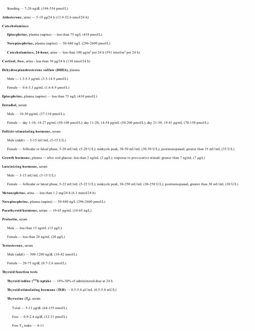
Standing — 7-20 ng/dL (194-554 pmol/L)
Aldosterone, urine — 5-19 μg/24 h (13.9-52.6 nmol/24 h)
Catecholamines
Epinephrine, plasma (supine) — less than 75 ng/L (410 pmol/L)
Norepinephrine, plasma (supine) — 50-440 ng/L (296-2600 pmol/L)
Catecholamines, 24-hour, urine — less than 100 μg/m2 per 24 h (591 nmol/m2 per 24 h)
Cortisol, free, urine - less than 50 μg/24 h (138 nmol/24 h)
Dehydroepiandrosterone sulfate (DHEA), plasma
Male — 1.3-5.5 μg/mL (3.5-14.9 μmol/L)
Female — 0.6-3.3 μg/mL (1.6-8.9 μmol/L)
Epinephrine, plasma (supine) — less than 75 ng/L (410 pmol/L)
Estradiol, serum
Male — 10-30 pg/mL (37-110 pmol/L);
Female — day 1-10, 14-27 pg/mL (50-100 pmol/L); day 11-20, 14-54 pg/mL (50-200 pmol/L); day 21-30, 19-41 pg/mL (70-150 pmol/L)
Follicle-stimulating hormone, serum
Male (adult) — 5-15 mU/mL (5-15 U/L)
Female — follicular or luteal phase, 5-20 mU/mL (5-20 U/L); midcycle peak, 30-50 mU/mL (30-50 U/L); postmenopausal, greater than 35 mU/mL (35 U/L)
Growth hormone, plasma — after oral glucose: less than 2 ng/mL (2 μg/L); response to provocative stimuli: greater than 7 ng/mL (7 μg/L)
Luteinizing hormone, serum
Male — 3-15 mU/mL (3-15 U/L)
Female — follicular or luteal phase, 5-22 mU/mL (5-22 U/L); midcycle peak, 30-250 mU/mL (30-250 U/L); postmenopausal, greater than 30 mU/mL (30 U/L)
Metanephrine, urine — less than 1.2 mg/24 h (6.1 mmol/24 h)
Norepinephrine, plasma (supine) — 50-440 ng/L (296-2600 pmol/L)
Parathyroid hormone, serum — 10-65 pg/mL (10-65 ng/L)
Prolactin, serum
Male — less than 15 ng/mL (15 μg/L)
Female — less than 20 ng/mL (20 μg/L)
Testosterone, serum
Male (adult) — 300-1200 ng/dL (10-42 nmol/L)
Female — 20-75 ng/dL (0.7-2.6 nmol/L)
Thyroid function tests
Thyroid iodine (131I) uptake — 10%-30% of administered dose at 24 h
Thyroid-stimulating hormone (TSH) — 0.5-5.0 μU/mL (0.5-5.0 mU/L)
Thyroxine (T4), serum
Total — 5-12 μg/dL (64-155 nmol/L)
Free — 0.9-2.4 ng/dL (12-31 pmol/L)
Free T 4 index — 4-11

Triiodothyronine, free (T3) — 3.6-5.6 ng/L (5.6-8.6 pmol/L)
Triiodothyronine, resin (T3) — 25%-35%
Triiodothyronine, serum (T3) — 70-195 ng/dL (1.1-3.0 nmol/L)
Vanillylmandelic acid, urine — less than 8 mg/24 h (40.4 μmol/24 h)
Vitamin D
1,25-dihydroxy, serum — 25-65 pg/mL (60-156 pmol/L)
25-hydroxy, serum — 25-80 ng/mL (62-200 nmol/L)
Urine
Albumin-creatinine ratio — less than 30 mg/g
Calcium — 100-300 mg/24 h (2.5-7.5 mmol/24 h) on unrestricted diet
Creatinine — 15-25 mg/kg per 24 h (133-221 mmol/kg per 24 h)
Glomerular filtration rate (GFR)
Normal
Male — 130 mL/min/1.73 m2
Female — 120 mL/min/1.73 m2
Stages of Chronic Kidney Disease
Stage 1 — greater than or equal to 90 mL/min/1.73 m2
Stage 2 — 60-89 mL/min/1.73 m2
Stage 3 — 30-59 mL/min/1.73 m2
Stage 4 — 15-29 mL/min/1.73 m2
Stage 5 — less than 15 mL/min/1.73 m2
5-Hydroxyindoleacetic acid (5-HIAA) — 2-9 mg/24 h (10.4-46.8 μmol/24 h)
Protein-creatinine ratio - less than or equal to 0.2 mg/mg
Sodium — 100-260 meq/24 h (100-260 mmol/24 h) (varies with intake)
Uric acid — 250-750 mg/24 h (1.48-4.43 mmol/24 h) (varies with diet)
Gastrointestinal
Gastrin, serum — 0-180 pg/mL (0-180 ng/L)
Stool fat — less than 5 g/d on a 100-g fat diet
Stool weight — less than 200 g/d
Pulmonary
Forced expiratory volume in 1 second (FEV1) — greater than 80% of predicted
Forced vital capacity (FVC) — greater than 80% of predicted
FEV1/FVC — greater than 75%
Cerebrospinal Fluid
Cell count — 0-5/μL (0-5 × 10⁶/L)
Glucose — 40-80 mg/dL (2.2-4.4 mmol/L); less than 40% of simultaneous plasma concentration is abnormal

Pressure (opening) — 70-200 mm H2O
Protein — 15-60 mg/dL (150-600 mg/L)
Hemodynamic Measurements
Cardiac index — 2.5-4.2 L/min/m2
Left ventricular ejection fraction — greater than 55%
Pressures
Pulmonary artery
Systolic — 20-25 mm Hg
Diastolic — 5-10 mm Hg
Mean — 9-16 mm Hg
Pulmonary capillary wedge — 6-12 mm Hg
Right atrium — mean 0-5 mm Hg
Right ventricle
Systolic — 20-25 mm Hg
Diastolic — 0-5 mm Hg

Plate 1. Gastroenterology and Hepatology, Item 46

Plate 2. General Internal Medicine, Item 55

Plate 3. General Internal Medicine, Item 56

Plate 4. General Internal Medicine, Item 57

Plate 5. General Internal Medicine, Item 59

Plate 6. General Internal Medicine, Item 60

Plate 7. General Internal Medicine, Item 61

Plate 8. General Internal Medicine, Item 62

Plate 9. General Internal Medicine, Item 63

Plate 10. General Internal Medicine, Item 64

Plate 11. Hematology, Item 4

Plate 12. Hematology, Item 5

Plate 13. Hematology, Item 8

Plate 14. Hematology, Item 9

Plate 15. Hematology, Item 18

Plate 16. Hematology, Item 20

Plate 17. Hematology, Item 21

Plate 18. Hematology, Item 22

Plate 19. Hematology, Item 27

Plate 20. Hematology, Item 30

Plate 21. Infectious Disease Medicine, Item 21

Plate 22. Infectious Disease Medicine, Item 26

Plate 23. Oncology, Item 2

Plate 24. Oncology, Item 22

Plate 25. Oncology, Item 23

Plate 26. Oncology, Item 24

Plate 27. Oncology, Item 25

Plate 28. Oncology, Item 26

Plate 29. Rheumatology, Item 2

Plate 30. Rheumatology, Item 25

Plate 31. Rheumatology, Item 30