Mitochondrial-localized Phosphatidylethanolamine in ... · PDF fileMitochondria were first...
127
Mitochondrial-localized Phosphatidylethanolamine in Mitochondrial Dynamics and Autophagy by Eliana Y.L. Chan A thesis submitted in conformity with the requirements for the degree of Doctor of Philosophy Graduate Department of Biochemistry University of Toronto © Copyright by Eliana Y. L. Chan 2014
-
Upload
trankhuong -
Category
Documents
-
view
226 -
download
1
Transcript of Mitochondrial-localized Phosphatidylethanolamine in ... · PDF fileMitochondria were first...

Mitochondrial-localized
Phosphatidylethanolamine in Mitochondrial Dynamics and Autophagy
by
Eliana Y.L. Chan
A thesis submitted in conformity with the requirements for the degree of Doctor of Philosophy
Graduate Department of Biochemistry University of Toronto
© Copyright by Eliana Y. L. Chan 2014

ii
Mitochondrial-localized Phosphatidylethanolamine in
Mitochondrial Dynamics and Autophagy
Eliana Y. L. Chan
Doctor of Philosophy
Graduate Department of Biochemistry University of Toronto
2014
ABSTRACT
Phosphatidylethanolamine (PE) constitutes a significant proportion of total phospholipids
in biological membranes. A major route of PE biosynthesis occurs via the decarboxylation of
phosphatidylserine (PS) by the inner-mitochondrial-membrane (IMM)-localized PS
decarboxylase (PSD). The only other known phospholipids that are synthesized in mitochondria
are the mitochondrial-enriched phosphatidylglycerol (PG) and cardiolipin (CL); the synthesis of
other biological lipids occurs mostly in the endoplasmic reticulum (ER). Hence, the
mitochondrial-localization of PSD led me to speculate that PE within the mitochondrial
membrane might have important functions. In this thesis, I describe my findings that
mitochondrial-localized PE plays an important role in mitochondrial membrane remodelling and
autophagy, a conserved quality control mechanism. Mitochondrial PE promotes mitochondrial
membrane fusion by enhancing the biogenesis of the short isoform of mitochondrial genome
maintenance 1 (s-Mgm1), a key mitochondrial fusion protein. The biophysical properties of PE
on the mitochondrial membrane also promote fusion likely by increasing the rate of lipid mixing

iii
during fusion. In addition, mitochondrial PE is important for oxidative phosphorylation and the
maintenance of mitochondrial ATP levels, crucial mitochondrial homeostatic functions. I also
demonstrate that a reduction in mitochondrial PE impairs autophagy likely by impeding
autophagosome formation, expansion and/or fusion with the vacuole in yeast. My results
provide additional insight into the functional importance of mitochondrial PE and highlight the
specific lipid composition of the mitochondrial membrane as a key player in mitochondrial
membrane fusion and autophagy.

iv
ACKNOWLEDGEMENTS
First and foremost, I would like to thank my supervisor, Angus, for his time and patience,
and for giving me the opportunity to learn and grow in his lab. He has been a wonderful mentor
both in and out of the lab and has given me an experience that will be deeply etched in my heart.
I am also immensely grateful to Jeff, our former post-doc, who taught me how to think
critically and make each experiment count. Without Jeff's tutelage, I am sure I will not be the
researcher I am today.
I would also like to thank my supervisory committee members, Dr. Reinhart Reithmeier
and Dr. Grant Brown for their guidance and insightful suggestions throughout my time as a
graduate student.
My gratitude also goes out to all past and present members of the McQuibban lab who
have supported me through the good times and the bad. My experience would not have been the
same without the friendliness and kindness from all members of the lab. I certainly had an
unforgettable time in the McQuibban lab.
I would also like to extend special thanks to Dr. Craig Smibert who has always been
around to give me advice from the time I was an undergraduate student. The members of the
Smibert lab have also been amazing neighbours and have given me invaluable help throughout
the years.
My appreciation also goes out to my wonderful and loving sister who has put up with my
procrastination in booking trips home. I promise I will try to be better!
I would also like to thank Harry and Justine for their continued love, support and
patience, without which, this degree would not have been possible.

v
Finally, I would like to dedicate this work to my parents. Their unconditional love and
thoughtful upbringing have made me the person I am today. Despite my shortcomings, they
always believed in me; and for that, I will be eternally grateful.

vi
TABLE OF CONTENTS
ABSTRACT ...................................................................................................................... ii
ACKNOWLEDGEMENTS ............................................................................................. iv
TABLE OF CONTENTS ................................................................................................. vi
LIST OF FIGURES .......................................................................................................... x
LIST OF ABBREVIATIONS .......................................................................................... xii
CHAPTER 1 INTRODUCTION ..................................................................................... 1
1.1 MITOCHONDRIAL MEMBRANE DYNAMICS ............................................... 1
1.1.1 The discovery of mitochondrial membrane dynamics ................................ 2
1.1.2 Mitochondrial membrane fission ................................................................ 3
1.1.3 Mitochondrial membrane fusion ................................................................. 8
1.1.3.1 Outer mitochondrial membrane fusion .......................................... 8
1.1.3.2 Inner mitochondrial membrane fusion .......................................... 10
1.1.3.3 Coordinating outer and inner mitochondrial membrane fusion .... 13
1.1.3.4 The role of lipids in mitochondrial membrane fusion ................... 15
1.1.4 Mitochondrial membrane dynamics and autophagy ................................... 16
1.2 AUTOPHAGY ...................................................................................................... 17
1.2.1 The autophagy pathway and its molecular machinery ............................... 19
1.2.1.1 The induction of autophagy .......................................................... 19
1.2.1.2 Phagophore formation ................................................................... 21
1.2.1.3 Phagophore expansion and autophagosome formation ................. 21
1.2.1.4 Autophagosome maturation and fusion with the
vacuole/lysosomes ......................................................................... 25

vii
1.2.2 The lipids implicated in autophagy ............................................................. 26
1.2.2.1 Phosphoinositides .......................................................................... 26
1.2.2.2 Phosphatidic acid ............................................................................ 27
1.2.2.3 Membrane composition and/or membrane curvature .................... 28
1.3 PHOSPHATIDYLETHANOLAMINE BIOSYNTHESIS IN
SACCHAROMYCES CEREVISIAE ....................................................................... 30
1.3.1 Phosphatidylserine decarboxylation ........................................................... 30
1.3.2 The Kennedy pathway ................................................................................ 36
1.3.3 Alternative pathways of phosphatidylethanolamine biosynthesis .............. 36
1.3.4 The different contributions of phosphatidylserine decarboxylation and the
Kennedy pathway ....................................................................................... 36
1.3.5 Functions of phosphatidylethanolamine ..................................................... 38
1.4 Specific interests and goals ................................................................................... 39
CHAPTER 2 MATERIALS AND METHODS .............................................................. 41
2.1 REAGENTS .......................................................................................................... 41
2.1.1 Yeast strains ................................................................................................ 41
2.1.2 Plasmids ...................................................................................................... 41
2.1.3 Growth conditions ....................................................................................... 42
2.1.4 Lipids .......................................................................................................... 42
2.2 FUSION ASSAYS ................................................................................................ 43
2.2.1 In vivo mitochondrial fusion assay ............................................................. 43
2.2.2 In vitro liposome fusion assay .................................................................... 43
2.3 MITOCHONDRIAL ASSAYS ................................................................................. 44

viii
2.3.1 Mitochondrial purification .......................................................................... 44
2.3.2 Oxidative phosphorylation ......................................................................... 45
2.3.3 Mitochondrial ATP measurements ............................................................. 45
2.4 CYCLOHEXIMIDE CHASE ............................................................................... 45
2.5 AUTOPHAGY ASSAYS ..................................................................................... 46
2.5.1 Determination of cell viability ................................................................... 46
2.5.2 Atg8/Atg8-PE analysis ............................................................................... 46
2.5.3 Vacuolar internalization of FM 4-64 .......................................................... 46
CHAPTER 3 PHOSPHATIDYLSERINE DECARBOXYLASE 1 (PSD1)
PROMOTES MITOCHONDRIAL FUSION BY REGULATING THE
BIOPHYSICAL PROPERTIES OF THE MITOCHONDRIAL MEMBRANE
AND ALTERNATIVE TOPOGENESIS OF MITOCHONDRIAL GENOME
MAINTENANCE 1 (MGM1) .................................................................................... 47
3.1 ABSTRACT .......................................................................................................... 47
3.2 INTRODUCTION ................................................................................................. 48
3.3 RESULTS ............................................................................................................... 50
3.3.1 Psd1 is required for normal mitochondrial morphology ............................ 50
3.3.2 Psd1 is required for proper mitochondrial fusion during yeast mating ...... 52
3.3.3 Phospholipid composition affects the rate of lipid mixing ......................... 54
3.3.4 Psd1 is required for proper mitochondrial activity ..................................... 56
3.3.5 Ethanolamine cannot rescue Δpsd1 mitochondrial-specific defects .......... 58
3.3.6 s*Mgm1 can rescue mitochondrial aggregation but not the
glycerol growth defect in Δpsd1 cells ......................................................... 59

ix
3.3.7 Psd1 regulates alternative topogenesis of Mgm1 ....................................... 61
3.4 DISCUSSION ....................................................................................................... 64
CHAPTER 4 MITOCHONDRIAL PHOSPHATIDYLETHANOLAMINE
IS IMPORTANT FOR STARVATION-INDUCED AUTOPHAGY
IN YEAST ................................................................................................................... 70
4.1 ABSTRACT .......................................................................................................... 70
4.2 INTRODUCTION ................................................................................................. 71
4.3 RESULTS .............................................................................................................. 73
4.3.1 Psd1 is required for proper autophagy in yeast ........................................... 73
4.3.2 The loss of Psd1 results in a reduction of autophagic bodies
in the yeast vacuole ..................................................................................... 74
4.3.3 Ethanolamine cannot rescue the autophagic defects in Δpsd1 cells ........... 76
4.4 DISSCUSSION ..................................................................................................... 78
CHAPTER 5 CONCLUDING PERSPECTIVES .......................................................... 81
5.1 BRIEF SUMMARY OF RESULTS ...................................................................... 81
5.2 PERSPECTIVES ................................................................................................... 82
5.2.1 Phosphatidylethanolamine in mitochondrial membrane dynamics ............ 82
5.2.2 Mitochondrial lipids in mitochondrial biology ........................................... 85
5.2.3 The specific role(s) of mitochondrial phosphatidylethanolamine
in autophagy ................................................................................................ 87
5.2.4 The composition of the mitochondrial membrane in autophagy ................ 89
5.3 CONCLUSION ..................................................................................................... 92
REFERENCES .................................................................................................................. 94

x
LIST OF FIGURES
Figure 1-1: Proposed model for yeast mitochondrial fission ......................................... 5
Figure 1-2: Schematic of the yeast mitochondrial fusion machinery ........................... 9
Figure 1-3: Alternative topogenesis of Mgm1 ................................................................. 11
Figure 1-4: Schematic of Mgm1-mediated inner mitochondrial membrane
fusion .............................................................................................................. 13
Figure 1-5: Different forms of autophagy ....................................................................... 18
Figure 1-6: Induction of autophagy ................................................................................. 20
Figure 1-7: Order of protein recruitment to the phagophore assembly site
in yeast ............................................................................................................ 22
Figure 1-8: The two ubiquitin-like conjugation systems in autophagy ........................ 23
Figure 1-9: Simplified overview of phosphatidylethanolamine biosynthesis in
Saccharomyces cerevisiae .............................................................................. 31
Figure 1-10: Phosphatidylethanolamine biosynthesis by phosphatidylserine
decarboxylation and the Kennedy pathway ................................................ 32
Figure 1-11: Domain organization of phosphatidylserine decarboxylases ................... 33
Figure 1-12: Phosphatidylserine decarboxylase proenzyme maturation ...................... 34
Figure 1-13: Phosphatidylserine decarboxylase enzymology ......................................... 35
Figure 3-1: Psd1 is required for normal mitochondrial morphology ........................... 51
Figure 3-2: Psd1 is required for mitochondrial fusion during yeast mating ............... 53
Figure 3-3: Liposomes with lipid compositions similar to ΔΔpsd1 mitochondria
have a reduced rate of lipid mixing ............................................................. 55
Figure 3-4: The ΔΔpsd1 strain has defects in mitochondrial activity ............................. 57

xi
Figure 3-5: s*Mgm1 suppresses ΔΔpsd1 mitochondrial aggregation ............................. 60
Figure 3-6: Psd1 regulates Mgm1 alternative topogenesis ............................................ 63
Figure 3-7: Model of Psd1-dependent mitochondrial regulation .................................. 69
Figure 4-1: Mitochondrial phosphatidylethanolamine is important for
autophagy ....................................................................................................... 75
Figure 4-2: Reducing mitochondrial phosphatidylethanolamine reduces
autophagic bodies in the vacuole .................................................................. 77
Figure 5-1: Overexpression of Psd1 results in mitochondrial fragmentation
independent of s-Mgm1 biogenesis .............................................................. 83

xii
LIST OF ABBREVIATIONS
- N Starvation medium
5-FOA 5-Fluoroorotic acid
ATP Adenosine triphosphate
BAR Bin-Amphiphysin-Rvs
BHK Baby hamster kidney
C12E8 Octaethylene glycol monododecyl ether
CCCP Carbonylcyanide m-chlorophenylhydrazone
CDP-DAG Cytidyldiphosphate diacylglycerol
CDP-Etn Cytidyldiphosphate ethanolamine
CHO Chinese hamster ovary
CHX Cycloheximide
CL Cardiolipin
CMA Chaperone-mediated autophagy
CTP Cytidyltriphosphate
Cvt Cytoplasm-to-vacuole targeting
DAG Diacylglycerol
DRP Dynamin-related protein
EM Electron microscopy
ER Endoplasmic reticulum
ERMES Endoplasmic reticulum-mitochondria encounter structure
ETC Electron transport chain
ETM Energy transfer motif

xiii
Etn Ethanolamine
GDP Guanosine diphosphate
GED Guanosine triphosphate effector domain
GTP Guanosine triphosphate
GTPase Guanosine triphosphate hydrolase
GUV Giant unilamellar vesicle
IMM Inner mitochondrial membrane
IMS Intermembrane space
lyso-PE Lyso-phosphatidylethanolamine
MAM Mitochondrial-associated endoplasmic reticulum membrane
MD Molecular dynamics
MICOS Mitochondrial contact site
mtBFP Mitochondrial-targeted blue fluorescent protein
mtGFP Mitochondrial-targeted green fluorescent protein
MPP Mitochondrial processing peptidase
MPTP Mitochondrial permeability transition pore
mTOR Mammalian target of rapamycin
mTORC1 Mammalian target of rapamycin Complex 1
MTS Mitochondrial targeting sequence
MVB Multivesicular body
NAT Nourseothricin
OD Optical density
OMM Outer mitochondrial membrane

xiv
ORF Open reading frame
PA Phosphatidic acid
PAS Phagophore assembly site
PC Phosphatidylcholine
PE Phosphatidylethanolamine
P-Etn Phosphoethanolamine
PG Phosphatidylglycerol
PHB Prohibitin
PI Phosphatidylinositol
PI3K Phosphatidylinositol 3-kinase
PI3P Phosphatidylinositol 3-phosphate
PM Plasma membrane
pmaER Plasma membrane-associated endoplasmic reticulum
PMSF Phenylmethylsulfonyl fluoride
PS Phosphatidylserine
PSD Phosphatidylserine decarboxylase
SC Synthetic complete
SUV Small unilamellar vesicle
TM Transmembrane
TOR Target of rapamycin
TORC1 Target of rapamycin Complex 1
TPR Tetratricopeptide repeat
WT Wild type

1
CHAPTER 1
INTRODUCTION
Some material in this chapter was previously published. Springer Publishing Company, 2011,
Mitochondrial dynamics and neurodegeneration, Chapter 1, pp 1-46, The genetics of
mitochondrial fusion and fission, Eliana Y. L. Chan, Jarungjit Rujiviphat and G. Angus
McQuibban, with kind permission from Springer Science and Business Media.
1.1 Mitochondrial membrane dynamics
Mitochondria were first observed in the 1850s when cytologists discovered granular
structures in the cytoplasm of living cells. In 1857, Swiss anatomist Albert von Kölliker
described these granular structures in the sarcoplasm of insect muscle cells and showed in 1888
that these granular structures swelled in water and possessed a membrane (1). These structures
had originally been named "bioblasts" and "sarcosomes" by Richard Altmann (2) and Gustaf
Retzius (3), respectively, but in 1898, Carl Benda introduced the term "mitochondrion", derived
from the Greek words mitos meaning threaded, and chondron meaning grain (reviewed in
reference (4)). Although the term "mitochondrion" has become the most widely accepted name
for this organelle, the term "sarcosome" is still used today to describe mitochondria in muscle
cells.

2
1.1.1 The discovery of mitochondrial membrane dynamics
One of the first in-depth descriptions of mitochondria in cells was achieved by Lewis and
Lewis in 1915 (5). Using cultured chick cells, they were able to observe live and fixed
mitochondria, and could describe mitochondrial movement, quantity and dynamics (5).
However, it was not until the 1990s with the development of fluorescent probes and the
resurgence of light microscopy that the movement, fusion and fission of mitochondria in living
cells were recorded and widely accepted (6). Since the identification of mitochondrial
membrane dynamics, technological advances together with genetic, structural and biochemical
approaches have enabled the field to dissect the molecular mechanisms of mitochondrial fusion
and fission and how this delicate network is maintained. What Lewis and Lewis referred to as
mitochondrial fusion and branching/separation (5) are now known to be distinct, regulated
processes required for proper mitochondrial function and inheritance (7-10). Mitochondrial
membrane dynamics are now known to be crucial mediators of apoptosis (11,12) and play a role
in neurodegeneration (13-15).
The shape of the mitochondrial network is maintained by the regulated balance of fusion
and fission events (16-21). Accordingly, decreased fission results in elongated mitochondria
(20-24), and decreased fusion results in mitochondria that appear fragmented and/or aggregated
(16-18). Notably, the phenotype resulting from reduced mitochondrial fusion can be rescued by
reducing mitochondrial fission (17,18) and vice versa (21), strongly indicating that normal
mitochondrial morphology is maintained by the balance of fusion and fission events rather than
the absolute number of each reaction.
The pioneering discoveries of the molecular machineries of mitochondrial membrane
fusion and fission were largely derived from genetic studies in yeast, flies, worms and

3
mammalian cells (reviewed in references (25,26)). Due the simplicity of a single cell system and
powerful genetic approaches, the molecular mechanisms of mitochondrial membrane dynamics
are best characterized in yeast. The key players described in yeast mitochondrial membrane
dynamics are the dynamin-related proteins (DRPs) (26). DRPs are large guanosine triphosphate
hydrolases (GTPases) involved in several membrane remodelling events (27). They are
distinguished from other GTPases by their ability to bind to lipids, self-assemble, and to undergo
oligomerization-stimulated guanosine triphosphate (GTP) hydrolysis (27). All DRPs contain
three conserved domains: (i) the GTPase domain, (ii) the middle domain, and (iii) the GTP
effector domain (GED). The catalytic GTPase domain serves as the site for GTP binding and
hydrolysis while the middle domain and GED are essential for oligomerization and self-
assembly. The region between the middle domain and the GED serves as the site for lipid
interactions (27). The four well-characterized DRPs in yeast are vacuolar protein sorting 1
(Vps1), dynamin-related 1 (Dnm1), mitochondrial genome maintenance 1 (Mgm1) and fuzzy
onions 1 (Fzo1). Vps1 mediates Golgi vesicle formation whereas the other three DRPs function
in mitochondrial membrane dynamics. Dnm1 directly mediates the fission of mitochondria
whereas Mgm1 and Fzo1 are the key players for mitochondrial fusion (26). These DRPs are
highly conserved proteins, and their higher eukaryotic counterparts are also fission and fusion
molecules.
1.1.2 Mitochondrial membrane fission
Although the regulation of mitochondrial membrane fission differs slightly from one
organism to another, the core components required remain highly conserved. In general,
mitochondrial membrane fission requires mitochondrial fission 1 (Fis1) and the DRP

4
Dnm1/Drp1. Deleting Fis1 or Dnm1 in yeast results in elongated mitochondria (20,22,28). In
yeast, Dnm1 exists as punctate structures on the outer mitochondrial membrane (OMM) and in
the cytosol (19,29), whereas mammalian Drp1 is mostly diffuse cytoplasmic with some puncta
on mitochondria (23). Despite their differences in localization, Dnm1/Drp1 in yeast, worms and
mammals localize to the OMM at sites of mitochondrial fission (30,31), consistent with their
roles as proteins that divide mitochondria. Although homologs have been identified in many
organisms, it is the detailed biochemical and structural analyses on yeast Dnm1 and mammalian
Drp1 that have shed light on the mechanism of DRP-mediated mitochondrial membrane fission.
Like dynamins, Dnm1/Drp1 assemble into dimers/tetramers (32,33) and organize into
ring-like structures (Figure 1-1) (23,34,35). The spirals formed by Dnm1 can constrict
liposomes, reducing their diameter to that of mitochondrial constriction sites (34). Independent
studies using truncated forms of yeast Dnm1 and mammalian Drp1 identified the GED as an
important region for the formation of these higher-ordered structures (32,36). The GED of
mammalian Drp1 coordinates intra- and intermolecular interactions - it forms intermolecular
interactions with itself, and intramolecular interactions with the GTPase domain (36). It is also
sufficient for the mitochondrial targeting of Drp1 (37). One of the characteristic features of
dynamins and DRPs is oligomerization-stimulated GTP hydrolysis. In the case of Dnm1/Drp1,
self-assembly into higher order oligomers occurs after recruitment to mitochondria and is the
rate-limiting step in GTP hydrolysis (34,38).
The recruitment of Dnm1/Drp1 to mitochondria occurs via Fis1, an integral membrane
protein on the OMM (Figure 1-1) (22,35). The N-terminus of Fis1 faces the cytosol and its C-
terminus faces the intermembrane space (IMS) (22,39). Most of the protein resides in the
cytosol and is anchored to the OMM by its C-terminal transmembrane (TM) segment (40).

5
Figure 1-1: Proposed model for yeast mitochondrial fission.
In yeast, Fis1 recruits the dynamin-related protein Dnm1 to sites of mitochondrial fission via the adaptor proteins Mdv1 and Caf4. The proposed model for Dnm1-dependent mitochondrial fission is depicted.
Dnm1
Fis1
Mdv1/
Caf4
OMM
GTP binding drives
Dnm1 self-assembly
Dnm1 self-assembly
constricts the membrane
GTP hydrolysis drives
Dnm1 depolymerization
and membrane instability
Membrane fission to
relieve instability

6
Unlike Dnm1/Drp1 that appear as discrete puncta, Fis1 is uniformly distributed throughout the
mitochondrial membrane (22). It organizes into six α-helices (α1-α6) that resemble
tetratricopeptide repeat (TPR) folds (41,42). TPR motifs mediate protein-protein interactions
and are usually a part of multiprotein complexes (reviewed in reference (43)). The α2-α3 and
α4-α5 helices of Fis1/hFis1 form two TPR motifs (TPR1 and TPR2), forming a concave
hydrophobic binding pocket (42,44). Although TPR motifs are proposed to mediate protein-
protein interactions, direct binding between Fis1/hFis1 and Dnm1/Drp1 remains controversial
(45-47), and adaptor proteins are proposed to mediate binding between the two. In yeast,
mitochondrial division 1 (Mdv1) and CCR4-associated factor (Caf4) serve as adaptors to recruit
Dnm1 to mitochondria (Figure 1-1) (28,48,49), whereas in higher eukaryotes, mitochondrial
fission factor (Mff), mitochondrial dynamics proteins of 49 and 51 kDa (Mid49 and Mid51,
respectively) and ganglioside-induced differentiation-associated protein 1 (GDAP1) are the
proposed receptors for Drp1 (50-56). A proposed model for mitochondrial fission in yeast is
shown in Figure 1-1.
Despite Dnm1/Drp1 and Fis1/hFis1 being the best characterized mitochondrial fission
proteins, studies in yeast and worms indicate that they may only be responsible for OMM fission.
Mitochondria of C. elegans expressing mutant forms of Drp-1 were still connected by thin
tubules of the OMM while inner mitochondrial membrane (IMM) fission still occurred (57).
Similarly, yeast Δdnm1 and Δfis1 mutants did not achieve complete mitochondrial fission, but
had matrix separation (58). These data suggest that Dnm1/Drp1 and Fis1/hFis1 are not required
for IMM fission, and that other components mediate this process.
Yeast mitochondrial distribution and morphology 33 (Mdm33), mammalian
mitochondrial protein of 18 kDa (Mtp18) and mitochondrial targeting GxxxG motif protein

7
(MTGM) were later identified and proposed to be factors mediating IMM fission (59-62).
Mdm33 has no known mammalian homologs, whereas Mtp18 has known homologs only in
metazoans (59-61). In contrast, MTGM has homologs in eukaryotes from yeast to human.
Remarkably, all the analyzed mammalian orthologs of MTGM have 100% amino acid sequence
identity (62). Reducing Mdm33, Mtp18 and MTGM protein levels resulted in elongated
mitochondria, whereas their overexpression led to fragmentation, phenotypes resembling that of
mitochondrial fission mutants (59-62). Although the topology of Mtp18 remains unclear, it
isproposed to reside in the IMS (61). Mdm33 is an integral protein of the IMM with its C-
terminus in the IMS and its predicted N-terminal coiled-coil domains in the mitochondrial
matrix. Analysis of its oligomeric state showed that Mdm33 has homotypic interactions, likely
mediated by its coiled-coil domains. Overexpressing Mdm33 resulted in IMM fragmentation
and loss of cristae, suggesting IMM fission defects. Based on these observations, Mdm33 on
apposing IMMs are proposed to form homotypic interactions via their coiled-coil domains in the
mitochondrial matrix, thereby mediating IMM fission (59).
MTGM was identified as a human nuclear gene whose product is highly enriched in brain
tumor cell lines and tumor tissues (62). Like Mdm33, MTGM is an integral membrane protein
of the IMM. Rather than coiled-coil domains, MTGM has a highly conserved tetrad of GxxxG
motif that also mediates protein-protein interactions. MTGM-induced mitochondrial
fragmentation is Drp1-dependent, indicating that it could be a regulator of Drp1. However, its
IMM localization suggests that MTGM might mediate IMM fission (62). Additional
experiments have to be conducted to determine if MTGM is directly involved in IMM fission or
whether it is simply a regulator of Drp1. A complete review of the mitochondrial membrane
fission machinery can be found in references (25,26,63).

8
1.1.3 Mitochondrial membrane fusion
Mitochondrial membrane fusion is unique and distinct from other intracellular membrane
fusion events because it requires the coordinate fusion of two separate membranes. OMM and
IMM fusions are separable events with distinct energy requirements (64), indicating the
existence of distinct OMM and IMM fusion machineries. However, they are temporally coupled,
strongly suggesting that the OMM and IMM fusion machineries must coordinate effectively to
ensure proper fusion of the organelle (26). Although some proteins of the mitochondrial fusion
machinery have been identified and characterized in higher organisms, studies in yeast have laid
the foundation for our current working model of mitochondrial membrane fusion. In yeast,
OMM fusion is mediated by Fzo1 (mitofusins 1 and 2 [Mfn1 and Mfn2, respectively] in
mammals), while IMM fusion is mediated by Mgm1 (optic atrophy 1 [OPA1] in mammals). A
fungal-specific protein, Ugo1 (Ugo is Japanese for fusion), which is a member of the
mitochondrial transporter family, serves as an adaptor protein, coordinating OMM and IMM
fusion in yeast (Figure 1-2).
1.1.3.1 Outer mitochondrial membrane fusion
Fzo1 is an OMM-localized protein with a bipartite transmembrane domain (16,65,66).
The majority of the protein and both the N- and C-terminus face the cytosol (16,65). Fzo1
contains a GTPase domain at its N-terminus (65) and a total of three heptad repeats predicted to
form coiled-coil domains that mediate stable cis and trans Fzo1-Fzo1 interactions (67). Similar
to Dnm1, self-assembly and GTPase activity are crucial for Fzo1 function (64). It is proposed
that GTP binding facilitates Fzo1 cis dimerization (68). Fzo1 dimers on opposing membranes
then associate in trans to tether the membranes and facilitate fusion (Figure 1-2) (64,68).

9
Subsequent GTP hydrolysis might induce a conformational change that results in Fzo1
ubiquitination and its proteasome-dependent degradation (68-70). Dimerization in trans appears
to be conserved, as the mammalian orthologs of Fzo1, Mfn1 and Mfn2, also oligomerize in trans
Figure 1-2: Schematic of the yeast mitochondrial fusion machinery.
The yeast mitochondrial fusion complex consists of Fzo1 and Ugo1 on the outer mitochondrial membrane (OMM) and Mgm1 on the inner mitochondrial membrane (IMM). Ugo1 serves as an adaptor, coordinating OMM and IMM fusion. The dimerization of Ugo1 and the binding of guanosine triphosphate (GTP) by Fzo1 allow Fzo1 to dimerize in cis and in trans to tether the opposing OMMs. Subsequent events leading to the fusion of the OMM are not known, but Fzo1-dependent GTP hydrolysis likely induces membrane stress and destabilization that can be relieved upon Ugo1-dependent lipid mixing of the OMM. Mgm1 also oligomerizes in cis and in trans to tether opposing IMMs. GTP hydrolysis induces a conformational change in Mgm1 that could also increase membrane stress and destabilization that can be relieved upon Ugo1-dependent IMM fusion.
OMM
IMM
Ugo1
Ugo1
Fzo1
Fzo1
s-Mgm1
l-Mgm1

10
to tether mitochondria (71-73). Moreover, in the absence of a functional GTPase domain in
Mfn1, mitochondria aggregate but maintain a uniform distance (73), suggesting that dimerization
in trans facilitates mitochondrial tethering, whereas GTP hydrolysis promotes mitochondrial
fusion.
The mechanistic details of Fzo1-mediated mitochondrial membrane fusion are poorly
described due to the lack of structural details on Fzo1. However, structural studies on the
bacterial dynamin-like protein, BDLP, which is closely related to Fzo1, might shed light on
Fzo1-mediated mitochondrial fusion. BDLP binds to and tubulates lipid bilayers and may induce
a compressed bilayer by distorting lipid tails (74,75). Guanosine diphosphate (GDP)-bound
BDLP has a lower affinity for the lipid bilayer and may possess a dimeric structure that differs
from the GTP-bound form. These data along with recent crystal structures of human dynamin 1
(76,77) suggest that GTP binding induces protein polymerization (75-77) and membrane
tubulation (75), whereas GTP hydrolysis leads to protein depolymerization (75-77), leaving a
highly curved outer leaflet in a high-energy state that can be stabilized upon fusion (75).
1.1.3.2 Inner mitochondrial membrane fusion
While OMM fusion is mediated by Fzo1, IMM fusion is dependent on Mgm1. Knocking
out Mgm1 in yeast or OPA1 in mammals results in fragmented mitochondria (78-80). Mgm1 is
a nuclear-encoded protein containing an N-terminal mitochondrial targeting sequence (MTS) that
is followed by two hydrophobic segments. It is targeted to the IMM via its MTS and undergoes
a unique form of processing known as alternative topogenesis that is dependent on adenosine
triphosphate (ATP) and a functional mitochondrial protein import machinery (81). Alternative
topogenesis of Mgm1 results in the formation of the two different isoforms of Mgm1, long-

11
Mgm1 (l-Mgm1) and short-Mgm1 (s-Mgm1) (Figure 1-3) (81). Upon import into mitochondria,
full-length Mgm1 (FL-Mgm1) can be inserted into the membrane by its first hydrophobic
segment; the subsequent cleavage of its MTS by the matrix processing peptidase (MPP) results
in the formation of l-Mgm1. In an ATP-dependent manner, FL-Mgm1 can bypass its first
hydrophobic segment and be embedded in the IMM by its second hydrophobic segment where it
can be proteolytically processed by the rhomboid intramembrane protease Rbd1/Pcp1 (82,83).
Maintaining approximately equal molar concentrations of l-Mgm1 and s-Mgm1 is crucial for
mitochondrial membrane fusion. Although both isoforms are necessary for proper fusion, a
Figure 1-3: Alternative topogenesis of Mgm1.
Mgm1 contains two N-terminal hydrophobic regions. Upon import into mitochondria, full-length Mgm1 (FL-Mgm1) can be inserted and anchored into the IMM by its first hydrophobic region. The proteolytic cleavage of its mitochondrial targeting sequence (MTS) by the mitochondrial processing peptidase (MPP) results in the formation of long-Mgm1 (l-Mgm1). Alternatively, in an adenosine triphosphate (ATP)-dependent manner, FL-Mgm1 can be inserted into the IMM by its second hydrophobic region that contains the cleavage site for the rhomboid intramembrane protease, Rbd1. Subsequent cleavage by Rbd1 results in the formation of short-Mgm1 (s-Mgm1) which is released into the intermembrane space (IMS).
IMS
matrix
FL-Mgm
1
MPP
l-Mgm
1
ATP
FL-Mgm
1
Rbd1
s-Mgm
1
IMM
12
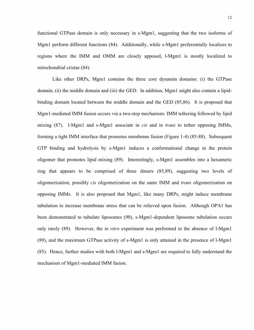
functional GTPase domain is only necessary in s-Mgm1, suggesting that the two isoforms of
Mgm1 perform different functions (84). Additionally, while s-Mgm1 preferentially localizes to
regions where the IMM and OMM are closely apposed, l-Mgm1 is mostly localized to
mitochondrial cristae (84).
Like other DRPs, Mgm1 contains the three core dynamin domains: (i) the GTPase
domain, (ii) the middle domain and (iii) the GED. In addition, Mgm1 might also contain a lipid-
binding domain located between the middle domain and the GED (85,86). It is proposed that
Mgm1-mediated IMM fusion occurs via a two-step mechanism: IMM tethering followed by lipid
mixing (87). l-Mgm1 and s-Mgm1 associate in cis and in trans to tether opposing IMMs,
forming a tight IMM interface that promotes membrane fusion (Figure 1-4) (85-88). Subsequent
GTP binding and hydrolysis by s-Mgm1 induces a conformational change in the protein
oligomer that promotes lipid mixing (89). Interestingly, s-Mgm1 assembles into a hexameric
ring that appears to be comprised of three dimers (85,89), suggesting two levels of
oligomerization, possibly cis oligomerization on the same IMM and trans oligomerization on
opposing IMMs. It is also proposed that Mgm1, like many DRPs, might induce membrane
tubulation to increase membrane stress that can be relieved upon fusion. Although OPA1 has
been demonstrated to tubulate liposomes (90), s-Mgm1-dependent liposome tubulation occurs
only rarely (89). However, the in vitro experiment was performed in the absence of l-Mgm1
(89), and the maximum GTPase activity of s-Mgm1 is only attained in the presence of l-Mgm1
(85). Hence, further studies with both l-Mgm1 and s-Mgm1 are required to fully understand the
mechanism of Mgm1-mediated IMM fusion.

13
Figure 1-4: Schematic of Mgm1-mediated inner mitochondrial membrane fusion.
Like Fzo1, Mgm1 dimerizes in cis and in trans. l-Mgm1 and s-Mgm1 dimerize on the same membrane whereas s-Mgm1 interacts with both l-Mgm1 and s-Mgm1 on opposing membranes. Mgm1 dimerization in trans tethers the opposing IMMs. GTP binding and hydrolysis induces a conformational change in Mgm1 that is proposed to increase membrane stress and instability which can be relieved upon fusion. Although Ugo1 is required for IMM fusion, its specific role still remains to be determined.
1.1.3.3 Coordinating outer and inner mitochondrial membrane fusion
Given that mitochondria have four distinct biochemical compartments (OMM, IMS,
IMM and mitochondrial matrix), and that the disruption of this organization can lead to cell
death by apoptosis, the coordination and regulation of membrane fusion is critically important.
In yeast, a relatively well-characterized protein, Ugo1, serves a bridging function during this
complex process and is required for both OMM and IMM fusion (91). Ugo1 directly interacts
with Fzo1 and Mgm1 at its N- and C-terminus, respectively (79,92). In the absence of Ugo1, the
Fzo1-Mgm1 interaction is abolished (92). Ugo1 has three TM-spanning domains and is
classified as a mitochondrial transport/carrier protein as it contains the characteristic energy
IMM
s-Mgm1l-Mgm1
?
Ugo1Ugo1

14
transfer motifs (ETMs). Its ETMs (ETMs 1 and 2) reside in TM regions 1 and 3, and facilitate
Ugo1 dimerization, creating a 6-TM complex reminiscent of transport/carrier proteins (91,93).
Although Ugo1 is not required for OMM or IMM tethering, it is required for the lipid-mixing
step of mitochondrial membrane fusion (91).
Based on the current findings, the proposed model for mitochondrial membrane fusion
involves Fzo1, Mgm1 and Ugo1 (Figures 1-2, 1-4). Ugo1 dimerization via its ETMs likely
promotes Fzo1 cis dimerization after GTP binding (68,91,93). Since Ugo1 also physically
interacts with Mgm1 (79,92), Ugo1 dimerization likely also induces Mgm1 oligomerization in
cis, although GTP-binding might not be a pre-requisite for Mgm1 oligomerization (89). OMM
fusion proceeds when Fzo1 oligomerizes in trans to tether the opposing OMMs (64,68).
Subsequent Fzo1-dependent GTP hydrolysis likely induces a conformational change (68) that
might result in membrane stress and destabilization that can be relieved by Ugo1-dependent lipid
mixing (91). Similarly, after OMM fusion, Mgm1 likely oligomerizes in trans to tether the
opposing IMMs (85,88,89), and GTP binding and hydrolysis induces a conformational change
(89) that also likely results in membrane stress and destabilization that can be relieved upon
IMM fusion. Notably, to date, no human homolog of Ugo1 has been identified. It is proposed
that a protein with a similar function to Ugo1 is required for bridging OPA1 and Mfn1/2, and
plays a role in mammalian mitochondrial membrane fusion. Given that Ugo1 plays such a
critical function in mitochondrial membrane fusion, the identity and characterization of this key
biochemical activity is needed to fully dissect mammalian mitochondrial fusion and represents a
key missing piece of the puzzle.
15
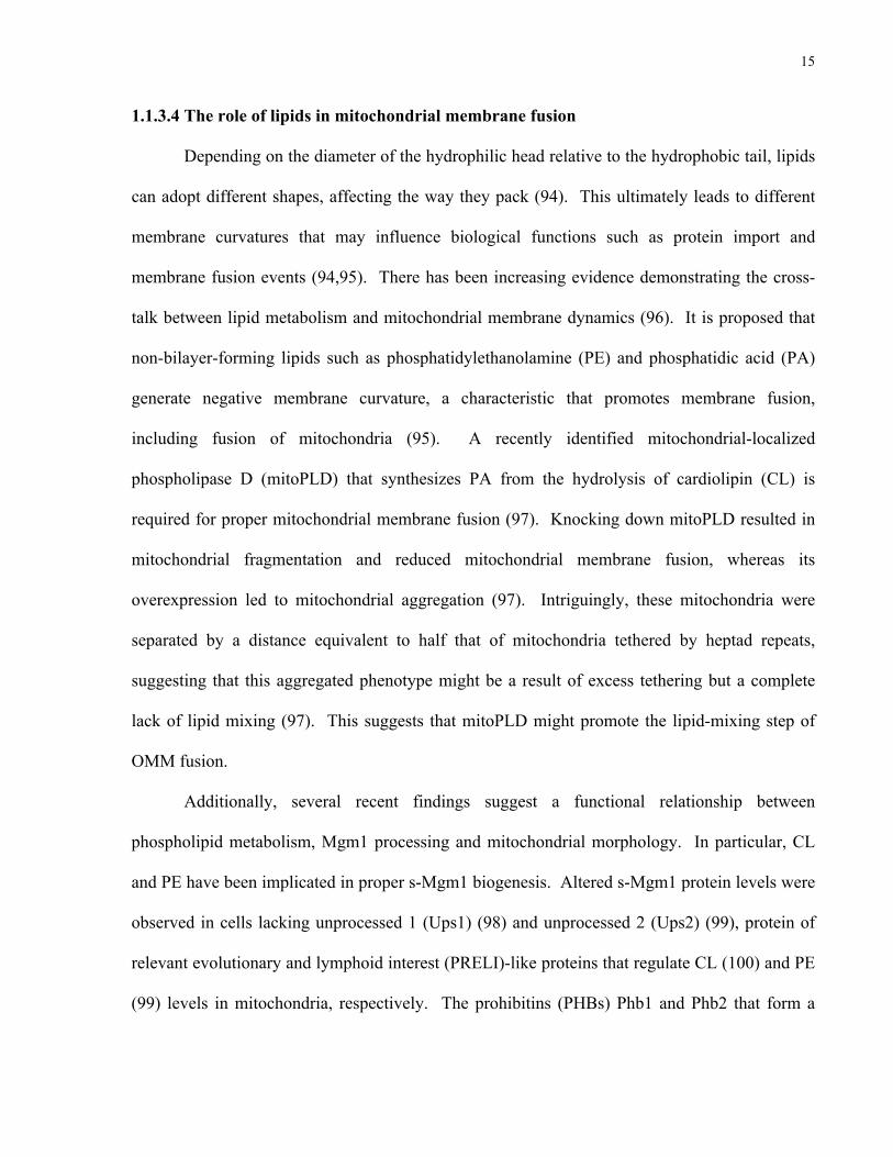
1.1.3.4 The role of lipids in mitochondrial membrane fusion
Depending on the diameter of the hydrophilic head relative to the hydrophobic tail, lipids
can adopt different shapes, affecting the way they pack (94). This ultimately leads to different
membrane curvatures that may influence biological functions such as protein import and
membrane fusion events (94,95). There has been increasing evidence demonstrating the cross-
talk between lipid metabolism and mitochondrial membrane dynamics (96). It is proposed that
non-bilayer-forming lipids such as phosphatidylethanolamine (PE) and phosphatidic acid (PA)
generate negative membrane curvature, a characteristic that promotes membrane fusion,
including fusion of mitochondria (95). A recently identified mitochondrial-localized
phospholipase D (mitoPLD) that synthesizes PA from the hydrolysis of cardiolipin (CL) is
required for proper mitochondrial membrane fusion (97). Knocking down mitoPLD resulted in
mitochondrial fragmentation and reduced mitochondrial membrane fusion, whereas its
overexpression led to mitochondrial aggregation (97). Intriguingly, these mitochondria were
separated by a distance equivalent to half that of mitochondria tethered by heptad repeats,
suggesting that this aggregated phenotype might be a result of excess tethering but a complete
lack of lipid mixing (97). This suggests that mitoPLD might promote the lipid-mixing step of
OMM fusion.
Additionally, several recent findings suggest a functional relationship between
phospholipid metabolism, Mgm1 processing and mitochondrial morphology. In particular, CL
and PE have been implicated in proper s-Mgm1 biogenesis. Altered s-Mgm1 protein levels were
observed in cells lacking unprocessed 1 (Ups1) (98) and unprocessed 2 (Ups2) (99), protein of
relevant evolutionary and lymphoid interest (PRELI)-like proteins that regulate CL (100) and PE
(99) levels in mitochondria, respectively. The prohibitins (PHBs) Phb1 and Phb2 that form a

16
multimeric complex are proposed to provide a scaffold to increase local concentrations of PE
(101) and CL (99). PHB1 genetically interacts with UPS1 and UPS2 (99) as well as genes
directly involved in lipid biosynthesis, CL synthase, CRD1, and phosphatidylserine (PS)
decarboxylase, PSD1 (99,101), the key yeast enzyme that synthesizes PE. These results indicate
that cells cannot tolerate a simultaneous reduction in Phb1 and CL or PE, suggesting that the
function of Phb1 becomes particularly important when CL and PE levels are reduced. Both CL
(102,103) and PE (104,105) have been shown to influence the activity of the supercomplexes in
the electron transport chain (ETC) and may be required for normal mitochondrial morphology as
PHB1 also genetically interacts with several genes involved in the maintenance of mitochondrial
morphology (99). Hence, it is clear that there is still much to discover with respect to the
control, orchestration and regulation of mitochondrial membrane fusion.
1.1.4 Mitochondrial membrane dynamics and autophagy
Recently, mitochondrial membrane dynamics have been implicated in autophagy, a
quality control system (106). Autophagy, which can be induced by starvation, is a mechanism
by which the cell sequesters cytoplasmic contents into double-membrane structures known as
autophagosomes. Subsequent fusion of the autophagosomes with the vacuole (in yeast) or
lysosomes (in mammals) allows for the degradation of its contents by the vacuolar/lysosomal
hydrolases. The degraded products can then be exported to the cytosol to be reused (discussed in
more detail in section 1.2). Under conditions of nutrient deprivation, defects in autophagy result
in increased cell death (107). A recent study demonstrated that mitochondrial elongation
prevented cell death during starvation-induced autophagy (106). Upon starvation, the
recruitment of the fission protein, Drp1, to mitochondria was reduced, resulting in decreased
17
mitochondrial fission with unopposed fusion. This led to elongated mitochondria that continued
producing ATP, thereby protecting the cell from death (106). This result supports the finding
that defects in mitochondrial function impair autophagy induction and autophagic flux (108) and
are the cause of death in autophagy-deficient mutants during starvation (109). Mitochondria are
also proposed to be a source of membranes for the growing autophagosome (110,111). These
data demonstrate the relationship between mitochondrial dynamics, function and autophagy, and
suggest that mitochondrial lipids may also play critical roles in the regulation of autophagy.
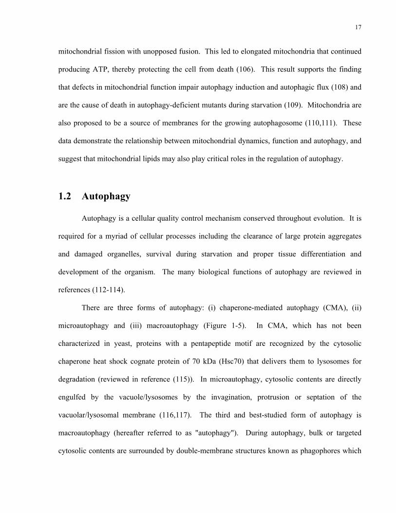
1.2 Autophagy
Autophagy is a cellular quality control mechanism conserved throughout evolution. It is
required for a myriad of cellular processes including the clearance of large protein aggregates
and damaged organelles, survival during starvation and proper tissue differentiation and
development of the organism. The many biological functions of autophagy are reviewed in
references (112-114).
There are three forms of autophagy: (i) chaperone-mediated autophagy (CMA), (ii)
microautophagy and (iii) macroautophagy (Figure 1-5). In CMA, which has not been
characterized in yeast, proteins with a pentapeptide motif are recognized by the cytosolic
chaperone heat shock cognate protein of 70 kDa (Hsc70) that delivers them to lysosomes for
degradation (reviewed in reference (115)). In microautophagy, cytosolic contents are directly
engulfed by the vacuole/lysosomes by the invagination, protrusion or septation of the
vacuolar/lysosomal membrane (116,117). The third and best-studied form of autophagy is
macroautophagy (hereafter referred to as "autophagy"). During autophagy, bulk or targeted
cytosolic contents are surrounded by double-membrane structures known as phagophores which

18
mature into autophagosomes, fully enclosing their contents. The outer membrane of the
autophagosomes then fuse with the vacuole/lysosomes, allowing the inner autophagosomal
membrane and its contents to be degraded by the vacuolar/lysosomal hydrolytic enzymes. The
products, such as amino acids, can then be translocated into the cytosol to be reused by the cell
(reviewed in references (118,119)).
Figure 1-5: Different forms of autophagy.
The three forms of autophagy: (i) chaperone-mediated autophagy (CMA), (ii) microautophagy and (iii) macroautophagy. In CMA, proteins with a pentapeptide motif are recognized by the cytosolic chaperone Hsc70 that delivers them to the lysosome to be degraded. In microautophagy, cytosolic contents are directly engulfed by the vacuole/lysosomes, whereas in macroautophagy, cytosolic contents are surrounded double-membrane phagophores which mature into autophagosomes. Fusion of the autophagosomes with the vacuole/lysosomes allows for the degradation of the inner autophagosomal membrane and its contents.
vacuole/
lysosome
pentapeptide
motif
Hsc70
chaperone-
mediated
autophagy
(CMA)
microautophagy
phagophore
autophagosome
macroautophagy
autolysosome

19
1.2.1 The autophagy pathway and its molecular machinery
In the last decade, a considerable number of studies have dramatically expanded our
knowledge of the molecular machinery that orchestrates autophagy. In fact, the yeast cytoplasm-
to-vacuole targeting (Cvt) pathway, which is a biosynthetic process that delivers vacuolar
hydrolases from the cytosol to the vacuole, shares the core autophagic machinery and hence is
also considered a form of selective autophagy (120). To date, over 30 autophagy-related (Atg)
genes have been identified in yeast (107,118,121-126). The proteins of the autophagic
machinery regulate processes such as cargo recognition, phagophore expansion and even the
release of degraded products into the cytosol.
1.2.1.1 The induction of autophagy
Autophagy is activated under conditions such as nutrient starvation, when the target of
rapamycin (TOR) pathway is inactivated. In yeast, TOR inactivation results in the formation of
the Atg1 complex which consists of Atg1, Atg13 and the Atg17-Atg31-Atg29 subcomplex, and
is the trigger for the induction of autophagy (Figure 1-6) (127-131). In mammalian cells, the
induction of autophagy occurs in a slightly different manner from yeast but is also triggered by
the inactivation of the mammalian TOR (mTOR) pathway and involves the formation of the
Unc-51-like kinase (ULK) complex, the mammalian ortholog of the Atg1 complex (Figure 1-6)
(132-138). The induction of autophagy by the Atg1/ULK complex is reviewed in reference
(139).

20
Figure 1-6: Induction of autophagy.
Autophagy is induced when the target of rapamycin (TOR) pathway is inactivated. In yeast, under nutrient-rich conditions, the active TOR Complex 1 (TORC1) hyperphosphorylates Atg13 which interacts with the Atg17-Atg31-Atg29 subcomplex. TORC1-dependent phosphorylation of Atg13 prevents its association with Atg1. Upon starvation, the inactive TORC1 no longer phosphorylates Atg13, resulting in its dephosphorylation and association with Atg1. The binding between Atg13 and Atg1 enhances the kinase activity of Atg1, resulting in its autophosphorylation. The formation of the Atg1-Atg13-Atg17-Atg31-Atg29 complex initiates autophagy in yeast. In mammalian cells, ULK1/2 (orthologs of yeast Atg1) constitutively associates with the complex that consists of mammalian Atg13, FIP200 (putative homolog of yeast Atg17) and Atg101 (an Atg13 binding protein). Under nutrient-rich conditions, mammalian TORC1 (mTORC1) also associates with this complex. mTORC1 phosphorylates ULK1/2, inhibiting its kinase activity. mTORC1 also hyperphosphorylates mammalian Atg13. Upon starvation, mTORC1 dissociates from the complex resulting in the dephosphorylation of ULK1/2, thereby enhancing its kinase activity and autophosphorylation. ULK1/2 also phosphorylates mammalian Atg13 and FIP200, inducing autophagy.
nutrient-rich nutrient-deprived
TORC1Atg1Atg13
Atg31
Atg29
PPP
TORC1
Atg1P
starvation
Yeast
mTORC1FIP200
FIP200PMammals
Atg101
P
ULK1/2
Atg101
PmTORC1
starvationULK1/2
Atg13
PPP
Atg13
P
Atg17
Atg13
Atg31
Atg29
Atg17

21
1.2.1.2 Phagophore formation
After the induction of autophagy, the Atg1 complex is responsible for the recruitment of
additional Atg proteins to initiate phagophore formation at the phagophore assembly site (PAS)
(128). In yeast, the PAS is juxtaposed to the vacuole (140). The recruitment of proteins to the
PAS follows a specific order (Figure 1-7) (141) and involves the activation of a Class III
phosphatidylinositol 3-kinase (PI3K) complex that also localizes to the PAS (142) (reviewed in
reference (121)). The activity of vacuolar protein sorting 34 (Vps34), the PI3K in this complex,
results in the formation of phosphatidylinositol 3-phosphate (PI3P) that assists in the recruitment
of additional Atg proteins (143,144). In mammalian cells, the PI3K complex localizes to a
subdomain of the endoplasmic reticulum (ER) known as the omegasome that is rich in PI3P
(145,146). The omegasome is proposed to serve as a platform for the recruitment of additional
Atg proteins, the expansion of phagophores and their maturation into autophagosomes (145,147).
Although the Class III PI3K is the major contributor of PI3P during autophagy, a very recent
study demonstrated that the Class II PI3K also contributes significantly to PI3P formation during
autophagy (148).
1.2.1.3 Phagophore expansion and autophagosome formation
After phagophore formation, additional proteins orchestrate the expansion and maturation
of phagophores into autophagosomes. This process requires two ubiquitin-like conjugation
systems and results in the formation of the Atg12-Atg5/Atg16 complex (149) and the Atg8-
phosphatidylethanolamine conjugate (Atg8-PE in yeast, LC3-II in mammals) (Figure 1-8) (150).
Atg12 is first activated by the E1-like enzyme, Atg7 (151). It is then transferred to the E2-like
enzyme, Atg10, which covalently conjugates it to Atg5 (149,152). The Atg12-Atg5 conjugate

22
then forms a complex with Atg16 via the direct interaction between Atg5 and Atg16 homo-
oligomers (153,154). This large ~350 kDa multimeric complex has E3-like properties and
promotes the conjugation of Atg8/LC3 to PE (155) via the other ubiquitin-like conjugation
system in autophagy (150).
Figure 1-7: Order of protein recruitment to the phagophore assembly site in yeast.
In yeast, autophagosomes form at the phagophore assembly site (PAS) in close proximity to the vacuole. Atg17 acts as a scaffold to recruit downstream Atg proteins to the PAS. The tip of each arrow points to the protein (or lipid) that recruits, whereas the protein at the start of each arrow is the protein that is recruited. The Atg12-Atg5/Atg16 complex is likely recruited to the PAS via the direct binding of Atg5 to the phagophore membrane.
Atg1
Atg13
vacuole
phagophore assembly site (PAS)
Atg9Atg31
Atg17
Atg29
Atg14Vps15 Vps30
Vps34
PI3P PI
Atg18Atg2 Atg12Atg5Atg16
Atg3Atg8

23
Figure 1-8: The two ubiquitin-like conjugation systems in autophagy.
Two key components of autophagy are formed by ubiquitin-like conjugation systems: (i) the Atg12-Atg5/Atg16 complex and (ii) the Atg8-phosphatidylethanolamine (Atg8-PE) conjugate. Atg12 is first activated by the E1-like enzyme, Atg7. It is then transferred to the E2-like enzyme Atg10 that covalently conjugates it to Atg5, forming the Atg12-Atg5 conjugate. The Atg12-Atg5 conjugate non-covalently associates with Atg16 to form the Atg12-Atg5/Atg16 complex that acts as an E3-like enzyme for the second ubiquitin-like conjugation system. In the second system, the C-terminal arginine (Arg) residue of Atg8 is removed by the cysteine protease Atg4, exposing the penultimate glycine residue. Atg8 is then activated by Atg7, the same E1-like enzyme that also activates Atg12. Activated Atg8 is then transferred to the E2-like enzyme Atg3 and finally to the E3-like enzyme, the Atg12-Atg5/Atg16 complex, to be covalently conjugated to the lipid phosphatidylethanolamine (PE).
Atg12
Atg5
Atg16
Atg7
Atg10
Atg7
Atg3
Atg12
Atg8
Atg8 Arg
Atg4
Atg8Atg7
Atg7
Atg10Atg5Atg12 Atg10
Atg12Atg5Atg16
Atg7
Atg7
Atg3Atg8
Atg3
PE
Atg8 PE

24
The first step in the activation of Atg8 requires the cleavage of the C-terminal arginine
residue by the cysteine protease, Atg4 (Figure 1-8) (156). This exposes a glycine residue on
Atg8 that is covalently conjugated to PE through a ubiquitin-like conjugation system (156).
Upon cleavage by Atg4, Atg8 is first activated by the E1-like enzyme Atg7 which also activates
Atg12 (Figure 1-8). Activated Atg8 is then transferred to the E2-like enzyme, Atg3 (150).
Finally, the Atg12-Atg5/Atg16 complex acts as an E3-like enzyme (155), covalently conjugating
Atg8 to PE (150), allowing it to be tightly bound to the membrane (156). Notably, the removal
of PE from Atg8 is also essential for autophagy. Mutations that prevented Atg4-dependent
removal of PE from Atg8-PE resulted in impaired autophagy (156,157).
During autophagosome expansion, the Atg12-Atg5/Atg16 complex localizes to the PAS
likely through the direct binding of Atg5 to the phagophore and mediates Atg8-PE conjugation
by recruiting the E2-like enzyme Atg3 to the phagophore (158). This recruitment likely occurs
via the direct binding of Atg12 to Atg3 (Figure 1-7) (159,160). After Atg8 recruitment to the
phagophore and its conjugation to PE, Atg8/LC3 was reported to promote membrane tethering
and hemifusion (161,162), facilitating phagophore expansion (157,163). The presence of the
Atg12-Atg5/Atg16 complex and perhaps additional Atg proteins are proposed to restrict the
access of Atg4 to Atg8-PE, preventing the premature cleavage of Atg8-PE, thereby ensuring
phagophore expansion (164). Shortly before or rapidly after autophagosome formation, the Atg
proteins dissociate from the autophagosome, exposing the Atg8-PE conjugate to Atg4 (164,165).
Subsequent removal of PE from the Atg8-PE conjugate by Atg4 reverses the membrane tethering
and hemifusion activity of Atg8 (161) and releases Atg8 from the completely formed
autophagosome (157). In mammalian cells, phagophore expansion also requires the homotypic
fusion of Atg16-containing vesicles in a soluble N-ethylmaleimide sensitive fusion protein

25
attachment protein receptor (SNARE)-dependent manner (166). Collectively, these data suggest
that both the ubiquitin-like conjugation systems are directly involved in phagophore expansion
and autophagosome formation.
1.2.1.4 Autophagosome maturation and fusion with the vacuole/lysosomes
Once autophagosomes have formed, they fuse with the vacuole/lysosomes where their
contents will be degraded by the vacuolar/lysosomal hydrolases and released back into the
cytosol to be reused. As mentioned above, in yeast, autophagosome formation occurs at the
PAS, in close proximity to the vacuole (140). The fusion of autophagosomes with the vacuole is
dependent on SNARE fusion proteins such as vacuolar morphogenesis 3 (Vam3) (167,168),
Vam7 (168) and Vps10 interacting protein 1 (Vti1) (169,170). In contrast to the simple and
direct system in yeast, in mammalian cells, autophagosomes have been reported to form at
multiple sites including the ER (or omegasome) (145,146), the Golgi (171), mitochondria (111),
mitochondrial-associated ER membranes (MAMs) (172) and the plasma membrane (PM) (173).
Moreover, the maturation of mammalian autophagosomes requires the sequential fusion of
autophagosomes with endosomes (174) and multivesicular bodies (MVBs) (175) (forming
amphisomes) followed by fusion with lysosomes (forming autolysosomes) (176,177). During
this maturation process, the lumen of the amphisomes becomes acidic, and they acquire
lysosomal proteins and hydrolytic enzymes (178-180). Similar to the yeast system, the
maturation of mammalian autophagosomes also requires the concerted effort of SNARE
proteins. Specifically, the formation of amphisomes requires VAMP3 (181), whereas the
formation of autolysosomes requires VAMP7 (181,182), VAMP8 (183), and VTIB (170,183).
The formation of autolysosomes in mammalian cells is also dependent on the interaction

26
between a Tectonin-domain-containing protein, TECPR1, and the Atg12-Atg5 conjugate (184).
TECPR1 was reported to selectively bind to the Atg12-Atg5 conjugate (without Atg16) and to
PI3P, and hence is proposed to selectively tether autophagosomes and lysosomes, initiating the
formation of autolysosomes (184). Upon fusion of the autophagosomes with the
vacuole/lysosomes, the contents of the autophagosomes are degraded and exported to the
cytosol. In yeast, the export of amino acids from the vacuole to the cytosol is mediated by the
vacuolar efflux pump, Atg22 (185).
1.2.2 The lipids implicated in autophagy
As detailed above, the protein machinery of autophagy has been extensively studied, but
the role of lipids in the regulation of autophagy is just beginning to be appreciated. As indicated
previously, autophagy is activated when the TOR/mTOR pathway is inactivated (119). Hence,
the phosphoinositides involved in the TOR/mTOR pathway are critical modulators of autophagy
induction (186). Phosphoinositides also play a key role in autophagosome formation (143,145),
maturation (164) and fusion with lysosomes (184,187). Recently, PA has also been implicated in
the regulation of autophagy. Phospholipase D 1 (PLD1) that synthesizes PA from
phosphatidylcholine (PC), promotes autophagosome maturation through its direct binding with
PI3P (188). The other key lipid in autophagy is PE, and the efficiency of its conjugation to Atg8
is dependent on the composition and/or the curvature of the lipid membrane (158,189).
1.2.2.1 Phosphoinositides
Under nutrient-rich conditions, the Class I PI3K phosphorylates PI(4,5)P2, resulting in the
formation of PI(3,4,5)P3. The signalling cascade that follows activates TOR/mTOR, thereby

27
suppressing autophagy (190,191). However, phosphoinositides also play an important role
downstream of the TOR/mTOR pathway. As described above in section 1.2.1.2, PI3P plays a
central role in autophagosome formation in yeast and in mammalian cells
(143,145,146,186,192). Studies in yeast revealed that the inner autophagosomal membrane is
enriched in PI3P. Interestingly, structures that are in close proximity to the phagophore are also
enriched in PI3P, suggesting that these structures may supply membranes for phagophore
expansion (193). PI3P also promotes phagophore expansion by recruiting additional Atg
proteins (both positive and negative regulators of autophagosome formation) to the PAS and
growing phagophore (reviewed in reference (186)). Furthermore, in mammalian cells, the
localization of the PI3K complex to the omegasome during the early stages of autophagosome
formation occurs at sites rich in PI3P (145). In addition to roles in autophagosome formation
and expansion, PI3P also plays a significant role in the final step of autophagy by facilitating
autophagosome fusion with lysosomes. The mammalian protein TECPR1 is proposed to tether
lysosomes and autophagosomes by selectively binding to PI3P and the Atg12-Atg5 conjugate
(184). Furthermore, the product of PI3P phosphorylation, PI(3,5)P2, has also been implicated in
autophagosome maturation (194-196). It is becoming increasingly clear that phosphoinositides
have a variety of functions during autophagy, and phosphoinositides other than PI3P and
PI(3,5)P2 have also been implicated in autophagy (reviewed in references (186,187)).
1.2.2.2 Phosphatidic acid
Although the phosphoinositides, particularly PI3P, are the best characterized lipids
involved in autophagy, other lipids, including PA have also been implicated in the regulation of
autophagy. Firstly, PA has previously been shown to activate mTOR (197), suggesting that PA

28
likely suppresses autophagy. Accordingly, reducing PA production induces autophagy, although
this induction was independent of the mTOR pathway (198). Consistent with the role of PA in
activating mTOR, a recent study found that amino acids that activate mTOR also activate PLD1,
a PA-synthesizing enzyme (199). Upon amino acid stimulation, PLD1 re-localized to lysosomes
where it activated mTOR Complex 1 (mTORC1). This re-localization was dependent on the
kinase activity of hVps34, the PI3K associated with autophagy (section 1.2.1.2), and the PI3P
binding ability of PLD1. Abrogating the former or the latter abolished amino-acid-stimulated
mTOR activation (199). Collectively, these results indicate a role for PA in the activation of
mTOR and hence a role in the suppression of autophagy. The intracellular re-localization of
PLD1 upon stimulation and the dependence of this re-localization on the kinase activity of
hVps34 and the PI3P binding ability of PLD1 were confirmed by another study (188). However,
in this study, the cells were stimulated by starvation rather than amino acids, and PLD1 re-
localized to amphisomes instead of lysosomes, promoting autophagy (188). These results
suggest that PA promotes or suppresses autophagy depending on the stimulus. The
reproducibility of PLD1 re-localization and its dependence on hVps34 and PI3P strongly suggest
that the binding of PLD1 to PI3P regulates its function. However, further studies need to be
conducted in order to determine the specific signals that trigger PLD1 to switch from a pro- to an
anti-autophagy factor, or vice versa.
1.2.2.3 Membrane composition and/or membrane curvature
Besides the role of individual phospholipids in regulating autophagy, mounting evidence
suggests that the general composition of the membrane might also be important for autophagy.
Bax-interacting factor-1 (Bif-1) is a protein that indirectly interacts with Beclin-1, a subunit of

29
the mammalian Class III PI3K complex that promotes phagophore expansion (200). Bif-1
positively regulates the activity of the Class III PI3K and promotes autophagosome formation
upon nutrient deprivation (200). Bif-1 contains a Bin-Amphiphysin-Rvs (BAR) domain that
senses membrane curvature (201) and likely binds to negatively-charged lipids (202,203). This
suggests that the curvature and presence of negatively-charged lipids on the emerging
phagophore might be important for Bif-1-mediated autophagy. In addition, the efficiency of
Atg8 conjugation to PE is also dependent on the composition (189) and possibly the curvature
(158) of the membrane. The efficiency of Atg8 conjugation to PE increases with increasing
concentrations of PE and negatively-charged phospholipids in an in vitro reconstitution
experiment (189). Moreover, the in vivo observation that Atg8-PE conjugation is strictly
dependent on the Atg12-Atg5/Atg16 complex can only be recapitulated in vitro when the
proteins were incubated with lipid vesicles that had higher curvature (158). Since the
composition of the membrane affects membrane curvature, collectively, these data demonstrate
the importance of the lipid composition of the autophagosomal membrane in autophagy. PE is a
key lipid in autophagy and is known for its ability to form hexagonal phases that increase
membrane curvature (204). Hence, its role and function within the autophagosomal membrane
warrants further study. Further exploring the requirements and functions of the autophagosomal
membrane will shed light on the role of specific phospholipids during autophagy and is a focus
of this thesis work.

30
1.3 Phosphatidylethanolamine biosynthesis in Saccharomyces
cerevisiae
PE was first discovered by Johann Thudichum in 1884 when he successfully separated
lipid fractions from brain tissue (205). Although Thudichum named the lipid "kephalin", it is
now known that "kephalin" or "cephalin" is a mixture of phospholipids including
phosphatidylserine (PS) (206) and PE (207,208). PE was eventually purified from cephalin by
Rudy and Page in 1930 (209), and possibly even purer fractions were obtained from egg yolk by
Lea et al (210) and from bovine liver by Klenk and Dohmen (211) in 1955 (212). The structure
of PE was solved by Baer et al in 1952 (213).
In the yeast Saccharomyces cerevisiae, PE is synthesized by two major routes: (i) the
decarboxylation of PS by the IMM-localized phosphatidylserine decarboxylase 1 (Psd1) and the
Golgi/vacuole-localized Psd2, and (ii) the cytidyldiphosphate-ethanolamine (CDP-Etn) branch of
the Kennedy pathway. PE can also be synthesized via a minor route by the acylation of lyso-
phosphatidylethanolamine (lyso-PE) (Figure 1-9).
1.3.1 Phosphatidylserine decarboxylation
Like all the major phospholipids, the synthesis of PE by PS decarboxylation occurs via
the cytidyldiphosphate diacylglycerol (CDP-DAG) pathway, which begins by the synthesis of
PA. In yeast, PA is first converted to the intermediate CDP-DAG by CDP-DAG synthase, Cds1.
CDP-DAG is then converted to PS by the PS synthase choline requiring 1 (Cho1) in the ER at
specialized MAMs (214). The decarboxylation of PS to PE occurs either in the IMM by Psd1
(215) or in the Golgi/vacuole by Psd2 (216,217). PE can be methylated in three sequential steps

31
Figure 1-9: Simplified overview of phosphatidylethanolamine biosynthesis in Saccharomyces cerevisiae.
In yeast, the major routes of PE biosynthesis are the decarboxylation of phosphatidylserine (PS) by PS decarboxylases (PSDs) and by the cytidyldiphosphate-ethanolamine (CDP-Etn) branch of the Kennedy pathway. PE can also be synthesized by the acylation of lyso-phosphatidylethanolamine (lyso-PE).
by the PE methyltransferases 1 and 2 (Pem1/Cho2 and Pem2/Opi3) to form the other major
phospholipid, PC (Figure 1-10) (phospholipid biosynthesis in S. cerevisiae is reviewed in
reference (217)). The transport of PS from MAMs to mitochondria for decarboxylation by Psd1
is dependent on methionine requiring 30 (Met30), a member of the Skp-Cullin-F-box-protein
(SCF) ubiquitin ligase complex (218). In contrast, the transport of PS to the site of Psd2-
dependent PS decarboxylation is dependent on two other proteins, namely, the
phosphatidylinositol 4-kinase, staurosporine and temperature sensitive 4 (Stt4) (219), and the
phosphatidylinositol transfer/binding protein PstB2 (220). Mammalian cells lack Psd2 and hence
PS decarboxylation occurs only in mitochondria (221) and is catalyzed by mammalian PISD.
PS
lyso-PE
PSD
Etn/P-Etn
CDP-Etn (Kennedy pathway)
PE
acylation
major routes
minor route

32
Figure 1-10: Phosphatidylethanolamine biosynthesis by phosphatidylserine decarboxylation and the Kennedy pathway.
PS synthesized by the cytidyldiphosphate diacylglycerol (CDP-DAG) pathway can be decarboxylated by the IMM-localized Psd1 or by the Golgi/vacuole-localized Psd2 to form PE. PE can also be synthesized by the CDP-Etn branch of the Kennedy pathway using ethanolamine (Etn) or phosphoethanolamine (P-Etn) as substrates. The methylation of PE results in the formation another major phospholipid, phosphatidylcholine (PC). PA, phosphatidic acid; PMME, phosphatidylmonomethylethanolamine; PDME, phosphatidyldimethylethanolamine.
PS decarboxylases (PSDs) are nuclear-encoded proteins (222,223) and are synthesized as
inactive precursors. The mitochondrial-localized PSDs (type I PSDs) contain an N-terminal
MTS followed by a hydrophobic transmembrane anchor. The C-terminus of the protein contains
the characteristic LGST motif that acts as an autocatalytic cleavage site, separating the
proenzyme into the α- and β-subunit (224-226) typical of pyruvoyl-dependent decarboxylases
(Figure 1-11) (pyruvoyl-dependent decarboxylases are reviewed in reference (227)). Serinolysis
between the G and S exposes the new N-terminus of the α-subunit, an electron-withdrawing
PACds1
CDP-DAGCho1
PSPsd2
PE
PSPsd1
PE
Pem1/
Cho2
PMME
Pem2/
Opi3
PDME
Pem2/
Opi3
PC
Extra-mitochondrial
space
Golgi/
vacuole
Mitochondria
Etn P-EtnEct1
CDP-Etn
Kennedy pathway
PEEpt1Eki1

33
Figure 1-11: Domain organization of phosphatidylserine decarboxylases.
Type I PSDs contain an N-terminal MTS followed by a hydrophobic transmembrane anchor (TM). The C-terminal LGST motif is a characteristic cleavage site that separates the PSD proenzyme into the α- and β-subunit. Type II PSDs contain an N-terminal sorting signal that targets them to the endomembrane system (SS). The sorting signal is followed by a C2 domain and, although the processing of type II PSDs has not been extensively studied, they contain a C-terminal GGST motif with a similar function as the LGST motif in type I PSDs. Arrows indicate sites within the protein that undergo proteolytic processing.
pyruvoyl group (Figure 1-12). Heterodimerization between the α- and β-subunit results in the
formation of the fully functional enzyme which associates with the IMM via the β-subunit (226).
The Golgi/vacuole-localized PSDs (type II PSDs) also contain an N-terminal targeting
sequence, although this sorting signal targets them to the endomembrane system. The sorting
signal is followed by a C2 domain that usually participates in lipid and protein interactions
(Figure 1-11). The C2 domain of yeast Psd2 has been proposed to be involved in membrane
docking and/or PS transport to the site of Psd2-dependent PS decarboxylation (228). Type II
PSDs also contain a GGST motif with a similar function to the LGST motif of the type I PSDs
(228). It should be noted, however, that the specific processing of type II PSDs has not been
studied in great detail.
MTS TMType I PSDs LGST
β α
Type II PSDs GGSTSS C2
β α

34
Figure 1-12: Phosphatidylserine decarboxylase proenzyme maturation.
The steps involved in the formation of the mature α- and β-subunit. Serinolysis between the glycine and serine residues results in the α, β elimination of the β-subunit. Hydration of the hydroalanine followed by the elimination of ammonium results in the formation of the N-terminal pyruvoyl group of the α-subunit. RN, R-group on the N-terminus; RC, R-group on the C-terminus.
In S. cerevisiae, PS decarboxylation, and in particular, by Psd1, is responsible for the
majority of the PE production (229) - Psd2 activity accounts for only 4-12% of the total PSD
activity (216). Thus, mitochondria are the major source of de novo PE in yeast cells. The
catalysis of PSDs occurs in a mechanism characteristic of pyruvoyl decarboxylases. During the
formation of the enzyme-substrate complex, the α-carbon of the pyruvoyl group on the α-subunit
of PSD forms a Schiff's base with the amino group on the serine moiety of PS. Subsequent
N C CRN RC
O
N C C
O
H H2 HCH2
H
O
H
N C CRN
O
H H2
O-
mature β-subunit
+ H2O
RCC C
O
CH2
+NH3
O H
H
RCC C
O
CH3
+NH3
O H
RCC C
O
CH3
+NH4
O
mature α-subunit
glycine serine
N C CRN RC
O
C C
O
H H2
C
H
OH2
+NH3

35
decarboxylation results in the formation of an intermediate complex that is protonated at the
azomethine carbon, generating PE in a Schiff's base complex with PSD. The addition of water
across the Schiff's base regenerates the enzyme and releases the product, PE (Figure 1-13).
Figure 1-13: Phosphatidylserine decarboxylase enzymology.
The α-subunit of the PSD interacts with PS to form a Schiff's base in the enzyme-substrate complex (E-S complex). Decarboxylation followed by the protonation of the azomethine carbon results in a Schiff's base of the enzyme and product. Hydration across the Schiff's base regenerates the enzyme and releases the product, PE.
RCC C
O
CH3
O
α-subunit
Phosphatidyl-serine
Ptd
+NH3
O CH2
CH
C
O
O-
H2O
RCC C
O
CH3
Ptd
+NH
O CH2
CH
C
O
O-
E-S complex
CO2+ H+
RCC CCH3
Ptd
+NH
O CH2
CH
O-
H+
RCC CCH3
Ptd
+NH
O CH2
O
CH2
+ H2O
Ptd O CH2
CH2
+NH3
Phosphatidyl-ethanolamine
Schiff’s base

36
1.3.2 The Kennedy pathway
In addition to PS decarboxylation, PE can also be synthesized via the CDP-Etn branch of
the Kennedy pathway. Ethanolamine (Etn), which is exogenously added to the medium or
formed by the breakdown of lipids, is first phosphorylated by the Etn kinase, Eki1, forming
phosphoethanolamine (P-Etn). P-Etn can also be formed by the catabolism of sphingolipids
(230) and supplied to the Kennedy pathway (231). In what is considered the rate-limiting step
(232), P-Etn is activated by cytidyltriphosphate (CTP) to form CDP-Etn by the P-Etn
cytidyltransferase, Ect1. Finally, the Etn phosphotransferase, Ept1, catalyzes the reaction
between CDP-Etn and diacylglycerol (DAG) to form PE (Figure 1-9).
1.3.3 Alternative pathways of phosphatidylethanolamine biosynthesis
In many cell types, PS decarboxylation and the CDP-Etn branch of the Kennedy pathway
are the major sources of PE. However, PE can also be synthesized by the acylation of lyso-PE
(Figure 1-8) which in yeast, is catalyzed by the lyso-PE acyltransferases Ale1 (233,234) and
Tgl3 (235). In mammalian cells, PE can also be synthesized by base exchange, where the serine
head group in PS is exchanged for Etn. This reaction that is catalyzed by PS synthase 2 (PSS2)
is considered a relatively minor contributor of PE biosynthesis (236).
1.3.4 The different contributions of phosphatidylserine decarboxylation and
the Kennedy pathway
In E. coli, PE is synthesized exclusively by PS decarboxylation (237), whereas PE in the
trypanosome T. brucei is synthesized solely by the Kennedy pathway (238). In yeast, PS
decarboxylation is the main source of cellular and mitochondrial PE (216,229). In mammalian

37
cells, however, the relative contributions of the two pathways appear to be tissue-specific. In
many cultured cells such as Chinese hamster ovary (CHO) cells (239) and baby hamster kidney
(BHK) cells (240), PE is primarily synthesized by PS decarboxylation, whereas in rat
liver/hepatocytes (241-243) and hamster heart (244), the Kennedy pathway contributes the
majority of the total PE. While the difference in the relative contributions of the PE biosynthetic
pathways is unknown, it is speculated that the availability of the substrates serine (for PS
decarboxylation) and Etn (for the Kennedy pathway) might contribute to these observed
differences (245).
In yeast, the three different sources of PE appear to serve different purposes. Psd1-
derived PE accounts for most of the cellular and mitochondrial PE synthesized. Psd2-derived PE
is the preferred substrate for methylation into PC, and PE in the microsomal membranes is
primarily derived from the Kennedy pathway (229). Although PE synthesized by Psd2 and the
Kennedy pathway can be imported into mitochondria, import is rather inefficient, and they
contribute mostly to the OMM rather than the IMM (229). It is noteworthy that the different
pathways in mammalian cells synthesize somewhat different species of PE (245,246). PS
decarboxylation preferentially synthesizes PE with polyunsaturated acyl chains, whereas the
Kennedy pathway preferentially synthesizes PE species with mono- and di-unsaturated acyl
chains (245). Although the yeast enzymes show no difference in substrate selectivity, there is
slightly more 34:2 PE in mitochondria than in the microsomal fraction (247). Hence, it is
tempting to speculate that the different species of PE that are derived from the different pathways
could contribute to the different functions of PE in different parts of the cell.

38
1.3.5 Functions of phosphatidylethanolamine
The importance of PE is evident by the severe defects resulting from its loss or mutations
in genes affecting its metabolism. In yeast, PE makes up about 25% of the cellular lipids, and
the loss of PE synthesis results in a strict auxotrophy for Etn, a substrate of the CDP-Etn pathway
for PE synthesis (248). This suggests that a minimum amount of PE is necessary for survival.
Similarly, homozygous deletion of the Pisd gene in mice results in embryonic lethality at day 9.5
of development, demonstrating that PS decarboxylation is essential for mouse development
(249). Although the heterozygous Pisd+/- mice are viable, PE synthesis via the Kennedy
pathway is up-regulated, again demonstrating that a minimum amount of PE is necessary for
survival (249).
PE is a major constituent of biological membranes. The bacterial membrane is comprised
of ~75% PE (250), and in yeast and mammalian cells, PE is the second most abundant
phospholipid after PC, accounting for ~25% of total phospholipids (248,251,252). Due the size
of its hydrophilic head relative to its hydrophobic tail, PE is considered a non-bilayer-forming
lipid as it tends to arrange into non-lamellar hexagonal phases that induce negative membrane
curvature. Hexagonal phases are proposed to promote membrane fusion (204) and create lateral
pressure that can alter the function of membrane proteins (94). Indeed, PE-induced
conformational changes in the protein and lipid bilayer promote the fusion of post-mitotic Golgi
vesicles (253). Furthermore, reducing the amount of PE in mitochondria, the organelles that
typically contain the highest amount of PE, results in impaired mitochondrial morphology in T.
brucei (254) and in mice (249). Proper mitochondrial oxidative phosphorylation and energy
production are also dependent on PE - PE is required for the assembly and optimum activity of
the complexes in the ETC including Complex I (105,255) and Complex IV (104,105,256). PE

39
also has an unexpected role as a chaperone, assisting protein folding in E. coli (257,258). Proper
folding of the membrane protein lactose permease (LacY) is dependent on the non-bilayer-
forming property of PE (258) (reviewed in reference (259)). Recently, PE was also identified as
a factor required for the propagation and infectivity of mouse and hamster prions in the brain
(260).
In addition to the above-mentioned functions, PE also serves as a precursor for the other
major phospholipid, PC. It can be methylated in three sequential steps by methyltransferases,
resulting in the formation of PC (217). PE is also the precursor of a bacterial- and
mitochondrial-specific phospholipid, CL. Recently, a novel CL biosynthetic pathway was
discovered in bacteria (261). The CL synthase ClsC synthesizes CL using PE and
phosphatidylglycerol (PG) (261). Moreover, PE is also the source of Etn that is covalently
conjugated to various proteins as an amino acid modification. For instance, PE is the direct
precursor of Etn-phosphoglycerol that is bound to the eukaryotic elongation factor 1A (eEF1A)
(262-265). Furthermore, as noted above in section 1.2.1.3, the covalent conjugation of PE to
Atg8/LC3 is a key step in autophagy, and the disruption of this process results in impaired
autophagy (150).
1.4 Specific interests and goals
It is becoming increasingly clear that lipids are not simply membrane constituents - they
modulate the function of integral membrane proteins and play critical roles in membrane
remodelling events. In this thesis, I describe additional roles of mitochondrial-localized PE in
mitochondrial membrane dynamics and autophagy. My work demonstrates that mitochondrial
PE plays an important role in the maintenance of mitochondrial morphology by promoting

40
mitochondrial membrane fusion. Mitochondrial PE increases the biogenesis of s-Mgm1, a key
mitochondrial fusion protein and likely enhances the rate of lipid mixing during fusion. I also
identify mitochondrial PE as an important factor during autophagy in yeast. My results suggest
that mitochondrial PE might enhance autophagosome formation, expansion and/or promote the
fusion of autophagosomes with the vacuole. From my studies, I elucidate the mechanism by
which lipids might be important factors in mitochondrial membrane fusion and autophagy.

41
CHAPTER 2
MATERIALS AND METHODS
2.1 Reagents
2.1.1 Yeast strains
All strains used in these studies are derivatives of BY4741 (MATa; his3Δ1; leu2Δ0;
met15Δ0; ura3Δ0). Gene deletions and genomic tagging were performed by homologous
recombination of gene-specific PCR products. To generate the Δpsd1Δmgm1 strain harbouring
the pRS315-WT-Mgm1 or pRS315-Mgm1-G100D plasmids, a MATa Δpsd1 haploid strain was
mated with a MATα Δmgm1 haploid strain harbouring the pRS316-WT-Mgm1 plasmid. After
sporulation and tetrad dissection, ploidy and mating type were determined by a mating type test,
and the haploid Δpsd1Δmgm1 double mutant retaining the pRS316-WT-Mgm1 plasmid was
identified by viability on medium lacking uracil and resistance to the antibiotics nourseothricin
(NAT) and G418. The strain was then transformed with either the pRS315-WT-Mgm1 or the
pRS315-Mgm1-G100D plasmid. Removal of the pRS316-WT-Mgm1 plasmid was determined
by cell viability on medium lacking leucine in the presence of 5-fluoroorotic acid (5-FOA).
2.1.2 Plasmids
pYX122-mtGFP (mtGFP), pYES-mtGFP and pYES-mtBFP (galactose-inducible mtGFP
and mtBFP, respectively) were kind gifts from Dr. Benedikt Westermann and have been
previously described (266). pRS315-s*Mgm1-3xHA (s*Mgm1) was generated by cloning a
SacI/SalI double-digested fragment from pRS314-s*Mgm1-3xHA (a gift from Dr. Andreas

42
Reichert and has been previously described (84)) into pRS315. pRS315-Mgm1-G100D was also
a gift from Dr. Andreas Reichert and was previously described (81). For the overexpression of
Psd1, the open reading frame (ORF) of Psd1 was amplified using a reverse primer with a 3xHA
sequence included in the primer. The amplified ORF with a 3xHA tag was cloned into pRS415-
GPD at the PstI and XhoI restriction sites.
2.1.3 Growth conditions
Synthetic complete (or minimal) media consisted of 0.17% yeast nitrogen base, 5%
ammonium sulphate, the appropriate amino acids and 2% glucose, galactose or glycerol as the
carbon source. Rich media consisted of 1% yeast extract, 2% peptone and 2% glucose or
galactose. Ethanolamine (Etn) supplementation was added to a final concentration of 5 mM.
Starvation medium (- N) consisted of 0.17% yeast nitrogen base (without amino acid, without
ammonium sulphate) and 2% glucose as the carbon source.
2.1.4 Lipids
All lipids were purchased from Avanti polar lipids. Non-fluorescent lipids used in these
studies include 1,2-dimyristoyl-sn-glycero-3-phosphocholine (DMPC, 14:0 PC), 1,2-dimyristoyl-
sn-glycero-3-phosphoethanolamine (DMPE, 14:0 PE), L-α-phosphatidylinositol (liver, bovine),
1,2-dimyristoyl-snglycero-3-phospho-L-serine (DMPS, 14:0 PS), 1',3'-bis[1,2-dimyristoyl-sn-
glycero-3-phospho]-sn-glycerol (TMCL, 14:0 CL) and 1,2-dimyristoyl-sn-glycero-3-phosphate
(DMPA, 14:0 PA). Head-group-labelled fluorescent lipids include 1,2-dimyristoyl-sn-glycero-3-
phosphoethanolamine-N-(7-nitro-2-1,3-benzoxadiazol-4-yl) (14:0 NBD-PE) and 1,2-

43
dimyristoyl-sn-glycero-3-phosphoethanolamine-N-(lissamine rhodamine B sulfonyl) (14:0
Rhodamine-PE).
2.2 Fusion assays
2.2.1 In vivo mitochondrial fusion assay
MATa cells expressing pYES-mtGFP and MATα cells expressing pYES-mtBFP were
grown to log phase in synthetic galactose medium to induce the expression of mtGFP and
mtBFP. An equal number of cells were mixed together and washed twice with synthetic glucose
medium to stop mtGFP and mtBFP production. Cells were resuspended in the same volume of
synthetic glucose medium, mixed thoroughly, and evenly spread onto a synthetic glucose agar
plate using a sterile inoculating loop. Liquid medium on the agar plate was air-dried, and the
plate was incubated for 3 h at 30°C for yeast mating. Cells were then carefully scraped off the
plate with a sterile inoculating loop, resuspended in synthetic glucose medium and analyzed by
fluorescence microscopy.
2.2.2 In vitro liposome fusion assay
Phospholipid compositions of WT and Δpsd1 mitochondria had been previously
determined (229). Briefly, liposomes with a lipid composition similar to WT mitochondria
contained (in mol%) 44% DMPC, 29% DMPE, 8% Liver PI, 4% DMPS, 6% TMCL and 9%
DMPA, whereas liposomes with a lipid composition similar to Δpsd1 mitochondria contained
52% DMPC, 9% DMPE, 19% Liver PI, 4% DMPS, 6% TMCL and 10% DMPA. For
fluorescence-labelled liposomes, 1.6% of the phosphatidylethanolamine (PE) was replaced with
0.8% NBD-PE and 0.8% Rhodamine-PE. Lipids were mixed in a glass tube and dried under a

44
gentle stream of nitrogen. Residual chloroform was removed under a rotary evaporator for 90
min. Lipid films were rehydrated to a concentration of 0.5 mM in liposome buffer (20 mM
HEPES pH 7.5, 150 mM NaCl) at 65°C for 1 h, vortexing every 15 min for homogeneity. To
make liposomes, hydrated lipids were extruded 15 times through a 1.0 µm filter membrane
(Avanti Polar Lipids). Liposome fusion was carried out in 96-well black plates in 100 µl
reactions. 1 µl of labelled liposomes was mixed with 9 µl of unlabelled liposomes in 80 µl of
liposome buffer. After reading basal fluorescence for 5 min, 10 µl of 1.0 M CaCl2 were added to
induce liposome fusion. Liposome buffer was used as a negative control, and maximum lipid
mixing was determined by adding 10 µl of 10% octaethylene glycol monododecyl ether (C12E8).
Readings were taken every minute for 90 min, and fluorescence was normalized to the initial
fluorescence reading and presented as a percentage of maximum lipid mixing. Relative rates of
lipid mixing were obtained by determining the slopes of the linear portion of the curves
immediately after the addition of CaCl2 and C12E8.
2.3 Mitochondrial assays
2.3.1 Mitochondrial purification
Cells were grown to log phase in 50 ml of rich galactose medium, and mitochondria were
purified as described in Gregg, et al with slight modifications (267). Briefly, after mitochondrial
enrichment, pellets were resuspended in 1 ml of resuspension buffer (homogenization buffer
without BSA) and gently homogenized 3 times with a tight dounce. Suspensions were then
pelleted at 3,000 x g for 5 min at 4°C. The resulting supernatants were then centrifuged at
12,000 x g for 15 min at 4°C to pellet mitochondria. Mitochondrial purity was assessed by
western blotting.

45
2.3.2 Oxidative phosphorylation
Cells were grown to log phase in rich galactose medium. 3 million cells (3 optical
densities [ODs]) were harvested per reading, pelleted and resuspended in 1 ml of fresh fully
oxygenated medium for oxygen consumption measurements using a Clark-type oxygen electrode
(Strathkelvin Instruments). 100% ethanol was added to a final concentration of 1% (v/v). After
the rate of oxygen consumption stabilized, 2 mM carbonylcyanide m-chlorophenylhydrazone
(CCCP) was added to a final concentration of 8 µM to induce maximum oxygen consumption.
2.3.3 Mitochondrial ATP measurements
Purified mitochondria were resuspended in 15 µl of 5.5% TCA, 2 mM EDTA and
incubated on ice for 10 min to extract ATP and precipitate mitochondrial proteins. Extracted
ATP was separated from precipitated proteins by centrifugation at 16,100 x g for 10 min at 4°C.
Supernatants containing ATP were collected for ATP measurements using an ATP
Determination Kit (Invitrogen) according to the manufacturer's instructions. Precipitated
proteins were washed twice with ice-cold acetone and heated at 95°C for 5 min to fully evaporate
the acetone. Pellets were then resuspended in 15 µl of reducing sample buffer and boiled for 5
min at 95°C. Protein concentration was determined using the RC DC protein assay (Bio-Rad).
2.4 Cycloheximide chase
Yeast cells were grown to log phase in rich galactose medium. Cycloheximide was
added to a final concentration of 100 µg/ml. Cells were collected at the indicated time points and
lysed by alkaline lysis (268) for western blot analysis. 0.5 ODs of cells were loaded per sample.
Mgm1 was detected using a rabbit polyclonal antibody against Mgm1, a kind gift from Dr.

46
Andreas Reichert. Fzo1 and Tom40 were detected using rabbit anti-sera against Fzo1 and
Tom40, generous gifts from Dr. Jodi Nunnari and Dr. Thomas Langer, respectively.
2.5 Autophagy assays
2.5.1 Determination of cell viability
Yeast cells were grown to log phase in synthetic minimal medium with glucose as the
carbon source. They were then transferred to - N medium, and cell viability was monitored by
the addition of an equal volume of Trypan blue. Cells positive for Trypan blue were counted as
dead cells.
2.5.2 Atg8/Atg8-PE analysis
Cells were grown in synthetic minimal medium with glucose as the carbon source,
washed twice with water and resuspended in the same amount of - N medium. At the indicated
time points, cells were harvested and lysed by alkaline lysis (268) for western blot analysis. 1
OD of cells was loaded per sample for each time point on a standard SDS-PAGE gel containing
13.5% acrylamide and 6 M urea in the separating gel (156). Atg8 and Atg8-PE were detected
using rabbit anti-serum against Atg8, a kind gift from Dr. Daniel Klionsky.
2.5.3 Vacuolar internalization of FM 4-64
Cells were grown to log phase in synthetic minimal medium with glucose as the carbon
source and the assay was performed as described in Journo, et al with the exception that FM 4-64
was added to a final concentration of 16 µM (269).

47
CHAPTER 3
PHOSPHATIDYLSERINE DECARBOXYLASE 1 (PSD1)
PROMOTES MITOCHONDRIAL FUSION BY REGULATING
THE BIOPHYSICAL PROPERTIES OF THE
MITOCHONDRIAL MEMBRANE AND ALTERNATIVE
TOPOGENESIS OF MITOCHONDRIAL GENOME
MAINTENANCE 1 (MGM1)
This research was originally published in The Journal of Biological Chemistry. Eliana Y. L.
Chan and G. Angus McQuibban. Phosphatidylserine decarboxylase 1 (Psd1) promotes
mitochondrial fusion by regulating the biophysical properties of the mitochondrial membrane
and alternative topogenesis of mitochondrial genome maintenance 1 (Mgm1). The Journal of
Biological Chemistry. 2013; 287:40131-40139. © The American Society for Biochemistry and
Molecular Biology.
3.1 Abstract
Non-bilayer-forming lipids such as cardiolipin (CL), phosphatidic acid (PA) and
phosphatidylethanolamine (PE) are proposed to generate negative membrane curvature,
promoting membrane fusion. However, the mechanism by which lipids regulate mitochondrial
fusion remains poorly understood. Here, I show that mitochondrial-localized phosphatidylserine
decarboxylase 1 (Psd1), the key yeast enzyme that synthesizes PE, is required for proper

48
mitochondrial morphology and fusion. Yeast cells lacking Psd1 exhibit fragmented and
aggregated mitochondria with impaired mitochondrial fusion during mating. I also demonstrate
that a reduction in PE reduces the rate of lipid mixing during fusion of liposomes with lipid
compositions reflecting the mitochondrial membrane. This suggests that the mitochondrial
fusion defect in the Δpsd1 strain could be due to the altered biophysical properties of the
mitochondrial membrane, resulting in reduced fusion kinetics. The Δpsd1 strain also has
impaired mitochondrial activity such as oxidative phosphorylation and reduced mitochondrial
adenosine triphosphate (ATP) levels which are due to a reduction in mitochondrial PE. The loss
of Psd1 also impairs the biogenesis of the short isoform of mitochondrial genome maintenance 1
(s-Mgm1), a protein essential for mitochondrial fusion, further exacerbating the mitochondrial
fusion defect of the Δpsd1 strain. Increasing s-Mgm1 levels in Δpsd1 cells markedly reduced
mitochondrial aggregation. My results demonstrate that mitochondrial PE regulates
mitochondrial fusion by regulating the biophysical properties of the mitochondrial membrane
and by enhancing the biogenesis of s-Mgm1. While several proteins are required to orchestrate
the intricate process of membrane fusion, I propose that specific phospholipids of the
mitochondrial membrane promote fusion by enhancing lipid mixing kinetics and by regulating
the action of pro-fusion proteins.
3.2 Introduction
Mitochondria are highly dynamic organelles, constantly undergoing fusion and fission
reactions to maintain a tubular network. While the protein machineries of mitochondrial fusion
and fission have been extensively studied, the role of lipids in regulating these processes is just
beginning to be uncovered. Non-bilayer-forming lipids such as PA, CL and PE are required for

49
proper mitochondrial morphology (97,100,249,270-272). Recently, a study examining the loss
of both CL and PE by deleting CL synthase, CRD1, and the mitochondrial PSD1, respectively,
demonstrated that both lipids are required for mitochondrial morphology and fusion (272). In a
mouse model, disrupting the mammalian homolog of PSD1, Pisd, resulted in abnormal
mitochondrial morphology (249). Furthermore, a reduction in the levels of a mitochondrial-
localized phospholipase D (mitoPLD that synthesizes PA from the hydrolysis of CL) resulted in
mitochondrial fragmentation (97). It is proposed that these non-bilayer-forming lipids induce
hexagonal phases, generating negative membrane curvature that promotes membrane fusion
(94,273). However, the mechanisms by which these lipids regulate mitochondrial dynamics
remain poorly understood.
The protein machinery of mitochondrial fusion is highly conserved from yeast to
mammals, and consists of large dynamin-like guanosine triphosphate hydrolases (GTPases). In
yeast, the outer mitochondrial membrane (OMM)-localized fuzzy onions 1 (Fzo1, mitofusins 1
and 2 [Mfn 1 and 2] in mammals) mediates OMM fusion, while the inner mitochondrial
membrane (IMM)-localized Mgm1 (optic atrophy [OPA1] in mammals) mediates IMM fusion
(65,80,274,275). A fungal-specific protein, Ugo1 (Ugo is Japanese for fusion), serves as an
adaptor, tethering Fzo1 and Mgm1, coordinating OMM and IMM fusion (92). Mgm1 exists as
two isoforms, long (l-Mgm1) and s-Mgm1 (276). A proper balance of l- and s-Mgm1 protein
levels is crucial for mitochondrial fusion - perturbing this ratio results in impaired mitochondrial
fusion (84). The formation of s-Mgm1 from full-length Mgm1 (FL-Mgm1) requires the
enzymatic activity of the mitochondrial rhomboid Rbd1/Pcp1, and is an ATP-dependent process
(81,82,276). Here, I show that Psd1 is required for maintaining mitochondrial ATP levels and
for the biogenesis of s-Mgm1 but does not impinge on Rbd1 activity. I also provide evidence

50
that Psd1 regulates mitochondrial fusion by regulating the biophysical properties of the
mitochondrial membrane, likely enhancing the rate of lipid mixing during fusion. Together, my
findings reveal the mechanisms by which Psd1-synthesized PE promotes mitochondrial fusion,
and demonstrate a complex interaction between lipid homeostasis, mitochondrial dynamics and
mitochondrial activity.
3.3 Results
3.3.1 Psd1 is required for normal mitochondrial morphology
To determine whether phospholipid composition plays a role in regulating mitochondrial
membrane dynamics, I examined the mitochondrial morphology of the Δpsd1 yeast strain. A
previous study demonstrated that the loss of Psd1 results in an alteration in the mitochondrial
phospholipid composition when cells are cultured in rich medium with lactate as a carbon
source. Despite the altered mitochondrial lipid composition, it was reported that the
mitochondrial morphology of these cells was unaffected (248). Indeed, I found that Δpsd1 cells
cultured in rich medium had mitochondria that were tubular, similar to that of wild type (WT)
cells (Figures 3-1A, B). However, upon further characterization, I found that 50% of Δpsd1 cells
had an intermediate mitochondrial morphology consisting of tubules, but lacking an organized
interconnected network that was observed in WT yeast cells (Figures 3-1A, B). Since rich
medium contains additional nutrients that can affect overall lipid metabolism, I cultured cells in
minimal medium, supplying only the nutrients necessary for survival. In contrast to cells
cultured in rich medium, 85% of Δpsd1 cells cultured in minimal medium had mitochondria that
were fragmented and aggregated (Figures 3-1A, B). This result is consistent with recent findings
that Psd1 is required for normal mitochondrial morphology (271,272). To further define the

51
C
n
v
WT
n
v
Δpsd1
A
Tubular
Intermediate
Fragmented
Aggregated
B
WT Δpsd1 WT Δpsd1 Δpsd1 + Etn
Rich Minimal

52
mitochondrial aggregation phenotype, I performed electron microscopic analysis. Unlike WT
mitochondria that were evenly distributed throughout the cytoplasm, Δpsd1 mitochondria were
clustered but not fused (Figure 3-1C). Furthermore, the IMM of Δpsd1 mitochondria was less
electron-dense, suggesting possible IMM defects (Figure 3-1C). Together, these data indicate
that Psd1 is required for normal mitochondrial morphology, highlighting the importance of
mitochondrial lipid metabolism in maintaining organellar morphology. The clustered
mitochondrial phenotype of the Δpsd1 strain resembles that of mitochondrial fusion mutants
(10), suggesting that Psd1 might regulate mitochondrial fusion.
3.3.2 Psd1 is required for proper mitochondrial fusion during yeast mating
To assess whether the mitochondrial morphology defect in Δpsd1 cells is due to a defect
in mitochondrial fusion, I performed an in vivo mitochondrial fusion assay. During yeast mating,
mitochondria fuse, and mitochondrial content mixing of the mating MATa and MATα haploids
can be used as an assay to monitor mitochondrial fusion. As a positive control, WT cells had
complete mixing of mitochondrial content (Figures 3-2A, B), indicating complete mitochondrial
fusion in this assay. In contrast, only 50% of Δpsd1 cells showed complete mitochondrial
______________________________________________________________________________
Figure 3-1: Psd1 is required for normal mitochondrial morphology.
Wild type (WT) and Δpsd1 cells were transformed with a mitochondrial-targeted green fluorescent protein (GFP) construct (mtGFP) and cultured in rich and minimal media with galactose as the carbon source. (A) Representative images of cells with tubular, intermediate, fragmented and aggregated mitochondria. (B) Quantification of the mitochondrial morphology of cells cultured in rich and minimal media. More than 300 cells were analyzed per strain. Error bars represent the standard deviation of 3 independent experiments. (C) Electron microscopic analysis of the WT and Δpsd1 strains cultured in minimal medium. Mitochondria (arrows), nuclei (n) and vacuoles (v) are indicated. Scale bars represent 500 nm. Etn, ethanolamine.

53
Figure 3-2: Psd1 is required for mitochondrial fusion during yeast mating.
(A) Representative images of cells that have undergone complete, partial and no mitochondrial fusion during yeast mating. Haploid WT and Δpsd1 strains were transformed with a galactose-inducible mtGFP or mtBFP. Strains were cultured and mated as described in section 2.2.1 and analyzed for mitochondrial content mixing. The extent of mitochondrial fusion was indicated by the amount of mixing between mtGFP and mtBFP. (B) Quantification of zygotes that have undergone complete, partial or no mitochondrial fusion. 20 zygotes were analyzed per strain.
A
Completefusion
Partialfusion
No fusion
mtGFP mtBFP Merge DIC
B
WT Δpsd1 Δpsd1 + Etn

54
content mixing while the remaining 50% had only partial mixing (Figures 3-2A, B). This result
is surprising considering the mitochondrial fragmentation and aggregation in the Δpsd1 strain.
Nevertheless, the ability of Δpsd1 mitochondria to undergo fusion indicates that Psd1 is not an
obligate member of the mitochondrial fusion machinery. However, the partial fusion phenotype
strongly implicates Psd1 as an important regulator, and that the altered phospholipid composition
of the mitochondrial membrane affects in vivo mitochondrial fusion during mating.
3.3.3 Phospholipid composition affects the rate of lipid mixing
Impaired mitochondrial fusion in the Δpsd1 strain during yeast mating suggests that the
phospholipid composition of the mitochondrial membrane plays an important role in promoting
mitochondrial fusion. To assess whether the altered lipid composition affects the biophysical
properties of the mitochondrial membrane that can influence mitochondrial fusion, I performed
an in vitro liposome fusion assay using liposomes with phospholipid compositions similar to that
of WT and Δpsd1 mitochondria (WT and Δpsd1 liposomes, respectively) (229). Upon the
addition of CaCl2 to induce fusion, Δpsd1 liposomes fused to the same extent as WT liposomes.
This indicates that altering the phospholipid composition does not affect the extent to which
liposomes fused (Figure 3-3A), and is consistent with our in vivo mitochondrial fusion assay that
Δpsd1 mitochondria can undergo complete fusion (Figure 3-2B). Interestingly, the slope of the
curve during mixing of Δpsd1 liposomes is different from that of WT liposomes (Figure 3-3A).
Since the slopes of the curves represent the relative rates of reaction, I analyzed the slopes to
determine if reduced fusion kinetics could contribute to the mitochondrial fusion defect in the
Δpsd1 strain. My analysis indicates that Δpsd1 liposomes had an ~75% reduction in their rates
of lipid mixing compared to WT liposomes (Figure 3-3B). Due to the absence of proteins in this

55
Figure 3-3: Liposomes with lipid compositions similar to ΔΔpsd1 mitochondria have a reduced rate of lipid mixing.
(A) In vitro liposome fusion was performed as described in section 2.2.2. Liposomes labelled with 7-nitro-2-1,3-benzoxadiazol-4-yl (NBD) and lissamine rhodamine B sulfonyl (Rhodamine) were mixed with unlabelled liposomes, and basal NBD fluorescence was monitored for 5 min. CaCl2 and C12E8 were added to induce fusion and maximum lipid mixing, respectively. The increase in NBD fluorescence indicates relief of NDB quenching by Rhodamine. (B) Quantification of the relative rates of lipid mixing indicated by the slopes of the curves in (A). Values represent the mean and standard deviation of three independent experiments. * p < 0.0001, N.S. Not significant (p > 0.05).
A
B
N.S.
+ CaCl2 + C12E8
60
55
50
10
5
0
*
N.S.

56
assay, my result indicates that changes in the phospholipid content of Δpsd1 mitochondria likely
alter the biophysical properties of the mitochondrial membrane, reducing the kinetics of lipid
mixing during mitochondrial fusion.
3.3.4 Psd1 is required for proper mitochondrial activity
To better define the importance of Psd1 in mitochondrial biology, I determined if Psd1 is
required for normal mitochondrial homeostatic functions such as cell growth and mitochondrial
bioenergetics. I analyzed cell growth under respiratory conditions, overall rate of oxidative
phosphorylation and mitochondrial ATP levels. Serial dilutions indicate that the Δpsd1 strain
grew slower than the WT strain on glycerol, a non-fermentable carbon source that necessitates
respiration (Figure 3-4A). To more quantitatively determine the mitochondrial metabolic defect
in the Δpsd1 strain, I analyzed oxidative phosphorylation by examining the rate of oxygen
consumed during ethanol metabolism. My data revealed that the basal rate of oxidative
phosphorylation in the Δpsd1 strain was 50% that of the WT strain (Figure 3-4B). The
mitochondrial uncoupler, carbonylcyanide m-chlorophenylhydrazone (CCCP) was added to
determine the maximum activity of the electron transport chain (ETC) (277). The maximum rate
of oxidative phosphorylation in the Δpsd1 strain was also 50% that of the WT strain (Figure 3-
4B). To determine the effect of impaired oxidative phosphorylation on energy production, a key
function of mitochondria, I analyzed the amount of ATP in purified mitochondria. Δpsd1
mitochondria had ~75% of WT mitochondrial ATP levels, indicating that Psd1 is required for the
maintenance of mitochondrial ATP (Figure 3-4C). Together, these results highlight the
importance of Psd1, and likely the phospholipid PE, in regulating crucial mitochondrial
components such as the ETC to maintain cellular respiration and energy production.

57
Figure 3-4: The ΔΔpsd1 strain has defects in mitochondrial activity.
(A) Serial dilutions of WT and Δpsd1 strains on glucose, glycerol and glycerol with Etn. (B) The rate of oxidative phosphorylation indicated by the rate of oxygen consumed (natom/minute/million cells) was measured as described in section 2.3.2. (C) Mitochondria were purified as described in section 2.3.1, and the amount of ATP (pmol/mg of protein) was measured using a luciferase assay as described in section 2.3.3. Δmgm1 is a respiratory-incompetent strain used as a negative control. (D) Western blot analysis of l-Mgm1 and s-Mgm1 protein levels in WT, Δpsd1 and Δpsd1 cell grown in the presence of Etn. (E) Quantification of western blots described in (D). All values represent the mean and standard deviation of three independent experiments. * p < 0.007, ** p < 0.02, N.S. Not significant (p > 0.05).
AGlycerol
WT
Δpsd1
Glucose Glycerol + Etn
B
*
*
**
*
N.S.
N.S.
C
*N.S.
*
WT Δpsd1 Δpsd1 + Etn
Δmgm1
* *
N.S.
WT Δpsd1 Δpsd1 + Etn
E
DWT
Δpsd1Δpsd1
+ Etn
48 kDTom40
s-Mgm1100 kD
75 kDl-Mgm1

58
3.3.5 Ethanolamine cannot rescue ΔΔpsd1 mitochondrial-specific defects
In Saccharomyces cerevisiae, the major route of PE synthesis is by the decarboxylation
of phosphatidylserine (PS) by the mitochondrial-localized Psd1 and the secretory pathway-
localized Psd2. Exogenously added ethanolamine (Etn) or Etn formed endogenously by lipid
metabolism can also be used to synthesize PE via the Kennedy pathway (Figure 1-9) (278).
Since Psd1-dependent PS decarboxylation is the major route of PE synthesis, the loss of Psd1
results in significantly reduced mitochondrial and cellular PE. It was also reported that
supplementing the Δpsd1 strain with exogenous Etn does not significantly increase
mitochondrial PE content, indicating that exogenous PE is not efficiently transported to
mitochondria (248). To directly demonstrate that the mitochondrial defects are a result of a
reduction in mitochondrial PE, I cultured Δpsd1 cells in media supplemented with Etn which
should increase extra-mitochondrial PE via the Kennedy pathway. The Δpsd1 glycerol growth
defect is partially rescued in the presence of Etn (Figure 3-4A). Interestingly, this rescue is
independent of mitochondrial bioenergetics. The addition of Etn could not rescue the Δpsd1
oxidative phosphorylation or mitochondrial ATP defects (Figures 3-4B, C). These results
strongly indicate that total cellular PE is crucial for growth, and that mitochondrial-localized PE
is required for efficient mitochondrial bioenergetics.
To assess if mitochondrial PE is important for the maintenance of proper mitochondrial
morphology, I analyzed the mitochondrial morphology of Δpsd1 cells grown in the presence of
Etn as previously described. Under these conditions, 50% of Δpsd1 cells had an intermediate
mitochondrial morphology. Mitochondria in these cells, while mostly tubular, were not
connected in a network like those of WT cells (Figures 3-1A, B). This resembles the
mitochondrial morphology of Δpsd1 cells cultured in rich medium, indicating that Psd1 is

59
required for proper mitochondrial morphology, especially under more stringent, nutrient-limiting
growth conditions.
Interestingly, it was previously observed that the loss of Psd1 results in altered ratios of l-
and s-Mgm1 (99). This could suggest that the mitochondrial morphology defect in Δpsd1 cells is
due to altered Mgm1 ratios. Thus, I determined whether the addition of Etn could alter Mgm1
protein ratios. Cells analyzed for their mitochondrial morphology were also harvested for
western blotting using Mgm1-specific antibodies. The ratio of l-Mgm1 to s-Mgm1 was not
altered in the presence of Etn (Figures 3-4D, E), indicating that the rescue of mitochondrial
fragmentation and aggregation in Δpsd1 is not a result of restoring Mgm1 ratios. The inability of
Etn to fully restore the tubular mitochondrial network and Mgm1 ratios suggests that the
imbalance of l- and s-Mgm1 protein levels contributes to the mitochondrial morphology defect in
the Δpsd1 strain.
3.3.6 s*Mgm1 can rescue mitochondrial aggregation but not the glycerol
growth defect in ΔΔpsd1 cells
A correct balance of l- and s-Mgm1 is required for proper mitochondrial fusion. l-Mgm1
exerts a dominant-negative effect and, increased accumulation of l-Mgm1 results in impaired
mitochondrial fusion (84). To address the possibility that the l- and s-Mgm1 protein imbalance
contributes to the mitochondrial morphology defect in the Δpsd1 strain, I artificially increased
the levels of s-Mgm1 by expressing a modified form of Mgm1 (s*Mgm1) on a plasmid (84).
Expression of s*Mgm1 in the Δpsd1 strain markedly reduced mitochondrial aggregation and
increased the number of cells with a tubular mitochondrial network (Figure 3-5A). Culturing
Δpsd1 cells expressing s*Mgm1 in Etn showed no further rescue of mitochondrial morphology

60
A
WT + vector
Δpsd1 + vector
Δpsd1 +vector+ Etn
Δpsd1 + s*Mgm1
Δpsd1 + s*Mgm1
+ Etn
100 kD75 kD48 kD
s*Mgm1
Tom40
WT + ve
ctor
Δpsd1 +
vecto
r
Δpsd1 +
s*Mgm1
Δpsd1 +
vecto
r + Etn
Δpsd1 +
s*Mgm1
+ Etn
C
B
WT + vector
Glucose
Δpsd1 + vector
WT + s*Mgm1
Δpsd1 + s*Mgm1
Glycerol

61
(Figure 3-5A). While s*Mgm1 could rescue the mitochondrial network and aggregation, serial
dilutions indicated that Δpsd1 cells expressing s*Mgm1 still exhibited impaired growth on
glycerol (Figures 3-5B, C). These results indicate that s*Mgm1 cannot restore mitochondrial
activity in Δpsd1 despite restoring mitochondrial morphology.
3.3.7 Psd1 regulates alternative topogenesis of Mgm1
My data indicate that the mitochondrial morphology defect in Δpsd1 cells is partly due to
altered l- and s-Mgm1 protein levels. To define how Psd1 regulates Mgm1, I determined
whether Psd1 regulates the biogenesis, proteolysis and/or degradation of Mgm1. Mgm1 has two
N-terminal hydrophobic regions. Insertion of the first hydrophobic region of full-length Mgm1
(FL-Mgm1) into the IMM results in the formation of l-Mgm1. In an ATP-dependent process,
FL-Mgm1 can be inserted into the membrane via the second hydrophobic region, bypassing the
first hydrophobic region (81). Subsequent proteolytic cleavage in the second hydrophobic region
of Mgm1 by the rhomboid protease Rbd1/Pcp1 results in the formation of s-Mgm1 (Figure 1-3)
(82,279). This alternative formation of l-Mgm1 and s-Mgm1 from FL-Mgm1 is known as
Mgm1 alternative topogenesis (81). It was previously shown that reducing the hydrophobicity of
the first hydrophobic region allows Mgm1 to bypass alternative topogenesis, resulting in most of
the Mgm1 protein being converted to s-Mgm1 (81). To determine if Psd1 regulates the balance
______________________________________________________________________________
Figure 3-5: s*Mgm1 suppresses ΔΔpsd1 mitochondrial aggregation.
(A) WT and Δpsd1 strains expressing mtGFP were transformed with an empty vector (vector) or s*Mgm1, and cultured in the presence or absence of Etn. Mitochondrial morphology was quantified as in Fig. 1B. Quantification represents the mean and standard deviation of three independent experiments. (B) Serial dilutions of WT and Δpsd1 cells with and without the s*Mgm1 expression plasmid grown on glucose and glycerol. (C) Western blot analysis indicating that s*Mgm1 is expressed.

62
of l- and s-Mgm1 by regulating Rbd1, I analyzed the processing of cytochrome c peroxidase 1
(Ccp1), another substrate of Rbd1. The processing of Ccp1 in the Δpsd1 strain was
indistinguishable from that of the WT strain (Figure 3-6A), indicating that Psd1 is not simply
required for the activity of Rbd1 but specifically regulates Mgm1. To determine if Psd1
regulates Mgm1 degradation, I performed a cycloheximide (CHX) chase. Consistently, I show
that the loss of Psd1 results in an increased l- to s-Mgm1 protein ratio (Figure 3-6B). To
determine if this altered ratio is due to altered Mgm1 degradation, I analyzed the change in s-
Mgm1 compared to total Mgm1 levels as a ratio during the CHX chase. A difference in the ratio
between the WT and Δpsd1 strains would indicate that Psd1 regulates the degradation of l-Mgm1
and/or s-Mgm1. Protein ratios at the different time points were normalized to the starting protein
ratio for each strain. My analysis indicated no change in the ratio of s-Mgm1 compared to total
Mgm1 levels over the course of 24 hours during the CHX chase (Figures 3-6B, C). This result
indicates that l- and s-Mgm1 are turned over at similar rates in the WT and Δpsd1 strains, and
that differential turnover of l- and s-Mgm1 is not the cause of altered l- and s-Mgm1 protein
ratios in the Δpsd1 strain. As a positive control for the CHX chase, I also analyzed the
degradation of Fzo1. In the WT strain, Fzo1 protein levels decreased over the course of the
CHX chase, indicating that Fzo1 is rapidly degraded (Figures 3-6B, C), as previously observed
(69). Interestingly, Fzo1 degradation is not impaired in the Δpsd1 strain. This is further
evidence that Psd1 specifically regulates Mgm1 protein levels rather than globally regulating the
stability of mitochondrial proteins. Since the formation of s-Mgm1 is ATP-dependent, my result
that Psd1 is required for the maintenance of mitochondrial ATP levels (Figure 3-4C) suggests
that Psd1 regulates the ATP-dependent mechanism of s-Mgm1 biogenesis. To more definitively
show that Psd1 regulates Mgm1 alternative topogenesis, I analyzed Δpsd1Δmgm1 cells express-

63
Figure 3-6: Psd1 regulates Mgm1 alternative topogenesis.
(A) Western blot analysis of WT and Δpsd1 cells expressing HA-tagged Ccp1. (B) A cycloheximide chase was performed for 24 hours. WT and Δpsd1 cells were harvested at the indicated time points and analyzed by western blotting using Mgm1-specific antibodies and Fzo1 and Tom40 anti-sera. (C) Fold changes in s-Mgm1/total Mgm1 and Fzo1 protein levels as quantified by densitometry of the western blots described in (B). Values represent the mean and standard deviation of three independent experiments. (D) Western blot analysis of Δpsd1Δmgm1 cells expressing WT or the G100D point mutant of Mgm1.
AWT Δpsd1
48 kD
35 kD
100 kD
75 kD
i-Ccp1
m-Ccp1
48 kD
l-Mgm1
s-Mgm1
Tom40
B
CHX (h)
100 kD
75 kD
48 kD
100 kD
0 3 10 24 0 3 10 24
WT Δpsd1
l-Mgm1
s-Mgm1
Fzo1
Tom40
C
D
48 kD
Tom40
WT
G100D
s-Mgm1
100 kD
75 kDl-Mgm1

64
ing WT Mgm1 or Mgm1-G100D, a previously described point mutant of Mgm1 (81). The
G100D point mutation results in reduced hydrophobicity in the first hydrophobic region of
Mgm1, allowing it to bypass alternative topogenesis. This results in most of the Mgm1-G100D
protein being converted to s-Mgm1 (81). Consistent with previous data, the Δpsd1Δmgm1 strain
expressing a plasmid-borne copy of WT Mgm1 had less s-Mgm1 compared to l-Mgm1 (Figure
3-6D). In contrast, the expression of Mgm1-G100D in Δpsd1Δmgm1 cells resulted in most of
the Mgm1 protein being converted to s-Mgm1 (Figure 3-6D). Taken together, my findings
indicate that Psd1 regulates the balance of l- and s-Mgm1 likely by regulating the ATP-
dependent mechanism of Mgm1 alternative topogenesis.
3.4 Discussion
Mitochondrial dynamics have emerged as a critical aspect of mitochondrial biology, and
they are implicated in several cellular outputs including apoptosis and autophagy. In the last
decade, the protein machineries of membrane fusion and fission have been the focus of intense
study and the integral protein units that orchestrate these membrane dynamics have been well
characterized. The next phase of discovery will be to understand the intricate mechanisms that
regulate mitochondrial dynamics and how they are integrated into cellular signalling networks.
Here, I have characterized the role of mitochondrial lipid metabolism in regulating overall
mitochondrial biology, and more specifically membrane fusion.
Previous reports have indicated that Psd1 impacts mitochondrial morphology. I have
identified the specific mitochondrial defects that arise when mitochondrial phospholipid
composition is altered. A recent study that dramatically reduced both CL and PE demonstrated
defects in mitochondrial membrane fusion and suggested overlapping roles of these

65
phospholipids in mitochondrial activity (272). I have uncovered several mechanistic details of
mitochondrial dysfunctions when only Psd1 is removed from mitochondria. Interestingly, most
of the defects are hidden when cells are grown in rich medium, likely the result of compensatory
lipid metabolic pathways that use the additional nutrients found in bacterial and yeast extracts
used in rich medium. Indeed, I have shown that mitochondria are tubular in Δpsd1 cells grown
in rich medium, but are fragmented and aggregated in minimal medium (Figure 3-1B). As
shown by rescue experiments, mitochondrial aggregation in the Δpsd1 strain grown in minimal
medium can be rescued by the addition of Etn (Figure 3-1B). This strongly indicates that
nutrients like Etn in the rich growth medium affect the mitochondrial morphology of Δpsd1 cells.
The ability of Etn to rescue mitochondrial aggregation but not the tubular mitochondrial
network in the Δpsd1 strain could be due to increased OMM PE due to lipid transfer at
mitochondrial-endoplasmic reticulum (ER) contact sites. It was recently shown that an ER-
mitochondria encounter structure (ERMES) complex consisting of maintenance of mitochondrial
morphology 1 (Mmm1), mitochondrial distribution and morphology 10 (Mdm10), Mdm12 and
Mdm34 tethers ER and mitochondria, facilitating phospholipid exchange between these
organelles (280). It has also been shown that the secretory pathway-localized Psd2 and the
Kennedy pathway contribute more PE to the OMM than to the IMM (229). This is further
supported by my data indicating that IMM functions are not restored with Etn supplementation.
Oxidative phosphorylation and mitochondrial ATP levels, which require a functional ETC in the
IMM, are not restored in the Δpsd1 strain in the presence of Etn (Figures 3-4B, C). Indeed, it has
previously been shown that PE influences the activity of enzymes in the ETC (281). This further
stresses the importance of Psd1 in supplying mitochondrial PE to maintain proper mitochondrial
bioenergetic activity. Curiously, although Etn supplementation could not rescue mitochondrial
66
bioenergetics, it could moderately suppress the glycerol growth defect in the Δpsd1 strain (Figure
3-4A). This result strongly indicates that lipid homeostasis not only affects mitochondrial
functions, but other pathways required for proper cellular growth, bringing further attention to
the importance of Psd1 in maintaining cellular and organellar lipid homeostasis.
In addition to a role in maintaining mitochondrial function, this study reveals that Psd1
maintains proper mitochondrial morphology by regulating mitochondrial fusion. My data
strongly indicate that the mitochondrial fusion defect in the Δpsd1 strain is due to both a reduced
rate of lipid mixing from the altered biophysical properties of the mitochondrial membrane and
also impaired Mgm1-driven mitochondrial fusion. Liposomes with phospholipid compositions
similar to that of Δpsd1 mitochondria fuse to the same extent as those with compositions similar
to that of WT mitochondria, however, the rate of fusion is only ~25% that of WT (Figures 3-3A,
B). Alterations in the lipid composition can affect many properties of the lipid membrane that
are believed to be important for membrane fusion. PE is known to generate negative membrane
curvature, increase lipid-packing stress and induce the formation of hexagonal phases – all of
which are properties that enhance membrane fusion (204,282,283). Here, I show that these
alterations to the biophysical properties of liposomes with a lipid composition similar to that of
the mitochondrial membrane reduce their fusion kinetics. However, this in vitro experiment was
performed in the absence of proteins and a second bilayer, factors that promote mitochondrial
fusion. Nevertheless, my result suggests that the lipid composition is unlikely to regulate the
extent of fusion, but might regulate the kinetics of fusion. To my knowledge, this is the first
indication that the kinetics of mitochondrial fusion are regulated by its phospholipid
composition.

67
The ability of s*Mgm1 to significantly reduce mitochondrial aggregation in the Δpsd1
strain strongly indicates that Psd1 regulates mitochondrial morphology in part by regulating
alternative topogenesis and the biogenesis of Mgm1. Although I expected the combination of
s*Mgm1 and Etn supplementation to fully rescue the mitochondrial morphology defect in the
Δpsd1 strain, I observed no significant improvement in the presence of Etn when Δpsd1 cells
expressed s*Mgm1 (Figure 3-5A). Although s*Mgm1 expression might have rescued the
imbalance of l-Mgm1 and s-Mgm1 in Δpsd1 cells, it is unlikely to restore the phospholipid
balance of the mitochondrial membrane. This is compelling evidence that Psd1 plays many non-
overlapping roles in mitochondrial regulation, all of which are critical in mitochondrial biology
and further substantiates the concept that the phospholipid composition of the IMM is critical for
proper mitochondrial activity.
Interestingly, I observed differing severities of the Δpsd1 mitochondrial morphology
defect. When Δpsd1 cells expressed the HIS3 gene (Figure 3-1B), the morphology defect was
more severe than when they expressed both the HIS3 and LEU2 genes (Figure 3-5A). Only 50%
of Δpsd1 cells had fragmented and aggregated mitochondria when expressing the HIS3 and
LEU2 genes compared to 85% when expressing only the HIS3 gene (Figures 3-5A, 3-1B,
respectively). This is strong indication that lipid homeostasis is closely linked to amino acid
biosynthesis. Indeed, leucine was previously identified as a precursor of phospholipids such as
PS, PE, and phosphatidylcholine (PC) (284). It is possible that the ability to synthesize leucine
allowed for the conversion of some leucine to PE, compensating for the reduction in PE levels in
the Δpsd1 strain.
This current study reveals that Psd1 regulates mitochondrial morphology by enhancing
the rate of lipid mixing during mitochondrial fusion. I also show that Psd1 plays crucial roles in

68
maintaining mitochondrial functions such as oxidative phosphorylation and maintaining ATP
levels. In addition, impaired Mgm1 alternative topogenesis in Δpsd1 cells impedes s-Mgm1
biogenesis, further exacerbating the mitochondrial fusion defect. A model of Psd1-dependent
mitochondrial regulation is shown in Figure 3-7. This work highlights the dual function of Psd1
in regulating mitochondrial fusion by enhancing lipid kinetics and the biogenesis of a
mitochondrial fusion protein.
While studies in the last decade have focused on the protein machinery of mitochondrial
dynamics, several recent reports have brought lipid homeostasis to the forefront of mitochondrial
biology. My work and work of others have identified critical roles of phospholipids in
maintaining cellular and organellar competence, highlighting their critical biological importance.

69
Figure 3-7: Model of Psd1-dependent mitochondrial regulation
PE generates negative membrane curvature, promoting fusion. In addition, the headgroup of PE provides favourable kinetic conditions. Since PE is known to regulate complexes of the electron transport chain (ETC), energy production is impaired with reduced PE, further contributing to the mitochondrial fusion defect by impairing s-Mgm1 protein biogenesis.
Non-bilayer-forming lipidse.g. PE, PA, CL
Bilayer-forming lipidse.g. PS, PI, PC
Increased curvature,favourable kinetic properties
ETCs-Mgm1
ATP
Mgm1
PS
Psd1
Endoplasmic reticulum
PE
70
CHAPTER 4
MITOCHONDRIAL PHOSPHATIDYLETHANOLAMINE IS
IMPORTANT FOR STARVATION-INDUCED AUTOPHAGY IN
YEAST
4.1 Abstract
During autophagy, contents of the cytosol are engulfed by double-membrane structures
known as autophagosomes. Subsequent fusion of the autophagosomes with the
vacuole/lysosomes enables the degradation of their contents, allowing for the recycling of
nutrients. The process of autophagy is of paramount importance when nutrients are limited - the
failure to undergo autophagy during nutrient starvation has been associated with increased cell
death. In this study, I demonstrate that mitochondrial phosphatidylethanolamine (PE) is
important for starvation-induced autophagy in yeast. Cells lacking phosphatidylserine
decarboxylase 1 (Psd1), the key yeast enzyme that synthesizes PE, die more quickly and show a
reduction in autophagy-related 8-phosphatidylethanolamine (Atg8-PE) levels compared to wild
type (WT) cells upon starvation. In addition, the Δpsd1 strain has a reduction in the number of
vacuolar autophagic bodies. Importantly, the addition of ethanolamine (Etn), which should
increase extra-mitochondrial PE, cannot rescue these defects, indicating that mitochondrial PE
plays an important role during autophagy. Given that the source of membranes for the
autophagosomes remains highly debated, my results support recent evidence that mitochondria
supply membranes to autophagosomes during starvation-induced autophagy. In addition, the
observed reduction in the number of vacuolar autophagic bodies in the Δpsd1 strain suggests that

71
mitochondrial PE might be important for autophagosome formation, expansion and/or fusion
with the vacuole.
4.2 Introduction
Autophagy is a highly conserved self-degradation mechanism that serves as a means of
cellular quality control through the removal of damaged organelles and large protein aggregates.
It also serves as a means of nutrient recycling which is particularly important during times of
starvation. During autophagy, double-membrane structures known as phagophores form around
the cytosolic cargo bound for degradation. The phagophores mature into autophagosomes and
subsequently fuse with the vacuole (in yeast) or lysosomes (in mammals) where
vacuolar/lysosomal hydrolytic enzymes degrade their contents (described in detail in section 1.2,
reviewed in references (118,119)).
The complex process of autophagy is orchestrated by a group of proteins collectively
known as the autophagy-related (Atg) proteins. To date, over 30 Atg genes have been identified
in yeast (107,118,121-126). A key step in the progression of autophagy is the covalent
conjugation of the ubiquitin-like protein Atg8 to the lipid PE (150). Atg8 is first proteolytically
processed by Atg4 at the C-terminus, exposing a glycine residue that is then convalently
conjugated to PE by a ubiquitin-like conjugation system (Figure 1-8) (156). After Atg4-
dependent cleavage, Atg8 is activated by Atg7, an E1-like enzyme. It is then transferred to Atg3,
an E2-like enzyme (150). The Atg12-Atg5/Atg16 complex, itself also formed by a ubiquitin-like
conjugation system (Figure 1-8) (149), serves as an E3-like enzyme that completes the covalent
conjugation of Atg8 to PE (155). The formation of the Atg8-PE conjugate is a crucial step in
autophagy - depleting cellular PE in flies and yeast results in impaired autophagy (285,286).

72
Besides PE, other lipids also play very important roles in autophagy (reviewed in
references (186,187)). The activation of autophagy is controlled by the target of rapamycin
(TOR) pathway. Activation of TOR by amino acids suppresses autophagy, whereas the
inactivation of TOR by starvation (or with the drug rapamycin) promotes autophagy. Since
phosphoinositides and their modifying enzymes play an indispensible role in the TOR pathway,
it should come as no surprise that they also play crucial roles in autophagy. The various
phosphorylated derivatives of phosphatidylinositol (PI) have been implicated both in the
activation and suppression of autophagy (reviewed in references (186,187)). In addition to PE
and the phosphoinositides, recently, phosphatidic acid (PA) has also been implicated in
autophagy. Phospholipase D 1 (PLD1), an enzyme that synthesizes PA from
phosphatidylcholine (PC), was shown to influence autophagy by promoting the biogenesis and
maturation of autophagosomes (188). However, a previous study demonstrated that PLD1
activates the mammalian TOR (mTOR) pathway in response to amino acid stimulation (199).
These data suggest that PA might function in both the activation and suppression of autophagy
depending on nutrient conditions, highlighting the importance of lipids in this pathway.
Recently, the mitochondrial-specific phospholipid, cardiolipin (CL), was also shown to
promote autophagy (287) and mitophagy (288), the specific clearance of damaged mitochondria
by autophagy. Under normal conditions, CL resides mostly on the inner mitochondrial
membrane (IMM). However, treating cells with mitochondrial stressors resulted in the
externalization of CL from the IMM to the outer mitochondrial membrane (OMM) where it
served as a signal to recruit microtubule-associated protein 1A/1B-light chain 3 (LC3), a
mammalian ortholog of yeast Atg8 to promote mitophagy (288). CL also binds to the immunity-
related guanosine triphosphate hydrolase (GTPase), IRGM, to promote autophagy. Non-CL-

73
binding isoforms of IRGM could not induce autophagy (287). These results suggest that
mitochondrial lipids may play a role in the regulation of autophagy. To further investigate the
role of mitochondria and mitochondrial lipids in autophagy, I studied the phenotypes resulting
from the loss of Psd1, the IMM-localized enzyme that synthesizes PE. I demonstrate that
mitochondrial PE is an important factor during autophagy likely by enhancing autophagosome
formation, expansion and/or fusion with the vacuole in yeast. Upon starvation, cell survival and
Atg8 lipidation are impaired in cells lacking Psd1. The loss of Psd1 also results in a reduction in
the number of autophagic bodies in the vacuole, suggesting that there may be fewer
autophagosomes in Δpsd1 cells, or that the fusion of autophagosomes with the vacuole is
impaired in the Δpsd1 strain. More importantly, these defects are specifically due to a reduction
in mitochondrial PE, as the addition of Etn, which should increase extra-mitochondrial PE, could
not rescue these defects. My results support recent findings that mitochondria may supply
membranes for the growing autophagosome (110,111) and reveal that the biophysical properties
of the mitochondrial membrane may be important for the fusion of autophagosomes with the
vacuole/lysosomes during starvation-induced autophagy.
4.3 Results
4.3.1 Psd1 is required for proper autophagy in yeast
Under conditions of nutrient starvation, yeast cells defective in autophagy lose the ability
to survive (107). To determine whether mitochondrial PE is required for autophagy, yeast cells
were cultured in synthetic complete (SC) medium, transferred to nitrogen starvation medium (-
N), and cell death was monitored by Trypan blue staining as described in section 2.5.1. As
expected, the autophagy-defective Δatg5 strain had increased cell death upon nitrogen starvation
74
compared to the WT strain (Figure 4-1A). Interestingly, an increase in cell death was observed
in the Δpsd1 strain, suggesting that Psd1 is required for proper autophagy (Figure 4-1A). To
more specifically determine if autophagy is impaired in the Δpsd1 strain, I analyzed the
conversion of Atg8 to Atg8-PE, the active form of Atg8 during autophagy (150). Consistent
with previous data, before starvation, little to no Atg8 was expressed (Figure 4-1B). However,
1h after nitrogen starvation, Atg8 was expressed at a much higher level in the WT, Δpsd1, and
Δatg5 strains (Figure 4-1B). Furthermore, in WT cells, 60% of Atg8 was expressed as Atg8-PE
1h after starvation and, 2h after starvation, 70% of Atg8 was expressed as Atg8-PE (Figures 4-
1B, C). In contrast, Δpsd1 cells had reduced amounts of Atg8-PE compared to WT cells; only
35% and 50% of Atg8 were expressed as Atg8-PE at the 1h and 2h time points, respectively
(Figures 4-1B, C). These data indicate that autophagy is impaired in the absence of Psd1.
4.3.2 The loss of Psd1 results in a reduction of autophagic bodies in the yeast
vacuole
During autophagy, in the presence of the protease inhibitor phenylmethylsulfonyl
fluoride (PMSF), autophagic bodies accumulate inside the vacuole of WT cells (107). To further
understand the requirement for Psd1 in autophagy, I analyzed the vacuolar internalization of the
dye FM 4-64 upon starvation. This assay takes advantage of the fact that FM 4-64 can diffuse
from the vacuolar membrane to the membranes of the autophagic bodies in the vacuolar lumen
(269). Cells were first cultured under conditions where FM 4-64 accumulates on the vacuolar
membrane. They were then transferred to - N medium in the presence of PMSF to inhibit the
degradation of autophagic bodies, and the staining of FM 4-64 was analyzed by fluorescence
microscopy. A reduction in the vacuolar internalization of FM 4-64 in the Δpsd1 strain when

75
Figure 4-1: Mitochondrial phosphatidylethanolamine is important for autophagy.
Cells were cultured in synthetic complete (SC) medium and transferred to starvation (- N) medium. (A) Cell death was monitored by Trypan blue staining as described in section 2.5.1. More than 250 cells per strain were counted at each time point. (B) Western blot analysis using an anti-serum against Atg8. Starved cells were harvested at the indicated time points and lysed by alkaline lysis. (C) Quantification of three independent western blots described in (B) by densitometry. Values represent the mean and standard deviation of three independent experiments. * p < 0.02, ** p < 0.05.
A
B- N (h)
WT Δpsd1 Δatg50
Atg8Atg8-PE
Cdk
2Δpsd1 + Etn
17 kD
48 kD35 kD
0 2 0 2 0 21 1 1 1
C
**** *

76
compared to the WT strain suggests a reduction in the number of autophagic bodies inside the
vacuole. 3h after starvation, 65% of WT cells had fully internalized FM 4-64 into the vacuolar
lumen, with no observable FM 4-64 staining on the vacuolar membrane. As a negative control, I
analyzed the vacuolar internalization of FM 4-64 in the autophagy-deficient Δatg8 strain. In
contrast to the WT strain, less than 10% of Δatg8 cells had complete vacuolar internalization of
FM 4-64 (Figures 4-2A, B). Instead, FM 4-64 remained on the vacuolar membrane in more than
70% of Δatg8 cells (Figures 4-2A, B), indicating impaired autophagy. Consistent with my
previous findings that Psd1 is required for proper autophagy, less than 20% of Δpsd1 cells had
complete vacuolar internalization of FM 4-64 (Figures 4-2A, B). However, 60% of the cells had
partial internalization of FM 4-64, with staining in the vacuolar lumen but also on the vacuolar
membrane, suggesting that there are fewer autophagic bodies in the vacuole of Δpsd1 cells as
compared to WT cells (Figures 4-2A, B). Interestingly, in all three assays (cell viability, Atg8-
PE/Atg8 expression and FM 4-64 vacuolar internalization), the autophagy defect in the Δpsd1
strain is less severe than the autophagy-deficient controls (Figures 4-1, 4-2). This suggests that
Psd1 is not essential for autophagy but instead enhances autophagy.
4.3.3 Ethanolamine cannot rescue the autophagic defects in ΔΔpsd1 cells
As previously described in sections 1.3.4 and 3.3.5, Psd1-dependent phosphatidylserine
(PS) decarboxylation is the major route of PE synthesis in S. cerevisiae. Hence, the loss of Psd1
results in a significant reduction in mitochondrial and cellular PE. Exogenously added Etn can
be used to synthesize PE via the Kennedy pathway (Figure 1-9), however, PE derived from the
Kennedy pathway is not efficiently transported to mitochondria (247). To determine whether the
observed autophagy defects are a result of reduced mitochondrial PE, Δpsd1 cells were cultured

77
B
WTΔpsd1
- N 1h - N 3h
Δpsd1 +
EtnΔatg8 W
TΔpsd1
Δpsd1 +
EtnΔatg8
A
WT
Δpsd1
Δpsd1 + Etn
Δatg8
FM 4-64 DIC Overlay- N 3h- N 1h
DIC OverlayFM 4-64
WT
Δpsd1
Δpsd1 + Etn
Δatg8
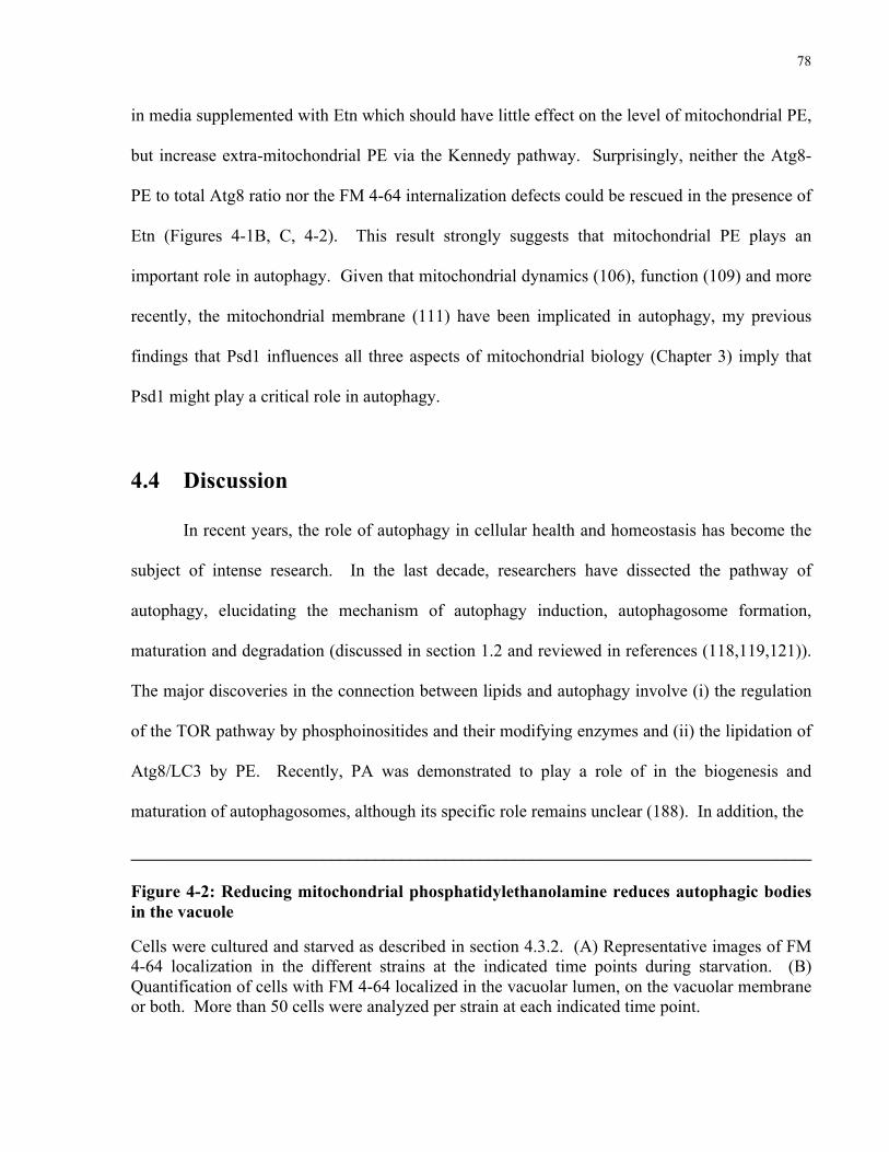
78
in media supplemented with Etn which should have little effect on the level of mitochondrial PE,
but increase extra-mitochondrial PE via the Kennedy pathway. Surprisingly, neither the Atg8-
PE to total Atg8 ratio nor the FM 4-64 internalization defects could be rescued in the presence of
Etn (Figures 4-1B, C, 4-2). This result strongly suggests that mitochondrial PE plays an
important role in autophagy. Given that mitochondrial dynamics (106), function (109) and more
recently, the mitochondrial membrane (111) have been implicated in autophagy, my previous
findings that Psd1 influences all three aspects of mitochondrial biology (Chapter 3) imply that
Psd1 might play a critical role in autophagy.
4.4 Discussion
In recent years, the role of autophagy in cellular health and homeostasis has become the
subject of intense research. In the last decade, researchers have dissected the pathway of
autophagy, elucidating the mechanism of autophagy induction, autophagosome formation,
maturation and degradation (discussed in section 1.2 and reviewed in references (118,119,121)).
The major discoveries in the connection between lipids and autophagy involve (i) the regulation
of the TOR pathway by phosphoinositides and their modifying enzymes and (ii) the lipidation of
Atg8/LC3 by PE. Recently, PA was demonstrated to play a role of in the biogenesis and
maturation of autophagosomes, although its specific role remains unclear (188). In addition, the
______________________________________________________________________________
Figure 4-2: Reducing mitochondrial phosphatidylethanolamine reduces autophagic bodies in the vacuole
Cells were cultured and starved as described in section 4.3.2. (A) Representative images of FM 4-64 localization in the different strains at the indicated time points during starvation. (B) Quantification of cells with FM 4-64 localized in the vacuolar lumen, on the vacuolar membrane or both. More than 50 cells were analyzed per strain at each indicated time point.

79
mitochondrial-enriched phospholipid, CL, has also been implicated in both autophagy (287) and
mitophagy (288). In this study, I have identified mitochondrial PE as an important factor during
autophagy in yeast.
My results reveal that the loss of Psd1, the key yeast enzyme that synthesizes PE, results
in impaired cell survival, altered Atg8-PE/Atg8 levels and a reduction in vacuolar autophagic
bodies upon starvation (Figures 4-1, 4-2). The inability of Etn to rescue the autophagic defects
in the Δpsd1 strain indicates that these defects are specifically due to a reduction in
mitochondrial PE (Figures 4-1B, C, 4-2). Although PE has previously been shown to be a
limiting factor during autophagy (285), the role of mitochondrial PE in autophagy has not been
extensively studied. Recently, mitochondrial dynamics and function have been shown to play
important roles during autophagy (discussed in section 1.1.4). Mitochondrial fusion during
starvation-induced autophagy was shown to be crucial in protecting cells from death (106).
Furthermore, mitochondrial dysfunction was shown to be the cause of death in autophagy-
deficient strains during starvation (109). My work and work of others have solidified the
requirement for Psd1 and PE in mitochondrial dynamics and function (105,249,272,289).
Hence, the observed autophagic defects resulting from the loss of Psd1 (Figures 4-1, 4-2) may be
indirect effects of impaired mitochondrial dynamics and function, and is in agreement with other
studies on the significance of mitochondrial dynamics and function in autophagy (106,109).
The observed reduction in the number of autophagic bodies in the Δpsd1 strain suggests
that autophagosome formation, expansion and/or fusion with the vacuole might be impaired. It
was recently shown that autophagosomes originate from mitochondria (111). Perhaps the
autophagic defects I observed were a result of impaired autophagosome formation from
mitochondria due to reduced mitochondrial PE in the Δpsd1 strain. In addition, in Chapter 3, I

80
describe my findings that Psd1 promotes mitochondrial membrane fusion by providing
favourable lipid conditions during fusion. It is possible that Psd1 also influences autophagy by
promoting the fusion of autophagosomes with the vacuole. In support of this, a recent study
demonstrated that alterations in the membrane composition of the lysosomal/autophagic
compartments results in reduced autophagosome/lysosome fusion by up to 70% (290), indicating
that the composition of the autophagosomal membrane plays a crucial role in ensuring proper
membrane fusion during autophagy.
In sum, this work demonstrates that Psd1 is important for survival during starvation and
that mitochondrial PE plays an important role in the maintenance of Atg8-PE/Atg8 levels and the
number of vacuolar autophagic bodies during autophagy. More importantly, my results raise the
exciting possibility that autophagosome formation, expansion and/or fusion with the
vacuole/lysosomes might be influenced by the composition of the mitochondrial membrane,
possibly addressing the debate on the source of membranes for autophagosomes.

81
CHAPTER 5
CONCLUDING PERSPECTIVES
5.1 Brief summary of results
Lipids are the main constituents of biological membranes and play profound roles in
biological processes including organellar dynamics and complex signalling networks. In this
thesis, I have specifically characterized the role of mitochondrial phosphatidylethanolamine (PE)
in mitochondrial fusion, mitochondrial function and autophagy. Using a series of in vitro and in
vivo studies, I demonstrate that PE plays an important role in mitochondrial membrane fusion.
The favourable biophysical properties of PE promote mitochondrial fusion likely by enhancing
the rate of lipid mixing during fusion. In addition, PE also promotes fusion by participating in
the biogenesis of the short isoform of mitochondrial genome maintenance 1 (s-Mgm1), a key
mitochondrial fusion protein. Since mitochondrial membrane fusion is proposed to maintain a
functional mitochondrial network, I also examined the role of PE in mitochondrial bioenergetics.
My findings indicate that a reduction in mitochondrial PE results in impaired oxidative
phosphorylation and reduced mitochondrial adenosine triphosphate (ATP) levels, critically
important mitochondrial homeostatic functions.
Recent studies have implicated mitochondrial dynamics and function in the regulation of
autophagy, a cellular quality control mechanism. Here, I extend these findings by demonstrating
that mitochondrial PE influences autophagy likely by promoting autophagosome formation,
expansion and/or fusion with the vacuole/lysosomes. This result highlights the intrinsic

82
importance of mitochondrial PE in autophagy and implicates unique characteristics of the
mitochondrial membrane in autophagy.
5.2 Perspectives
5.2.1 Phosphatidylethanolamine in mitochondrial membrane dynamics
In recent years, the results from several studies have significantly deepened our
knowledge of the connection between mitochondrial lipid metabolism and mitochondrial
biology. Although the two non-bilayer-forming lipids PE and cardiolipin (CL) have been shown
to play important roles in mitochondrial membrane fusion (271,272), the details of how these
lipids modulate membrane dynamics have yet to be uncovered. As an extension of my studies
examining the role of PE in mitochondrial membrane fusion, I overexpressed phosphatidylserine
decarboxylase 1 (Psd1), expecting that the increase in mitochondrial PE would result in
increased mitochondrial fusion and hence an increase in highly interconnected mitochondria.
Surprisingly, I observed that the overexpression of Psd1 resulted in fragmented mitochondria
(Figure 5-1A, B), a phenotype suggesting decreased fusion or increased fission. This fragmented
phenotype is independent of s-Mgm1 biogenesis, as the expression of the two different isoforms
of Mgm1 appeared to be unaltered (Figure 5-1C). The fragmented mitochondrial phenotype is
particularly surprising as it was previously reported that the overexpression of Psd1 did not alter
the phospholipid composition of mitochondrial membranes. It was proposed that
phosphatidylserine (PS) import to mitochondria was the rate-limiting step in Psd1-dependent PE
synthesis (248). However, in that study, cells were cultured in rich medium, and their
mitochondrial morphology was not analyzed (248). Given my findings that certain
mitochondrial defects are masked when Δpsd1 cells are cultured in rich medium (section 3.3.1,

83
Figure 5-1: Overexpression of Psd1 results in mitochondrial fragmentation independent of s-Mgm1 biogenesis.
Wild type (WT) cells were transformed with a mitochondrial-targeted GFP construct (mtGFP) and an empty vector or a Psd1 overexpression vector and cultured in selection medium with galactose as the carbon source. (A) Representative images of the mitochondrial morphology of cells expressing the empty vector (WT) and cells overexpressing Psd1 (Psd1 O/E). (B) Quantification of the mitochondrial morphology of cells described in (A). More than 200 cells were analyzed per strain. Error bars represent the standard deviation of three independent experiments. (C) Western blot analysis of cells expressing the empty vector (WT) or the Psd1 overexpression vector (Psd1 O/E).
Figures 3-1A, B), I believe that further exploration of the Psd1 overexpression phenotype
promises to elucidate more of the important roles that lipids play in mitochondrial membrane
dynamics. This unexpected result brings attention to the requirement for a specific amount of PE
and perhaps PS on the mitochondrial membrane. While too little PE reduced membrane
curvature and the propensity for fusion, too much PE (or too little PS) could result in membranes
B
C
WT Psd1 O
/E
48 kDTom40
s-Mgm1100 kD
75 kDl-Mgm1
63 kDPsd1-3HA
A
Psd1 O/E
WT

84
with too much curvature, either impeding fusion or promoting fission. This finding strongly
implicates the composition of the mitochondrial membrane as a critical regulatory factor in
mitochondrial membrane dynamics. PE is not simply a pro-fusion lipid - the equilibrium
between PE and its precursor, PS, might fine-tune the balance between adequate fusion and
insufficient fusion (or increased fission). It is intriguing that a lipid known for its fusogenic
properties may impede organelle fusion or promote organelle fission. The next phase of
discovery will be to examine how it does so and to determine its mechanism of action.
Continuing with yeast as a model system, one can investigate whether the Psd1 overexpression
phenotype is a fusion defect by employing the same in vivo mitochondrial fusion assay described
in section 3.3.2. To confirm if fusion is impaired, Psd1 can also be overexpressed in a
mitochondrial fission mutant. As discussed in section 1.1.1, decreased mitochondrial fusion can
be rescued by reducing mitochondrial fission; hence, if the overexpression of Psd1 impairs
mitochondrial fusion, its overexpression in a mitochondrial fission mutant should result in
tubular mitochondria similar to that of wild type (WT) cells.
Given that PE might play a role both in mitochondrial fusion and mitochondrial fission, it
would be immensely helpful to have a better knowledge of how PE production is regulated.
Although PE is the second most abundant phospholipid in the cell (after phosphatidylcholine
[PC]), the regulation of its biosynthesis remains largely uncharacterized. Mitochondria interact
with the endoplasmic reticulum (ER) at contact sites known as mitochondrial-associated ER
membranes (MAMs) (291). These points of contact are maintained by the ER-mitochondria
encounter structure (ERMES) (280), and serve critical functions in lipid biosynthesis (292).
MAMs not only act as sites of lipid transfer - they are also enriched in lipid-synthesizing
enzymes (291). The synthesis of PE involves the MAM-enriched enzyme PS synthase (292) that

85
converts cytidyldiphosphate diacylglycerol (CDP-DAG) to PS. PS from the ER must then be
transferred to the outer mitochondrial membrane (OMM) and then to the inner mitochondrial
membrane (IMM) where PS decarboxylase (PSD) resides (293). The transfer of PS from the
OMM to the IMM likely occurs at mitochondrial IMM-OMM contact sites (294,295) maintained
by the recently identified mitochondrial contact site (MICOS) complex (296). Since MAMs and
IMM-OMM contact sites facilitate lipid transfer, they could also regulate lipid biosynthesis. It
was recently reported that the synthesis of PC is regulated by contact sites between the plasma
membrane (PM) and the ER (PM-associated ER [pmaER]) (297). It is proposed that PM-ER
contact sites provide spatial regulation by bringing the lipid substrate and its modifying enzyme
into close proximity (297). Perhaps Psd1/PISD localize to IMM-OMM contact sites in close
proximity to imported PS. This localization would also facilitate the export of PE from
mitochondria. To determine the specific submitochondrial localization of Psd1/PISD,
immunogold electron microscopy (EM) and subcellular fractionation experiments can be
performed. Furthermore, if the MICOS complex is important in PE biosynthesis, disrupting the
MICOS complex will result in alterations in the lipid composition of the mitochondrial
membrane. To analyze the mitochondrial lipid composition of MICOS mutants with disrupted
IMM-OMM contacts, mitochondria can be purified, and lipids can be extracted and analyzed by
thin-layer chromatography and mass spectrometry.
5.2.2 Mitochondrial lipids in mitochondrial biology
The role of lipids in mitochondrial dynamics is a burgeoning area of research. In the last
few years, the role of lipids in mitochondrial membrane fusion has been the intensively studied.
The results from several studies, including my own, indicate that the processing of Mgm1, a key

86
mitochondrial fusion protein, is affected by lipid metabolism. Specifically, the enzymes that
regulate PE and CL levels have been shown to alter s-Mgm1 biogenesis (98-100). Mgm1 exists
as two cleavage isoforms, long-Mgm1 (l-Mgm1) and s-Mgm1. As discussed in sections 1.1.3.2
and 3.3.7, Mgm1 contains two N-terminal hydrophobic regions; l-Mgm1 is formed when the first
hydrophobic region is inserted into the IMM whereas s-Mgm1 is formed when full-length Mgm1
(FL-Mgm1) is further inserted into the membrane by its second hydrophobic region and
subsequently cleaved by the rhomboid intramembrane protease Rbd1/Pcp1 (Figure 1-3). Since
the insertion of the second hydrophobic region of FL-Mgm1 is ATP-dependent, the simple
explanation for the role of PE and CL in regulating s-Mgm1 biogenesis might be through the
regulation of mitochondrial ATP levels, as both PE and CL have been shown to interact with and
influence the activity of the supercomplexes in the electron transport chain (ETC) (104,105,255).
However, since Mgm1 undergoes alternative topogenesis, an exciting possibility is that PE and
CL could reduce the propensity of the first hydrophobic region in Mgm1 to remain in the IMM.
This would increase the likelihood that the second hydrophobic region will be inserted into the
IMM, thereby promoting the formation of s-Mgm1. PE, CL and Mgm1 localize to OMM-IMM
contact sites (294,295,298). It is proposed that prohibitins (PHBs) act as scaffolds to provide
high local concentrations of PE and CL (99,101), without which, mitochondria appear
fragmented (99). This strongly suggests that a specific local amount of PE and CL is required
for normal mitochondrial morphology. Perhaps the high local concentrations of PE and CL that
are achieved by the PHBs maintain mitochondrial morphology by ensuring the right balance of l-
Mgm1 and s-Mgm1. It is well-accepted that alternative topogenesis of Mgm1 is dependent on
ATP, the mitochondrial protein import machinery and the hydrophobicity of the first
hydrophobic region within Mgm1 (81). However, its dependence on the composition of the

87
mitochondrial membrane has not been explored. With recent advances in molecular dynamics
(MD) simulations, this area can now be investigated. However, to draw a strong conclusion
from MD simulations, the crystal structure of l-Mgm1 first needs to be obtained, and might
prove to be the biggest challenge in this area of study. Although l-Mgm1 has been purified (85),
its crystal structure has not been reported. An alternative to obtaining the crystal structure of l-
Mgm1 would be to model its structure on that of a similar dynamin or dynamin-related protein
(DRP) that has been crystallized. However, only the guanosine triphosphate hydrolase (GTPase)
domain of Mgm1 has a high sequence homology to dynamins and DRPs (85). Furthermore, no
crystallized dynamin or DRP has a transmembrane (TM) segment. Hence, it would be difficult
to accurately model the structure of l-Mgm1 on that of other crystallized dynamins or DRPs. l-
Mgm1 lacks a functional GTPase domain in vitro (85) and in vivo (84), and it is proposed that
the membrane insertion of l-Mgm1 restricts its GTPase activity - treating liposome-reconstituted
l-Mgm1 with a detergent resulted in observable l-Mgm1 GTPase activity (85). These data
suggest that the membrane insertion of l-Mgm1 plays an important role in regulating its function.
Thus, determining whether the lipid composition affects l-Mgm1 membrane insertion might shed
light on a possible mechanism of Mgm1 regulation. If true, this will be a novel regulatory
mechanism of mitochondrial membrane fusion.
5.2.3 The specific role(s) of mitochondrial phosphatidylethanolamine in
autophagy
Although a relatively recent discovery, it is well established that mitochondrial dynamics
and function play crucial roles in autophagy (106,108,109). Despite these findings, little is
known about the specific function of the mitochondrial membrane in autophagy. It was reported

88
that mitochondria contribute membranes to growing autophagosomes during starvation-induced
autophagy (111), however, the membrane origin of autophagosomes remains highly debated as
other studies have shown that organelles such as the ER (145,146) and the PM (173) also supply
membranes to growing autophagosomes (121). To begin addressing the role of the
mitochondrial membrane in autophagy, the functional importance of the autophagosomal
membrane first needs to be determined. A previous report demonstrated that the efficiency of
autophagy-related 8 (Atg8) conjugation to PE increases with increasing amounts of PE within
the membrane bilayer, peaking at ~70% PE in an in vitro reconstitution system (189). Strikingly,
mitochondria are the organelles that typically contain the highest amounts of PE in the cell. This
suggests that mitochondria, although not necessarily the only source, could be the preferred
source of membranes for growing autophagosomes due to its higher PE content.
Apart from the amount of PE, the type of PE could also differ between mitochondrial PE
and the PE in other membranes. The majority of mitochondrial PE is synthesized by the
decarboxylation of PS by PSD in the IMM; the Kennedy pathway contributes only a small
amount of PE to mitochondria (229,245,293). Interestingly, mammalian PSD has been shown to
preferentially decarboxylate certain species of PS (246,299), and PSD-derived PE usually
contain polyunsaturated acyl chains, whereas PE derived from the Kennedy pathway usually
contain mono- or di-unsaturated acyl chains (245). The increase in polyunsaturated acyl chains
may alter the distribution and/or packing of PE within the mitochondrial membrane and,
although the biological significance of the different acyl chains is still unknown, one cannot rule
out the possibility that these differences could influence autophagy. In support of this, my
finding that the loss of Psd1 results in a reduction in vacuolar autophagic bodies suggests that the
fusion of autophagosomes with the vacuole/lysosomes could be dependent on certain properties

89
of the autophagosomal membrane. Given that PE is known to play an important role in
membrane fusion and remodelling, it would be consistent with its role as a fusogenic lipid if PE
facilitates the fusion of autophagosomes with the vacuole/lysosomes. To test this hypothesis, a
tandem-tagged mRFP-GFP-LC3 fusion protein can be introduced into mammalian cells
(300,301), and autophagy can be induced by starvation. This assay monitors autophagosome
fusion with lysosomes and takes advantage of lysosomal quenching of GFP but not RFP. Upon
the induction of autophagy, autophagosomes fluoresce yellow due to the co-localization of
mRFP and GFP. However, autophagosomes that have fused with lysosomes will fluoresce red
since the acidic lysosomes quench GFP fluorescence, resulting in only detectable mRFP
fluorescence. If mitochondrial PE is important for the fusion of autophagosomes with
lysosomes, cells with reduced mitochondrial PE should have fewer red autophagosomes
compared to WT cells.
5.2.4 The composition of the mitochondrial membrane in autophagy
The model proposed above is simplistic in that it does not account for other lipids within
the membrane bilayer that could also play important roles in autophagy. Indeed, although PE is
the target lipid for Atg8/LC3, negatively-charged phospholipids also play an important role in
the efficiency of Atg8-PE conjugation (189). The amount of Atg8-PE conjugate formed in the
presence of liposomes containing 20% PE increased when the amounts of negatively-charged
phospholipids, phosphatidylinositol (PI), PS, phosphatidylglycerol (PG) or phosphatidic acid
(PA) were increased (189). This strongly suggests that the composition of the membrane bilayer
plays an important role in autophagy, and that different lipid compositions within the membrane
bilayer may influence Atg8-PE conjugation and perhaps even autophagosome formation.

90
Besides negatively-charged phospholipids, membrane curvature could also play a role in
Atg8-PE conjugation efficiency by influencing the E3-like enzyme, the Atg12-Atg5/Atg16
complex. Atg12-Atg5/Atg16-dependent Atg8-PE conjugation was more efficient in the presence
of giant unilamellar vesicles (GUVs) than small unilamellar vesicles (SUVs) (158). In this in
vitro system, GUVs were less abundant and had less curvature than SUVS, prompting the
authors to propose that membrane availability and/or membrane curvature could affect Atg12-
Atg5/Atg16-dependent Atg8-PE conjugation efficiency (158). Although this experiment was
performed in vitro, the use of GUVs, not SUVs, recapitulated previous in vivo observations that
Atg8-PE conjugation is strictly dependent on the Atg12-Atg5/Atg16 complex (140,158,160),
lending more credibility to this result. The postulation that autophagosomes might have
increased curvature is consistent with the requirement for the fusion of autophagosomes with the
vacuole/lysosomes. Lipids that induce membrane curvature have been shown to promote
membrane fusion (273). Together, these findings indicate that the membrane composition plays
an important role in autophagy, as lipids other than PE can modulate Atg8-PE conjugation
efficiency.
In addition to PI, PS, PG and PA, the mitochondrial membrane also contains CL, a
mitochondrial-specific phospholipid that carries two negative charges and induces increased
membrane curvature due to the size of its hydrophilic head relative to its hydrophobic tail.
Hence, CL fulfills both criteria discussed above: it is negatively-charged and promotes increased
membrane curvature. A well-characterized role of CL is its role in the initiation of apoptosis, a
highly conserved mechanism of programmed cell death (reviewed in reference (302)). Under
normal conditions, CL in the IMM interacts strongly with cytochrome c via electrostatic and
hydrophobic interactions, keeping cytochrome c in the intermembrane space (IMS) tightly
91
associated with the IMM (303-305). Upon apoptotic stimuli, CL is peroxidized and translocates
from the IMM to the OMM (306,307). Although the significance of CL translocation is
unknown, it is proposed that peroxidized CL on the OMM might serve as a signal that indicates
damaged mitochondria (306). The peroxidation of CL facilitates the opening of the
mitochondrial permeability transition pore (MPTP) and the release of cytochrome c (308) from
the mitochondrial IMS to the cytosol to initiate the caspase cascade and consequently, apoptosis
(303).
Interestingly, the translocation of CL from the IMM to the OMM is not unique to
apoptosis. Recently, CL translocation was shown to be important for the recruitment of LC3
during mitophagy, the selective degradation of damaged mitochondria by autophagy (288).
Mutating the residues required for CL binding on LC3 completely abolished mitochondrial co-
localization with autophagosomes, indicating severely impaired mitophagy (288). CL has also
been implicated in general autophagy (287). The immunity-related GTPase, IRGM, translocates
to mitochondria, selectively binds to CL and induces autophagy (287). Although these findings
do not pinpoint the specific role of CL, they strongly suggest that the mitochondrial membrane
might play an important role during autophagy. Although PE, PA and CL have individually
been shown to play important roles in autophagy, I propose that the specific composition of
mitochondrial phospholipids confer favourable properties for Atg8-PE conjugation (or LC3-II
formation) and autophagosome fusion with the vacuole/lysosomes, and hence could be the
preferred source of membranes for growing autophagosomes. As discussed above, CL has
previously been shown to influence autophagy, although its specific role remains unclear. To
determine if CL influences the efficiency of LC3 conjugation to PE, CL levels can be reduced by
knocking down CL synthase, and LC3-I conversion to LC3-II can be monitored by western

92
blotting after the induction of autophagy by starvation. A reduction in the amount of LC3-II in
cells with reduced CL levels would suggest impaired LC3-II formation or increased LC3-II
degradation. To distinguish between the two, this experiment can be repeated in the presence of
Bafilomycin A, a drug that inhibits the fusion of autophagosomes with lysosomes and hence
prevents LC3-II degradation. If mitochondrial CL levels play a role in LC3 conjugation to PE, in
the presence of Bafilomycin A, cells with reduced CL should have reduced levels of LC3-II
compared to WT cells. To determine if autophagosome formation is impaired, GFP-tagged LC3
(GFP-LC3) can be used to monitor autophagosome formation by fluorescence imaging where
autophagosome formation is indicated by GFP-LC3 puncta. If CL influences autophagosome
formation, cells with reduced CL levels should have fewer GFP-LC3 puncta than WT cells.
5.3 Conclusion
In the process of studying the protein components of mitochondrial membrane fusion and
autophagy, several studies have inadvertently discovered important roles of lipids in regulating
these processes. In the last few years, interest in lipid metabolism has resurfaced, and it is
becoming increasingly clear that lipids play crucial roles in modulating protein function and, in
the case of organelle fusion, may influence the kinetics of lipid mixing. The work presented in
this thesis provides new insight into the specific role of mitochondrial-localized PE during
mitochondrial membrane fusion and implicates specific properties of the mitochondrial
membrane during autophagy. Notably, although a reduction in mitochondrial PE compromises
mitochondrial fusion, function and autophagy, it is not essential, as the aforementioned functions
remained intact, albeit with significant defects. Coincidentally, this phenomenon is also
observed for CL - yeast cells lacking CL synthase have reduced mitochondrial activity but still
93
retain mitochondrial function (309) and survive under conditions that necessitate mitochondrial
respiration (310). The overlapping functions of PE and CL may provide a simple explanation for
these observations, as a simultaneous reduction in PE and CL is lethal in yeast (311) and results
in impaired mitochondrial fusion (272). However, these results strongly suggest that a specific
lipid composition is crucial for optimum function, and that the role of mitochondrial lipids in
mitochondrial membrane dynamics and autophagy warrants further study. At the rate that
scientific research is progressing, I have no doubt that the scientific community will continue to
uncover more of the subtle yet significant and important roles that lipids play in the regulation of
mitochondrial membrane dynamics and autophagy; and I look forward to the day that it does.

94
REFERENCES 1. Kölliker, A. (1888) Zur Kenntnis der Quergestreiften Muskelfasern. Z. wiss. Zool. 47,
689-710 2. Altmann, R. (1890) Die Elementarorganismen und ihre Beziehungen zu den Zellen,
Leipzig 3. Retzius, G. (1890) Muskelfibrille und sarcoplasm. Biol Untersuch 1, 51-88 4. Ernster, L., and Schatz, G. (1981) Mitochondria: a historical review. J Cell Biol 91, 227s-
255s 5. Lewis, M. R., and Lewis, W. H. (1915) Mitochondria (and other cytoplasmic structures)
in tissue cultures. Am J Anat 17, 339-401 6. Bereiter-Hahn, J., and Voth, M. (1994) Dynamics of mitochondria in living cells: shape
changes, dislocations, fusion, and fission of mitochondria. Microsc Res Tech 27, 198-219 7. Twig, G., Elorza, A., Molina, A. J., Mohamed, H., Wikstrom, J. D., Walzer, G., Stiles, L.,
Haigh, S. E., Katz, S., Las, G., Alroy, J., Wu, M., Py, B. F., Yuan, J., Deeney, J. T., Corkey, B. E., and Shirihai, O. S. (2008) Fission and selective fusion govern mitochondrial segregation and elimination by autophagy. EMBO J 27, 433-446
8. Chen, H. (2005) Disruption of Fusion Results in Mitochondrial Heterogeneity and Dysfunction. J Biol Chem 280, 26185-26192
9. Nunnari, J., Marshall, W. F., Straight, A., Murray, A., Sedat, J. W., and Walter, P. (1997) Mitochondrial transmission during mating in Saccharomyces cerevisiae is determined by mitochondrial fusion and fission and the intramitochondrial segregation of mitochondrial DNA. Mol Biol Cell 8, 1233-1242
10. Dimmer, K. S., Fritz, S., Fuchs, F., Messerschmitt, M., Weinbach, N., Neupert, W., and Westermann, B. (2002) Genetic basis of mitochondrial function and morphology in Saccharomyces cerevisiae. Mol Biol Cell 13, 847-853
11. Suen, D. F., Norris, K. L., and Youle, R. J. (2008) Mitochondrial dynamics and apoptosis. Genes Dev 22, 1577-1590
12. Sheridan, C., and Martin, S. J. (2010) Mitochondrial fission/fusion dynamics and apoptosis. Mitochondrion 10, 640-648
13. Chen, H., and Chan, D. C. (2009) Mitochondrial dynamics--fusion, fission, movement, and mitophagy--in neurodegenerative diseases. Human Molecular Genetics 18, R169-176
14. Chan, E. Y., Rujiviphat, J., and McQuibban, A. (2011) The genetics of mitochondrial fusion and fission, Springer, New York
15. Itoh, K., Nakamura, K., Iijima, M., and Sesaki, H. (2013) Mitochondrial dynamics in neurodegeneration. Trends Cell Biol 23, 64-71
16. Rapaport, D., Brunner, M., Neupert, W., and Westermann, B. (1998) Fzo1p is a mitochondrial outer membrane protein essential for the biogenesis of functional mitochondria in Saccharomyces cerevisiae. J Biol Chem 273, 20150-20155
17. Wong, E. D., Wagner, J. A., Gorsich, S. W., McCaffery, J. M., Shaw, J. M., and Nunnari, J. (2000) The dynamin-related GTPase, Mgm1p, is an intermembrane space protein required for maintenance of fusion competent mitochondria. J Cell Biol 151, 341-352
18. Sesaki, H., and Jensen, R. E. (2001) UGO1 encodes an outer membrane protein required for mitochondrial fusion. J Cell Biol 152, 1123-1134

95
19. Otsuga, D., Keegan, B. R., Brisch, E., Thatcher, J. W., Hermann, G. J., Bleazard, W., and Shaw, J. M. (1998) The dynamin-related GTPase, Dnm1p, controls mitochondrial morphology in yeast. J Cell Biol 143, 333-349
20. Bleazard, W., McCaffery, J. M., King, E. J., Bale, S., Mozdy, A., Tieu, Q., Nunnari, J., and Shaw, J. M. (1999) The dynamin-related GTPase Dnm1 regulates mitochondrial fission in yeast. Nat Cell Biol 1, 298-304
21. Cerveny, K. L., McCaffery, J. M., and Jensen, R. E. (2001) Division of mitochondria requires a novel DMN1-interacting protein, Net2p. Mol Biol Cell 12, 309-321
22. Mozdy, A. D., McCaffery, J. M., and Shaw, J. M. (2000) Dnm1p GTPase-mediated mitochondrial fission is a multi-step process requiring the novel integral membrane component Fis1p. J Cell Biol 151, 367-380
23. Smirnova, E., Griparic, L., Shurland, D. L., and van der Bliek, A. M. (2001) Dynamin-related protein Drp1 is required for mitochondrial division in mammalian cells. Mol Biol Cell 12, 2245-2256
24. Sugioka, R., Shimizu, S., and Tsujimoto, Y. (2004) Fzo1, a Protein Involved in Mitochondrial Fusion, Inhibits Apoptosis. J Biol Chem 279, 52726-52734
25. Cerveny, K., Tamura, Y., Zhang, Z., Jensen, R., and Sesaki, H. (2007) Regulation of mitochondrial fusion and division. Trends Cell Biol 17, 563-569
26. Hoppins, S., Lackner, L., and Nunnari, J. (2007) The machines that divide and fuse mitochondria. Annu Rev Biochem 76, 751-780
27. Praefcke, G. J., and McMahon, H. T. (2004) The dynamin superfamily: universal membrane tubulation and fission molecules? Nat Rev Mol Cell Biol 5, 133-147
28. Tieu, Q., and Nunnari, J. (2000) Mdv1p is a WD repeat protein that interacts with the dynamin-related GTPase, Dnm1p, to trigger mitochondrial division. J Cell Biol 151, 353-366
29. Naylor, K., Ingerman, E., Okreglak, V., Marino, M., Hinshaw, J. E., and Nunnari, J. (2005) Mdv1 Interacts with Assembled Dnm1 to Promote Mitochondrial Division. J Biol Chem 281, 2177-2183
30. Sesaki, H., and Jensen, R. E. (1999) Division versus fusion: Dnm1p and Fzo1p antagonistically regulate mitochondrial shape. J Cell Biol 147, 699-706
31. Legesse-Miller, A., Massol, R. H., and Kirchhausen, T. (2003) Constriction and Dnm1p Recruitment Are Distinct Processes in Mitochondrial Fission. Mol Biol Cell 14, 1953-1963
32. Fukushima, N. H., Brisch, E., Keegan, B. R., Bleazard, W., and Shaw, J. M. (2001) The GTPase effector domain sequence of the Dnm1p GTPase regulates self-assembly and controls a rate-limiting step in mitochondrial fission. Mol Biol Cell 12, 2756-2766
33. Shin, H. W., Takatsu, H., Mukai, H., Munekata, E., Murakami, K., and Nakayama, K. (1999) Intermolecular and interdomain interactions of a dynamin-related GTP-binding protein, Dnm1p/Vps1p-like protein. J Biol Chem 274, 2780-2785
34. Ingerman, E., Perkins, E. M., Marino, M., Mears, J. A., McCaffery, J. M., Hinshaw, J. E., and Nunnari, J. (2005) Dnm1 forms spirals that are structurally tailored to fit mitochondria. J Cell Biol 170, 1021-1027
35. Yoon, Y., Pitts, K. R., and McNiven, M. A. (2001) Mammalian dynamin-like protein DLP1 tubulates membranes. Mol Biol Cell 12, 2894-2905

96
36. Zhu, P. P., Patterson, A., Stadler, J., Seeburg, D. P., Sheng, M., and Blackstone, C. (2004) Intra- and Intermolecular Domain Interactions of the C-terminal GTPase Effector Domain of the Multimeric Dynamin-like GTPase Drp1. J Biol Chem 279, 35967-35974
37. Pitts, K. R., McNiven, M. A., and Yoon, Y. (2004) Mitochondria-specific Function of the Dynamin Family Protein DLP1 Is Mediated by Its C-terminal Domains. J Biol Chem 279, 50286-50294
38. Bhar, D., Karren, M. A., Babst, M., and Shaw, J. M. (2006) Dimeric Dnm1-G385D Interacts with Mdv1 on Mitochondria and Can Be Stimulated to Assemble into Fission Complexes Containing Mdv1 and Fis1. J Biol Chem 281, 17312-17320
39. James, D. I., Parone, P. A., Mattenberger, Y., and Martinou, J. C. (2003) hFis1, a Novel Component of the Mammalian Mitochondrial Fission Machinery. J Biol Chem 278, 36373-36379
40. Stojanovski, D., Koutsopoulos, O. S., Okamoto, K., and Ryan, M. T. (2004) Levels of human Fis1 at the mitochondrial outer membrane regulate mitochondrial morphology. J Cell Sci 117, 1201-1210
41. Suzuki, M., Jeong, S. Y., Karbowski, M., Youle, R. J., and Tjandra, N. (2003) The solution structure of human mitochondria fission protein Fis1 reveals a novel TPR-like helix undle. J Mol Biol 334, 445-458
42. Suzuki, M., Neutzner, A., Tjandra, N., and Youle, R. J. (2005) Novel Structure of the N Terminus in Yeast Fis1 Correlates with a Specialized Function in Mitochondrial Fission. J Biol Chem 280, 21444-21452
43. Blatch, G. L., and Lassle, M. (1999) The tetratricopeptide repeat: a structural motif mediating protein-protein interactions. Bioessays 21, 932-939
44. Dohm, J. A., Lee, S. J., Hardwick, J. M., Hill, R. B., and Gittis, A. G. (2004) Cytosolic domain of the human mitochondrial fission protein fis1 adopts a TPR fold. Proteins 54, 153-156
45. Yoon, Y., Krueger, E. W., Oswald, B. J., and McNiven, M. A. (2003) The Mitochondrial Protein hFis1 Regulates Mitochondrial Fission in Mammalian Cells through an Interaction with the Dynamin-Like Protein DLP1. Mol Cell Biol 23, 5409-5420
46. Yu, T., Fox, R. J., Burwell, L. S., and Yoon, Y. (2005) Regulation of mitochondrial fission and apoptosis by the mitochondrial outer membrane protein hFis1. J Cell Sci 118, 4141-4151
47. Wells, R. C., Picton, L. K., Williams, S. C. P., Tan, F. J., and Hill, R. B. (2007) Direct Binding of the Dynamin-like GTPase, Dnm1, to Mitochondrial Dynamics Protein Fis1 Is Negatively Regulated by the Fis1 N-terminal Arm. J Biol Chem 282, 33769-33775
48. Griffin, E. E., Graumann, J., and Chan, D. (2005) The WD40 protein Caf4p is a component of the mitochondrial fission machinery and recruits Dnm1p to mitochondria. J Cell Biol 170, 237-248
49. Cerveny, K. L., and Jensen, R. E. (2003) The WD-repeats of Net2p Interact with Dnm1p and Fis1p to Regulate Division of Mitochondria. Mol Biol Cell 14, 4126-4139
50. Gandre-Babbe, S., and van der Bliek, A. M. (2008) The Novel Tail-anchored Membrane Protein Mff Controls Mitochondrial and Peroxisomal Fission in Mammalian Cells. Mol Biol Cell 19, 2402-2412
51. Otera, H., Wang, C., Cleland, M. M., Setoguchi, K., Yokota, S., Youle, R. J., and Mihara, K. (2010) Mff is an essential factor for mitochondrial recruitment of Drp1 during mitochondrial fission in mammalian cells. J Cell Biol 191, 1141-1158

97
52. Palmer, C. S., Osellame, L. D., Laine, D., Koutsopoulos, O. S., Frazier, A. E., and Ryan, M. T. (2011) MiD49 and MiD51, new components of the mitochondrial fission machinery. EMBO Rep 12, 565-573
53. Zhao, J., Liu, T., Jin, S., Wang, X., Qu, M., Uhlen, P., Tomilin, N., Shupliakov, O., Lendahl, U., and Nister, M. (2011) Human MIEF1 recruits Drp1 to mitochondrial outer membranes and promotes mitochondrial fusion rather than fission. EMBO J 30, 2762-2778
54. Loson, O. C., Song, Z., Chen, H., and Chan, D. C. (2013) Fis1, Mff, MiD49, and MiD51 mediate Drp1 recruitment in mitochondrial fission. Mol Biol Cell 24, 659-667
55. Niemann, A., Ruegg, M., La Padula, V., Schenone, A., and Suter, U. (2005) Ganglioside-induced differentiation associated protein 1 is a regulator of the mitochondrial network: new implications for Charcot-Marie-Tooth disease. J Cell Biol 170, 1067-1078
56. Koirala, S., Guo, Q., Kalia, R., Bui, H. T., Eckert, D. M., Frost, A., and Shaw, J. M. (2013) Interchangeable adaptors regulate mitochondrial dynamin assembly for membrane scission. Proc Natl Acad Sci U S A 110, E1342-1351
57. Labrousse, A. M., Zappaterra, M. D., Rube, D. A., and van der Bliek, A. M. (1999) C. elegans dynamin-related protein DRP-1 controls severing of the mitochondrial outer membrane. Mol Cell 4, 815-826
58. Jakobs, S., Martini, N., Schauss, A. C., Egner, A., Westermann, B., and Hell, S. W. (2003) Spatial and temporal dynamics of budding yeast mitochondria lacking the division component Fis1p. J Cell Sci 116, 2005-2014
59. Messerschmitt, M., Jakobs, S., Vogel, F., Fritz, S., Dimmer, K. S., Neupert, W., and Westermann, B. (2003) The inner membrane protein Mdm33 controls mitochondrial morphology in yeast. J Cell Biol 160, 553-564
60. Tondera, D., Santel, A., Schwarzer, R., Dames, S., Giese, K., Klippel, A., and Kaufmann, J. (2004) Knockdown of MTP18, a Novel Phosphatidylinositol 3-Kinase-dependent Protein, Affects Mitochondrial Morphology and Induces Apoptosis. J Biol Chem 279, 31544-31555
61. Tondera, D., Czauderna, F., Paulick, K., Schwarzer, R., Kaufmann, J., and Santel, A. (2005) The mitochondrial protein MTP18 contributes to mitochondrial fission in mammalian cells. J Cell Sci 118, 3049-3059
62. Zhao, J., Liu, T., Jin, S. B., Tomilin, N., Castro, J., Shupliakov, O., Lendahl, U., and Nister, M. (2009) The novel conserved mitochondrial inner-membrane protein MTGM regulates mitochondrial morphology and cell proliferation. J Cell Sci 122, 2252-2262
63. van der Bliek, A. M., Shen, Q., and Kawajiri, S. (2013) Mechanisms of mitochondrial fission and fusion. Cold Spring Harb Perspect Biol 5
64. Meeusen, S., McCaffery, J. M., and Nunnari, J. (2004) Mitochondrial Fusion Intermediates Revealed in Vitro. Science 305, 1747-1752
65. Hermann, G. J., Thatcher, J. W., Mills, J. P., Hales, K. G., Fuller, M. T., Nunnari, J., and Shaw, J. M. (1998) Mitochondrial fusion in yeast requires the transmembrane GTPase Fzo1p. J Cell Biol 143, 359-373
66. Fritz, S., Rapaport, D., Klanner, E., Neupert, W., and Westermann, B. (2001) Connection of the mitochondrial outer and inner membranes by Fzo1 is critical for organellar fusion. J Cell Biol 152, 683-692
67. Griffin, E. E., and Chan, D. C. (2006) Domain Interactions within Fzo1 Oligomers Are Essential for Mitochondrial Fusion. J Biol Chem 281, 16599-16606
98
68. Anton, F., Fres, J. M., Schauss, A., Pinson, B., Praefcke, G. J., Langer, T., and Escobar-Henriques, M. (2011) Ugo1 and Mdm30 act sequentially during Fzo1-mediated mitochondrial outer membrane fusion. J Cell Sci 124, 1126-1135
69. Fritz, S., Weinbach, N., and Westermann, B. (2003) Mdm30 is an F-box protein required for maintenance of fusion-competent mitochondria in yeast. Mol Biol Cell 14, 2303-2313
70. Escobar-Henriques, M., Westermann, B., and Langer, T. (2006) Regulation of mitochondrial fusion by the F-box protein Mdm30 involves proteasome-independent turnover of Fzo1. J Cell Biol 173, 645-650
71. Rojo, M., Legros, F., Chateau, D., and Lombes, A. (2002) Membrane topology and mitochondrial targeting of mitofusins, ubiquitous mammalian homologs of the transmembrane GTPase Fzo. J Cell Sci 115, 1663-1674
72. Ishihara, N., Eura, Y., and Mihara, K. (2004) Mitofusin 1 and 2 play distinct roles in mitochondrial fusion reactions via GTPase activity. J Cell Sci 117, 6535-6546
73. Koshiba, T., Detmer, S. A., Kaiser, J. T., Chen, H., McCaffery, J. M., and Chan, D. C. (2004) Structural Basis of Mitochondrial Tethering by Mitofusin Complexes. Science 305, 858-862
74. Low, H. H., and Lowe, J. (2006) A bacterial dynamin-like protein. Nature 444, 766-769 75. Low, H. H., Sachse, C., Amos, L. A., and Lowe, J. (2009) Structure of a bacterial
dynamin-like protein lipid tube provides a mechanism for assembly and membrane curving. Cell 139, 1342-1352
76. Faelber, K., Posor, Y., Gao, S., Held, M., Roske, Y., Schulze, D., Haucke, V., Noe, F., and Daumke, O. (2011) Crystal structure of nucleotide-free dynamin. Nature 477, 556-560
77. Ford, M. G., Jenni, S., and Nunnari, J. (2011) The crystal structure of dynamin. Nature 477, 561-566
78. Wong, E. D., Wagner, J. A., Gorsich, S. W., McCaffery, J. M., Shaw, J. M., and Nunnari, J. (2000) The dynamin-related GTPase, Mgm1p, is an intermembrane space protein required for maintenance of fusion competent mitochondria. J Cell Biol 151, 341-352
79. Wong, E. D., Wagner, J. A., Scott, S. V., Okreglak, V., Holewinske, T. J., Cassidy-Stone, A., and Nunnari, J. (2003) The intramitochondrial dynamin-related GTPase, Mgm1p, is a component of a protein complex that mediates mitochondrial fusion. J Cell Biol 160, 303-311
80. Cipolat, S., Martins de Brito, O., Dal Zilio, B., and Scorrano, L. (2004) OPA1 requires mitofusin 1 to promote mitochondrial fusion. Proc Natl Acad Sci U S A 101, 15927-15932
81. Herlan, M., Bornhovd, C., Hell, K., Neupert, W., and Reichert, A. S. (2004) Alternative topogenesis of Mgm1 and mitochondrial morphology depend on ATP and a functional import motor. J Cell Biol 165, 167-173
82. McQuibban, G. A., Saurya, S., and Freeman, M. (2003) Mitochondrial membrane remodelling regulated by a conserved rhomboid protease. Nature 423, 537-541
83. Herlan, M., Vogel, F., Bornhovd, C., Neupert, W., and Reichert, A. S. (2003) Processing of Mgm1 by the rhomboid-type protease Pcp1 is required for maintenance of mitochondrial morphology and of mitochondrial DNA. J Biol Chem 278, 27781-27788
84. Zick, M., Duvezin-Caubet, S., Schafer, A., Vogel, F., Neupert, W., and Reichert, A. S. (2009) Distinct roles of the two isoforms of the dynamin-like GTPase Mgm1 in mitochondrial fusion. FEBS Lett 583, 2237-2243

99
85. DeVay, R. M., Dominguez-Ramirez, L., Lackner, L. L., Hoppins, S., Stahlberg, H., and Nunnari, J. (2009) Coassembly of Mgm1 isoforms requires cardiolipin and mediates mitochondrial inner membrane fusion. J Cell Biol 186, 793-803
86. Rujiviphat, J., Meglei, G., Rubinstein, J. L., and McQuibban, G. A. (2009) Phospholipid association is essential for dynamin-related protein Mgm1 to function in mitochondrial membrane fusion. J Biol Chem 284, 28682-28686
87. Meeusen, S., DeVay, R., Block, J., Cassidy-Stone, A., Wayson, S., McCaffery, J. M., and Nunnari, J. (2006) Mitochondrial inner-membrane fusion and crista maintenance requires the dynamin-related GTPase Mgm1. Cell 127, 383-395
88. Meglei, G., and McQuibban, G. A. (2009) The Dynamin-Related Protein Mgm1p Assembles into Oligomers and Hydrolyzes GTP To Function in Mitochondrial Membrane Fusion (dagger). Biochemistry 48, 1774-1784
89. Abutbul-Ionita, I., Rujiviphat, J., Nir, I., McQuibban, G. A., and Danino, D. (2012) Membrane tethering and nucleotide-dependent conformational changes drive mitochondrial genome maintenance (Mgm1) protein-mediated membrane fusion. J Biol Chem 287, 36634-36638
90. Ban, T., Heymann, J. A., Song, Z., Hinshaw, J. E., and Chan, D. C. (2010) OPA1 disease alleles causing dominant optic atrophy have defects in cardiolipin-stimulated GTP hydrolysis and membrane tubulation. Hum Mol Genet 19, 2113-2122
91. Hoppins, S., Horner, J., Song, C., McCaffery, J. M., and Nunnari, J. (2009) Mitochondrial outer and inner membrane fusion requires a modified carrier protein. J Cell Biol 184, 569-581
92. Sesaki, H., and Jensen, R. E. (2004) Ugo1p links the Fzo1p and Mgm1p GTPases for mitochondrial fusion. J Biol Chem 279, 28298-28303
93. Coonrod, E. M., Karren, M. A., and Shaw, J. M. (2007) Ugo1p Is a Multipass Transmembrane Protein with a Single Carrier Domain Required for Mitochondrial Fusion. Traffic 8, 500-511
94. van den Brink-van der Laan, E., Killian, J. A., and de Kruijff, B. (2004) Nonbilayer lipids affect peripheral and integral membrane proteins via changes in the lateral pressure profile. Biochim Biophys Acta 1666, 275-288
95. Osman, C., Voelker, D. R., and Langer, T. (2011) Making heads or tails of phospholipids in mitochondria. J Cell Biol 192, 7-16
96. Furt, F., and Moreau, P. (2009) Importance of lipid metabolism for intracellular and mitochondrial membrane fusion/fission processes. Int J Biochem Cell Biol 41, 1828-1836
97. Choi, S. Y., Huang, P., Jenkins, G. M., Chan, D. C., Schiller, J., and Frohman, M. A. (2006) A common lipid links Mfn-mediated mitochondrial fusion and SNARE-regulated exocytosis. Nat Cell Biol 8, 1255-1262
98. Sesaki, H., Dunn, C. D., Iijima, M., Shepard, K. A., Yaffe, M. P., Machamer, C. E., and Jensen, R. E. (2006) Ups1p, a conserved intermembrane space protein, regulates mitochondrial shape and alternative topogenesis of Mgm1p. J Cell Biol 173, 651-658
99. Osman, C., Haag, M., Potting, C., Rodenfels, J., Dip, P. V., Wieland, F. T., Brugger, B., Westermann, B., and Langer, T. (2009) The genetic interactome of prohibitins: coordinated control of cardiolipin and phosphatidylethanolamine by conserved regulators in mitochondria. J Cell Biol 184, 583-596

100
100. Tamura, Y., Endo, T., Iijima, M., and Sesaki, H. (2009) Ups1p and Ups2p antagonistically regulate cardiolipin metabolism in mitochondria. J Cell Biol 185, 1029-1045
101. Birner, R., Nebauer, R., Schneiter, R., and Daum, G. (2003) Synthetic lethal interaction of the mitochondrial phosphatidylethanolamine biosynthetic machinery with the prohibitin complex of Saccharomyces cerevisiae. Mol Biol Cell 14, 370-383
102. Zhang, M., Mileykovskaya, E., and Dowhan, W. (2002) Gluing the respiratory chain together. Cardiolipin is required for supercomplex formation in the inner mitochondrial membrane. J Biol Chem 277, 43553-43556
103. Pfeiffer, K., Gohil, V., Stuart, R. A., Hunte, C., Brandt, U., Greenberg, M. L., and Schagger, H. (2003) Cardiolipin stabilizes respiratory chain supercomplexes. J Biol Chem 278, 52873-52880
104. Bottinger, L., Horvath, S. E., Kleinschroth, T., Hunte, C., Daum, G., Pfanner, N., and Becker, T. (2012) Phosphatidylethanolamine and cardiolipin differentially affect the stability of mitochondrial respiratory chain supercomplexes. J Mol Biol 423, 677-686
105. Tasseva, G., Bai, H. D., Davidescu, M., Haromy, A., Michelakis, E., and Vance, J. E. (2013) Phosphatidylethanolamine deficiency in Mammalian mitochondria impairs oxidative phosphorylation and alters mitochondrial morphology. J Biol Chem 288, 4158-4173
106. Gomes, L. C., Di Benedetto, G., and Scorrano, L. (2011) During autophagy mitochondria elongate, are spared from degradation and sustain cell viability. Nat Cell Biol 13, 589-598
107. Tsukada, M., and Ohsumi, Y. (1993) Isolation and characterization of autophagy-defective mutants of Saccharomyces cerevisiae. FEBS Lett 333, 169-174
108. Graef, M., and Nunnari, J. (2011) Mitochondria regulate autophagy by conserved signalling pathways. EMBO J 30, 2101-2114
109. Suzuki, S. W., Onodera, J., and Ohsumi, Y. (2011) Starvation induced cell death in autophagy-defective yeast mutants is caused by mitochondria dysfunction. PLoS ONE 6, e17412
110. Reggiori, F., Shintani, T., Nair, U., and Klionsky, D. J. (2005) Atg9 cycles between mitochondria and the pre-autophagosomal structure in yeasts. Autophagy 1, 101-109
111. Hailey, D. W., Rambold, A. S., Satpute-Krishnan, P., Mitra, K., Sougrat, R., Kim, P. K., and Lippincott-Schwartz, J. (2010) Mitochondria supply membranes for autophagosome biogenesis during starvation. Cell 141, 656-667
112. Murrow, L., and Debnath, J. (2013) Autophagy as a stress-response and quality-control mechanism: implications for cell injury and human disease. Annu Rev Pathol 8, 105-137
113. Hale, A. N., Ledbetter, D. J., Gawriluk, T. R., and Rucker, E. B., 3rd. (2013) Autophagy: Regulation and role in development. Autophagy 9, 951-972
114. Patel, K. K., and Stappenbeck, T. S. (2013) Autophagy and intestinal homeostasis. Annu Rev Physiol 75, 241-262
115. Kaushik, S., Bandyopadhyay, U., Sridhar, S., Kiffin, R., Martinez-Vicente, M., Kon, M., Orenstein, S. J., Wong, E., and Cuervo, A. M. (2011) Chaperone-mediated autophagy at a glance. J Cell Sci 124, 495-499
116. Wang, C.-W., and Klionsky, D. J. (2004) Microautophagy, Landes Bioscience, Georgetown
117. Mijaljica, D., Prescott, M., and Devenish, R. J. (2011) Microautophagy in mammalian cells: revisiting a 40-year-old conundrum. Autophagy 7, 673-682

101
118. Nakatogawa, H., Suzuki, K., Kamada, Y., and Ohsumi, Y. (2009) Dynamics and diversity in autophagy mechanisms: lessons from yeast. Nat Rev Mol Cell Biol 10, 458-467
119. Chen, Y., and Klionsky, D. J. (2011) The regulation of autophagy - unanswered questions. J Cell Sci 124, 161-170
120. Umekawa, M., and Klionsky, D. J. (2012) The Cytoplasm-to-Vacuole Targeting Pathway: A Historical Perspective. Int J Cell Biol 2012, 142634
121. Mizushima, N., Yoshimori, T., and Ohsumi, Y. (2011) The role of Atg proteins in autophagosome formation. Annu Rev Cell Dev Biol 27, 107-132
122. Yuan, W., Tuttle, D. L., Shi, Y. J., Ralph, G. S., and Dunn, W. A., Jr. (1997) Glucose-induced microautophagy in Pichia pastoris requires the alpha-subunit of phosphofructokinase. J Cell Sci 110, 1935-1945
123. Sakai, Y., Koller, A., Rangell, L. K., Keller, G. A., and Subramani, S. (1998) Peroxisome degradation by microautophagy in Pichia pastoris: identification of specific steps and morphological intermediates. J Cell Biol 141, 625-636
124. Mukaiyama, H., Oku, M., Baba, M., Samizo, T., Hammond, A. T., Glick, B. S., Kato, N., and Sakai, Y. (2002) Paz2 and 13 other PAZ gene products regulate vacuolar engulfment of peroxisomes during micropexophagy. Genes Cells 7, 75-90
125. Titorenko, V. I., Keizer, I., Harder, W., and Veenhuis, M. (1995) Isolation and characterization of mutants impaired in the selective degradation of peroxisomes in the yeast Hansenula polymorpha. J Bacteriol 177, 357-363
126. Sun, L. L., Li, M., Suo, F., Liu, X. M., Shen, E. Z., Yang, B., Dong, M. Q., He, W. Z., and Du, L. L. (2013) Global analysis of fission yeast mating genes reveals new autophagy factors. PLoS Genet 9, e1003715
127. Kabeya, Y., Kawamata, T., Suzuki, K., and Ohsumi, Y. (2007) Cis1/Atg31 is required for autophagosome formation in Saccharomyces cerevisiae. Biochem Biophys Res Commun 356, 405-410
128. Kawamata, T., Kamada, Y., Kabeya, Y., Sekito, T., and Ohsumi, Y. (2008) Organization of the pre-autophagosomal structure responsible for autophagosome formation. Mol Biol Cell 19, 2039-2050
129. Kabeya, Y., Noda, N. N., Fujioka, Y., Suzuki, K., Inagaki, F., and Ohsumi, Y. (2009) Characterization of the Atg17-Atg29-Atg31 complex specifically required for starvation-induced autophagy in Saccharomyces cerevisiae. Biochem Biophys Res Commun 389, 612-615
130. Kamada, Y., Yoshino, K., Kondo, C., Kawamata, T., Oshiro, N., Yonezawa, K., and Ohsumi, Y. (2010) Tor directly controls the Atg1 kinase complex to regulate autophagy. Mol Cell Biol 30, 1049-1058
131. Kamada, Y., Funakoshi, T., Shintani, T., Nagano, K., Ohsumi, M., and Ohsumi, Y. (2000) Tor-mediated induction of autophagy via an Apg1 protein kinase complex. J Cell Biol 150, 1507-1513
132. Hosokawa, N., Hara, T., Kaizuka, T., Kishi, C., Takamura, A., Miura, Y., Iemura, S., Natsume, T., Takehana, K., Yamada, N., Guan, J. L., Oshiro, N., and Mizushima, N. (2009) Nutrient-dependent mTORC1 association with the ULK1-Atg13-FIP200 complex required for autophagy. Mol Biol Cell 20, 1981-1991

102
133. Chan, E. Y., Longatti, A., McKnight, N. C., and Tooze, S. A. (2009) Kinase-inactivated ULK proteins inhibit autophagy via their conserved C-terminal domains using an Atg13-independent mechanism. Mol Cell Biol 29, 157-171
134. Hara, T., Takamura, A., Kishi, C., Iemura, S., Natsume, T., Guan, J. L., and Mizushima, N. (2008) FIP200, a ULK-interacting protein, is required for autophagosome formation in mammalian cells. J Cell Biol 181, 497-510
135. Mercer, C. A., Kaliappan, A., and Dennis, P. B. (2009) A novel, human Atg13 binding protein, Atg101, interacts with ULK1 and is essential for macroautophagy. Autophagy 5, 649-662
136. Hosokawa, N., Sasaki, T., Iemura, S., Natsume, T., Hara, T., and Mizushima, N. (2009) Atg101, a novel mammalian autophagy protein interacting with Atg13. Autophagy 5, 973-979
137. Jung, C. H., Jun, C. B., Ro, S. H., Kim, Y. M., Otto, N. M., Cao, J., Kundu, M., and Kim, D. H. (2009) ULK-Atg13-FIP200 complexes mediate mTOR signaling to the autophagy machinery. Mol Biol Cell 20, 1992-2003
138. Ganley, I. G., Lam du, H., Wang, J., Ding, X., Chen, S., and Jiang, X. (2009) ULK1.ATG13.FIP200 complex mediates mTOR signaling and is essential for autophagy. J Biol Chem 284, 12297-12305
139. Mizushima, N. (2010) The role of the Atg1/ULK1 complex in autophagy regulation. Curr Opin Cell Biology 22, 132-139
140. Suzuki, K., Kirisako, T., Kamada, Y., Mizushima, N., Noda, T., and Ohsumi, Y. (2001) The pre-autophagosomal structure organized by concerted functions of APG genes is essential for autophagosome formation. EMBO J 20, 5971-5981
141. Suzuki, K., Kubota, Y., Sekito, T., and Ohsumi, Y. (2007) Hierarchy of Atg proteins in pre-autophagosomal structure organization. Genes Cells 12, 209-218
142. Obara, K., Sekito, T., and Ohsumi, Y. (2006) Assortment of phosphatidylinositol 3-kinase complexes--Atg14p directs association of complex I to the pre-autophagosomal structure in Saccharomyces cerevisiae. Mol Biol Cell 17, 1527-1539
143. Obara, K., Sekito, T., Niimi, K., and Ohsumi, Y. (2008) The Atg18-Atg2 complex is recruited to autophagic membranes via phosphatidylinositol 3-phosphate and exerts an essential function. J Biol Chem 283, 23972-23980
144. Watanabe, Y., Kobayashi, T., Yamamoto, H., Hoshida, H., Akada, R., Inagaki, F., Ohsumi, Y., and Noda, N. N. (2012) Structure-based analyses reveal distinct binding sites for Atg2 and phosphoinositides in Atg18. J Biol Chem 287, 31681-31690
145. Axe, E. L., Walker, S. A., Manifava, M., Chandra, P., Roderick, H. L., Habermann, A., Griffiths, G., and Ktistakis, N. T. (2008) Autophagosome formation from membrane compartments enriched in phosphatidylinositol 3-phosphate and dynamically connected to the endoplasmic reticulum. J Cell Biol 182, 685-701
146. Matsunaga, K., Morita, E., Saitoh, T., Akira, S., Ktistakis, N. T., Izumi, T., Noda, T., and Yoshimori, T. (2010) Autophagy requires endoplasmic reticulum targeting of the PI3-kinase complex via Atg14L. J Cell Biol 190, 511-521
147. Zhong, Y., Wang, Q. J., Li, X., Yan, Y., Backer, J. M., Chait, B. T., Heintz, N., and Yue, Z. (2009) Distinct regulation of autophagic activity by Atg14L and Rubicon associated with Beclin 1-phosphatidylinositol-3-kinase complex. Nat Cell Biol 11, 468-476
148. Devereaux, K., Dall'armi, C., Alcazar-Roman, A., Ogasawara, Y., Zhou, X., Wang, F., Yamamoto, A., De Camilli, P., and Di Paolo, G. (2013) Regulation of Mammalian

103
Autophagy by Class II and III PI 3-Kinases through PI3P Synthesis. PLoS ONE 8, e76405
149. Mizushima, N., Noda, T., Yoshimori, T., Tanaka, Y., Ishii, T., George, M. D., Klionsky, D. J., Ohsumi, M., and Ohsumi, Y. (1998) A protein conjugation system essential for autophagy. Nature 395, 395-398
150. Ichimura, Y., Kirisako, T., Takao, T., Satomi, Y., Shimonishi, Y., Ishihara, N., Mizushima, N., Tanida, I., Kominami, E., Ohsumi, M., Noda, T., and Ohsumi, Y. (2000) A ubiquitin-like system mediates protein lipidation. Nature 408, 488-492
151. Tanida, I., Mizushima, N., Kiyooka, M., Ohsumi, M., Ueno, T., Ohsumi, Y., and Kominami, E. (1999) Apg7p/Cvt2p: A novel protein-activating enzyme essential for autophagy. Mol Biol Cell 10, 1367-1379
152. Shintani, T., Mizushima, N., Ogawa, Y., Matsuura, A., Noda, T., and Ohsumi, Y. (1999) Apg10p, a novel protein-conjugating enzyme essential for autophagy in yeast. EMBO J 18, 5234-5241
153. Mizushima, N., Noda, T., and Ohsumi, Y. (1999) Apg16p is required for the function of the Apg12p-Apg5p conjugate in the yeast autophagy pathway. EMBO J 18, 3888-3896
154. Kuma, A., Mizushima, N., Ishihara, N., and Ohsumi, Y. (2002) Formation of the approximately 350-kDa Apg12-Apg5.Apg16 multimeric complex, mediated by Apg16 oligomerization, is essential for autophagy in yeast. J Biol Chem 277, 18619-18625
155. Hanada, T., Noda, N. N., Satomi, Y., Ichimura, Y., Fujioka, Y., Takao, T., Inagaki, F., and Ohsumi, Y. (2007) The Atg12-Atg5 conjugate has a novel E3-like activity for protein lipidation in autophagy. J Biol Chem 282, 37298-37302
156. Kirisako, T., Ichimura, Y., Okada, H., Kabeya, Y., Mizushima, N., Yoshimori, T., Ohsumi, M., Takao, T., Noda, T., and Ohsumi, Y. (2000) The reversible modification regulates the membrane-binding state of Apg8/Aut7 essential for autophagy and the cytoplasm to vacuole targeting pathway. J Cell Biol 151, 263-276
157. Xie, Z., Nair, U., and Klionsky, D. J. (2008) Atg8 controls phagophore expansion during autophagosome formation. Mol Biol Cell 19, 3290-3298
158. Romanov, J., Walczak, M., Ibiricu, I., Schuchner, S., Ogris, E., Kraft, C., and Martens, S. (2012) Mechanism and functions of membrane binding by the Atg5-Atg12/Atg16 complex during autophagosome formation. EMBO J 31, 4304-4317
159. Tanida, I., Tanida-Miyake, E., Komatsu, M., Ueno, T., and Kominami, E. (2002) Human Apg3p/Aut1p homologue is an authentic E2 enzyme for multiple substrates, GATE-16, GABARAP, and MAP-LC3, and facilitates the conjugation of hApg12p to hApg5p. J Biol Chem 277, 13739-13744
160. Fujita, N., Itoh, T., Omori, H., Fukuda, M., Noda, T., and Yoshimori, T. (2008) The Atg16L complex specifies the site of LC3 lipidation for membrane biogenesis in autophagy. Mol Biol Cell 19, 2092-2100
161. Nakatogawa, H., Ichimura, Y., and Ohsumi, Y. (2007) Atg8, a ubiquitin-like protein required for autophagosome formation, mediates membrane tethering and hemifusion. Cell 130, 165-178
162. Weidberg, H., Shpilka, T., Shvets, E., Abada, A., Shimron, F., and Elazar, Z. (2011) LC3 and GATE-16 N termini mediate membrane fusion processes required for autophagosome biogenesis. Dev Cell 20, 444-454

104
163. Fujita, N., Hayashi-Nishino, M., Fukumoto, H., Omori, H., Yamamoto, A., Noda, T., and Yoshimori, T. (2008) An Atg4B mutant hampers the lipidation of LC3 paralogues and causes defects in autophagosome closure. Mol Biol Cell 19, 4651-4659
164. Nair, U., Cao, Y., Xie, Z., and Klionsky, D. J. (2010) Roles of the lipid-binding motifs of Atg18 and Atg21 in the cytoplasm to vacuole targeting pathway and autophagy. J Biol Chem 285, 11476-11488
165. Mizushima, N., Yamamoto, A., Hatano, M., Kobayashi, Y., Kabeya, Y., Suzuki, K., Tokuhisa, T., Ohsumi, Y., and Yoshimori, T. (2001) Dissection of autophagosome formation using Apg5-deficient mouse embryonic stem cells. J Cell Biol 152, 657-668
166. Moreau, K., Ravikumar, B., Renna, M., Puri, C., and Rubinsztein, D. C. (2011) Autophagosome precursor maturation requires homotypic fusion. Cell 146, 303-317
167. Darsow, T., Rieder, S. E., and Emr, S. D. (1997) A multispecificity syntaxin homologue, Vam3p, essential for autophagic and biosynthetic protein transport to the vacuole. J Cell Biol 138, 517-529
168. Sato, T. K., Darsow, T., and Emr, S. D. (1998) Vam7p, a SNAP-25-like molecule, and Vam3p, a syntaxin homolog, function together in yeast vacuolar protein trafficking. Mol Cell Biol 18, 5308-5319
169. Ishihara, N., Hamasaki, M., Yokota, S., Suzuki, K., Kamada, Y., Kihara, A., Yoshimori, T., Noda, T., and Ohsumi, Y. (2001) Autophagosome requires specific early Sec proteins for its formation and NSF/SNARE for vacuolar fusion. Mol Biol Cell 12, 3690-3702
170. Moreau, K., Renna, M., and Rubinsztein, D. C. (2013) Connections between SNAREs and autophagy. Trends Biochem Sci 38, 57-63
171. Takahashi, Y., Meyerkord, C. L., Hori, T., Runkle, K., Fox, T. E., Kester, M., Loughran, T. P., and Wang, H. G. (2011) Bif-1 regulates Atg9 trafficking by mediating the fission of Golgi membranes during autophagy. Autophagy 7, 61-73
172. Hamasaki, M., Furuta, N., Matsuda, A., Nezu, A., Yamamoto, A., Fujita, N., Oomori, H., Noda, T., Haraguchi, T., Hiraoka, Y., Amano, A., and Yoshimori, T. (2013) Autophagosomes form at ER-mitochondria contact sites. Nature 495, 389-393
173. Ravikumar, B., Moreau, K., Jahreiss, L., Puri, C., and Rubinsztein, D. C. (2010) Plasma membrane contributes to the formation of pre-autophagosomal structures. Nat Cell Biol 12, 747-757
174. Morvan, J., Kochl, R., Watson, R., Collinson, L. M., Jefferies, H. B., and Tooze, S. A. (2009) In vitro reconstitution of fusion between immature autophagosomes and endosomes. Autophagy 5, 676-689
175. Fader, C. M., Sanchez, D., Furlan, M., and Colombo, M. I. (2008) Induction of autophagy promotes fusion of multivesicular bodies with autophagic vacuoles in k562 cells. Traffic 9, 230-250
176. Berg, T. O., Fengsrud, M., Stromhaug, P. E., Berg, T., and Seglen, P. O. (1998) Isolation and characterization of rat liver amphisomes. Evidence for fusion of autophagosomes with both early and late endosomes. J Biol Chem 273, 21883-21892
177. Eskelinen, E. L. (2005) Maturation of autophagic vacuoles in Mammalian cells. Autophagy 1, 1-10
178. Dunn, W. A., Jr. (1990) Studies on the mechanisms of autophagy: maturation of the autophagic vacuole. J Cell Biol 110, 1935-1945
179. Liou, W., Geuze, H. J., Geelen, M. J., and Slot, J. W. (1997) The autophagic and endocytic pathways converge at the nascent autophagic vacuoles. J Cell Biol 136, 61-70

105
180. Simonsen, A., and Tooze, S. A. (2009) Coordination of membrane events during autophagy by multiple class III PI3-kinase complexes. J Cell Biol 186, 773-782
181. Fader, C. M., Sanchez, D. G., Mestre, M. B., and Colombo, M. I. (2009) TI-VAMP/VAMP7 and VAMP3/cellubrevin: two v-SNARE proteins involved in specific steps of the autophagy/multivesicular body pathways. Biochim Biophys Acta 1793, 1901-1916
182. Pryor, P. R., Mullock, B. M., Bright, N. A., Lindsay, M. R., Gray, S. R., Richardson, S. C., Stewart, A., James, D. E., Piper, R. C., and Luzio, J. P. (2004) Combinatorial SNARE complexes with VAMP7 or VAMP8 define different late endocytic fusion events. EMBO Rep 5, 590-595
183. Furuta, N., Fujita, N., Noda, T., Yoshimori, T., and Amano, A. (2010) Combinational soluble N-ethylmaleimide-sensitive factor attachment protein receptor proteins VAMP8 and Vti1b mediate fusion of antimicrobial and canonical autophagosomes with lysosomes. Mol Biol Cell 21, 1001-1010
184. Chen, D., Fan, W., Lu, Y., Ding, X., Chen, S., and Zhong, Q. (2012) A mammalian autophagosome maturation mechanism mediated by TECPR1 and the Atg12-Atg5 conjugate. Mol Cell 45, 629-641
185. Yang, Z., Huang, J., Geng, J., Nair, U., and Klionsky, D. J. (2006) Atg22 recycles amino acids to link the degradative and recycling functions of autophagy. Mol Biol Cell 17, 5094-5104
186. Dall'Armi, C., Devereaux, K. A., and Di Paolo, G. (2013) The role of lipids in the control of autophagy. Curr Biol 23, R33-45
187. Knaevelsrud, H., and Simonsen, A. (2012) Lipids in autophagy: constituents, signaling molecules and cargo with relevance to disease. Biochim Biophys Acta 1821, 1133-1145
188. Dall'Armi, C., Hurtado-Lorenzo, A., Tian, H., Morel, E., Nezu, A., Chan, R. B., Yu, W. H., Robinson, K. S., Yeku, O., Small, S. A., Duff, K., Frohman, M. A., Wenk, M. R., Yamamoto, A., and Di Paolo, G. (2010) The phospholipase D1 pathway modulates macroautophagy. Nat Commun 1, 142
189. Ichimura, Y., Imamura, Y., Emoto, K., Umeda, M., Noda, T., and Ohsumi, Y. (2004) In vivo and in vitro reconstitution of Atg8 conjugation essential for autophagy. J Biol Chem 279, 40584-40592
190. Neufeld, T. P. (2010) TOR-dependent control of autophagy: biting the hand that feeds. Curr Opin Cell Biol 22, 157-168
191. Jung, C. H., Ro, S. H., Cao, J., Otto, N. M., and Kim, D. H. (2010) mTOR regulation of autophagy. FEBS Lett 584, 1287-1295
192. Kihara, A., Noda, T., Ishihara, N., and Ohsumi, Y. (2001) Two distinct Vps34 phosphatidylinositol 3-kinase complexes function in autophagy and carboxypeptidase Y sorting in Saccharomyces cerevisiae. J Cell Biol 152, 519-530
193. Obara, K., Noda, T., Niimi, K., and Ohsumi, Y. (2008) Transport of phosphatidylinositol 3-phosphate into the vacuole via autophagic membranes in Saccharomyces cerevisiae. Genes Cells 13, 537-547
194. de Lartigue, J., Polson, H., Feldman, M., Shokat, K., Tooze, S. A., Urbe, S., and Clague, M. J. (2009) PIKfyve regulation of endosome-linked pathways. Traffic 10, 883-893
195. Ferguson, C. J., Lenk, G. M., and Meisler, M. H. (2009) Defective autophagy in neurons and astrocytes from mice deficient in PI(3,5)P2. Hum Mol Genet 18, 4868-4878

106
196. Ferguson, C. J., Lenk, G. M., Jones, J. M., Grant, A. E., Winters, J. J., Dowling, J. J., Giger, R. J., and Meisler, M. H. (2012) Neuronal expression of Fig4 is both necessary and sufficient to prevent spongiform neurodegeneration. Hum Mol Genet 21, 3525-3534
197. Fang, Y., Vilella-Bach, M., Bachmann, R., Flanigan, A., and Chen, J. (2001) Phosphatidic acid-mediated mitogenic activation of mTOR signaling. Science 294, 1942-1945
198. Takita, T., Konuma, T., Hanazato, M., and Inoue, H. (2011) Diacylglycerol kinase inhibitor R59022-induced autophagy and apoptosis in the neuronal cell line NG108-15. Arch Biochem Biophys 509, 197-201
199. Yoon, M. S., Du, G., Backer, J. M., Frohman, M. A., and Chen, J. (2011) Class III PI-3-kinase activates phospholipase D in an amino acid-sensing mTORC1 pathway. J Cell Biol 195, 435-447
200. Takahashi, Y., Coppola, D., Matsushita, N., Cualing, H. D., Sun, M., Sato, Y., Liang, C., Jung, J. U., Cheng, J. Q., Mule, J. J., Pledger, W. J., and Wang, H. G. (2007) Bif-1 interacts with Beclin 1 through UVRAG and regulates autophagy and tumorigenesis. Nat Cell Biol 9, 1142-1151
201. Gallop, J. L., and McMahon, H. T. (2005) BAR domains and membrane curvature: bringing your curves to the BAR. Biochem Soc Symp, 223-231
202. Peter, B. J., Kent, H. M., Mills, I. G., Vallis, Y., Butler, P. J., Evans, P. R., and McMahon, H. T. (2004) BAR domains as sensors of membrane curvature: the amphiphysin BAR structure. Science 303, 495-499
203. Gallop, J. L., Jao, C. C., Kent, H. M., Butler, P. J., Evans, P. R., Langen, R., and McMahon, H. T. (2006) Mechanism of endophilin N-BAR domain-mediated membrane curvature. EMBO J 25, 2898-2910
204. Siegel, D. P., and Epand, R. M. (1997) The mechanism of lamellar-to-inverted hexagonal phase transitions in phosphatidylethanolamine: implications for membrane fusion mechanisms. Biophys J 73, 3089-3111
205. Thudichum, J. L. W. (1884) Die chemische Konstitution des Gehirus des Menschen und die Tiere, London
206. Folch, J. (1941) The Isolation of Phosphatidyl Serine from Brain Cephalin, and Identification of the Serine Component. J Biol Chem 139
207. Baumann, A. (1913) Biochemische Zeitschrift 54, 30-39 208. Renall, M. H. (1913) Biochemische Zeitschrift 55, 296-300 209. Rudy, H., and Page, L. H. (1930) Z Physiol Chem 193, 251-268 210. Lea, C. H., Rhodes, D. N., and Stoll, R. D. (1955) Phospholipids. 3. On the
chromatographic separation of glycerophospholipids. Biochem J 60, 353-363 211. Klenk, E., and Dohmen, H. (1955) [Isolation of colamine cephalin from bovine liver].
Hoppe Seylers Z Physiol Chem 299, 248-252 212. Ansell, G. B., and Spanner, S. (1982) Phosphatidylserine, phosphatidylethanolamine and
phosphatidylcholine, Elsevier B.V. 213. Baer, E., Maurukas, J., and Russell, M. (1952) Synthesis of Enantiomeric α-Cephalins. J
Am Chem Soc 74, 152-157 214. Gaigg, B., Simbeni, R., Hrastnik, C., Paltauf, F., and Daum, G. (1995) Characterization
of a microsomal subfraction associated with mitochondria of the yeast, Saccharomyces cerevisiae. Involvement in synthesis and import of phospholipids into mitochondria. Biochim Biophys Acta 1234, 214-220

107
215. Clancey, C. J., Chang, S. C., and Dowhan, W. (1993) Cloning of a gene (PSD1) encoding phosphatidylserine decarboxylase from Saccharomyces cerevisiae by complementation of an Escherichia coli mutant. J Biol Chem 268, 24580-24590
216. Trotter, P. J., and Voelker, D. R. (1995) Identification of a non-mitochondrial phosphatidylserine decarboxylase activity (PSD2) in the yeast Saccharomyces cerevisiae. J Biol Chem 270, 6062-6070
217. Carman, G. M., and Han, G. S. (2011) Regulation of phospholipid synthesis in the yeast Saccharomyces cerevisiae. Annu Rev Biochem 80, 859-883
218. Schumacher, M. M., Choi, J. Y., and Voelker, D. R. (2002) Phosphatidylserine transport to the mitochondria is regulated by ubiquitination. J Biol Chem 277, 51033-51042
219. Trotter, P. J., Wu, W. I., Pedretti, J., Yates, R., and Voelker, D. R. (1998) A genetic screen for aminophospholipid transport mutants identifies the phosphatidylinositol 4-kinase, STT4p, as an essential component in phosphatidylserine metabolism. J Biol Chem 273, 13189-13196
220. Wu, W. I., Routt, S., Bankaitis, V. A., and Voelker, D. R. (2000) A new gene involved in the transport-dependent metabolism of phosphatidylserine, PSTB2/PDR17, shares sequence similarity with the gene encoding the phosphatidylinositol/phosphatidylcholine transfer protein, SEC14. J Biol Chem 275, 14446-14456
221. Borkenhagen, L. F., Kennedy, E. P., and Fielding, L. (1961) Enzymatic Formation and Decarboxylation of Phosphatidylserine. J Biol Chem 236, PC28-PC30
222. Trotter, P. J., Pedretti, J., and Voelker, D. R. (1993) Phosphatidylserine decarboxylase from Saccharomyces cerevisiae. Isolation of mutants, cloning of the gene, and creation of a null allele. J Biol Chem 268, 21416-21424
223. Trotter, P. J., Pedretti, J., Yates, R., and Voelker, D. R. (1995) Phosphatidylserine decarboxylase 2 of Saccharomyces cerevisiae. Cloning and mapping of the gene, heterologous expression, and creation of the null allele. J Biol Chem 270, 6071-6080
224. Dowhan, W., and Li, Q. X. (1992) Phosphatidylserine decarboxylase from Escherichia coli. Methods Enzymol 209, 348-359
225. Kuge, O., Saito, K., Kojima, M., Akamatsu, Y., and Nishijima, M. (1996) Post-translational processing of the phosphatidylserine decarboxylase gene product in Chinese hamster ovary cells. Biochem J 319, 33-38
226. Horvath, S. E., Bottinger, L., Vogtle, F. N., Wiedemann, N., Meisinger, C., Becker, T., and Daum, G. (2012) Processing and topology of the yeast mitochondrial phosphatidylserine decarboxylase 1. J Biol Chem 287, 36744-36755
227. van Poelje, P. D., and Snell, E. E. (1990) Pyruvoyl-dependent enzymes. Annu Rev Biochem 59, 29-59
228. Kitamura, H., Wu, W. I., and Voelker, D. R. (2002) The C2 domain of phosphatidylserine decarboxylase 2 is not required for catalysis but is essential for in vivo function. J Biol Chem 277, 33720-33726
229. Burgermeister, M., Birner-Grunberger, R., Nebauer, R., and Daum, G. (2004) Contribution of different pathways to the supply of phosphatidylethanolamine and phosphatidylcholine to mitochondrial membranes of the yeast Saccharomyces cerevisiae. Biochim Biophys Acta 1686, 161-168
230. Saba, J. D., Nara, F., Bielawska, A., Garrett, S., and Hannun, Y. A. (1997) The BST1 gene of Saccharomyces cerevisiae is the sphingosine-1-phosphate lyase. J Biol Chem 272, 26087-26090

108
231. Mandala, S. M., Thornton, R., Tu, Z., Kurtz, M. B., Nickels, J., Broach, J., Menzeleev, R., and Spiegel, S. (1998) Sphingoid base 1-phosphate phosphatase: a key regulator of sphingolipid metabolism and stress response. Proc Natl Acad Sci U S A 95, 150-155
232. Sundler, R., and Akesson, B. (1975) Regulation of phospholipid biosynthesis in isolated rat hepatocytes. Effect of different substrates. J Biol Chem 250, 3359-3367
233. Riekhof, W. R., and Voelker, D. R. (2006) Uptake and utilization of lyso-phosphatidylethanolamine by Saccharomyces cerevisiae. J Biol Chem 281, 36588-36596
234. Riekhof, W. R., Wu, J., Jones, J. L., and Voelker, D. R. (2007) Identification and characterization of the major lysophosphatidylethanolamine acyltransferase in Saccharomyces cerevisiae. J Biol Chem 282, 28344-28352
235. Rajakumari, S., and Daum, G. (2010) Janus-faced enzymes yeast Tgl3p and Tgl5p catalyze lipase and acyltransferase reactions. Mol Biol Cell 21, 501-510
236. Sundler, R., Akesson, B., and Nilsson, A. (1974) Quantitative role of base exchange in phosphatidylethanolamine synthesis in isolated rat hepatocytes. FEBS Lett 43, 303-307
237. Lykidis, A. (2007) Comparative genomics and evolution of eukaryotic phospholipid biosynthesis. Prog Lipid Res 46, 171-199
238. Signorell, A., Rauch, M., Jelk, J., Ferguson, M. A., and Butikofer, P. (2008) Phosphatidylethanolamine in Trypanosoma brucei is organized in two separate pools and is synthesized exclusively by the Kennedy pathway. J Biol Chem 283, 23636-23644
239. Miller, M. A., and Kent, C. (1986) Characterization of the pathways for phosphatidylethanolamine biosynthesis in Chinese hamster ovary mutant and parental cell lines. J Biol Chem 261, 9753-9761
240. Voelker, D. R. (1984) Phosphatidylserine functions as the major precursor of phosphatidylethanolamine in cultured BHK-21 cells. Proc Natl Acad Sci U S A 81, 2669-2673
241. Tijburg, L. B., Houweling, M., Geelen, M. J., and van Golde, L. M. (1988) Effects of dietary conditions on the pool sizes of precursors of phosphatidylcholine and phosphatidylethanolamine synthesis in rat liver. Biochim Biophys Acta 959, 1-8
242. Tijburg, L. B., Geelen, M. J., and Van Golde, L. M. (1989) Biosynthesis of phosphatidylethanolamine via the CDP-ethanolamine route is an important pathway in isolated rat hepatocytes. Biochem Biophys Res Commun 160, 1275-1280
243. Sundler, R., and Akesson, B. (1975) Biosynthesis of phosphatidylethanolamines and phosphatidylcholines from ethanolamine and choline in rat liver. Biochem J 146, 309-315
244. Zelinski, T. A., and Choy, P. C. (1982) Phosphatidylethanolamine biosynthesis in isolated hamster heart. Can J Biochem 60, 817-823
245. Bleijerveld, O. B., Brouwers, J. F., Vaandrager, A. B., Helms, J. B., and Houweling, M. (2007) The CDP-ethanolamine pathway and phosphatidylserine decarboxylation generate different phosphatidylethanolamine molecular species. J Biol Chem 282, 28362-28372
246. Heikinheimo, L., and Somerharju, P. (1998) Preferential decarboxylation of hydrophilic phosphatidylserine species in cultured cells. Implications on the mechanism of transport to mitochondria and cellular aminophospholipid species compositions. J Biol Chem 273, 3327-3335
247. Burgermeister, M., Birner-Grunberger, R., Heyn, M., and Daum, G. (2004) Contribution of different biosynthetic pathways to species selectivity of aminoglycerophospholipids assembled into mitochondrial membranes of the yeast Saccharomyces cerevisiae. Biochim Biophys Acta 1686, 148-160

109
248. Birner, R., Burgermeister, M., Schneiter, R., and Daum, G. (2001) Roles of phosphatidylethanolamine and of its several biosynthetic pathways in Saccharomyces cerevisiae. Mol Biol Cell 12, 997-1007
249. Steenbergen, R., Nanowski, T. S., Beigneux, A., Kulinski, A., Young, S. G., and Vance, J. E. (2005) Disruption of the phosphatidylserine decarboxylase gene in mice causes embryonic lethality and mitochondrial defects. J Biol Chem 280, 40032-40040
250. Saha, S. K., Nishijima, S., Matsuzaki, H., Shibuya, I., and Matsumoto, K. (1996) A regulatory mechanism for the balanced synthesis of membrane phospholipid species in Escherichia coli. Biosci Biotechnol Biochem 60, 111-116
251. Storey, M. K., Clay, K. L., Kutateladze, T., Murphy, R. C., Overduin, M., and Voelker, D. R. (2001) Phosphatidylethanolamine has an essential role in Saccharomyces cerevisiae that is independent of its ability to form hexagonal phase structures. J Biol Chem 276, 48539-48548
252. Vance, J. E., and Tasseva, G. (2013) Formation and function of phosphatidylserine and phosphatidylethanolamine in mammalian cells. Biochim Biophys Acta 1831, 543-554
253. Pecheur, E. I., Martin, I., Maier, O., Bakowsky, U., Ruysschaert, J. M., and Hoekstra, D. (2002) Phospholipid species act as modulators in p97/p47-mediated fusion of Golgi membranes. Biochemistry 41, 9813-9823
254. Signorell, A., Gluenz, E., Rettig, J., Schneider, A., Shaw, M. K., Gull, K., and Butikofer, P. (2009) Perturbation of phosphatidylethanolamine synthesis affects mitochondrial morphology and cell-cycle progression in procyclic-form Trypanosoma brucei. Mol Microbiol 72, 1068-1079
255. Sharpley, M. S., Shannon, R. J., Draghi, F., and Hirst, J. (2006) Interactions between phospholipids and NADH:ubiquinone oxidoreductase (complex I) from bovine mitochondria. Biochemistry 45, 241-248
256. Otsuru, M., Yu, Y., Mizoi, J., Kawamoto-Fujioka, M., Wang, J., Fujiki, Y., and Nishida, I. (2013) Mitochondrial Phosphatidylethanolamine Level Modulates Cyt c Oxidase Activity to Maintain Respiration Capacity in Arabidopsis thaliana Rosette Leaves. Plant Cell Physiol 54, 1612-1619
257. Bogdanov, M., Sun, J., Kaback, H. R., and Dowhan, W. (1996) A phospholipid acts as a chaperone in assembly of a membrane transport protein. J Biol Chem 271, 11615-11618
258. Bogdanov, M., Umeda, M., and Dowhan, W. (1999) Phospholipid-assisted refolding of an integral membrane protein. Minimum structural features for phosphatidylethanolamine to act as a molecular chaperone. J Biol Chem 274, 12339-12345
259. Dowhan, W., and Bogdanov, M. (2012) Molecular genetic and biochemical approaches for defining lipid-dependent membrane protein folding. Biochim Biophys Acta 1818, 1097-1107
260. Deleault, N. R., Piro, J. R., Walsh, D. J., Wang, F., Ma, J., Geoghegan, J. C., and Supattapone, S. (2012) Isolation of phosphatidylethanolamine as a solitary cofactor for prion formation in the absence of nucleic acids. Proc Natl Acad Sci U S A 109, 8546-8551
261. Tan, B. K., Bogdanov, M., Zhao, J., Dowhan, W., Raetz, C. R., and Guan, Z. (2012) Discovery of a cardiolipin synthase utilizing phosphatidylethanolamine and phosphatidylglycerol as substrates. Proc Natl Acad Sci U S A 109, 16504-16509
262. Rosenberry, T. L., Krall, J. A., Dever, T. E., Haas, R., Louvard, D., and Merrick, W. C. (1989) Biosynthetic incorporation of [3H]ethanolamine into protein synthesis elongation

110
factor 1 alpha reveals a new post-translational protein modification. J Biol Chem 264, 7096-7099
263. Whiteheart, S. W., Shenbagamurthi, P., Chen, L., Cotter, R. J., and Hart, G. W. (1989) Murine elongation factor 1 alpha (EF-1 alpha) is posttranslationally modified by novel amide-linked ethanolamine-phosphoglycerol moieties. Addition of ethanolamine-phosphoglycerol to specific glutamic acid residues on EF-1 alpha. J Biol Chem 264, 14334-14341
264. Ransom, W. D., Lao, P. C., Gage, D. A., and Boss, W. F. (1998) Phosphoglycerylethanolamine posttranslational modification of plant eukaryotic elongation factor 1alpha. Plant Physiol 117, 949-960
265. Signorell, A., Jelk, J., Rauch, M., and Butikofer, P. (2008) Phosphatidylethanolamine is the precursor of the ethanolamine phosphoglycerol moiety bound to eukaryotic elongation factor 1A. J Biol Chem 283, 20320-20329
266. Westermann, B., and Neupert, W. (2000) Mitochondria-targeted green fluorescent proteins: convenient tools for the study of organelle biogenesis in Saccharomyces cerevisiae. Yeast 16, 1421-1427
267. Gregg, C., Kyryakov, P., and Titorenko, V. I. (2009) Purification of mitochondria from yeast cells. J Vis Exp, e1417
268. Kushnirov, V. V. (2000) Rapid and reliable protein extraction from yeast. Yeast 16, 857-860
269. Journo, D., Winter, G., and Abeliovich, H. (2008) Monitoring autophagy in yeast using FM 4-64 fluorescence. Methods Enzymol 451, 79-88
270. Sakamoto, T., Inoue, T., Otomo, Y., Yokomori, N., Ohno, M., Arai, H., and Nakagawa, Y. (2012) Deficiency of cardiolipin synthase causes abnormal mitochondrial function and morphology in germ cells of Caenorhabditis elegans. J Biol Chem 287, 4590-4601
271. Kuroda, T., Tani, M., Moriguchi, A., Tokunaga, S., Higuchi, T., Kitada, S., and Kuge, O. (2011) FMP30 is required for the maintenance of a normal cardiolipin level and mitochondrial morphology in the absence of mitochondrial phosphatidylethanolamine synthesis. Mol Microbiol 80, 248-265
272. Joshi, A. S., Thompson, M. N., Fei, N., Huttemann, M., and Greenberg, M. L. (2012) Cardiolipin and mitochondrial phosphatidylethanolamine have overlapping functions in mitochondrial fusion in Saccharomyces cerevisiae. J Biol Chem 287, 17589-17597
273. Churchward, M. A., Rogasevskaia, T., Brandman, D. M., Khosravani, H., Nava, P., Atkinson, J. K., and Coorssen, J. R. (2008) Specific lipids supply critical negative spontaneous curvature--an essential component of native Ca2+-triggered membrane fusion. Biophys J 94, 3976-3986
274. Eura, Y., Ishihara, N., Yokota, S., and Mihara, K. (2003) Two mitofusin proteins, mammalian homologues of FZO, with distinct functions are both required for mitochondrial fusion. J Biochem 134, 333-344
275. Sesaki, H., Southard, S. M., Yaffe, M. P., and Jensen, R. E. (2003) Mgm1p, a dynamin-related GTPase, is essential for fusion of the mitochondrial outer membrane. Mol Biol Cell 14, 2342-2356
276. Herlan, M., Vogel, F., Bornhovd, C., Neupert, W., and Reichert, A. S. (2003) Processing of Mgm1 by the rhomboid-type protease Pcp1 is required for maintenance of mitochondrial morphology and of mitochondrial DNA. J Biol Chem 278, 27781-27788

111
277. Chen, H. K., Ji, Z. S., Dodson, S. E., Miranda, R. D., Rosenblum, C. I., Reynolds, I. J., Freedman, S. B., Weisgraber, K. H., Huang, Y., and Mahley, R. W. (2011) Apolipoprotein E4 domain interaction mediates detrimental effects on mitochondria and is a potential therapeutic target for Alzheimer disease. J Biol Chem 286, 5215-5221
278. Daum, G., Lees, N. D., Bard, M., and Dickson, R. (1998) Biochemistry, cell biology and molecular biology of lipids of Saccharomyces cerevisiae. Yeast 14, 1471-1510
279. Sesaki, H., Southard, S. M., Hobbs, A. E., and Jensen, R. E. (2003) Cells lacking Pcp1p/Ugo2p, a rhomboid-like protease required for Mgm1p processing, lose mtDNA and mitochondrial structure in a Dnm1p-dependent manner, but remain competent for mitochondrial fusion. Biochem Biophys Res Commun 308, 276-283
280. Kornmann, B., Currie, E., Collins, S. R., Schuldiner, M., Nunnari, J., Weissman, J. S., and Walter, P. (2009) An ER-mitochondria tethering complex revealed by a synthetic biology screen. Science 325, 477-481
281. Daum, G. (1985) Lipids of mitochondria. Biochim Biophys Acta 822, 1-42 282. Epand, R. M., Fuller, N., and Rand, R. P. (1996) Role of the position of unsaturation on
the phase behavior and intrinsic curvature of phosphatidylethanolamines. Biophys J 71, 1806-1810
283. Yang, Q., Guo, Y., Li, L., and Hui, S. W. (1997) Effects of lipid headgroup and packing stress on poly(ethylene glycol)-induced phospholipid vesicle aggregation and fusion. Biophys J 73, 277-282
284. Ramsey, R. B. (1976) Leucine and D-3-hydroxybutyrate as lipid precursors in developing rat spinal cord and liver. Biochem J 158, 501-504
285. Nebauer, R., Rosenberger, S., and Daum, G. (2007) Phosphatidylethanolamine, a limiting factor of autophagy in yeast strains bearing a defect in the carboxypeptidase Y pathway of vacuolar targeting. J Biol Chem 282, 16736-16743
286. Midorikawa, R., Yamamoto-Hino, M., Awano, W., Hinohara, Y., Suzuki, E., Ueda, R., and Goto, S. (2010) Autophagy-dependent rhodopsin degradation prevents retinal degeneration in Drosophila. J Neurosci 30, 10703-10719
287. Singh, S. B., Ornatowski, W., Vergne, I., Naylor, J., Delgado, M., Roberts, E., Ponpuak, M., Master, S., Pilli, M., White, E., Komatsu, M., and Deretic, V. (2010) Human IRGM regulates autophagy and cell-autonomous immunity functions through mitochondria. Nat Cell Biol 12, 1154-1165
288. Chu, C. T., Ji, J., Dagda, R. K., Jiang, J. F., Tyurina, Y. Y., Kapralov, A. A., Tyurin, V. A., Yanamala, N., Shrivastava, I. H., Mohammadyani, D., Qiang Wang, K. Z., Zhu, J., Klein-Seetharaman, J., Balasubramanian, K., Amoscato, A. A., Borisenko, G., Huang, Z., Gusdon, A. M., Cheikhi, A., Steer, E. K., Wang, R., Baty, C., Watkins, S., Bahar, I., Bayir, H., and Kagan, V. E. (2013) Cardiolipin externalization to the outer mitochondrial membrane acts as an elimination signal for mitophagy in neuronal cells. Nat Cell Biol 15, 1197-1205
289. Chan, E. Y., and McQuibban, G. A. (2012) Phosphatidylserine decarboxylase 1 (Psd1) promotes mitochondrial fusion by regulating the biophysical properties of the mitochondrial membrane and alternative topogenesis of mitochondrial genome maintenance protein 1 (Mgm1). J Biol Chem 287, 40131-40139
290. Koga, H., Kaushik, S., and Cuervo, A. M. (2010) Altered lipid content inhibits autophagic vesicular fusion. Faseb J 24, 3052-3065

112
291. Rusinol, A. E., Cui, Z., Chen, M. H., and Vance, J. E. (1994) A unique mitochondria-associated membrane fraction from rat liver has a high capacity for lipid synthesis and contains pre-Golgi secretory proteins including nascent lipoproteins. J Biol Chem 269, 27494-27502
292. Vance, J. E. (1990) Phospholipid synthesis in a membrane fraction associated with mitochondria. J Biol Chem 265, 7248-7256
293. Shiao, Y. J., Lupo, G., and Vance, J. E. (1995) Evidence that phosphatidylserine is imported into mitochondria via a mitochondria-associated membrane and that the majority of mitochondrial phosphatidylethanolamine is derived from decarboxylation of phosphatidylserine. J Biol Chem 270, 11190-11198
294. Ardail, D., Privat, J. P., Egret-Charlier, M., Levrat, C., Lerme, F., and Louisot, P. (1990) Mitochondrial contact sites. Lipid composition and dynamics. J Biol Chem 265, 18797-18802
295. Simbeni, R., Paltauf, F., and Daum, G. (1990) Intramitochondrial transfer of phospholipids in the yeast, Saccharomyces cerevisiae. J Biol Chem 265, 281-285
296. Harner, M., Korner, C., Walther, D., Mokranjac, D., Kaesmacher, J., Welsch, U., Griffith, J., Mann, M., Reggiori, F., and Neupert, W. (2011) The mitochondrial contact site complex, a determinant of mitochondrial architecture. EMBO J 30, 4356-4370
297. Tavassoli, S., Chao, J. T., Young, B. P., Cox, R. C., Prinz, W. A., de Kroon, A. I., and Loewen, C. J. (2013) Plasma membrane--endoplasmic reticulum contact sites regulate phosphatidylcholine synthesis. EMBO Rep 14, 434-440
298. Vogel, F., Bornhovd, C., Neupert, W., and Reichert, A. S. (2006) Dynamic subcompartmentalization of the mitochondrial inner membrane. J Cell Biol 175, 237-247
299. Kevala, J. H., and Kim, H. Y. (2001) Determination of substrate preference in phosphatidylserine decarboxylation by liquid chromatography-electrospray ionization mass spectrometry. Anal Biochem 292, 130-138
300. Kimura, S., Noda, T., and Yoshimori, T. (2007) Dissection of the autophagosome maturation process by a novel reporter protein, tandem fluorescent-tagged LC3. Autophagy 3, 452-460
301. Mizushima, N., Yoshimori, T., and Levine, B. (2010) Methods in mammalian autophagy research. Cell 140, 313-326
302. McMillin, J. B., and Dowhan, W. (2002) Cardiolipin and apoptosis. Biochim Biophys Acta 1585, 97-107
303. Ow, Y. P., Green, D. R., Hao, Z., and Mak, T. W. (2008) Cytochrome c: functions beyond respiration. Nat Rev Mol Cell Biol 9, 532-542
304. Hanske, J., Toffey, J. R., Morenz, A. M., Bonilla, A. J., Schiavoni, K. H., and Pletneva, E. V. (2012) Conformational properties of cardiolipin-bound cytochrome c. Proc Natl Acad Sci U S A 109, 125-130
305. Bergstrom, C. L., Beales, P. A., Lv, Y., Vanderlick, T. K., and Groves, J. T. (2013) Cytochrome c causes pore formation in cardiolipin-containing membranes. Proc Natl Acad Sci U S A 110, 6269-6274
306. Garcia Fernandez, M., Troiano, L., Moretti, L., Nasi, M., Pinti, M., Salvioli, S., Dobrucki, J., and Cossarizza, A. (2002) Early changes in intramitochondrial cardiolipin distribution during apoptosis. Cell Growth Differ 13, 449-455
307. Kagan, V. E., Tyurin, V. A., Jiang, J., Tyurina, Y. Y., Ritov, V. B., Amoscato, A. A., Osipov, A. N., Belikova, N. A., Kapralov, A. A., Kini, V., Vlasova, II, Zhao, Q., Zou,

113
M., Di, P., Svistunenko, D. A., Kurnikov, I. V., and Borisenko, G. G. (2005) Cytochrome c acts as a cardiolipin oxygenase required for release of proapoptotic factors. Nat Chem Biol 1, 223-232
308. Ott, M., Robertson, J. D., Gogvadze, V., Zhivotovsky, B., and Orrenius, S. (2002) Cytochrome c release from mitochondria proceeds by a two-step process. Proc Natl Acad Sci U S A 99, 1259-1263
309. Jiang, F., Ryan, M. T., Schlame, M., Zhao, M., Gu, Z., Klingenberg, M., Pfanner, N., and Greenberg, M. L. (2000) Absence of cardiolipin in the crd1 null mutant results in decreased mitochondrial membrane potential and reduced mitochondrial function. J Biol Chem 275, 22387-22394
310. Jiang, F., Rizavi, H. S., and Greenberg, M. L. (1997) Cardiolipin is not essential for the growth of Saccharomyces cerevisiae on fermentable or non-fermentable carbon sources. Mol Microbiol 26, 481-491
311. Gohil, V. M., Thompson, M. N., and Greenberg, M. L. (2005) Synthetic Lethal Interaction of the Mitochondrial Phosphatidylethanolamine and Cardiolipin Biosynthetic Pathways in Saccharomyces cerevisiae. J Biol Chem 280, 35410-35416