
Mitochondrial Disease - amdf.org.au · mitochondrial or ‘mito’ disease, an underestimated...
56
Mitochondrial Disease Information Booklet for Medical Practitioners Produced by: Dr Karen Crawley MBBS FRACGP Overseen by: Professor Carolyn Sue MBBS PhD FRACP Professor John Cristodoulou MB BS PhD FRACP FRCPA FHGSA Professor David Thorburn BSc(Hons) PhD FHGSA FFS (RCPA) May 2014 www.amdf.org.au
Transcript of Mitochondrial Disease - amdf.org.au · mitochondrial or ‘mito’ disease, an underestimated...

Mitochondrial Disease
Information Booklet for Medical Practitioners
Produced by:Dr Karen Crawley MBBS FRACGP
Overseen by:Professor Carolyn Sue MBBS PhD FRACP
Professor John Cristodoulou MB BS PhD FRACP FRCPA FHGSAProfessor David Thorburn BSc(Hons) PhD FHGSA FFS (RCPA)
May 2014
www.amdf.org.au


1
CONTENTS
About the Australian Mitochondrial Disease Foundation ..............2
Could it be Mitochondrial Disease? ..............................................3
The Mitochondria ...........................................................................5
Definition of Mitochondrial Disease ...............................................9
Genetic Background ...................................................................12
Mitochondrial Symptoms .............................................................15
The Diagnostic Process ..............................................................18
Common Mitochondrial Disease Presentations ..........................25
Differential Diagnosis ..................................................................28
Management ...............................................................................29
Prognosis and Further Research .................................................41
Links to Other Diseases ..............................................................43
Appendix .....................................................................................44
References ..................................................................................47

2
ABOUT THE AUSTRALIAN MITOCHONDRIAL DISEASE FOUNDATIONThe Australian Mitochondrial Disease Foundation (AMDF) funds essential research into the diagnosis, treatment and cure of mitochondrial disorders, and supports sufferers and their families. AMDF also works to educate the general public and the medical profession about mitochondrial disease.
The AMDF hopes this booklet provides some understanding of mitochondrial or ‘mito’ disease, an underestimated medical enigma robbing patients of their energy and too often their life!
research supportto fund research into mitochondrial disease
our mission
to support sufferers of mitochondrial disease & their familiesto educate the general public & the medical profession about mitochondrial disease
educate

3
COULD IT BE MITOCHONDRIAL DISEASE?Most GPs will at some stage cared for a patient that seems to be neurotic or has ‘bad luck’. They return repeatedly with symptoms that just don’t seem to add up, don’t seem to match their appearance, or if they are real, the investigations are negative. The patient ends up with multiple pathological diagnoses that adds to further confusion. The practitioner often feels helpless, to clearly understand them, yet GPs have been taught to listen, believe, show compassion and to look for one pathology.
But where is that pathology, where is that link in the variety of presenting complaints? Do we then refer back to the list we so obligingly learnt at university? Have we considered the following possibilities?:
• Infective• Neoplastic • Autoimmune• Endocrine• Nutritional• Metabolic• Psychological• Vascular• Neurological...
Hopefully this booklet will assist you when the link is not in the organ, nor the hormone, nor the vascular system, nor the ‘mind’... but in the cell. More specifically, the problem is in the forgotten essential organelles that produce about 90% of the energy needed by the human body to function, sustain life and support growth.

4
This booklet may not be the answer to all your unsolved mysteries, but with recent research 1,2,5,6 demonstrating that mitochondrial DNA mutations are present in one in 200 people with at least one in 5,000 suffering severe disease, it is worth the consideration. That means in a 2,000+ patient general practice approximately 10 patients have a mutation that has the potential to cause mitochondrial disease. So when is it appropriate to consider the mitochondria as the guilty offender, and how do we even begin to investigate it?
(Diagram: courtesy of www.terrebonneonline.com)
Diagnosing mitochondrial disease can be a nightmare, due to the widespread variety and severity of symptoms in the large number of known sub-groups (>100). Many experts refer to it as the ‘notorious masquerader’ because it mimics so many different illnesses. It affects both children and adults, many of whom are either undiagnosed or misdiagnosed due to the complexity of the disease. So how do you then summarise the presenting mitochondrial patient? It can be summarised as …
any symptom, in any organ, at any age

5
THE MITOCHONDRIA
Mitochondria help to maintain proper concentration of calcium ions within the various compartments of the cell.Mitochondria store calcium.The major function of the mitochondria is to produce energy.Mitochondria help in the formation of blood components and hormones such as testosterone and oestrogen.Mitochondria in the liver help to detoxify ammonia.Production of heat is another function of mitochondria.Mitochondria help in the regulation of membrane potential, cell proliferation and cell metabolism.Mitochondria cause apoptosis or programmed cell death.Mitochondria help in the biosynthesis of heme and steroids.Functions of the Mitochondria (Diagram: courtesy of www.tutorvista.com)
Mitochondria perform many functions necessary for cell metabolism (see diagram), but the energy-producing pathways are the most important. These pathways allow us to break down carbohydrate, fat and oxygen to live. This process of burning food to make the energy molecule, adenosine triphosphate (ATP), is called oxidative phosphorylation (OXPHOS). Only mitochondria can do it. This highly efficient manufacturing process requires oxygen and is therefore called aerobic metabolism and produces approximately 90% of the energy needed by the human body.

6
Mitochondria also play an intimate role in most of the cell’s major metabolic pathways that build, break down, or recycle its molecular building blocks. Cells cannot make the RNA and DNA they need to grow and function without mitochondria4. However, for the purpose of this booklet, we will discuss in-depth only the mitochondria’s main role in energy production.
Over 70 different polypeptides or proteins interacting on the inner mitochondrial membrane make up the complex mitochondrial respiratory chain (also known as the electron transport chain) and allow OXPHOS to occur. Energy sources such as glucose are initially metabolized in the cytoplasm then imported via proteins at the beginning of the chain. Other proteins in the chain continue the process of catabolism using metabolic pathways such as the Krebs cycle, fatty acid oxidation, and amino acid oxidation.
This produces energy-rich electron donors whose electrons are then passed through the respiratory chain (a series of complex molecules I, II, III, and IV). Cytochrome oxidase or complex IV, passes the electrons
to oxygen which is reduced to water. ATP synthase or Complex V then uses the electrochemical proton gradient produced by the respiratory chain to finally make ATP. Although electron transport occurs with great efficiency, a small percentage of electrons are prematurely leaked to oxygen, resulting in the formation of the toxic free-radical superoxide.
Energy Production within mitochondria (for more detail see next diagram)(Diagram: courtesy of the Muscular Dystrophy Association and The Mitochondria Research Society)

7
Mitochondria synthesise adenosine triphosphate (ATP) from adenosine diphosphate (ADP) and inorganic phosphate in a process called oxidative phosphorylation. Simply put, they burn food in the presence of oxygen to produce ATP. The process, greatly simplified, has three main steps.
1. The citric acid cycle breaks down pyruvate (a product of glucose metabolism) and the beta oxidation spiral breaks down fatty acids. Both use the energy released to reduce (i.e. add electrons to) the electron carriers nicotinamide adenine dinucleotide (NAD+), yielding NADH, and flavin adenine dinucleotide (FAD), yielding FADH2
2. The electron transport chain (also called the respiratory chain) uses the energy from the electrons to pump hydrogen ions (protons) into the intermembrane space. The electron transport chain comprises five complexes designated I through V.
3. ATP synthesis takes place at complex V of the electron transport chain, which uses the energy of protons flowing back into the matrix to attach phosphorus atoms to ADP molecules, producing ATP. ATP exits through the adenosine nucleotide translocase (AND) channel, where ATP is exchanged for ADP.
inner membrane
intermembrane space
outer membrane
ATP (energy)

8
Mitochondria take on many different shapes, each being characteristic of the specialised cell in which it resides7 and tailored to meet the needs of that cell. All told, there are about 250 different cell types in the human body, many having their own distinct mitochondria with its own specialised metabolic function4. Most of our body’s nucleated cells contain 500 to 2,000 mitochondria, though some cell types have only a few mitochondria. For example, platelets have only two to six mitochondria whilst mature red blood cells do not contain any mitochondria, though its cellular precursor, the proerythroblast, is critically dependent on mitochondrial function for differentiation into its mature state.
The tissues that require lots of energy have the most mitochondria, and so these highly energy-dependent tissues or organs are the ones most often affected in mitochondrial disease. Therefore, damage is most commonly presenting in the cells of the muscles, brain, heart, liver, gastrointestinal tract, ears and eyes.

9
DEFINITION OF MITOCHONDRIAL DISEASEMitochondrial diseases are a clinically heterogeneous group of disorders that result from a dysfunction in the mitochondrial respiratory chain. The identification of the first genetic cause of mitochondrial diseases was not until 1988. Tissues and organs that are highly dependent upon this aerobic metabolism are most likely to be involved in mitochondrial disease. Common clinical features are ptosis, external ophthalmoplegia, proximal myopathy, exercise intolerance, cardiomyopathy, hyperglycaemia, liver failure, sensorineural deafness, optic atrophy, pigmentary retinal changes and central nervous system findings of fluctuating encephalopathy, seizures, dementia, migraine, stroke-like episodes, ataxia, and spasticity.
The ability for an organ to function normally depends partly on whether its energy production meets a minimum threshold for that organ, otherwise loss of function occurs. The organs that are more highly energy dependent may show symptoms with even a relatively small drop in energy production. For example, the central nervous system has a lower threshold than other organs at which it will start to show evidence of functional impairment.
Some mitochondrial disorders may affect a single organ only, such as the eye in Leber Hereditary Optic Neuropathy (LHON), but many involve multiple organ systems and often present with prominent neurologic and myopathic features. Many affected individuals display a cluster of clinical features that fall into a discrete clinical syndrome, such as ‘mitochondrial encephalopathy with lactic acidosis and stroke-like episodes’ (MELAS), or myoclonic epilepsy with ragged-red fibres (MERRF). However, considerable clinical variability exists and many individuals do not fit neatly into one particular category3. There is no consistent correlation between the severity of a particular biochemical defect and the severity of that patient’s presentation.

10
Where does Mitochondrial Disease Hide?Where does Mitochondrial Disease Hide?
CancerHaematology
Clinics
UnexplainedKidney Disease
GI Dysmotility Clinics
AtypicalLeukodystrophy
Cerebal PalsyClinics
AtypicalLearning
Disabilities
DiabetesClinics
LanguageDelay Clinics
AtypicalAutism
UnexplainedHeart Failure
UnexplainedBlindness
UnexplainedLiver
Failure
Rheumatology/Multiple Sclerosis
Clinics
Sudden Infant Death Sydnrome
SIDS
PsychiatricFacilities
EpilepsyClinics
The question often asked, not just by the general public but also by the medical profession, is “So why have we not heard of this illness yet?” Due to the multi-organ manifestations, its ability to mimic so many other illnesses, the ever-increasing spectrum of recognised phenotypes, the dual genome origins, the varying phenotypes of the same gene even within the same family, an unclear classification system, the relative newness of this illness (of which we have a great deal more to learn), the difficult and non-standardised diagnostic process (clinically, biochemically and genetically), coupled with what can be a very slow, deceptive and insidious onset in the majority (especially in adults), makes mitochondrial disease possibly the ultimate in the world of diagnostic challenges.

11
When a cell contains defective mitochondria, it not only becomes deprived of ATP, it also accumulates unused energy molecules and oxygen. The mitochondrial function worsens as these molecules are then used to make ATP by inefficient means, producing potentially harmful by-products such as lactic acid. This ‘lactic acidosis’ is associated with muscle fatigue and has the potential to damage muscle and nerve tissue. Another harmful by-product, called free radicals or ‘reactive oxygen species’ may also produce oxidative damage. Therefore, the combined effects of energy deprivation and toxin accumulation in these cells produce the main symptoms of mitochondrial myopathies and encephalomyopathies.
12
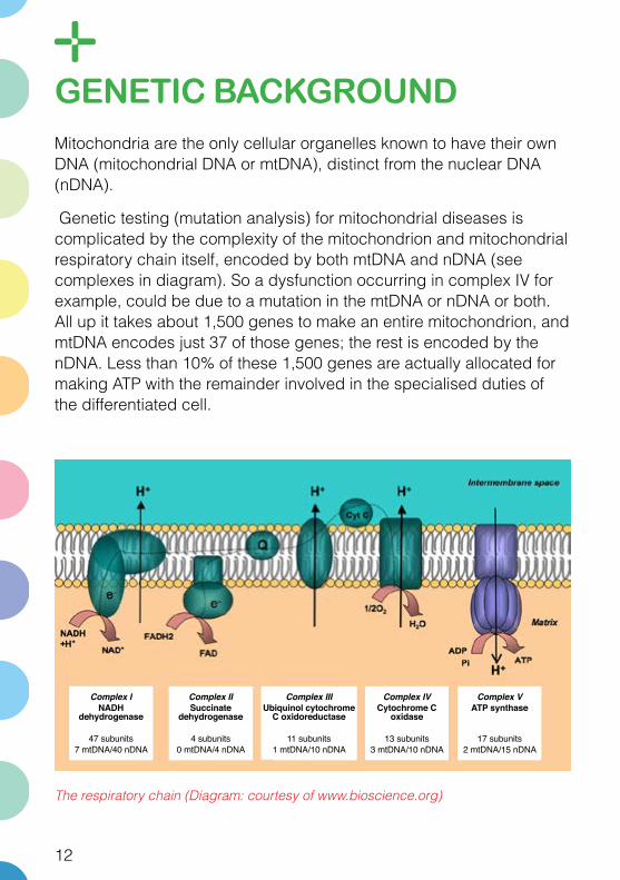
GENETIC BACKGROUNDMitochondria are the only cellular organelles known to have their own DNA (mitochondrial DNA or mtDNA), distinct from the nuclear DNA (nDNA).
Genetic testing (mutation analysis) for mitochondrial diseases is complicated by the complexity of the mitochondrion and mitochondrial respiratory chain itself, encoded by both mtDNA and nDNA (see complexes in diagram). So a dysfunction occurring in complex IV for example, could be due to a mutation in the mtDNA or nDNA or both. All up it takes about 1,500 genes to make an entire mitochondrion, and mtDNA encodes just 37 of those genes; the rest is encoded by the nDNA. Less than 10% of these 1,500 genes are actually allocated for making ATP with the remainder involved in the specialised duties of the differentiated cell.
The respiratory chain (Diagram: courtesy of www.bioscience.org)
Complex INADH
dehydrogenase
47 subunits7 mtDNA/40 nDNA
Complex IISuccinate
dehydrogenase
4 subunits0 mtDNA/4 nDNA
Complex IIIUbiquinol cytochrome
C oxidoreductase
11 subunits1 mtDNA/10 nDNA
Complex IVCytochrome C
oxidase
13 subunits3 mtDNA/10 nDNA
Complex VATP synthase
17 subunits2 mtDNA/15 nDNA

13
Defects in nDNA can be inherited in a Mendelian pattern from either parent or both, with most showing autosomal recessive inheritance. An estimated 75% of primary paediatric mitochondrial disease results from nDNA mutations32, which tend to present early and are usually fatal33.
Due to a quirk in the process of fertilisation, defects in the genes of the mtDNA are usually maternally transmitted. That’s because during conception, when the sperm fuses with the egg, the sperm’s mitochondria, and its mtDNA, are destroyed.
Each human cell contains thousands of copies of mtDNA which at birth are usually all identical, a state that is called homoplasmy. In contrast, individuals with mitochondrial disorders resulting from mtDNA mutations may harbour a mixture of mutant (dysfunctional) and wild-type (normal) mtDNA within each cell, and this is called heteroplasmy8,9. The proportion of mutant mtDNA must exceed a critical threshold level, ‘the threshold effect’, before a cell expresses a biochemical abnormality of the mitochondrial respiratory chain10. The percentage level of mutant mtDNA may vary among individuals within the same family, and also among organs and tissues within the same individual11. Therefore, the phenotypic (or symptomatic) expression of the mitochondrial disorder may vary according to the intrinsic pathogenicity of a mutation, its tissue distribution, the variable aerobic energy-demand of different tissues or organs and the individual genetic background28.
Simplistically, a child conceived from a ‘mostly healthy’ ovum probably won’t develop the disease, and a child conceived from a ‘mostly mutant’ ovum probably will. Please note that in some conditions all the mtDNA within a cell is mutant, hence homoplasmy can apply to either wild-type (normal) or mutant mtDNA. These mutations usually manifest as single-organ or even single cell-type-failure, like retinal ganglion cells in Leber Hereditary Optic Neuropathy (LHON).

14
Heteroplasmy (Diagram: courtesy of The Mitochondria Research Society)
Finally, the way that mtDNA and nDNA mutations interact with each other and with the environment can help determine if disease occurs as well. So, the link between genotype and phenotype in mitochondrial diseases has and will always be recognised as complex16.
mother with mild or no symptoms
small number of mother’s mitochondria, selected
randomly, goes into each early egg cell
contribution from mother
contribution from father
possible outcome
“Bottleneck Effect”
number of mitochondria
increases
80% mutant
50% mutant
20% mutant
child with severe disease?
child with mild disease?
child with no disease?
mother’s cells may have 20% mutant mitochondria
cells that will become egg cells mature egg cells
sperm cells (no mitochondria)
When you can’t make the diagnosis fit......think of mito!

15
MITOCHONDRIAL SYMPTOMSSince mitochondria are present in virtually all cells, symptoms are present in the large majority of organs and as time goes on, we learn also that mitochondrial dysfunction is increasingly being linked to many more pathologies than we could have ever imagined.
The first suspicion of a mitochondrial disorder comes from good clinical judgement, not just being aware of the range of its many symptoms but also the quality of those as well. As soon as the word ‘ATYPICAL’ is used by any specialist to describe an undiagnosed mitochondrial patient, whether it refers to their individual symptoms, their cluster of symptoms, or their previous diagnosis (e.g. atypical MS), or even used in association with a relative’s illness (e.g. “my aunty died of atypical Parkinsons”), then a mitochondrial disorder needs to be considered as a likely diagnostic candidate. It is the ‘square peg’ that is too frequently pushed into the ‘round hole’.
SYMPTOM RANGE
Organ / System Possible Problems
Brain Developmental delays, mental retardation/regression, dementia, seizures (especially atypical or refractory), coma, neuro-psychiatric disturbances, atypical cerebral palsy, myoclonus, movement disorders, ataxia, migraines, strokes.
Nerves Weakness (which may be intermittent), neuropathies, absent reflexes, fainting, absent or excessive sweating resulting in temperature regulation problems.
Muscles Weakness, hypotonia, cramping, muscle pain, recurrent rhabdomyolysis.
Kidneys Proximal renal tubular wasting resulting in loss of protein, magnesium, phosphorous, calcium and other electrolytes, aminoaciduria, nephrotic syndrome.

16
SYMPTOM RANGE (continued)
Organ / System Possible Problems
Heart Conduction defects (e.g., heart blocks, WPW), cardiomyopathy.
Liver Hypoglycaemia, unexplained liver failure, Valproate-induced liver failure.
Eyes Visual loss/blindness, optic atrophy, disorders of extra-ocular muscles, ptosis, retinal degeneration with signs of night blindness, colour-vision deficits, pigmentary retinal changes such as retinitis pigmentosa or ‘salt and pepper’ retinopathy.
Ears Hearing loss and deafness (especially sensorineural).
Pancreas Diabetes and exocrine pancreatic failure (inability to make digestive enzymes).
Systemic Exercise intolerance not in proportion to weakness, fatigue, short statue, respiratory problems including intermittent air hunger, hypersensitive to general anaesthetics.
GIT Gastro-oesophageal reflux, delayed gastric emptying, constipation, pseudo-obstruction, chronic or recurrent vomiting
Childhood IUGR, unexplained hypotonia, weakness, failure to thrive, or a metabolic acidosis (particularly lactic acidosis), infantile spasms, microcephaly, ‘SIDS’.
Skin Symmetrical lipomatosis.
Endocrine Diabetes, short stature, hypothyroidism, hypoparathyroidism.
Haematological Sideroblastic anaemia

17
SYMPTOM QUALITY – RED FLAGS
Clinical Manifestation Features suspicious of a mitochondrial disease
Sensorineural hearing loss
– Asymmetrical onset– Young age of onset– History of partial recovery after an insult, i.e.,
reversible– High frequencies affected first
Focal neurological deficits
– Young age of onset– Preceded by clinical prodrome– Nonvascular territory on neuroimaging– Predominantly grey matter affected– Associated basal ganglia calcification– Good clinical recovery from an ‘event’– Neuroradiological changes out of proportion to
clinical deficit– Associated focal seizures or status epilepticus– Raised CSF lactate
Seizures – Sudden onset status epilepticus– Recurrent physiological trigger– Severe episodes of seizures with good interval
periods (requiring no ACDs for control)– Worsened by sodium valproate
Ptosis – Asymmetrical onset– Slowly progressive with little diurnal variation– Accompanying PEO or retinal pigmentary changes
Retinal pigmentary changes
– Perimacular distribution– No drusen– Non-vision threatening
Diabetes – No associated diabetic retinopathy/peripheral neuropathy with respect to the length of diabetes onset
– Easily controlled with OHA with respect to duration of diabetes

18
THE DIAGNOSTIC PROCESSAs previously mentioned, mitochondrial diseases have varying presentations and the onset may present at any time, from before birth, as IUGR, to late adult life. Even within the same family, the same genetic mutation may affect individuals differently as there is no single identifying feature of mitochondrial disease.
So, to begin the diagnostic process, the foremost and utmost diagnostic tool is good clinical judgement.
The GP is first required to be aware of the existence and symptomatology of mitochondrial diseases, and to remember it in their differential diagnoses under ‘metabolic’.
We should begin to consider the possibility of mitochondrial disease when:
1. A ‘common disease’ has atypical features that set it apart from the pack.
2. Three or more organ systems are involved (or 1-2 of Red Flag symptoms above).
3. Recurrent setbacks/flare ups in a chronic disease occur with infections.4
When the puzzle is solved, we can now meet the challenge together!

19
STEP ONE
The diagnostic work-up for suspected mitochondrial disease is a stepwise procedure, which can be likened to constructing a jigsaw puzzle. As the pieces are collected and placed correctly, the picture begins to form.
Much of the work however, is done by an initial comprehensive history of the individual’s symptoms, the family history, and a full systems review. Clinical investigations are commenced by the GP to complete the systems review and confirm any symptomatology. For example, don’t just ask about deafness, but organise a formal audiological assessment as well.
STEP TWO
In the second step, clinicians need to decide:
1. If the individual’s presentation conforms to any of the more common and better understood mitochondrial syndromes such as MELAS or MERRF. These patients may then proceed directly to genetic testing as organised by a mitochondrial specialist, thereby potentially avoiding the long list of other investigations.
2. If the presentation does not conform to a particular syndrome, as is unfortunately the case for the majority, then the next step is direct referral to a mitochondrial specialist as the subsequent investigations include an array of biochemical tests of either serum, CSF and/or urine, additional electrophysiological studies, functional and imaging studies of the brain and other organs such as muscle, and an appropriate tissue biopsy of the muscle, skin, bowel or liver.
STEP THREE
Based on the patient’s presentation and the results thus far, the probability of a mitochondrial disorder should now be approaching a more definitive answer. Although genetic testing is by definition the gold standard to diagnose a genetic disease, the majority of mitochondrial patients currently don’t have testing, for reasons explained by the complicated and vastly unknown genetics of

20
the illness. So, although the third and final step is genetic testing, diagnosis greatly depends on steps 1 and 2 currently. However, this situation is expected to change quite quickly as exome sequencing or genome sequencing become more widely available in diagnostic rather than just research context within the next 1-2 years.
Unfortunately, many patients will have a presentation suspicious of a mitochondrial disorder, but have little evidence to reinforce a diagnosis. In these patients, regular review and assessment for further clinical deterioration and new organ involvement is essential over time, as even the most classical mitochondrial disorders may not present in their full clinical spectrum at the first medical visit.
To both aid and unify the diagnosis of mitochondrial disease, there have been a number of attempts to create an internationally accepted standard for its diagnostic criteria, such as those according to the Nijmegen, Bernier or Walker criteria29,30. Probably the closest so far, which still requires further verification, is the Nijmegen Clinical Criteria for Mitochondrial Disease (see appendix). It uses a point scoring system in the areas of clinical, biochemical and genetic criteria to classify the chance of mitochondrial disease as definite, highly probable, probable, possible or unlikely.
So, even though the only way to make a diagnosis of a primary mitochondrial disease with absolute certainty is to identify a mtDNA or nDNA abnormality that is known to cause disease, it is usually not possible. The best one can do at this point in time is to begin to ‘build a case’. Then referral to a mitochondrial specialist would be the next appropriate step to continue to ‘build that case’. The gathering of evidence is done by these diagnostic approaches:

21
DIAGNOSTIC TESTS IN MITOCHONDRIAL DISEASES
Type Test What It Shows
Personal & family history
Thorough history of patient and family members
Can sometimes indicate inheritance pattern by noting ‘soft signs’ in unaffected relatives. These include deafness, short stature, migraines and PEO (progressive external ophthalmoplegia).
Systems review and physical signs
Clinical examination, especially neurological Neuroradiological changes out of proportion to clinical deficit Associated focal seizures or status epilepticus Raised CSF lactate
Tests of strength and endurance, and neurological tests including reflexes, vision, speech, basic cognitive skills, and developmental assessment.
Multi-system/organ assessment (dependent upon the individual presentation)
1. Electrophysiology studies, especially EEG, (also NCS, EMG, etc)2. ECG/Holter/Echo3. Hearing tests4. Biochemistry/FBC5. GIT/transit studies 6. Ophthalmological examination7. Allied health team assessments (occupational therapist, speech pathologist, physiotherapist)
1. Monitoring or detection of any seizure activity2. Detection of cardiac abnormalities 3. Sensorineural hearing loss4. Renal tubular dysfunction, liver dysfunction, glucose (including HBA1C), TSH, B12, blood film, etc.5. Gut dysmotility, reflux/cyclical vomiting6. Ptosis, eye muscles, retinal changes of degeneration, pigmentation, and optic atrophy7. Initial assessments can be used to look for any abnormalities or used as baselines for further reviews

22
DIAGNOSTIC TESTS IN MITOCHONDRIAL DISEASES
Type Test What It Shows
Imaging studies (these and the following tests are best done by a mitochondrial specialist)
1. Muscle phosphorus magnetic resonance spectroscopy (MRS) 2. MRI (CT scan if MRI not freely available)
1. Measures the levels of lactate, phosphocreatine and ATP (often depleted in muscles affected by mitochondrial disease).2. Looking for:a) bilateral or symmetric lesions, especially in the basal ganglia or thalamus, brain stem, white matter, or cerebellum b) cerebral and/or cerebellar atrophy c) cortical lesions particularly in non-vascular territories d) diffuse leukoencephalopathy.
Blood, enzyme and biochemical tests (may include urine and CSF also)
1. Lactate and pyruvate levels2. Serum creatine kinase3. Serum carnitine levels (including total, acyl, free, and acyl/free ratio), serum ketones4.Whole blood ammonia5. Quantitative plasma (fasting) and/or urine amino acids. Urinary screening of amino or organic acids.
1. If elevated, may indicate deficiency in respiratory chain; abnormal ratios of the two may help identify the part of the chain that is blocked.2. May be slightly elevated in mitochondrial disease but usually only high in cases of mitochondrial DNA depletion.3-5. Further metabolic evaluations of mitochondrial function, e.g., defects in fatty acid metabolism/oxidation may demonstrate elevated plasma levels of free fatty acids, hypoketonaemia, hypocarnitinaemia, dicarboxylic aciduria, and the presence of Krebs cycle intermediates.

23
DIAGNOSTIC TESTS IN MITOCHONDRIAL DISEASES
Type Test What It Shows
Tissue biopsy(frequently muscle but not gold standard)
1. Histochemistry2. Immunohistochemistry3. Biochemistry4. Electron microscopy
1. Detects abnormal proliferation of mitochondria and deficiencies in cytochrome c oxidase (COX, which is complex IV in the respiratory chain). When treated with the modified Gomori trichrome stain, mitochondria become red, so muscle cells with excessive mitochondria appear as ‘ragged red fibres’. 2. Detects presence or absence of specific proteins. Can rule out other diseases or confirm loss of respiratory chain proteins.3. Measures activities of specific enzymes such as complexes I to IV of the respiratory chain. 4. May confirm abnormal size, shape, number and structure of mitochondria.
Genetic tests (with counselling)NB: Negative test results have a high false-negative rate13
1. Known mutations2. Rare or unknown mutations3. Mitochondrial DNA depletion
1. Uses muscle, urine, hair follicle or other tissue sample to screen for known mutations, looking for common mutations first.2. Can also look for rare or unknown mutations but may require samples from family members; this is more expensive, time-consuming and not freely available in Australia.3. Uses muscle or liver samples to test whether the amount of mtDNA per cell is adequate. mtDNA depletion is one of the most common causes of childhood mitochondrial disease.
(Original resource: courtesy of the Muscular Dystrophy Association)
24
Findings in any one of these categories are not sufficient to make a diagnosis as abnormalities can occur in other illnesses, or may be a secondary phenomenon reflecting mitochondrial dysfunction in another non-mitochondrial disease. Furthermore, some patients with proven disease may not show any biochemical, histological or imaging abnormalities18. For example, normal EMG findings can still be helpful since patients with most other forms of clinical myopathy (such as inflammatory myopathies) usually have diagnostic abnormalities on EMG testing31.
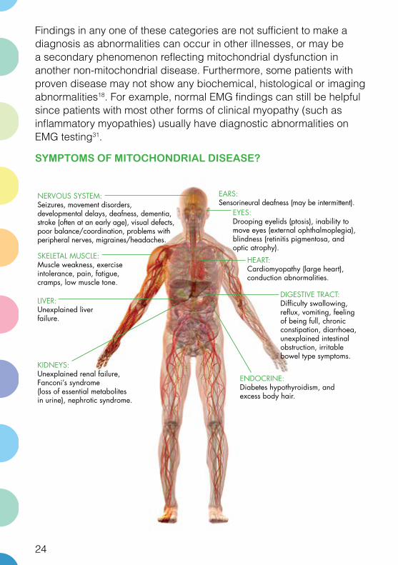
SYMPTOMS OF MITOCHONDRIAL DISEASE?Symptoms of Mitochondrial Disease?
NERVOUS SYSTEM:Seizures, movement disorders, developmental delays, deafness, dementia, stroke (often at an early age), visual defects, poor balance/coordination, problems with peripheral nerves, migraines/headaches.
EYES:Drooping eyelids (ptosis), inability to move eyes (external ophthalmoplegia), blindness (retinitis pigmentosa, and optic atrophy).
EARS:Sensorineural deafness (may be intermittent).
HEART:Cardiomyopathy (large heart),conduction abnormalities.
SKELETAL MUSCLE:Muscle weakness, exercise intolerance, pain, fatigue, cramps, low muscle tone.
LIVER:Unexplained liver failure.
DIGESTIVE TRACT:Difficulty swallowing, reflux, vomiting, feeling of being full, chronic constipation, diarrhoea, unexplained intestinal obstruction, irritable bowel type symptoms.
KIDNEYS:Unexplained renal failure,Fanconi’s syndrome(loss of essential metabolitesin urine), nephrotic syndrome.
ENDOCRINE:Diabetes hypothyroidism, and excess body hair.

25
COMMON MITOCHONDRIAL DISEASE PRESENTATIONSThe terminology used to describe mitochondrial disease can be confusing and it is still a struggle for many patients to be fitted into the groups we know. A single syndrome, with a combination of symptoms, may have many different genotypes, while more than one syndrome may have the same genotype. A diagnosis may be named for the cause, such as COX deficiency, or it may be based on the symptoms of the disease such as the following examples:
MELAS: Mitochondrial Encephalopathy, Lactic Acidosis and Stroke-like episodes. This is the most common type of mitochondrial encephalopathy.
Onset: Usually between 2 and 40 years of age, mean age is 10 years, but can be any age.
Disease characteristics: Hallmark sign is the ‘MELAS’ attack. General characteristics are exercise intolerance, seizures, dementia, muscle weakness, hearing loss, blindness, migraine-type headaches, myopathy, gastric dysmotility, polyneuropathy, ptosis, cardiomyopathy, diabetes, renal failure and short stature.
Inheritance: Maternal
MERRF: Myoclonic Epilepsy with Ragged-Red Fibres Onset: Usually late adolescence to adulthood; variable progression.
Disease characteristics: Myoclonic epilepsy, proximal myopathy, sensorineural deafness, ataxia, pigmentary retinopathy, coordination loss, dementia, distal sensory loss and optic atrophy.
Inheritance: Sporadic or maternal

26
KSS: Kearns-Sayre Syndrome Onset: Before age 20
Disease characteristics: Progressive External Ophthalmoplegia (PEO), ptosis, pigmentary degeneration of retina, heart block, myopathy, dysphagia most commonly associated with cricopharyngeal achalasia, hearing loss, ataxia, and dementia.
Inheritance: Sporadic
Leigh syndrome: Subacute necrotizing encephalomyopathyOnset: Infancy and progression can be fast or slow. Death often occurs within two years of onset.
Disease characteristics: Vomiting, ataxia, hypotonia or spasticity, seizures, feeding and speech difficulties, hearing loss, nystagmus, visual loss, choreoathetosis, peripheral neuropathy, hyperventilation, motor and intellectual regression.
Inheritance: Mendelian or maternal
MNGIE: Mitochondrial Neuro-GastroIntestinal EncephalopathyOnset: Often before age 20, range five months to 55 yearsDisease characteristics: External ophthalmoplegia, ptosis, digestive tract disorders due to visceral neuropathy with weight loss, retinal degeneration, neuropathy, short stature, myopathy, loss of coordination, leukoencephalopathy and hearing loss.Inheritance: Mendelian
NARP: Neuropathy, Ataxia and Retinitis Pigmentosa Onset: Infancy or childhoodDisease characteristics: Retinitis Pigmentosa causing visual loss, lack of coordination, ataxia, weakness, dementia, seizures and developmental delay. This syndrome may represent a less severe form of MILS (Maternally Inherited Leigh Syndrome).Inheritance: Maternal

27
PEO: Progressive External Ophthalmoplegia Onset: Usually in adolescence or early adulthood; slow progression.
Disease characteristics: Gaze limited in all directions, slow eye movements, bilateral, associated with ptosis, slowly progressive, and usually associated with muscle weakness and fatigue.
Inheritance: Maternal, Sporadic, and Mendelian. Often occurs in conjunction with other mitochondrial syndromes.
LHON: Leber Hereditary Optic NeuropathyOnset: Male predominance, usually by 30 years of age, range one to 70 years
Disease characteristics: Visual loss, pre-excitation cardiac conduction syndromes, spasticity, dystonia, ‘multiple sclerosis-like’ disorder and encephalopathy.
Inheritance: Maternal

28
DIFFERENTIAL DIAGNOSIS
Paediatric• Organic acidaemias: MSUD, propionic, isovaleric,
methylmalonic, others • Urea cycle defects: carbamyl phosphate synthetase deficiency,
OTC, citrullinaemia, argininosuccinic aciduria • Carbohydrate disorders: congenital disorders of glycosylation,
galactosaemia, hereditary fructose intolerance • Aminoacidopathies: homocystinuria, tyrosinemia, nonketotic
hyperglycinemia • Endocrinopathies: CAR, congenital diabetes• OXPHOS disorders • Prader-Willi, Angelman syndrome, Rett syndrome
Adult• Primary endocrine disease• Vitamin deficiency: B12 • Homocystinuria and associated disorders • Primary muscle disease: polymyositis, dystrophin associated
glycoprotein muscular dystrophies • Chronic fatigue syndrome • Autoimmune disorders • Glycogen storage disorders • Depression and related psychosomatic disorders • Other neurodegenerative disorders (MS, Parkinson’s, combined
systems degeneration)

29
MANAGEMENTDue to the complexity and chronicity of mitochondrial disease, the role of the GP is as team leader, coordinator and supervisor. Also, since our medical understanding and general awareness of mitochondrial disease is still in its infancy, the GP must be willing to accommodate these needs for the benefit of the patient:
1. That the physician is interested in learning about a complicated new disease in order to play a more effective role in the patient’s care
2. That the physician feels comfortable asking questions and when necessary calling whoever is overseeing the patient’s mitochondrial disease management
3. That the physician feels comfortable acting as an advocate for the patient as most medical providers and other services are not familiar with the disease and as a result patients meet many challenges to their care18.
As there is currently no cure, the initial approach and attitude by the medical profession that developed toward mitochondrial disorders in the 1990s and early 2000s, was basically one of despair and helplessness. So management of a patient was limited to diagnosis only with very little attention to other aspects. Unfortunately, this attitude still forms the basis of most management strategies by the medical profession today!
Also, too often a formal diagnosis is avoided or delayed due to the belief it won’t alter the prognosis. However, before the physician decides to take this pathway, strong consideration needs to be given to the benefits of a diagnosis to the patient, i.e., the dignity of having a diagnosis, access to services/resources, better quality of life by the improved management, understanding and monitoring of the illness, and prevention of unnecessary events. A formal genetic diagnosis if achievable, helps the family in regards to genetic counselling and

30
family planning as well. Overall, the data accumulated during the process of a diagnosis also greatly helps research into mitochondrial disease and will therefore increase our knowledge and understanding of the illness for the future.
Today, in the larger mitochondrial clinics, the approach is more encouraging with the focus shifted to not just diagnosis, but also full assessment of their disorder, symptomatic treatment, optimisation of function, and prophylaxis through healthy living. Even though these disorders are chronic and incurable, correct and aggressive early management of many issues will optimise the patient’s quality of life. For example, at times of stress, early detection or even prevention of a metabolic crisis may limit morbidity and the onset of further handicap.
GOALS OF “TREATMENT”
Brain• reduces seizures• improve attention and concentration• improve intellectual functioning• prevent headaches• prevent strokes• improve motor control
Muscle• improve strength• lessen pain• lessen fatigue• reverse cardiomyopathy
Liver• improve function , avoid “toxins”
Nerve• improve autonomic function• lessen pain• improve nerve conduction
Ears• prevent further hearing loss
GI• improve gastric and intestinal motility
Systemic• encourage growth and so prevent failure to thrive
Eyes• prevent further retinitis or optic atrophy
The management of the mitochondrial disease itself is largely supportive14, as there is no way of simply increasing the capacity of the cell to generate energy21. Treatment therefore involves optimising energy production, reducing energy losses, meeting lifestyle needs such as education, and monitoring for complications21. Fundamentally, it’s about keeping life’s energy equation of ‘ENERGY IN = ENERGY OUT’ balanced.

31
OPTIMISING ENERGY PRODUCTION
1. Adequate nutrition Adequate calories and nutrition can dramatically improve a patient’s overall clinical state and slow the progression of the illness21. Eating smaller meals more regularly and avoidance of fasting, particularly prolonged fasting is also extremely important in optimising mitochondrial function.
Special diets may benefit some patients, such as high fat diets with restriction of simple carbohydrates, fructose restriction, and/or high complex carbohydrate intake4. A ketogenic diet, for example, may be used for intractable seizures and this is not contraindicated in mitochondrial disease, particularly with complex I deficiency. However, ketogenic or other high fat diets are not recommended for long-term consumption due to the potential for cardiovascular risks, such as ischaemic heart disease and other atherosclerotic issues34.
2. Adequate sleep Improving sleep is a correctible component of fatigue. A sleep study may be advisable to identify treatable conditions such as apnoea, hypoxemia, restless legs syndrome, nocturnal myoclonus or seizures34. Central sleep apnoea can occur in more advanced disease, whereas obstructive sleep apnoea (due to muscle hypotonia or weakness) is more common21.
3. Promoting activity Regular exercise not only improves stamina it also improves mitochondrial function25,26. To maximise energy production, mitochondrial patients must remain active, though exhaustion should be avoided and achieving a ‘normal’ level of endurance is unrealistic21.

32
4. Supplementation with certain vitamins and cofactorsIt is considered standard care to trial a cocktail of vitamin and co-factor supplementation for patients with mitochondrial disorders, although there are a large variety of studies with even a larger variety of answers regarding their efficacy. At present, there are no cures for these disorders except in very rare and specific disorders such as primary carnitine deficiency or primary coenzyme Q10 deficiency4. The goals of supplementation are to improve symptoms and to halt the progression of the illness. The effectiveness of treatment varies with each patient but it will not reverse any damage that has already occurred23.There is no standard vitamin cocktail, commonly known as the ‘mito cocktail’. Ideally, vitamins should be started one at a time to allow observation of any benefit or adverse reaction. Frequently, the patient may not notice much benefit when first commencing various components of the ‘mito cocktail’; however, the difference can often be observed when the vitamin or co-factor is ceased17.

33
The following table lists substances often suggested as clinically effective in the literature or for which the literature contains anecdotal reports of potential positive treatment effects for mitochondriopathies (17,22,23). However, please note that a Cochrane Review concluded there was no proven efficacy for any of these substances27.
Vitamin/Cofactor Purpose/Effect Clinical Application
Quinones (Co-enzyme Q10=Ubiquinone, Synthetic ubiquinone= Idebenone)
Antioxidant, efficient electron transport in the respiratory chain depends upon high levels of CoQ10; stabilises the respiratory chain complexes; CoQ10 appears to improve stamina and reduce fatigue.
• Co-enzyme Q10 defect.• All mitochondrial diseases. • Friedreich Ataxia +/- cardiomyopathy (especially using Idebenone)24,• LHON
Vitamin E Antioxidants (help to clear free radicals, believed to accumulate in the respiratory chain and contribute to the pathogenesis of mitochondrial disease).
• Cardiomyopathy in Friedreich Ataxia.• Possibly all mitochondriopathies.• Unknown efficacy
Thiamine (vitamin B1)
Cofactor for decarboxylases, possibly boosts enzyme function, antioxidant, slow disease progression.
• PDHC E1 defect • Possibly all respiratory chain defects (antioxidant)
Riboflavin (vitamin B2)
Cofactor for respiratory chain, possibly boosts enzyme function, antioxidant, slow disease progression
• Mitochondrial myopathy • All respiratory chain defects (antioxidant) • Complex I and II deficiency• Anti-migraine agent in some mitochondrial disorders23
Nicotinamide (vitamin B3)
May boost respiratory chain activity
• Unknown efficacy

34
Vitamin/Cofactor Purpose/Effect Clinical Application
Creatine Beneficial to mitochondrial function; supplementation can help with stamina and improve muscle pain, weak antioxidant.
• Mitochondriopathies regardless of the biochemical defect
Ascorbic acid (vitamin C)
Antioxidant (helps to clear free radicals believed to accumulate in the respiratory chain and contribute to the pathogenesis of mitochondrial disease)
• All respiratory chain defects (antioxidant)
Vitamin K3 Antioxidant • Complex III deficiency
Dichloroacetate Reduces serum lactate levels through activation of pyruvate dehydrogenase complex, and has been shown to decrease cerebral lactic acidosis in patients. This agent was potentially very toxic and trials were stopped.
• Severe / chronic lactic acidosis, particularly for pyruvate dehydrogenase (PDHC) deficiency
Alpha lipoic acid Antioxidant, co-enzyme for pyruvate dehydrogenase and alpha ketoglutarate dehydrogenase
• PDHC E3 defect• Leigh Syndrome• Possibly all respiratory chain defects (antioxidant)
Succinate Boosts respiratory chain and citric acid cycle
• Complex 1 deficiency
Biotin (vitamin B7)
Cofactor for carboxylases, possibly boosts enzyme function, antioxidant, slow disease progression.
• Possibly all respiratory chain defects (antioxidant)• Unknown efficacy

35
Vitamin/Cofactor Purpose/Effect Clinical Application
L-carnitine (levo-carnitine)
Improves stamina and reduces muscle weakness and cramping, reduces headaches, transports long-chain fatty acids, binds unused metabolic products
• Mitochondriopathies regardless of the biochemical defect, with/without secondary carnitine deficiency
Magnesium Orotate
In theory it improves CoQ10 function by being converted to an electron donor. Also a potent antioxidant and helps the body synthesize more DNA precursors (purines).
• Animal studies have shown varying benefits in decreasing cardiac injury in times of stress and increasing exercise tolerance in animals with an injured heart muscle
L-Arginine Nitric oxide is formed from arginine via the enzyme nitric oxide synthase, which catalyses the conversion of arginine to citrulline, therefore it acts as a nitric oxide precursor39.
• The administration of L-arginine during the acute and interictal periods may represent a potential new therapy for MELAS. It reduces the brain damage due to impaired vasodilation in intracerebral arteries owing to nitric oxide depletion 37,38.

36
REDUCING ENERGY LOSSES
1. Prevention of infectionsPatients with mitochondrial disease frequently do not tolerate infections well and they may cause prolonged, debilitating fatigue and weakness, and possibly even death. As a result, their vaccinations should be kept up-to-date including the seasonal vaccinations (e.g. influenza), and antibiotics should be considered early in any infective illness.
2. Avoiding excessive physical activity‘Overdoing it’ produces no benefit and can leave a patient exhausted, in pain, nauseous, and miserable.
3. Treating emotional distress Frequent or persistent anxiety, depression, or obsessive-compulsive behaviours are very energy-demanding.
4. Maintaining a suitable ambient temperature. Patients with mitochondrial disease often don’t tolerate extremes of temperature.

37
AVOIDANCE OF TOXINS
Alcohol and smoking have been known to hasten the progression of some conditions so should be avoided. MSG (monosodium glutamate) can trigger migraine headaches in healthy people, so has the potential is to do the same in those susceptible with mitochondrial disease17. Iron can increase free radical production so although excessive amounts should be avoided, a normal diet containing iron is encouraged 17,23.
Antiretroviral medications are toxic to mitochondria, so AZT and other nucleoside drugs such as FIAU (Fialuridine) should be avoided. Similarly, doxorubicin, a chemotherapy medication can cause a cardiomyopathy through mitochondrial damage so it should also be avoided.
Sodium valproate, statins, erythromycin, aspirin and other NSAIDS, amphotericin and propofol are best avoided but are not absolute contraindications. Other amino-glycoside antibiotics such as gentamicin, streptomycin, and tobramycin can induce hearing loss by damaging mitochondria so alternatives should be sought.
In the anaesthetic situation, IV solutions that contain lactic acid (e.g. Ringers Lactate solution) should be avoided whilst some patients are more sensitive to volatile anaesthetics so lower doses are required.
MAJOR EXAMPLES OF MEDICATIONS TO AVOID IN PATIENTS WITH MITOCHONDRIAL DISORDERS 34.
Statins May deplete CoQ10
HIV Medications Inhibits polymerase gamma (mtDNA depletion)
Metformin Lactic acidosis due to mitochondrial impairment
Alcohol Increases oxidative stress, mitochondrial toxin
Smoking and Nicotine Gum/Patches
Inhibits complex IV, damages mitochondria
Aspirin Inhibits mitochondrial function, causes Reye disease in children

38
ALLEVIATING SYMPTOMS
Close monitoring, a good understanding and therefore early detection of symptoms, allows for optimal management for the patient. These may be in relation to the further deterioration of a currently affected organ or system, the onset of a dysfunction in a new one, or the early awareness of a prodromal episode that could be alleviated with swift action, such as that in a MELAS episode.
Some common and current examples of mitochondrial symptom management are given here:
• Cardiac abnormalities – medications, pacemakers, ablation therapy
• Hearing loss – hearing aids, cochlear implants, prophylaxis• Eye changes – silicone slings, surgery on muscles, monitor
intraocular pressures and treat early• Acute seizures – IV clonazepam, +/-thiopentone induced coma
(severe cases)• Seizure prophylaxis – Good results with levopiracetam and
clonazepam (myoclonus), avoid sodium valproate and possibly phenobarbitone. Consider L-Arginine for MELAS
• Delayed gastric emptying – use Motilium, and /or Cisapride• Constipation – osmotic laxatives such as Movicol, Osmolax,
Duphalac, Sorbilax, Epsom salts, and remember surgery is best avoided, especially in cases of pseudo-obstruction
EDUCATIONAL NEEDS
The majority of children with mitochondrial disease show learning and/or behavioural problems that are typically unique to each child, so an educational plan should be tailored to each child, not based on his/her diagnosis. However, a medical plan is frequently needed as well to create an optimal learning environment.
Points to consider include:
• Pacing the child to match their fatigability• Never forget they have ‘good and bad’ days
39
• A comfortable classroom temperature• Avoidance of infective illnesses• Avoidance of unnecessary emotional distress21
MONITORING COMPLICATIONS
The caring physician must have a thorough knowledge of the potential complications of mitochondrial disorders in order to prevent unnecessary morbidity and mortality. This includes the early diagnosis and/or management of diabetes mellitus, seizures, cardiac pacing, ptosis correction, and intraocular lens replacement for cataracts.
Unless a patient has a very specific disease with a predictable phenotype, routine monitoring of the following organs is generally on a 1-2 year schedule:
Blood and urine testing • Bone marrow involvement – FBC, WBC differential, platelets• Liver involvement – AST, ALT, bilirubin• Kidney involvement – Blood urea nitrogen (BUN), creatinine
(blood), urinalysis, urine amino acids (quantitative)• Muscle involvement – CK• Endocrine involvement – thyroid functions, calcium, phosphorus.
Adrenal insufficiency is also a possibility.• Metabolic status – lactate and pyruvate, carnitine and
acylcarnitines, leukocyte coenzyme Q10, urine organic acid analysis.
Other testing• Ophthalmological evaluation and especially screening for visual
function if concerned• Audiology testing• Cardiac evaluation, including ECG, echocardiogram• Developmental or neuropsychological testing according to the
patient’s needs 21

40
PALLIATION
Symptom Control• including seizures (esp. uncontrolled), autonomic instability,
gut dysmotility, constipation, pain/cramps, anxiety/dementia, aspiration, sepsis, weakness, end organ failures (e.g. cardiac, renal), blindness, deafness
Physical Support• home modification, beds, suction, oxygen, feeding supplies, lifts,
bath, home nursing and/or respite care
Psychosocial Support• treatment, advanced care planning, discussing anticipated
course, patient/family values, end-of-life preferences, realistic goals for life, maintenance of functionality – reframe/rethink
Emotional Support• caregivers, siblings of affected children, parents, grandparents
and spouses
Spiritual Support• spiritual care provider
Family Considerations• genetic counselling, coordination of care and case management,
finances (power of attorney)
End-of-Life Care• early referral to palliative care, helping family with memory
making (e.g. scrapbooking)

41
PROGNOSIS AND FURTHER RESEARCHAt present there is no known cure and it is not possible to predict the future of a person with mitochondrial disease as the expression of the illness in each individual is extremely variable and difficult to assess. The disease might progress quickly or slowly over decades, or it might appear stable for years. Research so far has helped some of the affected children and adults to live fairly normal lives but at the opposite end of the spectrum, many are severely affected, and many children do not survive to their teenage years.
Currently, the direction of research in terms of a ‘cure’, is more along the lines of ‘preventing’ a known mitochondrial genetic defect from continuing through a family line. This is available now through prenatal diagnosis and pre-implantation genetic diagnosis for families in whom a molecular diagnosis has been achieved.
Although research into mitochondrial genetic manipulation is well on the way around the world, particularly in the UK, there are many ethical and medical obstacles to overcome before it can truly be considered a viable form of mitochondrial disease prevention. The proposed procedure in the UK (see diagram over) requires more research to gather further information on the efficiency and safety before these forms of techniques are considered for introduction into clinical practice.

42
(Source: www.theguardian.com/science/2013/jun/28/uk-government-ivf-dna-three-people)

43
LINKS TO OTHER DISEASESEven when mitochondrial dysfunction is confirmed by sophisticated biochemical testing, it can be difficult to know whether the cause is primarily genetic (and directly impacting the mitochondrial respiratory chain), or if it is secondary to another unrelated genetic or environmental cause. For instance, mitochondrial dysfunction may be seen when the primary defect occurs in another energy-related metabolic pathway, such as fatty acid oxidation or amino acid metabolism. Mitochondrial dysfunction has also been shown to occur in vitro in disorders such as copper-metabolism disorders (Wilson disease and Menkes disease), some lysosomal disorders, and neonatal haemochromatosis, to name a few. In addition, decreased activities of ‘respiratory chain’ complexes in skeletal muscle may be seen in malnourished children, with correction to normal levels after improved nutrition13.
Researchers are also studying mitochondrial diseases looking for clues to other conditions such as cancer, diabetes, Parkinson’s disease, Alzheimer’s, and heart disease. Damage to the mitochondria is thought to be involved with all of these conditions, and a lifetime of mitochondrial damage may also be part of the aging process.
(Source: www.theguardian.com/science/2013/jun/28/uk-government-ivf-dna-three-people)

44
APPENDIXNijmegen Clinical Criteria for Mitochondrial Disease: Clinical scoring for diagnosis36
CLINICAL CRITERIA SCORE
Neuromuscular manifestations (Maximum of 2 points)
Central nervous system and other organ involvement (Maximum of 2 points)
Metabolic and imaging studies (Maximum of 4 points)
Tissue morphology (Maximum of 4 points)
a. Progressive external ophthalmoplegia (2 points)b. Ptosis (1 point)c. Exercise intolerance (1 point)d. Muscle weakness (1 point)e. Rhabdomyolysis (1 point)f. Abnormal electromyogram (1 point)
g. Isolated central nervous system involvement (1 point)h. Any other isolated organ system (1 point)i. Two or more organ systems (2 points)
j. Elevated blood lactate on three occasions (2 points)k. Elevated cerebrospinal fluid lactate (2 points)l. Elevated blood alanine (2 points)m. Elevated cerebrospinal fluid alanine (2 points)n. Elevated urine tricarboxylic acid (Kreb) cycle intermediates (2 points)o. Elevated urine ethylmalonic, 3-methylglutcaonic, ordicarboxylic acids (1 point)p. Abnormal 31P-MRS (magnetic resonance spectroscopy) inmuscle with reduced Phosphocreatine/Pi ratio (2 points)q. Abnormal T2 signal in basal ganglia on brain MRI (2 points)r. Decreased resting metabolic rate or abnormal exercise studies (cycle ergometry protocol) (2 points)
s. Ragged red fibers on muscle biopsy (2 points if present, 4 points if >2%)t. Diffuse reduction in cytochrome c oxidase histochemical reaction or scattered COX deficient fibres (4 points)u. Strongly succinate dehydrogenase positive vessels by histochemistry (1 point)
Scoring for evaluation of Clinical Criteria | Mitochondrial Diagnostic CriteriaDefinite: 8-12 pointsProbable: 5-7 pointsPossible: 2-4 points
Unlikely: 1 point

45
The Nijmegen Biochemical Criteria are modified for incorporation of contemporary technologies.
BIOCHEMICAL CRITERIA SCORE
1. Abnormal high resolution respirometry in muscle or fibroblasts (measurements <5% reference level). (Live or fresh tissue assessment of Complex V function, coupling and protein leak across the mitochondrial membrane)
2. Abnormal OXPHOS subunit immunohistochemistry or immunofluorescence in skeletal muscle tissue sections (Qualitative assessment of OXPHOS enzyme assembly within tissue sections. Defects in OXPHOS enzyme assembly are readily recognized by this testing.)
3. Abnormal OXPHOS enzymology (single or multiple enzyme defects) (activity measurements <5% reference level)
4. Abnormal quantitative Western Blot of Selected OXPHOS subunits from Complexes I-V (levels <5% reference level for subunit) (Western blot can detect defects that are not evident by other techniques).
5. Abnormal muscle CoQ10 level (<50% of control mean) (Allows assessment for primary defects in Coenzyme Q10 synthesis).
6. Supercomplex evaluation: Optimal OXPHOS function requires aggregation of individual OXPHOS enzymes into supercomplexes which allows efficient and rapid transport of electrons. Supercomplexes allow efficient formation of an electrochemical (proton) gradient created by Complexes I, III, and IV that is then used by Complex V to synthesize ATP. Supercomplex formation is impaired in a variety of OXPHOS diseases.a. Abnormal supercomplex formation (Score 0.5 if present only in Blue Native OR OXPHOS Clear Native Immunoblot. Score 1 if present in BOTH tests.)b. Abnormal monomeric OXPHOS enzyme complex formation (Score 0.5 if present only in Blue Native OR OXPHOS Clear Native Immunoblot. Score 1 if present in BOTH tests.)
7. Abnormal in-gel OXPHOS enzyme activity. (Qualitative in-gel assessment of OXPHOS enzyme activity in intact enzymes. This is particularly important in assessment of the ATPase activity of Complex V and Complex V assembly).
Scoring for evaluation of Biochemical Criteria | Mitochondrial Diagnostic CriteriaUnlikely: Criteria – Score = 0
Possible: Criteria – Score is ≥1 and <2Probable: Criteria – Score = 2
Highly Probable: Criteria – Score > 2

46
The Nijmegen Genetic Criteria
GENETIC CRITERIA SCORE
1. mtDNA depletion (mtDNA copy number <5% reference interval)
2. Identification of confirmed pathogenic mtDNA or nuclear DNA mutation
3. Identification of provisional pathogenic mtDNA or nuclear DNA mutation (i.e., mutation requires additional data supporting pathogenicity)
4. No mutation identified
Scoring for evaluation of Genetic CriteriaDefinite: Criteria 1 or 2 are abnormalProbable: Criteria for 3 is abnormalUndetermined: Criteria 4 is present
Note: Criteria 4 – The failure to find a mutation does not exclude mitochondrial disease due to the large number of genes associated with mitochondrial disease and the large
number of undiscovered genetic associations.

47
REFERENCES1. Manwaring, N., Jones, M.M., Wang, J.J., Rochtchina, E., Howard, C., Mitchell, P. & Sue. C.M. (2007). Population prevalence of the MELAS A3243G mutation. Mitochondrion, May, 7(3):230-3.2.
2. Elliott, H.R., Samuels, D.C., Eden, J.A., Relton, C.L. & Chinnery, P.F. (2008) Pathogenic mitochondrial DNA mutations are common in the general population. American Journal of Human Genetics, Aug, 83(2):254-60.
3. Chinnery, P.F. (2006). Mitochondrial disorders overview. GeneReviews
4. Naviaux, R.K. A Primary Care Physician’s Guide, the Spectrum of Mitochondrial Disease. United Mitochondrial Disease Foundation at www.umdf.org
5. Vandebona, H. et al. (2009). Prevalence of mitochondrial 1555ARG mutation in adults of European descent. N. Engl. J. Med. 360, 642-644.
6. Bitner-Glindzicz, M. et al. (2009). Prevalence of mitochondrial 1555ARG mutation in European children. N. Engl. J. Med. 360, 640-642.
7. Slides of the various shapes of mitochondria. Exceptional Parent, June 1997, pp. 40-42.
8. Holt, I.J., Harding, A.E. & Morgan-Hughes, J.A. (1988). Deletions of muscle mitochondrial DNA in patients with mitochondrial myopathies. Nature. 331: 717-9.
9. Holt, I.J., Harding, A.E., Petty, R.K., Morgan-Hughes, J.A. (1990). A new mitochondrial disease associated with mitochondrial DNA heteroplasmy. Am J Hum Genet. 46: 428-33
10. Schon, E.A., Bonilla, E. & DiMauro, S.J. (1997). Mitochondrial DNA mutations and pathogenesis. Bioenerg Biomembr. 29: 131-49.

48
11. Macmillan, C., Lach, B. & Shoubridge, E.A. (1993). Variable distribution of mutant mitochondrial DNAs (tRNA(Leu[3243])) in tissues of symptomatic relatives with MELAS: the role of mitotic segregation. Neurology. 43: 1586-90.
12. Vibration treatment for genetic disease. In press, ‘ www.news.com.au ’ August 13, 2009, 4:45AM.
13. Haas, R. et al. (2007). Mitochondrial disease: a practical approach for primary care physicians. Pediatrics. 120: 1326-33.
14. Chinnery, P.F. & Turnbull, D.M. (2001). Epidemiology and treatment of mitochondrial disorders. Am j Med Genet. 106: 94-101.
15. Lyndsey Craven et al. Pronuclear transfer in human embryos to prevent transmission of mitochondrial DNA disease. Nature Letters. 08958
16. Schapira, A.H.V. (2006). Mitochondrial disease. The Lancet. Jul 1-Jul 7, 368, 9529.
17. United Mitochondrial Disease Foundation www.umdf.org
18. MitoAction www.mitoaction.org
19. Muscular Dystrophy Association www.mda.org
20. The Mitochondria Research Society www.mitoresearch.org
21. A Clinician’s Guide to the management of Mitochondrial Disease: A manual for primary care providers developed by Margaret Klehm, Mark Korson, and mitoaction.org, 2008, www.mitoaction.org
22. Wolfgang, S. et al. (2008). Guidelines issued by the Working Group on Paediatric Metabolic Disorders; Diagnostic and Treatment Approaches to Mitochondriopathies in Children and Adolescents; Results of Three Conferences of Experts Current status of the guideline development: Development state 1.
23. Gropman, A.L. (2001) Diagnosis and Treatment of Childhood Mitochondrial Diseases. Current Neurology and Neuroscience Reports. 1: 185-194.

49
24. Rustin, P., von Kleist-Retzow, J.C., Chantrel-Groussard, K., et al. (1999). Effect of Idebenone on cardiomyopathy in Friedreich’s ataxia: a preliminary study. Lancet. 354: 477-479.
25. Mahoney, (2002): A Clinician’s Guide to the management of Mitochondrial Disease: A manual for primary care providers developed by Margaret Klehm, Mark Korson, and mitoaction.org, 2008: www.mitoaction.org
26. Jeppesen (2006): A Clinician’s Guide to the management of Mitochondrial Disease: A manual for primary care providers developed by Margaret Klehm, Mark Korson, and mitoaction.org, 2008: www.mitoaction.org
27. Pfeffer, G., Majamaa, K., Turnbull, D.M., Thorburn, D. & Chinnery, P.F. (2012). Treatment for mitochondrial disorders. Cochrane Database Syst Rev. 4:CD004426.
28. Cohen, B.H., Gold, D.R. (2001). Mitochondrial cytopathy in adults: what we know so far. Cleve Clin J Med. 68(7):625-626, 629-642.
29. Wolf, N.I., Smeitink, J.A. (2002). Mitochondrial disorders: A proposal for consensus diagnostic criteria in infants and children. Neurol. 59: 1402-5.
30. Bernier, F.P., Boneh, A., Dennett, X,, Chow, C.W., Cleary, M.A. & Thorburn, D.R. (2002). Diagnostic criteria for respiratory chain disorders in adults and children. Neurol. 59: 1406-11.31.
31. Mattman. A., O’Riley, M., Waters, P.J., Sinclair, G., Mezei, M.M., Clark, L., Hendson, G., Vallance, H. & Sirrs, S, (2011). Diagnosis and management of patients with mitochondrial disease. BCMJ, Vol. 53, No. 4: 177-182
32. DiMauro, S., Hirano, M. (2005). Mitochondrial encephalopathies: an update. Neuromusc Disord. 15 (4) 276-286
33. Schapira, A. (2012). Mitochondrial diseases: A Seminar. Lancet. 379: 1825-34

50
34. Exercise and Prevention. Tarnopolsky, M.A., Professor of Pediatrics and Medicine, Rm 2H26, Neuromuscular and Neurometabolic Disease Clinic, 1200 Main St. W., McMaster University Medical Center, Hamilton, Ontario, Canada.
35. http://www.muscular-dystrophy.org/research/news/5120_government_to_seek_public_views_on_allowing_prevention_of_mitochondrial_diseases
36. Wolf, N.I. & Smeitink, J.A. (2002). Mitochondrial disorders: a proposal for consensus diagnostic criteria in infants and children. Neurology. 59(9): 1402-5.
37. Koga, Y., Akita, Y. & Nishioka, J. et al. (2007). MELAS and L-arginine therapy: Mitochondrion 7 (1-2): 133-9.
38. Hirata, K., Akita, Y., Povalko, N., et al. (2007). Effect of L-arginine on synaptosomal mitochondrial function: Brain Dev 30(4): 238.
39. El-Hattab, A.W. et al. (2012). Restoration of impaired nitric oxide production in MELAS syndrome with citrulline and arginine supplementation: Mol Genet. Metab. doi; 10,1016/j.ymgme.2012.01.016

51
Resources for patients, their families, and their physicians.
The Internet is full of information on mitochondrial disease, but the credibility varies widely and the content changes frequently. The following are recommended.
MitoAction (www.mitoaction.org)An excellent, comprehensive, patient-friendly website with information for physicians as well. Includes:
• A primary care physician’s guide (www.mitoaction.org/guide/-contents).
• A list of medications that are mito-toxic.• Nutrition recommendations.• Practical resources for children and adults living with
mitochondrial disease.
United Mitochondrial Disease Foundation (www.umdf.org)A great resource for patients and professionals. Includes:
• A guide on patient evaluation for professionals (e.g deciding when to refer patients, diagnostic indicators).
• Mito 101: A primer for physicians and patients.• Treatment and therapy recommendations.
Muscular Dystrophy Association (www.mda.org)• Provides access to good explanations of mitochondrial disease
and the systems affected:• Mitochondrial myopathy
(www.mda.org/publications/Quest/q64mito2.htmll)• Mitochondrial disease in perspective
(www.mda.org/publications/Quest/q65mito.html).

52
The AMDF would like to give a very special thanks to the UMDF, MitoAction, the Muscular Dystrophy Association and the Mitochondrial Research Society for encouraging and supporting us in the establishment of our foundation, and by allowing us to resource the invaluable material in their websites.
For a detailed list of resources and services available for mitochondrial disease patients and their carers, please contact the AMDF
www.amdf.org.au
Australian Mitochondrial Disease Foundation – DisclaimerThis publication has been published by or on behalf of the Australian Mitochondrial Disease Foundation ABN 84 135 324 391, (AMDF). This publication is not intended to provide, and should not be treated as, medical advice, but provides general information on mitochondrial disease, for example, on the current approaches related to diagnosis, treatment, and supportive care. If you believe you or one of your patients have mitochondrial disease you should promptly consult your closest specialist (neurologist or geneticist) who has an interest in mitochondrial diseases. Never disregard or delay seeking medical advice because of the content of this publication. Although every effort is made to ensure that the content of this publication is accurate and current, all data is subject to change without notice. Reliance on the content of this publication is entirely at your own risk, and AMDF accepts no liability whatsoever for any injury, loss or damage suffered or incurred by your use of, or reliance on, the information provided in this publication.


www.amdf.org.au


