Missense mutations in the SH3TC2 protein causing Charcot-Marie ...
12
Missense mutations in the SH3TC2 protein causing Charcot-Marie-Tooth disease type 4C affect its localization in the plasma membrane and endocytic pathway Vincenzo Lupo 1,2 , Ma ´ximo I. Galindo 1,2 , Dolores Martı ´nez-Rubio 1,2 , Teresa Sevilla 3,4 , Juan J. Vı ´lchez 3,4 , Francesc Palau 1,2, and Carmen Espino ´s 2 1 Genetics and Molecular Medicine Unit, Instituto de Biomedicina de Valencia (IBV), CSIC, Valencia 46010, Spain, 2 CIBER de Enfermedades Raras (CIBERER), Valencia 46010, Spain, 3 Neurology Service, Hospital Universitari La Fe, Valencia 46009, Spain and 4 CIBER de Enfermedades Neurodegenerativas (CIBERNED), Valencia 46009, Spain Received July 8, 2009; Revised and Accepted September 3, 2009 Mutations in SH3TC2 (KIAA1985) cause Charcot-Marie-Tooth disease (CMT) type 4C, a demyelinating inher- ited neuropathy characterized by early-onset and scoliosis. Here we demonstrate that the SH3TC2 protein is present in several components of the endocytic pathway including early endosomes, late endosomes and clathrin-coated vesicles close to the trans-Golgi network and in the plasma membrane. Myristoylation of SH3TC2 in glycine 2 is necessary but not sufficient for the proper location of the protein in the cell mem- branes. In addition to myristoylation, correct anchoring also needs the presence of SH3 and TPR domains. Mutations that cause a stop codon and produce premature truncations that remove most of the TPR domains are expressed as the wild-type protein. In contrast, missense mutations in or around the region of the first- TPR domain are absent from early endosomes, reduced in plasma membrane and late endosomes and are variably present in clathrin-coated vesicles. Our findings suggest that the endocytic and membrane traffick- ing pathway is involved in the pathogenesis of CMT4C disease. We postulate that missense mutations of SH3TC2 could impair communication between the Schwann cell and the axon causing an abnormal myelin formation. INTRODUCTION Charcot-Marie-Tooth (CMT) disease is the most common inherited neuropathy with a prevalence of 28 in 100 000 (1,2). CMT refers to a clinically heterogeneous group of her- editary motor and sensory neuropathies that have as main clinical features progressive distal muscle weakness and atrophy, foot deformities and distal sensory loss. CMT is sep- arated into two groups: CMT1 or demyelinating forms with slow nerve conduction velocities (NCVs , 38 m/s) and seg- mental demyelination and remyelination on nerve biopsies; and CMT2 or axonal forms with normal or nearly normal NCV and nerve pathology with signs of axonal degeneration. CMT also presents a wide genetic heterogeneity, not only because 26 genes and more than 30 loci have been described to be associated with it, but also because the disease may seg- regate with different Mendelian patterns. Most patients present autosomal dominant CMT, although some patients develop autosomal recessive forms, which are particularly severe and disabling from infancy, named CMT4 for demyelinating forms and ARCMT2 for axonal variants. CMT4C is an early-onset demyelinating form characterized by a severe sco- liosis and is due to mutations in the SH3TC2 gene, also named KIAA1985 (3,4). To date, a total of 19 SH3TC2 mutations have been identified in Caucasian non-Gypsy families from Turkey, Germany, Italy, Greece, Iran and UK (3–6). We have described two SH3TC2 mutations, p.R1109X and p.C737_P738delinsX, in a series of To whom correspondence should be addressed at: Genetics and Molecular Medicine Unit, Instituto de Biomedicina de Valencia (IBV), CSIC, c/Jaume Roig, 11, Valencia 46010, Spain. Tel: þ34 963393773; Fax: þ34 963690800; Email: [email protected] # The Author 2009. Published by Oxford University Press. All rights reserved. For Permissions, please email: [email protected] Human Molecular Genetics, 2009, Vol. 18, No. 23 4603–4614 doi:10.1093/hmg/ddp427 Advance Access published on September 10, 2009 Downloaded from https://academic.oup.com/hmg/article-abstract/18/23/4603/666458 by guest on 04 April 2018
-
Upload
hoangtuyen -
Category
Documents
-
view
220 -
download
0
Transcript of Missense mutations in the SH3TC2 protein causing Charcot-Marie ...

Missense mutations in the SH3TC2 protein causingCharcot-Marie-Tooth disease type 4C affectits localization in the plasma membraneand endocytic pathway
Vincenzo Lupo1,2, Maximo I. Galindo1,2, Dolores Martınez-Rubio1,2, Teresa Sevilla3,4,
Juan J. Vılchez3,4, Francesc Palau1,2,� and Carmen Espinos2
1Genetics and Molecular Medicine Unit, Instituto de Biomedicina de Valencia (IBV), CSIC, Valencia 46010, Spain,2CIBER de Enfermedades Raras (CIBERER), Valencia 46010, Spain, 3Neurology Service, Hospital Universitari La Fe,
Valencia 46009, Spain and 4CIBER de Enfermedades Neurodegenerativas (CIBERNED), Valencia 46009, Spain
Received July 8, 2009; Revised and Accepted September 3, 2009
Mutations in SH3TC2 (KIAA1985) cause Charcot-Marie-Tooth disease (CMT) type 4C, a demyelinating inher-ited neuropathy characterized by early-onset and scoliosis. Here we demonstrate that the SH3TC2 protein ispresent in several components of the endocytic pathway including early endosomes, late endosomes andclathrin-coated vesicles close to the trans-Golgi network and in the plasma membrane. Myristoylation ofSH3TC2 in glycine 2 is necessary but not sufficient for the proper location of the protein in the cell mem-branes. In addition to myristoylation, correct anchoring also needs the presence of SH3 and TPR domains.Mutations that cause a stop codon and produce premature truncations that remove most of the TPR domainsare expressed as the wild-type protein. In contrast, missense mutations in or around the region of the first-TPR domain are absent from early endosomes, reduced in plasma membrane and late endosomes and arevariably present in clathrin-coated vesicles. Our findings suggest that the endocytic and membrane traffick-ing pathway is involved in the pathogenesis of CMT4C disease. We postulate that missense mutations ofSH3TC2 could impair communication between the Schwann cell and the axon causing an abnormal myelinformation.
INTRODUCTION
Charcot-Marie-Tooth (CMT) disease is the most commoninherited neuropathy with a prevalence of 28 in 100 000(1,2). CMT refers to a clinically heterogeneous group of her-editary motor and sensory neuropathies that have as mainclinical features progressive distal muscle weakness andatrophy, foot deformities and distal sensory loss. CMT is sep-arated into two groups: CMT1 or demyelinating forms withslow nerve conduction velocities (NCVs , 38 m/s) and seg-mental demyelination and remyelination on nerve biopsies;and CMT2 or axonal forms with normal or nearly normalNCV and nerve pathology with signs of axonal degeneration.CMT also presents a wide genetic heterogeneity, not only
because 26 genes and more than 30 loci have been describedto be associated with it, but also because the disease may seg-regate with different Mendelian patterns. Most patients presentautosomal dominant CMT, although some patients developautosomal recessive forms, which are particularly severe anddisabling from infancy, named CMT4 for demyelinatingforms and ARCMT2 for axonal variants. CMT4C is anearly-onset demyelinating form characterized by a severe sco-liosis and is due to mutations in the SH3TC2 gene, also namedKIAA1985 (3,4).
To date, a total of 19 SH3TC2 mutations have been identifiedin Caucasian non-Gypsy families from Turkey, Germany, Italy,Greece, Iran and UK (3–6). We have described two SH3TC2mutations, p.R1109X and p.C737_P738delinsX, in a series of
�To whom correspondence should be addressed at: Genetics and Molecular Medicine Unit, Instituto de Biomedicina de Valencia (IBV), CSIC, c/JaumeRoig, 11, Valencia 46010, Spain. Tel: þ34 963393773; Fax: þ34 963690800; Email: [email protected]
# The Author 2009. Published by Oxford University Press. All rights reserved.For Permissions, please email: [email protected]
Human Molecular Genetics, 2009, Vol. 18, No. 23 4603–4614doi:10.1093/hmg/ddp427Advance Access published on September 10, 2009
Downloaded from https://academic.oup.com/hmg/article-abstract/18/23/4603/666458by gueston 04 April 2018

Spanish Gypsy families with a childhood-onset demyelinatingCMT phenotype with autosomal recessive inheritance (7), pos-tulating a founder effect for the SH3TC2 p.R1109X in Gypsypopulation (7,8). Moreover, the p.R954X has been describedas a founder event in the French-Canadian population (5). Asa result of the characterization of these mutations affecting anumber of patients, SH3TC2 has gained interest in the fieldof CMT research. Very little is known about SH3TC2, apartfrom the fact that it is a conserved protein in vertebrates, withstrong expression in brain, spinal cord and sciatic nerve, andsomewhat weaker expression in skeletal muscles (3). Accordingto the presence of SH3 and TPR domains, the SH3TC2 proteinprobably interacts with other proteins and may be a constituentof multiprotein complexes (3).
The molecular mechanisms leading to CMT are as diverseand complex as its genetics, and the CMT-related genescode for proteins with a wide variety of roles in nervoussystem cells. Alterations in many of them cause demyelinatingneuropathies, although in some disorders the primary lesionoccurs in the axon. Moreover, muscular weakness andatrophy in demyelinating forms is secondary to axonal impair-ment. CMT1A, the most frequent type of CMT (9), is due to aduplication of 1.4 Mb that encompasses the PMP22 gene (10).The PMP22 protein is a structural constituent of myelin and itsoverexpression leads to the formation of aggregates that alterthe degradative pathway (11). MPZ/P0, related to CMT1B,and connexin32 (Cx32), causative of CMTX1, are also com-ponents of the myelin (12,13). PMP22 and Cx32/GJB1 areregulated by SOX10 in synergy with EGR2 (14–17) whosemutation leads to both CMT1D and CMT4E. Mitochondrialdynamics can be also impaired in CMT disease: MFN2 associ-ated with CMT2A2, a dynamin-like GTPase involved in mito-chondrial fusion, tethers endoplasmic reticulum membranes tomitochondria (18) and GDAP1, responsible for CMT4A andits allelic variants ARCMT2K and CMT2K, is required formitochondrial fission (19–21). Other CMT proteins areinvolved in the regulation of protein synthesis, sorting and/or degradation. LITAF/SIMPLE, causative of CMT1C, is asmall integral protein of the lysosome/late endosome thatplays a role in endosomal protein trafficking and degradation(22–24). MTMR2 and MTMR13/SBF2 responsible forCMT4B1 and CMT4B2, respectively, are both phospholipidphosphatase proteins found in the cytosol. MTMR2 could beimplicated in intracellular trafficking through its phosphataseactivity and is regulated by heterodimerization with inactivepartners, such as MTMR13/SBF2 (25,26). Rab7, mutated inCMT2B (27), and DNM2, responsible for dominant inter-mediate (DI) CMTB (28), take part in a wide variety of cellu-lar mechanisms, including membrane trafficking: Rab7 is aRas-related GTP-binding protein involved in transport fromearly-to-late endosomes (29–31), and DNM2 is a fissionprotein that participates in endocytic vesicle formation andplays a role in membrane trafficking from the late endosomeand Golgi (32–35). The knowledge of the many functionsto which CMT proteins contribute, and therefore theelucidation of the disease process, is far from complete. Oneof the most common themes to many of the proteins, andthe cellular events in which they participate, is the sortingand degradation of proteins and vesicles through the endocyticpathway.
In our clinical series, CMT4C is the most frequent form ofinherited peripheral neuropathy in the Gypsy population (7).Here we show results of the mutation analysis of SH3TC2gene in non-Gypsy families from such a series. As a firststep towards understanding the pathogenic mechanisms ofmutations, we have investigated the sub-cellular localizationof SH3TC2 and its possible involvement with known cellularpathways. We have observed that SH3TC2 participates in theendocytic pathway of cell traffic and is also anchored toplasma membrane. We demonstrate that whereas nullmutations do not affect proper localization of the truncatedprotein, missense mutations affect the correct localization ofthe protein in endosomes and plasma membrane.
RESULTS
Mutation screening of the SH3TC2 gene
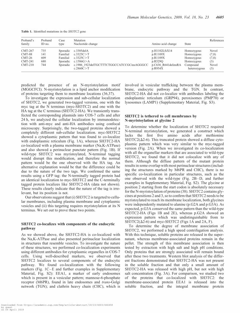
Fourteen probands were screened for mutations in the SH3TC2gene (see Materials and Methods). We found mutations in fiveof them, and two of these mutations were novel (Table 1 andSupplementary Material, Fig. S1). The first one, c.3305delA(proband ID no. 735), was homozygous. It causes a frameshiftand a premature stop 14 codons downstream. The second onewas found in proband ID no. 744, c.1906_1923delTGCTTTCTGGCCATCCGCinsAGGGCC, and was a compound het-erozygous with the c.2860C.T change. This latter change isvery interesting because it has been described to be afounder mutation in the French-Canadian population (5). Wesearched for both novel mutations in 200 chromosomes fromhealthy controls of Spanish ancestry and found neither,suggesting that they could be pathogenic. Among the rest ofthe already described mutations, proband ID no. 680, withan intermediate phenotype, carried the c.1586G.A change,which results in a missense p.R529Q mutation. In our series,we found two probands with the p.R1109X mutation, whichis a founder mutation in Gypsies (7,8). These probands wereunaware of any Gypsy ancestry, but after finding this mutationwe took a more detailed history of the patients, which didreveal a Gypsy background in one patient. The other patient’sorigin could not be confirmed. Both probands belong to con-sanguineous families and we confirmed co-segregation of theDNA changes with phenotype in these two families.
Including our two novel mutations, the list of changes inSH3TC2 has grown to more than 20, including nonsense, mis-sense and indels mutations. Little is known about the cellularand molecular role of the SH3TC2 protein, so we decided toembark in a functional study of this protein in a cell culturesystem.
Cloning and cell expression of the SH3TC2 cDNA
The longest transcript described for SH3TC2 codes for a 1288amino acids protein (3). We obtained a cDNA with the samecoding capacity by combining three partial cDNAs. The elec-tronic annotation of the protein (SwissProt Q8TF17) revealsthe presence of two SH3 and eight TPR domains. These twotypes of domain are involved in protein–protein interactions.In addition, we conducted an in silico analysis of theSH3TC2 sequence using the PSORT II software, which
4604 Human Molecular Genetics, 2009, Vol. 18, No. 23
Downloaded from https://academic.oup.com/hmg/article-abstract/18/23/4603/666458by gueston 04 April 2018
predicted the presence of an N-myristoylation motif(MGGCFCI). N-myristoylation is a lipid anchor modificationof proteins targeting them to membrane locations (36,37).
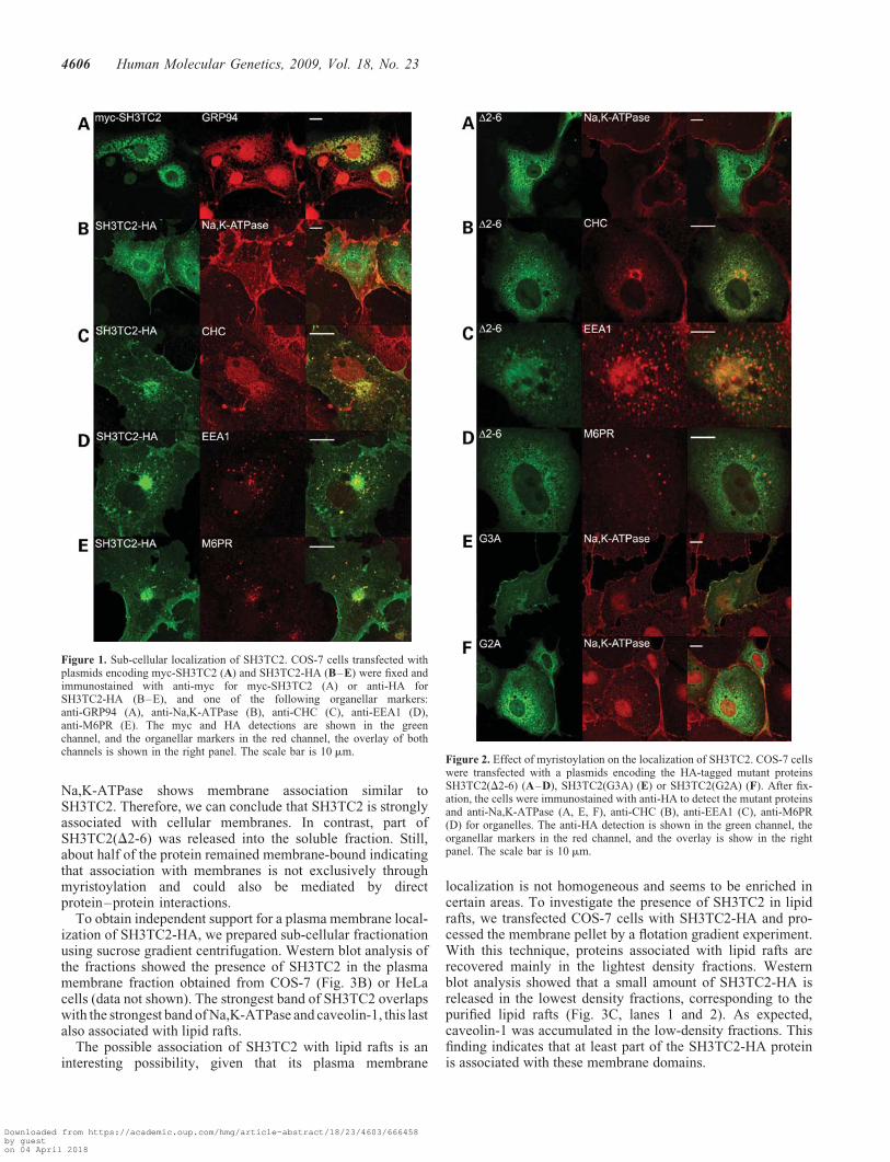
To investigate the expression and sub-cellular localizationof SH3TC2, we generated two-tagged versions, one with themyc tag at the N terminus (myc-SH3TC2) and one with theHA tag at the C terminus (SH3TC2-HA). We transiently trans-fected the corresponding plasmids into COS-7 cells and after24 h, we analyzed the cellular localization by immunodetec-tion with anti-myc and anti-HA antibodies using confocalmicroscopy. Surprisingly, the two-tagged proteins showed acompletely different sub-cellular localization. myc-SH3TC2showed a cytoplasmic pattern that was found to co-localizewith endoplasmic reticulum (Fig. 1A), whereas SH3TC2-HAco-localized with a plasma membrane marker (Na,K-ATPase)and also showed a perinuclear punctate pattern (Fig. 1B). Ifwild-type SH3TC2 was myristoylated, N-terminal taggingwould disrupt this modification, and therefore the normalpattern would be the one observed with the HA tag. Analternative explanation would be that the difference could bedue to the nature of the two tags. We confirmed the sameresults using a GFP tag: the N-terminally tagged protein hadan identical localization to myc-SH3TC2 and the C-terminallytagged protein localizes like SH3TC2-HA (data not shown).These results clearly indicate that the nature of the tag is irre-levant, but its position is not.
Our observations suggest that (i) SH3TC2 is targeted to cellu-lar membranes, including plasma membrane and cytoplasmicvesicles and (ii) this targeting requires myristoylation at its Nterminus. We set out to prove these two points.
SH3TC2 co-localizes with components of the endocyticpathway
As we showed above, the SH3TC2-HA is co-localized withthe Na,K-ATPase and also presented perinuclear localizationin structures that resemble vesicles. To investigate the natureof these structures, we performed co-localization experimentsusing different antibodies for cytoplasmic organelles in COS-7cells. Using well-described markers, we observed thatSH3TC2 localizes to several components of the endocyticpathway. We found co-localization with the followingmarkers (Fig. 1C–E and further examples in SupplementaryMaterial, Fig. S2): EEA1, a marker of early endosomeswhich is present in a vesicular pattern; mannose-6-phosphatereceptor (M6PR), found in late endosomes and trans-Golginetwork (TGN); and clathrin heavy chain (CHC), which is
involved in vesicular trafficking between the plasma mem-brane, endocytic pathway and the TGN. In contrast,SH3TC2-HA did not co-localize with antibodies labeling theendoplasmic reticulum (GRP94), peroxisomes (PMP70) orlysosomes (LAMP1) (Supplementary Material, Fig. S3).
SH3TC2 is tethered to cell membranes byN-myristoylation at glycine 2
To determine whether the localization of SH3TC2 requiredN-terminal myristoylation, we generated a construct whichlacks the first five amino acids after methionineSH3TC2(D2-6). This truncated protein showed a diffuse cyto-plasmic pattern which was very similar to the myc-taggedversion (Fig. 2A). When we investigated its co-localizationwith all the organellar markers that are associated to wild-typeSH3TC2, we found that it did not colocalize with any ofthem. Although the diffuse pattern of the mutant proteinresults in some overlap with most perinuclear structures (includ-ing the structures marked by M6PR and CHC), there is nospecific co-localization in particular structures, such as theone observed with the wild-type (Fig. 2B–D, and furtherexamples in Supplementary Material, Fig. S2). The glycine atposition 2 starting from the start codon is absolutely necessaryfor the N-myristoylation of proteins (38). SH3TC2 contains gly-cines at positions 2 and 3, so to confirm that SH3TC2 needs to bemyristoylated to reach its membrane localization, both glycineswere independently mutated to alanine (p.G2A and p.G3A). Asexpected, p.G3A conserved the same pattern than the wild-typeSH3TC2-HA (Figs 1B and 2E), whereas p.G2A showed anexpression pattern which was undistinguishable from toSH3TC2(D2-6) and myc-SH3TC2 (Figs 1A and 2A, F).
To determine the degree of membrane association ofSH3TC2, we performed a high speed centrifugation analysis.With this technique, soluble proteins are released in the super-natant, whereas membrane-associated proteins remain in thepellet. The strength of this membrane association is thentested by extraction with high salt and high pH conditions.Only proteins that are strongly associated will remain boundafter these two treatments. Western blot analysis of the differ-ent fractions demonstrated that SH3TC2-HA was not presentin the soluble fraction and that only a small amount ofSH3TC2-HA was released with high pH, but not with highsalt concentration (Fig. 3A). For comparison, we studied twoof the proteins that co-localized with SH3TC2: themembrane-associated protein EEA1 is released into thesoluble fraction, and the integral membrane protein
Table 1. Identified mutations in the SH3TC2 gene
Proband’sfamily
ProbandID no.
Casetype
Mutation ReferencesNucleotide change Amino acid change State
CMT-267 735 Sporadic c.3305delA p.H1102LfsX14 Homozygous NovelCMT-88 141 Familial c.3325C.T p.R1109X Homozygous (7,8)CMT-26 609 Familial c.3325C.T p.R1109X Homozygous (7,8)CMT-241 680 Sporadic c.1586G.A p.R529Q Homozygous (3)CMT-210 744 Sporadic c.1906_1923delTGCTTTCTGGCCATCCGCinsAGGGCC p.C636_R641delinsRA Compound
heterozygousNovel
c.2860C.T p.R954X (3)
Human Molecular Genetics, 2009, Vol. 18, No. 23 4605
Downloaded from https://academic.oup.com/hmg/article-abstract/18/23/4603/666458by gueston 04 April 2018
Na,K-ATPase shows membrane association similar toSH3TC2. Therefore, we can conclude that SH3TC2 is stronglyassociated with cellular membranes. In contrast, part ofSH3TC2(D2-6) was released into the soluble fraction. Still,about half of the protein remained membrane-bound indicatingthat association with membranes is not exclusively throughmyristoylation and could also be mediated by directprotein–protein interactions.
To obtain independent support for a plasma membrane local-ization of SH3TC2-HA, we prepared sub-cellular fractionationusing sucrose gradient centrifugation. Western blot analysis ofthe fractions showed the presence of SH3TC2 in the plasmamembrane fraction obtained from COS-7 (Fig. 3B) or HeLacells (data not shown). The strongest band of SH3TC2 overlapswith the strongest band of Na,K-ATPase and caveolin-1, this lastalso associated with lipid rafts.
The possible association of SH3TC2 with lipid rafts is aninteresting possibility, given that its plasma membrane
localization is not homogeneous and seems to be enriched incertain areas. To investigate the presence of SH3TC2 in lipidrafts, we transfected COS-7 cells with SH3TC2-HA and pro-cessed the membrane pellet by a flotation gradient experiment.With this technique, proteins associated with lipid rafts arerecovered mainly in the lightest density fractions. Westernblot analysis showed that a small amount of SH3TC2-HA isreleased in the lowest density fractions, corresponding to thepurified lipid rafts (Fig. 3C, lanes 1 and 2). As expected,caveolin-1 was accumulated in the low-density fractions. Thisfinding indicates that at least part of the SH3TC2-HA proteinis associated with these membrane domains.
Figure 2. Effect of myristoylation on the localization of SH3TC2. COS-7 cellswere transfected with a plasmids encoding the HA-tagged mutant proteinsSH3TC2(D2-6) (A–D), SH3TC2(G3A) (E) or SH3TC2(G2A) (F). After fix-ation, the cells were immunostained with anti-HA to detect the mutant proteinsand anti-Na,K-ATPase (A, E, F), anti-CHC (B), anti-EEA1 (C), anti-M6PR(D) for organelles. The anti-HA detection is shown in the green channel, theorganellar markers in the red channel, and the overlay is show in the rightpanel. The scale bar is 10 mm.
Figure 1. Sub-cellular localization of SH3TC2. COS-7 cells transfected withplasmids encoding myc-SH3TC2 (A) and SH3TC2-HA (B–E) were fixed andimmunostained with anti-myc for myc-SH3TC2 (A) or anti-HA forSH3TC2-HA (B–E), and one of the following organellar markers:anti-GRP94 (A), anti-Na,K-ATPase (B), anti-CHC (C), anti-EEA1 (D),anti-M6PR (E). The myc and HA detections are shown in the greenchannel, and the organellar markers in the red channel, the overlay of bothchannels is shown in the right panel. The scale bar is 10 mm.
4606 Human Molecular Genetics, 2009, Vol. 18, No. 23
Downloaded from https://academic.oup.com/hmg/article-abstract/18/23/4603/666458by gueston 04 April 2018

The SH3 and TPR domains are also involved inanchoring SH3TC2 to the cell membranes
The previous results demonstrated that SH3TC2 is stronglyassociated with membranes and that SH3TC2 needs to be myr-istoylated to efficiently localize to cellular membranes, butthey also disclosed association with membranes in theabsence of myristoylation. To understand other requirementsfor the anchoring of SH3TC2 protein, we generated a seriesof constructs containing engineered deletions (Fig. 4A). Atruncated protein lacking all SH3 and TPR domains, conser-ving only the first 180 amino acids (DSH3DTPRs) did notco-localize with plasma membrane (Fig. 4B) nor with trans-Golgi vesicles (Fig. 4C), but presented a cytoplasmic localiz-ation with a punctate pattern that co-localized weakly withboth early and late endosomes (Supplementary Material,Fig. S4A and B). Deletion of either the two SH3 domains(DSH3) or the C-terminal TPR domains (DTPR) did notaffect normal localization (Fig. 4D–G; SupplementaryMaterial, Fig. S4C–H). Occasionally, SH3TC2(DTPR) pre-sented the same punctate pattern as SH3TC2(DSH3DTPR).This result suggests that either SH3 or TPR domains in com-bination with myristoylation are sufficient to anchor SH3TC2protein to the plasma membrane and all the vesicular struc-tures where the wild-type protein is found. Both types ofdomain probably achieve this by mediating interactions withother proteins.
Plasma membrane localization is altered by SH3TC2missense mutations
We have shown that SH3TC2 is associated with componentsof the endocytic pathway through myristoylation and proteininteractions mediated by the SH3 and TPR domains. We
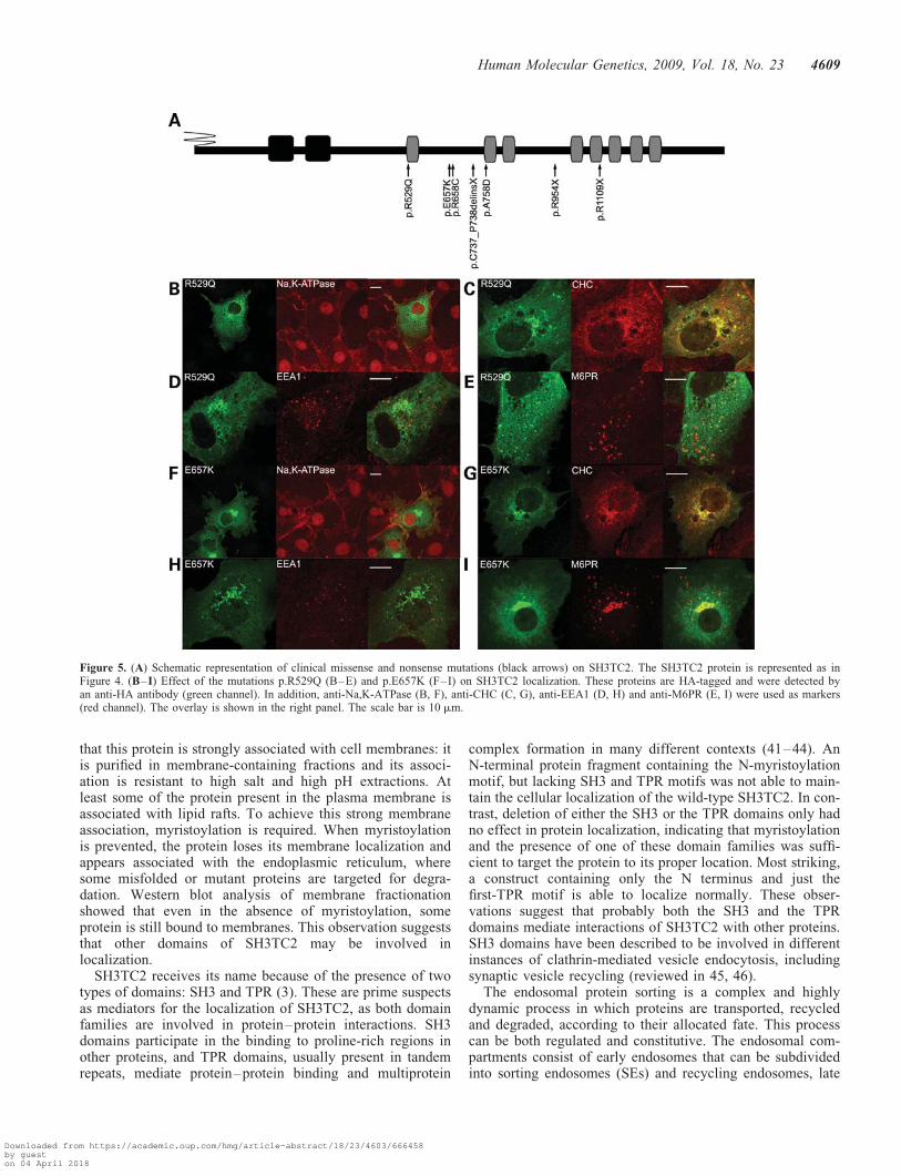
wondered if these findings may help to explain the pathogen-icity of known SH3TC2 mutations. To this end, we tested theeffect of disease-associated SH3TC2 mutations on proteinlocalization in COS-7 cells. For this, we decided to use the fol-lowing mutations (Fig. 5A): (i) mutations that generate stopcodons and truncated proteins: p.C737_P738delinsX andp.R1109X, the two mutations found in the Gypsy population,and p.R954X observed as a founder mutation in French-Canadians and present in other Caucasian populations; (ii)missense mutations: p.R529Q causative of intermediate phe-notype, p.E657K and p.R658C two neighboring mutationsexpressed as a typical phenotype and p.A758D the uniquemutation identified in a patient with evident demyelinatingCMT (39).
All three premature termination codon mutations and themissense mutation p.A758D showed the same pattern asthe wild-type SH3TC2-HA (data not shown). However, theremaining three missense mutations displayed some differ-ences with the localization of the canonical SH3TC2.p. R529Q had a variable pattern. In some cells, its distributionwas similar to the wild-type, but a high proportion of cellsshowed abnormal localization. Most cells showed strongreduction or absence from the plasma membrane, in fact wedid not find co-localization with Na,K-ATPase (Fig. 5B).Co-localization with CHC (Fig. 5C) showed a variable cellularlocalization in the trans-Golgi, but more striking completeabsence from cytoplasmic endosomes (Fig. 5D and E). Asimilar pattern, although slightly less severe, was observedfor p.E657K (Fig. 5F–I) and p.R658C (SupplementaryMaterial, Fig. S5) mutations. The similarity in the localizationpattern of these three mutant proteins seems to indicate thatthe region comprising the first-TPR domain is important forthe localization of SH3TC2. To investigate this, we generateda construct that included only the region of the first-TPR
Figure 3. SH3TC2 is strongly associated with cellular membranes. For each experiment, COS-7 cells were transfected with plasmid encoding HA-tagged ver-sions of SH3TC2 (A–C) and SH3TC2(D2-6) (A). (A) Cell lysates were centrifuged at 100 000g to produce soluble fraction (S100). The membrane-associatedpellet was sequentially extracted with high salt buffer (S100/NaCl), high pH buffer (S100/Na2CO3), and the final pellet was solubilized with RIPA buffer. Equalextractions were separated by SDS–PAGE and analyzed by western blotting with anti-HA, anti-EEA1 or anti-Na,K-ATPase. (B) To study membrane associationof SH3TC2-HA crude membrane extracts were fractionated in a sucrose gradient and analyzed by western blotting with anti-HA for the detection ofSH3TC2-HA, anti-Na,K-ATPase and anti-Caveolin-1. Above each fraction, the percentage of sucrose is indicated. (C) Flotation gradient fractions collectedfrom lowest (lane 1) to highest density (lane 8). SH3TC2-HA was detected with anti-HA, and Caveolin-1 was detected with anti-Caveolin-1.
Human Molecular Genetics, 2009, Vol. 18, No. 23 4607
Downloaded from https://academic.oup.com/hmg/article-abstract/18/23/4603/666458by gueston 04 April 2018
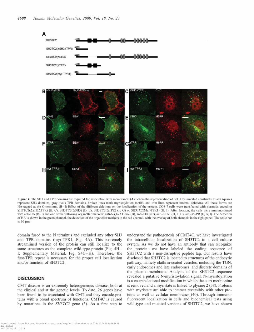
domain fused to the N terminus and excluded any other SH3and TPR domains (myr-TPR1, Fig. 4A). This extremelystreamlined version of the protein can still localize to thesame structures as the complete wild-type protein (Fig. 4H–I; Supplementary Material, Fig. S4G–H). Therefore, thefirst-TPR repeat is necessary for the proper cell localizationand/or function of SH3TC2.
DISCUSSION
CMT disease is an extremely heterogeneous disease, both atthe clinical and at the genetic levels. To date, 26 genes havebeen found to be associated with CMT and they encode pro-teins with a broad spectrum of functions. CMT4C is causedby mutations in the SH3TC2 gene (3). As a first step to
understand the pathogenesis of CMT4C, we have investigatedthe intracellular localization of SH3TC2 in a cell culturesystem. As we do not have an antibody that can recognizethe protein, we have labeled the coding sequence ofSH3TC2 with a non-disruptive peptide tag. Our results havedisclosed that SH3TC2 is located to structures of the endocyticpathway, namely clathrin-coated vesicles, including the TGN,early endosomes and late endosomes, and discrete domains ofthe plasma membrane. Analysis of the SH3TC2 sequencerevealed a putative N-myristoylation signal. N-myristoylationis a co-translational modification in which the start methionineis removed and a myristate is linked to glycine 2 (38). Proteinswith myristate are able to interact reversibly with other pro-teins as well as cellular membranes (40). Through immuno-fluorescent localization in cells and biochemical tests usingwild-type and mutated versions of SH3TC2, we have shown
Figure 4. The SH3 and TPR domains are required for association with membranes. (A) Schematic representation of SH3TC2 mutated constructs. Black squaresrepresent SH3 domains, gray ovals TPR domains, broken lines mark myristoylation motifs, and thin lines represent internal deletions. All these forms areHA-tagged at the C-terminus. (B–I) Effect of the different deletions on the localization of the protein. COS-7 cells were transfected with plasmids encodingSH3TC2(DSH3DTPR) (B, C), SH3TC2(DSH3) (D, E), SH3TC2(DTPR) (F, G) or SH3TC2(Myr-TPR1) (H, I). After fixation, the cells were immunostainedwith anti-HA (B–I) and one of the following organellar markers: anti-Na,K-ATPase (B), anti-CHC (C), anti-EEA1 (D, F, H), anti-M6PR (E, G, I). The detectionof HA is shown in the green channel, the detection of the organellar markers in the red channel, with the overlay of both channels in the right panel. The scale baris 10 mm.
4608 Human Molecular Genetics, 2009, Vol. 18, No. 23
Downloaded from https://academic.oup.com/hmg/article-abstract/18/23/4603/666458by gueston 04 April 2018
that this protein is strongly associated with cell membranes: itis purified in membrane-containing fractions and its associ-ation is resistant to high salt and high pH extractions. Atleast some of the protein present in the plasma membrane isassociated with lipid rafts. To achieve this strong membraneassociation, myristoylation is required. When myristoylationis prevented, the protein loses its membrane localization andappears associated with the endoplasmic reticulum, wheresome misfolded or mutant proteins are targeted for degra-dation. Western blot analysis of membrane fractionationshowed that even in the absence of myristoylation, someprotein is still bound to membranes. This observation suggeststhat other domains of SH3TC2 may be involved inlocalization.
SH3TC2 receives its name because of the presence of twotypes of domains: SH3 and TPR (3). These are prime suspectsas mediators for the localization of SH3TC2, as both domainfamilies are involved in protein–protein interactions. SH3domains participate in the binding to proline-rich regions inother proteins, and TPR domains, usually present in tandemrepeats, mediate protein–protein binding and multiprotein
complex formation in many different contexts (41–44). AnN-terminal protein fragment containing the N-myristoylationmotif, but lacking SH3 and TPR motifs was not able to main-tain the cellular localization of the wild-type SH3TC2. In con-trast, deletion of either the SH3 or the TPR domains only hadno effect in protein localization, indicating that myristoylationand the presence of one of these domain families was suffi-cient to target the protein to its proper location. Most striking,a construct containing only the N terminus and just thefirst-TPR motif is able to localize normally. These obser-vations suggest that probably both the SH3 and the TPRdomains mediate interactions of SH3TC2 with other proteins.SH3 domains have been described to be involved in differentinstances of clathrin-mediated vesicle endocytosis, includingsynaptic vesicle recycling (reviewed in 45, 46).
The endosomal protein sorting is a complex and highlydynamic process in which proteins are transported, recycledand degraded, according to their allocated fate. This processcan be both regulated and constitutive. The endosomal com-partments consist of early endosomes that can be subdividedinto sorting endosomes (SEs) and recycling endosomes, late
Figure 5. (A) Schematic representation of clinical missense and nonsense mutations (black arrows) on SH3TC2. The SH3TC2 protein is represented as inFigure 4. (B–I) Effect of the mutations p.R529Q (B–E) and p.E657K (F–I) on SH3TC2 localization. These proteins are HA-tagged and were detected byan anti-HA antibody (green channel). In addition, anti-Na,K-ATPase (B, F), anti-CHC (C, G), anti-EEA1 (D, H) and anti-M6PR (E, I) were used as markers(red channel). The overlay is shown in the right panel. The scale bar is 10 mm.
Human Molecular Genetics, 2009, Vol. 18, No. 23 4609
Downloaded from https://academic.oup.com/hmg/article-abstract/18/23/4603/666458by gueston 04 April 2018

endosomes and lysosomes (47). The SEs can have three differ-ent destinations: the plasma membrane, the TGN and the lyso-somes (47). In addition, there is a bi-directional traffic betweenthe endosomes and TGN (47–49). There is a clear linkbetween known CMT proteins and the process of endocytosis,and sorting from the trans-Golgi to the endocytic compart-ments (50). These include membrane trafficking regulatorssuch as the MTMR2 and MTMR13 myotubularins (51), theGTPases DNM2 (28) and Rab7 (52) and the ubiquitin ligaseLITAF (24). Some other important CMT proteins are amongthe cargo to be carried by the endocytic and sorting system,mainly the myelin components MPZ and PMP22. In themost prevalent form of CMT, CMT1A, caused by thePMP22 duplication, it has been observed that at least part ofthe pathology is due to alterations in intracellular traffickingand degradation of membrane proteins caused by the overex-pression of PMP22 protein (53). We have established thatSH3TC2 is located in some of the components of the endocy-tic pathway, in a fashion that is dependent on myristoylationand protein interactions mediated by SH3 and TPR domains.At this stage, we cannot determine whether it participates inthe trafficking machinery or whether it is dependent on thismachinery to reach its destination.
Our main goal is to understand the pathogenesis of CMT4C.To this end, we decided to study some disease-associatedmutations. Three of the missense mutations, p.R529Q,p.E657K and p.R658C, are clustered around the first-TPRdomain, whereas a fourth one, p.A758D, is downstream ofthese. Interestingly, the first three mutations have an effecton protein localization. This effect is variable, but in themost extreme cases implies an important reduction inplasma membrane localization and a complete absence fromcytoplasmic vesicles of all types where SH3TC2 is normallypresent. As for the TGN domain, we observed a variable peri-nuclear location for p.R529Q, and a strong retention of peri-nuclear location in the cases of p.E657K and p.R658C,which co-localized with clathrin-coated pits of the TGN.These observations suggest that the region of the first-TPRdomain is essential for the localization of the protein. Thisconclusion is reinforced by our observation that a constructwith the N-terminal portion and the first-TPR domain canlocalize correctly. Still, we cannot discount that the effectsof these mutations go beyond protein localization and mayalter function as well. In comparison with the other three mis-sense mutations, it is striking that the p.A758D mutation doesnot alter the cellular localization of SH3TC2. This change wasidentified in a sporadic case with a demyelinating CMT formand a second disease-causative mutation has not been found inthe SH3TC2 gene (39). Mutations in other CMT genes(PMP22, MPZ, GJB1, GDAP1, SIMPLE and EGR2) werealso excluded. The SH3TC2 p.A758D mutation was screenedin 101 healthy individuals and was not detected providingadditional support that this change could be pathological.However, in the light of our results, it is possible that it is apolymorphism, and mutations in an as yet unidentified CMTgene are responsible for the disease.
In contrast, none of the three nonsense mutations(p.C737_P738delinsX, p.R954X and p.R1109X) showed anyalteration in localization when compared with the wild-type.This result is not surprising, since our engineered construct
lacking the C-terminal half of the protein was localized cor-rectly. We assayed these proteins through directed mutagen-esis of cDNAs, but in their native context, the mutanttranscripts are probably degraded by the nonsense-mediatedmRNA decay pathway. Therefore, the issue of whether theycan be correctly localized or not is probably irrelevant forthe pathogenesis of CMT4C.
The dynamic communication between Schwann cells andaxons is fundamental for the correct function of the peripheralnerve. Nerve biopsies from CMT4C patients show demyeli-nated fibers that are usually surrounded by basal laminaonion bulbs (6). As we have discussed above, several CMTproteins are involved in endocytosis and trafficking of mem-brane components from the TGN to the endocytic compart-ments. These proteins are relevant for the maintenance andformation of the myelin (50). In addition, endocytosis isstrongly implicated in the regulation of signal transduction(54). We propose that SH3TC2 is a relevant protein of theSchwann cell physiology and molecular defects of thisprotein could alter its proper function in the endocyticpathway causing demyelinating neuropathy.
MATERIAL AND METHODS
Patients
Our clinical series comprise 177 Caucasian non-Gypsypatients who suffer from CMT, supervised at the NeurologyService of the University Hospital La Fe. Diagnostic criteriafor demyelinating CMT were median NCV under 38 m/sand nerve fiber demyelination with proliferation of Schwanncells forming ‘onion bulbs’. Patients with NCV between 30and 40 m/s and nerve pathology with both axonal and demye-linating features or undefined were classified as intermediateCMT. Eighty-eight probands were diagnosed with a demyeli-nating neuropathy and two patients with an intermediate form.A previous mutational screening of the CMT genes PMP22(CMT1A duplication and point mutations), MPZ, GJB1,GDAP1, SIMPLE, NEFL, EGR2, PRX and FIG4, allowed usto identify the causative mutations in 77 patients with demye-linating features. The remaining 11 probands and the twopatients with an intermediate form were included in a searchfor mutations in the SH3TC2 gene. In addition, we includedin this study a patient (ID no. 744) for whom a clinical diag-nosis of CMT4C was suspected whose sample was sent to usto perform a genetic test. The series was distributed as follows:ten simplex and four multiplex families. All patients and rela-tives were aware of the investigative nature of the studies andgave their consent.
Mutation screening of the SH3TC2 gene
The 17 exons of the SH3TC2 gene and their flanking intronicsequences were amplified using primers as described else-where (3). Mutation screening was performed by DenaturingHigh Performance Liquid Chromatography (DHPLC; Trans-genomic WAVE, Inc., San Jose, CA, USA) and those PCRproducts showing anomalous DHPLC patterns were sequencedin an ABI Prism 3130�l autoanalyser (Applied Biosystems,
4610 Human Molecular Genetics, 2009, Vol. 18, No. 23
Downloaded from https://academic.oup.com/hmg/article-abstract/18/23/4603/666458by gueston 04 April 2018

Foster City, CA, USA). Sequence alignment and analysis werecarried out with the BLAST program (55).
Cloning of SH3TC2
The full-length SH3TC2 transcript contains an ORF of 1288codons (3) and the corresponding protein contains two SH3domains and eight TPR domains (SwissProt Q8TF17). Toobtain a cDNA with the same coding region, we proceededas follows. We amplified a 3344 bp fragment by PCR on ahuman brain cDNA library (cat.10410-010 Invitrogen,Carlsbad, CA, USA), which comprises from position 565from the start codon, within exon 6, to the 30-UTR region(forward primer 50-ccaccagccgagaaggaagg-30, reverse primer50-agtctggccatgccaaatgtcc-30). This fragment was cloned intothe pGEM-T vector (Promega, Madison, WI, USA) resultingin plasmid pGEM-T::3344. The N-terminal region from thestart codon to position 564 was obtained from two imageclones (GeneService Ltd, UK). The first IMAGE clone (ID:6048901) contained from the 50-UTR to exon 5. The second(ID: 6055718) contained from the exon 1 (at position 43from ATG codon) to the exon 8 region. A BpiI–BamHIrestriction fragment was eliminated from the second cloneand substituted by with a BpiI–BamHI from the first clone,thus obtaining an uninterrupted ORF from the start codonthrough to exon 8. To generate the full length ORF, aSacII–BsmI fragment from this hybrid sequence containingthe N-terminal portion was cloned into pGEM-T::3344digested with the same enzymes (pGEM-T::SH3TC2).
We performed an in silico analysis of the human SH3TC2protein Q8TF17 using the protein knowledgebase UniProtKB(http://www.uniprot.org) and the PSORT II software (http://psort.ims.u-tokyo.ac.jp).
Plasmid construction and site-directed mutagenesis
For the synthesis of the tagged versions of SH3TC2 (SH3TC2-GFP, GFP-SH3TC2, SH3TC2-HA and myc-SH3TC2), thecoding sequence of SH3TC2 was first amplified by PCRfrom pGEM-T::SH3TC2. For each construct, a pair ofprimers was designed, containing adapters with restrictionsites that allowed in-frame cloning in the relevant vector.These vectors were pEGFP-C1, pEGFP-N1 and pCMV-mycfrom Clontech (Mountain View, CA, USA), andpcDNA3-HA, a vector derived from the Invitrogen pcDNA3kindly donated by D. Barettino. The sequences for theprimers used for the synthesis of each one of these constructsare given in Supplementary Material, Table S1.
The SH3TC2-HA constructs carrying the mutations D2-6,DSH3DTPRs and DTPR were made by inserting the sequenceof interest, amplified from the pGEM-T::SH3TC2 plasmid byPCR, into the pcDNA3-HA vector, resulting in the corre-sponding defective ORFs fused to a C-terminal HA tag. Theprimer sequences are given in Supplementary Material,Table S1.
The DSH3 mutation was generated in two segments,N-terminal and C-terminal to the SH3 domains, respectively.Each segment was obtained by PCR from pGEM-T::SH3TC2(primer sequences in Supplementary Material, Table S1).Then, a KpnI–EcoRI fragment from the N-terminal amplicon
and an EcoRI–EcoRV fragment from the second one weresequentially cloned into the pcDNA3-HA vector backbone.Any mutations introduced as a consequence of including therestriction site were removed by site-directed mutagenesis asdescribed below. The resulting plasmid was pcDNA3-HA::DSH3. The myr-TPR1 segment was obtained by amplifi-cation from pcDNA3-HA::DSH3 plasmid by PCR into thesame vector pcDNA3-HA.
The SH3TC2 variants p.G2A and p.G3A and thedisease-associated mutations (p.R529Q, p.E657K, p.R658C,p. C737_P738delinsX, p.A758D, p.R954X and p.R1109X)were generated by site-directed mutagenesis with specificprimers containing the nucleotide changes and following theinstruction manual from QuickChangeTM Site-DirectedMutagenesis kit (Stratagene). The sequences of all the con-structs were confirmed by automated DNA sequencing in anABI Prism 3130XL autoanalyser (Applied Biosystems).
Cell culture, immunocytochemistry and microscopy
COS-7 or HeLa cells were grown in a humidified incubatorwith 5% CO2 at 378C in DMEM containing 10% (v/v) fetalbovine serum (FBS) supplemented with 2 mM glutamine,100 IU/ml penicillin and 100 mg/ml streptomycin (Invitrogen).To carry out the protein expression and co-localization analy-sis, 100 000 COS-7 or 200 000 HeLa cells were cultured insix-well plates on glass coverslips. Next day, when theyreached 70–80% confluency, we transfected them with theCAPHOS kit (Sigma-Aldrich, St Louis, MO, USA) using5 mg of plasmidic DNA. We used lipofectamine (Roche) forHeLa cells, using a ration of 5 ml lipofectamine for each2 mg of plasmid DNA. Western blot analysis was performedafter transfection of each construct to test the integrity of theexpressed protein and to ensure that the different constructswere expressed at similar levels.
For co-localization studies, cells were fixed and processedfor immunofluorescence microscopy 24 h post-transfection.Briefly, cells were fixed in 4% (w/v) paraformaldehyde inphosphate-buffered saline (PBS) for 15 min or in methanolfor 20 min at 2208C, permeabilized in 0.5% (w/v)Triton X-100 in PBS for 30 min, blocked with 10% (v/v)FBS/0.1% (w/v) Triton X-100/0.5% (w/v) BSA in PBS for1 h and then probed with primary antibodies diluted in block-ing solution. The following primary antibodies were used:mouse monoclonal anti-Na,K-ATPase for plasma membrane(Abcam, Cambridge, UK; used at 1:100); rabbit polyclonalanti-GRP94 (Abcam; used at 1:100) for endoplasmic reticu-lum; mouse monoclonal anti-CHC for clathrin-coated vesicles(BD Biosciences, San Jose, CA, USA, used at 1:50); mousemonoclonal anti-EEA1 for early endosomes (BD Biosciences;used at 1:100); mouse monoclonal anti-M6PR for late endo-somes (Abcam; used at 1:250); polyclonal rabbitanti-PMP70 for peroxisomes (Invitrogen; used at 1:100),anti-LAMP1 clone H4A3 for lysosomes (developed byJ. Thomas August and James E.K. Hildreth, obtained fromthe Developmental Studies Hybridoma Bank, University ofIowa; used at 1:30). The secondary antibodies used weregoat anti-mouse or goat anti-rabbit immunoglobulinscoupled to Alexa Fluor 488 or Alexa Fluor 633 (Invitrogen;used at 1:250). The samples were mounted in Fluoromount-G
Human Molecular Genetics, 2009, Vol. 18, No. 23 4611
Downloaded from https://academic.oup.com/hmg/article-abstract/18/23/4603/666458by gueston 04 April 2018
(Southern Biotech, Birmingham, AL, USA). Cells were exam-ined using a Leica DM RXA2 microscope and Leica TCS SPConfocal System. Images were manipulated and analyzed withthe Leica Confocal Software and the ImageJ suite.
Membrane fractionation, lipid raft purification
To perform crude membrane preparation and the subsequentsucrose gradient fractionation from COS-7 or HeLa cells, weused previously described procedures (56) with minor modifi-cations. Details are available upon request.
To analyze the possible interaction of SH3TC2-HA withmembranes, we performed a high-speed centrifugation analy-sis. COS-7 cells transfected with pCDNA3-HA-SH3TC2 werescraped from 100 mm dishes 24 h after transfection andwashed twice with PBS. The cells were then resuspended in1 ml of lysis buffer (10 mM Tris–HCl pH 7.4, 2.5 mM
MgCl2, protease inhibitors) and transferred to ice for 15 min.Cells were lysed with 25–40 strokes of a dounce homogenizerand the lysate was gently layered over a sucrose cushion (0.5 M
sucrose, 10 mM Tris–HCl pH 7.4, 2.5 mM MgCl2). Nuclei anddebris were discarded by centrifugation at 5000g for 10 min at48C. The upper layer consisting of cytoplasmic extract wascollected and was centrifuged at 100 000g for 1 h in aMLA-130 rotor (Beckmann), resulting in a soluble supernatantand pellet containing membranes and membrane-associatedproteins. The pellet was resuspended in 100 ml of high-saltbuffer (10 mM Tris–HCl pH 7.4, 750 mM NaCl, with proteaseinhibitors) and centrifuged for 1 h at 100 000g yielding a high-salt supernatant and pellet. This pellet was then resuspended in100 ml of high pH buffer (10 mM Tris–HCl pH 7.4, 100 mM
Na2CO3, with protease inhibitors) and centrifuged for 1 h at100 000g yielding a high pH supernatant and final pellet.The final pellet was resuspended in 100 ml of RIPA buffer(50 mM Tris–HCl pH 7.4, 5 mM DTT, 150 mM NaCl, 1%NP-40, 0.5% deoxycholate). Equal cell equivalents wereresolved by SDS–PAGE and the relevant proteins weredetected by western blot analysis.
Flotation gradients to purify raft proteins were also per-formed with COS-7 24 h post-transfection. The cell pelletwas lysed in 500 ml of TNE buffer (50 mM Tris–HCl pH7.4, 150 mM NaCl, 5 mM EDTA, with protease inhibitors) by25–40 strokes of a dounce homogenizer. After discardingthe unbroken cells and debris, the clear lysate was incubatedwith Triton X-100 (1% final) for 30 min on ice. After theextraction with Triton X-100, the lysate (400 ml) was adjustedto 40% Optiprep by adding 800 ml of Optiprep solution(Sigma-Aldrich) and overlaid with 2.4 ml of 30% Optiprepin TXNE (TNE, 0.1% Triton X-100) and 400 ml of TXNE.The samples were centrifuged at 164 000 g for 18 h in aSW60Ti rotor (Beckmann). Fractions were collectedfrom the top, precipitated by adding two volumes of 15%trichloracetic.
All samples were analyzed by SDS/PAGE and western blot-ting. To perform western blot analysis, the following anti-bodies were used: rabbit polyclonal anti-HA (Sigma-Aldrich;used at 1:1000), mouse monoclonal anti-Na,K-ATPase(Abcam; used at 1:1000), rabbit polyclonal anti-Caveolin-1(Santa Cruz Biotechnology, Santa Cruz, CA, USA; used at
1:1000) and mouse monoclonal anti-EEA1 for early endo-somes (BD Biosciences; used at 1:1000).
SUPPLEMENTARY MATERIAL
Supplementary Material is available at HMG online.
ACKNOWLEDGEMENTS
We are grateful to patients and their families for their kind col-laboration. We thank B. Alarcon for his technical assistanceand also anonymous reviewers for their invaluable insightand suggestions.
Conflict of Interest statement. None declared.
FUNDING
This work was supported by the Fondo de InvestigacionSanitaria [grant numbers PI08/90857, PI08/0889, CP08/00053] and the Spanish Ministry Science and Innovation[grant number SAF2006-01047]. V.L. is a recipient of JAEpredoctoral fellowship from the Spanish Scientific ResearchCouncil (CSIC). M.I.G. has a ‘Ramon y Cajal’ contractfunded by the Ministry of Science and Innovation. C.E. hasa ‘Miguel Servet’ contract funded by the Fondo de Investiga-cion Sanitaria. Both CIBERER and CIBERNED are initiativesfrom the Instituto de Salud Carlos III.
REFERENCES
1. Skre, H. (1974) Genetic and clinical aspects of Charcot-Marie-Tooth’sdisease. Clin. Genet., 6, 98–118.
2. Combarros, O., Calleja, J., Polo, J.M. and Berciano, J. (1987) Prevalenceof hereditary motor and sensory neuropathy in Cantabria. Acta Neurol.Scand., 75, 9–12.
3. Senderek, J., Bergmann, C., Stendel, C., Kirfel, J., Verpoorten, N., DeJonghe, P., Timmerman, V., Chrast, R., Verheijen, M.H., Lemke, G. et al.(2003) Mutations in a gene encoding a novel SH3/TPR domain proteincause autosomal recessive Charcot-Marie-Tooth type 4C neuropathy.Am. J. Hum. Genet., 73, 1106–1119.
4. Azzedine, H., Ravise, N., Verny, C., Gabreels-Festen, A., Lammens, M.,Grid, D., Vallat, J.M., Durosier, G., Senderek, J., Nouioua, S. et al. (2006)Spine deformities in Charcot-Marie-Tooth 4C caused by SH3TC2 genemutations. Neurology, 67, 602–606.
5. Gosselin, I., Thiffault, I., Tetreault, M., Chau, V., Dicaire, M.J., Loisel, L.,Emond, M., Senderek, J., Mathieu, J., Dupre, N. et al. (2008) FounderSH3TC2 mutations are responsible for a CMT4C French-Canadianscluster. Neuromuscul. Disord., 18, 483–492.
6. Houlden, H., Laura, M., Ginsberg, L., Jungbluth, H., Robb, S.A., Blake, J.,Robinson, S., King, R.H. and Reilly, M.M. (2009) The phenotype ofCharcot-Marie-Tooth disease type 4C due to SH3TC2 mutations andpossible predisposition to an inflammatory neuropathy. Neuromuscul.Disord., 19, 264–269.
7. Claramunt, R., Sevilla, T., Lupo, V., Cuesta, A., Millan, J.M., Vilchez, J.,Palau, F. and Espinos, C. (2007) The p.R1109X mutation in SH3TC2 geneis predominant in Spanish Gypsies with Charcot-Marie-Tooth disease type4. Clin. Genet., 71, 343–349.
8. Gooding, R., Colomer, J., King, R., Angelicheva, D., Marns, L., Parman,Y., Chandler, D., Bertranpetit, J. and Kalaydjieva, L. (2005) A novelGypsy founder mutation, p.Arg1109X in the CMT4C gene, causesvariable peripheral neuropathy phenotypes. J. Med. Genet., 42, e69.
9. Reilly, M.M. (2005) Axonal Charcot-Marie-Tooth disease: the fog isslowly lifting!. Neurology, 65, 186–187.
4612 Human Molecular Genetics, 2009, Vol. 18, No. 23
Downloaded from https://academic.oup.com/hmg/article-abstract/18/23/4603/666458by gueston 04 April 2018

10. Lupski, J.R., de Oca-Luna, R.M., Slaugenhaupt, S., Pentao, L., Guzzetta,V., Trask, B.J., Saucedo-Cardenas, O., Barker, D.F., Killian, J.M., Garcia,C.A. et al. (1991) DNA duplication associated with Charcot-Marie-Toothdisease type 1A. Cell, 66, 219–232.
11. Fortun, J., Go, J.C., Li, J., Amici, S.A., Dunn, W.A. Jr and Notterpek, L.(2006) Alterations in degradative pathways and protein aggregation in aneuropathy model based on PMP22 overexpression. Neurobiol. Dis., 22,153–164.
12. Filbin, M.T., Walsh, F.S., Trapp, B.D., Pizzey, J.A. and Tennekoon, G.I.(1990) Role of myelin P0 protein as a homophilic adhesion molecule.Nature, 344, 871–872.
13. Bergoffen, J., Scherer, S.S., Wang, S., Scott, M.O., Bone, L.J., Paul, D.L.,Chen, K., Lensch, M.W., Chance, P.F. and Fischbeck, K.H. (1993)Connexin mutations in X-linked Charcot-Marie-Tooth disease. Science,262, 2039–2042.
14. Bondurand, N., Girard, M., Pingault, V., Lemort, N., Dubourg, O. andGoossens, M. (2001) Human Connexin 32, a gap junction protein alteredin the X-linked form of Charcot-Marie-Tooth disease, is directly regulatedby the transcription factor SOX10. Hum. Mol. Genet., 10, 2783–2795.
15. Nagarajan, R., Svaren, J., Le, N., Araki, T., Watson, M. and Milbrandt, J.(2001) EGR2 mutations in inherited neuropathies dominant-negativelyinhibit myelin gene expression. Neuron, 30, 355–368.
16. Inoue, K., Khajavi, M., Ohyama, T., Hirabayashi, S., Wilson, J., Reggin,J.D., Mancias, P., Butler, I.J., Wilkinson, M.F., Wegner, M. et al. (2004)Molecular mechanism for distinct neurological phenotypes conveyed byallelic truncating mutations. Nat. Genet., 36, 361–369.
17. Warner, L.E., Mancias, P., Butler, I.J., McDonald, C.M., Keppen, L.,Koob, K.G. and Lupski, J.R. (1998) Mutations in the early growthresponse 2 (EGR2) gene are associated with hereditary myelinopathies.Nat. Genet., 18, 382–384.
18. de Brito, O.M. and Scorrano, L. (2008) Mitofusin 2 tethers endoplasmicreticulum to mitochondria. Nature, 456, 605–610.
19. Pedrola, L., Espert, A., Valdes-Sanchez, T., Sanchez-Piris, M., Sirkowski,E.E., Scherer, S.S., Farinas, I. and Palau, F. (2008) Cell expression ofGDAP1 in the nervous system and pathogenesis of Charcot-Marie-Toothtype 4A disease. J. Cell Mol. Med., 12, 679–689.
20. Pedrola, L., Espert, A., Wu, X., Claramunt, R., Shy, M.E. and Palau, F.(2005) GDAP1, the protein causing Charcot-Marie-Tooth disease type4A, is expressed in neurons and is associated with mitochondria. Hum.
Mol. Genet., 14, 1087–1094.21. Niemann, A., Ruegg, M., La Padula, V., Schenone, A. and Suter, U.
(2005) Ganglioside-induced differentiation associated protein 1 is aregulator of the mitochondrial network: new implications forCharcot-Marie-Tooth disease. J. Cell Biol., 170, 1067–1078.
22. Moriwaki, Y., Begum, N.A., Kobayashi, M., Matsumoto, M., Toyoshima,K. and Seya, T. (2001) Mycobacterium bovis Bacillus Calmette-Guerinand its cell wall complex induce a novel lysosomal membrane protein,SIMPLE, that bridges the missing link between lipopolysaccharide andp53-inducible gene, LITAF(PIG7), and estrogen-inducible gene, EET-1.J. Biol. Chem., 276, 23065–23076.
23. Ludes-Meyers, J.H., Kil, H., Bednarek, A.K., Drake, J., Bedford, M.T. andAldaz, C.M. (2004) WWOX binds the specific proline-rich ligand PPXY:identification of candidate interacting proteins. Oncogene, 23,5049–5055.
24. Saifi, G.M., Szigeti, K., Wiszniewski, W., Shy, M.E., Krajewski, K.,Hausmanowa-Petrusewicz, I., Kochanski, A., Reeser, S., Mancias, P.,Butler, I. et al. (2005) SIMPLE mutations in Charcot-Marie-Tooth diseaseand the potential role of its protein product in protein degradation. Hum.
Mut., 25, 372–383.25. Robinson, F.L. and Dixon, J.E. (2005) The phosphoinositide-3-
phosphatase MTMR2 associates with MTMR13, a membrane-associatedpseudophosphatase also mutated in type 4B Charcot-Marie-Tooth disease.J. Biol. Chem., 280, 31699–31707.
26. Berger, P., Berger, I., Schaffitzel, C., Tersar, K., Volkmer, B. and Suter,U. (2006) Multi-level regulation of myotubularin-related protein-2phosphatase activity by myotubularin-related protein-13/set-bindingfactor-2. Hum. Mol. Genet., 15, 569–579.
27. Verhoeven, K., De Jonghe, P., Coen, K., Verpoorten, N., Auer-Grumbach,M., Kwon, J.M., FitzPatrick, D., Schmedding, E., De Vriendt, E., Jacobs,A. et al. (2003) Mutations in the small GTP-ase late endosomal proteinRAB7 cause Charcot-Marie-Tooth type 2B neuropathy. Am. J. Hum.
Genet., 72, 722–727.
28. Zuchner, S., Noureddine, M., Kennerson, M., Verhoeven, K., Claeys, K.,De Jonghe, P., Merory, J., Oliveira, S.A., Speer, M.C., Stenger, J.E. et al.(2005) Mutations in the pleckstrin homology domain of dynamin 2 causedominant intermediate Charcot-Marie-Tooth disease. Nat. Genet., 37,289–294.
29. Jager, S., Bucci, C., Tanida, I., Ueno, T., Kominami, E., Saftig, P. andEskelinen, E.L. (2004) Role for Rab7 in maturation of late autophagicvacuoles. J. Cell Sci., 117, 4837–4848.
30. Harrison, R.E., Bucci, C., Vieira, O.V., Schroer, T.A. and Grinstein, S.(2003) Phagosomes fuse with late endosomes and/or lysosomes byextension of membrane protrusions along microtubules: role of Rab7 andRILP. Mol. Cell Biol., 23, 6494–6506.
31. Press, B., Feng, Y., Hoflack, B. and Wandinger-Ness, A. (1998) MutantRab7 causes the accumulation of cathepsin D and cation-independentmannose 6-phosphate receptor in an early endocytic compartment. J. CellBiol., 140, 1075–1089.
32. Tanabe, K. and Takei, K. (2009) Dynamic instability of microtubulesrequires dynamin 2 and is impaired in a Charcot-Marie-Tooth mutant.J. Cell Biol., 185, 939–948.
33. Hinshaw, J.E. (2000) Dynamin and its role in membrane fission. Annu.Rev. Cell Dev. Biol., 16, 483–519.
34. Schafer, D.A., Weed, S.A., Binns, D., Karginov, A.V., Parsons, J.T. andCooper, J.A. (2002) Dynamin2 and cortactin regulate actin assembly andfilament organization. Curr. Biol., 12, 1852–1857.
35. Thompson, H.M., Cao, H., Chen, J., Euteneuer, U. and McNiven, M.A.(2004) Dynamin 2 binds gamma-tubulin and participates in centrosomecohesion. Nat. Cell Biol., 6, 335–342.
36. Boutin, J.A. (1997) Myristoylation. Cell Signal., 9, 15–35.37. Maurer-Stroh, S., Eisenhaber, B. and Eisenhaber, F. (2002) N-terminal
N-myristoylation of proteins: refinement of the sequence motif and itstaxon-specific differences. J. Mol. Biol., 317, 523–540.
38. Resh, M.D. (1999) Fatty acylation of proteins: new insights intomembrane targeting of myristoylated and palmitoylated proteins.Biochim. Biophys. Acta, 1451, 1–16.
39. Martinez-Rubio, D., Millan, J.M., Palau, F. and Espinos, C. (2008) Genesymbol: SH3TC2. Disease: Charcot-Marie-Tooth type 4C. Hum. Genet.,124, 320.
40. Peitzsch, R.M. and McLaughlin, S. (1993) Binding of acylated peptidesand fatty acids to phospholipid vesicles: pertinence to myristoylatedproteins. Biochemistry, 32, 10436–10443.
41. Morton, C.J. and Campbell, I.D. (1994) SH3 domains. Molecular‘Velcro’. Curr. Biol., 4, 615–617.
42. Mayer, B.J. (2001) SH3 domains: complexity in moderation. J. Cell Sci.,114, 1253–1263.
43. Pawson, T. (1995) Protein modules and signalling networks. Nature, 373,573–580.
44. Blatch, G.L. and Lassle, M. (1999) The tetratricopeptide repeat: astructural motif mediating protein-protein interactions. Bioessays, 21,932–939.
45. McPherson, P.S. (1999) Regulatory role of SH3 domain-mediatedprotein-protein interactions in synaptic vesicle endocytosis. Cell Signal.,11, 229–238.
46. Kim, Y. and Chang, S. (2006) Ever-expanding network ofdynamin-interacting proteins. Mol. Neurobiol., 34, 129–136.
47. Seaman, M.N. (2008) Endosome protein sorting: motifs and machinery.Cell Mol. Life Sci., 65, 2842–2858.
48. Ghosh, R.N., Mallet, W.G., Soe, T.T., McGraw, T.E. and Maxfield, F.R.(1998) An endocytosed TGN38 chimeric protein is delivered to the TGNafter trafficking through the endocytic recycling compartment in CHOcells. J. Cell Biol., 142, 923–936.
49. Hunt, K.A., McGovern, D.P., Kumar, P.J., Ghosh, S., Travis, S.P.,Walters, J.R., Jewell, D.P., Playford, R.J. and van Heel, D.A. (2005) Acommon CTLA4 haplotype associated with coeliac disease. Eur. J. Hum.Genet., 13, 440–444.
50. Niemann, A., Berger, P. and Suter, U. (2006) Pathomechanisms of mutantproteins in Charcot-Marie-Tooth disease. Neuromolecular Med., 8, 217–242.
51. Wishart, M.J. and Dixon, J.E. (2002) PTEN and myotubularinphosphatases: from 3-phosphoinositide dephosphorylation to disease.Trends Cell Biol., 12, 579–585.
52. Vonderheit, A. and Helenius, A. (2005) Rab7 associates with earlyendosomes to mediate sorting and transport of Semliki forest virus to lateendosomes. PLoS Biol., 3, e233.
Human Molecular Genetics, 2009, Vol. 18, No. 23 4613
Downloaded from https://academic.oup.com/hmg/article-abstract/18/23/4603/666458by gueston 04 April 2018

53. Ryan, M.C., Shooter, E.M. and Notterpek, L. (2002) Aggresomeformation in neuropathy models based on peripheral myelin protein 22mutations. Neurobiol. Dis., 10, 109–118.
54. von Zastrow, M. and Sorkin, A. (2007) Signaling on the endocyticpathway. Curr. Opin. Cell Biol., 19, 436–445.
55. Altschul, S.F., Gish, W., Miller, W., Myers, E.W. and Lipman, D.J. (1990)Basic local alignment search tool. J. Mol. Biol., 215, 403–410.
56. Coppi, M.V. and Guidotti, G. (1997) Intracellular localization ofNa,K-ATPase alpha2 subunit mutants. Arch. Biochem. Biophys., 346,312–321.
4614 Human Molecular Genetics, 2009, Vol. 18, No. 23
Downloaded from https://academic.oup.com/hmg/article-abstract/18/23/4603/666458by gueston 04 April 2018