Mishaps and serious complications in endodontic obturationrpcendo.com/jHfmPiks/PDF11.pdfpermanent...
19
Mishaps and serious complications in endodontic obturation ALAN H. GLUSKIN There are numerous examples reported in the literature that cite and document many disabling complications to the alveolar bone, neurovascular anatomy and maxillary sinus following overextension of root canal filling materials. Neural complications as a consequences of endodontic obturation as well as other server outcomes to overfill are a serious problem. These injuries require a thoughful strategy for prevention during endodontic procedures as well as a responsible systematic approach to management, should the outcome of endodontic therapy produce an injury.This monogrph will focus on measures that can prevent obturation mishaps which occur under the most vulnerable of circumstances during the course of endodontic therapy. This paper will also critically review the toxicity of materials used in endodontic obturation and seek to identify those principal factors which affect prognosis after injury. I have nerve damage to the inferior alveolar nerve that was done during a root canal procedure. To make matters worse, the root canal filling spilled out into the mandible (sic) canal on the nerve that was injured. I have shooting pain in almost all of my teeth which is unbearable. I also have other types of constant aching pain. My teeth are so super-sensitive at times, that even air bothers them. I don’t think I am going to make it. I can’t find the right kind of help. I have seen a pain specialist who has me on special drugs but it hasn’t helped. Is there anyone out there who can help me find a specialist who has expertise dealing with this type of problem? Is there a dental practitioner, reading the above comment posted to an Internet website for patients with facial pain, who would not empathize with the suffering of this patient? Unfortunately, there are numerous examples reported in the literature that cite and document many disabling complications to the alveolar bone, neurovascular bundle and maxillary sinus following inadvertent overextension of root canal-filling materials. Paresthesia as a consequence of endodontic obturation as well as other severe neural complications must be considered as a serious problem. These injuries require a thoughtful strategy for prevention during endodontic procedures as well as a responsible systema- tic approach to management, should the outcome of endodontic therapy produce an overfill injury. This monograph will focus on measures that can prevent and/or minimize accidents, which can occur under the most vulnerable of circumstances during the course of endodontic therapy. The identification of those principal factors that can affect prognosis after injury will also be an important focus of this article. Many of the root-filling materials used today are either chemically neurotoxic or can be mechanically destruc- tive to surrounding structures via compression injury. In addition, overinstrumentation errors in shaping root canal spaces may produce an abnormal overenlarge- ment of the apical constriction allowing overfill or instrument damage to structures through direct manipulation or rotation that severs susceptible tissues. With the use of rotation during instrumentation and heated obturation devices becoming increasingly avail- able to all practitioners in the last decade, the introduction of endodontic filling materials into periapical tissues is quite common. This is of major concern when the teeth being treated are in close proximity to anatomically important structures such as the maxillary sinus or inferior alveolar canal. Over- extension of the root canal-filling material risks injurious consequences if the underlying inferior alveolar nerve or sinus structures are initially penetrated with files. A pathway of entry into the inferior alveolar nerve or sinus space can more likely result when the pathway is created by overinstrumentation (Fig. 1). 52 Endodontic Topics 2005, 12, 52–70 All rights reserved Copyright r Blackwell Munksgaard ENDODONTIC TOPICS 2005 1601-1538
Transcript of Mishaps and serious complications in endodontic obturationrpcendo.com/jHfmPiks/PDF11.pdfpermanent...
Mishaps and serious complicationsin endodontic obturationALAN H. GLUSKIN
There are numerous examples reported in the literature that cite and document many disabling complications to the
alveolar bone, neurovascular anatomy and maxillary sinus following overextension of root canal filling materials.
Neural complications as a consequences of endodontic obturation as well as other server outcomes to overfill are a
serious problem. These injuries require a thoughful strategy for prevention during endodontic procedures as well as a
responsible systematic approach to management, should the outcome of endodontic therapy produce an injury.This
monogrph will focus on measures that can prevent obturation mishaps which occur under the most vulnerable of
circumstances during the course of endodontic therapy. This paper will also critically review the toxicity of materials
used in endodontic obturation and seek to identify those principal factors which affect prognosis after injury.
I have nerve damage to the inferior alveolar nerve that
was done during a root canal procedure. To make matters
worse, the root canal filling spilled out into the mandible
(sic) canal on the nerve that was injured. I have shooting
pain in almost all of my teeth which is unbearable. I also
have other types of constant aching pain. My teeth are so
super-sensitive at times, that even air bothers them. I
don’t think I am going to make it. I can’t find the right
kind of help. I have seen a pain specialist who has me on
special drugs but it hasn’t helped. Is there anyone out
there who can help me find a specialist who has expertise
dealing with this type of problem?
Is there a dental practitioner, reading the above
comment posted to an Internet website for patients
with facial pain, who would not empathize with the
suffering of this patient? Unfortunately, there are
numerous examples reported in the literature that cite
and document many disabling complications to the
alveolar bone, neurovascular bundle and maxillary sinus
following inadvertent overextension of root canal-filling
materials. Paresthesia as a consequence of endodontic
obturation as well as other severe neural complications
must be considered as a serious problem. These injuries
require a thoughtful strategy for prevention during
endodontic procedures as well as a responsible systema-
tic approach to management, should the outcome of
endodontic therapy produce an overfill injury.
This monograph will focus on measures that can
prevent and/or minimize accidents, which can occur
under the most vulnerable of circumstances during the
course of endodontic therapy. The identification of
those principal factors that can affect prognosis after
injury will also be an important focus of this article.
Many of the root-filling materials used today are either
chemically neurotoxic or can be mechanically destruc-
tive to surrounding structures via compression injury.
In addition, overinstrumentation errors in shaping root
canal spaces may produce an abnormal overenlarge-
ment of the apical constriction allowing overfill or
instrument damage to structures through direct
manipulation or rotation that severs susceptible tissues.
With the use of rotation during instrumentation and
heated obturation devices becoming increasingly avail-
able to all practitioners in the last decade, the
introduction of endodontic filling materials into
periapical tissues is quite common. This is of major
concern when the teeth being treated are in close
proximity to anatomically important structures such as
the maxillary sinus or inferior alveolar canal. Over-
extension of the root canal-filling material risks
injurious consequences if the underlying inferior
alveolar nerve or sinus structures are initially penetrated
with files. A pathway of entry into the inferior alveolar
nerve or sinus space can more likely result when the
pathway is created by overinstrumentation (Fig. 1).
52
Endodontic Topics 2005, 12, 52–70All rights reserved
Copyright r Blackwell Munksgaard
ENDODONTIC TOPICS 20051601-1538

The importance of accurateradiographic imaging
You don’t know . . . what you don’t know.
This common admonition by educators to many
students in the medical sciences, as they are learning
the principles and practice of diagnostic assessment, is a
fundamental truth. ‘You don’t know. . . what you don’t
know.’ For the practitioner embarking on therapy for a
molar or bicuspid tooth in the anatomical vicinity of the
maxillary sinus, mental foramen or inferior alveolar
nerve, this caveat is vital. Understanding the proximity
of roots to these important structures must be
recognized and accurate radiographic imaging is the
most direct route to that knowledge. When a radio-
graphic examination is performed or needed, the
practitioner assumes the responsibility to make accurate
interpretations from good discernable images of
diagnostic quality. It is a common understanding that
both large and small lesions as well as anatomical
entities are routinely missed in radiographic surveys
both by the operator and the limitations of the
technology when encountering differences in anatomic
variation (1).
Thus, viewing the radiographs as part of a routine and
standardized diagnostic sequence requires the follow-
ing principles be carefully observed (1, 2).
1. Interpretations should only be made from properly
exposed and processed radiographs (Fig. 2).
2. Use multiple views and imaging modalities (pano-
graphic) as often as necessary to demonstrate
important radiographic features in three dimen-
sions.
3. Use magnification and other optimal viewing
modalities to enhance all images.
4. Obtain special imaging studies if necessary (2).
These viewing principles, whether the images are
rendered on traditional film or digital, are critical to
extracting the maximum information for the purpose
of diagnostic interpretation (2). When special imaging
is required, dentistry is making rapid strides in the areas
of digital radiography and computed tomography
(CT).
Digital Radiography
Digital imaging of dental structures has become a
common diagnostic modality in the last decade in offices
around the world. A charged sensor and associated
computer hardware and software are the new paradigm
in dental imaging, often replacing traditional film in a
number of applications. The sensor can be positioned
once and the cone adjusted for several angulations,
Fig. 1. (A) Panoramic radiograph of completed root canal therapy of the lower left second molar. Radiograph showsproximity to the inferior alveolar canal; (B) isolated image of the second molar shows radiopaque material beyond themesial root and root shapes that have transported the original canal positions; (C) magnification identifies anoverinstrumented root canal providing a pathway for injury by instruments and/or sealer. Patient experiencedpermanent paresthesia in the inferior alveolar distribution on the face.
Fig. 2. Traditional radiographic images with slightangulation changes identifying the two-dimensionalrelationship of the molar roots to the inferior alveolarcanal.
Mishaps and serious complications in endodontic obturation
53

offering the possibility of multiple images, rendering a
better assessment of the third dimension. This affords
the practitioner instant processing and enhanced under-
standing via computer manipulation. Although con-
cerns still linger regarding image quality compared with
traditional film, these concerns are lessened by improv-
ing technology, magnification possibilities (3, 5), digital
subtraction (4), reduced radiation exposures, real-time
images, and archival benefits (2). The ability to control
the image that is on the computer screen is what allows
digital imaging its genuine power to aid in identification
and diagnosis. Some of the real brilliance associated with
these images are the so-called ‘smart technologies’
associated with the software. One of these smart
technologies can evaluate radiographs for anatomy as
well as pathology that are not visible to the human eye.
This smart technology software analyzes changes in
radiographic density to find hidden structures such as
the inferior alveolar canal, mental foramen or undetect-
able caries, and apical pathology (2) (Fig. 3).
Fig. 3. NewTomt cone beam CT imaging (Aperio Services, Sarasota, FL) with ‘smart software’ outlining the inferioralveolar canal as the scan provides 1 mm cross-sectional slices through the mandible.
Gluskin
54

Computed Tomography
While tomography can best be defined as a radiographic
slicing of a specific structure in thin millimeter sections
on any desired axis, CT offers the reality of three-
dimensional images. The safe placement of implants
around neurovascular danger and sinuses that course
deep around roots where implants are desired has made
the use of tomography and CT an indispensable special
imaging technique. In the United States, while CT
imaging is widely available for medical diagnosis, it is
slowly making its way into dental radiology. In other
countries of Europe and Asia, this technology has been
in use in dentistry for over a decade and the research
and development of applications in this area has made
significant advances in traditional CT and the three-
dimensional cone beam CT (6).
In the clinical scenario for cone beam CT, the patient is
positioned to the scanner. A small conical X-ray beam is
exposed to a high sensitivity Image Intensifier (sensor),
whereas the X-ray arm is rotating 3601 around the
region. The digital image is transmitted to a computer.
Images are generated by a reconstruction algorithm,
after mathematical data processing and displayed on a
PC monitor, which generates a three-dimensional image
of the oral and maxillofacial region of interest (7).
Regions that are required to be compared over time
need not be radiographed in exactly the same way as in
traditional radiography (6) (Figs 4 and 5).
Anatomic and ImagingCharacteristics of the MaxillarySinus
The maxillary sinus or antrum is an air-filled cavity lined
by respiratory mucosa. The inferior border of the sinus
lies in the alveolar process of the maxilla and is situated
above or often between the apices of the maxillary
posterior teeth. The sinus may also be separated by
bony walls into several compartments. Proper visuali-
zation of the maxillary sinuses is critical for interpreta-
tion of location and distances of root apices from the
sinus space (8).
Fig. 4. XYZ axis slices through the inferior alveolar canal of an edentulous mandible with the Accuitomot, three-dimensional cone beam imager (J. Morita Mfg. Co., Kyoto, Japan).
Mishaps and serious complications in endodontic obturation
55
Often a panoramic projection provides a clearer
imaging of the sinus than do periapical films. A standard
panoramic projection provides very good information
about the dimensions of the sinus space notwithstanding
the superimposition of the nasal and zygomatic struc-
tures in a panographic image. The size and configura-
tions of the sinuses vary greatly; in some individuals
there is considerable bone between the apices of the
teeth and the sinus cavity, whereas in others the roots
directly project into the sinus with minimal or no bony
covering (9–13). Information derived from cone beam
CT was reported very useful in a case involving surgical
access to an upper molar. The course of the sinus and its
relationship to the roots of the first molar as well as
potential sinus pathology were assessed (14).
Neural distribution to the sinus is diagnostically
important. The nerve supply is from the maxillary division
of the trigeminal nerve, with branches coming from the
posterior, middle, and anterior superior portions. The
inflammatory effects of overfilled endodontic materials as
well as dental sepsis can affect the differential diagnosis of
pain localized to the sinuses. High contrast three-
dimensional CT will greatly facilitate the evaluation of
the sinuses and determine the position of foreign
materials and their relation to the apices of teeth (8).
The inflammatory effects induced by irrigants, medica-
ments, and endodontic-filling materials will extend a
pathway to the sinus if proximity allows; more than the
usual care and discretion in the use of these products is
indicated in root canals that adjoin the sinus (9, 10).
Anatomic and ImagingCharacteristics of the InferiorAlveolar Canal and MandibularNerve
The inferior alveolar canal starts at the mandibular
foramen on the inner aspect of the ramus and passes in a
downward and forward direction through the mand-
ible. As it passes forward, it also moves from the lingual
side of the body of the mandible in the third molar area
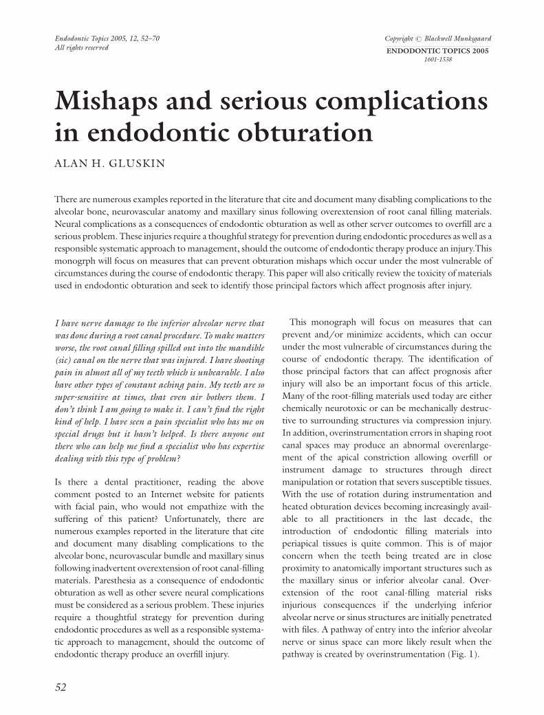
Fig. 5. XYZ axis slices showing the relationship of the apices of the lower left first molar to the inferior alveolar canal(Accuitomot, J. Morita Mfg.Co., Kyoto, Japan).
Gluskin
56
to the buccal side in the premolar area. In traditional
radiography and panographic films as well as digital
images, it appears as a narrow, radiolucent ribbon
between two radiopaque lines representing the canal
walls. It has been reported that CT imaging was highly
reliable when attempting to determine the relation
between periapical lesions and the mandibular canal
before endodontic surgery (15).
In the region of the mental foramen, the mandibular
nerve bifurcates into its two branches, the incisive and
mental nerves. The exact location of the inferior
alveolar canal and its mental branch is important in
the placement of implants, surgical extractions, and
endodontic therapy of roots within close proxi-
mity.(16) In a series of investigations on 75 human
skulls to identify the radiographic position of the
mental foramen on periapical and panographic film, the
authors found that the foramen could be seen on only
75% of the horizontal periapical radiographs.(17)
Visualization of the mental foramen was increased
and enhanced when a panoramic radiograph was
available because of a 23% magnification factor that
occurs in a panographic film.(18)
Interruptions in the protective wall of the canal by
dental manipulations can have serious and calamitous
effects on the patient’s sensibility in the distribution of
the nerve. The mandibular nerve is a peripheral nerve
consisting of an outer epineurium that encloses it as it
courses through the inferior alveolar canal. As an
anatomic structure, it is composed of bundles (fascicles)
of nerve fibers. The fascicles are wrapped by the
perineurium, and within each fascicle are the individual
axons ensheathed by Schwann cells and surrounded by a
delicate packing of loose vascular supporting tissue called
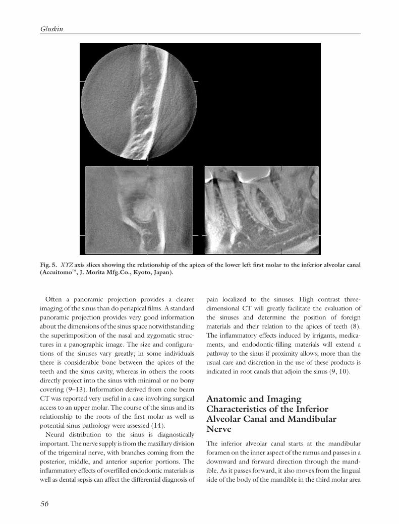
endoneurium (19, 20) (Fig. 6). Peripheral nerves receive
a rich blood supply via numerous penetrating vessels
from surrounding tissues and accompanying arteries. In
research that assessed the spatial relationship between
the posterior teeth and the mandibular canal in 22
mature dried human skulls accounting for 264 specimen
sections; second premolars and second molars had the
closest distances to the canal with a mean distance of 4.7
and 3.7 mm, respectively. The mandibular canal was
directly inferior to the root apices of the posterior teeth
5% of the time. The data also determined that as the
mandibular height decreased, the distance between the
canal and root apices also decreased (21). In prior
studies, investigators have found that 60% of mandible
specimens contained canals whereas 40% of the dissec-
tion samples had no distinct canals. Often branches of
the nerve showed significant morphologic variability and
occupied only a space in the bone as opposed to residing
within a distinct tunnel-like structure (22). This finding
has significant implications as we investigate the scientific
literature regarding the effects of endodontic medica-
ments, sealers, and materials on bone, connective tissue
and specifically the neurovascular elements found in the
antrum and inferior alveolar canal.
Inflammatory and Neurotoxic Effectsof Root Canal Materials
In a decade old consensus report of the European
Society of Endodontology regarding quality guidelines
Epincurium coveringperipheral nerve
Blood vessels
Epincurium
Endoneunium
Myelinated exon
Schwann cell
Parineurium(around one fascicle)
Fascicle
Fig. 6. Graphic illustration of the crossectional anatomyof the neurovascular complex of a peripheral nerve. Theboxed area represents the macroscopic relationship of thevasculature to the neural elements (reproduced fromMartini et al. (19); reprinted by permission of PearsonEducation, Inc).
Mishaps and serious complications in endodontic obturation
57
for endodontic therapy, it is clearly recommended that
‘the objective of any (endodontic) technique used
should be to apply a biocompatible hermetically sealing
canal filling that obturates the prepared canal space
from pulp chamber just to its apical termination’(23).
Currently, there is an important body of convincing
biologic literature that gives confidence to the science
describing host tissue reaction to many endodontic
obturation materials. The following conclusions re-
garding endodontic sealers have stood the test of time
in the last 50 years (24).
1. All obturation sealers are irritants in their freshly
mixed states.
2. After setting or curing, some sealers lose their
irritant components and become relatively inert.
3. All sealers are absorbable
4. Components of sealers will be managed by the
immune system in the process of absorption (24,
25).
5. Pastes intended to fill the entire root canal system
will be absorbed more rapidly than solid core
obturations with sealer (26).
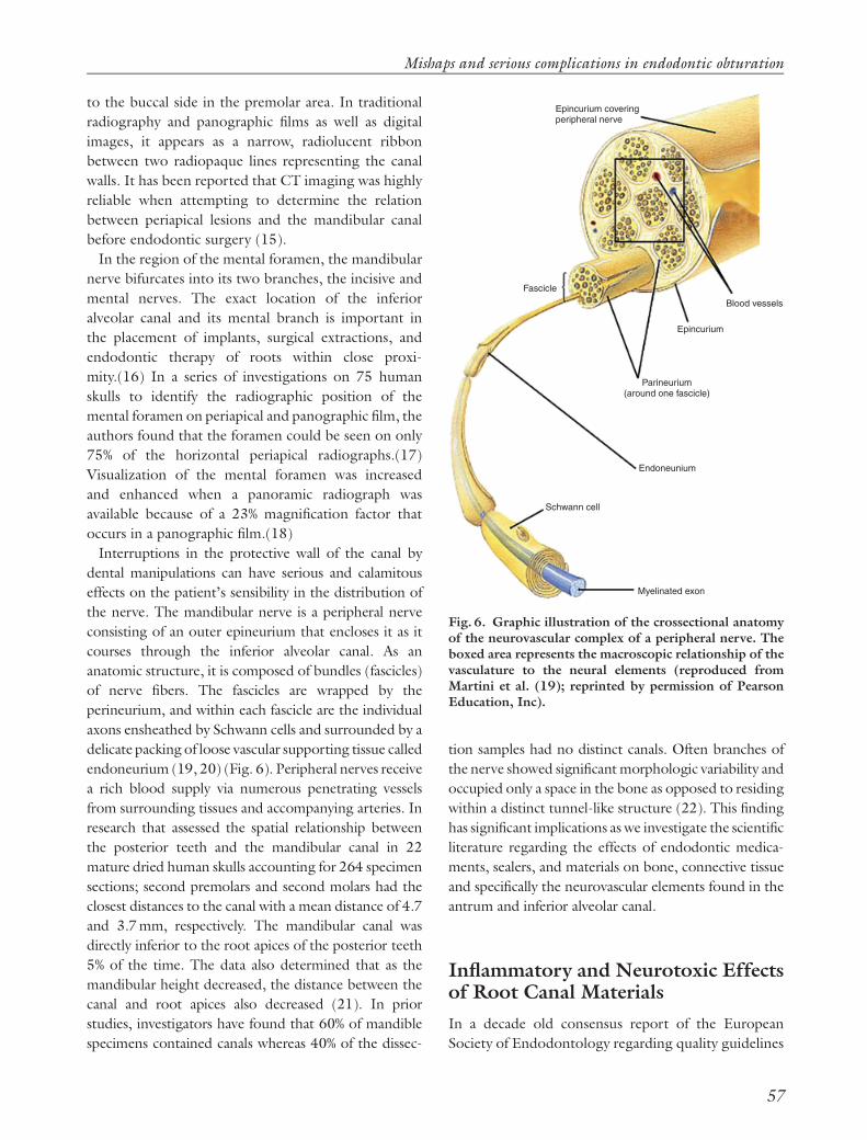
6. A minimum amount of sealer should be exposed to
periapical tissue (Fig. 7).
In endodontic therapy, sealers and cements are
primarily used to fill any irregularities at the interface
between the solid core root canal-filling material and
the walls of the canal system, ideally rendering the
system bacteria impervious. Endodontic failures caused
by a continued re-growth and proliferation of micro-
organisms because of apical percolation of blood-borne
proteins can still occur even within properly cleaned
and shaped teeth if the apical foramen is poorly sealed.
It has been reported that even in the absence of
microbial factors, root-filling substances can evoke a
foreign body reaction leading to the development of
periapical lesions that may be refractory to endodontic
therapy (27).
Many sealers, when used properly, are recognized to
have antimicrobial activity as well as the potential to
stimulate fibroblastic, osteoblastic, or cementoblastic
activity. Sealers can be grouped based on their primary
constituents, such as zinc oxide–eugenol, calcium
hydroxide, resins, glass ionomers, or resin-/compo-
site-based sealers.
The biological and irritational properties of root canal
sealing materials can be evaluated in a number of ways.
These have included tissue and cell culture studies (28–
30), bone and soft-tissue reactions to set and unset
implanted materials in experimental animals (31–33),
experimental and clinical studies on animals and
humans (34–36) and new assessments involving histo-
chemical analysis and X-ray microanalysis (37–39).
Early investigations into the absorbability of root
canal sealers in animal models showed that very hard
and compact sealers with low solubility became
encapsulated by fibrous connective tissue (26). Less
dense and more soluble sealers were dispersed and
absorbed more rapidly. Large quantities of excess filling
materials in the periapical tissues caused necrosis of
bone followed by bone resorption and then absorption
of the filling materials. Most root canal sealers produce
an initial acute inflammatory reaction in the connective
tissues (24). This is followed by the production of a
chronic foreign body reaction in which phagocytosis is
a recognized feature. As the material disintegrates in
tissue fluids, macrophages are a predominant element
in the removal of the foreign body. Such evidence
suggests that the presence of foreign material in large
quantities in the periapical tissues causes persistence of
breakdown and that persistence is fueled by the toxicity
of the engulfed material. In particular, the breakdown
products may have an adverse effect on the proliferation
and viability of periradicular cell populations that are
necessary for repair (39).
It is the sealers and components of sealers that are
recognized by the scientific literature as neurotoxic or
highly irritating that warrants more scrupulous atten-
tion and an equally careful recognition of their
potential for serious injury.
Fig. 7. Large zinc-oxide-eugenol sealer overfill wastolerated by the patient for many years and remainsasymptomatic today. There was no neurovascularinvolvement. Radiograph courtesy Dr. Roxanne Benison.
Gluskin
58
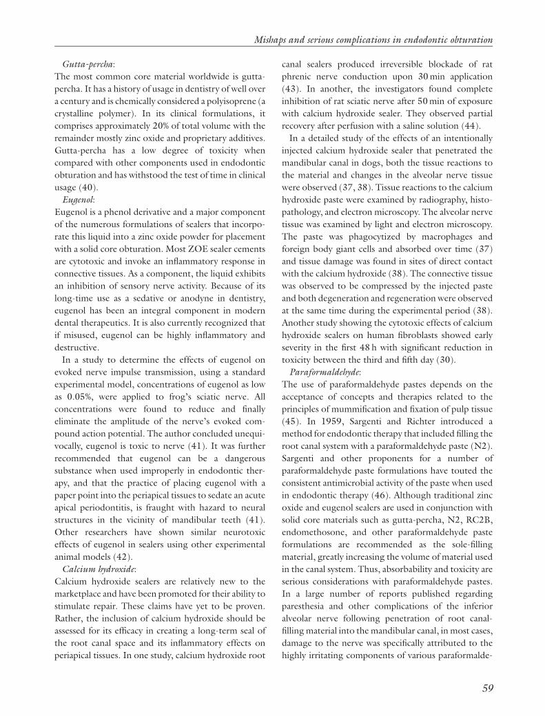
Gutta-percha:
The most common core material worldwide is gutta-
percha. It has a history of usage in dentistry of well over
a century and is chemically considered a polyisoprene (a
crystalline polymer). In its clinical formulations, it
comprises approximately 20% of total volume with the
remainder mostly zinc oxide and proprietary additives.
Gutta-percha has a low degree of toxicity when
compared with other components used in endodontic
obturation and has withstood the test of time in clinical
usage (40).
Eugenol:
Eugenol is a phenol derivative and a major component
of the numerous formulations of sealers that incorpo-
rate this liquid into a zinc oxide powder for placement
with a solid core obturation. Most ZOE sealer cements
are cytotoxic and invoke an inflammatory response in
connective tissues. As a component, the liquid exhibits
an inhibition of sensory nerve activity. Because of its
long-time use as a sedative or anodyne in dentistry,
eugenol has been an integral component in modern
dental therapeutics. It is also currently recognized that
if misused, eugenol can be highly inflammatory and
destructive.
In a study to determine the effects of eugenol on
evoked nerve impulse transmission, using a standard
experimental model, concentrations of eugenol as low
as 0.05%, were applied to frog’s sciatic nerve. All
concentrations were found to reduce and finally
eliminate the amplitude of the nerve’s evoked com-
pound action potential. The author concluded unequi-
vocally, eugenol is toxic to nerve (41). It was further
recommended that eugenol can be a dangerous
substance when used improperly in endodontic ther-
apy, and that the practice of placing eugenol with a
paper point into the periapical tissues to sedate an acute
apical periodontitis, is fraught with hazard to neural
structures in the vicinity of mandibular teeth (41).
Other researchers have shown similar neurotoxic
effects of eugenol in sealers using other experimental
animal models (42).
Calcium hydroxide:
Calcium hydroxide sealers are relatively new to the
marketplace and have been promoted for their ability to
stimulate repair. These claims have yet to be proven.
Rather, the inclusion of calcium hydroxide should be
assessed for its efficacy in creating a long-term seal of
the root canal space and its inflammatory effects on
periapical tissues. In one study, calcium hydroxide root
canal sealers produced irreversible blockade of rat
phrenic nerve conduction upon 30 min application
(43). In another, the investigators found complete
inhibition of rat sciatic nerve after 50 min of exposure
with calcium hydroxide sealer. They observed partial
recovery after perfusion with a saline solution (44).
In a detailed study of the effects of an intentionally
injected calcium hydroxide sealer that penetrated the
mandibular canal in dogs, both the tissue reactions to
the material and changes in the alveolar nerve tissue
were observed (37, 38). Tissue reactions to the calcium
hydroxide paste were examined by radiography, histo-
pathology, and electron microscopy. The alveolar nerve
tissue was examined by light and electron microscopy.
The paste was phagocytized by macrophages and
foreign body giant cells and absorbed over time (37)
and tissue damage was found in sites of direct contact
with the calcium hydroxide (38). The connective tissue
was observed to be compressed by the injected paste
and both degeneration and regeneration were observed
at the same time during the experimental period (38).
Another study showing the cytotoxic effects of calcium
hydroxide sealers on human fibroblasts showed early
severity in the first 48 h with significant reduction in
toxicity between the third and fifth day (30).
Paraformaldehyde:
The use of paraformaldehyde pastes depends on the
acceptance of concepts and therapies related to the
principles of mummification and fixation of pulp tissue
(45). In 1959, Sargenti and Richter introduced a
method for endodontic therapy that included filling the
root canal system with a paraformaldehyde paste (N2).
Sargenti and other proponents for a number of
paraformaldehyde paste formulations have touted the
consistent antimicrobial activity of the paste when used
in endodontic therapy (46). Although traditional zinc
oxide and eugenol sealers are used in conjunction with
solid core materials such as gutta-percha, N2, RC2B,
endomethosone, and other paraformaldehyde paste
formulations are recommended as the sole-filling
material, greatly increasing the volume of material used
in the canal system. Thus, absorbability and toxicity are
serious considerations with paraformaldehyde pastes.
In a large number of reports published regarding
paresthesia and other complications of the inferior
alveolar nerve following penetration of root canal-
filling material into the mandibular canal, in most cases,
damage to the nerve was specifically attributed to the
highly irritating components of various paraformalde-
Mishaps and serious complications in endodontic obturation
59

hyde pastes (47–53). Brodin (54) and other investiga-
tors (36, 55–59), have experimentally and convincingly
demonstrated the neurotoxicity of these paraformade-
hyde compounds. Furthermore, Brodin et al. (60) have
shown that N2, among other root-filling materials with
paraformaldehyde as a component, produced perma-
nent disruption of nerve conduction in vitro. Others
have demonstrated the systemic distribution of radio-
actively labeled paraformaldehyde incorporated within
formocresol in pulpotomized dogs (61). Disintegra-
tion products were found in periapical and periodontal
tissues remote from the pulpal wound site. The
radiolabeled paraformaldehyde was additionally found
in blood, regional lymph nodes, kidney and liver. It had
been recognized early on by researchers that every
effort should be made to confine these materials to the
canal (49, 55–57), as more and more clinical reports of
extreme complications were published (36, 50, 53, 62).
Because of the higher risks associated with parafor-
maldehyde-containing endodontic materials, the use of
N2 or similar type pastes are contraindicated as
permanent injury risk is substantially less with tradi-
tional-filling materials. When a safer, less hazardous
alternative therapy exists, it is unreasonable to elect an
unsafe methodology. This fact is highly relevant
because it is contrary to patient safety to require an
individual to assume inherently more dangerous
treatment risks that are reasonably avoidable, with safer
and more predictable methodologies.
Polymers, Resins, and Other SealerOptions
A number of currently available sealers are variations on
a resin/polymer formulation. This makes them options
in their own right, or they are a choice when resin
bonding within the canal is proposed, and the effects of
eugenol on dentin are not desired as a contaminant in
the bonding process.
The most commonly know sealer within this category
is AH26, AH26 Plus (Caulk/Dentsply, Milford, DE,
USA). The sealer is reported to have good handling
characteristics, seals well to dentin, and can be used
effectively with heat during obturation. The sealer has
been reported to be very toxic upon initial mixing (29,
63). This toxicity resolves rapidly during the setting
process, and after 24 h the sealer is reported to have a
relatively low toxicity. Spangberg et al. (63) have
reported this initial toxicity was due to the formation
of a very small amount of formaldehyde as a result of the
chemical setting process. They described the release of
formaldehyde as thousands of times lower than con-
ventional formaldehyde containing sealers such as N2,
and stated that after setting there was little toxic effect.
Diaket (ESPE, Seefeld, Germany) is a polyketone
compound containing vinyl polymers that mixed with
zinc oxide and bismuth phosphate, forms an adhesive
sealer. It has been demonstrated that it is relatively toxic
during setting and these effects are persistent (29, 39).
Resorcinol-formalin resin is a paste-filling material
that is commonly used in Russia, China, and India for
the treatment of pulpitis. Although there are many
variations of the resin pastes that are used, the main
ingredients are resorcinol and formaldehyde. The
principle behind ‘resinifying therapy’ is that of a liquid
phenolic resin being used which will solidify by
polymerization after being placed into the root canal.
The residual pulpal remnants are claimed to be
‘resinified’ and to be ‘rendered harmless’ (64). How-
ever, when set, this material creates an almost
impenetrable barrier and renders the tooth structure a
deep brownish to red color. Owing to remaining pulp
tissue in the apical part of the canal, complete absence
of cleaning and shaping procedures, and/or failure of
the resinifying agent to reach the apical portions of the
canal, this treatment may eventually fail, making
retreatment of these teeth necessary. Using pastes as a
sole root canal filling is generally not the treatment of
choice. It has many disadvantages which include the
lack of apical length control, inability to obtain a
compact obturation, frequent presence of voids and
possible severe toxicity if the paste material is extruded
beyond the apical foramen. A specific disadvantage of
resorcinol paste is the inability to retreat failed cases
because of the hardness of the material once it has set
(65). In the event of an overfill into the sinus or
neurovascular bundle, loss of the retreatment option
and the potential for severe irreversible damage makes
this choice unreasonable in the year 2006.
Arsenical pastes:
In the last century, arsenic played an important role in
the treatment of pulpitis at a time when anesthesia was
either unavailable or rudimentary. Its use was usually
accompanied by severe but short-lived pain. Danger to
the periodontal membrane and surrounding bone was
warned in textbooks of the time, and contained the
serious admonishment to contain the material within
Gluskin
60

the tooth. Even in our modern times, however, there is
the occasional report of the serious and damaging
consequences of using an arsenical paste (66).
Length Determination andInstrumentation Controversies inEndodontics
One of the major controversies in root canal treatment is
the apical end point of the working length. It is a
paradigm in modern endodontics that instrumentation
beyond the apical foramen should be avoided because it
is so often associated with a reduced success rate (67–71)
and exposes the patient to the potential for injury (72).
Generally most clinicians prefer to end the biomecha-
nical instrumentation at the apical constriction (narrowest
point in the canal at approximately the dentin-cemental
junction) (73), where the contact between root canal-
filling material and the apical tissues is minimal. In
addition, many dentists practice apical patency with small
files in order to maintain communication with the apical
tissues and prevent canal blockage and ledging coronal to
the determined end point.
Despite the limited three-dimensional information
provided by a conventional radiograph, radiography
remains the commonly used standard for working
length determination (74, 75). However, the accep-
tance of apex locators is widely increasing with the
introduction of devices well into their fourth genera-
tion. In addition, many clinicians use paper points to
help determine the juncture of the canal confines from
the serum of periapical tissues. Generally, a distance of
0-2 mm between radiographic apex and the obturation
material marking the end point of root canal instru-
mentation has been designated as acceptable when
evaluating postoperative radiographs. Accordingly, in a
retrospective study that investigated the influence of
the level of apical obturation on the treatment outcome
(69), a root canal filling was considered satisfactory, if
among other factors its apical level was 0–2 mm short of
the radiographic apex; this apical level contributing to
the highest success rates.
Stein and Corcoran (76) discussed the possibility of
unintentional overinstrumentation when radiographs
alone were used for working length determination.
They reported that the position of a file placed for
working length determination appeared radiographi-
cally 0.7 mm shorter than its actual position. The
results of another investigation suggest that a working
length which ends radiographically 0–2 mm short of
the radiographic apex does not guarantee that instru-
mentation beyond the apical foramen will be avoided in
premolars and molars. The authors conclude that
radiographic measurements should be combined with
electronic working length determination using modern
apex locators to better help identify the apical end point
of root canal preparation and avoid overinstrumenta-
tion (77).
In a recent review of the literature on the role of apical
instrumentation in root canal treatment (73), Baugh
and Wallace concluded that because the apical dimen-
sions of root canals range from very large to very small,
the clinician should seek instruments and techniques
that can help determine when instrumentation to the
correct apical size has been achieved and that additional
research was necessary given the controversy that still
lingers regarding final apical size. Other researchers
have shown the importance of combining therapies
such as rotary instrumentation using larger apical sizes
with the use of calcium hydroxide to reduce the
numbers of bacteria in root canals and increase long-
term success (78). In a recent meta-analysis of studies
performed over the last three decades on optimal
obturation length, the results demonstrated that
obturating materials extruding beyond the radio-
graphic apex correlated with a decreased prognosis
for repair (79). When faced with the possibility of
inadvertent overinstrumentation into neurovascular
danger, the research provides a substantial number of
appropriate caveats.
A Recipe for Disaster
Gross overextension of obturation materials usually
indicates faulty technique. However, as long as the
overextension is not in contact with vital structures,
such as the inferior alveolar nerve or sinuses, and the
apical terminus is well filled in three dimensions,
permanent harm is potentially small, unless the
obturation materials contain paraformaldehyde.
On the other hand, overextension of the root canal-
filling material risks serious and possibly permanent
consequences should the underlying inferior alveolar
nerve be adjacent to the root terminus or initially
penetrated with files. To create a mishap scenario that
includes the possibility for severe injury:
Mishaps and serious complications in endodontic obturation
61

� Take one part ignorance of the proximity of the root
end to the sinus or mandibular canal by inadequate
imaging.
� Add one part overinstrumentation, because of
inaccuracy about length of the root canal for lack
of electronic apex location.
� Add one part accidental extrusion of endodontic
pastes or sealers into neurovascular tissue; because
the hydraulics of flow is unpredictable and all
materials are initially toxic.
� Mix in one part compression of vital structures by
the overfill mass.
� And you have a recipe for disaster (Fig. 8).
Techniques for Obturation Control
There are a number of contributions to the literature
which assess techniques for apical control of obturation
materials. Tronstad (80) assessed the apical plug of
dentin chips in monkeys and showed that a plug of clean
dentin fillings could provide an apical matrix that was
well tolerated by the tissues and would provide an apical
barrier that would allow the canals to be well sealed yet
protect against impingement of filling materials on the
periodontal tissues. In a comprehensive study compar-
ing the apical plugs of dentin versus calcium hydroxide
to prevent overfilling, when the apical foramen had been
intentionally overinstrumented in cats, the investigators
found plugs of calcium hydroxide or dentin to work
equally well (81). However, the calcium hydroxide plugs
were less durable and produced foramina mineralization
that was less complete than the dentinal plugs. Periapical
healing was similar for both calcium hydroxide and
dentin. In another study that looked at foramen size as it
affected apical extrusion of thermoplasticized gutta-
percha, it was noted that overfills and the extrusion of
material occurred proportionately to the area of the
apical opening. An opening the size of a 40 (0.40 mm)
diameter file was found to be twice as likely to allow
extrusion of material than an apical diameter sized at 20
(0.20 mm)(82). When the sealing ability of laterally
condensed gutta-percha was compared with injection
molded thermoplasticized gutta-percha in straight and
curved canals, only the thermoplasticized technique
produced overextensions (83). It has also been shown
that great differences in flowability exist between gutta-
percha brands when used in a thermocompaction
technique (84). The recommendation to consider a
hybrid technique when using thermoplasticized materi-
als has often involved a cold condensation of gutta-
percha apically followed by a thermomechanical com-
paction, providing a safer barrier for limiting the
extrusion of material (82).
Thermoplasticized Gutta-Percha andthe Effects of Heat
In a series of investigations in vitro (85) and on
dogs (86), the heat of thermoplasticized gutta-percha
was assessed for its potential injurious effects. Levels
of heat generated by the plasticized gutta-percha
did not appear to be at clinically deleterious levels
and no apparent irreversible tissue destruction was
evident (86). Similar results have been obtained in
other in-vitro and in-vivo studies when manufacturer’s
protocols for usage have been followed (87–91).
Where deleterious effects are seen either experimentally
or in clinical situations, the lessons need to be
Fig. 8. (A) A section of a pre-operative panoramic radiograph showing the relationship of the inferior alveolar canal tothe lower right second molar; (B) post-operative periapical film of overfill into the inferior alveolar canal; thismagnification identifies the large mass of sealer (ZnOE) overfill exposing the patient to chemical toxicity and mechanicalcompression of the neurovascular anatomy; (C) the decision to ‘watch and wait’ for 3 years was misguided. The patientexhibits continued anesthesia in the distribution of the nerve and minimal absorption of sealer is apparent over time.
Gluskin
62
heeded. Bailey et al. in a study utilizing ultrasonic
condensation of gutta-percha found that the combina-
tion of a high power setting and a 15 s application
of activation energy resulted in temperature rise on
the root surface beyond the recognized deleterious
threshold of 101 centigrade (92). The use of heat and
the potential for injurious heat transfer to dentin
and bone has been investigated for a number of
different devices used in endodontics and associated
restorative procedures (93–97). It is generally accepted
that external root temperature increases that exceed
101C produce irreversible bone and attachment da-
mage as well as dehydration effects on dentin often
resulting in resorption (93, 94). Only a small number of
investigators and authors have cautioned that ultra-
sonic energy can be harmful through heat generation
(92, 95).
Clinical case reports involving overfill with heat
softened gutta-percha are increasing in the literature
(98, 99). The current practice of maintaining apical
patency and the popularity of thermoplastic gutta-
percha-filling techniques have increased the likelihood
that overfills can involve the neurovascular anatomy.
Fanibunda et al. (99) warn of the lesser-known danger
of thermal and mechanical insult from chemically ‘safer’
materials other than paraformaldehyde, being extruded
into the inferior alveolar canal. They report a case of
thermally compacted gutta-percha having a severe
affect on patient sensory loss after gross overfill into
the mandibular canal. In this case they identified a
mechanical (compression), chemical (calcium hydro-
xide sealer) and thermal insult (molten gutta-percha) to
the nerve (99).
Carrier-Based Gutta-Percha
Carrier-based gutta-percha was first introduced as
Thermafilt (Dentsply Tulsa Dental, Tulsa, OK.)
(100). The Thermafilt obturator currently consists of
a plastic carrier and is covered in a uniform layer of
gutta-percha. The carrier is constructed from a special
radiopaque plastic similar to a manual or rotary
endodontic instrument. The obturator is heated in a
special oven where the gutta-percha it carries assumes a
softened state with unique adhesive and flow char-
acteristics. The ideal canal preparation for a carrier-
based obturator must allow sufficient space for the flow
of cement and gutta-percha (101). Carrier-based
obturators use techniques that caution against the use
of excess cement because of the increased likelihood of
overfilling because of the piston-like effect of the
obturator during placement. As the risk of overfilling is
considered the only true limitation of carrier-based
obturators, authors and manufacturers’ caution against
the following major errors in technique:
� incorrect canal preparation including overinstru-
mentation and laceration of the apical terminus,
� excessive cement or gutta-percha,
� excessive force and velocity during insertion,
� improper obturator selection (101).
Damage to the NeurovascularAnatomy: Causes and Outcomes
Reports in the literature involving serious injury to the
inferior alveolar nerve have included paresthesia or
anesthesia associated with overfill of N2 and similar
paraformaldehyde pastes, (47, 48, 50, 52, 53),
Endomethasone, (49, 62), AH26 (102–105), calcium
hydroxide (99, 106), zinc-oxide and eugenol, and
gutta-percha (72, 104). There is almost no current
obturation material that has not been reported in the
literature to produce paresthesia when overfilled into
the neurovascular anatomy. If a new material(s) is not
currently cited, it is almost assured the mishap will
eventually find itself in the literature.
Ørstavik et al. (36) reviewed 24 published cases of
paresthesia involving the mandibular nerve. It was
reported that there was no indication of healing in 14 of
the 24 patients during the observation periods which
ranged from 3 months to 18 years after initiation of the
injury. They described the characteristic deficits of the
inferior alveolar nerve as unilateral loss of sensitivity of
the lip and gums; numbness, a tingling sensation and
dryness of the affected mucosa, often preceded by
intense pain in the affected area (36). All of the
reported cases were molars or involved the lower
second premolar. There were numerous materials
involved all of which are cited in this manuscript.
Paraformaldehyde pastes were well represented in the
materials cited but were not exclusive. They concluded
that neurotoxic and compressive effects are the most
frequent causes of paresthesia after endodontic overfill
into the mandibular canal and that the use of syringes
and rotary paste fillers should not be used to insert
Mishaps and serious complications in endodontic obturation
63
root-filling pastes or cements in teeth susceptible to
neurovascular damage.
A serious case of calcium hydroxide overfill has been
reported through a lower second molar causing severe
vasospasm and a resulting facial ischemia (107). The
ischemia caused cyanosis and necrosis on the face as
well as a total absence of function in the mandibular
and mental nerve. Again, the authors cautioned in this
case report, against the use of a syringe in applying the
calcium hydroxide with a known level of toxicity and a
high pH (12.4) (107).
It is difficult to ascertain with absolute certainty the
primary etiology for paresthesia when assessing the
sequelae and future prognosis for repair from overfill
into neurovascular tissue. The clinician, therefore, must
consider the following critical factors in all cases of
overfill: the chemical neurotoxicity of the materials
involved; the possibility of direct damage of files
(crushing and mechanical injury) (48, 104); the
compression damage of solid materials such as gutta-
percha (the pressure on the nerve bundle is directly
proportional to the amount of material pushed into the
canal) (99, 104); the possibility of epineural fibrosis
resulting in neuroma (50) (Fig. 9). In a clinical report
that suggests the use of corticosteroids may be helpful
in countering the injurious compression effects result-
ing in epineural and intraneural edema, Gatot and Tovi
(108) have reported that prednisone therapy may limit
the severity of injury as well as aid in the prevention of
fibrosis. Others also report the pharmacologic use of
corticosteroids in the treatment of paresthesia related
to overfill (109).
Inflammatory edema with resulting ischemia, that
compresses and compromises blood supply to soft
tissues and nerves in confined spaces such as the inferior
alveolar canal, is termed compartment syndrome (110).
Compartment syndromes are a group of conditions that
result from increased pressure within a limited anatomic
space, acutely compromising the circulation and ulti-
mately threatening the function of the tissue within that
space. Compartment syndrome occurs from an eleva-
tion of the interstitial pressure in a closed osseous
compartment that results in microvascular compromise.
The pathophysiology of compartment syndrome is an
insult to normal local tissue homeostasis that results in
increased tissue pressure, decreased capillary blood flow,
and local tissue necrosis caused by oxygen deprivation.
Compartment syndrome is caused by localized hemor-
rhage or post-ischemic swelling resulting in fibrosis that
obstructs axonal regeneration. The clinician should
have a high index of suspicion whenever a closed bony
nerve compartment has the potential for bleeding or
swelling. Compartment syndromes are characterized by
pain beyond what should be experienced from the initial
injury. Also, diminished sensation may be noted in the
distribution of the nerve within a compartment that is
being compressed (110).
Diagnosis of Inferior Alveolar NerveInjury
Nerve recovery subsequent to an endodontic mishap is
unpredictable. Because of this unpredictability, the
clinician is responsible for making a timely diagnosis
and monitoring neurosensory changes. Careful follow-
up evaluations and appropriate referral is our respon-
sibility to the patient (111–114).
Neurosensory function can be divided into two basic
categories based on the specific receptors that are either
stimulated or injured: mechanoreceptive and nocicep-
tive. The mechanoreceptive aspect of sensory perception
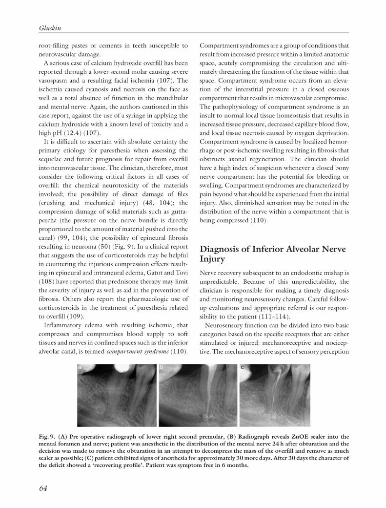
Fig. 9. (A) Pre-operative radiograph of lower right second premolar, (B) Radiograph reveals ZnOE sealer into themental foramen and nerve; patient was anesthetic in the distribution of the mental nerve 24 h after obturation and thedecision was made to remove the obturation in an attempt to decompress the mass of the overfill and remove as muchsealer as possible; (C) patient exhibited signs of anesthesia for approximately 30 more days. After 30 days the character ofthe deficit showed a ‘recovering profile’. Patient was symptom free in 6 months.
Gluskin
64

can be further divided into two-point discrimination and
brush directional stroke. The nociceptive path can be
subdivided into pinprick and thermal discrimination.
Each type of nerve fiber varies with respect to diameter,
conduction speed and physiologic function. Each
afferent nerve end offers a distinct receptor terminal
that is specific for warmth, cold, pain, and touch. The
first phase of any peripheral nerve regeneration is the
degeneration of the axon and its myelin sheath along the
nerve fiber at the site of injury. These changes along
the nerve are known as Wallerian degeneration and lead
to degeneration of the entire axon terminal over several
weeks following a severe injury. Neural injuries are most
commonly classified using the Seddon classification
system (115). Seddon’s classification is derived from
the extent of nerve injury. Neurotemesis is the most
severe nerve injury because conduction is completely
disrupted resulting in the loss of anatomic integrity of
the endoneurium, perineurium and epineurium. Axo-
notemesis, a less severe injury results in damage to the
axons, but the endoneurial and epineurial sheaths are
preserved. Neuropraxia occurs when a nerve is injured
and conduction is blocked but this does not lead to
Wallerian degeneration.
Clinically neurotemesis leads to anesthesia with a loss
of feeling or sensation. It can also produce dysesthesia,
an abnormal unpleasant sensation often burning in
character. Recovery is unlikely or limited. Axonotem-
esis causes paresthesia, an abnormal altered sensation,
which can show some degree of sensory recovery after
several months. Neuropraxia is usually a transient
paresthesia where recovery is complete from days to
weeks.
The clinical evaluation of a patient who suffers a
sensory loss in the oral or maxillofacial region
subsequent to an endodontic obturation should begin
by identifying the patient’s subjective assessment of
these alterations. The clinician should distinguish
between anesthesia, dysesthesia and paresthesia. Often
considerable variability will exist within the descriptions
outlined above and few patients will fit perfectly within
the Seddon categories (111). However, it is important
that any patient with anesthesia or a painful dysesthesia
be evaluated in a systematic fashion (111, 112). The
dentist should assess the chronologic history of the area
even if that history has only been in the last several
hours and note the patient’s chief complaint; the
nature, frequency and severity of the symptoms and
how they might be changing for better or worse as well
as the loss of function that is occurring. If the initial
symptom is anesthesia, the area of anesthesia should be
mapped and placed in the patient’s record. Any return
of sensation should be noted (113) (Fig. 10).
The physical evaluation should also include pin prick
for deep pain (small-myelinated A d fibers), brush
stroke for directional discrimination (mediated by
large-myelinated A a nerve fibers), and two-point
discrimination for proprioception (large-myelinated A
a fibers). The small myelinated fibers of the A delta
group and the smaller unmyelinated axons of the C
group are responsible for sensations of temperature
(111).
Should anesthesia and painful dysesthesia be con-
sequences of overfilling, the practitioner must under-
stand when the referral to a surgeon (oral-maxillofacial
or neurosurgeon) who is experienced in the surgical
therapies for relief and healing, may be required to
resolve the patient’s problem (114). The final manage-
ment of such a case depends on several factors. Even the
most acceptable materials can cause serious injury if
extruded in large volumes into sensitive structures.
Pastes and sealers that contain paraformaldehyde or
known safer materials are difficult to control and may
Fig. 10. Mapping of paresthesia deficits on a young manassociated with the overfill of a lower left second molarinto the inferior alveolar canal.
Mishaps and serious complications in endodontic obturation
65
additionally create injuries to the maxillary division of
the trigeminal nerve when extruded through maxillary
teeth or into the sinus membranes (116). While
paraformaldehyde-containing materials should never
be used because of the dangers of chemical injury they
present, all obturation materials should be used with
extreme caution in all circumstances, especially in teeth
intimately related to the inferior alveolar canal.
When presented with the extrusion of endodontic
obturation materials into the neurovascular tissues, and
after careful and systematic assessment of the nature
and course of the injury and its effects, a decision to
intervene surgically or delay and observe has to be
made (Fig. 11).
Management of Inferior AlveolarNerve Injuries
The oral surgery literature describes most inferior
alveolar nerve injuries as neuropraxias and thus they
resolve spontaneously within a 6-month time frame.
These are often lingual nerve injuries as well as
mandibular trauma subsequent to tooth removal. It is
reported that inferior alveolar nerve injuries heal better
than lingual nerve injuries because of the guidance
provided by the bony mandibular canal (113). This fact
has relevance if oral surgical procedures are likely and
decortication procedures and removal of overfill are
contemplated. The clinical examination that results in a
diagnosis of anesthesia or increasing painful dysesthesia
unresponsive to non-surgical therapy should help guide
this decision (111, 112). It is suggested that the
decision to intervene surgically should include the high
suspicion of injury resulting in the loss of conduction
within the nerve because of suspected chemical toxicity
and mechanical compression.
The favorable results for long-term spontaneous
recovery require thoughtful considerations for taking a
‘wait and observe’ approach. When a peripheral nerve is
injured, a non-surgical management that supports
spontaneous neurosensory recovery and promotes pa-
tient tolerance of the sensory loss is a viable option (113).
The most compelling reason to wait is that a majority of
injuries are known to recover spontaneously to some
degree. Higher levels of recovery can also be expected
when the patient is young and healthy. In addition, a
‘recovering patient profile’ with improving levels of
function, detection abilities and sensory symptoms
argues for restraint in management (111, 113).
In the final analysis, the decision of whether and when
to intervene surgically in the removal of overfill should
be based on objective criteria and a comprehensive
assessment of each individual patient. The current
guidelines for intervention are unfortunately not based
on satisfactory evidence-based science, and this leaves a
troublesome vacuum in our knowledge of effective
therapies, making prevention of this injury critical to
treatment planning before initiating root canal therapy.
Prevention of Obturation Mishapsand Concluding Remarks
This manuscript has offered a number of remedies to
provide a safe and prudent approach to the obturation
of posterior teeth in close proximity to the vulnerable
tissues of the sinus or inferior alveolar nerve. In
summary, the clinician is recommended to observe
the following counsel:
� It is essential to image and identify radiographically
the sensitive neural structures of the jaws in order to
clearly understand the proximal risk.
� It is critical to use obturation materials that are well
tolerated by the body after therapy, rather than
paraformaldehyde formulations that can cause
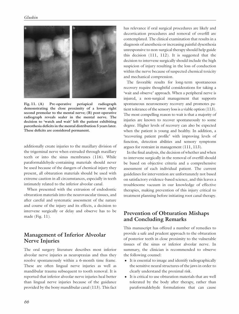
Fig. 11. (A) Pre-operative periapical radiographdemonstrating the close proximity of a lower rightsecond premolar to the mental nerve; (B) post-operativeradiograph reveals sealer in the mental nerve. Thedecision to ‘watch and wait’ left the patient exhibitingparesthesia deficits in the mental distribution 5 years later.These deficits are considered permanent.
Gluskin
66
irreversible sensory nerve damage and should not
be used in the good and safe practice of endodon-
tics.
� The clinician must practice careful and judicious
shaping strategies that use multiple confirmations
of working length and take serious precaution
against overinstrumentation.
� It is important to use ‘resistance form’ in control-
ling overfills. This ‘resistance form’ can be imparted
during canal preparation by producing funnel-
form, tapered preparations and by selecting gutta-
percha cones to match those canal shapes which will
resist the obturation forces which promote extru-
sion.
� When using thermoplastic techniques, it is impor-
tant to respect the flow characteristics of the
materials and the heat energy used.
� The use of paste-fillers and syringes for applying
endodontic sealers should be cautioned when there
is close proximity to neural structures and control is
compromised.
� In cases of extreme proximity to the neurovascular
anatomy, the importance of creating a clean dentin
plug or material barrier at the patent apical terminus
should be carefully planned when the risk of
extrusion is considerable.
Each endodontic procedure has a variable degree of
inherent risk. The standards of good practice require
that the clinician avoid unreasonable risks that may
harm the patient. Treatment is deemed negligent when
a reasonably careful clinician should have foreseen and
prevented unreasonable risk of harm to the patient.
The mishap of overfill that could cause permanent
sensory loss for a patient is disconcerting for any
practitioner to consider. We must recognize that these
injuries should encourage reflection on the safe and
prudent practice of endodontics that promotes safe-
guards. Our ethical obligation to protect patients from
harm is met when we as a profession can provide
advanced and sophisticated therapies in a safe and
controlled manner with patient safety as an overriding
priority.
References
1. Langlais RP, Rodriguez IE, Maselle I. Principles ofradiographic selection and interpretation. Dent ClinNorth Am 1994: 38: 1–12.
2. Benjamin D. Digital radiography: more than meets the
eye. Compendium 2005: 26: 464–467.3. Lavelle CLB. The role of direct intraoral sensors in the
provision of endodontic services. Endod Dent Trau-matol 1999: 15: 1–5.
4. Tyndall DA, Kapa SF, Bagnell CP. Digital subtractionradiography for detecting cortical and cancellous bonechanges in the periapical region. J Endod 1990: 16:173–178.
5. Ellingsen MA, Harrington GW, Hollender LG. Radio-visiography versus conventional radiography for detec-tion of small instruments in endodontic lengthdetermination. In vitro evaluation. J Endod 1995: 21:326–331.
6. Grondahl H-G, Huumonen S. Radiographic manifes-tations of periapical inflammatory lesions. Endod Topics2004: 8: 55–67.
7. www.jmoritaeurope.de/3d_accuitomo_eng.html8. Van Dis ML, Miles DA. Disorders of the maxillary
sinus. Dent Clin North Am 1994: 38: 155–166.9. Selden HS. The interrelationship between the maxillary
sinus and endodontics. Oral Surg 1974: 38: 623–629.10. Watzek G, Bernhart T, Ulm C. Complications of sinus
perforations and their management in endodontics.Dent Clin North Am 1997: 41: 563–583.
11. Dodd RB, Dodds RN, Holcomb JB. An endodonti-cally induced maxillary sinusitis. J Endod 1984: 10:504–506.
12. Selden HS. Endo-antral syndrome and various en-dodontic complications. J Endod 1999: 25: 389–393.
13. Selden HS. The Endo-antral syndrome. J Endod 1977:
3: 462–464.14. Rigolone M, Pasqualini D, Bianchi L, Berutti E,
Bianchi SD. Vestibular surgical access to the palatineroot of the superior first molar: ‘low-dose cone-beam’
CT analysis of the pathway and its anatomic varia-
tions. J Endod 2003: 29: 773–775.15. Velvart P, Hecker H, Tillinger G. Detection of the
apical lesion and the mandibular canal in conventionalradiography and computed tomography. Oral SurgOral Med Oral Path Oral Radiol Endod 2001: 92:682–688.
16. Phillips JL, Weller N, Kulild JC. The mental foramen:Part 1.Size, orientation, and positional relationship to
the mandibular second premolar. J Endod 1990: 16:221–223.
17. Phillips JL, Weller N, Kulild JC. The mental foramen:Part 2. Radiographic position in relation to the
mandibular second premolar. J Endod 1992: 18:271–274.
18. Phillips JL, Weller N, Kulild JC. The mental foramen:Part 3. Size and position on panoramic radiographs. JEndod 1992: 18: 383–386.
19. Martini FH, Timmons MJ, McKinley MP. HumanAnatomy, 3rd edn. New Jersey: Prentice-Hall, 2000:359.
20. Young B, Heath JW. Wheater’s Functional Histology, AText and Colour Atlas, 4th edn. Edinburgh: ChurchillLivingstone, 2000: 130.
Mishaps and serious complications in endodontic obturation
67

21. Denio D, Torabinejad M, Bakland LK. Anatomicalrelationship of the mandibular canal to its surroundingstructure in mature mandibles. J Endod 1992: 18: 161–165.
22. Carter RB, Keen EN. The intramandibular course ofthe inferior alveolar nerve. J Anat 1971: 108: 433–440.
23. European Society of Endodontology Consensus Re-port of the European Society of Endodontology onQuality Guidelines for Endodontic Treatment. IntEndod J 1994: 27: 115–124.
24. Langeland K. Root canal sealants and pastes. DentClin North Am 1974: 18: 309–327.
25. Block RM, Lewis RD, Hirsch J, Coffey J, Langeland K.Systemic distribution of [14C]-labeled paraformade-hyde incorporated within formocresol following pul-potomies in dogs. J Endod 1983: 9: 176–189.
26. Branstetter J, von Fraunhofer JA. The physical proper-ties and sealing action of endodontic sealer cements: areview of the literature. J Endod 1982: 8: 312–316.
27. Nair PNR, Sjogren U, Krey G, Sundqvist G. Therapy-resistant foreign body giant cell granuloma at theperiapex of a root-filled human tooth. J Endod 1990:
16: 589–595.28. Briseno BM, Willershausen B. Root canal sealer
cytotoxicity on human gingival fibroblasts. I. Zincoxide-eugenol-based sealers. J Endod 1990: 16: 383–386.
29. Briseno BM, Willershausen B. Root canal sealercytotoxicity on human gingival fibroblasts: II.
silicone- and resin-based sealers. J Endod 1991: 17:537–540.
30. Briseno BM, Willershausen B. Root canal sealer cyto-toxicity with human gingival fibroblasts: III. Calciumhydroxide-based sealers. J Endod 1992: 18: 110–113.
31. Curson I, Kirk EEJ. An assessment of root canal-sealing cements.Oral Surg Oral Med Oral Pathol 1968:26: 229–236.
32. Friend LA, Browne RM. Tissue reactions to some rootfilling materials. Br Dent J 1968: 125: 291–297.
33. Zmener O. Tissue response to a new methacrylate-based root canal sealer: preliminary observations in the
subcutaneous connective tissue of rats. J Endod 2004:
30: 348–351.34. Muruzabal M, Erausquin J, Devoto FCH. A study of
periapical overfilling in root canal treatment in themolar of rat. Arch Oral Biol 1966: 11: 373–383.
35. Rowe AHR. Effect of root filling materials on theperiapical tissues. Br Dent J 1967: 122: 98–102.
36. Ørstavik D, Brodin P, Aas E. Paraesthesia followingendodontic treatment: survey of the literature and
report of a case. Int Endod J 1983: 16: 167–172.37. Kawakami T, Nakamura C, Eda S. Effects of the
penetration of a root canal filling material in to themandibular canal.1. Tissue reaction to the material.Endod Dent Traumatol 1991: 7: 36–41.
38. Kawakami T, Nakamura C, Eda S. Effects of thepenetration of a root canal filling material into themandibular canal. 2. Changes in the alveolar nervetissue. Endod Dent Traumatol 1991: 7: 42–47.
39. Ørstavik D, Mjor IA. Histopathology and X-raymicroanalysis of the subcutaneous tissue response toendodontic sealers. J Endod 1988: 14: 13–23.
40. Spangberg L. Biologic effects of root canal fillingmaterials. 7. Reaction of bony tissue to implanted rootcanal filling material in guinea pigs. Odontol Tidskr1969: 77: 133–159.
41. Kozam G. The effect of eugenol on nerve transmis-sion. Oral Surg 1977: 44: 799–805.
42. Holland GR. A histological comparison of periapicalinflammatory and neural responses to two endodonticsealers in the ferret. Arch Oral Biol 1994: 39: 539–544.
43. Boiesen J, Brodin P. Neurotoxic effect of two rootcanal sealers with calcium hydroxide on rat phrenicnerve in vitro. Endod Dent Traumatol 1991: 7: 242–245.
44. Serper A, Ucer O, Onur R, Etikan I. Comparativeneurotoxic effects of root canal filling materials on ratsciatic nerve. J Endod 1998: 24: 592–594.
45. Seidler B. Irrationalized endodontics: N2 and us too.
J Am Dent Assoc 1974: 89: 1318–1331.46. Sargenti A, Richter S. Rationalized Root Canal
Treatment. New York: AGSA Scientific Publications,1959.
47. Montgomery S. Paresthesia following endodontictreatment. J Endod 1976: 2: 345–347.
48. Forman GH, Rood JP. Successful retrieval of endo-dontic material from the inferior alveolar nerve. J Dent1977: 5: 47–50.
49. Kaufman AY, Rosenberg L. Paresthesia caused byendomethasone. J Endod 1980: 6: 529–531.
50. LaBlanc JP, Epker BN. Serious inferior alveolar nervedysesthesia after endodontic procedure: report of threecases. J Am Dent Assoc 1984: 108: 605–607.
51. Fanibunda KB. Adverse response to endodonticmaterial containing paraformadehyde. Br Dent J1984: 157: 231–235.
52. Allard KUB. Paraesthesia – a consequence of acontroversial root-filling material? A case report. IntEndod J 1986: 19: 205–208.
53. Kleier DJ, Averbach RE. Painful dysesthesia of theinferior alveolar nerve following use of a paraforma-dehyde-containing root canal sealer. Endod DentTraumatol 1988: 4: 46–48.
54. Brodin P. Neurotoxic and analgesic effects of rootcanal cements and pulp- protecting dental materials.Endod Dent Traumatol 1988: 4: 1–11.
55. Barker BCW, Lockett BC. Periapical response to N2and other paraformadehyde compounds confinedwithin or extruded beyond the apices of dog rootcanals. Dent Practitioner 1972: 22: 370–379.
56. Cohler CM, Newton CW, Patterson SS, Kafrawy AH.Studies of Sargenti’s technique of endodontic treat-ment: short-term response in monkeys. J Endod 1980:
6: 473–478.57. Newton CW, Patterson SS, Kafrawy AH. Studies of
Sargenti’s technique of endodontic treatment: six-
month and one-year responses. J Endod 1980: 6: 509–517.
Gluskin
68
58. Lewis BB, Chestner SB. Formaldehyde in dentistry: areview of mutagenic and carcinogenic potential. J AmDent Assoc 1981: 103: 429–434.
59. Tagger E, Tagger M. Pulpal and periapical reactions toglutaraldehyde and paraformadehyde pulpotomy dres-sing in monkeys. J Endod 1984: 10: 364–371.
60. Brodin P, R�ed A, Aars H, Ørstavik D. Neurotoxiceffects of root filling materials on rat phrenic nerve invitro. J Dent Res 1982: 61: 1020–1023.
61. Block RM, Denby Lewis R, Hirsch J, Coffey J,Langeland K. Systemic distribution of [14C]-labeledparaformaldehyde incorporated within formocresolfollowing pulpotomies in dogs. J Endod 1983: 9:176–189.
62. Rowe AHR. Damage to the inferior dental nerveduring or following endodontic treatment. Br Dent J1983: 153: 306–307.
63. Spangberg LSW, Barbosa SV, Lavigne GD. AH26releases formaldehyde. J Endod 1993: 19 : 596–598.
64. Schwandt NW, Gound TG. Resorcinol-formaldehyderesin ‘Russian Red’ endodontic therapy. J Endod 2003:
29: 435–437.65. Vranas R, Hartwell GR, Moon PC. The effect of
endodontic solutions on resorcinol-formalin paste.J Endod 2002: 29: 69–72.
66. Smart ER, Barnes IE. Tissue necrosis after using anarsenical endodontic preparation: a case report. IntEndod J 1991: 24: 263–269.
67. Seltzer S, Soltanoff W, Sinai I, Goldenberg A,Bender IB. Biologic aspects of endodontics: Part III.Periapical tissue reactions to root canal instrumenta-
tion. Oral Surg Oral Med Oral Pathol 1968: 26:534–546.
68. Seltzer S, Soltanoff W, Sinai I, Smith J. Biologic aspectsof endodontics: part IV. Periapical tissue reactions toroot-filled teeth whose canals had been instrumented
short of their apices. Oral Surg Oral Med Oral Pathol1969: 28: 724–738.
69. Sjorgen U, Hagglund B, Sundqvist G, Wing K. Factorsaffecting the long-term results of endodontic treat-ment. J Endod 1990: 16: 498–504.
70. Smith CS, Setchell DJ, Harty FJ. Factors influencing thesuccess of conventional root canal therapy – a five yearretrospective study. Int Endod J 1993: 26: 321–333.
71. Ricucci D, Langeland L. Apical limit of root canalinstrumentation and obturation. Part 2. A histologicalstudy. Int Endod J 1998: 31: 394–409.
72. Neaverth EJ. Disabling complications following inad-vertent overextension of a root canal filling material.J Endod 1989: 15: 135–139.
73. Baugh D, Wallace J. The role of apical instrumentationin root canal treatment: a review of the literature.
J Endod 2005: 31: 333–340.74. Olson AK, Goerig AC, Cavataio RE, Luciano J. The
ability of the radiograph to determine the location ofthe apical foramen. Int Endod J 1991: 24: 28–35.
75. Powell-Cullingford AW, Pitt Ford TR. The use of E-speed film for root canal length determination. IntEndod J 1993: 26: 268–272.
76. Stein TJ, Corcoran JF. Radiographic working lengthrevisited. Oral Surg Oral Med Oral Pathol 1992: 74:796–800.
77. ElAyouti A, Weiger R, Lost C. Frequency of over-instrumentation with an acceptable radiographicworking length. J Endod 2001: 27: 49–52.
78. McGurkin-Smith R, Trope M, Caplan D, SigurdssonA. Reduction of intracanal bacteria using GT rotaryinstrumentation, 5.25% NaOCl, EDTA, andCa(OH)2. J Endod 2005: 31: 359–363.
79. Schaeffer MA, White RR, Walton RE. Determiningthe optimal obturation length: a meta-analysis of
literature. J Endod 2005: 31: 271–274.80. Tronstad L. Tissue reactions following apical plugging
of the root canal with dentin chips in monkey teethsubjected to pulpectomy.Oral Surg 1978: 45: 297–304.
81. Pitts DL, Jones JE, Oswald RJ. A histologicalcomparison of calcium hydroxide plugs and dentinplugs used for the control of gutta-percha root canalfilling material. J Endod 1984: 10: 283–293.
82. Ritchie GM, Anderson DM, Sakumura JS. Apicalextrusion of thermoplasticized gutta-percha used as aroot canal filling. J Endod 1988: 14: 128–132.
83. Mann SR, McWalter GM. Evaluation of apical seal andplacement control in straight and curved canalsobturated by laterally condensed and thermoplasti-cized gutta-percha. J Endod 1987: 13: 10–17.
84. Tagger M, Gold A. Flow of various brands of gutta-percha cones under in vitro thermomechanical com-paction. J Endod 1988: 14: 115–120.
85. Gutmann JL, Creel DC, Bowles WH. Evaluation ofheat transfer during root canal obturation withthermoplasticized gutta-percha. Part I. In vitro heatlevels during extrusion. J Endod 1987: 13: 378–383.
86. Gutmann JL, Rakusin H, Powe R, Bowles WH.Evaluation of heat transfer during root canal obtura-tion with thermoplasticized gutta-percha. Part II. Invivo response to heat levels generated. J Endod 1987:
13: 441–448.87. Saunders EM. In vivo findings associated with heat
generation during thermomechanical compaction ofgutta-percha. Part I. Temperature levels at the externalsurface of the root. Int Endod J 1990: 23: 263–267.
88. Saunders EM. In vivo findings associated with heatgeneration on during thermomechanical compactionof gutta-percha. Part II. Histological response totemperature elevation on the external surface of theroot. Int Endod J 1990: 23: 268–274.
89. Barkhordar RA, Goodis HE, Watanabe L, KoumdjianJ. Evaluation of temperature rise on the outer surfaceof teeth during root canal obturation techniques.Quint Int 1990: 21: 585–588.
90. Weller RN, Koch KA. In vitro radicular temperaturesproduced by injectable thermoplasticized gutta-percha. Int Endod J 1995: 28: 86–90.
91. Romero AD, Green DB, Wucherpfennig AL. Heattransfer to the periodontal ligament during rootobturation procedures using an in vitro model. JEndod 2000: 26: 85–87.
Mishaps and serious complications in endodontic obturation
69

92. Bailey GC, Cunnington SA, Ng Y-L, Gulabivala K,Setchell DJ. Ultrasonic Condensation of gutta-percha:the effect of power setting and activation time on
temperature rise at the root surface –An in vitro study.
Int Endod J 2004: 37: 447–454.93. Eriksson AR, Albrektsson T. Temperature threshold
levels for heat-induced bone tissue injury: a vital-
microscopic study in the rabbit. J Prosth Dent 1983:
50: 101–107.94. Atrizadeh F, Kennedy J, Zander H. Ankylosis of teeth
following thermal injury. J Periodont Res 1971: 6: 159–167.
95. Satterthwaite JD, Stokes AN, Frankel NT. Potentialfor temperature change during application of ultra-sonic vibration to intra-radicular posts. Eur J Prostho-don Restor Dent 2003: 11: 51–56.
96. Kreisler M, Haitham AH, d’Hoedt B. Intrapulpaltemperature changes during root surface irradiationwith an 809-nm gaaias laser. Oral Surg Oral Med OralPathol 2002: 93: 730–735.
97. Sweatman TL, Baumgartner JC, Sakaguchi RL.Radicular temperatures associated with thermoplasti-cized gutta-percha. J Endod 2001: 27: 512–515.
98. Blanas N, Kienle F, Sandor GKB. Injury to the inferioralveolar nerve due to thermoplastic gutta percha.J Oral Maxillofac Surg 2002: 60: 574–576.
99. Fanibunda K, Whitworth J, Steele J. The managementof thermomechanically compacted gutta percha ex-trusion in the inferior dental canal. Br Dent J 1998:
184: 330–332.100. Johnson WB. A new gutta-percha technique. J Endod
1978: 4: 184–188.101. Cantatore G, Johnson WB. The Thermafil System. In:
Castellucci A. Endodontics,Vol. II. Florence: EdizioniOdontoiatriche Il Tridente S.r.l, 2006: 702.
102. Spielman A, Gutman D, Laufer D. Anesthesia follow-ing endodontic overfilling with AH26. Oral Surg OralMed Oral Pathol 1981: 52: 554–556.
103. Tamse A, Kaffe I, Littner MM, Kozlovsky A. Parethesiafollowing overextension of AH-26: report of two cases
and review of the literature. J Endod, 8: 88–90.104. Nitzan DW, Stabholz A, Azaz B. Concepts of
accidental overfilling and overinstrumentation in the
mandibular canal during root canal treatment. J Endod1983: 9: 81–85.
105. Barkhordar RA, Nguyen NT. Paresthesia of the mentalnerve after overextension with AH26 and gutta-percha: report of case. J Am Dent Assoc 1985: 110:202–203.
106. Ahlgren FKEK, Johannessen AC, Hellem S. Displacedcalcium hydroxide paste causing inferior alveolar nerveparaesthesia: report of a case. Oral Surg Oral MedOral Pathol 2003: 96: 734–737.
107. Lindgren P, Eriksson K-F, Ringberg A. Severe facialischemia after endodontic treatment. J Oral Max-illofac Surg 2002: 60: 576–579.
108. Gatot A, Tovi F. Prednisone treatment for injury andcompression of inferior alveolar nerve: report of a caseof anesthesia following endodontic overfilling. OralSurg 1986: 62: 704–706.
109. Morse DR. Endodontic-related inferior alveolar nerveand mental foramen paresthesia. Compend ContinEduc Dent 1997: 18: 963–987.
110. Schwartz SI, Shires GT, Spencer FC. Principles ofSurgery, 7th ed. New York: McGraw-Hill, 1999.
111. Fleisher KE, Stevens MR. Diagnosis and managementof inferior alveolar nerve injury. Compend ContinEduc Dent (1995): 16: 1028–1040.
112. Donoff RB. Surgical management of inferior alveolarnerve injuries (Part I): the case for early repair. J OralMaxillofac Surg 1995: 53: 1327–1329.
113. Gregg JM. Surgical management of inferioralveolar nerve injuries (Part II): the case for delayed
management. J Oral Maxillofac Surg 1995: 53: 1330–1333.
114. Pogrel MA, Thamby S. The etiology of alteredsensation in the inferior alveolar, lingual, and mentalnerves as a result of dental treatment. CDA J 1999: 27:531–538.
115. Seddon HJ. Three types of nerve injuries. Brain 1943:
66: 237–288.116. Yaltirik M, Berberoglu HK, Koray M, Dulger O,
Yildirim S, BariS AA. Orbital pain and headachesecondary to overfilling a root canal. J Endod 2003:
29: 771–772.
Gluskin
70