Mischer NeuroscieNce iNstitute report...
116
MISCHER NEUROSCIENCE INSTITUTE REPORT 2012
Transcript of Mischer NeuroscieNce iNstitute report...

Mischer NeuroscieNce iNstitute report 2012

When physicians and patients choose
the Mischer Neuroscience Institute located at Memorial Hermann-Texas Medical Center, part of the 12-hospital Memorial Hermann Health System, they’re choosing the largest and most comprehensive neuroscience program in Texas. The renowned faculty at the Institute works together in a coordinated attack against neurological disease. Thanks to their knowledge and talent, the Mischer Neuroscience Institute is nationally recognized for leading-edge medicine and consistently ranked among quality benchmarking organizations as a leader in clinical quality and patient safety.
Table of Contents
02 About Our Institute
14 2012 Accolades
24 Scope of Services
— Brain Tumor — Cerebrovascular — Children’s Neuroscience — Epilepsy — Movement Disorders — Multiple Sclerosis — Neurocognitive Disorders — Neuromuscular Disorders — Neurorehabilitation — Neurotrauma/Critical Care — Spine
62 Research & Innovation
84 Selected Publications
94 Patient Stories
108 Staff Listing

James c. Grotta, M.D. CO-DIRECTOR MISCHER NEUROSCIENCE INSTITUTE AT MEMORIAL HERMANN PROFESSOR AND CHAIR DEPARTMENT OF NEUROLOGY UTHEALTH MEDICAL SCHOOL 713.500.7088
mhmni.com
Dear Esteemed Colleagues,
02 About Our Institute
14 2012 Accolades
24 Scope of Services
— Brain Tumor — Cerebrovascular — Children’s Neuroscience — Epilepsy — Movement Disorders — Multiple Sclerosis — Neurocognitive Disorders — Neuromuscular Disorders — Neurorehabilitation — Neurotrauma/Critical Care — Spine
62 Research & Innovation
84 Selected Publications
94 Patient Stories
108 Staff Listing
1Dong h. Kim, M.D. DIRECTOR MISCHER NEUROSCIENCE INSTITUTE AT MEMORIAL HERMANN PROFESSOR AND CHAIR VIVIAN L. SMITH DEPARTMENT OF NEUROSURGERY UTHEALTH MEDICAL SCHOOL 713.500.6170
2012 marks the fifth anniversary of the Mischer Neuroscience Institute (MNI) at Memorial Hermann. Since 2007, when
we organized Memorial Hermann-Texas Medical Center’s neurology and neurosurgery services under the MNI umbrella,
we have become the No. 1 neuroscience provider in Houston and the largest provider in the southern half of Texas.
We are pleased to share with you the Mischer Neuroscience Institute Clinical Achievements Report for fiscal year 2012,
which highlights our efforts in quality, safety, clinical care and research from July 2011 through June 2012. The report is
a publication of MNI, part of the 12-hospital Memorial Hermann system, in collaboration with The University of Texas
Health Science Center at Houston (UTHealth) Medical School. We hope you find the information interesting and valuable.
Our focus on innovation, quality outcomes and physician education continues to attract physician faculty members
to MNI and UTHealth Medical School. In this 12-month period, we welcomed 16 faculty members. At the same time
we expanded our capability to treat neurological disease through the addition of advanced technology, including a new
Varian Trilogy linear accelerator for stereotactic radiosurgery, which will allow for noninvasive, focused treatment of cranial
and spinal lesions. We have also added a second Magnes 3600 WH MEG brain imager, expanding our use of the
technology in diagnosing epilepsy, aneurysms, cortical brain lesions, arteriovenous malformations and brain tumors.
Through our telemedicine program, we offer patients in outlying communities access to our stroke and neurology
expertise and opportunities to participate in clinical trials. Eleven community hospitals in Southeast Texas are now linked
to MNI through remote presence robotic technology; nine went live in 2012. In addition, we are reaching larger numbers
of people and engaging them in a powerful way through new patient access portals on our website and social media
events, including a live Twittercast of a brain tumor resection.
In the last five years, we have seen strong growth in consumer preference for neuroscience care at Memorial Hermann.
During that time, we have reported mortality rates that are well below the national expected benchmark and seen a
greater than 50 percent reduction in length of stay, despite the increased acuity of the patients we treat.
We are proud of the work of our terrific physicians, nurses, scientists, and staff. Our promise to provide exceptional
patient care with the best possible outcomes remains front and center. Watch for new programs and services in 2013
and an expansion of our commitment to research and innovation.
Please feel free to contact us directly if you would like additional information about our services and programs.
With best wishes,

2
Shaping the future of neuroscience in a city known for medical
excellence takes dedication – to quality, patient satisfaction,
operational excellence, research, innovation and growth.
To accomplish these goals, Memorial Hermann’s Mischer
Neuroscience Institute brings together a team of world-class
clinicians, researchers and educators. A collaborative effort
between Memorial Hermann-Texas Medical Center and The
University of Texas Health Science Center at Houston (UTHealth)
Medical School, we are the largest neuroscience provider in
the southern half of Texas and one of only a few institutions in
the country to provide the full continuum of neuroscience care,
from neurology to neurosurgery to neurorehabilitation.
About Our Institute

3
mhmni.com
Our comprehensive, integrated approach has led to
the creation of Houston’s first and largest stroke team,
its leading epilepsy and neurotrauma programs, a
cerebrovascular center that treats more aneurysms and
arteriovenous malformations than any other center in
the region, a rapidly expanding pediatric neurosurgery
program and a brain tumor center that annually diagnoses
and treats hundreds of new tumor patients. We are proud
of our innovations in multiple sclerosis, brain injury, spine
surgery and more. And we make more neuroscience
breakthroughs every day.
As physicians, we find our personal interactions with
patients and the care we provide each one of them
enormously rewarding. Laboratory research and clinical
studies allow us to take our work a step further, extending
our expertise beyond our walls and communities
to patients across the nation and around the world.
Through basic science research, clinical discovery and
the development of new, breakthrough treatments, the
Mischer Neuroscience Institute is leading the way.

HARRIS
MONTGOMERY
LIBERT Y
FORT BEND
BRAZORIA GALVESTON
Memorial HermannThe Woodlands
Memorial HermannNortheast
Memorial HermannKaty
Memorial HermannSugar Land
Memorial HermannMemorial City Medical Center
Memorial HermannTexas Medical Center
Memorial HermannBaptist in Beaumont
Memorial HermannNorthwest
Memorial HermannSoutheast
Memorial HermannSouthwest
MONTGOM
Hospital
Hospital with Stroke Center
Hospital with Telemedicine
MNA Neurosurgeons
Center of Excellence
MNI’s infrastructure expansion has allowed the Institute to extend its neuroscience expertise and capabilities outside the Texas Medical Center and into the community through the development of neuroscience centers at Memorial Hermann community hospitals, creating five centers of excellence. Together, the centers bring distinctive subspecialty services to the community, and when combined with the specialized skills of neurosurgeons and neurologists at MNI, they offer suburban patients comprehensive consultation, evaluation and treatment for a range of disorders.
4
At a Glancephysician team
Staff Physicians 63 Clinical Residents and Fellows 42Medical Students on Rotation 310Research Fellows 16Advanced Practice Nurses 11Physician Assistants 3
inpatient Facilities
Total Neuro Beds 136Neuro ICU Beds 32Neuro Step Down Beds (IMU) 12Neuro Acute Care Beds 45Neuro Rehabilitation Beds 23Stroke Unit Beds 12Dedicated Operating Rooms 6EMU Beds – Pediatrics 6EMU Beds – Adult 6
research
Research Projects in Progress More than 200Grants Awarded $10.5 million
(Neurology and Neurosurgery)
specialty equipment includes:
• LeksellGammaKnife®Perfexion™• VarianTrilogyLinearAccelerator• SiemensArtis™zee(intra-operativeangiographysuite)• RP-7TMRemotePresenceSystem• 3DC-Arm• PhilipsHealthcareendovasculartemperature
modulation system• Simultaneouselectroencephalographyand
polysomnography• ContinuousEEGmonitoring• Magnetoencephalographyimaging(Magnes3600WH)• MRIcapableofadvancedspectroscopicanddiffusion
tensor imaging with tractotomy
Neurology
0
2
4
6
8
10
12
MH-TMC Methodist St. Luke's Houston NW Ben Taub Clear Lake
Neurosurgery
0
5
10
15
20
25
MH-TMC
Perc
ent M
arke
t Sha
re
Perc
ent M
arke
t Sha
re
Methodist St. Luke's Ben Taub UTMDA UTMB
Source: Texas Hospital Association Patient Data System (FY2010 Q1 – FY2012 Q3) provided by Thomson
Reuters. Texas Hospital Inpatient Discharge Public Use Data File, [FY2008 Q1 – FY2011 Q4] provided
by Texas Department of State Health Services, Center for Health Statistics; Q1 FY2012 – Q3 FY2012
discharges estimated by using historical data by hospital. Excludes Normal Newborns and SNF.
Expanded Greater Houston consists of 12 counties: Austin, Brazoria, Chambers, Fort Bend, Galveston,
Harris, Liberty, Montgomery, San Jacinto, Waller, Walker and Wharton.

5
The Patient Experience
mhmni.com
Patients from around the world come to the Mischer Neuroscience Institute for treatment, based on our high-quality
outcomes and our reputation for providing the best possible healthcare experiences. The close cooperation of MNI team
members, along with a redesigned administrative structure that allows nurses to spend more time with patients, has led
to an upward trend in patient satisfaction over the last five years. Data gathered by the Hospital Consumer Assessment
of Healthcare Providers and Systems (HCAHPS) survey shows consistent improvement in domains considered critical
to ensuring a high level of patient satisfaction.
0
10
20
30
40
50
60
70
80
90
100
Perc
ent
76% 74%68%
61% 61%58%
52%46%
79% 78%
67%
56%52%
48%
79%73% 71%
65%61%
55% 56%
81%76%
70%65%
62%
55% 56%
85%79%
74%69%
62%58% 57%
DischargeInformation
DoctorCommunication
NurseCommunication
PainManagement
HospitalEnvironment
CommunicationNew Medications
Responsivenessof Staff
FY08 FY09 FY10 FY11 FY12
HCAHPS Domains of Care Surveys Received FY08–12Respondents choosing "always" or "yes"
HCAHPS Overall Assessment
62%
70%67%73%70%
75%68%
73%74% 76%
0
10
20
30
40
50
60
70
80
90
100
Rate Hospital% of respondents choosing 9 or 10
Would Recommend% of respondents choosing “definitely yes”
Perc
ent
FY08 = 237 FY09 = 436 FY10 = 609 FY11 = 830 FY12 = 795
Total Survey Respondents
Source: Press Ganey, national hospital survey vendor, for all surveys received from patients discharged from 3 Jones, 7 Jones/NSICU, 4 Jones/NIMU/Stroke and EMU. HCAHPS scores have not been adjusted to account for a survey mode administration change

6
A History of Firsts• ThefirstStrokeCenterinHoustonandoneof
the first dedicated stroke programs in the world.
• ThefirstandonlyhospitalintheTexasMedical
Center to receive state of Texas designation as
a primary stroke center.
• ThefirstinHoustontoofferamyloidimaging,
a new diagnostic tool that enables physicians
to diagnose Alzheimer’s disease and will give
researchers insights into how they might one
day prevent the disorder.
• Thefirstcentertoconductanational,
multicenter trial for hypothermia in head injury.
• Thefirstneurosurgerycentertoofferall
advanced modalities of treatment – expert
microsurgery, interventional neuroradiology/
endovascularsurgeryandGammaKnife®
radiosurgery – for complex lesions.
• TheNorthAmericanleaderinstudiesof
primary progressive multiple sclerosis and the
most active center in Texas in the conduct of
organized clinical trials of new therapies for MS.
• ThefirstfacilityinHoustonandoneofthefirst
in the United States to test the clot-dissolving
drug tPA for acute stroke.
• ThefirstcenterinHoustontotestandprove
the efficacy of three disparate treatments
for stroke prevention: carotid surgery;
administration of antiplatelet drugs, including
aspirin; and patent foramen ovale closure.
• Thefirstfacilityintheregiontodovagusnerve
stimulation. We remain the No. 1 program in
the United States in the number of vagal nerve
stimulators implanted in epilepsy patients.
• Webroughtthefirstclinicalmagnetoence-
phalography (MEG) sensor to Houston and
recently updated the technology to the new
Magnes 3600 WH.
• Wehouseoneofonlyafewinpatient
Epilepsy Monitoring Units in the country
with the unique capability of simultaneously
performing electroencephalography and
polysomnography.
• TIRRMemorialHermannistheonlyhospitalin
Houston – and one of only seven designated
centers in the nation – in the Christopher
and Dana Reeve Foundation NeuroRecovery
Network.
• TIRRMemorialHermannisoneofonly16
Traumatic Brain Injury (TBI) Model Systems
funded by the National Institute on Disability
and Rehabilitation Research. TBI Model
Systems are national leaders in TBI-related
care and research.
7
mhmni.com

8
As healthcare reform moves hospitals away from the
former volume-driven, fee-for-service structure to a
pay-for-performance model tied to clinical outcomes,
the Mischer Neuroscience Institute (MNI) continues to
examine processes and investigate opportunities to
improve efficiencies and quality, with special attention
focused on patient outcomes, safety and satisfaction.
“Five years ago we faced enormous challenges in terms
of our rates of bloodstream infections, surgical site
infections and ventilator-associated pneumonias,” says
Imoigele Aisiku, M.D., director of neurocritical care at
Memorial Hermann-Texas Medical Center and associate
professor and vice chair of critical care in the Vivian L.
Smith Department of Neurosurgery at The University
of Texas Health Science Center at Houston (UTHealth)
Medical School. “Thanks to careful attention and tracking
using newly developed audit tools, we’ve had zero
bloodstream infections (BSIs) for the last seven months.”
Central line-associated bloodstream infections (CLABSIs)
have a reported mortality of 15 to 25 percent nationally,
Creating a Culture of Safety in an Era of Higher Patient Acuity
FEATurE

9
mhmni.com
according to the Centers for Disease Control and
Prevention. To reduce BSIs, quality teams at MNI looked
at the necessity for central-line utilization.
“Through close daily monitoring done as part of a nurse-
and physician-driven initiative, we assessed the need
for central lines on an individual patient basis, using an
array of audit tools piloted to evaluate their necessity,”
Dr. Aisiku says. “When we found that having central lines
in place longer than five or six days was associated with
a higher rate of infection, we became more aggressive
about noting when and where they were in place.
We reduced the number of central lines placed and the
duration of use, and our infection rate went down.”
In 2011, MNI reported five BSIs. In fiscal year 2012,
only two BSIs were logged in the first five months of the
year, marking two consecutive years of BSI reduction.
MNI also reduced the number of surgical site infections
in 2010, 2011 and 2012, in spite of higher patient
acuity and increased volumes. The Institute reported
a 70 percent reduction in patient falls for the last
four months of 2012, and reduced length of stay in
the Neuroscience ICU. Since 2007, MNI has reduced
observed-to-expected mortality, using University
HealthSystem Consortium benchmarks, by 50 percent.
“This is a five-year story,” says Miriam Morales, manager
of quality and performance for MNI. “Over time, we have
collected data and are building a data warehouse. Now,
we’re integrating those data with nursing and patient
safety metrics to provide physicians with more than
just patient outcomes. Physicians review dashboards
that include mortality, length of stay, infections, patient
satisfaction, nursing and patient safety metrics.”
“Ventilator-associated pneumonia (VAP) is our current
area of focus,” Dr. Aisiku says. “In the last six months
we’ve done a significant amount of education with
nursing and respiratory therapy to improve our VAPs.
It’s paying off. We reported zero VAPs in the last two
months of fiscal year 2012.”
The focus on patient safety is critical to MNI’s continued
leadership in the field of neuroscience. Beginning in
2013, the Centers for Medicare & Medicaid Services
(CMS) will use a value-based purchasing program for
acute care hospitals for inpatient services provided
to Medicare beneficiaries. Under the new rule, which
was created to improve clinical outcomes and patient
satisfaction, hospitals will receive value-based incentive
payments tied to clinical outcomes and patient
satisfaction with services provided.
“Quality and safety will continue to be the important
factors in healthcare delivery as hospitals are held
accountable for hospital-acquired conditions and
complications that result from unsafe care,” says
DongKim,M.D.,directorofMNIandprofessorand
chair of the Vivian L. Smith Department of Neurosurgery
at the UTHealth Medical School. “We strive every day to
increase efficiency of services and enhance quality
of care. We’re proud of the advances we’ve made and
will continue to provide the highest level of care to
our patients.”
10
Empowering nurses at the bedside to provide good
customer service and implement quality improvement
initiatives led to a dramatic upswing in patient satisfaction
scores at the Mischer Neuroscience Institute (MNI)
in 2012.
“Over the years, patient acuity and the demands on
nursing have increased. At the same time, nurses
are working 12-hour shifts two to three days a week,
which means patients have multiple nurses during
their hospitalization,” says Nicole Harrison, R.N.,
administrative director of nursing at MNI. “When a nurse
comes to work and assumes care of a patient for the
first time, there’s much to learn. Patients have more
complex medical histories to review and understand,
so nurses have to hit the ground running. We wanted
to provide greater consistency for our patients so we
restructured the neuroscience nursing team to focus
more strongly on quality and customer service. By doing
so, we’ve improved the overall patient experience.”
That improvement is reflected in MNI’s HCAHPS scores,
which improved in seven of the eight areas tracked,
including overall rating, communication about new
medication, discharge information, pain management,
nurse communication, responsiveness of hospital staff
and communication with physicians. HCAHPS (Hospital
Consumer Assessment of Healthcare Providers and
Systems), a standardized survey instrument and data
collection methodology that allows valid comparisons
to be made of hospitals across the country, is the first
national, publicly reported survey for measuring patients’
perceptions of their hospital experience.
Harrison has led the nursing initiative to improve the
patient experience since joining MNI at the end of
October 2011. “We redesigned our structure to give our
nurses more support at the bedside and in doing so,
we created a solid platform for the launch of our quality
and customer service initiatives,” she says. “Our nurses
are in charge, and they drive the unit. They’re involved
in decision-making through committees and one-on-one
meetings with directors. We’ve made it clear – from the
top down – that nurses are valued and equal members
of the patient care team. As the people who care for our
patients day in and day out, they’re aware of patient and
family concerns, and they notice the smallest changes
in the patient’s condition, which is especially important
after a neurological event. They make a vital contribution
to the physicians’ knowledge of their patients’ status by
participating in daily rounds.”
Harrison and her team have moved away from the
traditional nursing model that gives one charge nurse
responsibility for the flow of an entire unit, including
bed assignment, patient throughput, staffing issues,
quality and peer-to-peer support at the bedside. “With
the multitude of important, necessary functions of the
traditional charge nurse role, it was evident that our
charge nurses didn’t have the time to devote to each of
these details,” she says. “We started by taking a hard
look at where we were focusing our energy. We have
Driving Quality Improvements with Data: From Nursing Quality Initiatives to Physician Scorecards
FEATurE

11
mhmni.com
39 beds on the neuroscience unit. On a typical day,
between 11 and 15 patients are discharged or
admitted – a high number. We knew we were busy,
but we started wondering if the energy we were
expending was really making a difference in quality
and patient satisfaction. So rather than adding staff,
we restructured for more efficiency.”
The new structure includes an operational leader
responsible for bed assignment, timely discharge and
admission, staffing, patient flow and work flow – and
two team leaders, who split the unit in half and focus
on the quality and customer service portion of patient
care, allowing the team to provide very personalized
service. They share responsibility for rounding on each
patient and looking at specific patient satisfaction
measures using an audit tool designed by the nursing
team and based on HCAHPS survey questions. In
addition to those measures, they ascertain patient-
specific goals as identified by the patient and work to
implement a plan to meet those goals.
Harrison says the quality customer service initiatives
create a better experience for patients and families.
“This is about getting back to the basics of courtesy
and real caring. We have a great unit and a lot of really
dedicated nurses who want to do what’s best for the
patient. And our patients love it.”
DongKim,M.D.,directorofMNIandprofessorandchair
of the Vivian L. Smith Department of Neurosurgery at
The University of Texas Health Science Center at Houston
(UTHealth) Medical School, believes in forging a strong
physician partnership with nurses. “Part of empowering
nurses is encouraging them to think about what’s not
workingwell,”Dr.Kimsays.“Wewantthemtoshare
their ideas about transforming care at the bedside
with us. How can we improve the flow in the work
environment? No one knows our patients better than
the people working at the bedside. We also encourage
them to be engaged in evidence-based research and
to achieve certification in their discipline. We want
them to help us continue to improve.”
To ensure that new recruits have a solid foundation
in neuroscience and are well equipped to work
independently, Nicole Harrison and her team have
created a new nurse education structure. Rather than
reporting through the hospital’s Education department,
three neuroscience educators report to her. Open
positions are filled through the MNI Nursing Academy
using a model similar to a residency or internship with
the exception that the speed at which nurses advance
through the academy is personalized to the individual.
“Changing a culture takes time,” says James Grotta,
M.D., co-director of MNI and professor and chair of
the department of Neurology at the UTHealth Medical
School. “In a short time, we’ve done a good job of

12
getting the right people in the right roles. Leadership
is so important to drive the ship to quality outcomes.
We want leaders who empower our bedside nurses and
help us grow professional nursing within our service line.
Building a good foundation of nurses with expertise in
neuroscience and retaining them is one of our critical
success factors. It’s a work in progress.”
the Neuroscience research repository and Neurocore
The nursing quality initiatives are part of a larger focus
at MNI on collecting data and creating the capability to
use it to improve care, which includes the Neuroscience
Research Repository (NRR). A collaborative project of
Memorial Hermann and the Vivian L. Smith Center for
Neurologic Research at the UTHealth Medical School,
researchers at the NRR collect samples from consenting
patients for clinical, genomic and proteomic analysis.
These samples serve as the foundation for basic
and clinical studies, and are changing the way care
is delivered.
“Advances in biomedical research technology like the
NRR present a range of new opportunities for a greater
understanding of neurological illness and injury and
thedevelopmentofnoveltherapies,”Dr.Kimsays.
“The beauty of the NRR is that it integrates reliable
clinical data with biologic information from patient
tissue specimens, providing us with insight into a
broad spectrum of health issues related to injury and
disease of the brain, spine and central nervous system.”
Patient data for the NRR is gathered through a clinical
documentation and communication program called
Neurocore and electronically transferred to the NRR
database for analysis.
“Healthcare providers across the country are transitioning
as quickly as possible to electronic medical records.
We aim to maximize the potential of electronic systems,”
says Gigi Hergenroeder, M.H.A., R.N., director of the
NRR and an assistant professor in the Vivian L. Smith
DepartmentofNeurosurgery.“Dr.Kimhasworkedwith
Memorial Hermann-Texas Medical Center to put the main
source of the patient data for the NRR into place through
the development of Neurocore. Because of his vision
and the support of the Vivian L. Smith Foundation,
we now have a priceless resource combining clinical
data, which provides an overall picture of the patient’s
condition, with tissue samples for scientific analysis.
The result is a much more complete picture of the injury
or disease process.”
The system also offers clinical decision support through
embedded protocols for the treatment of specific
conditions. “Neurocore has the capability to analyze
information with a narrow and deep focus, making it
particularly useful in specialties like neuroscience,”
Dr.Kimsays.“Itfacilitatesresearchandevidence-
based medicine and supports our mission as a medical
school and teaching hospital to educate future leaders
in neuroscience by helping our residents advance their
knowledge and practice. It also offers us the opportunity
to monitor adherence to and departure from protocols,
as well as to change existing protocols and introduce
new ones.”
Researchers began enrolling patients in the NRR at
Memorial Hermann-TMC in the spring of 2009. As the
repository’s inventory expands, the availability of tissue
samples is quickening the pace of research. “If our
researchers want to study, for example, a brain tumor
population or an aneurysm population, our tissue and
serum stores catapult their research by eliminating the
time spent collecting data and samples,” Hergenroeder
says. “By speeding the creation of new knowledge, the
Vivian L. Smith Center is making a huge contribution
to neurological science and practice.”
FEATurE

13
mhmni.com
“Neurocore builds on the existing clinical electronic
record of the hospital by bringing us closer to a
largerandmoreintegrateddatawarehouse,”Dr.Kim
says. “That datamart, along with other technology
improvements on the horizon, will help us create an
environment that promotes the best delivery of care.”
using the Datamart to provide Feedback on Quality and outcomes
To effect change and improve quality, MNI’s datamart
facilitates quality data analyses, generates service-line
dashboards and develops physician scorecards.
“Neurocore improves physician documentation,
communication, protocol adherence and patient
care,” says Miriam Morales, manager of quality and
performance for MNI. Morales uses information
generated by Neurocore, integrated with nursing
and patient safety metrics, to provide MNI physicians
with data on discharges, length of stay, mortality
and infections. “Ultimately, we plan to integrate hospital
financial data, patient satisfaction and physician
outcomes,” she says.
Each month at neuroscience service-line meetings
performance is reviewed, action plans are developed or
updated and leaders are identified to address quality
concerns. These combined efforts have improved
physician documentation, improved clinical practice,
reduced observed-to-expected mortality ratios and
improved patient safety.
“While we’ve made excellent progress toward
improvement, there remains the goal of fully integrating
all our data across the neuroscience service line,”
Dr.Kimsays.“Webelievethatcurrentclinicaldata
is fundamental to performance improvement. Future
initiatives will include automation of dashboards and
physician-specific drill-down features. Once completed,
the datamart will integrate all financial and clinical
information and facilitate continued improvement.”
14

15
2012 Accolades
mhmni.com
We are proud to share with you highlights of our accomplishments in clinical care, research and academic endeavors as individuals and as an institution.
16
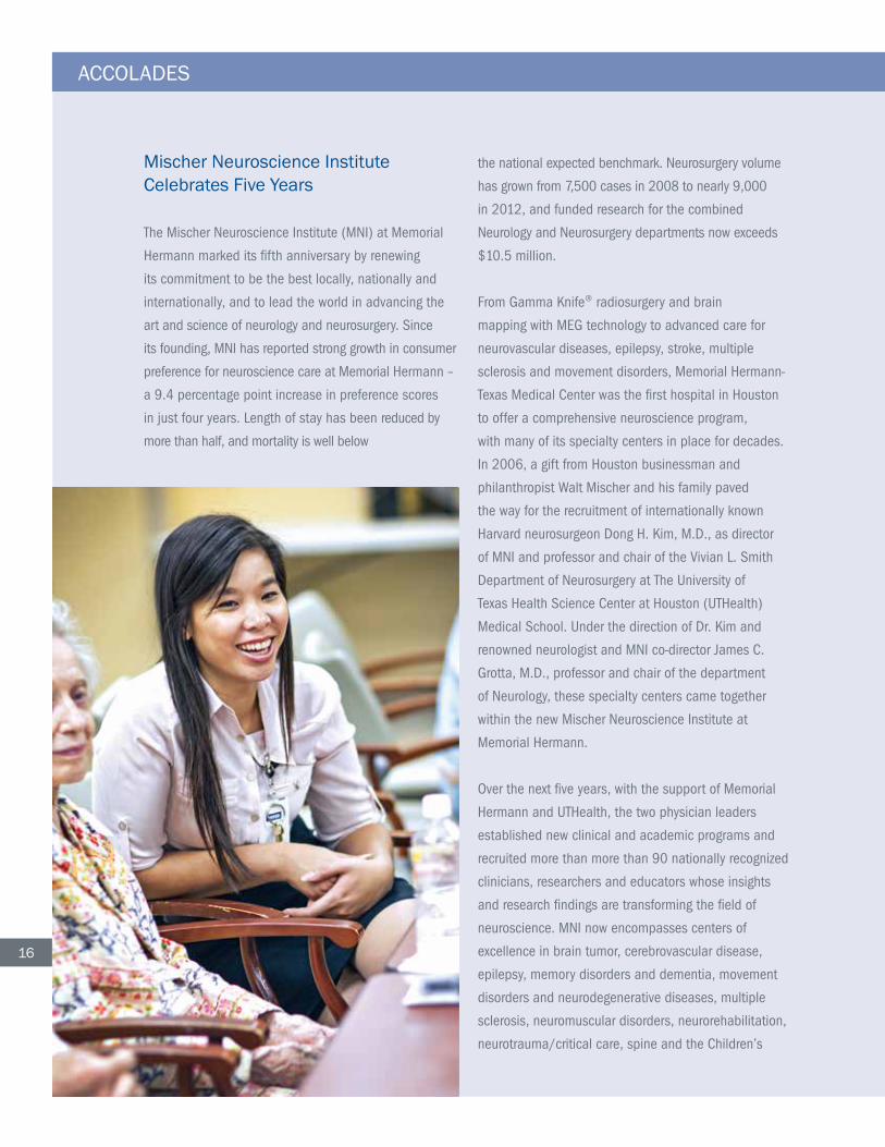
Mischer Neuroscience Institute Celebrates Five Years
The Mischer Neuroscience Institute (MNI) at Memorial
Hermann marked its fifth anniversary by renewing
its commitment to be the best locally, nationally and
internationally, and to lead the world in advancing the
art and science of neurology and neurosurgery. Since
its founding, MNI has reported strong growth in consumer
preference for neuroscience care at Memorial Hermann –
a 9.4 percentage point increase in preference scores
in just four years. Length of stay has been reduced by
more than half, and mortality is well below
the national expected benchmark. Neurosurgery volume
has grown from 7,500 cases in 2008 to nearly 9,000
in 2012, and funded research for the combined
Neurology and Neurosurgery departments now exceeds
$10.5 million.
FromGammaKnife®radiosurgeryandbrain
mapping with MEG technology to advanced care for
neurovascular diseases, epilepsy, stroke, multiple
sclerosis and movement disorders, Memorial Hermann-
Texas Medical Center was the first hospital in Houston
to offer a comprehensive neuroscience program,
with many of its specialty centers in place for decades.
In 2006, a gift from Houston businessman and
philanthropist Walt Mischer and his family paved
the way for the recruitment of internationally known
HarvardneurosurgeonDongH.Kim,M.D.,asdirector
of MNI and professor and chair of the Vivian L. Smith
Department of Neurosurgery at The University of
Texas Health Science Center at Houston (UTHealth)
MedicalSchool.UnderthedirectionofDr.Kimand
renowned neurologist and MNI co-director James C.
Grotta, M.D., professor and chair of the department
of Neurology, these specialty centers came together
within the new Mischer Neuroscience Institute at
Memorial Hermann.
Over the next five years, with the support of Memorial
Hermann and UTHealth, the two physician leaders
established new clinical and academic programs and
recruited more than more than 90 nationally recognized
clinicians, researchers and educators whose insights
and research findings are transforming the field of
neuroscience. MNI now encompasses centers of
excellence in brain tumor, cerebrovascular disease,
epilepsy, memory disorders and dementia, movement
disorders and neurodegenerative diseases, multiple
sclerosis, neuromuscular disorders, neurorehabilitation,
neurotrauma/critical care, spine and the Children’s
ACCOlADES

17
mhmni.com
Neuroscience Center. These centers are supported
by the Institute’s $13.5 million neuroscience intensive
care unit, which opened in 2009 and was designed
with input from physicians, nurses, patients and
family members.
The MNI neurosurgery program is complemented by
an equally strong neurology program, which boasts
the first stroke center in Houston, one of the first
dedicated stroke programs in the world and the first Joint
Commission-accredited primary stroke center in the region.
In 2009, MNI’s track record in the treatment of stroke
helped lead to the designation of five Memorial Hermann
hospitals as primary stroke centers by the
Texas Department of State Health Services: Memorial
Hermann-Texas Medical Center, Memorial Hermann
Memorial City Medical Center, Memorial Hermann
SouthwestHospital,MemorialHermannKatyHospital
and Memorial Hermann The Woodlands Hospital.
Telemedicine links were established between those
centers and MNI neurologists to ensure 24/7 emergency
consultation. Today, MNI’s remote-presence robotic
system, an advanced teleconferencing technology
equipped with two-way video capability, links neurologists
at the MNI Stroke Center to 11 outlying facilities, including
Baptist Beaumont Hospital, Baptist Orange Hospital,
Huntsville Memorial Hospital, Bellville General Hospital,
Matagorda Regional Medical Center, Memorial Livingston
Hospital, Citizens Medical Center in Victoria, Texas,
Cleveland Regional Medical Center, Memorial Hermann
Southwest Hospital, Medical Center of Southeast Texas
and St. Joseph Medical Center in Houston. The technology
allows emergency physicians in community hospitals to
consult with MNI neurologists, who can see patients and
view monitors and other clinical data sources firsthand
from remote locations.
“Our ultimate goal is to build a collaborative network
of hospitals working together to deliver comprehensive
neurological and neurosurgical care in the southern half
of Texas,” Dr. Grotta says. “As the Texas Medical Center
hub, MNI provides 24/7 stroke consultations, as well as
consults for other conditions, for our network hospitals.
The program allows us to treat as many patients as
possible at our partner hospitals, avoiding unnecessary
patient transfers.”
In2008,underDr.Kim’sleadership,theInstitute
broadened its commitment to teaching with the launch of
the UTHealth Medical School’s Neurosurgery Residency
Program. From 2011 to 2012, the number of neurosurgery
residents nearly doubled, and 16 research fellows
are now in training at MNI. “There’s a dire shortage of
neurosurgeonsthroughoutthecountry,”Dr.Kimsays.
“We have the expert faculty in place to support a
residency, and we’re the market leader in Houston
in cranial neurosurgery. The neurosurgery residency
reinforces the strength of our program at the Mischer
Neuroscience Institute and will lead to even more
research. With its addition, the medical school now has a
residency in every field.” During that same time period the
number of neurology residents jumped from 227 to 264.
MNI has also added new equipment to its arsenal
against neurological disease, including the state-
of-the-artLeksellGammaKnife®Perfexion™anda
SiemensArtis™zeebiplanesystem.Bothexpandthe
Institute’s treatment capability and allow physicians to
accommodate increases in patient volume. In 2011,
MNI opened two new specialized clinics: the Face Pain,
Trigeminal Neuralgia and Chiari I Clinic, which gives
patients suffering from difficult-to-diagnose face pain new
treatment options, and the Pituitary Tumor and Vision
Change Clinic, which brings together an interdisciplinary
team to provide comprehensive diagnosis and treatment
plans for patients with pituitary-region tumors.

18
Dr.Kim’svisiontoestablishatissuerepositoryfor
research into neurological injury, similar to the large
tumor repositories that support cancer research, led
to the development of the Neuroscience Research
Repository (NRR). “There was no such bio-bank of
human samples from patients suffering brain injuries
fromtrauma,strokeorhemorrhage,”Dr.Kimsays.
“We envisioned the NRR as a resource for investigators
already in the field and a tool that would stimulate
others to enter the field.” A collaborative project of
Memorial Hermann and the Vivian L. Smith Center
for Neurologic Research at the UTHealth Medical
School, the NRR collects samples from consenting
patients for clinical, genomic and proteomic analysis
to serve as the foundation for basic science and
clinical studies, which ultimately will change the way
care is delivered.
Future projects include a second Neuroscience ICU
and new subspecialty clinics. “We’ve set our sights high,”
Dr.Kimsays.“Weputtogetherourneurosciencestrategy
five years ago. Since then we’ve worked it aggressively
and are seeing tremendous results. Our aim is to push the
envelope in neurological and neurosurgical care in ways
that are appropriate for the patients we treat and that
advance the field of neuroscience.”
Arthur l. Day, M.D., Named President of the “Senior Society”
Arthur L. Day, M.D., vice chair, program director and
director of clinical education in neurosurgery at the
Mischer Neuroscience Institute at Memorial Hermann,
assumed the prestigious position of president of the
Society of Neurological Surgeons in May 2011.
ACCOlADES

19
mhmni.com
The oldest neurosurgical society in the world, the
Society of Neurological Surgeons, also known as the
“Senior Society,” counts among its members academic
department chairmen, residency program directors
and other neurosurgical leaders.
“Becoming the president of this esteemed group
is perhaps the pinnacle to which an academic
neurosurgeon can aspire,” says Dr. Day, who is
a professor in the Vivian L. Smith Department of
Neurosurgery at The University of Texas Health Science
Center at Houston (UTHealth) Medical School.
“I am honored and humbled by this appointment
and opportunity to serve our specialty.”
An accomplished clinician and researcher, Dr. Day
came to MNI from the department of Neurosurgery at
Harvard Medical School and Brigham and Women’s
Hospital in Boston, where he served as a professor and
program director of neurosurgery from 2002 to 2009,
and chair of the department from 2007 to 2009. He
received his medical degree at Louisiana State University
in New Orleans in 1972, followed by a neurological
residency and a neuropathology fellowship in brain
tumor immunology at the University of Florida (UF) at
Gainesville. After completing his training, he joined the
UF faculty and rose to the rank of professor, program
director and co-chair of the department.
Dr. Day’s clinical interests include stroke and carotid
artery disease, brain aneurysms, vascular malformations
of the brain and spinal cord, skull base and orbital
tumors, microsurgical treatment of all types of brain
tumors, trigeminal neuralgia and hemifacial spasm,
minimally invasive spinal surgery and sports-related
neurological injuries. His clinical research has focused on
new and safer surgical approaches to complex tumors
and vascular lesions of the skull base, especially those
affecting the visual system. His research interests have
included neuroprotection with estrogens in ischemic
and hemorrhagic stroke and biomarkers of neural injury.
A fellow of the American Surgical Association and
former governor of the American College of Surgeons,
Dr. Day served as vice president of the World Congress
of Neurological Surgery in 2009. He is a past president
of the Congress of Neurological Surgeons, past chair of
the American Board of Neurological Surgery, and recently
completed a six-year term as a member of the Residency
Review Committee for Neurosurgery. He has served on
the editorial board of numerous journals and has been
consistently named among the Best Doctors in America,
America’s Top Doctors, Best Doctors and America’s Top
Doctors for Cancer. He has published widely, including
authoring or coauthoring nearly 170 original articles
and book chapters, as well as co-editing a book about
neurological sports injuries. He has been invited as
a visiting professor at many prominent universities
throughout the United States and the world.

ACCOlADES
20
James Grotta, M.D., Named uTHealth President’s Scholar
In May 2011, The University of Texas Health Science
Center at Houston (UTHealth) Medical School presented
its highest academic honor, the President’s Scholar
Award for Research, to James C. Grotta, M.D., for his
achievements in the field of stroke research and treatment.
Dr. Grotta, who is co-director of the Mischer
Neuroscience Institute (MNI), joined the UTHealth
Medical School faculty in 1979. He is professor and
chair of the department of Neurology and the Roy M.
and Phyllis Gough Huffington Distinguished Chair at
the Medical School, as well as the director of the
Vascular Neurology Program.
From the early days in the late 1970s when Dr. Grotta
published his seminal articles on calcium’s role in focal
ischemic stroke and global cerebral ischemia, to his
groundbreaking research into the effectiveness of tissue
plasminogen activator (tPA), Dr. Grotta has positioned
UTHealth Medical School as a pioneer institution in the
field of stroke research and treatment.
Dr. Grotta has “devoted his entire career to creating an
internationally known clinical laboratory research and
clinical program in cerebrovascular disease,” noted
Giuseppe N. Colasurdo, M.D., president ad interim of
UTHealth and dean of the UTHealth Medical School, and
John H. Byrne, Ph.D., chairman of the department of
Neurobiology and Anatomy and assistant dean for research
at the Medical School, in their letter of nomination.

21
mhmni.com
To participate in the landmark 1995 clinical trial
investigating the use of tPA within three hours of the
onset of stroke symptoms, Dr. Grotta organized a
consortium of Houston Fire Department paramedics
and other Houston stroke centers equipped to rapidly
assess, triage and randomize patients within the three-
hour treatment window. “No such trial in stroke had
ever been conducted in the world,” Drs. Colasurdo and
Byrne wrote, and this success sent a “signal to the
National Institutes of Health that UTHealth was one
of only a handful of centers” in the country capable
of conducting hyperacute stroke trials.
Through his work with imaging, Dr. Grotta and his
fellows discovered that continuous transcranial
Doppler ultrasound monitoring with tPA produced
better outcomes than the use of tPA alone, leading to
a Phase IIb study, the results of which were published
in the New England Journal of Medicine in 2004.
As tPA has remained the only proven therapy for
acute stroke, the National Institute of Neurological
Disorders and Stroke (NINDS) created a program
grant called SPOTRIAS in the early 2000s that funds
centers to test new therapeutic approaches. The
UTHealth Medical School was selected to be one of
the four original centers in the United States to receive
SPOTRIAS funding. All of the original projects in the
program grant were based on research from the
MNI stroke program under Dr. Grotta’s direction.
UTHealth became “a beacon for highly talented
neurologists to train and complete a stroke fellowship”
with Dr. Grotta, his nominators wrote. His success
as a mentor garnered him a T32 training award from
the National Institutes of Health over the past 15 years
to develop academic leaders in stroke.
He has published extensively in both clinical and basic
research, and has nearly 300 peer-reviewed publications
to his name. He also has written 70 book chapters and
edited seven books. His 1995 New England Journal of
Medicine article on tPA use was recently named one of
the publication’s top nine articles of its 200-year history.
“Over two decades, Jim has epitomized the consummate
academicphysician,”saysDongH.Kim,M.D.,director
of MNI and chair of the Vivian L. Smith Department of
Neurosurgery at the UTHealth Medical School. “He has
built an internationally recognized stroke program that is
one of the best in the world, he has trained residents and
fellows who have also become leaders in the stroke field
and he has both performed and fostered groundbreaking
research that has increased our understanding of the
mechanisms of injury following stroke, and offered new
possibilities for treatment. We are all extremely fortunate
to have Jim lead us.”
Sean Savitz, M.D., Named Stroke Program Director
Sean Savitz, M.D., has been named director of the
Stroke Program at the Mischer Neuroscience Institute
(MNI) at Memorial Hermann. He has been co-director
of the Vascular Neurology Program, director of
translational stroke research and director of the
Vascular Neurology (Stroke) Fellowship Program at
The University of Texas Health Science Center at
Houston (UTHealth) Medical School since 2007.
“Sean brings great new ideas and leadership to the
program, while at the same time continuing its tradition
of excellent clinical research and education,” says
James Grotta, M.D., co-director of MNI, professor and
head of the department of Neurology at the UTHealth
Medical School and founder of the Stroke Program.

ACCOlADES
22
“Under Sean’s direction, the Vascular Neurology
Fellowship Program has doubled in size and
productivity. At the same time, he has developed
and funded the world’s leading translational research
program in stem cell therapy for stroke.”
Dr. Savitz graduated from Harvard University and
received his medical degree from the Albert Einstein
College of Medicine. He completed his residency and
fellowship in the Harvard Medical School Neurology
Program at Beth Israel Deaconess Medical Center
and Children’s Hospital in Boston.
uTHealth Fellow Honored with New Investigator Award
Amrou Sarraj, M.D., a vascular neurology fellow at
The University of Texas Health Science Center at
Houston (UTHealth) Medical School, received the
Mordecai Y. T. Globus New Investigator Award,
presented by the American Stroke Association at
the International Stroke Conference 2012.
Dr. Sarraj’s work “Optimizing Prediction Scores for
Poor Outcome After Intra-arterial Therapy for Anterior
Circulation Acute Ischemic Stroke” was presented on
February 2 in New Orleans. The study suggests that
combining critical and radiographic variables can
better predict poor outcome after patients undergo
intra-arterial thrombolysis.
The Globus Award is named for the late renowned
cerebrovascular researcher, Dr. Mordecai Y. T. Globus,
and is given to a researcher who is still in training.
“It’s a great honor for me and a significant achievement
for Mischer Neuroscience Institute’s Stroke Center
and the UTHealth Medical School,” Dr. Sarraj says.
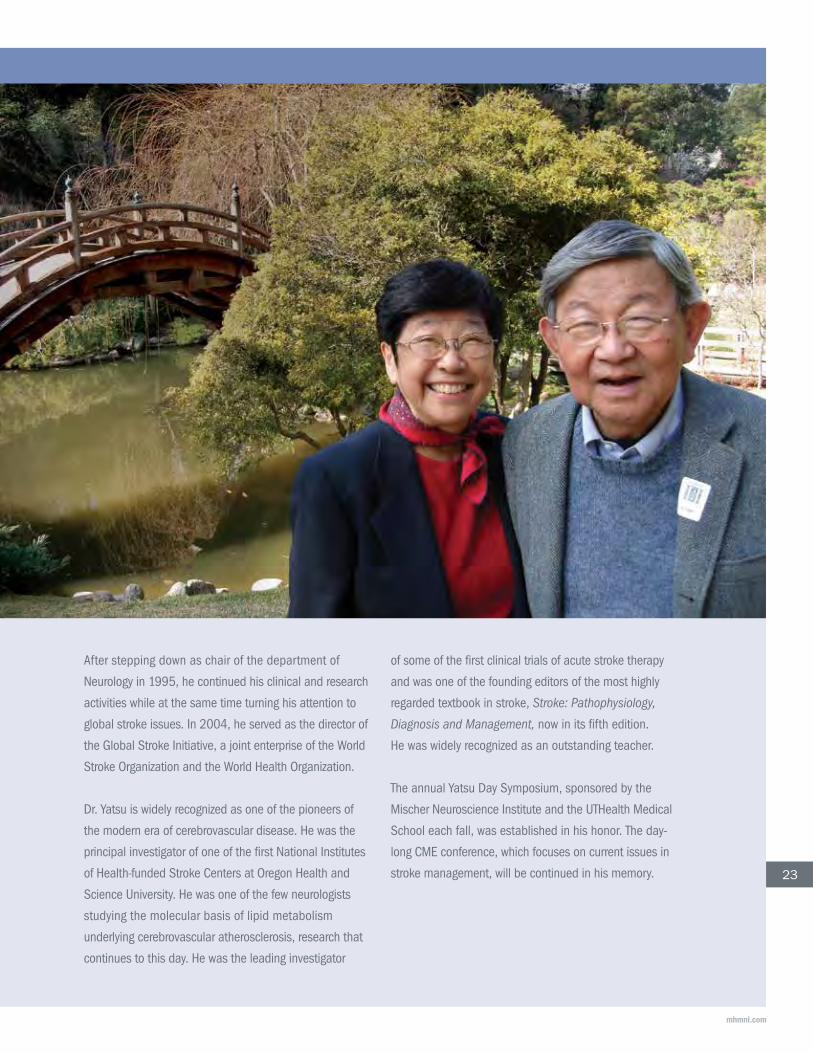
In Memoriam: Frank Yatsu, M.D.
Frank Yatsu, M.D., professor emeritus of neurology,
died March 9, 2012. He was 79.
Dr. Yatsu joined The University of Texas Health Science
Center at Houston (UTHealth) Medical School in 1982
as the second chair of the department of Neurology
and was celebrated at a retirement ceremony on
January 14, 2011.
A native of Los Angeles, Dr. Yatsu moved with his family
to Cleveland, Ohio, in the mid-1940s. As a Boy Scout in
Cleveland, he received a full scholarship to Phillips Academy
in Andover, Massachusetts. At Andover, he quickly became
popular with his gregarious sense of humor and wit, and
was referred to as the “walking dictionary.” He went on to
receive his baccalaureate from Brown University on a full
four-year wrestling scholarship, and completed medical
school at Case Western Reserve University. He completed
an internal medicine residency at University Hospital
in Cleveland and a neurochemistry fellowship at Albert
Einstein College of Medicine in New York.
From 1965 to 1967, Dr. Yatsu served as a lieutenant
commander at the U. S. Naval Academy in Great Lakes,
Illinois, and later went to work for the department of
Neurology at the University of California Medical Center,
where he became the vice chair of the department and
chief of neurology at San Francisco General Hospital.
From 1969 to 1974, Dr. Yatsu was appointed a trustee
of Brown University, the first Asian-American in the history
of the university to receive the honor. He was named
chair of neurology at the University of Oregon Health
Sciences Center in Portland in 1975, a position he
maintained until moving to Houston in 1982.
23
mhmni.com
After stepping down as chair of the department of
Neurology in 1995, he continued his clinical and research
activities while at the same time turning his attention to
global stroke issues. In 2004, he served as the director of
the Global Stroke Initiative, a joint enterprise of the World
Stroke Organization and the World Health Organization.
Dr. Yatsu is widely recognized as one of the pioneers of
the modern era of cerebrovascular disease. He was the
principal investigator of one of the first National Institutes
of Health-funded Stroke Centers at Oregon Health and
Science University. He was one of the few neurologists
studying the molecular basis of lipid metabolism
underlying cerebrovascular atherosclerosis, research that
continues to this day. He was the leading investigator
of some of the first clinical trials of acute stroke therapy
and was one of the founding editors of the most highly
regarded textbook in stroke, Stroke: Pathophysiology,
Diagnosis and Management, now in its fifth edition.
He was widely recognized as an outstanding teacher.
The annual Yatsu Day Symposium, sponsored by the
Mischer Neuroscience Institute and the UTHealth Medical
School each fall, was established in his honor. The day-
long CME conference, which focuses on current issues in
stroke management, will be continued in his memory.
24

25
mhmni.com
Scope of Services

Brain Tumor
26
The arrival of fellowship-trained neurologist and neuro-
oncologist Jay-Jiguang Zhu, M.D., Ph.D., in 2010, added
strength to the Mischer Neuroscience Institute’s Brain
Tumor Center. Dr. Zhu joined MNI from Boston, where he
trained at Massachusetts General Hospital and served
on the faculty of Tufts University. He focuses his practice
on primary brain tumors – gliomas, meningiomas and
pituitary adenomas – and primary CNS lymphomas,
as well as brain metastases and leptomeningeal
spread of systemic malignancies. He is also interested
in quality of life, including cognitive function during
and after radiotherapy and chemotherapy; neurological
complications of systemic chemotherapies; and clinical
trials focused on developing new treatment options
for primary brain tumors and CNS metastasis. He
currently serves as principal investigator in two trials
that give eligible study participants access to new and
advanced treatments.
The first is a Phase III, multicenter, randomized,
controlled trial designed to test the efficacy and safety
of a medical device called Novo TFF-100A for newly
diagnosed glioblastoma multiforme (GBM) patients
in combination with temozolomide, compared to
temozolomide alone. The device, which patients wear
on the scalp, emits a constant, safe, low-voltage signal
that has been shown to reduce tumor cell survival and
division capacity. Dr. Zhu is also principal investigator
of a randomized, double-blind, controlled Phase IIB
clinical trial of the safety and efficacy of the vaccine
ICT-107 for newly diagnosed GBM patients following
resection and chemoradiation, which began enrollment
in August 2011.
MNI expanded its neuro-oncology team with the addition
of Sigmund H. Hsu, M.D., in 2012. Dr. Hsu served as
assistant professor in the department of Neuro-oncology,
division of Cancer Medicine at The University of Texas
MD Anderson Cancer Center, where he completed a
fellowship in neuro-oncology. His clinical and research
interests include discovery of new and more effective
therapies for patients with primary brain tumors,
treatment of metastatic cancer to the brain and spinal
fluid, and the evaluation and treatment of neurological
problems in cancer patients. In his new role, Dr. Hsu
S C O P E O F S E r v I C E S

27
mhmni.com
will lead the new Cancer Neurology Clinic, designed
to help cancer patients overcome the neurotoxicity
associated with chemotherapy. He will also lead the
new Brain Metastases Clinic.
In 2011, we opened our Pituitary Tumor and Vision
Change Clinic to ensure early and precise diagnosis
of patients with pituitary and other parasellar tumors,
which may cause a broad range of disorders and
present with a variety of symptoms, including hormonal
changes, vision loss and infertility. Led by Arthur L.
Day, M.D., vice chair in neurosurgery at MNI, the
clinic uses the same integrative approach that has
brought national acclaim to MNI. Physicians at the
clinic incorporate neurology, endocrinology, neuro-
ophthalmology, stereotactic radiosurgery with Gamma
Knife®technology,diagnosticradiology,interventional
neuroradiology, radiation oncology and neuropathology
for a comprehensive diagnosis and treatment plan.
They are highly experienced in state-of-the-art
microscopic and endoscopic skull-base procedures,
including both transsphenoidal – the safest and
most effective first-line treatment route for pituitary
adenomas – and transcranial approaches.

28
Our team focuses on providing the best state-of-the-
art treatment and access to investigational trials
as appropriate. We use innovative and advanced
technologies, including motor and language mapping,
functional neuroimaging, frameless stereotactic
navigation in surgery, and awake craniotomies
performed under local anesthesia. We also
perform minimally invasive procedures, including
neuroendoscopy and stereotactic radiosurgery.
Weacquiredtheregion’sfirstLeksellGammaKnife®in
1993, and are now using the more advanced Leksell
GammaKnifePerfexion™.Patientswhobenefitfromthe
Perfexion’s sophisticated software with dose-to-target
conformation include those with meningiomas and
vestibular schwannomas; arteriovenous malformations;
medically refractory trigeminal neuralgia; and
metastases. Multiple intracranial metastases can
usually be treated in a single outpatient procedure.
Our clinical team works closely with referring physicians
throughouttheGammaKnifetreatmentprocess.
A neurosurgeon and a radiation oncologist assess
each candidate to determine whether radiosurgical
treatmentisthebestoption.OurGammaKnifenurse
navigators work directly with patients on scheduling and
pretreatment education, and provide support and care
on the day of treatment.
Breakthrough approaches to treatment at MNI are
allowing us to grow the number of patients treated for
brain tumors. Since 2009, our volumes have increased
by nearly 50 percent.
QuAlITY & OuTCOMES MEASurES brain tumor • SCOPE OF SErvICES
Brain Tumor Volume
050
100150200250300350400450
2009 2010 2011 2012
Number of Encounters
Brain Tumors: Length of Stay
0
0.5
1
1.5
2
2.5
3
3.5
FY 2009 FY 2010 FY 2011 FY 2012
ALO
S/CM
I
ALOS/CMI CMI
Brain Tumors: Inpatient Mortality
0
0.02
0.04
0.06
0.08
0.1
0.12
0.14
FY 2009 FY 2010 FY 2011 FY 20120
50
100
150
200
250
300
350
400
450Actual Expected Number of Encounters
Bette
r tha
n ex
pect
ed
Volu
me
Source: chart data based on ICD-9 coded diagnoses and procedures, per fiscal year
Source: chart data from the University HealthSystem Consortium
Source: chart data from the University HealthSystem Consortium

S C O P E O F S E r v I C E S
Opened in 1988 as one of the first dedicated stroke
programs in the world, Mischer Neuroscience Institute’s
Stroke Center is home to the 10-county Greater Houston
area’s largest onsite stroke team. Neurologists at the
Center use leading-edge technology to diagnose and
treat more than 1,000 patients annually, ensuring that
each patient gets the appropriate treatment as quickly
as possible. By working closely with the Houston Fire
Department and local EMS services, our stroke team
has logged an impressive record of success in the
administration of clot-dissolving tPA – more than
10 times the national average of 2 percent. The
Center was the first Joint Commission and state of
Texas-designated Primary Stroke Center in the region.
Our Telemedicine Program extends our stroke and
neurology expertise far beyond our walls, helping
emergency physicians in community hospitals
throughout Southeast Texas make accurate diagnoses
and save lives. Remote presence robotic technology
has enhanced MNI’s telemedicine program by linking
outlying hospitals electronically to the Neurology
department, providing real-time visual interaction
between neurologists and patients, and allowing
MNI neurologists to review CT scans and advise
local physicians on treatment outcomes. Through
telemedicine, we can now offer patients in outlying
communities an opportunity to participate in clinical
trials that would otherwise be unavailable to them,
which expands medical knowledge as it saves
lives. Baptist Beaumont Hospital and Memorial
Hermann Southwest Hospital were early adopters of
telemedicine. Nine hospitals in Southeast Texas went
live with the technology in 2012: Huntsville Memorial
Hospital, Bellville General Hospital, Matagorda Regional
Medical Center, Memorial Livingston Hospital, Citizens
Medical Center in Victoria, Cleveland Regional Medical
Center, Baptist Orange Hospital, the Medical Center
of Southeast Texas in Port Arthur, and St. Joseph
Hospital-Downtown in Houston.
In addition to breakthrough treatment for stroke,
our cerebrovascular team provides coordinated
care for patients with aneurysms, carotid occlusive
disease and intracranial vascular malformations,
including endovascular treatment. Procedures include
angioplasty, stenting and embolization. Radiosurgery
is also available for vascular malformations. Our
neurologists and neurosurgeons are skilled at
clot retrieval, hemicraniectomy for severe strokes,
Cerebrovascular
29
mhmni.com

30
Barreto AD, Alexandrov AV, Lyden P, Lee J, Martin-Schild S, Shen L, Wu T-C, Sisson A, Pandurengan R, Chen Z,
RahbarMH,BalucaniC,BarlinnK,SuggR,GaramiZ,TsivgoulisG,GonzalesNR,SavitzSI,MikulikR,Demchuk
AM, Grotta JC
AbstrAct
background and purpose: Argatroban is a direct thrombin inhibitor that safely augments recanalization
achieved by tissue-type plasminogen activator (tPA) in animal stroke models. The Argatroban tPA Stroke Study
was an open-label, pilot safety study of tPA plus Argatroban in patients with ischemic stroke due to proximal
intracranial occlusion.
Methods: During standard-dose intravenous tPA, a 100-μg/kg bolus of Argatroban and infusion for
48 hours was adjusted to a target partial thromboplastin time of 1.75× baseline. The primary outcome
was incidence of significant intracerebral hemorrhage defined as either symptomatic intracerebral
hemorrhage or Parenchymal Hematoma Type 2. Recanalization was measured at 2 and 24 hours by
transcranial Doppler or CT angiography.
results: Sixty-five patients were enrolled (45% men, mean age 63±14 years, median National Institutes of
Health Stroke Scale=13). The median (interquartile range) time tPA to Argatroban bolus was 51 (38–60)
minutes. Target anticoagulation was reached at a median (interquartile range) of 3 (2–7) hours. Significant
intracerebral hemorrhage occurred in 4 patients (6.2%; 95% CI, 1.7–15.0). Of these, 3 were symptomatic
(4.6%; 95% CI, 0.9–12.9). Seven patients (10%) died in the first 7 days. Within the 2-hour monitoring period,
transcranial Doppler recanalization (n=47) occurred in 29 (61%) patients: complete in 19 (40%) and
partial in another 10 (21%).
conclusions: The combination of Argatroban and intravenous tPA is potentially safe in patients
with moderate neurological deficits due to proximal intracranial arterial occlusions and may produce
more complete recanalization than tPA alone. Continued evaluation of this treatment combination
is warranted.
the Argatroban and tissue-type plasminogen Activator stroke studyFinal results of a pilot safety study
Stroke. 2012;43:770-775 published online before print January 5, 2012 doi:10.1161/STROKEAHA.111.625574
CerebrovascularrESEArCH HIGHlIGHT
31
mhmni.com
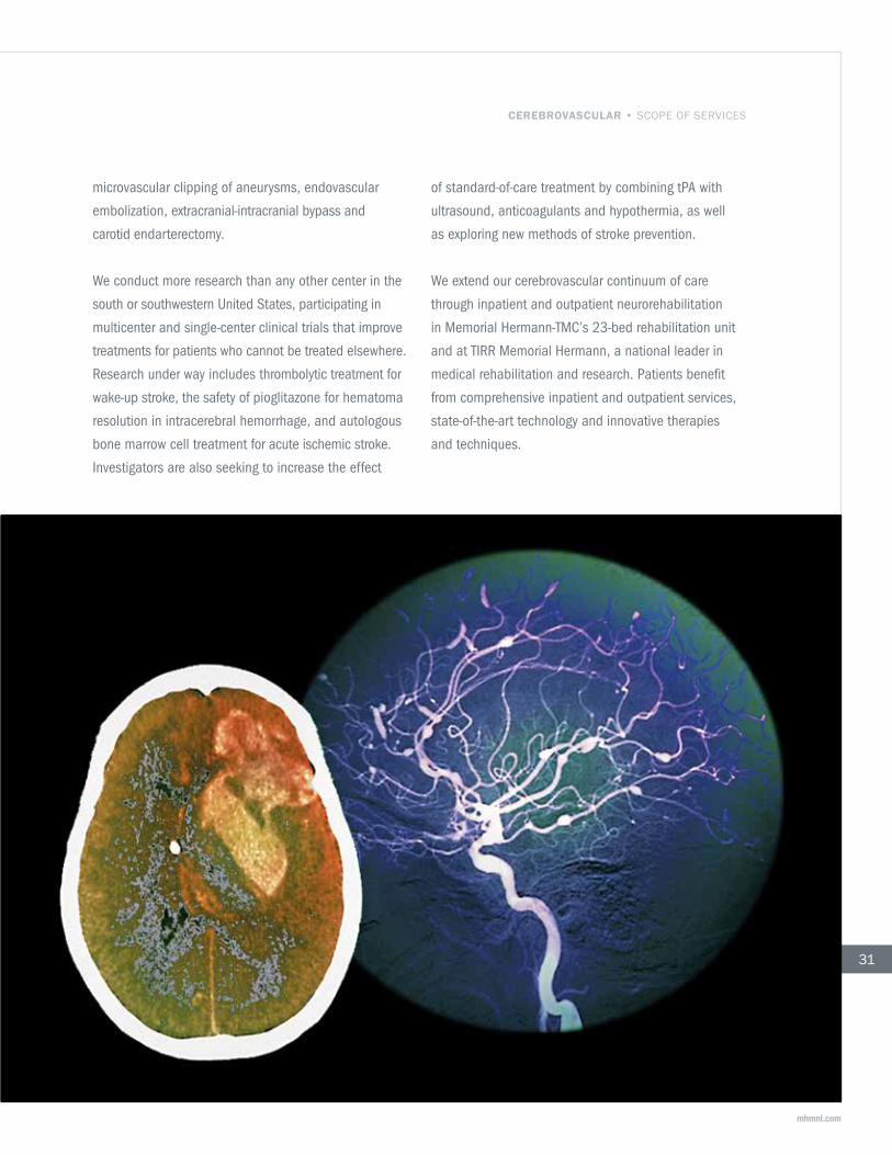
microvascular clipping of aneurysms, endovascular
embolization, extracranial-intracranial bypass and
carotid endarterectomy.
We conduct more research than any other center in the
south or southwestern United States, participating in
multicenter and single-center clinical trials that improve
treatments for patients who cannot be treated elsewhere.
Research under way includes thrombolytic treatment for
wake-up stroke, the safety of pioglitazone for hematoma
resolution in intracerebral hemorrhage, and autologous
bone marrow cell treatment for acute ischemic stroke.
Investigators are also seeking to increase the effect
of standard-of-care treatment by combining tPA with
ultrasound, anticoagulants and hypothermia, as well
as exploring new methods of stroke prevention.
We extend our cerebrovascular continuum of care
through inpatient and outpatient neurorehabilitation
in Memorial Hermann-TMC’s 23-bed rehabilitation unit
and at TIRR Memorial Hermann, a national leader in
medical rehabilitation and research. Patients benefit
from comprehensive inpatient and outpatient services,
state-of-the-art technology and innovative therapies
and techniques.
CerebrovasCular • SCOPE OF SErvICES

32
Acute Ischemic Stroke: Inpatient Mortality
0
0.02
0.04
0.06
0.08
0.1
0.12
0.14
FY 2009 FY 2010 FY 2011 FY 20120
200
400
600
800
1000
1200
1400Actual Expected Number of Encounters
Actual Expected Number of Encounters
Bette
r tha
n ex
pect
ed
Volu
me
Acute Ischemic Stroke: Length of Stay
0
0.5
1
1.5
2
2.5
3
3.5
FY 2009 FY 2010 FY 2011 FY 2012
FY 2009 FY 2010 FY 2011 FY 2012
ALO
S/CM
IAL
OS/
CMI
ALOS/CMI CMI
ALOS/CMI CMI
Cerebrovascular Volume
0
500
1,000
1,500
2,000
2,500
3,000
2009 2010 2011 2012
Number of Encounters
Intracerebral Hemorrhage: Inpatient Mortality
0
0.05
0.1
0.15
0.2
0.25
0.3
0.35
FY 2009 FY 2010 FY 2011 FY 20120
100
200
300
400
500
600
Bette
r tha
n ex
pect
ed
Volu
me
Intracerebral Hemorrhage: Length of Stay
0
0.5
1
1.5
2
2.5
3.5
4.5
3
4.0
Source: chart data based on ICD-9 coded diagnoses and procedures, per fiscal year
Source: chart data from the University HealthSystem ConsortiumSource: chart data from the University HealthSystem Consortium
Source: chart data from the University HealthSystem ConsortiumSource: chart data from the University HealthSystem Consortium
QuAlITY & OuTCOMES MEASurES

33
mhmni.com
FY 2009 FY 2010 FY 2011 FY 2012
ALO
S/CM
I
ALOS/CMI CMI
Sub-arachnoid Hemorrhage: Length of Stay
0
1
2
3
5
7
4
6
Sub-arachnoid Hemorrhage: Inpatient Mortality
0
0.05
0.1
0.15
0.2
0.25
FY 2009 FY 2010 FY 2011 FY 20120
50
100
150
200
250
300Actual Expected Number of Encounters
Bette
r tha
n ex
pect
ed
Volu
me
Aneurysm Unruptured: Length of Stay
0
.5
1
1.5
2.5
3.5
2
3
FY 2009 FY 2010 FY 2011 FY 2012
ALO
S/CM
I
ALOS/CMI CMI
Aneurysm Unruptured: Inpatient Mortality
0
0.01
0.02
0.03
0.04
0.05
0.06
0.07
FY 2009 FY 2010 FY 2011 FY 20120
20
40
60
80
100
180
160
140
120
Actual Expected Number of Encounters
Bette
r tha
n ex
pect
ed
Volu
me
Arteriovenous Malformation: Length of Stay
0
.5
1
1.5
2.5
3.5
2
3
FY 2009 FY 2010 FY 2011 FY 2012
ALO
S/CM
I
ALOS/CMI CMI
Arteriovenous Malformation: Inpatient Mortality
0
0.01
0.02
0.03
0.04
0.05
0.07
0.00% 0.00%
0.09
0.06
0.08
FY 2009 FY 2010 FY 2011 FY 20120
5
10
15
20
25
40
35
30
Actual Expected Number of Encounters
Bette
r tha
n ex
pect
ed
Volu
me
Source: chart data from the University HealthSystem ConsortiumSource: chart data from the University HealthSystem Consortium
Source: chart data from the University HealthSystem ConsortiumSource: chart data from the University HealthSystem Consortium
Source: chart data from the University HealthSystem ConsortiumSource: chart data from the University HealthSystem Consortium
Cerebrovascular

34
Children’s Neuroscience
With the arrival of David Sandberg, M.D., F.A.C.S.,
F.A.A.P., in June 2012, the Mischer Neuroscience
Institute added significant strength to its Children’s
Neuroscience Center. Dr. Sandberg joined the MNI
team as director of pediatric neurosurgery from Miami
Children’s Hospital and the University of Miami Miller
School of Medicine, where he was a voluntary associate
professor of clinical neurological surgery and pediatrics.
His major clinical interests include pediatric brain
tumors, minimally invasive endoscopic approaches
to brain tumors and hydrocephalus, congenital spinal
anomalies, vascular malformations, spasticity and
craniofacial disorders in children. Dr. Sandberg is a
national leader in developing novel techniques to treat
malignant brain tumors in children. Prior to arriving
in Houston, he performed translational studies that
demonstrated the safety of infusing chemotherapeutic
agents directly into the fourth ventricle to treat children
with malignant brain tumors in this location. The
promising results of these studies have led to the
initiation of a pilot clinical trial in collaboration with
The University of Texas MD Anderson Cancer Center,
which Dr. Sandberg is leading as principal investigator.
In addition to introducing novel treatment options for
pediatric brain tumors, Dr. Sandberg has published
extensively and lectured nationally and internationally
on minimally invasive endoscopic techniques to treat
both brain tumors and hydrocephalus. To avoid the
many complications of ventriculoperitoneal shunting
for children with hydrocephalus, our pediatric
neurosurgeons frequently perform endoscopic
techniques such as third ventriculostomy, septostomy,
choroid plexus coagulation and fenestration of
arachnoid cysts. Selected brain tumors can be biopsied
or removed completely via endoscopic techniques.
All of these procedures are performed via very small
incisions with minimal hair shaving. In collaboration
with our institution’s outstanding otolaryngology
colleagues, some tumors can be removed via
endoscopic transnasal approaches without an
external incision.
Our pediatric neurosurgeons at the Mischer
Neuroscience Institute are important members of
the Texas Fetal Center, a national leader in providing
diagnosis, treatment and complete care for mothers
with high-risk pregnancies and infants with congenital
anomalies or genetic conditions. The multidisciplinary
team performed the first fetal spina bifida repair in the
region, and patients are now being referred to our center
for fetal myelomeningocele repair from throughout
Texas and a number of surrounding states.
In collaboration with nationally recognized craniofacial
plastic surgeons, pediatric neurosurgeons at Children’s
Memorial Hermann Hospital perform both conventional
and minimally invasive endoscopic surgeries to repair
craniosynostosis and other complex craniofacial
anomalies. Our multidisciplinary Texas Cleft-Craniofacial
team was established in 1952 and has been a regional
leader for pediatric craniofacial surgery for decades.
S C O P E O F S E r v I C E S

35
mhmni.com
Mischer Neuroscience Institute is also a center
of excellence for pediatric epilepsy surgery and
comprehensive specialized care for children with
intractable epilepsy.
Our pediatric Epilepsy Monitoring Unit is the largest
and most comprehensive of its kind in the southwestern
United States. In addition to MRI and CT with low
radiation dose protocols for pediatric patients, we use
noninvasive magnetoencephalography (MEG) to map
brain activity to locate the source of epileptic seizures
and minimize risk for children undergoing resective
surgery for refractory epilepsy. For the most accurate
diagnosis we also use video EEG, PET, SPECT, memory
and speech (Wada) testing and neuropsychological
testing. We are one of only a few inpatient units in the
country with the capability to simultaneously perform
encephalography and polysomnography. Interventions
include medical management, immunotherapy and
the ketogenic diet as well as surgery, including vagus
nerve stimulation and laser ablation procedures.
We provide a broad range of diagnostic and treatment
services for children with complex neurological
problems, including autism, brachial plexus disorders,
brain tumors and malformations, cerebral palsy,
congenital hydrocephalus, craniofacial disorders,
developmental disorders, epilepsy, chronic headache
and migraine, head trauma, learning disabilities,
mitochondrial disorders, movement disorders,
myopathy, neurofibromatosis, neurometabolic disorders,
neuromuscular disorders, pediatric stroke, peripheral
nerve disorders, sleep disorders, spina bifida, Tourette
syndrome and tuberous sclerosis complex. We also
offer specialized pediatric neurosurgical expertise in
congenital malformations, including Chiari malformation,
endoscopic neurosurgery, and treatment for pediatric
stroke, spinal deformities and traumatic brain and
spine injury.
Care at Children’s Memorial Hermann Hospital is
delivered in a friendly, reassuring environment to
promote wellbeing and the best possible outcomes.
When surgery is required, we use advanced imaging
techniques and minimally invasive procedures that lower
patient risk. Onsite sedation is available for imaging
studies with care provided by specially trained pediatric
anesthesiologists and pediatric nurses.
36
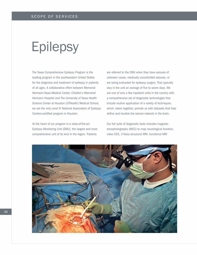
The Texas Comprehensive Epilepsy Program is the
leading program in the southwestern United States
for the diagnosis and treatment of epilepsy in patients
of all ages. A collaborative effort between Memorial
Hermann-Texas Medical Center, Children’s Memorial
Hermann Hospital and The University of Texas Health
Science Center at Houston (UTHealth) Medical School,
we are the only Level IV National Association of Epilepsy
Centers-certified program in Houston.
At the heart of our program is a state-of-the-art
Epilepsy Monitoring Unit (EMU), the largest and most
comprehensive unit of its kind in the region. Patients
are referred to the EMU when they have seizures of
unknown cause, medically uncontrolled seizures, or
are being evaluated for epilepsy surgery. They typically
stay in the unit an average of five to seven days. We
are one of only a few inpatient units in the country with
a comprehensive set of diagnostic technologies that
include routine application of a variety of techniques,
which, taken together, provide us with datasets that help
define and localize the seizure network in the brain.
Our full suite of diagnostic tools includes magneto-
encephalography (MEG) to map neurological function,
video EEG, 3-Tesla structural MRI, functional MRI
Epilepsy
S C O P E O F S E r v I C E S

37
mhmni.com
and diffusion tensor tractography, positron emission
tomography (PET), single photon emission computed
tomography (SPECT), memory and intracarotid amytal
(Wada) testing and in-depth neuropsychological
testing. We are a national leader in combining the
use of MEG and functional MRI to map the brain
and record brain activity. Each year, we perform more
than 150 MEG procedures on pediatric and adult patients.
We are also one of only a few inpatient units in the country
with the capability to perform electroencephalography
and polysomnography simultaneously.
The number of patients we treat annually continues
to grow. Today, our board-certified neurologists and
neurosurgeons diagnose and treat more than 450
pediatric and adult patients each year for seizures
and epilepsy. Genetics, brain trauma, structural
abnormalities, stroke and brain tumor rank among the
top underlying causes, but because epilepsy and other
types of seizures manifest differently among individuals,
determination of the origin of seizures is crucial to
planning the most effective treatment.
Once a diagnosis is made, we offer the most advanced
treatment options available, including drug therapy, the
ketogenic diet, vagus nerve stimulation (VNS), focal
cortical resection, lobectomy, hemispherectomy and
corpus callosotomy. Our surgical complication rates
have remained extremely low over the past eight years.
At the current time, our epilepsy surgeon has performed
Epilepsy Volume
0
100
200
300
400
500
2009 2010 2011 2012
Number of Encounters
Traumatic Brain Injury: Inpatient Mortality
0
0.02
0.04
0.06
0.08
0.10
0.14
0.12
FY 2009 FY 2010 FY 2011 FY 2012
0
Actual Expected Number of Encounters
Bette
r tha
n ex
pect
ed
Volu
me
200
400
600
800
1000
1200
1400
1600
Traumatic Brain Injury: Length of Stay
0
0.5
1
1.5
2
3
4
4.5
2.5
3.5
FY 2009 FY 2010 FY 2011 FY 2012
ALO
S/CM
I
ALOS/CMI CMI
The Texas Comprehensive Epilepsy Program has shown a dramatic increase in volumes in 2012. In the past year,
we have treated more than 450 pediatric and adult patients for seizures and epilepsy. Diagnostic tools that localize
the origin of seizures and map brain function, and advanced surgical procedures, including laser ablation and
stereo EEG, have allowed us to complete even more successful resective surgeries in fiscal year 2012.
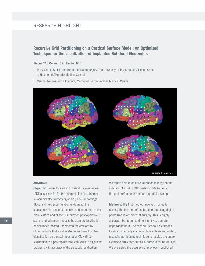
38
AbstrAct
objective: Precise localization of subdural electrodes
(SDEs) is essential for the interpretation of data from
intracranial electro-corticographic (ECoG) recordings.
Blood and fluid accumulation underneath the
craniotomy flap leads to a nonlinear deformation of the
brain surface and of the SDE array on post-operative CT
scans, and adversely impacts the accurate localization
of electrodes located underneath the craniotomy.
Older methods that localize electrodes based on their
identification on a post-implantation CT, with co-
registration to a pre-implant MRI, can result in significant
problems with accuracy of the electrode localization.
We report here three novel methods that rely on the
creation of a set of 3D mesh models to depict
the pial surface and a smoothed pial envelope.
Methods: The first method involves manually
picking the location of each electrode using digital
photographs obtained at surgery. This is highly
accurate, but requires time-intensive, operator-
dependent input. The second uses four electrodes
localized manually in conjunction with an automated,
recursive partitioning technique to localize the entire
electrode array constituting a particular subdural grid.
We evaluated the accuracy of previously published
recursive Grid partitioning on a cortical surface Model: An optimized technique for the Localization of implanted subdural electrodes
pieters tA1, conner cr1, tandon N1,2
1 The Vivian L. Smith Department of Neurosurgery, The University of Texas Health Science Center at Houston (UTHealth) Medical School
2 Mischer Neuroscience Institute, Memorial Hermann-Texas Medical Center
EpilepsyrESEArCH HIGHlIGHT
© 2012 Tandon Labs

methods by applying them to our data and comparing
them against the photograph-based localization.
Lastly, we further enhanced the usability of our new
methods by using automatic parcellation techniques
to assign anatomic labels to individual electrodes as
well as by implementing a strategy to generate an
inflated cortical surface model while still preserving
electrode locations relative to the cortical anatomy.
results: The recursive grid partitioning had the
least error in comparison to older methods (672
electrodes, 6.4 mm maximum single electrode error,
1.97 mm mean error, p <10-18). The maximum
errors derived using prior methods of localization
ranged from 8.20 mm to 11.73 mm for an individual
electrode, with mean errors ranging between 2.9
and 4.1 mm depending on the method used. We
also noted a larger error in all methods that used CT
scans alone to localize electrodes as compared to
those that used both post-op CT and post-op MRI.
The large mean errors reported with these methods
are liable to affect inter-modal data comparisons
(e.g., with functional mapping techniques) and may
impact surgical decision-making.
conclusion: We have presented several aspects of using
a new technique to visualize electrodes implanted
for localizing epilepsy. The ability to use automated
labeling schema to denote which gyrus a particular
electrode overlies is potentially of great utility in planning
resections, and in corroborating the results of extra-
operative stimulation mapping. Dilation of the pial mesh
model provides, for a first time, a sense of the cortical
surface not sampled by the electrode, and the potential
roles this “electrophysiologically hidden” cortex may
play in both eloquent functions and seizure onsets.
39
more than 350 craniotomies for the treatment of
epilepsy, with a zero percent mortality rate and a very
low rate of permanent morbidity from such operations.
We also go beyond diagnosis and treatment of epilepsy
by counseling patients in ways to cope with their
diagnosis. Specialized counselors ensure that recently
diagnosed patients have the emotional support they
need.
Our team has been involved in research related to most
epilepsy treatments approved in the United States
in the last 15 years, including a number of drug and
intravenous therapies and VNS therapy. Current research
includes lacosamide monotherapy and adjunctive
therapy for partial-onset seizures; an open-label
extension study of rufinamide as an adjunctive therapy
in patients with refractory partial-onset seizures; the
use of tractography to lateralize temporal lobe epilepsy
and to delineate the epileptogenic network; oxygen-
enhanced magnetic resonance imaging in non-lesional
focal epilepsy; correlation of waking background alpha
frequency with measures of attention and reaction;
and the use of intracranial electrocorticography to
study a variety of cognitive and language processes.
Another new innovation is the application of
stereoencephalography for the localization of epilepsy
arising in deep foci in the brain. This technique blends
the use of frame based stereotaxy with the 3D planning
capabilities of modern neuro-navigation software, to
help localize epilepsy in a minimally invasive fashion.
MNI is a pioneering site for the application of laser
surgery to the treatment of well-delineated focal
epilepsies, with carefully selected patients being treated
with this highly advanced, minimally invasive approach
to the ablation of the seizure focus.
epilepsy • SCOPE OF SErvICESEpilepsy
Journal of Neurosurgery. In press.
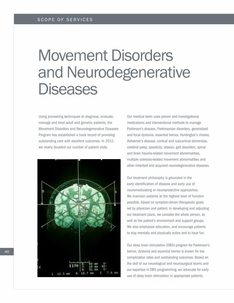
S C O P E O F S E r v I C E S
Movement Disorders and Neurodegenerative Diseases Using pioneering techniques to diagnose, evaluate,
manage and treat adult and geriatric patients, the
Movement Disorders and Neurodegenerative Diseases
Program has established a track record of providing
outstanding care with excellent outcomes. In 2012,
we nearly doubled our number of patient visits.
Our medical team uses proven and investigational
medications and interventional methods to manage
Parkinson’s disease, Parkinsonian disorders, generalized
and focal dystonia, essential tremor, Huntington’s chorea,
Alzheimer’s disease, cortical and subcortical dementias,
cerebral palsy, spasticity, ataxias, gait disorders, spinal
and brain trauma-related movement abnormalities,
multiple sclerosis-related movement abnormalities and
other inherited and acquired neurodegenerative diseases.
Our treatment philosophy is grounded in the
early identification of disease and early use of
neuromodulating or neuroprotective approaches.
We maintain patients at the highest level of function
possible, based on symptom-driven therapeutic goals
set by physician and patient. In developing and adjusting
our treatment plans, we consider the whole person, as
well as the patient’s environment and support groups.
We also emphasize education, and encourage patients
to stay mentally and physically active and to have fun.
Our deep brain stimulation (DBS) program for Parkinson’s
tremor, dystonia and essential tremor is known for low
complication rates and outstanding outcomes. Based on
the skill of our neurological and neurosurgical teams and
our expertise in DBS programming, we advocate for early
use of deep brain stimulation in appropriate patients.
40

41
mhmni.com
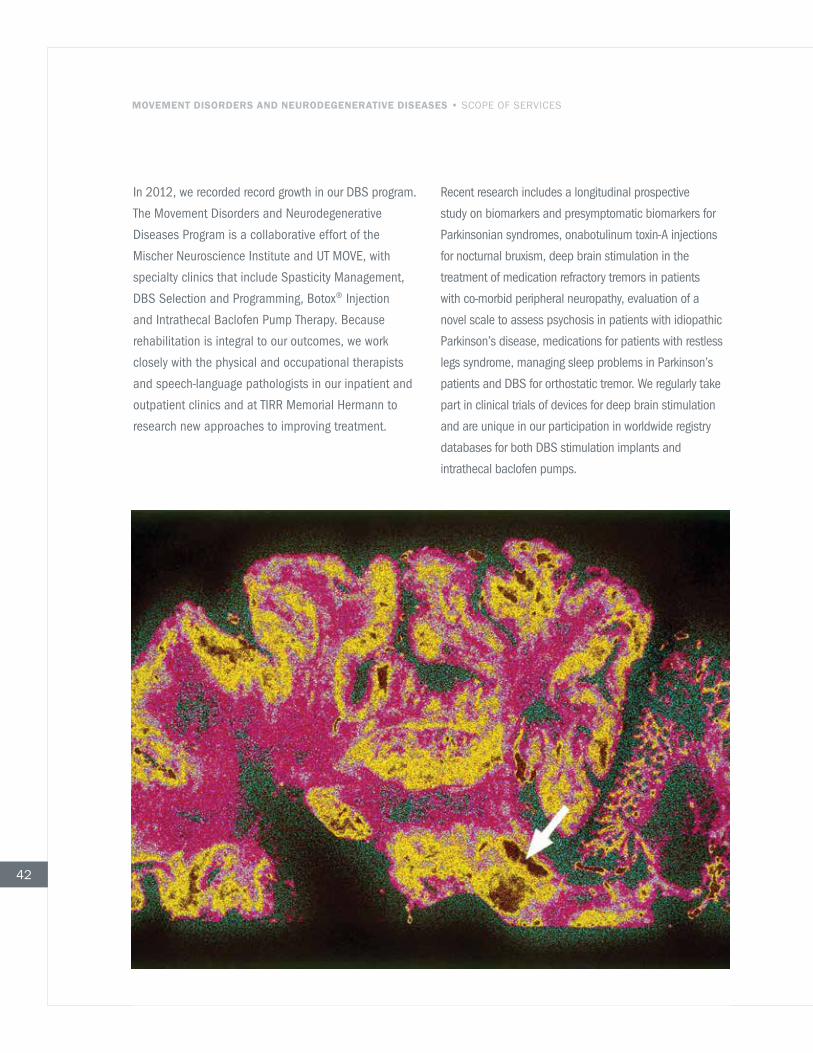
42
In 2012, we recorded record growth in our DBS program.
The Movement Disorders and Neurodegenerative
Diseases Program is a collaborative effort of the
Mischer Neuroscience Institute and UT MOVE, with
specialty clinics that include Spasticity Management,
DBSSelectionandProgramming,Botox®Injection
and Intrathecal Baclofen Pump Therapy. Because
rehabilitation is integral to our outcomes, we work
closely with the physical and occupational therapists
and speech-language pathologists in our inpatient and
outpatient clinics and at TIRR Memorial Hermann to
research new approaches to improving treatment.
Recent research includes a longitudinal prospective
study on biomarkers and presymptomatic biomarkers for
Parkinsonian syndromes, onabotulinum toxin-A injections
for nocturnal bruxism, deep brain stimulation in the
treatment of medication refractory tremors in patients
with co-morbid peripheral neuropathy, evaluation of a
novel scale to assess psychosis in patients with idiopathic
Parkinson’s disease, medications for patients with restless
legs syndrome, managing sleep problems in Parkinson’s
patients and DBS for orthostatic tremor. We regularly take
part in clinical trials of devices for deep brain stimulation
and are unique in our participation in worldwide registry
databases for both DBS stimulation implants and
intrathecal baclofen pumps.
movement DisorDers anD neuroDegenerative Diseases • SCOPE OF SErvICES

43
mhmni.com
Rapid eye movement sleep disorder (RBD), a sleep disturbance, is one of the most common non-motor
complications of Parkinson’s disease (PD). RBD frequently arises as an early manifestation in PD, likely reflecting
the anatomical areas affected by the neurodegenerative process. While specific neuropathological aspects
shared by RBD and PD have yet to be fully documented, further characterization is critical to discovering reliable
biomarkers that predict PD onset.
In a recent study, we tested the hypothesis of a diminished functional connection between substantia nigra
and striatum in patients diagnosed with RBD. We measured functional correlations of substantia nigra using
rest-state blood oxygen level-dependent functional magnetic resonance imaging (BOLD-fMRI) in a group of
idiopathic RBD patients at risk for developing PD, a group of diagnosed PD patients and a group of aged controls.
Significantly reduced correlations (p<0.01, corrected) in RBD and PD patients compared to controls were found
between substantia nigra and putamen using voxelwise tests and single-subject region of interest analyses
indicating impairment of nigrostriatal functional connectivity.
reduced resting state Functional connectivity as an imaging pre-symptomatic biomarker of parkinson’s Disease
PRINCIPAL INVESTIGATOR: Mya c. schiess, M.D.Professor and Adriana Blood Chair in NeurologyDepartment of Neurology The University of Texas Health Science Center at Houston (UTHealth) Medical School
Movement Disorders and Neurodegenerative Diseases Movement Disorders and Neurodegenerative Diseases rESEArCH HIGHlIGHT
1
0.8
0.6
0.4
0.2
-0.3 -0.2 -0.1 0 0.1 0.2 0.3 0.4 0.5
Prob
abili
ty d
ensi
ty e
stim
ate
Left SN – left putamen average functional correlation
PD RBD like PD RBD like CON CON
individual subject Left sN – Left putamen Average correlation Values and Modeled Distributions

44
The Mischer Neuroscience Institute’s Multiple Sclerosis
Program has established a track record of leading-edge
care using groundbreaking techniques to diagnose,
evaluate, manage and treat adult patients with MS
and other demyelinating disorders. Our scope of
expertise is broad and includes patients in all stages of
MS, as well as those with neuromyelitis optica, transverse
myelitis and optic neuritis. We are experienced in the
appropriate use of aggressive therapies in severe cases.
Organized in 1983, the Multiple Sclerosis Research
Group (MSRG) has participated in numerous clinical
trials of novel disease-modifying therapies, serving as
the lead center for numerous international studies,
several of which were pivotal in gaining FDA approval
of currently available treatments for MS. Recently
completed research includes a National Institutes
of Health-sponsored trial of combined therapy with
interferon beta-1a and glatiramer acetate in patients
with early relapsing MS (the CombiRx Trial); the safety
and efficacy of oral fampridine-SR, detection of MS-
related cognitive impairment, Epstein-Barr virus and
MS, and serial magnetic resonance spectroscopy in
MS, among others. Investigators in the MSRG and the
department of Diagnostic and Interventional Imaging
also recently completed a National MS Society-
sponsored study of chronic cerebrospinal vascular
insufficiency (CCSVI).
We were the first center in the world to conduct
preclinical studies on the effects of combined therapy
with immunomodulating drugs and to explore the
effects of oral cytokines in modulating MS and Type 1
diabetes. We are the first and only center in Houston
to direct national and international clinical trials in MS,
and we remain the North American leader in studies of
primary progressive multiple sclerosis, as well as the
most active center in Texas in the conduct of organized
clinical trials of new therapies for MS. Our physicians
are at the forefront of investigator-initiated research
Multiple Sclerosis
S C O P E O F S E r v I C E S

45
mhmni.com
in immune regulation in MS, infection as a cause of
MS, MS-related cognitive impairment and MS-related
MRI findings.
In our state-of-the-art Magnetic Resonance Imaging
Analysis Center, we use spectroscopic and diffusion
tensor imaging with tractotomy, as well as other
advanced diagnostic tools. Following diagnosis, patients
benefit from breakthrough treatment options that include
injectable immunomodulators, immunosuppressives
and other agents designed to treat the debilitating
symptoms of MS. Investigators also use the MRI
Analysis Center to monitor the effects of promising oral
drugs in pivotal efficacy trials.
Our goal is to maintain our patients at the highest level
of function possible, with early use of immunoactive
agents to prevent disease progression. Because
rehabilitation is integral to each patient’s treatment
plan, we work closely with the physical medicine
and rehabilitation specialists and therapists at TIRR
Memorial Hermann, a national leader in medical
rehabilitation and research, as well as the inpatient
neurorehabilitation team at MNI.
PRINCIPAL INVESTIGATOR: Jerry s. Wolinsky, M.D.Professor, Bartels Family Professor and Opal C. Rankin Professor in Neurology, Department of Neurology The University of Texas Health Science Center at Houston (UTHealth) Medical School
Many centers have postulated that there is a significant
impairment of cerebral venous drainage in multiple
sclerosis (MS), which has led to increased utilization of
imaging for chronic cerebrospinal venous insufficiency
(CCSVI). Our facility participated in a prospective
case-control study to evaluate the association of CCSVI
with MS. The study evaluated imaging of extracranial
and intracranial venous drainage among 276 patients:
206 had MS and 70 did not. Overall, 82 subjects
(29.7 percent) fulfilled one of five criteria for CCSVI;
13 (4.7 percent) fulfilled two criteria and none fulfilled
greater than two criteria. The distribution of subjects with
zero, one or two criteria did not differ significantly across
all diagnostic groups, between patients with or without
multiple sclerosis. No significant differences emerged
between MS and non-MS subjects for extracranial or
intracranial venous flow rates. Our findings described
as CCSVI are much less prevalent than previously
reported and do not distinguish MS subjects from other
subjects. The findings do not support the postulate that
CCSVI is causally associated with MS.
On May 10, 2012, the U.S. Food & Drug Administration released a safety communication alerting people with MS to the risk of serious injury and death associated with procedures to treat CCSVI. According to the FDA, “The benefits of these experimental procedures have not been proven, and their promotion as a treatment for MS may lead people with the disease to make treatment decisions without being aware of the serious risks involved.”
prospective case-control study of chronic cerebrospinal Venous insufficiency: Neurosonography results
00206000T O T A LT O T A L T O T A LT O T A L
7.14%
NON-MS PATIENTSMS PATIENTS
Neurosonographic Criteria of CCSVI Present
3.88%
007000
Multiple SclerosisrESEArCH HIGHlIGHT
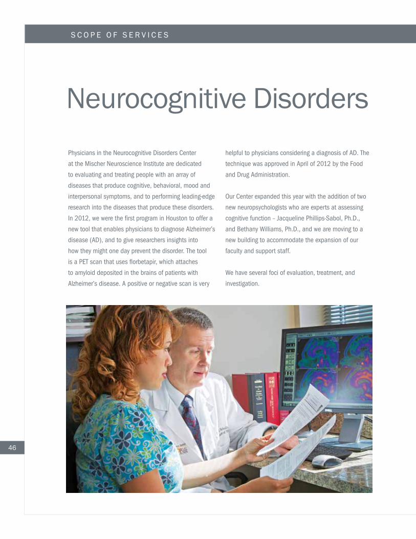
46
Physicians in the Neurocognitive Disorders Center
at the Mischer Neuroscience Institute are dedicated
to evaluating and treating people with an array of
diseases that produce cognitive, behavioral, mood and
interpersonal symptoms, and to performing leading-edge
research into the diseases that produce these disorders.
In 2012, we were the first program in Houston to offer a
new tool that enables physicians to diagnose Alzheimer’s
disease (AD), and to give researchers insights into
how they might one day prevent the disorder. The tool
is a PET scan that uses florbetapir, which attaches
to amyloid deposited in the brains of patients with
Alzheimer’s disease. A positive or negative scan is very
helpful to physicians considering a diagnosis of AD. The
technique was approved in April of 2012 by the Food
and Drug Administration.
Our Center expanded this year with the addition of two
new neuropsychologists who are experts at assessing
cognitive function – Jacqueline Phillips-Sabol, Ph.D.,
and Bethany Williams, Ph.D., and we are moving to a
new building to accommodate the expansion of our
faculty and support staff.
We have several foci of evaluation, treatment, and
investigation.
Neurocognitive Disorders
S C O P E O F S E r v I C E S

47
mhmni.com
rESEArCH HIGHlIGHT Neurocognitive Disorders
QureshiSU,KimbrellT,PyneJM,MagruderKM,HudsonTJ,PetersenNJ,YuH-J,SchulzPE,KunikME
A recent article demonstrated that the odds of being diagnosed with dementia are
2.3 times as high in veterans with post-traumatic stress disorder (PTSD). That was
true even when comparing veterans with PTSD, a psychologic trauma, to veterans
with combat-related injuries, which produce both physical and psychologic stress.
Other factors related to dementia risk were gender, race and medical illnesses,
including diabetes mellitus, elevated cholesterol, high blood pressure, coronary artery
disease, stroke, traumatic brain injury, smoking, alcohol abuse and dependency,
and drug abuse and dependency. This study, then, suggests that severe psychologic
stress can cause long-lasting changes in the brain that put people at very increased
risks for developing dementia. This finding is also important to civilians, who develop
PTSD at comparable rates to service men and women after stressful events, such as
serious car accidents.
Importantly, it was also found that one risk factor can increase the chances of having another risk factor for
dementia. For example, mild TBI at the time of a stressful event increases the odds of developing PTSD from
23 to 60 percent. In the other direction, we have demonstrated that controlling risk factors for dementia, like
hypertension, reduces the risk of developing the disorder.1
Current approaches to treating dementia are to implement treatment after a patient has been diagnosed
with dementia. At the Neurocognitive Disorders Center, we are investigating screening tools that may predict
dementia prior to its onset. In 2012, we were the first program in Houston to offer a new tool approved by
the FDA that enables physicians to diagnose Alzheimer’s disease (AD). A florbetapir PET scan demonstrates
amyloid plaques in the brains of patients with AD, thereby allowing a diagnosis of AD in a living patient, which
formerly was possible only at autopsy. This powerful tool may allow us to diagnose amyloid deposition before
the symptoms of dementia start. Additional research is being conducted to identify how to prevent or delay
dementia in patients in whom amyloid is visualized, and replacing lost neurons with stem cells.
1 JohnsonML,ParikN,KunikME,SchulzPE,PatelJG,ChenH,AparasuRR,MorganRO.AntihypertensiveDrug
Use and the Risk of Dementia in Patients with Diabetes Mellitus. Alzheimer’s and Dementia, 2012, 1-8.
Greater prevalence and incidence of Dementia in older Veterans with post-traumatic stress Disorder
Journal of the American Geriatrics Society, 2010, 58:1627-1633

48

49
mhmni.com
Our Memory Disorders and Dementia Clinic is dedicated
to assessing and treating all the known causes of
dementia, including infections, trauma, medications,
vitamin or hormone deficiencies, vascular disease and
strokes, and neurodegenerative disorders, such as
AD, frontotemporal dementia (FTD) and Parkinson’s
disease. Research efforts in the MNI Memory Disorders
and Dementia Clinic are directed at developing ways
to provide early diagnosis and treatment for these
devastating illnesses. This includes determining which
individuals will develop dementia, finding accurate tools
to diagnose dementia and determine the type from
which a person suffers, and finding ways to stop the
progression of dementing illnesses.
A second focus is in neuropsychiatry, in which we
determine whether patients with psychiatric symptoms
have an underlying neurologic cause, or whether
patients with neurologic symptoms may have an
underlying psychiatric disorder. The major foci of our
research in this area are traumatic brain injury (TBI) and
post-traumatic stress disorder (PTSD), both of which
cause neurologic and psychiatric symptoms. Together
with the Center for Translational Injury Research, we
are investigating treatments for TBI and the causes,
treatments and prevention of PTSD.
A third area of focus is our Stroke and Dementia
Prevention Program, which is dedicated to reducing
the risk of developing dementia and to developing
new diagnostic and treatment tools. Our team has
identified multiple risk factors for dementia, including
family history, head trauma, high blood pressure, high
cholesterol or triglycerides, diabetes, obesity, smoking,
post-traumatic stress disorder and many others. In
this clinic, physicians evaluate individuals at risk for
dementia who are asymptomatic, and treat their risk
factors. Risk factor reduction is associated with a
significant decrease in a person’s risk for developing
dementia. Research efforts in this clinic are focused on
investigating new treatments to delay or ameliorate the
development of dementia. We believe that treatment
interventions will be much more effective if introduced
early in the molecular cascade of events that lead to
these diseases – before dementia symptoms develop.
A fourth area of focus is frontotemporal dementia.
FTD typically onsets in the 50s and causes devastating
behavioral changes in people at the prime of their
lives. Our clinic is dedicated to evaluating and treating
patients with FTD, and investigating the environmental
and genetic causes of FTD. We are also investigating
its relationship to amyotrophic lateral sclerosis,
a devastating disorder that produces progressive
weakness.
Our laboratory partner is the George P. and Cynthia
W. Mitchell Center for Research in Alzheimer’s disease
and Related Brain Disorders under the direction of
Claudio Soto, Ph.D. Findings are taken from the clinic
to the laboratory and from the laboratory to the clinic
to develop new diagnostic methods and treatments
for dementia. For example, together we are identifying
biomarker changes in animals that can be studied
as diagnostic tools in humans, and we are testing
medications to treat these diseases.
By using sophisticated new technologies to study these
disorders at the cellular and clinical levels, we hope to
realize our goal of diagnosis before patients become
symptomatic with dementia and to find new treatments
to delay or prevent the development of dementia.
neuroCognitive DisorDers • SCOPE OF SErvICES

50
The GBS/CIDP Foundation International has designated
the Mischer Neuroscience Institute’s Neuromuscular
Disorders Program a center of excellence for the
diagnosis and treatment of Guillain-Barré syndrome,
chronic inflammatory demyelinating polyneuropathy
(CIDP) and other inflammatory peripheral neuropathies.
The designation was awarded in recognition of the
high standards maintained and quality of patient
care provided through the program, which is one of
only 10 such centers of excellence in the United States
and abroad.
Physicians affiliated with the Neuromuscular
Disorders Program are subspecialized in complex
neuromuscular disorders that are difficult to diagnose
and treat, including neurodegenerative disorders,
inflammatory nerve and muscle disorders, autoimmune
neuromuscular junction disorders, traumatic nerve
injuries and toxic metabolic disorders of the peripheral
nerves and muscles. The program records more than
2,000 patient visits annually, primarily adults age 18
and older. About two-thirds of our patients are over
the age of 50.
Our neurodiagnostic facilities include a state-of-the-art
Electromyography (EMG) Laboratory and a Muscle
and Nerve Laboratory. The EMG Lab provides
comprehensive nerve conduction studies and EMG
evaluations performed by expert staff.
Because electrodiagnostic evaluation is an extension
of clinical findings, our medical specialists perform a
focused neuromuscular examination, including history
and physical, before conducting the electrical test. In
addition to nerve conduction and EMG, electrodiagnostic
studies available at the lab include repetitive nerve
stimulation, blink reflexes, cranial nerve studies, single-
fiber electromyography and facial/trigeminal neuropathy.
An invaluable diagnostic test, EMG provides evidence
in support of diagnoses of peripheral neuropathies;
motor neuron diseases such as amyotrophic lateral
sclerosis and spinal muscular atrophy; muscle
disorders such as myopathy and muscular dystrophy;
neuromuscular junction disorders such as myasthenia
gravis; entrapment neuropathies such as carpal tunnel
syndrome, ulnar and peroneal neuropathies; and
Neuromuscular Disorders
S C O P E O F S E r v I C E S

51
mhmni.com
Movement Disorders and Neurodegenerative Diseases rESEArCH HIGHlIGHT Neuromuscular Disorders
Zhang G1, Lehmann HC2, Bogdanova N1, Gao T1, Zhang J3,SheikhKA1
1Department of Neurology, The University of Texas Health Science Center at Houston (UTHealth) Medical School, United States of America; 2Department of Neurology, Heinrich-Heine-University Düsseldorf, Düsseldorf, Germany; 3Department of Radiology, Johns Hopkins University, Baltimore, Maryland, United States of America
AbstrAct
Guillain-Barré syndrome (GBS) is a monophasic immune neuropathic disorder in which a significant proportion
of patients have incomplete recovery. The patients with incomplete recovery almost always have some degree of
failure of axon regeneration and target reinnervation. Anti-ganglioside antibodies (Abs) are the most commonly
recognized autoimmune markers in all forms of GBS and specific Abs are associated with the slow/poor
recovery. We recently demonstrated that specific anti-ganglioside Abs inhibit axonal regeneration and nerve
repair in preclinical models by activation of small GTPase RhoA and its downstream effectors. The objective
of this study was to determine whether erythropoietin
(EPO), a pleiotropic cytokine with neuroprotective and
neurotrophic properties, enhances nerve regeneration in
preclinical cell culture and animal models of autoimmune
neuropathy/nerve repair generated with monoclonal
and patient derived Abs. Primary neuronal cultures and
a standardized sciatic crush nerve model were used to
assess the efficacy of EPO in reversing inhibitory effects
of anti-ganglioside Abs on nerve repair. We found that
EPO completely reversed the inhibitory effects of anti-
ganglioside Abs on axon regeneration in cell culture models
and significantly improved nerve regeneration/repair in
an animal model. Moreover, EPO-induced proregenerative effects in nerve cells are through EPO receptors and
Janus kinase 2/Signal transducer and activator of transcription 5 pathway and not via early direct modulation of
small GTPase RhoA. These preclinical studies indicate that EPO is a viable candidate drug to develop further for
neuroprotection and enhancing nerve repair in patients with GBS.
copyright: 2011 Zhang et al. this is an open-access article distributed under the terms of the creative commons Attribution
License, which permits unrestricted use, distribution, and reproduction in any medium, provided the original author and
source are credited.
erythropoietin enhances Nerve repair in Anti-Ganglioside Antibody-Mediated Models of immune Neuropathy
PLoS ONE 6(10): e27067. doi:10.1371/journal.pone.0027067

52
traumatic nerve injury, including evaluation of the brachial
plexus and facial neuropathy. The Neuromuscular
Disorders Program is the only program in Houston that
provides single-fiber EMG.
Our Muscle and Nerve Laboratory helps improve
diagnosis in cases with limited neuromuscular findings
by locating abnormalities at a pathologic/microscopic
level. Affiliated subspecialists perform muscle, nerve
and skin biopsies, which are further processed by
highly experienced staff. Our preferred technique is
open biopsy under local anesthesia, which reduces the
likelihood of missing abnormalities in cases of patchy
involvement, such as in inflammatory myopathies.
In 2012, we added skin biopsies for the diagnosis of
small-fiber neuropathy to our comprehensive list of
diagnostic tests. The Neuromuscular Disorders Program
is the only center in Houston that processes skin biopsy
specimens for the diagnosis of small-fiber neuropathies.
Current research is focused on developing new
strategies to treat neuropathic disorders and enhance
nerve repair. With funding from the National Institutes
of Health and the GBS/CIDP Foundation International,
our investigators are studying the pathogenesis
of autoimmune neuropathies, immune effectors
and nerve repair, novel strategies to enhance axon
regeneration and nerve repair, and the development of
MRI technology to assess neuromuscular disorders in
preclinical and clinical studies.
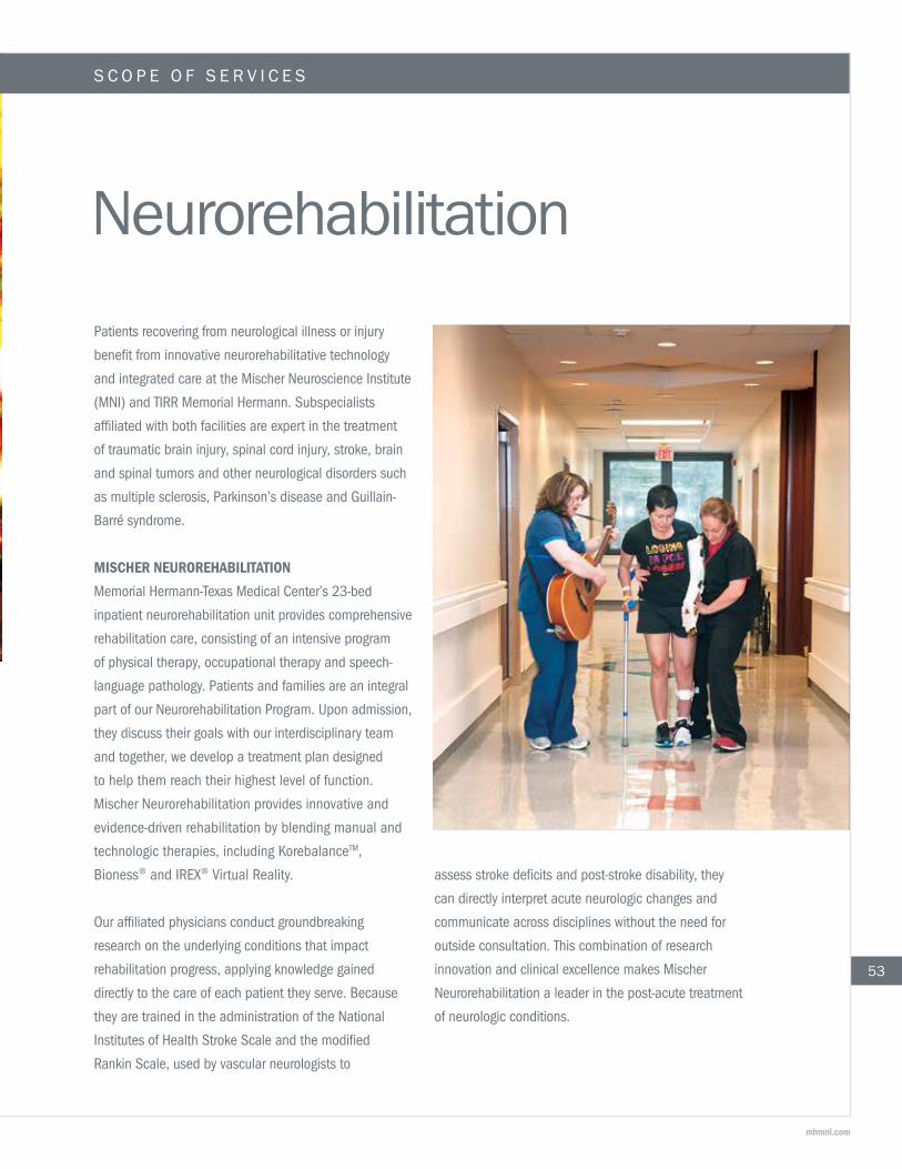
53
mhmni.com
Neurorehabilitation
Patients recovering from neurological illness or injury
benefit from innovative neurorehabilitative technology
and integrated care at the Mischer Neuroscience Institute
(MNI) and TIRR Memorial Hermann. Subspecialists
affiliated with both facilities are expert in the treatment
of traumatic brain injury, spinal cord injury, stroke, brain
and spinal tumors and other neurological disorders such
as multiple sclerosis, Parkinson’s disease and Guillain-
Barré syndrome.
Mischer NeurorehAbiLitAtioN
Memorial Hermann-Texas Medical Center’s 23-bed
inpatient neurorehabilitation unit provides comprehensive
rehabilitation care, consisting of an intensive program
of physical therapy, occupational therapy and speech-
language pathology. Patients and families are an integral
part of our Neurorehabilitation Program. Upon admission,
they discuss their goals with our interdisciplinary team
and together, we develop a treatment plan designed
to help them reach their highest level of function.
Mischer Neurorehabilitation provides innovative and
evidence-driven rehabilitation by blending manual and
technologictherapies,includingKorebalanceTM,
Bioness®andIREX®VirtualReality.
Our affiliated physicians conduct groundbreaking
research on the underlying conditions that impact
rehabilitation progress, applying knowledge gained
directly to the care of each patient they serve. Because
they are trained in the administration of the National
Institutes of Health Stroke Scale and the modified
Rankin Scale, used by vascular neurologists to
assess stroke deficits and post-stroke disability, they
can directly interpret acute neurologic changes and
communicate across disciplines without the need for
outside consultation. This combination of research
innovation and clinical excellence makes Mischer
Neurorehabilitation a leader in the post-acute treatment
of neurologic conditions.
S C O P E O F S E r v I C E S

37
$1.17 million collaborative grant from the National
Institutes of Health (NIH) and the President’s
National Robotics Initiative (NRI) will enable four
Houston institutions to create and validate a human-
robot interface with a noninvasive brain-machine
to help stroke patients recover use of their upper
limbs to the fullest extent possible. Awarded to Rice
University, the University of Houston (UH) and The
University of Texas Health Science Center at Houston
(UTHealth) Medical School, the grant will fund the
development of neurotechnology that will interpret
the brainwaves of stroke patients, allowing them
to use their thoughts to operate an exoskeleton
that wraps around the arm from the fingertips to
the elbow. The device will be validated by UTHealth
physicians and scientists at TIRR Memorial Hermann
in up to 40 volunteer patients during the final two
years of the four-year R01 award.
When set into motion, the intelligent exoskeleton
will use thoughts to trigger repetitive motions and
retrain the brain’s motor networks. Marcia O’Malley,
brain-Machine interface control of a therapeutic exoskeleton
PRINCIPAL INVESTIGATOR AT UTHEALTH: gerard e. Francisco, m.D. Professor and Chair, Department of Physical Medicine and RehabilitationThe University of Texas Health Science Center at Houston (UTHealth) Medical SchoolChief Medical Officer, TIRR Memorial HermannDirector, UTHealth Physical Medicine and Rehabilitation Motor Recovery Lab
PRINCIPAL INVESTIGATOR AT RICE UNIVERSITY: Marcia o’Malley, ph.D.Associate Professor of Mechanical Engineering and Materials ScienceDirector, Mechatronics and Haptic Interfaces (MAHI) LabRice UniversityDirector of Rehabilitation Engineering, TIRR Memorial Hermann
PRINCIPAL INVESTIGATOR AT THE UNIVERSITY OF HOUSTON: José contreras-Vidal, ph.D.Professor of Electrical and Computer EngineeringDirector of the UH Laboratory for Noninvasive Brain Machine Interface Systems The University of Houston
rESEArCH HIGHlIGHT
tirr MeMoriAL herMANN
A national leader in medical rehabilitation and
research, TIRR Memorial Hermann is a model for
interdisciplinary rehabilitation services, patient care,
education and research. The hospital has more than
50 years of experience in rehabilitation and research
and is one of only six in the nation designated as a
model system by the National Institute on Disability
and Rehabilitation Research (NIDRR) for its traumatic
brain injury program. For 23 consecutive years,
U.S.News & World Report has named the hospital
54
to its list of “America’s Best Hospitals.” In 2012,
TIRR Memorial Hermann rose from fourth in the nation
to third.
Research done at the hospital is conducted by
physicians and scientists, and also by our therapists,
nurses, chaplain and the residents who rely on us
to advance their knowledge in specialized areas of
rehabilitation medicine. Our Brain Injury Research
Center (BIRC) brings together world-renowned
researchers to study the many complicated facets

Ph.D., and her Rice University team are developing
the exoskeleton in the Mechatronics and Haptic
Interfaces (MAHI) Lab. At the University of
Houston, a team led by principal investigator
José Contreras-Vidal, Ph.D., is developing the
electroencephalograph-based neural interface.
Dr. Contreras-Vidal’s team was the first to
successfully reconstruct 3-D hand and walking
movements from brain signals recorded
noninvasively using an EEG brain cap. The
technology allows users to control robotic legs
with their thoughts and also permits below-elbow
amputees to control neuroprosthetic limbs.
The new project will be one of the first to design
a brain-machine interface (BMI) system for
stroke survivors.
Rice University’s robotic devices and UH’s neural
interfaces will make it possible for principal
investigator Gerard Francisco, M.D., and his team
to facilitate translational research that will fast track
engineering findings into clinical practice.
The project, funded through the National Institute
of Neurological Disorders and Stroke (NINDS), is
one of only a few projects selected by the NRI, a
collaborative partnership of the NIH, National Science
Foundation, NASA and the Department of Agriculture
to encourage the development of the next generation
of robots that will work closely with humans.
Neurorehabilitation
55
mhmni.com
of recovery from brain injury, leveraging resources
from NIDRR to conduct research identifying effective
treatments. In 2012, our Spinal Cord Injury Research
Center (SCIRC) broadened our team with the addition
of experts in cognitive and psychosocial research.
These new faculty allow us to use a holistic approach
to studying various aspects of recovery for people with
spinal cord injury and disorders across the lifespan,
and identify new ways to improve function and quality
of life. Ongoing research at the UTHealth Motor
Recovery Lab at TIRR Memorial Hermann revolves
around spasticity management, robotic therapy,
neural interface and noninvasive brain stimulation.
TIRR Memorial Hermann has added two Restorative
Therapies FES (functional electronic stimulator) bikes,
Bioness®handrehabilitationandfootdropsystems,
theVitalStimExperia™clinicalunitandIOPIMedical’s
Iowa Oral Performance Instrument to its growing list of
innovative rehabilitation technologies. A new inpatient
sleep lab provides diagnosis of sleep-disruptive disorders
in patients recovering from stroke or traumatic brain injury.

56
0
10
20
30
40
60
80
50
70
37.38
58.95
47.40
70.87
All Neurological
Neurorehabilitation: Functional Independence Measure Change
0
10
20
30
40
60
80
50
70
TIRR RegionNation
Brain Injuries Treated
Perc
ent T
raum
atic
(%)
0
10
20
30
40
60
80
50
70
TIRR RegionNation
Spinal Cord Injuries Treated
Perc
ent T
raum
atic
(%)
Admit FIM Discharge FIM
neurorehabilitation • SCOPE OF SErvICES
Our Outpatient Medical Clinic is a physician-based
clinic designed to meet the needs of individuals with
disabilities age 13 and older who require initial or
continuing care by a physician. The clinic is redefining
the hospital’s outpatient rehabilitation care model by
providing a patient-centered medical home for people
with disabilities. Seventeen specialty medical clinics
include brain injury, stroke, spasticity management,
neurosurgery, neurology, neuropsychology, psychiatry,
urology, gynecology, cardiology, cognitive behavioral
therapy and more.
Steady growth in the pediatric program at TIRR
Memorial Hermann Adult and Pediatric Outpatient
Rehabilitation led to an expansion of services in
a newly constructed space adjacent to the facility.
The expansion adds 2,900 square feet designed
solely for the use of pediatric patients.
Adult and Pediatric Outpatient Rehabilitation logged
more than 60,000 outpatient therapy visits during the
fiscal year. Innovative technology in use at the center
includesBionessequipmentandtheLokomat®,the
world’s first driven-gait orthosis. We are one of only two
facilities in Texas with pediatric legs for the Lokomat.
The outpatient center provides comprehensive physical,
occupational and speech therapy as well as support
groups, counseling and individualized training to
prepare families and caregivers for taking on the
additional responsibilities of caring for patients after
inpatient discharge.
Considered one of the nation’s premier brain injury
rehabilitation programs, the Challenge Program at TIRR
Memorial Hermann Adult and Pediatric Outpatient
Rehabilitation is one of only a few programs in the
country offering a holistic, community reintegration
model using interdisciplinary teams of professionals to
manage the rehabilitative care of brain-injured patients.
The program provides support for return to work, return
to academics and return to independence with a
specialized team of physical, occupational, speech
and vocational therapists, as well as licensed clinical
social workers and neuropsychologists.
Although our patients are much higher acuity than
most rehabilitation facilities nationwide, TIRR Memorial
Hermann consistently has significant, positive functional
independence measure (FIM) change scores.
Source: chart data based on calendar year 2011

57
mhmni.com

58
Neurotrauma/Critical Care The Mischer Neuroscience Institute’s Neurotrauma/
Critical Care Program is internationally recognized for the
treatment of high-acuity brain and spinal cord injuries. We
manage more neurotrauma cases than any other center in
the southwestern United States, with neurointensivists and
experienced mid-level practitioners staffing our dedicated
Neuro ICU around the clock to provide ongoing intensive
care to critically ill patients. Our faculty and program
continue to grow, with more than 1,300 traumatic brain
injury patients alone diagnosed in fiscal year 2012.
MNI’s Neurotrauma/Critical Care Program is an
international leader in research conducted on innovative
treatments following neurotrauma, including participation
in several multicenter trials. Investigators at MNI and
TIRR Memorial Hermann are studying biomarkers for
pain in spinal cord injury, cranioplasty outcome following
decompressive craniectomy, adult stem cell therapy
in severe traumatic brain injury (TBI) and acute stroke
patients, the effects of erythropoietin on cerebrovascular
dysfunction and anemia in TBI, neural and behavioral
sequelae of blast-related TBI, progesterone for the
treatment of TBI, the safety and pharmacokinetics of
riluzole in patients with traumatic acute spinal cord injury,
and other basic science research and clinical trials.
Patients with acute neurological injuries benefit from
Memorial Hermann-Texas Medical Center’s Level I Trauma
Center – one of only two in the area and one of the
busiest in the nation – and from Memorial Hermann Life
Flight®,thefirstairmedicaltransportserviceestablished
in Texas and the second in the nation.
Memorial Hermann Life Flight provides high-quality
care and safe air transport for critically ill and injured
patients via helicopter and fixed-wing aircraft. Our
helicopter service responds within a 150-mile radius of
Memorial Hermann-TMC. Fixed-wing service is available
beyond 150 miles with world operating authority.
Memorial Hermann Life Flight, the only hospital-based
air ambulance service in Houston, operates 24 hours
a day, 365 days a year, weather permitting. Since its
inaugural flight, the medical transport service has flown
more than 130,000 missions.
S C O P E O F S E r v I C E S
Traumatic Brain Injury: Inpatient Mortality
0
0.02
0.04
0.06
0.08
0.10
0.14
0.12
FY 2009 FY 2010 FY 2011 FY 20120
Actual Expected Number of Encounters
Bette
r tha
n ex
pect
ed
Volu
me
200
400
600
800
1000
1200
1400
1600
Traumatic Brain Injury: Length of Stay
00.5
11.5
2
3
44.5
2.5
3.5
FY 2009 FY 2010 FY 2011 FY 2012
ALO
S/CM
I
ALOS/CMI CMI
Source: chart data from University HealthSystem Consortium
Source: chart data from the University HealthSystem Consortium
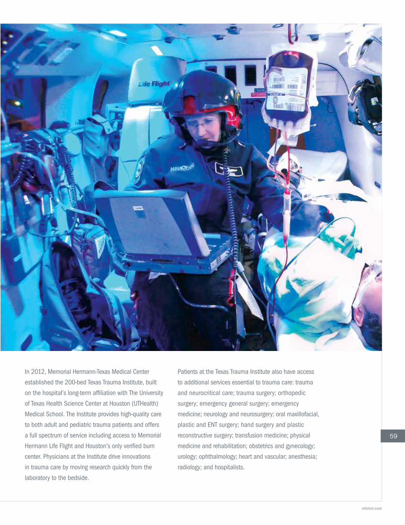
59
mhmni.com
In 2012, Memorial Hermann-Texas Medical Center
established the 200-bed Texas Trauma Institute, built
on the hospital’s long-term affiliation with The University
of Texas Health Science Center at Houston (UTHealth)
Medical School. The Institute provides high-quality care
to both adult and pediatric trauma patients and offers
a full spectrum of service including access to Memorial
Hermann Life Flight and Houston’s only verified burn
center. Physicians at the Institute drive innovations
in trauma care by moving research quickly from the
laboratory to the bedside.
Patients at the Texas Trauma Institute also have access
to additional services essential to trauma care: trauma
and neurocritical care; trauma surgery; orthopedic
surgery; emergency general surgery; emergency
medicine; neurology and neurosurgery; oral maxillofacial,
plastic and ENT surgery; hand surgery and plastic
reconstructive surgery; transfusion medicine; physical
medicine and rehabilitation; obstetrics and gynecology;
urology; ophthalmology; heart and vascular; anesthesia;
radiology; and hospitalists.

60
Spine The highly skilled spine surgeons at the Mischer
Neuroscience Institute performed more than 1,400
surgeries in 2012 in new, state-of-the-art facilities
equipped with advanced instruments and dynamic
imaging systems. These physicians are known for their
expertise in minimally invasive spine procedures and
innovative treatment options for patients with back
pain resulting from trauma, degenerative disc disease,
osteoporosis and related stress fractures, and deformity,
including kyphosis and scoliosis.
Our clinicians provide exceptional care for patients with
traumatic spine injury, including the 10 to 20 percent of
admissions through our Level I Trauma Center that involve
neurological damage. Based on benchmark University
HealthSystem Consortium data, the Spine Center’s
inpatient mortality for spine trauma, degenerative spine
disease and elective spine surgery has been consistently
lower than expected for the past five years.
We are skilled at innovative procedures for the relief
of neck and back pain, including minimally invasive
approaches. Specialties include lumbar fusion, lumbar
microdiscectomy, anterior cervical spine fusion,
scoliosis surgery, spine osteotomies, kyphoplasty,
thermal nucleoplasty, micro-endoscopic discectomy,
peripheral nerve repair, transforaminal lumbar
interbody fusion (TLIF) and disc replacement surgery.
Rehabilitation begins in the hospital following surgery.
Research under way at the MNI Spine Center is focused
on bringing promising therapies for spinal cord injury
(SCI) patients from the laboratory to clinical trials in
a manner that will provide evidence of effectiveness,
with maximum safety, to patients undergoing treatment.
Investigators are currently engaged in a Phase 2 trial of
the anticonvulsant drug riluzole in patients with acute
SCI, and a new stem cell trial for degenerative spine
and trauma spine fusions.
S C O P E O F S E r v I C E S

61
mhmni.com
QuAlITY & OuTCOMES MEASurES Spine
Spine Tumors: Length of Stay
0
1
2
3
4
6
5
FY 2009 FY 2010 FY 2011 FY 2012
ALO
S/CM
I
ALOS/CMI CMI
Spine Trauma: Inpatient Mortality
00.010.020.030.040.05
0.07
0.09
0.06
0.08
FY 2009 FY 2010 FY 2011 FY 20120
200
400
600
800
1000
1200
Bette
r tha
n ex
pect
ed
Volu
me
Spine Trauma: Length of Stay
00.5
11.5
2
3
4
5
2.5
3.5
4.5
FY 2009 FY 2010 FY 2011 FY 2012
ALO
S/CM
I
ALOS/CMI CMI
Actual Expected Number of Encounters
Source: chart data from the University HealthSystem Consortium
Spine Degenerative/Elective: Inpatient Mortality
00.005
0.010.0150.02
0.025
0.035
0.050.045
0.03
0.04
FY 2009 FY 2010 FY 2011 FY 20120
100
200
300
400
600
800
500
700
Bette
r tha
n ex
pect
ed
Volu
me
Spine Degenerative/Elective: Length of Stay
0
0.5
1
1.5
2
3.5
3
2.5
FY 2009 FY 2010 FY 2011 FY 2012
ALO
S/CM
I
ALOS/CMI CMI
Spine Tumors: Inpatient Mortality
0
0.02
0.04
0.06
0.08
0.10
0.14
0.12
FY 2009 FY 2010 FY 2011 FY 20120
Actual Expected Number of Encounters
Bette
r tha
n ex
pect
ed
Volu
me
20
40
60
80
100
120
140Actual Expected Number of Encounters
Source: chart data from the University HealthSystem Consortium
Spine Tumors: Length of Stay
0
1
2
3
4
6
5
FY 2009 FY 2010 FY 2011 FY 2012
ALO
S/CM
I
ALOS/CMI CMI
Spine Trauma: Inpatient Mortality
00.010.020.030.040.05
0.07
0.09
0.06
0.08
FY 2009 FY 2010 FY 2011 FY 20120
200
400
600
800
1000
1200
Bette
r tha
n ex
pect
ed
Volu
me
Spine Trauma: Length of Stay
00.5
11.5
2
3
4
5
2.5
3.5
4.5
FY 2009 FY 2010 FY 2011 FY 2012
ALO
S/CM
I
ALOS/CMI CMI
Actual Expected Number of Encounters
Source: chart data from the University HealthSystem Consortium
Spine Degenerative/Elective: Inpatient Mortality
00.005
0.010.0150.02
0.025
0.035
0.050.045
0.03
0.04
FY 2009 FY 2010 FY 2011 FY 20120
100
200
300
400
600
800
500
700
Bette
r tha
n ex
pect
ed
Volu
me
Spine Degenerative/Elective: Length of Stay
0
0.5
1
1.5
2
3.5
3
2.5
FY 2009 FY 2010 FY 2011 FY 2012
ALO
S/CM
I
ALOS/CMI CMI
Spine Tumors: Inpatient Mortality
0
0.02
0.04
0.06
0.08
0.10
0.14
0.12
FY 2009 FY 2010 FY 2011 FY 20120
Actual Expected Number of Encounters
Bette
r tha
n ex
pect
ed
Volu
me
20
40
60
80
100
120
140Actual Expected Number of Encounters
Source: chart data from the University HealthSystem Consortium

62
Research and Innovation

63
mhmni.com

64
cerebroVAscuLAr
A phase iii, randomized, placebo-controlled, Double-
blind study of the combined Lysis of thrombus with
ultrasound and systemic tissue plasminogen Activator
(tpA) for emergent revascularization (cLotbust-er) in
Acute ischemic stroke
PRINCIPAL INVESTIGATOR: Andrew D. barreto, M.D.
A randomized, placebo-controlled, double-blind Phase
III clinical study to evaluate the efficacy and safety of
ultrasound (US) using the SonoLysis headframe as an
adjunctive therapy to tissue plasminogen activator (tPA)
treatment in subjects with acute ischemic stroke.
A randomized Multicenter clinical trial of unruptured
brain AVMs (ArubA)
PRINCIPAL INVESTIGATOR: p. roc chen, M.D.
ARUBA is an FDA- and institutional IRB-approved
randomized multicenter international clinical trial designed
to determine whether medical management improves long-
term outcomes of patients with unruptured brain AVMs,
compared to interventional therapy (with endovascular
procedures, neurosurgery or radiotherapy, alone or in
combination). This trial tests whether medical management
or interventional therapy will reduce the risk of death
or stroke due to hemorrhage or infarction by at least
46 percent (an absolute magnitude of about 9.5 percent
over five years). A total of 400 patients will be enrolled in
order to detect the hypothesized 46 percent reduction in
event rate, analyzed using the intention-to-treat principle.
This sample size supports a test of non-inferiority if
medical management is not superior to interventional
therapy. Patients are followed for a minimum of five
years and a maximum of 10 years from randomization.
Artss-2: A pilot, phase iib, randomized, Multicenter
trial of Argatroban in combination with recombinant
tissue plasminogen Activator for Acute stroke
PRINCIPAL INVESTIGATOR: Andrew D. barreto, M.D.
All study participants in this randomized multicenter trial
will be treated with rt-PA (0-3 hours or 0-4.5 hours)
and randomized to one of three study arms: intravenous
tPA along with low-dose argatroban, high-dose of
argatroban or standard IV tPA alone. This trial is
designed to estimate overall treatment benefit among
stroke patients randomized to receive one of the three
treatments. The study will also verify the safety of a
low-dose combination of argatroban and rt-PA, test the
safety of a high-dose combination treatment and assess
the rates of early recanalization to determine treatment
effect and assess reliability in predicting outcomes of
the drug combination.
Physicians at the Mischer Neuroscience Institute and The University of Texas Health Science Center at Houston (UTHealth)
Medical School are engaged in a broad and intensive research program focused on the mechanisms, treatment and cure of
neurological disease and injury. We use diverse approaches – molecular, transgenic and electrophysiological techniques – in
biomedical studies, translational research, clinical trials, and technology development and assessment.
Our projects are supported by the National Institutes of Health, the Vivian L. Smith Foundation for Neurologic Disease, the
American Stroke Association and other granting agencies. They cover major areas of neurological disease, including stroke,
aneurysm, spinal cord injury, brain tumor, stem cell therapies, neuroprotection, hypoxic encephalopathy, epilepsy, traumatic
brain injury and Parkinson’s disease. During the 2012 calendar year, researchers at the Institute and the UTHealth Medical
School received more than $10.5 million in 200 grants and contracts. The following listing is a sample of ongoing or recently
completed research projects.

65
mhmni.com

66
Assessment of spleen size reduction and inflammatory
Markers in Acute stroke over time (Assist)
PRINCIPAL INVESTIGATOR: sean savitz, M.D.
An observational study to evaluate changes in spleen
size and blood flow over time using ultrasound and
corresponding changes in inflammatory cytokines in acute
stroke patients presenting within six hours of symptom
onset. The results of the study may provide insight into
potential future therapies for acute stroke by targeting
immune processes in the spleen.
bugher Foundation center for stroke prevention
PRINCIPAL INVESTIGATOR: Dong h. Kim, M.D.
This project is focused on identifying gene mutations
associated with cerebral aneurysm formation and
understanding the molecular mechanisms that lead
to disease.
carotid revascularization endarterectomy Versus
stenting trial (crest)
PRINCIPAL INVESTIGATOR: Nicole Gonzales, M.D.
To find better ways to prevent stroke in subjects
with carotid stenosis, this national, multicenter research
study is comparing carotid endarterectomy to the study
procedure – carotid artery stenting. Researchers are
evaluating the relative effectiveness of both treatments in
preventing stroke, myocardial infarction and death in the
30-day period immediately following the procedure.
combination treatment of rtpA and Apyrase for stroke
PRINCIPAL INVESTIGATOR: Jaroslaw Aronowski, ph.D.
This pre-clinical study is designed to evaluate the role of
apyrase, an endogenous vascular ATPase, as a mechanism
to prevent thrombosis after ischemic stroke when used in
combination with rtPA.
combination therapy of Aspirin and Apyrase for stroke
PRINCIPAL INVESTIGATOR: Jaroslaw Aronowski, ph.D.
This pre-clinical proposal is designed to evaluate the role of
apyrase, an endogenous vascular ATPase, in combination
with aspirin to prevent thrombosis after ischemic stroke.
Delay in evaluation and treatment of posterior circulation
stroke compared with Anterior circulation stroke
PRINCIPAL INVESTIGATOR: Amrou sarraj, M.D.
Failure to recognize early symptoms of acute posterior
circulation cerebral ischemia may delay timely diagnosis
and treatment. In this study, researchers are investigating
whether there were differences in symptom onset in arrival
at our Emergency department (ED), arrival to neurology
evaluation, and ED arrival to treatment between patients
with posterior circulation ischemia (PCI) versus anterior
circulation ischemia (ACI). We are also assessing whether
various symptoms are associated with differences in time
to evaluation or time to treatment between ACI and PCI.
DiAs 4 – Desmoteplase in Acute stroke
PRINCIPAL INVESTIGATOR: George Lopez, M.D., ph.D.
An efficacy study to determine whether the potent IV
clot-busting drug desmoteplase improves outcome in
patients who arrive too late for IV tPA but within nine hours
of stroke onset. Desmoteplase is derived from vampire
bat saliva and previous studies suggest benefit in patients
with normal CT scans and persisting arterial occlusion
beyond three hours.
r E S E A r C H
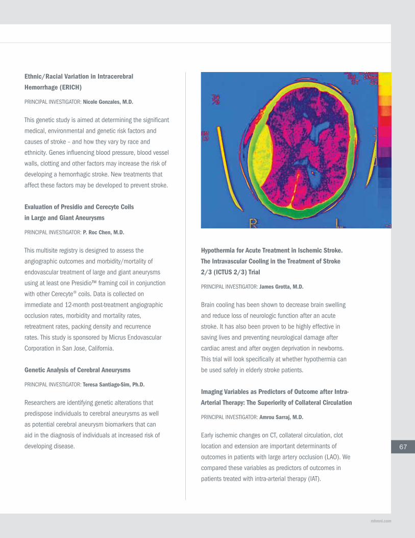
67
ethnic/racial Variation in intracerebral
hemorrhage (erich)
PRINCIPAL INVESTIGATOR: Nicole Gonzales, M.D.
This genetic study is aimed at determining the significant
medical, environmental and genetic risk factors and
causes of stroke – and how they vary by race and
ethnicity. Genes influencing blood pressure, blood vessel
walls, clotting and other factors may increase the risk of
developing a hemorrhagic stroke. New treatments that
affect these factors may be developed to prevent stroke.
evaluation of presidio and cerecyte coils
in Large and Giant Aneurysms
PRINCIPAL INVESTIGATOR: p. roc chen, M.D.
This multisite registry is designed to assess the
angiographic outcomes and morbidity/mortality of
endovascular treatment of large and giant aneurysms
usingatleastonePresidio™framingcoilinconjunction
withotherCerecyte®coils.Dataiscollectedon
immediate and 12-month post-treatment angiographic
occlusion rates, morbidity and mortality rates,
retreatment rates, packing density and recurrence
rates. This study is sponsored by Micrus Endovascular
Corporation in San Jose, California.
Genetic Analysis of cerebral Aneurysms
PRINCIPAL INVESTIGATOR: teresa santiago-sim, ph.D.
Researchers are identifying genetic alterations that
predispose individuals to cerebral aneurysms as well
as potential cerebral aneurysm biomarkers that can
aid in the diagnosis of individuals at increased risk of
developing disease.
hypothermia for Acute treatment in ischemic stroke.
the intravascular cooling in the treatment of stroke
2/3 (ictus 2/3) trial
PRINCIPAL INVESTIGATOR: James Grotta, M.D.
Brain cooling has been shown to decrease brain swelling
and reduce loss of neurologic function after an acute
stroke. It has also been proven to be highly effective in
saving lives and preventing neurological damage after
cardiac arrest and after oxygen deprivation in newborns.
This trial will look specifically at whether hypothermia can
be used safely in elderly stroke patients.
imaging Variables as predictors of outcome after intra-
Arterial therapy: the superiority of collateral circulation
PRINCIPAL INVESTIGATOR: Amrou sarraj, M.D.
Early ischemic changes on CT, collateral circulation, clot
location and extension are important determinants of
outcomes in patients with large artery occlusion (LAO). We
compared these variables as predictors of outcomes in
patients treated with intra-arterial therapy (IAT).
mhmni.com

68
influence of changes in corticospinal tract integrity
over time on clinical outcome in Acute ischemic stroke
(ict-Ais)
PRINCIPAL INVESTIGATOR: sean savitz, M.D.
This prospective pilot study is designed to evaluate the
influences of changes in corticospinal tract integrity over
time on motor and cognitive outcomes in patients with
acute ischemic stroke in the middle cerebral artery territory
within 48 hours of stroke onset. Patients will undergo
detailed cognitive and fine-motor testing as well as
advanced neuroimaging.
Minimally invasive surgery plus tpA for intracerebral
hemorrhage evacuation (Mistie)
PRINCIPAL INVESTIGATOR: George Lopez, M.D., ph.D.
This study was designed to produce data regarding the
capability of minimally invasive surgery with recombinant
tissue plasminogen activator (rt-PA) to remove blood clots
from intra-cerebral hemorrhage patients.
Neurofluctuations in patients with subcortical
ischemic stroke (Niss)
PRINCIPAL INVESTIGATOR: sean savitz, M.D.
The purpose of this prospective observational study is to
capture and report the incidence of neurological exam
fluctuations and their outcomes in subcortical (lacunar)
stroke patients receiving standard care. The results may
be used to evaluate the results of new treatments for this
disorder.
optimizing prediction scores for poor outcome After
intra-arterial therapy for Anterior circulation Acute
ischemic stroke*
PRINCIPAL INVESTIGATOR: Amrou sarraj, M.D.
Intra-arterial therapy (IAT) is an approach to promote
recanalization of large artery occlusions (LAO) in acute
ischemic stroke (AIS) but is resource intensive. Previous
studies evaluated different variables that affect clinical
outcome after IAT. To better identify patients who
r E S E A r C H

69
have poor outcomes despite IAT, we compared the
performance of previous predictive scoring systems that
relied either on clinical or imaging variables in patients
undergoing IAT. We then combined imaging and clinical
variables to optimize a score that would better predict
poor outcome after IAT for AIS.
*Amrou Sarraj, M.D., won the Mordecai Y.T. Globus New Investigator
Award at the 2012 International Stroke Conference for his work on
this project.
pleiotropic transcription Factors as a target for
intracerebral hemorrhage treatment
PRINCIPAL INVESTIGATOR: Jaroslaw Aronowski, ph.D.
Researchers are evaluating the role of transcription factor
Nrf2 in regulating cytoprotection, antioxidative defense
and detoxification of brains injured by intracerebral
hemorrhage.
prospective Analysis of the use of thrombelastography
(teG) in prediction of hemorrhage in stroke patients
PRINCIPAL INVESTIGATOR: James Grotta, M.D.
This is an observational study to evaluate the use
of thrombelastography (TEG) analysis to assess the
coagulation status of patients with acute stroke
presenting within three hours of symptom onset.
The purpose of the study is to evaluate the efficacy
of TEG as means of identifying those ischemic and
hemorrhagic stroke patients at increased risk
of bleeding.
pursuit: pre-hospital utility of rapid stroke
evaluation using in-ambulance telemedicine
PRINCIPAL INVESTIGATOR: tzu-ching Wu, M.D.
This trial is studying the feasibility of using telemedicine
to evaluate patients with acute stroke in the ambulance.
refining prediction scores for poor outcome After
intra-arterial therapy for Anterior circulation Acute
ischemic stroke: Adding collateral status improves
prediction scores
PRINCIPAL INVESTIGATOR: Amrou sarraj, M.D.
Intra-arterial therapy (IAT) is widely practiced, but is
of unproven benefit. To improve patient selection for IAT,
we sought to refine the HIAT2 (Houston Intra-Arterial
Therapy 2) score to include the presence of collateral
circulation to devise a HIAT3.
risk of hemorrhage in patients on Warfarin Who are
treated with tissue plasminogen Activator for Acute
ischemic stroke
PRINCIPAL INVESTIGATOR: Amrou sarraj, M.D.
Tissue plasminogen activator (tPA) remains the only
proven treatment for ischemic stroke but concurrent use
of warfarin at the time of stroke poses a potential risk for
symptomatic intracerebral hemorrhage (sICH). Few studies
have addressed this risk. We studied all patients admitted
to our service who were on warfarin at the time of their
acute ischemic stroke and were treated with IV tPA in
order to define incidence and risk factors of sICH.
safety/Feasibility of Autologous Mononuclear bone
Marrow cell treatment for Acute ischemic stroke
PRINCIPAL INVESTIGATOR: sean savitz, M.D.
Cell-based therapy has emerged as a novel investigational
approach to enhance recovery after ischemic stroke.
Numerous studies have shown that the administration
of cells is safe and improves outcome in animal models.
Our researchers will enroll 30 patients who have suffered
an ischemic stroke in a safety study of an IV infusion of
the patient’s own cells within 24 to 72 hours after the
onset of symptoms.
mhmni.com
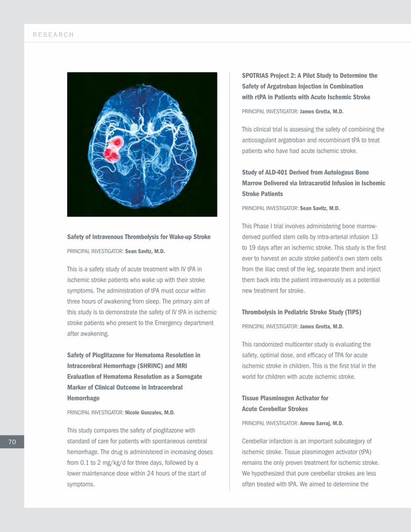
70
safety of intravenous thrombolysis for Wake-up stroke
PRINCIPAL INVESTIGATOR: sean savitz, M.D.
This is a safety study of acute treatment with IV tPA in
ischemic stroke patients who wake up with their stroke
symptoms. The administration of tPA must occur within
three hours of awakening from sleep. The primary aim of
this study is to demonstrate the safety of IV tPA in ischemic
stroke patients who present to the Emergency department
after awakening.
safety of pioglitazone for hematoma resolution in
intracerebral hemorrhage (shriNc) and Mri
evaluation of hematoma resolution as a surrogate
Marker of clinical outcome in intracerebral
hemorrhage
PRINCIPAL INVESTIGATOR: Nicole Gonzales, M.D.
This study compares the safety of pioglitazone with
standard of care for patients with spontaneous cerebral
hemorrhage. The drug is administered in increasing doses
from 0.1 to 2 mg/kg/d for three days, followed by a
lower maintenance dose within 24 hours of the start of
symptoms.
spotriAs project 2: A pilot study to Determine the
safety of Argatroban injection in combination
with rtpA in patients with Acute ischemic stroke
PRINCIPAL INVESTIGATOR: James Grotta, M.D.
This clinical trial is assessing the safety of combining the
anticoagulant argatroban and recombinant tPA to treat
patients who have had acute ischemic stroke.
study of ALD-401 Derived from Autologous bone
Marrow Delivered via intracarotid infusion in ischemic
stroke patients
PRINCIPAL INVESTIGATOR: sean savitz, M.D.
This Phase I trial involves administering bone marrow-
derived purified stem cells by intra-arterial infusion 13
to 19 days after an ischemic stroke. This study is the first
ever to harvest an acute stroke patient’s own stem cells
from the iliac crest of the leg, separate them and inject
them back into the patient intravenously as a potential
new treatment for stroke.
thrombolysis in pediatric stroke study (tips)
PRINCIPAL INVESTIGATOR: James Grotta, M.D.
This randomized multicenter study is evaluating the
safety, optimal dose, and efficacy of TPA for acute
ischemic stroke in children. This is the first trial in the
world for children with acute ischemic stroke.
tissue plasminogen Activator for
Acute cerebellar strokes
PRINCIPAL INVESTIGATOR: Amrou sarraj, M.D.
Cerebellar infarction is an important subcategory of
ischemic stroke. Tissue plasminogen activator (tPA)
remains the only proven treatment for ischemic stroke.
We hypothesized that pure cerebellar strokes are less
often treated with tPA. We aimed to determine the
r E S E A r C H

71
percentage of cerebellar strokes treated with tPA and
the main reasons why patients with cerebellar strokes
are not treated with tPA.
using propensity scores to simulate a randomized
controlled trial to Assess the Added benefit of
combining intra-arterial therapy with iV tpA
PRINCIPAL INVESTIGATOR: Amrou sarraj, M.D.
In light of recent studies, the superiority of combining
intra-arterial therapy (IAT) with IV tPA compared with
IV tPA alone is yet to be established. To test the
additive value of treating patients with IAT, we compared
outcomes in patients treated with IV tPA with those
who received IV tPA followed by intra-arterial intervention
in a novel study design.
epiLepsy
A bio-Nano-chip for Anticonvulsant Drug Assay
in epilepsy patients
PRINCIPAL INVESTIGATOR: Giridhar Kalamangalam, M.D., D.phil.
The goal of the study is to test a novel portable
“lab-on-a-chip” device for assaying common
anticonvulsant drugs in patients with epilepsy.
A prospective, open-label study of the structure
and Function of the retina in Adult patients with
refractory complex partial seizures treated with
Vigabatrin (sabril®)
PRINCIPAL INVESTIGATOR: Jeremy slater, M.D.
This study examines the efficacy of ocular computerized
tomography (OCT) in predicting the onset of retinal
dysfunction occurring in patients treated with the
antiepileptic drug vigabatrin.
Analysis of the role of sv2a phosphorylation in epilepsy
PRINCIPAL INVESTIGATOR: Nitin tandon, M.D.
The major goals of this project are to investigate the
mechanism of action of levetiracetam and the role of
SV2A in human epilepsy. This experiment will test the
hypothesis that epilepsy in human tissue leads to changes
in the phosphorylation of SV2A and if levetiracetam
treatment affects these changes.
Quantitative Analysis of electroencephalogram
in epilepsy
PRINCIPAL INVESTIGATOR: Giridhar Kalamangalam, M.D., D.phil.
By analyzing EEG and video EEG data already collected
for clinical purposes, this study seeks new ways of
understanding brain function in normal subjects and in
people with neurological problems such as seizures.
oxygen-enhanced Magnetic resonance imaging
in Non-lesional Focal epilepsy
PRINCIPAL INVESTIGATOR: Giridhar Kalamangalam, M.D., D.phil.
This ongoing study is evaluating how effective oxygen-
enhanced MRI scans are at identifying subtle brain lesions
in patients with refractory focal epilepsy.
pecA Visiting professorship to central America
PRINCIPAL INVESTIGATOR: Giridhar Kalamangalam, M.D., D.phil.
The goal of the opportunity is to develop collaborative
educational and clinical links to advance basic and
advanced epilepsy care in Central Panama, based
at Hospital Luis “Chicho” Fábrega in Santiago de
Veraguas, Panama.
mhmni.com
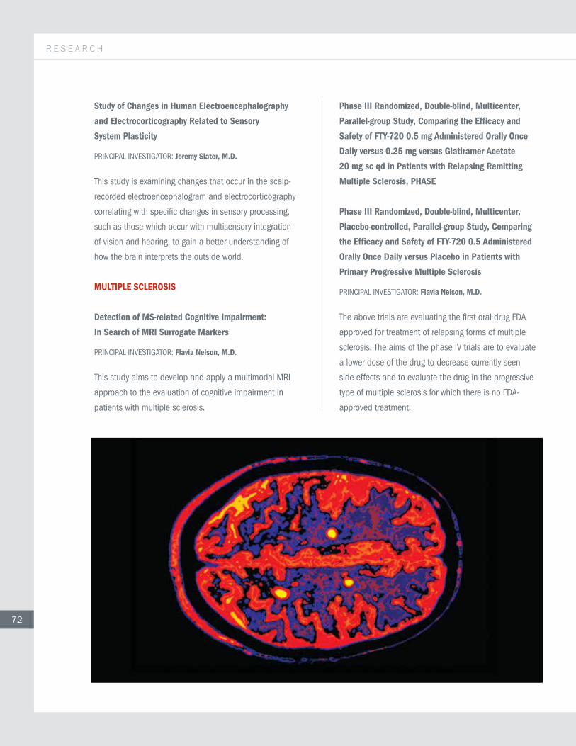
72
study of changes in human electroencephalography
and electrocorticography related to sensory
system plasticity
PRINCIPAL INVESTIGATOR: Jeremy slater, M.D.
This study is examining changes that occur in the scalp-
recorded electroencephalogram and electrocorticography
correlating with specific changes in sensory processing,
such as those which occur with multisensory integration
of vision and hearing, to gain a better understanding of
how the brain interprets the outside world.
MuLtipLe scLerosis
Detection of Ms-related cognitive impairment:
in search of Mri surrogate Markers
PRINCIPAL INVESTIGATOR: Flavia Nelson, M.D.
This study aims to develop and apply a multimodal MRI
approach to the evaluation of cognitive impairment in
patients with multiple sclerosis.
phase iii randomized, Double-blind, Multicenter,
parallel-group study, comparing the efficacy and
safety of Fty-720 0.5 mg Administered orally once
Daily versus 0.25 mg versus Glatiramer Acetate
20 mg sc qd in patients with relapsing remitting
Multiple sclerosis, phAse
phase iii randomized, Double-blind, Multicenter,
placebo-controlled, parallel-group study, comparing
the efficacy and safety of Fty-720 0.5 Administered
orally once Daily versus placebo in patients with
primary progressive Multiple sclerosis
PRINCIPAL INVESTIGATOR: Flavia Nelson, M.D.
The above trials are evaluating the first oral drug FDA
approved for treatment of relapsing forms of multiple
sclerosis. The aims of the phase IV trials are to evaluate
a lower dose of the drug to decrease currently seen
side effects and to evaluate the drug in the progressive
type of multiple sclerosis for which there is no FDA-
approved treatment.
r E S E A r C H
73
combination therapy in Multiple sclerosis
PRINCIPAL INVESTIGATOR: Jerry Wolinsky, M.D.
This study is determining if the combination of interferon
beta-1a and glatiramer acetate is superior to either drug
as monotherapy in relapsing-remitting multiple sclerosis.
Mri Analysis center for protocol eFc6058 - A
Multicenter Double-blind parallel-group placebo-
controlled study of the efficacy and safety of
teriflunomide in patients with relapsing Multiple
sclerosis Who Are treated with interferon-beta
PRINCIPAL INVESTIGATOR: Jerry Wolinsky, M.D.
This study provides quantitative image analysis measures
as supportive outcome measures.
Mri Analysis center for protocol eFc6260 – An
international, Multicenter, randomized, Double-blind,
placebo-controlled, parallel-group study to evaluate
the efficacy and safety of two-year treatment with
7 mg once Daily and 14 mg once Daily versus
placebo in patients with a First clinical episode
suggestive of Multiple sclerosis
PRINCIPAL INVESTIGATOR: Jerry Wolinsky, M.D.
This pivotal clinical trial provides quantitative image
analysis measures as supportive outcome measures.
Mri Analysis center for protocol Lts 6050 -- A Long-
term extension of the Multinational, Double-blind,
placebo-controlled study eFc6049 (hMr1726Dl3001)
to Document the safety of two Doses of teriflunomide
(7 and 14 mg) in patients with Multiple sclerosis
with relapses
PRINCIPAL INVESTIGATOR: Jerry Wolinsky, M.D.
This study provides quantitative image analysis measures
as supportive outcome measures.
mhmni.com
pilot clinical trial of ActharGel 14 days subcutaneous
(sQ) versus ActharGel Five Days sQ for the treatment
of Ms exacerbations
PRINCIPAL INVESTIGATOR: staley brod, M.D.
We will determine if ACTH injections given for 14 days
are superior to injections given for five days for recovery
from MS attacks. ACTH injections are an FDA-approved
treatment for MS attacks but it is not known if treatment
for more than five days improves recovery. We will examine
MR brain scans and immune function periodically for
90 days after MS attack to determine if ACTH improves
MR brain scans and immune function.
serial Magnetic resonance spectroscopy
in Multiple sclerosis
PRINCIPAL INVESTIGATOR: Jerry Wolinsky, M.D.
Researchers are using serial magnetic resonance imaging
(MRI) and magnetic resonance spectroscopy (MRS) to
gather data to better understand disease processes in
patients with multiple sclerosis.
MoVeMeNt DisorDers AND
NeuroDeGeNerAtiVe DiseAses
Amyloid-beta oligomers and Alzheimer’s Diagnosis
PRINCIPAL INVESTIGATOR: claudio soto, ph.D.
The major goal of this project is to adapt the protein
misfolding cyclic amplification (PMCA) technology for
the specific and highly sensitive detection of misfolded
A oligomers in human biological fluids. Investigators
are optimizing the experimental conditions of cyclic
amplification of A misfolding, identifying A misfolded
oligomers in AD biological fluids, and evaluating the
sensitivity and specificity and the earliest time during
the pre-symptomatic phase in which A oligomers can
be detected in biological fluids.

74
cD FLeX: An open-label, Non-inferiority study evaluating
the efficacy and safety of two injection schedules of
Xeomin® (incobotulinumtoxinA) [short Flex versus Long
Flex] in subjects with cervical Dystonia with < 10 Weeks
of benefit from onabotulinumtoxinA treatment
PRINCIPAL INVESTIGATOR: erin Furr-stimming, M.D.
Prospective, open-label, 1:1 randomized trial evaluating
twodosingschedulesofXeomin[ShortFlexandLongFlex]
in subjects who report that they receive therapeutic benefit
fromonabotulinumtoxinA(Botox®)treatmentforlessthan
10 weeks.
cyclic Amplification of prion protein Misfolding
PRINCIPAL INVESTIGATOR: claudio soto, ph.D.
The major goals of this project are to understand the
mechanism of prion replication and the nature of the
infectious agent, and to develop novel strategies for
diagnosis of prion diseases.
peripheral and central protein biomarkers of brain
Mr Activity in Demyelinating Disease
PRINCIPAL INVESTIGATOR: staley brod, M.D.
By studying patients with new or disappearing brain
lesions, it may be possible to identify protein markers that
repair damage to the brain and can be used as future
therapies. This sub-study is investigating whether specific
proteins in the blood and spinal fluid change in the
presence of new brain lesions.
pathogenesis, transmission and Detection of Zoonotic
prion Diseases
PRINCIPAL INVESTIGATOR: claudio soto, ph.D.
Researchers are studying the pathogenesis and routes of
propagation of bovine spongiform encephalopathy and
chronic wasting disease, and developing novel strategies
for the detection of infected animals.
pathogenic Mechanism of prion Disease
PRINCIPAL INVESTIGATOR: Claudio Soto, Ph.D.
This Program Project grant involves several groups. Our
major goal is to understand the molecular basis of human
prion replication and to develop novel strategies for
diagnosis.
potential biomarkers for parkinson’s Disease
PRINCIPAL INVESTIGATOR: ying Xia, M.D., ph.D.
To explore, through both clinical and laboratory
approaches, a potential biomarker for predicting the
development/severity of Parkinson’s disease. This project
is a collaboration with Chinese clinicians and scientists
with a grant application submitted in September to the
U.S.-China Program for Biomedical Collaborative Research
in cooperation with the National Institutes of Health.
r E S E A r C H
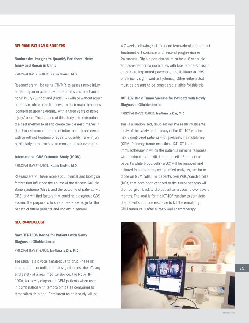
75
NeuroMuscuLAr DisorDers
Noninvasive imaging to Quantify peripheral Nerve
injury and repair in clinic
PRINCIPAL INVESTIGATOR: Kazim sheikh, M.D.
Researchers will be using DTI/MRI to assess nerve injury
and/or repair in patients with traumatic and mechanical
nerve injury (Sunderland grade II-V) with or without repair
of median, ulnar or radial nerves or their major branches
localized to upper extremity, within three years of nerve
injury/repair. The purpose of this study is to determine
the best method to use to create the clearest images in
the shortest amount of time of intact and injured nerves
with or without treatment/repair to quantify nerve injury
particularly to the axons and measure repair over time.
international Gbs outcome study (iGos)
PRINCIPAL INVESTIGATOR: Kazim sheikh, M.D.
Researchers will learn more about clinical and biological
factors that influence the course of the disease Guillain-
Barré syndrome (GBS), and the outcome of patients with
GBS, and will find factors that could help diagnose GBS
sooner. The purpose is to create new knowledge for the
benefit of future patients and society in general.
Neuro-oNcoLoGy
Novo ttF-100A Device for patients with Newly
Diagnosed Glioblastomas
PRINCIPAL INVESTIGATOR: Jay-Jiguang Zhu, M.D.
The study is a pivotal (analogous to drug Phase III),
randomized, controlled trial designed to test the efficacy
and safety of a new medical device, the NovoTTF-
100A, for newly diagnosed GBM patients when used
in combination with temozolomide as compared to
temozolomide alone. Enrollment for this study will be
4-7 weeks following radiation and temozolomide treatment.
Treatment will continue until second progression or
24 months. Eligible participants must be >18 years old
and screened for co-morbidities with labs. Some exclusion
criteria are implanted pacemaker, defibrillator or DBS,
or clinically significant arrhythmias. Other criteria that
must be present to be considered eligible for this trial.
ict- 107 brain tumor Vaccine for patients with Newly
Diagnosed Glioblastomas
PRINCIPAL INVESTIGATOR: Jay-Jiguang Zhu, M.D.
This is a randomized, double-blind Phase IIB multicenter
study of the safety and efficacy of the ICT-107 vaccine in
newly diagnosed patients with glioblastoma multiforme
(GBM) following tumor resection. ICT-107 is an
immunotherapy in which the patient’s immune response
will be stimulated to kill the tumor cells. Some of the
patient’s white blood cells (WBC) will be removed and
cultured in a laboratory with purified antigens, similar to
those on GBM cells. The patient’s own WBC/dendric cells
(DCs) that have been exposed to the tumor antigens will
then be given back to the patient as a vaccine over several
months. The goal is for the ICT-107 vaccine to stimulate
the patient’s immune response to kill the remaining
GBM tumor cells after surgery and chemotherapy.
mhmni.com

76
Eligible participants must be 18+ years old, with newly
diagnosed glioblastoma multiforme (GBM). Patients must
be consented after full tumor resection surgery and prior
to starting chemo-radiation therapy. Initial screening
procedures include HLA typing and apheresis to isolate
peripheral blood mononuclear cells (PBMCs) to be used
for the preparation of study treatment (ICT-107 and
control). Patients will be randomized by age in a 2:1 ratio
to ICT-107 or control.
identification of New Markers and therapeutic targets
in Glioblastoma Multiforme (GbM)
PRINCIPAL INVESTIGATOR: Min Li, ph.D.
This study is to identify new markers for diagnosis and
novel therapeutic targets for molecular-targeted therapy in
GBM using genetic and molecular approaches.
study Function of Zinc transporter (Zip4) in brain
tumor progression, Develop Novel Zip4 based
therapy for brain tumor
PRINCIPAL INVESTIGATOR: Min Li, ph.D.
Our previous studies indicate that ZIP4 plays a critical role
in tumor growth and metastasis. This project will further
characterize the molecular mechanism of ZIP4-induced
tumor progression.
identify New Markers and therapeutic targets in
brain tumor using Molecular and Genetic Approaches
PRINCIPAL INVESTIGATOR: Min Li, ph.D.
Gene profiling, microRNA profiling and proteomics will
be used to identify new target molecules that can be
used to develop personalized medicine.
study of the role of MicrorNAs in brain tumor
pathogenesis
PRINCIPAL INVESTIGATOR: Min Li, ph.D.
The function of oncogenic and tumor suppressor
microRNAs will be evaluated in brain tumor cells and
tissues, and targeted therapy will be designed based
on the expression profile and the function of those
microRNAs.
NeurorehAbiLitAtioN
the use of Ventriculostomy or hemicraniectomy as a
predictor of rehabilitation Level of care in patients
with intracerebral hemorrhage
PRINCIPAL INVESTIGATOR: Nneka ifejika-Jones, M.D.
The purpose of this research project is to determine
whether patients with intracerebral hemorrhage who
underwent surgical intervention for increased intracranial
pressure are more likely to receive post-stroke care at
an inpatient rehabilitation facility, a skilled nursing
facility or a long-term acute care facility.
r E S E A r C H

77
NeurotrAuMA/criticAL cAre
A Mechanism for Global cerebral edema after
subarachnoid hemorrhage: pathophysiology
of early brain injury
PRINCIPAL INVESTIGATOR: h. Alex choi, M.D.
Early brain injury after subarachnoid hemorrhage is
the most important determinant of outcome. Using
cerebrospinal fluid and serum markers of inflammation,
we are exploring the mechanisms of early brain
injury and global cerebral edema after subarachnoid
hemorrhage.
A pilot study to identify biomarkers Associated
with chronic tbi
PRINCIPAL INVESTIGATOR: pramod Dash, ph.D.
The specific aim of this research is to determine if the
biological fluids (blood/saliva) from chronic brain-injured
patients (both blast and non-penetrating TBI) contain
reproducible protein markers.
biomarkers for pain in spinal cord injury
PRINCIPAL INVESTIGATOR: Gigi hergenroeder, r.N.
Investigators in this clinical trial believe that spinal cord
injury (SCI) patients who develop chronic pain have
biomarkers in their blood that can predict their condition.
Patients two or more years post injury, who have been
identified as having neuropathic pain or no pain will be
asked to donate blood samples that will be evaluated for
biomarkers. The goal of the research is early intervention
to prevent the onset of chronic pain.
biomarkers prognostic for elevated intracranial pressure
PRINCIPAL INVESTIGATOR: pramod Dash, ph.D.
This study is determining the cutoff values of ceruloplasmin
and copper for patient classification and testing the
diagnostic accuracy of these markers in blinded samples,
and also determining if a temperature correction factor is
required for the use of these assays in future scenarios.
combinatory strategies to Functional remyelination
After spinal cord injury
PRINCIPAL INVESTIGATOR: Qi Lin cao, ph.D.
Researchers are identifying optimal strategies to
genetically modify oligodendrocyte precursor cells prior to
transportation to promote remyelination and functional
recovery after spinal cord injury (SCI).
csF Diversion Assessment and Ventriculoperitoneal
shunt Dependence study
PRINCIPAL INVESTIGATOR: Kiwon Lee, M.D.
This study aims to analyze the relationship between
the total amount of CSF diversion and long-term
ventriculoperitoneal shunt dependence. The important
variables for investigation are red blood cell clearance and
the ventriculoperitoneal shunt dependence. Evaluation of
predictors of VP shunt dependence is done particularly
for patients with high-grade aneurysmal subarachnoid
hemorrhage.
effects of erythropoietin on Anemia and Need for
transfusion (a component of the program project
“Vascular Mechanisms of secondary injury after
traumatic brain injury”)
PRINCIPAL INVESTIGATOR: imoigele Aisiku, M.D.
This study examines the effects of TBI on cerebral blood
flow and the effect of erythropoietin in CBF.
mhmni.com

78
ethnic Disparities in end-of-life care
in brain-injured patients
PRINCIPAL INVESTIGATOR: h. Alex choi, M.D.
The advancement of critical care has brought to the
forefront ethical issues regarding continuation of aggressive
medical measures to prolong life in the severely brain-
injured patient. Studies have shown minorities, especially
black and Hispanic patients, seek more care at the
end of life. We are studying this disparity in the acutely
brain-injured patients and their families and exploring
the possible social/cultural/religious reasons for these
differences.
Gene transcription and regulation
of stem cell Differentiation
PRINCIPAL INVESTIGATOR: Jiaqian Wu, ph.D.
This research combines stem cell biology and systems-
based approaches involving genomics, proteomics,
bioinformatics and functional assays to unravel gene
transcription and regulatory mechanisms governing stem
cell differentiation. One major focus is investigating stem
cell neural differentiation and developing effective and
safe treatment for spinal cord injury and neurological
diseases such as stroke. The other area lies in the
studies of the regulatory networks of hematopoietic
precursor cell self-renewal and differentiation using the
multipotent EML (erythroid, myeloid and lymphocytic)
cell as a model system.
hemodynamic optimization for early Goal-directed
therapy in severe brain injury
PRINCIPAL INVESTIGATOR: Kiwon Lee, M.D.
This clinical study investigates the different dynamic
variables in the intravascular volume status (including
stroke volume variation, pulse pressure variation,
cardiac indices and other pressure- and volume-related
variables) and their effects on the injured brain.
r E S E A r C H

79
human pluripotent stem cells in cell-based
therapy for cNs injury
PRINCIPAL INVESTIGATOR: ying Liu, ph.D.
This study focuses on dissecting the neural developmental
pathways and the corresponding pathogenesis in spinal
cord injury and stroke. Our long-term goal is to identify
therapeutic targets for the treatment of CNS injury and
neurodegenerative diseases.
hyperoxia and Delayed cerebral infarction in
subarachnoid hemorrhage
PRINCIPAL INVESTIGATOR: sang-beom Jeon, M.D., h. Alex choi,
M.D., and Kiwon Lee, M.D.
Hyperoxia has been proposed as a potential therapeutic
option for brain injury and has been correlated with
worse outcomes after brain injury. We are studying the
effects of hyperoxia on brain physiology and clinical
outcome after subrachnoid hemorrhage.
intrathecal Nicardipine injection via external
Ventricular Drain in Aneurysmal subarchnoid
hemorrhage
PRINCIPAL INVESTIGATOR: Kiwon Lee, M.D.
For patients suffering from angiographic and symptomatic
vasospasm, the treatment with calcium channel blocker
by injection via EVD has been anecdotally studied and
reported but the exact mechanisms remain elusive. It is
not clear whether the effect is on proximal vessel versus
distal vessels. The effect of the treatment has not been
studied systematically by angiogram before and after the
treatment. This is a prospective clinical trial investigating
the effect of intrathecal injection of L-type dihydropyridine
calcium channel blocker on angiographic and clinical
results for vasospasm. The endpoints will be digital
subtraction angiography performed on bleed day 0-1
and 7 compared with placebo arm.
Norepinephrine and tbi-associated prefrontal
Dysfunction: research supplement to promote Diversity
in health-related research
PRINCIPAL INVESTIGATOR: Nobuhide Kobori, M.D.
The overall goal of the project is to identify the biochemical
and cellular mechanisms underlying cognitive function
deficits due to traumatic brain injury. The National Institutes
of Health grant is particularly focused on the investigation of
the dysregulated neurotransmitter signaling (norepinephrine
and serotonin) in the prefrontal cortex.
North American clinical trials Network for the treatment
of spinal cord injury: spinal cord injury registry
PRINCIPAL INVESTIGATOR: Michele Johnson, M.D.
Researchers hope to bring promising therapies for
spinal cord injury (SCI) patients from the laboratory to
clinical trials in a manner that will provide evidence of
effectiveness, with maximum safety, to patients undergoing
treatment. This is an observational study charting the
natural course of SCI.
mhmni.com

80
Novel Neuroprotection therapeutic Approaches
for spinal cord injury
PRINCIPAL INVESTIGATOR: Qi Lin cao, ph.D.
The goal of this grant is to study the molecular mechanism
to regulate the blood-brain barrier of normal adult CNS
or after SCI, and to identify new therapeutic targets for
SCI and other neurological diseases by protecting the
blood-brain barrier.
Novel restorative therapy for spinal injury
PRINCIPAL INVESTIGATOR: Qi Lin cao, ph.D.
This study is examining the therapeutic potential of ApoE
peptides for spinal cord injury.
safety and pharmacokinetics of riluzole in patients
with traumatic Acute spinal cord injury
PRINCIPAL INVESTIGATOR: Michele Johnson, M.D.
The purpose of this study is to develop acute care safety
and pharmacokinetic profiles of riluzole in patients who
have sustained a traumatic spinal cord injury. Researchers
are also conducting exploratory analyses of functional
outcomes for purposes of planning a subsequent
Phase IIB – Phase III randomized study of the efficiency
of riluzole for the treatment of acute spinal cord injury.
use of Vasopressors and inotropes in optimizing
cardiac output for resuscitating severe brain injury
patients using Multimodality Monitoring
PRINCIPAL INVESTIGATOR: Kiwon Lee, M.D.
This clinical study investigates the use of different
vasoactive and inotropic agents for optimizing cardiac
output and assessing its relationship with the brain
oxygenation and cerebral energy metabolism using
multimodality monitoring.
other
A cross-model synthetic Approach to eloquent
cortical regions
PRINCIPAL INVESTIGATOR: Nitin tandon, M.D.
This investigation involves an integrated application of
functional MRI, diffusion tensor imaging tractography
and intra-cranial electrophysiology to understand the
mechanisms of language production.
Acupuncture therapy for Neurological Disorders
PRINCIPAL INVESTIGATOR: ying Xia, M.D., ph.D.
In collaboration with Chinese scientists, this study
tests the effects of electroacupuncture (EA) on several
neurological disorders including stroke, epilepsy and
Parkinson’s disease. EA is a relevant analogy of deep
brain stimulation (DBS). The major difference between
these two modalities is the area of stimulus, i.e., brain
(DBS) versus body (acupuncture).
chart review of patients Who underwent craniotomies
for tumor resection and epilepsy surgery
PRINCIPAL INVESTIGATOR: Nitin tandon, M.D.
This retrospective review of patients who have undergone
craniotomies will be used to create a database of patients
who have previously undergone surgery by the principal
investigator for central nervous system tumors or epilepsy.
clinical interventions to increase organ procurement,
Nutritional status and enteral Absorption capability
After brain Death
PRINCIPAL INVESTIGATOR: Gigi hergenroeder, r.N.
This study is gathering preliminary data evaluating the
effect on donor organ outcome of enteral feeding with
immunomodulating nutrition containing omega-3 and
omega-6 fatty acids, antioxidants and glutamine.
r E S E A r C H

81
mhmni.com

82
comparative Analysis of structural and Functional
characteristics of Language regions as Measured by
Functional imaging and invasive electrophysiology
PRINCIPAL INVESTIGATOR: Nitin tandon, M.D.
Researchers are working to accurately locate regions of
the brain involved in the making of language. Functional
MRI (fMRI) will be used to detect activity in various regions
of the brain during tasks performed by patients with brain
tumors or epilepsy, as well as normal subjects. The second
part of the study is focused on patients being evaluated
for epilepsy surgery. As part of the evaluation, they will
undergo electrical brain stimulation using the same
safety guidelines as used in standard medical care, to
closely study the areas of the brain involved in language,
movement and vision.
Fronto-basal-Ganglia circuits for selective
stopping and braking
PRINCIPAL INVESTIGATOR: Nitin tandon, M.D.
This project uses intra-cranial brain recordings and fMRI
to understand the dynamics of the brain substrates
involved in cognitive control.
Fronto-basal Ganglia circuits for self-control
PRINCIPAL INVESTIGATOR: Nitin tandon, M.D.
This proposal addresses the neural architecture underlying
how people are able to use their goals to control inappropriate
urges. Functional MRI and electro-corticography are used
to understand the substrates and timing in the network
involved in modulating and stopping action.
hypoxic Dysfunction of cortical Neurons
PRINCIPAL INVESTIGATOR: ying Xia, M.D., ph. D.
The study aims to investigate hypoxia-induced
dysfunction of cortical neurons that form the
pathophysiological basis of hypoxic encephalopathy.
This project is partially supported by NIH.
intracranial electrophysiology and connectivity
of Language regions in humans
PRINCIPAL INVESTIGATOR: Nitin tandon, M.D.
This proposal is designed to make accurate intermodal
comparisons of intracranial EEG, fMRI, DTI tractography
and electrical cortical stimulation mapping.
Nano-engineered, Multichannel scaffolds
for Axon regeneration
PRINCIPAL INVESTIGATOR: Qi Lin cao, ph.D.
Researchers are identifying the optimal nano-scaffolds
for axonal growth in vitro.
the Neural substrates of common and proper Naming
PRINCIPAL INVESTIGATOR: Nitin tandon, M.D.
This project uses intra-cranial brain recordings to
understand the location and interaction between the
substrates involved in fluent generation of nouns
and verbs, and in their failure to do so, so-called
“tip-of-tongue” phenomena.
r E S E A r C H

83
Neuroprotection Against hypoxic/ischemic injury
and other Neurological Disorders
PRINCIPAL INVESTIGATOR: ying Xia, M.D., ph.D.
This National Institutes of Health-funded study is
investigating brain protection against ischemia, hypoxic
dysfunction and epileptic hyper-excitability, and exploring
the effects of acupuncture on neurological disorders.
Neuroscience research repository (Nrr)
PRINCIPAL INVESTIGATOR: Dong h. Kim, M.D.
The NRR is a prospective database and tissue sample
bank that will improve knowledge of neurological illness
and injury, and ultimately change the way patient care
is delivered. The NRR collects samples from consenting
patients for clinical, genomic and proteomic analysis.
Researchers began enrolling patients in the NRR at Memorial
Hermann-Texas Medical Center in the spring of 2009.
representation and binding of spatial and temporal
episodic Memories in human hippocampus
PRINCIPAL INVESTIGATOR: Nitin tandon, M.D.
The goal of this project is to determine the neural basis of
human episodic memory using an innovative combination
of high-resolution functional magnetic resonance imaging
and intracranial EEG (iEEG).
mhmni.com
84
Selected Publications
cheN, p. roc
Chen PR, Abla A, McDougall C,
Spetzler RF, Albuquerque FC. Surgical
techniques for unclippable fusiform
A2-anterior cerebral artery aneurysms
and description of a frontopolar-to-A2
bypass. World Neurosurgery Oct
2012; pii: S1878-8750(12)01163-1.
Misra V, Lal AP, Chen PR, Savitz
S. Intra-arterial delivery of cell
therapies for stroke. Stem Cells and
Development May 2012; 21(7): 1007-
1015.
TezduyarTE,TakizawaK,Brummer
T, Chen PR. Space-time fluid-
structure interaction modeling of
patient-specific cerebral aneurysms.
International Journal for Numerical
Methods in Biomedical Engineering
Nov 2011; 27:1665-1710.
VivekM,KhouryRE,AroraR,Chen
PR, Suzuki S, Harun N, Gonzales
NR, Barreto AD, Grotta JC, Savitz S.
Safety of high doses of urokinase and
reteplase for acute ischemic stroke.
American Journal of Neuroradiology
Jun-Jul 2011; 32(6):998-1001.
choi, h. ALeX
Choi HA, Badjatia N, Mayer SA.
Hypothermia for acute brain injury –
Mechanisms and practical aspects.
Nature Review Neurology Feb 28
2012; 8(4):214-22.
ChoiHA,KoSB,ChenH,Gilmore
E, Carpenter AM, Lee D, Claassen
J,MayerSA,SchmidtJM,LeeK,
Connelly ES, Paik M, Badjatia
N. Acute effects of nimodipine
on cerebral vasculature and
brain metabolism in high grade
subarachnoid hemorrhage patients.
Neurocritical Care Jun 2012;
16(3):363-7.
KoSB,ChoiHA,LeeK.Clinical
syndromes and management of
intracerebral hemorrhage. Current
Atherosclerosis Reports Aug 2012;
14(4):307-13.
KoSB,ChoiHA,GilmoreE,Schmidt
JM,ClaassenJ,LeeK,MayerSA,
Badjatia N. Pearls & oysters: The
effects of renal replacement therapy
on cerebral autoregulation. Neurology.
7 Feb 2012; 78(6):e36-8.
KoSB,ChoiHA,ParikhG,Schmidt
JM,LeeK,BadjatiaN,ClaassenJ,
Connolly ES, Mayer SA. Real-time
estimation of brain water content
in comatose patients. Annals of
Neurology Sept 2012; 72(3):344-50.
Furr-stiMMiNG, eriN
Chiou-Tan FY, Harrell JS, Furr-Stimming
E,ZhangH,TaberKH.Procedure-
oriented sectional anatomy of the
wrist and hand. Journal of Computer
Assisted Tomography Jul 2012;
36(4):502-4.
hAGAN, JohN
Chang HM, Martinez NJ, Thornton JE,
HaganJP,NguyenKD,GregoryRI.
Trim71 cooperates with microRNAs
to repress Cdkn1a expression
and promote embryonic stem cell
proliferation. Nature Communications
26 Jun 2012; 3:923. doi: 10.1038/
ncomms1909.
Piskounova E, Polytarchou C, Thornton
JE, LaPierre RJ, Pothoulakis C, Hagan
JP, Iliopoulos D, Gregory RI. Lin28A
and Lin28B inhibit let-7 microRNA
biogenesis by distinct mechanisms.
Cell 23 Nov 2011; 147(5):1066-79.

85
mhmni.com

86
Wenzel PL, Chong JL, Sáenz-Robles
MT, Ferrey A, Hagan JP, Gomez YM,
Rajmohan R, Sharma N, Chen HZ,
Pipas JM, Robinson ML, Leone G. Cell
proliferation in the absence of E2F1-3.
Developmental Biology 1 Mar 2011;
351(1):35-45. Epub 23 2010 Dec.
Zhu H, Shyh-Chang N, Segrè AV,
Shinoda G, Shah SP, Einhorn WS,
Takeuchi A, Engreitz JM, Hagan JP,
KharasMG,UrbachA,ThorntonJE,
Triboulet R, Gregory RI; DIAGRAM
Consortium; MAGIC Investigators,
Altshuler D, Daley GQ. The Lin28/let-7
axis regulates glucose metabolism.
Cell 30 Sept 2011; 147(1):81-94.
herGeNroeDer, GeorGeNe
Jeter CB, Hergenroeder GW, Redell
JB, Hylin MJ, Johnson D, Moore
AN,DashPK.Biomarkersforthe
diagnosis and prognosis of mild
traumatic brain injury/concussion.
Journal of Neurotrauma 12 Oct 2012.
doi:10.1089/neu.2012.2439
Jeter CB, Hergenroeder GW, Ward
NH3rd,MooreAN,DashPK.Human
traumatic brain injury alters circulating
L-arginine and its metabolite levels:
Possible link to cerebral blood flow,
extracellular matrix remodeling and
energy status. Journal of Neurotrauma
1 Jan 2012; 29(1):119-27.
KiM, DoNG
Chao D, He S, Yang Y, Balboni G,
SalvadoriS,KimD,XiaY.Hydrogen
sulfide-induced disruption of
Na homeostasis in the cortex.
Toxicological Sciences 2012; 128 (1):
198-208.
HeX,SandhuH,YangY,HuaF,Beiser
N,KimD,XiaY.Neuroprotection
against hypoxia/ischemia: δ-opoid
receptor-mediated cellular/molecular
events. Cellular and Molecular Life
Sciences 2012. [E-Pub 27 Sept 2012]
LiJ,ZhouB,BianK,LiuJ,BoglerO,
McCutcheonI,NathR,KimD,Murad
F. Nitric oxide signaling component
expression in human embryonic stem
cells (H9) and meningioma-derived
tumor stem-like cells. Molecular
Cancer (in press).
Lee, KiWoN
Appelboom G, Bruce SS, Hickman ZL,
ZachariaBE,CarpenterA,VaughanK,
Duren A, Hwang RY, Piazza MA, Lee
K,ClaassenJ,MayerSA,BadjatiaN,
Connolly ES. Volume dependent effect
of peripheral edema on outcome
for spontaneous intracerebral
hemorrhages. Stroke 2012.
ChoiHA,KoSB,ChenH,Gilmore
E, Carpenter E, Carpenter AM, Lee
D, Claassen J, Mayer SA, Schmidt
JM,LeeK,ConnollyES,Paik
M, Badjatia N. Acute effects of
nimodipine on cerebral vasculature
and brain metabolism in high grade
subarachnoid hemorrhage patients.
Neurocritical Care Jun 2012;
16(3):363-7.
KoSB,ChoiHA,LeeK.Clinical
syndromes and management of
intracerebral hemorrhage. Current
Atherosclerosis Reports 2012;
14(4):307-13.
KoSB,ChoiHA,GilmoreE,Schmidt
JM,ClaassenJ,LeeK,MayerSA,
Badjatia N. Pearls & oysters: The
effects of renal replacement therapy
on cerebral autoregulation. Neurology
7 Feb 2012; 78(6):e36-8.
Lord AS, Fernandez L, Schmidt JM,
MayerSA,ClaassenJ,LeeK,Connolly
ES, Badjatia N. Effect of rebleeding
on the course and incidence of
vasospasm after subarachnoid
hemorrhage. Neurology 3 Jan 2012;
78(1)31-7.
Thomas J, Ortega-Gutierrez S, Reccius
A, Agarwal S, Lantigua H, Li M,
Carpenter AM, Mayer SA, Schmidt
JM,LeeK,ClaassenJ,BadjatiaN,
Lesch C. Effectiveness and safety of
nicardipine and labetalol infusion
for blood pressure management
in patients with intracerebral
and subarachnoid hemorrhage.
Neurocritical Care 9 Oct 2012.
S E l E C T E D P u B l I C A T I O N S

87
SchmidtJM,ClaassenJ,KoSB,
LantiquaH,PresciuttiM,LeeK,
Connolly ES, Mayer SA, Seres D,
Badjatia N. Nutritional support and
brain tissue glucose metabolism
in poor-grade SAH: A retrospective
observational study. Critical Care 25
Jan 2012; 16(1):R15.
Liu, yiNG
Lehmann HC, Chen W, Mi R, Wang
S, Liu Y, Rao M, Höke A. Human
Schwann cells retain essential
phenotype characteristics after
immortalization. Stem Cells and
Development 10 Feb 2012;
21(3):423-31.
MacarthurCC,XueH,VanHoof
D, Lieu PT, Dudas M, Fontes A,
Swistowski A, Touboul T, Seerke R,
Laurent LC, Loring JF, German MS,
ZengX,RaoMS,LakshmipathyU,
Chesnut JD, Liu Y. Chromatin insulator
elements block transgene silencing in
engineered human embryonic stem
cell lines at a defined chromosome 13
locus. Stem Cells and Development
20 Jan 2012; 21(2):191-205.
Wang YC, Nakagawa M, Garitaonandia
I, Slavin I, Altun G, Lacharite RM, Nazor
KL,TranHT,LynchCL,LeonardoTR,Liu
Y, Peterson SE, Laurent LC, Yamanaka
S, Loring JF. Specific lectin biomarkers
for isolation of human pluripotent stem
cells identified through array-based
glycomic analysis. Cell Research Nov
2011; 21(11):1551-63. doi: 10.1038/
cr.2011.148.
oNDo, WiLLiAM
Allen R, Ondo WG, Ball E, Calloway
M, Manjunath R, Higbie R, Lee M,
Nisbet P. Augmentation associated
with dopamine agonist and levodopa
usage in a community sample. Sleep
Medicine 2011; 12:431-439.
Baisabal-Carvallo J, Ondo WG.
Stereotypes as a manifestation of
acute hyperglycemia without ketosis.
Journal of the Neurological Sciences;
315(1-2):176-7.
ChaudhuriKR,Martinez-MartinP,
RolfeK,RockettC,GiorgiL,Ondo
WG. Improvements in nocturnal
symptoms with ropinirole prolonged
release in patients with advanced
Parkinson’s disease. European Journal
of Neurology 2012; 19:105-113.
ChenJJ,OndoWG,DashtipourK,
Swope D. Tetrabenazine for the
treatment of hyperkinetic movement
disorders: A review of the literature.
Clinical Therapeutics 2012;
34(7):1487-1504.
Diamond A, Ondo WG. Resolution of
severe obsessive-compulsive disorder
after a small unilateral nondominant
frontoparietal infarct. International
Journal of Neuroscience 2011;
121(7):405-407.
mhmni.com
88
ElbleR,LeWittP,LyonsK,OndoW,
PahwaR,SethiK,StoverR,Tarsy
D, Testa C, Tintner R, Zesiewicz Z.
Reliability of the Tremor Research
Group essential tremor rating
assessment scale (TETRAS).
Movement Disorders Oct 2012;
27(12):1567-9.
FeifeiLuoF,LiC,OndoWG,XieW,
Le W. The long-term effects of the
dopamine agonist pramipexole in a
restless legs syndrome animal model.
Sleep Medicine 2011; 12(1):41-46.
Fekete R, Davidson A, Ondo WG,
Cohen HS. Effect of tetrabenazine on
computerized dynamic posturography
in Huntington disease patients.
Parkinsonism & Related Disorders
2012; 18(7):896-8.
Gross R, Watts R, Hauser R, Bakay
R, Reichmann H, Ondo W, Steiner-
SchulzeH,SiedentopH,FichteK,
HongW,BeebeK,SandbrinkR,and
the Spheramine Investigational Group.
A double-blind, randomized, sham
surgery-controlled trial of intrastriatal
Spheramine®implantationinpatients
with advanced Parkinson’s disease.
Lancet Neurology 2011; 10:509-519.
Mehanna R, Ondo WG. Managing
sleep problems in Parkinson’s disease
patients. Neurodegenerative Disease
Management 2011; 1(4):307-321.
Ondo WG, Hunter C, Ferrara J, Mostile
G. Apomorphine injections: Predictors
of initial adverse events and long-term
tolerability. Parkinsonism & Related
Disorders 2012; 18(5):619-622.
Ondo WG. Dextromethorphan/
Quinidine for Chorea: An open
label assessment. Clinical
Neuropharmacology 2012; 35:53-54.
Ondo WG, Jankovic J, Jiminez-Shahed
J. Globus Pallidus Deep brain
stimulation for refractory idiopathic
restless legs syndrome. Sleep
Medicine Oct 2012; 13(9):1202-4.
Ondo W, Hewgley T, Almaguer M.
Interesting complication of the
Medtronic Activa RC Impulse Pulse
Generator. Movement Disorders 2012;
27:333-334.
OndoW,DavidsonA,SullivanK,
DaleidenW,HunterC,JahanI,Kenney
C, Miller A, Zesiewicz T. Lubiprostone
(Amitiza®)forconstipationin
Parkinson’s disease: A multicenter,
placebo-controlled parallel trial.
Neurology 2012; 78(21):1650-1654.
Ondo W, Shinawi L, Davidson A, Lai
D. Memantine for non-motor features
of Parkinson’s disease: A double-
blind, placebo-controlled exploratory
pilot trial. Parkinsonism & Related
Disorders 2011; 17(3):156-159.
Ondo WG. Motor Fluctuation in
Parkinson’s Disease. International
Journal of Neuroscience 2011;
121(supple)2:37-44.
Ondo WG. Pure Motor “Restless Legs
Syndrome” Mimicking Myoclonus.
Tremor and Other Hyperkinetic
Disorders 2012; http://www.
tremorjournal.org
Ondo W. Task specific writing tremor:
Clinical phenotypes, progression, and
treatment outcomes. International
Journal of Neuroscience 2012;
122:88-91.
S E l E C T E D P u B l I C A T I O N S
89
Ondo WG. Tetrabenazine Treatment
for Stereotypies /Tics Associated with
Dementia. Journal of Neuropsychiatry
and Clinical Neurosciences 2012;
24(2):208-214.
Ondo WG, Hunter CH, Isaacson
S, Silver D, Tetrud J, Stewart M,
Davidson A. Tolerability and efficacy of
switching from oral selegiline to Zydis
selegiline in patients with Parkinson’s
disease. Parkinsonism & Related
Disorders 2011; 17(2):117-118.
Winkelmann J, Czamara D, Schormair
B,KnaufF,SchulteEC,TrenkwalderC,
Dauvilliers Y, Polo O, Högl B, Berger
K,FuhsA,GrossN,Stiasny-KolsterK,
Oertel W, Bachmann CG, Paulus W,
XiongL,MontplaisirJ,RouleauGA,
FietzeI,VávrováJ,KemlinkD,Sonka
K,NevsimalovaS,LinSC,WszolekZ,
Vilariño-Güell C, Farrer MJ, Gschliesser
V, Frauscher B, Falkenstetter T, Poewe
W, Allen RP, Earley CJ, Ondo WG, Le
WD,SpielerD,KaffeM,Zimprich
A,KettunenJ,PerolaM,Silander
K,Cournu-RebeixI,FrancavillaM,
Fontenille C, Fontaine B, Vodicka
P, Prokisch H, Lichtner P, Peppard
P, Faraco J, Mignot E, Gieger C, Illig
T, Wichmann HE, Müller-Myhsok B,
Meitinger T. Genome-wide association
study identifies novel restless legs
syndrome susceptibility loci on 2p14
and 16q12.1. PLos Genetics 2011;
7(7):e1002171.
Yaltho T, Ondo WG. Thalamic deep
brain stimulation for orthostatic
tremor. Tremor and Other Hyperkinetic
Disorders 2011; http://www.
tremorjournal.org/article/view/26.
Yang Q, Li L, Chen Q, Foldvary-
Schaefer N, Ondo W, Wang Q.
Association studies of variants in
MEIS1,BTBD9,andMAP2K5/SKOR1
with restless legs syndrome in the
U.S. population. Sleep Medicine
2011; 12:800-804.
Yang Q, Li L, Yang R, Shen G, Chen Q,
Foldvary-Schaefer N, Ondo W, Wang
Q. Both family-based and population-
based association studies validate
PTPRD as a susceptibility gene for
restless legs syndrome. Movement
Disorders 2011; 26(3):516-519.
Zesiewicz T, Elble R, Louis E, Gronseth
G, Ondo W, Dewey R, Okun M,
SullivanK,WeinerW.Evidence-
based guideline update: Treatment of
essential tremor guideline. Neurology
2012; 77:1752-1755.
book chapters (ondo)
Ondo WG. Hyperglycemic Non-
ketotic States and Other Metabolic
Imbalances. In Weiner W. and
Tolosa E. (eds.) Handbook of Clinical
Neurology. Hyperkinetic Movement
Disorders. 2011, vol. 100:287-91.
Ondo WG. Treatment of Restless
Legs Syndrome. Lewitt P (ed.)
Parkinson’s Disease & Related
Movement Disorders. Cambridge
University Press (2012).
Ondo WG. Treatment Overview of
Hyperkinetic Movement Disorders.
Albanese A, Jankovic J (eds.)
Hyperkinetic Movement Disorders.
Diagnosis, Etiology and Treatment.
Oxford / Wiley (2012)
Ondo WG, Mehana R. Cranial
Dystonia. Stavey M (ed.) Handbook
of Dystonia (2nd edition). Informa
Healthcare New York NY. (2011):
127-143.
Ondo WG. Restless Legs Syndrome.
Chitnic S, Dewey R (eds.) Handbook
of Movement Disorders. Oxford Press.
(2011)
sANtiAGo-siM, teresA
Santiago-Sim T, Colosimo S, Powner
DJ. Introduction to genetic processes
in transplantation. Progress in
Transplantation 2012; 22(2):
192-198.
soto, cLAuDio
Diaz-Espinoza R, Mukherjee A, Soto
C.Kosmotropicanionspromote
conversion of recombinant prion
protein into a PrPSc-like misfolded
form. PLoS ONE 2012; 7: e31678.
Diaz-Espinoza R, Soto C. High-
resolution structure of infectious prion
protein: The final frontier. Nature
Structural & Molecular Biology 2012;
19: 370-377.
mhmni.com

90
Morales R, Duran-Aniotz C, Castilla J,
Estrada LD, Soto C. De novo induction
of amyloid-b deposition in vivo.
Molecular Psychiatry 2012. Advanced
Online Publication 4 Oct 2011 (DOI
10.1038/mp.2011.120)
Morales R, Duran-Aniotz C, Diaz-
Espinoza R, Camacho M, Soto C.
Protein misfolding cyclic amplification
(PMCA) of infectious prions. Nature
Protocols 2012; 7: 1397-1409.
Moreno-Gonzalez I, Soto C. Misfolded
protein aggregates: Mechanisms,
structures and potential for disease
transmission. Seminars in Cell &
Developmental Biology 2011; 22:
482-487.
Moreno-Gonzalez I, Soto C. Natural
models of neurodegenerative
protein misfolding diseases. Current
Pharmaceutical Design 2012; 18:
1148-1158.
Shahnawaz M, Soto C. Microcin
amyloid fibrils are a reservoir of
toxic oligomeric species. Journal of
Biological Chemistry 2012; 287:
11665-11676.
Soto C. Prion hypothesis: The end of
the controversy? Trends in Biochemical
Sciences 2011; 36: 151-158.
Soto, C. In vivo spreading of tau
pathology. Neuron 2012; 73: 621-623.
Soto, C. Transmissible proteins:
Expanding the prion heresy. Cell
2012; 149: 968-977.
Torres M, Encina G, Soto C, Hetz
C. Abnormal calcium homeostasis
and protein folding stress at the
ER: A common factor in familial
and infectious prion disorders.
Communicative & Integrative Biology
2011; 4: 258-261.
Urayama A, Morales R, Niehoff MI,
Banks WA, Soto C. Initial fate of
prions upon peripheral infection:
Half-life, distribution, clearance, and
tissue uptake. FASEB Journal 2011;
25: 2792-2803.
tANDoN, NitiN
Alexandrescu S, Brown RE, Tandon N,
Bhattacharjee MB. Neuron precursor
features of spindle cell oncocytoma of
adenohypophysis. Annals of Clinical
and Laboratory Science. Spring 2012;
42(2):123-9.
S E l E C T E D P u B l I C A T I O N S

91
EllmoreTM,TertelK,DiasNR,Tandon
N. Mapping Subcortical Connectivity
Related to Cortical Gamma and Theta
Oscillations.IEEEXplore(inpress).
Narayana S, Laird AR, Tandon
N, Franklin C, Lancaster JL, Fox
PT. Electrophysiological and
Functional Connectivity of the
Human Supplementary Motor Area.
Neuroimage; 62(1): 250-65, 2012.
Pieters TA, Conner CR, Tandon N.
Recursive Grid Partitioning on a
Cortical Surface Model: An Optimized
Technique for the Localization of
Implanted Subdural Electrodes.
Journal of Neurosurgery (in press).
Swann NC, Cai W, Conner CR, Pieters
TA, Claffey MP, George JS, Aron
AR, Tandon N. Roles for the pre-
supplementary motor area and the
right inferior frontal gyrus in stopping
action: Electrophysiological responses
and functional and structural
connectivity. NeuroImage 1 Feb 2012;
59(3):2860-70. [E-pub 29 Sept
2011].
Swann NC, Tandon N, Pieters
TA, Aron AR. Intracranial
electroencephalography reveals
different temporal profiles for dorsal-
and ventro-lateral prefrontal cortex
in preparing to stop action. Cerebral
Cortex 9 Aug 2012.
Watrous AJ, Tandon N, Conner CR,
Pieters TA, Elstrom AD. Frequency
Specific Network Dynamics Underpin
Spatiotemporal Memory Retrieval.
Nature Neuroscience (in press).
WoLiNsKy, Jerry s.
Cohen JA, Reingold SC, Polman
CH, Wolinsky JS, on behalf of the
International Advisory Committee on
Clinical Trials in Multiple Sclerosis.
Disability outcome measures in
multiple sclerosis clinical trials:
Current status and future prospects.
Lancet Neurology 2012; 11:467-76.
ColoradoRA,ShuklaK,ZhouY,
Wolinsky JS, Narayana PA. Multi-task
functional MRI in multiple sclerosis
patients without clinical disability.
NeuroImage 2012; 59:573-581.
Freedman MS, Wolinsky JS, Wamil
B,ConfavreuxC,ComiC,KapposL,
Olsson TP, Miller A, Benzerdjeb H, Li
H, O’Connor P, for the Teriflunomide
Multiple Sclerosis Trial Group and the
MRI Analysis Center. Teriflunomide
added to interferon-gamma in
relapsing multiple sclerosis: A
randomized phase II trial. Neurology
2012; 78:1877-85.
HasanKM,WalimuniIS,AbidH,Datta
S, Wolinsky JS, Narayana PA. Human
brain atlas-based multimodal MRI
analysis of volumetry, diffusimetry,
relaxometry and lesion distribution
in multiple sclerosis patients and
healthy adult controls: Implications
for understanding the pathogenesis of
multiple sclerosis and consolidation of
quantitative MRI results in MS. Journal
of Neuroscience 2012; 313:99-109.
HasanKM,WalimuniIS,HumairaA,
Wolinsky JS, Narayana PA. Multimodal
quantitative MRI investigation of
brain tissue neurodegeneration in
multiple sclerosis. Journal of
Magnetic Resonance Imaging 2012;
35:1300-11.
HasanKM,WalimuniIS,AbidH,
Frye RE, Ewing-Cobbs L, Wolinsky
JS, Narayana PA. Multimodal
quantitative magnetic resonance
imaging of thalamic development
and aging across the human lifespan:
Implications to neurodegeneration
in multiple sclerosis. Journal of
Neuroscience 2011; 31:16826-32.
Lindsey JW, Scott TF, Lynch SG,
Cofield SS, Nelson F, Conwit R,
Gustafson T, Cutter GR, Wolinsky
JS, Lublin FD, for the CombiRx
Investigators Group. The CombiRx trial
of combined therapy with interferon
and glatiramer acetate in relapsing
remitting MS: Design and baseline
characteristics. Multiple Sclerosis and
Related Disorders 2012; 1:81-86.
LindseyJW,MeulmesterKM,Brod
SA, Nelson F, Wolinsky JS. Variable
results after rituximab in neuromyelitis
optica. Journal of Neuroscience 2012;
317:103-05.
mhmni.com

92
Miller AE, O’Connor P, Wolinsky JS,
ConfavreuxC,KapposL,Olsson
TP, Truffinet P, Wang L, D’Castro
L, Comi G, Freedman MS, for the
Teriflunomide Multiple Sclerosis
Trial Group. Pre-specified subgroup
analyses of a placebo-controlled
phase III trial (TEMSO) of oral
teriflunomide in relapsing multiple
sclerosis. Multiple Sclerosis Journal
2012. [E-Pub 21 Jun 2012].
Wolinsky JS, Beck C. Editorial:
The long march to surrogates of
meaningful clinical outcomes in MS
trials -- Are we there yet? Neurology
2011; 17:1658-1659.
Zhang J, Waubant E, Cutter G,
Wolinsky JS, Glanzman R. EDSS
variability before randomization may
limit treatment discovery in primary
progressive MS. Multiple Sclerosis
Journal 2012. [E-Pub 1 Oct 2012].
Wu, JiAQiAN
Wu JQ, Seay M, Schulz V, Hariharan
M, Tuck D, Lian J, Du J, Shi M, Ye ZJ,
Gerstein M, Snyder M, Weissman S.
Tcf7 is a key regulator of the self-
renewal and differentiation switch in a
multipotential hematopoietic cell line.
PLoS Genetics 2012; 8(3): e1002565.
XiA, yiNG
AsakawaT,XiaY.Acupuncture
treatment for Parkinson’s disease.
InXiaetal.(eds):CurrentResearch
in Acupuncture, Springer, New York,
Heidelberg, Dordrecht, London, pp.
215-254, 2012.
AsakawaT,XiaY.Canacupuncture
treat Alzheimer’s disease and other
neurodegenerative disorders? In
Xiaetal.(eds):CurrentResearchin
Acupuncture (Research Monograph),
Springer, New York, Heidelberg,
Dordrecht, London, pp. 255-302,
2012.
AsakawaT,XiaY.Futureresearch
in acupuncture—Better design and
analysis for novel and valid findings.
InXiaetal.(eds):CurrentResearchin
Acupuncture (Research Monograph),
Springer, New York, Heidelberg,
Dordrecht, London, pp. 687-726,
2012.
ChaoDM,XiaY.FromAcupuncture
to interaction between delta-opioid
receptors and Na+ channels: A
potential pathway to inhibit epileptic
hyper-excitability. Evidence-Based
Complementary and Alternative
Medicine, in press.
ChaoDM,XiaY.Acupuncture
treatmentofepilepsy.InXiaet
al. (eds): Current Research in
Acupuncture, Springer, New York,
Heidelberg, Dordrecht, London, pp.
129-214, 2012.
ChaoDM,HeXZ,YangYL,Bazzy-
AsaadA,LazarusLH,BalboniG,Kim
DH,XiaY.DORactivationinhibits
anoxic/ischemic Na+ influx through
Na+channelsviaPKCmechanisms
in the cortex. Experimental Neurology
2012; 236:228-239.
ChaoD,HeX,YangY,Balboni
G,SalvadoriS,DongKH,XiaY.
Hydrogen sulfide induced disruption
of Na+ homeostasis in the cortex.
Toxicological Sciences 2012;
128:198-208.
DingGH,LiuRQ,ChaoDM,XiaY.
Neuronal responses to hypoxic stress
in mouse cognitive cortex, Circulation
Research 2012; 111: A338.
FengY,HeXZ,YangYL,ChenJS,
YinKS,XiaY.Effectofdelta-opioid
receptor over-expression on cortical
expression of GABAA receptor a1-
subunit in hypoxia, Chinese Journal of
Physiology 2011; 54:118-123.
FengY,HeXZ,YangYL,ChaoDM,
LazarusLH,XiaY.Currentresearchon
opioid receptor function. Current Drug
Targets 2012; 13:230-246.
HeXZ,SandhuHK,YangYL,
HuaF,BelserN,KimDH,XiaY.
Neuroprotection against hypoxia/
ischemia: A novel insight into
cellular/molecular event. Cellular
and Molecular Life Sciences, Invited
Review, in press, 2012.
HeXZ,YangYL,ZhiF,MooreML,Kang
XZ,ChaoDM,WangR,KimDH,XiaY.
Opiod receptor activation modifies
hypoxic expression of microRNAs in
the rat kidney. Circulation Research
2012; 111: A324.
S E l E C T E D P u B l I C A T I O N S

93
KangXZ,ShenXY,XiaY.
Acupoints and parameters for
electroacupuncture attenuation of
experimental epileptic seizures.
Evidence-Based Complementary and
Alternative Medicine, in press.
KimJH,SongSY,ParkSG,SongSU,
XiaY,SungJH.Primaryinvolvementof
NADPH oxidase 4 in hypoxia-Induced
generation of reactive oxygen species
in adipose-derived stem cells. Stem
Cells and Development 2012; 21:
2212-2221.
KimJH,ParkSH,ParkSG,ChoiJS,
XiaY,SungJK.Thepivotalroleof
reactive oxygen species generation
in the hypoxia-induced stimulation
of adipose-derived stem cells.
Stem Cells and Development 2011;
20:1753-1761.
LiangJF,XiaY.Acupuncture
modulation of neural transmitters/
modulators.InXiaetal.(eds):Current
Research in Acupuncture, Springer,
New York, Heidelberg, Dordrecht,
London, pp 1-36, 2012.
ParkSG,KimJH,XiaY,SungJH.
Generation of reactive oxygen species
in adipose derived stem cells: Friend
or foe? Expert Opinion on Therapeutic
Targets 2011; 15:1297-1306.
QianH,FengY,HeXZ,YangYL,Sung
JH,XiaY.Effectsofinhibitoryamino
acids on expression of GABAA and
glycine receptor in hypoxic cortical
neurons during development. Brain
Research 2011; 1425:1-12.
XiaY,DingGH,WuGC.Current
Research in Acupuncture (Research
Monograph), Springer, New York,
Heidelberg, Dordrecht, London, pp
1-752, 2012.
YangYL,ZhiF,HeXZ,MooreML,Kang
XZ,ChaoDM,WangR,KimDH,XiaY.
Yδ-opioid receptor mediated regulation
of cortical microRNAs in the rat under
hypoxia. Circulation Research 2012;
111: A329.
YunqiXuYQ,YanJQ,ZhouP,LiJJ,
GaoHM,XiaY,WangQ.
Neurotransmitter receptors and
cognitive dysfunctions in Alzheimer’s
disease and Parkinson’s disease.
Progress in Neurobiology 2012; 97:
1-13.
Zhou F, Guo JC, Cheng JS, Wu GC,
XiaY.Duration-dependenteffect
of electroacupuncture on cerebral
ischemia in the rat. Evidence-Based
Complementary and Alternative
Medicine, in press.
Zhou F, Guo JC, Cheng JS, Wu GC,
SunJ,XiaY.Electroacupuncture
and brain protection against
cerebral ischemia: Specific effects
of acupoints. Evidence-Based
Complementary and Alternative
Medicine, in press.
Zhou F, Guo JC, Cheng JS, Wu GC,
XiaY.Electroacupunctureincreased
cerebral blood flow and reduced
ischemic brain injury: Dependence on
stimulation intensity and frequency.
Journal of Applied Physiology 2011;
111:1877-1887.
mhmni.com
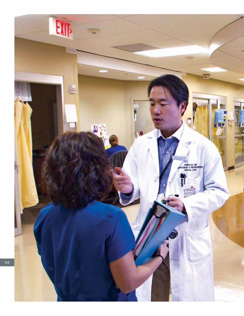
94

95
Patient Stories
mhmni.com

96
Van Buren, Arkansas, resident Lenord Lewis was working
in Houston when his recurrent headaches became so
bad that a co-worker drove him to Bayshore Medical
Center in Pasadena, Texas. After reviewing the results of
an MRI and CT scan, the emergency physician called an
ambulance to transport the 55-year-old to the Mischer
Neuroscience Institute (MNI) at Memorial Hermann-Texas
Medical Center.
“When I got there, they told me they were operating on
Monday,” says Lewis, who repairs electrical generators for
General Electric’s hydro and nuclear power operations.
OnApril12,2010,DongKim,M.D.,directorofMNIand
professor and chair of the Vivian L. Smith Department of
Neurosurgery at The University of Texas Health Science
Center at Houston (UTHealth) Medical School, performed
a craniotomy and resected a golf ball-sized brain tumor.
The pathology report confirmed glioblastoma multiforme
(GBM).
Of the estimated 19,000 primary brain tumors diagnosed
in the United States each year, approximately 60 percent
are gliomas, a heterogeneous group of neoplasms that
differ in location within the central nervous system,
growth potential, invasiveness and response to treatment.
With the exception of brainstem gliomas, GBM has the
worst statistical prognosis of any central nervous system
malignancy – a median survival of 14.6 months. Only
3 to 5 percent of patients survive for more than three
years, and they are classed as long-term survivors.
Following the surgery, Lewis was seen by Jay-Jiguang,
Zhu, M.D., Ph.D., a fellowship-trained neuro-oncologist
at MNI and an associate professor in the department of
Neurosurgery at the UTHealth Medical School. Dr. Zhu
had recently joined the MNI team from Boston, where
he trained at Massachusetts General Hospital and served
on the faculty of Tufts University. He worked with Lewis
to develop a plan of care.
Lewis received standard-of-care treatment – temozolomide
chemotherapy for 42 days with concurrent radiation. But
the tumor recurred. An MRI scan in August 2010 showed
a change at the surgical cavity in his left temporal lobe.
This small area of progression was treated with Gamma
Knife®radiosurgerybyDr.Kim,thenLewisresumed
chemotherapy with temozolomide until February 2011.
At that time, Dr. Zhu offered him the opportunity to try
a new technology that generates an electric field to the
brain through electrodes applied to the scalp. Made by
NovoCure, the NovoTTF-100A has been approved by the
FDA for treatment of recurrent GBM and is being tested
for safety and efficacy as an adjuvant treatment in newly
diagnosed GBM patients.
A locally or regionally delivered treatment, tumor treating
fields (TTF) therapy uses electric fields within the human
body to disrupt the rapid division and spread of cancer
cells. Developed to provide physicians and patients
with a fourth treatment option for cancer in addition to
surgery, radiation therapy and chemotherapy, TTF
therapy is designed for continuous use during the day
and night, with portable battery packs that allow
lenord lewis: beating the glioblastoma multiforme odds
p a t i e n t s t o r i e s

97
mhmni.com
patients to maintain their normal daily routine while
undergoing treatment.
“Studies have shown that low-intensity, intermediate
frequency electric fields stunt the growth of tumor cells,”
Dr. Zhu says. “The device is designed to administer
alternating electric fields to the region of the malignant
tumor by means of insulated surface electrode arrays.”
Lewis will wear the NovoTTF-100A for two years while
continuing the standard chemotherapy regimen of
temozolomide five days out of every 28, following an
initialfourcyclesofAvastin®onceeverytwoweeks.
Four pads, each of which contains nine electrodes,
cover his head. He wears the pads for three days, then
removes them, washes and shaves his head, and
applies a clean set of electrodes.
“With a median survival of 14.6 months, the odds of making
it three years after a diagnosis of GBM are low,” Dr. Zhu
says. “Lenord is able to maintain normal activities of daily
living, and he’s very upbeat. That’s the key to success in
addition to therapies. His wife and family provide excellent
care and support for him, which has allowed him to keep
up the long-distance medical care. We feel that our care
is top quality, and he and his wife feel that it’s worth the
travel time and expense to be treated at MNI.”
Lewisandhiswife,Celeste,participatedinthe5KRun
for the Rose in Houston in March 2012, an event that
funds brain cancer research. “There are so many cancer
patients,” he says. “I hurt for the others. If I can show
how good life is and how other people can enjoy it
despite cancer, that’s what I want to do. We don’t view
Dr.ZhuandDr.Kimasjustdoctors.Wefeellikethey’re
family helping us. If I can pull a couple more years with
the device and then a couple more years after that – and
a couple more years after that – that’s what I’m going for.
You’ve just got to bite the bullet and keep going.”
If I can show how good life
is and how other people
can enjoy it despite cancer,
that’s what I want to do.

98
Twenty-four-year-oldKelleeHearnehadsufferedsevere
headaches from the age of 10. Over the course of
those 14 years, several physicians diagnosed her with
migraines, but none of the medications they prescribed
eliminated the pain.
It was her Ob/Gyn who first suggested that the location
of the headaches indicated they might be something
other than migraines and referred her to a neurologist,
who ordered an MRI. Based on the results, he diagnosed
her with Chiari I malformation, a neurological disorder
in which the cerebellum – the part of the brain that
controls balance – descends out of the skull and into
the spinal area.
Normally, the cerebellum and parts of the brain
stem sit in an indented space at the lower rear of
the skull above the foramen magnum, a funnel-like
opening to the spinal canal. Chiari malformation is
diagnosed when part of the cerebellum is located
below the foramen magnum. The most common Chiari
malformation is Type I, which involves the extension
of the lower part of the cerebellum into the foramen
magnum, without involving the brain stem. Chiari I can
affect adolescents and adults, and often causes severe
symptoms.
Hearne’s mother learned about the Mischer Neuroscience
Institute’s Face Pain, Trigeminal Neuralgia and Chiari
Clinic during an Internet search. The clinic was started
byDongKim,M.D.,directorofMNIandprofessor
and chair of the Vivian L. Smith Department of
Neurosurgery, with neurologist Anita Madan, M.D., an
assistant professor of neurology, both at The University
of Texas Health Science Center at Houston (UTHealth)
Medical School. “Chiari I is a very specific syndrome
that occurs in people whose posterior fossa is more
shallow than normal or is situated lower in the brain,”
Dr. Madan says. “A portion of the cerebellum starts
herniating downward, causing pressure on the spinal
cord, which in turn causes symptoms.” Type I Chiari
can be asymptomatic, but patients may also experience
neck pain, muscle weakness, numbness, balance
problems, vision problems or difficulty swallowing,
in addition to headaches.
When Hearne and her mother saw Dr. Madan and
Dr.Kimattheclinic,theydiscussedsurgicaloptionsand
what to expect during recovery. “They left the decision
up to me, but I’d already decided on surgery because
I wanted to be here for my 2-year-old son,” Hearne says.
Whenworkingwithpatients,Dr.Kimlistenscarefully
to their descriptions of their symptoms. “It’s most
important to correlate the clinical picture and what the
patient is feeling with what we’re seeing on MRI,” he
says. “There’s no effective long-term treatment for Chiari
malformation other than surgery. Eventually, as the
symptoms worsen, medications begin to fail.”
Hearne’s surgery took place on June 20, 2012. “It’s a
straightforwardprocedure,”Dr.Kimsays.“Thegoalof
Kellee Hearne: relief from 14 years of headaches
p a t i e n t s t o r i e s
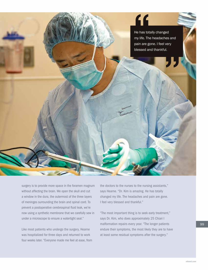
99
surgery is to provide more space in the foramen magnum
without affecting the brain. We open the skull and cut
a window in the dura, the outermost of the three layers
of meninges surrounding the brain and spinal cord. To
prevent a postoperative cerebrospinal fluid leak, we’re
now using a synthetic membrane that we carefully sew in
under a microscope to ensure a watertight seal.”
Like most patients who undergo the surgery, Hearne
was hospitalized for three days and returned to work
four weeks later. “Everyone made me feel at ease, from
the doctors to the nurses to the nursing assistants,”
saysHearne.“Dr.Kimisamazing.Hehastotally
changed my life. The headaches and pain are gone.
I feel very blessed and thankful.”
“The most important thing is to seek early treatment,”
saysDr.Kim,whodoesapproximately25ChiariI
malformation repairs every year. “The longer patients
endure their symptoms, the most likely they are to have
at least some residual symptoms after the surgery.”
mhmni.com
He has totally changed
my life. The headaches and
pain are gone. I feel very
blessed and thankful.

100
Tom Tinn’s stroke led him to rearrange his life in some
very interesting ways. The journey began on September 14,
2011. The 55-year-old had just finished breakfast when
his wife looked at him and noticed the telltale droop of
stroke on the left side of his face. “Why don’t you go lie
down and I’ll call 911,” he remembers her saying. When
he tried to stand up, he fell to the floor.
About two weeks earlier, he’d returned home following
surgery for prostate cancer at Memorial Hermann
Memorial City Medical Center. After the procedure,
he went back to the hospital twice to be treated
for allergic reactions to the antibiotics he was
prescribed.”When I had the stroke, the EMS team told
me, ‘We’re taking you to the best place to treat it.’”
The “best place” is Mischer Neuroscience Institute (MNI)
at Memorial Hermann, whose Stroke Center opened in
1988 as one of the first dedicated stroke programs in the
world. Today, the Center is home to the 10-county Greater
Houston area’s largest onsite stroke team. Neurologists
at the Center use leading-edge technology to diagnose
and treat more than 1,000 patients annually, ensuring
that each patient gets the appropriate treatment as
quickly as possible. By working closely with the Houston
Fire Department and local EMS services, the stroke
team has logged an impressive record of success in the
administration of clot-dissolving tPA – more than 10 times
the national average of 2 percent.
On arrival at Memorial Hermann-Texas Medical Center’s
Emergency Center, Tinn was seen by Osman Mir, M.D.,
a member of the MNI stroke team. He was completely
paralyzed on the left side and his score on the National
Institutes of Health Stroke Scale (NIHSS) was 11,
indicating a moderate-sized stroke. The NIHSS is a
neurologic assessment used to evaluate the severity of
stroke on a scale from 0, indicating no deficits, to 39.
Tinn was treated with tPA, the standard of care for acute
ischemic stroke, but he didn’t respond to the therapy. “In
fact, he got worse,” says George A. Lopez, M.D., Ph.D., a
neurologist affiliated with MNI and an associate professor
in the department of Neurology at The University of Texas
Health Science Center at Houston (UTHealth) Medical
School, who saw Tinn the next morning and directed his
care during his hospitalization. “When his stroke scale
score went from 11 to 13, we asked if he would be
interested in participating in an ongoing stroke clinical
trial called MR RESCUE.”
Begun in May 2004, the study, “Mechanical Retrieval
and Recanalization of Stroke Clots Using Embolectomy,”
nicknamed MR RESCUE, compares the effectiveness
of treating acute ischemic stroke with mechanical
embolectomy using a clot retriever within eight hours
of symptom onset to standard medical treatment.
The researchers also aim to identify people who
might benefit from mechanical embolectomy by the
appearance of the stroke on CT or MRI.
Tinn consented to participate in the study and was
assigned to the interventional arm. He was taken to
MNI’s angiography suite by neurointerventionalist
Tom Tinn: From stroke to a new life abroad
p a t i e n t s t o r i e s

101
Roc Chen, M.D., an assistant professor in the Vivian L.
Smith Department of Neurosurgery at UTHealth Medical
School, who performed a cerebral arteriogram showing
a blockage of the right middle cerebral artery. Dr. Chen
guided a small catheter into the artery and used a
retrieval device to extract the clot.
Immediately following the procedure, Tinn’s stroke scale
score dropped to 7. He was seen daily by a physical
therapist while in the hospital and discharged seven
days later on September 21. On November 1, after six
weeks of outpatient rehabilitation, he returned to his job
as a business development director for a global service
provider in the oil industry.
“I still have some issues that Dr. Lopez is aware of but
I consider myself 100 percent recuperated,” he says.
“It does take time to come back after a stroke. But I can
walk and talk and I’m back at work. I thank God and the
people at Memorial Hermann. They really saved me.”
Tinn and his wife relocated to Milan, Italy, at the end of
July 2012. “My wife is Italian, and our family is there,”
he says. “The events of the past year had an enormous
psychological effect on me. I’ve always put my work
before my family, but I feel I need to put them first now.”
Dr. Lopez considers Tinn’s recovery remarkable. “He went
back to work in a high-functioning environment a little
more than a month after the stroke,” he says. “We don’t
typically see recoveries like that. Usually, patients have
many more deficits to overcome.”
In an email Tinn sent to Dr. Lopez in June 2012, he wrote,
“Both my wife and I would like to offer our deepest and
sincere thanks to you and all the members of the stroke
team. Your kindness and professionalism is something
I’ll always treasure and remember in my life. I’m here
today because of them and God.”
mhmni.com
“I can walk and talk and
I’m back at work. I thank
God and the people at
Memorial Hermann. They
really saved me.”
Nine-year-old Lauren Lackey began displaying odd
behavior just before Thanksgiving in 2010. A straight-A
student, she was now struggling with routine assignments
and tests. She danced competitively – tap and jazz –
but was having difficulty concentrating and following
simple instructions.
“At first we thought she just needed some extra help
from her teachers,” says Lauren’s mother, Sandy
Lackey, who is a fifth-grade teacher. But shortly after
the New Year, Lackey was helping her daughter with
her homework when Lauren started writing random,
nonsensical letters and broke down crying. “As an
educator and a parent, I knew that all these things
didn’t add up, but never would we have expected
what was to come.”
A few days later, on January 26, 2011, Lauren complained
to her teacher of a headache and stomachache. The
school nurse examined Lauren and called her father,
Mark Lackey, to pick her up at her elementary school
in Cypress, Texas. But before they could leave the
school parking lot, Lauren’s condition deteriorated to
the point that she couldn’t walk or talk.
By the time she arrived at Children’s Memorial Hermann
Hospital, Lauren was in status epilepticus, had lapsed
102
lauren lackey: a life-saving endovascular procedure for Complex brain arteriovenous Fistulae
p a t i e n t s t o r i e s
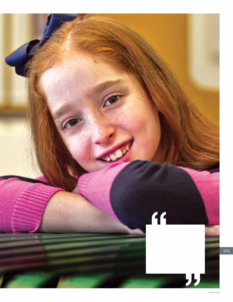
103
mhmni.com
She couldn’t move, and she
couldn’t speak. Dr. Chen told
us that her vessels were like
a 10-lane highway with eight
or nine lanes closed. We were
praying for a miracle.
into a coma and had to be intubated. Her MRI and
CT scans were reviewed by Ian Butler, M.D., a pediatric
neurologist affiliated with the hospital and professor
and Adriana Blood Chair in Neurology at The University
of Texas Health Science Center at Houston (UTHealth)
Medical School. When he saw an alarming blood vessel
abnormality, he called P. Roc Chen, M.D., to consult
on the case. A cerebrovascular neurosurgeon affiliated
with the Mischer Neuroscience Institute (MNI), Dr.
Chen specializes in adult and pediatric patients with
congenital vascular anomalies of the brain and spine,
and endovascular surgery for aneurysms.
“Dr. Butler and I were concerned about two things
that weren’t clear from the initial scans,” says Dr. Chen,
who is an assistant professor in the Vivian L. Smith
Department of Neurosurgery at the UTHealth Medical
School. “We had to determine if we were dealing with
an intracranial venous occlusion or a diffused neuro
arteriovenous fistula.” He accompanied Lauren to the
neurointerventional suite for a diagnostic angiogram,
where testing revealed that she had both conditions.
“She had lost her blood outflow channels almost
completely and was left with only one complete outflow
through her face. In addition, there were diffused
dural arteriovenous fistulae (AVF), which significantly
increased venous pressure to prevent blood flow
out of her brain,” he says. “Her brain was engorged
with blood, which caused the seizure, her cognitive
dysfunction and coma. The disease was so widespread
that every artery was shortcutting shunting to the
venous sinus [outflow channel]. Her condition was
extremely dire.”
The neurosurgeon injected 10 cubic centimeters of
Onyx®glueintotheabnormalvesselstorepairthedural
arteriovenous fistulae. He stopped after 70 minutes
when his team had reached the single-day radiation
and contrast safety limit, and brought her back the
following day to finish the procedure.
“When I first got to the hospital, Lauren was not in
good shape,” her mother says. “She couldn’t move,
and she couldn’t speak. Dr. Chen told us that her
vessels were like a 10-lane highway with eight or nine
lanes closed. We were praying for a miracle.”
Less than two weeks after she was admitted, Lauren
was discharged – walking and talking. She completed
a short course of outpatient rehabilitation at TIRR
Memorial Hermann Adult and Pediatric Outpatient
Rehabilitation. Her long-term prognosis is positive,
and her life has returned to normal.
“She lives with a single venous flow channel, but
she has done remarkably well,” says Dr. Chen, who
credits Lauren’s outcome to the experience and
professionalism of the multidisciplinary team that
provided her care. “Our expertise at MNI gives us the
ability and confidence to aggressively and swiftly treat
some of the most serious cerebral vascular conditions,
which, if not treated immediately, would result in a
devastating prognosis or death.”
Now 11, Lauren is doing “beyond great,” in the words of
her mother. “She just started the sixth grade. After all
she’d been through, it was difficult to think of her going
from elementary school to junior high with 1,200 kids
and eight different teachers, but she has adjusted well
socially and academically, and is taking all above-level
classes.” Lauren has resumed ballet and tap dancing
and this fall joined a community league softball team.
“As a parent you hope you never have to experience
anything like this,” Mrs. Lackey adds. “But from the
initial emergency room visit to her discharge, our
experience was absolutely great.”
104
Lauren Lackey patient stories
105
mhmni.com
Five years ago Megan Collins’ life was marked by pain,
social embarrassment and uncertainty about her medical
future. Today, the 35-year-old considers herself lucky.
Collins began having balance and coordination
problems in 2007 at the age of 30, and they worsened
progressively. An employee of the Children’s Neurology
Clinic in Fort Worth, Texas, she was referred to Mya
Schiess, M.D., by two pediatric neurologists who had
completed fellowships with the movement disorders
specialist at The University of Texas Health Science
Center at Houston (UTHealth) Medical School. Dr.
Schiess is professor and vice chair of the department
of Neurology and holds the Adriana Blood Endowed
Chair at UTHealth Medical School. She is affiliated
with the Mischer Neuroscience Institute and is also
the director of UT MOVE, a program focused on clinical
care, education and basic science research on the
neurological conditions of motor systems disruption,
including movement disorders, cerebral palsy, spasticity,
neurodegenerative diseases and dementias.
“We worked with Megan for about a year and responded
to the disorder before we knew exactly what we were
dealing with,” Dr. Schiess says. “It took us some time
to reach a conclusive diagnosis. Her father had similar
symptoms, so we suspected a genetic disorder. But
testing for a full panel of inherited ataxia disorders is
expensive, and Megan is a single working mom.”
Collins’ father eventually underwent testing paid for by
the Veterans Administration and was diagnosed with
spinocerebellar ataxia type 1 (SCA1). “When a family
member tests positive for a disorder and another
family member has the same symptomatology, we can
narrow down the testing to one relatively inexpensive
test,” Dr. Schiess says. “Megan also tested positive
for the disorder.”
Spinocerebellar ataxia type 1 is characterized by
progressive problems with movement. Patients with the
disorder initially experience problems with balance –
ataxia – and coordination. Other signs and symptoms
of the disorder may include speech and swallowing
difficulties, spasticity, and weakness in the muscles that
control involuntary eye movements. Individuals with
SCA1 may have cognitive impairment marked by difficulty
processing, learning and remembering information.
Over time, they may develop numbness, tingling or pain
in the arms and legs, muscle wasting, twitches and
uncontrolled muscle tensing, known as dystonia.
Over the course of the next year, Collins developed
the classic symptoms of craniocervical dystonia, a
movement disorder characterized by intermittent
spasms of the neck and shoulder muscles that cause
abnormal head movements. “Botulinum toxin injections
provided relief from the spasms,” Dr. Schiess says.
“After a lumbar puncture revealed neurotransmitter
deficits, we prescribed levodopa and sertraline to
target the deficiencies. The medications helped
alleviate symptoms, and subsequent analysis of the
cerebrospinal fluid showed a normalization of her levels.
Megan Collins: Deep brain stimulation Changes a life
p a t i e n t s t o r i e s

We also started her on baclofen for spasticity and
clonazepam for her dystonia.”
Collins responded favorably to the treatment regimen
for almost 18 months. Then her dystonia began to
worsen and evolved aggressively for almost a year,
despite increased dosage and distribution of botulinum
toxin injections. Eventually, her upper limbs and trunk
became involved, and her severe dystonic crises
became unresponsive to medication changes. She
required several emergency department visits
and hospitalizations.
“Megan reached the point that she was no longer
able to work,” Dr. Schiess says. “We kept her out of the
hospital as long as possible. She was on very high
doses of medication, and we realized that her disorder
was not coming under control despite our best efforts.”
Collins had read about deep brain stimulation (DBS),
which was approved by the FDA in 2003 for the
treatment of dystonia. “Dr. Schiess agreed that
I could see Dr. Fenoy for a consult,” she says. “When
I scheduled the appointment with him, I was in the
middle of a dystonic storm, never knowing when
I was going to have an episode. I ended up having
one in his office. It was very scary and crazy but I feel
lucky that he could see what was happening to me.”
Albert Fenoy, M.D., is a neurosurgeon and deep
brain stimulation specialist affiliated with the Mischer
Neuroscience Institute and an assistant professor in
the Vivian L. Smith Department of Neurosurgery at the
UTHealth Medical School. On May 22, 2012, with
Dr. Schiess attending in the OR, he placed a DBS
electrode into Collins’ globus pallidus internus.
“We placed a stereotactic frame on Megan’s head
and using MRI visualization, we made two burr holes
in the skull while she was awake, and descended
microelectrodes into the brain to verify neuronal activity
and confirm that we’d reached the target area,”
Dr. Fenoy says. “Then we placed the actual DBS leads
and test stimulated to see what kind of response she
had and whether there were any side effects.” Two
weeks later, on June 6, with Collins under general
anesthesia, they placed extensions from the electrodes
to a pulse generator and implanted the generator in
her chest under the clavicle.
On June 7, Dr. Schiess programmed the stimulator for the
first time. “It’s been a miracle,” Collins says. It’s amazing
what I can do now that I couldn’t do before. I can now
stand up with my feet together and move better than
I’ve been able to move in years. I would recommend
Dr. Schiess and Dr. Fenoy to anyone in the world.
They’ve changed my life.”
“When you understand a therapy and are involved in
the selection of many candidates over time and have the
opportunity to observe the results, you develop a good
sense of what it can and can’t do. We’re very good DBS
programmers here,” Dr. Schiess says. “We believe that
if DBS can help a patient improve in even a single area,
it should be applied. When indicated, it can greatly
improve a patient’s life. Four months out from her
surgery, Megan is holding her own, and her dystonia is
better controlled. As time goes on, through research,
we’ll find different targets in the brain and develop more
exquisite control. Science is an evolutionary process.
We keep driving forward.”
106
Megan Collins patient stories
107
mhmni.com
It’s been a miracle. It’s
amazing what I can do now
that I couldn’t do before.
I would recommend
Dr. Schiess and Dr. Fenoy
to anyone in the world.
They’ve changed my life.

108

109
mhmni.com
NeurosurGery
Dong Kim, M.D.DirectorMischer Neuroscience Institute
Chief of NeurosurgeryMemorial Hermann-Texas Medical Center
Professor and ChairVivian L. Smith Department of NeurosurgeryUTHealth Medical School
p. roc chen, M.D.Assistant ProfessorVivian L. Smith Department of NeurosurgeryUTHealth Medical School
Mark J. Dannenbaum, M.D.Associate ProfessorVivian L. Smith Department of NeurosurgeryUTHealth Medical School
Arthur L. Day, M.D.Director of Clinical EducationMischer Neuroscience Institute
Professor and Vice ChairVivian L. Smith Department of NeurosurgeryUTHealth Medical School
Albert Fenoy, M.D.Assistant ProfessorVivian L. Smith Department of NeurosurgeryUTHealth Medical School
Michele M. Johnson, M.D.DirectorSpine ProgramMischer Neuroscience Institute
Assistant ProfessorVivian L. Smith Department of NeurosurgeryUTHealth Medical School
Daniel h. Kim, M.D., F.A.c.s.DirectorReconstructive Spinal and Peripheral Nerve SurgeryMischer Neuroscience Institute
ProfessorVivian L. Smith Department of NeurosurgeryUTHealth Medical School
Karl schmitt, M.D.Assistant ProfessorVivian L. Smith Department of NeurosurgeryUTHealth Medical School
scott shepard, M.D.DirectorGammaKnifeRadiosurgeryProgramMischer Neuroscience Institute
Clinical Assistant ProfessorVivian L. Smith Department of NeurosurgeryUTHealth Medical School
Nitin tandon, M.D.Associate ProfessorVivian L. Smith Department of Neurosurgery
Associate Professor Department of Pediatric SurgeryUTHealth Medical School
NeuroLoGy
James c. Grotta, M.D.Co-DirectorMischer Neuroscience Institute
Chief of Neurology and DirectorStroke CenterMemorial Hermann-Texas Medical Center
Professor and ChairDepartment of Neurology Roy M. and Phyllis Gough HuffingtonDistinguished Chair in NeurologyUTHealth Medical School
parveen Athar, M.D.Assistant ProfessorDepartment of NeurologyUTHealth Medical School
Andrew barreto, M.D.Assistant ProfessorDepartment of NeurologyUTHealth Medical School
suur biliciler, M.D.Assistant ProfessorDepartment of NeurologyUTHealth Medical School
staley brod, M.D.ProfessorDepartment of NeurologyUTHealth Medical School
randall c. edgell, M.D.Associate ProfessorVivian L. Smith Department of Neurosurgery and Department of NeurologyUTHealth Medical School
James Ferrendelli, M.D.ProfessorDepartment of NeurologyUTHealth Medical School
erin Furr-stimming, M.D.Assistant ProfessorDepartment of Neurology
Movement Disorders SpecialistUT MOVE Clinic
DirectorNeurology Clerkship ProgramUTHealth Medical School
Nicole r. Gonzales, M.D.Assistant ProfessorDepartment of NeurologyUTHealth Medical School
omotola hope, M.D.Assistant ProfessorDepartment of Neurology
DirectorNeurology Residency Training ProgramUTHealth Medical School
sigmund h. hsu, M.D.Neuro-OncologistAssistant ProfessorVivian L. Smith Department of Neurosurgery Department of NeurologyUTHealth Medical School
Nneka ifejika, M.D.Assistant ProfessorDepartment of Neurology
Director of NeurorehabilitationUTHealth Medical School
ernesto infante, M.D.Assistant ProfessorDepartment of NeurologyUTHealth Medical School
Giridhar Kalamangalam,M.D., D.phil.Assistant ProfessorDepartment of NeurologyUTHealth Medical School
robert Knowlton, M.D.Associate ProfessorDepartment of NeurologyUTHealth Medical School
Staff listing

110
S T A F F l I S T I N G
John A. Lincoln, M.D.Assistant ProfessorDepartment of NeurologyUTHealth Medical School
John William Lindsey, M.D.ProfessorDepartment of NeurologyUTHealth Medical School
George Lopez, M.D.Associate ProfessorDepartment of NeurologyUTHealth Medical School
Anita Madan, M.D.Assistant ProfessorDepartment of NeurologyUTHealth Medical School
raymond A. Martin, M.D., F.A.A.N.ProfessorDepartment of Neurology
Medical DirectorOutpatient Neurology ClinicUTHealth Medical School
Vivek Misra, M.D.Clinical InstructorDepartment of NeurologyUTHealth Medical School
Flavia Nelson, M.D.Assistant ProfessorDepartment of Neurology
Associate DirectorMagnetic Resonance Imaging Analysis CenterUTHealth Medical School
elizabeth Noser, M.D.Medical DirectorNeurorehabilitationMemorial Hermann-Texas Medical Center
Clinical Assistant ProfessorDepartment of NeurologyUTHealth Medical School
thy Nguyen, M.D.Assistant ProfessorDepartment of Neurology
Co-Director Electromyography LaboratoryUTHealth Medical School
William ondo, M.D.ProfessorDepartment of NeurologyUTHealth Medical School
Jacqueline phillips-sabol, M.D.DirectorNeuropsychology ProgramMemorial Hermann-Texas Medical Center
Assistant ProfessorDepartment of NeurologyUTHealth Medical School
sean savitz, M.D.ProfessorDepartment of Neurology
DirectorStroke Program
DirectorVascular Neurology FellowshipUTHealth Medical School
Mya schiess, M.D.Professor and Vice ChairDepartment of NeurologyAdriana Blood Endowed Chairin Neurology
DirectorMovement Disorders andNeurodegenerative Disease
DirectorUT MOVE ClinicUTHealth Medical School
paul e. schulz, M.D.Associate Professor and Vice ChairDepartment of Neurology
DirectorDementia ProgramUTHealth Medical School
Kazim sheikh, M.D.ProfessorDepartment of Neurology
DirectorNeuromuscular ProgramUTHealth Medical School
Jeremy slater, M.D.Medical DirectorEpilepsy Monitoring Unit and NeurophysiologyMemorial Hermann-Texas Medical Center
Associate ProfessorDepartment of NeurologyUTHealth Medical School
sudha tallavajhula, M.D.Associate ProfessorDepartment of NeurologyUTHealth Medical School
Melissa thomas, M.D.Assistant ProfessorDepartment of NeurologyUTHealth Medical School
Gage Van horn, M.D.ProfessorDepartment of NeurologyUTHealth Medical School
holly Knudsen Varner, M.D.Assistant ProfessorDepartment of NeurologyUTHealth Medical School
bethany Williams, M.D.Assistant ProfessorDepartment of NeuropsychologyUTHealth Medical School
Jerry s. Wolinsky, M.D.Bartels Family and Opal C. RankinProfessorDepartment of Neurology
DirectorMultiple Sclerosis Research Group (MSRG)
DirectorMagnetic Resonance Imaging Analysis CenterUTHealth Medical School
tzu-ching (teddy) Wu, M.D.Telemedicine DirectorMischer Neuroscience Institute
Assistant ProfessorDepartment of NeurologyUTHealth Medical School
Jay-Jiguang Zhu, M.D., ph.D.Neuro-OncologistAssociate ProfessorVivian L. Smith Department of NeurosurgeryDepartment of NeurologyUTHealth Medical School
criticAL cAre
imo Aisiku, M.D.DirectorNeurocritical Care Mischer Neuroscience Institute
Associate ProfessorVivian L. Smith Department of Neurosurgery
Vice ChairDivision of Critical CareUTHealth Medical School

h. Alex choi, M.D. Assistant ProfessorVivian L. Smith Department of Neurosurgery and Department of NeurologyUTHealth Medical School
Kiwon Lee, M.D.Co-DirectorNeurocritical Care Mischer Neuroscience Institute
Associate ProfessorDepartment of Neurology andVivian L. Smith Department of Neurosurgery
Vice Chair of Clinical AffairsDepartment of Neurology UTHealth Medical School
bilal rana, D.o.Assistant ProfessorVivan L. Smith Department of NeurosurgeryUTHealth Medical School
hanh truong, M.D.Assistant ProfessorVivian L. Smith Department of NeurosurgeryUTHealth Medical School
George W. Williams, ii, M.D.Assistant ProfessorVivian L. Smith Department of NeurosurgeryUTHealth Medical School
Asma Zakaria, M.D.Assistant ProfessorVivian L. Smith Department of NeurosurgeryDepartment of NeurologyUTHealth Medical School
chiLDreN’s NeuroscieNce ceNter
ian butler, M.D.DirectorDivision of Child & Adolescent Neurology Department of Pediatric Surgery
ProfessorDivision of Child & Adolescent NeurologyDepartment of Pediatric SurgeryUTHealth Medical School
stephen Fletcher, D.o.Associate ProfessorDivision of Child & Adolescent Neurology,Department of Pediatric SurgeryUTHealth Medical School
Michael Gambello, M.D., ph.D.ProfessorDivision of Medical Genetics,Department of PediatricsUTHealth Medical School
Mary Kay Koenig, M.D.DirectorNeurometabolic & Mitochondrial Clinic
Assistant ProfessorDivision of Child & Adolescent NeurologyDepartment of PediatricsUTHealth Medical School
pedro Mancias, M.D.Associate ProfessorDivision of Child & Adolescent NeurologyDepartment of PediatricsUTHealth Medical School
hope Northrup, M.D.DirectorDivision of Medical GeneticsDepartment of Pediatrics
DirectorTuberous Sclerosis Complex Clinic
ProfessorDivision of Medical Genetics Department of PediatricsUTHealth Medical School
David sandberg, M.D., F.A.A.N.s., F.A.c.s., F.A.A.p.DirectorPediatric Neurosurgery Mischer Neuroscience Institute
Associate ProfessorVivian L. Smith Department of Neurosurgery
Associate ProfessorDepartment of Pediatric SurgeryUTHealth Medical School
Nitin tandon, M.D.Associate ProfessorDepartment of Pediatric Surgery
Associate ProfessorVivian L. Smith Department of NeurosurgeryUTHealth Medical School
Gretchen Von Allmen, M.D.DirectorPediatric Epilepsy Program
Co-DirectorEpilepsy Monitoring UnitMemorial Hermann-Texas Medical Center
Assistant ProfessorDivision of Child & Adolescent NeurologyDepartment of PediatricsUTHealth Medical School
reseArch FAcuLty
Jaroslaw (Jarek) Aronowski, ph.D.ProfessorDepartment of NeurologyUTHealth Medical SchoolIschemic Stroke and Brain Hemorrhage
Qilin cao, M.D.Associate ProfessorVivian L. Smith Department of NeurosurgeryUTHealth Medical SchoolStem Cell Therapy
Guy clifton, M.D.DirectorVivian L. Smith Center forNeurologic Research
ProfessorVivian L. Smith Department of NeurosurgeryUTHealth Medical SchoolBrain Injury
pramod Dash, M.D.ProfessorVivian L. Smith Department of Neurosurgery and Department of Neurobiology and AnatomyUTHealth Medical SchoolBrain Injury
timothy ellmore, ph.D.Assistant ProfessorVivian L. Smith Department of NeurosurgeryUTHealth Medical SchoolNeural Substrates of Language and Memory
Georgene hergenroeder, r.N.Assistant ProfessorVivian L. Smith Department of NeurosurgeryUTHealth Medical SchoolBioinformatics and Translational Research
Nobuhide Kobori, M.D., ph.D.Assistant ProfessorVivian L. Smith Department of NeurosurgeryUTHealth Medical SchoolBrain Injury and Memory
Min Li, ph.D.Associate ProfessorVivian L. Smith Department of Neurosurgery
DirectorCancer Research ProgramUTHealth Medical SchoolNeuro-Oncology
ying Liu, M.D., ph.D.Assistant ProfessorVivian L. Smith Department of NeurosurgeryUTHealth Medical SchoolStem Cell Research
S T A F F l I S T I N G
111
mhmni.com

ACrOSS THE SYSTEM
hariyadarshi pannu, ph.D.Assistant ProfessorVivian L. Smith Department of NeurosurgeryUTHealth Medical SchoolVascular Disease and Genetics
teresa santiago-sim, ph.D.Assistant ProfessorVivian L. Smith Department of NeurosurgeryUTHealth Medical SchoolVascular Disease and Genetics
claudio soto, ph.D.DirectorCenter for Neurodegenerative Diseases
Professor of NeurologyUTHealth Medical SchoolNeurodegenerative Disease
Jia Qian Wu, ph.D.Assistant ProfessorVivian L. Smith Department of NeurosurgeryUTHealth Medical SchoolStem Cell Research
ying Xia, M.D., ph.D.Professor and Vice ChairVivian L. Smith Department of NeurosurgeryUTHealth Medical SchoolCellular and Molecular Neuroscience
rong yu, ph.D.Assistant ProfessorVivian L. Smith Department of NeurosurgeryUTHealth Medical SchoolOxidative Stress and Brain Injury
Gang Zhang, M.D.Assistant ProfessorDepartment of NeurologyUTHealth Medical SchoolPeripheral Nervous System
Xiurong Zhao, M.D.Associate ProfessorDepartment of NeurologyUTHealth Medical SchoolIschemic Stroke and Brain Hemorrhage
MeMoriAL herMANN KAty hospitAL
Quoc Anh thai, M.D.Clinical Assistant ProfessorVivian L. Smith Department of NeurosurgeryUTHealth Medical School
MeMoriAL herMANN MeMoriAL city MeDicAL ceNter
paul boone, M.D.Clinical Assistant ProfessorVivian L. Smith Department of NeurosurgeryUTHealth Medical School
Anita Madan, M.D.Assistant ProfessorDepartment of NeurologyUTHealth Medical School
G. silky patel, M.D.Clinical Assistant ProfessorVivian L. Smith Department of NeurosurgeryUTHealth Medical School
scott shepard, M.D.DirectorGammaKnifeRadiosurgeryProgramMischer Neuroscience Institute
Clinical Assistant ProfessorVivian L. Smith Department of NeurosurgeryUTHealth Medical School
MeMoriAL herMANN NortheAst hospitAL
Albert Fenoy, M.D.Assistant ProfessorVivian L. Smith Department of NeurosurgeryUTHealth Medical School
MeMoriAL herMANN southeAst hospitAL
Kathleen eberle, M.D.Neurologist Houston Neurological Institute
Joseph c. hsieh, M.D.Assistant ProfessorVivian L. Smith Department of NeurosurgeryUTHealth Medical School
Kimberly Monday, M.D.Neurologist Houston Neurological Institute
Jack ownby, M.D.Neurologist Houston Neurological Institute
MeMoriAL herMANN the WooDLANDs hospitAL
raul J. cardenas, M.D.Clinical Assistant ProfessorVivian L. Smith Department of NeurosurgeryUTHealth Medical School
Kevin c. Gaffney, M.D.Neurologist Mischer Neuroscience Associates The Woodlands
Anita Madan, M.D.Assistant ProfessorDepartment of NeurologyUTHealth Medical School
ryan McDonald, M.D.Neurologist Mischer Neuroscience Associates The Woodlands
randall Wright, M.D.Neurologist Mischer Neuroscience Associates The Woodlands
Geoffrey p. Zubay, M.D., F.A.c.s.Clinical Assistant ProfessorVivian L. Smith Department of NeurosurgeryUTHealth Medical School
S T A F F l I S T I N G
112


Memorial Hermann-Texas Medical Center6411 Fannin
Houston,TX77030
mhmni.com 877.75.NEURO (63876)
4404946