MIPR Lecture 6 Copyright Oleh Tretiak, 2004 1 Medical Imaging and Pattern Recognition Lecture 6...
33
MIPR Lecture 6 Copyright Oleh Tretiak, 2004 1 Medical Imaging and Pattern Recognition Lecture 6 X-ray Imaging Oleh Tretiak
-
Upload
bryant-watkinson -
Category
Documents
-
view
214 -
download
0
Transcript of MIPR Lecture 6 Copyright Oleh Tretiak, 2004 1 Medical Imaging and Pattern Recognition Lecture 6...
MIPR Lecture 6Copyright Oleh Tretiak, 2004
1
Medical Imaging and Pattern Recognition
Lecture 6 X-ray ImagingOleh Tretiak

MIPR Lecture 6Copyright Oleh Tretiak, 2004
2
Wilhelm Conrad Roentgen
• Roentgen discovered penetrating radiation on 8 November 1895.
• The famous radiograph made by Roentgen on 22 December 1895, and sent to physicist Franz Exner in Vienna. This is traditionally known as "the first X-ray picture" and "the radiograph of Mrs. Roentgen's hand. "
• Roentgen received the first Nobel prize in physics in 1901
QuickTime™ and aTIFF (Uncompressed) decompressor
are needed to see this picture.

MIPR Lecture 6Copyright Oleh Tretiak, 2004
3
X-rays at Present• Superior definition• Clear images of bones• Some indication of
tissue• No tissue detail
(tendon, muscle, skin)• Negative image: bone
is white, air is black

MIPR Lecture 6Copyright Oleh Tretiak, 2004
4
Talk Outline
• Examples of X-ray imaging procedures
• Physics: X-ray attenuation, transmission, and contrast
• X-ray recording systems• Summary and new developments
MIPR Lecture 6Copyright Oleh Tretiak, 2004
5
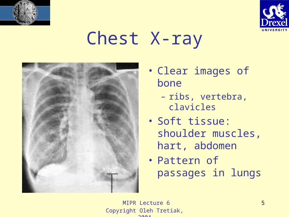
Chest X-ray
• Clear images of bone– ribs, vertebra,
clavicles
• Soft tissue: shoulder muscles, hart, abdomen
• Pattern of passages in lungs

MIPR Lecture 6Copyright Oleh Tretiak, 2004
6
Abdominal X-ray
• Visible: Bony structures– Vertebra, pelvic bones, legs,
ribs
• Soft tissues– liver, stomach, leg muscles
• Confusing image of intestines– Intestinal gas, walls
• Cannot see:– Details of liver, back muscles,
kidneys

MIPR Lecture 6Copyright Oleh Tretiak, 2004
7
Abdomen - more
• Abdomen after Barium contrast enema
• Large intestine easily visible

MIPR Lecture 6Copyright Oleh Tretiak, 2004
8
Another Abdomen
• Contrast medium in aorta (angiography)
• Visible: – descending aorta, – renal arteries, – iliac arteries

MIPR Lecture 6Copyright Oleh Tretiak, 2004
9
Pelvic X-Ray
• Can see– Fracture in
pelvis– Femur
• Cannot see– Soft tissues

MIPR Lecture 6Copyright Oleh Tretiak, 2004
10
Skull
• Can see bones, scalp
• Cannot see ventricles, blood vessels

MIPR Lecture 6Copyright Oleh Tretiak, 2004
11
Skull: Subtraction Angiography
MIPR Lecture 6Copyright Oleh Tretiak, 2004
12
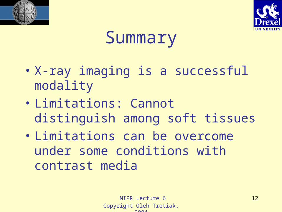
Summary
• X-ray imaging is a successful modality
• Limitations: Cannot distinguish among soft tissues
• Limitations can be overcome under some conditions with contrast media

MIPR Lecture 6Copyright Oleh Tretiak, 2004
13
X-Ray
• Schematic of x-ray imaging

MIPR Lecture 6Copyright Oleh Tretiak, 2004
14
What are X-rays?• X-rays (Roentgen rays) are electromagnetic, like
radio waves and light• There are three ways to measure the “quality” of
electromagnetic waves– Wavelength– Frequency– Photon energy
• f - frequency, Hertz (Hz)• - wavelength, meters (m)• E - photon energy, electron volts (Ev)• c - speed of light, 3x10¹⁰ m/sec• h - Planck’s constant, 4.1x10⁻¹⁵ Ev/Hz
€
=c / fE = hf

MIPR Lecture 6Copyright Oleh Tretiak, 2004
15
Examples
FrequencyWavelengt
hPhoton Energy
Radio1e6 Hz
1 Mega Hz300 m
4e-9 Ev4 nanoV
Green light 5.45e14 Hz0.55e-6 m0.55 m
2.2 Ev
X-ray 7.3e18 Hz 4.1e-11 m3e4 Ev30 kEv

MIPR Lecture 6Copyright Oleh Tretiak, 2004
16
Generation of X-rays
• X-rays are generated when electron hit a target
Cathode(electron source)
Anode(X-ray source)

MIPR Lecture 6Copyright Oleh Tretiak, 2004
17
X-ray Spectrum
• An X-ray tube produces a broad spectrum of energies
MIPR Lecture 6Copyright Oleh Tretiak, 2004
18
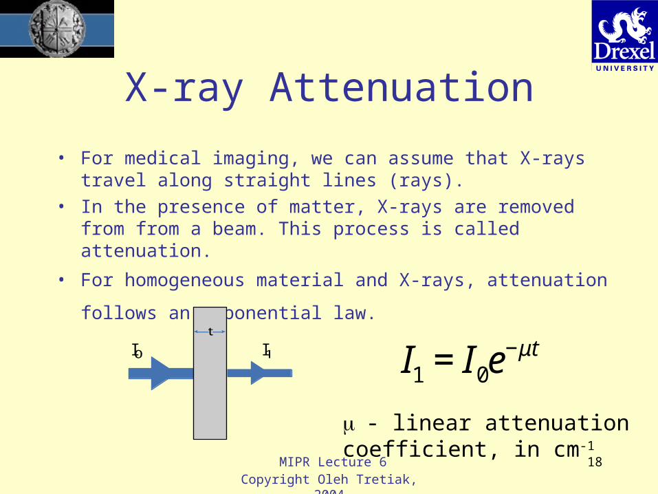
X-ray Attenuation
• For medical imaging, we can assume that X-rays travel along straight lines (rays).
• In the presence of matter, X-rays are removed from from a beam. This process is called attenuation.
• For homogeneous material and X-rays, attenuation
follows an exponential law. €
€
I1 = I0e−μtI I
- linear attenuation coefficient, in cm-1

MIPR Lecture 6Copyright Oleh Tretiak, 2004
19
Attenuation Coefficient Values
• Tables of X-ray attenuation and absorption coefficients can be found on the web - for example, http://physics.nist.gov/PhysRefData/XrayMassCoef/tab4.html

MIPR Lecture 6Copyright Oleh Tretiak, 2004
20
KEVFat, ρ = 0.916
, Muscle ρ = 1.04
, Bone ρ = 1.65
10 2.764 5.339 8.81015 0.924 1.668 2.75220 0.488 0.809 1.33530 0.271 0.380 0.62740 0.216 0.274 0.45250 0.193 0.233 0.38460 0.180 0.212 0.34980 0.164 0.189 0.312
100 0.154 0.176 0.290150 0.137 0.155 0.256200 0.125 0.141 0.233
Linear Attenuation
, [ ]Coefficient cm -1
Attenuation coefficient
0.100
1.000
10.000
10 100
KEV
µ (cm-1)
Fat, r = 0.916
Muscle, r = 1.04
Bone, r = 1.65

MIPR Lecture 6Copyright Oleh Tretiak, 2004
21
Examples
• Conclusion: High voltage photons are needed to penetrate thick objects.
20 kev 0.65 9.07E-08 0.2750 kev 0.21 4.90E-03 0.65
Abdomen t = 25 cm
Hand (palm) t = 2 cm
Values of transmission,T = exp(-t)

MIPR Lecture 6Copyright Oleh Tretiak, 2004
22
Contrast
t
t
m
m
z
( )I z
I(z)
z
I I
€
C =I0 − I1I0
If |(0–1)t1| is small,
€
C ≈ (μ0 −μ1)t1
MIPR Lecture 6Copyright Oleh Tretiak, 2004
23
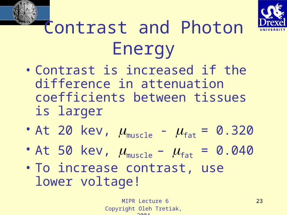
Contrast and Photon Energy
• Contrast is increased if the difference in attenuation coefficients between tissues is larger
• At 20 kev, muscle - fat = 0.320
• At 50 kev, muscle – fat = 0.040• To increase contrast, use lower
voltage!

MIPR Lecture 6Copyright Oleh Tretiak, 2004
24
Recording X-rays
• Direct film recording (like Roentgen)– Very low efficiency: film is thin, most X-
rays pass through the film emulsion• Screen-film combination
– Fluorescent screen captures X-rays and produces light
– Film exposed by light– Much more sensitivity than with film
alone

MIPR Lecture 6Copyright Oleh Tretiak, 2004
25
Recording X-rays
• Fluoroscope: Television camera observes fluorescent screen– Useful for real-time viewing– Lower image quality than screen-film
recording
• Computed radiography: use imaging plate instead of film to record image. – The plate is scanned with a laser and a
digital image is obtained
MIPR Lecture 6Copyright Oleh Tretiak, 2004
26
Recording X-rays
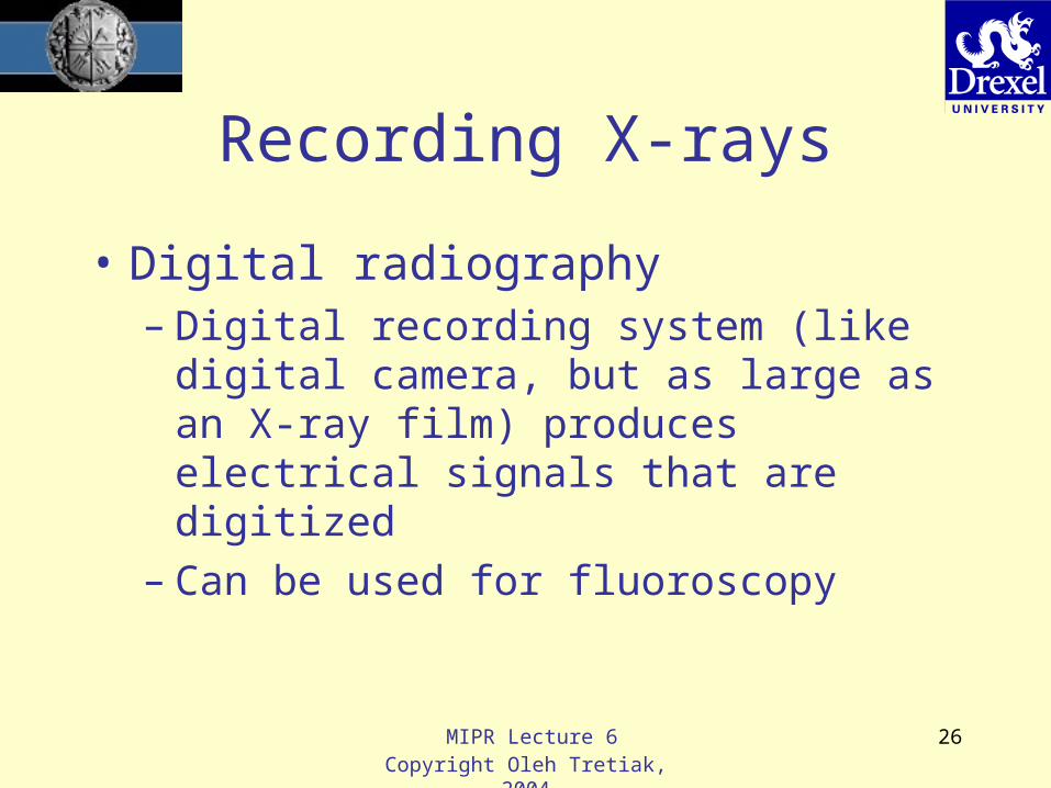
• Digital radiography– Digital recording system (like digital
camera, but as large as an X-ray film) produces electrical signals that are digitized
– Can be used for fluoroscopy

MIPR Lecture 6Copyright Oleh Tretiak, 2004
27
Comparison• SF ~ screen-film recording, CR ~
computed radiography, DR ~ digital radiography– Image quality: SF is best– Initial cost: SF is lowest– Operating cost: DR is lowest, film is highest– Sensitivity (patient exposure): DR and and
CR are better– Operating convenience: DR is best
• Conclusion: Each system has a use– Digital recording is displacing film

MIPR Lecture 6Copyright Oleh Tretiak, 2004
28
Big Picture
• Types of imaging procedures– Screening: detect disease when there are
no symptoms– Diagnosis: a disease is probably present,
identify the type of disease– Staging: we know the disease, what type of
treatment?– Treatment monitoring.
• Would like to screen, but there are few diseases that warrant it

MIPR Lecture 6Copyright Oleh Tretiak, 2004
29
Breast Cancer Screening
• Breast cancer screening requires high resolution and contrast
• Mostly done with screen-film at low voltage

MIPR Lecture 6Copyright Oleh Tretiak, 2004
30
Computer Interpretation• Reason for computer interpretation:
– Better accuracy than human?– Less expensive than human?– Human expert not available?
• Much research, many claims– In the US, a system must be tested and
approved by the Federal Drug Administration (FDA)
– There is an FDA approved system for mammography interpretation
– At present, used as adjunct for human doctors.

MIPR Lecture 6Copyright Oleh Tretiak, 2004
31
Other X-ray Applications
• Image from X-ray telescope
• Nebula left by exploding star
• X-ray telescopes are on satellites because X-rays do not penetrate the atmosphere

MIPR Lecture 6Copyright Oleh Tretiak, 2004
32
Summary
• X-rays are 100 years old• Created a revolution in medicine• Useful for many diagnostic tasks
– Limitation: cannot distinguish between soft tissues
– Contrast radiography helps
MIPR Lecture 6Copyright Oleh Tretiak, 2004
33
Developments in X-rays
• Digital recording systems are replacing film– Decrease in image quality– Improvement in sensitivity– More convenient
• Computer interpretation of X-rays is here– Now assisting mammography. May become
better.– I expect that procedures for cardiography are
next.