Minireview - C-MORE · Fluorescence in situ hybridization (FISH) is a new molecular-based technique...
16
http://wwwsoc.nii.ac.jp/jsme2/ Microbes Environ. Vol. 19, No. 2, 83–98, 2004 Minireview MAR-FISH—An Ecophysiological Approach to Link Phylogenetic Affiliation and In Situ Metabolic Activity of Microorganisms at a Single-Cell Resolution SATOSHI OKABE 1 *, TOMONORI KINDAICHI 1 and TSUKASA ITO 1 1 Department of Urban and Environmental Engineering, Graduate School of Engineering, Hokkaido University, North-13, West-8, Kita-ku, Sapporo 060–8628, Japan (Received March 22, 2004—Accepted April 22, 2004) A major goal of microbial ecology is to study the abundance, localization, and activities of microorganisms in situ in order to understand ecophysiological roles that the microorganisms play in complex natural ecosystems. In fact, in typical microbial habitats such as biofilms, sediments, and microbial aggregates, resources and physi- cochemical conditions are dynamically changing with time and across even a very tiny distance because of metabolic activities and substrate transport limitation. To directly correlate microbial identity (16S rRNA-based phylogeny) to the specific metabolic function of individual cells within such complex and heterogeneous micro- bial habitats, several new molecular-based techniques have been developed in the last decade. These techniques exploit in situ simultaneous phylogenetic identification and metabolic capabilities of even uncultured micro- organisms without the need to isolate them in culture. Microautoradiography is a powerful but “rather old” tool, with which the in situ uptake of specific radiolabeled substrates by individual cells can be determined. Fluorescence in situ hybridization (FISH) is a new molecular-based technique that allows the in situ phylogenetic identitification of individual cells. However, FISH cannot provide sufficient information on metabolic capabilities, because phy- logeny and phenotype are rarely congruent. Recently, microautoradiography and FISH have been successfully combined to further improve the complementary strengths of the two methods. Microautoradiography combined with FISH (MAR-FISH) can be used to simultaneously examine the phylogenetic identity and the relative or ac- tual specific activity of microorganisms within a complex microbial community at a single-cell level. This article overviews the principle, experimental protocol and application of the MAR-FISH technique, as well as current developments of other analytical techniques for in situ microbial functions (metabolic activities) from a single- cell level to community levels. Key words: microautoradiography combined with fluorescence in situ hybridization (MAR-FISH), microsensors, microbial community, in situ activity Introduction Understanding the structure and function of complex microbial communities is a central theme in microbial ecology. However, traditional cultivation-dependent methods are inadequate to fulfill this task because most members of microbial communities in natural and engineered systems cannot be cultured. It is now widely recognized that only a small fraction, possibly up to 1–10%, of the naturally occurring microorganisms in natural ecosystems have been isolated and characterized so far, even though a great diversity * Corresponding author; E-mail: [email protected], Tel: 81–11–706–6266, Fax: 81–11–706–6266
Transcript of Minireview - C-MORE · Fluorescence in situ hybridization (FISH) is a new molecular-based technique...

http://wwwsoc.nii.ac.jp/jsme2/
Microbes Environ. Vol. 19, No. 2, 83–98, 2004
Minireview
MAR-FISH—An Ecophysiological Approach to Link Phylogenetic
Affiliation and In Situ Metabolic Activity of Microorganisms at a
Single-Cell Resolution
SATOSHI OKABE1*, TOMONORI KINDAICHI
1 and TSUKASA ITO1
1 Department of Urban and Environmental Engineering, Graduate School of Engineering, Hokkaido University,
North-13, West-8, Kita-ku, Sapporo 060–8628, Japan
(Received March 22, 2004—Accepted April 22, 2004)
A major goal of microbial ecology is to study the abundance, localization, and activities of microorganisms in
situ in order to understand ecophysiological roles that the microorganisms play in complex natural ecosystems.
In fact, in typical microbial habitats such as biofilms, sediments, and microbial aggregates, resources and physi-
cochemical conditions are dynamically changing with time and across even a very tiny distance because of
metabolic activities and substrate transport limitation. To directly correlate microbial identity (16S rRNA-based
phylogeny) to the specific metabolic function of individual cells within such complex and heterogeneous micro-
bial habitats, several new molecular-based techniques have been developed in the last decade. These techniques
exploit in situ simultaneous phylogenetic identification and metabolic capabilities of even uncultured micro-
organisms without the need to isolate them in culture. Microautoradiography is a powerful but “rather old” tool,
with which the in situ uptake of specific radiolabeled substrates by individual cells can be determined. Fluorescence
in situ hybridization (FISH) is a new molecular-based technique that allows the in situ phylogenetic identitification
of individual cells. However, FISH cannot provide sufficient information on metabolic capabilities, because phy-
logeny and phenotype are rarely congruent. Recently, microautoradiography and FISH have been successfully
combined to further improve the complementary strengths of the two methods. Microautoradiography combined
with FISH (MAR-FISH) can be used to simultaneously examine the phylogenetic identity and the relative or ac-
tual specific activity of microorganisms within a complex microbial community at a single-cell level. This article
overviews the principle, experimental protocol and application of the MAR-FISH technique, as well as current
developments of other analytical techniques for in situ microbial functions (metabolic activities) from a single-
cell level to community levels.
Key words: microautoradiography combined with fluorescence in situ hybridization (MAR-FISH), microsensors, microbial
community, in situ activity
Introduction
Understanding the structure and function of complex
microbial communities is a central theme in microbial
ecology. However, traditional cultivation-dependent methods
are inadequate to fulfill this task because most members of
microbial communities in natural and engineered systems
cannot be cultured. It is now widely recognized that only
a small fraction, possibly up to 1–10%, of the naturally
occurring microorganisms in natural ecosystems have been
isolated and characterized so far, even though a great diversity
* Corresponding author; E-mail: [email protected], Tel:
�81–11–706–6266, Fax: �81–11–706–6266

OKABE et al.84
of microorganisms are present7,86). In recent years, new mo-
lecular biological techniques that are virtually based on the
16S rRNA gene sequence (the so-called full cycle rRNA ap-
proach) have been applied to analyze microbial community
structure. This approach includes the cloning and sequenc-
ing of PCR-amplified rRNA genes from samples of interest,
subsequent design of specific probes and eventually their
in situ detection by fluorescence in situ hybridization
(FISH)7,60). These techniques permit characterization of the
communities without the need to cultivate the micro-
organisms. Thus, the cultivation-independent 16S rRNA
approach allows one to obtain more complete phylogenetic
information on molecular biodiversity of microbial
communities and on the existence of heretofore unknown
microorganisms. Especially, the FISH technique is relative-
ly easy to perform and allows for in situ detection, localiza-
tion, and quantification of single microbial cells or cell clus-
ters in complex heterogeneous microbial communities such
as biofilms, aggregates and sediments. Several recent re-
views provide technical details, applications, and limita-
tions of FISH and the rRNA approach3,6,29,81,82). However,
16S rRNA-based phylogenetic identification generally does
not provide information on the physiology of the detected
microorganisms. 16S rRNA is highly conserved and there-
fore has low resolution, somewhere near the species level at
best. Knowing only what broad groups are present, it is usu-
ally difficult to say much for sure about specific function.
There are now many major groups of Bacteria and Archaea
known only from molecular sequences, and their eco-physi-
ological role in the environment is uncertain until these
microorganisms can be cultivated33). Even when a micro-
organism can be cultivated, eco-physiological properties
determined in the laboratory may not necessarily reflect the
in situ activities and physiology of their counterparts in
the environment. As a consequence, our understanding of
the ecological function of microbial populations in the en-
vironment is generally limited.
Microsensors
For analysis of microbial structure and function (activity)
of such complex microbial communities, classical microbi-
ological techniques like isolation and physiological charac-
terization have limitations. Therefore, appropriate methods
with sufficiently high spatial resolution are needed for (1)
in situ identification, localization, and quantification of
microbial populations, (2) the determination of the physico-
chemical microenvironment, and (3) the measurement of their
in situ activity5). Combination of FISH and microsensor tech-
nology became a powerful and reliable tool during the last
two decades. The spatial resolution of microsensors is about
two times the tip diameter of the sensors as long as analyte
consumption by the sensor is negligible and the sensor is
small enough to cause minimum disturbance73). The tip di-
ameter of microsensors applied to biofilms and aggregates
is about 10 �m, indicating a spatial resolution of about 20
�m. This resolution is good enough to characterize the con-
centration gradients across the biofilms, microbial mats and
sediments and to calculate the net rates (areal and volumet-
ric) of production and consumption at a certain depth or of a
whole microbial community. During the last decade, micro-
electrode measurement was nicely combined with FISH
to relate microbial community structure and function of
sulfate-reducing bacteria (SRB)35,57,65,71) and nitrifying
bacteria14,56,58,59,72,74–76) in biofilms. The combination of these
two methods allows relating in situ microbial activity di-
rectly to the occurrence of specific microorganisms within
complex microbial communities. Microelectrodes, how-
ever, only measure net chemical profiles, and the spatial
resolution is also above a single-cell level. To address the
question of the higher abundance and activity of SRB in
oxic zones of biofilms for example57), the resolution of
microelectrode measurements is not high enough. In
addition, when the resources used by an uncultured micro-
organism are unknown or the abundance of the targeted
microorganism is low in complex and heterogeneous
habitats, the chemical profiles and fluxes are not correlated
with the abundance of specific bacterial populations. There-
fore, an analytical method at a single-cell level that allows
us to more directly correlate 16S rRNA-based phylogenetic
affiliation and specific metabolic activity of individual cells
is desirable.
Combination of microautoradiography and FISH
Microautoradiography is a powerful tool, with which the
in situ uptake of specific radioisotopes by individual cells
can be determined. This method has been used to study the
in situ metabolic activity of microbes in many ecological
studies for a long time15,27,31,39,44,61). Measurement of in situ
nutrient uptake by heterotrophic bacteria via autoradiogra-
phy was first suggested in the mid 1960s by Brock and
Brock15) to determine the nutritional requirements of micro-
organisms and to estimate cell growth rate and production.
However, they noticed that autoradiography alone was lim-
ited in assigning function to specific groups of microorgan-
isms due to the lack of distinct morphological features
among these microorganisms. In previous studies, either the

Microautoradiography Combined with FISH 85
organisms of interest were identifiable by their morphologi-
cal features16,27) or the entire microbial community was stud-
ied as a group. These communities were considered to be
homogeneous both in terms of composition and function in
the uptake of substrates of interest23,39,54,83). The major limi-
tation is obviously the inability to link the substrate uptake
by individual cells to their phylogenetic identities28). Re-
cently, microautoradiography and FISH have been success-
fully combined to further improve the complementary
strengths of the two approaches. In this combination, the ac-
tivity or function of interest can be demonstrated by mi-
croautoradiography (the accumulation of a suitable isotope,
e.g., 3H, 14C, 35S, or 33P, inside or adjacent to the cells), and
then the phylogenetic identity of microorganisms can be
determined with FISH. This analysis will show which
phylogenetic types and/or groups actively uptake a specific
radiolabeled substrate during the time of incubation. Many
different names were given for this approach with slight
modifications, e.g., MAR-FISH (microautoradiography-
fluorescence in situ hybridization)42), STAR-FISH (sub-
strate tracking autoradiography-fluorescence in situ hybridi-
zation)62) and MICRO-FISH (microautoradiography-
fluorescence in situ hybridization)18). All the methods differ
only in detail; the idea and principle are identical, and we
shall use the term “MAR-FISH” in this review. We discuss
the combined application of MAR and FISH as a new tool
for cultivation independent analysis of the microbial
community structure and its function(s). Applying this tech-
nique, the phylogenetic identity and the specific activity of
the microorganisms can be simultaneously examined in
situ within a complex microbial community at a single-cell
level.
Description of methodology
The experimental procedure of MAR-FISH is summa-
rized from principle to application42,52). The typical MAR-
FISH procedure is composed of (i) incubation with radiola-
beled substrates, (ii) fixation and handling, (iii) staining
(FISH, DAPI, or gram-staining), (iv) microautoradiographic
procedure (MAR), and (v) microscopic observation (Fig. 1).
Several important methodological options have been previ-
ously described elsewhere52) concerning the application of
MAR-FISH to complex microbial communities. The fol-
lowing are several additional possible improvements and
important options described in recent publications. Lee et
al.42) discussed the order of MAR and FISH. In conclusion,
FISH should be conducted before MAR. Detachment of
biomass and autoradiographic film was frequently observed
if FISH was performed after the developing procedure
(MAR). The cellular signal intensities obtained by FISH
were significantly higher if the FISH procedure was per-
formed prior to the developing protocol (MAR).
Fig. 1. Flow scheme of overall procedure for microautoradiography combined with FISH analysis.

OKABE et al.86
(i) Incubation
It is very important to determine the following experi-
mental parameters in advance; the right amount of biomass,
the amount of radioactivity added (“hot substrate”), the
background concentration level of “cold” substrate, the
presence of other electron donors or acceptors (e.g., oxygen
or nitrite), and the length of incubation. A typical sample
volume is a few mL with a biomass concentration of 1–2 g
L�1. For this biomass concentration, a typical radioactivity
added would be 1–25 �Ci mL�1 (or 37–925 kBq mL�1)9).
Optimum incubation conditions must be determined in
advance, which totally depends on the microorganisms
targeted and radiolabeled substrates used. For anaerobic
incubation, it is critical to completely remove trace amounts
of oxygen, which may cause a false substrate uptake within
the initial few minutes. For examples, preincubation (1–3 h)
is often carried out for anaerobic experiments of SRB to
remove traces of oxygen34) and to release orthophosphate
in polyphosphate-accumulating organism (PAO) cells com-
pletely48). It is also important to include proper negative
controls to evaluate non-specific uptake or absorption of
radiolabeled substrates onto cell surfaces. The samples
must be incubated with radiolabeled substrates, but micro-
bial activity should be stopped by the addition of specific
inhibitors. For example, molybdate (MoO4) and bromo-
ethanesulfonic acid (BES) can be used as a specific inhibi-
tor for sulfate-reduction34) and for methane fermentation48),
respectively. Furthermore, arylethiourea (ATU) is a good
specific inhibitor for nitrifying activity. A pasteurized sample
(70�C, 10 min) is also used in parallel to check adsorption
phenomenon and chemography. It is also recommended
to run the incubations in duplicate or triplicate.
Selection of radiolabeled substrates
In order to obtain the single-cell level resolution in mi-
croautoradiography, adequate radiolabeled substrate(s) or
suitable isotopes must be selected for the microbial group(s)
targeted. The best spatial resolution is obtained with weak
beta-emitters such as 3H and 14C because the radioactivity
does not travel far from its source, permitting better local-
ization of the source. For 3H and 14C, the resolution is 0.5–2
�m70). Although 3H-labeled substrates have a good resolu-
tion, the sensitivity is lower than for 14C. 33P has also been
used for targeting PAOs41). Since MAR-FISH using 35S has
not been reported to date, it could be useful to apply to the
study of sulfur-oxidizing bacterial groups.
(ii) Fixation and handling (cryosectioning and
homogenization)
Samples were usually fixed in 4% paraformaldehyde
(PFA) for 3 h at 4�C. After fixation, the samples must be
washed three times by repeated centrifugation (at 14,000�g
for 10 min) and the addition of washing buffer to remove
excess radioactive substrate. Samples are then spotted
on gelatin (0.1% gelatin and 0.01% chromium potassium
sulfate)-coated cover glasses (24�60 mm) for air-drying.
The sample can be stored (frozen or cold and dry) until use.
When tritium-labeled substrates are used, the leakage
problem (excretion of 3H2O) of tritium-labeled compounds
from cells will occur if the fixation is not enough. The leak-
age significantly decreases the number of silver grains on
the bacterial cells. The fixation protocol has been recently
improved to overcome this leakage problem47). It indicates
that the fixation for 3 h in 4% PFA is long enough to mini-
mize the leakage. Repeated freezing and thawing of the
sample from freezer to room temperature also causes the
leakage. Therefore, immediately after fixation in PFA solu-
tion for 3 h, the cells should be immobilized on gelatin-coat-
ed cover glass and then stored at �20�C until use47). The
amount of radioactive substrate incorporated in the samples
is easily quantified by liquid scintillation counting. When3H-labeled substrate is used, the liquid scintillation counting
may underestimate the actual amount of the uptake since a
fraction of the radioactive label is excreted as 3H2O during
metabolic processes. In the case of studying phosphorus
(33Pi) uptake, an additional washing step at pH 2 should
be performed after normal fixation and washing in order to
remove chemical phosphate precipitation, which causes
false MAR-positive signals in the pasteurized negative con-
trol42).
Cryosectioning
Fixed samples (e.g., biofilms and microbial aggregates)
are embedded in Tissue-Tek OCT compound (Miles,
Elkhart, IN) overnight to infiltrate the OCT compound into
the samples and subsequently frozen at �20�C. The frozen
samples are cut into 5 to 10-�m-thick sections with a cry-
ostat (Reichert-Jung Cryocut 1800, Leica) at �20�C. Each
sectioned specimen should be placed on a gelatin- or poly-
L-lysine-coated cover glass and air-dried over night. The
specimen is finally dehydrated by successive passage
through 50, 80, and 98% ethanol washes (for 3 min each)
and air-dried. This cryosectioning step is needed to obtain a
single-cell resolution for samples with high biomass density
and activity like biofilms. Figure 2 shows a hypothetical il-

Microautoradiography Combined with FISH 87
lustration of the effect of increasing section thickness on ac-
cessibility of particles emitted to the emulsion layer. When
the section thickness is thin, the number of grains formed in
a given area of emulsion is proportional to the section thick-
ness (Fig. 2 (a)). With an increase in the section thickness,
not all of the particles emitted can reach the emulsion, so
there is no longer a linear proportionality between section
thickness and grain density (Fig. 2 (b) (c)). In general, the
thinner section (ca. 5 to 10 �m-thick depending on cell den-
sity and activity) gives the better spatial resolution of
MAR42,48).
Homogenization
It is sometimes required to homogenize the samples to
quantitatively count the MAR-positive cell numbers34,42).
The fixed and washed samples are homogenized with a mini
cordless grinder (Funakoshi, Tokyo, Japan) for a few min-
utes. The homogenized samples can be spotted on cover
glasses and prepared for the following FISH as described
above.
(iii) FISH
The in situ hybridization technique as described by
Amann et al.8) and Amann2) can be used for MAR-FISH.
Since the details of the FISH procedure can be found
elsewhere7), we do not cover them in this review. It should
be noted that the signal intensity of the hybridized cells
tends to be reduced after MAR development. The applica-
tion of Cy3- and/or Cy5-labeled probes is, therefore, recom-
mended since these fluorescent dyes show higher signal
intensity than FITC52). Alexa-labeled probes may show
more stable signals, which is independent of pH variation.
In addition to FISH, other staining techniques such as DAPI
and gram-staining can be performed at this stage.
(iv) Autoradiographic procedure (MAR)
Emulsion
After FISH, the cover glass with dried sample should be
coated with the sensitive liquid film emulsion (LM1; Amer-
sham Pharmacia Biotech) by a standardized procedure9,42). It
is apparent that increasing emulsion thickness increases
grain density where beta-particle range is greater than emul-
sion thickness. In the case of 3H, a thickness of 3–4 �m is
sufficient to absorb all beta-particles entering the emulsion
layer13). In the case of 14C (the long path of beta-particles), a
thicker (10–100 �m) and uniform emulsion layer is required
to absorb all particles and accurately determine grain densi-
ty. It should be noted that increasing the thickness of the
emulsion too much would negate the advantage for quantifi-
cation and increased efficiency of 14C microautoradiogra-
phy. To optimize the efficiency of 14C microautoradiogra-
phy, an emulsion of high sensitivity should be used.
Exposure
Exposure time is another important factor affecting the
resolution of MAR and consequently the interpretation of
results. In general, prolongation of exposure will increase
the grain density of a microautoradiograph. The exposure
times depend on the radiolabeled substrate used and the
amount of incorporation, and are usually 2 to 6 days. Long-
er exposure time often makes it difficult to distinguish
MAR positive cells from background radiation or radiation
emitted by larger adjacent colonies21). Therefore, correct ex-
posure times should be determined in advance by trial and
error.
Development
The number of silver grains on the surface of the cells is
linearly developed as a function of development time and
then saturated within a few minutes, whereas the back-
ground rises significantly after between 5 and 10 minutes.
To obtain a higher contrast (signal-to-background (noise)
ratio), the optimal length of development time should,
therefore, be somewhere between 2 and 5 min17).
Fig. 2. Top figure: vertical sections of a specimen coated with emul-
sion, showing the effect of increasing section thickness on grain
density (a)–(c). With increasing the section thickness, not all of
the particles emitted can reach the emulsion so there is no longer
a linear proportionality between section thickness and grain den-
sity (b–c). Bottom figure: graphic illustration of the relationship
between section thickness and grain density (modified from
Baker13)).

OKABE et al.88
(v) Microscopic observation
Stained bacteria can also be examined for radioactive la-
beling by a combination of phase-contrast and epifluores-
cence microscopy. The key to the high-resolution analysis is
to optimize the number of developed grains per radioactive-
ly-labeled bacterium, which can be achieved by balancing
the combination of isotope used, incubation conditions, ex-
posure time and development time. This can be done only
by trial and error. One crucial device to make MAR-FISH
more successful is the use of cover glasses instead of slide
glasses, on which the samples are immobilized. At this step,
FISH and MAR images can therefore be observed through
the cover glasses from the backside by either inverse or nor-
mal microscopy. The use of inverse microscopy is the better
way to avoid detachment or disruption of the autoradio-
graphic film by movement of the objective42). In the case of
normal microscopy, the cover glass is turned down and then
placed on the slide glass, which allows the same view as in
the case of inverse microscopy without putting pressure
on the cover glass (Fig. 3A). A confocal laser microscope
is highly recommended to use for acquiring sharp FISH
images and efficiently combining them with MAR images
(Figs. 3B and 3C).
Drawbacks
The substrates added are foreign to the microbial commu-
nities and may not represent the natural food source of the
microorganisms of interest. Furthermore, the number of
potential substrates present in samples (e.g., wastewater and
sediments) is so large that a comprehensive assessment
might be difficult. Therefore, it may be helpful to reduce the
domain substrates by determining what types of substrates
can be utilized by a population of interest and under what
conditions (e.g., oxic and anoxic conditions) in advance.
Substrate cross-feeding is of particular importance at a pro-
longed substrate incubation time. This effect could be ex-
Fig. 3. A: Schematic presentation of confocal laser scanning microscopic (CLSM) observation (modified from Lee et al.42)). A sample is viewed
through a cover glass located above the fixed sample with the microautoradiographic film. B: Microautoradiographic image of a thin section
(5 �m) of autotrophic nitrifying biofilm incubated with [14C]bicarbonate and NH4� as a sole electron donor under oxic conditions for 4 h. C: A
combined MAR image (B) and FISH image of the microscopic field in panel B. In situ hybridization was performed with TRITC (red)-labeled
probe Nso190 (specific for ammonia-oxidizing bacteria; AOB) and FITC (green)-labeled Ntspa1026 (specific for Nitrospira-like nitrite-
oxidizing bacteria; NOB). This Nitrospira-like bacterium is uncultured NOB and a dominant species of NOB in wastewater treatment
plants. Both AOB and NOB took up [14C]bicarbonate even though only NH4� was supplied as an electron donor, indicating that AOB oxidized
NH4� to NO2
-, which was subsequently utilized by NOB sitting adjacent to AOB. This evidence clearly demonstrated that efficient transfer
of NO2- occurred between AOB and NOB.

Microautoradiography Combined with FISH 89
ploited to investigate the transfer of carbon through micro-
bial communities but can cause problems in differentiating
between primary substrate consumers and microorganisms
that live on the secretions or lysis products of those primary
consumers.
The sensitivity and reproducibility of the MAR-FISH, in
general, depend on several factors. First, the concentration
of bacterial target cells in the system and their mean cellular
ribosome content determine the detection limit of MAR-
FISH. Second, it must be considered that the incorporation
rates of radiolabeled carbon into biomass might vary signif-
icantly depending on the type of microorganisms and the
activity of the same species. Third, the ratio of hot and cold
substrates and the specific activity of the radiolabeled sub-
strate selected significantly influence the sensitivity. For
successful MAR-FISH analysis, all these experimental fac-
tors must be carefully determined in advance basically by
trial and error, and the experiments must be conducted in
duplicate or triplicate. Thus, MAR-FISH is rather time con-
suming and tedious, and the radiolabeled compounds are
also expensive. In addition, legislation of the use of isotopes
in some countries restricts application of MAR (e.g., use of
certain isotopes).
Due to the low resolution of MAR, sample homogeniza-
tion or sectioning is sometimes required if single bacterial
cells within biofilms or thick sludge flocs are analyzed by
MAR. In contrast to fluorescently stained cells, the 3-D dis-
tribution of silver grains in the emulsion layer (e.g., above a
sample) cannot be accurately analyzed even by confocal
laser scanning microscopy (Figs. 3A and 3B).
Literature review
MAR-FISH is an elegant approach to evaluate substrate
uptakes by different phylogenetic groups in complex micro-
bial communities. To date, this technique has been success-
fully applied in a limited, but rapidly increasing, number of
studies on microbial communities in natural and engineered
environments as summarized in Table 1.
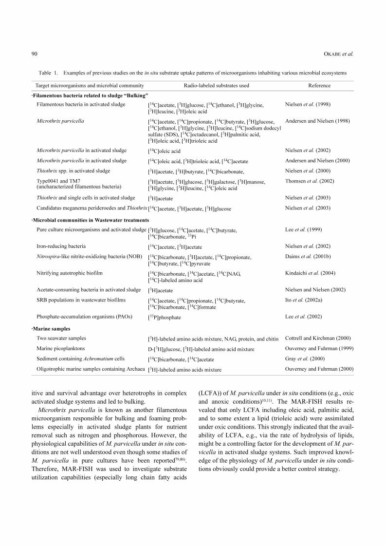
Filamentous bacteria causing bulking in activated
sludge
“Bulking” due to excessive growth of filamentous bacte-
ria in activated sludge treatment plants is a general and seri-
ous problem in many treatment plants. A better understand-
ing of factors controlling the growth of various filamentous
bacteria is necessary to efficiently control the bulking prob-
lems. This requires phylogenetic identification and detailed
knowledge about the ecophysiology of the different fila-
mentous bacteria, which are indistinguishable on the basis
of morphological observations alone. In addition, these fila-
mentous bacteria are notoriously difficult to maintain in lab-
oratory cultures. For these reasons, MAR-FISH has been
first applied to various industrial or municipal activated
sludge systems to investigate the in situ substrate uptake
patterns of the predominant filamentous bacteria especially
Thiothrix spp.47,49,51,84) and Microthrix parvicella10,53), which
were responsible for bulking and foaming problems. These
papers described how microautoradiography (MAR) and
FISH could be used to characterize and enumerate function-
ally important groups of microorganisms in activated
sludge. The principles for the methods, some important
methodological aspects and limitations of the procedures
were also briefly discussed52). The studies clearly demon-
strated that strain differences with regard to substrate utili-
zation were likely to occur among bacteria within the same
genera and designated types which are indistinguishable on
the basis of morphological observations alone and by the
16S rDNA probes used in the study.
It is well known that Thiothrix spp. are phylogenetically
related to Leucothrix and the filamentous sulfur-oxidizing
bacterium Eikelbooms Type 021N87,90). The morphology is
also very similar for Thiothrix spp. and Type 021N, hence a
proper identification can only be performed by using FISH
based on the 16S rRNA sequence30). Eikelbooms Type
021N bacteria have been recently reclassified as Thiothrix
and divided into three distinct groups on the basis of their
genotypic and phenotypic characteristics12,38). Both types
can be present simultaneously49,87). Identification and enu-
meration using FISH with species-specific 16S and 23S
rRNA probes revealed that 5–10% of the bacteria in the ac-
tivated sludge were Thiothrix spp.49). Physiological charac-
teristics of Thiothrix were also quantitatively determined by
counting the number of silver grains formed on the top of
the filaments49). MAR-FISH results indicated the Thiothrix
filaments were very versatile and showed incorporation of
[14C]-acetate and/or [14C]-bicarbonate under heterotrophic,
mixotrophic and chemolithoautotrophic conditions. The key
properties that Thiothrix spp. might be employing to out-
compete other microorganisms in activated sludge are prob-
ably related to the mixotrophic growth potential with strong
stimulation of acetate uptake by thiosulphate, as well as
stimulation of bicarbonate incorporation by acetate in the
presence of thiosulphate or intracellular elemental sulfur.
This phenomenon has not been described in pure culture
study. The occurrence of mixotrophic growth of Thiothrix
spp. was a significant factor giving Thiothrix spp. a compet-
OKABE et al.90
itive and survival advantage over heterotrophs in complex
activated sludge systems and led to bulking.
Microthrix parvicella is known as another filamentous
microorganism responsible for bulking and foaming prob-
lems especially in activated sludge plants for nutrient
removal such as nitrogen and phosphorous. However, the
physiological capabilities of M. parvicella under in situ con-
ditions are not well understood even though some studies of
M. parvicella in pure cultures have been reported79,80).
Therefore, MAR-FISH was used to investigate substrate
utilization capabilities (especially long chain fatty acids
(LCFA)) of M. parvicella under in situ conditions (e.g., oxic
and anoxic conditions)10,11). The MAR-FISH results re-
vealed that only LCFA including oleic acid, palmitic acid,
and to some extent a lipid (trioleic acid) were assimilated
under oxic conditions. This strongly indicated that the avail-
ability of LCFA, e.g., via the rate of hydrolysis of lipids,
might be a controlling factor for the development of M. par-
vicella in activated sludge systems. Such improved knowl-
edge of the physiology of M. parvicella under in situ condi-
tions obviously could provide a better control strategy.
Table 1. Examples of previous studies on the in situ substrate uptake patterns of microorganisms inhabiting various microbial ecosystems
Target microorganisms and microbial community Radio-labeled substrates used Reference
·Filamentous bacteria related to sludge “Bulking”
Filamentous bacteria in activated sludge [14C]acetate, [3H]glucose, [14C]ethanol, [3H]glycine, [3H]leucine, [3H]oleic acid
Nielsen et al. (1998)
Microthrix parvicella [14C]acetate, [14C]propionate, [14C]butyrate, [3H]glucose, [14C]ethanol, [3H]glycine, [3H]leucine, [14C]sodium dodecyl sulfate (SDS), [14C]octadecanol, [3H]palmitic acid, [3H]oleic acid, [3H]trioleic acid
Andersen and Nielsen (1998)
Microthrix parvicella in activated sludge [14C]oleic acid Nielsen et al. (2002)
Microthrix parvicella in activated sludge [14C]oleic acid, [3H]trioleic acid, [14C]acetate Andersen and Nielsen (2000)
Thiothrix spp. in activated sludge [3H]acetate, [3H]butyrate, [14C]bicarbonate, Nielsen et al. (2000)
Type0041 and TM7 (uncharacterized filamentous bacteria)
[3H]acetate, [3H]glucose, [3H]galactose, [3H]manose, [3H]glycine, [3H]leucine, [14C]oleic acid
Thomsen et al. (2002)
Thiothrix and single cells in activated sludge [3H]acetate Nielsen et al. (2003)
Candidatus meganema perideroedes and Thiothrix [14C]acetate, [3H]acetate, [3H]glucose Nielsen et al. (2003)
·Microbial communities in Wastewater treatments
Pure culture microorganisms and activated sludge [3H]glucose, [14C]acetate, [14C]butyrate, [14C]bicarbonate, 33Pi
Lee et al. (1999)
Iron-reducing bacteria [14C]acetate, [3H]acetate Nielsen et al. (2002)
Nitrospira-like nitrite-oxidizing bacteria (NOB) [14C]bicarbonate, [3H]acetate, [14C]propionate, [14C]butyrate, [14C]pyruvate
Daims et al. (2001b)
Nitrifying autotrophic biofilm [14C]bicarbonate, [14C]acetate, [14C]NAG, [14C]-labeled amino acid
Kindaichi et al. (2004)
Acetate-consuming bacteria in activated sludge [3H]acetate Nielsen and Nielsen (2002)
SRB populations in wastewater biofilms [14C]acetate, [14C]propionate, [14C]butyrate, [14C]bicarbonate, [14C]formate
Ito et al. (2002a)
Phosphate-accumulation organisms (PAOs) [33P]phosphate Lee et al. (2002)
·Marine samples
Two seawater samples [3H]-labeled amino acids mixture, NAG, protein, and chitin Cottrell and Kirchman (2000)
Marine picoplanktons D-[3H]glucose, [3H]-labeled amino acid mixture Ouverney and Fuhrman (1999)
Sediment containing Achromatium cells [14C]bicarbonate, [14C]acetate Gray et al. (2000)
Oligotrophic marine samples containing Archaea [3H]-labeled amino acids mixture Ouverney and Fuhrman (2000)

Microautoradiography Combined with FISH 91
Other filamentous bacteria in activated sludge
Among the filamentous bacteria occasionally causing
bulking problems in activated sludge treatment plants, three
morphotypes are common, Eikelboom Type 0041, Type
1851 and Type 170177,85). Very limited information is avail-
able about the phylogeny and physiology of these filamen-
tous bacteria. MAR-FISH was therefore performed to inves-
tigate the identity and in situ physiology of the Type 0041-
morphotype in two wastewater treatment plants84). The type
0041-morphotype is phylogenetically heterogeneous and
probably comprises a number of distinct species, of which
at least approximately 15% of the filaments belong to the
TM7 phylum, a recently recognized major lineage in the
bacterial domain32). This phylogenetic heterogeneity of
Type 0041 again highlights the inadequacy of a morpholo-
gy-based classification system. The in situ physiology of
Type 0041 using MAR-FISH, however, revealed that the
type 0041 filaments demonstrated several physiological
similarities, such as the ability to consume glucose under
oxic and anoxic conditions, and the inability to uptake
acetate under oxic conditions. These are the first data on
the in situ physiology of bacteria belonging to the almost
entirely uncharacterized TM7 phylum and show that TM7
filamentous bacteria are physiologically versatile.
Phosphate-accumulating organisms (PAOs)
The microbiology of enhanced biological phosphorus
removal (EBPR) in wastewater treatment plants is one of
the great challenges in activated sludge microbiology, since
the identification, population dynamics and interactions of
the PAOs with other competing bacterial groups including
glycogen-accumulating organisms (GAOs) are still largely
unresolved. More than a decade ago, previous molecular
studies demonstrated that Acinetobacter, the traditional
model microorganism for EBPR, does not catalyse phospho-
rous removal in these plants19,43,87). Recently, the population
size of PAOs in activated sludge was estimated to be ap-
proximately 4% of the total DAPI count on the basis of
uptake ability of radiolabeled acetate under anoxic condi-
tions46). The in situ physiology and identity of the PAOs in
pilot and full-scale activated sludge plants was also investi-
gated by MAR-FISH after incubations with radioactive 33Pi,
[3H]acetate, and D-[6-3H]glucose, respectively41). The result
revealed that a significant 33Pi uptake was mainly observed
for the Rhodocyclus-related bacteria within the beta-Proteo-
bacteria and the gram-positive Actinobacteria. The
Rhodocyclus-related bacteria also occurred in significant
numbers in these EBPR plants. However, not all of the
Rhodocyclus-related bacteria detectable with a specific
probe accumulated polyphosphate during aerobic growth
and showed uptake of [3H]acetate and D-[6-3H]glucose.
This evidence suggests that either a part of them is inactive
or that there are non-PAOs within this group. Furthermore,
the results suggested that the uptake of P is probably medi-
ated not only by the Rhodocyclus-related bacteria, but also
by other bacterial division, i.e., yet unidentified bacteria
groups.
Nitrifying bacteria community
Traditionally, Nitrobacter was considered to be the most
important nitrite-oxidizing bacteria in wastewater treatment
plants. Using the full cycle rRNA approach the occurrence
of yet uncultured Nitrospira-like nitrite-oxidizing bacteria
in nitrifying wastewater treatment plants (WWTPs) has
been often demonstrated20,21,24,36,40,56,58). By using MAR-
FISH, Daims and coworkers21) investigated the ecophysiolo-
gy of the uncultured Nitrospira-like nitrite-oxidizers in acti-
vated sludge and found that these bacteria are able to fix
bicarbonate and to simultaneously take up pyruvate but
not acetate, butyrate, and propionate. Nitrospira-like nitrite-
oxidizers are probably K-strategists (with high substrate
affinities and low maximum activity or growth rates) for
oxygen and nitrite and thus outcompete Nitrobacter under
substrate-limiting conditions in WWTPs76) or in the deeper
part of the biofilm where the O2 concentration is low56).
This hypothesis would also explain why Nitrobacter and
Nitrospira co-exist in sequencing batch biofilm reactors
with temporarily higher nitrite concentrations21).
It is also important to note that physiologically inactive
ammonia oxidizers are detected by FISH as these bacteria
maintain high cellular ribosome contents under unfavorable
conditions88). This is one of the difficult parts of the inter-
pretation of FISH data. However, the number of physiologi-
cally active ammonia oxidizers can accurately be deter-
mined using MAR-FISH with 14C-labeled bicarbonate as
substrate42).
Coexistence of heterotrophs in high abundance with nitri-
fiers has often been found even in autotrophic nitrifying
biofilms cultured without the external organic carbon
supply56,58). In such biofilm systems, the competitive inter-
action between heterotrophs and nitrifiers for dissolved ox-
ygen and space is well known55). In the presence of organic
carbon, nitrifiers are usually outcompeted by heterotrophs
due to the slow growth rate and low growth yield. They also
interact through the exchange of organic matter derived
OKABE et al.92
from nitrifying bacteria (e.g., biomass decay and production
of soluble organic matter). Such ecophysiological interac-
tion between nitrifiers and heterotrophic bacteria in a car-
bon-limited autotrophic nitrifying biofilm fed with only
NH4� as the energy source was investigated by a full cycle
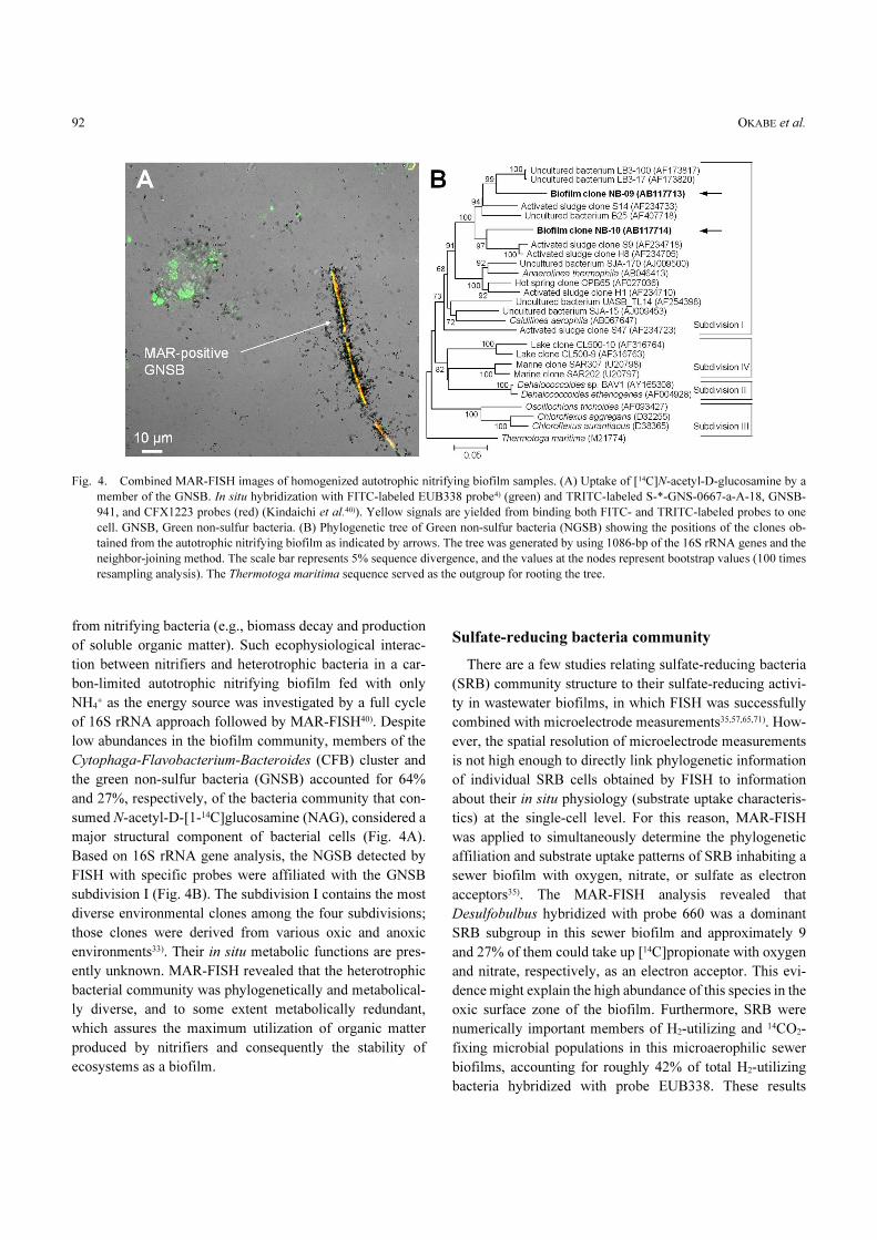
of 16S rRNA approach followed by MAR-FISH40). Despite
low abundances in the biofilm community, members of the
Cytophaga-Flavobacterium-Bacteroides (CFB) cluster and
the green non-sulfur bacteria (GNSB) accounted for 64%
and 27%, respectively, of the bacteria community that con-
sumed N-acetyl-D-[1-14C]glucosamine (NAG), considered a
major structural component of bacterial cells (Fig. 4A).
Based on 16S rRNA gene analysis, the NGSB detected by
FISH with specific probes were affiliated with the GNSB
subdivision I (Fig. 4B). The subdivision I contains the most
diverse environmental clones among the four subdivisions;
those clones were derived from various oxic and anoxic
environments33). Their in situ metabolic functions are pres-
ently unknown. MAR-FISH revealed that the heterotrophic
bacterial community was phylogenetically and metabolical-
ly diverse, and to some extent metabolically redundant,
which assures the maximum utilization of organic matter
produced by nitrifiers and consequently the stability of
ecosystems as a biofilm.
Sulfate-reducing bacteria community
There are a few studies relating sulfate-reducing bacteria
(SRB) community structure to their sulfate-reducing activi-
ty in wastewater biofilms, in which FISH was successfully
combined with microelectrode measurements35,57,65,71). How-
ever, the spatial resolution of microelectrode measurements
is not high enough to directly link phylogenetic information
of individual SRB cells obtained by FISH to information
about their in situ physiology (substrate uptake characteris-
tics) at the single-cell level. For this reason, MAR-FISH
was applied to simultaneously determine the phylogenetic
affiliation and substrate uptake patterns of SRB inhabiting a
sewer biofilm with oxygen, nitrate, or sulfate as electron
acceptors35). The MAR-FISH analysis revealed that
Desulfobulbus hybridized with probe 660 was a dominant
SRB subgroup in this sewer biofilm and approximately 9
and 27% of them could take up [14C]propionate with oxygen
and nitrate, respectively, as an electron acceptor. This evi-
dence might explain the high abundance of this species in the
oxic surface zone of the biofilm. Furthermore, SRB were
numerically important members of H2-utilizing and 14CO2-
fixing microbial populations in this microaerophilic sewer
biofilms, accounting for roughly 42% of total H2-utilizing
bacteria hybridized with probe EUB338. These results
Fig. 4. Combined MAR-FISH images of homogenized autotrophic nitrifying biofilm samples. (A) Uptake of [14C]N-acetyl-D-glucosamine by a
member of the GNSB. In situ hybridization with FITC-labeled EUB338 probe4) (green) and TRITC-labeled S-*-GNS-0667-a-A-18, GNSB-
941, and CFX1223 probes (red) (Kindaichi et al.40)). Yellow signals are yielded from binding both FITC- and TRITC-labeled probes to one
cell. GNSB, Green non-sulfur bacteria. (B) Phylogenetic tree of Green non-sulfur bacteria (NGSB) showing the positions of the clones ob-
tained from the autotrophic nitrifying biofilm as indicated by arrows. The tree was generated by using 1086-bp of the 16S rRNA genes and the
neighbor-joining method. The scale bar represents 5% sequence divergence, and the values at the nodes represent bootstrap values (100 times
resampling analysis). The Thermotoga maritima sequence served as the outgroup for rooting the tree.

Microautoradiography Combined with FISH 93
provide further insight into the correlation between the 16S
rRNA phylogenetic diversity and the in situ physiological
diversity of SRB populations inhabiting sewer biofilms.
Sulfur-oxidizing bacterium (Achromatium spp.)
Achromatium oxaliferum is a large, morphologically con-
spicuous sulfur-oxidizing bacterium found principally in
freshwater and brackish sediments. A recent study has
shown that natural communities of this uncultured bacteria
from the genus Achromatium comprised a number of phylo-
genetically, morphologically, and ecologically distinct
subpopulations26). In addition, a solely microautoradio-
graphic study of mixed natural populations of these bacteria
indicated that not all cells assimilated [14C]-bicarbonate and
[14C]-acetate, suggesting that the Achromatium community
exhibited physiological as well as phylogenetic diversity27).
These experimental results indicated that MAR-FISH
provides the only reliable means for identifying and differ-
entiating the coexisting Achromatium species present in
natural environments and investigating their carbon metabo-
lisms28). The results of the MAR-FISH study revealed that
Achromatium spp. probably exhibit a range of physiologies,
i.e., facultative chemolithoautotrophy, mixotrophy, and
chemoorganoheterotrophy, similar to other large sulfur-
oxidizing bacteria (e.g., Beggiatoa spp). This evidence can
explain the consistent presence of a large population of
Achromatium spp. within the natural environmental commu-
nities.
Denitrifying bacteria
Molecular studies of the community composition of deni-
trifying bacteria are difficult to perform since the denitrify-
ing phenotype cannot be inferred from the phylogeny of
microorganisms. However, the combination of FISH and
microautoradiography allows identification of denitrifiers
in situ by incubating with radiolabeled substrates (e.g.,
acetate) under anoxic conditions in the presence and absence
of nitrite and nitrate. The use of MAR-FISH in combination
with the full-cycle rRNA approach revealed that novel,
uncultured Beta-proteobacteria related to the Azoarcus-
Thauera complex are probably abundant denitrifiers in an
industrial nitrifying and denitrifying wastewater treatment
plant37). Further study is needed to identify truly important
microorganisms responsible for denitrification in various
WWTPs.
Degradation of xenobiotic compounds
MAR-FISH has been used for identification of micro-
organisms degrading xenobiotic contaminants such as o-nitro-
phenol and salicylate, both of which are environmentally
important widespread aromatic compounds91). MAR-FISH
allowed rapid and accurate identification of microorganisms
responsible for degradation of [14C] o-nitrophenol. In future,
further studies should be performed for biodegradation
of other contaminants such as petroleum hydrocarbons,
pesticides, and dyes.
Marine picoplnkton
The application of MAR-FISH to marine bacterioplank-
ton has demonstrated that nearly 90% of alpha-Proteo-
bacteria and members of the Cytophaga-Flavobacterium
group, which together accounted for 50–60% of the bacteri-
al cells present, assimilated tritiated amino acids62). MAR-
FISH has also been used to determine the relative contribu-
tion made to the utilization of marine dissolved organic
matter (DOM) by different prokaryotic groups18). The
results showed that no phylogenetic group dominated the
consumption of all DOM, suggesting that the participation
of a diverse assemblage of bacteria is essential for the
complete degradation of complex DOM in the ocean. Cottrell
and Kirchman18) also found that N-acetyl-D-glucosamine
(NAG), which constitutes the largest pools of amino sugars
in the ocean, was preferentially utilized by alpha-Proteo-
bacteria and member of the Cytophaga-Flavobacterium
group. Riemann and Azam68) demonstrated that all isolates
within the Vibrionaceae group of delta-Proteobacteria
could also take up NAG and were all facultative anaerobes.
This result suggests that a substantial fraction of bacteria in
the ocean are facultative anaerobes that might be predomi-
nantly responsible for degradation of marine dissolved
organic matter. The newly discovered low-temperature
marine Archaea, now known to be abundant in marine
environments, are also capable of assimilating dissolved
amino acids at low concentrations63).
Quantitative MAR-FISH
MAR is usually considered as qualitative or semi-quanti-
tative, especially when [14C], which is a relatively strong
beta-emitter, is used70). However, Nielsen et al.47) have re-
cently proposed quantitative microautoradiography
(QMAR) and FISH. The QMAR-FISH has a resolution of a
single cell and is based on an improved fixation protocol

OKABE et al.94
(preventing the leakage of tritium-labeled compounds from
cells42) and the use of an internal standard of bacteria with
known specific radioactivity). The leakage decreased the
number of silver grains formed on the bacteria cell, which
consequently lowered the resolution of MAR. The modified
fixation protocol was the immediate immobilization on gel-
atin-coated cover glass after fixation in 4% PFA for 3 h and
then storing at �20�C until use. With this technique, the
substrate affinity (Ks) for uptake of acetate by two filamen-
tous bacteria, the ‘Candidatus Meganema perideroedes’
and the Thiothrix spp., was directly determined in complex
environments in situ without previous cultivation or
enrichment47). This technique revealed further insight into
the activity variations between different species of interest
and within a single species. The filaments within both
probe-defined populations had threefold variation in activity
between the different filaments, demonstrating a large
variation in activity level within a single population in a
complex system.
CTC-FISH
Reliable methods to detect the viable fraction of microor-
ganisms in complex environments are of great interest in
microbial ecology. The available methods with the single-
cell resolution are FISH, microautoradiography (MAR),
and reduction of redox dye 5-cyano-2,3-tolyl-tetrazolium
chloride (CTC)69) and p-iodonitrotetrazolium (INT). Bac-
terial activity at the single cell level can be analyzed using
terazorium salts (e.g., CTC and INT) as indicators of bac-
terial respiration. In theory, FISH reflects the recent activity
state of the cell, but this depends very much on species and
growth conditions. For MAR, the active bacteria can be
directly detected in complex microbial communities. The
main problem is, however, to find substrate(s) that can be
taken up by all active bacteria. The CTC reduction method is
commonly applied to determine respiratory activity and valid-
ity of bacteria in complex microbial systems22,39). All three
of these methods were used simultaneously to investigate
viability and activity on a single-cell level of Thiothrix fila-
ments and single floc-forming bacteria in activated sludge50).
Their study demonstrated that the signals from MAR and
FISH analyses correlated well, whereas only 65% of the
MAR- and FISH-positive Thiothrix cells were also CTC
positive. For single floc-forming bacteria, only 41% of the
MAR- and FISH-positive cells were detectable by CTC
reduction. Weakly MAR-positive filamentous Thiothrix cells
and many single cells, even being MAR- and FISH-posi-
tive, were not detected by CTC. Based on these results, it
could be concluded that CTC targeted only the most active
cells and underestimated the real number of active cells.
This conclusion was in agreement with the previous studies
suggesting that the CTC was poisonous to some of the
cells78) and that universal 16S rRNA probe counts indicated
a population with at least minimal metabolic activity39).
Other methods for determination of microbial
structure and function
The 16S rRNA approach suffers from numerous biases
introduced in the DNA extraction, PCR amplification, and
cloning procedures. Therefore, quantitative data on the
microbial community composition can only be obtained if
the 16S rRNA approach is combined with quantitative in
situ hybridization analysis. A limitation of MAR-FISH is,
however, that FISH targets rRNA. The genes encoding this
macromolecule are highly conserved and cannot be used to
discriminate at the same phylogenetic resolution as functio-
nal genes or intergenic spacer regions. Thus, resolution of
the FISH technique is somewhere near the species level at
best. In addition, phylogeny based on rRNA sequence alone
is rarely a reliable indicator of microbial function. In recent
years, some techniques other than MAR-FISH have been
developed, which allow more comprehensive simultaneous
monitoring of the diversity and substrate uptake patterns of
complex microbial communities. Stable isotope probing
(SIP)67) and Isotope microarray1,64) techniques are briefly
reviewed in the following section.
Stable isotope probing (SIP)67)
DNA can be labeled with the heavy stable isotopes [13C]
after incubation with stable isotope-labeled substrates.
Heavy DNA (13C-DNA) and light DNA (12C-DNA) are sep-
arated by density-gradient centrifugation. The separated13C-DNA and 12C-DNA from the SIP experiment were
separately subjected to full-cycle rRNA analysis, which
included FISH probe design and MAR-FISH. The SIP with
full-cycle rRNA analysis enabled one to design specific
FISH probes for active microorganisms under conditions
tested based on the 13C-DNA clone library. Thus, the 13C-
DNA clone library represents active microbial community,
whereas the 12C-DNA clone library represents the overall
microbial community. The potential of this SIP technique
has been recently reviewed elsewhere66,89). This year, acti-
vated sludge was subjected to SIP after incubation with 13C-
methanol to label the DNA of the denitrifiers25). This study
reports the first combined use of SIP, full-cycle rRNA anal-

Microautoradiography Combined with FISH 95
ysis, which includes FISH probe design, and MAR-FISH to
establish a link between the phylogeny and physiology of
a methanol-utilizing denitrifying bacterial community. The
dominant 16S rRNA gene phylotype in the 13C-DNA clone
library was closely related to those of the obligate methyl-
otrophs Methylobacillus and Methylophilus in the order
Methylophilales of the Beta-proteobacteria. There was no
correlation between the denitrification rate and the relative
abundances of the well-known denitrifying genera Hypomi-
crobium and Paracoccus. MAR-FISH provided visual evi-
dence for the in situ utilization of methanol by targeted mi-
croorganisms. The application of SIP together with MAR-
FISH is, therefore, a very powerful and effective approach
to address the in situ physiology of microorganisms in com-
plex mixed-population consortia such as activated sludge
and biofilms. Although the SIP is safer than MAR-FISH,
there are the following concerns; (1) a rather longer incuba-
tion time is required for enough incorporation of stable
isotope into DNA or RNA, which may cause substrate
cross-feeding, (2) the SIP is strongly dependent on the
DNA extraction efficiency, and (3) stable isotope-labeled
substrates are less available on the market than radio-
labeled substrates.
Isotope microarray
A new microarray method, the isotope array approach,
for identifying microorganisms that consume a specific 14C-
labeled substrate within complex microbial communities
was recently developed1,64). This isotope microarray ap-
proach allows simultaneous determination of community
structure and specific substrate consumption by community
members via direct detection of environmentally retrieved
16S rRNA on an oligonucleotide microarray. Therefore,
this approach is not affected by the multiple biases associat-
ed with PCR amplification of rRNA genes but requires effi-
cient extraction of rRNA from samples. Compared to MAR-
FISH, the isotope array allows one to apply many probes in
parallel, which will be of major importance if the ecophysi-
ology of complex microbial communities is of interest. This
technique can be used as a new tool to directly link all
detected microorganisms with their specific activities and
functions in complex environments, if stringent hybridiza-
tion conditions can be achieved for all probes. For MAR-
FISH, FISH probes that will be used must be previously se-
lected based on 16S rRNA gene analysis or by trial and er-
ror. Therefore, this method is less tedious and advantageous
to study the functions of uncultured microorganisms, which
are usually in low abundance in complex microbial commu-
nities. However, the incorporation rate of labeled carbon
into rRNA varies significantly depending on the type of
labeled substrate and on the metabolism of the organisms,
which determines the sensitivity of this method. The sensi-
tivity of this method is sufficient to detect 14C incorporation
into the 16S rRNA of community members that make up
5–10% of the bacterial communities.
Future Researches
The recent application of 16S rRNA approaches has re-
vealed a remarkably vast microbial diversity including
many hitherto-recognized, yet uncultured species in natural
and engineered environments. This species biodiversity
must be related to the functional diversity. Despite the enor-
mous potential of MAR-FISH to simultaneously examine
phylogenetic identity and the in situ specific activity of the
microorganisms within a complex microbial community at
the single-cell level without the need for cultivation or iso-
lation, it is clear that there are inherent limitations. There-
fore, a logical use of MAR-FISH in combination with those
of various molecular techniques, classical microbiology,
and microelectrodes will provide the clearest view of the
in situ metabolic functions of microorganisms in complex
microbial communities, which is the central to microbial
ecology.
Acknowledgments
We thank the Central Institute of Isotope Science, Hok-
kaido University, for providing the facilities for the isotopic
experiments. This study was carried out as a part of “The
Project for Development of Technologies for Analyzing and
Controlling the Mechanism of Biodegrading and Process-
ing”, which was entrusted by the New Energy and Industrial
Technology Development Organization (NEDO). This
research was also partly supported by a Grant-in Aid
(No. 13650593) for Developmental Scientific Research from
the Ministry of Education, Science and Culture of Japan.
References
1) Adamczyk, J., M. Hesselsoe, N. Iversen, M. Horn, A. Lehner,
P.H. Nielsen, M. Schloter, P. Roslev and M. Wagner. 2003. The
isotope array, a new tool that employs substrate-mediated label-
ing of rRNA for determination of microbial community structure
and function. Appl. Environ. Microbiol. 69: 6875–6887.
2) Amann, R.I. 1995. In situ identification of micro-organisms by
whole-cell hybridization with rRNA-targeted nucleic acid probes,
p. 1–15. In A.D.L. Akkerman, J.D. van Elsas and F.J. de Bruijn

OKABE et al.96
(ed.), Molecular microbial ecology manual. Kluwer Academic
Publishers, Dordrecht, The Netherlands.
3) Amann, R., F.O. Glockner and A. Neef. 1997. Modern methods
in subsurface microbiology: in situ identification of microorgan-
isms with nucleic acid probes. FEMS Microbiol. Rev. 20: 191–
200.
4) Amann, R., L. Krumholz and D. A. Stahl. 1990. Fluorescent-oligo-
nucleotide probing of whole cells for determinative, phyloge-
netic, and environmental studies in microbiology. J. Bacteriol.
172: 762–770.
5) Amann, R. and M. Kuhl. 1998. In situ methods for assessment of
microorganisms and their activities. Curr. Opin. Microbiol. 1:
352–358.
6) Amann, R. and W. Ludwig. 2000. Ribosomal RNA-targeted
nucleic acid probes for studies in microbial ecology. FEMS
Microbiol. Rev. 24: 555–565.
7) Amann, R.I., W. Ludwig and K.-H. Schleifer. 1995. Phylogenetic
identification and in situ detection of individual microbial cells
without cultivation. Microbiol. Rev. 59: 143–169.
8) Amann, R.I., J. Stromley, R. devereux, R. Key and D.A. Stahl.
1992. Molecular and microscopic identification of sulfate-reduc-
ing bacteria in multispecies biofilms. Appl. Environ. Microbiol.
58: 614–623.
9) Andreasen, K. and P.H. Nielsen. 1997. Application of microauto-
radiography for the study of substrate uptake by filamentous
microorganisms in activated sludge. Appl. Environ. Microbiol.
63: 3662–3668.
10) Andreasen, K. and P.H. Nielsen. 1998. In situ characterization of
substrate uptake by Microthrix parvicella using microautoradiog-
raphy. Water Sci. Technol. 37(4/5): 19–26.
11) Andreasen, K. and P.H. Nielsen. 2000. Growth of Microthrix
parvicella in nutrient removal activated sludge plants: Studies of
in situ physiology. Water Sci. Technol. 34(5): 1559–1569.
12) Aruga, S., Y. Kamagata, T. Kohno, S. Hanada, K. Nakamura and
T. Kanagawa. 2002. Characterization of filamentous Eikelboom
type 021N bacteria and description of Thiothrix disciformis sp.
nov. and Thiothrix flexilis sp. nov. Int. J. Syst. Evol. Microbiol.,
52: 1309–1316.
13) Baker, J.R. 1989. Autoradiography: a comprehensive overview.
In Microscopy Handbooks 18, Royal Microscopical Society,
Oxford University Press.
14) Briones, A.M., S. Okabe, Y. Umemiya, N.-R. Ramsing, W.
Reichardt and H. Okuyama. 2002. Influence of different cultivars
on populations of ammonium-oxidizing bacteria in the root
environment of rice. App. Environ. Microbiol. 68: 3067–3075.
15) Brock, T.D. and M.L. Brock. 1968. Autoradiography as a tool in
microbial ecology. Nature 209: 734–736.
16) Carman, K.R. 1990. Radioactive labeling on a natural assemblage
of marine sedimentary bacteria and microalgae for the trophic
studies: an autoradiographic study. Microb. Ecol. 19: 279–290.
17) Caro, L.G. and R.P. Van Tubergen. 1962. High resolution auto-
radiography, 1. Methods, J. Cell. Biol. 15: 173–188.
18) Cottrell, M.T. and D.L. Kirchman. 2000. Natural assemblages of
marine Proteobacteria and members of the Cytophaga-
Flavobacter cluster consuming low- and high-molecular-weight
dissolved organic matter. Appl. Environ. Microbiol. 66: 1692–
1697.
19) Crocetti, G.R., P. Hugenholtz, P.L. Bond, A. Schuler, J. Keller,
D. Jenkins and L.L. Blackall. 2000. Identification of polyphos-
phate-accumulating organisms and design of 16S rRNA-directed
probes for their detection and quantification. Appl. Environ.
Microbiol. 66: 1175–1182.
20) Daims, H., U. Purkhold, L. Bjerrum, E. Arnold, P.A. Wilderer
and M. Wagner. 2001. Nitrification in sequencing biofilm batch
reactors: lessons from molecular approaches. Water Sci. Technol.
43: 9–18.
21) Daims, H., J.L. Nielsen, P.H. Nielsen, K.-H. Schleifer and M.
Wagner. 2001. In situ characterization of Nitrospira-like nitrite-
oxidizing bacteria active in wastewater treatment plants. Appl.
Environ. Microbiol. 67: 5273–5284.
22) Flood, J.A., N.J. Ashbolt and P.C. Pollard. 1999. Complementary
independent molecular, radioisotopic and fluorogenic techniques
to assess biofilm communities in two wastewater wetlands. Water
Sci. Technol. 39: 65–70.
23) Fuhrman, J.A. and F. Azam. 1982. Thymidine incorporation as a
measure of heterotrophic bacterioplankton production in marine
surface waters: evaluation and field results. Mar. Biol. 66: 109–
120.
24) Gieseke A., U. Purkhold, M. Wagner, R. Amann and A.
Schramm. 2001. Community structure and activity dynamics
of nitrifying bacteria in a phosphate-removing biofilm. Appl.
Environ. Microbiol. 67: 1351–1362.
25) Ginige, M.P., P. Hugenholtz, H. Daims, M. Wagner, J. Keller and
L.L. Blackall. 2004. Use of stable-isotope probing, full-cycle
rRNA analysis, and fluorescence in situ hybridization-microauto-
radiography to study a methanol-fed denitrifying microbial
community. Appl. Environ. Microbiol. 70: 588–596.
26) Gray, N.D., R. Howarth, A. Rowan, R.W. Pickup, J.G. Jones and
I.M. Head. 1999a. Natural communities of Achromatium
oxaliferum comprise genetically morphologically and ecological-
ly distinct sub-populations. Appl. Environ. Microbiol. 65: 5089–
5099.
27) Gray, N.D., R. Howarth, R.W. Pickup, J.G. Jones and I.M. Head.
1999b. Substrate utilization by uncultured bacteria from the ge-
nus Achromatium determined by the use of microautoradiogra-
phy. Appl. Environ. Microbiol. 65: 5100–5106.
28) Gray, N.D., R. Howarth, R.W. Pickup, J.G. Jones and I.M. Head.
2000. Use of combined microautoradiography and fluorescence
in situ hybridization to determine carbon metabolism in mixed
natural communities of uncultured bacteria from the genus
Achromatium. Appl. Environ. Microbiol. 66: 4518–4522.
29) Head, I.M., J.R. Saunders and R.W. Pickup. 1998. Microbial
evolution, diversity and ecology: a decade of ribosomal RNA
analysis of uncultured microorganisms. Microb. Ecol. 35: 1–21.
30) Howarth, R., R.F. Unz, E.M. Seviour, R.J. Seviour, L.L. Blackall,
R.W. Pickup, J. Gwyn Jones, J. Yaguchi and I.M. Head. 1999.
Phylogenetic relationships of filamentous sulfur bacteria
(Thiothrix spp. and Eikelboom type 021N bacteria) isolated
from wastewater treatment plants and description of Thiothrix
eikelboomii sp. nov., Thiothrix unzii sp. nov., Thiothrix
fructosivorans sp. nov. and Thithrix defluvii sp. nov. Int. J. Syst
Bacteriol., 49: 1817–1827.
31) Hu, S. and A.H.C. van Bruggen. 1997. Microbial dynamics asso-
ciated with multiphasic decomposition of 14C-labeled cellulose in
soil. Microb. Ecol. 33: 134–143.
32) Hugenholtz, P., G.W. Tyson, R.I. Webb, A.M. Wagner and L.L.
Blackall. 2001. Investigation of candidate division TM7, a recent-
ly recognized major lineage of the domain Bacteria with no
known pure-culture representative. Appl. Environ. Microbiol. 67:
411–419.

Microautoradiography Combined with FISH 97
33) Hugenholtz, P., B.M. Goebel and N.R. Pace. 1998. Impact of
culture-independent studies on the emerging phylogenetic view
of bacterial diversity. J. Bacteriol. 180: 4765–4774.
34) Ito, T., J.L. Nielsen, S. Okabe, Y. Watanabe and P.H. Nielsen.
2002a. Phylogenetic identification and substrate uptake patterns
of sulfate-reducing bacteria inhabiting an oxic-anoxic sewer
biofilm determined by combining microautoradiography and
fluorescent in situ hybridization. Appl. Environ. Microbiol. 68:
356–364.
35) Ito, T., S. Okabe, H. Satoh and Y. Watanabe. 2002b. Successional
development of sulfate-reducing bacterial populations and their
activities in a wastewater biofilm growing under microaerophilic
conditions. Appl. Environ. Microbiol. 68: 1392–1402.
36) Juretschko, S., G. Timmermann, M. Schmidt, K.-H. Schleifer, A.
Pommerening-Roser, H.-P. Koops and M. Wager. 1998. Com-
bined molecular and conventional analysis of nitrifying bacterial
diversity in activated sludge: Nitrosococcus mobilis and
Nitrospire-like bacteria as dominant populations. Appl. Environ.
Microbiol. 64: 3042–3051.
37) Juretschko, S., A. Loy, A. Lehner and M. Wager. 2002. The
microbial community composition of a nitrifying-denitrifying
activated sludge from an industrial sewage treatment plant
analyzed by the full-cycle rRNA approach. Syst. Appl. Micro-
biol. 25: 84–99.
38) Kanagawa, T., Y. Kamagata, S. Aruga, T. Kohno, M. Horn and
M. Wagner. 2000. Phylogenetic analysis of and oligonucleotide
probe development for Eikelboom type 021N filamentous bacte-
ria isolated from bulking activated sludge. Appl. Environ. Micro-
biol. 66: 5043–5052.
39) Karner, M. and J.A. Fuhrman. 1997. Determination of active
marine bacterioplankton: a comparison of universal 16S rRNA
probes, autoradiography, and nucleoid staining. Appl. Environ.
Microbiol. 63: 1208–1213.
40) Kindaichi T., T. Ito and S. Okabe. 2004. Eco-physiological inter-
action between nitrifying bacteria and heterotrophic bacteria in
autotrophic nitrifying biofilms. Appl. Environ. Microbiol. 70:
1641–1650.
41) Lee, N., J.C. Jansen, H. Aspegren, K. Dircks, M. Henze, K.-H.
Schleifer and M. Wagner. 2002. Population dynamics and in situ
physiology of phosphorus-accumulating bacteria in wastewater
treatment plants with enhanced biological phosphorus removal
operated with and without nitrogen removal. Water Sci. Technol.
46(1/2): 163–170.
42) Lee, N., P.H. Nielsen, K.H. Andersen, S. Juretschko, J.L.
Nielsen, K.-H. Schleifer and M. Wagner. 1999. Combination of
fluorescent in situ hybridization and microautoradiography—A
new tool for structure-function analyses in microbial ecology.
Appl. Environ. Microbiol. 65: 1289–1297.
43) Liu, W.T., A.T. Nielsen, J.H. Wu, C.S. Tsai, Y. Matsuo and
S. Molin. 2001. In situ identification of polyphosphate- and poly-
hydroxyalkanoate-accumulating traits for microbial populations
in a biological phosphorous removal process. Environ. Microbiol.
3: 110–122.
44) Maier, S. and M.A. Gallardo. 1984. Nutritional characteristics of
two marine thioplocas determined by autoradiography. Arch.
Microbiol. 139: 218–220.
45) Nielsen, J.L. and P.H. Nielsen. 2002. Enumeration of acetate-
consuming bacteria by microautoradiography under oxygen and
nitrate respiring conditions in activated sludge. Water Res. 36(2):
421–428.
46) Nielsen, J.L. and P.H. Nielsen. 2002. Quantification of functional
groups in activated sludge by microautoradiography. Water Sci.
Technol. 46(1/2): 389–395.
47) Nielsen, J.L., D. Christensen, M. Kloppenborg and P.H. Nielsen.
2003. Quantification of cell-specific substrate uptake by probe-
defined bacteria under in situ conditions by microautoradiogra-
phy and fluorescence in situ hybridization. Environ. Microbiol. 5:
202–211.
48) Nielsen, J.L., S. Juretschko, M. Wagner and P.H. Nielsen. 2002.
Abundance and phylogenetic affiliation of iron reducers in acti-
vated sludge as assessed by fluorescence in situ hybridization and
microautoradiography. Appl. Environ. Microbiol. 68: 4629–
4636.
49) Nielsen, P.H., de Muro, M.A. and J.L. Nielsen. 2000. Studies on
the in situ physiology of Thiothrix spp. Present in activated
sludge. Environ. Microbiol. 2: 389–398.
50) Nielsen, J.L., de Muro M. Aquino and P.H. Nielsen. 2003. Evalu-
ation of the redox dye 5-cyano-2,3-tolyl-tetrazolium chloride for
activity studies by simultaneous use of microautoradiography and
fluorescence in situ hybridization. Appl. Environ. Microbiol. 69:
641–653.
51) Nielsen, P.H., K. Andreasen, M. Wagner, L.L. Blackall, H.
Lemmer and R.J. Seviour. 1998. Variability of Type 021N in
activated sludge as determined by in situ substrate uptake pattern
and in situ hybridization with fluorescent rRNA targeted probes.
Water Sci. Technol. 37(4–5): 423–430.
52) Nielsen, P.H., K. Andreasen, N. Lee and M. Wagner. 1999. Use
of Microautoradiography and fluorescent in situ hybridization for
characterization of microbial activity in activated sludge. Water
Sci. Technol. 39: 1–9.
53) Nielsen, P.H., P. Roslev, T.E. Dueholm and J.L. Nielsen. 2002.
Microthrix parvicella, a specialized lipid consumer in anaerobic-
aerobic activated sludge plants. Water Sci. Technol. 46(1/2): 73–
80.
54) Novitsky, J.A. 1983. Heterotrophic activity throughout a vertical
profile of seawater and sediment in Halifax Harbor, Canada.
Appl. Environ. Microbiol. 63: 1753–1760.
55) Okabe, S., K. Hirata, Y. Ozawa and Y. Watanabe. 1996. Spatial
microbial distributions of nitrifiers and heterotrophs in mixed
population biofilms. Biotechnol. Bioeng. 50: 24–35.
56) Okabe, S., H. Satoh and Y. Watanabe. 1999a. In situ analyses of
nitrifying biofilms as determined by in situ hybridization and the
use of microelectrodes. Appl. Environ. Microbiol. 65: 3182–
3191.
57) Okabe, S., T. Ito, H. Satoh and Y. Watanabe. 1999a. Analyses of
spatial distributions of sulfate-reducing bacteria and their activity
in aerobic wastewater biofilms. Appl. Environ. Microbiol. 65:
5107–5116.
58) Okabe, S., H. Naitoh, H. Satoh and Y. Watanabe. 2002. Structure
and function of nitrifying biofilms as determined by molecular
techniques and the use of microelectrodes. Water Sci. Technol.
46(1/2): 233–241.
59) Okabe, S., H. Satoh and Y. Watanabe. 2001. Analysis of microbi-
al structure and function of nitrifying biofilms. Meth. Enzymol.
337: 213–224.
60) Olsen, G.J., D.J. Lane, S.J. Giovannoni, N.R. Pace and D.A.
Stahl. 1986. Microbial ecology and evolution: a ribosomal RNA
approach. Annu. Rev. Microbiol. 40: 337–365.
61) Otte, S., J.G. Kuenen, L.P. Nielsen, H.W. Paerl, J. Zopfi,
H.N. Schulz, A. Teske, B. Strotmann, V.A. Gallardo and B.B.

OKABE et al.98
Jorgensen. 1999. Nitrogen, carbon, and sulfur metabolism in
natural Thioploca samples. Appl. Environ. Microbiol. 65: 3148–
3157.
62) Ouverney, C.C. and J.A. Fuhrman. 1999. Combined microautora-
diography-16S rRNA probe technique for determination of
radioisotope uptake by specific microbial cell types in situ.
Appl. Environ. Microbiol. 65: 1746–1752.
63) Ouverney, C.C. and J.A. Fuhrman. 2000. Marine planktonic
archaea take up amino acids. Appl. Environ. Microbiol. 66:
4829–4833.
64) Polz, M.F., S. Bertilsson, S.G. Acinas and D. Hunt. 2003. A(r)
Ray of hope in analysis of the function and diversity of microbial
communities. Biol. Bull. 204: 196–199.
65) Ramsing, N.B., M. Kühl and B.B. Jørgensen. 1993. Distribution
of sulfate-reducing bacteria, O2, and H2S in photosynthetic bio-
films determined by oligonucleotide probe and microelectrodes.
Appl. Environ. Microbiol. 59: 3840–3849.
66) Radajewski, S., I.R. McDonald and J.C. Murrell. 2003. Stable-
isotope probing of nucleic acids: a window to the function of un-
cultured microorganisms. Curr. Opin. Biotechnol. 14: 296–302.
67) Radajewski, S., P. Ineson, N.R. Parekh and J.C. Murrell. 2000.
Stable-isotope probing as a tool in microbial ecology. Nature 403:
646–649.
68) Riemann, L. and F. Azam. 2002. Widespread N-Acetyl-D-
Glucosamine uptake among pelagic marine bacteria and its
ecological implications. Appl. Environ. Microbiol. 68: 5554–
5562.
69) Rodriguez, G.G., D. Phipps, K. Ishiguro and H.F. Ridgeway.
1992. Use of fluorescent redox probe for direct visualization of
actively respiring bacteria. Appl. Environ. Microbiol. 58: 1801–
1808.
70) Rogers, A.W. 1979. Techniques of autoradiography. (3rd edi-
tion). Elsevier, New York.
71) Santegoeds, C.M., T.G. Ferdelman, G. Muyzer and D. de Beer.
1998. Structural and functional dynamics of sulfate-reducing
populations in bacterial biofilms. Appl. Environ. Microbiol. 64:
3731–3739.
72) Satoh, H., S. Okabe, Y. Yamaguchi and Y. Watanabe. 2003.
Evaluation of the bioaugmentation and biostimulation by in situ
hybridization and microelectrodes. Water Res. 37: 2206–2216.
73) Schramm, A. 2003. In situ analysis of structure and activity of the
nitrifying community in biofilms, aggregate, and sediments.
Geomicrobiol. J. 20: 313–333.
74) Schramm, A., L.H. Larsen, N.P. Revsbech, R.I. Amann and K.-H.
Schleifer. 1996. Structure and function of a nitrifying biofilm as
determined by in situ hybridization and the use of microelec-
trodes. Appl. Environ. Microbiol. 62: 4641–4647.
75) Schramm, A., D. de Beer, A. Gieseke and R. Amann. 2000.
Microenvironments and distribution of nitrifying bacteria in a
membrane-bound biofilm. Environ. Microbiol. 2: 680–686.
76) Schramm, A., D. de Beer, J.C. van den Heuvel, S. Ottengraf and
R. Amann. 1999. Microscale distribution of populations and
activities of Nitrosospira and Nitrospira spp. along a macroscale
gradient in a nitrifying bioreactor: quantification by in situ
hybridization and the use of microsensors. Appl. Environ. Micro-
biol. 65: 3690–3696.
77) Seviour, E.M., C. Williams, B. DeGrey, J.A. Soddell, R.J.
Seviour and K.C. Lindrea. Studies on filamentous bacteria from
Australian sludge plants. Water Sci. Tech. 28: 2335–2342.
78) Sherr, B.F., P. del Giorgio and E.B. Sherr. 1999. Estimating
abundance and single-cell characteristics of respiring bacteria via
the redox dye, CTC. Aquat. Microb. Ecol. 18: 117–131.
79) Slijkhuis, H. 1983. Microthrix parvicella, a filamentous bacteri-
um isolated from activated sludge: cultivation in a chemically
defined medium. Appl. Environ. Microbiol. 46: 832–839.
80) Slijkhuis, H., J.W. van Groenestijn and D.J. Kylstra. 1984.
Microthrix parvicella, a filamentous bacterium isolated from
activated sludge: growth on Tween 80 as carbon and energy
source. J. Gen. Microbiol. 130: 2035–2042.
81) Stahl, D.A. 1995. Application of phylogenetically based hybrid-
ization probes to microbial ecology. Mol. Ecol. 4: 535–542.
82) Stahl D.A. and R. Amann. 1991. Development and application of
nucleic acid probes. p. 205–248. In E. Stackebrandt and M.
Goodfellow, (ed.), Nucleic acid techniques in bacterial systemat-
ics. John Wiley & Sons Ltd., New York.
83) Tabor, P.S. and R.A. Neihof. 1984. Direct determination of activ-
ities for microorganisms of Chesapeake Bay populations. Appl.
Environ. Microbiol. 48: 1012–1019.
84) Thomsen, T.R., B.V. Kjellerup, J.L. Nielsen, P. Hugenholtz
and P.H. Nielsen. 2002. In situ studies of the phylogeny and
physiology of filamentous bacteria with attached growth. Envi-
ron. Microbiol. 4: 383–391.
85) Wanner, J., I. Ruzickova, P. Jetmarova, O. Krhutkova and J.
Paraniakova. 1998. A national survey of activated sludge separa-
tion problems in the Czech republic: filaments, floc characteris-
tics and activated sludge metabolic properties. Water Sci. Tech.
37: 271–279.
86) Wagner, M., R. Amann, H. Lemmer and K.-H. Schleifer. 1993.
Probing activated sludge with oligonucleotides specific for
proteobacteria: inadequacy of culture-dependent methods for
describing microbial community structure. Appl. Environ. Micro-
biol. 59: 1520–1525.
87) Wagner, M., R. Erhart, W. Manz, R. Amann, H. Lemmer, D.
Wedi and K.-H. Schleifer. 1994. Development of an rRNA-tar-
geted oligonucleotide probe specific for the genus Acinetobacter
and its application for in situ monitoring in activated sludge.
Appl. Environ. Microbiol. 60: 792–800.
88) Wagner, M., G. Rath, R. Amann, H.-P. Koops and K.-H.
Schleifer. 1995. In situ identification of ammonia-oxidizing
bacteria. Syst. Appl. Microbiol. 18: 251–264.
89) Wellington, E.M.H., A. Berry and M. Krsek. 2003. Resolving
functional diversity in relation to microbial community structure
in soil: exploiting genomics and stable isotope probing. Curr.
Opin. Microbiol. 6: 1–7.
90) Williams, T.M. and R.F. Unz. 1985. Filamentous sulfur bacteria
of activated sludge: characterization of Thiothrix, Beggiatoa and
Eikelboom type 021N strains. Appl. Environ. Microbiol. 49:
887–898.
91) Yang. Y., A. Zarda and J. Zeyer. 2003. Combination of micro-
autoradiography and fluorescence in situ hybridization for
identification of microorganisms degrading xenobiotic contami-
nants. Environ. Toxicol. Chem. 22: 2840–2844.