MINIMALLY INVASIVE SACROCOLPOPEXY PROCEDURE USING … · the next hour you'll see a minimally...
23
MINIMALLY INVASIVE SACROCOLPOPEXY PROCEDURE USING THE da VINCI "S" SURGICAL SYSTEM DUKE UNIVERSITY MEDICAL CENTER DURHAM, NORTH CAROLINA September 17, 2007 00:00:15 ANNOUNCER: Welcome to Duke University Medical Center in Durham, North Carolina. Over the next hour you'll see a minimally invasive sacrocolpopexy procedure using the da Vinci S surgical system. In just moments, Dr. Anthony Visco, Associate Professor and Division Director of Urogynecology and Reconstructive Pelvic Surgery of the Department of Obstetrics and Gynecology at Duke University Medical Center will perform the state of the art procedure to surgically correct vaginal vault prolapse by resupporting the vagina to the sacrum using a polypropylene mesh. Dr. Jennifer Wu, Assistant Professor of Obstetrics and Gynecology, will moderate the event to explain critical portions of the surgery and answer email questions from viewers. Or-Live makes it easy for you to learn more. Just click on the "request information" button on your webcast screen and open the door to informed medical care. Now let's join the doctors. 00:01:13 JENNIFER WU, MD: Good afternoon, and welcome to our live webcast from Duke University. Today we will be performing a robotic sacrocolpopexy using the da Vinci S system. Just to give a little perspective, pelvic organ prolapse is a significant problem in the United States, and especially given the growing portion of our elderly population. The lifetime risk of undergoing surgery for prolapse and incontinence up to the age of 80 is 11%, and approximately 200,000 surgeries are performed annually for prolapse in the U.S. Surgery is an effective treatment for prolapse, and the sacrocolpopexy is an optimal procedure for advanced uterine or apical prolapse. A sacrocolpopexy can be performed abdominally, laparoscopically, or robotically. A robotic sacrocolpopexy offers distinct advantages over traditional laparoscopy, especially for the presacral dissection and for suturing. We will discuss a number of different aspects of the robotic sacrocolpopexy throughout the webcast today, but for now I'd like to go live to the operating room and to our OR team, which includes Dr. Anthony Visco. Dr. Visco, are you there? 00:02:19 ANTHONY G. VISCO, MD: Yes. Hi, I'd like to welcome everybody to our webcast. We're going to be doing a laparoscopic colpopexy -- or a robotic colpopexy here at Duke University. I'm going to introduce the OR staff in just a minute. Lee is our CRNA. If we can go around, I'm going to -- we have Scott, Rita, Tess, and Cynthia. At the bedside we have Nazama Sidiki, and Brandy is our resident. We're going to get going, and I'm going to orient you a little bit. If we could go to the robotic picture. Wonderful. So we -- this is a 60-year-old patient with vaginal vault prolapse, and despite our diligent attempt at obtaining the operative reports, we were kind of surprised, and I'll show you a little bit. When we -- the patient has had a prior TAHBSO through a midline incision. She also had an A and P repair and reportedly an NNK. She was very clear that she had not had mesh. However, when we got in we found that she had a prior -- what looks like a colpopexy. There was mesh remnants at the sacrum, but they were not at all attached to the vagina. I'm going to orient you to the dissection we've done so far. This is the bladder that was actually adhered to
Transcript of MINIMALLY INVASIVE SACROCOLPOPEXY PROCEDURE USING … · the next hour you'll see a minimally...
MINIMALLY INVASIVE SACROCOLPOPEXY PROCEDURE USING THE da VINCI "S" SURGICAL SYSTEM
DUKE UNIVERSITY MEDICAL CENTER DURHAM, NORTH CAROLINA
September 17, 2007 00:00:15 ANNOUNCER: Welcome to Duke University Medical Center in Durham, North Carolina. Over the next hour you'll see a minimally invasive sacrocolpopexy procedure using the da Vinci S surgical system. In just moments, Dr. Anthony Visco, Associate Professor and Division Director of Urogynecology and Reconstructive Pelvic Surgery of the Department of Obstetrics and Gynecology at Duke University Medical Center will perform the state of the art procedure to surgically correct vaginal vault prolapse by resupporting the vagina to the sacrum using a polypropylene mesh. Dr. Jennifer Wu, Assistant Professor of Obstetrics and Gynecology, will moderate the event to explain critical portions of the surgery and answer email questions from viewers. Or-Live makes it easy for you to learn more. Just click on the "request information" button on your webcast screen and open the door to informed medical care. Now let's join the doctors. 00:01:13 JENNIFER WU, MD: Good afternoon, and welcome to our live webcast from Duke University. Today we will be performing a robotic sacrocolpopexy using the da Vinci S system. Just to give a little perspective, pelvic organ prolapse is a significant problem in the United States, and especially given the growing portion of our elderly population. The lifetime risk of undergoing surgery for prolapse and incontinence up to the age of 80 is 11%, and approximately 200,000 surgeries are performed annually for prolapse in the U.S. Surgery is an effective treatment for prolapse, and the sacrocolpopexy is an optimal procedure for advanced uterine or apical prolapse. A sacrocolpopexy can be performed abdominally, laparoscopically, or robotically. A robotic sacrocolpopexy offers distinct advantages over traditional laparoscopy, especially for the presacral dissection and for suturing. We will discuss a number of different aspects of the robotic sacrocolpopexy throughout the webcast today, but for now I'd like to go live to the operating room and to our OR team, which includes Dr. Anthony Visco. Dr. Visco, are you there? 00:02:19 ANTHONY G. VISCO, MD: Yes. Hi, I'd like to welcome everybody to our webcast. We're going to be doing a laparoscopic colpopexy -- or a robotic colpopexy here at Duke University. I'm going to introduce the OR staff in just a minute. Lee is our CRNA. If we can go around, I'm going to -- we have Scott, Rita, Tess, and Cynthia. At the bedside we have Nazama Sidiki, and Brandy is our resident. We're going to get going, and I'm going to orient you a little bit. If we could go to the robotic picture. Wonderful. So we -- this is a 60-year-old patient with vaginal vault prolapse, and despite our diligent attempt at obtaining the operative reports, we were kind of surprised, and I'll show you a little bit. When we -- the patient has had a prior TAHBSO through a midline incision. She also had an A and P repair and reportedly an NNK. She was very clear that she had not had mesh. However, when we got in we found that she had a prior -- what looks like a colpopexy. There was mesh remnants at the sacrum, but they were not at all attached to the vagina. I'm going to orient you to the dissection we've done so far. This is the bladder that was actually adhered to
small bowel and large bowel over the apex of the mesh. We've cleared off the anterior vagina and we really want to get a really good attachment pretty far down. If you could deviate the vaginal EEA towards the sacrum a little bit and still maintain pushing it in. So we've dissected this down a fair amount down the anterior vagina so that we get a good fixation, because our goal is that we get fairly far down the anterior wall. And if you could deviate it towards the left a little bit. This just shows you a little bit of the dissection on the other side. And this bladder was reflected well over the apex posteriorly. If you could deviate that up. We had a rectal EEA also to help delineate the posterior vaginal wall. And we think most of her defect is anteriorly, so I think we have a pretty good dissection. This -- what you see is the peritoneum opened all the way to the sacrum. This was where the mesh was laying, and we'll get a sucker in there. If you could just irrigate a little bit -- or just suction, no irrigation. Thanks. 00:04:40 What we're going to do is here's our sacral promontory. The previous attachment of the mesh was a little bit further inferior. We're going to start about here. I'm going to use my third operative arm. I'm going to just orient you. So I have a right -- in my right hand I have an endowristed hot shears. They're scissors with monopolar energy. I have a fenestrated grasper in my third operative arm that I'm going to use really primarily to manipulate the mesh and to retract the sigmoid. And then I'm going to swap over -- I've got a Merrill on my left hand and the scissors in my right. I'm going to show you a little bit of the anatomy. We have the iliac going over to the right, and I'll show you the ureter that's running right along here. You can see it peristalsing. I'm going to just pick up here over the sacral promontory. We also are happy to entertain questions. Once I open up over here and as we get set up for the mesh and do some instrument swaps, we'll go back to Dr. Wu and she will go over a little bit of the port placement and a few of the steps of the colpopexy. 00:05:48 JENNIFER WU, MD: And for the audience -- 00:05:49 ANTHONY G. VISCO, MD: Can you reach in and grab this with the grasper, please? So this is the old mesh that's still adhering you see me working with. Just the peritoneum. 00:06:00 JENNIFER WU, MD: For the audience members, if you have questions you can log on on the website and send your questions via email and we will try to get to as many of the questions as possible throughout the webcast. Thanks. 00:06:11 ANTHONY G. VISCO, MD: And we will take this mesh off also, this previous mesh. We have resected it somewhat. And we'll get down to the anterior longitudinal ligament. It's very -- it's pretty uncommon to have a colpopexy recurrence. This colpopexy presumably was done in the -- around 1994. Can you pick that up? Thank you. And hold that. Great. Just running into the mesh a little bit. 00:06:47 JENNIFER WU, MD: Dr. Visco, I think you made an excellent point initially in that usually it's very rare for a colpopexy to fail. It's really one of the optimal procedures we have for advanced vaginal vault or advanced uterine prolapse, and when we first went in initially it appeared that the mesh had been detached from the vaginal cuff and was still attached here, as you can see, at the sacrum but was detached from the vaginal cuff, which then caused her recurrence. And currently, if you go by her POP-Q exam, the distal extent of her prolapse is 5 cm beyond the introitus, so certainly her prolapse has recurred to a significant degree. 00:07:22 ANTHONY G. VISCO, MD: Dr. Wu, would you mind going over the POP-Q for the people logging in that would be interested? 00:07:27
JENNIFER WU, MD: Sure. So the POP-Q is a pelvic organ prolapse quantification system, and it's one type of examination that we use to really quantify the degree of prolapse, and another common system is the Baden-Walker prolapse system. For her, her POP-Q, each of the points represents a particular part in the vagina. We usually break it down by compartments, either the anterior compartment, the apical compartment or the posterior compartment. Each measurement is performed in centimeters, and if the number is a positive number, that means it represents that many centimeters outside of the introitus. So for her anterior points, her AA and BA points, her AA was +3 and her BA was +5, meaning her anterior vaginal wall protruded to the most distal extent 5 cm beyond the introitus. The top of her vagina, the vaginal vault, also protruded 5 centimeters, so her C points was also +5. In this case, because she's had a prior hysterectomy, she did not have a D point, it was not applicable. Her AP point was -2.5. Her BP point was +5, and overall her total vaginal length was 10 cm. So she certainly has -- 00:08:39 ANTHONY G. VISCO, MD: Excuse me. I just want to make sure that everybody still has a view of what -- of the laparoscopic and robotic view. Thank you. Perfect. Thank you. Sorry. 00:08:54 JENNIFER WU, MD: So I think that the main point is that certainly her prior colpopexy had failed given the fact that her most distal prolapse was 5 cm beyond the introitus. 00:09:08 ANTHONY G. VISCO, MD: Yeah, and when we got in there, I mean this is -- there was no mesh attached to the -- to the vagina at all. So here I just have the middle sacral vessels and we're just coagulating these. 00:09:36 JENNIFER WU, MD: And typically when we're doing an abdominal -- or a sacrocolpopexy, one of our landmarks is the sacral promontory. We typically start a couple centimeters below the sacral promontory. In this case it might be a little bit different in terms of the anatomy given the fact that she's had a prior procedure in the same area. One of the critical factors when we're doing this dissection is that we want to be very careful in terms of the vasculature. Middle sacral vessels line the space as well as a number of venous plexuses, and so even for someone who's never had a procedure done in the presacral area, we are very careful during our dissection to be aware of the potential vascular problems that could happen. I think that's one of the features that's really nice about robotic surgery is that the view that Dr. Visco has is actually a three-dimensional view, which is different than what we're seeing through the webcast, which is just a two-dimensional view. What is great about the da Vinci system is in the camera there's actually one camera for the left eye and one camera for the right eye, and together that creates a three-dimensional image. In addition, Dr. Visco has a magnified view of approximately 10X, so he's having an even -- an enhanced view compared to what we're actually seeing through the webcast. 00:10:50 ANTHONY G. VISCO, MD: So I'm trying as I'm working down here, there is this kind of very adherent mesh that was -- that we're trying -- I'm trying to work through. Can you grab this here, Dr. [Stigey]? So here I'm just trying to free this up. Ideally I'd like to remove as much of this mesh as possible. It probably would be okay to leave it, and I think given the way that mesh is, there may be some fibers that are left. Let go for a second. Could you get the irrigator in there, please? There can be -- even on a primary repair -- suction, please, and up here a little bit -- there can be some bleeding that one encounters at the sacral dissection. If that were to happen, there are several options available, and one is to just hold pressure. You can hold pressure with just about any instrument you have. I'm just using -- touching, there's a little bit of monopolar energy. The tip of this instrument is about 4 millimeters, it's not terribly big, and we can usually control most of the bleeding. I have bipolar in my left hand as well. Can you reach down and grab this mesh piece? And retract it a little bit more laterally towards your side?
00:12:20 JENNIFER WU, MD: So again, for those of you who are joining us a little bit late, we're performing a robotic sacrocolpopexy, and the patient had said that she had had a prior hysterectomy through midline incision as well as what we thought was an MMK procedure. She stated she had not had any prior procedures with mesh and had a procedure that was an anti-incontinence procedure. When we entered the abdominal cavity, we realized that she had a piece of mesh that was still attached to the sacrum but had detached from the vaginal apex. There were some adhesions between the small bowel and the large bowel and the mesh, and we've removed a significant portion of the mesh; however, now that we're in the presacral area, we realize there's still a portion of the mesh attached to the sacrum, and that's what Dr. Visco is working on at this moment. You can see right now the graspers are on that smaller portion of mesh that remain. He's trying to detach that from the sacrum. 00:13:18 ANTHONY G. VISCO, MD: And then again, compared to a straight laparoscopy, I can actually move the instruments and bend them so that I get just kind of the angle that I want. Otherwise, we'd be kind of heading straight for things, we wouldn't be able to curve around as we could with the robotic instruments. 00:13:36 JENNIFER WU, MD: Dr. Visco, I wonder if this might be a time that I could review some of the different components of the robotic system and do a little bit on the PowerPoint slides. 00:13:44 ANTHONY G. VISCO, MD: That'd be fine. 00:13:46 JENNIFER WU, MD: Okay, wonderful. So on the PowerPoint slides on the webcast, you can see that there's the da Vinci system and the da Vinci S system. The more recent system is the da Vinci S system, which is what we're using, and this system has four separate arms to the system. One is for the camera, and there are three instrument arms. 00:14:10 ANTHONY G. VISCO, MD: Can you reach down and grab that, Naz? 00:14:11 JENNIFER WU, MD: The da Vinci system is comprised of the console, where the surgeon sits to control the robotic arms, the patient cart, which is what is docked near the patient, and the tower. At some point later we may be able to also show you what Dr. Visco looks like when he's sitting at the console. One of the nice features of the console, it's an ergonomic chair, and he's able to control all the movements of the instrument arms from this console. As you can see in this PowerPoint slide, the left diagram shows the surgeon's fingers in the black and white Velcro instrument, and that's what allows him to control the wristed instruments of the da Vinci system. This allows him seven degrees of movement, and he's able to, as you can see in the laparoscopy portion, move his hands in ways that he would not be able to do in a traditional laparoscopy. In addition, in the diagram on the left, it's an image of how the surgeon at the console has a three-dimensional image. Again, there's a camera for the left eye, a camera for the right eye, and in combination it creates a three-dimensional image that's magnified approximately 10X compared to the image that we're seeing the webcast. There's also a small diagram of the camera where you can see that there's a left and right camera and then also one for the light. The OR configuration for the four-arm setup is as follows. Initially we showed a view of the OR, but I think it was a little difficult with the number of people and the number of pieces in the operating room to get a sense of the configuration. But as you can see in this diagram, the surgical assistant, Dr. Sidiki, is standing on the patient's right-hand side. The patient cart is docked in between the patient's legs, and the patient is in a modified dorsal lithotomy position. Then the surgeon is sitting away from the patient at the console, and from there, again, we can maneuver all the three different instrument arms of the robotic system. As you can also see, the third
arm is placed on the patient's left-hand side. Dr. Visco, I'm going to continue with some of the PowerPoint slides if that's okay. 00:16:23 ANTHONY G. VISCO, MD: Yep, absolutely. I'll call you -- I'll let you know when we're ready to go. 00:16:26 JENNIFER WU, MD: Okay. 00:16:27 ANTHONY G. VISCO, MD: It's okay. 00:16:28 JENNIFER WU, MD: What are some of the benefits of using the da Vinci system, especially for this type of surgery, the sacrocolpopexy? Well, the variety of different benefits. Mostly sacrocolpopexies are performed via open procedure or laparotomy and not necessarily performed laparoscopically, and that's probably due to a variety of reasons. One would be difficulty dissecting the presacral space, difficulty suturing the mesh to the vagina and to the sacrum, and there's certainly a steep learning curve as you're learning how to perform any procedure that's more complicated laparoscopically. The da Vinci system actually overcomes a lot of these limitations of traditional laparoscopy, and you can actually convert a traditionally open surgery into a minimally invasive procedure. Well, there are benefits for the patients and benefits for the surgeon. For the patient, there's a shorter hospital stay since this is a minimally invasive procedure. That would be associated with less postoperative pain, a lower risk of infection, decreased blood loss, a shorter hospital stay, as I mentioned initially, and faster recovery, a quicker return to work and normal activities, and also there's a cosmetic factor with less scarring using just the laparoscopy -- robotic ports versus the steel incision. For the surgeon, there are a number of benefits with the da Vinci system, and I mentioned a couple of those previously. The magnified three-dimensional view, which you do not get with traditional laparoscopy, excellent surgical dexterity and precision, especially as you can see Dr. Visco working, he's able to move the instruments in angles he would not be able to do so with traditional laparoscopy. Also, there's a -- the rectovaginal on the presacral dissection are precipitated really because of the seven degrees of movement with these wristed instruments. There's improved handling of sutro and mesh because of the movement of these instrument arms, and as you'll see later when we're attaching the mesh to the top of the vagina and to the sacrum, that intracorporeal suturing is tremendously facilitated. So a lot of these limitations of traditional laparoscopy are overcome by using the da Vinci system. In addition, for this patient she had already had a prior hysterectomy, but you can also combine a robotic colpopexy with a total or a subtotal hysterectomy for uterine prolapse. Oh, that would be great. I think at this point we've had a couple email questions sent in, and one question is, "So are you detaching the vagina from the bladder or reattaching the vagina to the bladder, or am I completely off target?" And I think sometimes it's a little bit difficult to get caught up where we are in the procedure as we're sort of starting with the presacral dissection. Prior to the presacral dissection, as I mentioned, we had noted that the patient had a prior sacrocolpopexy and there was mesh that was adhesive to the small bowel and the large bowel and attached to the sacrum. Once we removed this mesh, we actually then dissected the bladder off of the top of the vagina interiorly, so we used an EEA in the vagina and then lifted and retracted the bladder off of the vagina and then opened up the space anteriorly to the vagina to reflect the bladder distally. In addition, we also opened up the rectovaginal space and dissected the rectum off of the posterior vagina. We placed an EEA in the vagina as well as in the rectum to identify the space in between the two so we could open up the rectovaginal space. And this really facilitates the area for the mesh to be placed. We like to -- 00:20:10 ANTHONY G. VISCO, MD: Dr. Wu?
00:20:11 JENNIFER WU, MD: Yes. 00:20:11 ANTHONY G. VISCO, MD: Sorry to interrupt. I just want to orient people again. 00:20:13 JENNIFER WU, MD: Oh, that would be great. 00:20:14 ANTHONY G. VISCO, MD: So over here is the sacral -- the anterior and longitudinal ligament of the sacrum. You can see these fibers running in the midline. This one we're going to put in probably a little bit higher than our previous one, given the previous mesh that was placed. And we're going to have an EEA pushed in the vagina. I'm going to switch out my instruments. Can we switch those out to needle drivers? I'd like a suture cut in the right hand. Yes. I use an AMS Y-mesh. I've already trimmed that. If you could swap out the instruments first. Suture cut in the right hand is what I would typically use. And a magna needle driver in the left. There are several options for needle drivers. You can have two large ones or two mega needles. This suture cut in the right hand that you're going to see has a black line that I'll show you in a moment that actually has a scissor mechanism, a cutting mechanism. And for the suture, we use CV2 Gore-tex. I use permanent mesh and permanent sutures for the colpopexy. So this is the suture cut. If you look carefully, at the apex there's a scissor mechanism. I'm using three robotic operative arms, so I have some flexibility. Okay, if I could have the mesh in just a second I'm going to reflect -- as I told you, her bladder was really adherent over the apex of her vagina. I'm just going to walk that down. Brandy, if you could push in and a little bit down towards the sacrum and kept the [EA cisor] in the midline. I'm really going to try to show you how far down we went. I think it's important, especially on this patient who's had a -- who's had a total of three previous surgeries that we go all the way down on the anterior wall. And if I could have the mesh, please. You see the anterior wall pretty well, this kind of white area on the anterior vaginal wall. It was fairly adherent when we first started. So hold on one second, thank you. So the AMS mesh that I'm using already comes preconfigured as a Y. You can see it here. The longer strap goes slightly more posteriorly and is contiguous with the suture that we're going to put in at the sacrum. This separate anterior mesh is attached with this blue permanent suture that you see. I have a 22-inch CV2 Gore-tex. I use at a 20-inch CV2 Gore-tex. I think that decreases the amount of the need to put a suture in and out when you're starting. I think it's helpful to have about an 8-inch suture and gradually increase. Whenever -- and I'm ready for the CV2 Gore-tex. And I'm going to introduce that, and you'll see me -- I put the corners of the mesh in first. I think these are probably the more difficult sutures to place. And then once these are placed -- thank you. 00:23:19 JENNIFER WU, MD: Dr. Visco, I think you made an excellent point of using permanent mesh and permanent suture because our goal is to make this colpopexy, this procedure, the most effective procedure for the patient and really try and attach the mesh pretty far down distally on the anterior vaginal wall with permanent material. 00:23:40 ANTHONY G. VISCO, MD: Yeah, so the thing you can really see is that you can pick which hole you want to put the suture in because you really have a lot of flexibility and dexterity. And then I'm just going to put that in first. Sometimes I'll lay it down flat, but in this patient especially I want to make sure I've gotten really far down. Can you push in with the EA? Push in a little more. All right. I really want to get down far on the anterior vaginal wall. So it's a little bit more -- the bladder was a little bit more redundant from her previous dissection in this space. I'm going to bring that back up through in just a moment, and we'll show you. 00:24:44
JENNIFER WU, MD: And for the audience members, one of the things that Dr. Visco is doing is he's able to control the wristed instruments as well as the camera on his own and as well as the third arm. And he's able to do all of this work at the console. And so really, the surgical assistant is an important addition to the procedure, they're helping with the surgery, but a lot of the camera movements, and again, the movement of these wristed instruments are all controlled by Dr. Visco at the console, away from the patient. 00:25:12 ANTHONY G. VISCO, MD: So I've pulled this through and I shorted that short end. And if you notice, I'm kind of keeping the suture pretty loose. I'm going to tie this down. I'm going to do a surgeon's followed by another surgeon's. Zoom in so you get a better view of that. I'm going to come back around. I'll do two half-hitches at this point. And then when I'm done with this I can just come in here and cut this. It's a very efficient way of tying the sutures. I don't go hand over hand, I just come back, look for the needle, pick it up again, and I'm ready to go for my second stitch. I'm going to bring this mesh over. Sometimes you can just hold the mesh down and sew right over it all in one, but her previous scarring in this area made it -- makes it a little difficult, and I really want to make sure, especially because of her three previous surgeries, that we really get down far on the anterior wall. I think you can see that we're pretty far down. 00:26:35 JENNIFER WU, MD: And I think it's important that we perform our robotic colpopexies in a similar fashion that we would do an open abdominal colpopexy. We place a similar number of sutures, we place a similar location -- you know, very far down on the anterior vaginal wall to really resuspend her vaginal apex. 00:26:54 ANTHONY G. VISCO, MD: And I'm just going to lay that down flat over it, push the suture through -- or the needle through, again, in a curved fashion. And then again, do a little -- pull this through. When I'm pulling it through, it's helpful to just zoom out so you get a better view. You can actually pull farther a little faster. And I'm always pulling with the excess suture on the left. That way I kind of know where it is. And then again, it's much looser than maybe you would think. Pull it through. I'm just going to snug this down. Being careful not to cut the suture with the suture cut. 00:27:45 JENNIFER WU, MD: And Dr. Visco, remind us again how long the suture is that you're using. 00:27:48 ANTHONY G. VISCO, MD: The suture that I'm using is 20 inches, and I wouldn't recommend starting with one that's 20 inches because it can get a little unwieldy in the beginning. I want to pull this through. But I would start probably with about an 8-inch suture, and then you can -- as you become more comfortable, you can increase that to 20. And then -- 00:28:11 JENNIFER WU, MD: Yeah, I think the tradeoff is that if you use a 20-inch suture, then you're not having to introduce new suture and remove suture as often; however, it does, I think, take some dexterity to get used to using a longer suture, so we would recommend starting with a shorter suture initially and then moving up towards a longer suture. 00:28:28 ANTHONY G. VISCO, MD: So again, I'm going right for the needle. I'm not going -- I'm not going to do hand over hand. If you can push in with the vaginal EA just a little bit. Hold on just one second. I'm going to just lay this down. I mean, this is the difference. I find it's much nicer to get the mesh exactly where I want it with the robotic instruments. Here I'm just going to hold this down. And once I get the first one, I'm just going to go through the mesh, vaginal wall, and then mesh again. And you can see that this is really going to pick up the pace as far as getting this down. Again, and the suture is always coming out towards the left. There are different ways of tying the knots. I would prefer just doing it one-handed. It's essentially a one-handed instrument tie as you would do if you were just using open
instruments. So I'm going to wrap this around, grab this. It's going to lay down flat, come back, do another surgeon's. We played around this in the dry lab, and it's a very tight suture when you do two surgeon's followed by two half hitches. I'm going to pull this previous. 00:29:42 JENNIFER WU, MD: And for the audience members who are used to suturing with traditional laparoscopy, I think you really can appreciate how much easier it is to perform the suturing with these wristed instruments. The degrees of freedom that you have with these instruments, the way you can manipulate the needle and the suture is significantly facilitated with the da Vinci system compared to traditional laparoscopy. 00:30:05 ANTHONY G. VISCO, MD: And you want to really get good bites of the tissue. In the beginning, we were worried that we might be going through into the vagina, so we checked and continue to do so. We found one suture on vaginal exam at the end, and all we did was trim that afterwards. But while it may not be an issue, we worry about mesh erosion. We have also not seen mesh erosions -- we've had about 70 robotic colpopexies that we've done, and you can see again and I'm going to point out that this is a very loose knot -- not a loose knot, I should say, but a loose suture. This loop -- move both of the hands over to grab that free end, pull it through, and I'm going to tie down nice and snugly, do another quick surgeon's. But we worried about mesh erosion, and what we've found is that it really -- we haven't experienced that. Now, we are -- our first one was about a year and a half ago, and we're working to compare that with an open colpopexies as well. The point is that we also switched our largest series -- colpopexies were actually developed at Duke about two decades ago by Christy Timmons and Al Addison. And in their largest series of record -- it was like 375 open colpopexies -- they used Mersilene mesh. Their rate was about 3%, and that's kind of what the quoted rate is. I think with these newer meshes, presumably probably propylene, we're going to see lower rates of erosion. And that's been our experience as well. 00:31:32 JENNIFER WU, MD: And Dr. Visco, can you talk a little bit about if she had had a uterus still how you'd perform the hysterectomy especially with regards to mesh erosion? 00:31:42 ANTHONY G. VISCO, MD: Yeah, so if we had -- if the patient had had that, there are two options, a total -- I'm only doing colpopexies, I'm not leaving the uterus and doing a uteropexy. However, the options are a total hysterectomy robotically or a supracervical, and it's been my practice to switch over and do more supracervicals. It takes about the same amount of time, which is probably as little as 30 minutes, not counting the morselation time. But our belief, and we've published, that there's lower rates of mesh erosion with a concurrent -- or a supracervical, that is, leaving the cervix there. That of course assumes that the Pap smear history is completely negative. And if it's not, I don't have a problem doing a concurrent total hysterectomy, but my preference would be to leave the cervix. 00:32:35 JENNIFER WU, MD: And I think one of the issues with performing a robotic or laparoscopic hysterectomy is that if you did a total hysterectomy, there's a significant amount of cautery that occurs at the vaginal cuff, and would this compromise the blood supply and the healing process so that maybe you would increase the risk of mesh erosion versus if you're doing an open hysterectomy, you typically aren't using cautery around the typical vaginal function, so that might change your risk for mesh erosion. 00:33:05 ANTHONY G. VISCO, MD: I just want to -- my concern is that we get a good bite of this. 00:33:08 JENNIFER WU, MD: Dr. Visco, there are a couple questions. Is that all right? Maybe we can go to a couple questions? All right. Previously, earlier on in the webcast, one question was,
"Is that the presacral space you're working on," and yes, we had been previously working on the presacral dissection. Again, it had a little bit of a different appearance, I'd say, for someone who's not had any surgery in the presacral space. This patient had reported a history of a prior MMK and a prior abdominal hysterectomy through vertical midline incisions, stated she definitely did not have any mesh placed, however, it turns out that she had a prior colpopexy and the mesh was still in place that had detached from the top of the vagina. This mesh was still attached to the sacrum, and so initially when we were doing the presacral dissection, higher up on the sacrum -- maybe one or a centimeter below the sacral promontory there was no scarring, but below that you could see the prior mesh from the colpopexy, so that's the dissection we had performed earlier. We will be moving back to that space once we attach the mesh to the anterior and posterior vagina. Then we'll attach the longer portion of the Y-mesh to the sacrum. Another question was, "How much prep and docking time was required for this patient after she had anesthesia induced?" 00:34:26 ANTHONY G. VISCO, MD: Yeah, that's a great question. That's a common thing that's always discussed in regards to robotic surgery. And in this patient, our docking time was probably about under five minutes, and that was trying to -- probably spend maybe a little bit more time than we would usually just to be sure all the ports were perfectly placed for this procedure. I think that the lap-- essentially when you're starting, you're putting in the ports that are essentially, they're robotic ports, but it's essentially putting in laparoscopic ports. You put them in with a trocar, just like you would if it were just straight laparoscopically. So that shouldn't take very long, you know, five, ten minutes maybe to get those in. She'd had two previous midline incisions that we were aware of, so we did an open technique. We got into the peritoneum safely. Can you take this out? We're going to swap out with another 20-inch suture. Just take this one out. Now sometimes we'll introduce the new suture before, but I'm going to use this opportunity -- I'm probably going to place one more here on the side just to make sure that we get good lateral attachment. That's great. Thanks, Brandy. 00:35:40 JENNIFER WU, MD: Dr. Visco, can you actually show the anterior vaginal wall again and show the number of sutures? I would say that even though we're performing this in a minimally invasive fashion, we still [place] a number of sutures. 00:35:50 ANTHONY G. VISCO, MD: Six in here anteriorly. You know what, I think that's probably fine. Hold that off to the side for just a minute. We're concerned about mesh erosion with putting the suture -- the mesh right at the ap-- the sutures right at the apex, which is often the less vascular -- the least vascular area of the vagina. What you're seeing is that I attached the mesh anteriorly to begin with. I'm going to just zoom out just a little bit to give you a little bit better perspective. This is the sacral end, or the end that's destined to the sacrum. I'm just going to roll this, and you see me just kind of rolling it. This really just shows the incredible dexterity that you have with these wristed instruments. And I'm just going to grab this with my third operative arm. If you could deviate the vagina anteriorly up towards the synthesis. Thanks very much. And I'm just going to basically park this. What I'm doing is swapping between my left and my right -- or my two left hands, rather. I'm going to lift this up and then give you a better view of the posterior wall. So this mesh is going to drape down along the posterior vagina. And you'll see that I think the tension on the anterior and posterior vagina are going to be pretty evenly spaced. Let me try to get in there and show you. We've opened up the space already along the posterior vagina. We had an EA in the rectum that I think helped delineate the space. We're going to tuck this in here and kind of pecks this down in this fashion. Can I have the mesh -- or the suture, please? 00:37:22
JENNIFER WU, MD: And Dr. Visco, if you were to say and estimate about how long it took to perform the anterior and the posterior dissection to open up those two spaces, what would you estimate? 00:37:31 ANTHONY G. VISCO, MD: In this patient, even with the scarring, it was under 30 minutes. I mean, we hadn't anticipated this degree of scarring from the previous surgeries that she had, so I think probably total dissection time was probably only about 30 minutes. I'm just going to attach this here. 00:37:50 JENNIFER WU, MD: And so certainly for someone who's not had a prior colpopexy, oftentimes the spaces are going to be not as scarred in and it's going to facilitate the dissection, so I don't think that that dissection necessarily takes a significant amount of time. I would say, I think, that for us, Dr. Visco's experience doing these robotic colpopexies, and so we really don't have a significant number of contraindications for which patients we would perform this procedure. I think the primary -- 00:38:20 ANTHONY G. VISCO, MD: Can you -- excuse me -- come down on the EA -- retract that EA, kind of pulling it out a little bit? A little bit more inferiorly. Hold that for a second. We're just trying to get an ideal visualization of the posterior vaginal wall. Okay. Great. Thank you. 00:38:38 JENNIFER WU, MD: I would say the primary contraindication for this procedure would be someone with significant pulmonary disease who would not tolerate the moderate Trindelenburg required for this case, and that's really so that we can get the small bowel up into the upper abdomen so that our operating field is clear. However, otherwise patients who have had prior surgery, we'd still consider them as candidates for a robotic colpopexy, knowing that they may have more adhesions. 00:39:04 ANTHONY G. VISCO, MD: Excuse me, I just want to point out, so I'm switching hands, I'm now sewing this in kind of backhanded with my left hand. There's a lot of flexibility that this system and this approach gives you. I'm going to reach around and grab that and pull that through. And then these inferior sutures are often the most difficult ones to put in. Once we get these two attached, you're going to see that putting in the mesh -- getting the sutures attached on the posterior wall becomes significantly easier. What I have not mentioned is I have a zero degree scope, and I typically will switch to a 30 degree down scope when I'm attaching the mesh to the sacrum and dissecting over the sacral promontory. In this case, given the fact that the scarring was present and I felt I could see over the promontory fairly well, we kept the zero degree scope, and I think we'll probably be okay with using the zero for the whole case, but I think in the beginning especially it's helpful to switch out to a 30 degree down scope when you're over the sacrum. So this, because it's a little challenging on this posterior stitch, I'm going to leave it long for a second. I'll show you how to deal with that. So I'm going to again do a surgeon's knot and I'm going to reach over with both of these -- both of these instruments and grab the suture and then pull it through gently. And that's still going to tie flat. I'll prove that to you in a second. 00:40:38 JENNIFER WU, MD: I do think it's very important to get as distally as you can far down on the posterior vaginal wall to really reduce the prolapse that she has, and I think you're doing a really nice job of getting far down on the posterior vaginal wall. 00:40:53 ANTHONY G. VISCO, MD: Yeah, I think that's important, especially in someone who's failed a previous repair. Sorry, this loops around the other way. Okay. And while it may not matter, I like to try to keep the stitches -- the knots square. And sometimes it's just helpful to switch hands to pull it through, especially on the most distal inferior sutures. If this were -- if we were using a 30 degree down, it would even be that much more challenging at this
point, so you can always switch to a zero if you have to. We'll get these in, and then I'll show you the suturing. We're I think doing pretty well, and I think we're going to be attaching it to the sacrum soon. 00:41:40 JENNIFER WU, MD: And I think Dr. Visco, maybe we can watch a couple more of these suture placements, but then I could review some slides on port placement, which you touched a little bit on earlier. We can talk a little bit about how we place the ports for the four-arm da Vinci system. 00:41:51 ANTHONY G. VISCO, MD: Yeah, I mean, if you want you're welcome to go -- I'll maybe put one more s-- actually, why don't you go now, and then what we'll do is when you come back I'll have a couple more stitches on the posterior wall. But the port placement is something that we really have spent a fair amount of time working through, and I think it's a helpful way of attaching the post-- or placing the ports in such a way that it really facilitates the procedure and doesn't result in a lot of competition among the robotic instruments. 00:42:22 JENNIFER WU, MD: Okay, well we're going to -- I think you'll still see in your webcast window the surgery continuing, so you'll still continue to watch Dr. Visco place the mesh to the posterior vagina with the CV2 Gore-tex sutures, but I'm going to review some slides on port placement. And again, this is for the four-arm system, so again, there's one camera arm and three instrument arms. And as you can see, the camera is in the blue dot in the diagram and it's a 12-millimeter camera port at the umbilicus. The surgical assistant stands on the patient's right-hand side. And the ports are overall in a W configuration. In more detail, usually what we'll do is we'll measure the port sites after insufflation. The right port, which is an 8 mm robotic port, is placed 10 centimeters to the right of the umbilicus with a 30 degree inferior direction to the camera port. So you can see in this diagram that it's the yellow dot on the patient's right-hand side, again, one of the robotic ports. And there's a similar port on the left-hand side 10 cm in a 30 degree down direction from the camera port, which is the green dot. 00:43:33 ANTHONY G. VISCO, MD: Hey, we're going to go to you, Dr. Wu, while we just get this set in here. 00:43:38 JENNIFER WU, MD: Okay. 00:43:39 ANTHONY G. VISCO, MD: We can go live, I just -- we're doing fine. Keep going. 00:43:45 JENNIFER WU, MD: Okay. So as you can see on the PowerPoint slide, there's also a diagram now for the other additional instruments. The third arm -- so again, this is a four-arm system, but the third arm is the third instrument arm. That's an 8mm port, and it's placed as far left of the patient and approximately at least 10 cm from the left port and 2 cm below the camera port, and that's a red dot in the diagram. You also have an assistant port, which is a 12 mm, and that's on the right-hand side, again, at least 10 cm away from the right port and 2 cm below the camera. The 12 mm port is important in order to introduce mesh and sutures and if you're performing a supracervical hysterectomy to allow for morselation. The reason that these measurements are so important is that we want to try to decrease the amount of interference that we have between the different arms, whether it's internally or externally. And so a lot of time is spent on the exact placement of these ports in order to minimize the interference that you have and to facilitate the actual procedure. I might move to another diagram here, which again shows the W configuration, and a diagram of the ports actually in the patient. So again, 8 mm ports for the robotic instruments, or there instruments with robotic arms, and then one surgical assistant port, which his a 12 mm.
And you can see the W configuration. What's also nice is that there's also sort of a pivoting point for all the robotic trocars, and that is a little black band on the robotic trocars that are supposed to be placed in the abdominal wall, and this really facilitates the synching of the robot with the patient and allowing for all the movement that is necessary with the system. And I think we're back live into the OR again. Dr. Visco is suturing the mesh to the posterior vagina using the CV2 Gore-tex suture, the permanent suture. Dr. Visco, there's one question here regarding the type of suture that we're using, and we're using a CV2 Gore-tex suture that works as a monofilament permanent suture. And the question was, "Gore-tex versus Ethibond?" What do you think? 00:45:51 ANTHONY G. VISCO, MD: Well, you know, this is a great question, and a lot of people ask whether or not we should use braided or monofilament. I think most people, admittedly with limited data, would say that the risk of mesh erosion may be lower with the monofilament suture, so that's why we've kind of tried to stay away from using Ethibond. People can use Prolene. I think that the Gore-tex suture ties kind of nicely, and I think that's a great reason. I wouldn't use Gore-tex -- thanks very much. Having a great beside assistant who anticipates things, like when I cut the suture, is key, and Dr. Sidiki is definitely doing a great job with that. As far as attaching -- or the mesh, I would use, personally, a wide-weave polypropylene. I wouldn't use Gore-tex mesh. It's been shown to have higher rates of erosion. Here we're just putting a couple more stitches in the posterior wall. And as soon as I'm pulling through, I'm kind of instinctively backing up the camera. I'm just going to go hand over hand, pull this through and put these down. I mean, my point about this is that whatever way that one does it open or even laparoscopically, whatever suture, they're probably going to be able to do it robotically. And my point about robotic colpopexies is that I'm able to do it exactly as I would do open and I'm not taking shortcuts. You can see the manipulation of the mesh and the suture is pretty straightforward. The intracorporeal knots I think save time as well, though I think people that are doing them laparoscopically have gotten pretty fast as well. The system gives the surgeon a lot of autonomy, and I think you can't underestimate the beside assistant, but I think you can also see that pretty much manipulation of the mesh and suture material, cutting the sutures, putting them in knots, are facilitates by the console surgeon pretty easily. 00:47:45 JENNIFER WU, MD: And again, Dr. Visco is able to control the movements of the camera as well as the three instrument arms. Again, not to minimize the importance of a good surgical assistant as well as the remainder of the OR team. A question earlier that we had was about docking, and really that is -- the time it takes to dock speaks to everyone in the OR and the staff and how excellent they are at being able to dock the patient and to be familiar with the different components of the da Vinci system, so a lot of that I think is our excellent team here in the operating room that really helps facilitate the process. 00:48:20 ANTHONY G. VISCO, MD: Yeah, and that can't be under-- you know, it can't be stressed enough that we're fortunate that we have a great group of people that really love doing these sorts of surgeries, and that really helps to facilitate a smooth procedure. 00:48:36 JENNIFER WU, MD: Dr. Visco, we also have one of the animations of docking and to talk a little bit about the things that we've done up until this point. Would this be a good time to maybe show that, or what do you think? 00:48:47 ANTHONY G. VISCO, MD: We can. You know, there's a lot of suturing in this particular procedure that may -- this may be a good opportunity. We don't have that many more to put on, and I just again want to stress I've had just -- I've used one suture anteriorly, one suture posteriorly, and I'm almost done with the posterior attachment of the mesh. We're
going to be attaching it to the sacrum. If you want to do that, we can keep one of the feeds on the live surgery. 00:49:13 JENNIFER WU, MD: Mm-hmm, I think one of them will stay on you while you're suturing. 00:49:17 ANTHONY G. VISCO, MD: Yeah, if you want to go over some of the docking, that'll be fine. 00:49:18 JENNIFER WU, MD: I think maybe it's now on me, but that's okay. 00:49:20 ANTHONY G. VISCO, MD: Because the docking is definitely one of the things that people worry a lot about and think it's going to take a lot of time. I think once you get a team that's comfortable with it, you'll find that the docking really does not take very long. 00:49:34 JENNIFER WU, MD: Okay, well let me just go over a couple slides. This is really -- prior to docking, first what we do is we actually inspect the pelvis. You know, especially for a patient like this who's had prior abdominal surgery, we are looking for pelvic adhesions. What's nice about these robotic trocars are 8 mm ports, but you can actually place a reducer cap on them to use 5 mm traditional laparoscopic instruments. We identify the key anatomic structures -- in this case, the ureter, the iliac vessels, the sacral promontory, and then also to assess the retractability of the sigmoid colon. We also move the small bowel into the upper abdomen and perform any adhesiolysis if adhesions are present. Again, one of the critical areas to assess in this procedure, the sacrocolpopexy, is really the sacral anatomy. We palpate the sacrum to identify the promontory and really assess that area where we'll need to do the presacral dissection. What's really critical for the robotic surgery is that prior to docking that you determine the degree of Trendelenburg that you need to really keep the small bowel in the upper abdomen. We lower the patient as possible because once the system is docked you cannot move the bed, which means you cannot move the bed up or down or to change your degree of Trendelenburg, because that really would then disrupt the synching of the robotic system with the patient, and so it's critical not to move the bed. We position the arms of the patient cart, which is the docking patient cart, as high as possible to clear the patient's legs. We want to make sure we push all the equipment aside, and then we roll the patient cart into position between the legs of the patient. We align the patient cart, the camera arm, and the umbilical camera port in a straight line and then dock the patient cart. And again, this is the part where the OR team is really important and helps to facilitate the process so that it doesn't really take a very long time, and I'd say in this case approximately five minutes to dock the system. And then we have a little video of docking which takes a little bit of time. Dr. Visco, would that be okay to show that at this point? 00:51:35 ANTHONY G. VISCO, MD: Sure, that'd be great. I'm just going to put about one or two more stitches on the posterior vagina, and then I'll be attaching it up to the sacrum. 00:51:43 JENNIFER WU, MD: Can you roll the clip on docking? Okay, and Dr. Visco, we've had a couple more questions if you wouldn't mind fielding these. So here's one question that pertains to what you're doing now: "How many centimeters distal from the apex did you take your anterior and posterior vaginal dissection?" And then the second part of the question is, "How long is each portion of the mesh?" 00:52:22 ANTHONY G. VISCO, MD: Yeah, it varies by -- you know, based on the patient's anatomy, but I would say several centimeters. So on the anterior wall, I think we probably went down about 6 centimeters, and posteriorly actually a little bit farther, maybe about 7 or 8. And I know that it's definitely farther posteriorly because I cut the mesh longer posteriorly. I think you have to get a good attachment, so I think if you're just putting in a few sutures at the
apex, it's probably not adequate. Can I actually have a maybe a 10-inch suture? I just want to put in one extra stitch posteriorly here. 00:52:57 JENNIFER WU, MD: And again, for those of you who are joining us more recently, this patient, her prolapse -- the distal extent of her prolapse extended -- 00:53:04 ANTHONY G. VISCO, MD: Yeah, the 12 I'm going to want to put at the sacrum, please. 00:53:05 JENNIFER WU, MD: Extended 5 cm beyond her introitus. So her POP-Q points -- 00:53:10 ANTHONY G. VISCO, MD: If you just cut the 12 and the -- excuse me -- and then why don't you just take this one out? You can hand that other one there. 00:53:17 JENNIFER WU, MD: POP-Q points were AA+3, BA+5, C+5, D non-applicable secondary to prior hysterectomy, AP-2.5, BP+5, and the TVL was 10. So again, +5 was the distal extent of her prolapse, which is really both anteriorly and posteriorly. Dr. Visco, can I ask you one more question? 00:53:37 ANTHONY G. VISCO, MD: Absolutely. 00:53:38 JENNIFER WU, MD: There's actually been several of these and I think they must know that you're part of the pelvic floor network and are talking about the CARE study. But the question is, "Do you plan a laparoscopic Birch or any other incontinence procedure with the robotic sacrocolpopexy?" 00:53:53 ANTHONY G. VISCO, MD: That's a great question. 00:53:55 JENNIFER WU, MD: And about three of those. 00:53:56 ANTHONY G. VISCO, MD: Yes. This patient does not complain of stress incontinence and is undergoing a colpopexy, albeit a robotic colpopexy, so it really is a very similar -- but this patient would have been a candidate otherwise for enrollment in our now finished CARE study. She underwent preoperative urodynamics that I think are predictive. Basically, the patients that are going to leak during that testing have about a twofold higher rate of leakage after surgery post-op. Now, that doubling in the rate of urinary incontinence post-op is present whether a Birch is performed or not. A Birch is still helpful. The difficulty nowadays is -- the question is can we extrapolate that to other minimally invasive options such as slings, and I really don't think you can do that. I had a lengthy discussion with a patient, in fact, documenting -- or a conversation regarding the CARE outcome. And the patient really did not want any additional surgery because she did not leak at all during the urodynamic testing. We even test with prolapse reduction and with the transurethral catheter removed, and was -- and in that situation I am okay with proceeding without doing a concurrent procedure. She was well aware of what her options were and to the fact that she may in fact require as some sort of an interval procedure and would rather do that than have a concurrent procedure. But conversely, I think if the patient had decided that she wanted one, my feeling would probably be in this situation to offer her a minimally invasive sling, however, recognizing that the CARE study wasn't really designed to answer that particular question. I'm going to leave that suture in there. If you could just suction some of that blood out. 00:55:44 JENNIFER WU, MD: Maybe Dr. Visco, could you also orient people who are joining us a little bit late to what was done so far in the surgery? 00:55:49
ANTHONY G. VISCO, MD: Yeah, so this doesn't look -- it doesn't look quite as nice I think as oftentimes they do. The patient is, I think if you had joined earlier kind of surprised us a little bit, despite our obtaining her operative reports felt to have had a previous MMK and a previous TAHBSO as well as an anterior repair -- anterior and posterior repair. However, when we got in there we found remnants that mesh that was not at all attached to her vagina but certainly attached to her sacrum. Yeah. So I'm just going to attach this for a second. This is her -- this is the midline. This is her sacral promontory about here. This is just below it. It was definitely scarred down more than usual, and you can -- if I zoom in a little bit you can see that there were -- it's not -- the anterior longitudinal ligament is not quite as pretty as it often is, but I will zoom in in just a second and show you that the ligaments -- or the fibers of the anterior longitudinal ligament here. We're probably going to attach our suture a little bit more superior than we typically would given her previous scarring in that area. Can you suction, please, some of that blood? So now we're attaching the mesh. And I've extended the peritoneum partly in order to resect the mesh all the way down, so I'm going to pick up the edge, and you can see that before we went live I just took the old mesh out that was just kind of laying in the space and extended the peritoneum all the way down to the posterior vaginal wall. And it was kind of -- it kind of was laying in this area. What I'm going to do is attach the mesh to her sacrum, and I'm having some assistants vaginally, which I appreciate. Now, the whole question is how much tension or should we put any tension on this colpopexy mesh. We also have pneumoperitoneum, and people who are not doing this laparoscopically may find that a bit different. But we do have some pressure pushing on the mesh. So can you relax for a second? And I'm going to just grab this again. Hold that in just a little bit. Usually I'll have somebody put their hands in vaginally. It looks to me that that probably will be okay. We'll end up closing over this mesh. What we worry about is mesh erosion, but we also worry about bowel obstruction. So we're really going to want to cover over this mesh. So after this is attached to her sacrum we're going to reach around and put this over like this, so to speak, to try to close over the mesh after we've attached it. Brandy or Naz, can you reach in and tell me what you think about the tension on the vagina? Probably be -- let me just regrab this just a little bit. Hold on one second, if you would. So I'm just going to hold it here in place. Okay, great. Okay. So I'm going to just zoom out for a second. I'm kind of in my mind's eye determining where I'm going to put the mesh -- or where I'm going to attach the suture. I'm going to reach around and put the needle in. I'm going to put the needle -- you see, I'm just kind of picking it up and I can put it exactly where I want it given the dexterity afforded by these instruments, by the wristed instruments. I'll just put this through. Okay, this is the slipknot or sliding knot that I'm going to put in. I'm going to switch this out for just a minute. I've got my other instrument arm parked a little bit towards the anterior abdominal wall. I'm going to expose this area a little bit better. I really want to stay oriented to the midline. And then I'm swapping, so now my instrument arm -- actually slipping a little bit, but we'll remain parked there in just a second. 01:00:09 JENNIFER WU, MD: And Dr. Visco, are you switching between your two left instrument arms? 01:00:13 ANTHONY G. VISCO, MD: Yep. And I have two instrument -- or two needle drivers available now. Could you suction in there one more time, Naz? So here's the midline, if you want to just retract that. Suction just down in there. And that's the previous scarring that was there. Can you retract this laterally a little bit? Get a good bite of this. And I actually let go of that once I'm into the ligament. I don't want to put a lot of undue tension. I think if I'm following the right path it should go in pretty easily. I actually feel much more comfortable doing this robotically than I ever did laparoscopically. Just coming up here. Again, I'm probably putting this a little bit more superior than I typically would, but given her previous dissection in this area, I don't want to necessarily go back into that space. I'm going to show you the slipknot
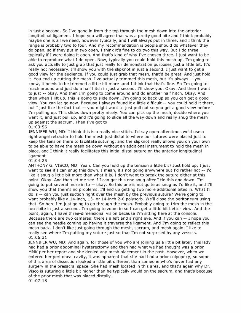
in just a second. So I've gone in from the top through the mesh down into the anterior longitudinal ligament. I hope you will agree that was a pretty good bite and I think probably maybe one is all we need, however typically, and I will always put in three, and I think the range is probably two to four. And my recommendation is people should do whatever they do open, so if they put in two open, I think it's fine to do two this way. But I do three typically if I were doing it open. And that's kind of why I've chosen three. I just want to be able to reproduce what I do open. Now, typically you could hold this mesh up. I'm going to ask you actually to just grab that just really for demonstration purposes just a little bit. It's really not necessary. I'll show you with the slipknot in just a second. I just want to get a good view for the audience. If you could just grab that mesh, that'd be great. And just hold it. You end up cutting the mesh. I've actually trimmed this mesh, but it's always -- you know, it needs to be trimmed a little bit more ,and I think that that's fine. So I'm going to reach around and just do a half hitch in just a second. I'll show you. Okay. And then I want to just -- okay. And then I'm going to come around and do another half hitch. Okay. And then when I lift up, this is going to slide down. I'm going to back up so you can get a good view. You can let go now. Because I always found it a little difficult -- you could hold it there, but I just like the fact that -- you might want to just pull out so you get a good view before I'm pulling up. This slides down pretty nicely. You can pick up the mesh, decide where you want it, and just pull up, and it's going to slide all the way down and really snug the mesh up against the sacrum. Then I've got to 01:03:56 JENNIFER WU, MD: I think this is a really nice stitch. I'd say open oftentimes we'd use a right angel retractor to hold the mesh just distal to where our sutures were placed just to keep the tension there to facilitate suturing, and the slipknot really allows you on your own to be able to have the mesh tie down without an additional instrument to hold the mesh in place, and I think it really facilitates this initial distal suture on the anterior longitudinal ligament. 01:04:25 ANTHONY G. VISCO, MD: Yeah. Can you hold up the tension a little bit? Just hold up. I just want to see if I can snug this down. I mean, it's not going anywhere but I'd rather not -- I'd like it snug a little bit more than what it is. I don't want to break the suture either at this point. Okay. And then let me see if I can get this one snug after I tie this one down. I'm going to put several more in to -- okay. So this one is not quite as snug as I'd like it, and I'll show you that there's no problems. I'll end up getting two more additional bites in. What I'll do is -- can you just suction right over the mesh by the previous suture? We're going to want probably like a 14-inch, 13- or 14-inch 2-0 polysorb. We'll close the peritoneum using that. So here I'm just going to go through the mesh. Probably going to trim the mesh in the next bite in just a second. I'm going to zoom in so I can get a little bit better view. And the point, again, I have three-dimensional vision because I'm sitting here at the console. Because there are two cameras: there's a left and a right eye. And if you can -- I hope you can see the needle coming up having it traverse the ligament. And I’m going to reflect this mesh back. I don't like just going through the mesh, sacrum, and mesh again. I like to really see where I'm putting my suture just so that I'm not surprised by any vessels. 01:06:31 JENNIFER WU, MD: And again, for those of you who are joining us a little bit later, this lady had had a prior abdominal hysterectomy and then had what we had thought was a prior MMK per her report and she denied any mesh placement in the past. However, when we entered her peritoneal cavity, it was apparent that she had had a prior colpopexy, so some of this area of dissection looked a little bit different than someone who's never had any surgery in the presacral space. She had mesh located in this area, and that's again why Dr. Visco is suturing a little bit higher than he typically would on the sacrum, and that's because of the prior mesh that was placed distally. 01:07:18
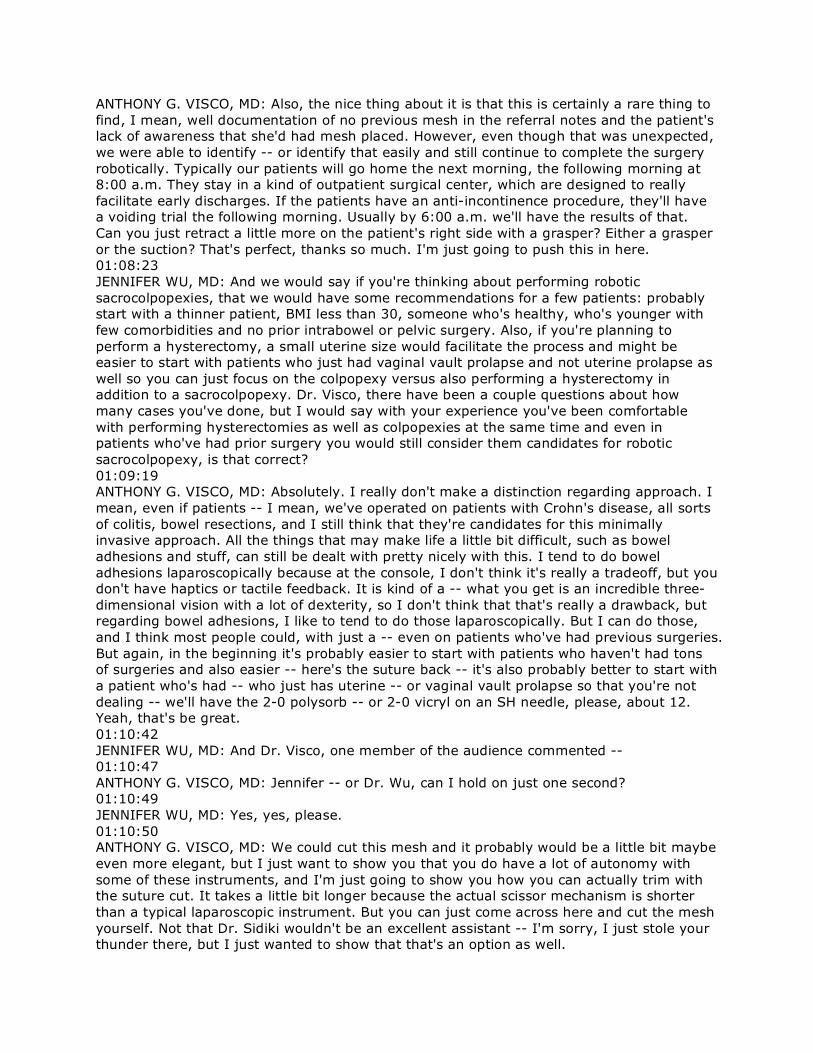
ANTHONY G. VISCO, MD: Also, the nice thing about it is that this is certainly a rare thing to find, I mean, well documentation of no previous mesh in the referral notes and the patient's lack of awareness that she'd had mesh placed. However, even though that was unexpected, we were able to identify -- or identify that easily and still continue to complete the surgery robotically. Typically our patients will go home the next morning, the following morning at 8:00 a.m. They stay in a kind of outpatient surgical center, which are designed to really facilitate early discharges. If the patients have an anti-incontinence procedure, they'll have a voiding trial the following morning. Usually by 6:00 a.m. we'll have the results of that. Can you just retract a little more on the patient's right side with a grasper? Either a grasper or the suction? That's perfect, thanks so much. I'm just going to push this in here. 01:08:23 JENNIFER WU, MD: And we would say if you're thinking about performing robotic sacrocolpopexies, that we would have some recommendations for a few patients: probably start with a thinner patient, BMI less than 30, someone who's healthy, who's younger with few comorbidities and no prior intrabowel or pelvic surgery. Also, if you're planning to perform a hysterectomy, a small uterine size would facilitate the process and might be easier to start with patients who just had vaginal vault prolapse and not uterine prolapse as well so you can just focus on the colpopexy versus also performing a hysterectomy in addition to a sacrocolpopexy. Dr. Visco, there have been a couple questions about how many cases you've done, but I would say with your experience you've been comfortable with performing hysterectomies as well as colpopexies at the same time and even in patients who've had prior surgery you would still consider them candidates for robotic sacrocolpopexy, is that correct? 01:09:19 ANTHONY G. VISCO, MD: Absolutely. I really don't make a distinction regarding approach. I mean, even if patients -- I mean, we've operated on patients with Crohn's disease, all sorts of colitis, bowel resections, and I still think that they're candidates for this minimally invasive approach. All the things that may make life a little bit difficult, such as bowel adhesions and stuff, can still be dealt with pretty nicely with this. I tend to do bowel adhesions laparoscopically because at the console, I don't think it's really a tradeoff, but you don't have haptics or tactile feedback. It is kind of a -- what you get is an incredible three-dimensional vision with a lot of dexterity, so I don't think that that's really a drawback, but regarding bowel adhesions, I like to tend to do those laparoscopically. But I can do those, and I think most people could, with just a -- even on patients who've had previous surgeries. But again, in the beginning it's probably easier to start with patients who haven't had tons of surgeries and also easier -- here's the suture back -- it's also probably better to start with a patient who's had -- who just has uterine -- or vaginal vault prolapse so that you're not dealing -- we'll have the 2-0 polysorb -- or 2-0 vicryl on an SH needle, please, about 12. Yeah, that's be great. 01:10:42 JENNIFER WU, MD: And Dr. Visco, one member of the audience commented -- 01:10:47 ANTHONY G. VISCO, MD: Jennifer -- or Dr. Wu, can I hold on just one second? 01:10:49 JENNIFER WU, MD: Yes, yes, please. 01:10:50 ANTHONY G. VISCO, MD: We could cut this mesh and it probably would be a little bit maybe even more elegant, but I just want to show you that you do have a lot of autonomy with some of these instruments, and I'm just going to show you how you can actually trim with the suture cut. It takes a little bit longer because the actual scissor mechanism is shorter than a typical laparoscopic instrument. But you can just come across here and cut the mesh yourself. Not that Dr. Sidiki wouldn't be an excellent assistant -- I'm sorry, I just stole your thunder there, but I just wanted to show that that's an option as well.
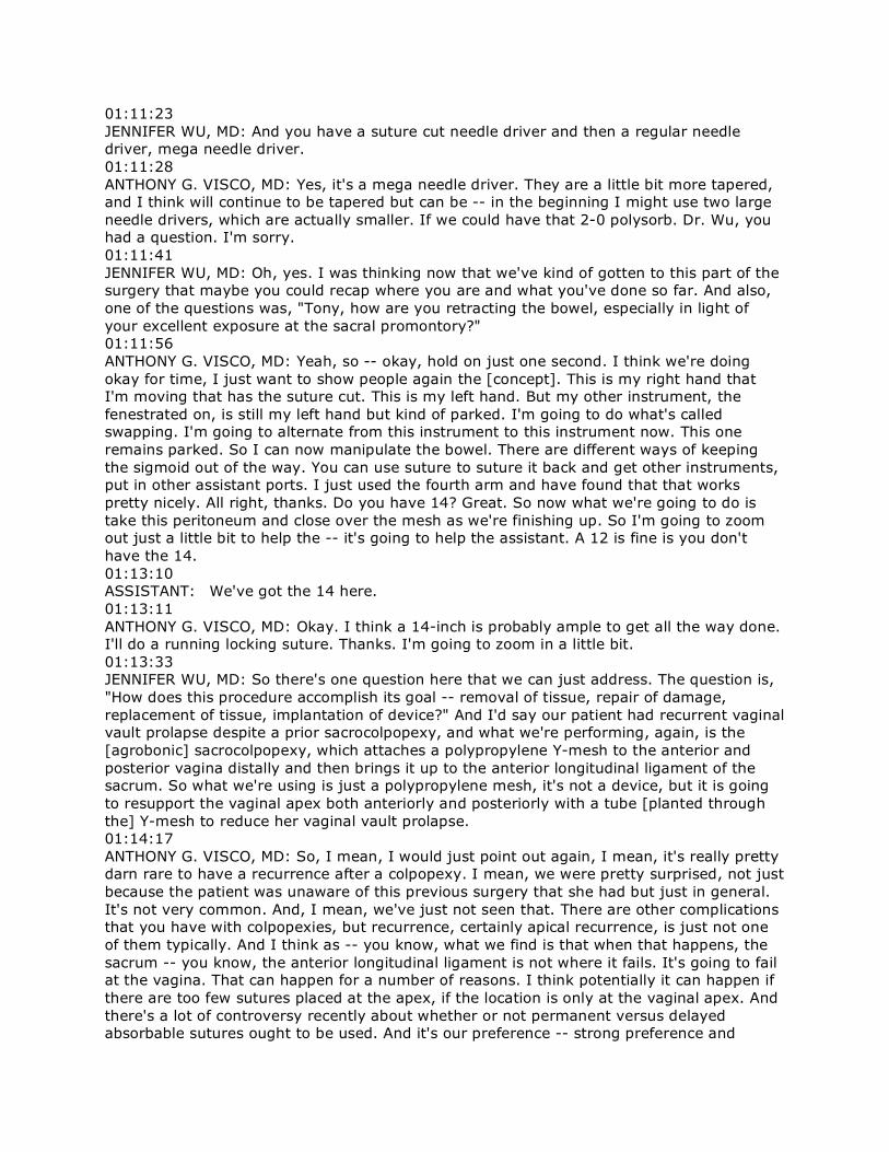
01:11:23 JENNIFER WU, MD: And you have a suture cut needle driver and then a regular needle driver, mega needle driver. 01:11:28 ANTHONY G. VISCO, MD: Yes, it's a mega needle driver. They are a little bit more tapered, and I think will continue to be tapered but can be -- in the beginning I might use two large needle drivers, which are actually smaller. If we could have that 2-0 polysorb. Dr. Wu, you had a question. I'm sorry. 01:11:41 JENNIFER WU, MD: Oh, yes. I was thinking now that we've kind of gotten to this part of the surgery that maybe you could recap where you are and what you've done so far. And also, one of the questions was, "Tony, how are you retracting the bowel, especially in light of your excellent exposure at the sacral promontory?" 01:11:56 ANTHONY G. VISCO, MD: Yeah, so -- okay, hold on just one second. I think we're doing okay for time, I just want to show people again the [concept]. This is my right hand that I'm moving that has the suture cut. This is my left hand. But my other instrument, the fenestrated on, is still my left hand but kind of parked. I'm going to do what's called swapping. I'm going to alternate from this instrument to this instrument now. This one remains parked. So I can now manipulate the bowel. There are different ways of keeping the sigmoid out of the way. You can use suture to suture it back and get other instruments, put in other assistant ports. I just used the fourth arm and have found that that works pretty nicely. All right, thanks. Do you have 14? Great. So now what we're going to do is take this peritoneum and close over the mesh as we're finishing up. So I'm going to zoom out just a little bit to help the -- it's going to help the assistant. A 12 is fine is you don't have the 14. 01:13:10 ASSISTANT: We've got the 14 here. 01:13:11 ANTHONY G. VISCO, MD: Okay. I think a 14-inch is probably ample to get all the way done. I'll do a running locking suture. Thanks. I'm going to zoom in a little bit. 01:13:33 JENNIFER WU, MD: So there's one question here that we can just address. The question is, "How does this procedure accomplish its goal -- removal of tissue, repair of damage, replacement of tissue, implantation of device?" And I'd say our patient had recurrent vaginal vault prolapse despite a prior sacrocolpopexy, and what we're performing, again, is the [agrobonic] sacrocolpopexy, which attaches a polypropylene Y-mesh to the anterior and posterior vagina distally and then brings it up to the anterior longitudinal ligament of the sacrum. So what we're using is just a polypropylene mesh, it's not a device, but it is going to resupport the vaginal apex both anteriorly and posteriorly with a tube [planted through the] Y-mesh to reduce her vaginal vault prolapse. 01:14:17 ANTHONY G. VISCO, MD: So, I mean, I would just point out again, I mean, it's really pretty darn rare to have a recurrence after a colpopexy. I mean, we were pretty surprised, not just because the patient was unaware of this previous surgery that she had but just in general. It's not very common. And, I mean, we've just not seen that. There are other complications that you have with colpopexies, but recurrence, certainly apical recurrence, is just not one of them typically. And I think as -- you know, what we find is that when that happens, the sacrum -- you know, the anterior longitudinal ligament is not where it fails. It's going to fail at the vagina. That can happen for a number of reasons. I think potentially it can happen if there are too few sutures placed at the apex, if the location is only at the vaginal apex. And there's a lot of controversy recently about whether or not permanent versus delayed absorbable sutures ought to be used. And it's our preference -- strong preference and
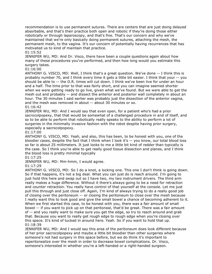
recommendation is to use permanent sutures. There are centers that are just doing delayed absorbable, and that's their practice both open and robotic if they're doing those either robotically or through laparoscopy, and that's fine. That's our concern and why we've maintained that we're only basically doing permanent sutures, attaching the mesh, the permanent mesh, to the vagina. It's our concern of potentially having recurrences that has motivated us to kind of maintain that practice. 01:15:52 JENNIFER WU, MD: And Dr. Visco, there have been a couple questions again about how many of these procedures you've performed, and then how long would you estimate this surgery takes. 01:16:00 ANTHONY G. VISCO, MD: Well, I think that's a great question. We've done -- I think this is probably number 70, and I think every time it gets a little bit easier. I think that your -- you should be able to -- the O.R. times will cut down. I think we've been live for under an hour and a half. The time prior to that was fairly short, and you can imagine seemed shorter when we were getting ready to go live, given what we've found. But we were able to get the mesh out and probably -- and dissect the anterior and posterior wall completely in about an hour. The 30 minutes I said earlier was probably just the dissection of the anterior vagina, and the mesh was removed in about -- about 30 minutes or so. 01:16:42 JENNIFER WU, MD: And I would say that even open, for a patient who's had a prior sacrocolpopexy, that that would be somewhat of a challenged procedure in and of itself, and so to be able to perform that robotically really speaks to the ability to perform a lot of surgeries in the minimally invasive fashion with the robot despite having prior surgery, especially a sacrocolpopexy. 01:17:00 ANTHONY G. VISCO, MD: Yeah, and also, this has been, to be honest with you, one of the bloodier cases, despite the fact that I think when I look it's -- you know, our total blood loss so far is about 25 millimeters. It just looks to me a little bit kind of redder than typically is the case. So I think you're able to get really good tissue dissection and planes, and I think the blood loss is pretty minimal typically 01:17:25 JENNIFER WU, MD: Mm-hmm, I would agree. 01:17:29 ANTHONY G. VISCO, MD: So I do a knot, a locking one. This one I don't think is going down. So if that happens, it's not a big deal. What you can just do is reach around. I'm going to just hold this here and swap out so I have two, my two instrument drivers. The third arm really makes a huge difference. Without it there's always going to be a need for retraction and counter retraction. You really have control of that yourself at the console. Let me just pull this through and just close off. Again, I'm kind of always trying to do a really good job of closing over the peritoneum -- or closing the peritoneum to close over the mesh because I really want this to look good and give the small bowel a chance of becoming adherent to it. When we first started this case, to be honest with you, there was a fair amount of small bowel -- if you want to pick up on that peritoneal, that'd be great. There was a fair amount of -- and you really want to make sure you get the edge, so try to reach around and grab that. Because you want to really get rough edge to rough edge when you're closing over this space. It's kind of tucked in around here. Yeah. So if you want to hold that up. 01:18:39 JENNIFER WU, MD: And I would say this area of the peritoneum does look different because of her prior sacrocolpopexy and maybe a little bit bloodier than other surgeries where someone's not had surgery in this space before, but we do think it's important to reperitonealize over the mesh in order to decrease bowel complications. Dr. Visco, someone's interested in whether you're a left-handed or a right-handed surgeon.
01:19:05 ANTHONY G. VISCO, MD: Oh, that's confidential. No, it is I am a right-handed surgeon. I think you could do -- you could -- it doesn't really matter. You could swap your instruments, but I'm definitely a right-handed surgeon. But left-handed surgeons -- can you let go of that? I actually want to try to snug that down. It just is a little bit scarred, so I don't want to tear it. But I'm going to have you pick up in just a second. 01:19:29 JENNIFER WU, MD: And the second part of that question is does that matter with the placement of the fourth arm? And I'd say I don't think that handedness really makes a difference for the placement of the fourth arm. 01:19:35 ANTHONY G. VISCO, MD: Absolutely not. 01:19:36 JENNIFER WU, MD: That really replaces the fourth arm on the patient's left-hand side to facilitate retracting the sigmoid colon from our area of dissection, which is [-- we're really] interested in the presacral space, and so that really helps facilitate moving the sigmoid colon out of the way so that our field of dissection is clear, and that's why we place the fourth arm on the left-hand side. 01:19:55 ANTHONY G. VISCO, MD: I also want to make a point that the suture -- I'm going to grab the -- this is a little trick. I'm going to grab the mesh with the actual needle, and I'm going to swap and reach around with my fourth arm and just grab the peritoneal reflection on that side. Hold on one second. And pull that out. And now I can let go of the mesh, and I've got the peritoneal edge right there, and I can just grab the actual edge of the peritoneum. And I think that's really helpful because oftentimes the actual peritoneal edge is below the mesh, underneath the mesh at this point, and I really want to try to follow that. What's not been asked but I just will mentioned is, you know, our concern of interseals as well, and a lot of our patients will have really large interseals. I think they can be fixed robotically as well. By extending the peritoneum all the way down to the vagina along the posterior vaginal wall, we really get a good placement of the - of the mesh and the ability to pull up the culdesac all the way up to the, uh, abdominal -- or to close over the mesh. 01:21:02 JENNIFER WU, MD: So do you think that really takes the place of performing additional culdoplasty? 01:21:04 ANTHONY G. VISCO, MD: Yeah. I think that that's -- we're essentially doing a culdoplasty as we're bringing up the mesh. We're going to bring this all the way up the posterior vaginal wall. 01:21:18 JENNIFER WU, MD: There's one question here about how does the cost of this procedure compare to the cost of conventional surgery, and does insurance reimburse for such a procedure? 01:21:26 ANTHONY G. VISCO, MD: Yeah, it's always a question that comes up with these procedures. So the technology, other than the fixed cost of the robot, is pretty comparable. So the instruments that you see, these robotic wristed instruments, have a life of 10 uses. So they're -- while they're more expensive, each -- you know, when you look at what the cost is per use, it's roughly the cost of a disposable laparoscopic instrument. So I think in that respect it's comparable. There's the upfront fixed cost, and the way to decrease those fixed costs is basically by volume. So you have to kind of ask yourself, and oftentimes your hospital will be asking the questions, is whether or not the volume is such that warrants a system, because there is that fixed cost of the unit. 01:22:14
JENNIFER WU, MD: And I would say in terms of billing for this type of procedure, we would bill this as a laparoscopic sacrocolpopexy as it's a robotic-assisted laparoscopic colpopexy, and there are not specific codes for a robotic procedure, but that we would -- it would be coded as a laparoscopic procedure. 01:22:32 ANTHONY G. VISCO, MD: And you can see we're really reaching down and grabbing the whole posterior peritoneum. I'm going to actually kind of grab that with a -- with a suture again. I'm going to free up my fourth arm. 01:22:48 JENNIFER WU, MD: And again, Dr. Visco has two instruments on the left-hand side, and he can toggle back and forth between the two different instruments with a foot pedal that he has at the console. And so he really has three arms that he's able to manipulate, and so then again, this system we have here is called a four-arm system: one for the camera, and then three instrument arms. And you can see a view of his feet at the console where he's able to toggle back and forth between the two instruments on his left-hand side. And then in addition to that, there's still the surgical assistant on the patient's right-hand side. 01:23:22 ANTHONY G. VISCO, MD: And again, I'm just doing locking sutures because I don't want them to slip, so I'm going to just let go of this, the third operative arm, so that this snugs in nicely. I'm going to tug -- or I'm going to tuck this in in a second and just pull this a little bit more tightly. And this -- I might have done -- I mean, I think the peritoneal closure is pretty good. You can see it all the way down. We're -- we'll include the bladder flap as we would open as we're getting a little farther down. My recommendation would probably be not to go for the bladder flap too early because you really want to get that culdesac closed. Okay. So we're -- I was just told that we're about an hour and 24 minutes in, and we've attached the mesh, attached it to the sacrum. Sure. And the -- so we're going to just zoom in just a little bit so I get a view of this. 01:24:30 JENNIFER WU, MD: There's one question here which we can field this. "Is this mesh temporary or will further surgery be required for a return to normal lifestyle?" And the mesh that we're using is a polypropylene mesh, it's a permanent mesh. It's a Y-mesh that's configured already for the procedure so that there's two ends of the Y, one on the anterior and one on the posterior vagina, and then a long arm, the Y, the single arm going to the sacrum. It's a permanent material, so it is quite unusual for someone to have a sacrocolpopexy performed in the past and for there to be a recurrent apical prolapse. I think the thing that's really important in this procedure is that we try to perform it like we do an open sacrocolpopexy. There are a number of sutures that are placed on the anterior and posterior vagina, and we do make sure that we dissect far enough down on the anterior and posterior vagina for the mesh placement. So we hope that this will relieve her symptoms and be very long-lasting for her and that this will be the last procedure she'll need for the prolapse. 01:25:28 ANTHONY G. VISCO, MD: Yeah, that's a great point because the kind of typical patient really wants the longest lasting repair, and assuming that we are attaching this well to the anterior vagina, I think that we can be relatively -- you know, nothing's 100%, but I think we can be relatively sure that her likelihood of recurrence, especially a recurrence, will be very low, definitely in the 90% success rate. And with the use of this mesh and our experience over the last year and a half, I really -- the rate of our mesh erosion is very low. I mean, actually it's been zero in our case -- I'm a little reluctant to ever say zero, but I think that it's certainly lower than probably the 3% that we would have expected, but we'll have to just -- as our series continues, we'll have to just see if that in fact holds true. I don't expect it to be higher, because I think our rate of erosion using Mersilene would have been higher. I just want to comment -- with a suture cut, it is possible that you
inadvertently cut the suture, and if that were to happen right now, like let's say I just cut it right here, it wouldn't be that big of a deal if I accidentally kind of reached in and cut it. I would just start a new suture, bring it to it, and then tie the two together. All right. We're almost done with this. 01:26:42 JENNIFER WU, MD: And I'd say postoperatively, you'd mentioned already we would treat her as a minimally invasive surgery. Often she would stay overnight. We discharge home on postoperative day number one. If she'd had a concurrent anti-incontinence procedure, we would have performed [boarding] trials the following morning. There is one question about do we routinely prescribe Estrus cream postoperatively or preoperatively, and then how long do we recommend using this. 01:27:08 ANTHONY G. VISCO, MD: I do not routinely. If the patient has really severe atrophic vaginitis or if we've seen patients who've failed other mesh procedures, which we've seen some, those patients, especially if they've had previous vaginal erosions, we will proceed with using a -- actually just go back here, if you guys can see, and just close this up. We would use the estrogen cream or Estrus cream or Premarin cream in those patients who had erosions previously but not patients who just routinely. I think it's probably fine to use that but just hasn't been our practice. 01:27:53 JENNIFER WU, MD: And I think that it's your practice and there are some people who tend to use a lot of Estrus cream postoperatively. We tend not to, and I think either way it's whatever your practice pattern is, however, we haven't seen any mesh erosions without using Estrus cream postoperatively routinely. But again, it does depend on the patient. And certainly, if preoperatively they had a lot of atrophy, we would consider it. 01:28:17 ANTHONY G. VISCO, MD: And this is our last stitch that I'm just putting in right now. Yep. Perfect. Just about finishing up here. I'm just going to put this stitch in and then I'll move away from the console. The console has a laser. It actually knows basically when your head is in the way, so I'm going to pull my head out in just a minute, and that deactivates the instruments so one can safely take their head out to communicate to the beside team, or in this case I'll be coming out to look in the camera and basically try to talk to you and just let you know, kind of give you a recap. 01:29:00 JENNIFER WU, MD: And I'd say otherwise in terms of -- 01:29:02 ANTHONY G. VISCO, MD: We're going to have the needle back in just a second. 01:29:03 JENNIFER WU, MD: In terms of other postoperative issues, really we treat them like other minimally invasive laparoscopic procedures at being discharged home on postoperative day one. Typically we do advise them to avoid any heavy lifting six weeks, at least until their postoperative visit, and really ideally three months. We encourage walking, we advance their diet after surgery, and we have them ambulating at least by postoperative day one before their discharge home. So it's a really nice minimally invasive way to proceed with important procedure for prolapse. And Dr. Visco, I'll let you kind of orient everyone and show them the final results. 01:29:44 ANTHONY G. VISCO, MD: To recap, when we entered there was clearly vault prolapse, there was some -- a fair amount of scar tissue from the small bowel to the large bowel, obstructing the view of the sacrum. And what we found as we were opening -- doing the adhesions was that there was some mesh, some permanent mesh that was attached to the sacrum but not to the vagina at all and ended about halfway down the distance from the sacrum to the vagina. We ended up resecting that mesh and then also opening up the
peritoneum and dissecting the anterior then the posterior vagina and were able to get a plane. The bladder had been adherent over the apex of the vagina. Then we introduced the mesh, attached the mesh to the anterior wall, rolled it up and held it up with our third operative arm, attached it to the posterior wall, and then attached it up to the sacrum using a sliding, or slipknot, and then have just finished closing over the mesh. I'm going to actually break. I appreciate everybody logging in and joining us here at Duke for the sacrocolpopexy. I'm happy to answer any questions. If you have additional questions, feel free to include those as well. Thanks very much. 01:30:56 JENNIFER WU, MD: And thank you very much for joining us. We really appreciate your participation. We really enjoyed all the questions from the audience, and we're sorry if we weren't able to get to everyone's questions. There were quite a lot of them. I just want to mention that this video will be archived on the OR-Live website, so please feel free to revisit the website and to watch the video once again. Thank you so much for your time. We really enjoyed you participating in this process. Thank you. 01:31:22 ANNOUNCER: Thank you for watching the minimally invasive sacrocolpopexy procedure using the da Vinci S surgical system performed at Duke University Medical Center in Durham, North Carolina. 01:32:05 [end of broadcast]