
Mini‑dental implants‑for rehabilitation of narrow …hitec-implants.com/publications/17_Mini...
9
Journal of Dental Implants | Jul - Dec 2013 | Vol 3 | Issue 2 125 ORIGINAL ARTICLE Mini‑dental implants‑for rehabilitation of narrow single tooth edentulous space: A clinical study of seven cases Manish Raghani, Bipin Sadhwani 1 , Sonal Anchlia 1 , Shaili Sadhwani 2 ABSTRACT Aims and Objectives: To evaluate the efficacy, clinical acceptability, and patient response to rehabilitation of single edentulous spaces with restorations over screw retained mini dental implants. Materials and Methods: This study consisted of seven patients (four female and three male). Single‑stage, 2.4 mm diameter and 10 and 13 mm long, screw form with integrated abutment, self‑tapping, threaded, acid‑etched and sandblasted, pure titanium mini dental implants were placed in 10 single, narrow, edentulous spaces (<6 mm bucco‑lingually) in the anterior region of the jaw (four in the maxilla and three in the mandible). Results: Post‑operative evaluation of mini dental implant was done 12 months of implant loading. Following clinical and radiographic parameters were evaluated: Gingival status (Gingival index), probing depth (By William’s periodontal probe), stability (periotest), patient compliance, prosthesis loosening and fracture, and marginal bone loss (using Intra‑oral periapical radiograph, orthopantomograph) Conclusion: Single‑tooth mini‑implant restorations demonstrated a rate of success similar to those reported by previous studies for standard single‑tooth implant restoration. Therefore, a mini‑implant may represent a valid treatment alternative when space problems do not permit the use of standard wide‑diameter implants. However, more long‑term studies are needed to determine the long term success rate of this self‑tapping mini implant design. KEY WORDS: Dental implants, immediate loading, narrow edentulous space, osseo‑integration INTRODUCTION Loss of tooth not only causes difficulty in mastication and maintenance of oral hygiene but is also psychologically disturbing on the part of the patient, as it compromises both, esthetics as well as speech. For these reasons, most patients want even a single lost tooth replaced. [1] Conventional rehabilitation of missing teeth with removable prostheses causes many problems like difficulty in mastication, psychological problems, poor esthetics, poor retention, and stability of prosthesis; all these leading to lack of confidence. In addition to these problems, fixed prostheses also require unnecessary grinding of adjacent healthy teeth, caries, short life span of prosthesis, etc. In 1952, professor Per‑Ingvar Brånemark discovered that metal implants could be structurally integrated into living bone with a very high degree of predictability and without long‑term soft tissue inflammation or ultimate fixture rejection. [2] Brånemark named the phenomenon osseo‑integration initially defined at a light microscopic level as “a direct structural and functional connection between ordered living bone and the surface of a load‑carrying implant. ”[3] Conventional theory held that the use of a standard size or a wide diameter implant was essential to ensure adequate bone‑to‑implant contact. It has been reported that space between implant and adjacent natural tooth should be at least 1.25 mm, Department of Oral and Maxillofacial Surgery, Govt Dental College, Raipur, Chhattisgarh, 1 Department of Oral and Maxillofacial Surgery, Govt Dental College, 2 Private Practitioner, Ahmedabad, Gujarat, India Address for correspondence: Dr. Bipin S Sadhwani, Department of Oral and Maxillofacial Surgery, Govt. Dental College and Hospital, Ahmedabad - 16, India. E-mail: [email protected] Access this article online Quick Response Code: Website: www.jdionline.org DOI: 10.4103/0974-6781.118880 [Downloaded free from http://www.jdionline.org on Sunday, August 30, 2015, IP: 90.211.34.142]
Transcript of Mini‑dental implants‑for rehabilitation of narrow …hitec-implants.com/publications/17_Mini...
Journal of Dental Implants | Jul - Dec 2013 | Vol 3 | Issue 2 125
ORIGINAL ARTICLE
Mini‑dental implants‑for rehabilitation of narrow single tooth edentulous space: A clinical study of seven cases
Manish Raghani, Bipin Sadhwani1, Sonal Anchlia1, Shaili Sadhwani2
ABSTRACT
Aims and Objectives: To evaluate the efficacy, clinical acceptability, and patient response to rehabilitation of single edentulous spaces with restorations over screw retained mini dental implants.Materials and Methods: This study consisted of seven patients (four female and three male). Single‑stage, 2.4 mm diameter and 10 and 13 mm long, screw form with integrated abutment, self‑tapping, threaded, acid‑etched and sandblasted, pure titanium mini dental implants were placed in 10 single, narrow, edentulous spaces (<6 mm bucco‑lingually) in the anterior region of the jaw (four in the maxilla and three in the mandible).Results: Post‑operative evaluation of mini dental implant was done 12 months of implant loading. Following clinical and radiographic parameters were evaluated: Gingival status (Gingival index), probing depth (By William’s periodontal probe), stability (periotest), patient compliance, prosthesis loosening and fracture, and marginal bone loss (using Intra‑oral periapical radiograph, orthopantomograph)Conclusion: Single‑tooth mini‑implant restorations demonstrated a rate of success similar to those reported by previous studies for standard single‑tooth implant restoration. Therefore, a mini‑implant may represent a valid treatment alternative when space problems do not permit the use of standard wide‑diameter implants. However, more long‑term studies are needed to determine the long term success rate of this self‑tapping mini implant design.
KEY WORDS: Dental implants, immediate loading, narrow edentulous space, osseo‑integration
INTRODUCTION
Loss of tooth not only causes difficulty in mastication and maintenance of oral hygiene but is also psychologically disturbing on the part of the patient, as it compromises both, esthetics as well as speech. For these reasons, most patients want even a single lost tooth replaced.[1]
Conventional rehabilitation of missing teeth with removable prostheses causes many problems like difficulty in mastication, psychological problems, poor esthetics, poor retention, and stability of prosthesis; all these leading to lack of confidence. In addition to these problems, fixed prostheses also require unnecessary grinding of adjacent healthy teeth, caries, short life span of prosthesis, etc.
In 1952, professor Per‑Ingvar Brånemark discovered that metal implants could be structurally integrated into living bone with a very high degree of predictability and without long‑term soft tissue inflammation or ultimate fixture rejection.[2] Brånemark named the phenomenon osseo‑integration initially defined at a light microscopic level as “a direct structural and functional connection between ordered living bone and the surface of a load‑carrying implant.”[3] Conventional theory held that the use of a standard size or a wide diameter implant was essential to ensure adequate bone‑to‑implant contact. It has been reported that space between implant and adjacent natural tooth should be at least 1.25 mm,
Department of Oral and Maxillofacial Surgery, Govt Dental College, Raipur, Chhattisgarh, 1Department of Oral and Maxillofacial Surgery, Govt Dental College, 2Private Practitioner, Ahmedabad, Gujarat, India
Address for correspondence: Dr. Bipin S Sadhwani, Department of Oral and Maxillofacial Surgery, Govt. Dental College and Hospital, Ahmedabad - 16, India. E-mail: [email protected]
Access this article onlineQuick Response Code:
Website:www.jdionline.org
DOI: 10.4103/0974-6781.118880
[Downloaded free from http://www.jdionline.org on Sunday, August 30, 2015, IP: 90.211.34.142]
Raghani, et al.: Mini‑dental implants‑for rehabilitation of narrow single tooth edentulous space
126 Journal of Dental Implants | Jul - Dec 2013 | Vol 3 | Issue 2
of which 0.25 mm space is required for periodontal ligament and 1 mm is required for bone to ensure proper blood supply necessary for osseo‑integration of implant. Small diameter implants (SDIs)/mini dental implant are the preferred treatment modality in cases of limited anatomical geography where mesio‑distal space between two adjacent teeth is inadequate (<6 mm) to place conventional smallest diameter implants (3.75 mm). Specifically, SDIs are indicated for the replacement of teeth with small cervical diameters and in cases of reduced inter‑radicular bone.[4] They have also been shown to be a viable alternative to bone augmentation when poor alveolar ridge width is encountered[5] and in cases of restricted mesio‑distal anatomy.[6] Victor I. Sendax expanded on Branemark’s ideas when he learned that long‑term denture stabilization could be similarly achieved with the use of small‑diameter posts inserted directly into the alveolar ridge.[7] Mini implants are one form of small diameter implants (less than 3.3 mm diameter). Unlike standard implants, mini implants allow immediate loading. These implants require drilling of bone only, one third to half of the total implant length, and are self‑tapped firmly into the bone, so integration is immediate. Balkin et al.[8] reported that histologically, the bone appeared to be integrated to the surface of the mini dental implants at the light microscopic level, and the bone appeared to be relatively mature and healthy.
Proposed advantages to the use of mini dental implants include reduced bleeding, decreased post‑operative discomfort, shortened healing time, placement into narrow ridges, and immediate loading.
MATERIALS AND METHODS
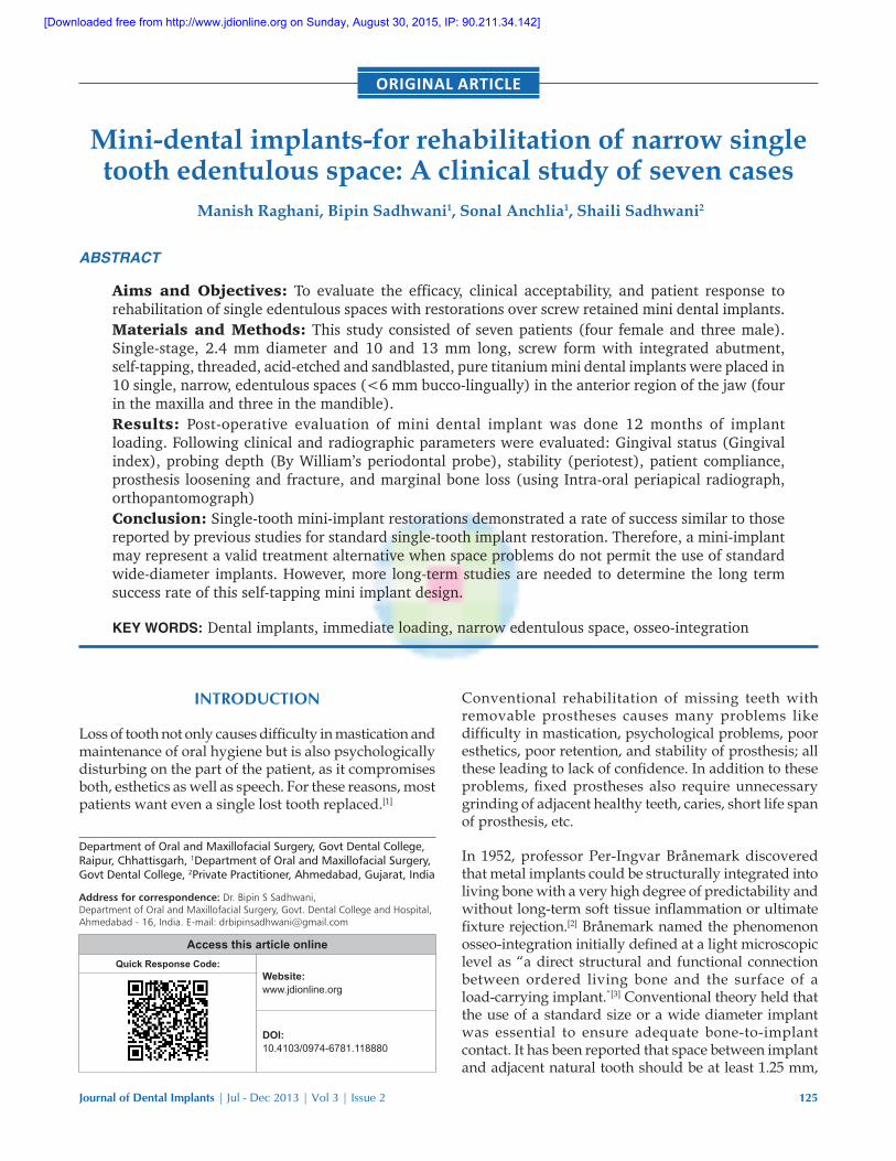
In the present study, seven patients were selected randomly from amongst the patients attending the Department of Oral and Maxillofacial Surgery, Government Dental College and Hospital, Ahmedabad for mini implant placement in single narrow edentulous spaces [Figure 1]. Patients selected for the study were between the age group of 18 and 35 years. Amongst them, three patients were female and four patients were male. Out of seven, four patients had congenitally missing teeth, two patients had tooth loss due to trauma and one patient had periodontal disease and one has undergone extraction of carious tooth.
Criteria for patient selectionInclusion criteria for patients were: Age of patient between 18‑35 yrs, bone quality D1, D2, D3 according to Lekholm and zarb, inadequate bucco‑lingual and mesio‑distal dimension of edentulous space <6 mm.[9,10]
Exclusion criteria were: Uncontrolled systemic disease, radiation therapy in the oro‑facial region, smoking,
pregnancy, alcohol abuse, poor oral hygiene, insufficient vertical inters arch space to accommodate the prosthetic components, incomplete facial growth and teeth when orthodontist was consulted regarding facial growth, psychological problems, and patients has unrealistic expectations.
Implant hardwareIn this study, (HI‑TEC, TRI‑N, life care, Mumbai, India) 1.8 and 2.4 mm wide and 11 mm and 13 mm long end osseous self‑tapping screw form, large grit sand‑blasted and acid‑etched titanium mini dental implants with integrated abutment were used. The body of the implant is self‑tapping mini dental screw characterized by having same thread units as the standard fixture (i.e. 12 in a 10 mm fixture) and up to the apical part. It has a pointed tip to facilitate easy penetration into the bone and to get sufficient apical anchorage from the bone. Anti‑rotational feature of the implant is a small groove along the apical region of the implant body. Integrated abutment of about 6.3 mm length for cemented restoration. The implant surface has about 1.7 mm polished integrated trans‑gingival section at the neck part of the implant, which promotes instant post‑operative healing of the soft‑tissue and minimizes crestal bone loss.
Pre‑operative evaluation of surgical site1. Clinical evaluation of soft tissue and dentition:
Gingiva was examined for texture, consistency, and thickness. Occlusion, periodontal integrity of dentition, teeth alignment and inter‑occlusal space were also assessed
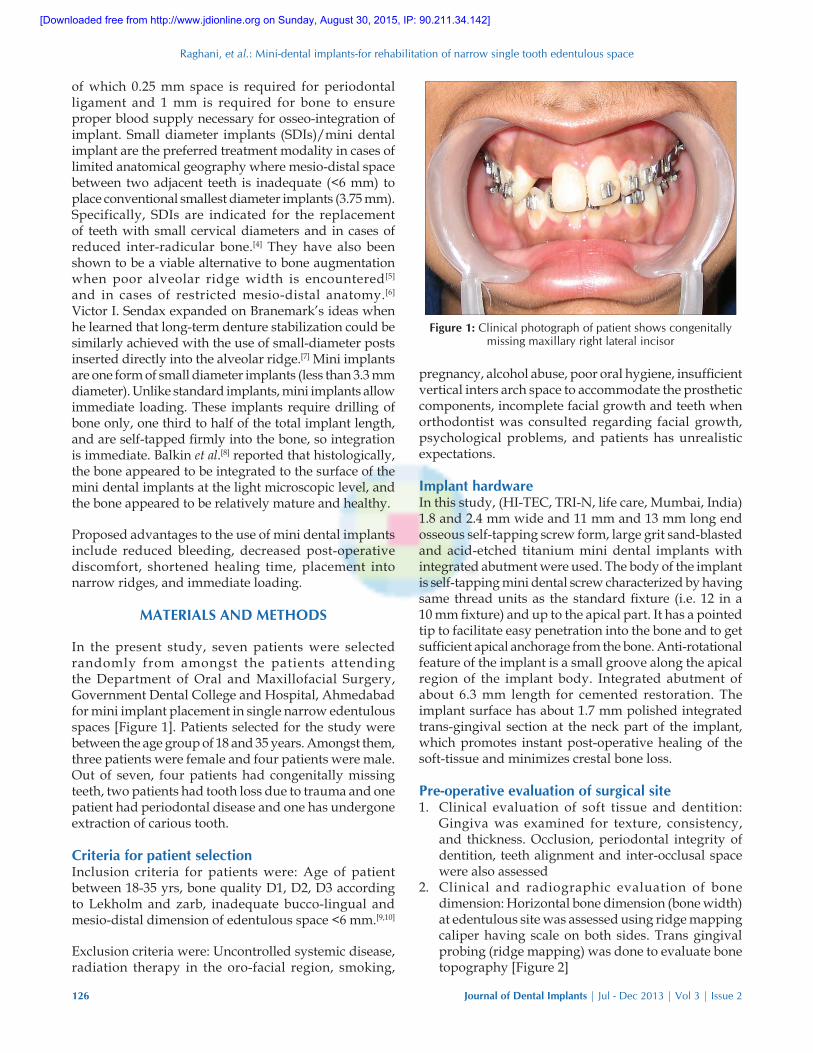
2. Clinical and radiographic evaluation of bone dimension: Horizontal bone dimension (bone width) at edentulous site was assessed using ridge mapping caliper having scale on both sides. Trans gingival probing (ridge mapping) was done to evaluate bone topography [Figure 2]
Figure 1: Clinical photograph of patient shows congenitally missing maxillary right lateral incisor
[Downloaded free from http://www.jdionline.org on Sunday, August 30, 2015, IP: 90.211.34.142]
Raghani, et al.: Mini‑dental implants‑for rehabilitation of narrow single tooth edentulous space
Journal of Dental Implants | Jul - Dec 2013 | Vol 3 | Issue 2 127
Horizontal bone space = Total horizontal width – Soft tissue thickness (Buccally + Lingually)
Digital ortho‑pentomograph was used to assess vertical bone dimension at edentulous site. Because of distortion and magnification of dimension usually occurs in OPG. Transparent radiographic implant template containing metal ball of known diameter was used to accurately assess the vertical bone dimension at edentulous site using following formula:
Actual vertical bone height
=Radiographic bone height×ball ddiameter
Radiographic ball diameter
3. Dental models and clinical photographs: Dental models were articulated on semi‑adjustable articulator to evaluate the centric relationship, inter‑arch occlusal clearance, and occlusal discrepancy for esthetic evaluation.
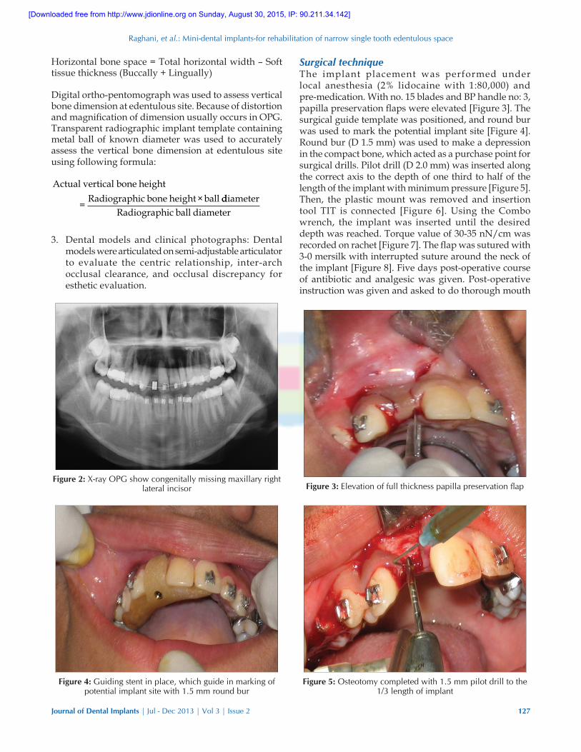
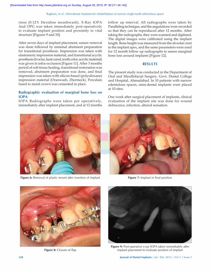
Surgical techniqueThe implant placement was performed under local anesthesia (2% lidocaine with 1:80,000) and pre‑medication. With no. 15 blades and BP handle no: 3, papilla preservation flaps were elevated [Figure 3]. The surgical guide template was positioned, and round bur was used to mark the potential implant site [Figure 4]. Round bur (D 1.5 mm) was used to make a depression in the compact bone, which acted as a purchase point for surgical drills. Pilot drill (D 2.0 mm) was inserted along the correct axis to the depth of one third to half of the length of the implant with minimum pressure [Figure 5]. Then, the plastic mount was removed and insertion tool TIT is connected [Figure 6]. Using the Combo wrench, the implant was inserted until the desired depth was reached. Torque value of 30‑35 nN/cm was recorded on rachet [Figure 7]. The flap was sutured with 3‑0 mersilk with interrupted suture around the neck of the implant [Figure 8]. Five days post‑operative course of antibiotic and analgesic was given. Post‑operative instruction was given and asked to do thorough mouth
Figure 3: Elevation of full thickness papilla preservation flap
Figure 4: Guiding stent in place, which guide in marking of potential implant site with 1.5 mm round bur
Figure 5: Osteotomy completed with 1.5 mm pilot drill to the 1/3 length of implant
Figure 2: X-ray OPG show congenitally missing maxillary right lateral incisor
[Downloaded free from http://www.jdionline.org on Sunday, August 30, 2015, IP: 90.211.34.142]
Raghani, et al.: Mini‑dental implants‑for rehabilitation of narrow single tooth edentulous space
128 Journal of Dental Implants | Jul - Dec 2013 | Vol 3 | Issue 2
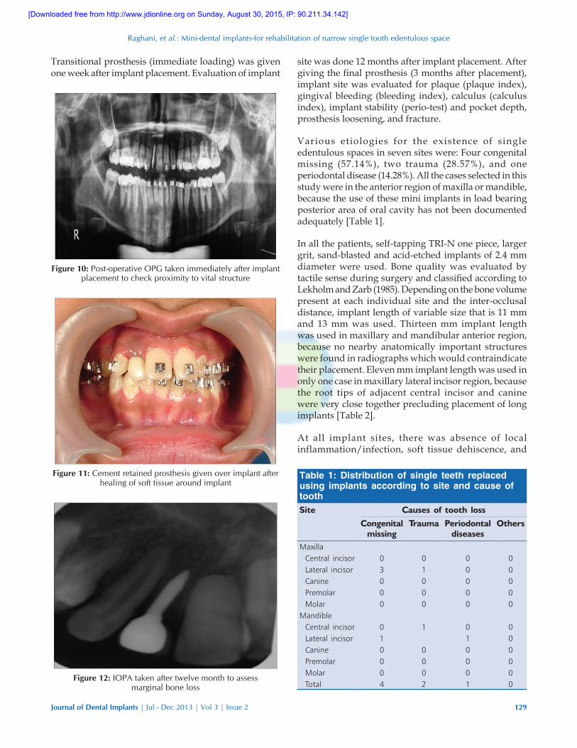
rinse (0.12% Hexidine mouthwash). X‑Ray IOPA And OPG was taken immediately post‑operatively to evaluate implant position and proximity to vital structure [Figures 9 and 10].
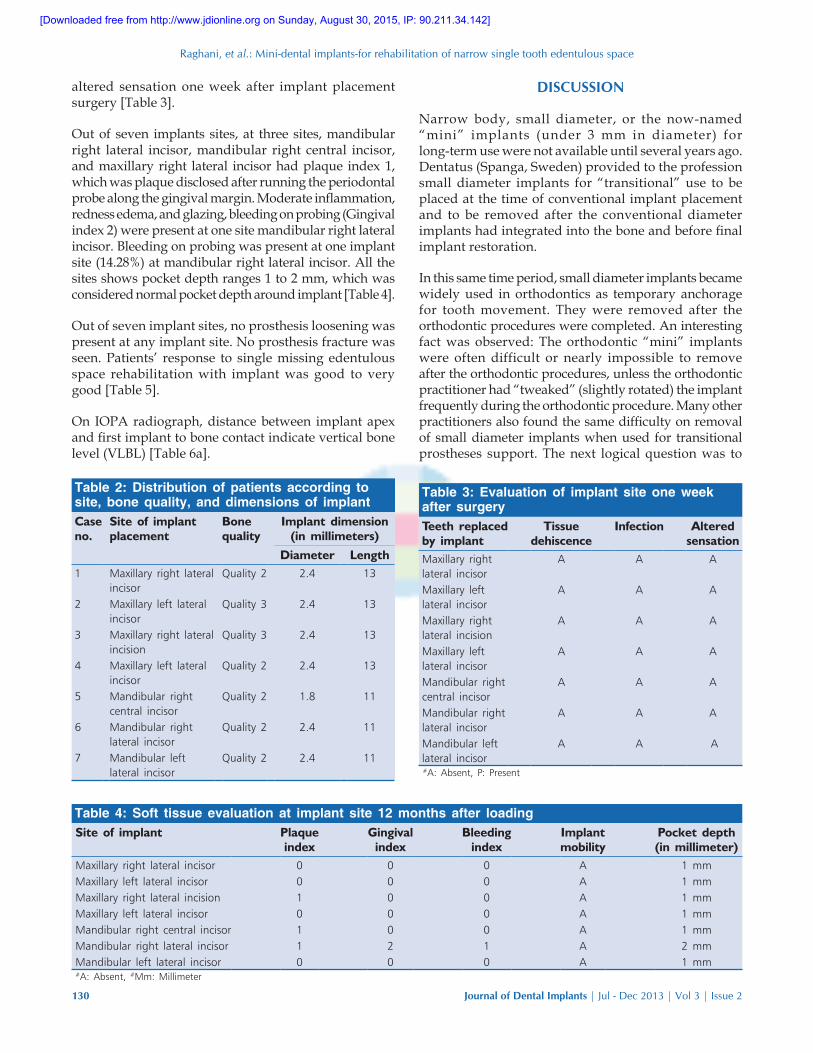
After seven days of implant placement, suture removal was done followed by minimal abutment preparation for transitional prosthesis. Impression was taken with elastomeric impression material, and transitional acyclic prosthesis (Ivoclar, heat cured, tooth color, acrylic material) was given in infra‑occlusion [Figure 11]. After 3 months period of soft tissue healing, transitional restoration was removed, abutment preparation was done, and final impression was taken with silicon‑based (polysiloxane) impression material (Oranwash, Zhermeck). Porcelain fused to metal crown was cemented in place.
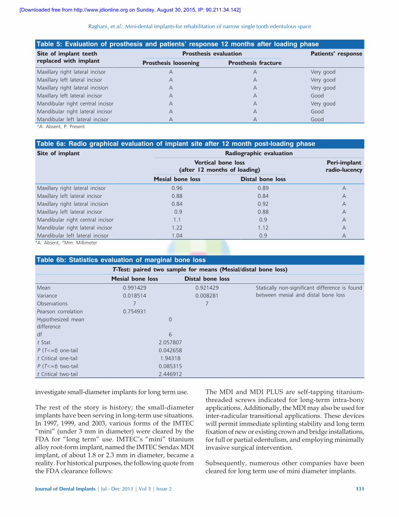
Radiographic evaluation of marginal bone loss on IOPAIOPA Radiographs were taken per operatively, immediately after implant placement, and at 12 months
follow up interval. All radiographs were taken by Paralleling technique, and the angulations were recorded so that they can be reproduced after 12 months. After taking the radiographs, they were scanned and digitized. The digital images were calibrated using the implant length. Bone height was measured from the alveolar crest to the implant apex, and the same parameters were used for 12 month follow up radiographs to assess marginal bone loss around implants [Figure 12].
RESULTS
The present study was conducted in the Department of Oral and Maxillofacial Surgery, Govt. Dental College and Hospital, Ahmadabad. In 10 patients with narrow edentulous spaces, mini‑dental implants were placed at 10 sites.
One week after surgical placement of implants, clinical evaluation of the implant site was done for wound dehiscence, infection, altered sensation.
Figure 6: Removal of plastic mount after insertion of implant Figure 7: Implant in final position
Figure 8: Closure of flapFigure 9: Post-operative x-ray IOPA taken immediately after
implant placement to evaluate position of implant
[Downloaded free from http://www.jdionline.org on Sunday, August 30, 2015, IP: 90.211.34.142]
Raghani, et al.: Mini‑dental implants‑for rehabilitation of narrow single tooth edentulous space
Journal of Dental Implants | Jul - Dec 2013 | Vol 3 | Issue 2 129
Transitional prosthesis (immediate loading) was given one week after implant placement. Evaluation of implant
site was done 12 months after implant placement. After giving the final prosthesis (3 months after placement), implant site was evaluated for plaque (plaque index), gingival bleeding (bleeding index), calculus (calculus index), implant stability (perio‑test) and pocket depth, prosthesis loosening, and fracture.
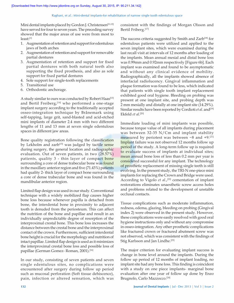
Various etiologies for the existence of single edentulous spaces in seven sites were: Four congenital missing (57.14%), two trauma (28.57%), and one periodontal disease (14.28%). All the cases selected in this study were in the anterior region of maxilla or mandible, because the use of these mini implants in load bearing posterior area of oral cavity has not been documented adequately [Table 1].
In all the patients, self‑tapping TRI‑N one piece, larger grit, sand‑blasted and acid‑etched implants of 2.4 mm diameter were used. Bone quality was evaluated by tactile sense during surgery and classified according to Lekholm and Zarb (1985). Depending on the bone volume present at each individual site and the inter‑occlusal distance, implant length of variable size that is 11 mm and 13 mm was used. Thirteen mm implant length was used in maxillary and mandibular anterior region, because no nearby anatomically important structures were found in radiographs which would contraindicate their placement. Eleven mm implant length was used in only one case in maxillary lateral incisor region, because the root tips of adjacent central incisor and canine were very close together precluding placement of long implants [Table 2].
At all implant sites, there was absence of local inflammation/infection, soft tissue dehiscence, and
Figure 10: Post-operative OPG taken immediately after implant placement to check proximity to vital structure
Figure 11: Cement retained prosthesis given over implant after healing of soft tissue around implant
Figure 12: IOPA taken after twelve month to assess marginal bone loss
Table 1: Distribution of single teeth replaced using implants according to site and cause of toothSite Causes of tooth loss
Congenital missing
Trauma Periodontal diseases
Others
MaxillaCentral incisor 0 0 0 0Lateral incisor 3 1 0 0Canine 0 0 0 0Premolar 0 0 0 0Molar 0 0 0 0
MandibleCentral incisor 0 1 0 0Lateral incisor 1 1 0Canine 0 0 0 0Premolar 0 0 0 0Molar 0 0 0 0Total 4 2 1 0
[Downloaded free from http://www.jdionline.org on Sunday, August 30, 2015, IP: 90.211.34.142]
Raghani, et al.: Mini‑dental implants‑for rehabilitation of narrow single tooth edentulous space
130 Journal of Dental Implants | Jul - Dec 2013 | Vol 3 | Issue 2
altered sensation one week after implant placement surgery [Table 3].
Out of seven implants sites, at three sites, mandibular right lateral incisor, mandibular right central incisor, and maxillary right lateral incisor had plaque index 1, which was plaque disclosed after running the periodontal probe along the gingival margin. Moderate inflammation, redness edema, and glazing, bleeding on probing (Gingival index 2) were present at one site mandibular right lateral incisor. Bleeding on probing was present at one implant site (14.28%) at mandibular right lateral incisor. All the sites shows pocket depth ranges 1 to 2 mm, which was considered normal pocket depth around implant [Table 4].
Out of seven implant sites, no prosthesis loosening was present at any implant site. No prosthesis fracture was seen. Patients’ response to single missing edentulous space rehabilitation with implant was good to very good [Table 5].
On IOPA radiograph, distance between implant apex and first implant to bone contact indicate vertical bone level (VLBL) [Table 6a].
DISCUSSION
Narrow body, small diameter, or the now‑named “mini” implants (under 3 mm in diameter) for long‑term use were not available until several years ago. Dentatus (Spanga, Sweden) provided to the profession small diameter implants for “transitional” use to be placed at the time of conventional implant placement and to be removed after the conventional diameter implants had integrated into the bone and before final implant restoration.
In this same time period, small diameter implants became widely used in orthodontics as temporary anchorage for tooth movement. They were removed after the orthodontic procedures were completed. An interesting fact was observed: The orthodontic “mini” implants were often difficult or nearly impossible to remove after the orthodontic procedures, unless the orthodontic practitioner had “tweaked” (slightly rotated) the implant frequently during the orthodontic procedure. Many other practitioners also found the same difficulty on removal of small diameter implants when used for transitional prostheses support. The next logical question was to
Table 4: Soft tissue evaluation at implant site 12 months after loadingSite of implant Plaque
indexGingival
indexBleeding
indexImplant mobility
Pocket depth (in millimeter)
Maxillary right lateral incisor 0 0 0 A 1 mmMaxillary left lateral incisor 0 0 0 A 1 mmMaxillary right lateral incision 1 0 0 A 1 mmMaxillary left lateral incisor 0 0 0 A 1 mmMandibular right central incisor 1 0 0 A 1 mmMandibular right lateral incisor 1 2 1 A 2 mmMandibular left lateral incisor 0 0 0 A 1 mm#A: Absent, #Mm: Millimeter
Table 2: Distribution of patients according to site, bone quality, and dimensions of implantCase no.
Site of implant placement
Bone quality
Implant dimension (in millimeters)
Diameter Length1 Maxillary right lateral
incisorQuality 2 2.4 13
2 Maxillary left lateral incisor
Quality 3 2.4 13
3 Maxillary right lateral incision
Quality 3 2.4 13
4 Maxillary left lateral incisor
Quality 2 2.4 13
5 Mandibular right central incisor
Quality 2 1.8 11
6 Mandibular right lateral incisor
Quality 2 2.4 11
7 Mandibular left lateral incisor
Quality 2 2.4 11
Table 3: Evaluation of implant site one week after surgeryTeeth replaced by implant
Tissue dehiscence
Infection Altered sensation
Maxillary right lateral incisor
A A A
Maxillary left lateral incisor
A A A
Maxillary right lateral incision
A A A
Maxillary left lateral incisor
A A A
Mandibular right central incisor
A A A
Mandibular right lateral incisor
A A A
Mandibular left lateral incisor
A A A
#A: Absent, P: Present
[Downloaded free from http://www.jdionline.org on Sunday, August 30, 2015, IP: 90.211.34.142]
Raghani, et al.: Mini‑dental implants‑for rehabilitation of narrow single tooth edentulous space
Journal of Dental Implants | Jul - Dec 2013 | Vol 3 | Issue 2 131
investigate small‑diameter implants for long term use.
The rest of the story is history; the small‑diameter implants have been serving in long‑term use situations. In 1997, 1999, and 2003, various forms of the IMTEC “mini” (under 3 mm in diameter) were cleared by the FDA for “long term” use. IMTEC’s “mini” titanium alloy root‑form implant, named the IMTEC Sendax MDI implant, of about 1.8 or 2.3 mm in diameter, became a reality. For historical purposes, the following quote from the FDA clearance follows:
The MDI and MDI PLUS are self‑tapping titanium‑threaded screws indicated for long‑term intra‑bony applications. Additionally, the MDI may also be used for inter‑radicular transitional applications. These devices will permit immediate splinting stability and long term fixation of new or existing crown and bridge installations, for full or partial edentulism, and employing minimally invasive surgical intervention.
Subsequently, numerous other companies have been cleared for long term use of mini diameter implants.
Table 5: Evaluation of prosthesis and patients’ response 12 months after loading phaseSite of implant teeth replaced with implant
Prosthesis evaluation Patients’ response
Prosthesis loosening Prosthesis fractureMaxillary right lateral incisor A A Very goodMaxillary left lateral incisor A A Very goodMaxillary right lateral incision A A Very goodMaxillary left lateral incisor A A GoodMandibular right central incisor A A Very goodMandibular right lateral incisor A A GoodMandibular left lateral incisor A A Good#A: Absent, P: Present
Table 6a: Radio graphical evaluation of implant site after 12 month post‑loading phaseSite of implant Radiographic evaluation
Vertical bone loss (after 12 months of loading)
Peri‑implant radio‑lucency
Mesial bone loss Distal bone lossMaxillary right lateral incisor 0.96 0.89 AMaxillary left lateral incisor 0.88 0.84 AMaxillary right lateral incision 0.84 0.92 AMaxillary left lateral incisor 0.9 0.88 AMandibular right central incisor 1.1 0.9 AMandibular right lateral incisor 1.22 1.12 AMandibular left lateral incisor 1.04 0.9 A
#A: Absent, #Mm: Millimeter
Table 6b: Statistics evaluation of marginal bone lossT‑Test: paired two sample for means (Mesial/distal bone loss)
Mesial bone loss Distal bone lossMean 0.991429 0.921429 Statically non-significant difference is found
between mesial and distal bone lossVariance 0.018514 0.008281Observations 7 7Pearson correlation 0.754931Hypothesized mean difference
0
df 6t Stat 2.057807P (T<=t) one-tail 0.042658t Critical one-tail 1.94318P (T<=t) two-tail 0.085315t Critical two-tail 2.446912
[Downloaded free from http://www.jdionline.org on Sunday, August 30, 2015, IP: 90.211.34.142]
Raghani, et al.: Mini‑dental implants‑for rehabilitation of narrow single tooth edentulous space
132 Journal of Dental Implants | Jul - Dec 2013 | Vol 3 | Issue 2
Mini dental implants placed by Gordon J. Christensen[11,12] have served for four to seven years. The preceding survey showed that the major areas of use were from most to least:[1]
1. Augmentation of retention and support for edentulous jaws of both arches
2. Augmentation of retention and support for removable partial dentures
3. Augmentation of retention and support for fixed partial dentures with both natural teeth also supporting the fixed prosthesis, and also as sole support for fixed partial dentures
4. Sole support for single‑tooth replacements5. Transitional use6. Orthodontic anchorage.
A study similar to ours was conducted by Robert Haas[13] and Bertil Freiberg,[14] who performed a one‑stage implant surgery according to the traditionally accepted osseo‑integration technique by Brånemark using self‑tapping, large grit, sand‑blasted and acid‑etched mini implants of diameter 2.4 mm with two different lengths of 11 and 13 mm at seven single edentulous spaces in different jaw areas.
Bone quality registration following the classification by Lekholm and zarb[15] was judged by tactile sense during surgery, the general location and radiographic evaluation. Out of seven patients, in two (28.57%) patients, quality 3 ‑ thin layer of compact bone surrounding a core of dense trabecular bone was found in the maxillary anterior region and five (71.43%) patients had quality 2‑ thick layer of compact bone surrounding a core of dense trabecular bone and was found in the mandibular anterior region.
Limited flap design was used in our study. Conventional technique with a widely mobilized flap causes higher bone loss because whenever papilla is detached from bone, the interdental bone in proximity to adjacent tooth is denuded from the periosteum. This can affect the nutrition of the bone and papillae and result in an individually unpredictable degree of resorption of the interproximal crestal bone. This bone loss increases the distance between the crestal bone and the interproximal contact of the crown. Furthermore, sufficient interdental bone height is crucial for the morphology and nutrition of intact papillae. Limited flap design is used as it minimizes the interproximal crestal bone loss and possible loss of papillae (German Gomez‑ Roman, 2001).[16]
In our study, consisting of seven patients and seven single edentulous sites, no complications were encountered after surgery during follow up period such as mucosal perforation (Soft tissue dehiscence), pain, infection or altered sensation, which was
consistent with the findings of Morgan Olsson and Bertil Friberg.[17]
The success criteria suggested by Smith and Zarb[18] for edentulous patients were utilized and applied to the seven implant sites, which were examined during the last recall visit at intervals of 12 months after loading of the implants. Mean annual mesial and distal bone loss was 0.99mm and 0.92mm respectively [Figure 6b]. Each implant was examined and found to be asymptomatic and without any clinical evidence of mobility. Radiographically, all the implants showed absence of interfacial radiolucency. Gingival inflammation and plaque formation was found to be less, which indicated that patients with single tooth implant replacement exhibited good oral hygiene. Bleeding on probing was present at one implant site, and probing depth was 2 mm mesially and distally at one implant site (14.29%). Similar results have been reported by Cordioti et al. and[19] Ekfeld et al.[20]
Immediate loading of mini implants was possible, because torque value of all implants during placement was between 32‑35 N/Cm and implant stability measured by periotest was between −8 and +9.[21] Implant failure was not observed 12 months follow up period of the study. A long‑term follow up is required to evaluate success of implants at individual sites; mean annual bone loss of less than 0.2 mm per year is considered successful for any implant. The technology of prosthetic replacement of missing single teeth is still evolving. In the present study, the TRI‑N one‑piece mini implants for replacing the Crown and Bridge were used. According to Vigolo et al.,[22] cementation of implant restorations eliminates unaesthetic screw access holes and problems related to the development of unstable occlusal contacts.
Tissue complications such as moderate inflammation, redness, edema, glazing, bleeding on probing (Gingival index 2) were observed in the present study. However, these complications were easily resolved with good oral hygiene instructions only and without any compromise in osseo‑integration. Any other prosthetic complications like fractured crown or fractured abutment screw was not observed, which was consistent with the findings of Stig Karlsson and Jan Lindhe.[23]
The major criterion for evaluating implant success is change in bone level around the implants. During the follow up period of 12 months of implant loading, no implant site had any bone loss. This finding is coincident with a study on one piece implants‑ marginal bone evaluation after one year of follow up done by Enzo Brugnolo, Carlo Mazzocco.[24]
[Downloaded free from http://www.jdionline.org on Sunday, August 30, 2015, IP: 90.211.34.142]
Raghani, et al.: Mini‑dental implants‑for rehabilitation of narrow single tooth edentulous space
Journal of Dental Implants | Jul - Dec 2013 | Vol 3 | Issue 2 133
8. Balkin BE, Steflik DE, Naval F. Mini dental implant insertion with the auto-advance Technique for ongoing Applications. J Oral Implant 2001;27:32-7.
9. Sugerman PB, Barber MT. Patient Selection for Endosseous Dental Implants: Oral and Systemic Considerations. Int J Oral Maxillofac Implants 2002;17:191‑201.
10. Spiekermann H, Donath K, Hassell T, Javanovic S, Richter J. Color atlas of dental medicine, implantology. Single tooth implants. New York, NY; Georg Thieme Verlag; 1995. p. 267‑98.
11. Christensen GJ. The “mini” implant has arrived. J Am Dent Assoc 2006;137:387‑90.
12. Christensen GJ. Mini Implants: Good or Bad for long term service? J Esthet Restor Dent 2008;20:343-8.
13. Haas R, Mensdorff‑Pouilly N, Mailath G, Watzek G. Brånemark Single tooth implants: A preliminary report of 76 implants. J Prosthet Dent 1995;73:274-9.
14. Friberg B, Grondahl K, Lekholm U. A new self‑tapping Brånemark implant Clinical and radiographic evaluation. Int J Oral Maxillofac Implants 1992;7:80‑5.
15. Misch CE, Judy WM. Classifications of the partially edentulous arches of implant dentistry. Int J Oral Implant 1987;4:29-58.
16. Gomez G. Med dent Influence of flap design on peri – implants inter- proximal crestal bone loss around single-tooth implants. Int J Oral Maxillofac Implants 2001;16:61‑7.
17. Olsson M, Friberg B, Nilson H, Kultje C. MkII‑ A modified self-tapping implant: 3-year results of a controlled prospective pilot study. Int J Oral Maxillofac Implants 1995;10:15‑21.
18. Schmitt A, Zarb GA. The longitudinal clinical effectiveness of osseointegrated dental implants for single tooth replacement. Int J Prosthodont 1993;6:197‑202.
19. Cordioli G, Castagna E. Single-tooth implant rehabilitation: A retrospective study of 67 implants. Int J Prosthidont 1994;7:525-31.
20. Ekfeldt A, Carlsson GE, Börjesson G. Clinical evaluation of single – tooth restoration supported by osseointegrated Implants: A retrospective study. Int J Oral Maxillofac implant 1994;9:179-83.
21. Dilek O, Tezulas E, Dincel M. Required minimum primary stability and torque Values for immediate loading of mini dental implants: An experimental study in nonviable Bovine femoral bone. Oral Surg Oral Med Oral Pathol Oral Radiol Endod 2008;105:e20-7.
22. Vigolo P, Givani A, Majzoub Z, Cordioli G. Cemented versus screw – retained implants – supported single – tooth crown: A 4‑year prospective clinical study. Int J Oral Maxillofac Implants 2004;19:260‑5.
23. Wennström JL, Ekestubbe A, Gröndahl K, Karlsson S, Lindhe J. Implant-supported single-tooth restorations: A 5-year prospective study. J Clin Periodontol 2005;32:567‑74.
24. Brugnolo E, Mazzocco C, Cordioll G, Majzoub Z. Clinical and radiographic findings following placement of single – tooth implants in young patients – case reports. Int J Periodont Rest Dent 1996;16:421‑33.
But, long term follow up needed to know the mean annual bone loss of 0.2 mm per year for success of an implant.
CONCLUSION
The “mini” or small‑diameter or narrow‑diameter implant (<3 mm in diameter) concept is making an impact on the profession amid controversy and debate. The introduction, approval, and continuing observation of success of smaller‑diameter mini‑implants have stimulated the use of implants in situations, in which standard‑sized implants could not have been used without grafting.
However, there is no question that these small implants, placed and restored properly, are serving patients well. This concept provides highly needed service for many patients who do not have enough bone present for conventional over 3‑mm‑diameter implants and cannot have or cannot afford bone grafting. The result has been more patients who have been served successfully at reduced cost with minimized pain and trauma patients who could not have been treated with implants otherwise.
More research is needed to find the best alloys for the implants, the most appropriate abutments, and the service potential of these small implants over many years.
REFERENCES
1. Carl E. Misch. Rationle for dental implants. In: Carl E.Misch, Editor. Text book of Contemporary implant dentistry, 3rd Ed.New Delhi; Elsevier; 2012. p: 11.
2. Brånemark PI. Osseointegration and its experimental background. J Prostheti Dent 1983;50:399-410.
3. Hansson HA, Albrektsson T, Brånemark PI. Structural aspects of the interface between tissue and titanium Implants. J Prosthet Dent 1983;50:108-13.
4. Davarpanah M, Martinez H, Tecucianu JF, Celletti R, Lazzara R. Small-diameter implants: Indications and contraindications. J Esthet Dent 2000;12:186‑94.
5. Froum SJ, Cho SC, Cho YS, Elian N, Tarnow D. Narrow‑diameter implants: a restorative option for limited interdental space. Int J Periodontics Restorative Dent. 2007;27:449-55.
6. Mangano C, Bartolucci EG. Single tooth Replacement by Morse taper connection Implants: A, Retrospective study of 80 Implants. Int J Oral Maxillofac Implant 2001;16:675‑80.
7. Griffins TC, Collins CP, Collins PC. Mini dental implants: Anadjunct for retention, stability, and comfort for the edentulous patient. Oral Surg Oral Med Oral Pathol Oral Radiol Endo 2005;100:E81-4.
How to cite this article: Raghani M, Sadhwani B, Anchlia S, Sadhwani S. Mini-dental implants-for rehabilitation of narrow single tooth edentulous space: A clinical study of seven cases. J Dent Implant 2013;3:125-33.
Source of Support: Nil, Conflict of Interest: None.
[Downloaded free from http://www.jdionline.org on Sunday, August 30, 2015, IP: 90.211.34.142]


![[Dental Implants Cost]](https://static.fdocuments.us/doc/165x107/55638cefd8b42ad2128b4ef9/dental-implants-cost-55849922c2925.jpg)