Mini-Symposium NonhumanPrimateOptogenetics ...
10
Mini-Symposium Nonhuman Primate Optogenetics: Recent Advances and Future Directions X Adriana Galvan, 1 * X William R. Stauffer, 2 * Leah Acker, 3 X Yasmine El-Shamayleh, 4 X Ken-ichi Inoue, 5,6 X Shay Ohayon, 7 and X Michael C. Schmid 8 1 Yerkes National Primate Research Center and Department of Neurology, School of Medicine, Emory University, Atlanta, Georgia 30329, 2 Department of Neurobiology, University of Pittsburgh, Pittsburgh, Pennsylvania 15261, 3 Department of Anesthesiology, Duke University Medical Center, Durham, North Carolina 27710, 4 Department of Physiology and Biophysics, Washington National Primate Research Center, University of Washington, Seattle, Washington 98195, 5 Department of Neuroscience, Primate Research Institute, Kyoto University, Inuyama, Aichi 484-8506, Japan, 6 PRESTO, Japan Science and Technology Agency, Kawaguchi, Saitama 332-0012, Japan, 7 McGovern Institute for Brain Research, Brain and Cognitive Sciences, Massachusetts Institute of Technology, Cambridge, Massachusetts 02139, and 8 Institute of Neuroscience, Newcastle University, Newcastle, United Kingdom NE2 4HH Optogenetics is the use of genetically coded, light-gated ion channels or pumps (opsins) for millisecond resolution control of neural activity. By targeting opsin expression to specific cell types and neuronal pathways, optogenetics can expand our understanding of the neural basis of normal and pathological behavior. To maximize the potential of optogenetics to study human cognition and behavior, optogenetics should be applied to the study of nonhuman primates (NHPs). The homology between NHPs and humans makes these animals the best experimental model for understanding human brain function and dysfunction. Moreover, for genetic tools to have translational promise, their use must be demonstrated effectively in large, wild-type animals such as Rhesus macaques. Here, we review recent advances in primate optogenetics. We highlight the technical hurdles that have been cleared, challenges that remain, and summa- rize how optogenetic experiments are expanding our understanding of primate brain function. Key words: monkey; NHP; opsins; optogenetic; optrode; promoter Introduction Cognitive, motor, and sensory functions of the brain depend on coordinated interactions between connected neurons and net- works. Abnormal activity patterns in functional brain networks are thought to underlie dysfunction in many brain diseases rang- ing from Parkinson’s disease to schizophrenia. Optogenetics, the use of genetically coded, light-driven ion channels and pumps (opsins) to excite or inhibit neurons, enables fast and focused in vivo manipulation of neural activity. Optogenetic techniques have rapidly become the standard tool used to understand how cell types, circuits, and systems operate in normal and patholog- ical states (Deisseroth, 2015). Recent breakthroughs using opto- genetics have confirmed that phasic dopamine responses are teaching signals (Steinberg et al., 2013; Sharpe et al., 2017), shown that amygdala ensembles code for rewarding and aversive stimuli (Gore et al., 2015), and demonstrated that different cortical in- terneurons have distinct behavioral roles (Kvitsiani et al., 2013). Therefore, optogenetics is a valuable tool kit for investigating the link between brain and behavior. However, optogenetic experi- ments have been largely restricted to small animal models and the numerous differences between rodent and primate brains limit the generality of insights gained from these experiments. The anatomical, physiological, genetic, and behavioral characteristics of nonhuman primates (NHPs) are closer to the human than any other experimentally accessible species. These homologies make NHPs the best animal model for human brain functions and disorders (Phillips et al., 2014; Roelfsema and Treue, 2014). Ap- plying optogenetics to study cell type-, circuit-, and system-level questions in NHPs promises to reveal fundamental mechanistic insights for human brain function and dysfunction. The first NHP optogenetic studies used optical stimulation to activate neurons in primary motor cortex (M1) or frontal eye field (FEF) (Han et al., 2009; Diester et al., 2011). Subsequent studies provided evidence that optogenetics can be used to ma- nipulate NHP behavior (Cavanaugh et al., 2012; Gerits et al., 2012; Jazayeri et al., 2012; Ohayon et al., 2013). Therefore, opto- genetics was shown to modulate neuronal activity and behavior in NHPs. Since then, studies have started to provide new insights Received Aug. 7, 2017; revised Sept. 29, 2017; accepted Oct. 2, 2017. This work was supported by the National Institutes of Health (Grants P50NS098685, P51OD011132, DP2MH113095-01, EY017292, 1R01DA029639, 1R01NS067199, R21EY024362, R01EY019258, R01EY023277, R01EY011378, P51OD010425, and P30 EY01730), University of Pittsburgh Brain Institute (Start-Up funds to W.R.S.); the National Science Foundation (Graduate Research Fellowship Program to L.A.); National Defense Science and Engineering Graduate Fellowships (to L.A.); the Friends of McGovern Institute (to L.A.); the Japan Science and Technology Agency (PRESTO Grant JPMJPR1683, to K.I.); KAKENHI (Grants 15H05879 and 17H05565, to K.I.); the Howard Hughes Medical Institute; the Life Sciences Research Foundation; the German Research Foundation (DFG Emmy Noether SCHM2806 to M.C.S.); and European Research Council (Optovision, to M.C.S.). We thank all those in our respective laboratories and our collaborators that contributed data and resources to the studies discussed in this review; our colleagues for constructive comments and discussions to parts of this manuscript, including Edward Boyden and Robert Desimone (L.A.), Gregory Horwitz (Y.E.S.), Masahiko Takada (K.I.), and Carsten Klein, Michael Ortiz-Rios, Beshoy Agayby, and Marcus Haag (M.C.S.); and Amber Torrise for developing the artwork in Figures 1 and 2. *A.G. and W.R.S. contributed equally to this work. Correspondence should be addressed to Adriana Galvan, PhD, Yerkes National Primate Research Center, Emory University, 954 Gatewood Road, NE, Atlanta, GA 30329. E-mail: [email protected]. DOI:10.1523/JNEUROSCI.1839-17.2017 Copyright © 2017 the authors 0270-6474/17/3710894-10$15.00/0 10894 • The Journal of Neuroscience, November 8, 2017 • 37(45):10894 –10903
Transcript of Mini-Symposium NonhumanPrimateOptogenetics ...
Mini-Symposium
Nonhuman Primate Optogenetics: Recent Advances andFuture Directions
X Adriana Galvan,1* X William R. Stauffer,2* Leah Acker,3 X Yasmine El-Shamayleh,4 X Ken-ichi Inoue,5,6
X Shay Ohayon,7 and X Michael C. Schmid8
1Yerkes National Primate Research Center and Department of Neurology, School of Medicine, Emory University, Atlanta, Georgia 30329, 2Department ofNeurobiology, University of Pittsburgh, Pittsburgh, Pennsylvania 15261, 3Department of Anesthesiology, Duke University Medical Center, Durham, NorthCarolina 27710, 4Department of Physiology and Biophysics, Washington National Primate Research Center, University of Washington, Seattle, Washington98195, 5Department of Neuroscience, Primate Research Institute, Kyoto University, Inuyama, Aichi 484-8506, Japan, 6PRESTO, Japan Science andTechnology Agency, Kawaguchi, Saitama 332-0012, Japan, 7McGovern Institute for Brain Research, Brain and Cognitive Sciences, Massachusetts Instituteof Technology, Cambridge, Massachusetts 02139, and 8Institute of Neuroscience, Newcastle University, Newcastle, United Kingdom NE2 4HH
Optogenetics is the use of genetically coded, light-gated ion channels or pumps (opsins) for millisecond resolution control of neuralactivity. By targeting opsin expression to specific cell types and neuronal pathways, optogenetics can expand our understanding of theneural basis of normal and pathological behavior. To maximize the potential of optogenetics to study human cognition and behavior,optogenetics should be applied to the study of nonhuman primates (NHPs). The homology between NHPs and humans makes theseanimals the best experimental model for understanding human brain function and dysfunction. Moreover, for genetic tools to havetranslational promise, their use must be demonstrated effectively in large, wild-type animals such as Rhesus macaques. Here, we reviewrecent advances in primate optogenetics. We highlight the technical hurdles that have been cleared, challenges that remain, and summa-rize how optogenetic experiments are expanding our understanding of primate brain function.
Key words: monkey; NHP; opsins; optogenetic; optrode; promoter
IntroductionCognitive, motor, and sensory functions of the brain depend oncoordinated interactions between connected neurons and net-works. Abnormal activity patterns in functional brain networksare thought to underlie dysfunction in many brain diseases rang-ing from Parkinson’s disease to schizophrenia. Optogenetics, theuse of genetically coded, light-driven ion channels and pumps(opsins) to excite or inhibit neurons, enables fast and focused invivo manipulation of neural activity. Optogenetic techniqueshave rapidly become the standard tool used to understand howcell types, circuits, and systems operate in normal and patholog-
ical states (Deisseroth, 2015). Recent breakthroughs using opto-genetics have confirmed that phasic dopamine responses areteaching signals (Steinberg et al., 2013; Sharpe et al., 2017), shownthat amygdala ensembles code for rewarding and aversive stimuli(Gore et al., 2015), and demonstrated that different cortical in-terneurons have distinct behavioral roles (Kvitsiani et al., 2013).Therefore, optogenetics is a valuable tool kit for investigating thelink between brain and behavior. However, optogenetic experi-ments have been largely restricted to small animal models and thenumerous differences between rodent and primate brains limitthe generality of insights gained from these experiments. Theanatomical, physiological, genetic, and behavioral characteristicsof nonhuman primates (NHPs) are closer to the human than anyother experimentally accessible species. These homologies makeNHPs the best animal model for human brain functions anddisorders (Phillips et al., 2014; Roelfsema and Treue, 2014). Ap-plying optogenetics to study cell type-, circuit-, and system-levelquestions in NHPs promises to reveal fundamental mechanisticinsights for human brain function and dysfunction.
The first NHP optogenetic studies used optical stimulation toactivate neurons in primary motor cortex (M1) or frontal eyefield (FEF) (Han et al., 2009; Diester et al., 2011). Subsequentstudies provided evidence that optogenetics can be used to ma-nipulate NHP behavior (Cavanaugh et al., 2012; Gerits et al.,2012; Jazayeri et al., 2012; Ohayon et al., 2013). Therefore, opto-genetics was shown to modulate neuronal activity and behaviorin NHPs. Since then, studies have started to provide new insights
Received Aug. 7, 2017; revised Sept. 29, 2017; accepted Oct. 2, 2017.This work was supported by the National Institutes of Health (Grants P50NS098685, P51OD011132,
DP2MH113095-01, EY017292, 1R01DA029639, 1R01NS067199, R21EY024362, R01EY019258, R01EY023277,R01EY011378, P51OD010425, and P30 EY01730), University of Pittsburgh Brain Institute (Start-Up funds to W.R.S.);the National Science Foundation (Graduate Research Fellowship Program to L.A.); National Defense Science andEngineering Graduate Fellowships (to L.A.); the Friends of McGovern Institute (to L.A.); the Japan Science andTechnology Agency (PRESTO Grant JPMJPR1683, to K.I.); KAKENHI (Grants 15H05879 and 17H05565, to K.I.); theHoward Hughes Medical Institute; the Life Sciences Research Foundation; the German Research Foundation (DFGEmmy Noether SCHM2806 to M.C.S.); and European Research Council (Optovision, to M.C.S.). We thank all those inour respective laboratories and our collaborators that contributed data and resources to the studies discussed in thisreview; our colleagues for constructive comments and discussions to parts of this manuscript, including EdwardBoyden and Robert Desimone (L.A.), Gregory Horwitz (Y.E.S.), Masahiko Takada (K.I.), and Carsten Klein, MichaelOrtiz-Rios, Beshoy Agayby, and Marcus Haag (M.C.S.); and Amber Torrise for developing the artwork in Figures 1 and 2.
*A.G. and W.R.S. contributed equally to this work.Correspondence should be addressed to Adriana Galvan, PhD, Yerkes National Primate Research Center, Emory
University, 954 Gatewood Road, NE, Atlanta, GA 30329. E-mail: [email protected]:10.1523/JNEUROSCI.1839-17.2017
Copyright © 2017 the authors 0270-6474/17/3710894-10$15.00/0
10894 • The Journal of Neuroscience, November 8, 2017 • 37(45):10894 –10903
about function and dysfunction of specific brain circuits (Afraz etal., 2015; Inoue et al., 2015; Lu et al., 2015; Nassi et al., 2015b;Acker et al., 2016; Galvan et al., 2016; Klein et al., 2016; Stauffer etal., 2016; El-Shamayleh et al., 2017; Tamura et al., 2017). Despitethese significant advances, the pace of NHP optogenetic studies isslowed by the current demand for tool development, modifica-tion, and improvement.
Optogenetic techniques have been modified and adapted to beused in NHP studies (Tamura et al., 2012; Ohayon et al., 2013;Ozden et al., 2013; Ruiz et al., 2013; Dai et al., 2015; Yazdan-Shahmorad et al., 2016), yet several hurdles remain to make NHPoptogenetics more effective and off-the-shelf, including thefollowing:
1. Targeting specific cell populationsGenetic engineering is commonly used to achieve cell type- andpathway-specific optogenetics in transgenic mice (Gong et al.,2007). Transgenic NHPs are not widely available (Izpisua Bel-monte et al., 2015), so viral vector delivery is the method of choiceto deliver opsins to NHPs. However, current technology has alimited ability to direct cell type specificity.
2. Brain sizeThe anatomical and genetic differences between rodents andNHPs mean that the strategies used in rodent optogenetics arenot always effective in NHPs. For example, given the larger size ofmacaque brains, the volume of tissue that should be engaged inoptogenetic modulation is far greater.
3. Minimizing tissue damageResearch in NHPs benefits from using the same animal for mul-tiple experiments, for example, in the context of assessing neuro-nal plasticity and the effects of learning. Therefore, reducingbrain tissue damage inflicted by probe penetrations, viral injec-tions, and light-induced heating is a priority.
4. In vivo assessment of the time course and expression level ofthe opsinsCurrently, postmortem inspection is typically used to verify op-sin expression, but in vivo assessment of expression levels wouldenhance the effectiveness of optogenetic manipulations inlonger-lasting NHP experiments.
Addressing these challenges will advance primate neurosci-ence and hasten translation to medicine. Optogenetic techniqueshave the potential to be used in prosthetic devices or as an alter-native to deep-brain stimulation (Busskamp et al., 2012; Chowand Boyden, 2013; Williams and Denison, 2013; Wykes et al.,2016). To maximize this translational potential, optogeneticsshould be broadly applicable in large, wild-type animals such asNHPs.
Here, we review recent breakthroughs in NHP optogeneticsfrom our research groups (Fig. 1) and use these reports and oth-ers to demonstrate how some obstacles have been surpassed togive way to new scientific insights. Accordingly, this review isintended to provide a broad overview of the current state of theart. Readers are referred to more specialized publications aboutviral vector strategy (Gerits et al., 2015; El-Shamayleh et al., 2016;Mendoza et al., 2017), behavioral modulation (Kinoshita and Isa,2015), and the use of NHPs (Galvan et al., 2017). The techniquescurrently used to gain genetic access to specific cell types, to de-liver sufficient viral vector loads and light intensities, and to min-imize tissue damage will lead the way toward safe, effective, andwidespread use of optogenetics in NHP-based neuroscience re-
search. In addition, these techniques can serve as a foundation forfuture circuit-based therapy options.
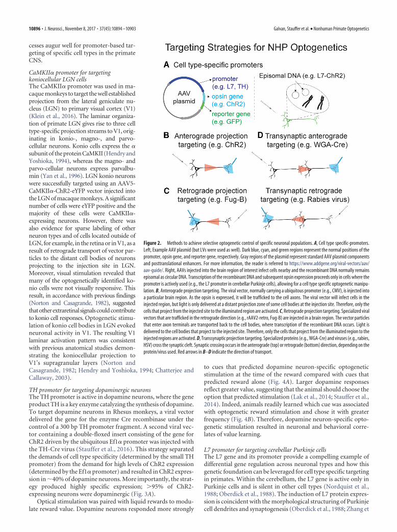
Targeting Specific Neuronal PopulationsA significant roadblock to effective NHP optogenetics is the lackof universal tools such as genetically modified Cre-driver lines(Gong et al., 2007) for selective targeting of neuronal populationsin NHPs. Efforts to circumvent this roadblock and achieve selec-tive neuronal manipulations in monkeys can be broadly classifiedinto two approaches: (1) those that use cell type specific genepromoters (Fig. 2A) and (2) those that use projection targeting.(Fig. 2B–D). Both approaches are based on the use of viral vectorsto deliver opsin genes to neurons. In particular, adeno-associatedvirus (AAV) and lentivirus (LV) are commonly used because theyare relatively safe and because they can infect nondividing cellssuch as neurons (Lentz et al., 2012; Kotterman et al., 2015).
The principal drawbacks to AAV and LV are their limitedgenetic capacities, �5 and �9 kb, respectively (Lentz et al., 2012).These limits require concise genetic sequences to control opsinexpression. One approach is to isolate or synthesize small pro-moters. Several general purpose promoters are commonly used,including CMV and Ef1�, but viruses containing these promot-ers transduce a variety of cell types including neurons and glia(Yizhar et al., 2011).
Targeting neuronal populations using cell type specificpromotersEarly breakthroughs have achieved cell type selectivity using viralvectors that carry small promoter sequences. The CaMKII� pro-moter has been often used in NHP experiments to target excit-atory neurons (Han et al., 2009; Dai et al., 2014; Lu et al., 2015;Nassi et al., 2015a). Targeting NHP inhibitory interneurons is nowwithin reach due to the development of mDlx enhancer elements(Dimidschstein et al., 2016). The tyrosine hydroxylase (TH) pro-moter has been used to label NHP dopamine neurons with GFP(Lerchner et al., 2014). Likewise, a promoter was developed to targetD2-expressing medium spiny neurons in wild-type rodents (Zalo-cusky et al., 2016). These studies and others provide evidence thatpromoters can be used to direct cell type specific gene expression.
Three recent NHP studies have used cell type specific promot-ers to enable optogenetic investigation of well defined neuronaltypes and their role in brain function and behavior. These suc-
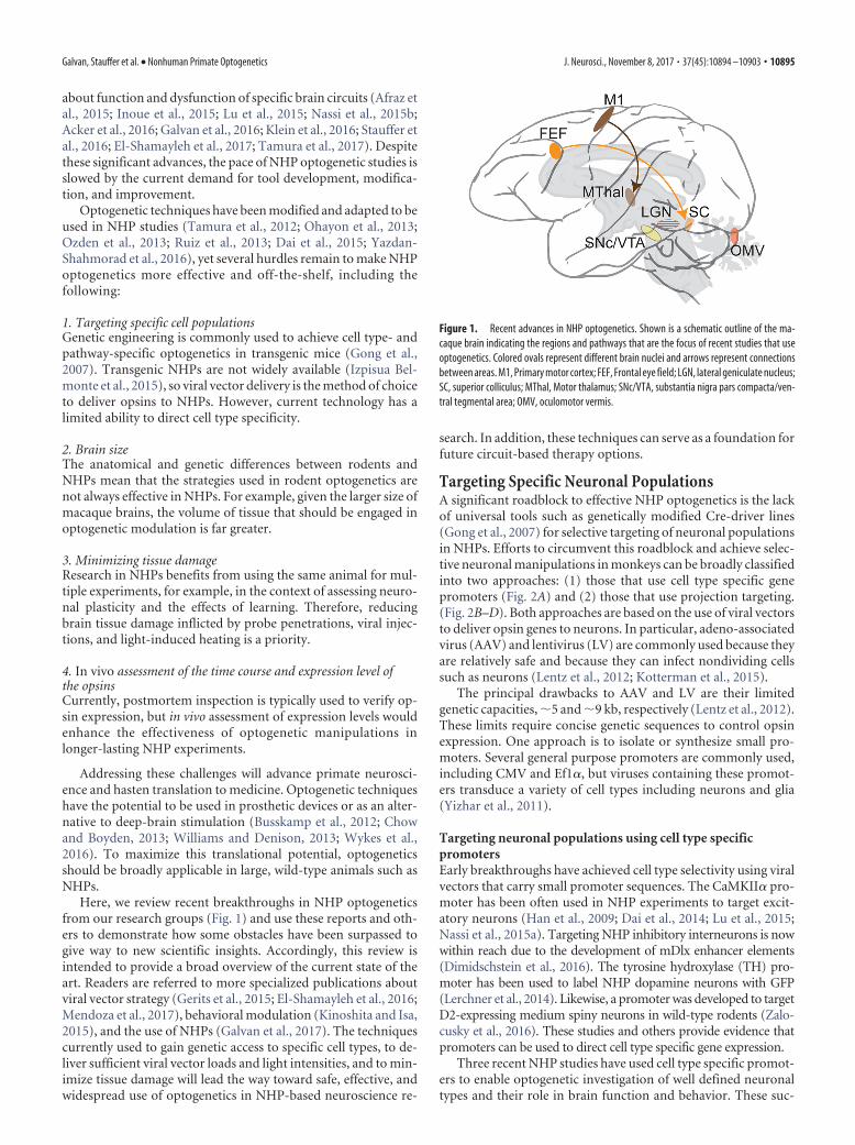
Figure 1. Recent advances in NHP optogenetics. Shown is a schematic outline of the ma-caque brain indicating the regions and pathways that are the focus of recent studies that useoptogenetics. Colored ovals represent different brain nuclei and arrows represent connectionsbetween areas. M1, Primary motor cortex; FEF, Frontal eye field; LGN, lateral geniculate nucleus;SC, superior colliculus; MThal, Motor thalamus; SNc/VTA, substantia nigra pars compacta/ven-tral tegmental area; OMV, oculomotor vermis.
Galvan, Stauffer et al. • Nonhuman Primate Optogenetics J. Neurosci., November 8, 2017 • 37(45):10894 –10903 • 10895
cesses augur well for promoter-based tar-geting of specific cell types in the primateCNS.
CaMKII� promoter for targetingkoniocellular LGN cellsThe CaMKII� promoter was used in ma-caque monkeys to target the well establishedprojection from the lateral geniculate nu-cleus (LGN) to primary visual cortex (V1)(Klein et al., 2016). The laminar organiza-tion of primate LGN gives rise to three celltype-specific projection streams to V1, orig-inating in konio-, magno-, and parvo-cellular neurons. Konio cells express the �subunit of the protein CaMKII (Hendry andYoshioka, 1994), whereas the magno- andparvo-cellular neurons express parvalbu-min (Yan et al., 1996). LGN konio neuronswere successfully targeted using an AAV5-CaMKII�-ChR2-eYFP vector injected intothe LGN of macaque monkeys. A significantnumber of cells were eYFP positive and themajority of these cells were CaMKII�-expressing neurons. However, there wasalso evidence for sparse labeling of otherneuron types and of cells located outside ofLGN, for example, in the retina or in V1, as aresult of retrograde transport of vector par-ticles to the distant cell bodies of neuronsprojecting to the injection site in LGN.Moreover, visual stimulation revealed thatmany of the optogenetically identified ko-nio cells were not visually responsive. Thisresult, in accordance with previous findings(Norton and Casagrande, 1982), suggestedthatotherextraretinal signalscouldcontributeto konio cell responses. Optogenetic stimu-lation of konio cell bodies in LGN evokedneuronal activity in V1. The resulting V1laminar activation pattern was consistentwith previous anatomical studies demon-strating the koniocellular projection toV1’s supragranular layers (Norton andCasagrande, 1982; Hendry and Yoshioka, 1994; Chatterjee andCallaway, 2003).
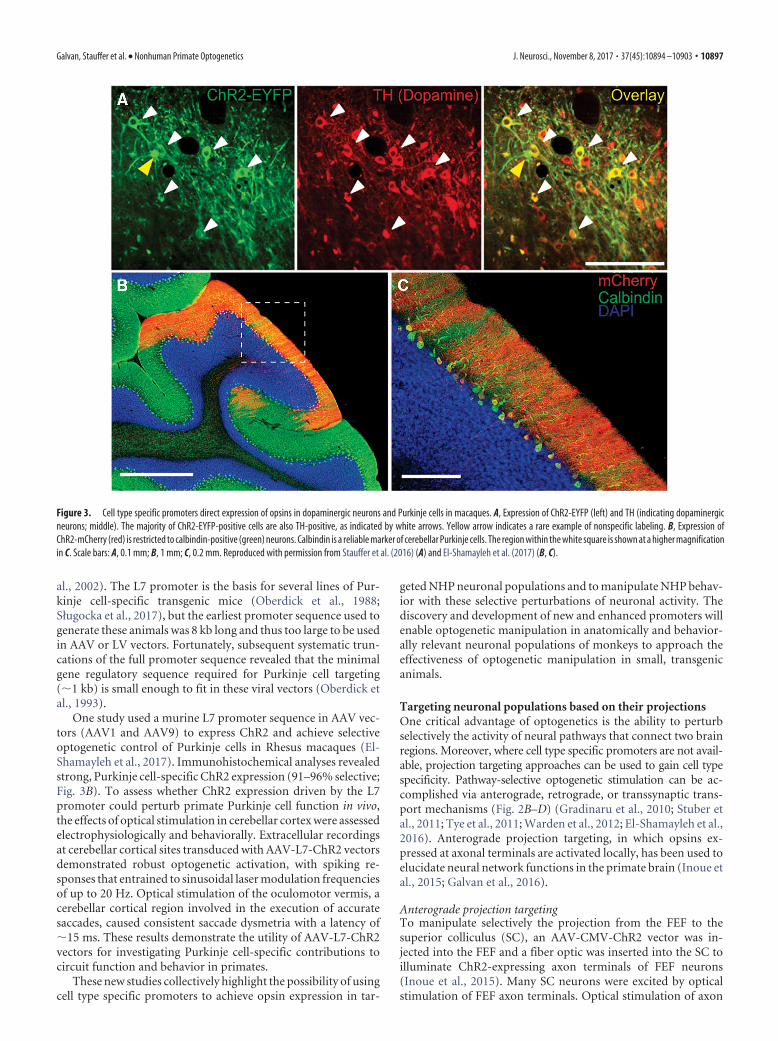
TH promoter for targeting dopaminergic neuronsThe TH promoter is active in dopamine neurons, where the geneproduct TH is a key enzyme catalyzing the synthesis of dopamine.To target dopamine neurons in Rhesus monkeys, a viral vectordelivered the gene for the enzyme Cre recombinase under thecontrol of a 300 bp TH promoter fragment. A second viral vec-tor containing a double-floxed insert consisting of the gene forChR2 driven by the ubiquitous Ef1� promoter was injected withthe TH-Cre virus (Stauffer et al., 2016). This strategy separatedthe demands of cell type specificity (determined by the small THpromoter) from the demand for high levels of ChR2 expression(determined by the Ef1� promoter) and resulted in ChR2 expres-sion in �40% of dopamine neurons. More importantly, the strat-egy produced highly specific expression; �95% of ChR2-expressing neurons were dopaminergic (Fig. 3A).
Optical stimulation was paired with liquid rewards to modu-late reward value. Dopamine neurons responded more strongly
to cues that predicted dopamine neuron-specific optogeneticstimulation at the time of the reward compared with cues thatpredicted reward alone (Fig. 4A). Larger dopamine responsesreflect greater value, suggesting that the animal should choose theoption that predicted stimulation (Lak et al., 2014; Stauffer et al.,2014). Indeed, animals readily learned which cue was associatedwith optogenetic reward stimulation and chose it with greaterfrequency (Fig. 4B). Therefore, dopamine neuron-specific opto-genetic stimulation resulted in neuronal and behavioral corre-lates of value learning.
L7 promoter for targeting cerebellar Purkinje cellsThe L7 gene and its promoter provide a compelling example ofdifferential gene regulation across neuronal types and how thisgenetic foundation can be leveraged for cell type specific targetingin primates. Within the cerebellum, the L7 gene is active only inPurkinje cells and is silent in other cell types (Nordquist et al.,1988; Oberdick et al., 1988). The induction of L7 protein expres-sion is coincident with the morphological structuring of Purkinjecell dendrites and synaptogenesis (Oberdick et al., 1988; Zhang et
Figure 2. Methods to achieve selective optogenetic control of specific neuronal populations. A, Cell type specific-promoters.Left, Example AAV plasmid (but LVs were used as well). Dark blue, cyan, and green regions represent the normal positions of thepromoter, opsin gene, and reporter gene, respectively. Gray regions of the plasmid represent standard AAV plasmid componentsand posttranslational enhancers. For more information, the reader is referred to https://www.addgene.org/viral-vectors/aav/aav-guide/. Right, AAVs injected into the brain region of interest infect cells nearby and the recombinant DNA normally remainsepisomal as circular DNA. Transcription of the recombinant DNA and subsequent opsin expression proceeds only in cells where thepromoter is actively used (e.g., the L7 promoter in cerebellar Purkinje cells), allowing for a cell type specific optogenetic manipu-lation. B, Anterograde projection targeting. The viral vector, normally carrying a ubiquitous promoter (e.g., CMV), is injected intoa particular brain region. As the opsin is expressed, it will be trafficked to the cell axons. The viral vector will infect cells in theinjected region, but light is only delivered at a distant projection zone of some cell bodies at the injection site. Therefore, only thecells that project from the injected site to the illuminated region are activated. C, Retrograde projection targeting. Specialized viralvectors that are trafficked in the retrograde direction (e.g., rAAV2-retro, Fug-B) are injected in a brain region. The vector particlesthat enter axon terminals are transported back to the cell bodies, where transcription of the recombinant DNA occurs. Light isdelivered to the cell bodies that project to the injected site. Therefore, only the cells that project from the illuminated region to theinjected regions are activated. D, Transsynaptic projection targeting. Specialized proteins (e.g., WGA-Cre) and viruses (e.g., rabies,HSV) cross the synaptic cleft. Synaptic crossing occurs in the anterograde (top) or retrograde (bottom) direction, depending on theprotein/virus used. Red arrows in B–D indicate the direction of transport.
10896 • J. Neurosci., November 8, 2017 • 37(45):10894 –10903 Galvan, Stauffer et al. • Nonhuman Primate Optogenetics
al., 2002). The L7 promoter is the basis for several lines of Pur-kinje cell-specific transgenic mice (Oberdick et al., 1988;Sługocka et al., 2017), but the earliest promoter sequence used togenerate these animals was 8 kb long and thus too large to be usedin AAV or LV vectors. Fortunately, subsequent systematic trun-cations of the full promoter sequence revealed that the minimalgene regulatory sequence required for Purkinje cell targeting(�1 kb) is small enough to fit in these viral vectors (Oberdick etal., 1993).
One study used a murine L7 promoter sequence in AAV vec-tors (AAV1 and AAV9) to express ChR2 and achieve selectiveoptogenetic control of Purkinje cells in Rhesus macaques (El-Shamayleh et al., 2017). Immunohistochemical analyses revealedstrong, Purkinje cell-specific ChR2 expression (91–96% selective;Fig. 3B). To assess whether ChR2 expression driven by the L7promoter could perturb primate Purkinje cell function in vivo,the effects of optical stimulation in cerebellar cortex were assessedelectrophysiologically and behaviorally. Extracellular recordingsat cerebellar cortical sites transduced with AAV-L7-ChR2 vectorsdemonstrated robust optogenetic activation, with spiking re-sponses that entrained to sinusoidal laser modulation frequenciesof up to 20 Hz. Optical stimulation of the oculomotor vermis, acerebellar cortical region involved in the execution of accuratesaccades, caused consistent saccade dysmetria with a latency of�15 ms. These results demonstrate the utility of AAV-L7-ChR2vectors for investigating Purkinje cell-specific contributions tocircuit function and behavior in primates.
These new studies collectively highlight the possibility of usingcell type specific promoters to achieve opsin expression in tar-
geted NHP neuronal populations and to manipulate NHP behav-ior with these selective perturbations of neuronal activity. Thediscovery and development of new and enhanced promoters willenable optogenetic manipulation in anatomically and behavior-ally relevant neuronal populations of monkeys to approach theeffectiveness of optogenetic manipulation in small, transgenicanimals.
Targeting neuronal populations based on their projectionsOne critical advantage of optogenetics is the ability to perturbselectively the activity of neural pathways that connect two brainregions. Moreover, where cell type specific promoters are not avail-able, projection targeting approaches can be used to gain cell typespecificity. Pathway-selective optogenetic stimulation can be ac-complished via anterograde, retrograde, or transsynaptic trans-port mechanisms (Fig. 2B–D) (Gradinaru et al., 2010; Stuber etal., 2011; Tye et al., 2011; Warden et al., 2012; El-Shamayleh et al.,2016). Anterograde projection targeting, in which opsins ex-pressed at axonal terminals are activated locally, has been used toelucidate neural network functions in the primate brain (Inoue etal., 2015; Galvan et al., 2016).
Anterograde projection targetingTo manipulate selectively the projection from the FEF to thesuperior colliculus (SC), an AAV-CMV-ChR2 vector was in-jected into the FEF and a fiber optic was inserted into the SC toilluminate ChR2-expressing axon terminals of FEF neurons(Inoue et al., 2015). Many SC neurons were excited by opticalstimulation of FEF axon terminals. Optical stimulation of axon
Figure 3. Cell type specific promoters direct expression of opsins in dopaminergic neurons and Purkinje cells in macaques. A, Expression of ChR2-EYFP (left) and TH (indicating dopaminergicneurons; middle). The majority of ChR2-EYFP-positive cells are also TH-positive, as indicated by white arrows. Yellow arrow indicates a rare example of nonspecific labeling. B, Expression ofChR2-mCherry (red) is restricted to calbindin-positive (green) neurons. Calbindin is a reliable marker of cerebellar Purkinje cells. The region within the white square is shown at a higher magnificationin C. Scale bars: A, 0.1 mm; B, 1 mm; C, 0.2 mm. Reproduced with permission from Stauffer et al. (2016) (A) and El-Shamayleh et al. (2017) (B, C).
Galvan, Stauffer et al. • Nonhuman Primate Optogenetics J. Neurosci., November 8, 2017 • 37(45):10894 –10903 • 10897

terminals often evoked saccadic eyemovements toward response fields corre-sponding to the stimulation sites in the SC(Fig. 4C,D; Inoue et al., 2015). This resultwas in contrast to activation of cell bodiesin the FEF, where optical stimulation de-creased reaction time but rarely evokedsaccades unless paired with electricalstimulation (Gerits et al., 2012; Ohayon etal., 2013). This suggests that the cell selec-tivity achieved via projection targetingcan result in strong behavioral effects.
Pathway-selective stimulation afteranterograde transport of opsins has alsobeen used to study corticothalamic motorcircuits in NHPs. AAV-CaMKII�-ChR2or AAV-CaMKII�-C1V1 was injectedinto the motor cortices. Optogenetic ac-tivation of corticothalamic terminalsmodulated activity in ventral motorthalamus neurons (Galvan et al., 2016).In contrast to the shorter latency excita-tions described in the oculomotor path-way (Inoue et al., 2015), selectiveoptogenetic activation of corticotha-lamic terminals resulted in long-latencyand complex physiological responses ofmotor thalamic neurons, suggesting amodulatory role for cortical afferents inthe primate motor thalamus.
Retrograde projection targetingRetrograde transport capabilities can beused to achieve cell type and pathway-selective optogenetic control (Kato et al.,2011; Oyibo et al., 2014; Tervo et al., 2016;Tanabe et al., 2017). In these cases, viralparticles enter axon terminals at the injec-tion site and are then transported alongthe axon back to the cell body, where thetransgene will be transcribed. Efforts are currently ongoing to testthe efficacy of several varieties of retrograde viruses in NHPs,including LVs with modified glycoproteins such as Fug-B, Fug-B2, Fug-E, and Tloop (Kato et al., 2011; Hirano et al., 2013; Cetinand Callaway, 2014; Kobayashi et al., 2016; Tanabe et al., 2017),herpes simplex virus (Neve et al., 2005; Fenno et al., 2014), canineadeno virus type 2 (Soudais et al., 2001; Salinas et al., 2009; Juny-ent and Kremer, 2015), AAV serotypes with endogenous retro-grade properties (Rothermel et al., 2013), and designer AAVswith engineered retrograde capabilities (Tervo et al., 2016). Onestudy showed that an optimized chimeric envelope glycoprotein(FuG-E) greatly accentuates the efficacy of retrograde gene deliv-ery of a pseudotyped LV vector in the primate brain. Striatalinjection of the FuG-E-GFP vector-labeled neurons in regionsthat project to the striatum, including cerebral cortex, thalamus,and substantia nigra (Tanabe et al., 2017).
Transsynaptic targetingTranssynaptic transport of viral particles or gene products holdsthe promise to create widespread yet circuit-specific labeling toinvestigate large-scale brain networks. In this case, the viral par-ticles or gene products will travel anterogradely to the axon ter-minals or retrogradely to the soma, where they will cross thesynapse to be incorporated into connected neurons (Gradinaru
et al., 2010; Nassi et al., 2015a). An important consideration isthat the retrograde spread may extend beyond one synapse. Thepotential benefit is that using a transsynaptic vector will provideoptogenetic control over multisynaptic circuits, but the peril isthat some commonly used transsynaptic viruses are cytotoxic(Nassi et al., 2015a). In rodents, some AAV serotypes (AAV1 andAAV9) show anterograde transsynaptic transduction properties(Zingg et al., 2017) that can be exploited along with Cre-dependent expression to identify and modulate specific neuronalpathways. Further engineering of transsynaptic vectors to reducetoxicity and enhance transgene expression (Nassi and Callaway,2007; Oyibo et al., 2014) will broaden their use in NHP optoge-netic experiments. Using novel viral vectors to achieve selectiveopsin expression based on synaptic connectivity would be partic-ularly advantageous for studying large-scale brain networks inNHPs.
Brain SizeRhesus macaque brains are 200 times larger than mouse brainsand contain two orders of magnitude more neurons (Herculano-Houzel, 2009). Therefore, a larger number of neurons should beengaged for optogenetic modulation of NHP behavior comparedwith rodent experiments. To achieve this, NHP optogenetic stud-
Figure 4. Neuronal and behavioral correlates of optical stimulation applied to specific neuronal populations. A, B, Neuronal andbehavioral correlates of stimulating dopamine neurons. A, Inset, Blue visual cue predicted liquid reward along with laser stimula-tion, whereas the red visual cue predicted the delivery of reward alone. Peristimulus time histogram (PSTH; top) and raster plot(bottom) demonstrate that dopamine neurons responded more strongly to the cue that predicted reward with laser stimulation(blue) compared with cues that predicted reward alone (red). B, Probability of choosing the option associated with reward andoptical stimulation. Animals chose between a cue that predicted reward with optical stimulation and a cue that predicted rewardalone. Blue data (� and line) from one session with optical fiber in the injected hemisphere. Red data (� and line) from onesession with optical fiber placed in the noninjected, control hemisphere. � indicates choices for the option associated with opticalstimulation (top) or option associated with reward alone (bottom). Lines represent moving averages (sliding window with 10steps) of the two choice sets. C, D, Neuronal and behavioral correlates of FEF to SC pathway stimulation. C, PSTH of SC neuronalresponses to FEF axon terminal stimulation separated according to whether a saccade was evoked (filled, red histogram) or notevoked (black line). SC neurons responded more strongly after stimulation events that evoked a saccade compared with stimula-tions that did not evoke a saccade. D, Polar plot of the magnitude (r) and direction (�) of optogenetically evoked saccades. Red linesindicate the averaged vector of evoked saccades at each stimulation site (n � 15). Saccade toward center of response field isrepresented by r � 1.0, � � 0. Reproduced with permission from Stauffer et al. (2016) (A, B) and Inoue et al. (2015) (C, D).
10898 • J. Neurosci., November 8, 2017 • 37(45):10894 –10903 Galvan, Stauffer et al. • Nonhuman Primate Optogenetics

ies require opsin expression across large brain areas, opsins withenhanced light sensitivity, and broad light delivery. High levels ofopsin expression across large brain areas can be achieved by usingenhanced viral vector delivery techniques and selecting viral vec-tor types that diffuse easily through brain tissue. MRI guidanceand convection enhanced infusion have been applied successfullyto deliver large volumes of viral vectors to precise locations in theNHP brain (Bankiewicz et al., 2000; Yazdan-Shahmorad et al.,2016). Similarly, LV and AAV viral vector subtypes can be se-lected to maximize the spread of the viral solution and transduc-tion of neurons (Gerits et al., 2015; El-Shamayleh et al., 2016).
Light is absorbed as it propagates in the brain. Oxygenatedhemoglobin is the major source of visible light absorption inliving brain tissue and red light is the least absorbed visible wave-length spectrum (Eggert and Blazek, 1987; Robles et al., 2010). Invivo measurements from rodent cortex at a depth of 1.5 mmshowed that five times as much red light remained unabsorbedcompared with green or blue light (Acker et al., 2016). Therefore,next generation opsins that are activated preferentially by wave-lengths closer to the red spectrum, including VChR1 (Zhang etal., 2008), C1V1 (Yizhar et al., 2011), Jaws (Chuong et al., 2014),Chrimson (Klapoetke et al., 2014), ReaChR (Lin et al., 2013), andBReaChES (Rajasethupathy et al., 2015), show promise for large-volume tissue modulation in primates. C1V1 stimulation of prin-cipal neurons in somatosensory cortex in macaques is sufficientto elicit a sensation (May et al., 2014), a finding that could beexploited to mimic sensory stimulation when sensory functionshave been compromised (e.g., in the development of neuropros-theses for stroke or spinal cord injury). Research with C1V1opsins, along with multielectrode arrays, revealed spatiotemporaldynamics in the monkey motor cortical networks (Lu et al., 2015).Likewise, C1V1 opsins have been used to test the computationalproperties of V1 neurons (Nassi et al., 2015b) and to demonstratethat optogenetic activation of the lateral intraparietal area can biaseye movements during a visuospatial discrimination task (Dai et al.,2014).
Light must be delivered more broadly in primates than inrodents to affect functionally relevant neuronal populations. In-deed, some of the early failures to observe behavioral effects ofoptogenetic stimulation in FEF (Han et al., 2009) have been over-come by improving light delivery (Gerits et al., 2012; Acker et al.,2016). Strategies to increase the extent of brain tissue illuminatedinclude the use of probes that combine multiple fibers (Tamura etal., 2012) or optic fibers with a tapered end (Dai et al., 2015; Ackeret al., 2016, 2017).
In one study, an improved optrode combined with the red-shifted inactivating opsin Jaws inhibited �90% of macaque FEFneurons over a 10 mm 3 tissue volume (Acker et al., 2016). Thisresult is comparable to the volumes inactivated in corticalcooling (Chafee and Goldman-Rakic, 2000) and pharmaco-logical inactivation studies (Sommer and Tehovnik, 1997;Tehovnik and Sommer, 1997; Martin and Ghez, 1999). Theoptogenetic inactivation was effective at low light intensitiesand resulted in large behavioral changes (Acker et al., 2016).Therefore, the use of enhanced light delivery and red-shiftedopsins to perturb large populations of neurons renders opto-genetic manipulations more effective at modulating NHP be-havior.
Minimizing Tissue DamageIn optogenetic experiments, tissue can be damaged by light-induced heating and repeated probe insertion. Limiting excessivetissue heating is essential because cortical temperature increases
of �4°C may induce damage. Moreover, temperature increasescould result in unwanted side effects such as neuronal firing in-creases in some heat-sensitive brain areas in response to �2°Ctemperature increases (Kiyatkin and Brown, 2004; Kiyatkin,2004; Kiyatkin, 2005, 2007). Illumination-induced heteroge-neous neuronal firing patterns can be effectively eliminated bymonitoring tissue heating and limiting light power to avoid a�1°C temperature increase (Acker et al., 2016).
Although experiments in superficial cortical regions can beperformed through optical windows using noninvasive lightsources (Ruiz et al., 2013; Yazdan-Shahmorad et al., 2016), stud-ies of deep cortical layers or subcortical structures require re-peated probe insertion, with consequent tissue damage. There is atradeoff between fiber outer diameter and tissue damage. Thickerfibers deliver more light but increase tissue damage induced byprobe insertion, especially in acute experiments. Furthermore,when combined recordings and stimulation are desired, the tra-ditional approach of using optrodes fabricated by gluing a fiber toan electrode might induce greater damage because these probesare prone to shearing and cutting brain tissue.
There has been steady progress in developing multishank andcoaxial probes (Abaya et al., 2012; Wang et al., 2012; Chen et al.,2013; Ozden et al., 2013; Lee et al., 2015; Naughton et al., 2016)that minimizes these problems. The use of tapered-end fibersresults in reduced brain damage compared with optical fiberswith a blunt end (Dai et al., 2015; Acker et al., 2016, 2017;Pisanello et al., 2017; Tamura et al., 2017). An alternative to therepeated optrode penetrations is the use of chronically implantedmultielectrode arrays that incorporate an optic fiber for opsinactivation (Lu et al., 2015), or micro-electrocorticographic lam-inar arrays for cortical surface recordings (Yazdan-Shahmorad etal., 2016).
Looking toward translational approaches may further inspireengineering innovations in primate optogenetics. For example,when laser thermal ablation is used to treat otherwise inoperablebrain tumors and epileptogenic foci in human patients, repeatedpenetrations are avoided, if possible, to minimize penetrationdamage (Missios et al., 2015). Further, in proportion to totalbrain size, the diameter of optical fibers currently in use for hu-man thermotherapy, �600 �m (Norred and Johnson, 2014), isless than that used in NHPs (about 200 – 400 �m for optrodes).During human surgeries, the temperature of surrounding tissueduring laser ablation is monitored to minimize collateral tissuedamage (Missios et al., 2015). Based on current laser use inhuman neurosurgery, reasonable future directions for NHP op-togenetics many include temperature monitoring, chronic orsemichronic light probes to limit repeated penetrations, and re-ductions in illuminator/electrode diameter.
In Vivo Assessment of the Temporal Course andExpression Level of the OpsinsCurrently, most researchers depend on postmortem histologicalexamination to confirm the correct targeting of brain regions orcell types and level of opsin expression. Ideally, a noninvasive invivo method would monitor opsin expression after virus injec-tions and before starting functional experiments. If the opsins areexpressed in neurons inhabiting superficial cortical layers, imag-ing of the fluorescent reporter can be used to gauge opsin expres-sion through optical windows (Ruiz et al., 2013). For subcorticalstructures, fluorescence can be detected in vivo using an opticfiber positioned in the injected area (Diester et al., 2011; Tamuraet al., 2012; Ozden et al., 2013; Tamura et al., 2017). This methodis encumbered by the damage inflected on the tissue by probe
Galvan, Stauffer et al. • Nonhuman Primate Optogenetics J. Neurosci., November 8, 2017 • 37(45):10894 –10903 • 10899

insertion, but may be preferable to postmortem verification. Asan example, in one study of the role of the perirhinal cortex inbehavioral judgements of object semantic value, researchers useda multifiber optogenetic probe that provided improved light de-livery, minimized tissue damage, and monitored opsin expres-sion (Tamura et al., 2017).
A radically different approach to minimizing tissue damageand assessing transgene expression is to pursue an all-optical in-terrogation (AOI) strategy (Rickgauer et al., 2014; Emiliani et al.,2015). AOI involves coexpression of an activity reporter gene(e.g., GCaMP) and an actuator (e.g., red-shifted opsin) and theuse of a single optical probe to image and perturb activity. AOIhas several advantages over traditional electrophysiology. First, amuch larger set of neurons can be monitored simultaneouslycompared with single electrode or even multishank probes. Sec-ond, it is possible to identify activity arising from specific neuronsreliably. Third, structured illumination patterns can be applied tostudy local connectivity and dynamics. AOI experiments in ro-dents have used gradient refractive index (GRIN) lenses (Ghoshet al., 2011; Ziv et al., 2013), but at the cost of significant tissuedamage above the region of interest (i.e., brain aspiration; Bar-retto and Schnitzer, 2012), which may not be acceptable in stud-ies involving NHPs. Furthermore, commonly available GRINlenses are 2–5 mm in length, not long enough to reach deepsubcortical regions in NHPs. A recent study tested a new mi-croendoscope design consisting of ultrathin multimode opticalfibers that are 5–10 times thinner than commercially availableGRIN lenses and have no length constraint (Ohayon et al., 2017).These fibers are of the same type as those traditionally used inNHP optogenetic experiments (Diester et al., 2011) and fiberphotometry (Gunaydin et al., 2014; Kim et al., 2016). However, incontrast to fiber photometry, this new design allows for full imagereconstruction beneath the fiber down to a micron-scale resolu-tion. Furthermore, it allows the delivery of light patterns (struc-tured light) at the fiber tip for precise stimulation of only a subsetof neurons in the field of view. Ongoing experiments are underway to test this technology in NHPs and make it accessible for endusers.
ConclusionHuman perception, action, and cognition are largely dependenton specialized brain networks that arose during the evolution ofprimates. This specialization renders NHPs, especially Old Worldmonkeys such as Rhesus macaques, an invaluable animal modelwith which to investigate human brain function. Modern neurosci-ence has been reshaped by game-changing technologies includingoptogenetics and genetically coded calcium indicators that aremost effectively applied to transgenic animals. Although trans-genic Rhesus macaques have been created successfully (Yang etal., 2008; Liu et al., 2016), economic considerations and the longgeneration time may limit the utility of transgenic Old-Worldmonkeys in neuroscientific research. Moreover, the successfulimplementation of emerging (optogenetic) technologies in wild-type macaques will provide a rich foundation for the translationalapplication of circuit-based therapy to humans. Therefore, devel-oping methods to facilitate the application of genetic techniquesfor controlling and monitoring neural activity in NHPs is one themost critical challenges in modern neuroscience.
Recent studies have achieved effective application of optoge-netics to NHPs by demonstrating cell type specific expressionusing gene promoters, selective projection targeting, improvedlight delivery, and opsins with improved light sensitivity. As re-viewed here, cell type specific promoters have been used to study
the function of konio cells in the LGN, dopamine neurons in themidbrain, and Purkinje cells in the cerebellum (Fig. 3). The use ofcell type specific promoters brings NHP neuroscience one stepcloser to the capabilities of transgenic mouse studies. Movingforward, the development or discovery of new regulatory se-quences that confer selective expression in a wide array of neuro-nal types will be one of the greatest challenges that we face. Mostgenes are expressed in many cell types, rather than being cell typespecific. Moreover, it is not clear how conserved noncoding tran-scriptional regulatory regions are, so it is not clear which mousepromoters will be effective in monkeys (Suzuki et al., 2004). Toadvance the field, studies in which promoters can be screened orengineered in a high-throughput fashion may hold the key tounlocking a wider array of cell type specific promoters (Portales-Casamar et al., 2010; Schlabach et al., 2010; Rajkumar andMaerkl, 2012; Smith et al., 2013).
Projection targeting, which does not rely on cell type specificpromoters, but rather relies on anterograde trafficking of opsinsor retrograde transport of viral particles (Fig. 2), has already beenused to reveal the functional roles of motor control networks(Inoue et al., 2015; Galvan et al., 2016). New viral vectors withselective and improved retrograde transport properties presentan opportunity to photo-identify cells that project to a region ofinterest. This technology will make it feasible to record selectivelyfrom afferent neurons and discover how neural circuits processinformation. Moreover, a likely contributor to the absence ofobserved behavioral correlates in early optogenetic experimentsis coactivation of competing neuronal populations that canceleach other’s effect. It may be possible, therefore, to increase thesize of observed behavioral effects by increasing cell type selectiv-ity. Indeed, targeting specific neuronal populations, whether viagene promoter or pathway tracing, has resulted in sizable behav-ioral effects after optical stimulation (Fig. 4) (Inoue et al., 2015;Stauffer et al., 2016; El-Shamayleh et al., 2017). These advancesbode well for the future of investigating neural signals and theirrelationship to well controlled behaviors in NHPs.
Traditionally, similar optical probes were used to deliver lightin mice and monkeys despite the fact that the monkey brain is twoorders of magnitude larger than that of the mouse. By combininglight delivery probes that have tapered endings with red-shiftedopsins, large volumes of brain tissue (�10 mm 3) can be illumi-nated by a single probe (Acker et al., 2016, 2017). Moreover,because probes with tapered ends reduce the damage caused byprobe insertions, multiple probes can be inserted, maximizingthe chance to (in-)activate entire monkey brain regions.
In summary, we are substantially closer to optogenetics beingapplied in NHPs as effectively as it is applied in rodents. Furtherprogress in NHP optogenetic experiments and translation tohuman medicine will depend on the rapid dissemination of tech-nical advances and setbacks faced in the course of these experi-ments. Indeed, given the larger expenses and time needed forprimate experiments relative to other species, communication ofnegative or incremental findings in optogenetic (or similar) tech-niques may be particularly beneficial for the NHP research com-munity. The new developments reviewed here highlight the valueof primate optogenetics to reveal the mechanistic insights intothe brain circuits that support perception, action, and cognitionand how their dysfunction gives rise to human pathologies.
ReferencesAbaya TV, Blair S, Tathireddy P, Rieth L, Solzbacher F (2012) A 3D glass
optrode array for optical neural stimulation. Biomed Opt Express 3:3087–3104. CrossRef Medline
10900 • J. Neurosci., November 8, 2017 • 37(45):10894 –10903 Galvan, Stauffer et al. • Nonhuman Primate Optogenetics

Acker L, Pino EN, Boyden ES, Desimone R (2016) FEF inactivation withimproved optogenetic methods. Proc Natl Acad Sci U S A 113:E7297–E7306. CrossRef Medline
Acker LC, Pino EN, Boyden ES, Desimone R (2017) Large volume illumi-nation for optogenetics in non-human primates. J Vis Exp 128:e56330.CrossRef
Afraz A, Boyden ES, DiCarlo JJ (2015) Optogenetic and pharmacologicalsuppression of spatial clusters of face neurons reveal their causal role inface gender discrimination. Proc Natl Acad Sci U S A 112:6730 – 6735.CrossRef Medline
Bankiewicz KS, Eberling JL, Kohutnicka M, Jagust W, Pivirotto P, Bringas J,Cunningham J, Budinger TF, Harvey-White J (2000) Convection-enhanced delivery of AAV vector in parkinsonian monkeys; in vivo de-tection of gene expression and restoration of dopaminergic functionusing pro-drug approach. Exp Neurol 164:2–14. CrossRef Medline
Barretto RP, Schnitzer MJ (2012) In vivo microendoscopy of the hippocam-pus. Cold Spring Harb Protoc 2012:1092–1099. CrossRef Medline
Busskamp V, Picaud S, Sahel JA, Roska B (2012) Optogenetic therapy forretinitis pigmentosa. Gene Ther 19:169 –175. CrossRef Medline
Cavanaugh J, Monosov IE, McAlonan K, Berman R, Smith MK, Cao V, WangKH, Boyden ES, Wurtz RH (2012) Optogenetic inactivation modifiesmonkey visuomotor behavior. Neuron 76:901–907. CrossRef Medline
Cetin A, Callaway EM (2014) Optical control of retrogradely infected neu-rons using drug-regulated “TLoop” lentiviral vectors. J Neurophysiol 111:2150 –2159. CrossRef Medline
Chafee MV, Goldman-Rakic PS (2000) Inactivation of parietal and prefron-tal cortex reveals interdependence of neural activity during memory-guided saccades. J Neurophysiol 83:1550 –1566. Medline
Chatterjee S, Callaway EM (2003) Parallel colour-opponent pathways toprimary visual cortex. Nature 426:668 – 671. CrossRef Medline
Chen S, Pei W, Gui Q, Chen Y, Zhao S, Wang H, Chen H (2013) A fiber-based implantable multi-optrode array with contiguous optical and elec-trical sites. J Neural Eng 10:046020. CrossRef Medline
Chow BY, Boyden ES (2013) Optogenetics and translational medicine. SciTransl Med 5:177ps175. CrossRef Medline
Chuong AS, et al. (2014) Noninvasive optical inhibition with a red-shiftedmicrobial rhodopsin. Nat Neurosci 17:1123–1129. CrossRef Medline
Dai J, Brooks DI, Sheinberg DL (2014) Optogenetic and electrical micro-stimulation systematically bias visuospatial choice in primates. Curr Biol24:63– 69. CrossRef Medline
Dai J, Ozden I, Brooks DI, Wagner F, May T, Agha NS, Brush B, Borton D,Nurmikko AV, Sheinberg DL (2015) Modified toolbox for optogeneticsin the nonhuman primate. Neurophotonics 2:031202. CrossRef Medline
Deisseroth K (2015) Optogenetics: 10 years of microbial opsins in neurosci-ence. Nat Neurosci 18:1213–1225. CrossRef Medline
Diester I, Kaufman MT, Mogri M, Pashaie R, Goo W, Yizhar O, Ramakrish-nan C, Deisseroth K, Shenoy KV (2011) An optogenetic toolbox de-signed for primates. Nat Neurosci 14:387–397. CrossRef Medline
Dimidschstein J, et al. (2016) A viral strategy for targeting and manipulatinginterneurons across vertebrate species. Nat Neurosci 19:1743–1749. CrossRefMedline
Eggert HR, Blazek V (1987) Optical properties of human brain tissue, me-ninges, and brain tumors in the spectral range of 200 to 900 nm. Neuro-surgery 21:459 – 464. CrossRef Medline
El-Shamayleh Y, Ni AM, Horwitz GD (2016) Strategies for targeting pri-mate neural circuits with viral vectors. J Neurophysiol 116:122–134.CrossRef Medline
El-Shamayleh Y, Kojima Y, Soetedjo R, Horwitz GD (2017) Selective opto-genetic control of purkinje cells in monkey cerebellum. Neuron 95:51–62.e4. CrossRef Medline
Emiliani V, Cohen AE, Deisseroth K, Hausser M (2015) All-optical interro-gation of neural circuits. J Neurosci 35:13917–13926. CrossRef Medline
Fenno LE, Mattis J, Ramakrishnan C, Hyun M, Lee SY, He M, Tucciarone J,Selimbeyoglu A, Berndt A, Grosenick L, Zalocusky KA, Bernstein H,Swanson H, Perry C, Diester I, Boyce FM, Bass CE, Neve R, Huang ZJ,Deisseroth K (2014) Targeting cells with single vectors using multiple-feature Boolean logic. Nat Methods 11:763–772. CrossRef Medline
Galvan A, Hu X, Smith Y, Wichmann T (2016) Effects of optogenetic acti-vation of corticothalamic terminals in the motor thalamus of awake mon-keys. J Neurosci 36:3519 –3530. CrossRef Medline
Galvan A, Caiola MJ, Albaugh DL (2017) Advances in optogenetic and che-
mogenetic methods to study brain circuits in non-human primates.J Neural Transm (Vienna), in press.
Gerits A, Farivar R, Rosen BR, Wald LL, Boyden ES, Vanduffel W (2012)Optogenetically induced behavioral and functional network changes inprimates. Curr Biol 22:1722–1726. CrossRef Medline
Gerits A, Vancraeyenest P, Vreysen S, Laramee ME, Michiels A, Gijsbers R,Van den Haute C, Moons L, Debyser Z, Baekelandt V, Arckens L, Van-duffel W (2015) Serotype-dependent transduction efficiencies of recombi-nant adeno-associated viral vectors in monkey neocortex. Neurophotonics2:031209. CrossRef Medline
Ghosh KK, Burns LD, Cocker ED, Nimmerjahn A, Ziv Y, Gamal AE,Schnitzer MJ (2011) Miniaturized integration of a fluorescence micro-scope. Nat Methods 8:871– 878. CrossRef Medline
Gong S, Doughty M, Harbaugh CR, Cummins A, Hatten ME, Heintz N,Gerfen CR (2007) Targeting Cre recombinase to specific neuron popu-lations with bacterial artificial chromosome constructs. J Neurosci 27:9817–9823. CrossRef Medline
Gore F, Schwartz EC, Brangers BC, Aladi S, Stujenske JM, Likhtik E, RussoMJ, Gordon JA, Salzman CD, Axel R (2015) Neural representations ofunconditioned stimuli in basolateral amygdala mediate innate andlearned responses. Cell 162:134 –145. CrossRef Medline
Gradinaru V, Zhang F, Ramakrishnan C, Mattis J, Prakash R, Diester I, Gos-hen I, Thompson KR, Deisseroth K (2010) Molecular and cellular ap-proaches for diversifying and extending optogenetics. Cell 141:154 –165.CrossRef Medline
Gunaydin LA, Grosenick L, Finkelstein JC, Kauvar IV, Fenno LE, Adhikari A,Lammel S, Mirzabekov JJ, Airan RD, Zalocusky KA, Tye KM, Anikeeva P,Malenka RC, Deisseroth K (2014) Natural neural projection dynamicsunderlying social behavior. Cell 157:1535–1551. CrossRef Medline
Han X, Qian X, Bernstein JG, Zhou HH, Franzesi GT, Stern P, Bronson RT,Graybiel AM, Desimone R, Boyden ES (2009) Millisecond-timescaleoptical control of neural dynamics in the nonhuman primate brain. Neu-ron 62:191–198. CrossRef Medline
Hendry SH, Yoshioka T (1994) A neurochemically distinct third channelin the macaque dorsal lateral geniculate nucleus. Science 264:575–577.CrossRef Medline
Herculano-Houzel S (2009) The human brain in numbers: a linearlyscaled-up primate brain. Front Hum Neurosci 3:31. CrossRef Medline
Hirano M, Kato S, Kobayashi K, Okada T, Yaginuma H, Kobayashi K (2013)Highly efficient retrograde gene transfer into motor neurons by a lentivi-ral vector pseudotyped with fusion glycoprotein. PLoS One 8:e75896.CrossRef Medline
Inoue K, Takada M, Matsumoto M (2015) Neuronal and behavioural mod-ulations by pathway-selective optogenetic stimulation of the primate oc-ulomotor system. Nat Commun 6:8378. CrossRef Medline
Izpisua Belmonte JC, et al. (2015) Brains, genes, and primates. Neuron 86:617– 631. CrossRef Medline
Jazayeri M, Lindbloom-Brown Z, Horwitz GD (2012) Saccadic eye move-ments evoked by optogenetic activation of primate V1. Nat Neurosci15:1368 –1370. CrossRef Medline
Junyent F, Kremer EJ (2015) CAV-2: why a canine virus is a neurobiologist’sbest friend. Curr Opin Pharmacol 24:86 –93. CrossRef Medline
Kato S, Kuramochi M, Takasumi K, Kobayashi K, Inoue K, Takahara D,Hitoshi S, Ikenaka K, Shimada T, Takada M, Kobayashi K (2011)Neuron-specific gene transfer through retrograde transport of lentiviralvector pseudotyped with a novel type of fusion envelope glycoprotein.Hum Gene Ther 22:1511–1523. CrossRef Medline
Kim CK, Yang SJ, Pichamoorthy N, Young NP, Kauvar I, Jennings JH, LernerTN, Berndt A, Lee SY, Ramakrishnan C, Davidson TJ, Inoue M, Bito H,Deisseroth K (2016) Simultaneous fast measurement of circuit dynam-ics at multiple sites across the mammalian brain. Nat Methods 13:325–328. CrossRef Medline
Kinoshita M, Isa T (2015) Potential of optogenetics for the behavior manip-ulation of non-human primates. In: Optogenetics: light-sensing proteinsand their applications (Yawo H, Kandori H, Koizumi A, eds), pp 279 –290. Tokyo: Springer Japan.
Kiyatkin EA (2004) Brain hyperthermia during physiological and patholog-ical conditions: causes, mechanisms, and functional implications. CurrNeurovasc Res 1:77–90. CrossRef Medline
Kiyatkin EA (2005) Brain hyperthermia as physiological and pathologicalphenomena. Brain Res Brain Res Rev 50:27–56. CrossRef Medline
Galvan, Stauffer et al. • Nonhuman Primate Optogenetics J. Neurosci., November 8, 2017 • 37(45):10894 –10903 • 10901
Kiyatkin EA (2007) Physiological and pathological brain hyperthermia.Prog Brain Res 162:219 –243. CrossRef Medline
Kiyatkin EA, Brown PL (2004) Modulation of physiological brain hyper-thermia by environmental temperature and impaired blood outflow inrats. Physiol Behav 83:467– 474. CrossRef Medline
Klapoetke NC, et al. (2014) Independent optical excitation of distinct neuralpopulations. Nat Methods 11:338 –346. CrossRef Medline
Klein C, Evrard HC, Shapcott KA, Haverkamp S, Logothetis NK, Schmid MC(2016) Cell-targeted optogenetics and electrical microstimulation revealthe primate koniocellular projection to supra-granular visual cortex.Neuron 90:143–151. CrossRef Medline
Kobayashi K, Kato S, Inoue K, Takada M, Kobayashi K (2016) Altering entrysite preference of lentiviral vectors into neuronal cells by pseudotypingwith envelope glycoproteins. Methods Mol Biol 1382:175–186. CrossRefMedline
Kotterman MA, Chalberg TW, Schaffer DV (2015) Viral vectors for genetherapy: translational and clinical outlook. Annu Rev Biomed Eng 17:63–89. CrossRef Medline
Kvitsiani D, Ranade S, Hangya B, Taniguchi H, Huang JZ, Kepecs A (2013)Distinct behavioural and network correlates of two interneuron types inprefrontal cortex. Nature 498:363–366. CrossRef Medline
Lak A, Stauffer WR, Schultz W (2014) Dopamine prediction error responsesintegrate subjective value from different reward dimensions. Proc NatlAcad Sci U S A 111:2343–2348. CrossRef Medline
Lee J, Ozden I, Song YK, Nurmikko AV (2015) Transparent intracorticalmicroprobe array for simultaneous spatiotemporal optical stimulationand multichannel electrical recording. Nat Methods 12:1157–1162.CrossRef Medline
Lentz TB, Gray SJ, Samulski RJ (2012) Viral vectors for gene delivery to thecentral nervous system. Neurobiol Dis 48:179 –188. CrossRef Medline
Lerchner W, Corgiat B, Der Minassian V, Saunders RC, Richmond BJ (2014)Injection parameters and virus dependent choice of promoters to im-prove neuron targeting in the nonhuman primate brain. Gene Ther 21:233–241. CrossRef Medline
Lin JY, Knutsen PM, Muller A, Kleinfeld D, Tsien RY (2013) ReaChR: ared-shifted variant of channelrhodopsin enables deep transcranial opto-genetic excitation. Nat Neurosci 16:1499 –1508. CrossRef Medline
Liu Z, et al. (2016) Autism-like behaviours and germline transmission intransgenic monkeys overexpressing MeCP2. Nature 530:98–102. CrossRefMedline
Lu Y, Truccolo W, Wagner FB, Vargas-Irwin CE, Ozden I, Zimmermann JB,May T, Agha NS, Wang J, Nurmikko AV (2015) Optogenetically in-duced spatiotemporal gamma oscillations and neuronal spiking activityin primate motor cortex. J Neurophysiol 113:3574–3587. CrossRef Medline
Martin JH, Ghez C (1999) Pharmacological inactivation in the analysisof the central control of movement. J Neurosci Methods 86:145–159.CrossRef Medline
May T, Ozden I, Brush B, Borton D, Wagner F, Agha N, Sheinberg DL,Nurmikko AV (2014) Detection of optogenetic stimulation in somato-sensory cortex by non-human primates: towards artificial tactile sensa-tion. PLoS One 9:e114529. CrossRef Medline
Mendoza SD, El-Shamayleh Y, Horwitz GD (2017) AAV-mediated deliveryof optogenetic constructs to the macaque brain triggers humoral immuneresponses. J Neurophysiol 117:2004 –2013. CrossRef Medline
Missios S, Bekelis K, Barnett GH (2015) Renaissance of laser interstitialthermal ablation. Neurosurg Focus 38:E13. CrossRef Medline
Nassi JJ, Callaway EM (2007) Specialized circuits from primary visual cortexto V2 and area MT. Neuron 55:799 – 808. CrossRef Medline
Nassi JJ, Cepko CL, Born RT, Beier KT (2015a) Neuroanatomy goes viral!Front Neuroanat 9:80. CrossRef Medline
Nassi JJ, Avery MC, Cetin AH, Roe AW, Reynolds JH (2015b) Optogeneticactivation of normalization in alert macaque visual cortex. Neuron 86:1504 –1517. CrossRef Medline
Naughton JR, Connolly T, Varela JA, Lundberg J, Burns MJ, Chiles TC,Christianson JP, Naughton MJ (2016) Shielded coaxial optrode arraysfor neurophysiology. Front Neurosci 10:252. CrossRef Medline
Neve RL, Neve KA, Nestler EJ, Carlezon WA Jr (2005) Use of herpes virusamplicon vectors to study brain disorders. Biotechniques 39:381–391.CrossRef Medline
Nordquist DT, Kozak CA, Orr HT (1988) cDNA cloning and characteriza-tion of three genes uniquely expressed in cerebellum by Purkinje neurons.J Neurosci 8:4780 – 4789. Medline
Norred SE, Johnson JA (2014) Magnetic resonance-guided laser inducedthermal therapy for glioblastoma multiforme: a review. Biomed Res Int2014:761312. CrossRef Medline
Norton TT, Casagrande VA (1982) Laminar organization of receptive-fieldproperties in lateral geniculate nucleus of bush baby (Galago crassicauda-tus). J Neurophysiol 47:715–741. Medline
Oberdick J, Levinthal F, Levinthal C (1988) A Purkinje cell differentiationmarker shows a partial DNA sequence homology to the cellular sis/PDGF2 gene. Neuron 1:367–376. CrossRef Medline
Oberdick J, Schilling K, Smeyne RJ, Corbin JG, Bocchiaro C, Morgan JI(1993) Control of segment-like patterns of gene expression in the mousecerebellum. Neuron 10:1007–1018. CrossRef Medline
Ohayon S, Grimaldi P, Schweers N, Tsao DY (2013) Saccade modulation byoptical and electrical stimulation in the macaque frontal eye field. J Neu-rosci 33:16684 –16697. CrossRef Medline
Ohayon S, Caravaca Aguirre AM, Piestun R, DiCarlo JJ (2017) Deep brainfluorescence imaging with minimally invasive ultra-thin optical fibers.bioRxiv. CrossRef
Oyibo HK, Znamenskiy P, Oviedo HV, Enquist LW, Zador AM (2014)Long-term Cre-mediated retrograde tagging of neurons using a novelrecombinant pseudorabies virus. Front Neuroanat 8:86. CrossRef Medline
Ozden I, Wang J, Lu Y, May T, Lee J, Goo W, O’Shea DJ, Kalanithi P, DiesterI, Diagne M, Deisseroth K, Shenoy KV, Nurmikko AV (2013) A coaxialoptrode as multifunction write-read probe for optogenetic studies in non-human primates. J Neurosci Methods 219:142–154. CrossRef Medline
Phillips KA, Bales KL, Capitanio JP, Conley A, Czoty PW, �t Hart BA, HopkinsWD, Hu SL, Miller LA, Nader MA, Nathanielsz PW, Rogers J, Shively CA,Voytko ML (2014) Why primate models matter. Am J Primatol 76:801–827. CrossRef Medline
Pisanello F, Mandelbaum G, Pisanello M, Oldenburg IA, Sileo L, MarkowitzJE, Peterson RE, Della Patria A, Haynes TM, Emara MS, Spagnolo B,Datta SR, De Vittorio M, Sabatini BL (2017) Dynamic illumination ofspatially restricted or large brain volumes via a single tapered optical fiber.Nat Neurosci 20:1180 –1188. CrossRef Medline
Portales-Casamar E, et al. (2010) A regulatory toolbox of MiniPromoters todrive selective expression in the brain. Proc Natl Acad Sci U S A 107:16589 –16594. CrossRef Medline
Rajasethupathy P, Sankaran S, Marshel JH, Kim CK, Ferenczi E, Lee SY,Berndt A, Ramakrishnan C, Jaffe A, Lo M, Liston C, Deisseroth K (2015)Projections from neocortex mediate top-down control of memory re-trieval. Nature 526:653– 659. CrossRef Medline
Rajkumar AS, Maerkl SJ (2012) Rapid synthesis of defined eukaryotic pro-moter libraries. ACS Synth Biol 1:483– 490. CrossRef Medline
Rickgauer JP, Deisseroth K, Tank DW (2014) Simultaneous cellular-resolution optical perturbation and imaging of place cell firing fields. NatNeurosci 17:1816 –1824. CrossRef Medline
Robles FE, Chowdhury S, Wax A (2010) Assessing hemoglobin concentra-tion using spectroscopic optical coherence tomography for feasibility oftissue diagnostics. Biomed Opt Express 1:310 –317. CrossRef Medline
Roelfsema PR, Treue S (2014) Basic neuroscience research with nonhumanprimates: a small but indispensable component of biomedical research.Neuron 82:1200 –1204. CrossRef Medline
Rothermel M, Brunert D, Zabawa C, Díaz-Quesada M, Wachowiak M(2013) Transgene expression in target-defined neuron populations me-diated by retrograde infection with adeno-associated viral vectors. J Neu-rosci 33:15195–15206. CrossRef Medline
Ruiz O, Lustig BR, Nassi JJ, Cetin A, Reynolds JH, Albright TD, Callaway EM,Stoner GR, Roe AW (2013) Optogenetics through windows on the brainin the nonhuman primate. J Neurophysiol 110:1455–1467. CrossRefMedline
Salinas S, Bilsland LG, Henaff D, Weston AE, Keriel A, Schiavo G, Kremer EJ(2009) CAR-associated vesicular transport of an adenovirus in motorneuron axons. PLoS Pathog 5:e1000442. CrossRef Medline
Schlabach MR, Hu JK, Li M, Elledge SJ (2010) Synthetic design of strongpromoters. Proc Natl Acad Sci U S A 107:2538 –2543. CrossRef Medline
Sharpe MJ, Chang CY, Liu MA, Batchelor HM, Mueller LE, Jones JL, Niv Y,Schoenbaum G (2017) Dopamine transients are sufficient and necessaryfor acquisition of model-based associations. Nat Neurosci 20:735–742.CrossRef Medline
Sługocka A, Wiaderkiewicz J, Barski JJ (2017) Genetic targeting in cerebellarpurkinje cells: an update. Cerebellum 16:191–202. CrossRef Medline
Smith RP, Riesenfeld SJ, Holloway AK, Li Q, Murphy KK, Feliciano NM,
10902 • J. Neurosci., November 8, 2017 • 37(45):10894 –10903 Galvan, Stauffer et al. • Nonhuman Primate Optogenetics

Orecchia L, Oksenberg N, Pollard KS, Ahituv N (2013) A compact, invivo screen of all 6-mers reveals drivers of tissue-specific expression andguides synthetic regulatory element design. Genome Biol 14:R72. CrossRefMedline
Sommer MA, Tehovnik EJ (1997) Reversible inactivation of macaque fron-tal eye field. Exp Brain Res 116:229 –249. CrossRef Medline
Soudais C, Laplace-Builhe C, Kissa K, Kremer EJ (2001) Preferential trans-duction of neurons by canine adenovirus vectors and their efficient ret-rograde transport in vivo. FASEB J 15:2283–2285. Medline
Stauffer WR, Lak A, Schultz W (2014) Dopamine reward prediction errorresponses reflect marginal utility. Curr Biol 24:2491–2500. CrossRef Medline
Stauffer WR, Lak A, Yang A, Borel M, Paulsen O, Boyden ES, Schultz W(2016) Dopamine neuron-specific optogenetic stimulation in Rhesusmacaques. Cell 166:1564 –1571.e6. CrossRef Medline
Steinberg EE, Keiflin R, Boivin JR, Witten IB, Deisseroth K, Janak PH (2013)A causal link between prediction errors, dopamine neurons and learning.Nat Neurosci 16:966 –973. CrossRef Medline
Stuber GD, Sparta DR, Stamatakis AM, van Leeuwen WA, Hardjoprajitno JE,Cho S, Tye KM, Kempadoo KA, Zhang F, Deisseroth K, Bonci A (2011)Excitatory transmission from the amygdala to nucleus accumbens facili-tates reward seeking. Nature 475:377–380. CrossRef Medline
Suzuki Y, Yamashita R, Shirota M, Sakakibara Y, Chiba J, Mizushima-SuganoJ, Nakai K, Sugano S (2004) Sequence comparison of human and mousegenes reveals a homologous block structure in the promoter regions.Genome Res 14:1711–1718. CrossRef Medline
Tamura K, Ohashi Y, Tsubota T, Takeuchi D, Hirabayashi T, Yaguchi M,Matsuyama M, Sekine T, Miyashita Y (2012) A glass-coated tungstenmicroelectrode enclosing optical fibers for optogenetic exploration inprimate deep brain structures. J Neurosci Methods 211:49 –57. CrossRefMedline
Tamura K, Takeda M, Setsuie R, Tsubota T, Hirabayashi T, Miyamoto K,Miyashita Y (2017) Conversion of object identity to object-general seman-tic value in the primate temporal cortex. Science 357:687–692. CrossRefMedline
Tanabe S, Inoue KI, Tsuge H, Uezono S, Nagaya K, Fujiwara M, Kato S,Kobayashi K, Takada M (2017) The use of an optimized chimeric enve-lope glycoprotein enhances the efficiency of retrograde gene transfer of apseudotyped lentiviral vector in the primate brain. Neurosci Res 120:45–52. CrossRef Medline
Tehovnik EJ, Sommer MA (1997) Effective spread and timecourse of neuralinactivation caused by lidocaine injection in monkey cerebral cortex.J Neurosci Methods 74:17–26. CrossRef Medline
Tervo DG, Hwang BY, Viswanathan S, Gaj T, Lavzin M, Ritola KD, Lindo S,Michael S, Kuleshova E, Ojala D, Huang CC, Gerfen CR, Schiller J, Dud-man JT, Hantman AW, Looger LL, Schaffer DV, Karpova AY (2016) Adesigner AAV variant permits efficient retrograde access to projectionneurons. Neuron 92:372–382. CrossRef Medline
Tye KM, Prakash R, Kim SY, Fenno LE, Grosenick L, Zarabi H, ThompsonKR, Gradinaru V, Ramakrishnan C, Deisseroth K (2011) Amygdala cir-cuitry mediating reversible and bidirectional control of anxiety. Nature471:358 –362. CrossRef Medline
Wang J, Wagner F, Borton DA, Zhang J, Ozden I, Burwell RD, Nurmikko AV,van Wagenen R, Diester I, Deisseroth K (2012) Integrated device forcombined optical neuromodulation and electrical recording for chronicin vivo applications. J Neural Eng 9:016001. CrossRef Medline
Warden MR, Selimbeyoglu A, Mirzabekov JJ, Lo M, Thompson KR, Kim SY,Adhikari A, Tye KM, Frank LM, Deisseroth K (2012) A prefrontalcortex-brainstem neuronal projection that controls response to behav-ioural challenge. Nature 492:428 – 432. CrossRef Medline
Williams JC, Denison T (2013) From optogenetic technologies to neuro-modulation therapies. Sci Transl Med 5:177ps176. CrossRef Medline
Wykes RC, Kullmann DM, Pavlov I, Magloire V (2016) Optogeneticapproaches to treat epilepsy. J Neurosci Methods 260:215–220. CrossRefMedline
Yan YH, Winarto A, Mansjoer I, Hendrickson A (1996) Parvalbumin, cal-bindin, and calretinin mark distinct pathways during development of mon-key dorsal lateral geniculate nucleus. J Neurobiol 31:189–209. Medline
Yang SH, Cheng PH, Banta H, Piotrowska-Nitsche K, Yang JJ, Cheng EC,Snyder B, Larkin K, Liu J, Orkin J, Fang ZH, Smith Y, Bachevalier J, ZolaSM, Li SH, Li XJ, Chan AW (2008) Towards a transgenic model of Hun-tington’s disease in a non-human primate. Nature 453:921–924. CrossRefMedline
Yazdan-Shahmorad A, Diaz-Botia C, Hanson TL, Kharazia V, LedochowitschP, Maharbiz MM, Sabes PN (2016) A large-scale interface for optoge-netic stimulation and recording in nonhuman primates. Neuron 89:927–939. CrossRef Medline
Yizhar O, Fenno LE, Prigge M, Schneider F, Davidson TJ, O’Shea DJ, SohalVS, Goshen I, Finkelstein J, Paz JT, Stehfest K, Fudim R, Ramakrishnan C,Huguenard JR, Hegemann P, Deisseroth K (2011) Neocortical excita-tion/inhibition balance in information processing and social dysfunction.Nature 477:171–178. CrossRef Medline
Zalocusky KA, Ramakrishnan C, Lerner TN, Davidson TJ, Knutson B, Deis-seroth K (2016) Nucleus accumbens D2R cells signal prior outcomes andcontrol risky decision-making. Nature 531:642–646. CrossRef Medline
Zhang F, Prigge M, Beyriere F, Tsunoda SP, Mattis J, Yizhar O, Hegemann P,Deisseroth K (2008) Red-shifted optogenetic excitation: a tool for fastneural control derived from Volvox carteri. Nat Neurosci 11:631– 633.CrossRef Medline
Zhang X, Zhang H, Oberdick J (2002) Conservation of the developmentallyregulated dendritic localization of a Purkinje-cell-specific mRNA thatencodes a G-protein modulator: comparison of rodent and humanPcp2(L7) gene structure and expression. Brain Res Mol Brain Res 105:1–10. CrossRef Medline
Zingg B, Chou XL, Zhang ZG, Mesik L, Liang F, Tao HW, Zhang LI (2017)AAV-mediated anterograde transsynaptic tagging: mapping corticocol-licular input-defined neural pathways for defense behaviors. Neuron 93:33– 47. CrossRef Medline
Ziv Y, Burns LD, Cocker ED, Hamel EO, Ghosh KK, Kitch LJ, El Gamal A,Schnitzer MJ (2013) Long-term dynamics of CA1 hippocampal placecodes. Nat Neurosci 16:264 –266. CrossRef Medline
Galvan, Stauffer et al. • Nonhuman Primate Optogenetics J. Neurosci., November 8, 2017 • 37(45):10894 –10903 • 10903