Mini Review Heavy metal biomarker: Fish behavior, cellular ...
20
© All Rights Reserved *Corresponding author. Email: [email protected] International Food Research Journal 22(2): 435-454 (2015) Journal homepage: http://www.ifrj.upm.edu.my 1,2 Sabullah, M. K., 1* Ahmad, S. A., 1 Shukor, M. Y., 2 Gansau, A. J., 1 Syed, M. A., 2 Sulaiman, M. R. and 3 Shamaan, N. A. 1 Department of Biochemistry, Faculty of Biotechnology and Biomolecular Sciences, Universiti Putra Malaysia, UPM 43400 Serdang, Selangor, Malaysia 2 Faculty of Food Science and Nutrition, Universiti Malaysia Sabah, Jalan UMS, 88400, Kota Kinabalu, Sabah, Malaysia 3 Faculty of Medicine and Health Sciences, Universiti Sains Islam Malaysia, 13 th Floor, Menara B, Persiaran MPAJ, Jalan Pandan Utama, Pandan Indah, 55100 Kuala Lumpur, Malaysia Heavy metal biomarker: Fish behavior, cellular alteration, enzymatic reaction and proteomics approaches Abstract Due to the latest industrial development, many dangerous chemicals have been released directly or indirectly which resulted in the polluted water bodies. Water rehabilitation is an alternative way to restore the quality of water, followed by the environmental management to control the waste discharge to ensure the balance of the degradation rates or detoxifying by environmental factors. However, this process consumed a lot of time and cost. Besides, most of the metal ions, especially copper which is capable to bioaccumulate in aquatic organism and at the elevated level may cause physiological and biochemical alteration which leads to mortality. Environmental monitoring is the initial step presupposed evaluating the potential toxicity of effluent gushing at its purpose to discharge, avoiding the determining effects of contaminant in water bodies. Due to the high sensitivity of the aquatic life towards dissolving toxicant, the fish has been utilized as the biological measurement (Biomarker) to indicate the existence of toxicant exposure and/or the impact towards the evaluation of molecular, cellular to physiological level. Thus, this paper gives an overview of the manipulation of fish as a biomarker of heavy metals through behavior response, hepatocyte alteration, enzymatic reaction and proteomic studies which have proven to be very useful in the environmental pollution monitoring. Introduction In the most recent decades, most of the country are undergoing a rapid industrial development, urbanization, construction, mining activities and deforestation. These activities may leads to the environmental problem such as land, air and water pollution. Water pollution is a major problem across the globe with the presence of harmful contaminants in the environment that had increased much concerns because of the green revolution (Skouras et al., 2003). Spain and New Delhi was reported to be contained with urban sewage, industrial liquid waste and liquid flows off from agricultural and industrial activities (Nagdeve, 2004; Moreno et al., 2006). Previous studies reported several rivers in Malaysia which has been polluted with pesticides and fertilizer residues from over-application of agricultural activities and heavy metals from domestic waste of industrial factory (Leong et al., 2007; Abbas Alkarkhi et al., 2008; Yap and Pang, 2011). Juru river, Penang is an example of the most polluted river in Malaysia due to the rapid development of the economy along with urbanization (Al-Shami et al., 2011). The other common hazardous material which caused negative impacts to the aquatic ecosystem are phenol (Busca et al., 2008), azodyes (Hong et al., 2007), acrylamide (Sathesh Prabu et al., 2007), automobile lubricant (Lopes and Bidoia, 2009), which can be eliminated through the remediation of either physical, chemical or biological. Remediation explanation was well reviewed by Wang et al. (2012). Bioremediation is a method capable of degrading environmental pollutants into a nontoxic compound by living organisms through enzymatic and metabolic action influence by the type of medium, pH, oxygen, temperature and nutrition available (Vidali, 2001). However, although bioremediation have many benefits such as lower cost, reduced site disruption and permanent removal of the waste, this method still have some limitation and controversial, such as numerous pollutants, especially heavy metals, and the unability to degrade radionuclide and chlorinated compound or the degradation may produce toxic Keywords Heavy metals Copper Behavior Cellular Enzyme Proteomics Article history Received: 13 July 2014 Received in revised form: 10 September 2014 Accepted: 19 September 2014 Mini Review
Transcript of Mini Review Heavy metal biomarker: Fish behavior, cellular ...

© All Rights Reserved
*Corresponding author. Email: [email protected]
International Food Research Journal 22(2): 435-454 (2015)Journal homepage: http://www.ifrj.upm.edu.my
1,2Sabullah, M. K., 1*Ahmad, S. A., 1Shukor, M. Y., 2Gansau, A. J., 1Syed, M. A., 2Sulaiman, M. R. and 3Shamaan, N. A.
1Department of Biochemistry, Faculty of Biotechnology and Biomolecular Sciences, Universiti Putra Malaysia, UPM 43400 Serdang, Selangor, Malaysia
2Faculty of Food Science and Nutrition, Universiti Malaysia Sabah, Jalan UMS, 88400, Kota Kinabalu, Sabah, Malaysia
3Faculty of Medicine and Health Sciences, Universiti Sains Islam Malaysia, 13th Floor, Menara B, Persiaran MPAJ, Jalan Pandan Utama, Pandan Indah, 55100 Kuala Lumpur, Malaysia
Heavy metal biomarker: Fish behavior, cellular alteration, enzymatic reaction and proteomics approaches
Abstract
Due to the latest industrial development, many dangerous chemicals have been released directly or indirectly which resulted in the polluted water bodies. Water rehabilitation is an alternative way to restore the quality of water, followed by the environmental management to control the waste discharge to ensure the balance of the degradation rates or detoxifying by environmental factors. However, this process consumed a lot of time and cost. Besides, most of the metal ions, especially copper which is capable to bioaccumulate in aquatic organism and at the elevated level may cause physiological and biochemical alteration which leads to mortality. Environmental monitoring is the initial step presupposed evaluating the potential toxicity of effluent gushing at its purpose to discharge, avoiding the determining effects of contaminant in water bodies. Due to the high sensitivity of the aquatic life towards dissolving toxicant, the fish has been utilized as the biological measurement (Biomarker) to indicate the existence of toxicant exposure and/or the impact towards the evaluation of molecular, cellular to physiological level. Thus, this paper gives an overview of the manipulation of fish as a biomarker of heavy metals through behavior response, hepatocyte alteration, enzymatic reaction and proteomic studies which have proven to be very useful in the environmental pollution monitoring.
Introduction
In the most recent decades, most of the country are undergoing a rapid industrial development, urbanization, construction, mining activities and deforestation. These activities may leads to the environmental problem such as land, air and water pollution. Water pollution is a major problem across the globe with the presence of harmful contaminants in the environment that had increased much concerns because of the green revolution (Skouras et al., 2003). Spain and New Delhi was reported to be contained with urban sewage, industrial liquid waste and liquid flows off from agricultural and industrial activities (Nagdeve, 2004; Moreno et al., 2006). Previous studies reported several rivers in Malaysia which has been polluted with pesticides and fertilizer residues from over-application of agricultural activities and heavy metals from domestic waste of industrial factory (Leong et al., 2007; Abbas Alkarkhi et al., 2008; Yap and Pang, 2011). Juru river, Penang is an example of the most polluted river in Malaysia due
to the rapid development of the economy along with urbanization (Al-Shami et al., 2011).
The other common hazardous material which caused negative impacts to the aquatic ecosystem are phenol (Busca et al., 2008), azodyes (Hong et al., 2007), acrylamide (Sathesh Prabu et al., 2007), automobile lubricant (Lopes and Bidoia, 2009), which can be eliminated through the remediation of either physical, chemical or biological. Remediation explanation was well reviewed by Wang et al. (2012). Bioremediation is a method capable of degrading environmental pollutants into a nontoxic compound by living organisms through enzymatic and metabolic action influence by the type of medium, pH, oxygen, temperature and nutrition available (Vidali, 2001). However, although bioremediation have many benefits such as lower cost, reduced site disruption and permanent removal of the waste, this method still have some limitation and controversial, such as numerous pollutants, especially heavy metals, and the unability to degrade radionuclide and chlorinated compound or the degradation may produce toxic
Keywords
Heavy metalsCopperBehaviorCellularEnzymeProteomics
Article history
Received: 13 July 2014Received in revised form: 10 September 2014Accepted: 19 September 2014
Mini Review

436 Sabullah et al./IFRJ 22(2): 435-454
metabolites (Boopathy, 2000; Shukor et al., 2006). Thus, preventive step is crucial for this situation.
Biomarker was considered as the reliable method to evaluate the biological response towards environmental risk so that preventive measures can be taken. This method has the advantage to elucidate the stress level through bioassay on the organism at various stages from biomolecular, histologically to physiological alteration caused by contaminant exposure. For example, the principle of bioassay is to measure the inhibition level of the cells biochemical characteristics in order to determine toxicant (Krawczyński, 2000). The application of cholinesterase (ChE) as a biomarker for metal and organochlorine compound in Kootenai River was also given as an example (Kruse and Scarnecchia, 2002). Biomarker for monitoring environmental quality in aquatic ecosystem had raised a great deal and promising tool of interest caused by its economical method, early warning signal, adequate in uncovering overall considered toxicities of complex mixtures and measurement precision (Mayeux, 2004; Paustenbach and Galbraith, 2006; Sarkar, 2006). In this review, we describe various fish biomarker and its significance as a diagnostic tool for determining fish health status and aquatic pollution monitoring. This review also focuses more on heavy metal; copper, especially as their adverse effect and pollutant source was higher compared to the others.
Fish as a biomarker toolFish has turned into a favorite subject biomarker
research caused by its sensitivity to temperature changes, natural surroundings and water quality deterioration and additionally aquatic contamination antagonistically influence the fish health, which might bring mortalities and ecosystem degradation (Skouras et al., 2003). Fish biomarker including the assessment of biomolecular, cellular and physiological alteration that were utilized for monitoring the biological effect of toxicant especially metal exposure. In the last 5 years, an increasing interest towards biomarkers of heavy metals have been recorded as observed in Figure 1, where the number patterns of papers published in these fields within the last 5 years was reported. The interest in biomarkers for heavy metals impact was defined parallels to the development of biomonitoring program, according to the test subject either had been exposed in the past or currently exposed to environmental stimuli. Fish liver shows the highest popularity in the study of heavy metals toxicity with the number of 85 papers published followed by muscle, gill, kidney and brain with the number of 62, 57, 36 and 23 papers, respectively.
The entrance of heavy metals from direct contact may cause inhalation to be bioaccumulated into the liver before it is being distributed to the body tissues of fish and caused metabolism abnormalities such homeostatic imbalance, enzyme inhibition and retarded of growth development at the elevated level (Cohn et al., 1992; Ali et al., 2003; Canli and Atli, 2003; Flora et al., 2008; Sarosiek et al., 2009; Lauer et al., 2012). Vutukuru et al. (2005) reported that heavy metal such as copper decreased the respiratory and metabolic rate of freshwater fish, Esomus danricus understudied. The significant decreasing in the number of glycogen, tissue oxygen consumption and piruvate level of the whole body of Cyprinus carpio was determined due to the stress by copper exposure (Reddy et al., 2008). Toxicity effects of pesticides on fish has been reported to give response to biochemical, cellular and proteome of the test body (Kruse and Scarnecchia, 2002; Matos et al., 2007; Biales et al., 2001; Anzolin et al., 2012; Sukumaran et al., 2013). Thus, more study can be conducted from this finding based on fish biomarker which can help on estimating the toxicity level through the observation of fish behavior, cellular alteration, enzymatic, and proteome response.
Fish behaviorThe studies on fish behaviors provide a lots of
knowledge and information because, any behavior alteration can be related to physiological biomarker in aquatic species (Kristiansen et al., 2004; Amiard-Triquet, 2009; Hellou, 2011). For example, the monitoring of behavioral response becomes an
Figure 1. Number of papers published in last 5 years. The research was carried out on Scopus by using five research queries, respectively: (Gill) “Fish gill” and “Heavy metal toxicity,”(Liver) “Fish liver” and “Heavy metal toxicity,”(Brain) “Fish brain” and “Heavy metal toxicity,”(Kidney) “Fish kidney” and “Heavy metal toxicity,”(Muscle) “Fish muscle” and “Heavy metal toxicity.” (Scopus, Febuary 2014)

Sabullah et al./IFRJ 22(2): 435-454 437
impending option to environmental change, disease, stress and the presence of toxic compound in water, which most of this condition initiates the variation of fish behavior (Petrell and Ang, 2001; Kane et al., 2004; Almazán-Rueda et al., 2004; Gerhardt, 2007). Fish behavior represents the fish physiological response towards the environmental factor. Moreover, the interaction of fish behavior related to the ecology can be easily observed even if it can quantified (Scott and Sloman, 2004) and at the same times effecting the consequences of metal toxicity upon the concentration and species, including size (Vosyliene et al., 2003; Hussain et al., 2011). For instance, the existence of metal ion in the environment mediation increased the mucus like secretion from gill, excessive excretion, anorexia and also the fin movement (Ezeonyejiaku et al., 2011). Chronic metal exposure includes complex physiological alterations in many body systems, involving the increased oxygen consumption, reduced mean swimming velocity or speed, up-regulation of ionic parameter,the decreasing amount of optimal lymphocyte and the increasing level of neutrophils, altered immunity system, the adjustment of Cu-dependent and independent enzyme activities, and abudance of epithelial cells in the gills or intestine (Handy, 2003). Alteration in behavior is considered as a sensitive biomarker to evaluate the toxicant exposure and/or effect (Gerhardt, 2007). Affected fish with behavior alteration toward toxicant especially pesticides or heavy metal has been reported by Patil and David (2010), Ezeonyejiaku et al. (2011) and Javed (2012). The parameter for behavior alteration measurement such as swimming performance, avoidance behavior and feed intake has been implemented.
Swimming performanceSwimming performance is considered as behavior
parameters to assess the physiological status of aquatic life to measure the presence and effects of contaminant (Ballesteros et al., 2009; Cailleaud et al., 2011; Almeida et al., 2012). The result clearly showed thet the dependence on the concentration of toxic causes the loss of resistance in the fish swims which has been proven by the study of Vieira et al. (2009). In the duration of copper exposure, the concentration level of Na+, K+ and Ca2+ in plasma decreased (Pilgaard et al., 1994; Beaumont et al., 1995) caused by the increasing of total plasma ammonia concentration affecting the swimming speed (Grosell et al., 2002). These compound ions are known to have a number of metabolic and physiological effects that may influence swimming performance by interfering with the metabolic status of the muscle or affect
central or peripheral nervous activity, transmission at the neuromuscular junction, excitation/contraction coupling or muscle electrophysiology (Beaumont et al., 1995). The excessive level of ammonium ions are capable of replacing K+ in the exchange of mechanism consequence in depolarization of neuron (Binstock and lecar, 1969) then causes fatigue associated with low contraction force in the skeletal muscle (SjØgaard, 1991). Swimming performance has been implemented by the previous study to assess the toxicity of the compound by the measurement of swimming velocity (cm s-1) based on swimming distance and time required to cover it, or through the critical swimming speed of calculation on maximal swimming speed (Umax) and exhaustion time (Waser et al., 2009; Almeida et al., 2010).
Avoidance behaviorPrevious authors recommended that the studies of
avoidance behavior can be utilize as a very sensitive indicator of ecotoxicology effects and should be used as a corresponding tool in risk evaluation (West and Ankley, 1998; Kravitz et al., 1999; Moreira-Santos et al., 2008). In this study, avoidance behavior was observed through fish that escape the water, excessive aggressiveness, agigatted or shows an unsteady swimming pattern with irregular movement (Ezeonyejiaku et al., 2011). Other reports strengthen the study about low concentration of copper exposure, fish activity that shows avoidence behavior, but not at a very high concentration (Giattina et al., 1982; Hansen et al., 1999). The induction of avoidence caused by irritation of the gill and taste, had also shows the aggressiveness of the fish at the same time. However, the high concentration had displayed a failure to avoid copper concentration which indicated that the detection and avoidance are not functioning properly, confusing the organisms which will causing it to be disoriented (Hansen et al., 1998). Other species such as prawn Palaemon serratus cannot avoid the high concentration of organophosphate fenitrothion (Oliveira et al., 2013). This proved that copper was capable to damage the function of the olfactory system by impairing olfactory epithelial structure and at the same time reducing the neurophysiological response towards the olfactory stimulant (Bjerselius et al., 1993). The avoidance behavior test has been well explained by Lopes et al. (2004) through calculation of the entrance and exited by a number of fish from the test compartment in the evidence test chamber, or from the other data that can be obtained from the computer vision monitoring such study developed by Jian-Yu et al. (2005). There were various chambers that has been developed for the avoidance behavior
438 Sabullah et al./IFRJ 22(2): 435-454
study such as Dornfeld et al. (2009) and Oliveira et al. (2013). Feed intake
Swimming performance and avoidance behavior affects the fish appetite because both of the activities is a part of the main nature determining the survival of the fish such as food obtaining, reproduce, and also avoid adverse conditions (Plaut, 2001). According to Ali et al. (2003) studies showed that the fish which were exposed to different concentration (0.15, 0.3 and 0.5 ppm) to affect their diet, reducing their feed consumption caused by refused feed after exposure about 4-5 hours. Pelgrom et al. (1994) observed the nutritional status of fish influenced by the accumulation of copper at the same times affected the other metal concentration content in the fish. Due to the study by Parveen and Javed, (2010), Fish Catla catla reduced the feed intake after a week exposure of sublethal copper at 19.44 ppm. Common carp also shows a reduction of food intake at 0.8 µM copper exposure which determined after a week of test. Copper exposure caused the increasing level of glucose in blood, which suppressed fish’s appetite and decreased food intake (Colgan, 1973). This process occurs from the hormonal change which turned the system to fooled into receiving the caloric intake more than normal then utilized the of liver glycogen into blood glucose. Moreover, a high energy demand is needed for metabolic coordination in the liver for maintaining the continuance of detoxification process to overcome chemical stress (Moreira et al., 2006; Amiard-Triquet, 2009; Oliveira et al., 2013) which proved by James and Sampth (1995) had showed the decreasing level of glycogen in freshwater fish, Heteropneustes fossilis liver exposed to sublethal of copper and ammonia or individually. Study done by Ezeonyejiaku et al. (2011) reported that the mortality response of the fish can be observed at the end of the exposure times, which the fish sank into the bottom of the containers and became motionless. Feed intake can be measured by the calculation of feed conversion ratio which the increasing value correspond to the increasing toxicity level in the medium (Vincent et al., 1996; Ali et al., 2003), or calculation on feed conversion efficiency which the value is lower compared to the control indication of the toxicity affection such as the study done by Javed (2012) which affected the selected fish after being exposed with different type of heavy metals.
Cellular observation
Molecular quantificationDeposition of pesticides and heavy metals in
digested fish protein were measured by previous studies using high performance liquid chromatography (Rao et al., 2010), gas chromatography (Fianko et al., 2011), Inductive couple plasma (Ribeiro et al., 2005) and atomic absorption spectraphotometry (Shukla et al., 2007). Due to fish gill properties, such high permeability and large area of contact with environment had made this organ to become the primary target of be toxicant to be accumulated before being transported into the fish’s body. Study done by Jayakumar and Paul (2006) reported that the exposure of sublethal concentration of cadmium showed that this metal ion was highly accumulated in the gill tissue compared to other organ within the first 10 days until it reached 40th days of exposure, which decreased the cadmium concentration but inversely proportional with the other organ; liver, kidney and muscle, which were increased with the exposure time. This situation has been explained by Jezierska and Witeska (2007) and Kim et al. (2011) which the metal exposure, such copper was rapidly increased in the early period of exposure and decline at recovery periods while other organ shows higher metal accumulation at the recovery periods. The liver is the main toxicant deposition as it neutralize and detoxify before distributed to another organ or eliminate through excretion. Previous studies shown that the liver can actually accumulate metals higher than other tissues like skin, muscle and gills (Yousafzai et al., 2009; Karayakar et al., 2010; Crafford and Avenant-Oldewage, 2011). However, the utilization of the fish organ as biomarker of toxicant has been implemented to assess the river contamination and fish health status, such as the study by Ribeiro et al. (2005), Qadir and Malik (2011), and the good example from Javed and Usmani (2011) which the accumulation of copper in selected organ of C. punctatus and L. Rohita; collected from fish market was reported to exceed the permissible limit set by WHO/FAO (1989) hence proves the capability to assess the toxicity level and health status.
Hepatosmotic index (HSI) is another parameter of ecotoxicology which the calculation was based on the ratio of liver weight per body weight (Jelodar and Fazli, 2012). HSI was reported to become a useful measurement to assess the pesticides and metal toxicity level, such as the study by Versonnen et al. (2003), Chandra et al. (2004), Abdel-Hameid (2008), and Kaoud et al. (2012). HSI is the measurement on the status of energy stored in a fish. It has been reported on the toxic environment that fish normally has a smaller liver, which reserve less energy in the liver that caused the HSI value to becomes higher than the normal condition such as the study done by

Sabullah et al./IFRJ 22(2): 435-454 439
Figueiredo-Fernandes et al., (2007) reported the HSI value of Nile tilapia liver increased with the increasing of the copper sulfate concentrations after 21 days exposure. Jelodar and Fazli (2012) and Lenhardt et al. (2009) studied based on HSI value to evaluate the contamination site for every month using a frog and starlet, respectively, as a biomarker tool. Thus, HSI was considered as a sensitive indicator to provide information on potential water contamination impact (Yang and Baumann, 2006; Lenhardt et al., 2009)
Microscopic observationSeveral fish organs such as gill, liver, kidney,
muscle and brain were selected to visualize the cellular alteration due to pesticides and metal toxicity impact (Rojik et al., 1983; Tort et al., 1996, Rodrigues and Fanta, 1998; Pugazhvendan et al., 2009; Kaoud et al., 2012; Patnaik et al., 2011; Ahmed et al., 2013; Al-Bairuty et al., 2013). Gill is the first direct contact with the external environment and changes in fish gill around the most usually distinguished reactions to environmental toxins (Au et al., 2004). Normal and affected gill tissues was visualized by Campagna et al. (2008) which the affected gill showed structural deformation such as epithelial lifting at secondary lamella, hyperplasia of primary epithelium, fusion of secondary lamella, aneurisms, necrosis and infiltration of inflammatory cells with the disintegrate of epithelial cells of secondary lamellae including mucus secretion and swollen mucocyte. Fish gill defense mechanism and its potential as biomarker has been well explained by Nascimento et al. (2012).
Fish liver and kidney also are an alternative biomarker tool to evaluate toxicity level. At the beginning of toxicity level, morphology of parenchyma cell shows the abnormalities such as cytoplasmic vacuolation along with dilation and congestion of sinusoid depending on duration and toxicant concentration exposure, such as the study by van Dyk et al. (2007) and Younis et al. (2013). At high toxicity level, other abnormalities appeared such as macrophage activity, hyalinization, hemorrhage, binucleai, apoptosis and necrosis development (Cavas et al., 2005; Wolf and Wolfe, 2005; Younis et al., 2013). Ultrastructure visualizations were performed by Gernhöfer et al. (2001) and Abdel-Moneim and Abdel-Mohsen (2010) by monitoring and evaluate the health status of fish in contaminated areas. Normal parenchyma cells which is untreated or unaffected by toxicant showed normal polygonal shape with the normal form of the nuclear envelope, endoplasmic reticulum, spherical shape of mitochondria and cytoplasm, but affected cell showed the clear development of karyorhexis, karyolysis and
pyknosis nucleus associated with the clumping of nuclear chromatin, fragmentation of endoplasmic reticulum, enlargement of hepatocyte bile canaliculi, lipid droplet accumulation, vacuolation, increasing number of lysosome, dilation and matrix dense in mitochondria until elimination processes such apoptosis; cell budding formation, and necrosis; cell swellon and ruptured membrane plasm observed (Paris-Palacios et al., 2000; Varanka et al., 2001; Jiraungkoorskul et al., 2007; Abdel-Moneim and Abdel-Mohsen, 2010; Costa et al., 2010; Narayan and Al-Bader, 2011; Salem, 2011). Unlike liver, affected kidney shows an additional impairment such as the damage to the epithelium of some renal tubules and increased Bowman’s space in the kidney while affected brain tissues showed the swelling of blood vessels on the ventral surface of the cerebellum, alteration in nerve cell bodies in the telencephalon and the thickness of the mesencephalon layers (Al-Bairuty et al., 2013).
This observation can be measured by semiquantitative analysis, such as the study by Gernhöfer et al. (2001), and Abdel-Moneim and Abdel-Mohsen (2010), or fully quantitative analysis done by Paris-Palacios et al. (2000) by measuring the surface area of clear, dark and nucleus of hepatocyte (µm2) with nucleolus diameter (µm) then compare it with concentration and duration of copper exposure, while Figueiredo-Fernandes
(2007) study was based on the calculation of hepatocyte nucleus per mm of hepatic tissue (Hepat. nucl. mm-2) and it had been compared with different copper concentration treatment.
Enzymatic biomarkerEnzyme-based biomarker was considered as
the most simple estimation for toxicant existence. This method gave multiple advantages such as rapid determination andit is also considered sensitive even the toxicant exist in low concentration, and low technical application. These properties together had mada it to become a highly promising method to be use in pharmacology, agriculture and environmental protection. Various sources of enzyme from bacteria, plant and animal was reported to be a sensitive biomarker with toxicant especially heavy metals (Table 1). Fish is considered as a biomarker tool and a highly sensitive enzyme as sentinel species allows the detection of lower contamination levels. Moreover, several manufactured substances caused an adverse effect in vivo and in vitro. Thus, the combination of in vivo and in vitro study gave multiple information aid standardization of environmental management and treatment to minimize and eliminate the toxicant

440 Sabullah et al./IFRJ 22(2): 435-454
Table 1. List of enzymes from various sources as biomarker candidate for Ecotoxicology monitoring. Ag, argentum (Silver); As, arsenic; Cd, Cadmium, Cr, Chromium; Co, cobalt; Cu, Copper; Fe, ferum; Hg,
Mercury; Pb, plumbum (Lead); Mo, molybdenum; Ni, nickel; Zn; zink

Sabullah et al./IFRJ 22(2): 435-454 441
exposure based on the mode of action and reaction with biological system
In vivo assayIn vivo test was implemented by previous study to
evaluate environmental risk, such as the effects metal ion on aquatic organism. (Singh et al., 2012; Saliu and Bawa-Allah, 2012; Han et al., 2013). Fish has been purposely exposed with toxicant either at different concentration or duration exposure time, and at the same time inducing the oxidative stress by generating highly reactive oxygen species (ROS) leading to cell death program (Kehrer, 2000; Do Lago et al., 2011). The entrance of toxicant may activate the function of detoxifying enzyme to neutralize the xenobiotic to non-toxic compound, and antioxidant enzyme plays an important role as a protective mechanism and hemostasis balance to remove ROS from either free and no free radicals (Tripathi and Gaur, 2004). Previous report mentioned the metabolic enzyme such as aspirate amino transferases (ASAT), alanine amino transferases (ALAT), superoxide dismutase (SOD), and gluthathione s-transferase (GST) had significantly increase in the activity induced by the presence of toxicant especially heavy metals (Singh et al., 2012; Saliu and Bawa-Allah, 2012; Han et al., 2013). However, it depends on the targeted organ and species suchas the study done by Crupkin and Menone (2013) mentioned that GST activity in liver had significantly decreased while increase in gill and brain of fish Australoheros facetus after exposed with cadmium. Chourpagar and Kulkarni (2012) reported GST from freshwater crab activity was decreased as the exposure periods increased by copper sulfate. A Biomarker of oxidative stress based on antioxidant enzyme such SOD, GST, catalase (CAT), and glutathione peroxidase (GPX) have been utilized to determine the toxicity level of metal ion in aquatic organism and has been proved to be sensitive (Radi and Matkovics, 1988; Lopes et al., 2001; Farombi et al., 2007).
Vutukuru et al. (2006) reported that the activity of SOD and CAT of Esomus danricus were decreased after being exposed to copper at different concentration and increasing exposure times. A similar result was reported by Zikić et al. (2001) which Carassius auratus gibelio Bloch SOD and CAT activity, had decreased for the first 4 days, then increase in the 7th day higher compared to control while transaminase keep increasing as the length of exposure times increase. Different result reported by Saliu and Bawa-Allah (2012) which the measurement of both SOD and CAT activity from juvenile Clarias gariepinus had increased for the first 7 days and 14
days respectively. after being exposed with Zn salt and Pb, then decreased onwards the exposure days. Vieira et al. (2009) report based on the exposure of estuarine fish Pomatoschistus microps with different concentration of copper and mercury ranging from 0 to 400 µg/L and 0 to 50 µg/L, respectively, which enzyme such as acetylcholinesterase (AChE) and 7-ethoxyresorufin-O-deethylase (EROD) activity had decreased, while GST, GPX, SOD, CAT and lactate dehydrogenase (LDH) associated with the increasing concentration of metal ion. AChE is capable to increase in activity at the low concentration of metal ion such as iron caused by up-regulated of cholinesterase (ChE) gene to produce more ChE to degrade the accumulation of acetylthiocholine in the synaptic cleft (Bainy et al., 2006; Sant’Anna et al., 2011). de Lima et al. (2013) proved the increasing activity of ChE after exposure 0.06 mg/L of copper on Danio rerio. Study of ChE was very significant caused by the relationship of this enzyme with the swimming activity of fish (Vieira et al., 2009; Tilton et al., 2011).
In vitro assayIn vitro assay was conducted by discriminating
a component of an organism in order to provide specific detail analysis. For example, ChE isolated from Torpedo californica was a study of the molecular structure including amino acids presented in the catalytic triad of enzyme responsible for substrate degradation (Sussman et al., 1991). From this understanding, other data can be obtain such as the interaction of the enzyme with toxicant which shows high affinity to interact with amino acids present at the active and the allosteric site of ChE. For example, pesticides such as carbamate and organophosphate are capable to bind at the anionic and esteric site of acetylcholinesterase through carbamoylation and phosphorylation (Forget and Bocquene, 1999; Rosenberry et al., 2005) at responsible amino acids such as serine (Ser), histidine (His), glutamic acid (Glu), tyrosine (Try), and Asparagine (Asp) (Main, 1979; Fukuto, 1990; Kovarik et al., 1999; Ma et al., 2010; Thompson et al., 2010). Glusker et al., 1999 mentioned that heavy metal plays important role as an enzyme cofactor by facilitating the substrate to enzyme to form an enzyme substrate, but the heavy metal tend to inhibit the enzyme at the elevated level. Heavy metal was also reported to inhibit cholinesterase activity (de Souza Dahm et al., 2006; Frasco et al., 2008; Sant’Anna et al., 2011). Cation- π attraction of the Imidazole group of His in cholinesterase strongly attract free metal ion such as Zn and Cu (Bhanumathy and Balasubramanian,
442 Sabullah et al./IFRJ 22(2): 435-454
1998; Abdelhamid et al., 2007; Rajesh et al., 2009). The negative charge of amino acid such glutamate and aspartate provides an attraction to bind with metal ion lead to structural alteration (Masson et al., 1996; Sarkarati et al., 1999). Metal ion also has an affinity to interact with other amino acid such as cysteine, methionine, phenylalanine, and tryptophan at the active and the allosteric site of protein (Glusker et al., 1999; Armentrout et al., 2013).
Thus, the in vitro assay had gave much information and increase the prediction during the term of toxicity of the compound and suitable treatment will be implemented to overcome this situation. Moreover, in vitro assay is a rapid detection as it reduces the animal testing (Takhar and Mahant, 2011). In vitro detection using acid and alkaline phosphatase (Mazorra et al., 2002; Safahieh et al, 2010) and Glucose 6-phosphate Dehydrogenase (Cankaya et al., 2011; Comakli et al., 2013) extracted from fish has been proven to be another alternative source which is sensitive towards heavy metals contamination. Although the sensitivity varies with other species, this information can be considered as a turning point for the development of biosensor kit for environmental contamination.
Proteomic analysisProteomic analysis had gave mass contribution
method to determine the answers for the questions about agriculture and aquaculture production, medical field development, and nutrition quality and safety, including halal management, which the related question is based on chemical and physical factor, including contaminant exposure concentration and duration, biological adaptation, treatment, changes in temperature, osmotic stress, oxygen consumption, in addition to the components of developmental pathway, infection and symbioses (Carbonaro, 2004; Tomanek, 2011; Rodrigues et al., 2012; Silvestre et al., 2012).
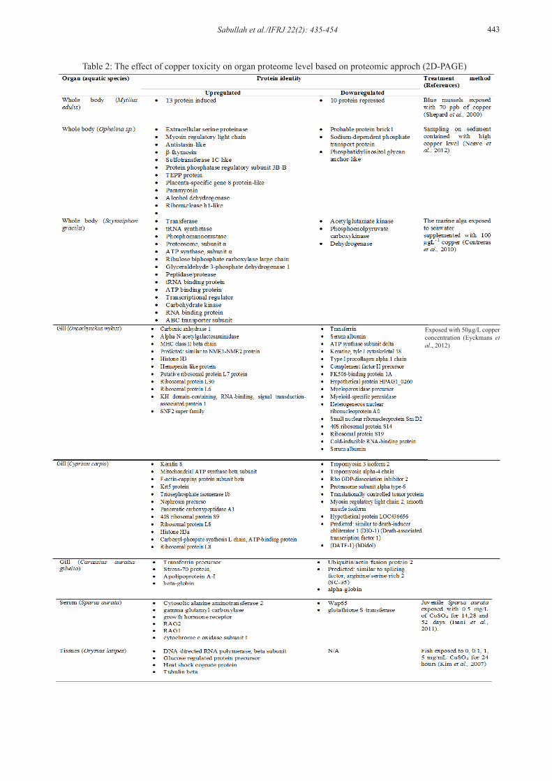
In the development of biomarker applications, proteomic-based approaches were implemented by the previous study to evaluate the impact of toxicant such as heavy metal towards sentinel fish targeted organ and was considered valuable in the detection of early response to this toxic compound (Wang et al., 2011; Lu et al., 2012). Ecotoxicoproteomic by the combination of gel electrophoresis and mass spectrometry was expected to locate the biochemical mechanism involved in acute or chronic toxicity of heavy metals through the identified protein expression signatures (PES) and pathway impairment. A combination of both studies may strengthen the analysis results (Khoudoli et al., 2004). Selected organ such as gill, liver, and brain
was reported to have a significant impact under in vivo heavy metal exposure at the proteomic level (Feng et al., 2003; Wang et al., 2011; Dorts et al., 2011; Eyckmans et al., 2012). The early relationship assessment between PES and the toxicant induction after run with one dimensional electrophoresis (1DE) or two dimensional electrophoresis (2DE) will be interpreted semiquantitatively either upregulations and downregulation of PES. For the examples, the study by Sanders et al. (1994) and Feng et al. (2003) based on 1DE showed upregulation of heat shock protein 70 parallel with copper concentration treatments and other stress protein were detected especially apoptotic factor such study by Kawakami et al., (2006).1DE result was limited to protein molecular weight separation while 2DE gave more detail result in which the protein has been separated according to their pI through isoelectrofocusing (IEF), then the second separation via molecular weight was performed by SDS-PAGE. The optimization of 2DE was well reported by Khoudoli et al. (2004).
There were various pattern types of PES; 1) Protein band or spot maintained their intensities, 2) Protein band or spot intensities kept on increasing (upregulation), and 3) Protein band or spot intensities kept on decreasing (Downregulation). Most of the type one PES are related to structural protein such as the study done by Wang et al. (2011) which actin, keratin and lamin were not affected by mercury. Another report also shows various functional proteins, especially enzyme remains unchanged during the copper treatment such as Fumarate hydratase, 4-Hydroxyphenylpyruvate dioxygenase, glutathione reductase, Uroporphyrinogen decarboxylase, Lactoylglutathione lyase, Homogentisate 1,2-dioxygenase, Serine/threonine kinase 3 and Xanthine dehydrogenase (Chen and Chan, 2011; Chen and Chan, 2012). Type two and three patterns was called the unique protein affected by any type of treatment concentration or/and duration (Tomanek, 2011). However, this unique PES are preferable for further analysis and would be identified using mass spectrometry (MS) such as matrix-assisted laser desorption/ionization-time of flight analysis/MS (MALDITOFF/MS) and liquid chromatography/MS (LC/MS).
Unique PESAn increase on the quantity of cellular component,
especially protein, in response to an external effect is called upregulation. Under stress conditions, related protein such antioxidant, detoxifying enzyme, apoptotic and necrotic factor were upregulated in order to maintain the homeostasis or deleterious
Sabullah et al./IFRJ 22(2): 435-454 443
Table 2: The effect of copper toxicity on organ proteome level based on proteomic approch (2D-PAGE)
Exposed with 50µg/L copper concentration (Eyckmans et al., 2012)

444 Sabullah et al./IFRJ 22(2): 435-454

Sabullah et al./IFRJ 22(2): 435-454 445
processes of affected parenchyma cells. For example, the presence of toxicant in biological system may regulate the synthesis of detoxifying enzyme such as GST and GPx the toxicant and produce ROS at the same time (Sreejai and Jaya, 2010; Hossain et al., 2012). ROS such as radical and non-radical compound activates the expression of antioxidant gene to synthesis SOD and CAT to decrease the intracellular ROS level (Sheehan et al., 2007; Yang et al., 2013). However, the elevated level of ROS may cause damages in intracellular component which leads to the synthesis of apoptotic or necrotic compound such as caspase, cytochrome c, BAX, and BAD (Kawakami et al., 2008).
Downregulation of protein by environmental stimuli are related to the suppression of protein synthesis by inhibition reaction, limitation or time delay for mRNA transcrption and translation, degradation, cellular damaged are repaired and harmful agents are neutralized or eliminated (Young et al., 1987; Jensen, 2006; Wan and Liu, 2008; Liu et al., 2013; Sánchez-García et al., 2013). For example, a study done by Tanimoto and Kizaki (2002) mentioned the effect of proteosome inhibitor obstruct Ras/ERK signaling pathway subsequent in the downregulation of Fas ligand expression associate with the inhibiting synthesis of apoptotic and necrotic compound. Another study done by Fernando et al. (2013) showed the downregulation of protein expression such as carbamoyl phosphate synthase 1 (CSP 1) and 78 kDa glucose regulated protein (GRP 78/HSPA5) in hepatocyte which both has been degraded affected by alcoholic steatosis, and both were selected as a biomarker for the early detection of hepatic lipidosis. Toxic metals such as As, Pb, Cd and Cu caused downregulation by anti-apoptotic compound, Bcl-2 where this protein losses their function to maintain the mitochondria membrane permeability leads to structural destruction and the release of cytochrome c to activate the executive enzyme for the cell death program (Mehta et al., 2006; Rana, 2008; Hughes et al., 2011; Siddiqui et al., 2013; Galano et al., 2014). As the toxicant gave a great impact to the proteome level, proteomic approach was utilized as a biomaker to evaluate the toxicity level of those compounds. Table 2 shows the example of proteomic study on varies organ and species toward copper toxicity and provide the capability as a sensitive biomarker for the environmental factor.
Conclusion
The study of fish behaviors, cellular alteration, enzymatic reaction and proteomic approach promises
the sensitive biomarker method to elucidate heavy metal on concentration, acute and chronic exposure toxicity. But, both methods have their own cons such as time consuming, cost and high technical ability. However, the combination of this method provides an integrative measurement and improves the understanding of the overall biological risk arising from the whole burden of bioavailable contaminants in areas contaminated especially by heavy metals. Thus, this method is supposes to be utilized in the biomonitoring program as a preliminary screening to elucidate other possible pollutant came from agricultural pesticides and fertilizer, industrial waste and civilization sewage.
References
Abbas Alkarkhi, F. M., Ismail, N. and Easa, A. M. 2008. Assessment of arsenic and heavy metal contents in cockles (Anadara granosa) using multivariate statistical techniques. Journal of Hazardous Materials 150: 783-789.
Abdel-Hameid, N. A. H. 2008. A protective effect of calcium carbonate against arsenic toxicity on the Nile catfish, larias gariepinus. Egyptian Journal of Aquatic Biology and Fish 12 (4): 143-163.
Abdelhamid, R. F., Obara, Y., Uchida, Y., Kohzuma, T., Dooley, D. M., Brown, D. E. and Hori, H. 2007. π-π interaction between aromatic ring and copper-coordinated His81 imidazole regulates the blue copper active-site structure. Journal of Biological Inorganic Chemistry 12: 165-173.
Abdel-Moneim, A. M. and Abdel-Mohsen, H. A. 2010. Ultrastructure changes in hepatocytes of catfish Clarias gariepinus from Lake Mariut, Egypt. Journal of Environmental Biology 31 (5): 715-720.
Ahmed, M. K., Habibullah-Al-Mamun, M., Parvin, E., Akter, M. S. and Khan, M. S. 2013. Arsenic induced toxicity and histopathological changes in gill and liver tissue of freshwater fish, tilapia (Oreochromis mossambicus). Experimental and Toxicologic Pathology 65 (6): 903-909.
Alarifi, S., Ali, D., Alkahtani, S., Siddiqui, M. A. and Ali, B. A., 2013. Arsenic trioxide-mediated oxidative stress and genotoxicity in human hepatocellular carcinoma cells. Onco Targets and Therapy 6: 75-84.
Al-Bairuty, G. A., Shaw, B. J., Handy, R. D. and Henry, T. B. 2013. Histopathological effects of waterborne copper nanoparticles and copper sulphate on the organs of rainbow trout (Oncorhynchus mykiss). Aquatic Toxicology 126: 104-115.
Al-Ghais, S. M. 2013. Acetylcholinesterase, glutathione and hepatosomatic index as potential biomarkers of sewage pollution and depuration in fish. Marine Pollution Bulletin 74: 183-186.
Ali, A., Al-Ogaily, S. M., Al-Asgah, N. A. and Gropp, J. 2003. Effect of sublethal concentrations of copper on the growth performance of Oreochromis niloticus.
446 Sabullah et al./IFRJ 22(2): 435-454
Journal of Applied Ichthyology 19: 183-188.Ali, N., Hoque, M. A., Haque, A., Salam, K. A., Karim, M.
R., Rahman, A., Islam, K., Saud, Z. A., Khalek, M. A., Akhand, A. A., Hossain, M., Mandal, A., Karim, M. R., Miyataka, H., Himeno, S. and Hossain, K. 2010. Association between arsenic exposure and plasma cholinesterase activity: a population based study in Bangladesh. Environmental Health 9: p. 36.
Almazán-Rueda, P., Schrama, J. W. and Verreth, J. A. J. 2004. Behavioural responses under different feeding methods and light regimes of the African catfish (Clarias gariepinus) juveniles. Aquaculture 231 (1-4): 347-359.
Almeida, J., Gravato, C. and Guilhermino, L. 2012. Challenges in assessing the toxic effects of polycyclic aromatic hydrocarbons to marine organisms: a case study on the acute toxicity of pyrene to the European seabass (Dicentrarchus labrax L.). Chemosphere 86: 926-937.
Almeida, J., Oliveira, C., Gravato, C. and Guilhermino, L. 2010. Linking behavioural alterations with biomarkers responses in the European seabass Dicentrarchus labrax L. exposed to the organophosphate pesticide fenitrothion. Ecotoxicology 19: 1369-1381.
Al-Shami, S. A., Md Rawi, C. S., Ahmad, A. H., Abdul Hamid, S. and Mohd Nor, S. A. 2011. Influence of agricultural, industrial and anthropogenic stresses on the distribution and diversity of macroinvertebrates in Juru River, Penang, Malaysia. Ecotoxicology and Environmental Safety 74 (5): 1195-1202.
Amiard-Triquet, C. 2009. Behavioural disturbences: The missing link between suborganismal and supra-organismal responses to stress? Prospects Based on aquatic research. Human and Ecological Risk Assessment 15: 87–110.
Anzolin, D. G., Sarkis, J. E. S., Diaz, E., Soares, D. G., Serrano, I. L., Borges, J. C. G., Souto, A. S., Taniguchi, S., Montone, R. C., Bainy, A. C. D. and Carvalho, P. S. M. 2012. Contaminant concentrations, biochemical and hematological biomarkers in blood of West Indian manatees Trichechus manatus from Brazil. Marine Pollution Bulletin 64: 1402-1408.
Armentrout, P. B., Yang, B. and Rodgers, M. T. 2013. Metal cation dependence of interactions with amino acids: bond energies of Rb+ and Cs+ to Met, Phe, Tyr, and Trp. Journal of Physical Chemistry B 117: 3771-3781.
Atif, F., Parvez, S., Pandey, S., Ali, M., Kaur, M., Rehman, H., Khan, H. A. and Raisuddin, S. 2005. Modulatory effect of cadmium exposure on deltamethrin-induced oxidative stress in Channa punctata Bloch. Archives of Environmental Contamination and Toxicology 49 (3): 371-377.
Au, D. W. T. 2004. The application of histo-cytopathological biomarkers in marine pollution monitoring: A review. Marine Pollution Bulletin 48: 817-834.
Bainy, A. C. D., de Medeiros, M. H. G., Mascio, P. D. and de Almeida, E. A. 2006. In vivo effects of metals on the acetylcholinesteras e activity of the Perna perna mussel’s digestive gland. Biotemas 19 (1): 35-39.
Ballesteros, M. L., Durando, P. E., Nores, M. L., Díaz, M. P., Bistoni, M. A. and Wundelin, D. A. 2009. Endolsufan induces changes in spontaneous swimming activity and acetylcholinesterase activity of Jenynsia multidentata (Anablepidae, Cyprinodontiformes). Environmental Pollution 157: 1573-1580.
Baskaran, G., Masdor, N. A., Syed, M. A. and Shukor, M. Y. 2013. An inhibitive enzyme assay to detect mercury and zinc using protease from Coriandrum sativum. The Scientific World Journal 2013: Article ID 678356.
Beaumont, M. W., Butler, P. J. and Taylor, E. W. 1995. Plasma ammonia concentration in brown trout (Salmo trutta) exposed to sub-lethal copper concentrations and its relationship to decreased swimming performance. Journal of Experimental Biology 198: 2213-2220.
Bhanumathy, C. D. and Balasubramanian, A. S. 1998. Selective inactivation of butyrylcholinesterase with metal chelators suggests there is more than one metal binding site. The International Journal of Biochemistry and Cell Biology 30: 695-705.
Biales, A. D., Bencic, D. C., Villeneuve, D. L., Ankley, G. T. and Lattier, D. L. 2001. Proteomic analysis of zebrafish brain tissue following exposure to the pesticide prochloraz. Aquatic Toxicology 105 (3-4): 618-628.
Binstock, L. and Lecar, H. 1969. Ammonium ion currents in the squid giant axon. Journal of General Physiology 53: 342-361.
Bjerselius, R., Winberg, S., Winberg, Y. Zeipel, K. 1993. Ca2+ protects olfactory receptor function against acute Cu(II) toxicity in Atlantic salmon. Aquatic Toxicology 25 (1–2): 125–137.
Boopathy, R. 2000. Factor limiting bioremediation technologies. Bioresource technology 74 (1): 63-67.
Busca, G., Berardinelli, S., Resini, C. and Arrighi, L. 2008. Technologies for the removal of phenol from fluid streams: a short review of recent developments. Journal of Hazardous Materials 160 (2-3): 265-288.
Cailleaud, K., Michalec, F., Forget-Leray, J., Budzinski, H., Hwang, J., Schmitt, F. G. and Souissi, S. 2011. Changes in the swimming behavior of Eurytemora affinis (Copepoda, Calanoida) in response to a sub-lethal exposure to nonylphenols. Aquatic Toxicology 102: 228–231.
Campagna, A. F., Fracácio, R., Rodrigues, B. K., Eler, M. N., Fenerich-Verani, N. and Espíndola, E. L. G. 2008. Effects of the copper in the survival, growth and gill morphology of Danio rerio (Cypriniformes, Cyprinidae). Acta Limnologica Brasiliensia 20: 253-259.
Cankaya, M., Sisecioglu, M., Ciftci, M. and Ozdemir, H. 2011. Effects of some metal ions on trout liver glucose 6-phosphate dehydrogenase. Research Journal of Environmental Toxicology 5: 385-391.
Canli, M. and Atli, G. 2003. The relationships between heavy metal (Cd, Cr, Cu, Fe, Pb, Zn) levels and the size of six Mediterranean fish species. Environmental Pollution 121: 129-136.
Carbonaro, M. 2004. Proteomics: present and future in food quality evaluation. Trends in Food Science and

Sabullah et al./IFRJ 22(2): 435-454 447
Technology 15: 209-216.Cavas, T., Garanko, N. N. and Arkhipchuk, V. V. 2005.
Induction of micronuclei and binuclei in blood, gill and liver cells of fishes subchronically exposed to cadmium chloride and copper sulphate. Food and Chemical Toxicology 43: 569-574.
Chandra, S., Ram, R. N. and Singh, I. J. 2004. First ovarian maturity and recovery response in common carp, Cyprinus carpio after exposure to carbofuran. Journal of Environmental Biology 25 (3): 239-249.
Chen D. S. and Chan, K. M. 2011. Differentially expressed proteins in zebrafish liver cells exposed to copper. Aquatic Toxicology 104: 270– 277.
Chen D. and Chan, K. M. 2012. Identification of hepatic copper-binding proteins from tilapia by column chromatography with proteomic approaches. Metallomics 4 (8): 820-834.
Chen, D. S. and Chan, K. M. 2009. Changes in the protein expression profiles of the Hepa-T1 cell line when exposed to Cu2+. Aquatic Toxicology 94: 163-176.
Chourpagar, A. R. and Kulkarni, G. K. 2012. Effect of sublethal concentration of copper sulphate on glutathione-s-transferase enzyme activity in various tissues of freshwater crab, Barytelphusa cunicularis (Westwood). European Journal of Biological Sciences 4 (2): 49-52.
Cohn, J., Widzowski, D. V. and Cory-Slechta, D. A. 1992. Lead retards development of Drosophila melanogaster. Comparative Biochemistry and Physiology, Part C 102(1): 45-49.
Colgan, P. 1973. Motivational analysis of fish feeding. Behavior. 45: 38-43
Comakli, V., Akkemik, E., Ciftci, M. and Kufrevioglu, O. I. 2013. Purification and characterization of glucose 6-phosphate dehydrogenase enzyme from rainbow trout (Oncorhynchus mykiss) liver and investigation of the effects of some metal ions on enzyme activity. Toxicology and Industrial Health. Downloaded from http:// http://tih.sagepub.com/content/early/2013/01/28/0748233713475514.
Contreras, L., Moenne, A., Gaillard, F., Potin, P. and Correa, J. A. 2010. Proteomic analysis and identification of copper stress-regulated proteins in the marine alga Scytosiphon gracilis (Phaeophyceae). Aquatic Toxicology 96 (2): 85-89.
Costa, P. M., Chicano-Gálvez, E., López Barea, J., DelValls, T. À. and Costa, M. H. 2010. Alterations to proteome and tissue recovery responses in fish liver caused by a short-term combination treatment with cadmium and benzo[a]pyrene. Environmental Pollution 158: 3338- 3346.
Crafford, D. and Avenant-Oldewage, A. 2011. Uptake of selected metals in tissues and organs of Clarias gariepinus (sharptooth catfish) from the Vaal River System – Chromium, copper, iron, manganese and zinc. Water SA 37 (2): 181-200
Crupkin, A. C. and Menone, M. L. 2013. Changes in the activities of glutathione-S-transferases, glutathione reductase and catalase after exposure to different concentrations of cadmium in Australoheros facetus
(Cichlidae, Pisces). Ecotoxicology and Environmental Contamination 8 (1): 21-25.
Cunha, I., Mangas-Ramirez, E. and Guilhermino, L. 2007. Effects of copper and cadmium on cholinesterase and glutathione S-transferase activities of two marine gastropods (Monodonta lineata and Nucella lapillus). Comparative Biochemistry and Physiology, Part C 145: 648-657.
de Lima, D., Roque, G. M. and de Almeida, E. A. 2013. In vitro and in vivo inhibition of acetylcholinesterase and carboxylesterase by metals in zebrafish (Danio rerio). Marine Environmental Research 91: 45-51.
de Souza Dahm, K. C. S., Rückert, C., Tonial, E. M. and Bonan, C. D. 2006. In vitro exposure of heavy metals on nucleotidase and cholinesterase activities from the digestive gland of Helix aspersa. Comparative Biochemistry and Physiology, Part C 143: 316-320.
Devi, M. and Fingerman, M. 1995. inhibition of acetylcholinesterase activity in the central nervous system of the red swamp crayfish, Procambarus clarkii, by mercury, cadmium, and lead. Bulletin of Environmental Contamination and Toxicology 55: 746-750.
Diamantino, T. C., Almeida, E., Soares, A. M. V. M. and Guilhermino, L. 2003. Characterization of cholinesterases from Daphnia magna straus and their inhibition by zinc. Bulletin of Environmental Contamination and Toxicology 71: 219 -225.
Do Lago, L. C. C., Matias, A. C., Nomura, C. S. and Cerchiaro, G. 2011. Radical production by hydrogen peroxide/bicarbonate and copper uptake in mammalian cells: Modulation by Cu(II) complexes. Journal of Inorganic Biochemistry 105: 189-194.
Dornfeld, C. B., Moreira-Santos, M., Espíndola, E. L. G. and Ribeiro, R. 2009. Do larvae and ovipositing Chironomus riparius (Diptera: Chironomidae) females avoid copper-contaminated environments? Human and Ecological Risk Assessment: An International Journal 15 (1): 63-75.
Dorts, J., Kestemont, P., Dieu, M., Raes, M. and Silvestre, F. 2010. Proteomic response to sublethal cadmium exposure in a sentinel fish species, Cottus gobio. Journal of Proteome Research 10 (2): 470-478.
El-Naga, E. H. A., El-Moselhy, K. M. and Hamed, M. A. 2005. Toxicity of cadmium and copper and their effect on some biochemical parameters of marine fish Mugil Seheli. Egyptian Journal of Aquatic Research 31 (2): 60-71.
Elumalai, M., Antunes, C. and Guilhermino, L. 2002. Effects of single metals and their mixtures on selected enzymes of Carcinus Maenas. Water, Air, and Soil Pollution 141: 273-280.
Eyckmans, M., Benoot, D., Raemdonck, G. A. A. V., Zegels, G., Ostade, X. W. M. V., Witters, E., Blust, R. and De Boeck, G. 2012. Comparative proteomics of copper exposure and toxicity in rainbow trout, common carp and gibel carp. Comparative Biochemistry and Physiology, Part D 7 (2): 220-232.
Ezeonyejiaku, C. D., Obiakor, M. O. and Ezenwelu, C. O. 2011. Toxicity of copper sulphate and behavioral
448 Sabullah et al./IFRJ 22(2): 435-454
locomotor response of tilapia (Oreochromis Niloticus) and catfish (Clarias Gariepinus) species. Online Journal Of Animal and Feed Research 1 (4): 130-134.
Farombi, E. O., Adelowo, O. A. and Ajimoko, Y. R. 2007. Biomarkers of oxidative stress and heavy metal levels as indicators of environmental pollution in african cat fish (Clarias gariepinus) from Nigeria Ogun river. International Journal of Environmental Research and Public Health 4 (2): 158-165.
Feng, Q., Boone, A. N. and Vijayan, M. M. 2003. Copper impact on heat shock protein 70 expression and apoptosis in rainbow trout hepatocytes. Comparative Biochemistry and Physiology, Part C 135: 345-355.
Fernando, H., Wiktorowicz, J. E., Soman, K. V., Kaphalia, B. S., Khan, M. F. and Ansari, G. A. S. 2013. Liver proteomics in progressive alcoholic steatosis. Toxicology and Applied Pharmacology 266: 470-480.
Fianko, J. R., Donkor, A., Lowor, S. T., Yeboah, P. O., Glover, E. T., Adom, T. and Faanu, A. 2011. Health risk associated with pesticide contamination of fish from the Densu river basin in Ghana. Journal of Environmental Protection 2: 115-123.
Figueiredo-Fernandes, A., Ferreira-Cardoso, J. V., Garcia-Santos, S., Monteiro, S. M., Carrola, J., Matos, P. and Fontaínhas-Fernandes, A. 2007. Histopathological changes in liver and gill epithelium of Nile tilapia, Oreochromis niloticus, exposed to waterborne copper. Pesquisa Veterinária Brasileira 27 (3): 103-109.
Flora, S. J., Mittal M. and Mehta, A. 2008. Heavy metal induced oxidative stress and its possible reversal by chelation therapy. Indian Journal of Medical Research 128 (4): 501-523.
Forget, J. and Bocquene, G. 1999. Partial purification and enzymatic characterization of acetylcholinesterase from the intertidal marine copepod Tigriopus brevicornis. Comparative Biochemistry and Physiology, Part B 123: 345-350.
Frasco, M. F., Fournier, D., Carvalho, F. and Guilhermino, L. 2008. Does mercury interact with the inhibitory effect of dichlorvos on Palaemon serratus (Crustacea: Decapoda) cholinesterase? Science of the Total Environment 404: 88-93.
Fukuto, T. R. 1990. Mechanism of action of organophosphate and carbamate insecticides. Environmental Health Perspectives 87: 245-254.
Galano, E., Arciello, A., Piccoli, R., Monti D. M. and Amoresanoa, A. 2014. A proteomic approach to investigate the effects of cadmium and lead on human primary renal cells. Metallomics 6 (3): 587-597.
Gerhardt, A. 2007. Aquatic behavioral ecotoxicology – prospects and limitations. Human and Ecological Risk Assessment 13: 481-491.
Gernhöfer, M., Pawert, M., Schramm, M., Müller, E. and Triebskorn, R. 2001. Ultrastructural biomarkers as tools to characterize the health status of fish in contaminated streams. Journal of Aquatic Ecosystem Stress and Recovery 8: 241-260.
Ghica, M. E., Carvalho, R. C., Amine, A. and Brett, C. M. A. 2013. Glucose oxidase enzyme inhibition sensors for heavy metals at carbon film electrodes modified
with cobalt or copper hexacyanoferrate. Sensors and Actuators B 178: 270-278.
Giattina, J. D., Garton, R.. R.. and Stevens, D. G. 1982. Avoidance of copper and nickel by rainbow trout as monitored by a computer-based data acquisition system. Transactions of the American Fisheries Society 111: 491-504.
Gill, T. S., Tewari, H. and Pandet, J. 1990. Use of the fish enzyme system in monitoring water quality: effects of mercury on tissue enzymes. Comparative Biochemistry and Physiology, Part C 97 (2): 287-292.
Glusker, J. P., Katz, A. K. and Bock, C. W. 1999. Metal ions in biological systems. The Rigaku Journal 16 (2): 8-16.
Grajeda y Ortega, M. A., Ortiz, O. E., Favari, L., Shibayama, M., Silva, O. A. and López, L. E. 2011. Biochemical and mitochondrial changes induced by Cd, Fe and Zn in Limnodrillus hoffmeisteri. International Journal of Morphology 29 (2): 412-419.
Grosell, M., Nielsen, C. and Bianchini, A. 2002. Sodium turnover rate determines sensitivity to acute copper and silver exposure in freshwater animals. Comparative Biochemistry And Physiology, Part C 133: 287-303.
Han, J., Won, E. J., Hwang, D. S., Rhee, J. S., Kim, I. C. and Lee, J. S. 2013. Effect of copper exposure on GST activity and on the expression of four GSTs under oxidative stress condition in the monogonont rotifer, Brachionus koreanus. Comparative Biochemistry and Physiology, Part C 158 (2): 91-100.
Handy, R. D. 2003. Chronic effects of copper exposure versus endocrine toxicity: two sides of the same toxicological process? Comparative Biochemistry and Physiology, Part A 135 (1): 25-38.
Hansen, J. A., Marr, J. C. A., Lipton, J., Cacela, D. and Bergman, H. L. 1999. Differences in neurobehavioral responses of Chinook salmon (Oncorhynchus tshawytscha) and rainbow trout (Oncorhynchus mykiss) exposed to copper and cobalt: Behavioral avoidance. Environmental Toxicology and Chemistry 18: 1972-1978.
Hansen, J. A., Rose, J. D., Jenkins, R. A., Gerow, K. G. and Bergman, H. L. 1998. Chinook Salmon (Oncorhynchus Tshawytscha) and Rainbow Trout (Oncorhynchus Mykiss) exposed to copper: Neurophysiological and histological effects on the olfactory system. Environmental Toxicology and Chemistry 18 (9): 1979-1991.
Hellou, J. 2011. Behavioural ecotoxicology, an ‘‘early warning’’ signal to assess environmental quality. Environmental Science and Pollution Research 18: 1-11.
Hong, Y., Chen, X., Guo, J., Xu, Z. , Xu, M. and Sun, G. 2007. Effects of electron donors and acceptors on anaerobic reduction of azo dyes by Shewanella decolorationis S12. Applied Microbiology and Biotechnology 74 (1): 230-238.
Hossain, M. A., Piyatida, P., da Silva, J. A. T. and Fujita, M. 2012. molecular mechanism of heavy metal toxicity and tolerance in plants: Central role of glutathione in detoxification of reactive oxygen species and
Sabullah et al./IFRJ 22(2): 435-454 449
methylglyoxal and in heavy metal chelation. Journal of Botany 2012: Article ID 872875.
Howcroft, C. F., Gravato, C., Amorim, M. J. B., Novais, S. C., Soares, A. M. V. M. and Guilhermino, L. 2011. Biochemical characterization of cholinesterases in Enchytraeus albidus and assessment of in vivo and in vitro effects of different soil properties, copper and phenmedipham. Ecotoxicology 20: 119-130.
Hughes, M. F., Beck, B. D., Chen, Y., Lewis A. S. and Thomas, D. J. 2011. Arsenic exposure and toxicology: a historical perspective. Toxicological Sciences 123 (2): 305-332.
Hussain, S. M., Javed, M., Javid, A., Javid, T. and Hussain, N. 2011. Growth responses of Catla catla, Labeo rohita and Cirrhina mrigala during chronic exposure of iron. Pakistan Journal of Agricultural Sciences 48: 225-230.
Isani, G., Andreani, G., Carpenè, E., Molfetta, S. D., Eletto, D. and Spisni, E. 2011. Effects of waterborne Cu exposure in gilthead sea bream (Sparus aurata): A proteomic approach. Fish and Shellfish Immunology 31: 1051-1058.
James, R. and Sampath, K. 1995. Sub-lethal effects of mixtures of copper and ammonia on selected biochemical and physiological parameters in the catfish Heteroneustes fossilus (Bloch). The Bulletin of Environmental Contamination and Toxicology 55: 187-194.
Javed, M. 2012. Growth responses of fish under chronic exposure of waterborne and dietary metals. International Journal of Agriculture 14: 281-285.
Javed, M., and Usmani, N. 2011. Accumulation of heavy metals in fishes: A human health concern. International Journal of Environmental Sciences 2 (2): 659-670.
Jayakumar, P. and Paul, V. I. 2006. Cadmium accumulation in selected tissues of Clarias batrachus. Veterinarski Arhiv 76 (2): 167-177.
Jelodar, H. T. and Fazli, H. 2012. Monthly changes in condition, hepatosomatic index and bioavailability in frogs (Rana ridibunda). Research Journal of Biology 2 (2): 9-14.
Jensen, O. N. 2006. Interpreting the protein language using proteomics. Nature Reviews Molecular Cell Biology 7: 391-403.
Jezierska, B. and Witeska, M. 2007. The metal uptake and accumulation in fish living in polluted waters. NATO 69: 1568-1238.
Jian-Yu, X., , Xiang-Wen, M., Ying, L. and Shao-Rong, S. 2005. Behavioral response of tilapia (Oreochromis niloticus) to acute ammonia stress monitored by computer vision. Journal of Zhejiang University. Science. B 6 (8): 812-816.
Jiraungkoorskul, W., Sahaphong, S. and Kangwanrangsan, N. 2007. Toxicity of copper in butterfish (Poronotus triacanthus): tissues accumulation and ultrastructural changes. Environmental Toxicology 22 (1): 92-100.
Jung, K., Bitton, G. and Koopman, B. 1995. Assessment of urease inhibition assay for measuring toxicity of environmental samples. Pergamon 29: 1029-1933.
Kane, A. S., Salierno, J. D., Gipson, G. T., Molteno,
T.C.A. and Hunter, C. 2004. A video-based movement analysis system to quantify behavioral stress responses of fish. Water Research 38 (18): 3993-4001.
Kaoud, H. A., Mahran, K. M. A., Rezk, A. and Khalf, M. A. 2012. Bioremediation the toxic effect of mercury on liver histopathology, some hematological parameters and enzymatic activity in Nile tilapia, Oreochromis niloticus. Researcher 4 (1): 60-70.
Karayakar, F., Cicik, B., Ciftci, N., Karaytug, S., Erdem, C. and Ozcan, A. Y. 2010. Accumulation of copper in liver, gill and muscle tissues of Anguilla anguilla (Linnaeus, 1758). Journal of Animal and Veterinary Advances 9: 2271-2274.
Kawakami, M., Inagawa, R., Hosokawa, T., Saito, T. and Kurasaki, M. 2008. Mechanism of apoptosis induced by copper in PC12 cells. Food and Chemical Toxicology 46: 2157-2164.
Kehrer, J. P. 2000. The Haber–Weiss reaction and mechanisms of toxicity. Toxicology 149: 43-50.
Khoudoli, G. A., Porter, I. M., Blow, J. J. and Swedlow, J. R. 2004. Optimisation of the two-dimensional gel electrophoresis protocol using the Taguchi approach. Proteome Science 2: 6. Downloaded from http://www.ncbi.nlm.nih.gov/pmc/articles/PMC517948/pdf/1477-5956-2-6.pdf.
Kim, S. G., Jang, S. W., Lee, Y. J. and Kim, S. S. 2011. Cu accumulation and elimination in the tissues of the olive flounder Paralichthys olivaceus. Fisheries and Aquatic Sciences 14 (3): 210-217.
Kim, W. K., Lee, S. K. and Kim, J. S. 2007. The identification of HSC70 as a biomarker for copper exposure in medaka fish. Journal of Environmental Toxicology 22 (3): 197-202.
Kovarik, Z., Radić, Z., Grgas, B., Skrinjarić-Spoljar, M., Reiner, E. and Simeon-Rudolf, V. 1999. Amino acid residues involved in the interaction of acetylcholinesterase and butyrylcholinesterase with the carbamates Ro 02-0683 and bambuterol, and with terbutaline. Biochim Biophys Acta 1433 (1-2): 261-271.
Kravitz, M. J., Lamberson, J. O., Ferraro, S. P., Swartz, R. C., Boese, B. L. and Specht D. T. 1999. Avoidance response of the estuarine amphipod Eohaustorius estuarius to polycyclic aromatic hydrocarbon-contaminated, field collected sediments. Environmental Toxicology and Chemistry 18: 1232-1235.
Krawczyński, T., Moszczyńska, M. G. and Trojanowicz, M. 2000. Inhibitive determination of mercury and other metals ion by potentiometric urea biosensor. Biosensors and Bioelectronics 15: 681-691.
Kristiansen, T. S., Ferno, A., Holm, J. C., Privitera, L., Bakke, S. and Fosseidengen, J. E. 2004. Swimming behaviour as an indicator of low growth rate and impaired welfare in Atlantic halibut (Hippoglossus hippoglossus L.) reared at three stocking densities. Aquaculture 230 (1-4): 137-151.
Kruse, G. O. and Scarnecchia, D. L. 2002. Assessment of bioaccumulated metal and organochlorine compounds in relation to physiological biomarkers in Kootenai
450 Sabullah et al./IFRJ 22(2): 435-454
River white sturgeon. Journal of Applied Ichthyology 18: 430-438.
Kumar, S. and Kayastha, A. M. 2010. Inhibition studies of soybean (Glycine max) urease with heavy metals, sodium salts of mineral acids, boric acid, and boronic acids. Journal of Enzyme Inhibition and Medicinal Chemistry 25 (5): 646-652.
Lauer, M. M., de Oliveira, C. B., Yano, N. L. and Bianchini, A. 2012. Copper effects on key metabolic enzymes and mitochondrial membrane potential in gills of the estuarine crab Neohelice granulata at different salinities. Comparative Biochemistry and Physiology, Part C 156 (3-4): 140-147.
Lenhardt, M., Jarić, I., Cakić, P., Cvijanović, G., Gačić, Z. and Kolarević, J. 2009. Seasonal changes in condition, hepatosomatic index and parasitism in sterlet (Acipenser ruthenus L.). Turkish Journal of Veterinary and Animal Sciences 33 (3): 209-214.
Leong, K. H., Benjamin, T. L. L. and Mustafa, M. A. 2007. Contamination levels of selected organochlorine and organophosphate pesticides in the Selangor river, Malaysia between 2002 to 2003. Chemosphere 66: 1153-1159.
Liu, D., Liu, C., Li, J., Azadzoi, K. and Yang, Y. 2013. Proteomic analysis reveals differentially regulated protein acetylation in human amyotrophic lateral sclerosis spinal cord. PLoS ONE 8 (12): e80779.
Lopes, P. A., Pinheiro, T., Santos, M. C., Mathias, M. D., Collares-Pereira, M. J. and Viegas-Crespo, A. M. 2001. Response of antioxidant enzymes in freshwater fish populations (Leuciscus alburnoides complex) to inorganic pollutants exposure. Science of the Total Environment 280: 153-163.
Lopes, I., Baird, D. J. and Ribeiro, R. 2004. Avoidance of copper contamination by field populations of Daphnia longispina. Environmental Toxicology and Chemistry 23:1702-1708.
Lopes, P. R. M. and Bidoia, E. D. 2009. Evaluation of the biodegradation of different types of lubricant oils in liquid medium. Brazilian Archives of Biology and Technology 52 (5): 1285-1290.
Lu, X. J., Chen, J., Zhuang, Z. A. H. L., Peng, L. Z. and Shi, Y. H. 2012. Influence of acute cadmium exposure on the liver proteome of a teleost fish, Ayu (Plecoglossus altivelis). Molecular Biology Reports 39: 2851-2859.
Ma, H. J., Xie, R. L., Zhao, Q. F., Mei, X. D. and Ning, J. 2010. Synthesis and insecticidal activity of novel carbamate derivatives as potential dual-binding site acetylcholinesterase inhibitors. Journal of Agricultural and Food Chemistry 58 (24): 12817-12821.
Mahmod, A. A. 2001. Investigation of the reversible inhibition of butyrylcholinesterase by mercury chloride. The Science 1 (4): 251-254.
Main, A. R. 1979. Mode of action of anticholinesterases. Pharmacology and Therapeutics 6: 579-628.
Masdor, N. A. and Said, N. A. M. 2011. Partial purification of crude stem bromelain improves it sensitivity as a protease inhibitive assay for heavy metals. Australian Journal of Basic and Applied Sciences 5 (10): 1295-1298.
Masson, P., Froment, M. T., Bartels, C. F. and Lockridge, O. 1996. Asp 70 in the peripheral anionic site of human butyrylcholinesterase. European Journal of Biochemistry 235: 36-48.
Mat-Jais, A. M. and Mohamed, M. Z. 2000. The role of extracellular Ca2+ on the inhibitory effects of acetylcholinesterase activity in haruan, Channa striatus block, brain tissue by heavy metals. Malaysian Applied Biology 29 (1-2): 69-74.
Matos, P. Fontaıínhas-Fernandes, A., Peixoto, F., Carrola, J. and Rocha, E. 2007. Biochemical and histological hepatic changes of Nile tilapia Oreochromis niloticus exposed to carbaryl. Pesticide Biochemistry and Physiology 89: 73-80.
Mayeux, R. 2004. Biomarker: Potential uses and limitations. NeuroRx 1(2): 182-188.
Mazorra, M. T., Rubio, J. A. and Blasco, J. 2002. Acid and alkaline phosphatase activities in the clam Scrobicularia plana: Kinetic characteristics and effects of heavy metals. Comparative Biochemistry and Physiology, Part B 131 (2): 241-249.
Mehta, R., Templeton, D. M. and O’Brien, P. J. 2006. Mitochondrial involvement in genetically determined transition metal toxicity II. Copper toxicity. Chemico-Biological Interactions 163: 77-85.
Mieiro, C. L., Pereira, M. E., Duarte, A. C. and Pacheco, M. 2011. Brain as a critical target of mercury in environmentally exposed fish (Dicentrarchus labrax) -Bioaccumulation and oxidative stress profiles. Aquatic Toxicology 103 (3-4): 233-240.
Moreira, S. M., Lima, I., Ribeiro, R.. and Guilhermino, L. 2006. Effects of estuarine sediment contamination on feeding and on key physiological functions of the polychaete Hediste diversicolor: laboratory and in situ assays. Aquatic Toxicology 78: 186-201.
Moreira-Santos, M., Donato, C., Lopes, I. and Ribeiro, R. 2008. Avoidance tests with small fish: Determination of the median avoidance concentration and of the lowest-observed-effect-gradient.Environmental Toxicology and Chemistry 27: 1576-1582.
Moreno, J. L., Navarro, C. and De Las Heras, J. 2006. Abiotic ecotypes in south-central Spanish rivers: reference conditions and pollution. Environmental Pollution 143 (3): 388-396.
Nagdeve, D. A. 2004. Environmental pollution and control: A case study of Delhi Mega City. Population and Environment 25 (5): 461-473.
Narayana, K. and Al-Bader, M. 2011. Ultrastructural and DNA damaging effects of lead nitrate in the liver. Experimental and Toxicologic Pathology 63: 43-51.
Nascimento, A. A., Araújo, F. G., Gomes, I. D., Mendes, R. M. and Sales, A. 2012. Fish gills alterations as potential biomarkers of environmental quality in a eutrophized tropical river in south-eastern Brazil. Anatomia, histologia and Embryologia 41 (3): 209-216.
Neave, M. J., Streten-Joyce, C., Nouwens, A. S., Glasby, C. J., McGuinness, K. A., Parry, D. L. and Gibb, K. S. 2012. The transcriptome and proteome are altered in marine polychaetes (Annelida) exposed to elevated
Sabullah et al./IFRJ 22(2): 435-454 451
metal levels. Journal of Proteomics 75: 2721- 2735.
Ní Shúilleabháina, S., Mothersilla, C., Sheehanb, D., O’Brienb, N. M., O’ Halloranb, J., Van Peltb, F. N. A. M. and Davorena, M. 2004. In vitro cytotoxicity testing of three zinc metal salts using established fish cell lines. Toxicology In Vitro 18: 365-376.
Oliveira, C., Almeida, J. R., Guilhermino, L., Soares, A. M. V. M. and Gravato, C. 2013. Swimming velocity, avoidance behavior and biomarkers in Palaemon serratus exposed to fenitrothion. Chemosphere 90: 936-944.
Paris-Palacios, S., Biagianti-Risbourg, S. and Vernet, G. 2000. Biochemical and (ultra)structural hepatic perturbations of Brachydanio rerio (Teleostei, Cyprinidae) exposed to two sublethal concentrations of copper sulfate. Aquatic Toxicology 50: 109-124.
Parveen, A. and Javed, M. 2010. Effect of Water-borne copper on the growth performance of fish Catla catla. International Journal of Agriculture 12: 950-952.
Patil, V. K. and David, M. 2010. Behavioral and morphological endpoints: as an early response to sublethal malathion intoxication in the freshwater fish, Labeo rohita. Drug and Chemical Toxicology 33 (2): 160-165.
Patlolla, A. K. and Tchounwou, P. B. 2005. Serum acetyl cholinesterase as a biomarker of arsenic induced neurotoxicity in sprague-dawley Rats. International Journal of Environmental Research and Public Health 2: 80-83.
Patnaik, B. B., Howrelia, H. J., Mathews, T. and Selvanayagam, M. 2011. Histopathology of gill, liver, muscle and brain of Cyprinus carpio communis L. exposed to sublethal concentration of lead and cadmium. African Journal of Biotechnology. 10 (57): 12218-12223.
Paustenbach, D. and Galbraith, D. 2006. Biomonitoring and biomarker: exposure assessment will never be the same. Environmental Health Perspectives 114 (8): 1143-1149.
Pelgrom, S. M. G. J., Lamers, L. P. M., Garritsen, J. A. M., Pels, B. M., Lock, R. A. C., Balm, P. H. M. and Bonga, S. E. W. 1994. Interactions between copper and cadmium during single and combined exposure in juvenile tilapia Oreochromis mossambicus: Influence of feeding condition on whole body metal accumulation and the effect of the metals on tissue water and ion content. Aquatic Toxicology 30: 117-135.
Petrell, R. J. and Ang, K. P. 2001. Effects of pellet contrast and light intensity on salmonid feeding behaviours. Aquacultural Engineering 25 (3): 175-186.
Pilgaard, L., Malte, H. and Jensen, F. B. 1994. Physiological effects and tissue accumulation of copper in freshwater rainbow trout (Oncorhynchus mykiss) under normoxic and hypoxic conditions. Aquatic Toxicology 29 (3-4): 197-212.
Plaut, I. 2001. Critical swimming speed: its ecological relevance. Comparative Biochemistry and Physiology, Part A 131: 41-50.
Pugazhvendan, S. R., Narendiran, N. J., Kumaran, R. G., Kumaran, S. and Alagappan, K. M. 2009. Effect of malathion toxicity in the freshwater fish Ophiocephalus punctatus-A histological and histochemical study. World Journal of Fish and Marine Sciences 1 (3): 218-224.
Qadir, A. and Malik, R. N. 2011. heavy metals in eight edible fish species from two polluted tributaries (Aik and Palkhu) of the River Chenab, Pakistan. Biological Trace Element Research 143: 1524-1540.
Radi, A. A. and Matkovics, B. 1988. Effects of metal ions on the antioxidant enzyme activities, protein contents and lipid peroxidation of carp tissues. Comparative Biochemistry and Physiology, Part C 90 (1): 69-72.
Rajamanickam, V. and Muthuswamy, N. 2008. Effect of heavy metals induced toxicity on metabolic biomarkers in common carp (Cyprinus carpio L.). Maejo International Journal of Science 2 (1): 192-200.
Rajesh, R. V., Balasubramanian, A. S. and Boopathy, R. 2009. Evidence for presence of Zn+2-binding site in acetylcholinesterase. Biochimie 91: 526-532.
Rana, S. V. S. 2008. Metals and apoptosis: Recent developments. Journal of Trace Elements in Medicine and Biology 22: 262-284.
Rao, P. S., Babu, N. B. and Raju, R. R. 2010. Study the effect of chlorpyrifos on proteins in freshwater fish Labeo rohita by using HPLC Method. International Journal of Research in Pharmaceutical and Biomedical Sciences 1: 1-5.
Reddy, S., Venkata, Reddy M. and Radhakrishnaiah, K. 2008. Impact of copper on the oxidative metabolism of the fry of common carp, Cyprinus carpio (Linn.) at different pH. Journal of Environmental Biology 29 (5): 721-724.
Ribeiro, C. A. O., Vollaire, Y., Sanchez-Chardi, A. and Roche, H. 2005. Bioaccumulation and the effects of organochlorine pesticides, PAH and heavy metals in the Eel (Anguilla anguilla) at the Camargue Nature Reserve, France. Aquatic Toxicology 74 (1): 53-69.
Rodrigues, E. L. and Fanta, E. 1998. Liver histopathology of the fish Brachydanio rerio hamilton-buchman after acute exposure to sublethal levels of the organophosphate Dimethoate 500,” Revista Brasileira de Zoologia 15 (2): 441- 450.
Rodrigues, P. M., Silva, T. S., Dias, J. and Jessen, F. 2012. Proteomics in aquaculture: applications and trends. Journal of Proteomics 75: 4325- 4345.
Rojik, I., Nemcsók, J. and Boross, L. 1983. Morphological and biochemical studies on liver, kidney and gill of fishes affected by pesticides. Acta Biologica Hungarica 34 (1): 81-92.
Rosenberry, T. L., Johnson, J. L., Cusack, B., Thomas, J. L., Emani, S. and Venkatasubban, K. S. 2005. Interactions between the peripheral site and the acylation site in acetylcholinesterase. Chemico-Biological Interactions 15: 181-189.
Sabullah, M. K., Ahmad, S. A., Sulaiman, M. R., Shukor, M. Y., Syed, M. A. and Shamaan, N. A. 2013. The development of an inhibitive assay for heavy metals using the acetylcholinesterase from Periophtalmodon

452 Sabullah et al./IFRJ 22(2): 435-454
schlosseri. Journal of Environmental Bioremediation and Toxicology 1 (1): 20-24.
Safahieh, A., Hedayati, A., Savari, A. and Movahedinia, A. 2010. Effect in vitro exposure of mercury chloride on phosphatase enzymes in yellowfin sea bream (Acanthopagrus latus). American-Eurasian Journal of Toxicological Sciences 2 (4): 208-214.
Salem, M. M. 2011. Toxic effects of mancozeb containing formulations and neemix pesticides on kidney function and ultrastructure of albino rats. Egyptian Academic Journal of Biologica. Sciences 3 (1): 17-30.
Saliu, J. K. and Bawa-Allah, K. A. 2012. Toxicological effects of lead and zinc on the antioxidant enzyme activities of post juvenile Clarias gariepinus. Resources and Environment 2 (1): 21-26.
Samphao, A., Rerkchai, H., Jitcharoen, J., Nacapricha, D. and Kalcher, K. 2012. Indirect determination of mercury by inhibition of glucose oxidase immobilized on a carbon paste electrode. International Journal of Electrochemical Science 7: 1001-1010.
Sánchez-García, A., Ríos-Ibarra, C. P., Rincón-Sánchez, A. R., Ortiz-López, R., Garza-Juárez, A., Morlett-Chávez, J., Martínez-Rodríguez, H. and Rivas-Estilla, A. M. 2013. Use of proteomic analysis tools to identify HCV-proteins down-regulated by acetylsalicylic acid. Annals of Hepatology 12 (5): 725-732.
Sanders, B. M., Martin, L. S., Howe, S. R., Nelson, W. G., Hegre, E. S. and Phelps, D. K. 1994. Tissue-specific difference in accumulation of stress protein in Mytilus edulis exposed to a range of copper concentration. Toxicology and Applied Pharmacology 125: 125-213.
Sant’Anna, M. C., Soares, V. M., Seibt, K. J., Ghisleni, G., Rico, E. P. and Rosemberg, D. B. 2011. Iron exposure modifies acetylcholinesterase activity in zebrafish (Danio rerio) tissues: distinct susceptibility of tissues to iron overload. Fish Physiology and Biochemistry 37 (3): 573-581.
Sarkar A., Ray, D., Shrivastava, A. N. and Sarker, S. 2006. Molecular Biomarkers: Their significance and application in marine pollution monitoring. Ecotoxicology 15 (4): 333-340.
Sarkarati, B., Çokuğraş, A. N. and Tezcan, E. F. 1999. Inhibition kinetics of human serum butyrylcholinesterase by Cd2+, Zn2+ and Al3+: comparison of the effects of metal ions on cholinesterases. Comparative Biochemistry and Physiology, Part C 122: 181-190.
Sarosiek, B., Pietrusewicz, M., Radziwoniuk, J. and Glogowski, J. 2009. The effect of copper, zinc, mercury and cadmium on some sperm enzyme activities in the common carp (Cyprinus carpio L.),” Reproductive Biology 9 (3): 295-301.
Sathesh Prabu, C. and Thatheyus, A. J. 2007. Biodegradation of acrylamide employing free and immobilized cells of Pseudomonas aeruginosa. International Biodeterioration and Biodegradation 60 (2): 69-73.
Scott, G. R.. and Sloman, K. A. 2004. The effects of environmental pollutants on complexfish behaviour: integrating behavioural and physiological indicators
of toxicity. Aquatic Toxicology 68: 369-392.Sheehan, D., Tyther, R., Dowling, V. and McDonagh, B.
2007. Redox proteomics – a route to the identifi cation of damaged proteins multiple stressors: A challenge for the future. NATO Science for Peace and Security Series pp. 295-308.
Shepard, J. L., Olsson, B., Tedengren, M. and Bradley, B. P. 2000. Protein expression signatures identified in Mytilus edulis exposed to PCBs, copper and salinity stress. Marine Environmental Research 50: 337-340.
Shukla, V., Dhankhar, M., Prakash J. and Sastry, K. V. 2007. Bioaccumulation of Zn, Cu and Cd in Channa punctatus. Journal of Environmental Biology. 28(2): 395-397.
Shukor, M. Y., Baharom, N. A., Masdor, N. A., Abdullah, M. P. A., Shamaan, N. A., Jamal, J. A. and Syed, M. A. 2009a. The development of an inhibitive determination method for zinc using a serine protease. Journal of Environmental Biology 30(1): 17-22.
Shukor, M. Y., Bakar, N. A., Othman, A. R., Yunus, I., Shamaan, N. A. and Syed, M. A. 2009b. Development of an inhibitive enzyme assay for copper. Journal of Environmental Biology 30 (1): 39-44.
Shukor, M. Y., Tham, L. G., Halmi, M. I. E. Khalid, I., Begum, G. and Syed, M. A. 2013. Development of an inhibitive assay using commercial Electrophorus electricus acetylcholinesterase for heavy metal detection. Journal of Environmental Biology 34: 967-970.
Shukor, Y., Baharom, N. A., Rahman, F. A., Abdullah, M. P., Shamaan, N. A. and Syed, M. A. 2006. Development of a heavy metals enzymatic-based assay using papain. Analytica Chimica Acta 566: 283-289.
Siddiqui, M. A., Alhadlaq, H. A., Ahmad, J., Al-Khedhairy, A. A., and Musarrat, J. 2013. Copper oxide nanoparticles induced mitochondria mediated apoptosis in human hepatocarcinoma cells. PLoS ONE 8 (8) e69534.
Silvestre, F., Gillardin V. and Dorts, J. 2012. Proteomics to assess the role of phenotypic plasticity in aquatic organisms exposed to pollution and global warming. Integrative and Comparative Biology 52 (5): 681-691.
Simpson, D. M., Mobasheri, A., Haywood S. and Beynon, R. J. 2006. A proteomics study of the response of North Ronaldsay sheep to copper challenge. BMC Veterinary Research 2: p. 36
Singh, D., Katiyar, S. and Verma, A. 2012. Role of copper sulphate on oxidative and metabolic enzymes of freshwater fish; Channa punctatus. Journal of Environmental and Analytical Toxicology 2: 121
SjØgaard, G. 1991. Role of exercise-induced potassium fluxes underlying muscle fatigue: a brief review. Canadian Journal of Physiology and Pharmacology 69: 238-245.
Skouras, A., Broeg, K., Dizer, H., von Westernhagen, H., Hansen, P. D. and Steinhagen, D. 2003. The use of innate immune responses as biomarkers in a programme of integrated biological effects monitoring on flounder (Platichthys flesus) from the southern North Sea. Helgoland Marine Research 57: 190-198.

Sabullah et al./IFRJ 22(2): 435-454 453
Sreejai, R. and Jaya, D. S. 2010. Studies on the changes in lipid peroxidation and antioxidants in fishes exposed to hydrogen sulfide. Toxicology International 17 (2): 71-77.
Sukumaran, M., Manikandan, S. R., Nathiya, N., and Muthukumaravel, K. 2013. Impact of pesticides monocrotophos on histological changes in the liver of Mystus vittatus. International Journal of Current Research 5 (10): 3244-3247.
Sussman, J. L., Harel, M., Frolow, F., Oefner, C., Goldman, A., Toker, L. and Silman, I. 1991. Atomic structure of acetylcholinesterase from Torpedo californica: a prototypic acetylcholine binding protein. Science 253 : 872-879.
Takhar, P. and Mahant, S. 2011. In vitro methods for nanotoxicity assessment: Advantages and applications. Archives of Applied Science Research 3 (2): 389-403.
Tanimoto, Y. and Kizaki, H. 2002. Proteasome inhibitors block Ras/ERK signaling pathway resulting in the downregulation of Fas ligand expression during activation-induced cell death in T cells. Journal of Biochemistry 131: 319-326.
Thompson, C. M., Prins, J. M. and George, K. M. 2010. Mass spectrometric analyses of organophosphate insecticide oxon protein adducts. Environmental Health Perspectives 118 (1): 11-19.
Tilton, F. A., Bammler, T. K. and Gallagher, E. P. 2011. Swimming impairment and acetylcholinesterase inhibition in zebrafish exposed to copper or chlorpyrifos separately, or as mixtures. Comparative Biochemistry and Physiology, Part C 153 (1): 9-16.
Tomanek, L. 2011. Environmental proteomics: changes in the proteome of marine organisms in response to environmental stress, pollutants, infection, symbiosis, and development. Annual Review of Marine Science 3: 373-399.
Tort, L., Kargacin, B., Torres, P., Giralt, M. and Hidalgo, J. 1996. The effect of cadmium exposure and stress on plasma cortisol, metallothionein levels and oxidative status in rainbow trout (Oncorhynchus mykiss) liver,” Comparative Biochemistry and Physiology, Part C 114 (1): 29-34.
Tripathi, B. N. and Gaur, J. P. 2004. Relationship between copper and zinc induced oxidative stress and proline accumulation in Scenedesmus sp. Planta 219: 397-404.
van Dyk, J. C., Pieterse, G. M. and van Vuren, J. H. J. 2007. Histological changes in the liver of Oreochromis mossambicus (Cichlidae) after exposure to cadmium and zinc. Ecotoxicology and Environmental Safety 66: 432-440.
Varanka, Z., Rojik, I., Varanka, I., Nemcśok, J. and Ábraĥam, M. 2001. Biochemical and morphological changes in carp (Cyprinus carpio L.) liver following exposure to copper sulfate and tannic acid. Comparative Biochemistry and Physiology, Part C 128: 467-478.
Versonnen, B. J., Arijs, K., Verslycke, T., Lema, W. and Janssen, C. R. 2003. In vitro and in vivo estrogenicity and toxicity of O-, M-, and P-Dichlorobenzene.
Environmental Toxicology and Chemistry 22 (2): 329-335.
Vidali, M. 2001. Bioremediation. An Overview. Pure and Applied Chemistry 73 (7): 1163-1172.
Vieira, L. R., Gravato, C., Soares, A. M. V. M., Morgado, F. and Guilhermino, L. 2009. Acute effects of copper and mercury on the estuarine fish Pomatoschistus microps: Linking biomarkers to behaviour. Chemosphere 76: 1416-1427.
Vincent, S., Kumar, L. and Ambrose, T. 1996. Impact of heavy metal chromium on bioenergetics of the Indian major carp (Catla catla). Pollution Research 15: 273-275.
Vosyliene, M. Z., Kazlauskiene, N. and Svecevicius, G. 2003. Effect of a heavy metal model mixture on biological parameters of rainbow trout, Oncorhynchus mykiss,” Environmental Science and Pollution Research. 10: 103-107.
Vutukuru, S. S., Chintada, S., Madhavi, K. R., Rao, J. V. and Anjaneyulu, Y. 2006. Acute effects of copper on superoxide dismutase, catalase and lipid peroxidation in the freshwater teleost fish, Esomus danricus. Fish Physiology and Biochemistry 32: 221-229.
Vutukuru, S. S., Suma, C., Radha Madhavi, K., Juveria, Smitha Pauleena, J., Venkateswara Rao, J. and Anjaneyulu, Y. 2005. Studies on the development of potential biomarkers for rapid assessment of copper toxicity to freshwater fish using Esomus danricus as model. International Journal of Environmental Research and Public Health 2 (1): 63-73.
Wan, X. Y. and Liu, J. Y. 2008. Comparative proteomics analysis reveals an intimate protein network provoked by hydrogen peroxide stress in rice seedling leaves. Molecular & Cellular Proteomics 7 (8): 1469-1488
Wang, J., Liu, X. D. and Lu, J. 2012. The 18th biennial conference of international society for ecological modelling. Urban river pollution control and remediation. Procedia Environmental Sciences 13: 1856- 1862.
Wang, M., Wang, Y., Wang, J., Lin, L., Hong, H. and Wang, D. 2011. Proteome profiles in medaka (Oryzias melastigma) liver and brain experimentally exposed to acute inorganic mercury. Aquatic Toxicology 103: 129-139.
Waser, W., Bausheva1, O. and Nikinmaa, M. 2009. The copper-induced reduction of critical swimming speed in rainbow trout (Oncorhynchus mykiss) is not caused by changes in gill structure. Aquatic Toxicology 94: 77-79.
West, C. W. and Ankley, G. T. 1998. A laboratory assay to assess avoidance of contaminated sediments by the freshwater oligochaete Lumbriculus variegates. Archives of Environmental Contamination and Toxicology 35: 20-24.
WHO\FAO. 1989. National Research Council Recommended Dietary Allowances 10th Edn. National Academy Press, Washington, DC. USA.
Wolf, J. C., and Wolfe, M. J. 2005. A brief overview of nonneoplastic hepatic toxicity in fish. Toxicologic Pathology 33 (1): 75-85.

454 Sabullah et al./IFRJ 22(2): 435-454
Yang, J., Dong, S., Jiang, Q., Si, Q., Liu, X. and Yang J. 2013. Characterization and expression of cytoplasmic copper/zinc superoxide dismutase (CuZn SOD) gene under temperature and hydrogen peroxide (H2O2) in rotifer Brachionus calyciflorus. Gene 518 (2): 388-396.
Yang, X. and Baumann, P. C. 2006. Biliary PAH metabolites and the hepatosomatic index of brown bullheads from Lake Erie tributaries. Ecological Indicators 6: 567-574.
Yap, C. K. and Pang, B. H. 2011. Assessment of Cu, Pb and Zn contamination in sediment of north western Peninsular Malaysia by using sediment quality values and different geochemical indice. Environmental Monitoring and Assessment 183 (1-4): 23-39.
Young, S., Parker, P. J., Ullrich A. and Stabel, S. 1987. Down-regulation of protein kinase C is due to an increased rate of degradation. Biochemical Journal 244: 775-779.
Younis, E. M., Abdel-Warith, A. A., Al-Asgah, N. A., Ebaid, H. and Mubarak, M. 2013. Histological changes in the liver and intestine of Nile Tilapia, Oreochromis niloticus, exposed to sublethal concentrations of cadmium. Pakistan Journal of Zoology 45 (3): 833-841.
Yousafzai, A. M., Khan, A. R. and Shakoori, A. R. 2009. Trace metal accumulation in the liver of an endangered South Asian fresh water fish dwelling in sub-lethal. Pollution Pakistan Journal of Zoology 41 (1): 35-41.
Zikić, R. V., Stajn, A. S., Pavlović, S. Z., Ognjanović, B. I. and Saićić, Z. S. 2001. Activities of superoxide dismutase and catalase in erythrocytes and plasma transaminases of goldfish (Carassius auratus gibelio Bloch.) exposed to cadmium. Physiological Research 50 (1): 105-111.