Mind matters: placebo enhances reward learning in Parkinson's disease
10
© 2014 Nature America, Inc. All rights reserved. NATURE NEUROSCIENCE ADVANCE ONLINE PUBLICATION ARTICLES Expectations are profound incentives for behavior. They affect percep- tion, decision-making, and action. Perhaps the most striking example of the power of expectations is the ‘placebo effect’, wherein the mere expectation of medical treatment leads to physiological effects that mimic the benefits of the treatment itself. These effects pose a crucial challenge for clinical trials, as they can obscure the benefits of an effective intervention. However, if harnessed, they offer the promise of enhancing treatment by combining psychological and pharmaco- logical factors. Thus, understanding the neurobiological basis of how placebo treatment affects behavior is of great importance. In Parkinson’s disease (PD) patients, preliminary data from clinical trials 1–3 and a few landmark experimental studies 4–6 have suggested that placebo effects may be substantial and may involve modulation of dopamine release in the striatum, presumably via nerve terminals whose origins are located in the midbrain. PD is characterized by changes in motor, cognitive and emotional systems. Dopaminergic treatment can improve motor symptoms, but it also has broader con- sequences for behavior, including restored (and sometimes excessive) reward-driven motivation and learning 7 . Although there is evidence for placebo effects on motor function 1,4 , placebo effects on the cogni- tive and affective aspects of PD have not been examined. We sought to investigate the effects of placebo on reward learning in PD and its neurobiological mechanisms. Building on prior data sug- gesting that placebo treatment may cause endogenous recruitment of brain mechanisms that underpin reward learning 5,8–10 , we combined placebo and pharmacological manipulations with a well-characterized instrumental learning procedure 7,11 and used functional magnetic resonance imaging (fMRI) to link learning behavior to learning- and value-related brain activation. The ability to learn from rewarding outcomes is known to depend on midbrain dopamine neurons and their striatal and prefrontal targets. In particular, computational models of reinforcement learning have highlighted two key variables related to such learning: expected value and prediction error 12 . The expected value of an option is assumed to guide choices on each trial. After an outcome is received, this value is updated on the basis of the prediction error, which quantifies how much the outcome deviated from what was expected on the basis of past experience 12–15 . fMRI studies in healthy partici- pants have shown that expected value correlates with blood oxygen level–dependent (BOLD) activity in the ventromedial prefrontal cortex (vmPFC) 16–18 , whereas reward prediction errors correlate with activity in the striatum 19–22 . Moreover, the loss of midbrain dopamine neurons that occurs in PD is associated with changes in these reward-related processes 23–25 . Guided by the well-established role of the vmPFC and the ventral striatum in feedback learning, we focused our hypotheses on these regions and asked three questions. First, behaviorally, does placebo mimic the effects of dopaminergic medication on learning in PD patients? Second, does placebo affect value representation in the vmPFC and prediction error representation in the ventral striatum? Third, does placebo differentially affect learning from reward versus punishment? We predicted that both placebo and dopaminergic med- ication would facilitate reward learning in PD and that this modula- tion would be reflected in value coding in the vmPFC and reward prediction errors in the ventral striatum. We further predicted that the enhancing effects of placebo and dopaminergic medication on learning would be selective to learning about rewards, as compared with learning about losses. A within-patient design allowed us to selectively test the effects of dopaminergic medication (comparing on versus off drug) and whether or not placebo had a similar effect (comparing placebo ver- sus off drug) (Supplementary Fig. 1). Each patient was tested under 1 Psychology Department, Columbia University, New York, New York, USA. 2 Psychology Department, University of Colorado at Boulder, Boulder, Colorado, USA. 3 Kavli Center for Brain Science, Columbia University, New York, New York, USA. 4 These authors contributed equally to this work. Correspondence should be addressed to D.S. ([email protected]) or T.D.W. ([email protected]). Received 15 April; accepted 16 September; published online 19 October 2014; doi:10.1038/nn.3842 Mind matters: placebo enhances reward learning in Parkinson’s disease Liane Schmidt 1 , Erin Kendall Braun 1 , Tor D Wager 2,4 & Daphna Shohamy 1,3,4 Expectations have a powerful influence on how we experience the world. Neurobiological and computational models of learning suggest that dopamine is crucial for shaping expectations of reward and that expectations alone may influence dopamine levels. However, because expectations and reinforcers are typically manipulated together, the role of expectations per se has remained unclear. We separated these two factors using a placebo dopaminergic manipulation in individuals with Parkinson’s disease. We combined a reward learning task with functional magnetic resonance imaging to test how expectations of dopamine release modulate learning-related activity in the brain. We found that the mere expectation of dopamine release enhanced reward learning and modulated learning-related signals in the striatum and the ventromedial prefrontal cortex. These effects were selective to learning from reward: neither medication nor placebo had an effect on learning to avoid monetary loss. These findings suggest a neurobiological mechanism by which expectations shape learning and affect.
Transcript of Mind matters: placebo enhances reward learning in Parkinson's disease

©20
14 N
atu
re A
mer
ica,
Inc.
All
rig
hts
res
erve
d.
nature neurOSCIenCe advance online publication �
a r t I C l e S
Expectations are profound incentives for behavior. They affect percep-tion, decision-making, and action. Perhaps the most striking example of the power of expectations is the ‘placebo effect’, wherein the mere expectation of medical treatment leads to physiological effects that mimic the benefits of the treatment itself. These effects pose a crucial challenge for clinical trials, as they can obscure the benefits of an effective intervention. However, if harnessed, they offer the promise of enhancing treatment by combining psychological and pharmaco-logical factors. Thus, understanding the neurobiological basis of how placebo treatment affects behavior is of great importance.
In Parkinson’s disease (PD) patients, preliminary data from clinical trials1–3 and a few landmark experimental studies4–6 have suggested that placebo effects may be substantial and may involve modulation of dopamine release in the striatum, presumably via nerve terminals whose origins are located in the midbrain. PD is characterized by changes in motor, cognitive and emotional systems. Dopaminergic treatment can improve motor symptoms, but it also has broader con-sequences for behavior, including restored (and sometimes excessive) reward-driven motivation and learning7. Although there is evidence for placebo effects on motor function1,4, placebo effects on the cogni-tive and affective aspects of PD have not been examined.
We sought to investigate the effects of placebo on reward learning in PD and its neurobiological mechanisms. Building on prior data sug-gesting that placebo treatment may cause endogenous recruitment of brain mechanisms that underpin reward learning5,8–10, we combined placebo and pharmacological manipulations with a well-characterized instrumental learning procedure7,11 and used functional magnetic resonance imaging (fMRI) to link learning behavior to learning- and value-related brain activation.
The ability to learn from rewarding outcomes is known to depend on midbrain dopamine neurons and their striatal and prefrontal targets.
In particular, computational models of reinforcement learning have highlighted two key variables related to such learning: expected value and prediction error12. The expected value of an option is assumed to guide choices on each trial. After an outcome is received, this value is updated on the basis of the prediction error, which quantifies how much the outcome deviated from what was expected on the basis of past experience12–15. fMRI studies in healthy partici-pants have shown that expected value correlates with blood oxygen level–dependent (BOLD) activity in the ventromedial prefrontal cortex (vmPFC)16–18, whereas reward prediction errors correlate with activity in the striatum19–22. Moreover, the loss of midbrain dopamine neurons that occurs in PD is associated with changes in these reward-related processes23–25.
Guided by the well-established role of the vmPFC and the ventral striatum in feedback learning, we focused our hypotheses on these regions and asked three questions. First, behaviorally, does placebo mimic the effects of dopaminergic medication on learning in PD patients? Second, does placebo affect value representation in the vmPFC and prediction error representation in the ventral striatum? Third, does placebo differentially affect learning from reward versus punishment? We predicted that both placebo and dopaminergic med-ication would facilitate reward learning in PD and that this modula-tion would be reflected in value coding in the vmPFC and reward prediction errors in the ventral striatum. We further predicted that the enhancing effects of placebo and dopaminergic medication on learning would be selective to learning about rewards, as compared with learning about losses.
A within-patient design allowed us to selectively test the effects of dopaminergic medication (comparing on versus off drug) and whether or not placebo had a similar effect (comparing placebo ver-sus off drug) (Supplementary Fig. 1). Each patient was tested under
1Psychology Department, Columbia University, New York, New York, USA. 2Psychology Department, University of Colorado at Boulder, Boulder, Colorado, USA. 3Kavli Center for Brain Science, Columbia University, New York, New York, USA. 4These authors contributed equally to this work. Correspondence should be addressed to D.S. ([email protected]) or T.D.W. ([email protected]).
Received 15 April; accepted 16 September; published online 19 October 2014; doi:10.1038/nn.3842
Mind matters: placebo enhances reward learning in Parkinson’s diseaseLiane Schmidt1, Erin Kendall Braun1, Tor D Wager2,4 & Daphna Shohamy1,3,4
Expectations have a powerful influence on how we experience the world. Neurobiological and computational models of learning suggest that dopamine is crucial for shaping expectations of reward and that expectations alone may influence dopamine levels. However, because expectations and reinforcers are typically manipulated together, the role of expectations per se has remained unclear. We separated these two factors using a placebo dopaminergic manipulation in individuals with Parkinson’s disease. We combined a reward learning task with functional magnetic resonance imaging to test how expectations of dopamine release modulate learning-related activity in the brain. We found that the mere expectation of dopamine release enhanced reward learning and modulated learning-related signals in the striatum and the ventromedial prefrontal cortex. These effects were selective to learning from reward: neither medication nor placebo had an effect on learning to avoid monetary loss. These findings suggest a neurobiological mechanism by which expectations shape learning and affect.

©20
14 N
atu
re A
mer
ica,
Inc.
All
rig
hts
res
erve
d.
� advance online publication nature neurOSCIenCe
a r t I C l e S
three conditions: off drug (no treatment), placebo (sham treatment) and on drug (standard dopaminergic medication). In the off drug condition, participants were withdrawn overnight from dopaminer-gic medication. In the on drug condition, participants received their standard dopaminergic medication crushed into orange juice before scanning. In the placebo condition, participants watched their verum medication being crushed and dissolved in a glass of orange juice. Unbeknownst to them, they were administered a glass of orange juice containing crushed placebo (starch) pills.
While being scanned with fMRI, PD patients performed an instrumental learning task (Fig. 1) that was previously shown to be influenced by dopaminergic medication7,11. Participants learned by trial-and-error to maximize monetary payoff. For each of a series of trials, participants had to choose between two shapes to obtain a monetary feedback. In the GAIN condition, the correct choice was usually reinforced with a reward of $10, whereas the incor-rect choice was usually not reinforced ($0). This condition tested learning from monetary reward. In the LOSS condition, the correct choice was usually not reinforced ($0) and the incorrect choice was usually punished with a loss of $10. This condition tested learning from monetary punishment and served as a comparison condition against which to test the specificity of effects to reward learning. For each of the two pairs of shapes (gains, losses), feedback prob-abilities were either 0.75 or 0.25.
RESULTSPlacebo enhances reward learningTo test the effects of dopaminergic medication and placebo on learn-ing, we estimated learning curves for each patient. These curves reflect the increase in the proportion of correct choices across blocks of trials as learning progressed. Multilevel linear regression tested the effects of drug and placebo (versus off drug). PD patients showed sub-stantial learning across all conditions. Notably, there were fundamental
differences in observed learning curves between conditions that were also captured by fitted learning curves derived from a standard reinforcement learning model (Fig. 2).
As predicted, reward learning was modulated by both phar-macological (drug) and psychological (placebo) manipulations. Reward learning was enhanced when patients were on drug (block*On>Off drug: t14 = 1.8, P < 0.05). Notably, reward learning was also enhanced by placebo (block*Placebo>Off drug: t14 = 2.3, P < 0.05), with no difference between placebo and on drug conditions (block*On>Placebo: t14 = −0.2, P = 0.39). An exploratory analysis of individual differences in drug and placebo effects revealed that these learning effects paralleled drug and placebo effects on motor symptoms (Supplementary Fig. 2).
Finally, the effect of placebo on learning was selective to the gain condition, as indicated by a significant three-way interaction (block*Placebo>Off*Gains>Losses: t11 = 1.9, P < 0.05). In the loss condition, the slope of learning over time was not significantly enhanced by drug or placebo (block*On>Off drug: t14 = 0, P = 0.49; block*Placebo>Off drug: t14 = −0.53, P = 0.3).
We next analyzed the behavioral data by applying a standard rein-forcement learning model12,14. Quantification of model fits across the different conditions indicated that the model provided a good fit to the data in all conditions, with no differences between them (Online Methods and Supplementary Table 1). Furthermore, the model-derived learning curves across trials were consistent with the observed learning curves (Fig. 2), confirming that the reinforcement learn-ing model captured the qualitative pattern of performance improve-ments detailed above. In addition, a comparison of model parameters revealed parallel effects of placebo and drug on the model-derived learning rate parameter (Supplementary Table 1). Specifically, in the gain condition, the average learning rate under placebo was significantly lower than the learning rate in the off drug condition (t14 = 2.6, P < 0.05, two tailed, paired t test). The average learning rate on drug was numerically, but not significantly, lower than in the off drug condition (t14 = 1.5, P = 0.16, two tailed). There were no significant differences in learning rate between placebo and on drug (t14 = 1.24, P = 0.23, two tailed). In the loss condition, there were no differences in learning rates between treatment conditions (Off versus Placebo: t14 = −0.73, P = 0.47; Off versus On: t14 = −1.79, P = 0.10; On versus Placebo: t14 = 0.97, P = 0.34). The finding of reduced learning rates in the reinforcement learning model suggests greater integration
Gain condition
3 s
+
$10
$0
or
2 s
75%
25%
Loss condition
+
–$10
$0
3 s
or
2 s
75%
25%
Figure 1 Instrumental learning task. Successive screenshots are displayed. On each trial, participants chose between two shapes and subsequently observed an outcome. Participants were instructed to maximize their winnings by learning the optimal shapes. In the gain condition, the optimal choice led to a gain ($10) 75% of the time and to no reward ($0) 25% of the time. In the loss condition, the optimal choice led to no reward ($0) 75% of the time and to a loss (−$10) 25% of the time. Gain and loss trials, distinguished by unique pairs of symbols, were randomly intermixed in a learning session.
Off drugPlaceboOn drug
Learning by block
Block
90
65
401 2 3 4
Obs
erve
d ch
oice
(%
)
Gain condition
Learning trial by trial
Trial
90
65
401 16 32
Obs
erve
d ch
oice
(%
)
Modeled learning
Trial
90
65
401 16 32
Mod
eled
cho
ice
(%)
90
65
40
Obs
erve
d ch
oice
(%
)
321 4Block
Learning by block Modeled learning 90
65
40
Mod
eled
cho
ice
(%)
Trial1 16 32
Loss condition
90
65
40
Obs
erve
d ch
oice
(%
)
Trial1 3216
Learning trial by trialFigure 2 Behavioral results for the gain and loss conditions (n = 18). Percentage of optimal choices averaged across blocks (first, second, third and fourth) of eight trials (left), smoothed (middle), and fitted by a standard reinforcement learning model (right) in the gain (top) and loss condition (bottom). The learning curves depict how often subjects chose the 75% rewarding shape during the gain condition (block: t14 = 5.4, P < 0.001, multilevel linear regression) and the 75% nothing cue during the loss condition (block: t14 = 3.5, P < 0.005, multilevel linear regression). Error bars represent within-subject s.e.m.

©20
14 N
atu
re A
mer
ica,
Inc.
All
rig
hts
res
erve
d.
nature neurOSCIenCe advance online publication �
a r t I C l e S
of reward information across trials, which is beneficial for this kind of probabilistic learning task.
Placebo enhances value representation in the vmPFCHaving established that placebo selectively enhances reward learning in PD, we next investigated the neural substrates of this effect. To determine whether placebo enhances BOLD correlates of learning, we derived trial-by-trial measures of expected values and predic-tion errors from the reinforcement learning model12,14 and regressed them against the BOLD signal at the times of choice and feedback, respectively.
Multilevel linear regression tested the effects of drug and placebo (versus off drug) on parameter estimates from regions of inter-est (ROIs) in the vmPFC and the ventral striatum. Expected value was associated with vmPFC increases across all conditions (Fig. 3). Notably, we found that vmPFC activation related to the parameter of expected value was enhanced both by drug (On>Off: t11 = 2.8, P < 0.05) and by placebo (Placebo>Off: t11 = 2.0, P < 0.05), with no difference between drug and placebo (On>Placebo: t11 = 0.52, P = 0.31). The enhancing effect of placebo on value representation in vmPFC was selective to the reward learning condition: vmPFC correlates of expected value during loss learning were not affected by either drug or placebo (On>Off: t11 = 0.1, P = 0.46; Placebo>Off: t11 = 1.3, P = 0.11). Similar results were obtained for correct versus incorrect responses at the time of choice, outside of the rein-forcement learning framework, with stronger vmPFC responses under drug and placebo treatment relative to the off drug treatment (Supplementary Fig. 3a).
Placebo attenuates prediction error responses in the striatumNext, we tested how drug and placebo affected striatal prediction errors at the time of feedback. Reward prediction errors were associ-ated with increased activation in the ventral striatum across all condi-tions (Fig. 4). Prior studies in healthy participants have suggested that dopamine increases the correlation between striatal BOLD and reward prediction error11,23–25. When we looked at the gain trials, we found
the opposite to be true: a robust striatal prediction error response in the off drug condition that was weakened by dopaminergic medica-tion (Off>On: t11 = 2.06, P < 0.05) as well as by placebo (Off>Placebo: t11 = 2.13, P < 0.05) (Fig. 4b). As with the effect on expected value, effects on prediction error were found only for reward learning and not for loss learning (Off>Placebo*Gains>Losses: t8 = 2.58, P < 0.05; Off>On: t11 = 0.19, P = 0.42; Off>Placebo: t11 = −1.38, P = 0.09). A similar pattern of results was obtained for an analysis of actual observed correct versus incorrect feedback at the time of outcome, outside of the reinforcement learning framework (Supplementary Fig. 3b): reward-related responses were reduced in both on drug and placebo treatments relative to the off drug treatments.
An analysis breaking down the prediction error into its algebraic components26–28 (expected value and reward, at the time of outcome) revealed that placebo effects on learning signals in the brain were underpinned by a significant effect on the reward component, but not the value component (Online Methods and Supplementary Fig. 4). Striatal responses to reward were significantly weaker under placebo than off drug (Placebo<Off: t14 = 2.14, P < 0.05, two-tailed, paired t test) and we observed numerically, but not significantly, weaker responses in on drug compared with off drug (On<Off: t14 = 1.63, P = 0.12, two-tailed, paired t test), with no difference between pla-cebo and on drug (Placebo>On: t14 = −0.21, P = 0.83, two-tailed, paired t test). By contrast, there were no differences across treatment conditions related to choice value (Placebo<Off: t14 = 1.14, P = 0.27; On<Off: t14 = 0.45, P = 0.65; Placebo>On: t14 = −0.82, P = 0.42, two-tailed, paired t test).
In summary, both drug and placebo were associated with better behavioral performance, enhanced value signals in the vmPFC and weaker reward responses in the striatum. Notably, the selectivity of placebo and drug effects to the reward condition, in contrast with the null-effect in the loss condition, suggests that the placebo effects are not the results of global effects of treatment on any broad psycho-logical or physiological variables, as supported by additional control analyses (Online Methods).
b
a Off drug Placebo On drug
x = 0 x = 0 x = –2
Gains
Off Placebo On
0.6
0.3
0
–0.3
–0.6
Par
amet
er e
stim
ate
Losses
Off Placebo
0.6
0.3
0
–0.3
–0.6
Par
amet
er e
stim
ate
On
Figure 3 vmPFC value activity (n = 15). (a) BOLD activity in the vmPFC correlated with expected value (pFWE < 0.05, small volume corrected), collapsed across gain and loss conditions during learning. Statistical parametric maps (SPMs) are shown for each treatment condition at P < 0.005, uncorrected and masked for the vmPFC. (b) Parameter estimates (betas) for expected value from the vmPFC ROI in the gain (left) and loss condition (right) for off drug (gray), placebo (blue) and on drug (black) treatments. Error bars represent within-subject s.e.m.
Off drug
y = 20
Placebo
y = 4
On drug
y = 6a
b Gains
Off Placebo On–0.1
0.1
0.3
0.5
Par
amet
er e
stim
ate
Losses
Off Placebo On
0.1
0.3
0.5
–0.1
Par
amet
er e
stim
ate
Figure 4 Striatal prediction error activity (n = 15). (a) BOLD activity in the ventral striatum correlated with prediction error during the off drug and the placebo sessions (PFWE < 0.05, small volume corrected), collapsed across gain and loss conditions during learning. SPMs are shown for each treatment condition at P < 0.005, uncorrected and masked for the ventral striatum. (b) Parameter estimates (betas) for prediction error from the ventral striatum ROI in the gain (left) and loss condition (right) for off drug (gray), placebo (blue) and on drug (black) conditions. Error bars represent within-subject s.e.m.
©20
14 N
atu
re A
mer
ica,
Inc.
All
rig
hts
res
erve
d.
� advance online publication nature neurOSCIenCe
a r t I C l e S
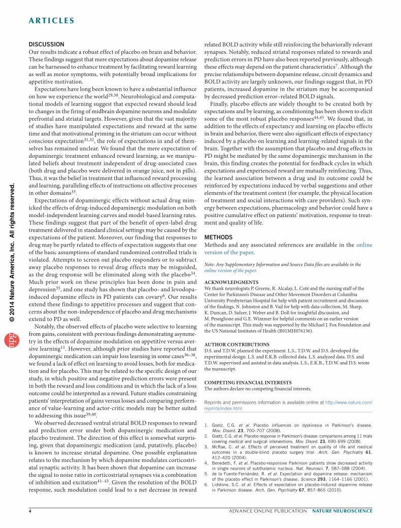
DISCUSSIONOur results indicate a robust effect of placebo on brain and behavior. These findings suggest that mere expectations about dopamine release can be harnessed to enhance treatment by facilitating reward learning as well as motor symptoms, with potentially broad implications for appetitive motivation.
Expectations have long been known to have a substantial influence on how we experience the world29,30. Neurobiological and computa-tional models of learning suggest that expected reward should lead to changes in the firing of midbrain dopamine neurons and modulate prefrontal and striatal targets. However, given that the vast majority of studies have manipulated expectations and reward at the same time and that motivational priming in the striatum can occur without conscious expectation31,32, the role of expectations in and of them-selves has remained unclear. We found that the mere expectation of dopaminergic treatment enhanced reward learning, as we manipu-lated beliefs about treatment independent of drug-associated cues (both drug and placebo were delivered in orange juice, not in pills). Thus, it was the belief in treatment that influenced reward processing and learning, paralleling effects of instructions on affective processes in other domains33.
Expectations of dopaminergic effects without actual drug mim-icked the effects of drug-induced dopaminergic modulation on both model-independent learning curves and model-based learning rates. These findings suggest that part of the benefit of open-label drug treatment delivered in standard clinical settings may be caused by the expectations of the patient. Moreover, our finding that responses to drug may be partly related to effects of expectation suggests that one of the basic assumptions of standard randomized controlled trials is violated. Attempts to screen out placebo responders or to subtract away placebo responses to reveal drug effects may be misguided, as the drug response will be eliminated along with the placebo34. Much prior work on these principles has been done in pain and depression35, and one study has shown that placebo- and levodopa-induced dopamine effects in PD patients can covary6. Our results extend these findings to appetitive processes and suggest that con-cerns about the non-independence of placebo and drug mechanisms extend to PD as well.
Notably, the observed effects of placebo were selective to learning from gains, consistent with previous findings demonstrating asymme-try in the effects of dopamine modulation on appetitive versus aver-sive learning11. However, although prior studies have reported that dopaminergic medication can impair loss learning in some cases36–38, we found a lack of effect on learning to avoid losses, both for medica-tion and for placebo. This may be related to the specific design of our study, in which positive and negative prediction errors were present in both the reward and loss conditions and in which the lack of a loss outcome could be interpreted as a reward. Future studies constraining patients’ interpretation of gains versus losses and comparing perform-ance of value-learning and actor-critic models may be better suited to addressing this issue39,40.
We observed decreased ventral striatal BOLD responses to reward and prediction error under both dopaminergic medication and placebo treatment. The direction of this effect is somewhat surpris-ing, given that dopaminergic medication (and, putatively, placebo) is known to increase striatal dopamine. One possible explanation relates to the mechanism by which dopamine modulates corticostri-atal synaptic activity. It has been shown that dopamine can increase the signal to noise ratio in corticostriatal synapses via a combination of inhibition and excitation41–43. Given the resolution of the BOLD response, such modulation could lead to a net decrease in reward
related BOLD activity while still reinforcing the behaviorally relevant synapses. Notably, reduced striatal responses related to rewards and prediction errors in PD have also been reported previously, although these effects may depend on the patient characteristics7. Although the precise relationships between dopamine release, circuit dynamics and BOLD activity are largely unknown, our findings suggest that, in PD patients, increased dopamine in the striatum may be accompanied by decreased prediction error–related BOLD signals.
Finally, placebo effects are widely thought to be created both by expectations and by learning, as conditioning has been shown to elicit some of the most robust placebo responses44,45. We found that, in addition to the effects of expectancy and learning on placebo effects in brain and behavior, there were also significant effects of expectancy induced by a placebo on learning and learning-related signals in the brain. Together with the assumption that placebo and drug effects in PD might be mediated by the same dopaminergic mechanism in the brain, this finding creates the potential for feedback cycles in which expectations and experienced reward are mutually reinforcing. Thus, the learned association between a drug and its outcome could be reinforced by expectations induced by verbal suggestions and other elements of the treatment context (for example, the physical location of treatment and social interactions with care providers). Such syn-ergy between expectations, pharmacology and behavior could have a positive cumulative effect on patients’ motivation, response to treat-ment and quality of life.
METHODSMethods and any associated references are available in the online version of the paper.
Note: Any Supplementary Information and Source Data files are available in the online version of the paper.
AcknowledgmenTSWe thank neurologists P. Greene, R. Alcalay, L. Coté and the nursing staff of the Center for Parkinson’s Disease and Other Movement Disorders at Columbia University Presbyterian Hospital for help with patient recruitment and discussion of the findings, N. Johnston and B. Vail for help with data collection, M. Sharp, K. Duncan, D. Sulzer, J. Weber and B. Doll for insightful discussion, and M. Pessiglione and G.E. Wimmer for helpful comments on an earlier version of the manuscript. This study was supported by the Michael J. Fox Foundation and the US National Institutes of Health (R01MH076136).
AUTHoR conTRIBUTIonSD.S. and T.D.W. planned the experiment. L.S., T.D.W. and D.S. developed the experimental design. L.S. and E.K.B. collected data. L.S. analyzed data. D.S. and T.D.W. supervised and assisted in data analysis. L.S., E.K.B., T.D.W. and D.S. wrote the manuscript.
comPeTIng FInAncIAl InTeReSTSThe authors declare no competing financial interests.
Reprints and permissions information is available online at http://www.nature.com/reprints/index.html.
1. Goetz, C.G. et al. Placebo influences on dyskinesia in Parkinson’s disease. Mov. Disord. 23, 700–707 (2008).
2. Goetz, C.G. et al. Placebo response in Parkinson’s disease: comparisons among 11 trials covering medical and surgical interventions. Mov. Disord. 23, 690–699 (2008).
3. McRae, C. et al. Effects of perceived treatment on quality of life and medical outcomes in a double-blind placebo surgery trial. Arch. Gen. Psychiatry 61, 412–420 (2004).
4. Benedetti, F. et al. Placebo-responsive Parkinson patients show decreased activity in single neurons of subthalamic nucleus. Nat. Neurosci. 7, 587–588 (2004).
5. de la Fuente-Fernández, R. et al. Expectation and dopamine release: mechanism of the placebo effect in Parkinson’s disease. Science 293, 1164–1166 (2001).
6. Lidstone, S.C. et al. Effects of expectation on placebo-induced dopamine release in Parkinson disease. Arch. Gen. Psychiatry 67, 857–865 (2010).
©20
14 N
atu
re A
mer
ica,
Inc.
All
rig
hts
res
erve
d.
nature neurOSCIenCe advance online publication �
a r t I C l e S
7. Voon, V. et al. Mechanisms underlying dopamine-mediated reward bias in compulsive behaviors. Neuron 65, 135–142 (2010).
8. de la Fuente-Fernández, R. et al. Dopamine release in human ventral striatum and expectation of reward. Behav. Brain Res. 136, 359–363 (2002).
9. Schweinhardt, P., Seminowicz, D.A., Jaeger, E., Duncan, G.H. & Bushnell, M.C. The anatomy of the mesolimbic reward system: a link between personality and the placebo analgesic response. J. Neurosci. 29, 4882–4887 (2009).
10. Zubieta, J.K. & Stohler, C.S. Neurobiological mechanisms of placebo responses. Ann. NY Acad. Sci. 1156, 198–210 (2009).
11. Pessiglione, M., Seymour, B., Flandin, G., Dolan, R.J. & Frith, C.D. Dopamine-dependent prediction errors underpin reward-seeking behavior in humans. Nature 442, 1042–1045 (2006).
12. Sutton, R.S. & Barto, A.G. Reinforcement Learning: an Introduction (Bradford Book, 1998).
13. Barto, A.G. Reinforcement learning and dynamic programming. in Analysis, Design and Evaluation of Man-Machine Systems, Vol. 1,2 (ed. Johannsen, G.) 407–412 (1995).
14. Daw, N.D. in Trial-by-trial data analysis using computational models. Decision Making, Affect, Learning: Attention and Performance XXIII, Vol. 1 (eds. Delgado, M.R., Phelps, E.A. & Robbins, T.W.) 3–38 (Oxford University Press, 2011).
15. Daw, N.D., O’Doherty, J.P., Dayan, P., Seymour, B. & Dolan, R.J. Cortical substrates for exploratory decisions in humans. Nature 441, 876–879 (2006).
16. Hare, T.A., O’Doherty, J., Camerer, C.F., Schultz, W. & Rangel, A. Dissociating the role of the orbitofrontal cortex and the striatum in the computation of goal values and prediction errors. J. Neurosci. 28, 5623–5630 (2008).
17. Levy, D.J. & Glimcher, P.W. The root of all value: a neural common currency for choice. Curr. Opin. Neurobiol. 22, 1027–1038 (2012).
18. Plassmann, H., O’Doherty, J.P. & Rangel, A. Appetitive and aversive goal values are encoded in the medial orbitofrontal cortex at the time of decision making. J. Neurosci. 30, 10799–10808 (2010).
19. McClure, S.M., Berns, G.S. & Montague, P.R. Temporal prediction errors in a passive learning task activate human striatum. Neuron 38, 339–346 (2003).
20. O’Doherty, J.P., Dayan, P., Friston, K., Critchley, H. & Dolan, R.J. Temporal difference models and reward-related learning in the human brain. Neuron 38, 329–337 (2003).
21. Daw, N.D. & Doya, K. The computational neurobiology of learning and reward. Curr. Opin. Neurobiol. 16, 199–204 (2006).
22. Rushworth, M.F., Mars, R.B. & Summerfield, C. General mechanisms for decision making. Curr. Opin. Neurobiol. 19, 75–83 (2009).
23. Chowdhury, R. et al. Dopamine restores reward prediction errors in old age. Nat. Neurosci. 16, 648–653 (2013).
24. Schönberg, T., Daw, N.D., Joel, D. & O’Doherty, J.P. Reinforcement learning signals in the human striatum distinguish learners from nonlearners during reward-based decision making. J. Neurosci. 27, 12860–12867 (2007).
25. Schönberg, T. et al. Selective impairment of prediction error signaling in human dorsolateral but not ventral striatum in Parkinson’s disease patients: evidence from a model-based fMRI study. Neuroimage 49, 772–781 (2010).
26. Behrens, T.E., Hunt, L.T., Woolrich, M.W. & Rushworth, M.F. Associative learning of social value. Nature 456, 245–249 (2008).
27. Li, J., Delgado, M.R. & Phelps, E.A. How instructed knowledge modulates the neural systems of reward learning. Proc. Natl. Acad. Sci. USA 108, 55–60 (2011).
28. Niv, Y., Edlund, J.A., Dayan, P. & O’Doherty, J.P. Neural prediction errors reveal a risk-sensitive reinforcement-learning process in the human brain. J. Neurosci. 32, 551–562 (2012).
29. Kirsch, I. Response expectancy as a determinant of experience and behavior. Am. Psychol. 40, 1189–1202 (1985).
30. Wager, T.D. et al. Placebo-induced changes in FMRI in the anticipation and experience of pain. Science 303, 1162–1167 (2004).
31. Pessiglione, M. et al. Subliminal instrumental conditioning demonstrated in the human brain. Neuron 59, 561–567 (2008).
32. Schmidt, L., Palminteri, S., Lafargue, G. & Pessiglione, M. Splitting motivation: unilateral effects of subliminal incentives. Psychol. Sci. 21, 977–983 (2010).
33. Benedetti, F. How the doctor’s words affect the patient’s brain. Eval. Health Prof. 25, 369–386 (2002).
34. Fournier, J.C. et al. Antidepressant drug effects and depression severity: a patient-level meta-analysis. J. Am. Med. Assoc. 303, 47–53 (2010).
35. Kirsch, I. & Sapirstein, G. Listening to Prozac but hearing placebo: a meta-analysis of antidepressant medication. Prev. Treat. 1, 2a (1998).
36. Bódi, N. et al. Reward-learning and the novelty-seeking personality: a between- and within-subjects study of the effects of dopamine agonists on young Parkinson’s patients. Brain 132, 2385–2395 (2009).
37. Frank, M.J., Seeberger, L.C. & O’Reilly, R.C. By carrot or by stick: cognitive reinforcement learning in parkinsonism. Science 306, 1940–1943 (2004).
38. Palminteri, S. et al. Pharmacological modulation of subliminal learning in Parkinson’s and Tourette’s syndromes. Proc. Natl. Acad. Sci. USA 106, 19179–19184 (2009).
39. Maia, T.V. Reinforcement learning, conditioning, and the brain: successes and challenges. Cogn. Affect. Behav. Neurosci. 9, 343–364 (2009).
40. Gold, J.M. et al. Negative symptoms and the failure to represent the expected reward value of actions: behavioral and computational modeling evidence. Arch. Gen. Psychiatry 69, 129–138 (2012).
41. Bamford, N.S. et al. Heterosynaptic dopamine neurotransmission selects sets of corticostriatal terminals. Neuron 42, 653–663 (2004).
42. Wang, W. et al. Regulation of prefrontal excitatory neurotransmission by dopamine in the nucleus accumbens core. J. Physiol. (Lond.) 590, 3743–3769 (2012).
43. Bamford, N.S. et al. Dopamine modulates release from corticostriatal terminals. J. Neurosci. 24, 9541–9552 (2004).
44. Colloca, L. et al. Learning potentiates neurophysiological and behavioral placebo analgesic responses. Pain 139, 306–314 (2008).
45. Voudouris, N.J., Peck, C.L. & Coleman, G. The role of conditioning and verbal expectancy in the placebo response. Pain 43, 121–128 (1990).
©20
14 N
atu
re A
mer
ica,
Inc.
All
rig
hts
res
erve
d.
nature neurOSCIenCe doi:10.1038/nn.3842
ONLINE METHODSParticipants. We recruited 21 patients with mild-to-moderate PD (13 male, 8 female) through neurologists at Columbia University’s Center for Parkinson’s Disease and Other Movement Disorders and advertisements on the websites of the Michael J. Fox Foundation and the Parkinson’s Disease Foundation. The Columbia University Medical Center Institutional Review Board approved the study procedures and we obtained informed written consent from all participants. During recruitment, patients were told that the study aim was to test how PD medications, such as carbidopa/levodopa, affects learning, memory and decision-making and the way the brain functions. Thus, until a final debriefing session at the end of the experiment, participants remained blind to the placebo. Patients were remunerated for their time ($20 per h), as well as 5% of their winnings averaged across the six learning task runs.
Before participation, all of the patients were screened using the following inclu-sion criteria: diagnosis of mild-to-moderate PD (Hoehn and Yahr stages 1 to 3), age between 50 and 85 years, treatment of PD by carbidopa/levodopa for at least 3 months, responsive to PD medication, medication schedule with at least two and up to four daily doses (for example, morning, lunch, afternoon, bedtime), no report of psychiatric history or neurological disease other than PD, normal to corrected-to-normal vision, and right handedness. It has been shown that, even at the initial diagnosis, PD patients have already lost a substantial amount of striatal dopamine46. Thus, for this initial study, we deliberately recruited patients at a stage with substan-tial dopamine loss and enough experience with the medication to elicit expectations about its effects while having mild enough symptoms to perform a cognitive task in an MRI scanner. A formal power analysis was not used to select the sample size a priori, as the effect sizes could not be estimated before the study. The sample size was chosen to be similar to those generally employed in the field.
All patients were treated with levodopa/carbidopa. In addition, patients were prescribed the following PD medications: Selegiline (five patients), Rasagiline (five patients), Amantadine (four patients), Entacapone (one patient) and Pramiprexole (one patient). Because these medications were part of the PD treatment, they were crushed and dissolved in orange juice together with the levodopa/carbidopa medication. Some patients also were prescribed antide-pressant and anxiolytic treatment that included Nortriptyline (two patients), Amitriptyline (one patient), Citalopram (one patient), Clonazepam (two patients) and Lorazepam (two patients). On the morning of the experiment, patients did not take any antidepressant or anxiolytic medications.
We excluded six patients from analysis as a result of extreme anxiety related to scanning (n = 4, 2 of these 4 patients agreed to participate behaviorally, the other 2 patients withdrew completely), abnormally fast overall reaction times (3 s.d. below the group average response times), suggesting a failure to follow task instructions (n = 1, excluded from both behavioral and imaging data), and poor fMRI image quality (n = 1, included in the behavioral data analysis). In sum, we included 18 patients in the behavioral data analysis and 15 patients in the fMRI data analysis.
experimental design. To minimize variance resulting from individual differences, we employed a within-subject design in which each patient was scanned three times: off drug, on placebo and on drug (Supplementary Fig. 1a). Patients refrained from taking their morning dose of PD medication, following conventional procedures for comparing PD patients on versus off medication47–50. Patients were withdrawn from their medication for at least 16 h. The order of the off drug and placebo sessions was counterbalanced across patients; however, the on drug condition was always last, as the half-life of levodopa/carbidopa combina-tion requires at least 7 h to be metabolized and excreted. Placebo and levodopa/carbidopa treatment were each administered 30 min before scanning.
Placebo administration. On the morning of the experiment and before scan-ning, patients were interviewed regarding their daily medication schedule. In particular, patients were asked: how many daily doses they take, at what time of the day, and if and when they feel an improvement from the drug. Patients were told that because of the initial time to ‘kick in’ between 30 and 45 min, their typi-cal daily dose of medication would be administered 30 min before scanning and thus, together with the time it will take to place them in the scanner (15 min), they could expect to be at their best at start of the learning task.
All patients were off their PD medication during the placebo scan session, but they believed that they had taken their medication. 30 min before the placebo
scan session started, the patients observed the experimenter crushing and dis-solving their daily morning PD medication into a glass of orange juice. Notably, to avoid carry over into subsequent sessions, we told patients that the reason for crushing the medication (that is, destroying its coating) and dissolving it into orange juice was to make it ‘faster acting’ and ‘shorter lasting’. Unbeknownst to the patients, we replaced the glass of orange juice containing their medication with a glass of orange juice containing placebo (starch) pills. Thus, participants were led to expect that they were taking their typical medication, and it was implied that the medication would help them function optimally on all tasks, without providing specific expectancies about performance. Participants were not informed that their medication, or dopamine in general, might specifically improve reward-based learning; however, it is likely that participants expected that performance in general on all tasks might depend on dopamine and be affected by their daily medication in some positive way.
All patients enrolled in this study were on medication schedules of 2–4 doses per d (for example, morning, lunch, afternoon, bedtime). If the placebo session was the first scan session, patients were told they were taking their morning dose; if the placebo session was the second scan session, patients were told they were taking a late morning/lunch dose; and for the third on drug scan, patients were told that they were taking a late lunch/early afternoon dose.
The rationale for this placebo manipulation was that patients may benefit from conditioned responses that have built up over the duration of their Parkinson’s therapy. Our goal was to address the question of whether the expectation of relief from PD symptoms associated to taking a medication affects reward learning and learning-related signals in the brain. By making the patients believe that they were taking their daily medication, we intended to induce patient-specific expectations and associations, which are built up over months and years of experience with the positive and negative contingencies of a daily medication and cannot be elicited by an unfamiliar study drug.
drug administration. The same procedure was repeated 30 min before the third scan session (on drug session). Again, the patients observed the experimenter crushing and dissolving their second dose of daily PD medication into orange juice; however, this time glasses were not switched and the patients were admin-istered their medication.
Timing of sessions. Given that we administered both the placebo and drug 30 min before scanning, each scan session lasted for 60 min and there was a 60-min break, the placebo session and the following session were separated by 2 h 30 min. This timing follows the design used by a PET study of placebo effects on dopamine release.
Although we cannot completely rule out the possibility of a persistence of the placebo effect into subsequent scan sessions (that is, off or on drug), we believe it is unlikely to confound our findings. The critical comparison between the placebo and off drug conditions was counterbalanced across patients. Any persistence of placebo effects into subsequent off drug sessions would decrease differential effects (that is, placebo versus off drug) on behavior and brain activity. Moreover, recent work on placebo in PD suggests that placebo effects on PD last on the order of 30 min (refs. 51,52), although much remains to be learned about the time course of placebo effects across samples and placebo inductions.
debriefing on placebo manipulation. At the end of the experiment, we informed patients that one of the two drug doses administered during test-ing was a placebo, and they were asked to guess which dose was the placebo. Five patients guessed incorrectly (that is, they thought the placebo was the real drug), five patients guessed correctly, and seven patients reported that they could not guess.
effects of treatment on clinical symptoms. After each scan session, emotional symptoms were assessed using the Starkstein Apathy Scale53 and the State-Trait Anxiety Inventory (STAI)54. Working memory was assessed with the Digit Span, and motor symptoms were assessed with the Unified Parkinson’s Disease Rating Scale III (UPDRS III)55. In addition, expectancy ratings were collected before each scan session: patients indicated how much they expected their PD symptoms to improve following treatment using a visual analog scale ranging from 0 (no improvement) to 100 (maximum improvement).

©20
14 N
atu
re A
mer
ica,
Inc.
All
rig
hts
res
erve
d.
nature neurOSCIenCedoi:10.1038/nn.3842
Paired one-sample t tests between treatment conditions were not significant for apathy, anxiety or digit span measures. However, expectancy scores indicated that patients expected significantly more improvement after having been admin-istered a placebo or their drug, relative to the off drug condition (Placebo>Off: t17 = 2.9, P < 0.05; On>Off: t17 = 3.7, P < 0.05, two-tailed, paired t test; Supplementary Table 2).
We videotaped patients while the UPDRS III was assessed (no rigidity meas-ures were collected), and an experimenter blind to treatment rated motor symp-toms. We observed a significant improvement resulting from being on drug in comparison to being either on placebo (On>Placebo: t17 = −5.1, P < 0.001, two tailed) or off drug (On>Off: t17 = −4.1, P < 0.001, two tailed). Given that differ-ent individuals typically show medication-related improvements on a unique set of patient-specific motor behaviors, and patients were in the mild-to-moderate stages of the disease that may restrict drug effects to specific symptoms, we also calculated UPDRS III scores for off and placebo across the specific subset of items that showed a drug effect (items where On – Off < 0) for each patient. We found that, for the individualized sub-items that showed an improvement with medication, there was an improvement with placebo (Placebo<Off: 7.86 ± 1.21 versus 10.2 ± 1.59: t17 = 4.2, P < 0.001, two-tailed, paired t test).
Behavioral task. In each of the three fMRI scan sessions, patients performed two runs of an instrumental learning task. The aim of the task was to maximize monetary payoff. Each run consisted of 64 trials: 32 gain trials and 32 loss trials. Gain and loss trials were randomly intermixed in a run. On each trial, two shapes were displayed on a computer screen, and patients chose one of them to receive a monetary feedback. Choices were followed by one of three possible outcomes. Subjects could win $10, get nothing ($0) or lose $10. In the GAIN condition choosing the optimal (that is, correct) shape was reinforced with a reward of $10 on 75% of trials (25% of trials were non-reinforced), whereas choosing the non-optimal (that is, incorrect) shape was not reinforced ($0) in 75% of trials (25% of the trials were rewarded $10). In the LOSS condition, the optimal choice (that is, correct) was not reinforced ($0) on 75% of trials (but was punished on 25% of trials −$10), and the non-optimal (that is, incorrect) choice was punished with a loss of $10 on 75% of trials (but 25% of trials were not punished $0). Thus, in gain trials we tested the ability to learn from financial rewards (reward learning), and in loss trials we tested the ability to avoid financial punishment (loss learning).
We jittered the duration of inter-trial intervals (2–6 s) and of the choice- feedback interval (0.5–3 s) by drawing from an exponential distribution. We paid the patients according to their performance (5% of the payoff averaged across the six learning task runs), in addition to compensation for their participation.
Individual differences in drug and placebo effects. The effects of drug and placebo on the slope of reward learning trended toward a correlation across subjects (zero-order correlation of placebo and drug effects: r = 0.59, P < 0.01; partial correlation controlling for Off drug: r = 0.34, P = 0.08, one tailed; Supplementary Fig. 2). An exploratory analysis of the UPDRS III scores found parallel effects of placebo and drug for clinically relevant motor symptoms of PD (zero-order correlation placebo and drug effects: r = 0.68, P < 0.001; partial correlation controlling for Off drug: r = 0.59, P < 0.01).
computational model. We used a standard temporal difference learning algorithm to calculate the expected value of choices (that is, choice value) and prediction error based on individual trial-by-trial choices and feedback56. The expected value (that is, choice value), termed a Q value, corresponded to the expected reward obtained by choosing a particular cue. Q values were set to zero at the beginning of each run. After each trial, t > 0, the value of the chosen cue was updated according to the rule Qchosen_cue(t + 1) = Qchosen_cue(t) + α*δ(t). Central to this algorithm is prediction error, δ(t), which is defined as δ(t) = R(t) − Qchosen_cue(t), or the difference between expected feedback Qchosen_cue(t) and actual feedback R(t). The reward magnitude was set +1, 0 and −1 for outcomes +$10, $0 and −$10, respectively.
Given the Q values, the probability of selecting each action was calculated by implementing the softmax rule
P t Q t Q tchosen_c e chosen_cue chosen_cueu ( ) ( ) ( )= exp / / exp /( ) ( (b b))
( )exp /+ ( )Q tunchosen_cue b
The two free parameters, α (learning rate) and β (temperature), were fit indi-vidually for each subject to maximize the probability of actual choices under the model. To improve the model’s subsequent fit to fMRI data, we used the average estimates for α and β calculated across patients in each treatment condition.
Note that we tested potential confounds resulting from differences in learn-ing rate and temperature between gain and loss trials. In a control analysis, one learning rate (α = 0.2) and one temperature (β = 0.4) fitted across all subjects, treatments and trial conditions were used to calculate trial-by-trial expected val-ues and prediction errors. The control fMRI results were consistent with main findings and are reported below.
comparison of reward learning model fits between treatments. To rule out potential confounds resulting from qualitative differences in model fit between off drug, placebo and on drug, the following standard measures of model fit were calculated for each subject in each treatment condition: the Bayesian Information Criterion defined by BIC = 2*LLE+2*log(n), the Akaike Information Criterion defined by AIC = 2 * LLE + 2 * 2, and pseudoR2 values defined by pseudo R2 = 1−LLEmodel/LLEchance. Moreover, we tested how well the reward learning model fit the observed data compared with chance by performing a likeli-hood ratio test defined by D = 2*LLEmodel − 2*LLEchance. LLEmodel corresponds to the maximum logarithmic likelihood of the observed choices under the model. LLEchance corresponds to the logarithmic likelihood of choices at chance (LLEchance = n*log(0.5), with n = number of trials). The average of each measure across patients in each condition and treatment are reported in Supplementary Table 1.
Analysis of variance compared pseudoR2 values across treatments (off drug, placebo, on drug) and across learning conditions (gains, losses) and revealed no main effect of treatment (F2,107 = 0.79, P = 0.45), no main effect of gains versus losses (F1,107 = 2.38, P = 0.12), and no interaction of treatment by gains versus losses (F2,107 = 0.16, P = 0.85). Post hoc, one-tailed t tests further tested differences in pseudoR2 values across treatments. These too revealed no significant differences between treatments (pseudoR2 values: On>Off gains: t17 = 1.16, P = 0.12; losses: t17 = 0.50, P = 0.30; Placebo Off gains: t17 = 0.26, P = 0.39; losses: t17 = −0.34, P = 0.63; On>Placebo gains: t17 = 0.83, P = 0.20; losses: t17 = 1.17, P = 0.13).
Treatment effects on reinforcement learning parameters. To investigate indi-vidual differences between treatment conditions, we individually estimated the free parameters, learning rate, and inverse softmax temperature, which are also referred to as random effect estimations (that is, one set of parameters for each subject for each treatment condition). The average learning rates and temperature across subjects in each treatment condition are summarized in Supplementary Table 1. Analysis of variance tested for main effects of treatment, gains versus losses, and the interaction of treatment by gains versus losses in learning rate and softmax inverse temperature, separately.
For the learning rate, we found no main effect of treatment (F2,89 = 0.94, P = 0.39), a significant main effect of trial type (F1,89 = 3.65, P < 0.05), and a trend interaction treatment by gains versus losses (F2,89 = 2.4, P = 0.09). When comparing the treatments during learning from gains, patients on placebo had significantly smaller learning rates compared to off drug (Off>Placebo: t14 = 2.6, P < 0.05) and smaller learning rates on drug compared to off drug (Off>On: t14 = 1.5, P = 0.16), with no differences between placebo and on drug (On>Placebo: t14 = 1.24, P = 0.11). The numerical differences in learning rate across treatment conditions, although less robust statistically, were consistent with behavioral performances and suggest that placebo led to more incremental learning. We did not find differences between treatment conditions for learning rates in the loss condition (Off>Placebo: t14 = −0.73, P = 0.47; Off>On: t14 = −1.79, P = 0.18; On>Placebo: t14 = 0.97, P = 0.34; Supplementary Table 1).
Behavioral data analysis. Data collection and analysis were not performed blind to the treatments of the experiment, but all analyses were conducted using automated methods and a priori statistical tests to avoid the possibility of experimenter bias. All of the motor scores (UPDRS III) were coded by a rater blind to the experimental treatments.
To assess learning, the 32 gain and the 32 loss trials were each binned into four blocks of eight trials (first, second, third and fourth block). Within each block, we calculated the percentage of optimal choices, resulting in a learning curve

©20
14 N
atu
re A
mer
ica,
Inc.
All
rig
hts
res
erve
d.
nature neurOSCIenCe doi:10.1038/nn.3842
composed of these four optimal choice scores for each patient. These curves reflected the increase in the proportion of correct choices across time (that is, first, second, third and fourth block) as learning progressed. Multilevel general linear models (GLMs) using iterative generalized least-squares (IGLS)57 tested for drug and placebo effects on reward and loss learning.
GLM1 tested for a drug effect and included three regressors: Block, On>Off drug, Block*On>Off drug.
Block (dummy coded: −3, −1, 1, 3) tested the increase of learning across time (that is, the slope of learning over the four blocks of eight trials). On>Off drug tested for a main effect of drug compared to Off drug (−1 for Off drug, 1 for On drug). Block*On>Off drug tested if the slope of learning on drug was enhanced compared to off drug.
GLM2 tested for a placebo effect, analogous to GLM1: Block, Placebo>Off drug, Block*Placebo>Off drug.
GLM3 tested for the selectivity of effects on reward learning compared to pun-ishment learning. It included six regressors: Block, Placebo>Off, Gains>Losses, Block*Gains>Losses, Placebo>Off drug*Gains>Losses, Block*Placebo>Off drug*Gains>Losses.
We fit individual regression coefficients obtained for each regressor into a second-level group analysis to provide population inferences against zero. We entered the order of placebo administration as a second level covariate (see below for further control analyses of potential confounds due to treatment order). We report the significance of regression coefficients at a threshold of P < 0.05, one tailed.
We applied an analogous analysis to parameter estimates extracted from ROIs in the brain and reaction times for choices (see below).
To test whether drug and placebo effects on reward learning covaried on the individual level, we obtained individual slopes of reward learning by multiplying the four trial blocks (dummy coded: −3, −1, 1, 3) by the percentage of optimal choices in each block. The differences in learning slopes, on – off drug and placebo – off drug, were then correlated across all patients by using Pearson’s correlation coefficient and partial correlations controlling for off drug (Supplementary Fig. 2). An analogous correlation analysis was done to test for individual placebo (that is, placebo – off drug) and drug effects (that is, on – off drug) on motor symptoms measured by the UPDRS III (Supplementary Fig. 2).
All statistical tests were conducted with the Matlab Statistical Toolbox (Matlab 2010b, MathWorks) and a Matlab implementation of IGLS. No statistical methods were used to predetermine sample sizes, but our sample sizes are similar to those generally employed in the field. Data distribution was assumed to be normal and was examined visually to check for normality. Statistical tests for normality were not conducted, because they are generally inconclusive for sample sizes in the range employed here.
Reaction times. Reaction times (RTs) measured the time patients took to choose between the two shape choices on each trial. Average RTs for each patient were calculated for gain and loss trials separately. To test whether there were significant differences in RTs across trials, we applied the same behavioral analysis used to assess learning to the RT data. RTs for gain and loss trials were averaged across blocks of eight trials separately. Two GLMs, analogous to the GLMs that analyzed optimal choices, tested for placebo and drug effects, respectively. RTs decreased with time in the gain condition (t14 = −6.1, P < 0.05), but not in the loss condition (t14 = −0.64, P = 0.5) (Supplementary Fig. 5). We found no significant effect of either drug or placebo on RTs.
learning in the subset of fmRI patients. Behavioral results in the 15 fMRI patients were consistent with the findings observed across the whole patient group. Learning from gains was enhanced by dopaminergic medication (Block*On>Off: t11 = 1.8, P < 0.05) and by placebo (Block*Placebo>Off: t11 = 2.3, P < 0.05) (Supplementary Figs. 6 and 7). We did not observe treatment effects for learning from losses in the subset of fMRI patients either.
order effects. Although the order of placebo and off drug sessions was counter-balanced across patients, the on drug session was always the last session of the experiment. To control for order effects, we regressed the order (dummy coded: 1, 2, 3) of the scan sessions against optimal choices and parameter estimates from the vmPFC and ventral striatum separately. For both the gain and the loss
condition, no significant effect of order was observed (on optimal choices in gain trials: t16 = 0.89, P > 0.05; in loss trials: t16 = 1.6, P > 0.05; on parameter estimates from the vmPFC for choice value in gain trials: t13 = 0.65, P > 0.05; in loss trials: t13 = −0.10, P > 0.05; on parameter estimates from the ventral striatum for predic-tion error in gain trials: t13 = −1.16, P > 0.05; in loss trials: t13 = −0.2, P > 0.05). To further test if the order of the placebo administration interacted with placebo effects, order (dummy coded: −1 for placebo first, 1 for placebo second) was a second level covariate in the GLMs testing for placebo effects on optimal choices and parameter estimates from the vmPFC and ventral striatum.
No second level interactions between order and block (t14 = 0.6, P > 0.05), Placebo>Off drug (t14 = 0.04, P > 0.05) or Block*Placebo>Off drug (t14 = −0.10, P > 0.05) were found for learning from gains. Similarly, no second-level inter-actions between order and block (t14 = −0.83, P > 0.05), Placebo>Off drug (t14 = 0.8, P > 0.05) or Block*Placebo>Off drug (t14 = 1.4, P > 0.05) were observed for learning from losses.
Consistent with the behavioral findings, order of placebo administration did not interact with the effect of block (gains: t11 = –0.13, P > 0.05; losses: t11 = 0.5, P > 0.05), Placebo>Off drug (gains: t11 = −0.67, P > 0.05; losses: t11 = −1.1, P = 0.05), On>Off (gains: t11 = –0.24, P > 0.05; losses: t11 = –0.27, P > 0.05), Block*Placebo>Off drug (gains: t11 = 0.59, P > 0.05; losses: t11 = −0.78, P > 0.05) or Block*On>Off drug (gains: t11 = –0.27, P > 0.05; losses: t11 = −1.18, P > 0.05), choice value activation in the vmPFC or prediction error activation in the ventral striatum (Block (gains: t11 = –0.27, P > 0.05; losses: t11 = 0.5, P > 0.05), Placebo>Off drug (gains: t11 = −0.4, P > 0.05; losses: t11 = 0.25, P > 0.05), On>Off (gains: t11 = –0.13, P > 0.05; losses: t11 = 0.5, P > 0.05), Block*Placebo>Off drug (gains: t11 = −0.25, P > 0.05; losses: t11 = 0.65, P > 0.05) or Block*On>Off drug (gains: t11 = −0.45, P > 0.05; losses: t11 = −1.12, P > 0.05)).
Pleasantness and liking ratings of shapes. Following each run of the learning task, patients were asked to rate their preference and liking of the shapes that were presented during learning. Patients rated preferences on a four-point Lickert scale and liking on a visual analog scale that ranged from most unpleasant to most pleasant. Ratings were subjected to a three-way analysis of variance to test for effects of treatment (off, placebo, on drug), shape (optimal, non-optimal), and task condition (gains, losses). Analysis of variance revealed that patients preferred (F1,211 = 92.04, P < 0.001) and liked (F1,211 = 60.2, P < 0.001) the optimal shape more than the non–optimal shape. This was selective to the gain condi-tion (preferences: F1,211 = 30.7, P < 0.001; liking: F1,211 = 14.97, P < 0.001) as indicated by a significant interaction by task condition (preferences: F1,211 = 2.85, P = 0.09; liking: F1,211 = 6.04, P < 0.05). No main effect of treatment (preferences: F2,211 = 0.66, P = 0.51; liking: F2,211 = 0.76, P = 0.47) or any other interactions were significant.
Image acquisition. We acquired T2*-weighted echo spiral in/out images with blood oxygenation level dependent (BOLD) contrast using a 1.5-T scanner (GE Medical Systems). Each volume comprised of 26 interleaved, 4-mm-thick axial slices and 3.5-mm in-plane resolution, using the following parameters: TR = 2,000 ms, TE = 35 ms, flip angle = 84°, FOV = 192 mm, 64 × 64 matrix. For each subject a total number of 600 volumes were obtained, 300 in each learning task run, corresponding to 10 min of scanning for each run. In addition, a single high-resolution T1-weighted structural image (spoiled gradient echo sequence) was acquired for each patient at the beginning of the on drug scan session, before performing the on drug learning task runs of the experiment.
fmRI analysis. All functional images were screened on quality using in-house software. Specifically, spike and high movement time points were identified and corrected by interpolating adjacent time points. In addition to this, images were screened on fast movement (≥1 mm/TR) and corrected using the Art Repair toolbox58.
Analysis of fMRI data was conducted using the statistical parametric mapping software SPM8 (Wellcome Department of Imaging Neuroscience). We normal-ized the structural scans to a standard T1 template and created a normalized structural average image upon which the t-maps were superimposed for anatomi-cal localization. Preprocessing of the functional scans consisted of spatial realign-ment to the first volume, spatial normalization to a standard T2* template with a resampled voxel size of 2 × 2 mm, and spatial smoothing using a Gaussian kernel

©20
14 N
atu
re A
mer
ica,
Inc.
All
rig
hts
res
erve
d.
nature neurOSCIenCedoi:10.1038/nn.3842
with a full width at half maximum of 6 mm. We analyzed the functional images in an event–related manner, within a GLM, and included subject specific realign-ment parameters into each GLM as covariates to control for motion artifacts.
First, we tested if expected value activated the vmPFC and if prediction error activated the ventral striatum irrespective of trial condition (gains, losses) or time point (first, second, third or fourth block). Two onset regressors (that is, onset choice, onset feedback) were respectively modulated by expected value and prediction error, obtained for each trial by a standard temporal difference learning algorithm that assigns a value to each cue based on the subjects’ previous choices and reward received. Delta functions of onset regressors and parametric modulators were convolved with the canonical hemodynamic response function and regressed against each subject’s fMRI data. Linear contrasts were fit into a second-level random effects analysis, which used one-sample t tests to identify brain regions underpinning expected value and prediction error in each treat-ment condition. We applied small volume correction to determine the effects of expected value and prediction error in each treatment condition with a lenient initial threshold of P < 0.01, uncorrected.
Having identified vmPFC responses to expected value and ventral striatum responses to prediction error, we investigated the specificity of these activations. Building on the behavioral results that suggested that placebo and drug selectively enhance the slope of learning from gains, we used a second GLM to model fMRI data in each subject similarly to the behavioral analysis. Specifically, trials at time of choice and feedback were binned into four regressors per trial condition (that is, gain trials, loss trials). Each onset regressor reflected a given time during the task (first, second, third and fourth block of eight trials) and was modulated by expected value (at choice onset) and prediction error (at feedback onset), respectively. Delta functions of the 16 onset regressors and the 16 parametric modulators were convolved with the canonical hemodynamic response function and regressed against each subject’s fMRI data. Average parameter estimates for the parametric modulators were extracted from ROIs located in the vmPFC (for expected value) and the ventral striatum (for prediction error). Thus, we obtained, per patient for both expected value and prediction error, four parameter estimates for the gain condition and four parameters estimates for the loss condition.
Analysis of parameter estimates. A multilevel linear regression model using IGLS57 tested for drug and placebo effects. Analogous to the analysis of optimal choices, the parameter estimates from each ROI were fit by GLM1 (testing for drug effects), GLM2 (testing for placebo effects) and GLM3 (testing for interac-tion with trial type: gains, losses) from the behavioral analysis. Individual betas were entered into second-level group analyses to provide population inferences against zero. We entered the order of placebo administration (dummy coded: −1 for placebo first, 1 for placebo second) as a second-level covariate. Similarly to our behavioral findings, order did not confound drug and placebo effects. We used a statistical significance threshold of P < 0.05, one tailed, to evaluate regression coefficients.
Note, we observed significant main effects of drug (On>Off drug) and pla-cebo (Placebo>Off drug) on both vmPFC and the ventral striatum parameter estimates. To better display the main effects, parameter estimates were averaged across patients and blocks (that is, the first, second, third and fourth block of eight trials) in Figures 3b and 4b.
RoIs. ROIs in the vmPFC and the ventral striatum were defined a priori from independent fMRI studies. The vmPFC ROI was defined by the Montreal Neurological Institute (MNI) space coordinates [x = −1, y = 27, z = −18] reported for value previously59. The ventral striatum ROI was defined by the MNI coordi-nates [x = −10, y = 12, z = −8] previously reported for striatal prediction errors60. Average parameter estimates (that is, betas) were extracted from a 10-mm diam-eter sphere centered on the vmPFC and ventral striatal coordinates.
direct comparison of treatment conditions. Direct comparisons of treat-ment conditions did not reveal any differences in whole brain activation for expected value or prediction error at an uncorrected threshold of P < 0.001. See Supplementary Figure 8 for whole brain activations at P < 0.05, uncorrected.
effects of scaling choice value and prediction error. One possible interpreta-tion of our findings is that the observed brain activations are due to individual variance in learning. For example, choice values or prediction errors may reach
a different maximum and vary on different scales between treatment conditions and subjects. Thus, to rule out the possibility that these differences in parametric modulators contributed to the observed drug and placebo effects, we z-scored expected values and prediction error regressors. The temporal difference learn-ing algorithm used to obtain trial-by-trial expected values and prediction errors used one optimal learning rate (α = 0.2) and one optimal temperature (β = 0.4), which were fit across all subjects, treatment conditions and trials. This was done in order to avoid additional confounds due to differences in learning models between subjects, treatment, and trial conditions (gains, losses).
Consistent with our main findings, this supplemental GLM analysis, which controlled for scaling and learning model differences, revealed that the placebo and on drug conditions enhanced choice value representation in the vmPFC (Placebo>Off drug: t11 = 1.4, P = 0.08; On>Off drug: t11 = 1.7, P < 0.05) compared to off drug, with no difference between placebo and on drug (On>Placebo: t11 = 0.75, P > 0.05). Prediction error activation of the ventral striatum was decreased (Placebo<Off drug: t11 = 2.1, P < 0.05; On<Off drug: t11 = 1.8, P < 0.05) when patients were treated by a placebo or dopaminergic medication compared to off drug with no difference between placebo and on drug (On<Placebo: t11 = 0.23, P > 0.05). The observed placebo and drug effects were selective to learning from gains. No significant differences in treatment conditions were observed for learning from losses. Thus, we concluded that placebo and drug effects on vmPFC and ventral striatum BOLD responses were not confounded by individual differences in magnitude and scale of learning.
learning related brain responses outside the reward learning framework. To test for learning related responses in vmPFC and ventral striatum outside of a reinforcement learning model framework, we used raw observed responses and replaced the expected value regressor at time of choice with actual observed choices (dummy coded: 1 for optimal choice, −1 for non-optimal choice) and replaced the prediction error regressor at time of outcome by actual observed feedback (dummy coded: 1 for correct, −1 for incorrect). Delta functions of the onset regressors and parametric modulators were convolved with the canonical hemodynamic response function and regressed against each individual’s fMRI data. We then used paired t tests to examine the effects of placebo and drug on parameter estimates in independently defined ROIs in the vmPFC and the ventral striatum.
The results of this analysis are markedly similar to what we observed with the model-derived regressors for expected value and prediction error. Choice-related activation in the vmPFC was enhanced both by drug and by pla-cebo compared to off drug (On>Off drug: t14 = 2.9, P < 0.05; Placebo>Off: t14 = 2.38, P < 0.05, two-tailed, paired t test), with no differences between drug and placebo (On>Placebo: t14 = 1.05, P = 0.3, two tailed) (Supplementary Fig. 3). Moreover, these effects were selective to learning from gains, as indicated by a trend interaction (Gains>Losses*Placebo>Off drug: t14 = 1.96, P = 0.06, two tailed). The interaction was significant for (Gains>Losses*On>Off: t14 = 2.9, P < 0.05, two tailed). No significant differences were found for learning from losses (Supplementary Fig. 3a).
Activation to correct versus incorrect feedback in the ventral striatum was also similar to the results from reward learning–model derived analyses: we found greater activation for correct versus incorrect feedback during learning from gains when patients were off drug compared to on drug (Off>On: t14 = 1.97, P = 0.06, two tailed) or placebo (Off>Placebo: t14 = 2.27, P < 0.05, two tailed). No differences were observed between on drug and placebo (On>Placebo: t14 = 0.63, P = 0.5, two tailed). Again, this finding was selective to learning from gains: the interactions (Gains>Losses*Off>Placebo: t14 = 2.9, P < 0.05, two tailed) and (Gains>Losses*Off>On: t14 = 1.7, P = 0.10, two tailed) were significant. No effects were found for learning from losses (Supplementary Fig. 3b).
Striatal responses to components of the prediction error. To test how treatment affects different aspects of the reinforcement learning signal, we broke down the prediction error signal to its separate algebraic components, expectation and reward (following refs. 61–63), and examined how each of these signals were affected by treatment. Specifically, we built a GLM comprising two onset regres-sors: time of choice and time of outcome. Additionally, at time of outcome, two parametric modulators were included that corresponded to the algebraic com-ponents of the prediction error: expected value of the choice (derived from the reward learning model) and reward magnitude (dummy coded: 1 for a gain of $10,

©20
14 N
atu
re A
mer
ica,
Inc.
All
rig
hts
res
erve
d.
nature neurOSCIenCe doi:10.1038/nn.3842
0 for $0 and −1 for a loss of $10). Delta functions of onset regressors and para-metric modulators were convolved with the canonical hemodynamic response function and regressed against each subject’s fMRI data. Individual parameter estimates for expected value and reward were extracted from the independently defined ventral striatum ROI (MNI coordinates = [–10, 12, −8]; from ref. 60). Two-tailed paired t tests were used to test for treatment effects on both expected value and reward related activation. Although there were no significant treat-ment effects for expected value related ventral striatum activation (On>Off: t14 = 1.14; Placebo>Off: t14 = 0.45 and On>Placebo: t14 = 0.82, P > 0.05), reward magnitude elicited significantly stronger responses off drug compared to placebo (Off>Placebo: t14 = 2.14, P < 0.05) and numerically, but not significantly, stronger responses compared to on drug (Off>On: t14 = 1.63, P = 0.12), with no differences between placebo and on drug (On>Placebo: t14 = 0.21, P > 0.05) (Supplementary Fig. 4). We concluded from these finding that placebo and drug alter the reward component of the prediction error at time of outcome.
control analyses. If placebo and drug affected BOLD activity in general, rather than specifically affecting learning-related activity, then effects of treatment would be expected at task-irrelevant events (for example, fixation) and/or in other brain regions.
Main effect of treatment condition at time of fixation. To test the possibility of a global effect of treatment on BOLD activity, we examined brain activation at time of fixation. The GLM included one regressor at onset fixation. Boxcar functions for the whole fixation duration were convolved with the hemodynamic response function in each trial. Individual contrast images were entered into a second–level random effects analysis. A one-way ANOVA revealed no main effect of treatment conditions (F2,42 = 8.2, P > 0.001, uncorrected).
Treatment effects in control ROIs. To test the possibility of treatment effects on BOLD activity in other brain regions, we defined control ROIs in the primary motor cortex and the primary visual cortex. ROIs were defined anatomically using the AAL package implemented in the WFU-Pickatlas software of SPM8 (ref. 64). The motor cortex ROI comprised the left precentral gyrus and the visual cortex ROI comprised the calcarine sulcus. We applied small volume correction to determine the effects of value and prediction error in each treatment condi-tion with a lenient initial threshold of P < 0.01, uncorrected. We found no effect of drug or placebo on BOLD activity in these control regions for either value or prediction error related activity.
Task instructions. The game consists of 2 sessions, each comprising 64 trials and lasting for 10 min. The first session will be preceded by a training session outside the scanner comprising 32 trials.
The word ‘ready’ will be displayed 6 s before the game starts.In each trial you have to choose between the two cues displayed on the screen,
on the left and right of a central cross.
–to choose the right cue, press the right mouse button–to choose the left cue, press the left mouse button
As soon as you press the button, a white arrow displayed at the bottom of the selected cue will indicate your choice.
As an outcome of your choice you may
-get nothing ($0)-win a ten dollar bill (+$10)
-lose a ten dollar bill (−$10)To have a chance to win, you must make a choice and press one of
the two buttons. If you do nothing, the white arrow will remain at the central cross.
The two cues displayed on a same screen are not equivalent in terms of out-come: with one you are more likely to get nothing than with the other. Each cue has got its own meaning, regardless of where (left or right of the central cross) or when it is displayed.
The aim of the game is to win as much money as possible.Good luck!A Supplementary methods checklist is provided.
46. Kordower, J.H. et al. Disease duration and the integrity of the nigrostriatal system in disease. Brain 136, 2419–2431 (2013).
47. Cools, R., Barker, R.A., Sahakian, B.J. & Robbins, T.W. Enhanced or impaired cognitive function in Parkinson’s disease as a function of dopaminergic medication and task demands. Cereb. Cortex 11, 1136–1143 (2001).
48. Frank, M.J., Seeberger, L.C. & O’Reilly, R.C. By carrot or by stick: cognitive reinforcement learning in parkinsonism. Science 306, 1940–1943 (2004).
49. Shohamy, D., Myers, C.E., Geghman, K.D., Sage, J. & Gluck, M.A. L-dopa impairs learning, but spares generalization, in Parkinson’s disease. Neuropsychologia 44, 774–784 (2006).
50. Shohamy, D., Myers, C.E., Grossman, S., Sage, J. & Gluck, M.A. The role of dopamine in cognitive sequence learning: evidence from Parkinson’s disease. Behav. Brain Res. 156, 191–199 (2005).
51. Benedetti, F. et al. Placebo-responsive Parkinson patients show decreased activity in single neurons of subthalamic nucleus. Nat. Neurosci. 7, 587–588 (2004).
52. Benedetti, F. et al. Electrophysiological properties of thalamic, subthalamic and nigral neurons during the anti-parkinsonian placebo response. J. Physiol. (Lond.) 587, 3869–3883 (2009).
53. Starkstein, S.E. et al. Reliability, validity, and clinical correlates of apathy in Parkinson’s disease. J. Neuropsychiatry Clin. Neurosci. 4, 134–139 (1992).
54. Spielberger, C.D. State-Trait Anxiety Inventory: Bibliography, 2nd edn. (Consulting Psychologists Press, 1989).
55. Movement Disorder Society Task Force on Rating Scales for Parkinson’s Disease. The Unified Parkinson’s Disease Rating Scale (UPDRS): status and recommendations. Mov. Disord. 18, 738–750 (2003).
56. Sutton, R.S. & Barto, A.G. Reinforcement Learning: an Introduction (Bradford Book, 1998).
57. Lindquist, M.A., Spicer, J., Asllani, I. & Wager, T.D. Estimating and testing variance components in a multi-level GLM. Neuroimage 59, 490–501 (2012).
58. Mazaika, P., Hoeft, F., Glover, G.H. & Reiss, A.L. Methods and software for fMRI analysis of clinical subjects. Organ. Hum. Brain Mapp 475 (2009).
59. Hare, T.A., O’Doherty, J., Camerer, C.F., Schultz, W. & Rangel, A. Dissociating the role of the orbitofrontal cortex and the striatum in the computation of goal values and prediction errors. J. Neurosci. 28, 5623–5630 (2008).
60. Pessiglione, M., Seymour, B., Flandin, G., Dolan, R.J. & Frith, C.D. Dopamine-dependent prediction errors underpin reward-seeking behavior in humans. Nature 442, 1042–1045 (2006).
61. Behrens, T.E., Hunt, L.T., Woolrich, M.W. & Rushworth, M.F. Associative learning of social value. Nature 456, 245–249 (2008).
62. Niv, Y., Edlund, J.A., Dayan, P. & O’Doherty, J.P. Neural prediction errors reveal a risk-sensitive reinforcement-learning process in the human brain. J. Neurosci. 32, 551–562 (2012).
63. Li, J., Delgado, M.R. & Phelps, E.A. How instructed knowledge modulates the neural systems of reward learning. Proc. Natl. Acad. Sci. USA 108, 55–60 (2011).
64. Maldjian, J.A., Laurienti, P.J., Kraft, R.A. & Burdette, J.H. An automated method for neuroanatomic and cytoarchitectonic atlas–based interrogation of fMRI data sets. Neuroimage 19, 1233–1239 (2003).