Making Sense of Science Mike Szydlowski K-12 Science Coordinator Columbia Public Schools.
Upload
ilene-lucasCategory
view
218download
0
ProTECT III ImagingMike Lunney
Research CoordinatorCentral Reading
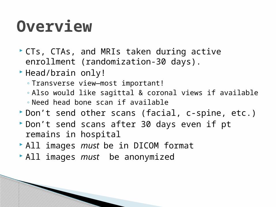
CTs, CTAs, and MRIs taken during active enrollment (randomization-30 days).
Head/brain only!◦ Transverse view—most important!◦ Also would like sagittal & coronal views if available◦ Need head bone scan if available
Don’t send other scans (facial, c-spine, etc.) Don’t send scans after 30 days even if pt
remains in hospital All images must be in DICOM format All images must be anonymized
Overview

Transverse view
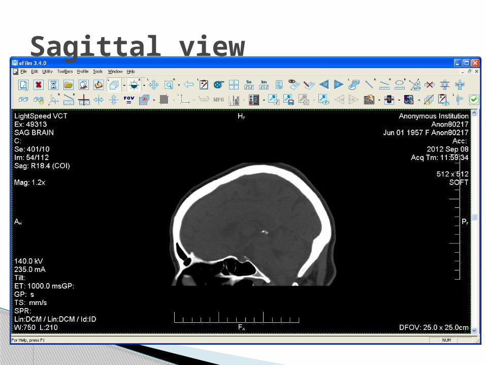
Sagittal view

Coronal view

CTA
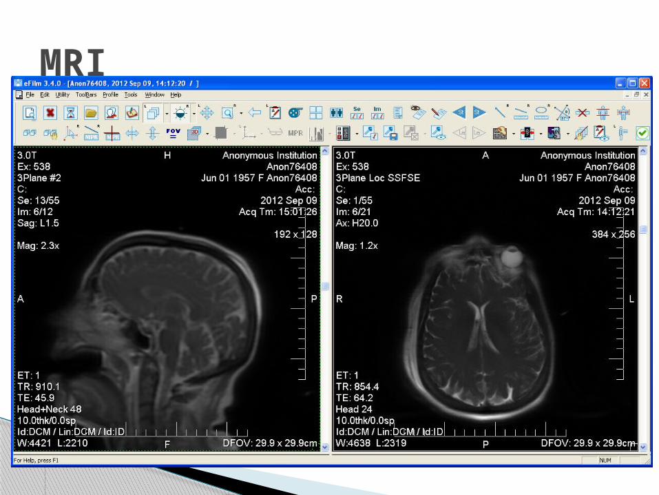
MRI
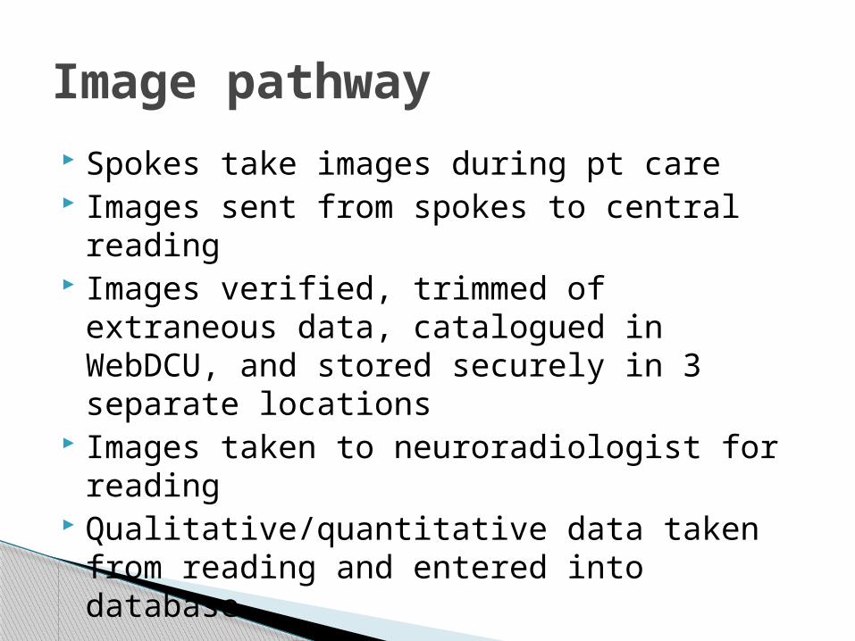
Spokes take images during pt care Images sent from spokes to central reading Images verified, trimmed of extraneous
data, catalogued in WebDCU, and stored securely in 3 separate locations
Images taken to neuroradiologist for reading Qualitative/quantitative data taken from
reading and entered into database.
Image pathway

Physically (FedEx, etc.)◦ Needs to be traceable◦ Typically CDs made by imaging department◦ Image submission form
ProTECT image upload site https://som.emory.org/ProTECT/
◦ FTP site able to handle files up to 200 MB Larger files must be split into parts
◦ Files must be compressed (zipped)
DICOM file type (*.DCM) the only acceptable file type!◦ (Your imaging department knows what that means)
Spoke/hub delivery options

MOST IMPORTANT IMAGE!◦Must be a complete head/brain CT◦Must be a complete head/brain CT!
◦Most likely this will be the first image taken
◦Needs to be within 4 hours of randomization
Baseline image

For uploads only, not required if mailing CDs
####_DDMMMYY_xOFx
CTs: get no extension
MRIs: “filename_MRI”
CTAs: “filename_CTA”
File naming

Example: ◦MRI of patient 1525 taken 11/29/2011◦3rd image of 4 total scans being sent to CR
1525_29NOV11_3OF4_MRI
◦ Split larger files into parts less than 200 MB◦ “3aOF4”◦ “3bOF4”◦ “3cOF4”
Please contact me if you need access/assistance with imaging site!
Naming, cont.

Please fill out an image upload form
Spoke contact name/info
PATIENT #!!
Time/date/type of each scan
Mailing CDs

Examples of what to do & what not to do

Example of perfect image

OMG, really?
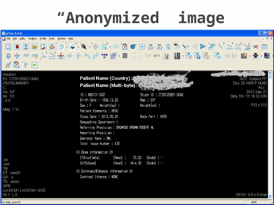
“Anonymized” image
Please wait until all scans for single pt ready before sending/uploading
Please write me a note if you’re unable to upload all scans for one pt in single session (let me know to expect more images)

What is the first image that we want to see?
◦ A) Chest x-ray confirming ET tube placement
◦ B) Facebook picture of the EMT at the accident scene
◦ C) Brain and bone head CT within 4 hours of randomization
◦ D) Full-body trauma scan, including the “Grady Handshake”
Quiz
In which file type do images need to be?
◦ A) JPEG
◦ B) DICOM
◦ C) SPI
◦ D) TIFF
Which images do we not want?
◦ A) Images taken after Day 30
◦ B) The resident’s cell phone video of the attending’s Babinski assessment
◦ C) The c-spine MRI
◦ D) Any image containing PHI

Your diligence and persistence in delivering comprehensive and pertinent imaging data is helping complete a vital component of this study!
THANK YOU FOR ALL OF YOUR HARD WORK!!!