Midterm results of balloon dilation of congenital aortic stenosis: Predictors of success
7
JACC Vol. 27, No. 5 1257 April 1996:1257-63 Midterm Results of Balloon Dilation of Congenital Aortic Stenosis: Predictors of Success PHILLIP MOORE, MD,* ERYBERTO EGITO, MD, HEATHER MOWREY, BS, STANTON B. PERRY, MD, JAMES E. LOCK, MD, FACC, JOHN F. KEANE, MD Boston and Cambridge, Massachusetts Objectives. We evaluated patient and procedural characteristics that influence the midterm success of balloon dilation of congen- ital aortic stenosis. Background. Balloon dilation is a new treatment for congenital aortic stenosis. Factors that influence midterm success are unknown. Methods. We performed a retrospective review of 148 children > 1 month old who underwent balloon dilation for aortic stenosis. Results. Balloon dilation was successful in 87% of patients, with a procedural mortality rate of 0.7%. The average immediate peak to peak gradient reduction was 56.4 + 19.9% (mean _+ SD). Prior valvotomy was the only factor that significantly reduced the immediate gradient reduction after dilation (47.1 -+ 21.8% vs. 57.8 -+ 19.6%, p < 0.01). Survival after dilation was 95% at 8 years. Seventy-five percent of patients were free of repeat intervention 4 years after dilation, whereas 50% remained free of repeat inter- vention at 8 years. Asymmetrically thick valve leaflets (risk ratio [RR] 0.17, p < 0.01) and prior aortic valvotomy (RR 0.35, p = 0.02) decreased the risk of repeat intervention. Aortic regurgita- tion grade >3 (RR 4.27, p = 0.04) and residual gradient after dilation (RR 1.63 for 10 mm Hg, p < 0.01) increased the risk. Conclusions. The 8.year survival rate after dilation was 95%, with 50% of patients free of repeat intervention. Factors that increased the risk for repeat intervention included symmetrically thin or thick aortic valve leaflets, regurgitation grade >3 after dilation and a high residual gradient after dilation. The incidence of repeat intervention after dilation was high owing to its pallia- tive nature. (JAm CoU Cardiol 1996;27:1257-63) Balloon dilation has become a common treatment for congen- ital aortic stenosis at many institutions. Immediate and short- term results are well reported (1-4) and compare favorably with standard surgical valvotomy (5). Reports of midterm results of balloon dilation are few (6), in part because of its recent development in the early 1980s (7). In addition, patient characteristics influencing the midterm success of balloon dilation for aortic stenosis are unknown. This report describes the midterm survival and need for repeat intervention after balloon dilation for aortic stenosis in patients 1 month to 20 years old. In addition, patient and procedural characteristics before and after balloon dilation were evaluated to elucidate predictors of immediate and midterm success. Methods This study included all patients 1 month to 20 years old with aortic stenosis who underwent balloon dilation at the Chil- From the Children's Hospital, Boston and Harvard Medical School, Cam- bridge, Massachusetts. This study was funded by the Children's Heart Founda- tion, Boston, Massachusetts. All editorial decisions for this article, including selection of referees, were made by a Guest Editor. This policy applies to all articles with authors from the University of California, San Francisco. Manuscript received August 14, 1995; revised manuscript received Novem- ber 13, 1995, accepted December 11, 1995. *Present address and address for corresoondence: Phillip Moore, Box 0632, C-346, University of California, San Francisco, 521 Parnassus Ave, San Fran- cisco, California 94143-0632. dren's Hospital in Boston between December 1984 and De- cember 1991. No patient was excluded because of associated defects. The indications for balloon dilation were a peak to peak systolic aortic valve gradient >50 mm Hg, with mild aortic regurgitation at most. The initial balloon size chosen was 80% to 100% of the angiographically measured aortic annulus. If inadequate gradient relief was achieved, serial dilations to a maximal balloon size of 137% of the annulus were performed. Each patient's medical record, including hospital notes, electrocardiogram (ECG), two-dimensional echocardiographic report and catheterization report, was reviewed. Variables, including the intensity of systolic and diastolic murmurs, presence of left ventricular hypertrophy and strain on ECG, type of commissural fusion, echocardiographic left ventricular dimensions and balloon/annulus ratio, were evaluated and compared with midterm success. Before and after balloon dilation we measured peak systolic valve gradient, left ventric- ular end-diastolic pressure, severity of aortic regurgitation and severity of peak instantaneous and mean transthoracic Dopp- ler aortic valve gradients and compared these variables with midterm success. Cineangiography performed at the time of balloon dilation provided estimates of the thickness of the aortic valve leaflets, aortic annulus size and severity of regurgitation. These assess- ments yielded three different angiographic morphologies: a "thin" aortic valve (<2 mm thick), an "asymmetrically thick" valve (a portion of the valve leaflets ->2 mm, usually one leaflet on the anteroposterior or lateral projections) and a "diffusely ©1996 by the American College of Cardiology. 0735-1097/96/$15.00 Published by Elsevier Science Inc. SSDI 0735-1097(95)00608-7
-
Upload
phillip-moore -
Category
Documents
-
view
213 -
download
1
Transcript of Midterm results of balloon dilation of congenital aortic stenosis: Predictors of success

JACC Vol. 27, No. 5 1257 April 1996:1257-63
Midterm Results of Balloon Dilation of Congenital Aortic Stenosis: Predictors of Success
P H I L L I P M O O R E , MD,* E R Y B E R T O EGITO, MD, H E A T H E R M O W R E Y , BS,
S T A N T O N B. P E R R Y , MD, J A M E S E. LOCK, MD, FACC, J O H N F. KEANE, M D
Boston and Cambridge, Massachusetts
Objectives. We evaluated patient and procedural characteristics that influence the midterm success of balloon dilation of congen- ital aortic stenosis.
Background. Balloon dilation is a new treatment for congenital aortic stenosis. Factors that influence midterm success are unknown.
Methods. We performed a retrospective review of 148 children > 1 month old who underwent balloon dilation for aortic stenosis.
Results. Balloon dilation was successful in 87% of patients, with a procedural mortality rate of 0.7%. The average immediate peak to peak gradient reduction was 56.4 + 19.9% (mean _+ SD). Prior valvotomy was the only factor that significantly reduced the immediate gradient reduction after dilation (47.1 -+ 21.8% vs. 57.8 -+ 19.6%, p < 0.01). Survival after dilation was 95% at 8 years. Seventy-five percent of patients were free of repeat intervention 4
years after dilation, whereas 50% remained free of repeat inter- vention at 8 years. Asymmetrically thick valve leaflets (risk ratio [RR] 0.17, p < 0.01) and prior aortic valvotomy (RR 0.35, p = 0.02) decreased the risk of repeat intervention. Aortic regurgita- tion grade >3 (RR 4.27, p = 0.04) and residual gradient after dilation (RR 1.63 for 10 mm Hg, p < 0.01) increased the risk.
Conclusions. The 8.year survival rate after dilation was 95%, with 50% of patients free of repeat intervention. Factors that increased the risk for repeat intervention included symmetrically thin or thick aortic valve leaflets, regurgitation grade >3 after dilation and a high residual gradient after dilation. The incidence of repeat intervention after dilation was high owing to its pallia- tive nature.
(JAm CoU Cardiol 1996;27:1257-63)
Balloon dilation has become a common treatment for congen- ital aortic stenosis at many institutions. Immediate and short- term results are well reported (1-4) and compare favorably with standard surgical valvotomy (5). Reports of midterm results of balloon dilation are few (6), in part because of its recent development in the early 1980s (7). In addition, patient characteristics influencing the midterm success of balloon dilation for aortic stenosis are unknown.
This report describes the midterm survival and need for repeat intervention after balloon dilation for aortic stenosis in patients 1 month to 20 years old. In addition, patient and procedural characteristics before and after balloon dilation were evaluated to elucidate predictors of immediate and midterm success.
M e t h o d s
This study included all patients 1 month to 20 years old with aortic stenosis who underwent balloon dilation at the Chil-
From the Children's Hospital, Boston and Harvard Medical School, Cam- bridge, Massachusetts. This study was funded by the Children's Heart Founda- tion, Boston, Massachusetts.
All editorial decisions for this article, including selection of referees, were made by a Guest Editor. This policy applies to all articles with authors from the University of California, San Francisco.
Manuscript received August 14, 1995; revised manuscript received Novem- ber 13, 1995, accepted December 11, 1995.
*Present address and address for corresoondence: Phillip Moore, Box 0632, C-346, University of California, San Francisco, 521 Parnassus Ave, San Fran- cisco, California 94143-0632.
dren's Hospital in Boston between December 1984 and De- cember 1991. No patient was excluded because of associated defects. The indications for balloon dilation were a peak to peak systolic aortic valve gradient >50 mm Hg, with mild aortic regurgitation at most. The initial balloon size chosen was 80% to 100% of the angiographically measured aortic annulus. If inadequate gradient relief was achieved, serial dilations to a maximal balloon size of 137% of the annulus were performed.
Each patient's medical record, including hospital notes, electrocardiogram (ECG), two-dimensional echocardiographic report and catheterization report, was reviewed. Variables, including the intensity of systolic and diastolic murmurs, presence of left ventricular hypertrophy and strain on ECG, type of commissural fusion, echocardiographic left ventricular dimensions and balloon/annulus ratio, were evaluated and compared with midterm success. Before and after balloon dilation we measured peak systolic valve gradient, left ventric- ular end-diastolic pressure, severity of aortic regurgitation and severity of peak instantaneous and mean transthoracic Dopp- ler aortic valve gradients and compared these variables with midterm success.
Cineangiography performed at the time of balloon dilation provided estimates of the thickness of the aortic valve leaflets, aortic annulus size and severity of regurgitation. These assess- ments yielded three different angiographic morphologies: a "thin" aortic valve (<2 mm thick), an "asymmetrically thick" valve (a portion of the valve leaflets ->2 mm, usually one leaflet on the anteroposterior or lateral projections) and a "diffusely
©1996 by the American College of Cardiology. 0735-1097/96/$15.00 Published by Elsevier Science Inc. SSDI 0735-1097(95)00608-7

1258 MOORE ET AL. JACC Vol. 27, No. 5 RESULTS OF BALLOON DILATION OF AORTIC STENOSIS April 1996:1257-63
thick" valve (the entire valve >-2 mm thick). Aortic annulus and leaflet thickness measurements were made using the catheter size to correct for magnification. Aortic regurgitation was graded angiographically using a scale of 0 to 4 (0 = absent; 1 = trivial; 2 = mild; 3 = moderate; 4 = severe) modified from the criterion of Hunt et al. (8). The modification combined all patients with grade 4 or 5 regurgitation, as described by Hunt et al., into a single group that we defined as grade 4 (severe).
Follow-up information obtained directly from the patient or parent by a mailed questionnaire included current state of health (alive or dead), current medications and need for repeat intervention. Repeat intervention was defined as any proce- dure performed on the aortic valve after the initial balloon dilation. This included repeat balloon dilation, surgical valvotomy, surgical valvuloplasty and valve replacement (ho- mograft, prosthetic, autologous transplantation of the pulmo- nary valve). In addition, surgical repair of mitral regurgitation caused by balloon dilation of the aortic valve was included as a repeat intervention. Recent clinic notes, ECG reports and echocardiographic reports, collected from the referring cardi- ologist, were analyzed for the presence of left ventricular hypertrophy or strain on ECG, peak and mean Doppler aortic gradients, severity of regurgitation and left ventricular short- ening fraction as determined by echocardiography. Because we analyzed echocardiographic reports and not echocardiographic tapes in many patients, the echocardiographic grade of regur- gitation (0 = absent; 1 = trivial; 2 = mild; 3 = moderate; 4 = severe) was not standardized but was left to the subjective assessment of the echocardiographer. If performed, follow-up catheterization data, including peak systolic valve gradient, left ventricular end-diastolic pressure and severity of regurgitation, were analyzed. Collection of follow-up data was completed in April 1994.
Statistical analysis. Data are expressed as mean value +_ SD, except for proportions, which are reported as mean value + SE. Comparisons were made using the paired t test, t test and analysis of variance (ANOVA) with the Bonferroni correction for interval data. Comparisons were made with the chi-square test for ordinal data. Univariant and multivariant proportional hazards models were compared before and after balloon dilation variables with time to repeat intervention. A life table is generated for survival and repeat intervention free survival curves (9).
Results
Patient characteristics. Between December 1984 and De- cember 1991, 150 patients 1 month to 20 years old underwent balloon dilation for congenital aortic stenosis at our center. The early results of balloon dilation in 71 of these patients have been included in prior reports (10,11). Of the 150 patients, two were lost to follow-up immediately after the balloon dilation because they resided outside the United States; therefore, they were excluded from the study. The remaining 148 patients constitute our study group. The patients' mean age at balloon dilation was 7.4 _+ 6.1 years (range 6 weeks to 20 years).
Twenty-four percent of the patients were <1 year old, 24% were between 1 and 5 years old, 15% were between 6 and 10 years old, and the remaining 37% were >10 years old. Of the 148 patients, 35.1% had associated cardiac defects, and 18.2% had undergone prior surgical valvotomy. Symptoms, including syncope, dyspnea on exertion, chest pain and palpitations, were present in 25.7%, whereas 10.8% were taking cardiac medications. Left ventricular hypertrophy was present on the ECG in 50% of patients, whereas only 8.8% had evidence of left ventricular strain with ST-T wave changes.
Immediate results. Balloon dilation was completed in all patients. The final balloon size used ranged from 0.68 to 1.37 times the annulus size. One death was associated with balloon dilation, for a procedural mortality rate of 0.7%. This death occurred in a 6-week old infant who had severe congestive heart failure due to left ventricular dysfunction with a mea- sured cardiac index of 1.5 liters/min per m 2. The infant died 30 rain after balloon dilation owing to ventricular fibrillation arrest. The mean peak systolic valve gradient before balloon dilation was 74.8 _ 17.0 mm Hg. The mean immediate peak systolic valve gradient reduction was 56.4 + 19.9%. A ->40% gradient reduction occurred in 85.1% of the patients. The mean residual peak systolic valve gradient was 32.1 _+ 15.2 mm Hg, and 12.8% of patients had residual gradients >-50 mm Hg. The cardiac index decreased slightly after balloon dilation (4.2 _ 1.1 vs. 3.8 +__ 0.9 liters/min per m 2, p = 0.014); however, there was no change in left ventricular end-diastolic pressure (13.1 _+ 5.6 vs. 12.1 _+ 5.5 mm Hg, p = 0.12).
Balloon dilation resulted in an increase in the grade of regurgitation in 58.1% of patients, 12.8% of whom had a regurgitation grade >-3 after the procedure. In addition to regurgitation, transient arrhythmias, particularly left bundle branch block, and pulse loss were the most common compli- cations, occurring in 13% and 7% of patients, respectively. Serious complications, including arrest requiring resuscitation or defibrillation, aortic valve leaflet prolapse, mitral valve injury, intimal tear of the aorta, femoral artery pseudoaneu- rysm, blood loss requiring a transfusion and bacterial endocar- ditis, occurred in less than 3% of patients.
Factors influencing immediate results (Table 1). Valve thick- ness was the only factor that influenced the severity of stenosis before balloon dilation. Thin valves had a significantly lower peak systolic valve gradient before balloon dilation than either asymmetrically or diffusely thick valves (ANOVA, p < 0.01). Prior valvotomy, however, was the only factor that influenced the percent gradient reduction in response to balloon dilation (t test, p = 0.01). Although thick valves had on average less gradient reduction than thin valves, the difference was not significant.
The severity of regurgitation before balloon dilation was influenced by both prior valvotomy and age. As expected, patients who had undergone prior surgical valvotomy had a higher incidence of regurgitation grade ->2 before balloon dilation than those who had not had prior valvotomy (chi- square, p < 0.01). The data strongly suggest that patients <1 year old had less regurgitation before balloon dilation than

JACC Vol. 27, No. 5 MOORE ET AL. 1259 April 1996:1257-63 RESULTS OF BALLOON DILATION OF AORTIC STENOSIS
Table 1. Clinical Characteristics Before and After Balloon Dilation (mean _+ SD)
No. of Pre-BD PSEG % ~2+ AR % ~ 3 + AR Pts (mm Hg) % Decrease in PSEG Pre-BD Post-BD B/A Ratio
Age (yr)
<1 35 75.8 _+ 18.0 53.9 _+ 23.6 0 _+ 0 0 _+ 0 0.92 _+ 0.15
1-5 35 72.l _+ 15.0 57.6 _+ 21.0 20.0 _+ 6.8 25.7 _+ 7.4* 0.97 _+ 0.18
6-10 22 73.8 _+ 13.8 56.0 + 21.7 18.2 _+ 8.2 0 _+ 0 1.01 + 0.14
>10 56 76.1 _+ 19.1 56.3 _+ 18.1 19.6 + 5.3 17.9 + 5.1 0.98 _+ 0.14
Prior valvotomy
Yes 27 75.1 _+ 16.5 47.5 _+ 22.0* 48.1 _+ 9.6* 33,3 _+ 9.1' 0.97 _+ 0.13
No 121 74.6 _+ 17.3 57.9 + 19.8 7.4 _+ 2.4 8,3 _+ 2.5 0.97 _+ 0.16
Leaflet thickness
Thin 70 70.8 + 17.7" 59.3 _+ 20.7 17.1 + 4.5 12.9 _+ 4.0 0.96 _+ 0.16
Asymm 39 78.7 + 18.3 60.0 _+ 15.9 12.8 _+ 5.3 10,3 - 4.9 0.93 _+ 0.21
Thick 21 79.9 _+ 13.4 52.0 _+ 18.7 4.8 + 4.7 23.8 -+ 9.3 1.01 _+ 0.18
Pre-BD AR grade
0 87 74.2 + 16.5 57.2 -+ 19.9 - - 4,6 + 2.2 0.98 + 0.16
1 39 76.2 -+ 17.8 56.5 +- 18.4 - - 15.4 -+ 5.8 0.95 -+ 0.12
->2 22 74.7 -+ 18.9 50.2 -+ 25.7 - - 31.6 _+ 10.7" 0.96 _+ 0.19
Commissural fusion
Right-left 39 73.5 +_ 17.3 59.2 _+ 17.2 2.6 + 2.5 10.3 -+ 4.9 0.97 _+ 0.16
Right-none 42 73.9 _+ 16.9 54.6 _+ 22.3 14.3 _+ 5.4 11.9 + 5.0 0.95 _+ 0.13
Partial? 24 76.5 _+ 15.6 54.8 _+ 24.0 16.7 _+ 7.6 12.5 _+ 6.8 0.98 _+ 0.13
Complete:~ 11 73.1 _+ 17.7 66.2 _+ 11.5 18.2 _+ 11.6 18.2 + 11.6 0.86 _+ 0.15
*p < 0.05. ?Partial fusion of one commissure with complete fusion of a second commissure. :~Complete fusion of both the right-left commissure and the right
noncommissure. AR = aortic regurgitation; Asym = asymmetric; B/A = baUoon/annulus; BD = balloon dilation; PSEG = peak to peak systolic ejection gradient; Pts =
patients.
older patients (chi-square, p = 0.06). This may reflect in part the low incidence of prior valvotomy (2.9 _+ 2.9%) in patients <1 year old as compared with the older age groups (1 to 5 years: 28.6 _+ 7.6%; 5 to 10 years: 27.3 _+ 9.5%; and >10 years: 17.9 _+ 4.8%).
The increase in regurgitation in response to balloon dilation was influenced by age, prior valvotomy and the grade of regurgitation before balloon dilation. Children aged 1 to 5 years were more likely to have aortic regurgitation grade ->3 after balloon dilation (chi-square, p -< 0.01). Both prior valvotomy and aortic regurgitation grade ->2 before balloon dilation were risk factors for aortic regurgitation grade ->3 after balloon dilation (chi-square, p ~ 0.01). No other factors, including balloon/annulus ratio, influenced the severity of regurgitation or the increase in regurgitation in response to balloon dilation.
Midterm results, Clinical follow-up information, including survival and need for repeat intervention, was available for all 148 children at a mean of 46.4 _+ 27.3 months after balloon dilation. Follow-up echocardiographic assessment was avail- able in 125 (84%) patients; 60% of the echocardiograms were performed at outside institutions. Follow-up catheterization data were available in 48 (32%) patients.
Midterm survival of patients with aortic stenosis after balloon dilation was excellent, with 97% survival at 5 years and 95% survival at 8 years (Fig. 1). Of the four late deaths, three were related to congestive heart failure associated with addi- tional lesions. A 4-month old infant with William's syndrome, aortic stenosis, supravalvar aortic stenosis, valvar pulmonary
stenosis and peripheral pulmonary stenosis arrested and died in the hospital 2 months after simultaneous balloon dilation of her aortic and pulmonic stenosis. A 5-month old infant with
Figure 1. Survival after balloon dilation (BD) for aortic stenosis in patients >1 month old (n = 148). Percentages were calculated using the life-table approach. Error bars = 1 SD.
(148) 100-
8O
~ 60
~ 4O
20,
0
(139) (111) (63) (32) o - - - - o [] ~ , , , [] []
YEARS AFTER BD

1260 M O O R E ET AL. JACC V o l . 2 7 , N o . 5
RESULTS OF BALLOON DILATION OF AORTIC STENOSIS April 1996:1257-63
%
80
40
(148)
136) 3,- ( 9 4 )
)
1 20. % FREE OF REPEAT INTERVENTION
% FREE OF SURGERY
i | i i i i f i 1 2 3 4 5 6 7 8
YEARS AFTER B D
Figure 2. Percent of patients free of any repeat intervention and those free of aortic valve surgery after balloon dilation (BD) for aortic stenosis in patients >1 month old (n = 148). Percentages were calculated using the life-table approach; error bars represent 1 SD.
Shone's complex with moderate recoarctation stenosis, subaor- tic stenosis and mitral stenosis died of progressive congestive failure 4 months after balloon dilation. A 3-month old infant with severe subaortic stenosis and a hypoplastic mitral valve died of progressive left ventricular dysfunction 9 months after balloon dilation. The remaining late death occurred unexpect- edly 64 months after balloon dilation in an 8.5-year old boy with mild residual aortic stenosis (echocardiographic gradient 30 mm Hg) and moderate regurgitation who was recovering from bacterial pneumonia.
Of the 148 patients, 37 (25%) required repeat intervention 30.7 _+ 26.2 months (range 0.1 to 91) after balloon dilation. The indications for repeat intervention included aortic stenosis in 25 patients, regurgitation in 7, a combination of aortic stenosis and regurgitation in 3, mitral regurgitation in 1 and a combi- nation of mitral regurgitation and aortic stenosis in 1. Of the 37 patients, 17 required only repeat balloon dilation. Twenty patients (13%) underwent surgery, with aortic valve replace- ment in nine patients (five prosthetic valves, three homografts and one valved conduit), aortic valve repair in four, aortic valvotomy in three, pulmonary autograft in two, mitral valve repair in one and repeat balloon dilation with mitral valve repair in one. No patient required immediate surgical inter- vention after balloon dilation, although three patients had surgical intervention within 1 month. Of these last three patients, two <1 year of age required surgical valvotomy for inadequate gradient relief. In one of these patients, no com- missural incision was made at the time of surgery because of
100-
80 -
60- E E
~ 40-
2 0 -
0 PRE POST F-UP
Figure 3. Mean maximal instantaneous Doppler gradient (MIG) for 70 patients who had a serial evaluation before balloon dilation (PRE), immediately after balloon dilation (POST) and at an average follow-up time of 41.5 +_ 23.5 months (F-UP). *Analysis of variance, p < 0.01, before versus after balloon dilation and versus follow-up.
optimal commissural tearing from balloon dilation. The resid- ual gradient was due to aortic annulus hypoplasia. The third patient required surgical repair of a tear in the anterior leaflet of the mitral valve due to balloon dilation using an anterograde approach; the aortic valve orifice achieved by balloon dilation was thought to be satisfactorily opened at this procedure.
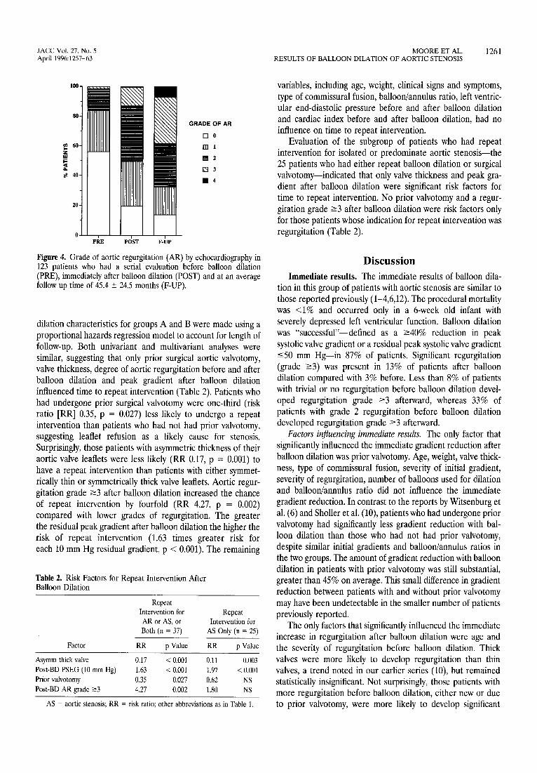
More than 80% of patients with aortic stenosis were free of any type of repeat intervention 4 years after balloon dilation, whereas 40% remained free at 8 years (Fig. 2). Seventy-six percent of patients were free of aortic valve surgery 8 years after initial balloon dilation (Fig. 2). Of the 148 patients, 70 had serial echocardiograms immediately after balloon dilation and at the time of follow-up (Fig. 3). Both the post-balloon dilation and follow-up mean maximal instantaneous Doppler gradients were decreased as compared with the pre-balloon dilation gradient (ANOVA, p < 0.01). There was no difference in the mean maximal instantaneous Doppler gradient at a mean follow-up of 41.5 + 23.5 months compared with the post-balloon dilation gradient. At follow-up in these 70 pa- tients, the maximal instantaneous Doppler gradient was un- changed or decreased in 53%, increased by -<25 mm Hg in 34% and increased by >25 mm Hg in 13% compared with the immediate post-balloon dilation values. In the 123 patients who had a serial echocardiographic evaluation for regurgita- tion before and after balloon dilation and at follow-up, there was significant progression of regurgitation at follow-up (Wil- coxon signed-rank test, p = 0.00), with 38.2% of these patients having a regurgitation grade ->3 as compared with 13.2% immediately after dilation (Fig. 4).
Factors influencing midterm results. To evaluate possible predictors of repeat intervention after balloon dilation, the study patients were divided into two groups consisting of 112 patients who did not have a repeat intervention on their aortic valve during the follow-up period (group A) and 37 patients who did (group B). Comparisons of pre- and post-balloon
JACC Vol. 27, No. 5 MOORE ET AL. 1261 April 1996:1257-63 RESULTS OF BALLOON DILATION OF AORTIC STENOSIS
100.
80-
m 60-
z _m
# 4o-
20-
m
l l PRE POST F - U P
GRADE OF AR
D e
[ ] 1
B 2
[ ] 3
I 1 4
Figure 4. Grade of aortic regurgitation (AR) by echocardiography in 123 patients who had a serial evaluation before balloon dilation (PRE), immediately after balloon dilation (POST) and at an average follow up time of 45.4 -- 24.5 months (F-UP).
dilation characteristics for groups A and B were made using a proportional hazards regression model to account for length of follow-up. Both univariant and multivariant analyses were similar, suggesting that only prior surgical aortic valvotomy, valve thickness, degree of aortic regurgitation before and after balloon dilation and peak gradient after balloon dilation influenced time to repeat intervention (Table 2). Patients who had undergone prior surgical valvotomy were one-third (risk ratio [RR] 0.35, p = 0.027) less likely to undergo a repeat intervention than patients who had not had prior valvotomy, suggesting leaflet refusion as a likely cause for stenosis. Surprisingly, those patients with asymmetric thickness of their aortic valve leaflets were less likely (RR 0.17, p = 0.001) to have a repeat intervention than patients with either symmet- rically thin or symmetrically thick valve leaflets. Aortic regur- gitation grade ->3 after balloon dilation increased the chance of repeat intervention by fourfold (RR 4.27, p = 0.002) compared with lower grades of regurgitation. The greater the residual peak gradient after balloon dilation the higher the risk of repeat intervention (1.63 times greater risk for each 10 mm Hg residual gradient, p < 0.001). The remaining
Table 2. Risk Factors for Repeat Intervention After Balloon Dilation
Repeat Intervention for Repeat AR or AS, or Intervention for Both (n = 37) AS Only (n = 25)
Factor RR p Value RR p Value
Asymm thick valve 0.17 < 0.001 0.11 0.003
Post-BD PSEG (10 mm Hg) 1.63 < 0.001 1.97 < 0.001
Prior valvotomy 0.35 0.027 0.62 NS
Post-BD AR grade ->3 4.27 0.002 1.80 NS
AS = aortic stenosis; RR = risk ratio; other abbreviations as in Table 1.
variables, including age, weight, clinical signs and symptoms, type of commissural fusion, balloon/annulus ratio, left ventric- ular end-diastolic pressure before and after balloon dilation and cardiac index before and after balloon dilation, had no influence on time to repeat intervention.
Evaluation of the subgroup of patients who had repeat intervention for isolated or predominate aortic stenosis--the 25 patients who had either repeat balloon dilation or surgical valvotomy--indicated that only valve thickness and peak gra- dient after balloon dilation were significant risk factors for time to repeat intervention. No prior valvotomy and a regur- gitation grade ->3 after balloon dilation were risk factors only for those patients whose indication for repeat intervention was regurgitation (Table 2).
D i s c u s s i o n
Immediate results. The immediate results of balloon dila- tion in this group of patients with aortic stenosis are similar to those reported previously (1-4,6,12). The procedural mortality was <1% and occurred only in a 6-week old infant with severely depressed left ventricular function. Balloon dilation was "successful"--defined as a ->40% reduction in peak systolic valve gradient or a residual peak systolic valve gradient -<50 mm Hg--in 87% of patients. Significant regurgitation (grade ->3) was present in 13% of patients after balloon dilation compared with 3% before. Less than 8% of patients with trivial or no regurgitation before balloon dilation devel- oped regurgitation grade ->3 afterward, whereas 33% of patients with grade 2 regurgitation before balloon dilation developed regurgitation grade ->3 afterward.
Factors influencing immediate results. The only factor that significantly influenced the immediate gradient reduction after balloon dilation was prior valvotomy. Age, weight, valve thick- ness, type of commissural fusion, severity of initial gradient, severity of regurgitation, number of balloons used for dilation and balloon/annulus ratio did not influence the immediate gradient reduction. In contrast to the reports by Witsenburg et al. (6) and Sholler et al. (10), patients who had undergone prior valvotomy had significantly less gradient reduction with bal- loon dilation than those who had not had prior valvotomy, despite similar initial gradients and balloon/annulus ratios in the two groups. The amount of gradient reduction with balloon dilation in patients with prior valvotomy was still substantial, greater than 45% on average. This small difference in gradient reduction between patients with and without prior valvotomy may have been undetectable in the smaller number of patients previously reported.
The only factors that significantly influenced the immediate increase in regurgitation after balloon dilation were age and the severity of regurgitation before balloon dilation. Thick valves were more likely to develop regurgitation than thin valves, a trend noted in our earlier series (10), but remained statistically insignificant. Not surprisingly, those patients with more regurgitation before balloon dilation, either new or due to prior valvotomy, were more likely to develop significant

1262 M O O R E ET AL. JACC Vol. 27, No. 5 RESULTS OF BALLOON DILATION OF AORTIC STENOSIS April 1996:1257-63
regurgitation after balloon dilation. The association between increased aortic regurgitation after balloon dilation and age of 1 to 5 years is most likely the result of the slightly higher percentage of patients with prior valvotomy in this age group.
All other factors, including balloon/annulus ratio within the ranges used in this study, had no significant influence on the immediate increase in regurgitation after balloon dilation. Our results differ from the report by the Valvuloplasty and Angio- plasty of Congenital Anomalies Registry (4), which showed that the average balloon/annulus ratio was larger in patients who developed moderate to severe regurgitation. This differ- ence is likely the result of using a more consistent technique from a single center and a smaller maximal balloon/annulus ratio in our study (1.3 vs. 1.5). As previously reported (12), although not statistically significant, the average increase in regurgitation was larger in patients in whom a balloon/annulus ratio of >1.25 was used. We believe that a balloon/annulus ratio of >1.25 increases the risk of developing regurgitation without significant benefit.
Midterm results. The midterm survival results of balloon dilation are encouraging. Although direct comparison with surgical valvotomy is not possible because of study design, comparison with historical reports suggests the two techniques are comparable. The natural history study reports an 8-year actuarial survival of 94% for patients over 2 years of age with a gradient ->50 mm Hg at presentation (13,14). Five-year surgical survival, excluding operative mortality, in patients >1 year of age after valvotomy was 94% (15). Eight-year actuarial survival in our study group was 95%: If we exclude mortality in children <2 years old, our 8-year survival would exceed 99%. In addition, the incidence of sudden death in our study was low. This may reflect our study's shorter follow-up period compared with the historical reports; nevertheless, it is encour- aging. Despite differences in study design, these data suggest that balloon dilation for congenital aortic stenosis may im- prove survival.
The high incidence of repeat intervention after balloon dilation is of concern. Some type of repeat intervention for either aortic stenosis or regurgitation was required in 25% of patients at 4 years and 50% of patients 8 years after balloon dilation. Thirty percent of patients required surgical interven- tion 8 years after balloon dilation. The natural history study reports 50% of patients with gradients 50 to 79 mm Hg managed medically required surgical intervention 10 years after enrollment (14). Repeat operation was required in 15% to 40% of patients 5 to 10 years after surgical valvotomy (5,14-17). These comparisons are of limited value because of the inconsistent criteria used to determine the need for repeat intervention. Criteria used in the natural history study included peak systolic valve gradient ->80 mm Hg or left ventricular strain pattern on ECG. The criteria used in our study group were variable and often determined by the referring cardiolo- gist. For example, repeat balloon dilation was often performed in patients with a peak systolic valve gradient -<60 mm Hg and no left ventrieular hypertrophy or ST-T wave changes on the ECG. The incidence of repeat intervention after balloon
dilation in our patients may simply reflect less stringent criteria for timing of repeat intervention, particularly repeat balloon dilation. Further investigation is warranted to optimize patient selection for balloon dilation and appropriate criteria for the timing of repeat intervention. The low incidence of sudden death in our series, however, seems to support our selection criteria for early and repeat intervention.
The 13% incidence rate of significant aortic regurgitation immediately after balloon dilation and the 38% incidence rate at follow-up is of concern. The natural history study reports a 7% incidence rate of significant aortic regurgitation as deter- mined by repeat catheterization 3 to 9 years after valvotomy (13) and a 15.3% incidence rate as determined by eehocardi- ography at late follow-up (14). Despite this discrepancy, the need for repeat intervention for aortic regurgitation after balloon dilation (27%) was similar to that after surgical valvotomy (23%) (13). These historical comparisons are of limited value because of the inconsistent definitions and tech- niques for assessing aortic regurgitation. They do suggest that aortic regurgitation may be a more significant problem after balloon dilation than surgical valvotomy.
Factors influencing midterm results. Better patient selection is optimized by knowing in advance which patients will have a favorable result from balloon dilation. Proportional hazards analysis showed that the only factors that predicted no repeat intervention at midterm follow-up were prior valvotomy, no or trivial regurgitation, asymmetrically thick aortic valve leaflets and a low residual peak systolic valve gradient. As expected, only valve leaflet thickness and peak systolic valve gradient after balloon dilation were predictors of the need for repeat balloon dilation or surgical valvotomy for residual or recurrent stenosis. No prior valvotomy and a moderate degree of regur- gitation after balloon dilation were also risk factors for patients who had repeat intervention for regurgitation.
Study limitations. Results of this study must be interpreted in light of its limitations. As is the case with similar studies of medical or surgical management, this is a retrospective study with a heterogeneous population, with respect to age, severity of disease at presentation and associated cardiac anomalies. Follow-up evaluations were performed at a variety of institu- tions and were therefore not uniform. The echocardiographic assessment of the grade of regurgitation was not standardized. The criteria used to determine the need for repeat intervention was at the discretion of the primary cardiologist and therefore varied between patients. During the 7-year period of proce- dural data collection many changes in equipment and tech- nique occurred, which may have influenced these midterm results. Although comparison of the results of balloon dilation performed between 1984 and 1987 with balloon dilation per- formed between 1988 and 1991 showed no differences, this study does not evaluate specific changes in equipment or technique.
Conclusions. Our results show that the actuarial survival rate 8 years after balloon dilation for aortic stenosis is 95%, with 70% of patients free from operation and 50% of these patients free from repeat intervention. Survival after balloon

JACC Vol. 27, No. 5 MOORE ET AL. 1263 April 1996:1257-63 RESULTS OF BALLOON DILATION OF AORTIC STENOSIS
dilation is equal or superior to survival after valvotomy. Factors that decrease the risk for early repeat intervention include asymmetrically thick aortic valve leaflets, prior valvot- omy, mild regurgitation after balloon dilation and a lower peak systolic valve gradient after balloon dilation. The greater the residual peak gradient after balloon dilation, the higher the risk of repeat intervention. Because of the palliative nature of the procedure, the incidence of repeat intervention after balloon dilation is high.
References 1. O'Connor BK, Beekman RH, Rocchini AP, Rosenthal A. Intermediate term
effectiveness of balloon valvuloplasty for congenital aortic stenosis. A prospective follow up study. Circulation 1991;84:732-8.
2. Vogel M, Benson L, Burrows P, Smallhorn JF, Freedom RM. Balloon dilation of congenital aortic valve stenosis in infants and children: short and intermediate results. Br Heart J 1989;62:148-53.
3. Shaddy RE, Boucek MM, Sturtevant JE, Ruttenberg HD, Orsmond GS. Gradient reduction, aortic valve regurgitation and prolapse after balloon aortic valvuloplasty in 32 consecutive patients with congenital aortic stenosis. J Am Coil Cardiol 1990;16:451-6.
4. Rocchini AP, Beekman RH, Shachar GB, Benson L, Schwartz D, Kan JS. Balloon aortic valvuloplasty: results of the Valvuloplasty and Angioplasty of Congenital Anomalies Registry. Am J Cardiol 1990;65:784-9.
5. Kugelmeier J, Egloff L, Real F, Rothlin M, Turina M, Senning A. Congenital aortic stenosis: early and late results of aortic valvotomy. Thorac Cardiovasc Surg 1982;30:91-5.
6. Witsenburg M, Cromme-Dijkhuis AlI, Frohn-Mulder IM, Hess J. Short and mid-term results of balloon valvuloplasty for valvular aortic stenosis in children. Am J Cardiol 1992;69:945-50.
7. Labibidi Z, Wu JR, Walls JT. Percutaneous balloon aortic valvuloplasty: results in 23 patients. Am J Cardiol 1984;53:194-7.
8. Hunt D, Baxley DA, Kennedy JW, Judge TP, Williams JE, Dodge HT. Quantitative evaluation of cineaortography in the assessment of aortic regurgitation. Am J Cardiol 1973;31:696-700.
9. Colton T. Statistics in Medicine. Boston: Little, Brown, 1974:237-50. 10. Sholler GF, Keane JF, Perry SB, Sanders SP, Lock JE. Balloon dilation of
congenital aortic stenosis: results and influence of technical and morpholog- ical features on outcome. Circulation 1988;78:351-60.
11. Rosenfeld HM, Landzberg MJ, Perry SB, Colan SD, Keane JF, Lock JE. Balloon aortic valvuloplasty in the young adult with congenital aortic stenosis. Am J Cardiol 1994;73:1112-7.
12. Rao SP, Thapar MK, Wilson AD, Levy JM, Chopra PS. Intermediate term follow up results of balloon aortic valvuloplasty in infants and children with special references to causes of restenosis. Am J Cardiol 1989;64:1356-60.
13. Wagner HR, EHison C, Keane JF, Humphries JQ, Nadas AS. Clinical course in aortic stenosis. Circulation 1977;56 Suppl I:1-47-I-56.
14. Keane JF, Driscoll DJ, Gersony WM, et al. Second natural history study of congenital heart defects: results of treatment of patients with aortic valvar stenosis. Circulation 1993;87 Suppl I:I-16-I-27.
15. l-lsieh KS, Keane JF, Nadas AS, Bernhard WF, Casteneda AR. Long term follow up of valvotomy before 1968 for congenital AS. Am J Cardiol 1986;58:338-41.
16. Presbitero P, Somerville J, Revel-Chion R, Ross D. Open aortic valvotomy for congenital aortic stenosis. Late results. Br Heart J 1982;47:26-34.
17. Dobell ARC, Bloss RS, Gibbons JE, Collins GF. Congenital valvar aortic stenosis: surgical management and long term results. J Thorac Cardiovasc Surg 1981;81:916-20.