Midbrain dopaminergic neuron fate specification of ...Midbrain dopaminergic neuron fate...
182
Midbrain dopaminergic neuron fate specification of pluripotent stem cells Ines Jaeger Thesis submitted for the degree of Doctor of Philosophy MRC Clinical Sciences Centre, Imperial College London 2012
Transcript of Midbrain dopaminergic neuron fate specification of ...Midbrain dopaminergic neuron fate...

Midbrain dopaminergic neuron fate specification of
pluripotent stem cells
Ines Jaeger
Thesis submitted for the degree of Doctor of Philosophy
MRC Clinical Sciences Centre, Imperial College London 2012

I declare that the work described in this Thesis is my own unless otherwise stated.
Ines Jaeger

Acknowledgements
It is a pleasure to thank the many people who made this PhD a reality. First and foremost,
my deepest sense of gratitude goes to my supervisor Dr. Meng Li. This work would not have
been possible without her continuous support, guidance and patience. I expand my thanks
to the entire lab, past and present, especially, Emily, Xinsheng and Charlie, who have made
this entire process not only bearable, but entertaining and enjoyable. I am thankful to my
office mate, colleague, collaborator and friend Jess who had always time when I needed
help. I also would like to thank our collaborators Mark Ungless, Steve Dunnett, Sophie
Precious, and Andreas Heuer. Thanks go also to Dirk Dorrman, Lein Snipes, Ki Heng, and
James Elliot for their help on the fluorescence activated cell sorters and microscopes.
I am especially grateful to my beloved friends, Aida, Bibi, Carina, Claire, Hegias, Hendrik, Isi,
Joseba, Kerstin, Korinna, Michel-Raphael, Sarah, Silke and Sybille for their support and love
during the journey in obtaining my PhD.
And most importantly, I would like to thank my parents, Doris and Olaf Jaeger. They were
there to commiserate with me when I was discouraged and celebrate with me when I had
successes. Their love and encouragement have been a constant source for me since I was
little; therefore, I dedicate this thesis to them.
Abstract
Disorders of dopaminergic neurons in the midbrain are associated with various brain
diseases such as Parkinson’s disease (PD), for which there are no effective treatments.
Pluripotent stem cells (PSCs) offer a remarkable potential for finding new therapeutic
strategies because they are self-renewable and capable to differentiate into all cell types.
However, biomedical applications of PSCs require knowledge of the molecular mechanisms
directing PSCs into midbrain dopaminergic (mDA) cell fate.
During development the generation of mDA neurons is induced by the extrinsic molecules
SHH, FGF8 and WNT1 in the caudal region of the Otx2 positive domain, which marks the
presumptive fore- and midbrain, and rostrally to the future hindbrain expressing Gbx2.
In this study I have further investigated the role of FGF signalling in the generation of mDA
neurons. Here, I have used mouse epiblast stem cells (EpiSCs). Based on their
developmentally primed pluripotent state, we assumed that they represent a more suitable
system when compared with mouse embryonic stem cells. I validated neural differentiation
of EpiSCs as an alternative model to study neural development in vitro. I found that inhibition
of the FGF/ERK activity at the onset of EpiSC differentiation initiated expression of Wnt and
Shh and further induced ventral midbrain progenitor markers, accompanied by suppressing
caudalisation as well as forebrain induction. To maintain ventral midbrain progenitor fate,
cells required a period of endogeneous FGF/ERK signalling. Subsequent treatment of FGF8
and SHH, which restricts progenitors from adopting alternative fates, led to highly efficient
production of authentic mDA neurons. These neurons exhibited functional, neuron-like
properties and when implanted into the striatum of mouse PD model strongly restored
parkinsonian features without any signs of overgrowth.
In conclusion, a temporally controlled modulation of FGF/ERK activity during neural
differentiation from PSCs is crucial for reliable and highly efficient generation of functional
authentic mDA neurons.
Table of Contents
List of Figures
List of Tables
List of Abbreviation
1 Introduction ............................................................................................................. 1
1.1 Pluripotent stem cells .......................................................................................... 1
1.1.1 Stem Cells .................................................................................................... 1
1.1.2 Characteristics of pluripotency...................................................................... 2
1.1.3 Pluripotent stem cell lines ............................................................................. 2
1.1.4 Pluripotent stem cells as model to study neural development ....................... 5
1.2 Midbrain dopaminergic development ................................................................... 5
1.2.1 Neural induction ........................................................................................... 5
1.2.2 Neural patterning – regionalisation and specification of midbrain
dopaminergic neurons .................................................................................. 8
1.2.3 Isthmus assiociated factors ........................................................................ 10
1.2.4 Floorplate associated factors ...................................................................... 17
1.2.5 Genes regulating midbrain dopaminergic development .............................. 19
1.3 Strategies for neural differentiation in vitro of pluripotent stem cells ................... 21
1.3.1 Embryoid body differentiation ..................................................................... 21
1.3.2 Stromal co-culture system .......................................................................... 21
1.3.3 Monolayer differentiation ............................................................................ 22
1.4 FGF pathway ..................................................................................................... 22
1.4.1 FGF ligands ................................................................................................ 22

1.4.2 FGF receptors ............................................................................................ 23
1.4.3 FGF signalling ............................................................................................ 23
1.4.4 FGF modulation ......................................................................................... 25
1.4.5 Multiple roles of the FGF pathway during neural development ................... 26
1.5 Hypthesis and aims ........................................................................................... 28
2 Materials and Methods .......................................................................................... 33
2.1 Cell Culture ....................................................................................................... 33
2.1.1 Routine ESC and iPSC culture ................................................................... 33
2.1.2 Derivation of EpiSC from ESC .................................................................... 33
2.1.3 Routine EpiSC culture ................................................................................ 34
2.1.4 Freezing ESCs, iPSCs and EpiSCs ............................................................ 34
2.1.5 Thawing ESCs, iPSCs and EpiSCs ............................................................ 34
2.2 Neural differentiation ......................................................................................... 34
2.3 Flow cytometry .................................................................................................. 36
2.4 Electophysiological studies ................................................................................ 36
2.5 Animal work ....................................................................................................... 37
2.5.1 Animals ...................................................................................................... 37
2.5.2 Lesion surgery ............................................................................................ 37
2.5.3 Graft surgery .............................................................................................. 38
2.5.4 Drug-induced rotations ............................................................................... 38
2.6 Molecular biology methods ................................................................................ 39
2.6.1 Quantitative PCR........................................................................................ 39
2.6.2 Protein extraction and quantification ........................................................... 39

2.6.3 Western Blot analysis ................................................................................. 40
2.6.4 Immunofluorescence staining ..................................................................... 40
2.6.5 Quantitative analysis of immunocytochemistry ........................................... 41
3 Epiblast stem cells as an alternative model to study neural differentiation in vitro.. 45
3.1 Comparison of neural induction in ESCs cell and EpiSCs ................................. 46
3.2 Discussion ......................................................................................................... 51
4 Temporally controlled modulation of FGF/ERK signalling directs a midbrain
dopaminergic fate .................................................................................................. 53
4.1 Blockade of FGF/ERK signalling upon exit of epiblast pluripotent state
accelerates neural fate commitment ................................................................. 54
4.2 Inhibition of FGF/ERK activity at the onset of EpiSC differentiation promotes mDA
progenitor characteristics .................................................................................. 59
4.3 FGF/ERK inhibition affects anterior-posteriorisation and the maintenance of
OTX2 in nascent neural progenitors .................................................................. 65
4.4 Highly reliable and efficient production of mDA neurons by ERK inhibitor-primed
neural progenitors ............................................................................................. 70
4.5 Dopaminergic neuron differentiation of mouse iPSCs in response to ERK
inhibition ........................................................................................................... 73
4.6 Differential activity of SHH and FGF8 in mDA fate specification and differentiation
......................................................................................................................... 75
4.7 WNT alone or in combination with SHH cannot replace ERK inhibitor in inducing
midbrain progenitor cell fate .............................................................................. 80
4.7.1 The effects of early SHH activation on mDA induction ................................ 80
4.7.2 The effects of GSK3ß inhibition on mDA induction ..................................... 81

4.7.3 The effects of combined treatment of SHH and CHIR99021 on mDA
induction .................................................................................................... 83
4.8 Discussion ......................................................................................................... 95
5 Functional analysis of in vitro derived midbrain dopaminergic neurons ................ 100
5.1 In vitro generated mDA neurons exhibit functional neuronal characteristics .... 100
5.2 Transplantation of in vitro generated mDA progenitors in a mouse model of
Parkinson’s disease ........................................................................................ 104
5.2.1 Modification of differentiation protocol ...................................................... 104
5.2.2 Transplantation of EpiSC-derived mDA progenitors; pilot study................ 109
5.2.3 Head to head comparison of EpiSC-derived mDA grafts with ventral midbrain
tissue in 6-OHDA-lesioned mouse models of PD ..................................... 114
5.3 Discussion ....................................................................................................... 119
6 Concluding remarks and future directions............................................................ 121
7 Bibliography ........................................................................................................ 127
8 Appendix ............................................................................................................. 156

List of Figures
Figure 1.1 Schematic illustration of the signalling centres which establish the antero-
posterior axis. ..................................................................................................................... 29
Figure 1.2 Drawing of midbrain DA neural development ..................................................... 30
Figure 1.3 Schematic illustration of the pathways implicated in the generation of mDA
neurons. .............................................................................................................................. 31
Figure 3.1 Accelerated neural differentiation potential of EpiSCs. ....................................... 49
Figure 3.2 Synchronised neuroectodermal differentiation from EpiSCs. .............................. 50
Figure 4.1 Inhibition of FGF/ERK activity accelerates the exit of EpiSCs from a pluripotent
stage. .................................................................................................................................. 56
Figure 4.2 Inhibition of FGF/ERK activity accelerates neural fate conversion of EpiSCs. .... 58
Figure 4.3 Inhibition of FGF/ERK on the exit of the epiblast pluripotent stage promotes the
generation of mDA progenitor cells. .................................................................................... 63
Figure 4.4 Blockade of FGF/PI3K signalling does not induce expression of mDA regulatory
genes. ................................................................................................................................. 64
Figure 4.5 Early blockade of FGF/ERK activity modulates anterior –posterior characteristics
of neural progenitor cells. .................................................................................................... 68
Figure 4.6 Early blockade of FGF/PI3K activity does not promote the generation of neural
progenitors with anterior characteristics. ............................................................................. 69
Figure 4.7 Temporally controlled FGF/ERK activity promotes the production of functional,
authentic mDA neurons. ...................................................................................................... 72
Figure 4.8 Efficient generation of mDA neurons from mouse iPSCs. ................................... 74
Figure 4.9 Temporally controlled FGF/ERK activity in combination with SHH and FGF8
promotes the production of DA neurons. ............................................................................. 77
Figure 4.10 Activation of SHH signalling promotes the production of DA neurons by
repressing the transcript level of genes for a GABAergic neuronal fate. .............................. 78

Figure 4.11 Effect of SHH and/or FGF8 in EpiSC MD. ........................................................ 79
Figure 4.12 Activation of SHH pathway is not sufficient for complete mDA induction. ......... 85
Figure 4.13 Early inhibition of FGF/ERK acts partly via the SHH pathway. .......................... 86
Figure 4.14 SHH treatment cannot induce midbrain neural progenitor fate. ........................ 88
Figure 4.15 Activation of SHH alone cannot replace early blockade of FGF/ERK pathway in
the generation of DA neurons. ............................................................................................ 89
Figure 4.16 Activation of the WNT pathway does not mirror the effects of early ERK inhibition
in neural differentiation. ....................................................................................................... 91
Figure 4.17 SHH and CHIR99021 treatment partly mimics early ERK inhibition. ................. 93
Figure 4.18 Early stimulation of SHH and WNT pathway cannot substitute ERK inhibition in
the generation of DA neurons. ............................................................................................ 94
Figure 5.1 Pitx3-GFP positive cells exhibit functional neuron-like electrophysiological
properties. (kindly provided by Dr. Jessica Risner-Janiczek). ............................................ 103
Figure 5.2 Early blockade of FGF signalling and postliminary enhancement of SHH signalling
support generation of ventral midbrain progenitor cells. .................................................... 107
Figure 5.3 ERK inhibition followed by stimulation of the SHH signalling promotes the
generation of DA neurons. ................................................................................................ 108
Figure 5.4 EpiSC-derived cells display robust induction of ventral midbrain progenitor fate at
the day of transplantation. ................................................................................................. 111
Figure 5.5 Bar graph of amphetamine-induced rotational behaviour 8 weeks after
transplantation of lesioned, d5 and d7 MD cultures mice (kindly provided by Dr. Sophie
Precious and Andreas Heuer). .......................................................................................... 112
Figure 5.6 Substantial graft-derived reinnervation of the lesioned striatum 8 weeks after
transplantation of d5 and d7 MD cultures (kindly provided by Dr. Sophie Precious and
Andreas Heuer). ................................................................................................................ 113
Figure 5.7 Behavioural analysis in E12.5 VM- versus d7 MD-grafted mice (kindly provided by
Dr. Sophie Precious and Andreas Heuer). ........................................................................ 116

Figure 5.8 Characterisation of neurons in 6-OHDA lesioned mouse models of PD grafted
with either d7 MD or VM cells (kindly provided by Dr. Sophie Precious and Andreas Heuer).
......................................................................................................................................... 118
Figure 6.1 Proposed model of FGF signalling in mDA neural differentiation. ..................... 126

List of Tables
Table 1.1 The 23 members of the FGF family can be divided into 7 groups according their
evolutionary relationship and and gene-location analysis. ................................................... 32
Table 2.1 Pluripotent stem cell lines used for the investigations of this thesis. .................... 42
Table 2.2 List of factors used for neural differentiation. ....................................................... 42
Table 2.3 List of gene specific primers for qPCR. ................................................................ 43
Table 2.4 List of primary antibodies for immunocytochemistry. ........................................... 44
List of Abbreviation
AP Anterior-posterior
ANOVA Analysis of Variance
ANR Anterior neural ridge
AVE Anterior visceral endoderm
cDNA Complementary DNA
d day
DA Dopaminergic
DNA Deoxyribonucleic acid
E Embryonic day
EB Embryoid bodies
EpiSC Epiblast stem cell
ESC Embryonic stem cell
GFP Green fluorescence protein
hESC human ESC
ICM Inner cell mass
iPSC Induced pluripotent stem cell
IsO Isthmic Organiser
LIF Leukemia inhibitory factor
MD Monolayer differentiation
mDA Midbrain dopaminergic
MHB Midbrain hindbrain border
mRNA Messenger RNA
NPC Neural progenitor cell
NSC Neural stem cell
PCR Polymerase chain reaction

PD Parkinson’s disease
PD0325901 potent FGF/ERK inhibitor
PSC Pluripotent stem cell
RNA Ribonucleic acid
SN Substantia Nigra
VTA Ventral tegmental area
ZLI Zona limitans intrathalamica

1
1 Introduction
In the nervous system dopaminergic (DA) neurons exist in different regions including the
olfactory bulb, retina, hypothalamus and the ventral midbrain. DA neurons are usually
identified by their expression of tyrosine hydroxylase (TH), the rate-limiting enzyme in the
synthesis of dopamine. The ventral midbrain contains two cores of DA neurons, the
substantia nigra (SN) and the ventral tegmental area (VTA). The SN is a part of the
nigrostrial system and regulates motor function whilst the VTA belongs to the mesolimbic
system and is involved in emotional behaviour. Disorders involving neurons of the VTA are
linked to Schizophrenia and depression whereas the preferential degeneration of DA
neurons in the SN is believed to be the primary cause of Parkinson’s disease (PD) (Hirsch,
Graybiel et al. 1988; Sesack and Carr 2002; Dailly, Chenu et al. 2004).
Stem cell-based transplantation is a promising approach for the treatment of PD. The
generation of patient-specific induced pluripotent stem cells offers further, perhaps more
realistic in shorter term, biomedical applications of stem cells in disease modelling and drug
development. However, realising the potential of stem cells requires in depth understanding
of the mechanisms that control the generation of mDA neurons during development.
1.1 Pluripotent stem cells
1.1.1 Stem Cells
Stem cells have the potential to self-renew for indefinite periods in culture and to give rise to
one or more specialised cell types along the developmental path. There are many kinds of
stem cells that can be distinguished by their capacity to differentiate.

2
The zygote and the first blastomeres, derived from the first division of the zygote, are
totipotent stem cells (Tarkowski 1959; Gardner 1998). They are capable of forming the entire
embryo including the extra-embryonic tissue. As the blastomeres continue to cleave and
divide, they form the trophoblast and the inner cell mass (ICM). The ICM further develops
into the hypoblast and epiblast. Cells from the epiblast are pluripotent as they contribute to
all cell lineages and can form a chimera when transplanted into another blastocyst (Gardner,
1998). Through the subsequent process of gastrulation, cells of the epiblast give rise to the
three germ layers: mesoderm, ectoderm, and endoderm. Cells of the mesoderm, ectoderm,
and endoderm are considered to be multipotent as they are the progenitor cells of only one
germ layer and terminally differentiate into a somatic cell type specific to that germlayer.
1.1.2 Characteristics of pluripotency
Pluripotency is defined as the ability of a single cell to generate all cell lineages of an
organism. Stable pluripotent stem cell (PSC) lines can be derived from the epiblast and
teratomas under specific culture conditions or generated from somatic cells by introduction
of specific transcription factors.
The functional pluripotency of a cell can be determined by three independent experiments:
1) the spontaneous differentiation of PSCs in culture, 2) the formation of teratomas and
teratocarcinomas by injection of PSCs into an immune compromised adult animal, and 3)
chimera incorporation and germline transmission when PSCs are injected into a pre-
implantation blastocyst.
1.1.3 Pluripotent stem cell lines
Embryonic stem cells
Embryonic stem cells (ESCs) are pluripotent cells derived from the epiblast of the pre-
implantation embryo (Kaufman et al., 1983). These cells are defined by their expression of
the key pluripotency markers Nanog, Oct4, Sox2, Klf4, Rex1, and Stella (Avilion et al., 2003;
Guo et al., 2009; Mitsui et al., 2003; Okamoto et al., 1990; Payer et al., 2003; Ramalho-

3
Santos et al., 2002; Rogers et al., 1991). Furthermore female ESCs exist in a pre-X-
inactivation state maintaining two active X-chromosomes (Nichols and Smith, 2009).
Initially, mouse ESCs were maintained on mitotically inactivated mouse embryonic fibroblast
cells in the presence of fetal bovine serum (Evans and Kaufman, 1981). Later studies
identified the leukemia inhibitory factor (LIF) as an important component to maintain
pluripotency by activating the transcription factor STAT3 (Matsuda et al., 1999; Niwa et al.,
1998; Stewart et al., 1992). LIF in combination with the bone morphogenetic protein (BMP)
were able to suppress differentiation and allowed the propagation of ESCs in a feeder layer
free condition (Ying et al., 2003a). However, ESCs expanded in fully defined medium
containing LIF and the two small molecules that inhibit mitogen-activated protein kinase
(MAPK) signalling and glycogen synthase kinase-3 ß (GSK3ß) showed an upregulation of
Nanog and Oct4 positive cells representing the so called ground state of pluripotency
(Nichols and Smith, 2009; Silva et al., 2008a).
Epiblast stem cells
The generation of a stable PSC line from the epiblast of a postimplantation embryo has been
first reported 2007 (Brons et al., 2007; Tesar et al., 2007). These cells are consequently
called epiblast stem cells (EpiSCs). The maintenance of their pluripotent state requires
Activin A and fibroblast growth factor 2 (FGF2) (Tesar et al., 2007). Using the same
condition, EpiSCs can also be produced from ESCs (Guo et al., 2009). Though EpiSCs
express the core pluripotent markers Nanog, Oct4, and Sox2, they are distinct from ESCs
(Chenoweth and Tesar, 2010; Eggan, 2007). Unlike ESCs, EpiSCs display a high
expression level of genes that mark the postimplantation epiblast such as Fgf5, Brachyury,
Gata6, Nodal, Foxa2, and Otx2 (Brons et al., 2007; Tesar et al., 2007). In addition, these
cells grow as flat compact colonies and need cell-cell interaction to survive (Brons et al.,
2007). Studies on EpiSCs further observed that one copy of the X chromosome is
epigenetically silenced in female cells (Guo et al., 2009). Although EpiSCs can form
teratomas, they have very limited capacity to contribute to blastocyst chimeras (Brons et al.,

4
2007; Rossant, 2008; Tesar et al., 2007). EpiSCs are therefore thought to be more
advanced in their development when compared to ESCs and thus they are also called
primed PSCs (Nichols and Smith, 2009).
Induced pluripotent stem cells
Reprogramming of somatic cells to become induced pluripotent stem cells (iPSCs) by
overexpressing the key transcription factors for pluripotency, Oct4, Sox2, Klf4 and c-Myc,
was first reported in 2006 by Takahashi and Yamanaka (Takahashi and Yamanaka, 2006).
Since then it has been demonstrated that iPSCs can also be derived from a variety of mouse
tissues (Aoi et al., 2008; Eminli et al., 2008; Kim et al., 2008; Stadtfeld et al., 2008). These
cells functionally resemble ESCs as they differentiate in vitro into the cell types of all three
germ layers, form teratomas and contribute to chimeras and germline transmission (Maherali
and Hochedlinger 2008, Okita et al 2007, Wernig et al 2007). Genome-wide microarray
analyses further revealed that the global expression pattern of iPSCs is more similar to
ESCs than the somatic cells from which they were derived, and that a majority of genes
specific for ESCs are reactivated in iPSCs (Okita et al., 2007; Wernig et al., 2007).
Additionally, during reprogramming of female somatic cells the silenced X-chromosome is
again activated (Maherali et al., 2007) as it is the case for ESCs.
After successful reprogramming of mouse somatic cells, scientists demonstrated
reprogramming of various human somatic cells (Aasen et al., 2008; Giorgetti et al., 2009;
Park et al., 2008; Takahashi et al., 2007; Yu et al., 2007) as well as the generation of
disease-specific iPSC lines (Carvajal-Vergara et al., 2010; Ebert et al., 2009; Lee et al.,
2009; Marchetto et al., 2010; Park et al., 2008; Raya et al., 2009; Soldner et al., 2009).
These disease-specific iPSCs provide a powerful tool for disease modelling in vitro and drug
development. Once technical limitations are eliminated, iPSCs represent an alternative
source for cellular replacement therapies because patient–specific iPSCs for cell or tissue
grafts will not lead to immunological rejection.

5
Human embryonic stem cells
In 1998, human ESCs were derived from pre-implantation stage embryos that were donated
for research after being targeted for disposal either because they were surplus embryos or
because they were of poor quality. These cells are considered to be pluripotent because
they express the core genes of pluripotency Nanog, Oct4 and Sox2 (Adachi et al., 2010; Hay
et al., 2004; Matin et al., 2004; Xu et al., 2009; Xu et al., 2008; Zaehres et al., 2005),
differentiate into cells of all three germ layers (Hoffman and Carpenter, 2005), and form
teratomas after injection into an immune-deficient animal (Thomson et al., 1998). Human
ESCs share several features with mouse EpiSCs forming cell colonies with a flat
morphology, reduced tolerance to passaging as single cells, dependence on FGF2 and
Activin signalling (Xu et al., 2008), and the tendency towards X chromosome inactivation in
female cell lines (Silva et al., 2008b). Consequently, they display more a primed pluripotent
state than the ground state of mouse ESCs.
1.1.4 Pluripotent stem cells as model to study neural development
PSCs can be stimulated to differentiate into many different neuronal subtypes by use of
in vitro models mimicking underlying molecular and cellular mechanisms of in vivo
developmental processes (Lee et al., 2000b; Li et al., 2005; Perrier et al., 2004; Watanabe et
al., 2005; Wichterle et al., 2002; Ying et al., 2003b). Conversely, insights into the
mechanisms that direct stem cells into a specific neural fate may provide an improved
understanding of the mechanism of neural differentiation in vivo, particularly for early human
development. Therefore, the approach of hESC and disease-specific hiPSC allows us to
dissect the complex mechanism of normal and pathological neurodevelopment.
1.2 Midbrain dopaminergic development
1.2.1 Neural induction

6
The first insights into the understanding of how cells of the ectoderm become neural tissue
came from the pioneering work of Spemann and Mangold nearly 100 years ago (Spemann
and Mangold, 1923). Using differently pigmented species of amphibians to distinguish the
cells of donor and host, they transplanted a small piece of the dorsal blastopore lip of the
gastrulating embryo, now known as Spemann’s ‘‘organizer’’, into the ventral side of another
embryo at gastrula stage. They found that the cells of host tissue formed a secondary axis
composed of a neural tube, notochord and paraxial muscles demonstrating the capability of
the organiser to induce neural fate in vertebrate embryos. Equivalent organizing regions
were found in zebrafish (shield), chick (Henson’s node), and mouse (node) embryos
(Beddington, 1994; Boettger et al., 2001; Shih and Fraser, 1996). Each of these organisers
induces a neural plate not only within the same species but also when transplants are
carried out across classes (Blum et al., 1992; Hatta and Takahashi, 1996; Kintner and Dodd,
1991), suggesting an evolutional conserved strategy for neural induction.
Experiments showed that explants from the frog ectoderm, a region fated to become
epidermis and neural tissue, gave rise to epidermal cells when cultured without the addition
of growth factors. In contrast, when the ectodermal explants were dissociated into single
cells, the cells became neural (Godsave and Slack, 1989; Grunz and Tacke, 1989). A
number of BMP antagonists secreted by the organizer have been identified as essential
molecules in neural induction such as Noggin (Furthauer et al., 1999; Lamb et al., 1993;
Smith and Harland, 1992; Smith et al., 1993; Zimmerman et al., 1996), Chordin (Piccolo et
al., 1996; Sasai et al., 1995; Sasai et al., 1994), Follistatin (Fainsod et al., 1997; Hemmati-
Brivanlou et al., 1994), Cerberus (Bouwmeester et al., 1996; Piccolo et al., 1999), XNr3
(Hansen et al., 1997; Smith et al., 1995), Gremlin/Drm and Dan (Dionne et al., 2001; Eimon
and Harland, 2001; Hsu et al., 1998; Khokha et al., 2003; Pearce et al., 1999). Based on the
findings in amphibians, the phenomenon of neural induction was explained by the ‘default
model’ which proposes that ectodermal cells convert into neural plate in the absence of

7
extrinsic signals, while BMP activity directs them to become epidermis (Munoz-Sanjuan and
Brivanlou, 2002).
Recent observations in chick and mice indicate that the process of neural induction is more
complex than can be explained by the default model alone. In chick, mis-expression of BMP
antagonists in the extra-embryonic or non-neural ectoderm does not induce the expression
neural markers (Streit et al., 1998; Streit and Stern, 1999a, b) and grafted protein sources of
BMP do not prevent neural development in the future neural plate (Streit et al., 1998),
assuming that inhibition of BMP signalling alone is not sufficient for the specification of
neural tissue. In support to this data, mouse mutants lacking the endogenous BMP
antagonists Cerberus, Noggin and/or Chordin still develop a nervous system (Bachiller et al.,
2000; Belo et al., 2000; McMahon et al., 1998; Mukhopadhyay et al., 2001). It is worth noting
that the loss of BMP signalling in mouse mutants missing the BMP receptor 1a (Bmpr1a)
resulted in an early neural differentiation, implicating a role of BMP signalling in timing of
neural induction (Di-Gregorio et al., 2007). The outcome of this study also suggests that
neural induction appears earlier in development than thought. Indeed, gene expression
analysis in chick embryos revealed that neural induction seems to be already activated
before gastrulation as Erni, a marker for neural induction, is expressed before the organizer
region has formed (Streit et al., 2000).
BMP antagonists appeared not to be sufficient to induce neural cells, indicating that signals
distinct from BMP antagonists are required for induction of neural fate. It has been proposed
that FGFs and WNTs are involved in this process. Indeed, recent studies demonstrated that
both FGF and WNT signalling can regulate BMP signalling (Baker et al., 1999; Eivers et al.,
2008; Gomez-Skarmeta et al., 2001; Pera et al., 2003). Whether these pathways have
independent functions in neural induction, beside their effects on BMP signalling, remains
elusive.
Several studies indicated that the organiser itself is not required for neural induction because
Foxa2 mouse mutants lacking the node are still able to form a neural plate (Ang and

8
Rossant, 1994; Episkopou et al., 2001; Klingensmith et al., 1999). Reinforcing this
hypothesis; a neural plate is still formed after the gastrula organizer is surgically removed in
chick, frog, zebrafish and mouse embryos (Davidson et al., 1999; Shih and Fraser, 1996;
Smith and Schoenwolf, 1989). It has been proposed that the embryo possesses more than
one organizer. Indeed, the anterior visceral endoderm (AVE) might be involved in the
formation of neural tissue as it exists at the right time to induce neural tissue, being present
shortly after implantation (Thomas et al., 1998). Recent results show that the AVE plays
mainly a role in the formation of the anterior neural tissue, since embryos where the AVE is
removed display a dramatic loss of anterior neural tissue (Martinez-Barbera and Beddington,
2001; Thomas and Beddington, 1996). However, anterior neural induction is still initiated in
Nodal mutants in the absence of the AVE (Camus et al., 2006) suggesting the AVE might
not always be required for anterior neural induction and is likely to play an indirect or
secondary role.
1.2.2 Neural patterning – regionalisation and specification of midbrain
dopaminergic neurons
After initial neural induction, the neural plate folds and fuses at the midline to generate the
neural tube. During this process of neurulation, cells of the neuroectoderm become
patterned along the anterior-posterior and dorsal-ventral axis. The major mechanism
underlying patterning is based on the actions of inductive signals that direct the fate of
neural progenitors in a concentration dependent manner. There are three local signalling
centres which establish the antero-posterior axis: the anterior neural ridge (ANR) at the
anterior end of the neural plate (Houart et al., 1998; Shimamura and Rubenstein, 1997), the
zona limitans intrathalamica (ZLI) in the middle of the diencephalon (Echevarria et al., 2003;
Kiecker and Lumsden, 2004; Vieira et al., 2005) and the isthmic organiser (IsO) at the
midbrain hindbrain border (MHB) (Martinez et al., 1991). These domains are characterised
by their complex expression of sonic hedgehog (Shh, in ZLI), the fibroblast growth factor 8
(Fgf8, in ANR, ZLI, and IsO), the BMP antagonists Chordin and Noggin (in ANR), and Wnt1

9
(in IsO) (Figure 1.1). As a result of their combined actions, the most anterior end of the
neural tube gives rise to the forebrain, while more posterior regions form the midbrain, the
hindbrain and the spinal cord.
The establishment of the dorsal-ventral axis is a consequence of the antagonistic interaction
between the ventralising factor, SHH, emanating from the notochord and the ventral midline
of the neural tube and the dorsalising factors WNT and BMP from the roof plate (Chizhikov
and Millen, 2005; Lee et al., 2000a; Lee and Jessell, 1999). Cells along the dorsal-ventral
axis are subdivided into the floor plate at the ventral side, the basal plate, the alar plate, and
the roof plate in the dorsal most region of the neural tube by their expression of specific
genes.
DA neurons of the midbrain arise ventrally and rostrally to the midbrain-hindbrain border
(MHB) as consequence of the morphogenetic gradient between SHH and FGF8. SHH
secreted from the ventral midline cells of the floorplate induces mDA neurons in the ventral
region of the neural tube, whereas the isthmic secretion of FGF8 determines the position
and size of the mDA neuron field along the anterior-posterior axis (Figure 1.1) (Burbach and
Smidt, 2006; Prakash and Wurst, 2006; Ye et al., 1998). Additionally, members of the WNT
family of secreted glycoproteins are expressed in the midbrain close to the MHB and may
indirectly enhance the production or the survival of mDA neurons (Figure 1.1) (Castelo-
Branco et al., 2003; Danielian and McMahon, 1996; Smidt and Burbach, 2007). The
induction and positioning of mDA progenitors in the neural tube occur between embryonic
days (E) 7.5 and E9 of mouse development (Gale and Li, 2008).
As cell proliferation and differentiation progress during the formation of the brain, the
developing ventral midbrain becomes thicker and layered into a ventricular zone, an
intermediate zone, and a marginal zone based on their regional traits and expression of
combinations of specific genes. In the ventricular zone, the cells are marked by the
expression of Lmx1a, Lmx1b, Ngn2, Msx1, Fox2a, and Dmrt5 (Andersson et al., 2006a;
Andersson et al., 2006b; Ferri et al., 2007; Gennet et al., 2011; Kele et al., 2006; Smidt et

10
al., 2000). These cells retain their proliferative neural stem cell properties. As soon as the
cells become part of the intermediate zone, they exit the cell cycle and start the post-mitotic
differentiation into mDA neurons (E11.5-15). As they differentiate, the cells migrate ventrally
into the mantle zone and then laterally. In contrast to mDA progenitors, these cells express
the mDA neuronal markers Nurr1, Pitx3, and TH (Maxwell et al., 2005; Saucedo-Cardenas
et al., 1998; Smidt et al., 1997; Zetterstrom et al., 1997).
1.2.3 Isthmus assiociated factors
The IsO is located at the border between the expression domains of the transcription factors
Otx2, expressed in the presumptive fore- and midbrain, and Gbx2, expressed in the
presumptive hindbrain and spinal cord (Joyner et al., 2000). This organizer is required for the
specification of all cells in the posterior midbrain and in the anterior hindbrain. When the IsO
was transplanted into naive diencephalic tissue then these presumptive forebrain cells
differentiated into midbrain cells (Martinez et al., 1991) whereas the transplantation of IsO
into the posterior hindbrain resulted in the formation of an ectopic cerebellum (Marin and
Puelles, 1994). Based on these observations the IsO was identified as signaling centre.
OTX2
OTX2 is a homeodomain-containing transcription factor. At E6, it is detected in the visceral
endoderm, the presumptive AVE implicated in the formation of anterior neural tissue, and the
entire epiblast (Ang et al., 1994; Simeone et al., 1993). As gastrulation proceeds, Otx2
expression becomes restricted to the anterior end of the embryo covering all three germ
layers (Ang et al., 1994; Simeone et al., 1993; Simeone et al., 1995). In the neuroectoderm,
it is later confined to the presumptive forebrain and midbrain with a sharp boundary at the
MHB (Acampora et al., 1995; Ang et al., 1994; Millet et al., 1996; Simeone et al., 1992).
The evidence that OTX2 has an important role in anterior neural patterning derives from
studies on mice lacking Otx2. These embryos die early during embryogenesis having lost
the anterior neuroectoderm (Acampora et al., 1995; Ang et al., 1996; Matsuo et al., 1995). In

11
addition, mouse mutant embryos where Otx2 was replaced by the lacZ reporter gene
displayed a loss of lacZ expression in the epiblast indicating that Otx2 itself is required for its
induction in the epiblast (Acampora et al., 1995). Moreover, OTX2 seems to be involved in
anteriorising the visceral endoderm as it remained to the distal region in these mutant
embryos (Acampora et al., 1995). Chimeric embryos containing Otx2–/– epiblast and wild-
type visceral endoderm rescued the induction of the early anterior neural plate in the mutant
embryos, but failed to develop fore- and midbrain tissue (Rhinn et al., 1998). Altogether, the
results of these experiments emphasise the necessity of OTX2 for the induction and
regionalization of the anterior neuroectoderm.
As early as E7.5, Gbx2 is expressed in the anterior hindbrain adjacent to the posterior end of
the Otx2 expression domain. The border of these mutually exclusive expression domains
marks the prospective midbrain hindbrain border where the IsO is located (Joyner et al.,
2000). An important function of OTX2 is to control proper positioning of the MHB as well as
the IsO. In vivo studies revealed that ectopic expression of Gbx2 or reduced Otx2
expression in the midbrain shifts the IsO to a more anterior position and subsequently
ablates the midbrain section of mDA neurons (Acampora et al., 1997; Millet et al., 1999;
Puelles et al., 2003); whereas overexpression of Otx2 or loss of Gbx2 in the hindbrain
results in an expansion of the midbrain caused by the caudal shift of the IsO (Broccoli et al.,
1999; Sunmonu et al., 2011; Wassarman et al., 1997).
It was demonstrated that in the ventral midbrain (E11.5) Otx2 is a direct downstream target
of WNT1/β-catenin signalling as Chip-qPCR analysis uncovered a direct association of the
β-catenin complex to the Otx2 promotor (Chung et al., 2009). Additionally, detailed analysis
of mouse embryos at E11.5 in that Otx2 was inserted into the gene locus of Engrailed 1
(En1, En1+/Otx2), which is expressed in the midbrain region from E8, showed an expansion of
the Wnt1 expression domain while lack of Otx2 in En1+/Cre; Otx2flox/flox embryos (E12.5)
resulted in a loss of Wnt1 expression in the ventral midbrain (Prakash et al., 2006) indicating

12
a positive regulatory feedback loop between OTX2 and WNT1 to maintain their expression in
the ventral midbrain.
OTX2 may also play a role during neurogenesis of mDA neurons. Mouse mutants in which
Otx2 was conditionally inactivated by Cre recombinase under the transcriptional control of
En1 (En1+/Cre; Otx2flox/flox) showed a reduced number of TH positive cells in the midbrain
(Puelles et al., 2004). Consistent to this finding, mouse mutants overexpressing (ov) Otx2 by
En1-Cre activity (En1+/Cre; Otx2ov/+) displayed an increased number of TH positive cells
throughout the ventral midbrain due to an enhanced proliferation of mDA progenitor cells
(Omodei et al., 2008). Moreover, OTX2 controls the identity and fate of neural mDA
progenitors by suppressing the expression of the transcription factor Nkx2.2 which has been
shown to be sufficient to repress the mDA neuronal fate and to induce serotonergic 5HT
positive neurons in the ventral midbrain (Prakash et al., 2006; Puelles et al., 2004).
Recent findings demonstrated a specific role for OTX2 in a subtype of mDA neurons as in
the adult mouse brain OTX2 is restricted to the VTA neurons and excluded from SN neurons
(Di Salvio et al., 2010a; Di Salvio et al., 2010b). Two VTA specific functions of OTX2 have
been uncovered: firstly, OTX2 reduces the DA uptake in VTA neurons and, secondly, it
antagonizes vulnerability to the Parkinsonian toxin MPTP (1-methyl-4-phenyl-1,2,3,6-
tetrahydropyridine) (Di Salvio et al., 2010a; Di Salvio et al., 2010b).
FGF8
The fibroblast growth factor 8 (FGF8) is an important diffusible signalling molecule of the
IsO. FGF8 containing beads can mimic the isthmic transplant’s ability to induce midbrain and
cerebellum characteristics in the diencephalon or cerebellum properties in the posterior
hindbrain (Crossley and Martin, 1995; Crossley et al., 1996; Irving and Mason, 2000;
Martinez et al., 1999). From E8.5, Fgf8 is expressed in the caudal, Gbx2-positive expression
domain of the MHB, a region that will form the isthmus and anterior hindbrain (Crossley and
Martin, 1995). Genetic studies in mice have demonstrated the crucial role of Fgf8 in the
development of the midbrain as homozygous Fgf8 mouse mutants lack the posterior

13
midbrain where mDA neurons arise (Meyers et al., 1998). Fgf8 is in a cross-regulatory
network with Wnt1, En1, En2, Pax2, and Pax5 which establishes and maintains the IsO
(Wurst and Bally-Cuif, 2001; Ye et al., 2001). FGF8 is required to maintain the expression of
these early midbrain-hindbrain regulatory factors and additionally stable expression of Fgf8
is controlled by the correct expression of Wnt1, En1, En2, Pax2, and Pax5 (Lee et al., 1997;
Lun and Brand, 1998; McMahon et al., 1992; Meyers et al., 1998; Reifers et al., 1998;
Schwarz et al., 1997; Wurst et al., 1994; Wurst and Bally-Cuif, 2001; Ye et al., 2001; Ye et
al., 1998).
FGF8 has further been suggested to stimulate cell proliferation in the midbrain and play a
role in mDA development. In embryos where Fgf8 was ectopically expressed under the
control of the Wnt1 enhancer the midbrain expanded in size. The self-renewability of mDA
neural progenitors was supported in vitro by addition of FGF8 (Chung et al., 2011; Lee et al.,
1997). Additionally, mDA neurons in E9 midbrain explants are only generated in the
presence of FGF8 indicating a requirement of FGF8 in the specification mDA neurons (Ye et
al., 1998).
WNTs
Members of the WNT family are secreted palmitoylated glycoproteins (Willert et al., 2003). In
the nervous system, they are involved in cell proliferation (Castelo-Branco et al., 2003;
Chenn and Walsh, 2002; Megason and McMahon, 2002; Taipale and Beachy, 2001), fate
decision (Baker et al., 1999; Castelo-Branco et al., 2003; Dorsky et al., 1998; Wilson et al.,
2001), neuronal differentiation (Hall et al., 2000; Krylova et al., 2002; Patapoutian and
Reichardt, 2000), and neuroprotection (Alvarez et al., 2004; De Ferrari et al., 2003;
Fuentealba et al., 2004; L'Episcopo et al., 2011; Zhang and Carthew, 1998). So far, 19 Wnt
ligands have been identified in the mouse genome. Thirteen of these were shown to be
expressed in the developing midbrain (Rawal et al., 2006). Among them, Wnt1 and Wnt5a
have been demonstrated to play an important role in the development of mDA neurons
(Castelo-Branco et al., 2006; Castelo-Branco et al., 2003; Prakash et al., 2006; Schulte et

14
al., 2005). WNT proteins have been postulated to activate three different pathways: the
canonical WNT pathway, ß-catenin mediated, the non-canonical WNT pathway and the
WNT/Ca2+ pathway. Much work has been done to understand WNT/β-catenin signalling
(Barker, 2008; Logan and Nusse, 2004). Briefly, WNT ligand binds to the receptor protein
Frizzled (FZD) and the co-receptor low-density lipoprotein receptor related protein 5/6
(LRP5/6) (Figure 1.3 B). The formation of this complex leads to the intracellular
phosphorylation of LRP5/6 and thus mediates the WNT signal into the cell which activates
Dishevelled (DVL) by phosphorylation and subsequently inactivates glycogen-synthase-
kinase-3β (GSK3β), a key modulator of this pathway. In the absence of WNT signals,
GSK3β forms a complex with Axin, adenomatous polyposis coli (APC) and Diversin resulting
in the phosphorylation and degradation of β-catenin. When cells receive WNT signals, the
degradation of β-catenin is inhibited by physically displacing GSK3β from the destruction
complex. Stabilised β-catenin enters the nucleus and interacts with T-cell factor/lymphoid
enhancer binding factor (TCF/LEF) to affect transcription of WNT target genes such as
Cyclin D1, Axin2, and Myc.
Wnt1 is detected as early as E8.5 in the mouse embryo rostral and adjacent to the Fgf8
expression domain at the MHB (Davis and Joyner, 1988; Wilkinson et al., 1987). In the
midbrain, its expression continues until E14 and becomes restricted to the roofplate, the
dorsal midline, and the floorplate (Davis and Joyner, 1988; Parr et al., 1993; Wilkinson et al.,
1987). Wnt1 is crucial for the establishment of the MHB as mouse embryos homozygous for
Wnt1 null alleles display a loss of the midbrain and anterior hindbrain by E9.5 (Bally-Cuif et
al., 1995; McMahon and Bradley, 1990; McMahon et al., 1992; Thomas and Capecchi,
1990). In this context it is worth noting that Wnt1 is not required to induce Fgf8 expression
because in mice lacking Wnt1, Fgf8 expression is initially induced and later lost (McMahon
and Bradley, 1990; McMahon et al., 1992). In addition, the expression pattern of Fgf8 is not
changed in embryos where Wnt1 is expressed under the control of the endogenous En1
promotor, a gene which is expressed in the midbrain and hindbrain domain from E8

15
(Panhuysen et al., 2004). More likely, Wnt1 plays an important role in stabilising and
maintaining Fgf8 expression at the MHB as it was shown in chick embryos (Canning et al.,
2007).
WNT1 signalling is also important in maintaining En1 expression as in Wnt1-null mutant
mice its expression is lost (Danielian and McMahon, 1996; McMahon et al., 1992). Since
En1 and En1/2 knock-outs display a similar phenotype to the Wnt1-null in mice (Simon et al.,
2001; Wurst et al., 1994) and overexpression of En1 in Wnt-/- mice rescued the mid–
hindbrain phenotype (Danielian and McMahon, 1996), En1 is thought to be a target of Wnt1.
WNT1 is generally considered a canonical WNT as it co-localizes with β-catenin in the
ventral midbrain (Castelo-Branco et al., 2003). Reinforcing this link, inhibition of GSK3β or
overexpression of β-catenin, which are downstream targets of the canonical WNT pathway,
resulted in similar phenotypes in the ventral midbrain as Wnt1 activation (Castelo-Branco et
al., 2004; Castelo-Branco et al., 2003; Danielian and McMahon, 1996; Prakash et al., 2006;
Tang et al., 2010). In the development of mDA neurons, WNT1 appears to have multiple
roles. WNT1 regulates the expression of Lmx1a, a key transcription factor of mDA neurons,
(Chung et al., 2009) and activates Otx2 that in turn inhibits Nkx2.2 expression, a suppressor
of the DA lineage (Prakash et al., 2006). Several independent studies further demonstrated
a role of WNT1 in the proliferation of DA neural precursors (Castelo-Branco et al., 2003;
Megason and McMahon, 2002; Panhuysen et al., 2004; Rimerman et al., 2000). Treatment
of neural stem cells with WNT1 yields an increase of proliferating NURR1 positive
precursors and an upregulation of Cyclin D1 and D3 which are involved in regulating cell
cycle progression (Castelo-Branco et al., 2003). Confirming the importance of WNT1 in the
proliferation of mDA precursors in vivo, mouse embryos display an expansion of the caudal
midbrain when Wnt1 is ectopically expressed in the En1 expression domain (Panhuysen et
al., 2004).
At E9.5, Wnt5 is detected in the floor plate and basal plate of the developing midbrain
(Andersson et al., 2008; Blakely et al., 2011; Yamaguchi et al., 1999). From E12.5 until

16
E14.5, Wnt5a expression is extended from the ventricular zone into the intermediate and
marginal zone of the ventral midbrain (Andersson et al., 2008; Blakely et al., 2011). Later on,
in the postnatal mouse, Wnt5 expression is lost in SN neurons while neurons of the VTA still
show a low expression of Wnt5a (Andersson et al., 2008).
Several studies investigated the molecular mechanism of WNT5a in the cells of the
developing ventral midbrain and they found that WNT5a activates specific components of
the non-canonical WNT pathway: Casein kinase 1, DVL and the small GTPase RAC1
(Andersson et al., 2008; Blakely et al., 2011; Bryja et al., 2007; Schulte et al., 2005). WNT5A
plays an important role in the terminal differentiation of mDA neurons as it increases the
number of TH positive neurons by promoting DA differentiation of NURR1 positive
precursors (Andersson et al., 2008; Bryja et al., 2007; Castelo-Branco et al., 2003).
Moreover, when ventral midbrain explants were exposed to WNT5a, the axons of DA
neurons were significantly elongated whereas mice lacking Wnt5a showed a reduced length
of mDA neurites (Blakely et al., 2011) demonstrating the importance of WNT5a in axon
outgrowth of DA neurons.
Engrailed genes
Engrailed 1 and 2 (EN1/2) are homeobox transcription factors. They are expressed across
the MHB in the caudal midbrain and rostral hindbrain during early neural development from
about E8 (Davidson et al., 1988; Davis and Joyner, 1988; Davis et al., 1988; McMahon et
al., 1992; Wurst et al., 1994). The expression of both genes is maintained in adult SN and
VTA (Davis and Joyner, 1988; Davis et al., 1988; Sgado et al., 2006; Simon et al., 2001).
Mutant mice homozygous null for En1/2 (En1-/-; En2-/-) completely lack the mid-hindbrain
region, a region where mDA neurons arise. Interestingly, neither single mutants show a
completely loss of the mid-hindbrain region (Liu and Joyner, 2001; Simon et al., 2001)
implying a functional compensation for each other. EN1/2 have at least two distinct functions
during the development of mDA neurons. Firstly, they take part in the regionalisation of the
MHB as they are essential to maintain the expression of the isthmic related factors Wnt1 and

17
Fgf8 (Liu and Joyner, 2001). Secondly, as development progresses, EN1/2 are required for
the survival of mDA neurons. These neurons are initially generated in En1/2 double mutant
mice but soon thereafter die through an activation of Caspase-3, a crucial factor of apoptosis
(Alberi et al., 2004; Sgado et al., 2006; Simon et al., 2001). In this context it was recently
reported that infusion of EN1/2 into the SN of mice treated with MPTP, a mitochondrial
complex I toxin which is commonly used for PD models, resulted in a rescue of mDA
neurons against cell death through upregulation of translation of the two mitochondrial
complex I proteins NDUFS1 and NDUFS3 (Alvarez-Fischer et al., 2011).
LMX1B
Ever since the LIM homeodomain transcription factor LMX1B was detected in DA neurons of
the adult SN and VTA as well as in mDA progenitors (Guo et al., 2008; Smidt et al., 2000),
LMX1B has been suggested to be required for mDA development. At E8.5, Lmx1b is
expressed at the MHB (Guo et al., 2007). Mouse embryos lacking Lmx1b show a loss of
Fgf8 expression and isthmus regulatory genes Wnt1, En1/2, Pax2, and Gbx2 revealing a
role for Lmx1b in the development of the IsO (Guo et al., 2007). In these mouse mutant
embryos, all mDA neurons are lost by E15.5. This phenotype is most likely caused by an
increased cell death (Guo et al., 2007) as a consequence of abolished En1/2 expression.
However, conditional knock-out of Lmx1b in DA neurons (using TH-Cre mice) has no effect
on terminal differentiation and survival of mDA neurons. It is worth noting that Lmx1a,
another member of the LIM homeodomain transcription factor family, is also expressed in
mDA neurons (Andersson et al., 2006b) and might compensate the loss of Lmx1b at later
stages (Nakatani et al., 2010; Yan et al., 2011).
1.2.4 Floorplate associated factors
SHH
In the early stages of development, Shh is expressed in the notochord and the floor plate of
the neural tube. It is one of the first factors involved in inducing the ventral cell types,

18
including DA neurons (Bayly et al., 2007; Blaess et al., 2006; Fedtsova and Turner, 2001;
Hynes et al., 1995; Ye et al., 1998). Mechanisms underlying SHH signal transduction have
not been fully elucidated. Broadly, secreted SHH binds to the twelve-pass membrane
hedgehog receptor patched (PTC) to relieve inhibition of the seven-membrane spanning G-
protein smoothened (SMO) by PTC (Figure 1.3 C). As soon as SMO inhibition is relieved,
the GLI zinc finger transcription factors are modified so that GLI2 and GLI1 are activated
while GLI3 is inactivated (Blaess et al., 2006; Fuccillo et al., 2006).
Analysis of Shh-null mutant mice revealed a defect in the developing midbrain and forebrain
by E11.5 (Chiang et al., 1996), confirming its requirement. In the absence of Shh, floor plate
cells were not present, suggesting that Shh is required for the induction of the floor plate
(Chiang et al., 1996). Several studies have postulated an antagonistic effect between the
canonical WNT signalling and the SHH pathway in mDA development. Shh expression is
maintained in the ventral midbrain when ß-catenin was conditionally abolished by Shh-Cre
(Joksimovic et al., 2009) whereas in mouse embryos with Shh-Cre-mediated expression of
stabilized ß-catenin resulted in a reduction of Shh and its target genes (Tang et al., 2010). In
contrast, Chung et al (2009) recently reported that overexpression of the WNT and SHH
target genes Oxt2, Lmx1a, and Foxa2 significantly promoted the generation of mDA neurons
during in vitro differentiation assuming a synergistic interaction of the SHH and WNT
pathways (Chung et al., 2009).
FOXA2
FOXA2 is a forkhead/winged helix transcription factor. During embryonic development, it is
expressed within the central nervous system in the node, the notochord and floor plate
(Sasaki and Hogan, 1993). The expression of Foxa2 can be induced by SHH via activation
of GLI1 (Hynes et al., 1997; Ruiz i Altaba, 1998; Sasaki and Hogan, 1994; Sasaki et al.,
1997). Mouse embryos where Foxa2 expression is abolished show an absence of the node
and notochord (Ang and Rossant, 1994; Weinstein et al., 1994). As a consequence of the
missing notochord, the SHH signalling is absent therefore the floor plate is not formed (Ang

19
and Rossant, 1994). Two independent studies on conditional Foxa1 and Foxa2 double
mutant mouse embryos have demonstrated that Foxa1/2 regulate the expression of
midbrain-specific developmental factors such as Lmx1a/b, Nurr1, TH, and Ngn2 (Ferri et al.,
2007; Lin et al., 2009). Moreover, it was reported that forced expression of Foxa2 in
differentiating mouse ES cells and NPCs resulted in a significant increase of TH positive
neurons (Kittappa et al., 2007; Lee et al., 2010). Together, these data suggest a central role
of FOXA2 in mDA development.
1.2.5 Genes regulating midbrain dopaminergic development
LMX1A
LMX1A is a member of the LIM homeodomain transcription factor family. The expression of
Lmx1a is initiated at around E9.5 and maintained in differentiated NURR1 and LMX1B
double positive DA neurons (Andersson et al., 2006b). In the chick, Lmx1a appears to be
crucial for mDA development, as knock-down of Lmx1a using siRNA reduces the number of
postmitotic NURR1/LMX1B double positive DA neurons (Andersson et al., 2006b). In
contrast, loss of Lmx1a in mice results in only minor mDA defects (Ono et al., 2007).
Additionally, forced expression of Lmx1a in stably transformed mouse ES cells increases the
number of mDA neurons during in vitro differentiation shown by the mDA neuronal identity
seen in 75-95% of the derived neurons (Friling et al., 2009).
SHH appeared to be an inducer of Lmx1a since chick midbrain explants exposed to SHH
show an increase of LMX1A positive cells (Andersson et al., 2006b). However, a recent
study reported an induction of Lmx1a by WNT signalling, independent of SHH signalling
(Chung et al., 2009).
LMX1A induces the expression of Msx1 which inhibits expression of negative regulators of
neurogenesis, such as Nkx6.1, and induces the neurogenic factor, Ngn2 (Andersson et al.,
2006b). Notably, the same study showed that Msx1 is neither necessary nor sufficient for
mDA generation. Additional pathways may function downstream of LMX1A. Indeed, Chung

20
et al. (2009) showed a direct regulation of Otx2, Pitx3, and Nurr1 by LMX1A (Chung et al.,
2009).
NURR1
NURR1 is an orphan nuclear receptor which belongs to the conserved family of ligand-
activated transcription factors (Aranda and Pascual, 2001; Giguere, 1999; Perlmann and
Wallen-Mackenzie, 2004). At E10.5, it is expressed in the ventral midbrain before the onset
expression of the DA marker TH (E11.5). Its expression continues into adulthood in mDA
neurons (Saucedo-Cardenas et al., 1998; Smidt et al., 1997; Wallen et al., 1999; Zetterstrom
et al., 1997). Analysis of Nurr1 knock-out mice revealed that NURR1 has multiple roles
during mDA development.
Firstly, it regulates the transcription of genes involved in maintenance and survival of mDA
neurons such as the receptor tyrosine kinase (Ret) which in turn responds to trophic factors
such as glial cell line-derived neurotrophic factor (GDNF) (Galleguillos et al., 2010;
Kadkhodaei et al., 2009; Perlmann and Wallen-Mackenzie, 2004; Saucedo-Cardenas et al.,
1998; Smits et al., 2003; Zetterstrom et al., 1997). Secondly, it regulates the transcription of
the genes which determine the DA neuron transmitter phenotype such as TH, the vesicular
dopamine transporter 2 (Vmat2), and the DA transporter (Dat). In this context,
overexpression of Nurr1 in immortalized neural stem cells and in ESCs during differentiation
specifies cells towards DA lineage (Chung et al., 2002; Kim et al., 2002; Kim et al., 2003;
Lee et al., 2010; Sakurada et al., 1999). This leads to the suggestion that other factors are
required to direct the DA precursor cells into mature mDA neurons.
PITX3
The paired-like homeodomain transcription factor Pitx3 has a tightly regulated expression in
the SN and VTA in the brain where it is first detected at E11.5 (Smidt et al., 1997).
Furthermore, DA cells in the SN express PITX3 prior to TH, whilst it sequence is reversed for

21
DA cells of the VTA (Maxwell et al., 2005). Accordingly, Pitx3 deficiency resulted in the
preferential loss of SN TH positive neurons during foetal development (Smidt et al., 2004).
1.3 Strategies for neural differentiation in vitro of pluripotent stem
cells
So far, there are three types of protocols being used for differentiating ES cells towards a
neuronal fate. These protocols are typically used to initiate a general differentiation
programme and in recent years have been modified to include instructive factors that direct
more lineage-specific neuronal differentiation, including the DA phenotype.
1.3.1 Embryoid body differentiation
This approach is based on the formation of embryoid bodies (EB) that are similar to the ICM
of a blastocyst as it consists of an outer hypoblast-like layer and an epiblast-like core which
is capable of differentiation into tissue of all three germlayers (Coucouvanis and Martin,
1995; Keller, 2005; Keller, 1995). The EB differentiation protocol can be divided into four
main steps: 1) Differentiation of ES cells into EBs containing all three germ layers. 2)
Enrichment of the neuroectodermal fate by inhibiting mesodermal cells using selection
medium. 3) Expansion and maintenance of neural precursor cells in the presence of FGF2
followed by an induction of DA neural cell fate using Ascorbic acid and the two morphogens
SHH and FGF8. 4) Induction of neuronal differentiation in serum free medium. It has been
demonstrated that over 30% of neurons express TH (Lee et al., 2000b). However, it is still
unclear to what extent these cells represent a midbrain DA phenotype.
1.3.2 Stromal co-culture system
Neural differentiation of ESCs has been shown to be induced by the stromal derived
inducing activity (SDIA). This method involves the use of the skull bone marrow derived

22
stromal cell line PA6 (Kodama et al., 1986) that efficiently induces neuronal differentiation of
ESCs with a high proportion of TH positive neurons (Kawasaki et al., 2000). About 30 %
neurons were TH positive of which only 10-15% displayed a midbrain DA phenotype
(Kawasaki et al., 2000; Kawasaki et al., 2002; Zhao et al., 2004). The nature of SDIA is still
not known. It was suggested that SDIA may be a secreted factor that is secondarily bound to
the cell surface, as treatment with Heparin removes the neural inducing activity and neural
differentiation is still induced in the absence of physical contact to ESC.
1.3.3 Monolayer differentiation
In this method, undifferentiated ES cells are dissociated and plated at a low density onto
gelatinised culture plates in chemical-defined N2B27 medium (Ying and Smith, 2003; Ying et
al., 2003b). Through this method, it was demonstrated that about 75% cells express the
neuroepithelial marker SOX1 by day 3 and 4 (Ying and Smith, 2003; Ying et al., 2003b).
Only few TH positive cells are generated but the number of DA neurons can be significantly
increased by the addition of SHH and FGF8 (Ying and Smith, 2003; Ying et al., 2003b). The
serum-free chemical-defined condition of this protocol allows studies of the role of signalling
molecules in neural differentiation.
1.4 FGF pathway
Fibroblast growth factor ligands and receptors are involved in diverse processes of early
brain development including antero-posterior patterning, cell proliferation, survival and
migration (Oki et al., 2010; Partanen, 2007; Sun et al., 1999).
1.4.1 FGF ligands
Fibroblast growth factors (FGF) are secreted glycoproteins. They are structurally related as
they share a central core of ~120 amino acids and a high affinity for Heparin and Heparan

23
sulphate proteoglycans (Itoh and Ornitz, 2004; Ornitz and Itoh, 2001). The family includes 23
members (FGF1-23) of which FGF19 has not been identified in mouse yet (Itoh and Ornitz,
2004; Ornitz, 2000; Ornitz and Itoh, 2001). FGFs can be divided into 7 groups by
phylogenetic and gene-location analysis (Table 1). In terms of their manner of action, they
can be further sorted into hormone-like (FGF19: FGF19/21/23), intracellular (FGF11:
FGF11-14) and canonical FGFs (FGF1: FGF1-2; FGF4: FGF4-6; FGF7: FGF3/7/22; FGF9:
FGF9/16/20; FGF8: FGF8/17/18) whereby the canonical FGFs mediate the signal into the
cell by binding Fibroblast growth factor receptors (FGFR) (Itoh and Ornitz, 2004, 2008).
1.4.2 FGF receptors
The FGFRs are single transmembrane proteins. The extracellular region contains the Ig-like
domain that is required for binding the FGF ligands. The intracellular domain of FGFRs holds
the domain responsible for the tyrosine kinase activity. There are five FGFRs (FGFR1-5). It
is worth noting that FGFR5 does not have a tyrosine kinase domain and therefore might be
involved in other processes (Wiedemann and Trueb, 2000). The FGFRs undergo alternative
splicing in their extracellular domain in order to achieve a large variety of receptors with
diverse affinities and specificities for their ligands (Powers et al., 2000; Zhang et al., 2006).
After receptor-ligand binding, the receptors dimerise and mediate the signal into the cell by
structural alterations that in turn activate the intracellular kinase domain and result in
phosphorylated tyrosine residues inducing downstream signalling pathways such as
Mitogen-activated protein kinases (MAPK/ERK), Phosphatidylinositol-3 kinase (PI3K) and
Phospholipase C gamma (PLC-γ) (Figure 1.3 A) (Beenken and Mohammadi, 2009;
Eswarakumar et al., 2005; Powers et al., 2000; Turner and Grose, 2010).
1.4.3 FGF signalling
The PLC-γ pathway is induced when phospholipase C-γ binds to the intracellular
phosphorylated domain of FGFR (Burgess et al., 1990; Mohammadi et al., 1992;
Mohammadi et al., 1991; Peters et al., 1992). Activated PLC-γ then hydrolyses

24
Phosphatidyl-inositol-4,5-bisphosphate to inositol trisphosphate (IP3) and Diacylglycerol
(DAG) (Klint and Claesson-Welsh, 1999). IP3 cause the release of calcium from the
endoplasmic reticulum, while DAG stimulates protein kinase C (PKC). In Xenopus, this
pathway has been suggested to be involved in the caudalization of neural tissue by fgfr4
(Umbhauer et al., 2000).
Before activation of PI3K, FGFR substrate 2 (FRS2 also known as SNT1) binds to the
phosphorylated FGFR allowing the phosphorylation of FRS2 (Hadari et al., 2001; Xu et al.,
1998). As a consequence, Growth factor receptor-bound 2 (GRB2) recognise the
phosphorylated sites of FRS2 and forms a complex in order to recruit the GRB2 associated
binding protein 1 (GAB1) and subsequently induces the PI3K pathway (Hadari et al., 2001;
Kim et al., 1994; Kim et al., 1998; Kouhara et al., 1997; Ong et al., 2000; Ong et al., 2001;
Rodrigues et al., 2000; Xu et al., 1998). Activated PI3K leads to the generation of
Phosphatidylinositol -3, 4, 5-tripphosphate (PIP3) via phosphorylation of
Phosphatindylinositol-4,5-diphosphate (PIP2) (Denley et al., 2009; Rodriguez-Escudero et
al., 2005; Whitman et al., 1988; Whitman et al., 1987). PIP3 mediates the translocation of
the serine/threonine kinase AKT1 (also called protein kinase B) to its membrane-bound
activator, the Phosphoinositide-dependent kinase (PDK1) (Fayard et al., 2010; Rodriguez-
Escudero et al., 2005).
The MAPK/ERK pathway also requires the activation of the multi-protein complex FGFR-
FRS2-GRB2. To induce the GTP binding protein RAS, GRB2 binds the adopter protein Son
of sevenless (SOS) which drives the exchange of GDP for GTP in RAS (Clark et al., 1992;
Kouhara et al., 1997; Lowenstein et al., 1992; Schlessinger, 1994). RAS then stimulates the
MAPK/ERK pathway consisting of RAF1, RAC1, MEKKs (also called MAPKKK), MEK (also
known as MAPKK), and ERK1/2 (extra-cellular signal-regulated kinases, alias MAPK)
(Gardner et al., 1994; Kouhara et al., 1997; Marshall, 1995). This cascade controls the
phosphorylation of target transcription factors which initiate expression of genes such as c-
25
Myc or Cyclin D1 (Balmanno and Cook, 1999; Chuang and Ng, 1994; Lavoie et al., 1996;
Sears et al., 1999; Terada et al., 1999).
1.4.4 FGF modulation
Initial studies in Zebrafish and Xenopus identified several Fgf target genes that share the
same expression pattern as Fgf activity and are supposed to be in a negative feedback loop
with FGF signalling (Gawantka et al., 1998; Kudoh et al., 2001): Sprouty (spry1-4)
(Furthauer et al., 2001; Kramer et al., 1999), Sprouty related EVH1 domain proteins
(spreds1-3) (Wakioka et al., 2001), Map kinase phosphatase 3 (mkp3) (Groom et al., 1996),
sef (Furthauer et al., 2002; Lin et al., 2002; Tsang et al., 2002) and Fibronectin-leucine-rich
transmembrane protein 3 (flrt3) (Bottcher et al., 2004).
Gain of function studies in mouse showed an antagonistic role of Spry in FGF signalling
(Mailleux et al., 2001; Minowada et al., 1999). It was shown that SPRY is able to interfere
with the FGF signalling in two different ways: 1) Inactivation of GRB2 (Hanafusa et al., 2002)
or 2) Inhibition of RAF and consequently preventing the activation of the MAPK/ERK
pathway (Sasaki et al., 2003).
SPRY and SPRED proteins share a cisteine-rich domain at the C terminus (Kato et al.,
2003; Wakioka et al., 2001). However, the mechanism of SPREDs in inhibition of FGF
signalling has not been identified yet. Most likely SPRED proteins interfere with FGF activity
by binding RAF and/or RAS (Sasaki et al., 2003; Wakioka et al., 2001).
In agreement with the initial results for fgf target genes in Zebrafish and Xenopus, studies in
mouse confirmed that MPK3 (also called PYST1 or DUSP6) also has an antagonistic role in
FGF signalling (Urness et al., 2008). Moreover, it has been demonstrated that MKP3 inhibits
the MAPK/ERK pathway via inactivation of ERK1/2 (Urness et al., 2008). Its expression
pattern is very similar to that of the Fgf8 indicating a regulatory effect of FGF8 on Mkp3
expression (Eblaghie et al., 2003; Kawakami et al., 2003; Vieira and Martinez, 2005).

26
The transmembrane protein SEF, one of the other feedback regulators of FGF originally
identified in zebrafish, prevents FGF signalling via FGFR1 as overexpression of Sef resulted
in a reduced phosphorylation of FGFR1 (Kovalenko et al., 2006; Kovalenko et al., 2003).
1.4.5 Multiple roles of the FGF pathway during neural development
FGF signalling has been shown to be involved in neural induction (Alvarez et al., 1998;
Hudson and Lemaire, 2001; Inazawa et al., 1998; Kengaku and Okamoto, 1995; Kudoh et
al., 2004; Lamb and Harland, 1995; Rodriguez-Gallardo et al., 1997; Storey et al., 1998) but
whether FGF signalling is necessary to initiate the neuroepithelium fate is still controversial
((Alvarez et al., 1998; Kengaku and Okamoto, 1995; Lamb and Harland, 1995; Storey et al.,
1998; Streit et al., 2000) vs (Ribisi et al., 2000; Wills et al., 2010)). In the stem cell model, a
blockade of FGF signalling via the MAPK/ERK pathway is required for maintaining mESCs in
a pluripotent state (Silva et al., 2008a; Ying et al., 2008) and thus activation of this pathway
drives ESCs into differentiation (Kunath et al., 2007; Stavridis et al., 2007). In this context,
Kunath et al. (Kunath et al., 2007) suggest that ERK dependent FGF signalling is required
for the exit from an ICM-like state toward epiblast ectoderm, whereas Stavridis et al. (2007,
2010) argue that this signalling acts in the transition of epiblast-like cells to neural
progenitors. Additionally, other studies demonstrated that FGF signalling modulates BMP
signalling which in turn promotes neural induction (Marchal et al., 2009; Pera et al., 2003;
Stuhlmiller and Garcia-Castro, 2011). However, beyond the controversial role of FGF
signalling in neural induction, a consensus is emerging that FGF signalling does play an
important role during the induction of the posterior nervous system (Cox and Hemmati-
Brivanlou, 1995; Doniach, 1995; Holowacz and Sokol, 1999; Kengaku and Okamoto, 1995;
Lamb and Harland, 1995; Rentzsch et al., 2004; Ribisi et al., 2000; Wills et al., 2010).
Several independent studies demonstrated that, FGF2 or FGF8 induce hindbrain
characteristics in explants of forebrain tissue (Cox and Hemmati-Brivanlou, 1995; Crossley

27
and Martin, 1995; Crossley et al., 1996; Irving and Mason, 2000; Kengaku and Okamoto,
1995; Lamb and Harland, 1995; Martinez et al., 1999). Reinforcing the link that FGF
signalling is involved in caudalisation of the neuroectoderm, mouse embryos at E~7.5
lacking Fgf8 or exposed to small molecules blocking FGF signalling show an increase of the
anterior tissue markers Otx2 and Six3 and a reduction of the hindbrain marker Gbx2 (Oki et
al., 2010; Sun et al., 1999).
Analysis of the spatial and temporal patterns of phosphorylated ERK during early mouse
development uncovered an activation of ERK midbrain-hindbrain boundary at E8.5-10.5
which coincides with the expression of Fgf8 (Crossley and Martin, 1995). When the embryos
were exposed to FGFR specific inhibitors the phosphorylated ERK domain at the MHB was
lost (Corson et al., 2003) implying that FGF acts via the MAPK/ERK pathway in mDA
development.
So far only five FGF ligands have been shown to be expressed in embryos at gastrulation:
FGF3 (Niswander and Martin, 1992; Wilkinson et al., 1988); FGF4 (Hebert et al., 1990;
Niswander and Martin, 1992); FGF5 (Goldfarb et al., 1991; Haub and Goldfarb, 1991; Hebert
et al., 1991); FGF8 (Crossley and Martin, 1995; Mahmood et al., 1995) and FGF17
(Maruoka et al., 1998). Of these ligands, Fgf3, Fgf5, and Fgf17 null mutant mice are viable
and fertile (Hebert et al., 1994; Mansour et al., 1993; Xu et al., 2000) whereas homozygous
Fgf4 mutant mouse embryos die shortly before gastrulation starts (Feldman et al., 1995).
Mouse embryos lacking Fgf8 are also not viable and die around E9.5 (Sun et al., 1999). As
already mentioned above, besides the defects in the mesoderm and endoderm, Fgf8 null
embryos display an expansion of factors that are expressed in the anterior region of the
developing embryo such as Otx2 and Six3 and a reduction of Gbx2, a marker for the
presumptive hindbrain (Sun et al., 1999).
Furthermore, Fgfr1 and Fgfr2 are expressed and required at gastrulation stage shown by the
death of gastrulating embryos lacking one of these genes (Arman et al., 1998; Deng et al.,
1994; Orr-Urtreger et al., 1991; Yamaguchi et al., 1992; Yamaguchi et al., 1994). As the

28
phenotypes of Fgf8 and Fgfr1 mutants are similar, FGFR1 is considered to be the receptor
of FGF8.
1.5 Hypthesis and aims
Effective induction of mDA neurons from stem cells is fundamental for realising their
potential in biomedical applications for neurodegenerative diseases such as PD. This thesis
focuses on the temporal actions of FGF signalling in mDA neuron fate specification of mouse
PSCs. During early stages of neural development, FGF is involved in the formation of the
hindbrain and forebrain. Therefore we supposed that inhibition of FGF activity guides PSCs
into midbrain fate including the cell population from where mDA neurons arise. To study the
effects of FGF blockade during DA neuron differentiation in vitro, I used mouse EpiSCs. I
supposed that these cells potentially offer a better and more synchronised in vitro
differentiation model than mouse ESCs. Therefore, I firstly validated mouse EpiSCs as an
alternative model to study neural differentiation in vitro. Secondly, I investigated the effects
of FGF/ERK inhibition during mDA neural differentiation of EpiSCs. Lastly, I demonstrate the
functional capability of mouse EpiSC-derived mDA neurons in vitro and in vivo.

29
Figure 1.1 Schematic illustration of the signalling centres which establish the antero-
posterior axis.
These centres are characterised by their complex expression of Shh, marked in blue, Fgf8,
labelled in red, the BMP antagonists Chordin and Noggin (in ANR, not shown), and Wnt1,
displayed in green. (Echevarria et al., 2003)

30
Figure 1.2 Drawing of midbrain DA neural development
(A) Cell lineage specification into the three germlayers takes place during gastrulation from
E~6.5. From E8.5-11, patterning factors such as FGF8, WNT1, and SHH are essential to
induce the gene expression of transcription factors that guide the cells into mDA neurons.
First mDA neurons are born around E11.5 when TH expression is initiated. (B) Timescale
showing the expression of factors related to development of mDA neurons. Dashed lines for
Dmrt5 indicate that this gene isnot expressed in cells becoming mDA neurons.
sagittal section
of the midbrain

31
Figure 1.3 Schematic illustration of the pathways implicated in the generation of mDA
neurons.
(A) FGF pathway. (B) WNT/ß-catenin pathway. (C) SHH pathway. PD0325901, PD173074
and CHIR99021 are small molecules that modulate the pathways by specific inhibition of the
indicated enzymes.
A)
B)
C)

32
Table 1.1 The 23 members of the FGF family can be divided into 7 groups according
their evolutionary relationship and and gene-location analysis.
Family Members
FGF1 FGF1, FGF2
FGF4 FGF4, FGF5, FGF6
FGF7 FGF3, FGF7, FGF22
FGF9 FGF9, FGF16, FGF20
FGF8 FGF8, FGF17, FGF18
FGF19 FGF19, FGF21, FGF23
FGF11 FGF11, FGF12, FGF13, FGF14
33
2 Materials and Methods
2.1 Cell Culture
Unless stated otherwise reagents and materials for tissue culture were purchased from
Invitrogen, Nunc, or Sigma.
All steps were carried out inside a laminar flow sterile hood (Nuaire) to avoid contamination.
Cells were incubated at 37°C with 5% CO2 in a humidified incubator (Sanyo).
2.1.1 Routine ESC and iPSC culture
Culture dishes were coated for at least 30 min with 0.1% Gelatine in PBS at room
temperature. ESCs and iPSCs were propagated on gelatinized culture dishes in 1x Glasgow
minimum essential medium (GMEM) supplemented with 10% (v/v) foetal calf serum (FCS),
1 mM sodium pyruvate, 1x non-essential amino acid (NEAA), 2 mM l-glutamine, 0.1 mM
ß-mercaptoethanol, and 100 units/ml leukaemia inhibitory factor (LIF) which was prepared in
house by transfecting COS-7 cells with a human LIF expression plasmid. The culture was
passaged every 2 to 3 days.
ESCs and iPSCs used in this thesis can be found in Table 2.1.
2.1.2 Derivation of EpiSC from ESC
ESCs were plated at three different cell densities on gelatinized culture dishes in ESC
medium (1x10⁴, 1x10⁵, and 1x10⁶ cells/cm²). The day after, ESCs were washed twice with
PBS and cultured in retinol-free N2B27 medium with 10 ng/ml FGF2 (Peprotech) and
20 ng/ml Activin A (R&D). The composition of N2B27 was 50% DMEM/F12 and 50%
Neurobasal supplemeted with 1:200 N2 and 1:100 B27 supplements. Cells were passaged
every 2-3 days as EpiSCs. After ~5 passages, cultures should become more homogenous
and EpiSC-like.

34
2.1.3 Routine EpiSC culture
EpiSCs were routinely passaged every 2 days. Cells were incubated in PBS without
magnesium and calcium for ~3 min at room temperature. PBS was then gently replaced by
retinol-free N2B27 medium with 10 ng/ml FGF2 (Peprotech) and 20 ng/ml Activin A (R&D).
EpiSCs were mechanically dissociated and seeded into plates which were coated either with
FCS or 15 µg/ml Fibronectin (Millipore) for 30 min at 37°C and washed twice with PBS. After
2 days in culture, the surviving EpiSCs formed large compact, flattened colonies.
2.1.4 Freezing ESCs, iPSCs and EpiSCs
Cells in tissue culture dishes were harvested as described above and centrifuged for 5 min
at 1000 rpm. Cells were resuspended in freezing medium consisting of 10% dimethyl
sulphoxide (DMSO) and culture medium and transferred into a cryotube. Tubes were placed
in a styropor box at -20°C for ~3 hours and then at -80°C for overnight. The next day, cells
were transferred to a liquid nitrogen cell bank.
2.1.5 Thawing ESCs, iPSCs and EpiSCs
Frozen cryotubes were retrieved from liquid nitrogen storage and placed immediately into
37°C water bath. Once the cells were thawed, they were transferred into 9.5 ml warm media
to dilute out the DMSO. The cell suspension was then centrifuged for 5 min at 1000 rpm and
the cells were gently resuspended in the appropriate media and transferred to suitable
culture plates. The media was then refreshed about 10 hours later to remove any dead cells
and dilute out the remaining DMSO.
2.2 Neural differentiation
Monolayer differentiations (MD) from ESCs and iPSCs were carried out as decribed by Ying
et al. (2003). Briefly, MD of ES cells was performed on 0.1% Gelatine pre-coated plates.
Routinely, 0.5-1.5x10⁴ cells/cm2 were plated in retinol-free N2B27 medium that consisted of

35
50% DMEM/F12 and 50% Neurobasal and was completed with 1:200 N2 and 1:100 B27
supplements. Medium was changed every other day. According to their characteristic
morphology, first neurons could be seen around day (d) 5 of differentiation. During the
course of differentitiation the number of cells highly increases. Therefore, cells were replated
between d5 and d8 of differentiation on poly-L-Lysine and Laminin coated dishes to obtain
an optimal yield of neurons. Plates were incubated in 0.002% solution of poly-L-Lysine for 2
hours at 37°C, rinsed twice with PBS and then covered with 10 µg/ml Laminin for 1 hour at
37°C.
For neural differentiation of EpiSCs, EpiSCs were plated on Fibronectin-coated plates and
cultured in EpiSC media until cells reached a confluency of 60-80%. If the densitiy was too
low, cells differentiated into non-neuronal cells with a big flat morphology which persisted
and increased during the whole course of differentiation. Cells were rinsed twice with PBS
and cultured in retinol-free N2B27. The day cells were switched to retinol-free N2B27 is
designated as d0 MD. Due to the initial high number of EpiSCs at d0, half of the medium
was replaced by fresh medium at d1. As a result of the high cell densitiy and continuing
proliferation, cells formed a 3-dimensional layer by d2 of differentiation. At this stage, cells
were gently rinsed and incubated for few seconds in PBS. Cells were mechanically
dissociated in retinol-free N2B27 medium and seeded onto Fibronectin pre-coated plates at
different dilutions from 1:3 to 1:6. The optimal plating densitiy for the generation of mDA
neurons is 1:6 but this can slightly vary between cell lines. Medium was changed every other
day, as with ESC differentiation. Cells were not further replated during differentiation as this
might reduce the already existing neuronal cell population.
Factors and chemicals used for MD as indicated elsewhere in the chapters can be found in
Table 2.2.

36
2.3 Flow cytometry
I used flow cytometry sorting to establish GFP profiles at different stages during neural
differentiation. Cells were trypsinised and resuspended in PBS with 10% FCS. Cells were
washed twice in PBS. Then, cells were immediately resuspended in 2% paraformaldehyde
(PFA) and fixed for 15 min at 4°C. After washing with high volume of PBS, cells were
resuspended in sorting buffer (1 mM EDTA, 25 mM HEPES pH7, 1% FCS in PBS without
Ca2+ and Mg2+). To ensure that the cells are single cells, the cell suspension was transferred
into tubes by passing through cell-strainer caps (BD Falcon). Cells were kept on ice and
analysed with BD LSRII (Becton Dickison Biosciences), BD FACS Divaaria software (Becton
Dickison Biosciences) and FlowJo v7.6.5 (TreeStar). The analysis was supervised by Phil
Huxley in Flow Cytometry of MRC Clinical Sciences Centre, London.
2.4 Electophysiological studies
D14-16 MD cultures derived from Pitx3-GFP EpiSCs were placed on a recording chamber
and viewed using an Olympus BX51WI microscope with a 40x water immersion lens and
DIC optics. Cells were bathed in a solution containing: 140 mM NaCl, 3.5 mM KCl, 1.25 mM
NaH2PO4, 2 mM CaCl2, 1 mM MgCl2, 10 mM Glucose, and 10 mM HEPES. For whole-cell
electrophysiological recordings, low resistance recording pipettes (9-12 MΩ) were pulled
from capillary glass (Harvard Apparatus Ltd.) and coated with ski wax to reduce pipette
capacitance. Recording pipettes were filled with a solution containing: 140 mM K-gluconate,
5 mM NaCl, 2 mM Mg-ATP, 0.5 mM LiGTP, 0.1 mM CaCl2, 1 mM MgCl2, 1 mM ethylene
glycol-bis (b-aminoethyl ether) -N,N,N',N'-tetraacetic acid (EGTA), and 10 mM HEPES.
Osmolarity and pH of both solutions were adjusted before experiments. Prior to recording,
the GFP positive neurons were identified for recording using fluorescence via a GFP
selective filter (X-Cite series 120, EXFO). Data were acquired at room temperature (20 -

37
22°C) using an Axon Multiclamp 700B amplifier and a Digidata 1440a acquisition system,
with pClamp 10 software (Molecular Devices). Data analysis was carried out using Clampfit
10.2 software (Axon Instruments), OriginPro 8.1 (OriginLab Corporation), and Spike2v5
software (Cambridge Electronic Design). Data are presented as the mean ± sem (standard
error of the mean). This work was performed and analysed by Dr. Jessica Risner-Janiczek
from our group.
2.5 Animal work
The studies on mice were carried out by Andreas Heuer and Dr. Sophie Precious from the
research group of Prof. Steve Dunnett at the Cardiff University.
2.5.1 Animals
Male adult mice of the C57/Black 6 strain (Charles River, UK) were used that were 20-25 g
at the start of the experiment. All animals were housed in standard laboratory cages in
groups of 4-6 with access to food and water ad libitum. All procedures were in accordance to
the UK Scientific Procedures (1986) Act and to local ethical review.
2.5.2 Lesion surgery
Animals were placed into an induction box where anaesthesia was induced by 5% isoflurane
using oxygen as carrier gas. Subsequently the head of the animal was shaved and cleaned
and the mouse was placed into a sterotaxic frame (Kopf instruments) with the incisior bar set
to 0.0mm on the intra-aural line. Anaesthesia was maintained via a nose mask delivering 1-
2% isoflurane in a 2:1 mixture of O2/NO.
Lesions were performed by unilateral infusions of the neurotoxin 6-hydroxydopamine, also
called 6-OHDA, at a concentration of 6 µg/µl calculated from free-base weight
(hydrobromide salt, Sigma Chemicals, UK) in a solution of 0.9% sterile saline in 0.2 mg/ml
Ascorbic acid. Lesions were aimed at the medial forebrain bundle at the sterotaxic

38
coordinates AP: -1.2, ML: -1.2, DV: -4.75. 1 µl of 6-OHDA was delivered via a 30-gauge
stainless steel cannula connected via polyethylene tubing to a 10 µl Hamilton syringe which
was mounted on a microdrive pump. The infusion rate of the toxin was 0.5 µl/min and the
injection needle was left in place for an additional 3 min to allow for diffusion of the toxin
before careful retraction of the needle. At the end of each surgical session the wound was
cleaned and sutured and the animal was given a subcutaneous injection of 0.5 ml 0.9%
sterile saline containing 4% glucose and 0.03 ml Metacam as analgesic.
For 14 days post lesion the animals’ body weights were recorded and all lesioned animals
received injections of glucose-saline (0.5 ml) three times daily in addition to wet palletable
food during the post-lesion recovery period.
2.5.3 Graft surgery
Preparation for the surgery was as described above. Graft placement was performed at
coordinates AP: +0.8, ML: -1.9, DV: -3.0 and -2.8. Cells were implanted via injection through
a 10 µl Hamilton syringe at a density of 300.000 cells in 2 µl at a flow rate of 1 µl/min with an
additional 3 min allowed for diffusion.
For grafting of the primary fetal tissue, pregnant C57/Bl6 dams were sacrificed and the
embryos (E12.5) removed from the amniotic sack. The ventral mesencephalic area was
dissected and pooled from all pups that were dissected. Cells were ezymatically dissociated
by incubation in trypsin for 10 min at 37°C followed by mechanical dissociation into a quasi-
single cell suspension. After inhibition of trypsin, the cells were centrifugated and
resuspended to the required concentration of 150.000 cells per microliter.
2.5.4 Drug-induced rotations
Mice were injected with methamphetamine (2.5 mg/kg; Sigma) before they were placed into
automated rotometer bowls, designed after the model by Ungerstedt (Ungerstedt and
Arbuthnott, 1970). The machine recorded the rotations over a period of 60 min. Data were
expressed as average net number of rotations per min.

39
2.6 Molecular biology methods
Unless otherwise stated analytical grade chemicals were obtained from Sigma, Fisher or
BDH Laboratory Supplies (Merck Ltd). Stock solutions were prepared with reverse osmosis
purified water and filtered or autoclaved as necessary. Agarose for electrophoresis was
supplied by Invitrogen. Synthetic oligonucleotides were synthesised by Sigma.
2.6.1 Quantitative PCR
Total RNA was extracted using TRIReagent (Sigma) and reverse transcribed using the
Quantitect RT kit (Qiagen). Remaining RNA was removed afterwards by RNase H
(2 units/µl, Invitrogen). qPCR was performed using iQ™SYBR®Green Supermix (Biorad) on
a Chromo4-detection system (Biorad). To validate the primers, I included five serial dilutions
(in steps of 2-fold) of a positive control cDNA from a whole mouse embryo at E12. Synthetic
oligonucleotides were synthesised by Sigma (Genosys, Table 4). All PCR data were
normalized to the average of the reference genes Cyclophillin and Hmbs. In addition to
normalisation to reference genes, a reference sample was chosen as the calibrator. The
relative expression of all samples in the assay was given in relation to the calibrator.
Primers used to study neural differentiation can be found in Table 2.3.
2.6.2 Protein extraction and quantification
Cells were washed twice with PBS (137 mM NaCl, 2.7 mM KCl, 4.3 mM Na2HPO4 x7H2O,
1.4 mM KH2PO4, pH 7.4) and lysed in lysis buffer containing 10 mM Tris-HCl (pH8.0, Fisher),
50 mM EDTA (Fisher), 100 mM NaCl (BDH) and 0.5% sodium dodecyl sulfate (SDS, Fisher).
RC DC Protein Assay kit from Biorad was used to measure the protein concentration.
Albumin from bovine serum (BSA, Sigma) was used as protein standard.

40
2.6.3 Western Blot analysis
The SDS PAGE was carried out using the Mini-PROTEAN®3 system from Biorad following
manufacture’s instructions. 10% polyacrylamide separating gel and a 4.3% polyacrylamide
stacking gel were used in a discontinuous buffer system. Protein was loaded into the well of
a SDS-gel and ran for 45 min at 150 V in running buffer consisting of 0.025 M Tris, 0.25 M
glycine, and 0.1% SDS.
Separated proteins in SDS-gel were transferred using semidry transfer cell (Trans-Blot SD,
Biorad) in transfer buffer (3.9 mM glycine, 4.8 mM Tris, 0,037% SDS, 20% methanol) to
nitrocellulose membrane (GE healthcare) for 1 hour at 15 V and constant 0.27 A (for two
gels á 9x6 cm). Then membrane was blocked in PBS (137 mM NaCl, 2.7 mM KCl, 4.3 mM
Na2HPO4x7H2O, 1.4 mM KH2PO4, pH 7.4) containing 0.1% Tween 20 (hereafter called
PBST) and 3% dried non-fat milk (blocking solution) for 1 hour at room temperature on a
rotator. The membrane was incubated with primary antibody in the appropriate dilution in
blocking solution at 4°C for over night. After washing with PBST, the membrane was
incubated with horseradish peroxidase (HRP)-conjugated secondary antibody for 1 hour at
room temperature. The signals were then detected with enhanced chemiluminescence
(Roche) and Hyperfilm ECL (GE Healthcare). Primary antibodies used for Western Blot were
rabbit anti-pERK (1:1000, Cell Signalling) and rabbit-anti ERK1/2 (1:500, Cell Signalling).
Secondary antibodies were donkey anti-rb IgG-HRP (1:2000, Santa Crusz).
2.6.4 Immunofluorescence staining
Cells were washed twice with PBS (137 mM NaCl, 2.7 mM KCl, 4.3 mM Na2HPO4 x7H2O,
1.4 mM KH2PO4, pH 7.4) and fixed in 4% PFA at room temperature for 10 min. Then, they
were washed with PBS-0.3% TritonX-100 (PBST) three times for 5 min each. Fixed cells
were blocked in blocking solution consisting of PBST and 5% normal serum for 10 till 20 min
followed by incubation over night with primary antibodies. Cells were washed three times
with PBST followed by incubation for 1 hour with fluorescence-labelled secondary antibodies
41
(1:200, Invitrogen). Again, cells were washed with PBST three times and counterstained with
DAPI (1:1000, Molecular Probes).
Primary antibodies used for immunochemical analysis can be found in Table 2.4.
2.6.5 Quantitative analysis of immunocytochemistry
Coverslips were examined using a Leica TCS SP5 confocal microscope equipped with 405
Diode, Argon, DPSS 561, HeNe 594 and HeNe 633 lasers. To compare each set of
experiment, pictures were taken with the same parameters on the confocal software. The
number of positively stained cells was quantified by counting randomly selected fields from
at least 3 independent experiments. To determine the number of TH positive, PITX3/TH
double positive, and FOXA2/TH double positive cells, all positively stained cells were
counted manually. To evaluate the proportion of OTX2, LMX1A, FOXA2 or DMRT5 positive
cells, a custom written Image J macro was used (rsb.info.nih.gov/ij/download/). Tresholds
were applied to each channel independently to select clearly labeled cells and to avoid
detection of unspecific background staining. Binarised images were merged and the
proportion of OTX2, LMX1A, FOXA2 or DMRT5 positive cells were determined by the total
number of pixels of these positive labeled cells to the total pixel number of DAPI labeled
cells. Statistical significance was determined using two tailed student t-test. The values of
p<0.01 were considered significant. Analysis was performed using GraphPad software
(www.graphpad.com, Dr. Harvey Motulsky, San Diego, California, USA)). Each set of values
was expressed as means ± standard deviation.

42
Table 2.1 Pluripotent stem cell lines used for the investigations of this thesis.
Cell lines
E14TG2a (Hooper et al., 1987)
Lmx1a-GFP (Dr. Johan Ericson and Dr. Thomas Perlmann lab)
Pitx3-GFP (Zhao et al., 2004)
Sox1-GFP (Ying et al., 2003b)
NSAOK6 mouse iPSCs were reprogrammed by Oct4 and Klf4 from Neural Stem Cells as described by Jose Silva et al. (Silva et al., 2008a)
Table 2.2 List of factors used for neural differentiation.
Factors Supplier Final Concentration
Ascorbic acid Sigma 200 µM
BDNF Preprotech 10 ng/ml
Cyclopamine Sigma 10 µM
CHIR99021 Miltenyi Biotec 0.5 µM and 2 µM
FGF8b, human Preprotech 100 ng/ml
GDNF Preprotech 10 ng/ml
LY294002 Cambridge Bioscience 2 µM
PD173074 Sigma 50 ng/ml
PD0325901 Axon 1 µM
SHH (C25 II-N) R&D 200 ng/mL

43
Table 2.3 List of gene specific primers for qPCR.
Gene Forward primer Reverse primer
Cyclophilin GGCAAATGCTGGACCAAACAC TTCCTGGACCCAAAACGCTC
Hmbs ACTGGTGGAGTCTGGAGTCTAGATGGC GCCAGGCTGATGCCCAGGTT
Axin2 GGTTGGCTTGTCCAGACAAAACT GCTGGTGGCTGGTGCAAAGAC
Dmbx1 ACTCCCTGCACGCTATGAACT GGCCTCCAAGATGATGTCAGCTAG
Dmrt5 GAGAGCCGAGGCGCGAGCCGAGCT GTCCCGTCGCTGTCGCTGCTGCTGT
En1 CAAGACTGACTCACAGCAACC ACTCCGCCTTGAGTCTCTGC
En2 ACTGCACGCGCTATTCTGAC TGTTGCCCGTGGCTTTCTTG
Foxa2 CTGGGAGCCGTGAAGATGGA ATTCCAGCGCCCACATAGGA
Foxg1 GACCCGTCGAGCGACGACG GGACAGGAAGGGCGACATGG
Gad1 CGCAGGCACGACTGTTTACG CCAGGTGACTGAATTGGCCC
Gata2 TCACCCCTAAGCAGAGAAGC CGCCATAAGGTGGTGGTTGTC
Gbx2 AGAGAGAAGCTCTCCTCCTTG CGGCAACTTCGACAAAGCCG
Gli1 TCAGGGTCCCAGGGTTATGG CGAGGCTGGCATCAGAAAGG
Helt GGAGGGACCGAATTAACCGC CATGCACTCCGTGGTAACCGT
Lmx1a GGACCATAAGCGACCCAAAC CCTGAACCACACGGACACTC
Mash1 CCCTGAAACTGGGTTGATGT AAAGGCTGTCCGAGAACTGA
Msx1 GCCTCTCGGCCATTTCTCAG CGGTTGGTCTTGTGCTTGCG
Nanog CTTACAAGGGTCTGCTACTGAGATGC TGCTTCCTGGCAAGGACCTT
Ncam TGAGCCTGAGAGTATCGACTG AGTTTGCCGTTCCTGAACC
Nestin TGAGAACTCTCGCTTGCAGACAC GGTCCTCTGGTATCCCAAGGAAATG
Ngn2 GCTGTGGGAATTTCACCTGT AAATTTCCACGCTTGCATTC
Oct4 CACGAGTGGAAAGCAACTCA AGATGGTGGTCTGGCTGAAC
Otx2 TCGAAGAGCTAAGTGCCGCC GGCAATGGTTGGGACTGAGG
Shh AGCATCCCTGCAGCGCAATC GCTGGACTTGACCGCCATTC
Six3 GGTTTAAGAACCGGCGACAG TACCGAGAGGATCGAAGTGC
Sox1 GGGCGCCCTCGGATCTCTGGTCAA GCCCTGGTAGTGCTGTGGCAGCGAGT
Tubb3 ATCAGCAAGGTGCGTGAGGAG ATGGACAGGGTGGCGTTGTAG
Wnt1 TGAGCCGCAAGCAGCGGCGA TGGGGCCCCGGAGCAGTGGG

44
Table 2.4 List of primary antibodies for immunocytochemistry.
Antigen Species Supplier dilution
5HT mouse Abcam 1:50
DMRT5 rabbit Custom-made antibody 1:1000
FOXA2 goat Santa Cruz 1:200
GAD67 mouse Millipore 5406 1:300
GFP rabbit Invitrogen 1:500
GFP mouse Invitrogen 1:500
LMX1A ginuin pig Custom-made antibody 1:500
Nestin chicken Neuromics 1:500
Nestin mouse DSHB 1:200
NEUN mouse Chemicon 1:200
NURR1 rabbit Santa Cruz 1:200
OCT4 goat Santa Cruz 1:500
OTX2 rabbit Millipore 1:300
OTX2 goat R&D 1:500
PAX6 mouse DSHB 1:1000
PITX3 rabbit gift of M.Smidt, University of Amsterdam 1:500
SOX2 rabbit Chemicon 1:500
TH rabbit Pelfreeze 1:500
TH mouse Millipore 1:500
TH sheep Pelfreeze 1:300
TUBB3 rabbit Covance 1:1000
TUBB3 mouse Covance 1:500
TUBB3 mouse Sigma 1:1000
VMAT2 rabbit Santa Cruz 1:500

45
3 Epiblast stem cells as an alternative model to
study neural differentiation in vitro
Pluripotent stem cells are capable of differentiating into cells from all three germ layers.
Knowledge gained from in vivo studies about the mechanisms controlling the fate choice of
stem cells during development has been used to design protocols directing differentiation of
PSCs in vitro. Although efficient methods for directed differentiation of ESCs towards
neuronal subtypes have been established (Eiraku et al., 2008; Gaspard et al., 2009;
Gaspard et al., 2008; Lee et al., 2000b; Murashov et al., 2005; Salero and Hatten, 2007; Su
et al., 2006; Wataya et al., 2008; Wichterle et al., 2002), ESC differentiation in vitro is
asynchronised and heterogeneous in nature. During differentiation, progenitors of various
spatial and temporal developmental stages co-exist and response differently to given
inductive signals according to their distinct cellular properties.
Furthermore, to study FGF signalling in neural differentiation, mouse EpiSCs may be a
better cell source than ESCs for the following reasons: Firstly, the transition from ESCs to
EpiSCs requires the activation of FGF signalling (Kunath et al., 2007). Secondly, EpiSCs are
developmentally primed PSCs and may potentially offer a more accelerated and
synchronised in vitro differentiation model than ESCs. Lastly, human ESCs seem to be more
similar to mouse EpiSCs than mouse ESCs (Brons et al., 2007; Rossant, 2008; Tesar et al.,
2007; Vallier et al., 2009; Xu et al., 2008).
To validate EpiSCs as an alternative in vitro differentiation model, I performed comparative
neural differentiation from mouse ESCs and mouse EpiSCs. I used the monolayer
differentiation (MD) protocol described by Ying et al. (2003), because it represents a well
defined system ideally for studying the effect of molecular manipulations on cell lineage
choice. I found that neural differentiation occurs more homogeneously and faster in EpiSCs
than in ESCs. Furthermore, EpiSCs differentiated into multiple neuronal subtypes
demonstrating their capacity of making neurons is not restricted to a specific subtype.

46
3.1 Comparison of neural induction in ESCs cell and EpiSCs
To gain insight into the progress of in vitro neural differentiation in ESCs and EpiSCs, MD of
ESCs and EpiSCs were analysed for pluripotent and neural lineage markers by
immunocytochemistry and qPCR.
Immunostaining for OCT4 demonstrated the pluripotent state of ESC and EpiSC cultures as
the majority of the cells express OCT4 (Figure 3.1 B). In agreement with the published data,
the expression level of Oct4 was similar in ESCs and EpiSCs whereas expression of Nanog
was reduced in EpiSCs when compared to ESCs (Figure 3.1 A) (Greber et al., 2010; Han et
al., 2010; Najm et al., 2011). In MD cultures of EpiSC, Nanog expression was already
substantially decreased at day (d) 2 MD and extinguished at d4 MD. In contrast, the loss of
Nanog mRNA was delayed in ESCs to d6 MD (Figure 3.1 A). Oct4 was significantly
downregulated in EpiSC MD cultures by d2 whereas in ESC MD the expression level of Oct4
decreased at d4 MD (Figure 3.1 A). Consistent with this gene expression analysis, an
antibody against OCT4 detected few OCT4 positive cells at d4 in MD of EpiSCs whereas at
the same time a high number of OCT4 positive cells were still present in MD cultures of
ESCs. Furthermore, SOX2, which is expressed in both pluripotent stem cells and
neuroepithelial stem cells, was detected in the majority of OCT4 negative cells at d4 in
EpiSC MD, indicating neural lineage commitment.
The neuroepithelial markers SOX1 and Nestin were used to analyse the kinetics of neural
lineage conversion from the pluripotent stem cells, EpiSCs and ESCs. SOX1 is specifically
expressed in the neuroectoderm at the headfold stage (Wood and Episkopou, 1999) and
Nestin is an intermediate filament protein found to be expressed in neuroepithelial stem cells
(Cattaneo and McKay, 1990; Lendahl et al., 1990). A robust upregulation of Sox1 was
observed in MD of EpiSCs at d2, followed by a high increase of Nestin expression at d6 MD
(Figure 3.1 A). In addition, the expression of the pro-neural genes, mammalian achaete-
scute homolog 1 (Mash1) (Guillemot and Joyner, 1993; Sommer et al., 1995; Verma-Kurvari
47
et al., 1996) and Neurogenin2 (Ngn2) (Sommer et al., 1996), started to increase at d2 and
d4 MD respectively in EpiSC cultures. In comparison to the observation of neural lineage
markers in EpiSC MD, upregulation of Sox1, Nestin, Mash1, and Ngn2 transcription was
delayed by 2 days in MD of ESCs (Figure 3.1 A). An accelerated neural differentiation of
EpiSCs was further confirmed by immunocytochemistry, EpiSC cultures at d4 contained
numerous SOX1 and Nestin positive cells whereas there were significantly fewer in ESC
MD. As a consequence of a more rapid neural differentiation in EpiSC MD, the neuronal
marker genes Ncam and Tubb3 were markedly increased at d6 in MD of EpiSC compared to
ESC MD (Figure 3.1 A,B). However, by d12 of MD, the mRNA levels of the neuronal marker
genes in ESC MD were similar to those of EpiSCs (Figure 3.1 A), suggesting the efficiency
of neural conversion is similar between the two protocols. Immunostaining for TUBB3 further
confirmed that the quantitative conversion to a neuronal phenotype was comparable at d12
MD in ESC and EpiSC cultures (Figure 3.1 B). It is worth noting that at d12 MD, SOX1
positive neural progenitor cells were still present in MD cultures of ESCs and EpiSCs. I also
observed a variety of neuronal subtypes in d14 MD cultures of EpiSCs verified by the
expression of the serotonergic marker 5HT, the GABAergic marker GAD67 or the DA marker
TH (Figure 3.1 C), as was previously demonstrated for MD of ESCs (Ying et al., 2003b),
Interestingly, antibody for TUBB3 revealed neurons of mature morphology with long axons
as well as cells with less pronounced neuronal morphology at d6 of ESC MD while in d6
EpiSC MD all TUBB3 positive cells displayed an immature neuronal morphology indicating a
more synchronised neuronal differentiation of EpiSCs. To strengthen this observation, I used
the reporter cell line 46C in which GFP is inserted into the Sox1 locus (Ying et al., 2003b).
EpiSCs were derived from 46C ESCs by culturing ESCs in N2B27 supplemented with 20
ng/ml Activin A and 12 ng/ml FGF2 (Guo et al., 2009). 46C-ESCs and 46C-EpiSCs were
subjected to MD and Sox1-GFP expression was detected by flow cytometry. Sox1-GFP was
undetectable in 46C-ESCs and 46C-EpiSCs, but as cells differentiated, Sox1-GFP reporter
expression was activated in both types of MD culture (Figure 3.2). In vivo data indicates that

48
as neural development progresses, Sox1 expression is switched off in most neurons and
remains only expressed in the lens and subgranular layer of the hippocampus (Aubert et al.,
2003). In 46C-EpiSC MD the number of Sox1-GFP positive cells was dramatically increased
by d4 of MD. In agreement with the in vivo data, I observed that most of the cells lost Sox1-
GFP by d6 of MD and only a small subset of cells maintained Sox1-GFP expression (Figure
3.2). In contrast, MD of ESCs did not show a strong increase of Sox1-GFP positive cells until
d6 of MD. Furthermore, although the neural differentiation of ESCs was successful which
was observed by the presence of Sox1-GFP cells, the flow cytometry profile at d6 of ESC
MD displayed the presence of a large Sox1-GFP negative cell population (Figure 3.2). These
results indicate that EpiSCs progress more uniformly toward the neural lineage than ESCs.
Taken together, the analysis of neural lineage markers by qPCR and immunocytochemistry
as well as the detection of Sox1-GFP during differentiation revealed that EpiSCs commit to
neuronal lineage more rapidly and in a more synchronised manner than ESCs. Furthermore,
neural marker genes are upregulated at d2-4 in MD of EpiSCs, indicating the time window
when neural induction is activated.

49
Figure 3.1 Accelerated neural differentiation potential of EpiSCs.
(A) qPCR analysis for the expression of pluripotency and neural lineage genes over 12-days
MD cultures of ESCs and EpiSCs. Data represent mean±s.e.m. from triplicate cultures of a
single experiment. (B) Mouse ESC and EpiSC cultures at d0, d4, d6 and d12 MD
immunostained for the pluripotent marker OCT4, the pluripotent and neuroepithelial marker
SOX2, and the neural lineage marker proteins SOX2, SOX1, Nestin, and TUBB3. DAPI
labeling is in blue. (C) At day 14 of neural differentiation from EpiSCs, cultures were stained
for the serotonergic marker 5HT, GABAergic marker GAD67, dopaminergic marker TH, all
labelled in green, and the neuronal markers, TUBB3 and NeuN, shown in red. DAPI labeling
is in blue. Scale bars: 100 µm
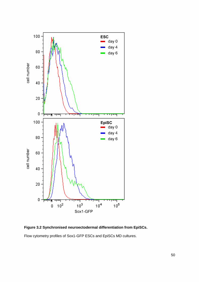
50
Figure 3.2 Synchronised neuroectodermal differentiation from EpiSCs.
Flow cytometry profiles of Sox1-GFP ESCs and EpiSCs MD cultures.

51
3.2 Discussion
Realising the potential of human ESCs in regenerative medicine requires an in-depth
understanding of the mechanisms that control the fate choice of stem cells during
differentiation. Recent studies reported that hESCs appear to be functionally more akin to
mouse EpiSCs than mouse ESCs (Brons et al., 2007; Rossant, 2008; Tesar et al., 2007;
Vallier et al., 2009). Therefore, mouse EpiSCs are an attractive model system for studying
neuroectodermal specification in vitro. Knowledge gained from neural differentiation of
mouse EpiSCs may provide a better understanding of neural commitment in hESCs and
improve techniques for the generation of particular neuronal subtypes from hESCs for
biomedical applications. Mouse EpiSCs have been shown to be capable of multilineage
differentiation in vitro (Bernemann et al., 2011; Brons et al., 2007; Tesar et al., 2007; Vallier
et al., 2009) and to form teratomas when grafted into adult mice (Brons et al., 2007; Tesar et
al., 2007). However, a direct comparison of mouse EpiSCs and mouse ESCs in neural
differentiation has not been described.
In this chapter, I report that EpiSCs are competent to commit efficiently to a neuronal fate. It
was demonstrated that neuronal cells derived from ESCs are not restricted to a specific
neuronal fate (Ying and Smith, 2003; Ying et al., 2003b) which was also the case for EpiSC-
derived neurons. I observed a more rapid loss of pluripotent markers in neural differentiation
of EpiSCs when compared to ESC in MD (Figure 3.1). This observation might be explained
by the developmentally advanced stage of EpiSCs (Brons et al., 2007; Tesar et al., 2007).
EpiSCs are developmentally equivalent to cells from the postimplantation epiblast at E~6.5
while ESCs are isolated from E3.5 embryos. Though EpiSCs express the pluripotent marker
genes and readily form teratomas upon injection into immunocompromised mice, they make
minimal contribute to the formation of chimeras (Brons et al., 2007; Tesar et al., 2007). In
conclusion, ESCs represent a more immature stage of differentiation than EpiSCs which are
poised to differentiate more readily.

52
The rapid exit of EpiSCs from a pluripotent stage was accompanied by an early,
synchronised conversion into a neuroectodermal lineage whereas neural induction in ESC
MD seemed to be a more heterogeneous progress (Figure 3.2). Consistent with this finding,
it has been reported that ESC cultures are composed of cells with different pluripotency
levels (Chambers et al., 2007; Furusawa et al., 2004; Hayashi et al., 2008; Toyooka et al.,
2008), consequently resembling different stages of development in vivo. Therefore, the
asynchronous neural differentiation observed in ESCs might be due to the presence of
distinct cell subpopulations within ESC cultures that when exposed to a given activation
stimuli respond differently according their developmental stages. In this respect, MD of
EpiSCs appeared as a more coordinated process, indicating that EpiSCs are more
homogeneous in nature. The relative synchronous nature of EpiSCs during neural
differentiation provides a more precise system to study the mechanisms of neural induction
and specification than ESC MD.
However, MD of ESCs and EpiSCs at d12 displayed comparable amounts of TUBB3 and
Sox1-GFP positive cells (Figure 3.1). The Sox1-GFP positive cells are mostly seen in areas
of high cell density concomitant with less TUBB3 positive cells. Since these areas seem to
create a “niche” where neural precursors are maintained and neuronal differentiation is
suppressed, dissociation of the cells might disrupt the niches with their cell-cell contacts and
subsequently improve neuronal differentiation. It needs to be taken into consideration that
replating of cells at this stage has a negative impact on the survival of the existing neuronal
cell population.

53
4 Temporally controlled modulation of FGF/ERK
signalling directs a midbrain dopaminergic fate
Extrinsic signals, such as FGF, WNT, BMP, and SHH, are essential for governing stem cell
fate decision and lineage specification. This chapter focuses on FGF signalling which plays
multiple roles during development depending on the time and the tissue.
During gastrulation, FGFs are involved in the posteriorisation of the neuroepithelium (Cox
and Hemmati-Brivanlou, 1995; Doniach, 1995; Holowacz and Sokol, 1999; Kengaku and
Okamoto, 1995; Lamb and Harland, 1995; Rentzsch et al., 2004; Ribisi et al., 2000; Wills et
al., 2010). At E~6.5, loss of Fgf8 expression in mouse embryos results in an expansion of
Otx2 and Six3 positive anterior tissue accompanied with the loss of posterior tissue marked
by Gbx2 (Sun et al., 1999). Consistent with this finding, Oki et al (2010) demonstrated that
potent FGFR inhibitors block posteriosation of mouse embryos during gastrulation. The
same study further reported that MAPK/ERK inhibitor could mainly replace the FGFR
inhibitor whereas antagonists of PLC-γ or PI3K pathway had no effect, implying FGF
signalling acts via the MAPK/ERK pathway in this context.
If in vitro differentiation of ESCs resembles the developmental process, inhibition of
FGF/ERK pathway during early stages of in vitro neural differentiation should induce the
production of cells with anterior characteristics (Otx2), including the cells from which mDA
neurons arise, at the expense of posterior neuroepithelial cells (Gbx2). We proposed that a
direct induction of early region-specific progenitors should improve the production of DA
neurons. I therefore tested this hypothesis using EpiSCs in vitro differentiation model
because they have already completed the transition from ESCs which involves the activation
of FGF signalling (Kunath et al., 2007). Additionally, EpiSCs are more advantageous than
ESCs because their in vitro differentiation towards neuroectoderm is more synchronously as
shown in Chapter 3. In this chapter I show that regulating the level of FGF/ERK activity

54
during distinct phases of neural differentiation in vitro is crucial for efficient generation of
mDA neurons.
4.1 Blockade of FGF/ERK signalling upon exit of epiblast
pluripotent state accelerates neural fate commitment
Self-renewal of EpiSCs in vitro is maintained by FGF2 and Activin A. I proposed that, in the
absence of Activin A, inhibition of FGF signalling promotes differentiation. To investigate
whether early blockade of FGF/ERK promotes differentiation into neuroectoderm, MD of
EpiSC was performed with either the FGFR inhibitor PD173074 (Stavridis et al., 2007) or the
potent MEK (MAPKK) blocker PD0325901 (Ying et al., 2008), which prevents ERK activation
and is referred as ERK inhibitor hereafter. As shown by Western Blot, phosphorylated ERK
was diminished in cultures treated with PD173074 or PD0325901 (Figure 4.1 A).
To determine whether FGF/ERK inhibition promotes the exit from the pluripotent stage,
immunostaining for the pluripotent marker OCT4 was performed on MD cultures at d1-3 with
and without PD0325901. As expected, less OCT4 positive cells were seen in cultures
exposed to the FGF/ERK inhibitor when compared to control cultures from d2 MD and
onward (Figure 4.1 B).
The patterns of stage-specific gene expression observed in Chapter 3 suggest that neural
induction in MD of EpiSCs occurred during the first 2-4 days of differentiation. In order to
examine whether inhibition of FGF/ERK signalling further promotes the transition of EpiSCs
into neuroectodermal cell fate, EpiSCs were cultured with PD0325901 and the expression of
the pro-neural genes Ngn2 and Mash1 and the neuronal marker gene Tubb3 were analysed
by qPCR (Figure 4.2 A). The levels of Mash1 were increased from d3 MD with or without
PD0325901. However, Mash1 expression level was higher in cultures with PD0325901.
Interestingly, the expression of the pro-neural gene Ngn2 was already highly upregulated at

55
d3 MD in PD0325901 treated cultures whereas in no-PD0325901 control cultures an
increase of Ngn2 expression was not observed until d4 of differentiation. Moreover, in
cultures that were exposed to PD0325901, the elevation of Tubb3 mRNA was shifted
forward to d3 of differentiation when compared with no-PD0325901 controls.
To strengthen the observation that inhibition of the FGF/ERK pathway causes a more rapid
neuroectodermal differentiation, I used Sox1-GFP knock-in (46C) reporter EpiSCs (Ying et
al., 2003b) to monitor differentiation. Sox1 is specifically expressed in the neuroectoderm
from the headfold stage onwards (Wood and Episkopou, 1999), thus it is commonly used as
a marker gene for neural progenitors. To gain insight into the kinetics of neural induction,
Sox1-GFP positive cells were detected by flow cytometry and the percentage of Sox1-GFP
positive cells was determined between d1 and d6 MD. As shown, Sox1-GFP was
undetectable before differentiation (Figure 4.2 C, D). At d2 of differentiation, a high
proportion of Sox1-GFP positive cells (44.15%±1.2) were already detected in cultures with
PD0325901, compared to control cultures without PD0325901 (7.55%±0.64). Furthermore
and consistent with the observation above, immunocytochemical staining of PLZF, a marker
of neuroectodermal cells (Avantaggiato et al., 1995), indicated that PD0325901 treatment
accelerated and enhanced the generation of PLZF positive cells compared to controls
cultures (Figure 4.2 B).
Taking together, the results from the analysis of the flow cytometry data, immunostaining
and gene expression demonstrate that inactivation of FGF/ERK signalling accelerates the
transition from a pluripotent epiblast stage toward neuroectoderm.

56
Figure 4.1 Inhibition of FGF/ERK activity accelerates the exit of EpiSCs from a
pluripotent stage.
(A) Western blot analysis for phosphorylated ERK in d2 mouse EpiSC differentiation cultures
treated with PD0325901 (PD03) or PD173074 (PD17). Samples were loaded undiluted
(1; 30 µg) or diluted 1:2 in loading buffer. (B) Antibody staining for the pluripotent marker
OCT4, labelled with green, in d1, d2 and d3 MD cultures with or without PD0325901 (PD03).
DAPI labelling is in blue. Scale bars: 100 µm.

57

58
Figure 4.2 Inhibition of FGF/ERK activity accelerates neural fate conversion of
EpiSCs.
(A) qPCR analysis for the expression of neural lineage marker genes over 4 days MD
cultures of EpiSCs. Data represent mean±s.e.m. from triplicate cultures of a single
experiment. (B) Immunostaining for the neural rosette marker PLZF, shown in green, at d2,
d4 and d6 MD with or without PD0325901 (PD03). DAPI labelling is in blue. Scale bars: 100
µm. (C) Antibody staining for a neuroepithelium marker SOX1, red, in d2-5 MD cultures with
or without PD0325901 (PD03). DAPI labelling is in blue. Scale bars: 100 µm. (D) MD
cultures of Sox1-GFP EpiSCs in the presence or absence of PD0325901 (PD03). Sox1-GFP
reporter expression was analyzed by flow cytometry and the percentage of Sox1-GFP
positive cells was determined from three independent cultures. Error bars indicate s.e.m.

59
4.2 Inhibition of FGF/ERK activity at the onset of EpiSC
differentiation promotes mDA progenitor characteristics
To investigate the effect of early FGF/ERK inhibition on the generation of mDA neurons from
EpiSCs, cells were exposed for 2 days with either PD173074 or PD0325901 at the onset of
neural differentiation and the expression of mDA regulatory genes, Foxa2, Lmx1a, and
Dmrt5, were tested. Compared to no-factor controls, the expression levels of Lmx1a, Foxa2,
and Dmrt5 were already upregulated at d2 MD in cultures that experienced FGF/ERK
blockade. Moreover, the increase of Lmx1a, Foxa2, and Dmrt5 continued after withdrawal of
FGF/ERK inhibitor (Figure 4.3 B, C).
To determine whether longer exposure to the ERK inhibitor could further enhance the
expression of mDA regulatory genes, I compared the effects of 5 days PD0325901 treatment
(d0-5) to that of 2 days (d0-2) during MD. Contrary to short exposure of PD0325901, no
increase of Foxa2 and Dmrt5 was observed after 5 days PD0325901 treatment. Moreover,
the transcript level of Lmx1a was reduced in cultures continuously exposed to PD0325901
when compared with the no-PD0325901 culture at d5. Notably, detection of p-ERK by
Western Blot analysis demonstrated the presence of FGF/ERK signalling at d4 MD in
cultures which had been treated with PD0325901 for 2 days (d0-2) (Figure 4.3 A). These
results indicate that although FGF/ERK inhibition at early stages initiates EpiSC
differentiation, endogenous FGF/ERK signalling is necessary from d3 to acquire mDA
progenitor characteristics.
To confirm the results of qPCR analysis, EpiSC cultures at d5 MD were stained for LMX1A,
FOXA2, and DMRT5. The proportion of cells expressing either one of the mDA progenitor
marker in the total cell population was determined by pixel-analysis. At d5, when the majority
of cells are Nestin positive, the percentage of neural progenitors expressing LMX1A,
FOXA2, or DMRT5 increased greatly in PD0325901 treated cultures (LMX1A: 12±4.3%;
FOXA2: 59±10.82%; DMRT5: 35±7.65%) compared to cultures without PD0325901

60
treatment (LMX1A: 3±1.27%; FOXA2: 24±8.6%; DMRT5: 6±2.45%) (Figure 4.3 E). As
treatment with either PD0325901 or PD173074 displayed similar results, PD0325901 was
used for further studies unless stated otherwise.
Gene expression analysis by qPCR further revealed an increased expression of Shh and
Wnt1 in PD0325901 treated cultures when compared to control cultures. SHH and WNT1
are signalling molecules which were shown to be required for the induction of mDA
progenitor cells by forming regulatory loops with LMX1A and FOXA2 (Andersson et al.,
2006b; Chung et al., 2009; Echelard et al., 1993; Mavromatakis et al., 2010). At d2 MD, the
expression level rose 1.5-fold and 4-fold in cultures with PD173074 and PD0325901,
respectively, when compared to no-factor control. The expression of Shh was further
upregulated between d3 and d5 after withdrawal of PD173074 and PD0325901. However,
long exposure of PD0325901 displayed similar transcript levels of Shh as in cultures without
PD0325901 (Figure 4.3 B). Wnt1 expression was already highly upregulated at d1 and d2
MD in PD173074- or PD0325901-treated cultures whereas in control cultures the transcript
levels of Wnt1 were low (Figure 4.3 B,C). In contrast to the observed Shh expression pattern
during the first day of differentiation, Wnt1 expression decreased from d3 and onward even
after withdrawal of the small molecule ERK inhibitor, implying the activation of a pathway
negatively regulating Wnt1 expression. Consistent with a previous study (Chung et al.,
2009), I observed similar expression pattern of Wnt1 and Lmx1a at d1 and d2 MD.
Furthermore, the kinetics of Shh expression correlates with the pattern of Foxa2 expression,
supporting published results of a regulatory relationship between SHH and FOXA2 (Ang and
Rossant, 1994; Mavromatakis et al., 2010).
The FGF family can activate 3 different signalling pathways: Mitogen-activated protein
kinase (MAPK/ERK), Phosphatidylinositol-3 kinase (PI3K) and Phospholipase C gamma
(PLC-γ) (Figure 1.3 A). To address whether the induction of mDA regulators is specific to the
blockade of FGF/ERK pathway, neural differentiation of EpiSC was carried out in the
presence of the PI3K inhibitor LY294002 (2 µM) from d0-2 MD (Figure 4.4 A). RNA was

61
collected every day during d1-3 of MD, and the expression levels of Wnt1, Shh, Foxa2,
Dmrt5, Lmx1a, and Msx1, a downstream target of LMX1A (Andersson et al., 2006b), were
assessed by qPCR (Figure 4.4 B). I found similar level of mRNA for Wnt1, Shh, Dmrt5, and
Msx1 between LY2949002 treated cultures and control cultures. The expression of Foxa2
and Lmx1a dropped below control levels at d2-3. These findings indicate that early inhibition
of FGF/PI3K signalling is not involved in mDA neural specification.
Taking together, blockade of FGF/ERK activity at the onset of the epiblast pluripotent state
induces the expression of Shh and Wnt1, signalling molecules essential for mDA generation,
and enhances the generation of mDA progenitor cells marked by FOXA2, LMX1A, and
DMRT5. However, these newly converted neural progenitors require endogenous FGF/ERK
activity to maintain and stabilise mDA neural progenitor fate.

62

63
Figure 4.3 Inhibition of FGF/ERK on the exit of the epiblast pluripotent stage promotes
the generation of mDA progenitor cells.
(A) Western blot analysis for phosphorylated ERK (p-ERK) and total ERK in d4 mouse
EpiSC differentiation cultures treated with PD0325901 (PD03) and FGF8 (15 µg protein). (B)
E14tg2a mouse EpiSC MD cultures were exposed to PD0325901 from d0 MD for either 2
days (PD03 d0-2) or 5 days (PD03 d0-5). Cultures were harvested at d1, d2, d3 and d5 MD
for qPCR analysis. Gene expressions of Wnt1, Shh, Lmx1a, Foxa2, and Dmrt5 were tested.
The value of the d1 MD control was set as 1. Error bars indicate s.e.m. of two sets of
experiments performed in duplicate. (C) E14tg2a mouse EpiSC MD cultures were exposed
to PD173074 from d0 MD for 2 days (PD17 d0-2) and cultures were harvested at d2, d4 and
d6 MD. qPCR analysis of Wnt1, Shh, Lmx1a,Foxa2, and Dmrt5. The value of the d2 MD
control was set as 1. Error bars indicate mean±s.e.m. of two sets of experiments performed
in duplicate. (D) MD of EpiSCs exposed to PD0325901 (PD03) from d0 MD for 2 days were
immunostained at d5 for LMX1A, FOXA2, and DMRT5 and counterstained with DAPI. Scale
bar: 100 µm. (E) Quantification of immunostaining in D showing the percentage of cells
expressing each mDA progenitor marker protein.

64
Figure 4.4 Blockade of FGF/PI3K signalling does not induce expression of mDA
regulatory genes.
(A) Experimental scheme of neural differentiation with E14tg2a mouse EpiSCs. MD cultures
were exposed to PD0325901 (PD03) or LY294002 from d0 MD for 2 days. (B) Cultures were
harvested at d1, d2, and d3 MD for qPCR analysis. Gene expressions of Shh, Wnt1, Lmx1a,
Foxa2, Dmrt5 and Msx1 were tested. The value of the d1 MD control was set as 1. Error
bars indicate s.e.m. of two sets of experiments performed in duplicate.

65
4.3 FGF/ERK inhibition affects anterior-posteriorisation and the
maintenance of OTX2 in nascent neural progenitors
It has been shown in mouse embryos that blockade of FGF signalling during gastrulation
results in an expansion of anterior and a reduction of posterior tissue (Oki et al., 2010; Sun
et al., 1999). Therefore, it is possible that inhibition of FGF/ERK promotes the generation of
ventral midbrain progenitor cells via modulating anterior–posterior characteristics of neural
progenitor cells. To verify whether early blockade of FGF/ERK signalling maintains anterior
neural character during neural differentiation, EpiSC MD cultures were analysed for anterior-
posterior markers.
In order to examine the anterior character of EpiSC MD, cultures were analysed by
immunostaining for OTX2 which is an anteriorly expressed homeobox protein (Acampora et
al., 1995; Ang et al., 1994; Millet et al., 1996; Simeone et al., 1992; Simeone et al., 1995).
As expected, EpiSC cultures exposed to PD0325901 on exit of the pluripotent stage
exhibited an enlarged OTX2 positive cell population when compared to control cultures at
d3-5 MD (control, d5: 11.75±4.25%; PD0325901, d5: 69.48±17.89%) (Figure 4.5 A, B).
Consistent with the immunostaining for OTX2, transcript levels of Gbx2, expressed in vivo in
areas becoming the anterior hindbrain and spinal cord, increased from d1 until d3 of MD in
cultures without PD0325901 whereas the expression decreased with PD0325901 (Figure
4.5 C). Together, these data indicate that early FGF/ERK blockade promotes and/or
maintains an anterior character of neural progenitor cells.
However, FGFs are expressed in the anterior neural ridge where they are involved in the
formation of the forebrain (Paek et al., 2009; Shimamura and Rubenstein, 1997). To
investigate whether the generation of cells with forebrain characteristic is affected by
inhibition of FGF/ERK activity, the expressions of the forebrain marker genes Six3 and
Foxg1 were tested in MD cultures with or without PD0325901 at d0-2 (Figure 4.5 D) (Hebert
and McConnell, 2000; Jeong et al., 2008; Lavado et al., 2008). The transcript level of Six3

66
and Foxg1 were similar at d2 MD in both conditions. Interestingly, in cultures exposed to
PD0325901 the expression of Six3 and Foxg1 decreased after PD0325901 was withdrawn.
Furthermore, their expression levels were reduced at d3-4 when compared to control MD,
suggesting that early inhibition of FGF/ERK activity suppress the commitment of newly born
neural progenitor cells toward anterior forebrain fate.
The Otx2 positive domain in the neuroepithelium spans the prospective forebrain and
midbrain. Following the observation on decreased forebrain marker gene expression in
PD0325901-treated cultures, I examined the expression of midbrain marker genes En2 and
Dmbx1 (Davidson et al., 1988; Davis et al., 1988; Gogoi et al., 2002; Ohtoshi and Behringer,
2004). The transcript level of En2 increased steadily in both culture conditions from d2 until
d5 of MD. However, a greater increase was observed in PD0325901-treated cultures
compared to the control. An increase of Dmbx1 was already evident from d1 MD with
PD0325901 treatment when compared to control, although its level in the control cultures
catched up at d5 MD.
To test whether FGF/PI3K pathway plays a role in anterior-posterior modulation of neural
progenitor, EpiSC cultures at d5 MD treated with or without PD0325901 or LY294002 were
assessed by immunocytochemistry for OTX2. An increase of the OTX2 positive cell
population was only observed in PD0325901-treated cultures while LY294002-treated
cultures had a similar proportion of OTX2 positive cells as those in the control.
Taken together, these results suggest that early inhibition of FGF/ERK activity primes cells
for a midbrain progenitor cell fate by blocking forebrain and hindbrain cell fate.

67

68
Figure 4.5 Early blockade of FGF/ERK activity modulates anterior –posterior
characteristics of neural progenitor cells.
(A) Antibody staining for the anterior marker OTX2 shown in red in d3, d4 and d5 MD
cultures with or without PD0325901 (PD03). DAPI is labelled in blue. (B) Quantification of
immunostaining in A illustrating the percentage of cells expressing OTX2. Mean±s.e.m. of
ten fields from two independent experiments. *, P<0.01 relative to no-PD0325901 control
(Student’s t-test). (C) Mouse E14tg2a EpiSC MD cultures were exposed to PD0325901
(PD03) from d0 MD for 2 days. Cultures were harvested at d1, d2, d3 or d5 for qPCR
analysis of Gbx2. (D) E14tg2a EpiSC MD cultures were treated with PD0325901 cultures
were harvested at d2-4 for qPCR analysis of the forebrain marker genes Foxg1 and Six3. (E)
EpiSCs were differentiated in the presence of PD0325901 from d0 MD for 2 days, collected
as in C and tested for the midbrain marker genes En2 and Dmbx1 by qPCR. The value of
the d3 MD control was set as 1. Error bars indicate s.e.m. of two sets of experiments
performed in triplicates.
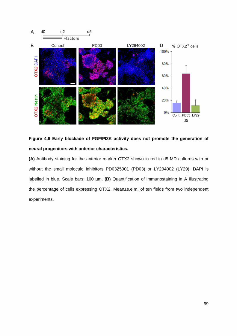
69
Figure 4.6 Early blockade of FGF/PI3K activity does not promote the generation of
neural progenitors with anterior characteristics.
(A) Antibody staining for the anterior marker OTX2 shown in red in d5 MD cultures with or
without the small molecule inhibitors PD0325901 (PD03) or LY294002 (LY29). DAPI is
labelled in blue. Scale bars: 100 µm. (B) Quantification of immunostaining in A illustrating
the percentage of cells expressing OTX2. Mean±s.e.m. of ten fields from two independent
experiments.

70
4.4 Highly reliable and efficient production of mDA neurons by
ERK inhibitor-primed neural progenitors
To investigate whether the primed mDA progenitor cells generated above can terminally
differentiate into DA neurons (TH positive cells) with midbrain-specific identity, SHH and
FGF8 were applied to induce DA differentiation in MD cultures at d5 when the majority of
cells were Nestin positive as they are used in standard mDA differentiation protocols (Figure
4.7 A) (Barberi et al., 2003; Kawasaki et al., 2000; Kim et al., 2002; Lee et al., 2000b;
Parmar and Li, 2007; Perrier et al., 2004). D14 MD cultures were then analysed in detail for
mDA neuronal markers by immunocytochemistry (Figure 4.7 B-D). I found that MD cultures
exposed to PD0325901 at d0-2 followed by SHH and FGF8 at d5-9 contained 4-fold more
TH positive neurons than no-PD0325901 cultures (PD, 52.3±8%; control, 13.5±9%) (Figure
4.7 B, C).
In order to examine the midbrain-specific characteristics of the derived DA neurons, EpiSC
were derived from Pitx3-GFP knock-in ESCs (Maxwell et al., 2005; Zhao et al., 2004).
During midbrain development, Pitx3 expression starts at E11.5 and is restricted to SN and
VTA (Smidt et al., 1997). Therefore, co-expression of TH and PITX3 is used as a marker for
mDA neurons. Pitx3-GFP EpiSCs were subjected to neural differentiation as illustrated in
Figure 4.7 A. Cultures were assessed by immunstaining for TH and GFP at d14 MD. A high
proportion (73.8±10.2%) of TH positive neurons co-expressed Pitx3-GFP in cultures
received PD0325901 at d0-2 and SHH and FGF8 at d5-9 (Figure 4.7 B, C). In addition,
these TH positive neurons also co-expressed midbrain transcription factors, LMX1A
(88.5±3.9%) and FOXA2 (99.5±1.2%), and pan-dopaminergic-neuronal markers, NURR1
(also known as NR4A2, 85.4±4%) and VMAT (known as SLC18A1, 85.6±12%). In contrast,
37.1±9.5% of TH positive neurons co-expressed Pitx3-GFP in control cultures exposed to
SHH and FGF8 only. In conclusion, midbrain progenitor cells induced by FGF/ERK inhibition
can efficiently generate authentic mDA neurons.

71

72
Figure 4.7 Temporally controlled FGF/ERK activity promotes the production of
functional, authentic mDA neurons.
(A) Experimental scheme of neural differentiation with Pitx3-GFP EpiSCs. MD cultures were
exposed to PD0325901 (PD03) from d0 MD for 2 days. SHH and FGF8 were applied from
d5 until d9. Medium was then changed to N2B27 plus GDNF, BDNF and ascorbic acid and
cultured for another 5 days. (B) MD cultures at d14 were double stained for (b1-4) TH, red,
and TUBB3, blue, as well as (b5-8) TH, red, and Pitx3-GFP labelled in green. (C)
Quantification of the immunostaining in B, showing the percentage of TH positive neurons
and TH positive neurons co-expressing Pitx3-GFP. Mean±s.e.m. *, P<0.01 relative to no-
PD0325901 control (Student’s t-test). (D) Immunostaining of mouse d14 MD cultures for TH
and mDA neural markers. Scale bars: 200 µm for B (b1,2,5,6) and 50 µm for B (b2,3,7,8), D.

73
4.5 Dopaminergic neuron differentiation of mouse iPSCs in
response to ERK inhibition
Next I wanted to test whether this protocol can be applied to mouse induced pluripotent stem
cells. Considering that inhibition of FGF/ERK signalling maintains ESCs in the naïve
pluripotent state (Silva et al., 2008a; Ying et al., 2008) and activation of FGF signalling is
necessary to drive ESCs toward the primitive ectoderm/epiblast stage (Kunath et al., 2007),
PD0325901 was applied for 2 days either during d1-3 or d2-4 of MD. To support the
generation of mDA neurons, SHH and FGF8 were added from d6 until d10 of MD (see
Figure 4.8 A). Immunostaining for TUBB3, TH and FOXA2 was carried out at d14 MD to
observe the generation of mDA neurons. During development, the specification of mDA
neurons, which are TH positive, starts in the medial domain of the Foxa2-expressing floor
plate (Ferri et al., 2007). Consequently, co-expression of TH and FOXA2 can be used as a
marker for mDA neurons. Consistent with the observation in EpiSC MD, mDA neurons (TH
and FOXA2 double positive) neurons were also efficiently generated in MD cultures of iPSCs
exposed to PD0325901 either at d1-3 or d2-4 MD when compared to control (Figure 4.8 B),
supporting our hypothesis that early blockade of FGF signalling during neural induction
primes a midbrain regional fate in derived neural progenitors, which subsequently
differentiate into DA neurons exhibiting midbrain characteristics.

74
Figure 4.8 Efficient generation of mDA neurons from mouse iPSCs.
(A) Experimental scheme of neural differentiation with mouse iPSCs. MD cultures were
exposed to PD0325901 (PD03, green) at either d1-3 or d2-4 of MD. SHH and FGF8 (red)
were applied from d6 until d10 of MD. Medium was then changed to N2B27 plus GDNF,
BDNF and ascorbic acid and cultured for another 4 days. (B) MD cultures at d14 were
double stained for TH shown in green, FOXA2, blue, and TUBB3, red. Scale bar: 200 µm.

75
4.6 Differential activity of SHH and FGF8 in mDA fate specification
and differentiation
Ye and colleagues reported (1998) that only the combinatorial treatment of SHH and FGF8
can induce DA fate in E9 ventral caudal forebrain rat explants. In agreement with this study, I
also observed a robust generation of TH positive neurons at d14 MD in PD0325901-treated
cultures exposed to SHH and FGF8 from d5 until d9 whereas few TH positive cells were
detected in cultures treated with PD0325901 alone (Figure 4.9). A large number of TH
positive cells were also detected in MD cultures which received PD0325901 at d0-2 followed
by SHH alone at d5-9. However, these DA neurons showed a low expression of TH with
immature neuronal morphology. In contrast, TH positive neurons in d14 MD cultures treated
with PD0325901 followed by FGF8 alone displayed a more mature morphology. The number
of TH positive cells in this condition was comparable with control MD cultures exposed to
SHH and FGF8 in the absence of PD0325901.
I next analysed the expression of gene markers associated with GABAergic phenotype by
qPCR. The RNA levels of Helt, Gata2, and Gad1 were determined at d5 and d8 and the data
was presented as the fold change to the level of d5 in control MD cultures with no added
factors. I detected the highest fold increase of these GABAergic marker genes in control
cultures without exogenous stimuli (Gata2, 38-fold; Helt, 293-fold; Gad1, 401-fold) (Figure
4.10) whereas in cultures with SHH the expression of these genes was substantially
repressed. In cultures exposed to PD0325901 followed by SHH, the mRNA levels of Helt
and Gad1 were only 10% of the level of MD cultures treated with SHH alone and were less
than 1% of the level of no-factor controls.
To further elucidate how SHH and FGF8 may shape neural progenitor cell fate, SHH and
FGF8, either alone or in combination, were applied at d3-6 of EpiSCs, a stage when neural
progenitor arise (Figure 3.1 and Figure 4.2), and changes in the expression levels of mDA
regulator genes were monitored at d6 of differentiation when the majority of cells is Nestin

76
positive (Figure 4.3 D). The expression levels of Dmrt5, Foxa2, and Msx1 were upregulated
in cultures exposed to SHH at d3-6 while the mRNA transcript levels of Lmx1a and Shh were
not enhanced (Figure 4.11 B, C). In contrast, adding FGF8 alone or with SHH abolished the
increased expression of Dmrt5, Foxa2, Msx1, Lmx1a, and Shh caused by PD0325901 or
PD0325901-SHH. Moreover, activation of FGF signalling by FGF8 resulted in an increase of
isthmus associated genes Wnt1, En1, and En2, in agreement with previously published data
(Lee et al., 1997; Liu and Joyner, 2001; Liu et al., 1999).
The results shown in chapter 4.2 indicated that a period of autocrine FGF/ERK signalling is
essential for newly born neural progenitor cell to become specified into a ventral midbrain
fate characterised by DMRT5, LMX1A, and FOXA2. However, enhancement of FGF
signalling by FGF8 in newly born neural progenitors eliminated the PD0325901-mediated
upregulation of mDA regulatory genes Dmrt5, Lmx1a, Foxa2, Shh and Msx1. These data
demonstrate the importance of temporally controlled modulation of FGF/ERK signalling in
the specification of neural progenitors towards a ventral midbrain fate. Furthermore, SHH
and FGF8 co-operatively direct the conversion of PD0325901-generated d5 neural
progenitors into mDA neurons where SHH signalling specifies a DA phenotype and FGF8
facilitates progenitor differentiation.

77
Figure 4.9 Temporally controlled FGF/ERK activity in combination with SHH and FGF8
promotes the production of DA neurons.
(A) Experimental schemes for data in B. (B) MD cultures of EpiSCs at d14 were double
stained for TH, shown in red, and TUBB3, labelled with green. Scale bar: 200 µm.

78
Figure 4.10 Activation of SHH signalling promotes the production of DA neurons by
repressing the transcript level of genes for a GABAergic neuronal fate.
(A) Gene expression analysis of Helt, Gata2, and Gad1 of d5 and d9 MD cultures by qPCR.
Fold change analysis is used to determine the difference of gene expression between d5
and d9 in control MD cultures with no added factors, control cultures treated with SHH, and
cultures exposed to PD0325901 at d0-2 followed by SHH. Error bars indicate s.e.m. of
triplicate cultures from one representative experiment.
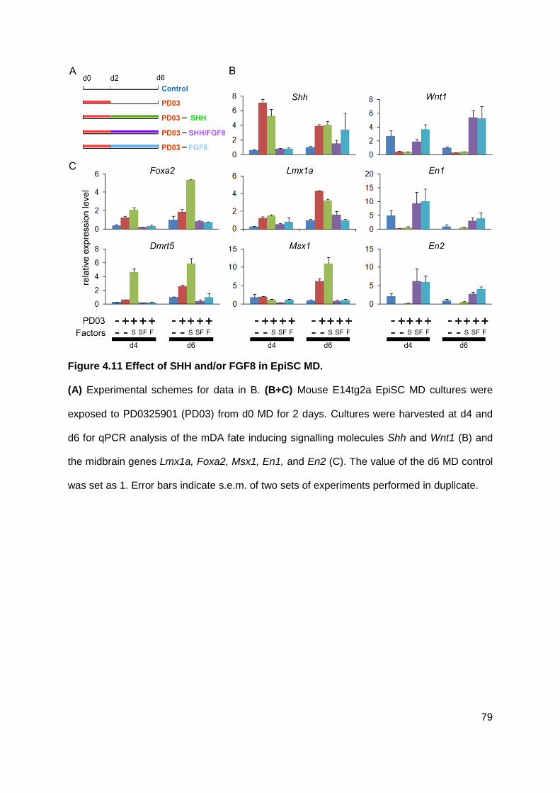
79
Figure 4.11 Effect of SHH and/or FGF8 in EpiSC MD.
(A) Experimental schemes for data in B. (B+C) Mouse E14tg2a EpiSC MD cultures were
exposed to PD0325901 (PD03) from d0 MD for 2 days. Cultures were harvested at d4 and
d6 for qPCR analysis of the mDA fate inducing signalling molecules Shh and Wnt1 (B) and
the midbrain genes Lmx1a, Foxa2, Msx1, En1, and En2 (C). The value of the d6 MD control
was set as 1. Error bars indicate s.e.m. of two sets of experiments performed in duplicate.

80
4.7 WNT alone or in combination with SHH cannot replace ERK
inhibitor in inducing midbrain progenitor cell fate
The induction of signalling molecules Shh and Wnt1 by either PD173074 or PD0325901 and
the concomitant induction of their target genes Foxa2 and Lmx1a, respectively (Figure 4.3 B,
C), prompted me to investigate whether SHH and WNT1 are the mediators of early
FGF/ERK blockade that led to induced efficient mDA fate specification and differentiation.
4.7.1 The effects of early SHH activation on mDA induction
In order to investigate the contribution of an early activation of SHH signalling, MD
differentiation was firstly carried out in the presence of SHH, PD0325901+SHH, or
PD0325901 alone from d0. RNA was collected from d1-3 of MD and analysed for the
expression of Shh, the SHH downstream targets Gli1 and Foxa2 and the mDA regulator
genes Dmrt5, Lmx1a, and Msx1 (Figure 4.12). As expected, in MD cultures exposed to SHH
alone (d0-2) showed an increased mRNA level of Gli1 and Foxa2 from d2 or d3 respectively
when compared to control cultures. However, the transcript levels of the mDA regulator
genes Lmx1a, Msx1, and Dmrt5 were similar to the expression levels in control cultures.
Next I wanted to determine whether the observed induction of mDA marker gene expression
is SHH dependent. To this end I performed an additional MD in the presence of the SHH
inhibitor cyclopamine with or without PD0325901 (Figure 4.13). MD cultures exposed to
cyclopamine (10 µM) in the presence or absence of PD0325901 displayed a reduction in the
expression levels of the SHH downstream target genes Gli1 at d1-3 and Foxa2 at d2-3 to
control cultures, confirming its SHH inhibitory activity. Cyclopamine had little effect on the
expression of Shh, Msx1, and Dmrt5. Interestingly, PD0325901-induced expression of Dmrt5
and Msx1 was further enhanced by cyclopamine, suggesting a SHH-independent
mechanism. However, cyclopamine treatment attenuated PD0325901-mediated induction of
Foxa2 and Lmx1a (Fig 4.7.2).

81
Because SHH treatment alone could increase the expression of Foxa2 and Lmx1a from d3
onward, we asked whether an early addition of SHH is sufficient to direct MD cultures
towards a midbrain neural progenitor fate. I first tested the expression of the hindbrain
marker gene Gbx2 and midbrain marker genes En1 and Dmbx1 during 3 days of
differentiation (Figure 4.14 A). The data revealed that, unlike PD0325901, SHH did not
suppress the hindbrain marker gene Gbx2 and upregulated the midbrain marker genes En1
and Dmbx1. Consistent with the qPCR data, I did not observe an increase of OTX2 positive
cells at d5 and d9 in cultures exposed to early SHH (Figure 4.14 B). As shown in Figure
4.14 C, only cultures exposed to PD0325901 had a significant proportion of OTX2 positive
cells at d9 MD (PD0325901, 43.88±5.32%; SHH, 6.75±2.75%; control, 7.25±2.29%).
Surprisingly, at d9, no obvious difference was seen in the number of FOXA2 and LMX1A
positive neural progenitors in PD0325901-, SHH-treated and no-factor cultures. However, a
high proportion of TH positive neurons were only obtained in PD0325901-treated cultures
(Figure 4.15 B). Taken together, these results suggest that SHH is not sufficient to replace
PD0325901 at d0-2 for complete mDA induction.
4.7.2 The effects of GSK3ß inhibition on mDA induction
Early inhibition of FGF/ERK activity also induced the expression of Wnt1. This factor is
generally considered a canonical WNT because it co-localizes with β-catenin in the ventral
midbrain and mutation of Wnt1 in vivo lead to a similar phenotype as ß-catenin mutations
(Castelo-Branco et al., 2004; Castelo-Branco et al., 2003; Danielian and McMahon, 1996;
Prakash et al., 2006; Tang et al., 2010). To test whether activation of WNT signalling can
bring about similar effects to PD0325901, I used the small molecule CHIR99021, a selective
inhibitor of the Glycogen synthase kinase-3 (GSK3ß) with an IC50 less than 10 nM (Bain et
al., 2007; Murray et al., 2004; Ring et al., 2003). In the absence of WNT, ß-catenin is
phosphorylated by a GSK3ß containing destruction protein complex inducing its
degradation. In the presence of WNT, GSK3ß is physically displaced from the complex of

82
regulatory binding partners preventing the phosphorylation and degradation of ß-catenin.
Thus, inhibition of GSK3ß by CHIR99021 results in the maintenance and accumulation of
ß-catenin mimicking WNT signalling (Ring et al., 2003). As a transcriptional readout of active
canonical WNT signalling, I analysed the expression of the downstream target Axin2 by
qPCR (Jho et al., 2002). As expected, the expression level of Axin2 increased when
CHIR99021 was added at d0-2 of MD confirming the stimulation of ß-catenin/TCF signalling
(Figure 4.16 B). The effect of CHIR99021 on Axin2 expression was dose-dependent, as MD
cultures exposed to 2 µM CHIR99021 showed an enhanced mRNA level of Axin2 when
compared to cultures that experienced only 0.5 µM. The expression level of Axin2 dropped
to the transcript level of control cultures at d3 MD after withdrawal of CHIR99021.
PD0325901 treatment resulted in upregulation of Axin2 (2.5 to 3.7x fold compared to control
cultures), suggesting that activation of canonical WNT signalling may be involved (Figure
4.16 B).
It has been reported that LMX1A is in a autoregulatory loop with the WNT pathway in the
context of mDA neurogenesis (Chung et al., 2009). I therefore asked whether CHIR99021
could mimic PD0325901 in upregulating of Lmx1a. The expression of Lmx1a in cultures
exposed to CHIR99021 was similar to control cultures at d1 whereas Lmx1a induction was
already evident in PD0325901-treated cultures (Figure 4.16 B). However, at d2 MD, the
mRNA level of Lmx1a visibly increased in CHIR99021-treated cultures compared to cultures
without factor. Furthermore, expression of Dmrt5 was not affected by CHIR99021. These
findings suggest that other pathway(s) may contribute to the induction of the mDA neural
progenitor fate.
It has been reported that in the developing midbrain OTX2 and WNT1 are in a positive
regulatory feedback loop (Chung et al., 2009; Prakash et al., 2006). To investigate whether
the observed Otx2 upregulation is mediated by WNT1, I examined OTX2 and Gbx2 by
immunocytochemistry and qPCR (Figure 4.16 C-E). No increase of OTX2 positive cells was
observed at d5 MD with any concentrations of CHIR990221 (0.5 µM: 6±2.02%; 2.0 µM:

83
16±6.02%) when compared to control cultures (16±3.57%), whereas in PD0325901-treated
sister cultures the majority of cells were OTX2 positive neural progenitors (64±13.64%)
(Figure 4.16 C, D). In addition, no reduction of Gbx2 expression was seen in cultures
experienced early inhibition of GSK3ß (Figure 4.16 E).
4.7.3 The effects of combined treatment of SHH and CHIR99021 on mDA
induction
Because SHH and CHIR99021 only partially induced mDA neural progenitor characteristics
(↑Foxa2, ↑Lmx1a), I next studied their combination in mDA induction. In the presence of both
CHIR99021 and SHH at d0-2 MD, the expression of Lmx1a and Foxa2 at d2 MD increased
up to ~3 times compared to control cultures, similar to that observed in cultures exposed to
PD0325901 (Figure 4.17 B). However, unlike PD0325901 treatment, no upregulation of
Dmrt5 was observed (Figure 4.17 B). Furthermore, the proportion of OTX2 positive cells in
cultures with SHH and CHIR99021 (SHH & 0.5 µM CHIR99021: 5.56±2.5%; SHH & 2µM
CHIR99021: 0.35±0.22%) was even below the percentage observed in control cultures at d5
MD (16±3.57%) (Figure 4.17 C, D). In keeping with this observation, a downregulation of
Gbx2, as in PD0325901 treated cultures, was not detected in cultures exposed to SHH and
CHIR99021 (Figure 4.17 E). Nonetheless, I performed terminal neural differentiation with
PD0325901, CHIR99021 alone or with SHH and no-factor, as illustrated in Figure 4.18 A,
and tested whether robust induction of the mDA regulators Lmx1a and Foxa2 potentiate
mDA neuronal production. I applied immunofluorescent staining at d14 MD to examine the
generation of DA neurons with midbrain character, marked by TH and FOXA2 double
positive cells. As anticipated, most of FOXA2 and TH double positive cells were detected in
cultures treated with PD0325901 at d0-2 (Figure 4.18 B). Only few TH positive cells were
observed in MD cultures with CHIR99021, CHIR99021-SHH or without factors. Most of these
TH positive cells did not co-express FOXA2, suggesting that they might be a different
subpopulation of DA cells. On the other hand, the cell population of FOXA2 positive cells
was slightly but consistently increased in cultures exposed to CHIR99021 alone or its

84
combination with SHH. It is possible that CHIR99021 treated cells respond more efficiently to
secreted SHH.
Taken together, neither SHH, canonical WNT signalling nor their combination can replace
early inhibition of FGF/ERK activity in the generation of mDA neurons, indicating that other
pathways are involved in this process.

85
Figure 4.12 Activation of SHH pathway is not sufficient for complete mDA induction.
(A) Experimental schemes for data in B. (B) Mouse E14tg2a EpiSC MD cultures were
exposed to PD0325901 (PD03) and/or SHH from d0 MD for 2 days. Cultures were harvested
at d1-3 for qPCR analysis of the mDA fate inducing signalling molecule Shh and its
downstream target genes Gli1 and Foxa2 as well as the midbrain genes Lmx1a, Msx1, and
Dmrt5. The value of the d1 MD control was set as 1. Error bars indicate s.e.m. of two sets of
experiments performed in triplicate.

86
Figure 4.13 Early inhibition of FGF/ERK acts partly via the SHH pathway.
(A) Experimental schemes for data in B. (B) Mouse E14tg2a EpiSC MD cultures were
treated with PD0325901 (PD03), the SHH antagonist cyclopamine or a combination of the
two as indicated. Cultures were harvested at d1, d2, and d3 for qPCR analysis of the mDA
fate inducing signalling molecule Shh and its downstream target genes Gli1 and Foxa2 as
well as the midbrain progenitor genes Lmx1a, Msx1, and Dmrt5. The value of the d1 MD
control was set as 1. Error bars indicate s.e.m. of two sets of experiments performed in
triplicate.

87

88
Figure 4.14 SHH treatment cannot induce midbrain neural progenitor fate.
(A) Mouse E14tg2a EpiSC MD cultures were exposed to PD0325901 (PD03), SHH or to no-
factor control from d0 MD for 2 days. Cultures were harvested at d1, d2, and d3 for qPCR
analysis of the hindbrain marker gene Gbx2 and the midbrain genes En1 and Dmbx1. The
value of the d1 MD control was set as 1. Error bars indicate mean±s.e.m. of two sets of
experiments performed in triplicate. (B) EpiSC MD cultures were treated with PD0325901
(PD03), SHH, or without a factor (Control) at d0-2 and exposed to SHH and FGF8 from d5
for 4 days. MD cultures were fixed at d5 and d9 for analysis by immuncytochemistry.
Immunostaining of d5 and d9 MD E14tg2a EpiSCs (top two rows) and Lmx1a-GFP EpiSCs
(bottom two rows). Antibodies against OTX2, red, and Nestin, green, or OTX2 alone, red,
were used in E14tg2a EpiSCs. FOXA2 and GFP, green, and Nestin, red, were used in the
Lmx1a-GFP panels. Scale bar: 100 µm. (C) Quantification of OTX2 immunostaining at d9 in
E illustrating the percentage of cells expressing OTX2. Mean±s.e.m. of ten fields from two
independent experiments. *, P<0.01 relative to no-PD0325901 control (Student’s t-test).

89
Figure 4.15 Activation of SHH alone cannot replace early blockade of FGF/ERK
pathway in the generation of DA neurons.
(A) Experimental schemes for data in B. (B) MD cultures of EpiSCs at d14 were double
stained for TH, red, and TUBB3, green. Scale bar: 200 µm.

90
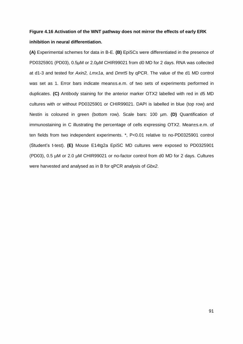
91
Figure 4.16 Activation of the WNT pathway does not mirror the effects of early ERK
inhibition in neural differentiation.
(A) Experimental schemes for data in B-E. (B) EpiSCs were differentiated in the presence of
PD0325901 (PD03), 0.5µM or 2.0µM CHIR99021 from d0 MD for 2 days. RNA was collected
at d1-3 and tested for Axin2, Lmx1a, and Dmrt5 by qPCR. The value of the d1 MD control
was set as 1. Error bars indicate mean±s.e.m. of two sets of experiments performed in
duplicates. (C) Antibody staining for the anterior marker OTX2 labelled with red in d5 MD
cultures with or without PD0325901 or CHIR99021. DAPI is labelled in blue (top row) and
Nestin is coloured in green (bottom row). Scale bars: 100 µm. (D) Quantification of
immunostaining in C illustrating the percentage of cells expressing OTX2. Mean±s.e.m. of
ten fields from two independent experiments. *, P<0.01 relative to no-PD0325901 control
(Student’s t-test). (E) Mouse E14tg2a EpiSC MD cultures were exposed to PD0325901
(PD03), 0.5 µM or 2.0 µM CHIR99021 or no-factor control from d0 MD for 2 days. Cultures
were harvested and analysed as in B for qPCR analysis of Gbx2.

92

93
Figure 4.17 SHH and CHIR99021 treatment partly mimics early ERK inhibition.
(A) Experimental schemes for data in B-E. (B) EpiSCs were differentiated in the presence of
no factors, PD0325901 (PD03), and CHIR99021 together with SHH from d0 until d2 of MD.
RNA was collected at d2 and tested for Axin2, Lmx1a, Foxa2, and Dmrt5 by qPCR. The
value of control was set as 1. Error bars indicate s.e.m. of two sets of experiments
performed in duplicates. (C) Antibody staining for the anterior marker OTX2 labelled with red
in d5 MD cultures without or with factors as indicated. DAPI is labelled in blue (top row) and
Nestin is coloured in green (bottom row). Scale bars: 100 µm. (D) Quantification of
immunostaining in C illustrating the percentage of cells expressing OTX2. Mean±s.e.m. of
ten fields from two independent experiments. *, P<0.01 relative to no-PD0325901 control
(Student’s t-test). (E) EpiSCs were differentiated in the presence of no factors, PD0325901
(PD03), and CHIR99021 together with SHH at d0-2 MD RNA was collected at d1, d2 and d3
MD and analysed for Gbx2 by qPCR. The value of the d1 MD control was set as 1. Error
bars indicate mean±s.e.m. of two sets of experiments performed in duplicates.

94
Figure 4.18 Early stimulation of SHH and WNT pathway cannot substitute ERK
inhibition in the generation of DA neurons.
(A) Experimental scheme of neural differentiation with mouse EpiSCs. MD cultures were
exposed to PD0325901 (PD03), CHIR99021 and SHH or to no-factor control at d0-2 MD.
SHH and FGF8 were applied from d5 until d9 of MD. Medium was then changed to N2B27
plus GDNF, BDNF and ascorbic acid and cultured for another 4 days. (B) MD cultures at d14
were double stained for TH, red, and FOXA2, green. DAPI is labelled in blue. Scale bar:
200 µm.
A
B

95
4.8 Discussion
Midbrain characteristic DA neurons derived from stem cells provides a unique opportunity in
cell therapy and drug discovery for developing novel strategies for brain repair in PD.
However, the generation of ‘authentic’ mDA neurons in a robust and reproducible manner
requires an in-depth understanding of the mechanisms that control the fate choice of stem
cells during differentiation. The data of the present chapter revealed that a stepwise control
of FGF/ERK activity during specific stages of neural differentiation can effectively promote
the generation of authentic mDA neurons from mouse PSCs.
Regulation of mDA differentiation by controlling FGF signalling
Compared to ESCs, EpiSCs, the in vitro counterpart of postimplantation epiblast stem cells,
have already completed the transition from a naive pluripotent stage toward a primed stage
of lineage commitment which involves the activation of FGF signalling (Kunath et al., 2007;
Ying et al., 2008). As described in chapter 3, EpiSCs are more synchronised in the process
of neural induction than ESCs. I found that inactivation of FGF/ERK pathway by either FGFR
or ERK inhibitor upon the exit of the epiblast pluripotent stage specifies nascent neural
progenitor cells towards mDA progenitor fate. Notable, our strategy appears to induce
region-specific gene expression almost at the same time when neural induction occurs,
implying that coupling neural induction and regionalisation may be crucial for generating
specific neuronal cell types in vitro.
To maintain the mDA neural progenitor characteristics, these cells require endogeneous
FGF signalling (Figure 4.3 A). Addition of FGF8 or continued PD0325901-treatment after the
onset of a ventral midbrain fate abolished the early increased expression levels of mDA key
regulator genes (Foxa2, Lmx1a, Dmrt5) caused by early FGF/ERK blockade (Figure 4.3 B
and 4.11), suggesting that the level of FGF signalling is important. Previous in vivo studies
revealed that high levels of FGF8 specify cells of the presumptive midbrain to a hindbrain
fate (Liu and Joyner, 2001; Liu et al., 2003; Sato et al., 2001) whereas reduction of FGF

96
signalling in the midbrain instructs cells toward anterior cell fates (Basson et al., 2008; Lahti
et al., 2012). In keeping with these findings, an increase or decrease of FGF8 in the
prospective diencephalon or hindbrain respectively induce a midbrain identity (Crossley et
al., 1996; Lee et al., 1997; Liu et al., 1999; Martinez et al., 1999; Sato et al., 2001; Sato and
Nakamura, 2004; Shamim et al., 1999). I also found that increase of FGF signalling resulted
in an upregulation of the isthmus organizer related genes, Wnt1, En1, and En2 (Figure
4.11), supporting previously reported cross-regulatory network between these molecules
(Wurst and Bally-Cuif, 2001; Ye et al., 2001). Furthermore, these genes have been shown to
be essential for the differentiation and survival of DA neurons (Prakash et al., 2006; Simon
et al., 2001; Tang et al., 2009). However, it has been reported that SHH and canonical WNT
signalling have opposing roles in mDA neurogenesis (Joksimovic et al., 2009; Tang et al.,
2010). Thus, for an efficient generation of mDA neurons, it might be crucial to allow SHH-
dependent progenitor specification and expansion before activating FGF signalling by FGF8
which indirectly stimulates WNT signalling (Andersson et al., 2006b; Hynes et al., 1997;
Sasaki et al., 1997). These findings point out the importance of precise temporal control of
FGF signalling to orchestrate mDA midbrain induction and neurogenesis.
Early inhibition of FGF/ERK signalling promotes acquisition of a midbrain
neural progenitor identity
I found that blocking specifically FGF/ERK signalling at the onset of EpiSC MD directs a
midbrain fate in the anterior-posterior axis by suppressing both hindbrain (Gbx2 expression)
and the forebrain fates (Foxg1 and Six3 expression) (Figure 4.5) whereas blockade of
FGF/PI3K did not modulate anterior-posterior characteristics (Figure 4.6). However, it is
unclear whether blockade of the FGF/PLC-γ pathway instruct cells toward a midbrain fate.
Recent reports demonstrated that forced expression of any of the mDA regulator genes
(Lmx1a, Otx2, Dmrt5, Foxa2) lead to an increased production of DA neurons (Chung et al.,
2009; Friling et al., 2009; Gennet et al., 2011; Kittappa et al., 2007) and combined
overexpression of Oxt2, Lmx1a, and Foxa2 significantly improved the generation of mDA

97
neurons (Chung et al., 2009). My results showed that a complete set of the key mDA
progenitor transcription factors (FOXA2, LMX1A, DMRT5 and OTX2) was already induced at
d5 MD in PD0325901-treated neural progenitor cells (Figure 4.3 and 4.5). These cells
exposed to SHH and FGF8 differentiated efficiently into mDA neurons. Although, untreated
neural progenitors exposed to SHH and FGF8 for 4 days (d5-9) showed an increase in the
number of FOXA2 and LMX1A positive cells at d9 MD (Figure 4.14 B), their efficiency to
convert into mDA neurons was low compared to PD0325901-treated MD cultures (Figure 4.7
and 4.9). These data suggest that an early induction of Lmx1a and Foxa2 is required for
highly efficient production of mDA neurons.
Implications of SHH and WNT1
SHH and WNT1 are required for the expression of Otx2 (Chung et al., 2009; Prakash et al.,
2006; Puelles et al., 2004), Lmx1a (Andersson et al., 2006b; Chung et al., 2009) and Foxa2
(Hynes et al., 1997; Sasaki et al., 1997). These factors form a network that coordinates mDA
differentiation (Chung et al., 2009; Lin et al., 2009; Nakatani et al., 2010; Prakash et al.,
2006; Tang et al., 2010). The results of this chapter revealed a robust induction of Shh and
Wnt when exposed to FGF/ERK inhibitor (Figure 4.3). One might ask whether the effect of
early FGF/ERK inhibition is based on the initiation of these signalling molecules. However,
MD of EpiSCs in the presence of either SHH or canonical Wnt pathway or both only partially
mimicked PD0325901 effect on nascent neural progenitors (↑Foxa2, ↑Lmx1, ↓Dmrt5, ↓Otx2)
and failed to produce a high number of DA neurons (Figures 4.12 and 4.16-17), suggesting
that a fully correct region-specific identity is essential for efficient generation of mDA
neurons.
It was demonstrated that SHH establishes the dorso-ventral axis and subsequently
controlling the positioning and generation of DA neurons in the ventral midbrain (Andersson
et al., 2006b; Ye et al., 1998). The same studies showed that SHH has no effect on the
formation of the anteror-posterior axis. Our results also revealed that SHH applied at d0-2

98
had no effect on anterior-posterior identity (Gbx2, Otx2) (Figure 4.14). Thus, other factors
are needed to synchronise and modulate the cell fate of distinct anteror-posterior identities
during differentiation of EpiSC. Chung et al. (2009) demonstrated that WNT1 positively
regulates expression of Otx2 via the canonical pathway, making it a good candidate to shift
the cell fate toward an anterior character during neural in vitro differentiation. However,
mimicking the canonical WNT signalling via the GSK3ß inhibitor at d0-2 did neither increase
the percentage of OTX2 positive cells. In addition, expression of Dmrt5 was neither induced
by activation of canonical WNT or SHH signalling indicating the involvement of other
unknown pathways/factors.
Although activation of canonical WNT pathway resulted in an upregulation of Lmx1a
expression (Chung et al., 2009), in PD0325901-treated neural differentiation the expression
of Lmx1a steadily increased even after Wnt1 expression dropped below the levels observed
in control EpiSC MD cultures (Figure 4.3 B, C). A previous study indicated that Lmx1a is
induced by SHH (Andersson et al., 2006b) as midbrain explants from chick embryos
displayed LMX1A positive cells only after SHH treatment. In PD0325901 treated MD
cultures, Shh expression increased continuously until d5 of differentiation as seen for Lmx1a
expression (Figure 4.3 B, C), which was enhanced by the addition of SHH to PD0325901 at
d0-2 and when SHH signalling was blocked by cyclopamine Lmx1a expression dropped at
d3 after an initial burst of upregulation, implying a role of SHH in the regulation of Lmx1a
expression. These findings lead to an interesting question whether Lmx1a is initially induced
by canonical WNT pathway and later on maintained and stabilised by SHH signalling.
Efficient generation of mDA neurons from PD0325901-treated EpiSCs
Differentiation protocols using growth factors to control the process have so far been highly
variable and limited in the generation of DA neurons (Kawasaki et al., 2000; Lee et al.,
2000b; Ying and Smith, 2003). Cells derived by these strategies represented a mixed
population containing neural cells at different developmental stages, or other embryonic
germ layer lineages and undifferentiated ESCs which used in cell replacement therapy can

99
cause the formation of tumour or transplant-induced diseases such as dyskinesia. Although
overexpression of mDA transcription factors increases markedly the efficacy of ESC-derived
DA neurons (Chung et al., 2009; Chung et al., 2002; Friling et al., 2009; Kittappa et al.,
2007; Maxwell et al., 2005), the genetic manipulations are a burden for the production of DA
neurons for clinical trials. Therefore, there is a pressing need to develop a robust chemically
defined condition that can rapidly convert stem cells into DA neurons of midbrain identity in a
synchronised manner. In this chapter, I have presented a novel strategy of highly reliable
nature that allows producing mDA neurons in high yield and fully chemical defined condition
free from genetic manipulation. The presence of the FGF/ERK inhibitor at the onset of EpiSC
MD resulted in a remarkably increase of TH positive cells. A common problem of DA
differentiation protocols is the sub-specification into distinct DA cell phenotype. The majority
of DA neurons generated by my new method are mDA specific as confirmed by co-
expression of the marker genes LMX1A, FOXA2, NURR1, TH, PITX3, and VMAT.
It is worth to note that, even without FGF/ERK inhibitor treatment, nearly 40% of TH positive
neurons were immunopositive for PITX3 in MD of EpiSCs which is much higher than that
from ESCs (Andersson et al., 2006b; Friling et al., 2009; Parmar and Li, 2007). This
suggests that using the more synchronised nature of EpiSC differentiation is important for
the effective enrichment of mDA-competent progenitors.

100
5 Functional analysis of in vitro derived midbrain
dopaminergic neurons
A major goal of in vitro derived mDA neurons will be their use for disease modeling and cell
therapy. Thus, it is essential to test the functional properties of these cells and their
functional integration to the surrounding host tissue. I therefore set up collaborations to
investigate whether the cells I generated and described in Chapter 4 exhibit
electrophysiological properties similar to those of mDA neurons in vivo as well as their ability
to integrate into the striatum of a 6-hydroxy-dopamine-lesioned mouse model PD.
5.1 In vitro generated mDA neurons exhibit functional neuronal
characteristics
Electrophysiological characterisation of mDA neurons was carried out in collaboration with
Dr. Jessica Risner-Janiczek in our laboratory, using facilities of Dr. Mark Ungless at the
MRC Clinical Sciences Centre in London. For this study, I employed a reporter cell line in
which GFP is inserted into the Pitx3 locus allowing specific labelling of mDA neurons (Zhao
et al., 2004). Pitx3-GFP EpiSCs were first cultured for two days with or without PD0325901.
From d5 until d9 of MD the cultures were treated with SHH and FGF8 and were then further
differentiated in the presence of BDNF, GDNF, and ascorbic acid. GFP positive neurons
were analysed by whole-cell recordings at d14-16 MD. Neurons derived from EpiSC-MD
cultures with or without PD0325901 exhibited a negative resting membrane potential which
is a characteristic feature of neurons (control, –67±1.3 mV, n=6; PD0325901, –45±4.7, n=13;
P<0.05). Neurons generated with PD0325901 treatment showed a slightly more depolarized
resting membrane potential when compared to those in no-PD0325901 control cultures. The
input resistance was similar for neurons generated under both conditions (control, 422±198

101
MΩ, n=8; PD0325901, 558±79 MΩ, n=15; P>0.05). Furthermore, no difference was seen
between the groups in whole-cell capacitance (control, 9.6±1 pF, n=8; PD0325901, 12±5 pF,
n=15; P>0.05). As we did not observe any changes in the input resistance and whole-cell
capacitance in neurons from PD0325901-treated MD cultures, we assume that PD0325901
treatment had no affect on the cell size. 14% of Pitx3-GFP positive neurons showed multiple
spontaneous potentials (n=14) (Figure 5.1 B). Stimulation with a depolarizing current pulse
resulted in an overshooting action potential in all cells (n=14) of which 19% (3/14) responded
by firing multiple action potentials with a low frequency (0.95±0.14 Hz; n=3) and in an
irregular pattern (CV-ISI, 1.19±0.24; n=3). When cells were cultured in medium containing
BDNF, GDNF, and ascorbic acid until d40, we observed 67% (PD0325901, 6/9) of cells firing
spontaneous action potentials (3.54±0.8 Hz; CV-ISI, 0.49±0.08; n=6). These cells displayed
a pacemaker-like activity typical of mDA neurons (Figure 5.1 E). Moreover, all Pitx3-GFP
positive neurons demonstrated a large outward current in voltage–clamp mode, typical of a
delayed-rectifier K+ current (2.19±0.28 nA, n=15; P>0.05) (Figure 5.1 D, left panel). The
cells exhibited fast activating, fast-inactivating inward currents that is a typical feature of Na+
currents (1.69±0.25 nA, n=15; P>0.05) (Figure 5.1 D, right panel).
In summary, these results provide evidence that the mouse EpiSC-derived Pitx3-GFP
positive neurons acquired functional, neuron-like properties that were similar to those of
mDA neurons in vivo.

102

103
Figure 5.1 Pitx3-GFP positive cells exhibit functional neuron-like electrophysiological
properties. (kindly provided by Dr. Jessica Risner-Janiczek).
(A) DIC and GFP image showing a recording pipette patched onto a mouse Pitx3-GFP
positive cell. (B) Spontaneous action potentials fired by a d14 neuron. Vertical scale bar, 20
mV; horizontal scale bar, 5 ms. (C) A single spontaneous action potential (left). A single
action potential fired in response to a 50 pA depolarizing current pulse (middle). Multiple
action potentials fired in response to a 50 pA depolarizing current pulse (right). Vertical bars,
20 mV; horizontal bars, 50, 100, and 100 ms, respectively. (D) Outward current evoked by a
serious of voltage steps (left). Fast inward current evoked by a series of voltage steps (right).
Vertical bars, 1 nA; horizontal bars, 50 ms. (E) Pacemaker-like spontaneous firing a d41
neuron. Vertical bar, 20 mV; horizontal bar, 1 second.

104
5.2 Transplantation of in vitro generated mDA progenitors in a
mouse model of Parkinson’s disease
I then investigated whether DA neurons generated by the present protocol can functionally
integrate in the brain of a mouse PD model. This part of the work was carried out in
collaboration with Prof. Steve Dunnett’s laboratory at Cardiff University.
5.2.1 Modification of differentiation protocol
Many lines of previous studies demonstrated that the developmental stage of the donor cells
is pivotal for survival and efficient functional integration in animal models of PD0325901. The
most suitable donor cells appear to be those that are mDA specified and yet to leave the cell
cycle (Brundin et al., 1988; Das et al., 1980; Finger and Dunnett, 1989; Torres et al., 2007).
From a practical standpoint, a shorter differentiation time prior to transplantation may benefit
cell survival during transportation of the cells. Therefore, I sought to modify the protocol to
accelerate and possibly enhance mDA lineage commitment. SHH is a well known
ventralising factor that induces the generation of floorplate cells including cells from which
mDA neurons derive (Bayly et al., 2007; Blaess et al., 2006; Fedtsova and Turner, 2001;
Hynes et al., 1995; Ye et al., 1998). I therefore proposed that SHH added at d2-5 straight
after PD0325901 treatment would enhance the induction of ventral midbrain progenitor cells
and subsequently the conversion into mDA neurons. MD cultures were stained at d5 MD for
OTX2, LMX1A, FOXA2, and DMRT5, and the percentage of cells expressing one of these
markers were quantified by pixel analysis (Figure 5.2 B, C). I found that early SHH
treatment, either alone or following PD0325901 treatment, significantly increased the
number of FOXA2 positive cells (control: 24±9%; SHH: 77±6%; PD0325901-SHH: 40±18%)
by d5 MD (Figure 5.2 C). Furthermore, early SHH following PD0325901 also resulted in an
increase of LMX1A positive cells compared to with PD0325901 alone cultures (PD0325901:
12±4%; PD0325901-SHH: 17±4%) (Figure 5.2 C). However, SHH had no effect on the
generation of DMRT5 and OTX2 positive cells (Figure 5.2 C).

105
To see how above changes might impact on the generation of DA cells, progenitors in all
four MD cultures were allowed to differentiate till d14 of MD (Figure 5.3 A). Immunostaining
for TH revealed that cultures with the earlier SHH treatment following PD0325901 generated
the most numbers of TH positive cells (Figure 5.3 B). Interestingly, no-PD0325901 MD
cultures treated with SHH at d3-5 also generated more TH positive cells compared to the
basal control condition. This number is still lower than those original plus PD0325901
condition. This data indicates that enhanced induction of FOXA2 alone by SHH at d5 is not
sufficient for DA commitment.

106

107
Figure 5.2 Early blockade of FGF signalling and postliminary enhancement of SHH
signalling support generation of ventral midbrain progenitor cells.
(A) Experimental schemes for data in B. (B) Mouse E14tg2a EpiSC MD cultures were
treated with PD0325901 (PD03) at d0-2 or SHH at d2-5 or a combination of the two as
indicated in A. At d5, cultures were analysed by immunocytochemistry for the anterior
marker OTX2, red, and the midbrain progenitor markers LMX1A, FOXA2, and DMRT5, all
shown in green. DAPI is labelled in blue. Scale bars: 200 µm. (C) Quantification of
immunostaining in B illustrating the percentage of cells expressing OTX2, FOXA2, LMX1A,
or DMRT5. Mean±s.e.m. of ten fields from two independent experiments. *, P<0.05 relative
to no-PD0325901 control (Student’s t-test).

108
Figure 5.3 ERK inhibition followed by stimulation of the SHH signalling promotes the
generation of DA neurons.
(A) Experimental schemes for data in B. (B) Mouse E14tg2a EpiSC MD cultures were
treated with PD0325901 (PD03) at d0-2 or SHH at d2-5 or a combination of the two as
indicated in A. From d5 until d9 of MD, all cultures were exposed to SHH and FGF8.
Cultures were stained for the DA marker TH labelled with at d14 MD. DAPI is labelled in
blue. Scale bars: 100 µm.

109
5.2.2 Transplantation of EpiSC-derived mDA progenitors; pilot study
The pathophysiology of PD is the preferential loss of a specific DA neuronal subpopulation,
those in the Substantia Nigra (SN). These cells project to the striatum and form the
dopaminergic nigrostriatal pathway which controls motor functions. Embryonic midbrain cells
transplanted into the striatum has been shown to differentiate into DA neurons, survive
longterm, re-innervate the striatum and improve motor control (Bjorklund et al., 1980; Bolam
et al., 1987; Doucet et al., 1989; Dunnett et al., 1983; Dunnett et al., 1987; Lindvall and
Bjorklund, 1989; Madrazo et al., 1988; Mendez et al., 2005; Schmidt et al., 1983). These
findings demonstrate the functionality of cell derived from embryonic midbrain tissue. To test
whether the cells generated by the present protocol have the potential to replace the loss of
mDA neurons, we have used 6-hydroxydopamine (6-OHDA) treated mouse models of PD.
Injection of the catecholaminergic neurotoxin 6-OHDA into the area where the ascending DA
fibres is at its most compact, the medial forebrain bundle, results in a robust degeneration of
the dopaminergic nigrostriatal pathway. In order to reduce health problems of the animals,
unilateral depletion was carried out which in addition allows a variety of behavioural tests
(Heuer et al., 2012). Cells transplanted into the SN do not reconnect with the striatum
(Collier et al., 2002; Mukhida et al., 2001; Nikkhah et al., 1994; Starr et al., 1999). Therefore,
cells are generally placed within the striatal target region.
Previous transplantation studies demonstrate that maximum functional integration is
achieved with ventral midbrain progenitors prior to neurogenesis (Brundin et al., 1988; Das
et al., 1980; Finger and Dunnett, 1989; Torres et al., 2007). In my culture system, active
neuronal differentiation occurs from d7 MD. We therefore transplanted cells of d5 and d7 MD
into striatum of mice that had been lesioned with 6-OHDA. Both d5 and d7 cultures were
exposed to PD0325901 at d0-2 followed by SHH treatment from d2 till transplantation. D7
cultures received two days exposure of FGF8. At the time of transplantation most of the cells
were Nestin positive (Figure 5.4 A). Immunostaining and pixel analysis revealed a similar
110
proportion of cells positive for OTX2, FOXA2, LMX1, and DMRT5 in d5 and d7 MD cultures
(Figure 5.4 A, B).
To investigate the integration, differentiation, and neuronal function, drug-induced behavioral
rotation tests were performed. Here we used the DA agonist amphetamine which induces
typical body rotations in unilateral 6-OHDA lesioned mice to the side of the DA-depletion.
Consequently, any recovery by the transplanted cells will be observed by a significant
reduction in rotational behaviour. In vivo DA neurons of the SN are part of the Basal Ganglia
which forms a circuit that controls their activity in the striatum. Grafts that are transplanted
within the target side, the striatum, are not under the control of this regulatory network.
Therefore, grafts that are enriched with DA neurons display an overcompensatory response
which correlates with the induced Dopamine level.
Eight weeks after transplantation, animals grafted with d7 MD cultures showed a significant
reduction in amphetamine-induced rotation scores and over-compensatory response in
6-OHDA lesioned mice (Figure 5.5). However, mice that received d5 MD cultures showed no
recovery compared to lesion control group. (Figure 5.5).
To verify whether the observed difference in recovery correlates with DA neuronal
differentiation and survival after transplantation, immunostaing for TH was carried out on
brain sections with grafted cells of d5 and d7 MD cultures after 8 weeks of transplantation.
The loss of DA fibres in the striatum was clearly visible in these sections, demonstrating the
unilateral depletion of the DA nigrostial pathway by 6-OHDA (Figure 5.6 A). TH positive
neurons were observed in both d5 and d7 MD grafts, demonstrating their potential for DA
lineage commitment in vivo. However, d7 MD grafts had significantly more TH positive
neurons than d5 MD grafts (Figure 5.6 B-E), which could explain the difference of the
amphetamine-induced rotation behavioural test (Figure 5.5).
One of the concerns for cell replacement therapy in PD is the formation of tumours and
neural overgrowth. Strikingly, none of the animals exhibited tumour formation or extensive
overgrowths in the surviving grafts.

111
Figure 5.4 EpiSC-derived cells display robust induction of ventral midbrain progenitor
fate at the day of transplantation.
(A) Mouse Pitx3-GFP EpiSC MD cultures were treated with PD0325901 at d0-2 and SHH at
d2-5. D7 cultures were exposed to FGF8 and SHH from d5 for 2 days of differentiation. At
the day of transplantation, cells were fixed and analysed by immunocytochemistry for
anterior marker OTX2 and the midbrain progenitor markers FOXA2, LMX1A, and DMRT5.
DAPI is labelled in blue and Nestin is shown in green. (B) Quantification of immunostaining
in B illustrating the percentage of cells expressing OTX2, FOXA2, LMX1A, or DMRT5.
Mean±s.e.m. of ten fields from two independent experiments.

112
Figure 5.5 Bar graph of amphetamine-induced rotational behaviour 8 weeks after
transplantation of lesioned, d5 and d7 MD cultures mice (kindly provided by Dr.
Sophie Precious and Andreas Heuer).
Eight weeks after transplantation, amphetamine-induced rotations in unilateral 6-OHDA
lesioned mice with or without grafts were determined by the number of rotations per minute.
The black line displays the rotational behaviour of control mice without any lesion. Recovery
by d7 MD grafts was observed by a significant reduction in the number of rotations to the the
side of DA-depletion, an overcompensatory response which correlates with the induced
Dopamine level. The bar graphs represent 11 mice for lesion control group, 16 mice for d5
MD cell group and 15 mice that received cells of d7 MD cultures. Data represent mean ±
SEM.

113
Figure 5.6 Substantial graft-derived reinnervation of the lesioned striatum 8 weeks
after transplantation of d5 and d7 MD cultures (kindly provided by Dr. Sophie
Precious and Andreas Heuer).
(A) Immunohistochemistry for TH illustrates the typical dense staining of DA fibers in the
intact striatum (left) and almost complete loss of TH fibers is seen in 6-OHDA lesioned
striatum (right). (B+C) Graft-derived re-innervation of the lesioned striatum 8 weeks after
transplantation (boxed area, graft core). The graft cores in B+C contain stained TH positive
neurons. (D+E) The boxed area in B+C is shown at larger magnification.

114
5.2.3 Head to head comparison of EpiSC-derived mDA grafts with
ventral midbrain tissue in 6-OHDA-lesioned mouse models of PD
Transplantation of embryonic ventral midbrain (VM) tissue into dopamine-depleted animals
has been studied in detail. In summary, embryonic VM cells have been demonstrated to
survive, reinnervate into the host striatum, make synaptic connections onto the appropriate
neuronal populations, and reduce many of the motor deficits caused by dopamine depleting
lesions (Bjorklund et al., 1980; Bjorklund and Stenevi, 1979; Bjorklund et al., 1983; Dunnett
et al., 1981; Dunnett et al., 1988; Freed et al., 1980; Perlow et al., 1979; Schmidt et al.,
1983; Schmidt et al., 1982; Schultz, 1982; Schultzberg et al., 1984; Stromberg et al., 1985;
Wuerthele et al., 1981). These very promising results led to clinical trials started in 1987
where cells of the human fetal mesencephalic tissue were transplanted to the striatum of PD
patients. However, the use of fetal tissue involves ethical concerns. Pluripotent stem cells
(PSC) offer a promising alternative to obtain mDA neurons in vitro but to date it has been a
challenge to generate PSC-derived mDA neurons that functionally engraft in vivo as
successfully as embryonic VM cells.
In the section above (5.2.2), I showed that d7 MD cultures were able to reduce drug-induced
rotational behaviour in 6-OHDA lesioned mice after 8 weeks of tranplantation. To valuate d7
MD cultures for cell replacement in PD, we transplanted the d7 MD cells side-by-side with
ventral E12.5 foetal ventral midbrain cells. Amphetamine-induced rotational test was
performed before transplantation and at 4, 8, and 16 weeks after transplantation. As
expected, VM graft goups displayed a decrease in rotations and an over-compensated
rotational response by 4 weeks post-grafting which was maintained until 16 weeks after
transplantation (the latest time point examined) (Figure 5.7). The group of mice that received
d7 MD cultures also showed a decrease in the rotational behaviour but the score was less
when compared to VM grafts (Figure 5.7). However, the level of over-compensatory
response increased over the time in d7 MD grafted animals, indicating an increased level of
Dopamine.

115
Following the functional assessment, graft survival and neuron cell differentiation was
evaluated by immunostaining. Both groups displayed good TH positive cell survival within
the transplanted striatum after 16 weeks of transplantation (Figure 5.8 A). Counting analysis
of immunohistochemistry data further revealed large numbers of TH positive cells within the
grafts wich received VM or d7 MD cells (Figure 5.8 A, C), indicating therapeutical potential of
d7 MD cultures in the treatment of PD.
A negative side effect of cell replacement in PD is the appearance of graft-induced
dyskineasia, showing abnormal involuntary movements and postures. The presence of
serotonergic neurons within grafts is thought to play an important role in graft-induced
dyskinesia (Braak and Del Tredici, 2008; Carta et al., 2007). Therefore, we performed an
immunostaing for 5HT to detect serotonergic neurons. As shown in Figure 5.8 B, both the
VM and d7 MD grafts contained 5HT positive neurons. It has been suggested that the ratio
of DA neurons to serotonergic neurons may be inversely correlated to the development of
graft-induced dyskineasia. In this study, the TH/5HT ratio of d7 MD grafts was higher than
the one of VM grafts (Figure 5.8 C), suggesting that animals which received d7 MD grafts
might be less likely to develop graft-induced dyskineasia.
In summary, these data demonstrate that the cells produced by the present protocol may
potentially provide a useful therapeutic source for cell replacement therapy in PD.

116
Figure 5.7 Behavioural analysis in E12.5 VM- versus d7 MD-grafted mice (kindly
provided by Dr. Sophie Precious and Andreas Heuer).
Amphetamine-induced rotational test of no-lesion control (12 mice), lesioned (12 mice), VM-
(29 mice) and d7 MD grafted mice (12 mice) in response to amphaetamine. Bars underlain
by a grey background show drug induced rotations after 6-OHDA treatment (post lesion).
Test was performed 4, 8 (control: n=12; lesion: n=12; VM: n=29; d7 MD EpiSC: n=28) and
16 weeks after transplantation (control: n=12; lesion: n=12; VM: n=21; d7 MD EpiSC: n=20).

117

118
Figure 5.8 Characterisation of neurons in 6-OHDA lesioned mouse models of PD
grafted with either d7 MD or VM cells (kindly provided by Dr. Sophie Precious and
Andreas Heuer).
(A) Immunohistochemistry for TH on coronal sections illustrating TH positive cell survival
and striatal reinnervation on the lesioned and grafted striatum with cells of d7 MD cultures or
VM (E12.5) after 8 and 16 weeks of Transplantation. (B) Immunohistochemistry for the
serotonergic marker 5HT in 6-OHDA lesioned mice 8 weeks posttransplantation. (C) The
total number of TH positive and 5HT positive neurons oin the lesioned and grafted striatum
shown in A and B.

119
5.3 Discussion
In this chapter, I show that EpiSC-derived mDA neurons exhibit electrophysiological
properties typical of primary mDA neurons. Moreover, after transplantation into 6-OHDA
lesioned mouse striatum, the mDA progenitor cells survived and differentiated into DA
neurons, and mediated behavioural improvements. Thus, neural progenitors with ventral
midbrain characteristics may represent a valuable source for functional and transplantable
mDA neurons in clinical applications for the treatment of PD.
Although cells of d5 and d7 MD display the same profile for the molecular markers examined
(Figure 5.4), only d7 MD cells markedly restored amphetamine-induced behaviour which
correlates with the observed increase of TH positive cells in the grafts. D7 MD cultures
received 2 days of FGF8 whereas d5 MD cultures did not, suggesting that the activation of
FGF signalling is essential for the acquisition of DA properties. These results point towards
an important point for FGF8 in mDA neural progenitor cells.
During development of the midbrain and hindbrain region, FGF8 has been reported to be
involved in patterning (Basson et al., 2008; Crossley et al., 1996; Sato and Joyner, 2009),
proliferation (Lahti et al., 2011; Xu et al., 2000), and cell survival (Basson et al., 2008; Chi et
al., 2003). I found that when FGF8 was added during differentiation, the expression of the
En1/2 was highly increased (Figure 4.11). EN1/2 are implicated in regionalisation of the
midbrain and hindbrain (Crossley et al., 1996; Liu and Joyner, 2001; Simon et al., 2001) and
in the survival of mDA neurons (Alberi et al., 2004; Alvarez-Fischer et al., 2011; Sgado et al.,
2006; Simon et al., 2001). Thus, the increase of TH positive cells in grafts with d7 MD
cultures may be a result of FGF-induced upregulation of En expression, which in turn result
in decreased cell death and/or proper induction of DA neuronal phenotype.
To reinforce the importance of FGF8 for the specification of mDA neurons (Ye et al., 1998),
only EpiSC MD cultures exposed to SHH and FGF8 efficiently produced DA neurons (Figure
4.7 and 4.9). In addition, I observed that DA neurons exposed to FGF8 displayed a

120
morphologically more mature phenotype compared to cultures with SHH alone (Figure 4.9).
These data suggest an integral role for FGF8 in the maturation of functional mDA neurons.
Torres et al (2007) reported that the developmental age of the donor cells has a significant
impact in cell replacement of DA neurons. Cells derived from ventral midbrain of rat embryos
(E~12) at a stage, when no DA neurons are present, differentiated into DA neurons after
transplantation. These cells survived and integrated into the host striatum of 6-OHDA-
lesioned rat PD models. However, foetal ventral midbrain cells taken from E~11 rat embryos
failed to generate DA neurons post-implantation. Therefore, the possibility cannot be
excluded that the stage of maturation might also be a reason for the difference in TH positive
cells in d5 and d7 MD grafts.
Taken together, temporally controlled modulation of FGF signalling during MD of pluirpotent
stem cells is crucial for obtaining functional authentic mDA neurons. Furthermore, it provides
a fully chemically defined system that resembles an attractive tool for biomedical
applications.

121
6 Concluding remarks and future directions
The characteristics of pluripotent stem cells in broad differentiation potential and self-renewal
represent a remarkable model to study development in vitro and further offer an attractive
tool for biomedical application such as cell therapy, disease modelling and drug screening.
Several studies have demonstrated that ESC-derived mDA neurons transplanted into animal
PD models can survive and alleviate behaviour deficits (Bjorklund et al., 2002; Cho et al.,
2008; Friling et al., 2009; Hargus et al., 2010; Kim et al., 2002; Kriks et al., 2011; Rodriguez-
Gomez et al., 2007; Roy et al., 2006; Sanchez-Pernaute et al., 2008; Tabar et al., 2008;
Takagi et al., 2005). However, due to pluripotent nature of ESCs, the generation of a desired
cell type is often accompanied by other cells that may negatively interfere with the benefits of
biomedical applications. In order to achieve a pure mDA cell population, it is essential to
understand the molecular mechanisms responsible for the generation of mDA neurons that
in turn enable us to commad stem cell fate specification and differentiation.
The work presented in this thesis is focused on the role of FGF signalling in mDA neuronal
differentiation using mouse pluripotent stem cells. In summary (Figure 6.1), the findings
demonstrate a previously unrecognized inhibitory role of FGF/ERK in the institution of a
midbrain progenitor cell fate and the induction of Wnt and Shh. I show that a period of
endogenous FGF activity is essential to confer a ventral midbrain progenitor fate and that
later in differentiation activation of FGF signalling is required to generate mDA neurons.
EpiSC-derived mDA neurons exhibit functional, neuron-like properties and when implanted
into the striatum of mouse PD model strongly restored amphetamine-induced rotation
behaviour without any signs of overgrowth. Thus, a stepwise control of FGF/ERK activation
during specific stages of neural differentiation from mouse stem cells is crucial for generation
of authentic, functional mDA neurons. Importantly, this small-molecular-based protocol is
fully chemically defined, free from genetic manipulation and represents a method for

122
significantly improved efficiency and high reproducibility compared to previously reported
transgene-free protocols.
In collaboration with the group of Steve Dunnett we have begun detailed analysis of the
potential of the mDA precursor cells generated by the current protocol. These studies will
involve the validation of a midbrain specific DA phenotype in grafts and the precise
characterisation of grafted non-DA neuronal cells. It cannot be excluded that the observed
reduced rotation behaviour was caused by cells releasing neurotrophic factors which have
the ability to regulate the survival of specific neuronal populations in the central nervous
system. One potential candidate is the glial cell line-derived neurotrophic factor (GDNF)
which has been demonstrated to protect and restore mDA neurons (Behrstock et al., 2006;
Dezawa et al., 2004; Redmond et al., 2007). Moreover, by using behaviour tests, we will
further investigate the consequences of the presence of serotonergic neurons in grafts which
are assumed to be in causative relationship with graft induced dyskinesia (Carlsson et al.,
2007; Politis et al., 2011; Politis et al., 2010). However, for future therapies of PD, the results
obtained on mouse cells have to be confirmed on human ESCs.
Charlie Arber in our group has addressed this point and has obtained encouraging results.
Briefly, human ESCs were exposed to PD0325901 at d3 MD, a time point when the primitive
ectoderm/epiblast marker FGF5 was peaked. Indeed, blockade of FGF/ERK signalling after
initial neurectodermal induction resulted in a direct and early induction of the midbrain
characteristics at the expense of forebrain and caudal neural fates. Exposure of these cells
to SHH and FGF8 led to the robust production of DA neurons expressing marker genes for
correct, ventral midbrain-specific identity. We are currently in the process of validating the
potential of these hESC-derived ventral midbrain neuronal cells to restore parkinsonian
symptoms in a rat model of PD.
Given the similarities in the response to early Erk inhibition during differentiation, cell fate
decisions of human ESC and mouse EpiSC may be instructed by the same signalling
pathways. Yet, Kriks et al. (2011) recently reported that early stimulation of SHH and FGF

123
signalling followed by activation of WNT signalling, using the GSK3ß inhibitor CHIR99021,
can direct human ESC toward an mDA specific fate (↑FOXA2, ↑LMX1A, ↑OTX2). It is worth
noting that FGF signalling activates the expression of the FGF signalling inhibitors MPK3,
SPRY and SEF (Eblaghie et al., 2003; Kawakami et al., 2003; Kovalenko et al., 2006;
Kovalenko et al., 2003; Urness et al., 2008; Vieira and Martinez, 2005). It might be therefore
possible that such a negative feedback regulation of FGF signalling results in a reduction of
FGF activity at a time when regional specification occurs. However, this controversy raises
the need for more direct in vitro analysis of FGF regulation during differentiation toward mDA
neurons.
The results of this thesis also revealed that neither stimulation of SHH pathway nor
activation of canonical WNT signalling or their combination could replace early inhibition of
FGF/ERK activity in the specification of mouse EpiSCs toward a ventral midbrain progenitor
cell fate. This observation opens up the possibility that early FGF/ERK inhibition allows the
activation of other pathways that promote midbrain induction. For example, although
expression of Lmx1a and Foxa2 were increased by SHH treatment and/or activation of
canonical WNT signalling, the number of OTX2 positive cells and the expression of Dmrt5
were not altered. In order to identify pathways that are activated or blocked in a FGF/ERK
deficient environment, I have recently performed a microarray gene expression assay of d3
MD EpiSC cultures with and without PD0325901. The outcome of this study would widen our
knowledge in the molecular interactions between signalling pathways during the process of
regional specification. Furthermore, the microarray might identify novel surface markers
specific for ventral midbrain progenitor fate which in turn allows the purification of mDA
progenitors by fluorescence-activated cell sorting.
Previous studies indicated that Msx1 is directly induced by LMX1A (Andersson et al., 2006a;
Chung et al., 2009). In the developing midbrain Msx1 is expressed in the ventral and dorsal
midline as Lmx1a (Andersson et al., 2006b; Bach et al., 2003; Mishima et al., 2009; Yan et
al., 2011) strentghten that Msx1 is a downstream target of LMX1A. Andersson et al. (2006)

124
recently reported that SHH can induce the expression of Lmx1a and Msx1 in neural tube
explants of chick embryos. However, my findings presented in chapter 4.7 show that
expression of Msx1 was not increased by SHH (Figure 4.12) and treatment with the SHH
antagonist cyclopamine did not reduce PD0325901-induced Msx1 mRNA level (Figure 4.13),
implying a SHH-independent activation of Msx1 expression. Interstingly, induction of Lmx1a
by PD0325901 was abolished at d3 of MD in cultures with cyclopamine. Another study
demonstrated that WNT1 and LMX1A form a regulatory loop in the gerenation of mDA
neurons (Chung et al., 2009). However the same study showed that overexpression of Wnt1
during neural differentiation did not significantly alter the expression of Msx1 while the level
of Lmx1 expression was highly induced. These observations raise the question of which
other extrinsic or intrinsic factors regulate Msx1. In other tissues, Msx1 was demonstrated to
be a downstream target of BMP (Furuta et al., 1997; Liu et al., 2004; Suzuki et al., 1997;
Timmer et al., 2002) which, in the developing midbrain, is expressed in the roofplate (Bach
et al., 2003). Therefore, for detailed analysis of the molecular mechanisms during mDA
neural differentiation, further anaylsis should be carried out to elucidate whether EpiSC-
derived midbrain cell population by early ERK inhibition contain cells of a dorsal midbrain
progenitor identity.
The results of chapter 4 pointed out further an important role of DMRT5 in the generation of
mDA neurons. Dmrt5 is one of the genes upregulated by Erk inhibition, implying a crucial
role of DMRT5 in the differentiation of mDA neurons as previously suggested (Gennet et al.,
2011). Our lab has recently generated a conditional Dmrt5 KO allele in mouse ES cells for
elucidating the role of DMRT5 in this process. This model should facilitate the identification
of factors that are regulated by DMRT5 and to unreveal the precise mechanism in which this
factor is involved.
Here, we have focused on the inhibition of FGF signaling via ERK. It would be of importance
to verify whether FGF/ERK inhibition has an effect on the other two FGF mediated
pathways, FGF/PI3K and FGF/PLC-γ. I would also like to investigate whether modulation of

125
FGF/PLC-γ has any role in the promotion of mDA neurons by using the PLC-γ inhibitor
U73122.
Previous studies demonstrated that dopaminergic neurons of the midbrain targeting the
striatum are implicated in drug addiction (Reading et al., 1991; Robbins et al., 1990; White,
1989). In this context, electophysiological studies on Pitx3-GFP positive cells revealed
features similar to adult dopamine neurons observed in vivo midbrain DA system; even more
than ex vivo brainslices (unpublished observation by Dr. Jessica Risner-Janiczek). Thus,
EpiSC-derived mDA neurons represent an attractive model to study in detail the impacts of
drug exposure: firstly, the desired neurons are easily accessible in vitro for
electrophysiological studies and, secondly, this strategy allows a powerful high-throughput
screening of mDA neurons.
Finally, using the knowledge obtained from in vivo developmental studies, it might be
interesting to test whether the strategy of early specification can be applied for the
generation towards other disease-relevant neurons such as medium spiny GABAergic
neurons lost in Huntington’s disease, basal forebrain cholinergic neurons implicated in
Alzheimer’s disease or motor neurons affected in spinal cord injury, amyotrophic lateral
sclerosis, and spinal muscular atrophy.

126
Figure 6.1 Proposed model of FGF signalling in mDA neural differentiation.

127
7 Bibliography
Aasen, T., Raya, A., Barrero, M.J., Garreta, E., Consiglio, A., Gonzalez, F., Vassena, R., Bilic, J., Pekarik, V., Tiscornia, G., et al. (2008). Efficient and rapid generation of induced pluripotent stem cells from human keratinocytes. Nat Biotechnol 26, 1276-1284. Acampora, D., Avantaggiato, V., Tuorto, F., and Simeone, A. (1997). Genetic control of brain morphogenesis through Otx gene dosage requirement. Development 124, 3639-3650. Acampora, D., Mazan, S., Lallemand, Y., Avantaggiato, V., Maury, M., Simeone, A., and Brulet, P. (1995). Forebrain and midbrain regions are deleted in Otx2-/- mutants due to a defective anterior neuroectoderm specification during gastrulation. Development 121, 3279-3290. Adachi, K., Suemori, H., Yasuda, S.Y., Nakatsuji, N., and Kawase, E. (2010). Role of SOX2 in maintaining pluripotency of human embryonic stem cells. Genes Cells 15, 455-470. Alberi, L., Sgado, P., and Simon, H.H. (2004). Engrailed genes are cell-autonomously required to prevent apoptosis in mesencephalic dopaminergic neurons. Development 131, 3229-3236. Alvarez-Fischer, D., Fuchs, J., Castagner, F., Stettler, O., Massiani-Beaudoin, O., Moya, K.L., Bouillot, C., Oertel, W.H., Lombes, A., Faigle, W., et al. (2011). Engrailed protects mouse midbrain dopaminergic neurons against mitochondrial complex I insults. Nat Neurosci 14, 1260-1266. Alvarez, A.R., Godoy, J.A., Mullendorff, K., Olivares, G.H., Bronfman, M., and Inestrosa, N.C. (2004). Wnt-3a overcomes beta-amyloid toxicity in rat hippocampal neurons. Exp Cell Res 297, 186-196. Alvarez, I.S., Araujo, M., and Nieto, M.A. (1998). Neural induction in whole chick embryo cultures by FGF. Dev Biol 199, 42-54. Andersson, E., Jensen, J.B., Parmar, M., Guillemot, F., and Bjorklund, A. (2006a). Development of the mesencephalic dopaminergic neuron system is compromised in the absence of neurogenin 2. Development 133, 507-516. Andersson, E., Tryggvason, U., Deng, Q., Friling, S., Alekseenko, Z., Robert, B., Perlmann, T., and Ericson, J. (2006b). Identification of intrinsic determinants of midbrain dopamine neurons. Cell 124, 393-405. Andersson, E.R., Prakash, N., Cajanek, L., Minina, E., Bryja, V., Bryjova, L., Yamaguchi, T.P., Hall, A.C., Wurst, W., and Arenas, E. (2008). Wnt5a regulates ventral midbrain morphogenesis and the development of A9-A10 dopaminergic cells in vivo. PLoS One 3, e3517. Ang, S.L., Conlon, R.A., Jin, O., and Rossant, J. (1994). Positive and negative signals from mesoderm regulate the expression of mouse Otx2 in ectoderm explants. Development 120, 2979-2989. Ang, S.L., Jin, O., Rhinn, M., Daigle, N., Stevenson, L., and Rossant, J. (1996). A targeted mouse Otx2 mutation leads to severe defects in gastrulation and formation of axial mesoderm and to deletion of rostral brain. Development 122, 243-252. Ang, S.L., and Rossant, J. (1994). HNF-3 beta is essential for node and notochord formation in mouse development. Cell 78, 561-574.

128
Aoi, T., Yae, K., Nakagawa, M., Ichisaka, T., Okita, K., Takahashi, K., Chiba, T., and Yamanaka, S. (2008). Generation of pluripotent stem cells from adult mouse liver and stomach cells. Science 321, 699-702. Aranda, A., and Pascual, A. (2001). Nuclear hormone receptors and gene expression. Physiol Rev 81, 1269-1304. Arman, E., Haffner-Krausz, R., Chen, Y., Heath, J.K., and Lonai, P. (1998). Targeted disruption of fibroblast growth factor (FGF) receptor 2 suggests a role for FGF signaling in pregastrulation mammalian development. Proc Natl Acad Sci U S A 95, 5082-5087. Aubert, J., Stavridis, M.P., Tweedie, S., O'Reilly, M., Vierlinger, K., Li, M., Ghazal, P., Pratt, T., Mason, J.O., Roy, D., et al. (2003). Screening for mammalian neural genes via fluorescence-activated cell sorter purification of neural precursors from Sox1-gfp knock-in mice. Proc Natl Acad Sci U S A 100 Suppl 1, 11836-11841. Avantaggiato, V., Pandolfi, P.P., Ruthardt, M., Hawe, N., Acampora, D., Pelicci, P.G., and Simeone, A. (1995). Developmental analysis of murine Promyelocyte Leukemia Zinc Finger (PLZF) gene expression: implications for the neuromeric model of the forebrain organization. J Neurosci 15, 4927-4942. Bach, A., Lallemand, Y., Nicola, M.A., Ramos, C., Mathis, L., Maufras, M., and Robert, B. (2003). Msx1 is required for dorsal diencephalon patterning. Development 130, 4025-4036. Bachiller, D., Klingensmith, J., Kemp, C., Belo, J.A., Anderson, R.M., May, S.R., McMahon, J.A., McMahon, A.P., Harland, R.M., Rossant, J., et al. (2000). The organizer factors Chordin and Noggin are required for mouse forebrain development. Nature 403, 658-661. Bain, J., Plater, L., Elliott, M., Shpiro, N., Hastie, C.J., McLauchlan, H., Klevernic, I., Arthur, J.S., Alessi, D.R., and Cohen, P. (2007). The selectivity of protein kinase inhibitors: a further update. Biochem J 408, 297-315. Baker, J.C., Beddington, R.S., and Harland, R.M. (1999). Wnt signaling in Xenopus embryos inhibits bmp4 expression and activates neural development. Genes Dev 13, 3149-3159. Bally-Cuif, L., Cholley, B., and Wassef, M. (1995). Involvement of Wnt-1 in the formation of the mes/metencephalic boundary. Mech Dev 53, 23-34. Balmanno, K., and Cook, S.J. (1999). Sustained MAP kinase activation is required for the expression of cyclin D1, p21Cip1 and a subset of AP-1 proteins in CCL39 cells. Oncogene 18, 3085-3097. Barberi, T., Klivenyi, P., Calingasan, N.Y., Lee, H., Kawamata, H., Loonam, K., Perrier, A.L., Bruses, J., Rubio, M.E., Topf, N., et al. (2003). Neural subtype specification of fertilization and nuclear transfer embryonic stem cells and application in parkinsonian mice. Nat Biotechnol 21, 1200-1207. Barker, N. (2008). The canonical Wnt/beta-catenin signalling pathway. Methods Mol Biol 468, 5-15. Basson, M.A., Echevarria, D., Ahn, C.P., Sudarov, A., Joyner, A.L., Mason, I.J., Martinez, S., and Martin, G.R. (2008). Specific regions within the embryonic midbrain and cerebellum require different levels of FGF signaling during development. Development 135, 889-898. Bayly, R.D., Ngo, M., Aglyamova, G.V., and Agarwala, S. (2007). Regulation of ventral midbrain patterning by Hedgehog signaling. Development 134, 2115-2124. Beddington, R.S. (1994). Induction of a second neural axis by the mouse node. Development 120, 613-620.

129
Beenken, A., and Mohammadi, M. (2009). The FGF family: biology, pathophysiology and therapy. Nat Rev Drug Discov 8, 235-253. Behrstock, S., Ebert, A., McHugh, J., Vosberg, S., Moore, J., Schneider, B., Capowski, E., Hei, D., Kordower, J., Aebischer, P., et al. (2006). Human neural progenitors deliver glial cell line-derived neurotrophic factor to parkinsonian rodents and aged primates. Gene Ther 13, 379-388. Belo, J.A., Bachiller, D., Agius, E., Kemp, C., Borges, A.C., Marques, S., Piccolo, S., and De Robertis, E.M. (2000). Cerberus-like is a secreted BMP and nodal antagonist not essential for mouse development. Genesis 26, 265-270. Bernemann, C., Greber, B., Ko, K., Sterneckert, J., Han, D.W., Arauzo-Bravo, M.J., and Scholer, H.R. (2011). Distinct developmental ground states of epiblast stem cell lines determine different pluripotency features. Stem Cells 29, 1496-1503. Bjorklund, A., Dunnett, S.B., Stenevi, U., Lewis, M.E., and Iversen, S.D. (1980). Reinnervation of the denervated striatum by substantia nigra transplants: functional consequences as revealed by pharmacological and sensorimotor testing. Brain Res 199, 307-333. Bjorklund, A., and Stenevi, U. (1979). Reconstruction of the nigrostriatal dopamine pathway by intracerebral nigral transplants. Brain Res 177, 555-560. Bjorklund, A., Stenevi, U., Schmidt, R.H., Dunnett, S.B., and Gage, F.H. (1983). Intracerebral grafting of neuronal cell suspensions. II. Survival and growth of nigral cell suspensions implanted in different brain sites. Acta Physiol Scand Suppl 522, 9-18. Bjorklund, L.M., Sanchez-Pernaute, R., Chung, S., Andersson, T., Chen, I.Y., McNaught, K.S., Brownell, A.L., Jenkins, B.G., Wahlestedt, C., Kim, K.S., et al. (2002). Embryonic stem cells develop into functional dopaminergic neurons after transplantation in a Parkinson rat model. Proc Natl Acad Sci U S A 99, 2344-2349. Blaess, S., Corrales, J.D., and Joyner, A.L. (2006). Sonic hedgehog regulates Gli activator and repressor functions with spatial and temporal precision in the mid/hindbrain region. Development 133, 1799-1809. Blakely, B.D., Bye, C.R., Fernando, C.V., Horne, M.K., Macheda, M.L., Stacker, S.A., Arenas, E., and Parish, C.L. (2011). Wnt5a regulates midbrain dopaminergic axon growth and guidance. PLoS One 6, e18373. Blum, M., Gaunt, S.J., Cho, K.W., Steinbeisser, H., Blumberg, B., Bittner, D., and De Robertis, E.M. (1992). Gastrulation in the mouse: the role of the homeobox gene goosecoid. Cell 69, 1097-1106. Boettger, T., Knoetgen, H., Wittler, L., and Kessel, M. (2001). The avian organizer. Int J Dev Biol 45, 281-287. Bolam, J.P., Freund, T.F., Bjorklund, A., Dunnett, S.B., and Smith, A.D. (1987). Synaptic input and local output of dopaminergic neurons in grafts that functionally reinnervate the host neostriatum. Exp Brain Res 68, 131-146. Bottcher, R.T., Pollet, N., Delius, H., and Niehrs, C. (2004). The transmembrane protein XFLRT3 forms a complex with FGF receptors and promotes FGF signalling. Nat Cell Biol 6, 38-44. Bouwmeester, T., Kim, S., Sasai, Y., Lu, B., and De Robertis, E.M. (1996). Cerberus is a head-inducing secreted factor expressed in the anterior endoderm of Spemann's organizer. Nature 382, 595-601. Braak, H., and Del Tredici, K. (2008). Assessing fetal nerve cell grafts in Parkinson's disease. Nat Med 14, 483-485.

130
Broccoli, V., Boncinelli, E., and Wurst, W. (1999). The caudal limit of Otx2 expression positions the isthmic organizer. Nature 401, 164-168. Brons, I.G., Smithers, L.E., Trotter, M.W., Rugg-Gunn, P., Sun, B., Chuva de Sousa Lopes, S.M., Howlett, S.K., Clarkson, A., Ahrlund-Richter, L., Pedersen, R.A., et al. (2007). Derivation of pluripotent epiblast stem cells from mammalian embryos. Nature 448, 191-195. Brundin, P., Strecker, R.E., Widner, H., Clarke, D.J., Nilsson, O.G., Astedt, B., Lindvall, O., and Bjorklund, A. (1988). Human fetal dopamine neurons grafted in a rat model of Parkinson's disease: immunological aspects, spontaneous and drug-induced behaviour, and dopamine release. Exp Brain Res 70, 192-208. Bryja, V., Schulte, G., Rawal, N., Grahn, A., and Arenas, E. (2007). Wnt-5a induces Dishevelled phosphorylation and dopaminergic differentiation via a CK1-dependent mechanism. J Cell Sci 120, 586-595. Burbach, J.P., and Smidt, M.P. (2006). Molecular programming of stem cells into mesodiencephalic dopaminergic neurons. Trends Neurosci 29, 601-603. Burgess, W.H., Dionne, C.A., Kaplow, J., Mudd, R., Friesel, R., Zilberstein, A., Schlessinger, J., and Jaye, M. (1990). Characterization and cDNA cloning of phospholipase C-gamma, a major substrate for heparin-binding growth factor 1 (acidic fibroblast growth factor)-activated tyrosine kinase. Mol Cell Biol 10, 4770-4777. Camus, A., Perea-Gomez, A., Moreau, A., and Collignon, J. (2006). Absence of Nodal signaling promotes precocious neural differentiation in the mouse embryo. Dev Biol 295, 743-755. Canning, C.A., Lee, L., Irving, C., Mason, I., and Jones, C.M. (2007). Sustained interactive Wnt and FGF signaling is required to maintain isthmic identity. Dev Biol 305, 276-286. Carlsson, T., Carta, M., Winkler, C., Bjorklund, A., and Kirik, D. (2007). Serotonin neuron transplants exacerbate L-DOPA-induced dyskinesias in a rat model of Parkinson's disease. J Neurosci 27, 8011-8022. Carta, M., Carlsson, T., Kirik, D., and Bjorklund, A. (2007). Dopamine released from 5-HT terminals is the cause of L-DOPA-induced dyskinesia in parkinsonian rats. Brain 130, 1819-1833. Carvajal-Vergara, X., Sevilla, A., D'Souza, S.L., Ang, Y.S., Schaniel, C., Lee, D.F., Yang, L., Kaplan, A.D., Adler, E.D., Rozov, R., et al. (2010). Patient-specific induced pluripotent stem-cell-derived models of LEOPARD syndrome. Nature 465, 808-812. Castelo-Branco, G., Rawal, N., and Arenas, E. (2004). GSK-3beta inhibition/beta-catenin stabilization in ventral midbrain precursors increases differentiation into dopamine neurons. J Cell Sci 117, 5731-5737. Castelo-Branco, G., Sousa, K.M., Bryja, V., Pinto, L., Wagner, J., and Arenas, E. (2006). Ventral midbrain glia express region-specific transcription factors and regulate dopaminergic neurogenesis through Wnt-5a secretion. Mol Cell Neurosci 31, 251-262. Castelo-Branco, G., Wagner, J., Rodriguez, F.J., Kele, J., Sousa, K., Rawal, N., Pasolli, H.A., Fuchs, E., Kitajewski, J., and Arenas, E. (2003). Differential regulation of midbrain dopaminergic neuron development by Wnt-1, Wnt-3a, and Wnt-5a. Proc Natl Acad Sci U S A 100, 12747-12752. Cattaneo, E., and McKay, R. (1990). Proliferation and differentiation of neuronal stem cells regulated by nerve growth factor. Nature 347, 762-765.

131
Chambers, I., Silva, J., Colby, D., Nichols, J., Nijmeijer, B., Robertson, M., Vrana, J., Jones, K., Grotewold, L., and Smith, A. (2007). Nanog safeguards pluripotency and mediates germline development. Nature 450, 1230-1234. Chenn, A., and Walsh, C.A. (2002). Regulation of cerebral cortical size by control of cell cycle exit in neural precursors. Science 297, 365-369. Chenoweth, J.G., and Tesar, P.J. (2010). Isolation and maintenance of mouse epiblast stem cells. Methods Mol Biol 636, 25-44. Chi, C.L., Martinez, S., Wurst, W., and Martin, G.R. (2003). The isthmic organizer signal FGF8 is required for cell survival in the prospective midbrain and cerebellum. Development 130, 2633-2644. Chiang, C., Litingtung, Y., Lee, E., Young, K.E., Corden, J.L., Westphal, H., and Beachy, P.A. (1996). Cyclopia and defective axial patterning in mice lacking Sonic hedgehog gene function. Nature 383, 407-413. Chizhikov, V.V., and Millen, K.J. (2005). Roof plate-dependent patterning of the vertebrate dorsal central nervous system. Dev Biol 277, 287-295. Cho, M.S., Lee, Y.E., Kim, J.Y., Chung, S., Cho, Y.H., Kim, D.S., Kang, S.M., Lee, H., Kim, M.H., Kim, J.H., et al. (2008). Highly efficient and large-scale generation of functional dopamine neurons from human embryonic stem cells. Proc Natl Acad Sci U S A 105, 3392-3397. Chuang, C.F., and Ng, S.Y. (1994). Functional divergence of the MAP kinase pathway. ERK1 and ERK2 activate specific transcription factors. FEBS Lett 346, 229-234. Chung, S., Leung, A., Han, B.S., Chang, M.Y., Moon, J.I., Kim, C.H., Hong, S., Pruszak, J., Isacson, O., and Kim, K.S. (2009). Wnt1-lmx1a forms a novel autoregulatory loop and controls midbrain dopaminergic differentiation synergistically with the SHH-FoxA2 pathway. Cell Stem Cell 5, 646-658. Chung, S., Moon, J.I., Leung, A., Aldrich, D., Lukianov, S., Kitayama, Y., Park, S., Li, Y., Bolshakov, V.Y., Lamonerie, T., et al. (2011). ES cell-derived renewable and functional midbrain dopaminergic progenitors. Proc Natl Acad Sci U S A 108, 9703-9708. Chung, S., Sonntag, K.C., Andersson, T., Bjorklund, L.M., Park, J.J., Kim, D.W., Kang, U.J., Isacson, O., and Kim, K.S. (2002). Genetic engineering of mouse embryonic stem cells by Nurr1 enhances differentiation and maturation into dopaminergic neurons. Eur J Neurosci 16, 1829-1838. Clark, S.G., Stern, M.J., and Horvitz, H.R. (1992). C. elegans cell-signalling gene sem-5 encodes a protein with SH2 and SH3 domains. Nature 356, 340-344. Collier, T.J., Sortwell, C.E., Elsworth, J.D., Taylor, J.R., Roth, R.H., Sladek, J.R., Jr., and Redmond, D.E., Jr. (2002). Embryonic ventral mesencephalic grafts to the substantia nigra of MPTP-treated monkeys: feasibility relevant to multiple-target grafting as a therapy for Parkinson's disease. J Comp Neurol 442, 320-330. Corson, L.B., Yamanaka, Y., Lai, K.M., and Rossant, J. (2003). Spatial and temporal patterns of ERK signaling during mouse embryogenesis. Development 130, 4527-4537. Coucouvanis, E., and Martin, G.R. (1995). Signals for death and survival: a two-step mechanism for cavitation in the vertebrate embryo. Cell 83, 279-287. Cox, W.G., and Hemmati-Brivanlou, A. (1995). Caudalization of neural fate by tissue recombination and bFGF. Development 121, 4349-4358.

132
Crossley, P.H., and Martin, G.R. (1995). The mouse Fgf8 gene encodes a family of polypeptides and is expressed in regions that direct outgrowth and patterning in the developing embryo. Development 121, 439-451. Crossley, P.H., Martinez, S., and Martin, G.R. (1996). Midbrain development induced by FGF8 in the chick embryo. Nature 380, 66-68. Danielian, P.S., and McMahon, A.P. (1996). Engrailed-1 as a target of the Wnt-1 signalling pathway in vertebrate midbrain development. Nature 383, 332-334. Das, G.D., Hallas, B.H., and Das, K.G. (1980). Transplantation of brain tissue in the brain of rat. I. Growth characteristics of neocortical transplants from embryos of different ages. Am J Anat 158, 135-145. Davidson, B.P., Kinder, S.J., Steiner, K., Schoenwolf, G.C., and Tam, P.P. (1999). Impact of node ablation on the morphogenesis of the body axis and the lateral asymmetry of the mouse embryo during early organogenesis. Dev Biol 211, 11-26. Davidson, D., Graham, E., Sime, C., and Hill, R. (1988). A gene with sequence similarity to Drosophila engrailed is expressed during the development of the neural tube and vertebrae in the mouse. Development 104, 305-316. Davis, C.A., and Joyner, A.L. (1988). Expression patterns of the homeo box-containing genes En-1 and En-2 and the proto-oncogene int-1 diverge during mouse development. Genes Dev 2, 1736-1744. Davis, C.A., Noble-Topham, S.E., Rossant, J., and Joyner, A.L. (1988). Expression of the homeo box-containing gene En-2 delineates a specific region of the developing mouse brain. Genes Dev 2, 361-371. De Ferrari, G.V., Chacon, M.A., Barria, M.I., Garrido, J.L., Godoy, J.A., Olivares, G., Reyes, A.E., Alvarez, A., Bronfman, M., and Inestrosa, N.C. (2003). Activation of Wnt signaling rescues neurodegeneration and behavioral impairments induced by beta-amyloid fibrils. Mol Psychiatry 8, 195-208. Deng, C.X., Wynshaw-Boris, A., Shen, M.M., Daugherty, C., Ornitz, D.M., and Leder, P. (1994). Murine FGFR-1 is required for early postimplantation growth and axial organization. Genes Dev 8, 3045-3057. Denley, A., Gymnopoulos, M., Kang, S., Mitchell, C., and Vogt, P.K. (2009). Requirement of phosphatidylinositol(3,4,5)trisphosphate in phosphatidylinositol 3-kinase-induced oncogenic transformation. Mol Cancer Res 7, 1132-1138. Dezawa, M., Kanno, H., Hoshino, M., Cho, H., Matsumoto, N., Itokazu, Y., Tajima, N., Yamada, H., Sawada, H., Ishikawa, H., et al. (2004). Specific induction of neuronal cells from bone marrow stromal cells and application for autologous transplantation. J Clin Invest 113, 1701-1710. Di-Gregorio, A., Sancho, M., Stuckey, D.W., Crompton, L.A., Godwin, J., Mishina, Y., and Rodriguez, T.A. (2007). BMP signalling inhibits premature neural differentiation in the mouse embryo. Development 134, 3359-3369. Di Salvio, M., Di Giovannantonio, L.G., Acampora, D., Prosperi, R., Omodei, D., Prakash, N., Wurst, W., and Simeone, A. (2010a). Otx2 controls neuron subtype identity in ventral tegmental area and antagonizes vulnerability to MPTP. Nat Neurosci 13, 1481-1488. Di Salvio, M., Di Giovannantonio, L.G., Omodei, D., Acampora, D., and Simeone, A. (2010b). Otx2 expression is restricted to dopaminergic neurons of the ventral tegmental area in the adult brain. Int J Dev Biol 54, 939-945. Dionne, M.S., Skarnes, W.C., and Harland, R.M. (2001). Mutation and analysis of Dan, the founding member of the Dan family of transforming growth factor beta antagonists. Mol Cell Biol 21, 636-643.

133
Doniach, T. (1995). Basic FGF as an inducer of anteroposterior neural pattern. Cell 83, 1067-1070. Dorsky, R.I., Moon, R.T., and Raible, D.W. (1998). Control of neural crest cell fate by the Wnt signalling pathway. Nature 396, 370-373. Doucet, G., Murata, Y., Brundin, P., Bosler, O., Mons, N., Geffard, M., Ouimet, C.C., and Bjorklund, A. (1989). Host afferents into intrastriatal transplants of fetal ventral mesencephalon. Exp Neurol 106, 1-19. Dunnett, S.B., Bjorklund, A., Schmidt, R.H., Stenevi, U., and Iversen, S.D. (1983). Intracerebral grafting of neuronal cell suspensions. V. Behavioural recovery in rats with bilateral 6-OHDA lesions following implantation of nigral cell suspensions. Acta Physiol Scand Suppl 522, 39-47. Dunnett, S.B., Bjorklund, A., Stenevi, U., and Iversen, S.D. (1981). Behavioural recovery following transplantation of substantia nigra in rats subjected to 6-OHDA lesions of the nigrostriatal pathway. I. Unilateral lesions. Brain Res 215, 147-161. Dunnett, S.B., Hernandez, T.D., Summerfield, A., Jones, G.H., and Arbuthnott, G. (1988). Graft-derived recovery from 6-OHDA lesions: specificity of ventral mesencephalic graft tissues. Exp Brain Res 71, 411-424. Dunnett, S.B., Whishaw, I.Q., Rogers, D.C., and Jones, G.H. (1987). Dopamine-rich grafts ameliorate whole body motor asymmetry and sensory neglect but not independent limb use in rats with 6-hydroxydopamine lesions. Brain Res 415, 63-78. Ebert, A.D., Yu, J., Rose, F.F., Jr., Mattis, V.B., Lorson, C.L., Thomson, J.A., and Svendsen, C.N. (2009). Induced pluripotent stem cells from a spinal muscular atrophy patient. Nature 457, 277-280. Eblaghie, M.C., Lunn, J.S., Dickinson, R.J., Munsterberg, A.E., Sanz-Ezquerro, J.J., Farrell, E.R., Mathers, J., Keyse, S.M., Storey, K., and Tickle, C. (2003). Negative feedback regulation of FGF signaling levels by Pyst1/MKP3 in chick embryos. Curr Biol 13, 1009-1018. Echelard, Y., Epstein, D.J., St-Jacques, B., Shen, L., Mohler, J., McMahon, J.A., and McMahon, A.P. (1993). Sonic hedgehog, a member of a family of putative signaling molecules, is implicated in the regulation of CNS polarity. Cell 75, 1417-1430. Echevarria, D., Vieira, C., Gimeno, L., and Martinez, S. (2003). Neuroepithelial secondary organizers and cell fate specification in the developing brain. Brain Res Brain Res Rev 43, 179-191. Eggan, K. (2007). Short-circuiting epiblast development. Cell Stem Cell 1, 131-132. Eimon, P.M., and Harland, R.M. (2001). Xenopus Dan, a member of the Dan gene family of BMP antagonists, is expressed in derivatives of the cranial and trunk neural crest. Mech Dev 107, 187-189. Eiraku, M., Watanabe, K., Matsuo-Takasaki, M., Kawada, M., Yonemura, S., Matsumura, M., Wataya, T., Nishiyama, A., Muguruma, K., and Sasai, Y. (2008). Self-organized formation of polarized cortical tissues from ESCs and its active manipulation by extrinsic signals. Cell Stem Cell 3, 519-532. Eivers, E., Fuentealba, L.C., and De Robertis, E.M. (2008). Integrating positional information at the level of Smad1/5/8. Curr Opin Genet Dev 18, 304-310. Eminli, S., Utikal, J., Arnold, K., Jaenisch, R., and Hochedlinger, K. (2008). Reprogramming of neural progenitor cells into induced pluripotent stem cells in the absence of exogenous Sox2 expression. Stem Cells 26, 2467-2474. Episkopou, V., Arkell, R., Timmons, P.M., Walsh, J.J., Andrew, R.L., and Swan, D. (2001). Induction of the mammalian node requires Arkadia function in the extraembryonic lineages. Nature 410, 825-830.

134
Eswarakumar, V.P., Lax, I., and Schlessinger, J. (2005). Cellular signaling by fibroblast growth factor receptors. Cytokine Growth Factor Rev 16, 139-149. Evans, M.J., and Kaufman, M.H. (1981). Establishment in culture of pluripotential cells from mouse embryos. Nature 292, 154-156. Fainsod, A., Deissler, K., Yelin, R., Marom, K., Epstein, M., Pillemer, G., Steinbeisser, H., and Blum, M. (1997). The dorsalizing and neural inducing gene follistatin is an antagonist of BMP-4. Mech Dev 63, 39-50. Fayard, E., Xue, G., Parcellier, A., Bozulic, L., and Hemmings, B.A. (2010). Protein kinase B (PKB/Akt), a key mediator of the PI3K signaling pathway. Curr Top Microbiol Immunol 346, 31-56. Fedtsova, N., and Turner, E.E. (2001). Signals from the ventral midline and isthmus regulate the development of Brn3.0-expressing neurons in the midbrain. Mech Dev 105, 129-144. Feldman, B., Poueymirou, W., Papaioannou, V.E., DeChiara, T.M., and Goldfarb, M. (1995). Requirement of FGF-4 for postimplantation mouse development. Science 267, 246-249. Ferri, A.L., Lin, W., Mavromatakis, Y.E., Wang, J.C., Sasaki, H., Whitsett, J.A., and Ang, S.L. (2007). Foxa1 and Foxa2 regulate multiple phases of midbrain dopaminergic neuron development in a dosage-dependent manner. Development 134, 2761-2769. Finger, S., and Dunnett, S.B. (1989). Nimodipine enhances growth and vascularization of neural grafts. Exp Neurol 104, 1-9. Freed, W.J., Perlow, M.J., Karoum, F., Seiger, A., Olson, L., Hoffer, B.J., and Wyatt, R.J. (1980). Restoration of dopaminergic function by grafting of fetal rat substantia nigra to the caudate nucleus: long-term behavioral, biochemical, and histochemical studies. Ann Neurol 8, 510-519. Friling, S., Andersson, E., Thompson, L.H., Jonsson, M.E., Hebsgaard, J.B., Nanou, E., Alekseenko, Z., Marklund, U., Kjellander, S., Volakakis, N., et al. (2009). Efficient production of mesencephalic dopamine neurons by Lmx1a expression in embryonic stem cells. Proc Natl Acad Sci U S A 106, 7613-7618. Fuccillo, M., Joyner, A.L., and Fishell, G. (2006). Morphogen to mitogen: the multiple roles of hedgehog signalling in vertebrate neural development. Nat Rev Neurosci 7, 772-783. Fuentealba, R.A., Farias, G., Scheu, J., Bronfman, M., Marzolo, M.P., and Inestrosa, N.C. (2004). Signal transduction during amyloid-beta-peptide neurotoxicity: role in Alzheimer disease. Brain Res Brain Res Rev 47, 275-289. Furthauer, M., Lin, W., Ang, S.L., Thisse, B., and Thisse, C. (2002). Sef is a feedback-induced antagonist of Ras/MAPK-mediated FGF signalling. Nat Cell Biol 4, 170-174. Furthauer, M., Reifers, F., Brand, M., Thisse, B., and Thisse, C. (2001). sprouty4 acts in vivo as a feedback-induced antagonist of FGF signaling in zebrafish. Development 128, 2175-2186. Furthauer, M., Thisse, B., and Thisse, C. (1999). Three different noggin genes antagonize the activity of bone morphogenetic proteins in the zebrafish embryo. Dev Biol 214, 181-196. Furusawa, T., Ohkoshi, K., Honda, C., Takahashi, S., and Tokunaga, T. (2004). Embryonic stem cells expressing both platelet endothelial cell adhesion molecule-1 and stage-specific embryonic antigen-1 differentiate predominantly into epiblast cells in a chimeric embryo. Biol Reprod 70, 1452-1457.

135
Furuta, Y., Piston, D.W., and Hogan, B.L. (1997). Bone morphogenetic proteins (BMPs) as regulators of dorsal forebrain development. Development 124, 2203-2212. Gale, E., and Li, M. (2008). Midbrain dopaminergic neuron fate specification: Of mice and embryonic stem cells. Mol Brain 1, 8. Galleguillos, D., Fuentealba, J.A., Gomez, L.M., Saver, M., Gomez, A., Nash, K., Burger, C., Gysling, K., and Andres, M.E. (2010). Nurr1 regulates RET expression in dopamine neurons of adult rat midbrain. J Neurochem 114, 1158-1167. Gardner, A.M., Vaillancourt, R.R., Lange-Carter, C.A., and Johnson, G.L. (1994). MEK-1 phosphorylation by MEK kinase, Raf, and mitogen-activated protein kinase: analysis of phosphopeptides and regulation of activity. Mol Biol Cell 5, 193-201. Gardner, R.L. (1998). Contributions of blastocyst micromanipulation to the study of mammalian development. Bioessays 20, 168-180. Gaspard, N., Bouschet, T., Herpoel, A., Naeije, G., van den Ameele, J., and Vanderhaeghen, P. (2009). Generation of cortical neurons from mouse embryonic stem cells. Nat Protoc 4, 1454-1463. Gaspard, N., Bouschet, T., Hourez, R., Dimidschstein, J., Naeije, G., van den Ameele, J., Espuny-Camacho, I., Herpoel, A., Passante, L., Schiffmann, S.N., et al. (2008). An intrinsic mechanism of corticogenesis from embryonic stem cells. Nature 455, 351-357. Gawantka, V., Pollet, N., Delius, H., Vingron, M., Pfister, R., Nitsch, R., Blumenstock, C., and Niehrs, C. (1998). Gene expression screening in Xenopus identifies molecular pathways, predicts gene function and provides a global view of embryonic patterning. Mech Dev 77, 95-141. Gennet, N., Gale, E., Nan, X., Farley, E., Takacs, K., Oberwallner, B., Chambers, D., and Li, M. (2011). Doublesex and mab-3-related transcription factor 5 promotes midbrain dopaminergic identity in pluripotent stem cells by enforcing a ventral-medial progenitor fate. Proc Natl Acad Sci U S A 108, 9131-9136. Giguere, V. (1999). Orphan nuclear receptors: from gene to function. Endocr Rev 20, 689-725. Giorgetti, A., Montserrat, N., Aasen, T., Gonzalez, F., Rodriguez-Piza, I., Vassena, R., Raya, A., Boue, S., Barrero, M.J., Corbella, B.A., et al. (2009). Generation of induced pluripotent stem cells from human cord blood using OCT4 and SOX2. Cell Stem Cell 5, 353-357. Godsave, S.F., and Slack, J.M. (1989). Clonal analysis of mesoderm induction in Xenopus laevis. Dev Biol 134, 486-490. Gogoi, R.N., Schubert, F.R., Martinez-Barbera, J.P., Acampora, D., Simeone, A., and Lumsden, A. (2002). The paired-type homeobox gene Dmbx1 marks the midbrain and pretectum. Mech Dev 114, 213-217. Goldfarb, M., Bates, B., Drucker, B., Hardin, J., and Haub, O. (1991). Expression and possible functions of the FGF-5 gene. Ann N Y Acad Sci 638, 38-52. Gomez-Skarmeta, J., de La Calle-Mustienes, E., and Modolell, J. (2001). The Wnt-activated Xiro1 gene encodes a repressor that is essential for neural development and downregulates Bmp4. Development 128, 551-560. Greber, B., Wu, G., Bernemann, C., Joo, J.Y., Han, D.W., Ko, K., Tapia, N., Sabour, D., Sterneckert, J., Tesar, P., et al. (2010). Conserved and divergent roles of FGF signaling in mouse epiblast stem cells and human embryonic stem cells. Cell Stem Cell 6, 215-226.

136
Groom, L.A., Sneddon, A.A., Alessi, D.R., Dowd, S., and Keyse, S.M. (1996). Differential regulation of the MAP, SAP and RK/p38 kinases by Pyst1, a novel cytosolic dual-specificity phosphatase. EMBO J 15, 3621-3632. Grunz, H., and Tacke, L. (1989). Neural differentiation of Xenopus laevis ectoderm takes place after disaggregation and delayed reaggregation without inducer. Cell Differ Dev 28, 211-217. Guillemot, F., and Joyner, A.L. (1993). Dynamic expression of the murine Achaete-Scute homologue Mash-1 in the developing nervous system. Mech Dev 42, 171-185. Guo, C., Qiu, H.Y., Huang, Y., Chen, H., Yang, R.Q., Chen, S.D., Johnson, R.L., Chen, Z.F., and Ding, Y.Q. (2007). Lmx1b is essential for Fgf8 and Wnt1 expression in the isthmic organizer during tectum and cerebellum development in mice. Development 134, 317-325. Guo, C., Qiu, H.Y., Shi, M., Huang, Y., Johnson, R.L., Rubinstein, M., Chen, S.D., and Ding, Y.Q. (2008). Lmx1b-controlled isthmic organizer is essential for development of midbrain dopaminergic neurons. J Neurosci 28, 14097-14106. Guo, G., Yang, J., Nichols, J., Hall, J.S., Eyres, I., Mansfield, W., and Smith, A. (2009). Klf4 reverts developmentally programmed restriction of ground state pluripotency. Development 136, 1063-1069. Hadari, Y.R., Gotoh, N., Kouhara, H., Lax, I., and Schlessinger, J. (2001). Critical role for the docking-protein FRS2 alpha in FGF receptor-mediated signal transduction pathways. Proc Natl Acad Sci U S A 98, 8578-8583. Hall, A.C., Lucas, F.R., and Salinas, P.C. (2000). Axonal remodeling and synaptic differentiation in the cerebellum is regulated by WNT-7a signaling. Cell 100, 525-535. Han, D.W., Tapia, N., Joo, J.Y., Greber, B., Arauzo-Bravo, M.J., Bernemann, C., Ko, K., Wu, G., Stehling, M., Do, J.T., et al. (2010). Epiblast stem cell subpopulations represent mouse embryos of distinct pregastrulation stages. Cell 143, 617-627. Hanafusa, H., Torii, S., Yasunaga, T., and Nishida, E. (2002). Sprouty1 and Sprouty2 provide a control mechanism for the Ras/MAPK signalling pathway. Nat Cell Biol 4, 850-858. Hansen, C.S., Marion, C.D., Steele, K., George, S., and Smith, W.C. (1997). Direct neural induction and selective inhibition of mesoderm and epidermis inducers by Xnr3. Development 124, 483-492. Hargus, G., Cooper, O., Deleidi, M., Levy, A., Lee, K., Marlow, E., Yow, A., Soldner, F., Hockemeyer, D., Hallett, P.J., et al. (2010). Differentiated Parkinson patient-derived induced pluripotent stem cells grow in the adult rodent brain and reduce motor asymmetry in Parkinsonian rats. Proc Natl Acad Sci U S A 107, 15921-15926. Hatta, K., and Takahashi, Y. (1996). Secondary axis induction by heterospecific organizers in zebrafish. Dev Dyn 205, 183-195. Haub, O., and Goldfarb, M. (1991). Expression of the fibroblast growth factor-5 gene in the mouse embryo. Development 112, 397-406. Hay, D.C., Sutherland, L., Clark, J., and Burdon, T. (2004). Oct-4 knockdown induces similar patterns of endoderm and trophoblast differentiation markers in human and mouse embryonic stem cells. Stem Cells 22, 225-235. Hayashi, K., Lopes, S.M., Tang, F., and Surani, M.A. (2008). Dynamic equilibrium and heterogeneity of mouse pluripotent stem cells with distinct functional and epigenetic states. Cell Stem Cell 3, 391-401.

137
Hebert, J.M., Basilico, C., Goldfarb, M., Haub, O., and Martin, G.R. (1990). Isolation of cDNAs encoding four mouse FGF family members and characterization of their expression patterns during embryogenesis. Dev Biol 138, 454-463. Hebert, J.M., Boyle, M., and Martin, G.R. (1991). mRNA localization studies suggest that murine FGF-5 plays a role in gastrulation. Development 112, 407-415. Hebert, J.M., and McConnell, S.K. (2000). Targeting of cre to the Foxg1 (BF-1) locus mediates loxP recombination in the telencephalon and other developing head structures. Dev Biol 222, 296-306. Hebert, J.M., Rosenquist, T., Gotz, J., and Martin, G.R. (1994). FGF5 as a regulator of the hair growth cycle: evidence from targeted and spontaneous mutations. Cell 78, 1017-1025. Hemmati-Brivanlou, A., Kelly, O.G., and Melton, D.A. (1994). Follistatin, an antagonist of activin, is expressed in the Spemann organizer and displays direct neuralizing activity. Cell 77, 283-295. Heuer, A., Smith, G.A., Lelos, M.J., Lane, E.L., and Dunnett, S.B. (2012). Unilateral nigrostriatal 6-hydroxydopamine lesions in mice I: motor impairments identify extent of dopamine depletion at three different lesion sites. Behav Brain Res 228, 30-43. Hoffman, L.M., and Carpenter, M.K. (2005). Characterization and culture of human embryonic stem cells. Nat Biotechnol 23, 699-708. Holowacz, T., and Sokol, S. (1999). FGF is required for posterior neural patterning but not for neural induction. Dev Biol 205, 296-308. Hooper, M., Hardy, K., Handyside, A., Hunter, S., and Monk, M. (1987). HPRT-deficient (Lesch-Nyhan) mouse embryos derived from germline colonization by cultured cells. Nature 326, 292-295. Houart, C., Westerfield, M., and Wilson, S.W. (1998). A small population of anterior cells patterns the forebrain during zebrafish gastrulation. Nature 391, 788-792. Hsu, D.R., Economides, A.N., Wang, X., Eimon, P.M., and Harland, R.M. (1998). The Xenopus dorsalizing factor Gremlin identifies a novel family of secreted proteins that antagonize BMP activities. Mol Cell 1, 673-683. Hudson, C., and Lemaire, P. (2001). Induction of anterior neural fates in the ascidian Ciona intestinalis. Mech Dev 100, 189-203. Hynes, M., Porter, J.A., Chiang, C., Chang, D., Tessier-Lavigne, M., Beachy, P.A., and Rosenthal, A. (1995). Induction of midbrain dopaminergic neurons by Sonic hedgehog. Neuron 15, 35-44. Hynes, M., Stone, D.M., Dowd, M., Pitts-Meek, S., Goddard, A., Gurney, A., and Rosenthal, A. (1997). Control of cell pattern in the neural tube by the zinc finger transcription factor and oncogene Gli-1. Neuron 19, 15-26. Inazawa, T., Okamura, Y., and Takahashi, K. (1998). Basic fibroblast growth factor induction of neuronal ion channel expression in ascidian ectodermal blastomeres. J Physiol 511 ( Pt 2), 347-359. Irving, C., and Mason, I. (2000). Signalling by FGF8 from the isthmus patterns anterior hindbrain and establishes the anterior limit of Hox gene expression. Development 127, 177-186. Itoh, N., and Ornitz, D.M. (2004). Evolution of the Fgf and Fgfr gene families. Trends Genet 20, 563-569. Itoh, N., and Ornitz, D.M. (2008). Functional evolutionary history of the mouse Fgf gene family. Dev Dyn 237, 18-27.

138
Jeong, Y., Leskow, F.C., El-Jaick, K., Roessler, E., Muenke, M., Yocum, A., Dubourg, C., Li, X., Geng, X., Oliver, G., et al. (2008). Regulation of a remote Shh forebrain enhancer by the Six3 homeoprotein. Nat Genet 40, 1348-1353. Jho, E.H., Zhang, T., Domon, C., Joo, C.K., Freund, J.N., and Costantini, F. (2002). Wnt/beta-catenin/Tcf signaling induces the transcription of Axin2, a negative regulator of the signaling pathway. Mol Cell Biol 22, 1172-1183. Joksimovic, M., Yun, B.A., Kittappa, R., Anderegg, A.M., Chang, W.W., Taketo, M.M., McKay, R.D., and Awatramani, R.B. (2009). Wnt antagonism of Shh facilitates midbrain floor plate neurogenesis. Nat Neurosci 12, 125-131. Joyner, A.L., Liu, A., and Millet, S. (2000). Otx2, Gbx2 and Fgf8 interact to position and maintain a mid-hindbrain organizer. Curr Opin Cell Biol 12, 736-741. Kadkhodaei, B., Ito, T., Joodmardi, E., Mattsson, B., Rouillard, C., Carta, M., Muramatsu, S., Sumi-Ichinose, C., Nomura, T., Metzger, D., et al. (2009). Nurr1 is required for maintenance of maturing and adult midbrain dopamine neurons. J Neurosci 29, 15923-15932. Kato, R., Nonami, A., Taketomi, T., Wakioka, T., Kuroiwa, A., Matsuda, Y., and Yoshimura, A. (2003). Molecular cloning of mammalian Spred-3 which suppresses tyrosine kinase-mediated Erk activation. Biochem Biophys Res Commun 302, 767-772. Kaufman, M.H., Robertson, E.J., Handyside, A.H., and Evans, M.J. (1983). Establishment of pluripotential cell lines from haploid mouse embryos. J Embryol Exp Morphol 73, 249-261. Kawakami, Y., Rodriguez-Leon, J., Koth, C.M., Buscher, D., Itoh, T., Raya, A., Ng, J.K., Esteban, C.R., Takahashi, S., Henrique, D., et al. (2003). MKP3 mediates the cellular response to FGF8 signalling in the vertebrate limb. Nat Cell Biol 5, 513-519. Kawasaki, H., Mizuseki, K., Nishikawa, S., Kaneko, S., Kuwana, Y., Nakanishi, S., Nishikawa, S.I., and Sasai, Y. (2000). Induction of midbrain dopaminergic neurons from ES cells by stromal cell-derived inducing activity. Neuron 28, 31-40. Kawasaki, H., Suemori, H., Mizuseki, K., Watanabe, K., Urano, F., Ichinose, H., Haruta, M., Takahashi, M., Yoshikawa, K., Nishikawa, S., et al. (2002). Generation of dopaminergic neurons and pigmented epithelia from primate ES cells by stromal cell-derived inducing activity. Proc Natl Acad Sci U S A 99, 1580-1585. Kele, J., Simplicio, N., Ferri, A.L., Mira, H., Guillemot, F., Arenas, E., and Ang, S.L. (2006). Neurogenin 2 is required for the development of ventral midbrain dopaminergic neurons. Development 133, 495-505. Keller, G. (2005). Embryonic stem cell differentiation: emergence of a new era in biology and medicine. Genes Dev 19, 1129-1155. Keller, G.M. (1995). In vitro differentiation of embryonic stem cells. Curr Opin Cell Biol 7, 862-869. Kengaku, M., and Okamoto, H. (1995). bFGF as a possible morphogen for the anteroposterior axis of the central nervous system in Xenopus. Development 121, 3121-3130. Khokha, M.K., Hsu, D., Brunet, L.J., Dionne, M.S., and Harland, R.M. (2003). Gremlin is the BMP antagonist required for maintenance of Shh and Fgf signals during limb patterning. Nat Genet 34, 303-307. Kiecker, C., and Lumsden, A. (2004). Hedgehog signaling from the ZLI regulates diencephalic regional identity. Nat Neurosci 7, 1242-1249.

139
Kim, H.H., Sierke, S.L., and Koland, J.G. (1994). Epidermal growth factor-dependent association of phosphatidylinositol 3-kinase with the erbB3 gene product. J Biol Chem 269, 24747-24755. Kim, H.H., Vijapurkar, U., Hellyer, N.J., Bravo, D., and Koland, J.G. (1998). Signal transduction by epidermal growth factor and heregulin via the kinase-deficient ErbB3 protein. Biochem J 334 ( Pt 1), 189-195. Kim, J.B., Zaehres, H., Wu, G., Gentile, L., Ko, K., Sebastiano, V., Arauzo-Bravo, M.J., Ruau, D., Han, D.W., Zenke, M., et al. (2008). Pluripotent stem cells induced from adult neural stem cells by reprogramming with two factors. Nature 454, 646-650. Kim, J.H., Auerbach, J.M., Rodriguez-Gomez, J.A., Velasco, I., Gavin, D., Lumelsky, N., Lee, S.H., Nguyen, J., Sanchez-Pernaute, R., Bankiewicz, K., et al. (2002). Dopamine neurons derived from embryonic stem cells function in an animal model of Parkinson's disease. Nature 418, 50-56. Kim, J.Y., Koh, H.C., Lee, J.Y., Chang, M.Y., Kim, Y.C., Chung, H.Y., Son, H., Lee, Y.S., Studer, L., McKay, R., et al. (2003). Dopaminergic neuronal differentiation from rat embryonic neural precursors by Nurr1 overexpression. J Neurochem 85, 1443-1454. Kintner, C.R., and Dodd, J. (1991). Hensen's node induces neural tissue in Xenopus ectoderm. Implications for the action of the organizer in neural induction. Development 113, 1495-1505. Kittappa, R., Chang, W.W., Awatramani, R.B., and McKay, R.D. (2007). The foxa2 gene controls the birth and spontaneous degeneration of dopamine neurons in old age. PLoS Biol 5, e325. Klingensmith, J., Ang, S.L., Bachiller, D., and Rossant, J. (1999). Neural induction and patterning in the mouse in the absence of the node and its derivatives. Dev Biol 216, 535-549. Klint, P., and Claesson-Welsh, L. (1999). Signal transduction by fibroblast growth factor receptors. Front Biosci 4, D165-177. Kodama, H., Hagiwara, H., Sudo, H., Amagai, Y., Yokota, T., Arai, N., and Kitamura, Y. (1986). MC3T3-G2/PA6 preadipocytes support in vitro proliferation of hemopoietic stem cells through a mechanism different from that of interleukin 3. J Cell Physiol 129, 20-26. Kouhara, H., Hadari, Y.R., Spivak-Kroizman, T., Schilling, J., Bar-Sagi, D., Lax, I., and Schlessinger, J. (1997). A lipid-anchored Grb2-binding protein that links FGF-receptor activation to the Ras/MAPK signaling pathway. Cell 89, 693-702. Kovalenko, D., Yang, X., Chen, P.Y., Nadeau, R.J., Zubanova, O., Pigeon, K., and Friesel, R. (2006). A role for extracellular and transmembrane domains of Sef in Sef-mediated inhibition of FGF signaling. Cell Signal 18, 1958-1966. Kovalenko, D., Yang, X., Nadeau, R.J., Harkins, L.K., and Friesel, R. (2003). Sef inhibits fibroblast growth factor signaling by inhibiting FGFR1 tyrosine phosphorylation and subsequent ERK activation. J Biol Chem 278, 14087-14091. Kramer, S., Okabe, M., Hacohen, N., Krasnow, M.A., and Hiromi, Y. (1999). Sprouty: a common antagonist of FGF and EGF signaling pathways in Drosophila. Development 126, 2515-2525. Kriks, S., Shim, J.W., Piao, J., Ganat, Y.M., Wakeman, D.R., Xie, Z., Carrillo-Reid, L., Auyeung, G., Antonacci, C., Buch, A., et al. (2011). Dopamine neurons derived from human ES cells efficiently engraft in animal models of Parkinson's disease. Nature 480, 547-551.

140
Krylova, O., Herreros, J., Cleverley, K.E., Ehler, E., Henriquez, J.P., Hughes, S.M., and Salinas, P.C. (2002). WNT-3, expressed by motoneurons, regulates terminal arborization of neurotrophin-3-responsive spinal sensory neurons. Neuron 35, 1043-1056. Kudoh, T., Concha, M.L., Houart, C., Dawid, I.B., and Wilson, S.W. (2004). Combinatorial Fgf and Bmp signalling patterns the gastrula ectoderm into prospective neural and epidermal domains. Development 131, 3581-3592. Kudoh, T., Tsang, M., Hukriede, N.A., Chen, X., Dedekian, M., Clarke, C.J., Kiang, A., Schultz, S., Epstein, J.A., Toyama, R., et al. (2001). A gene expression screen in zebrafish embryogenesis. Genome Res 11, 1979-1987. Kunath, T., Saba-El-Leil, M.K., Almousailleakh, M., Wray, J., Meloche, S., and Smith, A. (2007). FGF stimulation of the Erk1/2 signalling cascade triggers transition of pluripotent embryonic stem cells from self-renewal to lineage commitment. Development 134, 2895-2902. L'Episcopo, F., Serapide, M.F., Tirolo, C., Testa, N., Caniglia, S., Morale, M.C., Pluchino, S., and Marchetti, B. (2011). A Wnt1 regulated Frizzled-1/beta-Catenin signaling pathway as a candidate regulatory circuit controlling mesencephalic dopaminergic neuron-astrocyte crosstalk: Therapeutical relevance for neuron survival and neuroprotection. Mol Neurodegener 6, 49. Lahti, L., Peltopuro, P., Piepponen, T.P., and Partanen, J. (2012). Cell-autonomous FGF signaling regulates anteroposterior patterning and neuronal differentiation in the mesodiencephalic dopaminergic progenitor domain. Development 139, 894-905. Lahti, L., Saarimaki-Vire, J., Rita, H., and Partanen, J. (2011). FGF signaling gradient maintains symmetrical proliferative divisions of midbrain neuronal progenitors. Dev Biol 349, 270-282. Lamb, T.M., and Harland, R.M. (1995). Fibroblast growth factor is a direct neural inducer, which combined with noggin generates anterior-posterior neural pattern. Development 121, 3627-3636. Lamb, T.M., Knecht, A.K., Smith, W.C., Stachel, S.E., Economides, A.N., Stahl, N., Yancopolous, G.D., and Harland, R.M. (1993). Neural induction by the secreted polypeptide noggin. Science 262, 713-718. Lavado, A., Lagutin, O.V., and Oliver, G. (2008). Six3 inactivation causes progressive caudalization and aberrant patterning of the mammalian diencephalon. Development 135, 441-450. Lavoie, J.N., L'Allemain, G., Brunet, A., Muller, R., and Pouyssegur, J. (1996). Cyclin D1 expression is regulated positively by the p42/p44MAPK and negatively by the p38/HOGMAPK pathway. J Biol Chem 271, 20608-20616. Lee, G., Papapetrou, E.P., Kim, H., Chambers, S.M., Tomishima, M.J., Fasano, C.A., Ganat, Y.M., Menon, J., Shimizu, F., Viale, A., et al. (2009). Modelling pathogenesis and treatment of familial dysautonomia using patient-specific iPSCs. Nature 461, 402-406. Lee, H.S., Bae, E.J., Yi, S.H., Shim, J.W., Jo, A.Y., Kang, J.S., Yoon, E.H., Rhee, Y.H., Park, C.H., Koh, H.C., et al. (2010). Foxa2 and Nurr1 synergistically yield A9 nigral dopamine neurons exhibiting improved differentiation, function, and cell survival. Stem Cells 28, 501-512. Lee, K.J., Dietrich, P., and Jessell, T.M. (2000a). Genetic ablation reveals that the roof plate is essential for dorsal interneuron specification. Nature 403, 734-740. Lee, K.J., and Jessell, T.M. (1999). The specification of dorsal cell fates in the vertebrate central nervous system. Annu Rev Neurosci 22, 261-294.

141
Lee, S.H., Lumelsky, N., Studer, L., Auerbach, J.M., and McKay, R.D. (2000b). Efficient generation of midbrain and hindbrain neurons from mouse embryonic stem cells. Nat Biotechnol 18, 675-679. Lee, S.M., Danielian, P.S., Fritzsch, B., and McMahon, A.P. (1997). Evidence that FGF8 signalling from the midbrain-hindbrain junction regulates growth and polarity in the developing midbrain. Development 124, 959-969. Lendahl, U., Zimmerman, L.B., and McKay, R.D. (1990). CNS stem cells express a new class of intermediate filament protein. Cell 60, 585-595. Li, X.J., Du, Z.W., Zarnowska, E.D., Pankratz, M., Hansen, L.O., Pearce, R.A., and Zhang, S.C. (2005). Specification of motoneurons from human embryonic stem cells. Nat Biotechnol 23, 215-221. Lin, W., Furthauer, M., Thisse, B., Thisse, C., Jing, N., and Ang, S.L. (2002). Cloning of the mouse Sef gene and comparative analysis of its expression with Fgf8 and Spry2 during embryogenesis. Mech Dev 113, 163-168. Lin, W., Metzakopian, E., Mavromatakis, Y.E., Gao, N., Balaskas, N., Sasaki, H., Briscoe, J., Whitsett, J.A., Goulding, M., Kaestner, K.H., et al. (2009). Foxa1 and Foxa2 function both upstream of and cooperatively with Lmx1a and Lmx1b in a feedforward loop promoting mesodiencephalic dopaminergic neuron development. Dev Biol 333, 386-396. Lindvall, O., and Bjorklund, A. (1989). Transplantation strategies in the treatment of Parkinson's disease: experimental basis and clinical trials. Acta Neurol Scand Suppl 126, 197-210. Liu, A., and Joyner, A.L. (2001). EN and GBX2 play essential roles downstream of FGF8 in patterning the mouse mid/hindbrain region. Development 128, 181-191. Liu, A., Li, J.Y., Bromleigh, C., Lao, Z., Niswander, L.A., and Joyner, A.L. (2003). FGF17b and FGF18 have different midbrain regulatory properties from FGF8b or activated FGF receptors. Development 130, 6175-6185. Liu, A., Losos, K., and Joyner, A.L. (1999). FGF8 can activate Gbx2 and transform regions of the rostral mouse brain into a hindbrain fate. Development 126, 4827-4838. Liu, Y., Helms, A.W., and Johnson, J.E. (2004). Distinct activities of Msx1 and Msx3 in dorsal neural tube development. Development 131, 1017-1028. Logan, C.Y., and Nusse, R. (2004). The Wnt signaling pathway in development and disease. Annu Rev Cell Dev Biol 20, 781-810. Lowenstein, E.J., Daly, R.J., Batzer, A.G., Li, W., Margolis, B., Lammers, R., Ullrich, A., Skolnik, E.Y., Bar-Sagi, D., and Schlessinger, J. (1992). The SH2 and SH3 domain-containing protein GRB2 links receptor tyrosine kinases to ras signaling. Cell 70, 431-442. Lun, K., and Brand, M. (1998). A series of no isthmus (noi) alleles of the zebrafish pax2.1 gene reveals multiple signaling events in development of the midbrain-hindbrain boundary. Development 125, 3049-3062. Madrazo, I., Leon, V., Torres, C., Aguilera, M.C., Varela, G., Alvarez, F., Fraga, A., Drucker-Colin, R., Ostrosky, F., Skurovich, M., et al. (1988). Transplantation of fetal substantia nigra and adrenal medulla to the caudate nucleus in two patients with Parkinson's disease. N Engl J Med 318, 51. Maherali, N., Sridharan, R., Xie, W., Utikal, J., Eminli, S., Arnold, K., Stadtfeld, M., Yachechko, R., Tchieu, J., Jaenisch, R., et al. (2007). Directly reprogrammed fibroblasts show global epigenetic remodeling and widespread tissue contribution. Cell Stem Cell 1, 55-70.

142
Mahmood, R., Bresnick, J., Hornbruch, A., Mahony, C., Morton, N., Colquhoun, K., Martin, P., Lumsden, A., Dickson, C., and Mason, I. (1995). A role for FGF-8 in the initiation and maintenance of vertebrate limb bud outgrowth. Curr Biol 5, 797-806. Mailleux, A.A., Tefft, D., Ndiaye, D., Itoh, N., Thiery, J.P., Warburton, D., and Bellusci, S. (2001). Evidence that SPROUTY2 functions as an inhibitor of mouse embryonic lung growth and morphogenesis. Mech Dev 102, 81-94. Mansour, S.L., Goddard, J.M., and Capecchi, M.R. (1993). Mice homozygous for a targeted disruption of the proto-oncogene int-2 have developmental defects in the tail and inner ear. Development 117, 13-28. Marchal, L., Luxardi, G., Thome, V., and Kodjabachian, L. (2009). BMP inhibition initiates neural induction via FGF signaling and Zic genes. Proc Natl Acad Sci U S A 106, 17437-17442. Marchetto, M.C., Carromeu, C., Acab, A., Yu, D., Yeo, G.W., Mu, Y., Chen, G., Gage, F.H., and Muotri, A.R. (2010). A model for neural development and treatment of Rett syndrome using human induced pluripotent stem cells. Cell 143, 527-539. Marin, F., and Puelles, L. (1994). Patterning of the embryonic avian midbrain after experimental inversions: a polarizing activity from the isthmus. Dev Biol 163, 19-37. Marshall, M.S. (1995). Ras target proteins in eukaryotic cells. FASEB J 9, 1311-1318. Martinez-Barbera, J.P., and Beddington, R.S. (2001). Getting your head around Hex and Hesx1: forebrain formation in mouse. Int J Dev Biol 45, 327-336. Martinez, S., Crossley, P.H., Cobos, I., Rubenstein, J.L., and Martin, G.R. (1999). FGF8 induces formation of an ectopic isthmic organizer and isthmocerebellar development via a repressive effect on Otx2 expression. Development 126, 1189-1200. Martinez, S., Wassef, M., and Alvarado-Mallart, R.M. (1991). Induction of a mesencephalic phenotype in the 2-day-old chick prosencephalon is preceded by the early expression of the homeobox gene en. Neuron 6, 971-981. Maruoka, Y., Ohbayashi, N., Hoshikawa, M., Itoh, N., Hogan, B.L., and Furuta, Y. (1998). Comparison of the expression of three highly related genes, Fgf8, Fgf17 and Fgf18, in the mouse embryo. Mech Dev 74, 175-177. Matin, M.M., Walsh, J.R., Gokhale, P.J., Draper, J.S., Bahrami, A.R., Morton, I., Moore, H.D., and Andrews, P.W. (2004). Specific knockdown of Oct4 and beta2-microglobulin expression by RNA interference in human embryonic stem cells and embryonic carcinoma cells. Stem Cells 22, 659-668. Matsuda, T., Nakamura, T., Nakao, K., Arai, T., Katsuki, M., Heike, T., and Yokota, T. (1999). STAT3 activation is sufficient to maintain an undifferentiated state of mouse embryonic stem cells. EMBO J 18, 4261-4269. Matsuo, I., Kuratani, S., Kimura, C., Takeda, N., and Aizawa, S. (1995). Mouse Otx2 functions in the formation and patterning of rostral head. Genes Dev 9, 2646-2658. Mavromatakis, Y.E., Lin, W., Metzakopian, E., Ferri, A.L., Yan, C.H., Sasaki, H., Whisett, J., and Ang, S.L. (2010). Foxa1 and Foxa2 positively and negatively regulate Shh signalling to specify ventral midbrain progenitor identity. Mech Dev 128, 90-103. Maxwell, S.L., Ho, H.Y., Kuehner, E., Zhao, S., and Li, M. (2005). Pitx3 regulates tyrosine hydroxylase expression in the substantia nigra and identifies a subgroup of mesencephalic dopaminergic progenitor neurons during mouse development. Dev Biol 282, 467-479.

143
McMahon, A.P., and Bradley, A. (1990). The Wnt-1 (int-1) proto-oncogene is required for development of a large region of the mouse brain. Cell 62, 1073-1085. McMahon, A.P., Joyner, A.L., Bradley, A., and McMahon, J.A. (1992). The midbrain-hindbrain phenotype of Wnt-1-/Wnt-1- mice results from stepwise deletion of engrailed-expressing cells by 9.5 days postcoitum. Cell 69, 581-595. McMahon, J.A., Takada, S., Zimmerman, L.B., Fan, C.M., Harland, R.M., and McMahon, A.P. (1998). Noggin-mediated antagonism of BMP signaling is required for growth and patterning of the neural tube and somite. Genes Dev 12, 1438-1452. Megason, S.G., and McMahon, A.P. (2002). A mitogen gradient of dorsal midline Wnts organizes growth in the CNS. Development 129, 2087-2098. Mendez, I., Sanchez-Pernaute, R., Cooper, O., Vinuela, A., Ferrari, D., Bjorklund, L., Dagher, A., and Isacson, O. (2005). Cell type analysis of functional fetal dopamine cell suspension transplants in the striatum and substantia nigra of patients with Parkinson's disease. Brain 128, 1498-1510. Meyers, E.N., Lewandoski, M., and Martin, G.R. (1998). An Fgf8 mutant allelic series generated by Cre- and Flp-mediated recombination. Nat Genet 18, 136-141. Millet, S., Bloch-Gallego, E., Simeone, A., and Alvarado-Mallart, R.M. (1996). The caudal limit of Otx2 gene expression as a marker of the midbrain/hindbrain boundary: a study using in situ hybridisation and chick/quail homotopic grafts. Development 122, 3785-3797. Millet, S., Campbell, K., Epstein, D.J., Losos, K., Harris, E., and Joyner, A.L. (1999). A role for Gbx2 in repression of Otx2 and positioning the mid/hindbrain organizer. Nature 401, 161-164. Minowada, G., Jarvis, L.A., Chi, C.L., Neubuser, A., Sun, X., Hacohen, N., Krasnow, M.A., and Martin, G.R. (1999). Vertebrate Sprouty genes are induced by FGF signaling and can cause chondrodysplasia when overexpressed. Development 126, 4465-4475. Mishima, Y., Lindgren, A.G., Chizhikov, V.V., Johnson, R.L., and Millen, K.J. (2009). Overlapping function of Lmx1a and Lmx1b in anterior hindbrain roof plate formation and cerebellar growth. J Neurosci 29, 11377-11384. Mohammadi, M., Dionne, C.A., Li, W., Li, N., Spivak, T., Honegger, A.M., Jaye, M., and Schlessinger, J. (1992). Point mutation in FGF receptor eliminates phosphatidylinositol hydrolysis without affecting mitogenesis. Nature 358, 681-684. Mohammadi, M., Honegger, A.M., Rotin, D., Fischer, R., Bellot, F., Li, W., Dionne, C.A., Jaye, M., Rubinstein, M., and Schlessinger, J. (1991). A tyrosine-phosphorylated carboxy-terminal peptide of the fibroblast growth factor receptor (Flg) is a binding site for the SH2 domain of phospholipase C-gamma 1. Mol Cell Biol 11, 5068-5078. Mukhida, K., Baker, K.A., Sadi, D., and Mendez, I. (2001). Enhancement of sensorimotor behavioral recovery in hemiparkinsonian rats with intrastriatal, intranigral, and intrasubthalamic nucleus dopaminergic transplants. J Neurosci 21, 3521-3530. Mukhopadhyay, M., Shtrom, S., Rodriguez-Esteban, C., Chen, L., Tsukui, T., Gomer, L., Dorward, D.W., Glinka, A., Grinberg, A., Huang, S.P., et al. (2001). Dickkopf1 is required for embryonic head induction and limb morphogenesis in the mouse. Dev Cell 1, 423-434. Munoz-Sanjuan, I., and Brivanlou, A.H. (2002). Neural induction, the default model and embryonic stem cells. Nat Rev Neurosci 3, 271-280.

144
Murashov, A.K., Pak, E.S., Hendricks, W.A., Owensby, J.P., Sierpinski, P.L., Tatko, L.M., and Fletcher, P.L. (2005). Directed differentiation of embryonic stem cells into dorsal interneurons. FASEB J 19, 252-254. Murray, J.T., Campbell, D.G., Morrice, N., Auld, G.C., Shpiro, N., Marquez, R., Peggie, M., Bain, J., Bloomberg, G.B., Grahammer, F., et al. (2004). Exploitation of KESTREL to identify NDRG family members as physiological substrates for SGK1 and GSK3. Biochem J 384, 477-488. Najm, F.J., Chenoweth, J.G., Anderson, P.D., Nadeau, J.H., Redline, R.W., McKay, R.D., and Tesar, P.J. (2011). Isolation of epiblast stem cells from preimplantation mouse embryos. Cell Stem Cell 8, 318-325. Nakatani, T., Kumai, M., Mizuhara, E., Minaki, Y., and Ono, Y. (2010). Lmx1a and Lmx1b cooperate with Foxa2 to coordinate the specification of dopaminergic neurons and control of floor plate cell differentiation in the developing mesencephalon. Dev Biol 339, 101-113. Nichols, J., and Smith, A. (2009). Naive and primed pluripotent states. Cell Stem Cell 4, 487-492. Nikkhah, G., Bentlage, C., Cunningham, M.G., and Bjorklund, A. (1994). Intranigral fetal dopamine grafts induce behavioral compensation in the rat Parkinson model. J Neurosci 14, 3449-3461. Niswander, L., and Martin, G.R. (1992). Fgf-4 expression during gastrulation, myogenesis, limb and tooth development in the mouse. Development 114, 755-768. Niwa, H., Burdon, T., Chambers, I., and Smith, A. (1998). Self-renewal of pluripotent embryonic stem cells is mediated via activation of STAT3. Genes Dev 12, 2048-2060. Ohtoshi, A., and Behringer, R.R. (2004). Neonatal lethality, dwarfism, and abnormal brain development in Dmbx1 mutant mice. Mol Cell Biol 24, 7548-7558. Oki, S., Kitajima, K., and Meno, C. (2010). Dissecting the role of Fgf signaling during gastrulation and left-right axis formation in mouse embryos using chemical inhibitors. Dev Dyn 239, 1768-1778. Okita, K., Ichisaka, T., and Yamanaka, S. (2007). Generation of germline-competent induced pluripotent stem cells. Nature 448, 313-317. Omodei, D., Acampora, D., Mancuso, P., Prakash, N., Di Giovannantonio, L.G., Wurst, W., and Simeone, A. (2008). Anterior-posterior graded response to Otx2 controls proliferation and differentiation of dopaminergic progenitors in the ventral mesencephalon. Development 135, 3459-3470. Ong, S.H., Guy, G.R., Hadari, Y.R., Laks, S., Gotoh, N., Schlessinger, J., and Lax, I. (2000). FRS2 proteins recruit intracellular signaling pathways by binding to diverse targets on fibroblast growth factor and nerve growth factor receptors. Mol Cell Biol 20, 979-989. Ong, S.H., Hadari, Y.R., Gotoh, N., Guy, G.R., Schlessinger, J., and Lax, I. (2001). Stimulation of phosphatidylinositol 3-kinase by fibroblast growth factor receptors is mediated by coordinated recruitment of multiple docking proteins. Proc Natl Acad Sci U S A 98, 6074-6079. Ono, Y., Nakatani, T., Sakamoto, Y., Mizuhara, E., Minaki, Y., Kumai, M., Hamaguchi, A., Nishimura, M., Inoue, Y., Hayashi, H., et al. (2007). Differences in neurogenic potential in floor plate cells along an anteroposterior location: midbrain dopaminergic neurons originate from mesencephalic floor plate cells. Development 134, 3213-3225.
145
Ornitz, D.M. (2000). FGFs, heparan sulfate and FGFRs: complex interactions essential for development. Bioessays 22, 108-112. Ornitz, D.M., and Itoh, N. (2001). Fibroblast growth factors. Genome Biol 2, REVIEWS3005. Orr-Urtreger, A., Givol, D., Yayon, A., Yarden, Y., and Lonai, P. (1991). Developmental expression of two murine fibroblast growth factor receptors, flg and bek. Development 113, 1419-1434. Paek, H., Gutin, G., and Hebert, J.M. (2009). FGF signaling is strictly required to maintain early telencephalic precursor cell survival. Development 136, 2457-2465. Panhuysen, M., Vogt Weisenhorn, D.M., Blanquet, V., Brodski, C., Heinzmann, U., Beisker, W., and Wurst, W. (2004). Effects of Wnt1 signaling on proliferation in the developing mid-/hindbrain region. Mol Cell Neurosci 26, 101-111. Park, I.H., Zhao, R., West, J.A., Yabuuchi, A., Huo, H., Ince, T.A., Lerou, P.H., Lensch, M.W., and Daley, G.Q. (2008). Reprogramming of human somatic cells to pluripotency with defined factors. Nature 451, 141-146. Parmar, M., and Li, M. (2007). Early specification of dopaminergic phenotype during ES cell differentiation. BMC Dev Biol 7, 86. Parr, B.A., Shea, M.J., Vassileva, G., and McMahon, A.P. (1993). Mouse Wnt genes exhibit discrete domains of expression in the early embryonic CNS and limb buds. Development 119, 247-261. Partanen, J. (2007). FGF signalling pathways in development of the midbrain and anterior hindbrain. J Neurochem 101, 1185-1193. Patapoutian, A., and Reichardt, L.F. (2000). Roles of Wnt proteins in neural development and maintenance. Curr Opin Neurobiol 10, 392-399. Pearce, J.J., Penny, G., and Rossant, J. (1999). A mouse cerberus/Dan-related gene family. Dev Biol 209, 98-110. Pera, E.M., Ikeda, A., Eivers, E., and De Robertis, E.M. (2003). Integration of IGF, FGF, and anti-BMP signals via Smad1 phosphorylation in neural induction. Genes Dev 17, 3023-3028. Perlmann, T., and Wallen-Mackenzie, A. (2004). Nurr1, an orphan nuclear receptor with essential functions in developing dopamine cells. Cell Tissue Res 318, 45-52. Perlow, M.J., Freed, W.J., Hoffer, B.J., Seiger, A., Olson, L., and Wyatt, R.J. (1979). Brain grafts reduce motor abnormalities produced by destruction of nigrostriatal dopamine system. Science 204, 643-647. Perrier, A.L., Tabar, V., Barberi, T., Rubio, M.E., Bruses, J., Topf, N., Harrison, N.L., and Studer, L. (2004). Derivation of midbrain dopamine neurons from human embryonic stem cells. Proc Natl Acad Sci U S A 101, 12543-12548. Peters, K.G., Marie, J., Wilson, E., Ives, H.E., Escobedo, J., Del Rosario, M., Mirda, D., and Williams, L.T. (1992). Point mutation of an FGF receptor abolishes phosphatidylinositol turnover and Ca2+ flux but not mitogenesis. Nature 358, 678-681. Piccolo, S., Agius, E., Leyns, L., Bhattacharyya, S., Grunz, H., Bouwmeester, T., and De Robertis, E.M. (1999). The head inducer Cerberus is a multifunctional antagonist of Nodal, BMP and Wnt signals. Nature 397, 707-710. Piccolo, S., Sasai, Y., Lu, B., and De Robertis, E.M. (1996). Dorsoventral patterning in Xenopus: inhibition of ventral signals by direct binding of chordin to BMP-4. Cell 86, 589-598. Politis, M., Oertel, W.H., Wu, K., Quinn, N.P., Pogarell, O., Brooks, D.J., Bjorklund, A., Lindvall, O., and Piccini, P. (2011). Graft-induced dyskinesias in Parkinson's
146
disease: High striatal serotonin/dopamine transporter ratio. Mov Disord 26, 1997-2003. Politis, M., Wu, K., Loane, C., Quinn, N.P., Brooks, D.J., Rehncrona, S., Bjorklund, A., Lindvall, O., and Piccini, P. (2010). Serotonergic neurons mediate dyskinesia side effects in Parkinson's patients with neural transplants. Sci Transl Med 2, 38ra46. Powers, C.J., McLeskey, S.W., and Wellstein, A. (2000). Fibroblast growth factors, their receptors and signaling. Endocr Relat Cancer 7, 165-197. Prakash, N., Brodski, C., Naserke, T., Puelles, E., Gogoi, R., Hall, A., Panhuysen, M., Echevarria, D., Sussel, L., Weisenhorn, D.M., et al. (2006). A Wnt1-regulated genetic network controls the identity and fate of midbrain-dopaminergic progenitors in vivo. Development 133, 89-98. Prakash, N., and Wurst, W. (2006). Development of dopaminergic neurons in the mammalian brain. Cell Mol Life Sci 63, 187-206. Puelles, E., Acampora, D., Lacroix, E., Signore, M., Annino, A., Tuorto, F., Filosa, S., Corte, G., Wurst, W., Ang, S.L., et al. (2003). Otx dose-dependent integrated control of antero-posterior and dorso-ventral patterning of midbrain. Nat Neurosci 6, 453-460. Puelles, E., Annino, A., Tuorto, F., Usiello, A., Acampora, D., Czerny, T., Brodski, C., Ang, S.L., Wurst, W., and Simeone, A. (2004). Otx2 regulates the extent, identity and fate of neuronal progenitor domains in the ventral midbrain. Development 131, 2037-2048. Rawal, N., Castelo-Branco, G., Sousa, K.M., Kele, J., Kobayashi, K., Okano, H., and Arenas, E. (2006). Dynamic temporal and cell type-specific expression of Wnt signaling components in the developing midbrain. Exp Cell Res 312, 1626-1636. Raya, A., Rodriguez-Piza, I., Guenechea, G., Vassena, R., Navarro, S., Barrero, M.J., Consiglio, A., Castella, M., Rio, P., Sleep, E., et al. (2009). Disease-corrected haematopoietic progenitors from Fanconi anaemia induced pluripotent stem cells. Nature 460, 53-59. Reading, P.J., Dunnett, S.B., and Robbins, T.W. (1991). Dissociable roles of the ventral, medial and lateral striatum on the acquisition and performance of a complex visual stimulus-response habit. Behav Brain Res 45, 147-161. Redmond, D.E., Jr., Bjugstad, K.B., Teng, Y.D., Ourednik, V., Ourednik, J., Wakeman, D.R., Parsons, X.H., Gonzalez, R., Blanchard, B.C., Kim, S.U., et al. (2007). Behavioral improvement in a primate Parkinson's model is associated with multiple homeostatic effects of human neural stem cells. Proc Natl Acad Sci U S A 104, 12175-12180. Reifers, F., Bohli, H., Walsh, E.C., Crossley, P.H., Stainier, D.Y., and Brand, M. (1998). Fgf8 is mutated in zebrafish acerebellar (ace) mutants and is required for maintenance of midbrain-hindbrain boundary development and somitogenesis. Development 125, 2381-2395. Rentzsch, F., Bakkers, J., Kramer, C., and Hammerschmidt, M. (2004). Fgf signaling induces posterior neuroectoderm independently of Bmp signaling inhibition. Dev Dyn 231, 750-757. Rhinn, M., Dierich, A., Shawlot, W., Behringer, R.R., Le Meur, M., and Ang, S.L. (1998). Sequential roles for Otx2 in visceral endoderm and neuroectoderm for forebrain and midbrain induction and specification. Development 125, 845-856. Ribisi, S., Jr., Mariani, F.V., Aamar, E., Lamb, T.M., Frank, D., and Harland, R.M. (2000). Ras-mediated FGF signaling is required for the formation of posterior but not anterior neural tissue in Xenopus laevis. Dev Biol 227, 183-196.

147
Rimerman, R.A., Gellert-Randleman, A., and Diehl, J.A. (2000). Wnt1 and MEK1 cooperate to promote cyclin D1 accumulation and cellular transformation. J Biol Chem 275, 14736-14742. Ring, D.B., Johnson, K.W., Henriksen, E.J., Nuss, J.M., Goff, D., Kinnick, T.R., Ma, S.T., Reeder, J.W., Samuels, I., Slabiak, T., et al. (2003). Selective glycogen synthase kinase 3 inhibitors potentiate insulin activation of glucose transport and utilization in vitro and in vivo. Diabetes 52, 588-595. Robbins, T.W., Giardini, V., Jones, G.H., Reading, P., and Sahakian, B.J. (1990). Effects of dopamine depletion from the caudate-putamen and nucleus accumbens septi on the acquisition and performance of a conditional discrimination task. Behav Brain Res 38, 243-261. Rodrigues, G.A., Falasca, M., Zhang, Z., Ong, S.H., and Schlessinger, J. (2000). A novel positive feedback loop mediated by the docking protein Gab1 and phosphatidylinositol 3-kinase in epidermal growth factor receptor signaling. Mol Cell Biol 20, 1448-1459. Rodriguez-Escudero, I., Roelants, F.M., Thorner, J., Nombela, C., Molina, M., and Cid, V.J. (2005). Reconstitution of the mammalian PI3K/PTEN/Akt pathway in yeast. Biochem J 390, 613-623. Rodriguez-Gallardo, L., Climent, V., Garcia-Martinez, V., Schoenwolf, G.C., and Alvarez, I.S. (1997). Targeted over-expression of FGF in chick embryos induces formation of ectopic neural cells. Int J Dev Biol 41, 715-723. Rodriguez-Gomez, J.A., Lu, J.Q., Velasco, I., Rivera, S., Zoghbi, S.S., Liow, J.S., Musachio, J.L., Chin, F.T., Toyama, H., Seidel, J., et al. (2007). Persistent dopamine functions of neurons derived from embryonic stem cells in a rodent model of Parkinson disease. Stem Cells 25, 918-928. Rossant, J. (2008). Stem cells and early lineage development. Cell 132, 527-531. Roy, N.S., Cleren, C., Singh, S.K., Yang, L., Beal, M.F., and Goldman, S.A. (2006). Functional engraftment of human ES cell-derived dopaminergic neurons enriched by coculture with telomerase-immortalized midbrain astrocytes. Nat Med 12, 1259-1268. Ruiz i Altaba, A. (1998). Combinatorial Gli gene function in floor plate and neuronal inductions by Sonic hedgehog. Development 125, 2203-2212. Sakurada, K., Ohshima-Sakurada, M., Palmer, T.D., and Gage, F.H. (1999). Nurr1, an orphan nuclear receptor, is a transcriptional activator of endogenous tyrosine hydroxylase in neural progenitor cells derived from the adult brain. Development 126, 4017-4026. Salero, E., and Hatten, M.E. (2007). Differentiation of ES cells into cerebellar neurons. Proc Natl Acad Sci U S A 104, 2997-3002. Sanchez-Pernaute, R., Lee, H., Patterson, M., Reske-Nielsen, C., Yoshizaki, T., Sonntag, K.C., Studer, L., and Isacson, O. (2008). Parthenogenetic dopamine neurons from primate embryonic stem cells restore function in experimental Parkinson's disease. Brain 131, 2127-2139. Sasai, Y., Lu, B., Steinbeisser, H., and De Robertis, E.M. (1995). Regulation of neural induction by the Chd and Bmp-4 antagonistic patterning signals in Xenopus. Nature 376, 333-336. Sasai, Y., Lu, B., Steinbeisser, H., Geissert, D., Gont, L.K., and De Robertis, E.M. (1994). Xenopus chordin: a novel dorsalizing factor activated by organizer-specific homeobox genes. Cell 79, 779-790.

148
Sasaki, A., Taketomi, T., Kato, R., Saeki, K., Nonami, A., Sasaki, M., Kuriyama, M., Saito, N., Shibuya, M., and Yoshimura, A. (2003). Mammalian Sprouty4 suppresses Ras-independent ERK activation by binding to Raf1. Nat Cell Biol 5, 427-432. Sasaki, H., and Hogan, B.L. (1993). Differential expression of multiple fork head related genes during gastrulation and axial pattern formation in the mouse embryo. Development 118, 47-59. Sasaki, H., and Hogan, B.L. (1994). HNF-3 beta as a regulator of floor plate development. Cell 76, 103-115. Sasaki, H., Hui, C., Nakafuku, M., and Kondoh, H. (1997). A binding site for Gli proteins is essential for HNF-3beta floor plate enhancer activity in transgenics and can respond to Shh in vitro. Development 124, 1313-1322. Sato, T., Araki, I., and Nakamura, H. (2001). Inductive signal and tissue responsiveness defining the tectum and the cerebellum. Development 128, 2461-2469. Sato, T., and Joyner, A.L. (2009). The duration of Fgf8 isthmic organizer expression is key to patterning different tectal-isthmo-cerebellum structures. Development 136, 3617-3626. Sato, T., and Nakamura, H. (2004). The Fgf8 signal causes cerebellar differentiation by activating the Ras-ERK signaling pathway. Development 131, 4275-4285. Saucedo-Cardenas, O., Quintana-Hau, J.D., Le, W.D., Smidt, M.P., Cox, J.J., De Mayo, F., Burbach, J.P., and Conneely, O.M. (1998). Nurr1 is essential for the induction of the dopaminergic phenotype and the survival of ventral mesencephalic late dopaminergic precursor neurons. Proc Natl Acad Sci U S A 95, 4013-4018. Schlessinger, J. (1994). SH2/SH3 signaling proteins. Curr Opin Genet Dev 4, 25-30. Schmidt, R.H., Bjorklund, A., Stenevi, U., Dunnett, S.B., and Gage, F.H. (1983). Intracerebral grafting of neuronal cell suspensions. III. Activity of intrastriatal nigral suspension implants as assessed by measurements of dopamine synthesis and metabolism. Acta Physiol Scand Suppl 522, 19-28. Schmidt, R.H., Ingvar, M., Lindvall, O., Stenevi, U., and Bjorklund, A. (1982). Functional activity of substantia nigra grafts reinnervating the striatum: neurotransmitter metabolism and [14C]2-deoxy-D-glucose autoradiography. J Neurochem 38, 737-748. Schulte, G., Bryja, V., Rawal, N., Castelo-Branco, G., Sousa, K.M., and Arenas, E. (2005). Purified Wnt-5a increases differentiation of midbrain dopaminergic cells and dishevelled phosphorylation. J Neurochem 92, 1550-1553. Schultz, W. (1982). Depletion of dopamine in the striatum as an experimental model of Parkinsonism: direct effects and adaptive mechanisms. Prog Neurobiol 18, 121-166. Schultzberg, M., Dunnett, S.B., Bjorklund, A., Stenevi, U., Hokfelt, T., Dockray, G.J., and Goldstein, M. (1984). Dopamine and cholecystokinin immunoreactive neurons in mesencephalic grafts reinnervating the neostriatum: evidence for selective growth regulation. Neuroscience 12, 17-32. Schwarz, M., Alvarez-Bolado, G., Urbanek, P., Busslinger, M., and Gruss, P. (1997). Conserved biological function between Pax-2 and Pax-5 in midbrain and cerebellum development: evidence from targeted mutations. Proc Natl Acad Sci U S A 94, 14518-14523. Sears, R., Leone, G., DeGregori, J., and Nevins, J.R. (1999). Ras enhances Myc protein stability. Mol Cell 3, 169-179.

149
Sgado, P., Alberi, L., Gherbassi, D., Galasso, S.L., Ramakers, G.M., Alavian, K.N., Smidt, M.P., Dyck, R.H., and Simon, H.H. (2006). Slow progressive degeneration of nigral dopaminergic neurons in postnatal Engrailed mutant mice. Proc Natl Acad Sci U S A 103, 15242-15247. Shamim, H., Mahmood, R., Logan, C., Doherty, P., Lumsden, A., and Mason, I. (1999). Sequential roles for Fgf4, En1 and Fgf8 in specification and regionalisation of the midbrain. Development 126, 945-959. Shih, J., and Fraser, S.E. (1996). Characterizing the zebrafish organizer: microsurgical analysis at the early-shield stage. Development 122, 1313-1322. Shimamura, K., and Rubenstein, J.L. (1997). Inductive interactions direct early regionalization of the mouse forebrain. Development 124, 2709-2718. Silva, J., Barrandon, O., Nichols, J., Kawaguchi, J., Theunissen, T.W., and Smith, A. (2008a). Promotion of reprogramming to ground state pluripotency by signal inhibition. PLoS Biol 6, e253. Silva, S.S., Rowntree, R.K., Mekhoubad, S., and Lee, J.T. (2008b). X-chromosome inactivation and epigenetic fluidity in human embryonic stem cells. Proc Natl Acad Sci U S A 105, 4820-4825. Simeone, A., Acampora, D., Gulisano, M., Stornaiuolo, A., and Boncinelli, E. (1992). Nested expression domains of four homeobox genes in developing rostral brain. Nature 358, 687-690. Simeone, A., Acampora, D., Mallamaci, A., Stornaiuolo, A., D'Apice, M.R., Nigro, V., and Boncinelli, E. (1993). A vertebrate gene related to orthodenticle contains a homeodomain of the bicoid class and demarcates anterior neuroectoderm in the gastrulating mouse embryo. EMBO J 12, 2735-2747. Simeone, A., Avantaggiato, V., Moroni, M.C., Mavilio, F., Arra, C., Cotelli, F., Nigro, V., and Acampora, D. (1995). Retinoic acid induces stage-specific antero-posterior transformation of rostral central nervous system. Mech Dev 51, 83-98. Simon, H.H., Saueressig, H., Wurst, W., Goulding, M.D., and O'Leary, D.D. (2001). Fate of midbrain dopaminergic neurons controlled by the engrailed genes. J Neurosci 21, 3126-3134. Smidt, M.P., Asbreuk, C.H., Cox, J.J., Chen, H., Johnson, R.L., and Burbach, J.P. (2000). A second independent pathway for development of mesencephalic dopaminergic neurons requires Lmx1b. Nat Neurosci 3, 337-341. Smidt, M.P., and Burbach, J.P. (2007). How to make a mesodiencephalic dopaminergic neuron. Nat Rev Neurosci 8, 21-32. Smidt, M.P., Smits, S.M., Bouwmeester, H., Hamers, F.P., van der Linden, A.J., Hellemons, A.J., Graw, J., and Burbach, J.P. (2004). Early developmental failure of substantia nigra dopamine neurons in mice lacking the homeodomain gene Pitx3. Development 131, 1145-1155. Smidt, M.P., van Schaick, H.S., Lanctot, C., Tremblay, J.J., Cox, J.J., van der Kleij, A.A., Wolterink, G., Drouin, J., and Burbach, J.P. (1997). A homeodomain gene Ptx3 has highly restricted brain expression in mesencephalic dopaminergic neurons. Proc Natl Acad Sci U S A 94, 13305-13310. Smith, J.L., and Schoenwolf, G.C. (1989). Notochordal induction of cell wedging in the chick neural plate and its role in neural tube formation. J Exp Zool 250, 49-62. Smith, W.C., and Harland, R.M. (1992). Expression cloning of noggin, a new dorsalizing factor localized to the Spemann organizer in Xenopus embryos. Cell 70, 829-840.

150
Smith, W.C., Knecht, A.K., Wu, M., and Harland, R.M. (1993). Secreted noggin protein mimics the Spemann organizer in dorsalizing Xenopus mesoderm. Nature 361, 547-549. Smith, W.C., McKendry, R., Ribisi, S., Jr., and Harland, R.M. (1995). A nodal-related gene defines a physical and functional domain within the Spemann organizer. Cell 82, 37-46. Smits, S.M., Ponnio, T., Conneely, O.M., Burbach, J.P., and Smidt, M.P. (2003). Involvement of Nurr1 in specifying the neurotransmitter identity of ventral midbrain dopaminergic neurons. Eur J Neurosci 18, 1731-1738. Soldner, F., Hockemeyer, D., Beard, C., Gao, Q., Bell, G.W., Cook, E.G., Hargus, G., Blak, A., Cooper, O., Mitalipova, M., et al. (2009). Parkinson's disease patient-derived induced pluripotent stem cells free of viral reprogramming factors. Cell 136, 964-977. Sommer, L., Ma, Q., and Anderson, D.J. (1996). neurogenins, a novel family of atonal-related bHLH transcription factors, are putative mammalian neuronal determination genes that reveal progenitor cell heterogeneity in the developing CNS and PNS. Mol Cell Neurosci 8, 221-241. Sommer, L., Shah, N., Rao, M., and Anderson, D.J. (1995). The cellular function of MASH1 in autonomic neurogenesis. Neuron 15, 1245-1258. Spemann, H., and Mangold, H. (1923). Induction of embryonic primordia by implantation of organizers from a different species. 1923. Int J Dev Biol 45, 13-38. Stadtfeld, M., Brennand, K., and Hochedlinger, K. (2008). Reprogramming of pancreatic beta cells into induced pluripotent stem cells. Curr Biol 18, 890-894. Starr, P.A., Wichmann, T., van Horne, C., and Bakay, R.A. (1999). Intranigral transplantation of fetal substantia nigra allograft in the hemiparkinsonian rhesus monkey. Cell Transplant 8, 37-45. Stavridis, M.P., Lunn, J.S., Collins, B.J., and Storey, K.G. (2007). A discrete period of FGF-induced Erk1/2 signalling is required for vertebrate neural specification. Development 134, 2889-2894. Stewart, C.L., Kaspar, P., Brunet, L.J., Bhatt, H., Gadi, I., Kontgen, F., and Abbondanzo, S.J. (1992). Blastocyst implantation depends on maternal expression of leukaemia inhibitory factor. Nature 359, 76-79. Storey, K.G., Goriely, A., Sargent, C.M., Brown, J.M., Burns, H.D., Abud, H.M., and Heath, J.K. (1998). Early posterior neural tissue is induced by FGF in the chick embryo. Development 125, 473-484. Streit, A., Berliner, A.J., Papanayotou, C., Sirulnik, A., and Stern, C.D. (2000). Initiation of neural induction by FGF signalling before gastrulation. Nature 406, 74-78. Streit, A., Lee, K.J., Woo, I., Roberts, C., Jessell, T.M., and Stern, C.D. (1998). Chordin regulates primitive streak development and the stability of induced neural cells, but is not sufficient for neural induction in the chick embryo. Development 125, 507-519. Streit, A., and Stern, C.D. (1999a). Establishment and maintenance of the border of the neural plate in the chick: involvement of FGF and BMP activity. Mech Dev 82, 51-66. Streit, A., and Stern, C.D. (1999b). Mesoderm patterning and somite formation during node regression: differential effects of chordin and noggin. Mech Dev 85, 85-96.

151
Stromberg, I., Johnson, S., Hoffer, B., and Olson, L. (1985). Reinnervation of dopamine-denervated striatum by substantia nigra transplants: immunohistochemical and electrophysiological correlates. Neuroscience 14, 981-990. Stuhlmiller, T.J., and Garcia-Castro, M.I. (2011). FGF/MAPK signaling is required in the gastrula epiblast for avian neural crest induction. Development. Su, H.L., Muguruma, K., Matsuo-Takasaki, M., Kengaku, M., Watanabe, K., and Sasai, Y. (2006). Generation of cerebellar neuron precursors from embryonic stem cells. Dev Biol 290, 287-296. Sun, X., Meyers, E.N., Lewandoski, M., and Martin, G.R. (1999). Targeted disruption of Fgf8 causes failure of cell migration in the gastrulating mouse embryo. Genes Dev 13, 1834-1846. Sunmonu, N.A., Li, K., Guo, Q., and Li, J.Y. (2011). Gbx2 and Fgf8 are sequentially required for formation of the midbrain-hindbrain compartment boundary. Development 138, 725-734. Suzuki, A., Ueno, N., and Hemmati-Brivanlou, A. (1997). Xenopus msx1 mediates epidermal induction and neural inhibition by BMP4. Development 124, 3037-3044. Tabar, V., Tomishima, M., Panagiotakos, G., Wakayama, S., Menon, J., Chan, B., Mizutani, E., Al-Shamy, G., Ohta, H., Wakayama, T., et al. (2008). Therapeutic cloning in individual parkinsonian mice. Nat Med 14, 379-381. Taipale, J., and Beachy, P.A. (2001). The Hedgehog and Wnt signalling pathways in cancer. Nature 411, 349-354. Takagi, Y., Takahashi, J., Saiki, H., Morizane, A., Hayashi, T., Kishi, Y., Fukuda, H., Okamoto, Y., Koyanagi, M., Ideguchi, M., et al. (2005). Dopaminergic neurons generated from monkey embryonic stem cells function in a Parkinson primate model. J Clin Invest 115, 102-109. Takahashi, K., Tanabe, K., Ohnuki, M., Narita, M., Ichisaka, T., Tomoda, K., and Yamanaka, S. (2007). Induction of pluripotent stem cells from adult human fibroblasts by defined factors. Cell 131, 861-872. Takahashi, K., and Yamanaka, S. (2006). Induction of pluripotent stem cells from mouse embryonic and adult fibroblast cultures by defined factors. Cell 126, 663-676. Tang, M., Miyamoto, Y., and Huang, E.J. (2009). Multiple roles of beta-catenin in controlling the neurogenic niche for midbrain dopamine neurons. Development 136, 2027-2038. Tang, M., Villaescusa, J.C., Luo, S.X., Guitarte, C., Lei, S., Miyamoto, Y., Taketo, M.M., Arenas, E., and Huang, E.J. (2010). Interactions of Wnt/beta-catenin signaling and sonic hedgehog regulate the neurogenesis of ventral midbrain dopamine neurons. J Neurosci 30, 9280-9291. Terada, Y., Inoshita, S., Nakashima, O., Kuwahara, M., Sasaki, S., and Marumo, F. (1999). Regulation of cyclin D1 expression and cell cycle progression by mitogen-activated protein kinase cascade. Kidney Int 56, 1258-1261. Tesar, P.J., Chenoweth, J.G., Brook, F.A., Davies, T.J., Evans, E.P., Mack, D.L., Gardner, R.L., and McKay, R.D. (2007). New cell lines from mouse epiblast share defining features with human embryonic stem cells. Nature 448, 196-199. Thomas, K.R., and Capecchi, M.R. (1990). Targeted disruption of the murine int-1 proto-oncogene resulting in severe abnormalities in midbrain and cerebellar development. Nature 346, 847-850. Thomas, P., and Beddington, R. (1996). Anterior primitive endoderm may be responsible for patterning the anterior neural plate in the mouse embryo. Curr Biol 6, 1487-1496.

152
Thomas, P.Q., Brown, A., and Beddington, R.S. (1998). Hex: a homeobox gene revealing peri-implantation asymmetry in the mouse embryo and an early transient marker of endothelial cell precursors. Development 125, 85-94. Thomson, J.A., Itskovitz-Eldor, J., Shapiro, S.S., Waknitz, M.A., Swiergiel, J.J., Marshall, V.S., and Jones, J.M. (1998). Embryonic stem cell lines derived from human blastocysts. Science 282, 1145-1147. Timmer, J.R., Wang, C., and Niswander, L. (2002). BMP signaling patterns the dorsal and intermediate neural tube via regulation of homeobox and helix-loop-helix transcription factors. Development 129, 2459-2472. Torres, E.M., Monville, C., Gates, M.A., Bagga, V., and Dunnett, S.B. (2007). Improved survival of young donor age dopamine grafts in a rat model of Parkinson's disease. Neuroscience 146, 1606-1617. Toyooka, Y., Shimosato, D., Murakami, K., Takahashi, K., and Niwa, H. (2008). Identification and characterization of subpopulations in undifferentiated ES cell culture. Development 135, 909-918. Tsang, M., Friesel, R., Kudoh, T., and Dawid, I.B. (2002). Identification of Sef, a novel modulator of FGF signalling. Nat Cell Biol 4, 165-169. Turner, N., and Grose, R. (2010). Fibroblast growth factor signalling: from development to cancer. Nat Rev Cancer 10, 116-129. Umbhauer, M., Penzo-Mendez, A., Clavilier, L., Boucaut, J., and Riou, J. (2000). Signaling specificities of fibroblast growth factor receptors in early Xenopus embryo. J Cell Sci 113 ( Pt 16), 2865-2875. Ungerstedt, U., and Arbuthnott, G.W. (1970). Quantitative recording of rotational behavior in rats after 6-hydroxy-dopamine lesions of the nigrostriatal dopamine system. Brain Res 24, 485-493. Urness, L.D., Li, C., Wang, X., and Mansour, S.L. (2008). Expression of ERK signaling inhibitors Dusp6, Dusp7, and Dusp9 during mouse ear development. Dev Dyn 237, 163-169. Vallier, L., Touboul, T., Chng, Z., Brimpari, M., Hannan, N., Millan, E., Smithers, L.E., Trotter, M., Rugg-Gunn, P., Weber, A., et al. (2009). Early cell fate decisions of human embryonic stem cells and mouse epiblast stem cells are controlled by the same signalling pathways. PLoS One 4, e6082. Verma-Kurvari, S., Savage, T., Gowan, K., and Johnson, J.E. (1996). Lineage-specific regulation of the neural differentiation gene MASH1. Dev Biol 180, 605-617. Vieira, C., Garda, A.L., Shimamura, K., and Martinez, S. (2005). Thalamic development induced by Shh in the chick embryo. Dev Biol 284, 351-363. Vieira, C., and Martinez, S. (2005). Experimental study of MAP kinase phosphatase-3 (Mkp3) expression in the chick neural tube in relation to Fgf8 activity. Brain Res Brain Res Rev 49, 158-166. Wakioka, T., Sasaki, A., Kato, R., Shouda, T., Matsumoto, A., Miyoshi, K., Tsuneoka, M., Komiya, S., Baron, R., and Yoshimura, A. (2001). Spred is a Sprouty-related suppressor of Ras signalling. Nature 412, 647-651. Wallen, A., Zetterstrom, R.H., Solomin, L., Arvidsson, M., Olson, L., and Perlmann, T. (1999). Fate of mesencephalic AHD2-expressing dopamine progenitor cells in NURR1 mutant mice. Exp Cell Res 253, 737-746. Wassarman, K.M., Lewandoski, M., Campbell, K., Joyner, A.L., Rubenstein, J.L., Martinez, S., and Martin, G.R. (1997). Specification of the anterior hindbrain and establishment of a normal mid/hindbrain organizer is dependent on Gbx2 gene function. Development 124, 2923-2934.

153
Watanabe, K., Kamiya, D., Nishiyama, A., Katayama, T., Nozaki, S., Kawasaki, H., Watanabe, Y., Mizuseki, K., and Sasai, Y. (2005). Directed differentiation of telencephalic precursors from embryonic stem cells. Nat Neurosci 8, 288-296. Wataya, T., Ando, S., Muguruma, K., Ikeda, H., Watanabe, K., Eiraku, M., Kawada, M., Takahashi, J., Hashimoto, N., and Sasai, Y. (2008). Minimization of exogenous signals in ES cell culture induces rostral hypothalamic differentiation. Proc Natl Acad Sci U S A 105, 11796-11801. Weinstein, D.C., Ruiz i Altaba, A., Chen, W.S., Hoodless, P., Prezioso, V.R., Jessell, T.M., and Darnell, J.E., Jr. (1994). The winged-helix transcription factor HNF-3 beta is required for notochord development in the mouse embryo. Cell 78, 575-588. Wernig, M., Meissner, A., Foreman, R., Brambrink, T., Ku, M., Hochedlinger, K., Bernstein, B.E., and Jaenisch, R. (2007). In vitro reprogramming of fibroblasts into a pluripotent ES-cell-like state. Nature 448, 318-324. White, N.M. (1989). Reward or reinforcement: what's the difference? Neurosci Biobehav Rev 13, 181-186. Whitman, M., Downes, C.P., Keeler, M., Keller, T., and Cantley, L. (1988). Type I phosphatidylinositol kinase makes a novel inositol phospholipid, phosphatidylinositol-3-phosphate. Nature 332, 644-646. Whitman, M., Kaplan, D., Roberts, T., and Cantley, L. (1987). Evidence for two distinct phosphatidylinositol kinases in fibroblasts. Implications for cellular regulation. Biochem J 247, 165-174. Wichterle, H., Lieberam, I., Porter, J.A., and Jessell, T.M. (2002). Directed differentiation of embryonic stem cells into motor neurons. Cell 110, 385-397. Wiedemann, M., and Trueb, B. (2000). Characterization of a novel protein (FGFRL1) from human cartilage related to FGF receptors. Genomics 69, 275-279. Wilkinson, D.G., Bailes, J.A., and McMahon, A.P. (1987). Expression of the proto-oncogene int-1 is restricted to specific neural cells in the developing mouse embryo. Cell 50, 79-88. Wilkinson, D.G., Peters, G., Dickson, C., and McMahon, A.P. (1988). Expression of the FGF-related proto-oncogene int-2 during gastrulation and neurulation in the mouse. EMBO J 7, 691-695. Willert, K., Brown, J.D., Danenberg, E., Duncan, A.W., Weissman, I.L., Reya, T., Yates, J.R., 3rd, and Nusse, R. (2003). Wnt proteins are lipid-modified and can act as stem cell growth factors. Nature 423, 448-452. Wills, A.E., Choi, V.M., Bennett, M.J., Khokha, M.K., and Harland, R.M. (2010). BMP antagonists and FGF signaling contribute to different domains of the neural plate in Xenopus. Dev Biol 337, 335-350. Wilson, S.I., Rydstrom, A., Trimborn, T., Willert, K., Nusse, R., Jessell, T.M., and Edlund, T. (2001). The status of Wnt signalling regulates neural and epidermal fates in the chick embryo. Nature 411, 325-330. Wood, H.B., and Episkopou, V. (1999). Comparative expression of the mouse Sox1, Sox2 and Sox3 genes from pre-gastrulation to early somite stages. Mech Dev 86, 197-201. Wuerthele, S.M., Freed, W.J., Olson, L., Morihisa, J., Spoor, L., Wyatt, R.J., and Hoffer, B.J. (1981). Effect of dopamine agonists and antagonists on the electrical activity of substantia nigra neurons transplanted into the lateral ventricle of the rat. Exp Brain Res 44, 1-10.

154
Wurst, W., Auerbach, A.B., and Joyner, A.L. (1994). Multiple developmental defects in Engrailed-1 mutant mice: an early mid-hindbrain deletion and patterning defects in forelimbs and sternum. Development 120, 2065-2075. Wurst, W., and Bally-Cuif, L. (2001). Neural plate patterning: upstream and downstream of the isthmic organizer. Nat Rev Neurosci 2, 99-108. Xu, H., Lee, K.W., and Goldfarb, M. (1998). Novel recognition motif on fibroblast growth factor receptor mediates direct association and activation of SNT adapter proteins. J Biol Chem 273, 17987-17990. Xu, J., Liu, Z., and Ornitz, D.M. (2000). Temporal and spatial gradients of Fgf8 and Fgf17 regulate proliferation and differentiation of midline cerebellar structures. Development 127, 1833-1843. Xu, N., Papagiannakopoulos, T., Pan, G., Thomson, J.A., and Kosik, K.S. (2009). MicroRNA-145 regulates OCT4, SOX2, and KLF4 and represses pluripotency in human embryonic stem cells. Cell 137, 647-658. Xu, R.H., Sampsell-Barron, T.L., Gu, F., Root, S., Peck, R.M., Pan, G., Yu, J., Antosiewicz-Bourget, J., Tian, S., Stewart, R., et al. (2008). NANOG is a direct target of TGFbeta/activin-mediated SMAD signaling in human ESCs. Cell Stem Cell 3, 196-206. Yamaguchi, T.P., Bradley, A., McMahon, A.P., and Jones, S. (1999). A Wnt5a pathway underlies outgrowth of multiple structures in the vertebrate embryo. Development 126, 1211-1223. Yamaguchi, T.P., Conlon, R.A., and Rossant, J. (1992). Expression of the fibroblast growth factor receptor FGFR-1/flg during gastrulation and segmentation in the mouse embryo. Dev Biol 152, 75-88. Yamaguchi, T.P., Harpal, K., Henkemeyer, M., and Rossant, J. (1994). fgfr-1 is required for embryonic growth and mesodermal patterning during mouse gastrulation. Genes Dev 8, 3032-3044. Yan, C.H., Levesque, M., Claxton, S., Johnson, R.L., and Ang, S.L. (2011). Lmx1a and lmx1b function cooperatively to regulate proliferation, specification, and differentiation of midbrain dopaminergic progenitors. J Neurosci 31, 12413-12425. Ye, W., Bouchard, M., Stone, D., Liu, X., Vella, F., Lee, J., Nakamura, H., Ang, S.L., Busslinger, M., and Rosenthal, A. (2001). Distinct regulators control the expression of the mid-hindbrain organizer signal FGF8. Nat Neurosci 4, 1175-1181. Ye, W., Shimamura, K., Rubenstein, J.L., Hynes, M.A., and Rosenthal, A. (1998). FGF and Shh signals control dopaminergic and serotonergic cell fate in the anterior neural plate. Cell 93, 755-766. Ying, Q.L., Nichols, J., Chambers, I., and Smith, A. (2003a). BMP induction of Id proteins suppresses differentiation and sustains embryonic stem cell self-renewal in collaboration with STAT3. Cell 115, 281-292. Ying, Q.L., and Smith, A.G. (2003). Defined conditions for neural commitment and differentiation. Methods Enzymol 365, 327-341. Ying, Q.L., Stavridis, M., Griffiths, D., Li, M., and Smith, A. (2003b). Conversion of embryonic stem cells into neuroectodermal precursors in adherent monoculture. Nat Biotechnol 21, 183-186. Ying, Q.L., Wray, J., Nichols, J., Batlle-Morera, L., Doble, B., Woodgett, J., Cohen, P., and Smith, A. (2008). The ground state of embryonic stem cell self-renewal. Nature 453, 519-523.

155
Yu, J., Vodyanik, M.A., Smuga-Otto, K., Antosiewicz-Bourget, J., Frane, J.L., Tian, S., Nie, J., Jonsdottir, G.A., Ruotti, V., Stewart, R., et al. (2007). Induced pluripotent stem cell lines derived from human somatic cells. Science 318, 1917-1920. Zaehres, H., Lensch, M.W., Daheron, L., Stewart, S.A., Itskovitz-Eldor, J., and Daley, G.Q. (2005). High-efficiency RNA interference in human embryonic stem cells. Stem Cells 23, 299-305. Zetterstrom, R.H., Solomin, L., Jansson, L., Hoffer, B.J., Olson, L., and Perlmann, T. (1997). Dopamine neuron agenesis in Nurr1-deficient mice. Science 276, 248-250. Zhang, J., and Carthew, R.W. (1998). Interactions between Wingless and DFz2 during Drosophila wing development. Development 125, 3075-3085. Zhang, X., Ibrahimi, O.A., Olsen, S.K., Umemori, H., Mohammadi, M., and Ornitz, D.M. (2006). Receptor specificity of the fibroblast growth factor family. The complete mammalian FGF family. J Biol Chem 281, 15694-15700. Zhao, S., Maxwell, S., Jimenez-Beristain, A., Vives, J., Kuehner, E., Zhao, J., O'Brien, C., de Felipe, C., Semina, E., and Li, M. (2004). Generation of embryonic stem cells and transgenic mice expressing green fluorescence protein in midbrain dopaminergic neurons. Eur J Neurosci 19, 1133-1140. Zimmerman, L.B., De Jesus-Escobar, J.M., and Harland, R.M. (1996). The Spemann organizer signal noggin binds and inactivates bone morphogenetic protein 4. Cell 86, 599-606.

156
8 Appendix
1DEVELOPMENT AND STEM CELLS RESEARCH ARTICLE
INTRODUCTIONThe midbrain dopaminergic (mDA) neuron has been a prime targetin stem cell research and developmental neurobiology owing to itsassociation with Parkinson’s disease. Recent years have witnesseda rapid advancement in understanding the regulatory cascade thatgoverns ventral midbrain neuroepithelial cell fate specification andthe differentiation and maintenance of mDA neurons (Anderssonet al., 2006; Ferri et al., 2007; Nakatani et al., 2009).Overexpression of mDA transcription factors offers a valid strategyfor generating DA neurons from embryonic stem cells (ESCs)(Andersson et al., 2006; Chung et al., 2005; Kittappa et al., 2007;Konstantoulas et al., 2010; Maxwell et al., 2005). However, mDAspecification via genetic manipulation is limited and contextdependent (Andersson et al., 2006; Friling et al., 2009; Parmar andLi, 2007; Roybon et al., 2008). This could be attributed to the factthat, during development, the transcription factors employed actonly on regionally specified mDA-competent ventral midbrainprogenitors, but the extent to which such progenitor populationscan be generated from pluripotent stem cells using currentlyavailable paradigms remains unclear. Several studies indicate thatESCs give rise to forebrain-like identity under defined conditionsor to heterogeneous progenitor phenotypes, which exhibit a broad
range of anterior-posterior domain-specific expression profiles,when differentiated under the influence of stromal feeders and/orpatterning cues (Bouhon et al., 2005; Gaspard et al., 2008;Kawasaki et al., 2000; Watanabe et al., 2005).
During development, mDA neurons are generated from theventral midbrain floor plate cells under the cooperative action ofthe homeobox transcription factor Lmx1a and the forkheadtranscription factor Foxa2 (Andersson et al., 2006; Chung et al.,2009; Lin et al., 2009; Nakatani et al., 2009). A recent gain-of-function study in ESCs reported that the early expression of anLmx1a transgene in ESC-derived neural progenitors is crucial forits mDA neuron-inducing activity (Friling et al., 2009), suggestingthat temporally restricted characteristics essential for the mDA-competent state become limited in ESC-derived neural culturessoon after neural induction. This change in progenitor competencymight explain why sonic hedgehog (Shh), which plays a pivotalrole in ventral midbrain patterning and mDA neuron fatespecification during development (Ye et al., 1998), is reported tohave a widely varying ability to promote mDA neuron productionfrom mouse or human ESCs (Andersson et al., 2006; Friling et al.,2009; Kim et al., 2002; Parmar and Li, 2007; Perrier et al., 2004;Yan et al., 2005). Thus, there is a pressing need to elucidate theessential attributes of mDA-competent neural progenitors and theregulators that are capable of inducing this state.
Molecular signaling is essential for shaping cell fate choiceduring development. FGF signaling plays multiple roles during theinduction and maintenance of the telencephalon (Paek et al., 2009)and, together with Wnt, participates in the formation of the isthmusorganizer (IsO) at the midbrain and hindbrain boundary (Olanderet al., 2006). However, prior to IsO induction, phosphorylated
Development 138, 0000-0000 (2011) doi:10.1242/dev.066746© 2011. Published by The Company of Biologists Ltd
1Stem Cell Neurogenesis, MRC Clinical Sciences Centre, Faculty of Medicine,Imperial College, London W12 0NN, UK. 2Neurophysiology, MRC Clinical SciencesCentre, Faculty of Medicine, Imperial College, London W12 0NN, UK.
*These authors contributed equally to this work†Author for correspondence ([email protected])
Accepted 4 August 2011
SUMMARYEffective induction of midbrain-specific dopamine (mDA) neurons from stem cells is fundamental for realizing their potential inbiomedical applications relevant to Parkinson’s disease. During early development, the Otx2-positive neural tissues are patternedanterior-posteriorly to form the forebrain and midbrain under the influence of extracellular signaling such as FGF and Wnt. In themesencephalon, sonic hedgehog (Shh) specifies a ventral progenitor fate in the floor plate region that later gives rise to mDAneurons. In this study, we systematically investigated the temporal actions of FGF signaling in mDA neuron fate specification ofmouse and human pluripotent stem cells and mouse induced pluripotent stem cells. We show that a brief blockade of FGFsignaling on exit of the lineage-primed epiblast pluripotent state initiates an early induction of Lmx1a and Foxa2 in nascentneural progenitors. In addition to inducing ventral midbrain characteristics, the FGF signaling blockade during neural inductionalso directs a midbrain fate in the anterior-posterior axis by suppressing caudalization as well as forebrain induction, leading tothe maintenance of midbrain Otx2. Following a period of endogenous FGF signaling, subsequent enhancement of FGF signalingby Fgf8, in combination with Shh, promotes mDA neurogenesis and restricts alternative fates. Thus, a stepwise control of FGFsignaling during distinct stages of stem cell neural fate conversion is crucial for reliable and highly efficient production offunctional, authentic midbrain-specific dopaminergic neurons. Importantly, we provide evidence that this novel, small-molecule-based strategy applies to both mouse and human pluripotent stem cells.
KEY WORDS: Pluripotent stem cell, Dopamine neuron, Cell fate, Mouse, Human
Temporally controlled modulation of FGF/ERK signalingdirects midbrain dopaminergic neural progenitor fate inmouse and human pluripotent stem cellsInes Jaeger1, Charles Arber1,*, Jessica R. Risner-Janiczek1,*, Judit Kuechler1, Diana Pritzsche1, I-Cheng Chen1,Thulasi Naveenan1, Mark A. Ungless2 and Meng Li1,†
DEVELO
PMENT
Development ePress online publication date 31 August 2011

2
ERK1/2, which marks regions of FGF signaling, is scarcelydetectable in the epiblast of pre-streak vertebrate embryos (Lunn etal., 2007) and in the prospective ventral midbrain of earlygastrulating embryos (Corson et al., 2003; Lunn et al., 2007). Thistemporospatial restriction of FGF/ERK signaling might benecessary for initiating the regulatory cascade that inducescompetency and cell fate potentials of prospective midbrain neuralprogenitors.
Using mouse and human pluripotent stem cells and mouseinduced pluripotent stem cells (iPSCs), we report here for the firsttime that a pharmacological blockade of FGF/ERK signaling uponneural induction induces midbrain-specific characteristics, whereasa subsequent activation of FGF signaling consolidates andmaintains dopaminergic traits. Combinatorial stimulation with Shhin this experimental paradigm leads to robust production of‘authentic’ mDA neurons from mouse and human pluripotent stemcells.
MATERIALS AND METHODSCell culture and neural differentiationMouse ESCs, mouse iPSCs and mouse epiblast stem cells (EpiSCs) weremaintained feeder-free as previously described (Guo et al., 2009; Parmarand Li, 2007; Ying et al., 2003). Mouse EpiSCs were established fromE14tg2a, Sox1-GFP (also referred to as 46C), Pitx3-GFP and Lmx1a-GFPmouse ESCs as described (Guo et al., 2009). Human ESCs (hESCs; H1and H7) were cultured on mitomycin C-inactivated feeder cells in knockoutDMEM supplemented with 20% knockout serum replacement (KSR) and8 ng/ml FGF2. Neural differentiation of hESCs was induced with a 3-daytreatment with Smad inhibitor, but otherwise similar to that described byChambers et al. (Chambers et al., 2009). Monolayer differentiation ofEpiSCs was developed based on the method by Ying et al. (Ying et al.,2003). Briefly, EpiSCs were plated on fibronectin-coated plastics andcultured in EpiSC media until 50% confluency. Cells were then rinsedtwice with PBS and cultured in retinol-free N2B27. Medium was refreshedevery other day, as with ESC differentiation, where the day cells areswitched to N2B27 is designated as d0 MD. Where indicated, PD0325901(1 M, Axon), PD173074 (50 ng/ml, Sigma), FGF8b (100 ng/ml,Peprotech), Shh (200 ng/ml, C25 II-N, R&D) or cyclopamine (2 M,Sigma) were added to the cultures.
ImmunocytochemistryCultures were washed twice in PBS then fixed in 4% paraformaldehydefor 20 minutes. Fixed cultures were washed three times in PBScontaining 0.3% Triton X-100 followed by incubation in blockingsolution containing the above plus 1% BSA and 10% serum compatiblewith the secondary antibodies. Cells were then incubated overnight at4°C with the appropriate primary antibody diluted in blocking solution.Cells were washed three times in PBS followed by incubation for 1-2hours with fluorescently labeled secondary antibodies and DAPI(Molecular Probes). Primary antibodies used were: TH (rabbit, Pel-Freez), TH (sheep, Pel-Freez), 3-tubulin (mouse, Babco), Pitx3 (rabbit,gift of M. Smidt, University of Amsterdam), Foxa2 (goat, Santa Cruz),Lmx1a (rabbit, gift of M. German, University of California, SanFrancisco), nestin (DSHB), GFP (mouse, Roche), Otx2 (rabbit,Millipore) and Nurr1 (rabbit, Santa Cruz).
Images were captured using a Leica TCS SP5 confocal microscope. Thenumber of Otx2+ cells was determined using ImageJ macro (NIH), basedon the total number of pixels of Otx2-labeled and DAPI-labeled nuclei.Quantification of other markers was carried out manually by examiningrandomly selected fields from at least three independent experiments anddata are presented as mean ± s.e.m. Statistical significance was determinedusing a two-tailed Student’s t-test.
Quantitative (q) PCRTotal RNA was extracted using TRI Reagent (Sigma) and processed forRT-PCR on a Chromo4 real-time PCR detection system according to themanufacturer’s protocols (Bio-Rad). All PCR data were normalized to the
average of two reference genes: Hmbs and cyclophilin (peptidylprolylisomerase A). For PCR primers, see Table S1 in the supplementarymaterial. All qPCR data are presented as mean ± s.e.m. of at least threebiological replicates.
ElectrophysiologyDay 14-16 MD cultures derived from Pitx3-GFP EpiSCs were placed in arecording chamber and viewed using an Olympus BX51WI microscopewith a 40 water-immersion lens and DIC optics. Cells were bathed in 140mM NaCl, 3.5 mM KCl, 1.25 mM NaH2PO4, 2 mM CaCl2, 1 mM MgCl2,10 mM glucose, 10 mM HEPES pH 7.4. For whole-cell recordings, low-resistance recording pipettes (9-12 M) were pulled from capillary glass(Harvard Apparatus) and coated with ski wax to reduce pipette capacitance.Recording pipettes were filled with 140 mM potassium gluconate, 5 mMNaCl, 2 mM MgATP, 0.5 mM LiGTP, 0.1 mM CaCl2, 1 mM MgCl2, 1 mMEGTA, 10 mM HEPES pH 7.4. The osmolarity and pH of both solutionswere adjusted before experiments. Prior to recording, the GFP+ neuronswere identified and targeted for recording using fluorescence via a GFP-selective filter (X-Cite series 120, EXFO). For analysis of action potentialfrequency and the coefficient of variation of the interspike interval (CV-ISI), the first 30 seconds of recording time were used to avoid potentialeffects of washout. Data were acquired at room temperature (20-22°C)using an Axon Multiclamp 700B amplifier and a Digidata 1440aacquisition system, with pClamp 10 software (Molecular Devices). Dataanalysis was carried out using Clampfit 10.2 (Axon), OriginPro 8.1(OriginLab) and Spike2v5 (Cambridge Electronic Design) software. Dataare presented as mean ± s.e.m.
RESULTSEpiSCs offer an alternative model for efficientneural induction in vitroIn vitro differentiation of ESCs is asynchronous, generatingscenarios in which intermediate progenitors of distinctdevelopmental stages and/or positional identity co-exist and elicitheterogeneous responses to a given inductive signal. Mouse ESCsdifferentiate into somatic cells via the primitive ectoderm/epiblaststage, when lines of EpiSCs can be established (Brons et al., 2007;Guo et al., 2009; Tesar et al., 2007). Thus, EpiSCs aredevelopmentally primed pluripotent stem cells, which might offera better in vitro differentiation model than mouse ESCs. Wetherefore subjected EpiSCs to a monolayer differentiation (MD)protocol previously developed in ESCs (Ying et al., 2003). Wefound that EpiSCs convert to neuroectoderm cells more quicklythan ESCs (Fig. 1). At day 4 (d4) MD, when Oct4+ cells were stillabundant in ESC MD cultures, few were detected in EpiSCcultures. However, Sox2, which is expressed in both pluripotentstem cells and neuroepithelial stem cells, was detected in the Oct4–
d4 EpiSC derivatives (Fig. 1A). At d6, EpiSC cultures containednumerous nestin+ neural progenitors, whereas significantly fewerwere found in ESC progeny.
Consistent with the above observation, qPCR analysis showed amore rapid reduction of Oct4 (Pou5f1 – Mouse GenomeInformatics) and Nanog transcripts in EpiSC than in ESC MDcultures. By contrast, we observed a greater rate of upregulation ofthe neuroepithelial genes Sox1 and nestin in EpiSC cultures. Thiswas followed by an earlier upregulation of the pro-neural genesNgn2 and Mash1 (Neurog2 and Ascl1 – Mouse GenomeInformatics) and, subsequently, of the neuronal marker genesNcam1 and 3-tubulin (Tubb3) in EpiSC cultures (Fig. 1B).
Taken together, this survey of marker expression indicates thatEpiSCs offer a more rapid neural differentiation system than ESCs.Furthermore, the pattern of stage-specific gene expression observedindicates that neural induction occurs during the first 2-3 days ofMD using the EpiSC system.
RESEARCH ARTICLE Development 138 (20)
DEVELO
PMENT

FGF/ERK signaling blockade accelerates theconversion from the epiblast pluripotent statetoward neuroectodermFGF/ERK signaling promotes the conversion of ESCs towardneural progenitors and this signal may be required at multiplepoints during neural differentiation (Kunath et al., 2007; Stavridiset al., 2007). Kunath et al. (Kunath et al., 2007) suggest that ERK-dependent FGF signaling is required for the exit from an inner cellmass-like state toward epiblast ectoderm, whereas Stavridis et al.(Stavridis et al., 2007) argue that this signaling acts in the transitionof epiblast-like cells to neural progenitors. To distinguish betweenthese possibilities, we performed EpiSC MD in the presence of thespecific FGF receptor inhibitor PD173074 or the potent MEKblocker PD0325901 (Stavridis et al., 2007; Ying et al., 2008). Bothcompounds prevented the phosphorylation of ERK1/2 (Mapk3/1 –Mouse Genome Informatics) (Fig. 2A). Using EpiSCs derivedfrom the 46C ESCs, which harbor a knock-in GFP reporter in theSox1 locus (Ying et al., 2003), we observed an early appearance ofSox1-GFP+ neural progenitors (Fig. 2B). This was supported byimmunocytochemical analysis for another neuroepithelial marker,PLZF (Zbtb16 – Mouse Genome Informatics) (Fig. 2C). Thisfinding indicates that FGF/ERK signaling blockade acceleratesneural induction of EpiSCs.
Blockade of FGF/ERK signaling at the onset ofEpiSC differentiation induces mDA neuralprogenitor characteristicsIsthmus-derived Fgf8 plays an important role in midbrain-rostralhindbrain patterning and mDA development and has been used asa standard inductive cue in all ESC differentiation protocolstargeting dopamine neurons (Barberi et al., 2003; Kawasaki et al.,2000; Kim et al., 2002; Lee et al., 2000; Perrier et al., 2004; Ye etal., 1998). Surprisingly, we found that treatment with eitherPD173074 or PD0325901 at the onset of MD led to early inductionof Lmx1a and Foxa2 (Fig. 3A,B, see Fig. S1 in the supplementarymaterial). Moreover, cultures that experienced FGF/ERK blockadefor the first 2 days continued to show elevated levels of Lmx1a andFoxa2 between d3 and d5, as compared with no-PD controls. Bycontrast, MD cultures exposed to PD0325901 continuously for 5days showed a similar level of Foxa2 and a reduced level of Lmx1atranscript compared with the no-PD culture at d3 and d5 (Fig.3A,B). Consistent with the RNA analysis, at d5 MD, we observeda pronounced increase of neural progenitors expressing Foxa2
protein or a knock-in Lmx1a-GFP reporter in cultures exposed to2 days of PD173074 or PD0325901 (Fig. 3C). These resultsdemonstrate that blocking FGF/ERK activity on exit of the epiblastpluripotent state induces appropriate gene markers for thepresumptive ventral midbrain in newly converted neuralprogenitors, which then require FGF/ERK signaling to maintainand consolidate this progenitor trait.
We obtained similar results with PD0325901 and PD173074 forthe gene markers described above (Fig. 3) and the additionalmarkers presented in Fig. S1A in the supplementary material. Thestudies described below were therefore performed with PD0325901(hereafter referred to as PD) unless stated otherwise.
Temporally controlled modulation of FGF/ERKsignaling leads to highly reliable and efficientproduction of mDA neuronsTo investigate whether the observed induction of the mDAprogenitor phenotype translates to mDA neuron production eitherquantitatively [the number of tyrosine hydroxylase-positive (Th+)neurons] or qualitatively (the midbrain-specific identity of Th+
neurons), we assessed a panel of markers by immunocytochemistryat d14 MD. We employed a scheme that is analogous to most DAdifferentiation protocols in which Shh is applied to nestin+ cells(Fig. 4A, see Fig. S2 in the supplementary material) (Barberi et al.,2003; Kawasaki et al., 2000; Kim et al., 2002; Lee et al., 2000;Parmar and Li, 2007; Perrier et al., 2004). We found that, despitethe robust induction of Foxa2+ Lmx1a+ neural progenitors at d5,cultures treated with PD alone did not produce more Th+ neuronsthan the standard control culture treated with Shh and FGF8 (SFcontrol, see Fig. S2A,B in the supplementary material). This mightindicate that the d5 Foxa2+ Lmx1a+ neural progenitors were notsufficiently specified toward an mDA fate. However, when furtherstimulated with Shh and FGF8 for 4 days, PD-treated cultures hadan almost 4-fold increase in Th+ neurons as compared with the SFcontrol (PD, 52.3±8%; control, 13.5±9%) (Fig. 4B,C). Consistentwith the increase of Th+ neurons, we detected a near 3-foldincrease in extracellular dopamine levels in PD-treated culturesupernatant compared with the SF controls (see Fig. S2D in thesupplementary material). Using another line of EpiSCs, whichcarrys a GFP reporter knocked into the Pitx3 locus and drivesmDA-specific GFP expression (Maxwell et al., 2005; Zhao et al.,2004), we found a substantial increase of Pitx3-GFP+ cells in PD-treated cultures as compared with the SF controls (40±6.6% versus
3RESEARCH ARTICLEFGF blockade directs mDA fate
Fig. 1. EpiSCs enter the neuroectodermlineage faster than ESCs. (A)Mouse ESCand EpiSC MD cultures immunostained forOct4 and Sox2 at day 4 (top) and nestin atday 6 (bottom). DAPI labeling is in blue.(B)qPCR analysis for the expression ofpluripotency and neural lineage genes over12-day MD cultures of ESCs and EpiSCs.Data represent mean±s.e.m. from triplicatecultures of a single experiment.
DEVELO
PMENT

4
5±3.2%; P<0.01) (Fig. 4Be-h). Furthermore, the majority of Th+
neurons in PD-treated cultures were Pitx3-GFP+ (73.8±10.2%).This was also the case for other mDA and pan-DA neuronalmarkers such as Lmx1a (88.5±3.9%), Foxa2 (99.5±1.2%), Nurr1(Nr4a2 – Mouse Genome Informatics) (85.4±4%) and VMAT(Slc18a1 – Mouse Genome Informatics) (85.6±12%) (Fig. 4D, seeFig. S2C in the supplementary material).
We next tested the efficacy of this experimental scheme onmouse iPSCs. Consistent with the report that FGF/ERK signalingis required for the progression from the naïve to the primedpluripotent state (Kunath et al., 2007), we found that PD treatmentfrom d0 MD inhibited neural induction (data not shown). However,PD treatment for 2 days either at d1-3 or d2-4 MD significantlyenhanced the production of Th+ neurons compared with controlswithout PD (see Fig. S3 in the supplementary material). Similar toEpiSCs, iPSC-derived Th+ neurons co-expressed Foxa2. Together,these data demonstrate that FGF signaling blockade during neuralinduction primes a midbrain regional fate in derived neuralprogenitors that is necessary for their terminal differentiation intoDA neurons exhibiting midbrain characteristics.
Blockade of FGF signaling upon exit from theepiblast pluripotent state induces Shh and Wnt1We next investigated how FGF inhibition confers mDA competence.Wnt1 and Shh are two essential signaling molecules that governmDA progenitor fate specification and neurogenesis by formingregulatory loops with Lmx1a and Foxa2 (Andersson et al., 2006;Chung et al., 2009; Echelard et al., 1993). We examined Wnt1 andShh expression by qPCR in MD cultures treated with PD at d0-2. PDtreatment resulted in a sharp increase in Wnt1 transcript at d1, witha further increase at d2 (Fig. 5A, see Fig. S2B in the supplementary
material). This effect was also observed with PD173074 (see Fig.S1A in the supplementary material). Intriguingly, Wnt1 transcriptreturned to, or dropped below, control levels at d3-5 irrespective ofthe continued presence or absence of PD, indicating that earlyFGF/ERK blockade in EpiSCs initiates downstream molecularevents that negatively regulate Wnt1 expression. For Shh, similar(low) levels of transcript were detected at d1 in PD173074- orPD0325901-treated culture and in non-treated cultures. However,1.5-fold and 4-fold increases of Shh transcript were observed at d2in PD173074 and PD0325901, respectively. The level of Shhcontinued to increase (up to 8-fold) post-PD exposure between d3and d5 (Fig. 5A, see Fig. S1A in the supplementary material). Thekinetics of PD-mediated Wnt1 induction at d1 and d2 MD is similarto that of Lmx1a, whereas the pattern of Shh upregulation is similarto that of Foxa2 (Fig. 3, see Fig. S1B in the supplementary material),indicating a regulatory relationship.
We then investigated the contribution of Shh signaling to theupregulation of Lmx1a and Foxa2, as these genes are known to beinduced by Shh (Andersson et al., 2006; Chung et al., 2009). Weperformed additional MD in the presence of the Shh inhibitorcyclopamine. This treatment abolished the induction of Foxa2 byPD, suggesting that PD-mediated Foxa2 regulation is strictlydependent on Shh signaling (Fig. 5B). Interestingly, cyclopamine didnot affect PD-induced Lmx1a expression at d1-2, but did exhibit aneffect at d3 and d5 (Fig. 5B). Because the Lmx1a transcript wasalready induced at d1 MD after 24 hours of FGF/ERK inhibition,and because Shh was not induced until d2 (Fig. 5A,B, see Fig. S1Bin the supplementary material), our data suggest that the earlyinduction of Lmx1a by PD might be elicited by Wnt1 in an Shh-independent feed-forward fashion. However, sustained expression ofLmx1a between d3 and d5 MD requires Shh signaling.
RESEARCH ARTICLE Development 138 (20)
Fig. 2. FGF/ERK inhibition accelerates neural fateconversion of EpiSCs. (A)Western blot analysis forphospho-ERK in day-2 mouse EpiSC differentiationcultures treated with PD0325901 (PD) or PD173074.Samples were loaded undiluted (1) or diluted 1:2 inloading buffer. (B)MD cultures of Sox1-GFP EpiSCs inthe presence or absence of PD. Sox1-GFP reporterexpression was analyzed by flow cytometry and thepercentage of Sox1-GFP+ cells was determined fromthree independent cultures. Error bars indicate s.e.m. (C) Antibody staining for a neural rosette marker PLZF(green) in d2, d4 and d6 MD cultures with or withoutPD. DAPI labeling is in blue. Scale bars: 100m.
DEVELO
PMENT

FGF/ERK inhibition promotes mDA characteristicsvia modulation of anterior-posteriorization andthe maintenance of Otx2Although PD induces Shh, we found that replacing PD with Shh atd0-2 did not recapitulate the highly efficient production of mDAneurons at d14 MD (see Fig. S2A-C in the supplementarymaterial). This could be due to the relatively late and weakerstimulation of Lmx1a and Foxa2 by Shh compared with PD (seeFig. S4A in the supplementary material). However, it is alsopossible that FGF/ERK inhibition induces other properties that arenecessary for mDA fate specification.
An attractive hypothesis is that blocking FGF/ERK maintainsanterior neural character, as FGF is known to promote caudalattributes (for a review, see Wilson and Rubenstein, 2000).Consistent with this, we found using qPCR analysis that culturesexposed to PD for 2 days consistently exhibited reduced levelsof Gbx2 (Fig. 5C), which encodes a homeobox protein that isexpressed in the anterior hindbrain and prospective spinal cord.By contrast, 2 days of stimulation by Shh had no effect on Gbx2expression. Also consistent with this hypothesis, neuralprogenitors co-expressing Otx2, an anteriorly expressedhomeobox protein that forms a mutually repressive circuit with
Gbx2, were significantly more abundant in PD-treated culturesthan in control cultures (Fig. 5E, see Fig. S4B in thesupplementary material) (Millet et al., 1999). Again, Shh inplace of PD at d0-2 MD did not increase the number of Otx2+
neural progenitors at d5 MD (Fig. 5E,F). Furthermore, PD-treated and non-treated cultures exposed to Shh and FGF8 for afurther 4 days at d5-9 maintained the differential expression ofOtx2 at d9 MD (Fig. 5D-F). These findings suggest thatFGF/ERK blockade promotes the generation of anterior neuralprogenitors by suppressing posteriorization.
FGF signaling from the anterior neural ridge plays an importantrole in the formation of forebrain (Paek et al., 2009). We thereforedetermined the effect of FGF/ERK inhibition on the expression ofthe telencephalic gene markers Six3 and Foxg1. As expected,increases of Six3 and Foxg1 transcripts were readily detected fromd2 and d3 MD, respectively, in the control cultures without PD(Fig. 5G). By contrast, PD-treated cultures did not upregulate eitherof the telencephalic marker genes. Furthermore, we detected asharp increase in the mesencephalic transcription factor genesengrailed 1 (En1) at d1 and En2 from d2 MD in PD-treated, but notShh-treated, cultures (Fig. 5C). Taken together with the finding thatPD induces Wnt1, these data suggest that FGF/ERK inhibitionconfers a midbrain bias within the Otx2+ anterior neural progenitorpopulation. Otx2 has been shown to play a regulatory role inLmx1a transcription and in the proliferation and differentiation ofmDA progenitors (Brodski et al., 2003; Chung et al., 2009; Omodeiet al., 2008; Ono et al., 2007). Thus, the retention of Otx2 in neuralprogenitors, and the early induction of isthmus-patterningmolecules by FGF/ERK inhibition, are likely to serve as additionalattributes essential for the observed efficient production of mDAneurons.
It is worth noting that, at d9 MD following 4 days exposure toShh/FGF8, PD-treated and untreated control cultures had similarproportions of Foxa2+/Lmx1a+ neural progenitors (Fig. 5E),indicating that either Foxa2/Lmx1a neural progenitor expression atlater stage of MD, or Foxa2/Lmx1a expression alone, is notsufficient for mDA commitment, at least in pluripotent stem cellcultures in vitro.
Shh and Fgf8 participate in distinct regulatorypathways governing mDA fate specification anddifferentiationOur study shows that both Shh and Fgf8 are necessary during theperiod d5-9 for achieving the highest number of mDA neurons:those co-expressing Th, Pitx3, Lmx1a and Foxa2. Cultures treatedwith PD followed by Shh alone contained a large number of faintlystained Th+ cells with immature neuronal morphology. In addition,few of these Th+ neurons co-expressed Pitx3-GFP (see Fig. S2 inthe supplementary material). By contrast, Th+ neurons present incultures treated with PD, followed by FGF8 alone, were mature inappearance and mostly Pitx3-GFP+. However, the number of Th+
neurons was similar to that found in the no-PD control cultures.These observations suggest that PD-generated d5 neuralprogenitors require sustained Shh signaling to specify adopaminergic phenotype, whereas FGF8 facilitates progenitordifferentiation involving Pitx3 expression.
Day 5-9 MD spans the peak of neurogenesis during which theexpression of neuronal subtype determination factors rapidlyincreases (Fig. 1; data not shown). To further elucidate how Shhand FGF affect neural progenitor behavior, we examined theireffect on the expression of a number of genes that encode mDAand non-dopaminergic neural progenitor domains by RT-PCR (Fig.
5RESEARCH ARTICLEFGF blockade directs mDA fate
Fig. 3. FGF/ERK blockade at the onset of neural fate conversioninduces mDA regulatory genes. (A)Experimental schemes. E14tg2amouse EpiSC monolayer cultures were exposed to PD0325901 (PD)from d0 MD for either 2 days (PD0-2) or 5 days (PD0-5). Cultures wereharvested at d1, d2, d3 and d5 MD. (B)qPCR analysis of Lmx1a andFoxa2. The value of the d1 MD control (Cont) was set as 1. Error barsindicate mean±s.e.m. of two sets of experiments performed induplicate. (C) d5 MD of Lmx1a-GFP EpiSCs exposed to PD from d0 MDfor 2 days were immunostained for Foxa2 and GFP (both green) andcounterstained with DAPI (red). Scale bar: 100m.
DEVELO
PMENT
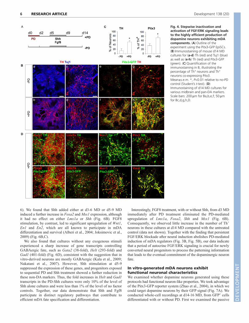
6
6). We found that Shh added either at d3-6 MD or d5-9 MDinduced a further increase in Foxa2 and Msx1 expression, althoughit had no effect on either Lmx1a or Shh (Fig. 6B). FGF8stimulation, by contrast, led to significant upregulation of Wnt1,En1 and En2, which are all known to participate in mDAdifferentiation and survival (Alberi et al., 2004; Joksimovic et al.,2009) (Fig. 6B,C).
We also found that cultures without any exogenous stimuliexperienced a sharp increase of gene transcripts controllingGABAergic fate, such as Gata2 (38-fold), Helt (293-fold) andGad1 (401-fold) (Fig. 6D), consistent with the suggestion that invitro-derived neurons are mostly GABAergic (Kala et al., 2009;Nakatani et al., 2007). However, Shh stimulation at d5-9suppressed the expression of these genes, and progenitors exposedto sequential PD and Shh treatment showed a further reduction inthese non-DA markers. Thus, the fold increases in Helt and Gad1transcripts in the PD-Shh cultures were only 10% of the level ofShh alone cultures and were less than 1% of the level of no factorcontrols. Together, our data demonstrate that Shh and Fgf8participate in distinct regulatory pathways that contribute toefficient mDA fate specification and differentiation.
Interestingly, FGF8 treatment, with or without Shh, from d3 MDimmediately after PD treatment eliminated the PD-mediatedupregulation of Lmx1a, Foxa2, Shh and Msx1 (Fig. 6B).Consequently, we observed little increase in the number of Th+
neurons in these cultures at d14 MD compared with the untreatedcontrol (data not shown). Together with the finding that persistentFGF/ERK blockade after neural induction abolishes PD-mediatedinduction of mDA regulators (Fig. 3B, Fig. 5B), our data indicatethat a period of autocrine FGF/ERK signaling is crucial for newlyconverted neural progenitors to process the patterning informationthat leads to the eventual commitment of the dopaminergic neuronfate.
In vitro-generated mDA neurons exhibitfunctional neuronal characteristicsWe examined whether dopamine neurons generated using theseprotocols had functional neuron-like properties. We took advantageof the Pitx3-GFP reporter system (Zhao et al., 2004), in which wecould target dopamine neurons by their GFP signal (Fig. 7A). Weconducted whole-cell recordings at d14-16 MD, from GFP+ cellsdifferentiated with or without PD. First we examined the passive
RESEARCH ARTICLE Development 138 (20)
Fig. 4. Stepwise inactivation andactivation of FGF/ERK signaling leadsto the highly efficient production ofdopamine neurons exhibiting mDAcomponents. (A)Outline of theexperiment using the Pitx3-GFP EpiSCs.(B)Immunostaining of mouse d14 MDcultures for (a-d) Th (red) and Tuj1 (blue)as well as (e-h) Th (red) and Pitx3-GFP(green). (C)Quantification of theimmunostaining in B, illustrating thepercentage of Th+ neurons and Th+
neurons co-expressing Pitx3.Mean±s.e.m. *, P<0.01 relative to no-PDcontrol (Student’s t-test). (D)Immunostaining of d14 MD cultures forvarious midbrain and pan-DA markers.Scale bars: 200m for Ba,b,e,f; 50mfor Bc,d,g,h,D.
DEVELO
PMENT
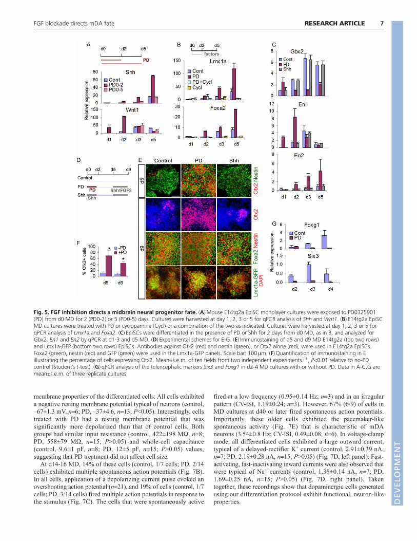
membrane properties of the differentiated cells. All cells exhibiteda negative resting membrane potential typical of neurons (control,–67±1.3 mV, n6; PD, –37±4.6, n13; P<0.05). Interestingly, cellstreated with PD had a resting membrane potential that wassignificantly more depolarized than that of control cells. Bothgroups had similar input resistance (control, 422±198 M, n8;PD, 558±79 M, n15; P>0.05) and whole-cell capacitance(control, 9.6±1 pF, n8; PD, 12±5 pF, n15; P>0.05) values,suggesting that PD treatment did not affect cell size.
At d14-16 MD, 14% of these cells (control, 1/7 cells; PD, 2/14cells) exhibited multiple spontaneous action potentials (Fig. 7B).In all cells, application of a depolarizing current pulse evoked anovershooting action potential (n21), and 19% of cells (control, 1/7cells; PD, 3/14 cells) fired multiple action potentials in response tothe stimulus (Fig. 7C). The cells that were spontaneously active
fired at a low frequency (0.95±0.14 Hz; n3) and in an irregularpattern (CV-ISI, 1.19±0.24; n3). However, 67% (6/9) of cells inMD cultures at d40 or later fired spontaneous action potentials.Importantly, these older cells exhibited the pacemaker-likespontaneous activity (Fig. 7E) that is characteristic of mDAneurons (3.54±0.8 Hz; CV-ISI, 0.49±0.08; n6). In voltage-clampmode, all differentiated cells exhibited a large outward current,typical of a delayed-rectifier K+ current (control, 2.91±0.39 nA,n7; PD, 2.19±0.28 nA, n15; P>0.05) (Fig. 7D, left panel). Fast-activating, fast-inactivating inward currents were also observed thatwere typical of Na+ currents (control, 1.38±0.14 nA, n7; PD,1.69±0.25 nA, n15; P>0.05) (Fig. 7D, right panel). Takentogether, these recordings show that dopaminergic cells generatedusing our differentiation protocol exhibit functional, neuron-likeproperties.
7RESEARCH ARTICLEFGF blockade directs mDA fate
Fig. 5. FGF inhibition directs a midbrain neural progenitor fate. (A)Mouse E14tg2a EpiSC monolayer cultures were exposed to PD0325901(PD) from d0 MD for 2 (PD0-2) or 5 (PD0-5) days. Cultures were harvested at day 1, 2, 3 or 5 for qPCR analysis of Shh and Wnt1. (B)E14tg2a EpiSCMD cultures were treated with PD or cyclopamine (Cycl) or a combination of the two as indicated. Cultures were harvested at day 1, 2, 3 or 5 forqPCR analysis of Lmx1a and Foxa2. (C)EpiSCs were differentiated in the presence of PD or Shh for 2 days from d0 MD, as in B, and analyzed forGbx2, En1 and En2 by qPCR at d1-3 and d5 MD. (D)Experimental schemes for E-G. (E)Immunostaining of d5 and d9 MD E14tg2a (top two rows)and Lmx1a-GFP (bottom two rows) EpiSCs. Antibodies against Otx2 (red) and nestin (green), or Otx2 alone (red), were used in E14tg2a EpiSCs.Foxa2 (green), nestin (red) and GFP (green) were used in the Lmx1a-GFP panels. Scale bar: 100m. (F)Quantification of immunostaining in Eillustrating the percentage of cells expressing Otx2. Mean±s.e.m. of ten fields from two independent experiments. *, P<0.01 relative to no-PDcontrol (Student’s t-test). (G)qPCR analysis of the telencephalic markers Six3 and Foxg1 in d2-4 MD cultures with or without PD. Data in A-C,G aremean±s.e.m. of three replicate cultures.
DEVELO
PMENT
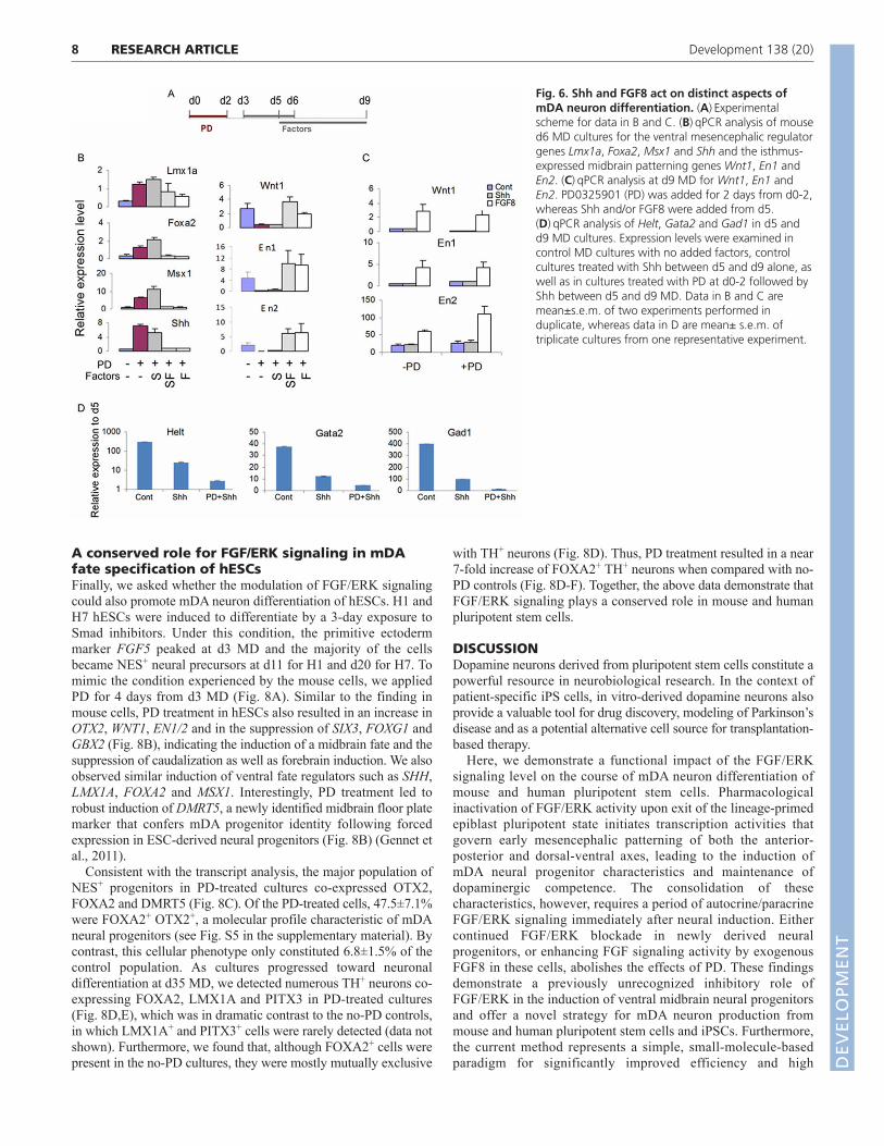
8
A conserved role for FGF/ERK signaling in mDAfate specification of hESCsFinally, we asked whether the modulation of FGF/ERK signalingcould also promote mDA neuron differentiation of hESCs. H1 andH7 hESCs were induced to differentiate by a 3-day exposure toSmad inhibitors. Under this condition, the primitive ectodermmarker FGF5 peaked at d3 MD and the majority of the cellsbecame NES+ neural precursors at d11 for H1 and d20 for H7. Tomimic the condition experienced by the mouse cells, we appliedPD for 4 days from d3 MD (Fig. 8A). Similar to the finding inmouse cells, PD treatment in hESCs also resulted in an increase inOTX2, WNT1, EN1/2 and in the suppression of SIX3, FOXG1 andGBX2 (Fig. 8B), indicating the induction of a midbrain fate and thesuppression of caudalization as well as forebrain induction. We alsoobserved similar induction of ventral fate regulators such as SHH,LMX1A, FOXA2 and MSX1. Interestingly, PD treatment led torobust induction of DMRT5, a newly identified midbrain floor platemarker that confers mDA progenitor identity following forcedexpression in ESC-derived neural progenitors (Fig. 8B) (Gennet etal., 2011).
Consistent with the transcript analysis, the major population ofNES+ progenitors in PD-treated cultures co-expressed OTX2,FOXA2 and DMRT5 (Fig. 8C). Of the PD-treated cells, 47.5±7.1%were FOXA2+ OTX2+, a molecular profile characteristic of mDAneural progenitors (see Fig. S5 in the supplementary material). Bycontrast, this cellular phenotype only constituted 6.8±1.5% of thecontrol population. As cultures progressed toward neuronaldifferentiation at d35 MD, we detected numerous TH+ neurons co-expressing FOXA2, LMX1A and PITX3 in PD-treated cultures(Fig. 8D,E), which was in dramatic contrast to the no-PD controls,in which LMX1A+ and PITX3+ cells were rarely detected (data notshown). Furthermore, we found that, although FOXA2+ cells werepresent in the no-PD cultures, they were mostly mutually exclusive
with TH+ neurons (Fig. 8D). Thus, PD treatment resulted in a near7-fold increase of FOXA2+ TH+ neurons when compared with no-PD controls (Fig. 8D-F). Together, the above data demonstrate thatFGF/ERK signaling plays a conserved role in mouse and humanpluripotent stem cells.
DISCUSSIONDopamine neurons derived from pluripotent stem cells constitute apowerful resource in neurobiological research. In the context ofpatient-specific iPS cells, in vitro-derived dopamine neurons alsoprovide a valuable tool for drug discovery, modeling of Parkinson’sdisease and as a potential alternative cell source for transplantation-based therapy.
Here, we demonstrate a functional impact of the FGF/ERKsignaling level on the course of mDA neuron differentiation ofmouse and human pluripotent stem cells. Pharmacologicalinactivation of FGF/ERK activity upon exit of the lineage-primedepiblast pluripotent state initiates transcription activities thatgovern early mesencephalic patterning of both the anterior-posterior and dorsal-ventral axes, leading to the induction ofmDA neural progenitor characteristics and maintenance ofdopaminergic competence. The consolidation of thesecharacteristics, however, requires a period of autocrine/paracrineFGF/ERK signaling immediately after neural induction. Eithercontinued FGF/ERK blockade in newly derived neuralprogenitors, or enhancing FGF signaling activity by exogenousFGF8 in these cells, abolishes the effects of PD. These findingsdemonstrate a previously unrecognized inhibitory role ofFGF/ERK in the induction of ventral midbrain neural progenitorsand offer a novel strategy for mDA neuron production frommouse and human pluripotent stem cells and iPSCs. Furthermore,the current method represents a simple, small-molecule-basedparadigm for significantly improved efficiency and high
RESEARCH ARTICLE Development 138 (20)
Fig. 6. Shh and FGF8 act on distinct aspects ofmDA neuron differentiation. (A)Experimentalscheme for data in B and C. (B)qPCR analysis of moused6 MD cultures for the ventral mesencephalic regulatorgenes Lmx1a, Foxa2, Msx1 and Shh and the isthmus-expressed midbrain patterning genes Wnt1, En1 andEn2. (C)qPCR analysis at d9 MD for Wnt1, En1 andEn2. PD0325901 (PD) was added for 2 days from d0-2,whereas Shh and/or FGF8 were added from d5.(D)qPCR analysis of Helt, Gata2 and Gad1 in d5 andd9 MD cultures. Expression levels were examined incontrol MD cultures with no added factors, controlcultures treated with Shh between d5 and d9 alone, aswell as in cultures treated with PD at d0-2 followed byShh between d5 and d9 MD. Data in B and C aremean±s.e.m. of two experiments performed induplicate, whereas data in D are mean± s.e.m. oftriplicate cultures from one representative experiment.
DEVELO
PMENT

reproducibility compared with previously reported transgene-freeprotocols. Importantly, our strategy directs a midbrain regionalidentity in the derived dopamine neurons, a property that isessential for functional integration of transplanted dopamineneurons in the Parkinsonian brain (Hudson et al., 1994).
Stimulation of ESC-derived neural progenitors with Shh andFGF8 is used by almost all dopamine differentiation protocols(Barberi et al., 2003; Kim et al., 2002; Lee et al., 2000; Perrier etal., 2004; Yan et al., 2005). However, unless combined with geneticmanipulation of mDA transcription factors, such as Pitx3 or Lmx1a(Andersson et al., 2006; Chung et al., 2002; Konstantoulas et al.,
2010; Maxwell et al., 2005), the midbrain regional identity of thedopamine neurons generated has remained uncertain. Furthermore,the yield of Th+ neurons has often proved unreliable betweenexperiments and even highly variable between differentmicroscopic fields within a single culture. A major limiting factoris the temporal and spatial heterogeneity of ESC-derived neuralprogenitors. Our findings demonstrate that the above issues can beaddressed using EpiSCs. We found that, in the absence ofFGF/ERK signaling manipulation, nearly 40% of Th+ neuronsgenerated by EpiSCs already co-expressed Pitx3. This represents asignificant improvement over ESC-derived monolayer cultures,where Pitx3+ neurons are rarely observed (Andersson et al., 2006;Friling et al., 2009; Parmar and Li, 2007). This improvement islikely to be due to the more synchronous conversion of EpiSCs tothe neuroepithelial fate, which would allow for the effectivecapture of mDA-competent progenitors.
However, without additional FGF/ERK inhibitor treatment at theneural induction phase, the total numbers of Th+ Pitx3+ cellsremained low due to the overall poor efficiency in producing Th+
cells. The early induction of both Lmx1a and Foxa2 by inhibitingFGF receptor or ERK is likely to be a key factor in the observedhigh efficiency in our experiments. This hypothesis is based on thefollowing observations: (1) d5 PD-treated (EpiSC) MD cultures arehighly enriched for Foxa2+ Lmx1a+ neural progenitors comparedwith untreated controls; (2) although Shh treatment in d5-9 MDresults in comparable numbers of Foxa2+ Lmx1a+ cells in PD-primed and no-PD cultures, mDA neuron production was notenhanced in the manner observed with PD treatment; (3) replacingPD with Shh, which turned out to be a slower and less effectiveinducer of Lmx1a and Foxa2, also led to poor mDA production;and (4) previous reports have credited the dopaminergic-promotingactivity of Lmx1a to its early transgene expression in ESC-derivedneural progenitors (Friling et al., 2009) and indicated that Lmx1afunctions by cooperating with Foxa2 in specifying mDA fateduring midbrain development (Lin et al., 2009; Nakatani et al.,2009).
The robust induction of Wnt1 and its targets in naïve neuralprogenitors is likely to be a key downstream mediator that confersthe observed early induction of Lmx1a, in light of the recent findingthat it can be directly regulated by Wnt1/-catenin signaling(Chung et al., 2009). The same study also showed that, althoughOtx2 itself had no effect in promoting the expression of terminalmDA neuronal marker genes such as Th, Pitx3 and Nurr1, itsignificantly enhanced the regulatory effect of Lmx1a and Foxa2on the expression of these genes. Thus, Otx2 plays a permissiverole in Lmx1a/Foxa2-mediated mDA neuronal production. It isworth noting that a significant effect of FGF/ERK blockade is themaintenance of Otx2 in derived neural progenitors.
Our study also shows that, in addition to inducing a regulatorycascade for ventralizing nascent neural progenitors, FGF/ERKinhibition suppresses forebrain specification while promotinganterior neural induction, as demonstrated by the strong andconsistent repression of the forebrain regulator genes Six3 andFoxg1 and the hindbrain marker Gbx2. Thus, blocking FGF/ERKat the onset of neural induction leads to a direct and early inductionof the midbrain fate at the expense of forebrain and caudal neuralfates. Our finding is consistent with the developmental role of FGFsignaling in regionalization of the forebrain (Corson et al., 2003;Shimamura and Rubenstein, 1997).
Furthermore, we demonstrate the importance of precise temporalcontrol of cell signaling and its cross-regulation with other signalingpathways in mDA neuronal fate specification. During development,
9RESEARCH ARTICLEFGF blockade directs mDA fate
Fig. 7. Pitx3-GFP+ cells exhibit functional neuron-likeelectrophysiological properties. (A)DIC and GFP image showing arecording pipette patched onto a mouse Pitx3-GFP+ cell.(B)Spontaneous action potentials fired by a day-14 neuron. Verticalscale bar, 20 mV; horizontal scale bar, 5 mseconds. (C)A singlespontaneous action potential (left). A single action potential fired inresponse to a 50 pA depolarizing current pulse (middle). Multiple actionpotentials fired in response to a 50 pA depolarizing current pulse(right). Vertical bars, 20 mV; horizontal bars, 50, 100 and 100mseconds, respectively. (D)Outward current evoked by a serious ofvoltage steps (left). Fast inward current evoked by a series of voltagesteps (right). Vertical bars, 1 nA; horizontal bars, 50 mseconds.(E)Pacemaker-like spontaneous firing a day-41 neuron. Vertical bar, 20mV; horizontal bar, 1 second.DEVELO
PMENT

10
Fgf8-mediated signaling can induce the patterned expression ofmany midbrain/rostral hindbrain genes and is required for normaldevelopment of the midbrain and cerebellum (Chi et al., 2003; Liuet al., 1999; Meyers et al., 1998). Fgf8-induced Wnt1 and engrailedare key regulators of midbrain and cerebellum patterning, as well asof the differentiation and survival of dopamine neurons (Prakash etal., 2006; Simon et al., 2001; Tang et al., 2009). In EpiSC-derivedneural cultures, after an initial burst of upregulation induced by PDexposure, Wnt1 expression was subsequently reduced to a levelbelow the no-PD control by unknown factors in the newly generatedneural progenitors in d3-5 MD. This is the period when Shh, Lmx1aand Foxa2 expression levels continued to rise. Given that Shh andWnt1 play opposing roles with regard to mDA neurogenesis(Joksimovic et al., 2009), our findings suggest that the delay in FGFreactivation, which suppresses Wnt1 levels, might be crucial forachieving high numbers of Th+ neurons by consolidating Lmx1a andFoxa2 expression via Shh signaling.
From a technological standpoint, we describe a novel method ofmDA neuron differentiation that employs temporally controlledexposure of human and mouse pluripotent stem cells to anFGF/ERK-deficient environment. The highly reliable nature of this
method was demonstrated using five independent mouse EpiSClines, a mouse iPS cell line and two human ESC lines. This protocoloffers several advantages over current methods of generatingmidbrain-specific DA neurons in that it is adherent culture-based andfree from genetic manipulation and thus could be readily applied toother cell lines of interest. Furthermore, because it is fully chemicallydefined, this paradigm could be readily adapted for use in a clinicalsetting or scaled up for toxicity and drug screening relevant todeveloping new therapeutics for Parkinson’s disease.
AcknowledgementsWe thank Dr Michael German for Lmx1a antibody, Dr Marten Smidt for Pitx3antibody, Drs Johan Ericson and Thomas Perlmann for the Lmx1a-GFP ESCs, DrDirk Dormann for expert advice on confocal microscopy and Drs Austin Smith,Tilo Kunath and Tristan Rodriguez for discussion and critical reading of themanuscript.
FundingThis work was supported by the UK Medical Research Council (MRC,G117/560, U120005004) and EU framework program 7 NeuroStemcell, no.222943 to M.L.; and by the MRC (U120085816) and a Royal Society UniversityResearch Fellowship to M.A.U. M.L. was an MRC Senior Non-Clinical ResearchFellow. Deposited in PMC for release after 6 months.
RESEARCH ARTICLE Development 138 (20)
Fig. 8. Generation of mDA neurons from humanESCs. (A)Experimental schemes. (B)qPCR analysis ofregional markers in d7 and d11 H7 MD cultures withor without PD0325901 (PD). Data are mean±s.e.m. ofthree replicate cultures. (C)Double immunostaining ofd13 H1 MD cultures for nestin (green) plus OTX2,FOXA2 or DMRT5 (all in red). (D)Triple antibodystaining of d35 H7 cultures for FOXA2 (red), TH(green) and 3-tubulin (TUJ1, blue).(E)Immunostaining of PD-treated d32 H7 MD culturesfor PITX3 (red) and TH (green), and for LMX1Aa (red)and TH (green). (F)Quantification of theimmunostaining in E, illustrating the percentage ofTH+ neurons and TH+ neurons co-expressing FOXA2.Error bars indicate s.e.m. Scale bars: 75m.
DEVELO
PMENT

Competing interests statementThe authors declare no competing financial interests.
Supplementary materialSupplementary material for this article is available athttp://dev.biologists.org/lookup/suppl/doi:10.1242/dev.066746/-/DC1
ReferencesAlberi, L., Sgado, P. and Simon, H. H. (2004). Engrailed genes are cell-
autonomously required to prevent apoptosis in mesencephalic dopaminergicneurons. Development 131, 3229-3236.
Andersson, E., Tryggvason, U., Deng, Q., Friling, S., Alekseenko, Z., Robert,B., Perlmann, T. and Ericson, J. (2006). Identification of intrinsic determinantsof midbrain dopamine neurons. Cell 124, 393-405.
Barberi, T., Klivenyi, P., Calingasan, N. Y., Lee, H., Kawamata, H., Loonam,K., Perrier, A. L., Bruses, J., Rubio, M. E., Topf, N. et al. (2003). Neuralsubtype specification of fertilization and nuclear transfer embryonic stem cellsand application in parkinsonian mice. Nat. Biotechnol. 21, 1200-1207.
Bouhon, I. A., Kato, H., Chandran, S. and Allen, N. D. (2005). Neuraldifferentiation of mouse embryonic stem cells in chemically defined medium.Brain Res. Bull. 68, 62-75.
Brodski, C., Weisenhorn, D. M., Signore, M., Sillaber, I., Oesterheld, M.,Broccoli, V., Acampora, D., Simeone, A. and Wurst, W. (2003). Location andsize of dopaminergic and serotonergic cell populations are controlled by theposition of the midbrain-hindbrain organizer. J. Neurosci. 23, 4199-4207.
Brons, I. G., Smithers, L. E., Trotter, M. W., Rugg-Gunn, P., Sun, B., Chuva deSousa Lopes, S. M., Howlett, S. K., Clarkson, A., Ahrlund-Richter, L.,Pedersen, R. A. et al. (2007). Derivation of pluripotent epiblast stem cells frommammalian embryos. Nature 448, 191-195.
Chambers, S. M., Fasano, C. A., Papapetrou, E. P., Tomishima, M., Sadelain,M. and Studer, L. (2009). Highly efficient neural conversion of human ES andiPS cells by dual inhibition of SMAD signaling. Nat. Biotechnol. 27, 275-280.
Chi, C. L., Martinez, S., Wurst, W. and Martin, G. R. (2003). The isthmicorganizer signal FGF8 is required for cell survival in the prospective midbrain andcerebellum. Development 130, 2633-2644.
Chung, S., Sonntag, K. C., Andersson, T., Bjorklund, L. M., Park, J. J., Kim, D.W., Kang, U. J., Isacson, O. and Kim, K. S. (2002). Genetic engineering ofmouse embryonic stem cells by Nurr1 enhances differentiation and maturationinto dopaminergic neurons. Eur. J. Neurosci. 16, 1829-1838.
Chung, S., Hedlund, E., Hwang, M., Kim, D. W., Shin, B. S., Hwang, D. Y.,Jung Kang, U., Isacson, O. and Kim, K. S. (2005). The homeodomaintranscription factor Pitx3 facilitates differentiation of mouse embryonic stem cellsinto AHD2-expressing dopaminergic neurons. Mol. Cell. Neurosci. 28, 241-252.
Chung, S., Leung, A., Han, B. S., Chang, M. Y., Moon, J. I., Kim, C. H., Hong,S., Pruszak, J., Isacson, O. and Kim, K. S. (2009). Wnt1-lmx1a forms a novelautoregulatory loop and controls midbrain dopaminergic differentiationsynergistically with the SHH-FoxA2 pathway. Cell Stem Cell 5, 646-658.
Corson, L. B., Yamanaka, Y., Lai, K. M. and Rossant, J. (2003). Spatial andtemporal patterns of ERK signaling during mouse embryogenesis. Development130, 4527-4537.
Echelard, Y., Epstein, D. J., St-Jacques, B., Shen, L., Mohler, J., McMahon, J.A. and McMahon, A. P. (1993). Sonic hedgehog, a member of a family ofputative signaling molecules, is implicated in the regulation of CNS polarity. Cell75, 1417-1430.
Ferri, A. L., Lin, W., Mavromatakis, Y. E., Wang, J. C., Sasaki, H., Whitsett, J.A. and Ang, S. L. (2007). Foxa1 and Foxa2 regulate multiple phases of midbraindopaminergic neuron development in a dosage-dependent manner.Development 134, 2761-2769.
Friling, S., Andersson, E., Thompson, L. H., Jonsson, M. E., Hebsgaard, J. B.,Nanou, E., Alekseenko, Z., Marklund, U., Kjellander, S., Volakakis, N. et al.(2009). Efficient production of mesencephalic dopamine neurons by Lmx1aexpression in embryonic stem cells. Proc. Natl. Acad. Sci. USA 106, 7613-7618.
Gaspard, N., Bouschet, T., Hourez, R., Dimidschstein, J., Naeije, G., van denAmeele, J., Espuny-Camacho, I., Herpoel, A., Passante, L., Schiffmann, S.N. et al. (2008). An intrinsic mechanism of corticogenesis from embryonic stemcells. Nature 455, 351-357.
Gennet, N., Gale, E., Nan, X., Farley, E., Takacs, K., Oberwallner, B.,Chambers, D. and Li, M. (2011). Doublesex and mab-3-related transcriptionfactor 5 promotes midbrain dopaminergic identity in pluripotent stem cells byenforcing a ventral-medial progenitor fate. Proc. Natl. Acad. Sci. USA 108,9131-9136.
Guo, G., Yang, J., Nichols, J., Hall, J. S., Eyres, I., Mansfield, W. and Smith, A.(2009). Klf4 reverts developmentally programmed restriction of ground statepluripotency. Development 136, 1063-1069.
Hudson, J. L., Bickford, P., Johansson, M., Hoffer, B. J. and Stromberg, I.(1994). Target and neurotransmitter specificity of fetal central nervous systemtransplants: importance for functional reinnervation. J. Neurosci. 14, 283-290.
Joksimovic, M., Yun, B. A., Kittappa, R., Anderegg, A. M., Chang, W. W.,Taketo, M. M., McKay, R. D. and Awatramani, R. B. (2009). Wnt antagonismof Shh facilitates midbrain floor plate neurogenesis. Nat. Neurosci. 12, 125-131.
Kala, K., Haugas, M., Lillevali, K., Guimera, J., Wurst, W., Salminen, M. andPartanen, J. (2009). Gata2 is a tissue-specific post-mitotic selector gene formidbrain GABAergic neurons. Development 136, 253-262.
Kawasaki, H., Mizuseki, K., Nishikawa, S., Kaneko, S., Kuwana, Y.,Nakanishi, S., Nishikawa, S. I. and Sasai, Y. (2000). Induction of midbraindopaminergic neurons from ES cells by stromal cell-derived inducing activity.Neuron 28, 31-40.
Kim, J. H., Auerbach, J. M., Rodriguez-Gomez, J. A., Velasco, I., Gavin, D.,Lumelsky, N., Lee, S. H., Nguyen, J., Sanchez-Pernaute, R., Bankiewicz, K.et al. (2002). Dopamine neurons derived from embryonic stem cells function inan animal model of Parkinson’s disease. Nature 418, 50-56.
Kittappa, R., Chang, W. W., Awatramani, R. B. and McKay, R. D. (2007). Thefoxa2 gene controls the birth and spontaneous degeneration of dopamineneurons in old age. PLoS Biol. 5, e325.
Konstantoulas, C. J., Parmar, M. and Li, M. (2010). FoxP1 promotes midbrainidentity in embryonic stem cell-derived dopamine neurons by regulating Pitx3. J.Neurochem. 113, 836-847.
Kunath, T., Saba-El-Leil, M. K., Almousailleakh, M., Wray, J., Meloche, S. andSmith, A. (2007). FGF stimulation of the Erk1/2 signalling cascade triggerstransition of pluripotent embryonic stem cells from self-renewal to lineagecommitment. Development 134, 2895-2902.
Lee, S. H., Lumelsky, N., Studer, L., Auerbach, J. M. and McKay, R. D. (2000).Efficient generation of midbrain and hindbrain neurons from mouse embryonicstem cells. Nat. Biotechnol. 18, 675-679.
Lin, W., Metzakopian, E., Mavromatakis, Y. E., Gao, N., Balaskas, N., Sasaki,H., Briscoe, J., Whitsett, J. A., Goulding, M., Kaestner, K. H. et al. (2009).Foxa1 and Foxa2 function both upstream of and cooperatively with Lmx1a andLmx1b in a feedforward loop promoting mesodiencephalic dopaminergicneuron development. Dev. Biol. 333, 386-396.
Liu, A., Losos, K. and Joyner, A. L. (1999). FGF8 can activate Gbx2 andtransform regions of the rostral mouse brain into a hindbrain fate. Development126, 4827-4838.
Lunn, J. S., Fishwick, K. J., Halley, P. A. and Storey, K. G. (2007). A spatial andtemporal map of FGF/Erk1/2 activity and response repertoires in the early chickembryo. Dev. Biol. 302, 536-552.
Maxwell, S. L., Ho, H. Y., Kuehner, E., Zhao, S. and Li, M. (2005). Pitx3regulates tyrosine hydroxylase expression in the substantia nigra and identifies asubgroup of mesencephalic dopaminergic progenitor neurons during mousedevelopment. Dev. Biol. 282, 467-479.
Meyers, E. N., Lewandoski, M. and Martin, G. R. (1998). An Fgf8 mutant allelicseries generated by Cre- and Flp-mediated recombination. Nat. Genet. 18, 136-141.
Millet, S., Campbell, K., Epstein, D. J., Losos, K., Harris, E. and Joyner, A. L.(1999). A role for Gbx2 in repression of Otx2 and positioning the mid/hindbrainorganizer. Nature 401, 161-164.
Nakatani, T., Minaki, Y., Kumai, M. and Ono, Y. (2007). Helt determinesGABAergic over glutamatergic neuronal fate by repressing Ngn genes in thedeveloping mesencephalon. Development 134, 2783-2793.
Nakatani, T., Kumai, M., Mizuhara, E., Minaki, Y. and Ono, Y. (2009). Lmx1aand Lmx1b cooperate with Foxa2 to coordinate the specification ofdopaminergic neurons and control of floor plate cell differentiation in thedeveloping mesencephalon. Dev. Biol. 339, 101-113.
Olander, S., Nordstrom, U., Patthey, C. and Edlund, T. (2006). Convergent Wntand FGF signaling at the gastrula stage induce the formation of the isthmicorganizer. Mech. Dev. 123, 166-176.
Omodei, D., Acampora, D., Mancuso, P., Prakash, N., Di Giovannantonio, L.G., Wurst, W. and Simeone, A. (2008). Anterior-posterior graded response toOtx2 controls proliferation and differentiation of dopaminergic progenitors inthe ventral mesencephalon. Development 135, 3459-3470.
Ono, Y., Nakatani, T., Sakamoto, Y., Mizuhara, E., Minaki, Y., Kumai, M.,Hamaguchi, A., Nishimura, M., Inoue, Y., Hayashi, H. et al. (2007).Differences in neurogenic potential in floor plate cells along an anteroposteriorlocation: midbrain dopaminergic neurons originate from mesencephalic floorplate cells. Development 134, 3213-3225.
Paek, H., Gutin, G. and Hebert, J. M. (2009). FGF signaling is strictly required tomaintain early telencephalic precursor cell survival. Development 136, 2457-2465.
Parmar, M. and Li, M. (2007). Early specification of dopaminergic phenotypeduring ES cell differentiation. BMC Dev. Biol. 7, 86.
Perrier, A. L., Tabar, V., Barberi, T., Rubio, M. E., Bruses, J., Topf, N., Harrison,N. L. and Studer, L. (2004). Derivation of midbrain dopamine neurons fromhuman embryonic stem cells. Proc. Natl. Acad. Sci. USA 101, 12543-12548.
Prakash, N., Brodski, C., Naserke, T., Puelles, E., Gogoi, R., Hall, A.,Panhuysen, M., Echevarria, D., Sussel, L., Weisenhorn, D. M. et al. (2006).A Wnt1-regulated genetic network controls the identity and fate of midbrain-dopaminergic progenitors in vivo. Development 133, 89-98.
11RESEARCH ARTICLEFGF blockade directs mDA fate
DEVELO
PMENT

12
Roybon, L., Hjalt, T., Christophersen, N. S., Li, J. Y. and Brundin, P. (2008).Effects on differentiation of embryonic ventral midbrain progenitors by Lmx1a,MSX1, Ngn2, and Pitx3. J. Neurosci. 28, 3644-3656.
Shimamura, K. and Rubenstein, J. L. (1997). Inductive interactions direct earlyregionalization of the mouse forebrain. Development 124, 2709-2718.
Simon, H. H., Saueressig, H., Wurst, W., Goulding, M. D. and O’Leary, D. D.(2001). Fate of midbrain dopaminergic neurons controlled by the engrailedgenes. J. Neurosci. 21, 3126-3134.
Stavridis, M. P., Lunn, J. S., Collins, B. J. and Storey, K. G. (2007). A discreteperiod of FGF-induced Erk1/2 signalling is required for vertebrate neuralspecification. Development 134, 2889-2894.
Tang, M., Villaescusa, J. C., Luo, S. X., Guitarte, C., Lei, S., Miyamoto, Y.,Taketo, M. M., Arenas, E. and Huang, E. J. (2009). Interactions of Wnt/beta-catenin signaling and sonic hedgehog regulate the neurogenesis of ventralmidbrain dopamine neurons. J. Neurosci. 30, 9280-9291.
Tesar, P. J., Chenoweth, J. G., Brook, F. A., Davies, T. J., Evans, E. P., Mack, D.L., Gardner, R. L. and McKay, R. D. (2007). New cell lines from mouse epiblastshare defining features with human embryonic stem cells. Nature 448, 196-199.
Watanabe, K., Kamiya, D., Nishiyama, A., Katayama, T., Nozaki, S.,Kawasaki, H., Watanabe, Y., Mizuseki, K. and Sasai, Y. (2005). Directed
differentiation of telencephalic precursors from embryonic stem cells. Nat.Neurosci. 8, 288-296.
Wilson, S. W. and Rubenstein, J. L. (2000). Induction and dorsoventralpatterning of the telencephalon. Neuron 28, 641-651.
Yan, Y., Yang, D., Zarnowska, E. D., Du, Z., Werbel, B., Valliere, C., Pearce, R.A., Thomson, J. A. and Zhang, S. C. (2005). Directed differentiation ofdopaminergic neuronal subtypes from human embryonic stem cells. Stem Cells23, 781-790.
Ye, W., Shimamura, K., Rubenstein, J. L., Hynes, M. A. and Rosenthal, A.(1998). FGF and Shh signals control dopaminergic and serotonergic cell fate inthe anterior neural plate. Cell 93, 755-766.
Ying, Q. L., Stavridis, M., Griffiths, D., Li, M. and Smith, A. (2003). Conversionof embryonic stem cells into neuroectodermal precursors in adherentmonoculture. Nat. Biotechnol. 21, 183-186.
Ying, Q. L., Wray, J., Nichols, J., Batlle-Morera, L., Doble, B., Woodgett, J.,Cohen, P. and Smith, A. (2008). The ground state of embryonic stem cell self-renewal. Nature 453, 519-523.
Zhao, S., Maxwell, S., Jimenez-Beristain, A., Vives, J., Kuehner, E., Zhao, J.,O’Brien, C., de Felipe, C., Semina, E. and Li, M. (2004). Generation ofembryonic stem cells and transgenic mice expressing green fluorescence proteinin midbrain dopaminergic neurons. Eur. J. Neurosci. 19, 1133-1140.
RESEARCH ARTICLE Development 138 (20)
DEVELO
PMENT