Microvascular endothelial cells play potential immunoregulatory roles in the immune response to...
6
Microvascular endothelial cells play potential immunoregulatory roles in the immune response to foot‐and‐mouth disease vaccines Tao Zhang 1 , Lifang Tian 2 , Ge Hu 1 , Kedao Teng 3 * and Xiang Mu 1 * 1 Beijing Key Laboratory of Traditional Chinese Veterinary Medicine, Beijing University of Agriculture, Beijing, China 2 Animal Husbandry and Veterinary Department, Baoding Vocational and Technical College, Baoding, China 3 College of Veterinary Medicine, China Agricultural University, Beijing, China Microvascular endothelial cells (MVECs) have been documented to have important immunoregulatory effects. Exploring their roles in the immune response to foot‐and‐mouth disease (FMD) vaccines would help to improve their efficacy. In this study, the effects of FMD vaccine 146s antigens on gene expression profiles of rat intestinal mucosal and myocardial MVECs were analysed using microarray, and their effects on transendothelial migration (TEM) of peripheral blood mononuclear cells (PBMC) were investigated by the Transwell migration assay. Both kinds of MVECs displayed significant responses to 146s antigens, and 252 and 67 genes were differentially expressed in rat intestinal mucosal and myocardial MVECs, respectively. Despite different altered gene expression patterns, many immune‐associated genes were involved in both kinds of MVECs. The gene expression changes by microarray were confirmed by real‐time reverse transcription‐ polymerase chain reaction. Transwell migration analysis indicated that the TEM of PBMC was increased by 146s antigens, which could be partially inhibited by blocking vascular cell adhesion molecule 1 in MVECs. This study suggests that MVECs play potential immunoregulatory roles in the immune response to FMD vaccines, one of which is influencing the TEM of immune cells. Copyright © 2011 John Wiley & Sons, Ltd. key words — microvascular endothelial cells; foot‐and‐mouth disease; 146s; immune‐associated gene; transendothelial migration; microarray INTRODUCTION Foot‐and‐mouth disease (FMD) is classified as a list A disease by the World Organization for Animal Health because it is highly contagious and economically damaging. Current FMD vaccines have poor efficacy and only provide temporary immunity (about 6 months). 1 Much effort has been put into the vaccine immunogenicity and much attention have been paid to the classic immune cells such as T and B cells, while the safe and efficient vaccine has not been developed yet. A comprehensive investigation into host cell responses to FMD vaccines would contribute to solving this problem. Microvascular endothelial cells (MVECs) have been documented to be critical for innate and adaptive immunity, 2 however, what roles they play in the immune response to FMD vaccines remains unclear. The aim of this study was to appreciate the general response of MVECs to FMD vaccine antigens. Considering commercial availability of rat cDNA microarray and susceptibility of rat to foot‐ and‐mouth disease virus (FMDV), 3 MVECs from rat intestinal mucosa and myocardium, which are commonly injured by FMDV infection, 4 were used in our experiments. The effects of FMD vaccine 146s antigens on gene expression profiles of both kinds of MVECs were investigated using microarray, and their effects on transen- dothelial migration (TEM) of peripheral blood mononuclear cells (PBMC) were investigated by Transwell migration assay. MATERIALS AND METHODS FMD vaccine antigens Inactivated whole FMD virions (146s particles) are the essential immunogenic component of current FMD vac- cines. 5 In the study, 146s particles were purified as described previously 6 from FMDV O/China99 strain infected BHK‐21 cell cultures, which were kindly gifted from Lanzhou Veterinary Research Institute of Chinese Academy of Agricultural Science (Lanzhou, China). In brief, cell cultures were sedimented with PEG‐6000, and after centrifugation, the resuspension was emulsionized and purified by differential centrifugation and sucrose density gradient centrifugation. FMDV 146s particles were ob- served by transmission electron microscope after staining with 1% uranyl acetate and quantified by Bradford assay. 7 * Correspondence to: X. Mu and K. Teng, Department of Animal Science and Technology, Beijing University of Agricultural, Beijing 102206, China. College of Veterinary Medicine, China Agricultural University, Beijing 100094, China. E‐mail: [email protected]; [email protected] Received 23 January 2011 Revised 21 March 2011 Accepted 22 March 2011 Copyright © 2011 John Wiley & Sons, Ltd. cell biochemistry and function Cell Biochem Funct 2011; 29: 394 – 399. Published online 19 April 2011 in Wiley Online Library (wileyonlinelibrary.com) DOI: 10.1002/cbf.1763
Transcript of Microvascular endothelial cells play potential immunoregulatory roles in the immune response to...

cell biochemistry and functionCell Biochem Funct 2011; 29: 394–399.Published online 19 April 2011 in Wiley Online Library(wileyonlinelibrary.com) DOI: 10.1002/cbf.1763
Microvascular endothelial cells play potential immunoregulatory rolesin the immune response to foot‐and‐mouth disease vaccines
Tao Zhang1, Lifang Tian2, Ge Hu1, Kedao Teng3* and Xiang Mu1*1Beijing Key Laboratory of Traditional Chinese Veterinary Medicine, Beijing University of Agriculture, Beijing, China2Animal Husbandry and Veterinary Department, Baoding Vocational and Technical College, Baoding, China3College of Veterinary Medicine, China Agricultural University, Beijing, China
Microvascular endothelial cells (MVECs) have been documented to have important immunoregulatory effects. Exploring their roles in theimmune response to foot‐and‐mouth disease (FMD) vaccines would help to improve their efficacy. In this study, the effects of FMD vaccine146s antigens on gene expression profiles of rat intestinal mucosal and myocardial MVECs were analysed using microarray, and their effectson transendothelial migration (TEM) of peripheral blood mononuclear cells (PBMC) were investigated by the Transwell migration assay.Both kinds of MVECs displayed significant responses to 146s antigens, and 252 and 67 genes were differentially expressed in rat intestinalmucosal and myocardial MVECs, respectively. Despite different altered gene expression patterns, many immune‐associated genes wereinvolved in both kinds of MVECs. The gene expression changes by microarray were confirmed by real‐time reverse transcription‐polymerase chain reaction. Transwell migration analysis indicated that the TEM of PBMC was increased by 146s antigens, which could bepartially inhibited by blocking vascular cell adhesion molecule 1 in MVECs. This study suggests that MVECs play potentialimmunoregulatory roles in the immune response to FMD vaccines, one of which is influencing the TEM of immune cells. Copyright © 2011John Wiley & Sons, Ltd.
key words—microvascular endothelial cells; foot‐and‐mouth disease; 146s; immune‐associated gene; transendothelial migration;microarray
INTRODUCTION
Foot‐and‐mouth disease (FMD) is classified as a list Adisease by the World Organization for Animal Healthbecause it is highly contagious and economically damaging.Current FMD vaccines have poor efficacy and only providetemporary immunity (about 6months).1 Much effort hasbeen put into the vaccine immunogenicity and muchattention have been paid to the classic immune cells suchas T and B cells, while the safe and efficient vaccine has notbeen developed yet. A comprehensive investigation into hostcell responses to FMD vaccines would contribute to solvingthis problem. Microvascular endothelial cells (MVECs) havebeen documented to be critical for innate and adaptiveimmunity,2 however, what roles they play in the immuneresponse to FMD vaccines remains unclear. The aim of thisstudy was to appreciate the general response of MVECs toFMD vaccine antigens. Considering commercial availabilityof rat cDNA microarray and susceptibility of rat to foot‐and‐mouth disease virus (FMDV),3 MVECs from ratintestinal mucosa and myocardium, which are commonly
* Correspondence to: X. Mu and K. Teng, Department of Animal Scienceand Technology, Beijing University of Agricultural, Beijing 102206, ChinaCollege of Veterinary Medicine, China Agricultural University, Beijing100094, China. E‐mail: [email protected]; [email protected]
Copyright © 2011 John Wiley & Sons, Ltd.
.
injured by FMDV infection,4 were used in our experiments.The effects of FMD vaccine 146s antigens on geneexpression profiles of both kinds of MVECs wereinvestigated using microarray, and their effects on transen-dothelial migration (TEM) of peripheral blood mononuclearcells (PBMC) were investigated by Transwell migrationassay.
MATERIALS AND METHODS
FMD vaccine antigens
Inactivated whole FMD virions (146s particles) are theessential immunogenic component of current FMD vac-cines.5 In the study, 146s particles were purified asdescribed previously6 from FMDV O/China99 straininfected BHK‐21 cell cultures, which were kindly giftedfrom Lanzhou Veterinary Research Institute of ChineseAcademy of Agricultural Science (Lanzhou, China). Inbrief, cell cultures were sedimented with PEG‐6000, andafter centrifugation, the resuspension was emulsionized andpurified by differential centrifugation and sucrose densitygradient centrifugation. FMDV 146s particles were ob-served by transmission electron microscope after stainingwith 1% uranyl acetate and quantified by Bradford assay.7
Received 23 January 2011Revised 21 March 2011
Accepted 22 March 2011

395146s antigens induce endothelial responses
Cell isolation and culture
Rat intestinal mucosal and myocardial MVECs wereisolated from 10–15 day Sprague–Dawley rats as previouslydescribed8,9 with modification. In brief, the jejunums andthe hearts were removed from anesthetized and heparinizedrats, placed into ice‐cold Hank’s balanced salt solution(HBSS, Sigma, USA), and then used as following.
The jejunum segments were opened longitudinally andrinsed with ice‐cold HBSS. The intestinal mucosa wasscraped, minced and digested in 0·2% collagenase II(Gibco, USA) for 20min. The digested solution was filteredthrough 75 µm mesh filter and centrifuged at 1000 rpmfor 10min. Cells were resuspended in Dulbecco’s modi-fied Eagle’s medium (DMEM, Gibco, USA) containing20% foetal bovine serum (FBS, PAA Laboratories GmbH,Austria), 2mmol l−1 L‐glutamine, 100 IUml−1 penicillin,100 µgml−1 streptomycin and plated. After 2‐h incuba-tion, unattached cells were removed to allow differentialadhesion. When cobblestone‐like cells formed clusters, pureintestinal mucosal MVECs were obtained by physicallyscraping other cells around them.
Rat hearts were cut free of connective tissue, the atria,right ventricle and all valvular tissues, and left ventriculartissues were opened and immersed in 70% ethanol for 30 s.The epicardial and endocardial surfaces were removed, andthe remaining left ventricular myocardium was minced anddigested with 0·2% collagenase II and 0·02% trypsin(Sigma, USA) for 20min, respectively. Then the filtration,centrifugation, plating and differential adhesion wereoperated as above. The confluent myocardial MVECs wereobtained after about 5 days.
Both myocardial and intestinal mucosal MVECs wereidentified by the cobblestone like or polygonal appearanceand expression of platelet endothelial cell adhesionmolecule‐1 (PECAM‐1/CD31). Passage 3–5 cells wereused for experiments, and subconfluent cells were incubatedin maintenance medium [Dulbecco’s Modified Eagle’sMedium (DMEM) containing 2% FBS] overnight beforetreated with or without 146s antigens.
Microarray
Microarray analysis was carried out using 27K rat genome‐wide oligonucleotide microarrays in CapitalBio Corp.(Beijing, China) as previously described.10 Briefly, MVECswere exposed to 0 or 20 µgml−1 146s antigens for 24 h, andtotal RNA was extracted with Trizol (Invitrogen, USA).After purified with NucleoSpin® RNA clean‐up (Macherey‐Nagel, Germany) and validated qualitatively with denaturedagarose gel containing formaldehyde, 5μg DNase‐treatedtotal RNA was prepared and fluorescent dye (Cy5 and Cy3‐dCTP)‐labelled cDNA, produced through Eberwine’s linearRNA amplification method11 and subsequent enzymaticreaction, was then hybridized to an array at 42 °C overnight.Finally, arrays were scanned with a dual channel confocalLuxScanTM scanner (CapitalBio), and the obtained imageswere analysed with LuxScan 3·0 (CapitalBio). A space andintensity‐dependent normalization based on a Lowess
Copyright © 2011 John Wiley & Sons, Ltd.
program was employed.12 Genes with the signal intensity(Cy3 or Cy5) >800 were regarded as the expressed ones.For each sample, two hybridizations were performed byusing a reversal fluorescent strategy (namely dye swaps,where opposite dye orientations were used on replicatemicroarrays). Only genes whose alteration tendency keptconsistent in both microarrays and whose mean expressionratios averaged above twofold were selected as differentiallyexpressed genes.
Quantitative RT‐PCR
To validate the microarray results, the expression of 5 rep-resentative genes was analysed using quantitative reversetranscription‐polymerase chain reaction (qRT‐PCR). Cellswere treated as microarray analysis, and total RNAs weredigested with RNase‐free DNase I (Promega, USA) andreverse transcribed into cDNA using Superscript II reversetranscriptase (Invitrogen, USA). The real‐time RT‐PCRreactions in triplicate were carried out with the LightCycler‐FastStart DNA Master SYBR Green I Kit (Roche,Switzerland). The primers were designed with the PrimerPremier 5·0 software (Premier, Canada) and synthesized byInvitrogen. To check the specificity of the amplified product,melting curve analyses were carried out and agarose gelelectrophoresis were taken. Reaction containing no reversetranscribed total RNA samples was processed to demonstrateabsence of genomic DNA contamination. The comparativethreshold cycle method was used for the calculation ofamplification fold. The expression level of each gene wasnormalized by divided by that of the housekeeping geneglyceraldehyde 3‐phosphate dehydrogenase.
Transwell migration assays
Transwell migration assays were used to further study theeffect of FMD vaccine 146s antigens on TEM of PBMC.PBMCs were isolated by Histopaque‐1083 (Sigma, USA)density centrifugation from heparinized rat blood, andfinally resuspended in DMEM at a density of 1 × 10⁶ cellsml−1. MVECs were grown to confluence on collagen‐coated24‐well Transwell (Corning, USA) inserts with 5‐µm‐porepolycarbonate membranes. Endothelial monolayer integ-rity on Transwell inserts was assessed by measuringtransendothelial electrical resistance across the upper andlower Transwell chamber using aMillicell ERS‐2 (Millipore,USA). At confluence, a marked increase in resistance wasdetected of 15–20Ω. Maintenance of this higher resistancelevel for 24 h was considered to ensure that endothelial layerswere confluent and leak‐free. Endothelial cell layers onTranswell inserts were incubated with (146s group) orwithout (control group) 20 µgml−1 146s antigens for 24 h, orincubated with 20 µgml−1 146s antigens for 24 h andadditional 10 µgml−1 anti‐rat vascular cell adhesion mol-ecule 1 (VCAM‐1) antibody (Zsbio, China) for the last 2 h(146s +VCAM‐1 Ab group). Then 100 µl of PBMC wasadded into the upper chamber. After 6‐h incubation, themigrated PBMCs in lower chamber were collected intriplicate and counted. Data are expressed as mean ± standard
Cell Biochem Funct 2011; 29: 394–399.
396 t. zhang ET AL.
deviation, and statistical comparisons were performed byStudent’s t‐test. Differences were considered statisticallysignificant at p< 0·05 or 0·01.
RESULTS
Electron microscopy of FMD vaccine 146s particles
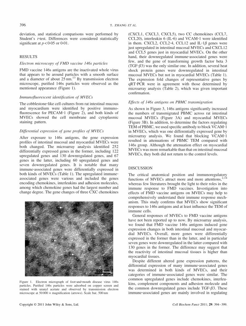
FMD vaccine 146s antigens are the inactivated whole virusthat appears to be around particles with a smooth surfaceand a diameter of about 25 nm.13 By transmission electronmicroscope, purified 146s particles were observed as thementioned appearance (Figure 1).
Immunofluorescent identification of MVECs
The cobblestone‐like cell cultures from rat intestinal mucousand myocardium were identified by positive immuno-fluorescence for PECAM‐1 (Figure 2), and both kinds ofMVECs showed the cell membrane and cytoplasmicstaining pattern.
Differential expression of gene profiles of MVECs
After exposure to 146s antigens, the gene expressionprofiles of intestinal mucosal and myocardial MVECs wereboth changed. The microarray analysis identified 252differentially expressed genes in the former, including 122upregulated genes and 130 downregulated genes, and 67genes in the latter, including 60 upregulated genes andseven downregulated genes. It is notable that manyimmune‐associated genes were differentially expressed inboth kinds of MVECs (Table 1). The upregulated immune‐associated genes were various and included the genesencoding chemokines, interleukins and adhesion molecules,among which chemokine genes had the largest number andchange degree. The gene changes of three CXC chemokines
Figure 1. Electron micrograph of foot‐and‐mouth disease virus 146sparticles. Purified 146s particles were adsorbed on copper screen andstained with uranyl acetate and observed by transmission electronmicroscope at 50 000 × magnification (arrows). Scale bar, 500 nm
Copyright © 2011 John Wiley & Sons, Ltd.
(CXCL1, CXCL3, CXCL5), two CC chemokines (CCL7,CCL20), interleukin 6 (IL‐6) and VCAM‐1 were identifiedin them. CXCL2, CCL2‐4, CCL12 and IL‐1β genes werejust upregulated in intestinal mucosal MVECs and CXCL12and CCL5 genes just in myocardial MVECs. On the otherhand, their downregulated immune‐associated genes werefew, and the gene of transforming growth factor beta 3(TGF‐β3) was the only similar one. In addition, several heatshock protein genes were downregulated in intestinalmucosal MVECs but not in myocardial MVECs (Table 1).The expression fold changes of representative genes byqRT‐PCR were in agreement with those determined bymicroarray analysis (Table 2), which was given importantconfirmation.
Effects of 146s antigens on PBMC transmigration
As shown in Figure 3, 146s antigens significantly increasedthe numbers of transmigrated PBMC across rat intestinalmucosal MVECs (Figure 3A) and myocardial MVECs(Figure 3B). In addition, to determine the factors regulatingTEM of PBMC, we used specific antibody to block VCAM‐1in MVECs, which was one differentially expressed gene bymicroarray analysis. We found that blocking VCAM‐1resulted in attenuations of PBMC TEM compared with146s group. Although the attenuation effect on myocardialMVECs was more remarkable than that on intestinal mucosalMVECs, they both did not return to the control levels.
DISCUSSION
The critical anatomical position and immunoregulatoryfunctions of MVECs attract more and more attentions,2,14
whereas few literatures brought the light to their roles in theimmune response to FMD vaccines. Investigation intoeffects of FMD vaccine antigens on MVECs may help tocomprehensively understand their immune response mech-anism. This study confirms that MVECs show significantresponses to 146s antigens and at least influence the TEM ofimmune cells.General responses of MVECs to FMD vaccine antigens
have not been reported up to now. By microarray analysis,we found that FMD vaccine 146s antigens induced geneexpression changes in both intestinal mucosal and myocar-dial MVECs. Overall, more genes were differentiallyexpressed in the former than in the latter, and in particularseven genes were downregulated in the latter compared with130 genes in the former. The difference may suggest thatthe reactivity of intestinal mucosal tissues is higher thanmyocardial tissues.Despite different altered gene expression patterns, the
differential expression of many immune‐associated geneswas determined in both kinds of MVECs, and theircategories of immune‐associated genes were similar. Thecommon upregulated genes include chemokines, interleu-kins, complement components and adhesion molecule andthe common downregulated genes include TGF‐β3. Theseimmune‐associated genes are mainly involved in regulating
Cell Biochem Funct 2011; 29: 394–399.

Figure 2. Immunofluorescent staining of platelet endothelial cell adhesion molecule‐1 (PECAM‐1) in microvascular endothelial cells (MVECs). (A) Ratintestinal mucosal MVECs. (B) Rat myocardial MVECs. MVECs were grown on round glass cover slips for 48 h and fixed with cold acetone. After 2‐hincubation with rabbit anti‐rat PECAM‐1 antibody and successive 1‐h incubation with fluorescein isothiocyanate ‐conjugated goat anti‐rabbit ImmunoglobulinG, cells were analysed by fluorescence microscopy. Scale bar, 50 µm
Table 1. Differentially expressed immune‐associated genes in MVECs induced by 146s antigens
Gene name Description
Fold change a
In intestinal mucosal MVECs In myocardial MVECs
Upregulated genesCxcl1 Growth regulated oncogene alpha (CXCL1) 9·31 2·71Cxcl2 Macrophage inflammatory protein 2 (CXCL2) —b 2·32Rn30026248 Macrophage inflammatory protein 2 beta (CXCL3) 9·65 4·65LOC60665 Small inducible cytokine b5 precursor (CXCL5) 16·32 4·58Rn30012491 Chemokine (C‐X‐C motif) ligand 12 3·86 —Ccl2 Monocyte chemoattractant protein 1 (CCL2) — 2·24Ccl3 Macrophage inflammatory protein 1 alpha (CCL3) — 3·09Ccl4 Macrophage inflammatory protein 1 beta (CCL4) — 2·08Ccl5 Chemokine (C‐C motif) ligand 5 6·75 —Rn30000167 Monocyte chemotactic protein 3 (CCL7) 2·12 2·16Ccl12 Monocyte chemoattractant protein 3 (CCL12) — 2·51Ccl20 Macrophage inflammatory protein 3 alpha (CCL20) 11·71 2·78Il1a Interleukin 1 alpha — 2·54Il1b Interleukin 1 beta — 3·62Il6 Interleukin‐6 4·49 3·22Vcam1 Vascular cell adhesion protein 1 3·69 3·02C1s Complement component 1, s subcomponent 2·19 —C3 Complement component 3 2·46 2·61C6 Complement component 6 — 2·45Csf1 Macrophage colony stimulating factor‐1 2·10 2·04Csf3 Granulocyte colony stimulating factor 3 3·38 —Downregulated genesTgfb3 Transforming growth factor beta 3 0·26 0·44Tgfb2 Transforming growth factor beta 2 0·44 —Hspa1a Heat shock 70 kda protein 1/2 0·45 —Rn30009378 Heat‐shock protein, beta‐7 0·48 —Hspa5 Heat shock 70 kda protein 5 0·49 —
MVECs, microvascular endothelial cells; FMD, foot‐and‐mouth disease.aRelative mRNA expression of MVECs in the treatment group (20 µgml−1 FMD vaccine 146s antigens) versus the control group.bChange magnitude was <2 and these genes could be considered as exhibiting no change.
397146s antigens induce endothelial responses
leukocyte migration and activation. It was reported thatleaderlessorwild typeFMDVinfectionupregulatedexpressionof some chemokine genes in embryonic bovine kidney cells,such as CCL2 and CXCL3.15 Chemokinesmay be involved inconferring cell‐mediated protection against FMDV.16 CXCchemokines are primarily in attraction of neutrophils, and CC
Copyright © 2011 John Wiley & Sons, Ltd.
chemokines have powerful chemoattractant formonocytes andT cells,17 as well as probably for dendritic cells and naturalkiller cells.18 Some chemokine gene changes were identifiedin just one kind of MVECs, whose implications still needfurther investigations. IL‐6 is also increasingly focused onowning to its immune activities, such as resolving innate
Cell Biochem Funct 2011; 29: 394–399.

Table 2. Quantitative RT‐PCR confirmation of representative genes from microarray analysis
Gene name
Fold change in intestinal mucosal MVECs Fold change in myocardial MVECs
qRT‐PCR a Microarray b qRT‐PCR a Microarray b
Cxcl1 10·84 9·31 2·65 2·71Ccl7 1·75 2·12 2·41 2·16Il6 4·45 4·49 1·66 3·22Vcam1 3·15 3·69 2·15 3·02Tgfb3 0·31 0·26 0·35 0·44
MVECs, microvascular endothelial cells; qRT‐PCR, quantitative reverse transcription‐polymerase chain reaction; GAPDH, glyceraldehyde‐3‐phosphatedehydrogenase; FMD, foot‐and‐mouth disease.aRelative expression abundance compared with GAPDH in the treatment group (20 µgml−1 FMD vaccine 146s antigens) versus the control group.bFold change in microarray analysis.
Figure 3. Effects of foot‐and‐mouth disease vaccine 146s antigens on transendothelial migration of peripheral blood mononuclear cell (PBMC). (A) Ratintestinal mucosal microvascular endothelial cells (MVECs). (B) Rat myocardial MVECs. MVECs were grown to confluence on collagen‐coated Transwellinserts with 5‐µm‐pore polycarbonate membranes. Endothelial monolayers were incubated with (146s group) or without (control group) 20 µgml−1 146santigens for 24 h or incubated with 20 µgml−1 146s antigens for 24 h and additional 10 µgml−1 anti‐rat vascular cell adhesion molecule 1 (VCAM‐1)antibody for the last 2 h (146s +VCAM‐1 Ab group). Then 100 µl of PBMC were added into the upper chamber, and after 6‐h incubation, the migratedPBMCs in lower chamber were collected in triplicate and counted. Data are expressed as mean ± standard deviation. Student’s t‐test, * p< 0·05 comparedwith control, ** p< 0·01 compared with control group, # p< 0·05 compared with 146s group
398 t. zhang ET AL.
immunity and promoting acquired immune responses.19 It wasreported that FMD vaccines could induce the elevation of IL‐6production in vaccinated animals,20 which may be partiallyattributed to MVECs considering our results here. In a word,MVECs may influence the immune response to 146s antigensby regulating the trafficking and activation of lymphocytes.In Transwell migration assays, the permeability of MVECs
to PBMC was just partially lowered by blocking VCAM‐1with its antibody. TEM of lymphocytes were related to manyfactors, such as adhesion molecules and chemokines.21
Therefore, the upregulation ofmany chemokine genes inducedby FMD vaccine 146s antigens may be another importantcontributor. On the other hand, antibody blockade of VCAM‐1 resulted in more remarkable reduction of PBMC transmi-gration across rat myocardial MVECs than intestinal mucosalMVECs (Figure 3), and the attenuation effect on the latter didnot reach statistical significance. The reason perhaps is thatthere are one or more other factors influencing the TEM ofPBMC in rat intestinal mucosal MVECs, which were notchanged at mRNA level by 146s antigens.Considering the species heterogeneity of MVECs,22 the
results from ratsmaynot be true to cloven‐hoof animals, suchas pigs, cattle and sheep. However, it is important that this
Copyright © 2011 John Wiley & Sons, Ltd.
studyprovidesnewideas forcomprehensivelyunderstandingthe immune response to FMD vaccine antigens. Furthercomparative studies of effects of FMD vaccine antigens andanother strong antigen on MVECs may help to clarify themechanism of its weak immunogenicity and explore newapproach to improving the efficacy of FMD vaccines.In conclusion, here we have paid attention to the response of
MVECs to FMD vaccine antigens and determined expressionchanges of many immune‐associated genes, some of whichinfluenced the TEM of PBMC. The result suggests thatMVECs play potential immunoregulatory roles in the immuneresponse to FMD vaccines, and it is worth further studies toexplore the approach to improving their efficacy.
CONFLICT OF INTEREST
The authors have declared that there is no conflict ofinterest.
ACKNOWLEDGEMENTS
This study was supported by Beijing Natural ScienceFoundation (No. 6061001) and National Natural Science
Cell Biochem Funct 2011; 29: 394–399.

399146s antigens induce endothelial responses
Foundation of China (No. 31001083). We thank LanzhouVeterinary Research Institute of Chinese Academy ofAgricultural Science for the FMDV‐infected cell culture.
REFERENCES
1. Doel TR. FMD vaccines. Virus Res 2003; 91: 81–99.2. Danese S, Dejana E, Claudio F. Immune regulation by microvascular
endothelial cells: directing innate and adaptive immunity, coagulation,and inflammation. J Immunol 2007; 178: 6017–6022.
3. Capel‐Edwards M. Foot‐and‐mouth disease in the brown rat. J CompPathol 1970; 80: 543–548.
4. Gong TL, Sun LJ, Mao JD, Ren JQ, Lian CY. Survey on foot andmouth disease. J Inn Mong Univ Nation, Nat Sci 2001; 16(4):393–396. (Article in Chinese with an abstract in English)
5. Doel TR, Chong WKT. Comparative immunogenicity of 146s, 75s,and 12s particles of FMDV. Arch Virol 1982; 73: 185–191.
6. Wang CY, Ma JW, Fu XP. Purification and examination of foot‐and‐mouth disease virus. Chin J Vet Sci Technol 2004; 34: 66–69.
7. Bradford, MM. A rapid and sensitive method for quantitation ofmicrogram quantities of protein utilizing the principle of protein‐dye‐binding. Anal Biochem 1976; 72: 248–254.
8. Rafiee P, Johnson CP, Li MS, et al. Cyclosporine A enhancesleukocyte binding by human intestinal microvascular endothelialcells through inhibition of p38 MAPK and iNOS. Paradoxicalproinflammatory effect on the microvascular endothelium. J Biol Chem2002; 277: 35605–35615.
9. Nishida M, Carley WW, Gerritsen ME, Ellingsen O, Kelly RA, SmithTW. Isolation and characterization of human and rat cardiacmicrovascular endothelial cells. Am J Physiol 1993; 264: H639–H652.
Copyright © 2011 John Wiley & Sons, Ltd.
10. Guo Y, Guo H, Zhang L, et al. Genomic analysis of anti‐hepatitis Bvirus (HBV) activity by small interfering RNA and lamivudine instable HBV‐producing cells. J Virol 2005; 79: 14392–14403.
11. van Gelder RN, von Zastrow ME, Yool A, Dement WC, Barchas JD.Eberwine JH. Amplified RNA synthesized from limited quantities ofheterogeneous cDNA. Proc Natl Acad Sci USA 1990; 87: 1663–1667.
12. Yang YH, Dudoit S, Luu P, et al. Normalization for cDNA microarraydata: a robust composite method addressing single and multiple slidesystematic variation. Nucleic Acids Res 2002; 30: e15.
13. Bachrach HL. Foot‐and‐mouth disease. Annu Rev Microbiol 1968; 22:201–244.
14. Biedermann BC. Vascular endothelium: checkpoint for inflammationand immunity. News Physiol Sci 2001; 16: 84–88.
15. Zhu J, Weiss M, Grubman MJ, De Los Santos T. Differential geneexpression in bovine cells infected with wild type and leaderless foot‐and‐mouth disease virus. Virology 2010; 404: 32–40.
16. Diaz‐San Segundo F, Moraes MP, De Los Santos T, Dias CCA,Grubman MJ. Interferon‐induced protection against foot‐and‐mouthdisease virus correlates with enhanced tissue specific innate immunecell infiltration and interferon stimulated gene expression. J Virol2010; 84: 2063–2077.
17. Rollins BJ. Chemokines. Blood 1997; 90: 909–928.18. Christopherson II K, Hromas R. Chemokine regulation of normal and
pathologic immune responses. Stem Cells 2001; 19: 388–396.19. Jones SA. Directing transition from innate to acquired immunity:
defining a role for IL‐6. J Immunol 2005; 175: 3463–3468.20. Cox SJ, Aggarwal N, Statham RJ, Barnett PV. Longevity of antibody
and cytokine responses following vaccination with high potencyemergency FMD vaccines. Vaccine 2003; 21: 1336–1347.
21. Fernandez‐Borja M, van Buul JD, Hordijk PL. The regulation ofleucocyte transendothelial migration by endothelial signalling events.Cardiovasc Res 2010; 86: 202–210.
22. Aird WC. Phenotypic heterogeneity of the endothelium: I. structure,function, and mechanisms. Circ Res 2007; 100: 158–173.
Cell Biochem Funct 2011; 29: 394–399.