Microtubule-targeted antitumor drugs - Gene Therapy & Molecular
46
Gene Therapy and Molecular Biology Vol 12, page 313 313 Gene Ther Mol Biol Vol 12, 313-357, 2008 Microtubule-targeted antitumor drugs: chemistry, mechanisms and nanoparticle formulations Review Article Teni Boulikas 1,2 *, Ioannis Tsogas 3 1 Regulon, Inc. 715 North Shoreline Blvd, Mountain View, California 94043, USA 2 Regulon AE, Afxentiou 7, Alimos, Athens 17455, Greece 3 Dendrigen AE, Afxentiou 3, Alimos, Athens 17455, Greece __________________________________________________________________________________ *Correspondence: Teni Boulikas, Ph.D., Regulon AE, Afxentiou 7, Alimos, Athens 17455, Greece; Tel: +30-210-9858454; Fax: +30- 210-9858453; e-mail: [email protected] Keywords: Microtubules, tubulin, vinca alkaloids, vinblastine, vinorelbine, vincristine, vindesine, vinflunine, taxanes, paclitaxel, docetaxel, epothilones A, B, D, ixabepilone, sagopilone, discodermolide, P-glycoprotein, dictyostatin, abraxane, colchicine, aplidine, nocodazole, dictyostatin, peloruside A, cyclostreptin, vinflunine, griseofulvin, nanoparticle Abbreviations: 2′,3′-cyclic nucleotide-3′-phosphodiesterase, (CNP); Breast Cancer Gene 1, (BRCA1); disease control rate, (DCR); Epidermal Growth Factor Receptor, (EGFR); Guanosine 5'-Triphosphate, (GTP); half maximal inhibitory concentration, (IC 50 ); high- density lipoprotein, (HDL); metastatic breast cancer, (MBC); Microtubules, (MTs); overall response rate, (ORR); renal cell carcinoma, (RCC); Retinoblastoma, (RB); response rate, (RR); vascular endothelial growth factor, (VEGF) Received: 30 September 2008; revised: 10 December 2008; Accepted: 19 December 2008 Electronically published: 19 December 2008 Summary The ingenious application of vinca alkaloids (vinblastine, vinorelbine, vincristine, vindesine, vinflunine) destabilizing microtubules and of taxanes (paclitaxel, docetaxel) stabilizing microtubules has been a milestone achievement in oncology. Recent investigations into their molecular mechanism revealed that all compounds possess additional pleiotropic effects that converge on induction of apoptosis in cancer cells via activation of signaling pathways. Their success has prompted vigorous investigations into microtubule-targeting activity from natural products as well as synthetic molecules arising from molecular modeling that led to the identification of Epothilones A, B, D, Ixabepilone, Sagopilone, Discodermolides, Dictyostatin, Peloruside A, and ABT-751; an additional purpose for epothilone drug discovery has been to bypass paclitaxel resistance mainly arising from the efflux function of P- glycoprotein. Nanoparticles provide a new mode of cancer drug delivery functioning as a carrier for entry through fenestrations in tumor vasculature. The 130-nm nanoparticle formulation albumin-bound paclitaxel (Nab- paclitaxel, Abraxane™) utilizes the natural properties of albumin to reversibly bind paclitaxel, transport it across the endothelial cell and concentrate in tumors; Abraxane™ received regulatory approval in the USA from a higher response rate and longer time to progression than Taxol in patients with metastatic breast cancer. The success of Abraxane™ led to an explosion in research on polymer nanoparticle formulations for taxanes using micellar PEGylated hyperbranched polyesters, polyglycerol-polyethylene glycol copolymers, cyclodextrin nanoparticles, polylactide-co-glycolide PEG and many others. Several such formulations are expected to enter the market. Other tubulin polymerization inhibitors reviewed here include tubulysin A, a highly cytotoxic peptide from myxobacteria, CC-5079, bisbenzylisoquinoline alkaloids, aplidine, nocodazole, GMC-5-193, cyclostreptin, colchicine, TLK-286 and vinflunine, a novel third generation vinca alkaloid. Structural similarities have been used to further modify the successful microtubule-targeted drugs in order to seek molecules of improved efficacy, of lower toxicity or able to overcome tumor resistance. We review the molecular mechanism of these drugs, whenever feasible, we suggest correlations between their chemical structure and mechanism and point to the importance of drug delivery for success. I. Introduction Cancer is one of the leading causes of death worldwide; it claimed 7.6 million deaths (13.1%) in 2005 out of 58 million deaths from all causes. In 2002, an estimated 6.72 million people worldwide were newly diagnosed with any the most 10 prevalent forms of solid cancers; of these, 4.15 million died within the same year. Based on projections, the death toll will rise to 9 million cancer deaths in 2015.
Transcript of Microtubule-targeted antitumor drugs - Gene Therapy & Molecular
Gene Therapy and Molecular Biology Vol 12, page 313
313
Gene Ther Mol Biol Vol 12, 313-357, 2008
Microtubule-targeted antitumor drugs: chemistry, mechanisms and nanoparticle formulations Review Article Teni Boulikas1,2*, Ioannis Tsogas3 1Regulon, Inc. 715 North Shoreline Blvd, Mountain View, California 94043, USA 2Regulon AE, Afxentiou 7, Alimos, Athens 17455, Greece 3Dendrigen AE, Afxentiou 3, Alimos, Athens 17455, Greece __________________________________________________________________________________ *Correspondence: Teni Boulikas, Ph.D., Regulon AE, Afxentiou 7, Alimos, Athens 17455, Greece; Tel: +30-210-9858454; Fax: +30-210-9858453; e-mail: [email protected] Keywords: Microtubules, tubulin, vinca alkaloids, vinblastine, vinorelbine, vincristine, vindesine, vinflunine, taxanes, paclitaxel, docetaxel, epothilones A, B, D, ixabepilone, sagopilone, discodermolide, P-glycoprotein, dictyostatin, abraxane, colchicine, aplidine, nocodazole, dictyostatin, peloruside A, cyclostreptin, vinflunine, griseofulvin, nanoparticle Abbreviations: 2′,3′-cyclic nucleotide-3′-phosphodiesterase, (CNP); Breast Cancer Gene 1, (BRCA1); disease control rate, (DCR); Epidermal Growth Factor Receptor, (EGFR); Guanosine 5'-Triphosphate, (GTP); half maximal inhibitory concentration, (IC50); high-density lipoprotein, (HDL); metastatic breast cancer, (MBC); Microtubules, (MTs); overall response rate, (ORR); renal cell carcinoma, (RCC); Retinoblastoma, (RB); response rate, (RR); vascular endothelial growth factor, (VEGF)
Received: 30 September 2008; revised: 10 December 2008; Accepted: 19 December 2008
Electronically published: 19 December 2008
Summary The ingenious application of vinca alkaloids (vinblastine, vinorelbine, vincristine, vindesine, vinflunine) destabilizing microtubules and of taxanes (paclitaxel, docetaxel) stabilizing microtubules has been a milestone achievement in oncology. Recent investigations into their molecular mechanism revealed that all compounds possess additional pleiotropic effects that converge on induction of apoptosis in cancer cells via activation of signaling pathways. Their success has prompted vigorous investigations into microtubule-targeting activity from natural products as well as synthetic molecules arising from molecular modeling that led to the identification of Epothilones A, B, D, Ixabepilone, Sagopilone, Discodermolides, Dictyostatin, Peloruside A, and ABT-751; an additional purpose for epothilone drug discovery has been to bypass paclitaxel resistance mainly arising from the efflux function of P-glycoprotein. Nanoparticles provide a new mode of cancer drug delivery functioning as a carrier for entry through fenestrations in tumor vasculature. The 130-nm nanoparticle formulation albumin-bound paclitaxel (Nab-paclitaxel, Abraxane™) utilizes the natural properties of albumin to reversibly bind paclitaxel, transport it across the endothelial cell and concentrate in tumors; Abraxane™ received regulatory approval in the USA from a higher response rate and longer time to progression than Taxol in patients with metastatic breast cancer. The success of Abraxane™ led to an explosion in research on polymer nanoparticle formulations for taxanes using micellar PEGylated hyperbranched polyesters, polyglycerol-polyethylene glycol copolymers, cyclodextrin nanoparticles, polylactide-co-glycolide PEG and many others. Several such formulations are expected to enter the market. Other tubulin polymerization inhibitors reviewed here include tubulysin A, a highly cytotoxic peptide from myxobacteria, CC-5079, bisbenzylisoquinoline alkaloids, aplidine, nocodazole, GMC-5-193, cyclostreptin, colchicine, TLK-286 and vinflunine, a novel third generation vinca alkaloid. Structural similarities have been used to further modify the successful microtubule-targeted drugs in order to seek molecules of improved efficacy, of lower toxicity or able to overcome tumor resistance. We review the molecular mechanism of these drugs, whenever feasible, we suggest correlations between their chemical structure and mechanism and point to the importance of drug delivery for success.
I. Introduction Cancer is one of the leading causes of death
worldwide; it claimed 7.6 million deaths (13.1%) in 2005 out of 58 million deaths from all causes. In 2002, an
estimated 6.72 million people worldwide were newly diagnosed with any the most 10 prevalent forms of solid cancers; of these, 4.15 million died within the same year. Based on projections, the death toll will rise to 9 million cancer deaths in 2015.

Boulikas and Tsogas: Microtubule-targeting anticancer drugs page
314
Chemotherapy, surgery and radiotherapy continue to be the mainstay treatments of cancer. Over 700 FDA-approved drugs have entered into clinical practice during the last 30 years; these are classified into six major groups that include (1) the platinum coordination complex, (2) antimicrotubule agents (vinca alkaloids, taxanes), (3) antimetabolites (methotrexate, fluoropyrimidines, cytocine arabinose, gemcitabine), (4) antitumor antibiotics (actinomycin D, mitomycin C, bleomycin, anthracyclines, podofylotoxines, camptothecines), (5) alkylating agents, and (6) others including a number of biological drugs or monoclonal antibodies that target specific pathways such as EGFR or angiogenesis.
Tubulin has been an attractive anticancer target since the dawn of medical oncology because of its role in chromatid separation in mitosis and the possibility of intervention at this step of the cell cycle with agents that stabilize tubulin polymers (microtubules). Microtubules (MTs) can be viewed as dynamically unstable tubulin polymers in equilibrium with monomers that interconvert stochastically between growing and shrinking states, a property central to their cellular functions. Each time a tubulin monomer is incorporated into the polymer, one GTP molecule is hydrolyzed. The GTPase activity of tubulin is enhanced by stathmin-like domains and colchicine and is inhibited by vinblastine and by the N-terminal part of stathmin-like domains. The residues involved in GTPase activity comprise the β-tubulin GTP binding site and α-tubulin residues that participate in intermolecular interactions in protofilaments (Wang et al, 2007).
We have previously reviewed platinum drugs that have been synthesized for cancer and entered into clinical trials, including successful nanoparticle formulations (Boulikas et al, 2007). We review here the class of antimicrotubule agents.
II. Microtubules (MTs) as dynamic structures involved in spindle formation
A. Microtubules and the centrosome The mitotic spindle is a specialized structure required
for exact chromosome segregation in mitosis. Spindle formation is dependent on the reorganization of interphase microtubule network, regulated by cell cycle-regulatory mechanisms. The centrosome (known as the microtubule organizing center) is involved in the formation of spindle poles during mitosis, which ensures the distribution of the correct number of chromosomes to daughter cells. Aberrant centrosome duplication could cause centrosome amplification and chromosomal instability.
In many animal cells, minus ends of microtubules are capped by the centrosome whereas plus ends are free and display dynamic instability. The role of the centrosome in microtubule dynamics was explored by studying microtubule behavior in cytoplasts from which the centrosome was removed. Fibroblast (CHO-K1) and epithelial (BSC-1) cells were investigated. Removal of the nucleus through centrifugation after cytochalasin/nocodazole treatment gave cytoplasts that either contained the centrosome and displayed a dense radial microtubule array (Figure 1, Middle pictures) or lacked the centrosome and displayed a loose and sparse network of randomly arranged microtubules (Figure 1, Right). The pattern of MT dynamics were evaluated by injecting intact cells with Cy3-tubulin, preparing cytoplasts, and acquiring digital-fluorescence time-lapse sequences of images. Living fibroblast cytoplasts containing or lacking centrosomes were readily distinguishable by the high density and radial arrangement of MTs invariably associated with the centrosome.
Figure 1. Distribution of microtubules in intact cells and cytoplasts. Intact cells (Left) and cytoplasts containing (Center) or lacking (Right) the centrosome were fixed and triple-stained with α-tubulin antibody for microtubules (red), γ-tubulin antibody for the centrosome (green, but superposition makes the spot appear yellow) and 4′,6-diamidino-2-phenylindole (DAPI) for the nucleus (blue). (Upper) Fibroblast CHO cells; (Lower) epithelial BSC-1 cells. In contrast to fibroblast cells, minus ends of MTs in epithelial cells did not depend on the centrosome for their stability. (Bar = 10 μm.) From "Rodionov V, Nadezhdina E, Borisy G (1999) Proc. Natl. Acad. Sci. USA. 96: 115-120. Copyright 1999 National Academy of Sciences, U.S.A."

Gene Therapy and Molecular Biology Vol 12, page 315
315
These studies suggested that a minus-end depolymerization mechanism functioned to eliminate errors in microtubule organization and that dynamic instability of plus ends was a result of capping of minus ends by the centrosome (Rodionov et al, 1999). Fluorescence imaging of microtubules in living cytoplasts with or without a centrosome is shown in Figure 2. In CHO cytoplasts containing the centrosome, most MTs (68%) displayed dynamic instability behavior with alternating phases of growth and shortening at their distal (plus) ends. In contrast, in cytoplasts lacking the centrosome, dynamic instability was never observed. In contrast to fibroblast cells, minus ends of MTs in epithelial cells did not depend on the centrosome for their stability.
B. Association of tubulin with plasma
membrane The bulk of cellular tubulin is cytoplasmic, but a
significant fraction is embedded in, or firmly associated with, the plasma membrane and other membranes. A possible linker protein for microtubules to the plasma
membrane as 2′,3′-cyclic nucleotide-3′-phosphodiesterase (CNP). The CNP is both prenylated and palmitoylated, providing hydrophobic domains for membrane intercalation. The association of CNP and tubulin in membrane and cytoskeletal compartments was shown with immunofluorescence. Fluorescent labeling of microtubules (green) and CNP (red) of FRTL-5 cells revealed patches of colocalization (yellow) of the two proteins at the membrane and subplasmalemmal region. The patches are interrupted by zones, containing only CNP. Microtubules extending toward the membrane show no association with CNP, but the perinuclear region again shows abundant overlap of the two proteins. Thus, CNP acts as a microtubule-associated protein and this activity resides in the C terminus of the enzyme. Submembranous colocalization of the proteins and CNP-dependent microtubule organization (Figure 3) suggest that CNP is a membrane-bound microtubule-associated protein that can link tubulin to membranes and may regulate cytoplasmic microtubule distribution (Bifulco et al, 2002).
Figure 2. Fluorescence imaging of microtubules in living cytoplasts. In centrosome containing cytoplasts (Top), microtubules (MTs) showed dynamic instability at their plus ends whereas free MTs rapidly shortened from their minus ends. In centrosome-free cytoplasts (Middle), MTs persistently grew at one end and shortened at the other end. Injection of tubulin at low concentration resulted in nonuniform incorporation of subunits along MTs and produced “speckles” along their length. Speckles remained stationary, indicating that the mechanism of translocation was treadmilling (Bottom). Black arrowheads point to shortening and white arrowheads to growing ends. White arrows point to speckles. Numbers indicate time in seconds. (Bars = 1 μm). From "Rodionov V, Nadezhdina E, Borisy G (1999) Proc. Natl. Acad. Sci. USA. 96: 115-120. Copyright 1999 National Academy of Sciences, U.S.A."
Boulikas and Tsogas: Microtubule-targeting anticancer drugs page
316
Figure 3. Colocalization of tubulin and CNP in FRTL-5 rat thyroid cells. Cells were grown on coverslips, fixed, and stained with a monoclonal anti-α-tubulin antibody [fluorescein (a)] or an antibody to CNP1 [rhodamine (b)]. (c) Double immunofluorescence (superimposition of a and b staining). (d) Enlarged membrane region from c. (e) Overlap of CNP and tubulin in a cytoskeleton prepared from a COS cell. Cells were fixed and incubated with monoclonal anti-α-tubulin antibody (Amersham Pharmacia) for 1 h, followed by tetramethylrhodamine isothiocyanate (TRITC)-labeled goat anti-mouse IgG (Sigma) for 1 h. Secondary antibodies for tubulin were either fluorescein- or rhodamine-labeled. From "Bifulco M, Laezza C, Stingo S, Wolff J (2002) Proc. Natl. Acad. Sci. U S A. 99: 1807-1812, Copyright 2002 National Academy of Sciences, U.S.A."
C. Tumor suppressor proteins are localized in centrosomes
The centrosome controls assembly of microtubules, a process that plays a central role in organizing cell structure, determining cell polarity, directing cell movement during interphase, and orchestrating formation of the bipolar spindle during mitosis. Furthermore, centrosomes play an important role in maintaining the fidelity of chromosome distribution during cell division. Loss of these functions might cause chromosomal instability and aneuploidy. Centrosome amplification drives chromosomal instability in tumor development.
Tumor suppressor proteins such as p53 and retinoblastoma (RB) have been localized to the centrosome in a cell cycle-dependent manner. A number of proteins involved in the G2/M checkpoint were also found to be associated with centrosomes during cell cycling, e.g., cyclin B, p34cdc2, and 14-3-3. Poly(ADP-ribose) polymerase 1 involved in opening up chromatin for DNA repair and transcription and able to bind to DNA strand breaks was also found to be localized to the centrosomes during mitosis and was suggested to be involved in maintenance of chromosomal stability (Kanai et al, 2007).
BRCA1, a suppressor of tumorigenesis in breast and ovary, is a protein of 1,863 amino acids with an N-terminal zinc-ring domain, a C-terminal transactivation domain and two putative nuclear localization signals. BRCA1 is involved in progression of the cell cycle. Its expression and phosphorylation are cell cycle dependent and its overexpression induces growth arrest or apoptosis. BRCA1 plays a continuous role throughout the cell cycle: expression and phosphorylation is induced at G1/S
transition, and a complex with Rad51 is associated with chromosomes during S phase and participates in DNA repair. BRCA1 is a component of RNA polymerase II and a coactivator of p53-mediated transcription, and may thus regulate the expression of other genes required for cell cycle progression.
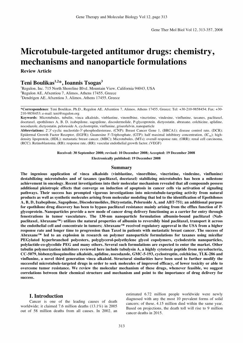
BRCA1 was associated with the centrosome during mitosis. This was shown by coimmunostaining of COS-7 cells with mouse monoclonal γ-tubulin antibody and rabbit polyclonal BRCA1 antibody (Figure 4). Two BRCA1-specific antibodies were used: MS110, a mouse mAb raised against a BRCA1-GST fusion protein containing amino acids 1-304 of human BRCA1 protein, and C-20, a rabbit polyclonal antibody raised against a peptide corresponding to amino acids 1843-1862 of human BRCA1. A series of double-staining experiments have confirmed the localization of BRCA1 protein at mitotic centrosomes using C-20 and MS110 for BRCA1 staining, γ-tubulin and pericentrin antibodies for centrosome staining, and γ-tubulin antibody for microtubule staining. In addition, DAPI staining of DNA was used to locate the nucleus. Double-staining of COS-7 cells with γ-tubulin antibody and BRCA1 C-20 antibody provided direct evidence for the presence of BRCA1 protein at mitotic centrosomes. Two-color (BRCA1+γ-tubulin) or three-color (BRCA1+γ-tubulin+DAPI) composite images showed colocalization of the BRCA1 and γ-tubulin signals at mitotic centrosomes (Figure 4). The concentration of BRCA1 signal at mitotic centrosomes was apparent from prometaphase to metaphase (Figure 4A) and early anaphase (Figure 4B). BRCA1 staining diminished at the centrosome as cells proceeded to late anaphase (Figure 4C). Results were similar when COS-7 cells were

Gene Therapy and Molecular Biology Vol 12, page 317
317
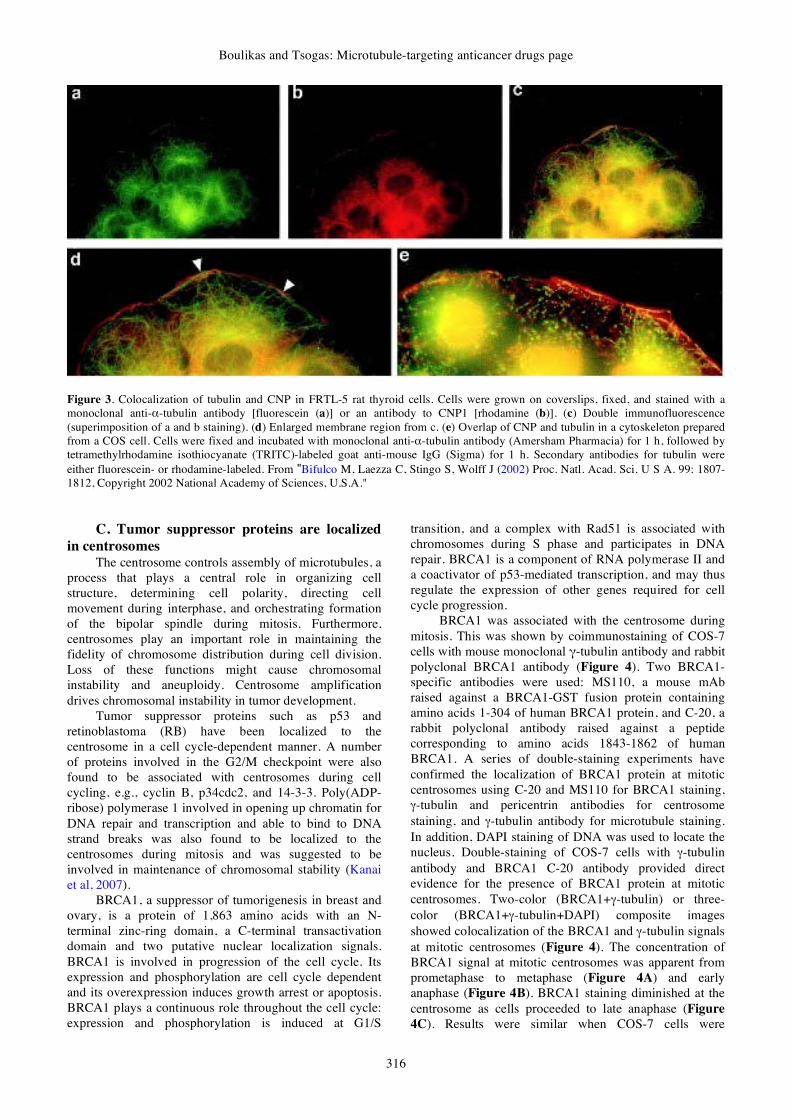
costained with the MS110 BRCA1-specific antibody (Figure 5) (Hsu and White, 1998).
A hypophosphorylated form of BRCA1, a suppressor of tumorigenesis in breast and ovary, localized with the centrosome during mitosis and coimmunoprecipitated with γ-tubulin, a centrosomal component essential for nucleation of microtubules (Figure 5). Immunofluorescence staining of a population of replicating COS-7 cells revealed the usual dot pattern in the nucleus. However, a unique staining pattern reminiscent of centrosomes was observed in mitotic cells. These data suggested that BRCA1 also plays a functional role with mitotic centrosomes (Hsu and White, 1998).
The cellular targets for estramustine, an antitumor drug used in the treatment of hormone-refractory prostate cancer, are believed to be the spindle microtubules responsible for chromosome separation at mitosis. Video microscopy showed that estramustine strongly stabilized growing and shortening dynamics at plus ends of bovine brain microtubules devoid of microtubule-associated proteins. The combined suppressive effects of vinblastine and estramustine on the rate and extent of shortening and dynamicity were additive. Thus, like the antimitotic mechanisms of action of the antitumor drugs vinblastine and taxol, the antimitotic mechanism of action of estramustine may be due to kinetic stabilization of spindle microtubule dynamics (Panda et al, 1997).
D. Microtubule-associated proteins A number of microtubule-associated proteins have
been reported; CLIPs are proteins known to associate specifically with the ends of growing microtubules, and CLASPs colocalize with CLIPs at the microtubule plus distal ends. CLASP1 localizes near the plus ends of growing spindle microtubules and is required for attachment of microtubules to kinetochore. hOrbit1 plays a role in polymerization of tubulin and interaction between microtubules (Figure 6) (Aonuma et al, 2005). Time-lapse fluorescence microscopy demonstrated that noncentrosomal MTs in cultured epithelial cells arise primarily by constitutive nucleation at, and release from, the centrosome. After release, MTs moved away from the centrosome and tended to depolymerizes (Keating et al, 1997). Aurora B is a protein kinase and a chromosomal passenger protein that undergoes dynamic redistribution during mitosis. Aurora B was found at centromeres at prophase but redistributed to the spindle midzone and became concentrated at the equator along midzone microtubules. Depolymerization of microtubules inhibited the dissociation of aurora B from centromeres at early anaphase and caused the dispersion of aurora B from the spindle midzone at late anaphase (Murata-Hori et al, 2002).
Figure 4. Coimmunostaining of COS-7 cells with mouse monoclonal γ-tubulin antibody and rabbit polyclonal BRCA1 antibody C-20. (A) a prometaphase to metaphase cell; (B) an early anaphase cell, and (C) a late anaphase cell. The colocalized signals of BRCA1 and γ-tubulin are yellow. DAPI counterstained DNA. Arrows indicate the positions of centrosomes. From "Hsu LC, White RL (1998) Proc. Natl. Acad. Sci. U S A. 95: 12983-12988, Copyright 1998 National Academy of Sciences, U.S.A."
Boulikas and Tsogas: Microtubule-targeting anticancer drugs page
318
Figure 5. COS-7 and E6/BE46 cells were costained with mouse monoclonal BRCA1 antibody MS110 (a) and rabbit polyclonal pericentrin antibody 4B (b); with MS110 (d) and rabbit polyclonal BRCA1 antibody C-20 (e); with mouse monoclonal γ-tubulin antibody (g) and C-20 (h); or with mouse monoclonal α-tubulin antibody (j) and C-20 (k). c, f, i, and l include DAPI counterstain of DNA. a-f highlight mitotic COS-7 cells; g-i are interphase COS-7 cells; j-l illustrate a mitotic E6/BE46 cell. Arrows indicate the positions of centrosomes. From "Hsu LC, White RL (1998) Proc. Natl. Acad. Sci. U S A. 95: 12983-12988, Copyright 1998 National Academy of Sciences, U.S.A."

Gene Therapy and Molecular Biology Vol 12, page 319
319
Figure 6. Localization of GFP-hOrbit1 NF (GFP-hOrbit1 N-terminal fragment) and microtubules. (A-D) Immunofluorescent images of GFP-hOrbit1 NF expression cell. Cells were cultured for 16 h after transfection. Microtubules immunostained with anti-α-tubulin antibody, and DNA stained with DAPI. GFP-hOrbit1 NF image (A), microtubule image (B) and DNA image (C) were merged (D). In the merged image, the yellow color indicates colocalization of GFP-hOrbit1 NF and microtubules. (E) Immunofluorescent images of control cells. The cells without GFP-hOrbit1 NF transfection were immunostained with anti-α-tubulin antibody, and DNA was stained with DAPI. Microtubule image (red) and DNA image (blue) were overlaid. Bar represents 10 μm. From: Aonuma M, Miyamoto M, Inoue YH, Tamai K, Sakai H, Kamasawa N, Matsukage A (2005) Cell Struct Funct. 30: 7-13, with kind permission from the Japanese Society for Cell Biology The orbit encodes Orbit/Mast, a 165-kDa microtubule-associated protein (MAP) with GTP-binding motifs; hypomorphic mutations in the Drosophila orbit gene cause abnormal chromosome segregation (Aonuma et al, 2005). Two human homologues of the Orbit/Mast, CLASP1 (hOrbit1) and CLASP2 (hOrbit2) have been identified. Using an antibody, the 150 kDa CLASP1/hOrbit1 polypeptide was found to be associated with microtubules. Figure 6 shows the subcellular localization of GFP-hOrbit1 after trasfection of cells with a plasmid expressing a fusion protein of the putative microtubule-binding domain (1-662 out of 1289 residues) of hOrbit1 with GFP. Confocal laser scanning microscopic observation revealed that the GFP-fluorescence associated with short and thin filaments in the perinuclear region during the short period after plasmid transfection, and colocalized with only part of the microtubules. GFP fluorescence was later detected on the abnormally longer and thick bundles of microtubule filaments. Finally the bundles formed networks in the perinuclear region. The results suggest that the GFP-hOrbit1 N-terminal fragment (GFP-hOrbit1 NF) binds to the newly formed microtubules rather than the pre-formed ones (Aonuma et al, 2005).
III. Taxanes When taxanes (paclitaxel, docetaxel) were introduced
as anticancer agents some 20 years ago, their broad spectrum of activity was striking. Taxanes were shown to be microtubule-targeting agents, able to stabilize tubulin polymers but also to disrupt additional cellular processes and to induce apoptosis (reviewed by Rowinsky and Calvo, 2006). A plethora of clinical trials was set to optimize the different ways drugs can be administered; for example, the addition of cisplatin or carboplatin to paclitaxel resulted in higher response rates than for each of the drugs as single agents (reviewed by Ranson and Thatcher, 1999).
Antimicrotubule agents appear to act also by induction of apoptosis by convergence of several signaling pathways ending with caspase activation and chromatin fragmentation. A functional p53 protein and expression of the apoptosis-promoting protein, bax are the most studied pathways for drug-induced apoptosis. Necrosis can be assessed by Annexin binding and propidium iodide permeability in aqueous medium (Serafin and Bohm, 2005).

Boulikas and Tsogas: Microtubule-targeting anticancer drugs page
320
A. Paclitaxel Paclitaxel (Taxol, Onxol) is a naturally occurring
taxane molecule that inhibits depolymerization of tubulin in the spindle apparatus thus inducing apoptosis in dividing cells. It is FDA approved for salvage therapy in ovarian cancer and in both metastatic and adjuvant setting in breast cancer. It is also used in lung, head and neck and bladder cancers. Figure 7 shows its chemical structure and with its differentiating features from docetaxel marked in red.
B. Docetaxel Docetaxel (Taxotere, Sanofi) is a semisynthetic
taxane, from a class of compounds that inhibit the mitotic spindle apparatus by stabilizing tubulin polymers, leading to death of mitotic cells. FDA has approved it for metastatic breast cancer and first and second line treatment for non-small cell lung cancer. However, clinical experience is increasing in ovarian cancer, prostate cancer, stomach cancer and other epithelial neoplasms and it is likely to get approvals for additional indications.
C. Molecular mechanism of taxanes The mechanism of action of taxanes against tumor
cells is by alteration of microtubule dynamics, which causes cell-cycle arrest during mitosis. Docetaxel binds to the microtubules with a higher affinity than paclitaxel, and over a broader range of cell-cycle activities. It has also been shown to promote apoptosis via Bcl2 phosphorylation.
Recently, it has been shown that Bcl2 may protect cancer cells from apoptosis induced by a variety of anticancer agents. The precise mechanism of the Bcl2-induced multi-drug resistance is unknown. Microtubule-stabilizing agents such as paclitaxel and docetaxel have antimitotic and apoptosis-inducing activity. Human leukemic, breast cancer, and prostate cancer cells exposed to paclitaxel express a phosphorylated form of Bcl2 and
undergo apoptosis, suggesting that phosphorylation of Bcl2 may inhibit Bcl2 function. In addition, Bcl2 phosphorylation appears to inhibit its binding to Bax, since less Bax was observed in an immunocomplex with Bcl2 in taxol-treated cancer cells. Overexpression of Bcl2 counteracts the apoptotic effects of low doses of paclitaxel but has no effect against high doses. Furthermore, microtubule-damaging drugs such as paclitaxel induced apoptosis, caused growth arrest in G2/M phase of the cell cycle, induced caspase 3 activation as well as poly(ADP-ribose) polymerase (PARP) degradation, but did not induce p53 (Srivastana et al, 1998).
A model showing the binding of Taxol with tubulin is shown in Figure 8.
Incubation of cells with high Taxol concentrations leads to the formation of stable bundles of microtubules that disrupt the normal polymerization/depolymerization cycle of microtubules and results in the arrest of cells in the G2/M phase of the cell cycle. In addition to massive bundle formation, increased microtubule polymer mass is observed after treatment with high concentrations of Taxol. However, low concentrations of Taxol (10 nM) inhibit mitosis in HeLa cells by suppressing microtubule dynamics rather than by altering the microtubule polymer mass or inducing bundle formation. The mitotic block induced by 10 nM Taxol is sufficient to induce apoptosis. These observations in HeLa cells indicate that Taxol-induced cell death may result from different mechanisms depending on drug concentration. Taxol alters specific intracellular signal transduction events. It has been reported that Taxol induces the production of cytokines, interleukin-1, and tumor necrosis factor α (TNF-α) and increases tyrosine phosphorylation of proteins, including mitogen-activated protein kinase. Prolonged exposure of cells to Taxol induces DNA fragmentation, characteristic of apoptotic cell death, and it has been suggested that Raf-1 is a mediator of Taxol-induced apoptosis.
O
O NH
O
O
H3C
HO
O
OOH
O
O
CH3
O
O
OH
OH
H
Paclitaxel Docetaxel
H
O
O NH
O
O
H3C
O
O
OOH
O
O
CH3
O
OOH
OH
H
H
O
Figure 7. Structure of Paclitaxel and Docetaxel; their differentiating features are marked in red.

Gene Therapy and Molecular Biology Vol 12, page 321
321
Figure 8. Taxol binding site on mammalian β-tubulin. The location of residues Lys-19, Val-23, Asp-26, His-227, and Phe-270 are indicated and are shown in dark gray. Labels on Taxol (gray) denote the following: I, C3′ phenyl ring; II, C3′ benzamido phenyl ring; and III, C2 benzoyl phenyl ring. Specific regions of β-tubulin that form the binding pocket are labeled, including α-helices H1, H7, H9, and H10, the β-strands B7-B10, and the B7-H9 M-loop. From "Gupta, Jr. LM, Bode CJ, Georg, GI, Himes RH (2003) Proc. Natl. Acad. Sci. USA 100: 6394–6397, Copyright 2003 National Academy of Sciences, U.S.A." Raf-1 is known as an important intermediate in the transmission of proliferative and developmental signals, connecting upstream tyrosine kinases with downstream serine/threonine kinases. It is not clear whether Raf-1 activation is a consequence of disruption of the normal microtubule cytoskeleton and/or of activating the spindle mitotic checkpoint (Torres and Horwitz, 1998).
Paclitaxel is known to induce a proinflammatory response in macrophages; combination therapy with trastuzumab and paclitaxel for inflammatory local recurrence after breast conserving surgery has been suggested as a treatment of choice (Nomura et al, 2005). Paclitaxel might have a role in inhibiting angiogenesis and vascularization (Shnyder et al, 2005).
D. Indications of taxanes Paclitaxel is indicated as first-line treatment for
advanced ovarian carcinoma in combination with cisplatin; as first-line treatment of Non Small Cell Lung Cancer (NSCLC, stages IIIb and IV) in combination with cisplatin; for the adjuvant treatment of node-positive breast cancer administered sequentially to standard doxorubicin-containing combination; for the treatment of breast cancer after failure of combination chemotherapy for metastatic disease or relapse within 6 months of adjuvant chemotherapy; and for the second-line treatment of AIDS-related Kaposi's sarcoma.
In hormone-refractory prostate cancer, docetaxel has been studied as both a single agent and in combination with estramustine, and in different treatment schedules, with demonstrated efficacy. Two phase III trials have confirmed a survival benefit, making docetaxel the first chemotherapy agent with proven efficacy against prostate cancer (Mackler and Pienta, 2005).
In urothelial cancer, docetaxel has demonstrated activity and has been investigated as a single agent and in combination regimens. A phase III trial comparing docetaxel and cisplatin to methotrexate, vinblastine, doxorubicin, and cisplatin was inferior when evaluating response rates and overall survival. More recent phase II trials combining docetaxel with two additional agents have shown promise, but confirmatory trials are needed (Mackler and Pienta, 2005).
E. Side effects of taxanes The dose limiting effect of paclitaxel is
myelosuppression (mostly neutropenia) that can be reduced with shorter infusions of the same dose. Other common side effects are mucositis (especially after longer infusions), peripheral neuropathy (that increases with cumulative dose), acute neuromyopathy (that occurs for several days after infusion and could require opiate analgesics in order to control pain), cardiovascular side effects, including hypertension, hypotension, premature contractions, bradyarrhythmias and hypersensitivity

Boulikas and Tsogas: Microtubule-targeting anticancer drugs page
322
reactions to the drug including chest pain, dyspnea, urticaria, wheezing, hypotension (that can be reduced by premedication with corticosteroids and H1, H2 histamine receptor blockers). Alopecia is one of the expected side effects, whereas nausea, vomiting, diarrhea, liver toxicity and interstitial pneumonitis are uncommon (Saville et al, 1995; Blum et al, 2006; Langer et al, 2007).
The severe side effects of docetaxel include myelotoxicity, allergic reactions during infusion, diarrhea that can be severe in some patients, nausea and/or vomiting, hair loss occurring in most patients (including the hair on head, underarm hair, pubic hair, eyebrows, and eyelashes), fatigue in about 10% of patients, and muscle pain that is rarely severe in about 20% of the infusions. Rash occurs commonly but is severe in about 5% of patients. About half of patients feel numbness, tingling, or burning sensations in their hands and feet.
Myelosuppression and alopecia are universal side effects of docetaxel with myelosuppression being the dose limiting effect. Edema and fluid accumulation, including pleural effusions and ascites are common and can be dose limiting. Fluid accumulation can be partially prevented with corticosteroid treatment before and after each cycle of docetaxel. Mild sensory or sensorimotor neuropathy is common. Mucositis and diarrhea are common and usually mild. Hypersensitivity reactions are uncommon and can be prevented through premedication with corticosteroids and antihistamines. Rash and elevated liver function tests are uncommon (Baker et al, 2004; Georgoulias et al, 2005).
Taxanes require pre-medication and may cause important side effects such as febrile neutropenia and neuropathy. Neuropathy is a major adverse effect of microtubule-stabilizing agent-based chemotherapy, with severe peripheral neuropathy (grade 3 or 4) occurring in as many as 30% of patients treated. Neuropathy usually presents as sensory neuropathy and is more common with paclitaxel than docetaxel; it depends on the drug dose per treatment cycle, the schedule of treatment, and the duration of the infusion (reviewed by Lee and Swain, 2006).
F. Taxane drug resistance The majority of initially responsive breast cancer
patients treated with taxanes eventually develop resistance to taxanes (acquired resistance) and a non-negligible percentage of patients are primarily resistant to these agents (de novo resistance). Taxane drug resistance is caused by the drug efflux pump protein, P-glycoprotein. P-glycoprotein, produced by the multidrug resistance-1 gene (mdr-1), is a main mechanism developed by cancer cells to guard against anticancer drugs. Alterations of DNA methylation of the mdr-1 gene promoter are known to be linked to mdr-1 gene expression and are probably related to intracellular S-adenosyl-methionine.
Overexpression of P-glycoprotein is associated with resistance to taxanes, but not ixabepilone, in vitro. Obviously, different functional groups on the paclitaxel (or docetaxel) molecules are involved in tubulin binding and in interaction with P-glycoprotein; the similarity in molecular structures between taxanes on one hand and epothilones in the other is shown by molecular modeling
(Figure 9). However, mutations in β-tubulin are also linked to resistance to taxanes but not epothilones in vitro (Pusztai, 2007). Resistance against paclitaxel also correlates with an increase in the relative abundance of tubulin isoform βIII; the mode of recognition and the mechanism of stabilization of paclitaxel with the type I and III isoforms of β-tubulin are different; no preference for any of the two isoforms can be detected for epothilone A known to bypass paclitaxel resistance (Magnani et al, 2006).
Figure 9. Structural similarities between paclitaxel and epothilone B. Paclitaxel structure is shown in grey and epothilone B is superimposed in yellow (A), while in (B) Paclitaxel is the yellow structure and epothilone B is the grey.

Gene Therapy and Molecular Biology Vol 12, page 323
323
G. Mechanisms of taxane drug resistance One mechanism of resistance to taxanes involves
mutations, especially L and V substitutions at proline 220 of β-tubulin. Indeed, expression of tubulin containing the P220L and P220V mutations from expression vectors reduced microtubule assembly, conferred resistance to paclitaxel and epothilone A (microtubule-stabilizing drugs), but increased sensitivity to colcemid and vinblastine (microtubule-destabilizing drugs). An important aspect of these studies is that different substitutions at the same amino acid residue in β1-tubulin can confer cellular resistance to either microtubule-stabilizing or microtubule-destabilizing drugs (Yin et al, 2007). Additional mutations involved in resistance to paclitaxel are the L215, L217, and L228 in the H6/H7 loop region of β1-tubulin as shown in Chinese hamster ovary cells selected in the presence of paclitaxel.
IV. Nab-paclitaxel (ABI-007,
Abraxane) Taxane delivery systems through tumor cell surface
receptor-targeted delivery mechanisms such as small-molecule peptides and monoclonal antibodies, as well as those on non-targeted procedures such as liposomes, nanostructures, and natural and synthetic polymers hold promise for improving the toxicity profiles and biodistribution of the drug.
Albumin is emerging as a versatile protein carrier for drug targeting and for improving the pharmacokinetic profile of peptide or protein-based drugs. Nab-paclitaxel (ABI-007, Abraxane) is a novel albumin-bound (nab) paclitaxel formulated into 130-nm particles. This differs from the more conventional formulation of Taxol, which uses cremophor (castor oil) to increase the solubility of paclitaxel and is responsible for side effects, especially neutropenia and peripheral neuropathy. Abraxane utilizes the natural properties of albumin to reversibly bind paclitaxel, transport it across the endothelial cell and concentrate it in tumors. The proposed mechanism involves an endothelial cell-surface albumin receptor (gp60) and an albumin-binding protein expressed by tumor cells and secreted into the tumor interstitium (secreted protein, acidic and rich in cysteine, SPARC). Thus, glycoprotein 60-mediated endothelial cell transcytosis of the albumin nanoparticles and tumor accumulation by binding to SPARC enhances the therapeutic efficacy of ABI-007 compared to the free drug. The albumin receptor-mediated paclitaxel-transport mechanism is analogous to the opening of a 'trapdoor' on the endothelial cell wall within blood vessels. This facilitates the passage of ABI 007 from the bloodstream via the blood vessels to the underlying tumor tissue (reviewed by Moreno-Aspitia and Perez, 2005; Gradishar, 2006).
Studies in rats have shown that ABI-007 differs from paclitaxel formulated as Taxol, with a higher plasma clearance and a larger volume of distribution. The same study also showed that fecal excretion was the main elimination pathway with both formulations (Sparreboom et al, 2005).
Preclinical xenograft studies comparing ABI-007 and Taxol showed that both caused tumor regression and prolonged survival in various tumors; the order of sensitivity was lung > breast congruent with ovary > prostate > colon. The LD50 and Maximum Tolerated Dose (MTD) for ABI-007 were 47 and 30 mg/kg/d and for Taxol were 30 and 13.4 mg/kg/d, respectively. At equitoxic dose, the ABI-007-treated groups showed more regressions and prolonged survival supposedly from a higher intratumoral accumulation of ABI-007; finally ABI-007 exhibited enhanced endothelial cell binding and transcytosis compared to Taxol (Desai et al, 2006).
A. Dose-limiting toxicity (DLT) of Abraxane Clinical studies have shown that nab-paclitaxel has
almost double the response than paclitaxel formulated as Cremophor EL (Taxol) with an increased time to disease progression and increased survival in second-line patients. Nab-paclitaxel showed a lower rate of severe neutropenia compared to Taxol. Also the combination of ABI-007 and carboplatin may have significant activity in a variety of tumor types including non-small and small cell lung cancer, ovarian cancer, and breast cancer. However, life-threatening toxicities with metastatic breast cancer and hepatic insufficiency have been observed (Lee Villano et al, 2006).
In a Phase I study ABI-007 was administered in three different schedules in combination with carboplatin at AUC of 6 on day 1. Myelosuppression was the primary dose limiting toxicity. Responses were seen in melanoma, lung, bladder, esophageal, pancreatic, breast cancer, and cancer of unknown primary among 41 patients. The MTD of ABI-007 was 300 mg/m2 administered on day 1 every 21 days; 100 mg/m2 administered on days 1, 8, and 15 every 28 days; and 125 mg/m2 administered on days 1 and 8 every 21 days (Stinchcombe et al, 2007). In a different Phase I dose-escalating study the dose-limiting toxicity (DLT), which occurred in 3 of 6 patients treated at level 3 (375 mg/m2), consisted of sensory neuropathy (3 patients), stomatitis (2 patients), and superficial keratopathy (2 patients). The MTD was thus determined to be 300 mg/m2 (level 2). Identified features of clinical interest of ABI-007, included rapid infusion rate, absence of requirement for steroid premedication, and a high paclitaxel MTD (Ibrahim et al, 2002).
B. Maximum tolerated dose (MTD) of
Abraxane In a Phase I study the MTDs for heavily and lightly
pretreated patients were 100 and 150 mg/m2, respectively; and the dose-limiting toxicities were grade 4 neutropenia and grade 3 peripheral neuropathy, respectively (Nyman et al, 2005).
An interim analysis from a more recent randomized Phase II trial suggested that weekly nab-paclitaxel was more effective and safer than either 3-weekly nab-paclitaxel or 3-weekly docetaxel. The superior efficacy of nab-paclitaxel was presumably due to the improved safety profile, which allows for the administration of higher doses, a greater proportion of which actually reaches the tumor (reviewed by Henderson and Bhatia, 2007).
Boulikas and Tsogas: Microtubule-targeting anticancer drugs page
324
Additional studies include a phase II trial of ABI 007 in metastatic breast cancer patients who have failed taxane therapy evaluating a weekly rather than 3-weekly regimen and a multicentre phase II trial in patients with metastatic melanoma to evaluate both chemotherapy-naive patients (at a dose of 150 mg/m2 administered weekly) and to pretreated patients (at a dose of 100 mg/m2 administered weekly). ABI 007 is also being evaluated for the treatment of NSCLC, ovarian and cervical cancers.
C. Clinical development of Abraxane A multicenter phase II study was designed to
evaluate the efficacy and safety of Abraxane 260 mg/m2 without premedication every 3 weeks in NSCLC patients; ORR was 16%; the disease control rate (ORR plus stable disease) was 49% and the 1-year survival was 45%. Side effects included neuropathy, fatigue but no severe hypersensitivity reactions and no grade 4 treatment-related toxicity (Green et al, 2006).
Paclitaxel albumin-bound particles improved outcomes when compared against single agent cremophor-based paclitaxel; addition of bevacizumab (10 mg/kg), gemcitabine (1000 mg/m2) or both agents to abraxane (100mg/m2) also improved outcomes in clinical trials with manageable peripheral neuropathy and thrombocytopenia (Lobo et al, 2007).
A multicenter phase II study in MBC on 63 women at the M.D. Anderson Cancer Center, Houston was the prelude of the pivotal Phase III trial. ABI-007 at 300 mg/m2 was given to previously-treated or chemonaive patients. The overall response rates was 48% but for the group of patients who received ABI-007 as first-line treatment, the response rate was 64% and for the group of patients who received ABI-007 as second- or third-line treatment the response rate was 21%. Toxicities observed were typical of paclitaxel and included grade 4 neutropenia (24%), grade 3 sensory neuropathy (11%), and grade 4 febrile neutropenia (5%). Median time to disease progression was 26.6 weeks, and median survival was 63.6 weeks (Ibrahim et al, 2005).
In a randomized trial that formed the basis of its regulatory approval in the USA, 3-weekly nab-paclitaxel induced a higher response rate and longer time to progression than Taxol in patients with metastatic breast cancer. Except for grade 3 sensory neuropathy, nab-paclitaxel was also safer. The pivotal phase III study was performed to confirm preclinical studies demonstrating superior efficacy and reduced toxicity of ABI-007 compared with standard paclitaxel. ABI-007 at 260 mg/m2 was given intravenously without corticosteroid premedication compared to 175 mg/m2 Taxol with premedication both in 3-week cycles and demonstrated significantly higher response rates compared with standard paclitaxel (33% versus 19%, respectively) and significantly longer time to tumor progression (23.0 v 16.9 weeks, respectively). The incidence of grade 4 neutropenia was significantly lower for ABI-007 compared with Taxol (9% v 22%, respectively). Grade 3 sensory neuropathy was more common in the ABI-007 arm than in the standard paclitaxel arm (10% v 2%, respectively) (Gradishar et al, 2005).
American Pharmaceutical Partners (APP) secured exclusive North American marketing and manufacturing rights to ABI 007 in November 2001 and formed Abraxis Oncology for marketing purposes; APP is the owner of the US patent No. 6,506,405, which has 89 claims covering compositions of matter and unit dosage forms and patent No. 5,780,653 which covers three next-generation taxane anticancer compounds. In addition, the patent covers methods of use without the requirement for pretreatment with steroid therapy or growth factor support. The US FDA granted fast-track status to ABI 007 for metastatic breast cancer in January 2003. In September 2003, APP and American BioScience jointly announced positive interim results from the trial, which shows that the primary efficacy objective had been exceeded. Enrollment was completed in December 2002 with 460 first- and second-line metastatic breast cancer patients enrolled. A Data Monitoring Committee concluded in October 2002 that a sample-size adjustment of the phase III trial was not required and that the study could be continued to completion.
D. Targeting to tumors in animals Tumor-homing peptides were used to target abraxane
to tumors in mice. The targeting was accomplished with two peptides, CREKA and LyP-1 (CGNKRTRGC). The CREKA pentapeptide binds to clotted plasma proteins and homes to tumors because interstitial tissue of tumors and the vessel walls contain clotted plasma proteins, whereas the vessels in normal tissues do not. Fluorescein (FAM)-labeled CREKA-abraxane, injected intravenously into mice bearing human cancer xenografts, accumulated in tumor blood vessels, forming aggregates that contained red blood cells and fibrin. FAM-LyP-1-abraxane co-localized with extravascular islands expressing its receptor, p32. Untargeted FAM-abraxane was detected in the form of a faint meshwork in tumor interstitium. LyP-1-abraxane produced a statistically highly significant inhibition of tumor growth compared with untargeted abraxane in the xenografts (Karmali et al, 2008).
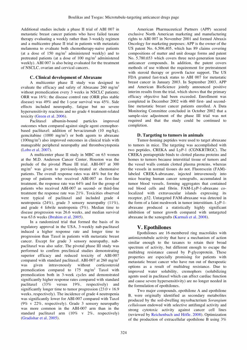
V. Epothilones Epothilones are 16-membered ring macrolides with
antimicrotubule activity that have a mechanism of action similar enough to the taxanes to retain their broad spectrum of activity, but different enough to escape the multidrug resistance caused by P-glycoprotein. These properties are especially promising for patients with metastatic breast cancer who have run out of therapeutic options as a result of multidrug resistance. Due to improved water solubility, cremophors (solubilizing agents used in paclitaxel which can affect cardiac function and cause severe hypersensitivity) are no longer needed in the formulation of epothilones.
Two major compounds, epothilone A and epothilone B, were originally identified as secondary metabolites produced by the soil-dwelling myxobacterium Sorangium cellulosum endowed with selective antifungal activity and strong cytotoxic activity against cancer cell lines (reviewed by Reichenbach and Höfle, 2008). Optimization of the production of extracellular epothilone B using 3%

Gene Therapy and Molecular Biology Vol 12, page 325
325
calcium alginate carrier for immobilization of Sorangium cellulosum has been described able to produce 5.03 mg/l/day (Park et al, 2007). Additional anticancer substances appear to be present in Sorangium cellulosum strains isolated in China (Guo et al, 2007). Epothilones are potential anticancer drugs that stabilize microtubules by binding to tubulin in a manner similar to paclitaxel. Cytochrome P450epoK (P450epoK), a heme containing monooxygenase involved in epothilone biosynthesis in the myxobacterium Sorangium cellulosum, catalyzes the epoxidation of epothilones C and D into epothilones A and B, respectively.
A. Epothilone drug discovery Epothilones are anticancer agents with a taxane-like
mechanism of action that have demonstrated activity in taxane-resistant tumors. Marine invertebrate animals such as sponges, gorgonians, tunicates and bryozoans are under intense investigation as sources of medicinal natural products.
Non-taxane microtubule-stabilizers of diverse chemical structures, including the epothilones and discodermolide have been advanced in chemical synthesis and been promoted to clinical trials (reviewed by Cardoso et al, 2008). Non-taxane microtubule-stabilizers of diverse chemical structures show promising preclinical activities and several are progressing through clinical trials (Mooberry, 2007).
The structural diversity of the epothilone group of clinical compounds is rather limited, as their structures show little divergence from the original natural product leads. A series of epothilone-derived macrolactones were elaborated to discover improved versions some of which had potent antiproliferative activities, promoted tubulin polymerization and induced mitotic arrest; changes were at the natural epoxide geometry from cis to trans, the incorporation of a conformationally constrained side chain, the removal of the C3-hydroxyl group, and the replacement of C12 with nitrogen. Interesting from many points of view appeared to be the 12-aza-epothilone ("azathilone" 40) especially because it lies outside of the general scope of Nature's biosynthetic machinery for polyketide synthesis but nevertheless retains most of the overall structural characteristics of a true natural product (Feyen et al, 2008).
B. Epothilones under clinical evaluation At least six epothilones are in clinical trials for
cancer treatment. Molecules under phase I or II clinical evaluation include epothilone B (patupilone; EPO960), epothilone D (KOS-862) and their second-generation (ixabepilone, BMS-310705, KOS-1584) and third-generation (ZK-EPO, ABJ-879) derivatives. Although similar in chemical structure, the epothilones demonstrated a striking difference in toxicity profile in phase I studies. Diarrhea was the dose-limiting toxicity (DLT) associated with patupilone, whereas neurotoxicity and neutropenia are the DLTs most commonly encountered with other epothilones (reviewed by Fumoleau et al, 2007). Epothilones have demonstrated activity in lung, ovarian, breast, prostate, and renal carcinomas and in non-
Hodgkin's lymphoma in phase II studies. Response rates in taxane-refractory metastatic breast cancer are relatively modest; however, ixabepilone and patupilone have shown promising efficacy in hormone-refractory metastatic prostate cancer and in taxane-refractory ovarian cancer (reviewed by Larkin and Kaye, 2006).
C. Mechanism of action of epothilones Like taxol, epothilone B binds to the αβ-tubulin
heterodimer subunit. Once bound, the rate of αβ-tubulin dissociation decreases, thus stabilizing the microtubules. Furthermore, epothilone B has also been shown to induce tubulin polymerization into microtubules without the presence of GTP. This is caused by formation of microtubule bundles throughout the cytoplasm. Finally, epothilone B also causes cell cycle arrest at the G2/M transition phase, thus leading to cytotoxicity and eventually cell apoptosis (Balog et al, 1996).
Epothilone B and D enhanced the constitutional activation of nuclear factor-κB (NF-κB) via IκB degradation through IκB kinase (IKKα and IKKβ) activation, and this resulted in p50 and p65 translocation to the nucleus in SW620 colon cancer cells. Moreover, epothilone B and D increased the expressions Bax, p53, caspase-3, but reduced Bcl-2 expression. Cells treated with sodium salicylate, an IKK inhibitor, did not show epothilone-induced cell growth inhibition or p50 translocation. These studies have unraveled additional mechanisms of epothilone-induced apoptosis in a tubulin polymerization-independent manner (Lee et al, 2007).
D. Epothilone B (patupilone; EPO960) Patupilone (Figure 10) is a novel tubulin-
polymerizing agent with activity against paclitaxel-resistant cell lines. Patients with refractory solid tumors received fixed doses of gemcitabine (1,000 or 750 mg/m2) along with escalating doses of patupilone (1.5-3 mg/m2) on days 1 and 8 of a 21-day cycle. The recommended phase II dose was gemcitabine 750 mg/m2 and patupilone 1.5 mg/m2 on days 1 and 8 of a 21-day cycle. DLTs were grade 3 asthenia and grade 3 dehydration but also asthenia and persistent nausea were observed (Schelman et al, 2008).
The principal mechanism of the epothilone class is inhibition of microtubule function (Goodin et al, 2004). Microtubules are essential to cell division, and epothilones therefore stop cells from properly dividing. Epothilone B possesses the same biological effects as taxol both in vitro and in cultured cells. This is due to the fact that they share the same binding site, as well as binding affinity to the microtubule. Epothilone B binds to the αβ-tubulin heterodimer subunit. Once bound, the rate of αβ-tubulin dissociation decreases, thus stabilizing the microtubules. Furthermore, epothilone B has also been shown to induce tubulin polymerization into microtubules without the presence of GTP. This is caused by formation of microtubule bundles throughout the cytoplasm. Finally, epothilone B also causes cell cycle arrest at the G2/M transition phase, thus leading to cytotoxicity and eventually cell apoptosis (Balog et al, 1996). Epothilone B has similar biological properties to Epothilone A.
Boulikas and Tsogas: Microtubule-targeting anticancer drugs page
326
O
O
HO
OH
N
S
O O
NH
O
HO
OH
N
S
O O
H3C
H3C
O
O
HO
OH
N
S
O O
H3C
O
HO
OH
N
S
O O
O
HO
OH
N
S
O O
Epothilone B Ixabepilone Sagopilone
Epothilone D KOS-1584
Figure 10. Structures of Epothilone B, Ixabepilone, Sagopilone, Epothilone D and KOS-1584. Structural differences are marked red. However, Epothilone B is 10-fold more potent than Epothilone A against P-glycoprotein-expressing multidrug resistant (MDR) cells. (-)-Epothilone B is similar to paclitaxel in binding displacement, and a substitution for paclitaxel in dependent cell growth. Epothilone B causes cell cycle arrest (IC50=3.5 nM).
Epothilone B shares the same or an overlapping binding site with paclitaxel and has been reported to have the same effects as paclitaxel on purified microtubules in vitro and on mitotic spindle structure and function in cultured cells. Epothilone B and paclitaxel alter the same microtubule dynamic parameters and to a similar extent. At the IC50 for mitotic arrest, dynamicity was reduced by 54% by paclitaxel compared with 62% for epothilone B. In addition, no anaphase or telephone figures were observed at the IC50 for both drugs, and in 65% of the cells treated with paclitaxel, the microtubules were completely stabilized compared with 80% for epothilone B. Thus, the effects of epothilone B on microtubule dynamics are remarkably similar to those of paclitaxel, suggesting that both drugs work by the same mechanism to induce mitotic block. Epothilone B overcomes P-glycoprotein-mediated paclitaxel resistance and is more water-soluble than paclitaxel (Klamath and Jordan, 2003).
Epothilone B is a 16-membered polyketide macrolactone with a methylthiazole group connected to the macrocycle by an olefinic bond (Molnár et al, 2000). Cytochrome P450epoK (P450epoK), a heme containing monooxygenase involved in epothilone biosynthesis in the myxobacterium Sorangium cellulosum, catalyzes the epoxidation of epothilones C and D into epothilones A and B, respectively. The epothilones are positioned with the macrolide ring roughly perpendicular to the heme plane and I helix, and the thiazole moiety provides key interactions that very likely are critical in determining substrate specificity. Interestingly, there are strong
parallels between the epothilone/P450epoK and paclitaxel/tubulin interactions (Nagano et al, 2003).
E. Ixabepilone (BMS-247550) Ixabepilone (BMS-247550, azaepothilone B) is a
semi-synthetic, microtubule stabilizing epothilone B analogue developed by Bristol-Myers Squibb (Stachel et al, 2000), which is more potent than taxanes and has displayed activity in taxane-resistant patients. Like all epothilones, Ixabepilone binds to tubulin and promotes tubulin polymerization and microtubule stabilization, thereby arresting cells in the G2/M phase of the cell cycle and inducing tumor cell apoptosis.
Ixabepilone demonstrated antineoplastic activity against taxane-resistant cell lines. FDA approved ixabepilone in 2007 (IXEMPRA, Bristol) for the treatment of aggressive metastatic or locally advanced breast cancer based on two multicenter trials on 878 patients using IXEMPRA either as a monotherapy or in combination with capecitabine. A single-arm monotherapy Phase II enrolled 126 patients with metastatic or locally advanced breast cancer resistant to three prior therapies (an anthracycline, a taxane and capecitabine). The primary endpoint was objective response rate and gave a partial response of 12.4% in 113 response-evaluable patients. Treatment-related non-hematological adverse events (greater than or equal to 20%) included: peripheral sensory neuropathy 62% (Grade 3/4: 14%), fatigue/asthenia 56% (Grade 3/4: 13%), myalgia/arthralgia 49% (Grade 3/4: 8%), alopecia 48% (Grade 3/4: 0%), nausea 42% (Grade 3/4: 2%), stomatitis/mucositis 29% (Grade 3/4: 6%), vomiting 29% (Grade 3/4: 1%), diarrhea 22% (Grade 3/4: 1%), and musculoskeletal pain 20% (Grade 3/4: 3%). Treatment-related hematological adverse events (greater than or equal to 20%) included: neutropenia (Grade 3/4: 54%) and leukopenia (Grade 3/4: 49%).
Gene Therapy and Molecular Biology Vol 12, page 327
327
A randomized Phase III trial evaluated the efficacy and safety of IXEMPRA in combination with capecitabine in comparison with capecitabine as monotherapy. This trial included 752 patients who were previously treated with anthracyclines and taxanes, and whose tumors had demonstrated prior resistance to these therapies. Evaluation of the primary endpoint demonstrated that IXEMPRA in combination with capecitabine resulted in a statistically significant improvement in progression-free survival compared to capecitabine monotherapy - median 5.7 vs. 4.1 months. Adverse events included neutropenia (Grade 3/4: 68%), leukopenia (Grade 3/4: 57%), peripheral neuropathy 65% (Grade 3/4: 21%), palmar-plantar erythrodysesthesia (hand-foot) syndrome 64% (Grade 3/4: 18%), fatigue/asthenia 60% (Grade 3/4: 16%), and others.
A randomized phase II trial in 92 chemotherapy-naive patients with progressive castrate metastatic prostate cancer treated with ixabepilone either as a single agent or in combination with estramustine phosphate reported a PSA RR of 48% in the ixabepilone arm and 69% in the combination arm, with an acceptable safety profile (Galsky et al, 2005). The predominant toxicities in the patients treated with ixabepilone alone or in combination with estramustine phosphate included grade 3/4 neutropenia (22% vs. 29%, respectively), grade 3 neuropathy (13% vs. 7%), and grade 3 fatigue (9% vs. 9%). A study of single-agent ixabepilone (SWOG S0111) reported a PSA RR of 33% in 42 evaluable patients with HRPC (Hussain et al, 2005). Although there were no confirmed objective responses in the 20 patients with measurable disease, 1 patient (5%) achieved an unconfirmed complete response, and 2 patients (10%) achieved an unconfirmed PR. Estimated median PFS was 6 months (95% CI, 4-8 months), and median survival was 18 months (95% CI, 13-24 months). A phase II trial conducted by the Eastern Cooperative Oncology Group (E3803) is evaluating the efficacy of ixabepilone in patients with no prior chemotherapy as well as in patients who have received ≤ 2 prior regimens, and initial findings from a noncomparative phase II trial evaluating ixabepilone and mitoxantrone/prednisone in patients with taxane-refractory pretreated Hormone Refractory Prostate Cancer (HRPC) were reported recently (Small et al, 2006). Eligible patients were randomized to second-line treatment with ixabepilone or mitoxantrone/prednisone, and, upon progression, patients were allowed to cross over to the alternate regimen for third-line treatment. Clinical outcomes of patients with taxane-resistant HRPC who were treated with subsequent chemotherapy were also reported recently. A total of 82 evaluable patients were treated with ixabepilone or mitoxantrone/prednisone. Median OS from protocol entry was 12.5 months for the patients in the ixabepilone arm and 13.0 months for those in the mitoxantrone/prednisone arm. Seventeen percent of the patients in the ixabepilone group and 20% of those in the mitoxantrone/prednisone group achieved a second-line PSA decline (≥ 50%). One of the 23 evaluable patients receiving second-line ixabepilone and 1 of the 20 evaluable patients receiving second-line mitoxantrone/prednisone achieved a PR. Neutropenia was
the most frequent grade 3/4 adverse event associated with second line therapy (41% in the ixabepilone arm, 56% in the mitoxantrone/prednisone arm). Third-line crossover therapy occurred in 39% of the patients treated with ixabepilone and 68% of those treated with mitoxantrone/prednisone. Third-line therapy yielded a PSA decline (≥ 50%) in 3 of the 25 patients (12%) treated with ixabepilone and 4 of the 13 patients (31%) treated with mitoxantrone/prednisone.
Vascular smooth muscle cells (VSMCs) form the lining of arterial walls and their abnormal proliferation is linked with atherosclerosis and restenosis after angioplasty. Epothilone B has been used in preventing postangioplasty restenosis. Epothilone B treatment of VSMCs caused a significant decrease in the level of cyclin-dependent protein kinase (CDK) 2 and down-regulated the phosphorylation of retinoblastoma, which plays a critical role in cell cycle regulation. Furthermore, levels of p27, an inhibitor of cyclin E/CDK2 complex, were significantly increased in VSMCs treated with epothilone B, indicating that this might be a major molecular mechanism for the inhibitory effects of epothilone B on the proliferation and cell cycle of VSMCs (Lim et al, 2007).
The excretory pathways of ixabepilone were studied in Eight patients with advanced cancer who received 70 mg, 80 nCi of [14C]ixabepilone over 3 h; fecal excretion was 52.2% and urinary excretion was 25.1% (Beumer et al, 2007). Neuropathy has led to the uneven and slower than expected clinical development of ixabepilone. Single-agent ixabepilone also demonstrated robust antitumor activity in women with metastatic breast cancer that was resistant to taxanes, anthracyclines and capecitabine in phase II trials (reviewed by Pronzato, 2008).
A phase II study of ixabepilone monotherapy in metastatic renal-cell carcinoma (RCC) administered at a dose of 40 mg/m2 every 21 days did not give objective responses in the first 12 evaluable patients, but six patients showed stable disease for more than 18 weeks on therapy ensuring advancement in Phase III against RCC. Grade 3 adverse events included lymphopenia, neutropenia, leukopenia, diarrhea, and infection. Common grade 2 toxicities included alopecia, fatigue and anemia (Posadas et al, 2007).
F. Epothilone D (KOS-862) Epothilone D (KOS-862; 12,13-desoxyepothilone B)
was also isolated from the myxobacterium Sorangium cellulosum. The drug was shown to reduce neointimal hyperplasia after in vivo rat carotid artery injury; from the significantly decrease in the level of CDK2 protein and inhibition of the phosphorylation of Rb, it was concluded that the major molecular mechanism of Epothilone D may involve regulation of the cell cycle G1-checkpoint proteins (Kim et al, 2007).
Epothilone D is capable of causing mitotic arrest by stabilizing tubulin polymerization. Epothilone D has demonstrated in vitro cytotoxic activity in a panel of human cell lines, equipotent to that of paclitaxel. Epothilone D is more potent than paclitaxel in p-glycoprotein overexpressing cell lines that demonstrate

Boulikas and Tsogas: Microtubule-targeting anticancer drugs page
328
multiple drug resistant activities. Epothilone D has also been shown to be active in the androgen-independent PC-3 human prostate cancer cells with IC50 of 0.0128 μM. In vivo, Epothilone D has shown significant antitumor activity in a range of xenograft models, including those that are resistant to paclitaxel. The dose limiting toxicity for epothilone D in phase I studies has been neurologic including both central and peripheral neurologic toxicity. Approximately 75% of patients enrolled in phase I studies experienced at least one neurologic toxicity (Beer et al, 2007). Epothilone D is being tested in combination with trastuzumab (Herceptin) in patients with HER-2 metastatic breast cancer. Adverse events were mostly Grade 1 or 2, with three Grade 3 incidents of sensory neurological toxicities
G. KOS-1584 KOS-1584 (9,10-didehydroepothilone D) along with
KOS-862 (Epothilone D) are the most advanced epothilone drug candidates of Kosan Biosciences. KOS-1584 is a second-generation compound with increased potency, favorable tissue distribution, and ease of formulation. Kosan is developing its epothilone compounds in collaboration with Roche.
KOS-1584 has demonstrated in vitro cytotoxicity, more potent or equipotent to that of paclitaxel in a panel of human cell lines. KOS-1584 has shown significant antitumor activity in a range of xenograft models, including non-small cell lung cancer models resistant to paclitaxel and those expressing multidrug resistance. A long elimination half-life was seen in Phase I trials. KOS-1584 has demonstrated antitumor activity and favorable tolerability in Phase 1 trials in patients with solid tumors, including substantial tumor shrinkage measured by objective responses in non-small cell lung, ovarian and pancreatic cancers, as well as durable stable disease in many additional tumors.
In April 2008, Kosan initiated a Phase 2 trial of epothilone KOS-1584 in patients with non-small cell lung cancer who have previously received only one prior chemotherapy regimen. The primary endpoint of the Phase 2 trial is objective response rate. KOS-1584 will be administered via a 3-hour intravenous infusion weekly for two weeks out of every three weeks at a dose of 25 mg/m2.
H. Conformationally restrained epothilones Stereoselective total syntheses of two novel
conformationally restrained epothilone analogues has been
described using a convergent synthetic approach; the aim of this synthesis was the restriction of the mobility of the aromatic side chain. The C1-C8 sector remained unchanged, in view of its seemingly crucial role in biological activity. A single methylene bridge was introduced between C14 and C17 (Figure 11). The resulting cyclopentene moiety, which incorporated the C16-C17 double bond, was designed to rigidify the side chain while still permitting sufficient mobility of the pyridine ring to allow for the preferred N-atom orientation for a hydrogen bonding interaction with the receptor. These epothilone analogues showed strong and selective growth inhibitory activity against leukemia cell lines (Alhamadsheh et al, 2008).
I. Sagopilone The process of sagopilone development at Bayer
Schering Pharma is an interesting story. One day, an external researcher offered the company a substance he had discovered, which he considered as a possible anticancer medication but the offer was rejected. By the time interest developed a rival company had already secured the rights to the substance. Bayer Schering Pharma now owns sagopilone, an epothilone analogue, with a structure somewhat different to the naturally occurring one; chemical ingenuity was used to modify the structure deriving at 450 compounds and broadening the therapeutic window of natural epothilones. Approximately half of the compounds synthesized, differed only slightly from the prototype epothilone, were promising with respect to toxicity, therapeutic efficacy and development of resistance after treatment of cells in culture and ease of chemical synthesis. A total of 39 chemical steps were used to produce three initial subunits of Sagopilone, which were then used to construct the finished molecule. How could such a complex process ever be implemented cost-effectively on a large scale? Converting one step in the chemical process from the laboratory to production-scale (up scaling), normally takes a month and for the 39 steps of sagopilone synthesis mere up scaling would take more than three years. Acceleration of this process resulted in the production of 36 grams of the substance in six months, sufficient for the treatment of approximately 200 patients whereas further improvements and up scaling took place. Bayer is now the only company with a fully synthetic epothilone, sagopilone, in advanced clinical development.
O
HO
OHO O
O
HO
OHO O
N14
17
14
17
Figure 11. Structures of conformationally restrained epothilone analogues.

Gene Therapy and Molecular Biology Vol 12, page 329
329
The effectiveness of sagopilone, the first fully synthetic third generation epothilone in clinical development, against paclitaxel in a breast cancer bone metastasis model was evaluated in a recent study. The therapeutic effect of sagopilone and paclitaxel on tumor-induced osteolysis and tumor burden in athymic nude mice inoculated intracardially with MDA-MB-231(SA)/luc human breast cancer cells was investigated. On day 12 after tumor cell inoculation, animals were randomized into treatment groups according to the lesion size as measured by radiography. A single dose of sagopilone was administered on day 13 (10 mg/ kg, i.v.). Paclitaxel was given once daily on days 13-17 (9 mg/ kg, i.p.). At sacrifice (day 23), bone lesions were measured using radiography and micro-CT. Tumor cell dissemination was detected by bioluminescence imaging and histomorphometry. The effect of compounds on bone resorption was determined by measuring Tartrate-resistant acid phosphatase 5b (TRACP5b) in serum and by counting the number of osteoclasts on tumor-bone interface by histomorphometry. Radiographic analysis revealed that further tumor-induced bone destruction was merely reduced by paclitaxel, whereas sagopilone completely
prevented it. Sagopilone-treated animals had also higher bone volume compared to other groups as measured by micro-CT. A marked effect of sagopilone on tumor growth in bone was observed as reduced bioluminescence signal intensity in vivo. This was confirmed by histomorphometry in H&E-stained bone sections. Paclitaxel had no significant effect on tumor growth in bone. Sagopilone had significantly better effect on reducing bone destruction and tumor burden when compared to paclitaxel in a mouse model of established breast cancer bone metastasis (Strube et al, 2008).
J. Discodermolide and analogues Discodermolide (Figure 12) stabilizes microtubules
and blocks cells at the G2/M phase of the cell cycle in a manner similar to that of Taxol. Non-taxane microtubule-stabilizers including the epothilones and discodermolide, are progressing through clinical trials. An advanced synthesis for discodermolide was based on the readily available fermentation product oleandomycin (Parker and Wang, 2007).
O
HO
OH O
OH
OH
O
HN
O
O
3H
O
HO
OH O
OH
OH
O NH2
O
OH O
OH
NH2
OO
O
Discodermolide
C19-[4-(4-3H-benzoyl-phenyl)-carbamate]-discodermolide
Coumarin-derived discodermolide analogue
Figure 12. Structures of discodermolide, C19-[4-(4-3H-benzoyl-phenyl)-carbamate]-discodermolide and coumarin-derived discodermolide analogue. Structural differences are marked in red.

Boulikas and Tsogas: Microtubule-targeting anticancer drugs page
330
Discodermolide has been shown to inhibit the proliferation of human cells by arresting the cell cycle in G2/M phase. Discodermolide hyperstabilized microtubules, an event especially prevalent during cell division (Day et al, 1996). Hyperstabilization of the mitotic spindle causes cell cycle arrest and cell death by apoptosis. Over a variety of cell lines, activity has been measured at IC50 = 3-80 nM. Discodermolide competed with paclitaxel for microtubule binding, but with higher affinity and was also effective in paclitaxel- and epothilone-resistant cancer cells (Jordan, 2002). It has been reported that Discodermolide-treated breast carcinoma cells displayed spectacular rearrangement of the microtubule cytoskeleton, including extensive microtubule bundling. Microtubule rearrangement that occurred with 10 nM discodermolide required 1 μM Taxol. Discodermolide had equally impressive effects on tubulin assembly in vitro. Near-total polymerization occurred at 0 °C with tubulin plus microtubule-associated proteins (MAPs) under conditions in which Taxol at an identical concentration was inactive. Without MAPs and/or without GTP, tubulin assembly was also more vigorous with discodermolide than with Taxol under every reaction condition examined. Discodermolide-induced polymer differed from Taxol- induced polymer in that it was completely stable at 0 °C in the presence of high concentrations of Ca2+. In a quantitative assay designed to select for agents more effective than Taxol in inducing assembly, discodermolide had an EC50 value of 3.2 μM versus 23 μM for Taxol (Day et al, 1996; Amos et al, 2003). In a recent comparative study A549 human lung carcinoma cells were treated with discodermolide at concentrations higher than 8nM, accelerated cell aging was observed by a functional β-galactosidase activity at pH=6. When the cells were treated with IC50 concentrations of doxorubicin, Taxol and discodermolide, only the latter two resulted in aberrant mitosis. However only discodermolide produced an aging associated β-galactosidase activity (Horwitz et al, 2005).
Coumarin-derived discodermolide analogues (Figure 12) possessing equivalent antiproliferative activity to the natural product have been described; the complete C-1 to C-7 fragment was replaced with a coumarin moiety in these compounds (Shaw et al, 2007). Photoaffinity-labeled discodermolide analogues have been used to investigate their binding site in tubulin; all promoted microtubule polymerization in the absence of GTP. The analogue, C19-[4-(4-3H-benzoyl-phenyl)-carbamate] -discodermolide (Figure 12) had the highest extent of photoincorporation, it was more cytotoxic than discodermolide, could displace [3H]Taxol from microtubules, and bound amino acid residues 355-359, in β-tubulin, which is in close proximity to the Taxol binding site (Xia et al, 2006).
VI. Vinca alkaloids The Vinca alkaloids are a subset of drugs that are
derived from the periwinkle plant, Catharanthus roseus (also Vinca rosea, Lochnera rosea, and Ammocallis rosea). While it has been historically used to treat numerous diseases, it has most recently been employed for its anti-cancer properties. All vinca alkaloids are administered
intravenously (IV). After injection, they are eventually metabolized by the liver and excreted. They work in a cell-cycle specific manner, halting mitosis of affected cells and causing cell death. The mechanism of action involves binding to the tubulin monomers and keeping the microtubules (spindle fibers) from forming. These alkaloids also seem to interfere with cells' ability to synthesize DNA and RNA. Although the plant has medical uses, it can produce many serious side effects if smoked or ingested.
There are four major vinca alkaloids in clinical use: Vinblastine, vinorelbine, vincristine and vindesine.
A. Vinblastine Vinblastine (Velban, Velsar), a vinca alkaloid acts as
an inhibitor of tubulin polymerization and mitosis. This contrasts the stabilization of tubulin polymers by taxanes. It also seems to exert its anticancer properties by interfering with glutamic acid metabolism (specifically, the pathways leading from glutamic acid to the Krebs cycle and to urea formation). Vinblastine treatment suppresses angiogenesis (Albertsson et al, 2008).
Vinblastine (Figure 13), is approved by FDA for multiple hematologic and solid tumors. It is most often used in Hodgkin's disease, non-Hodgkin's lymphoma, breast cancer, and germ cell tumors. Because it is a vesicant, extravasation precautions are required during administration.
The major adverse effect of vinblastine is
hematologic toxicity which occurs much more frequently than with vincristine therapy. Leukopenia (granulocytopenia) occurs most commonly and is usually the dose-limiting factor in vinblastine therapy. Other side effects include nausea, vomiting and constipation, dyspnea, chest or tumor pain, wheezing and fever during administration. Some rare cases of syndrome of inappropriate antidiuretic hormone secretion as well as angina pectoris have been reported (Gobbi et al, 2005; Connors, 2005).
Vinblastine induced an accumulation of the G2/M cells with the appearance of aneugenic micronuclei during monocyte-macrophage differentiation induced in a monocytic cell line by phorbol 12-myristate 13-acetate (PMA). Actinomycin D on the contrary, induced under similar conditions a drastic reduction of the G2/M cells (Spano et. al, 2007). Apoptosis is not a major mechanism of cell death induced by etoposide, vinblastine and estramustine in prostate cell lines and appeared to be independent of p53 status and bax/bcl-2 expression (Serafin and Bohm, 2005).
1. Mechanisms of vinblastine Vinblastine is a microtubule-depolymerizing drug.
The X-ray structure of vinblastine bound to tubulin in a complex with the RB3 protein stathmin-like domain (RB3-SLD) showed that vinblastine introduces a wedge at the interface of two tubulin molecules and thus interferes with tubulin assembly; furthermore, EM studies showed that vinblastine induced tubulin self-association into spiral aggregates at the expense of microtubule growth in a

Gene Therapy and Molecular Biology Vol 12, page 331
331
reaction that appears to be regulated by the C-terminus of β-tubulin and is enhanced by GDP and GTP. Vinblastine also arrests the cell cycle in G2/M-phase by blocking mitotic spindle formation. Triggers Raf-1 activation, phosphorylation of bcl-2-family proteins, induction of p53 expression, and apoptosis in several tumor cell lines (Rai and Wolff, 1998). Vinblastine and the amino-terminal part of RB3-SLD binding sites share a hydrophobic groove on the alpha-tubulin surface that is located at an intermolecular contact in microtubules. Drugs designed to perturb microtubule dynamics by interfacial interference at this site of α-tubulin, can be an attractive anticancer target (Gigant et al, 2005).
A novel way of diminishing cellular resistance to
vinblastine has been suggested: Alterations in DNA methylation of the mdr-1 gene promoter are known to be linked to mdr-1 gene expression and related to intracellular S-adenosyl-methionine; cobalamin, a cofactor of methionine-synthase, downregulates mdr-1 gene expression, as well as P-glycoprotein expression and function, and significantly increases cytotoxicity of vinblastine (Marguerite et al, 2007).
Tubulin, the major protein of microtubules, is posttranslationally modified by palmitoylation; this modification is inhibited in vitro by stoichiometric levels of vinblastine; a low, clinically relevant dose of vinblastine, inhibited palmitoylation of tubulin in human leukemic lymphocytes, microtubules were disassembled and cells became apoptotic (Caron and Herwood, 2007).
Efflux of the P-glycoprotein substrate vinblastine sulfate through Caco-2 cell monolayers was reduced by polyoxyethylene 40 stearate (PS40) thus enhancing the antitumor activity of vinblastine (Luo et al, 2007).
2. Regimens containing vinblastine Advanced urothelial cancer is treated with
conventional frontline chemotherapy with gemcitabine and cisplatin; however, traditional or dose-dense methotrexate, vinblastine, doxorubicin and cisplatin regimens are also in current use (Sonpavde et al, 2008).
3. Preclinical regimens containing vinblastine A significant synergistic effect of vinblastine and
HSV-Tk/GCV "suicide" gene therapy mediated by human serum albumin (HSA)-associated lipoplexes has been found in cell cultures (Faneca et al, 2008).
4. Reactive oxygen species (ROS), vinblastine
and apoptosis Reactive oxygen species (ROS), such as superoxide
anion radicals (O2-) and hydrogen peroxide (H2O2) are
potentially harmful by-products of normal cellular metabolism; ROS is generated by all aerobic organisms and it seems to be indispensable for signal transduction pathways. Overproduction of reactive oxygen species initiate lethal chain reactions involving oxidation and damage to cellular structures, that are crucial for integrity and survival.
N
N
NH
H3CO
N
H3COOC
OH
OCOCH3HCOOCH3
H
C2H5
CH3
Vinblastine
N
N
NH
H3CO
N
H3COOC
OH
OCOCH3HCOOCH3
H
C2H5
CHO
C2H5
Vincristine
N
N
NH
H3CO
N
COOCH3
OH
OCOCH3HCOOCH3
H
C2H5
CH3
C2H5
Vinorelbine
N
N
NH
H3CO
N
COOCH3
OH
OHH
CONH2
H
C2H5
CH3
Vindesine
OH
C2H5
OH
C2H5
Figure 13. Structures of vinblastine, vinorelbine, vincristine and vindesine. Structural differences are marked in red.

Boulikas and Tsogas: Microtubule-targeting anticancer drugs page
332
Vinblastine, cisplatin, mitomycin C, doxorubicin, camptothecin, inostamycin, neocarzinostatin and other anticancer drugs exhibit antitumor activity via ROS-dependent activation of apoptotic cell death. The "oxidation therapy of cancer" has been attempted either by inducing the generation of ROS in solid tumors by delivery for example, H2O2, or by inhibiting the antioxidative enzyme (defense) system of tumor cells. However no successful and practical results were obtained probably because of the lack of tumor selective ROS delivery and in induction of severe side effects. Delivering PEGylated enzymes generating reactive oxygen species, such as xanthine oxidase and D-amino acid oxidase (DAO) is an interesting approach. In addition, a PEGylated zinc protoporphyrin (PEG-ZnPP) and a highly water soluble micellar formulation of ZnPP based on amphiphilic styrene maleic acid copolymer were prepared, which were potent inhibitors of heme oxygenase-1 for oxidation therapy of cancer (Fang et al, 2007).
Whereas antioxidative enzymes such as catalase and SOD are down-regulated in most solid tumors in vivo, heme oxygenase-1 is highly upregulated exerting antiapoptosis in tumors and therefore, its targeting by zinc protoporphyrin might find application as an anticancer drug (Fang et al, 2007).
B. Vinorelbine Vinorelbine (Navelbine, Figure 13) is a
semisynthetic vinca alkaloid that acts the same way as Vinblastine. Vinorelbine and mitoxantrone (a topoisomerase II inhibitor) have both been demonstrated to have significant antitumor activity in patients with breast cancer. Vinorelbine is approved from the FDA for the treatment of relapsed metastatic breast cancer and for NSCLC either as a single agent or combined with a platinating agent. It is a mild vesicant, requiring extravasation precautions. Myelosuppression -mostly leucopenia- is the dose limiting effect. Not significant nausea and vomiting are reported as well neuropathy -but milder than it is observed with vinblastine- and tumor pain during administration. Acute reaction, such as dyspnea, chest pain and wheezing has been reported during administration but can be prevented in some cases by premedication with corticosteroids (Zelek et al, 2001; Gridelli et al, 2003; Georgoulias et al, 2005).
1. Vinorelbine mechanisms In addition to exerting its antitumor activity by
interfering with the polymerization of tubulin, vinorelbine showed antiproliferative effects in human osteosarcoma cells inducing apoptosis without affecting phosphorylation of Bcl-2 but rather by down-regulation of cyclin D1 and up-regulation of p53 expression in wild-type cells; in p53 negative osteosarcoma cells, however, vinorelbine showed no alteration in cyclin D1 (Roncuzzi et al, 2006).
The total enthalpy change in dipalmytoylphosphatidylcholine (DPPC) bilayers was increased by vinorelbine indicating a partial interdigitation of the lipid alkyl chains. The presence of cholesterol in Vinorelbine-DPPC bilayers induced an obstruction of the
interdigitation, since cholesterol interrupts the upraise of enthalpy change (Koukoulitsa et al, 2006).
Vinorelbine has a number of side-effects that can limit its use: Lowered resistance to infection, bruising or bleeding, anemia, constipation, diarrhea, nausea, numbness or tingling in hands or feet (peripheral neuropathy), tiredness and a general feeling of weakness (asthenia), inflammation of the vein into which it was injected (phlebitis). Less common effects are hair loss and allergic reaction.
C. Vincristine Vincristine (Oncovin, Vincasar), (Figure 13), is
another vinca alkaloid compound that inhibits tubulin polymerization. As it is a vesicant it should be administered with extravasation precautions. It is FDA approved to treat acute leukemia, rhabdomyosarcoma, neuroblastoma, Wilm’s tumor, Hodgkin's disease and other lymphomas. Vincristine is administrated intravenously.
Peripheral neuropathy is the dose limiting side effect, because of this, people with neuromuscular disorders should steer clear of this drug if possible. Likewise, people with some forms of Charcot-Marie-Tooth syndrome should avoid vincristine. Other side effects include mild myelosuppression, constipation -rather commonly-, autonomic neuropathy, CNS toxicity, nausea and vomiting. There have been reported a few cases of acute cardiopulmonary or pain symptoms during administration as well as transient elevation of liver function tests (Pappo et al, 2007; Chow et al, 2006).
As the microtubule is a dynamic protein, constantly polymerizing and depolymerizing, vinca alkaloid poisoned dimers could easily be incorporated into the microtubule polymer, preventing further growth. The incorporation of vincristine onto the heterodimer is rapidly reversible, and appears to occur at two sites per tubulin dimer. At higher concentrations of drug, microtubular crystals are formed, consisting of two intertwined helices of tubulin. Arrests cell cycle in G2/M-phase by blocking mitotic spindle formation. Triggers Raf-1 activation, phosphorylation of bcl-2-family proteins, induction of p53 expression, and apoptosis in several tumor cell lines. Inhibits VEGF production in leukemia cell lines. MRP1 transports vincristine in an ATP- and GSH-dependent manner induces the destabilization of polymerized tubulin, by binding to a site recently localized on β-tubulin, and has a high affinity for the protein.
Accidental injection of vinca alkaloids into the spinal canal (intrathecal administration) is highly dangerous, with a mortality rate approaching 100%. The medical literature documents cases of ascending paralysis due to massive encephalopathy and spinal nerve demyelination, accompanied by intractable pain, almost uniformly leading to death; a handful of survivors were left with devastating neurological damage with no hope of recovery. Rescue treatments consist of washout of the cerebrospinal fluid and administration of protective medications. A significant series of inadvertent intrathecal vincristine administration occurred in China in 2007 when batches of cytarabine and methotrexate (both often used intrathecally) manufactured

Gene Therapy and Molecular Biology Vol 12, page 333
333
by the company Shanghai Hualian were found to be contaminated with vincristine.
D. Vindesine Vindesine (Eldisine and Fildesin) has a serum half-
life of about 24 hours and is administered at a dose of 3 milligrams per square meter of body surface. Its toxicity and side effects are similar to those of vinblastine. Vindesine (Figure 13) is used mainly to treat melanoma and lung cancers (carcinomas) and, with other drugs, to treat uterine cancers.
A study published in 2006 reported the effects of vindesine and gemcitabine on patients with advanced non-small cell lung cancer (NSCLC). 44 patients (36 males and 8 females with a median age of 70 years and a median Karnofsky performance score of 60) were recruited between January 1998 and June 2001. 9 patients (20.5%) were stage IIIB patients and 35 (79.5%) were stage IV patients. 20 patients (45.5%) had squamous carcinoma and 24 (54.5%) non-squamous carcinoma. The patients received gemcitabine 1000 mg/m2 and vindesine 3mg/m2 (max 5mg) on days 1 and 8 every 3 weeks, and were all evaluable for response and toxicity: 17 (38.6%) were partial responders, 17 (38.6%) experienced stable disease, and 10 (22.3%) progressive disease. Grade 3-4 anemia, neutropenia and thrombocytopenia were observed in, respectively, 6.8, 9.1 and 2.3% of the patients, and grade 2-3 fatigue, paresthesias and skin toxicity in, respectively, 11.4, 20.4 and 2.3%. After a median follow-up of 54 months, 43/44 patients died; median survival was 12 months, and a clinical benefit was observed in 54.5% of cases. Gemcitabine plus vindesine proved to be an active and well-tolerated schedule (Cetto et al, 2006).
E. Vinflunine Vinflunine is a novel third generation vinca alkaloid
with superior antitumor activity in preclinical models, a more favorable toxicity profile compared to the other vinca alkaloids and with radiosensitizing potential (Simoens et al, 2006, 2008). Vinflunine (Javlor) is the first fluorinated microtubule inhibitor and is obtained by semisynthesis using superacidic chemistry to selectively introduce two fluorine atoms at the 20' position of the catharanthine moiety (Figure 14).
Its mechanism of action differs from other members of the vinca alkaloid class in terms of tubulin-binding affinity, microtubule dynamics, spiral formation, and intracellular accumulation. Vinflunine altered adhesion site targeting by microtubules and suppressed the microtubule (+) end pause that occurs at adhesion sites during endothelial cell migration; it has, therefore, an antiangiogenesis potential (Honoré et al, 2008). The high antitumor activity of vinflunine is not well understood since it binds to tubulin with an overall affinity several-fold lower than that of vinblastine or vincristine; vinflunine suppressed calmodulin interaction with the microtubule-associated protein STOP (stable tubule only polypeptide); this finding and the different binding modes of vinflunine and vinblastine to calmodulin could explain the remarkable antitumor activity of vinflunine (Makarov et al, 2007).
On the basis of encouraging preclinical activity vinflunine has been selected for clinical development in patients with a wide spectrum of solid tumors. Clinically significant activity has been seen in phase II studies, mainly in the treatment of transitional cell carcinoma of the urothelial tract, non-small cell lung cancer, and carcinoma of the breast. Vinflunine is currently in phase III trial assessment in patients with (second line) transitional cell carcinoma of the urothelium and first-line advanced breast cancer. The efficacy of vinflunine in patients with advanced non-small cell lung cancer previously treated with a platinum-containing regimen was confirmed by a large phase III trial (Nguyen et al, 2008). In a phase II study the efficacy of vinflunine against malignant pleural mesothelioma (MPM) was assessed. Patients with a histologically confirmed diagnosis of MPM were eligible for enrollment onto this multicenter phase II trial if they had not received prior chemotherapy or radiotherapy and had measurable lesions by Response Evaluation Criteria in Solid Tumors (RECIST). Vinflunine 320 mg/m2 by 10-minute intravenous infusion was administered on day 1 of 21-day cycles. A response rate of 13.8% was found. The median survival was 10.8 months. The most common adverse events were anemia, neutropenia, fatigue, constipation, and nausea. Of grade 3 and 4 toxicities, neutropenia and constipation were the most common (45% and 9% of patients, respectively) (Talbot et al, 2007).
VII. Other tubulin polymerization
inhibitors A. Tubulysin A Tubulysin A (Figure 15A) is a highly cytotoxic
peptide from myxobacteria with antimitotic activity. Tubulysin A inhibited tubulin polymerization more efficiently than vinblastine, induced depolymerization of isolated microtubule preparations and triggered the apoptotic process (Figure 15B); electron microscopy studies showed that tubulysin A induced the formation of rings, double rings, and pinwheel structures. The mode of action of tubulysin A resembled that of peptide antimitotics dolastatin 10, phomopsin A, and hemiasterlin (Khalil et al, 2006).
N
N
NH
H3CO
N
H3COOC
OH
OCOCH3HCOOCH3
H
C2H5
CH3
H
F
F
Vinflunine
Figure 14. Structure of Vinflunine with the differences marked red.

Boulikas and Tsogas: Microtubule-targeting anticancer drugs page
334
N
HN
O
N
OOO
N
SNH
OO
OH
OH
O
O
Figure 15. (Top) Structure of Tubulisin A. (A, B) Tubulysin induces a depletion of microtubules. PtK2 potoroo kidney cells were cultured in the absence and presence of tubulysin A (59 nm) for 4 h. (A) The control sample shows the normal microtubular network (green) in interphase cells and two mitotic bipolar spindles. Chromosomes and nuclei are stained blue. (B) In tubulysin treated cells the microtubule network is less dense and the centrosomes become visible. The mitotic cell in the middle shows metaphase chromosomes and few longer microtubules that do not form a mitotic spindle. From Khalil MW, Sasse F, Lünsdorf H, Elnakady YA, Reichenbach H Mechanism of action of tubulysin, an antimitotic peptide from myxobacteria. Chembiochem. 2006, 7, 678-683. Copyright Wiley-VCH Verlag GmbH & Co.KGaA. Reproduced with permission.

Gene Therapy and Molecular Biology Vol 12, page 335
335
Tubulysins are cytotoxic peptides, which include 9 members (A-I). Tubulysin A has potential application as an anticancer agent. It arrests cells in the G2/M phase. Tubulysin A inhibits polymerization more efficiently than vinblastine and induces depolymerization of isolated microtubules. Tubulysin A has potent cytostatic effects on various tumor cell lines with IC50 in the picomolar range. Tubulysin A is a highly cytotoxic peptide with antimitotic activity that induces depletion of cell microtubules and triggers the apoptotic process. Treated cells accumulated in the G2/M phase. Tubulysin A inhibited tubulin polymerization more efficiently than vinblastine and induced depolymerization of isolated microtubule preparations. In competition experiments, tubulysin A strongly interfered with the binding of vinblastine to tubulin in a noncompetitive way (Reichenbach et al, 2006).
B. CC-5079 The synthetic compound CC-5079 (Celgene,
Summit, NJ) potently inhibits cancer cell growth associated with cell cycle arrest in G2/M phase. CC-5079 (Figure 16) prevents polymerization of purified tubulin, competes with colchicines (but not with paclitaxel or vinblastine) for binding to tubulin, and depolymerizes microtubules in cultured cancer cells. Furthermore, CC-5079 remains active against multidrug-resistant cancer cells, inhibits the enzymatic activity of phosphodiesterase type 4 and thus, inhibits tumor necrosis factor-α (TNF-α)
secretion from lipopolysaccharide-stimulated human peripheral blood mononuclear cells (Zhang et al, 2006).
C. Bisbenzylisoquinoline alkaloids Bisbenzylisoquinoline alkaloids (Figure 17) and
their derivatives are possible candidates as modifiers of MDR in cancer chemotherapy by enhancing the cytotoxic effect of vinblastine; partially synthesized compounds from fangchinoline and tetrandrine at 0.1 microM were as potent as 10 μM verapamil in enhancing the cyctotoxic effect of vinblastine in the resistant cell line P388/ADR; a compound with a 5,14-dibromotetrandrine group showed the strongest potentiation effect as an inhibitor of P-gp without affecting the expression of P-gp (Wang et al, 2005).
O
O O
O
N
Figure 16. Structure of CC-5079.
H3CN
OCH3
R1
OH
H
NCH3
H3CO
H3CO
HO
H3CN
OCH3
R1
O
H
NR3
H3CO
R2
HO
H3CN
OCH3
OCH3
O
H
NCH3
H3CO
HO
HO
OH
Figure 17. Structure of different bisbenzylisoquinoline alkaloids.

Boulikas and Tsogas: Microtubule-targeting anticancer drugs page
336
D. Halichondrin B and E7389 First isolated from Halichondria okadai and later
from the unrelated sponges Axinella carteri and Phankella carteri, halichondrin B (Figure 18) is a complex polyether macrolide, which has recently been synthesized and arrests cell growth at subnanomolar concentrations. In cell line screenings, halichondrin B showed a similar pattern of activity to other antitubulin drugs, suggesting its possible mode of action as an antimitotic agent, and this prediction was later confirmed experimentally. Halichondrin B is noncompetitive inhibitor of the binding of both vincristine and vinblastine to tubulin, suggesting that the drug binds to the vinca binding site, or a site nearby. The isolation of halichondrin B from two unrelated genera of sponge,
however, has led to speculation that it is in reality a microbial, rather than sponge metabolite, as sponges are known to support a wide range of microbes. If this is the case, fermentation technologies could provide a useful supply of the drug.
E7389 (Figure 18), which is in phase I and II clinical trials, is a synthetic macrocyclic ketone analogue of the marine sponge natural product halichondrin B; its main target seems to be tubulin and the microtubules by an end-poisoning mechanism that results predominantly in inhibition of microtubule growth, but not shortening and by induction of tubulin aggregates. E7389 may suppress mitosis by directly binding to microtubule ends as unliganded E7389 (Jordan et al, 2005).
O
O
O
O
O
O
O
O
O
O
O
O
O
O
OH
CH3
O
O
H
H
H
H
H
OH
HO
CH3
H
H H
H
CH3
H
CH3
H
H
H
Halichondrin B
O
O
O
O
O
O
O
O
O
H
CH3
H
H
O
H2N
HO
H3CO
E7389
Figure 18. Structure of Halichondrin B (left) and E7389 (right).
E. Aplidine Aplidine (plitidepsin, Figure 19) is a novel
anticancer drug isolated from the marine tunicate Aplidium albicans, currently in phase II clinical trials in a variety of solid tumors and hematologic malignancies. It displays a broad spectrum of antitumor activities, inducing apoptosis by triggering mitochondrial cytochrome c release, initiating the Fas/DC95, JNK pathway and activating caspase 3 activation. This agent also inhibits elongation factor 1-a, thereby interfering with protein synthesis, and induces G1 arrest and G2 blockade, thereby inhibiting tumor cell growth. Aplidine's mechanism of action involves several pathways, including cell cycle arrest, inhibition of protein synthesis and antiangiogenic activity. Phase I studies have been reported for a number of several schedules including 1-hour, 3-hour and 24-hour infusion. Evidences of antitumor activity and clinical benefit of aplidine in several tumor types were noted across phase I trials, particularly in advanced medullar thyroid carcinoma. Phase II studies are underway. Within the entire phase I program, dose-limiting toxicities of aplidine were neuromuscular toxicity, asthenia, skin toxicity, and diarrhea. Interestingly, no hematological toxicity was observed. Aplidine displayed a very peculiar delayed neuromuscular toxicity that was found to be closely related to the symptoms described in the adult form of carnitine palmitoyl transferase deficiency type 2, which is a genetic disease treated with L-carnitine. Consistently, concomitant administration of L-carnitine
allowed improving aplidine-induced neuromuscular toxicity. In summary, aplidine is a novel marine anticancer agent with a very particular delayed neuromuscular toxicity that requires careful follow-up with promising antitumor activity (Faivre et al, 2007). Aplidine generally lacks cross-resistance with other known cytotoxic drugs. Aplidine induces an early oxidative stress response, which results in a rapid and sustained activation of EGFR, of the nonreceptor protein tyrosine kinase Src, of the c-Jun NH2-terminal kinase, of the p38 mitogen-activated protein kinase; furthermore, Aplidine induces the cyclin-dependent kinase inhibitor p27kip1 (p27) through an
N
O
OO
N
NH
O
O NH
OH
O
O
O
HN
N
O
O
O
O
N
OCH3 Figure 19. Structure of Aplidine.

Gene Therapy and Molecular Biology Vol 12, page 337
337
oxidation-dependent mechanism. Because sensitivity to Aplidine correlates inversely with the levels of expression of p27, analysis of p27 levels are used to establish response to Aplidine in currently ongoing clinical trials (Moneo et al, 2007).
F. Nocodazole Nocodazole (Figure 20) is an antimitotic agent that
disrupts microtubules by binding to β�tubulin and preventing formation of one of the two interchain disulfide linkages, thus inhibiting microtubule dynamics, disruption of mitotic spindle function, and fragmentation of the Golgi complex. Arrests the cell cycle at G2/M phase. Prevents phosphorylation of the T cell antigen receptor and inhibits its activity. Stimulates the intrinsic GTPase activity of tubulin. Activates the JNK/SAPK signaling pathway and induces apoptosis in several normal and tumor cell lines (Ley et al, 1998).
G. GMC-5-193 GMC-5-193 (GMC) is a novel anticancer small-
molecule quinazolinone analogue with properties that possesses antimicrotubule activity. In a recent study a liposomal formulation for tumor-targeting delivery of GMC was prepared and systemically administered to enhance the anticancer effect of this compound and evaluate its bioefficacy. GMC was encapsulated within a cationic liposome, which was decorated on the surface with an anti-transferrin receptor single-chain antibody fragment (TfRscFv) as the tumor-targeting moiety. Confocal imaging of fluorescent GMC uptake in a human melanoma cell line, MDA-MB-435, showed higher cellular uptake of GMC when delivered via the liposomal formulation compared with free GMC. Delivery of GMC by the tumor-targeting liposomal formulation also resulted in a 3- to 4-fold decrease in IC50 values in human cancer cells DU145 (prostate) and MDA-MB-435, compared with the effects of GMC administered as free GMC. In addition, the GMC liposomal formulation increased the sensitivity of cancer cells to doxorubicin, docetaxel, or mitoxantrone by 3- to 30-fold. In the MDA435/LCC6 athymic nude mice xenograft lung metastases model, GMC was specifically delivered to tumors by the liposomal formulation (Chang et al, 2008).
H. Dictyostatin and 10,11-Dihydrodictyostatin
The total synthesis of 10,11-Dihydrodictyostatin (Figure 21) was achieved; the compound was found to retain potent antimitotic activity, with a comparable profile to discodermolide and Taxol, functioning by microtubule stabilization and G2/M arrest (Paterson et al, 2008). A recent study demonstrated that dictyostatin-1 (Figure 21) induced a strong G2/M block and accumulation of A549 cells in the S phase at concentrations as low as 10 nM. A large increase of sub-G1 population was also observed, indicative of cells undergoing active apoptosis. Paclitaxel induced similar changes at 10 nM, but these were not as extensive as those produced by dictyostatin-1. The microtubule morphology was also investigated, where it became evident that dictyostatin-1 promoted the rearrangement of microtubules into bundle formations similar to those induced by paclitaxel, although dictyostatin-1 had much more prominent effects at lower concentrations. The percentage of the microtubule matrix condensed into bundles increased with higher concentrations of dictyostatin-1, although there appeared to be little additional bundling at concentrations greater than 100 nM. Dictyostatin-1 also proved to be highly potent in two paclitaxel-resistant human cancer cell lines expressing active P-glycoprotein. Together, these results indicate that dictyostatin-1 is a potent inducer of tubulin polymerization and retains activity in cells expressing the P-glycoprotein efflux pump (Wright et al, 2003).
S
N
HN
O
NH
O
O
Figure 20. Structure of Nocodazole.
O O
OH
HO
OH
OH
11
10
O O
OH
HO
OH
OH
Figure 21. Structure of Dictyostatin (left) and 10,11-Dihydrodictyostatin (right).

Boulikas and Tsogas: Microtubule-targeting anticancer drugs page
338
I. Peloruside A Peloruside A (Figure 22) is a naturally occurring
compound isolated from a New Zealand marine sponge. Peloruside A binds to a unique site on the tubulin αβ-heterodimer, the laulimalide site, which is distinct from the taxoid site and thus differentiates it from paclitaxel, docetaxel, epothilone A, and discodermolide (Hamel et al, 2006). Peloruside A exhibited synergy with paclitaxel and epothilone A in cell proliferation assays (Wilmes et al, 2007). Peloruside, unlike paclitaxel, does not induce murine macrophages to produce the proinflammatory mediators interleukin-12p40 (IL-12p40), tumor necrosis factor-α (TNF-α), and nitric oxide; thus, peloruside may prove to be an effective anti-inflammatory agent (Crume et al, 2007).
Like paclitaxel and other microtubule stabilizing agents, Peloruside A binds to and stabilizes the polymerized form of tubulin, thereby blocking cells in G2/M of the cell cycle. Peloruside A is a good candidate for development as an anticancer drug. It has a similar mechanism of action to paclitaxel and docetaxel (Taxotere), but has a number of distinct advantages compared to the taxanes. For example, it is less lipophilic and therefore easier to solubilize and deliver as a drug (although a disadvantage for liposomal formulation at the lipid bilayer and delivery as a nanoparticle). Peloruside A is also more likely to be effective against cells that express the multidrug resistance (MDR) phenotype. Most microtubule stabilizing drugs like paclitaxel, docetaxel, Epothilones and discodermolide bind to a well-defined site (taxoid site) on β-tubulin. However, Peloruside A has a unique binding site on tubulin that differs from the taxoid site; therefore, β-tubulin mutant cell lines with amino acid substitutions in the taxoid binding site are more susceptible to Peloruside A than to paclitaxel or Epothilones. In a recent study, it was established that Peloruside A bind to a distinct site on the α-tubulin monomer, and not to the β-tubulin monomer like the taxoid drugs (Miller et al, 2007).
J. Function-oriented synthesis (FOS) of
therapeutic drugs Many biologically active natural products are scarce
or difficult to obtain, highly complex and not optimally suitable for therapeutic use. FOS can provide access to newly designed structures with novel activities while at the same time allowing for synthetic innovation by target
O
O
HO
OH
OMe
HO
OMe
OH
MeO
OH
O
Figure 22. Structure of Peloruside A.
design. Thus, the function of a biologically active lead structure can be recapitulated, tuned, or greatly enhanced with simpler scaffolds. Analogs of bryostatin, a natural product that restores apoptosis in cancer cells, reverses MDR, and bolsters the immune system, synthesized according to the FOS principle were superior to bryostatin (Wender et al, 2008).
K. Cyclostreptin Studies with cyclostreptin (Figure 23) that
irreversibly stabilizes microtubules, inhibits paclitaxel binding to microtubules but weakly induces tubulin assembly, have given important clues on the mechanism by which taxoid-site compounds reach the kinetically unfavorable lumenal site. Cyclostreptin was able to form covalent crosslinks to β-tubulin in cellular microtubules distributed between Thr220 (at the outer surface of a pore in the microtubule wall) and Asn228 (at the lumenal paclitaxel site); unpolymerized tubulin was only labeled at Thr220 (Buey et al, 2007).
Although cyclostreptin weakly stimulates tubulin assembly, it avidly binds to microtubules, thereby strongly inhibiting the binding of other microtubule-stabilizing agents to polymer. In addition, cyclostreptin-stabilized microtubules disassemble at 0 oC more slowly than paclitaxel- stabilized microtubules. Cyclostreptin interacts covalently both in vitro and in cells with polymerized tubulin, blocking the binding of even the most potent taxoid-site ligands. Moreover, cyclostreptin is fully active in multidrug-resistant (MDR) ovarian carcinoma (A2780/AD) cells overexpressing P-glycoprotein, which indicates that covalent binding might be a way to overcome MDR.
L. ABT-751, Colchicine, TLK-286. ABT-751 (Figure 24) is an agent that targets the
colchicine site of tubulin. Another approach is to enhance the activity of currently available agents by targeting detoxification and resistance pathways. TLK-286 is a novel agent that may enhance the activity of previously available agents by inhibiting GST-pi, a detoxifying mechanism that may be of particular relevance to platinum agents (reviewed by Edelman, 2006).
ABT-751 is a novel oral antimitotic agent that binds to the colchicine site on tubulin and inhibits polymerization of microtubules. Disruption of new microtubules leads to arrest in the cell division cycle and
O
O
O
OH
HO
Figure 23. Structure of Cyclostreptin.

Gene Therapy and Molecular Biology Vol 12, page 339
339
induction of apoptosis. There are currently no colchicine-site agents approved for cancer chemotherapy. ABT-751 has demonstrated anti-tumor activity against a variety of syngeneic and human xenograft tumor models including colon, lung, stomach, breast, and nasopharyngeal cancer models, and is also active in vivo against vincristine-, cisplatin- and 5-fluorouracil-resistant cells. ABT-751 is not a multiple drug resistance (MDR) substrate. A phase I trial of orally administered ABT-751 given daily x 7 every 3 weeks was instituted to determine the dose limiting toxicity (DLT), maximum tolerated dose (MTD) and pharmacokinetics. A total of 15 patients have been studied to date. Doses of 200 and 250 mg pc qd x 7 were found to be tolerable. DLT was seen in 2/6 patients (neuropathy and ileus) at the qd dose of 300 mg/d. One of 3 patients studied to date at the 125 mg pc bid x 7d dose developed a supraventricular arrhythmia. This occurred 17 days after ABT dosing and is thought unlikely to be drug related (Rothenberg et al, 2002).
Grade 2 toxicities noted to date include constipation, fatigue, nausea, vomiting, myalgias, and anemia. Myelosuppression has not been noted. Pharmacokinetic studies were performed on day 7 of therapy.
Colchicine (Figure 24) inhibits microtubule polymerization by binding to tubulin (Banerjee, 1997) one of the main constituents of microtubules. Availability of tubulin is essential to mitosis, and therefore colchicine effectively functions as a "mitotic poison" or spindle poison. Since one of the defining characteristics of cancer cells is a significantly increased rate of mitosis, this means that cancer cells are significantly more vulnerable to colchicine poisoning than are normal cells. However, the therapeutic value of colchicine against cancer is (as is typical with chemotherapy agents) limited by its toxicity against normal cells. Side effects include gastro-intestinal upset and neutropenia. High doses can also damage bone marrow and lead to anemia. Note that all of these side effects can result from hyper-inhibition of mitosis. The toxicity of colchicine poisoning has been compared to arsenic poisoning: symptoms start 2 to 5 hours after the toxic dose has been ingested and include burning in the mouth and throat, fever, vomiting, diarrhea, abdominal pain and kidney failure. Death from respiratory failure can
follow. There is no specific antidote for colchicine, although various treatments do exist.
TLK-286 (Figure 24) is a novel prodrug that is preferentially activated by glutathione S-transferase P1-1 (GSTP1-1) and is currently developed by Telik, Inc. (Palo Alto, CA, USA). TLK-286 is the lead clinical candidate from a group of rationally designed glutathione analogues designed to exploit high GSTP1-1 levels in solid tumors and drug-resistant cell populations. This concept was based on extensive literature showing that the overexpression of GSTP1-1 in human tumours is associated with malignancy, poor prognosis and the development of drug resistance. Thus, the selective targeting of susceptible tumour phenotypes is a strategy that should result in the release of more active drug in malignant cells compared with normal tissue, thereby achieving an improved therapeutic index. In a series of Phase II clinical trials, TLK-286 was initially shown to have clinical activity and a favorable toxicity profile as a single agent in the salvage setting in ovarian, non-small cell lung, breast and colorectal cancers. Recently, Phase II trials have been reported that demonstrated TLK-286 is active and did not increase the toxicity in combination treatment regimens with standard chemotherapeutic agents, including platinums, taxanes and anthracyclines in previously treated patients with ovarian and non-small cell lung cancers, and in the first-line treatment setting in non-small cell lung cancer patients. TLK-286 is also presently under active testing in Phase III settings for non-small cell lung and ovarian cancers (Kenneth, 2005).
M. Colchicine-like microtubule binding
agents Known antitumor substances isolated from
microorganism metabolites (including anthracyclines and mitomycins) exhibit antitumor activity by binding to DNA. Diketopiperazine-type metabolites (Figure 25) isolated from various fungi as mycotoxins (Ali et al, 1989) or as secondary metabolites (Smedsgaard, Frisvad et al, 1996) also possess antitumor activity.
The closely related (-)-Phenylahistine and (-)-Aurantiamine (dehyro-(2,5)-diketopiperazines, Figure 25) have been isolated from Aspergillus ustus NSC-F038
O
H3CO
H3CO
OCH3
OCH3
HN
O
Colchicine
N
HN
HO
HN
S
O
O
O
ABT-751
H2NOH
O O
O
S
O
O
O PH
HO O
N
NO
Cl
Cl
Cl
Cl
TLK-286
C
B
A
Figure 24. Structures of ABT-751, colchicine and TLK-286.
Boulikas and Tsogas: Microtubule-targeting anticancer drugs page
340
(Katada et al, 1997) and Penicillium aurantiogriseum (Larsen et al, 1992), respectively. These diketopiperazine derivatives consist of an L-phenylalanine or L-valine residues, respectively, and a unique isoprenylated dehydro-histidine residue with a quaternary carbon at the 5-position of the imidazole ring.
The former is a member of a new class of colchicine-like microtubule binding agents that exhibit cytotoxic activities against a wide variety of tumor cell lines. This activity was found to be similar to the Vinca alkaloids. Vincristine and vinblastine are cross-resistant to multidrug resistant cancer cells, which overexpress the P-glycoprotein drug efflux pump; on the contrary, (-)-phenylahistine showed no such cross resistance (Fenical et al, 2003). Similar to the Vinca alkaloids, it is collaterally sensitive to a paclitaxel resistant cell line with altered β-tubulin. (-)-Phenylahistine exhibited antitumor activity against 8 tumor cell lines with IC50 values ranging from 1.8 x 10⁻⁷ to 3.7 x 10⁻⁷ M, while (+)-phenylahistine exhibited less potent activity than (-)-phenylahistine. Also, it showed antitumor activity against P388 leukemia and Lewis lung carcinoma in vivo (Kanoh et al, 1999).
(-)-Aurantiamine (Figure 25) was isolated as a blue fluorescence compound for chemotaxonomic indication of growth in some toxigenic varieties of P. aurantiogriseum and lately it showed antibacterial activity (Khaddor et al, 2007). In spite the structural similarities these molecules, exhibit different biological activities. The solution phase (Hayashi et al, 2000) and solid phase total synthesis of these two natural products gave a novel approach to their synthesis (Couladouros and Magos, 2005a). An essential component of their synthesis was the construction of a dehydro-(2,5)-diketopiperazine library with a rapid parallel synthesis of a large number of diversified structures, molecules with potential anticancer activity (Couladouros and Magos, 2005b).
The antifungal drug griseofulvin (Figure 26, Left)
inhibits mitosis strongly in fungal cells and weakly in mammalian cells by affecting mitotic spindle microtubule function. Griseofulvin also blocked cell-cycle progression at G2/M and induced apoptosis in human tumor cell lines. Very high concentrations of griseofulvin (>100 microM) were required to inhibit microtubule polymerization in vitro but lower drug concentrations (1-20 microM) strongly suppressed the dynamic instability behavior of the microtubules (Figure 26, Right). It was suggested that griseofulvin could be an interesting anticancer agent inhibiting mitosis by suppressing spindle microtubule dynamics in a manner qualitatively similar to that of vinca alkaloids and taxanes (Panda et al, 2005).
VIII. Nanoparticle formulations of
taxanes A number of tubulin stabilizing agents are under
clinical evaluation and more, currently at preclinical stage, are expected to be clinically tested (Table 1).
A. Polymeric micelle formulations of
paclitaxel Following the success of Abraxane, there has been an
explosion in the area of research on nanoparticle formulations of taxanes and other microtubule-targeting agents. Poly-(DL)-lactide co-glycolide 50:50 microspheres of 50 µm in diameter impregnated with paclitaxel were used for intra-articular injection to suppress proliferative synovitis in animal models (Bragdon et al, 2001). A new polymer-conjugated derivative of paclitaxel, PNU166945, was investigated in a dose-finding phase I study in patients with refractory solid tumors. PNU16645 was administered
HN
NH
O
O
NH
N
HN
NH
O
O
Ph
NH
N
(-)-Phenylahistine (-)-Aurantiamine
HN
NH
O
O
Ph
NH
N
(+)-Phenylahistine
HN
NH
O
O
R
Dehydro-(2,5)-Diketopiperazinelibrary
Heterocycle
L-Aminoacidcore
Glycine Backbone
Figure 25. Dehydro-(2,5)-diketopiperazines: (±)-phenylahistine and (-)-aurantiamine.

Gene Therapy and Molecular Biology Vol 12, page 341
341
O
O
O Cl
OO
O
Figure 26 (Left) Structure of the antifungal drug griseofulvin. (Right) Effects of 20, 40, and 120 μM griseofulvin on interphase microtubules in HeLa cells. HeLa cells were incubated with the indicated concentrations of griseofulvin for 40 h. At 40 μM griseofulvin, 85-90% of the interphase cells were multinuclear and significantly larger than control cells. At 120 μM griseofulvin, which completely inhibited proliferation, ~50% of the cells remained in interphase and had substantial numbers of microtubules, although the density of the microtubules was decreased, compared with the density in control cells. Cells were fixed and incubated with mouse monoclonal anti-α-tubulin antibody (Sigma) at 1:300 dilution for 2 h at 37°C then with anti-mouse IgG antibody labeled with Alexa Fluor 568 (Molecular Probes) at 1:100 dilution for 1 h at 37°C then then rinsed with PBS and incubated with DAPI (1 μg/ml) for 20 s for DNA staining. From "Panda D, Rathinasamy K, Santra MK, Wilson L (2005) Proc. Natl. Acad. Sci. USA 102: 9878–9883, Copyright 2005 National Academy of Sciences, U.S.A." Table 1. Microtubule stabilizing agents
Name or formulation Active compound and properties
Development stage Reference
CT-2103 (Poliglumex, PG-TXL)
Conjugated poly(L-glutamic acid)- Paclitaxel
Phase III NSCLC Phase II ovarian Phase II in breast cancer was terminated because of toxicity
Nemunaitis et al, 2005; Lin et al, 2007; O'Brien et al, 2008; Paz-Ares et al, 2008
BMS-188797 Semi-synthetic derivative of Paclitaxel Phase I/II Advani et al, 2003;
Fishman et al, 2006 BMS-184476 Ether derivative of Paclitaxel Phase I/II Sun et al, 2003
BMS-275183
orally administered C-4 methyl carbonate paclitaxel analogue Phase I/II Bröker et al, 2006, 2007
RPR 109881A (Figure 27) semisynthetic taxoid with a similar mechanism to docetaxel Phase I Kurata et al, 2000; Sessa
et al, 2002
PNU166945 Polymer- conjugated Paclitaxel
Phase I was discontinued prematurely due to severe neurotoxicity
Meerum Terwogt et al, 2001
NK105 (Figure 29) polymeric micelle carrier system for paclitaxel Phase I Hamaguchi et al, 2005,
2007; Negishi et al, 2006
BMS-185660 Semi-synthetic derivative of Paclitaxel Preclinical Rose et al, 2000
Polymeric micellar Paclitaxel Poly(DL-lactide)-b-mPEG micellar Paclitaxel Preclinical Zhang et al, 1997
thermosensitive micellar poly(N-isopropylacrylamide-co-acrylamide)-b-poly(DL-lactide) Preclinical Liu et al, 2008

Boulikas and Tsogas: Microtubule-targeting anticancer drugs page
342
delivery system for docetaxel and paclitaxel PTX-DLPC Aerosol-liposomal Paclitaxel Preclinical Koshkina et al, 2001
FR182877 (WS9885B) Natural product Early preclinical Vanderwal et al, 1999
micellar PEGylated hyperbranched polyester
drug delivery system for paclitaxel Early preclinical Kontoyianni et; al, 2008
Hyaluronic acid (HA)-paclitaxel conjugate micelles (Figure 28)
Nanocomplexes of Paclitaxel chemically conjugated to HA via an ester linkage
Early preclinical Lee et al, 2008
PEG and poly(2-(4-vinylbenzyloxy)-N,N-diethylnicotinamide) amphiphilic copolymers
micellar paclitaxel Early preclinical Lee et al, 2007
hyperbranched polyglycerol-polyethylene glycol copolymers
paclitaxel with sustained drug release characteristics Early preclinical Kainthan et al, 2008
Paclitaxel in micellar lecithin Paclitaxel in 5% egg lecithin / water dispersion Early preclinical Sznitowska et al, 2008
hydrotropic polymer micelles paclitaxel-loaded polymeric micelles Early preclinical Huh et al, 2008
amphiphilic copolymers of poly(ethyl ethylene phosphate) and poly(ε-caprolactone)
paclitaxel-loaded polymeric micelles Early preclinical Wang YC et al, 2008
poly(N-isopropylacrylamide-co-acrylamide)-b-poly(DL-lactide) copolymers
docetaxel or paclitaxel polymeric thermosensitive micelles Early preclinical Liu et al, 2008
mPEG-b-oligo(ε-caprolactone) micelles (Figure 29)
docetaxel or paclitaxel in 10-nm micelles Early preclinical Carstens et al, 2008
P105 or L101 Pluronic block copolymers with folate as a targeting ligand
paclitaxel micelles to overcome MDR and target ovarian cancer Early preclinical Wang Y et al, 2007
P123 Pluronic polymeric micellar formulation of paclitaxel
paclitaxel micelles Early preclinical Han et al, 2006
SC16OH cyclodextrin nanoparticles
Docetaxel-loaded SC16OH nanoparticles Early preclinical Quaglia et al, 2009
hepatoma-targeted solid lipid nanoparticle (tSLN)
docetaxel-loaded galactosylated dioleoylphosphatidyl ethanolamine
Early preclinical Xu et al, 2009
A mixture of poly(lactide-b-ethylene glycol-b-lactide) and prostate specific membrane antigen-inhibitor bound α-amino-ω-hydroxy terminated poly (ethylene glycol-b-ε-caprolactone)
nanoparticles loaded with docetaxel Early preclinical Chandran et al, 2008
polylactide-co-glycolide-polyethylene glycol-folate (PLGA-PEG-FOL) conjugate
folate-receptor-targeted nanoparticles loaded with docetaxel
Early preclinical Esmaeili et al, 2008
DTX-HGC nanoparticles (hydrophobically modified glycol chitosan)
docetaxel-loaded Early preclinical Hwang et al, 2008
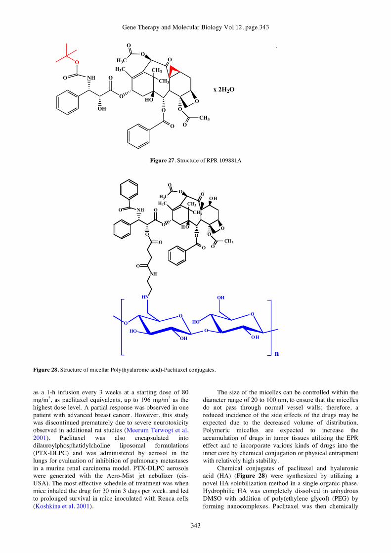
Gene Therapy and Molecular Biology Vol 12, page 343
343
O
O NH
O
O
H3C
O
O
H3C O
O
CH3
CH3
HO
O
O
CH3
O
OOH
x 2H2O
.
Figure 27. Structure of RPR 109881A
O NH
O
O
H3C
O
O
H3CO
O
CH3
CH3
HO
O
O
CH3
O
OO
OH
NH
HN
O
O
O
O
HO
OH
O
O
HO
OH
OH
n
Figure 28. Structure of micellar Poly(hyaluronic acid)-Paclitaxel conjugates. as a 1-h infusion every 3 weeks at a starting dose of 80 mg/m2, as paclitaxel equivalents, up to 196 mg/m2 as the highest dose level. A partial response was observed in one patient with advanced breast cancer. However, this study was discontinued prematurely due to severe neurotoxicity observed in additional rat studies (Meerum Terwogt et al, 2001). Paclitaxel was also encapsulated into dilauroylphosphatidylcholine liposomal formulations (PTX-DLPC) and was administered by aerosol in the lungs for evaluation of inhibition of pulmonary metastases in a murine renal carcinoma model. PTX-DLPC aerosols were generated with the Aero-Mist jet nebulizer (cis-USA). The most effective schedule of treatment was when mice inhaled the drug for 30 min 3 days per week. and led to prolonged survival in mice inoculated with Renca cells (Koshkina et al, 2001).
The size of the micelles can be controlled within the diameter range of 20 to 100 nm, to ensure that the micelles do not pass through normal vessel walls; therefore, a reduced incidence of the side effects of the drugs may be expected due to the decreased volume of distribution. Polymeric micelles are expected to increase the accumulation of drugs in tumor tissues utilizing the EPR effect and to incorporate various kinds of drugs into the inner core by chemical conjugation or physical entrapment with relatively high stability.
Chemical conjugates of paclitaxel and hyaluronic acid (HA) (Figure 28) were synthesized by utilizing a novel HA solubilization method in a single organic phase. Hydrophilic HA was completely dissolved in anhydrous DMSO with addition of poly(ethylene glycol) (PEG) by forming nanocomplexes. Paclitaxel was then chemically
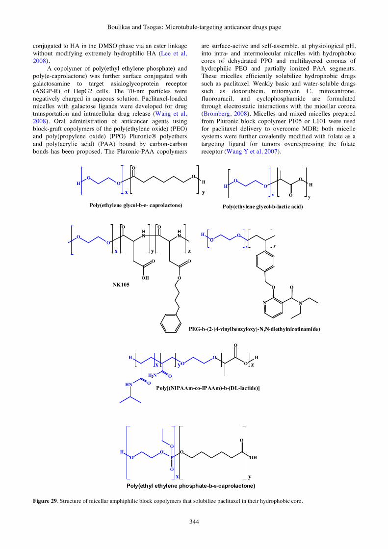
Boulikas and Tsogas: Microtubule-targeting anticancer drugs page
344
conjugated to HA in the DMSO phase via an ester linkage without modifying extremely hydrophilic HA (Lee et al, 2008).
A copolymer of poly(ethyl ethylene phosphate) and poly(e-caprolactone) was further surface conjugated with galactosamine to target asialoglycoprotein receptor (ASGP-R) of HepG2 cells. The 70-nm particles were negatively charged in aqueous solution. Paclitaxel-loaded micelles with galactose ligands were developed for drug transportation and intracellular drug release (Wang et al, 2008). Oral administration of anticancer agents using block-graft copolymers of the poly(ethylene oxide) (PEO) and poly(propylene oxide) (PPO) Pluronic® polyethers and poly(acrylic acid) (PAA) bound by carbon-carbon bonds has been proposed. The Pluronic-PAA copolymers
are surface-active and self-assemble, at physiological pH, into intra- and intermolecular micelles with hydrophobic cores of dehydrated PPO and multilayered coronas of hydrophilic PEO and partially ionized PAA segments. These micelles efficiently solubilize hydrophobic drugs such as paclitaxel. Weakly basic and water-soluble drugs such as doxorubicin, mitomycin C, mitoxantrone, fluorouracil, and cyclophosphamide are formulated through electrostatic interactions with the micellar corona (Bromberg, 2008). Micelles and mixed micelles prepared from Pluronic block copolymer P105 or L101 were used for paclitaxel delivery to overcome MDR; both micelle systems were further covalently modified with folate as a targeting ligand for tumors overexpressing the folate receptor (Wang Y et al, 2007).
O
O
O
y
HO
H
x
Poly(ethylene glycol-b-!- caprolactone)
HN
O
O
y
O
x
OH
O
HN
O
z
O
O
H
O
O
y
O
H
x O
H
O
O
P
O
OH
O
O
O
x y
Poly(ethyl ethylene phosphate-b-!-caprolactone)
H
O
O
O
H
O
H2N O
HN O
x y z
Poly[(NIPAAm-co-IPAAm)-b-(DL-lactide)]
Poly(ethylene glycol-b-lactic acid)
NK105
N
O
N
O
OO
H
x y
PEG-b-(2-(4-vinylbenzyloxy)-N,N-diethylnicotinamide)
Figure 29. Structure of micellar amphiphilic block copolymers that solubilize paclitaxel in their hydrophobic core.

Gene Therapy and Molecular Biology Vol 12, page 345
345
Amphiphilic block copolymers consisted of a micellar shell-forming poly(ethylene glycol) (PEG) block and a core-forming poly(2-(4-vinylbenzyloxy)-N,N-diethylnicotinamide) (Figure 29) were used to load paclitaxel up to 37.4 wt %; the presence of N,N-Diethylnicotinamide (DENA) in the micellar inner core resulted in effective paclitaxel solubilization and stabilization. The maximum loading capacity of poly(ethylene glycol)-b-P(D,L-lactide) (PEG-b-PLA) micelles (Figure 29) for comparison was 27.6 wt %. The hydrotropic polymer micelles were more effective than PEG-PLA micelle formulations in inhibiting the proliferation of human cancer cells (Lee et al, 2007).
The polymeric micellar formulation of paclitaxel with Pluronic P123 can effectively solubilize, prolong blood circulation time, and modify the biodistribution of paclitaxel (Han et al, 2006).
Hydrophobically modified hyperbranched polyglycerol-polyethylene glycol copolymers assume unimolecular micellar structures in aqueous solution were used for paclitaxel; although these polymers aggregate weakly in solution, the aggregates are broken down by low shear forces or by encapsulating a hydrophobic ligand within the polymer (Kainthan et al, 2008). A biocompatible, lecithin-based carrier for paclitaxel suitable for intravenous infusion has been proposed; among aqueous dispersions of egg or soya lecithin, mixed micellar solutions of egg lecithin and sodium deoxycholate, and formulations containing lecithin plus the co-surfactants and co-solvents poloxamer, polysorbate, Span, benzalkonium chloride, and macrogol the best formulation was a 5% egg lecithin / water dispersion containing 1 mg/ml paclitaxel (Sznitowska et al, 2008). The poorly water soluble paclitaxel was formulated into hydrotropic polymer micelles without using organic solvents required for solubilization; poly(ethylene glycol) was used as a hydrophilic block and poly[4-(2-vinylbenzyloxy-N-picolylnicotinamide)] as hydrotropic
block. The hydrotropic block copolymers synthesized were able to form paclitaxel-loaded polymeric micelles in aqueous solution and were proposed as novel carriers for hydrophobic drugs (Huh et al, 2008). Biodegradable triblock copolymers composed of a hydrophobic core of poly(ε-caprolactone) (PCL) and a hydrophilic shell of poly(ethyl ethylene phosphate) (PEEP) formed spherical micelles in aqueous solution; paclitaxel was successfully loaded into the micelles and were proposed as novel carriers for hydrophobic drug delivery (Wang YC et al, 2008). A polymeric thermosensitive micellar delivery system for docetaxel or paclitaxel based on [poly(N-isopropylacrylamide-co-acrylamide)-b-poly(DL-lactide) was proposed (Figure 29); hyperthermia greatly enhanced the antitumor effect of micellar taxanes in mouse xenografts (Liu et al, 2008). Oligomeric micelles composed of mPEG750-b-oligo(e-caprolactone)5 (mPEG750-b-OCL5) with a hydroxyl (OH), benzoyl (Bz) or naphthoyl (Np) end group were studied; the presence of an aromatic end group (Bz or Np) resulted in the formation of 10-nm, almost monodisperse, micelles with stable encapsulation of 10% (w/w) docetaxel or paclitaxel dissolved in the core (Carstens et al, 2008). Hydrophobic prodrugs of paclitaxel were synthesized via DCC/DMAP or anhydride chemistry to overcome the poor loading (<1% w/w) of paclitaxel in amphiphilic block co-polymer micelles of poly(ethylene glycol)-b-poly(ε-caprolactone) (PEG-b-PCL) (Forrest et al, 2008). The diblock copolymer poly(ethylene oxide)-b-poly (ε-caprolactone) (Figure 29) was used for core-loading of paclitaxel. This polymer formed two types micelles: worm-like micelles of nanometer in cross-section that spontaneously formed stable lengths of microns with improved solubilization efficiency for paclitaxel, and spherical micelles that exhibited lower drug loads (Cai et al, 2007). A schematic represenation of micellar amphiphilic block copolymers incorporating paclitaxel is shown in Figure 30.
Figure 30: Schematic represenation of micelles formed by amphiphilic block copolymers having a hydrophobic core
and a hydrophilic corona.
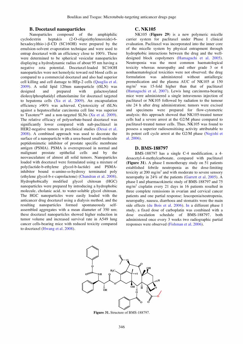
Boulikas and Tsogas: Microtubule-targeting anticancer drugs page
346
B. Docetaxel nanoparticles Nanoparticles composed of the amphiphilic
cyclodextrin heptakis (2-O-oligo(ethyleneoxide)-6-hexadecylthio-)-β-CD (SC16OH) were prepared by the emulsion-solvent evaporation technique and were used to entrap docetaxel with an efficiency close to 100%. These were determined to be spherical vesicular nanoparticles displaying a hydrodynamic radius of about 95 nm having a negative zeta potential. Docetaxel-loaded SC16OH nanoparticles were not hemolytic toward red blood cells as compared to a commercial docetaxel and also had superior cell killing and cell damage to HEp-2 cells (Quaglia et al, 2009). A solid lipid 120nm nanoparticle (tSLN) was designed and prepared with galactosylated dioleoylphosphatidyl ethanolamine for docetaxel targeted to hepatoma cells (Xu et al, 2009). An encapsulation efficiency >90% was achieved. Cytotoxicity of tSLNs against a hepatocellular carcinoma cell line was superior to Taxotere™ and a non-targeted SLNs (Xu et al, 2009). The relative efficacy of polysorbate-based docetaxel was significantly lower compared with nab-paclitaxel in HER2-negative tumors in preclinical studies (Desai et al, 2008). A combined approach was used to decorate the surface of a nanoparticle with a urea-based small-molecule peptidomimetic inhibitor of prostate specific membrane antigen (PSMA). PSMA is overexpressed in normal and malignant prostate epithelial cells and by the neovasculature of almost all solid tumors. Nanoparticles loaded with docetaxel were formulated using a mixture of poly(lactide-b-ethylene glycol-b-lactide) and PSMA-inhibitor bound α-amino-ω-hydroxy terminated poly (ethylene glycol-b-ε-caprolactone) (Chandran et al, 2008). Hydrophobically modified glycol chitosan (HGC) nanoparticles were prepared by introducing a hydrophobic molecule, cholanic acid, to water-soluble glycol chitosan. The HGC nanoparticles were easily loaded with the anticancer drug docetaxel using a dialysis method, and the resulting nanoparticles formed spontaneously self-assembled aggregates with a mean diameter of 350 nm; these docetaxel nanoparticles showed higher reduction in tumor volume and increased survival rate in A549 lung cancer cells-bearing mice with reduced toxicity compared to docetaxel (Hwang et al, 2008).
C. NK105 NK105 (Figure 29) is a new polymeric micelle
carrier system for paclitaxel under Phase I clinical evaluation. Paclitaxel was incorporated into the inner core of the micelle system by physical entrapment through hydrophobic interactions between the drug and the well-designed block copolymers (Hamaguchi et al, 2005). Neutropenia was the most common haematological toxicity whereas neuropathy and other grade 3 or 4 nonhaematological toxicities were not observed; the drug formulation was administered without antiallergic premedication and the plasma AUC of NK105 at 150 mg/m2 was 15-fold higher than that of paclitaxel (Hamaguchi et al, 2007). Lewis lung carcinoma-bearing mice were administered a single intravenous injection of paclitaxel or NK105 followed by radiation to the tumour site 24 h after drug administration; tumors were excised and specimens were prepared for flow-cytometric analysis; this approach showed that NK105-treated tumor cells had a severe arrest at the G2/M phase compared to paclitaxel-treated tumor cells. Thus, NK105 was found to possess a superior radiosensitising activity attributable to its potent cell cycle arrest at the G2/M phase (Negishi et al, 2006).
D. BMS-188797 BMS-188797 has a single C-4 modification, a 4-
desacetyl-4-methylcarbonate, compared with paclitaxel (Figure 31). A phase I monotherapy study on 51 patients established febrile neutropenia as the dose-limiting toxicity at 200 mg/m2 and with moderate to severe sensory neuropathy in 24% of the patients (Garrett et al, 2005). A phase I and pharmacokinetic study of BMS-188797 and 75 mg/m2 cisplatin every 21 days in 16 patients resulted in three complete remissions in ovarian and cervical cancer patients and one partial response; leucopenia/neutropenia, neuropathy, nausea, diarrhoea and stomatits were the main side effects (du Bois et al, 2006). In a different phase I study, a fixed dose of carboplatin was combined with a dose escalation schedule of BMS-188797, both administered once every 3 weeks two radiographic partial responses were observed (Fishman et al, 2006).
O
O NH
O
O
H3C
O
O
OOH
O
O
O
O
OOH
OH
H
H
O
Figure 31. Structure of BMS-188797.

Gene Therapy and Molecular Biology Vol 12, page 347
347
E. BMS-184476 BMS-184476 is a 7-methylthiomethyl ether
derivative of paclitaxel (Figure 32) that inhibits the growth of paclitaxel-resistant human tumor cell lines with multidrug resistance mediated by either P-glycoprotein or mutated tubulin. Given the known synergy between taxanes and cisplatin in vitro and their clinical activity in combination, a Phase I trial was performed with BMS-184476 and cisplatin on 27 patients (Sun et al, 2003).
F. BMS-275183 BMS-275183 (Figure 33) is an orally administered
C-4 methyl carbonate paclitaxel analogue able to induce growth suppression, cell-cycle arrest, and apoptosis. BMS-275183 altered the expression of cell-cycle regulators, such as cyclin A and cyclin B1 whereas the expression of E2F and p27 was decreased and increased, respectively, in all HNSCC cell lines examined; the mechanism involved molecular pathways similar to other taxanes (Yoo et al, 2007). It showed promising activity in a phase I trial investigating a weekly treatment regimen, but was associated with a relatively high incidence of neuropathic side effects. Based on these results, a phase II trial in NSCLC has been initiated using the twice weekly schedule (Bröker et al, 2006, 2007).
O
O NH
O
O
H3C
O
O
OOH
O
O
CH3
O
OOH
O
H
H
O
S
Figure 32. Structure of BMS-184476
O
O NH
O
O
H3C
O
O
OOH
O
O
O
O
OOH
OH
H
H
O
Figure 33. Structure of BMS-275183
G. Paclitaxel poliglumex Targeted drug delivery aims to increase the
therapeutic index by making more drug molecules available at the diseased sites while reducing systemic drug exposure. Paclitaxel poliglumex (PPX, XYOTAX or CT-2103; Figure 34) is a macromolecular drug conjugate that links paclitaxel with a biodegradable polymer, poly-L-glutamic acid, giving a water-soluble conjugate with a prolonged half-life and limited volume of distribution (Boddy et al, 2005; Chipman et al, 2006). It is currently under Phase III evaluation (O'Brien et al, 2008). In a Phase I study CT-2103 administered at 225 mg/m2 every 21 days in combination with carboplatin administered at AUC 6 had a manageable safety profile in patients with solid tumors (Nemunaitis et al, 2005). However, another Phase I study showed that neuropathy was more severe than expected (Veronese et al, 2005). PPX enhances tumor exposure by taking advantage of the hyperpermeable vasculature and suppressed lymphatic clearance characteristic of tumor tissue. Single-agent PPX, dosed at 175 mg/m2, was active and well tolerated in patients with advanced NSCLC. Patients on PPX required fewer red blood cell transfusions, hematopoietic growth factors, opioid analgesics, and clinic visits than patients receiving gemcitabine or vinorelbine (O'Brien et al, 2008). It is also being evaluated with carboplatin as first-line therapy in ovarian, peritoneal and fallopian tube cancer (Morgan et al, 2008). Neurotoxicity and hypersensitivity reactions led to early termination of a Phase II trial in with HER2-negative metastatic breast cancer (Lin et al, 2007). Of the 20 patients with stage IV NSCLC, 2 exhibited a PR, and 13 exhibited SD at a dose of 175 mg/m2 as first-line monotherapy (Richards et al, 2005).
H. Microtubule destabilizing agents As of 2008 there were 17 microtubule destabilizing
agents under clinical assessment, most in Phase I (Carlson, 2008). Table 2 gives a glimpse of new molecules under development destabilizing microtubules. It is worth mentioning that Stathmin 1 (STMN1), also known as metablastin, oncoprotein 18, LAP 18 and Op18, a 19 kDa cytosolic protein, is the first discovered member of a family of microtubule-destabilizing phosphoproteins critically involved in the construction and function of the mitotic spindle; its therapeutic manipulation has an anticancer potential (Rana et al, 2008).
IX. Prospects Docetaxel and paclitaxel are highly active agents in
metastatic breast cancer and may represent a safer alternative to anthracycline-based regimens when combined with the human epidermal growth factor receptor (HER)-2-targeted agent trastuzumab (Herceptin). Results from two large, phase III trials that examined the addition of carboplatin to a taxane-trastuzumab doublet did not demonstrate a difference in survival with carboplatin. In one study, the addition of carboplatin to paclitaxel-trastuzumab therapy resulted in a higher response rate and longer progression-free survival time; in

Boulikas and Tsogas: Microtubule-targeting anticancer drugs page
348
O
O
NH
O
O
H3C
OO
O
OH O
O
CH3
O
O
O
HO
H
H
O
HN
HN
O
O
OHO
O
OHO
a b
x
O
Figure 34. Structure of Paclitaxel Polyglumex. Table 2. Microtubule destabilizing agents
Name Active compound Development stage Reference
Onco-TCS Liposomal Vincristine Phase II/III Sarris et al, 2000; Gelmon et al, 1999
Cryptophycin 52 Derivative of Cryptophycin Phase I Teicher et al, 2000
LY355703 synthetic product structurally related to cryptophycin
phase II in in patients with platinum-resistant ovarian cancer and NSCLC
Edelman et al, 2003; D'Agostino et al, 2006
Dolastatin-10 Dolastatin-10
Phase I/II in Metastatic Soft Tissue Sarcomas, pancreaticobiliary cancers and advanced breast cancer
von Mehren et al, 2004; Kindler et al, 2005; Perez et al, 2005
Tasidotin (ILX651) a peptide analogue of the antimitotic depsipeptide dolastatin 15
Phase II Bai et al, 2008
TZT-1027 synthetic dolastatin 10 derivative
Phase I & II in metastatic soft-tissue sarcomas and NSCLC
Patel et al, 2006; Riely et al, 2007; Tamura et al, 2007; Horti et al, 2008
ENMD-1198 novel tubulin-binding agent Preclinical Moser et al, 2008 combretastatin-A4-phosphate (CA4P) microtubule-targeting agent Preclinical Petit et al, 2008
a second study, the docetaxel-trastuzumab and docetaxel-trastuzumab-carboplatin combinations were equally effective (Bullock and Blackwell, 2008). Future studies may show better synergies in the various regimens for MBC as well as other cancers using other microtubule-targeted agents reviewed here.
Preclinical studies have shown that frequent administration in vivo of low doses of chemotherapeutic
drugs ("metronomic" dosing) can affect tumor endothelium and inhibit tumor angiogenesis with concomitant reduction in side effects even after chronic treatment. The continuous low-dose therapy with paclitaxel, 4-hydroperoxycyclophosphamide, the oral taxane BMS-275183, doxorubicin, epothilone B (EpoB) and its analogue 5-methylpyridine EpoB may have a

Gene Therapy and Molecular Biology Vol 12, page 349
349
highly selective effect against cycling vascular endothelial cells (Bocci et al, 2002).
Most of the anticancer molecules described in this review have side effects. The side effects of paclitaxel include myelosuppression, mucositis, peripheral neuropathy, and cardiovascular toxicity. The side effects of docetaxel include myelotoxicity, allergic reactions during infusion, diarrhea, nausea / vomiting, and hair loss. The side effects of vinblastine, vinorelbine, vincristine and vindesine include myelosuppression, constipation, diarrhea, nausea, peripheral neuropathy, and asthenia. Encapsulation into liposome nanoparticles or entrapment into dendrimers and polymers is expected to lower side effects and improve tumor targeting in a way similar to that successfully attempted for cisplatin (Boulikas, 2007).
Albumin-bound paclitaxel 100 mg/m2 given weekly demonstrated the same antitumor activity as albumin-bound paclitaxel 125 mg/m2 weekly and a more favorable safety profile in patients with metastatic breast cancer (MBC) that had progressed with previous taxane therapy; the carrier-mediated paclitaxel has already shown significant efficacy in taxane resistant cancers, an approach highly relevant in prostate cancer, where taxanes are the treatment of choice (Blum et al, 2007). Second-generation enhanced-delivery taxanes, including Tocosol Paclitaxel (Sonus Pharmaceuticals, Inc., Bothell, Washington) and paclitaxel poliglumex, use biocompatible drug delivery vehicles to eliminate the need for toxic conventional solvents and exploit tumor pathophysiological phenomena such as enhanced permeability and retention (Perez, 2007). Certainly drug delivery plays a major role especially nanoparticle formulations that are able to preferentially concentrate at tumor sites using imperfections in the endothelium of tumor vasculature. Our group has demonstrated targeting of liposomal cisplatin nanoparticles (Lipoplatin) in patients (Boulikas et al, 2005). A similar liposomal formulation of paclitaxel is at preclinical testing.
Nanotechnology promises to wrap up drugs and optimize in vivo delivery (Boulikas, 2007). Nanoparticles provide a new mode of cancer drug delivery functioning as a carrier for entry through fenestrations in tumor vasculature allowing direct cell access. Taxane delivery systems through tumor cell surface receptor-targeted delivery mechanisms such as small-molecule peptides and monoclonal antibodies, as well as those on non-targeted procedures such as liposomes, nanostructures, and natural and synthetic polymers hold promise for improving the toxicity profiles and biodistribution of the drug (Safavy, 2008; Haley and Frenkel, 2008). Other modifications including transferrin receptor and folate receptor targeted drug delivery molecules are emerging.
PEG2000-stearic acid was used to prepare PEGylated solid lipid nanoparticles loading vinorelbine bitartrate of a particle size of 180-250nm; octadecylamine-fluorescein isothiocynate was used as a fluorescence marker to incorporate into nanoparticles and study their cellular uptake (Wan et al, 2008). Poly(D,L-lactide-co-glycolide) (PLGA) was used to load the vinca alkaloid vinpocetine forming microparticles suited for intramuscular injection (Li et al, 2008). An ultrasonic-solvent emulsification
technique was adopted to prepare vinpocetine loaded Glyceryl monostearate (GMS) nanodispersions with narrow size distribution (Luo et al, 2006). PLGA nanoparticles of a size of 140 nm simultaneously loaded with vincristine sulfate and quercetin were prepared via oil/water emulsion solvent evaporation (Song et al, 2008). Vinorelbine bitartrate (VB) was loaded into solid lipid nanoparticles (SLNs) of 150 to 350 nm by a cold homogenization technique; enhancement of lecithin content in lipid matrix resulted in smaller particle of SLNs (You et al, 2007). Nanoparticles allow exquisite modification for binding to cancer cell membranes, the microenvironment, or to cytoplasmic or nuclear receptor sites.
Intact plasma lipoproteins are complex macromolecules that transport highly water-insoluble compounds (cholesteryl esters and triacylglycerols) from the intestine to the liver through blood; this property renders them ideal carriers of hydrophobic drugs. Both low and high density lipoprotein-based formulations have been used as drug carriers (Lacko et al, 2007). One important factor to be taken into consideration in anticancer drug delivery using HDL is the promotion of angiogenesis by DHL, a property that may counteract the beneficial effect of the drug in the induction of tumor apoptosis (Chen et al, 2007). Spherical nanoparticles of reconstituted (synthetic) high-density lipoproteins (HDL) carrying paclitaxel have been reported with high drug to lipoprotein ratio with exceptional stability for up to 6 months. The nanoparticles had superior cytotoxicity against MCF7, DU145, OV1063 and OVCAR-3 cancer cell lines and their IC50 was 5-20 times lower than that of the free drug (McConathy et al, 2008).
Docetaxel arose from the natural compound paclitaxel by substitution of the benzyl- group by a tert-butoxy-group (Figure 7). Fluorination of the ethyl group in the vinblastine molecule at the 20' position of the catharanthine moiety led to the introduction of vinflunine (Figures 13 and 14). Other type of modifications could have given molecules of superior activity with lower side effects in human trials; however, because of the huge cost involved in testing so many substances into humans, the industry is limited in the number of molecules to be promoted and the decision on the molecules to be tested in clinical trials is based on preclinical studies that may not reflect the human experience.
Structural similarities can be used to further modify a successful anticancer drug in order to seek molecules of improved efficacy with lower toxicity or able to overcome tumor resistance of the prototype molecule; because of the complexity of the biological systems and the number of parameters that define a successful drug (its interaction with serum components, biodistribution, circulation time and urine or other route of excretion, interaction with cell surface molecules, entry across the cell membrane, resistance mechanisms, type of subcellular components it damages, induction of signaling pathways resulting to apoptosis and others) it is very difficult to predict effectiveness and side effects from mere chemistry. Trial and error on cell lines, animals and ultimately human patients remains the only way to test new molecules.

Boulikas and Tsogas: Microtubule-targeting anticancer drugs page
350
Acknowledgements Supported by the European grants RIGHT: LSH-CT-
2005-005276, NanoBiopharmaceutics: NMP4-CT-2006-026723 and MYASTAID: LSHM-CT-2006-037833, to Regulon AE as well as two grants from the Hellenic government (General Secretariat of Research and Development) for the clinical development of Lipoplatin, Lipoxal and LipoVIL12. We are grateful to Alexandros Magos, Petros Christofis, Alexandros Pantos and Evagelos Bellis for stimulating discussions.
References
Advani R, Fisher GA, Lum BL, Jambalos C, Cho CD, Cohen M, Gollerkeri A, Sikic BI (2003) Phase I and pharmacokinetic study of BMS-188797, a new taxane analog, administered on a weekly schedule in patients with advanced malignancies. Clin Cancer Res 9, 5187-5194.
Albertsson P, Lennernäs B, Norrby K (2008) Dose effects of continuous vinblastine chemotherapy on mammalian angiogenesis mediated by VEGF-A. Acta Oncol 47, 293-300.
Alhamadsheh MM, Gupta S, Hudson RA, Perera L, Tillekeratne LM (2008) Total Synthesis and Selective Activity of a New Class of Conformationally Restrained Epothilones. Chemistry 14, 570-581.
Ali M, Mohammed N, Alnaqeeb MA, Hassan RA, Ahmad HS (1989) Toxicity of echinulin from Aspergillus chevalieri in rabbits Toxicol Lett 48(3), 235-41.
Altmann KH, Gertsch J (2007) Anticancer drugs from nature--natural products as a unique source of new microtubule-stabilizing agents. Nat Prod Rep 24, 327-357.
Amos LA, Kara S, Florenceb GJ, Patersonb I (2003) Discodermolide interferes with the binding of tau protein to microtubules. FEBS Lett 539, 34-36.
Aonuma M, Miyamoto M, Inoue YH, Tamai K, Sakai H, Kamasawa N, Matsukage A (2005) Microtubule bundle formation and cell death induced by the human CLASP/Orbit N-terminal fragment. Cell Struct Funct 30, 7-13.
Bai R, Edler MC, Bonate PL, Copeland TD, Pettit GR, Luduena RF, Hamel E (2008) Intracellular activation and deactivation of tasidotin, an analogue of dolastatin 15, correlation with cytotoxicity. Mol Pharmacol, in press.
Baiguera S, Conconi MT, Guidolin D, Mazzocchi G, Malendowicz LK, Parnigotto PP, Spinazzi R, Nussdorfer GG (2004) Ghrelin inhibits in vitro angiogenic activity of rat brain microvascular endothelial cells. Int J Mol Med 14, 849-854.
Baker SD, Zhao M, Lee CKK, Verweij J, Zabelina Y, Brahmer JR, et al (2004) Comparative pharmacokinetics of weekly and every-three-weeks docetaxel. Clin Cancer Res 10, 1976-1983.
Balog DM, Meng D, Kamanecka T, Bertinato P, Su DS, Sorensen EJ, Danishefsky SJ (1996) Total Synthesis of (-)-Epothilone A Angew Chem 108, 2976.
Banerjee A (1997) Differential Effects of Colchicine and Its B-ring Modified Analog MTPT on the Assembly-Independent GTPase Activity of Purified β-Tubulin Isoforms from Bovine Brain Biochem Biophys Res Commun 231, 698-700.
Beer TM, Higano CS, Saleh M, Dreicer R, Hudes G, Picus J, Rarick M, Fehrenbacher L, Hannah AL (2007) Phase II study of KOS-862 in patients with metastatic androgen independent prostate cancer previously treated with docetaxel. Invest New Drugs 25, 565-70.
Beumer JH, Garner RC, Cohen MB, Galbraith S, Duncan GF, Griffin T, Beijnen JH, Schellens JH (2007) Human mass
balance study of the novel anticancer agent ixabepilone using accelerator mass spectrometry. Invest New Drugs 25, 327-334.
Bifulco M, Laezza C, Stingo S, Wolff J (2002) 2',3'-Cyclic nucleotide 3'-phosphodiesterase, a membrane-bound, microtubule-associated protein and membrane anchor for tubulin. Proc Natl Acad Sci U S A 99, 1807-1812.
Blum JL, Dees EC, Chacko A, Doane L, Ethirajan S, Hopkins J, McMahon R, Merten S, Negron A, Neubauer M, Ilegbodu D, Boehm KA, Asmar L, O'Shaughnessy JA (2006) Phase II trial of capecitabine and weekly paclitaxel as first-line therapy for metastatic breast cancer. J Clin Oncol 24, 4384-4390.
Blum JL, Savin MA, Edelman G, Pippen JE, Robert NJ, Geister BV, Kirby RL, Clawson A, O'Shaughnessy JA (2007) Phase II study of weekly albumin-bound paclitaxel for patients with metastatic breast cancer heavily pretreated with taxanes. Clin Breast Cancer 7, 850-856.
Bocci G, Nicolaou KC, Kerbel RS (2002) Protracted low-dose effects on human endothelial cell proliferation and survival in vitro reveal a selective antiangiogenic window for various chemotherapeutic drugs. Cancer Res 62, 6938-6943.
Borowiec AS, Hague F, Harir N, Guénin S, Guerineau F, Gouilleux F, Roudbaraki M, Lassoued K, Ouadid-Ahidouch H (2007) IGF-1 activates hEAG K+ channels through an Akt-dependent signaling pathway in breast cancer cells, role in cell proliferation. J Cell Physiol 212, 690-701.
Boulikas T (2007) Molecular mechanisms of cisplatin and its liposomally encapsulated form, Lipoplatin™. Lipoplatin™ as a chemotherapy and antiangiogenesis drug. Cancer Ther 5, 351-376.
Boulikas T, Pantos A, Bellis E, Christofis P (2007) Designing platinum compounds in cancer, structures and mechanisms. Cancer Ther 5, 537-583.
Boulikas T, Stathopoulos GP, Volakakis N and Vougiouka M (2005) Systemic Lipoplatin infusion results in preferential tumor uptake in human studies. Anticancer Res 25, 3031-3040,.
Bragdon B, Bertone AL, Hardy J, Simmons EJ, Weisbrode SE (2001) Use of an isolated joint model to detect early changes induced by intra-articular injection of paclitaxel-impregnated polymeric microspheres. J Invest Surg 14, 169-182.
Bröker LE, de Vos FY, van Groeningen CJ, Kuenen BC, Gall HE, Woo MH, Voi M, Gietema JA, de Vries EG, Giaccone G (2006) Phase I trial with BMS-275183, a novel oral taxane with promising antitumor activity. Clin Cancer Res 12, 1760-1767.
Bröker LE, Veltkamp SA, Heath EI, Kuenen BC, Gall H, Astier L, Parker S, Kayitalire L, Lorusso PM, Schellens JH, Giaccone G (2007) A phase I safety and pharmacologic study of a twice weekly dosing regimen of the oral taxane BMS-275183. Clin Cancer Res 13, 3906-3912.
Bromberg L (2008) Polymeric micelles in oral chemotherapy. J Control Release 128, 99-112.
Budillon A, Di Gennaro E, Bruzzese F, Rocco M, Manzo G, Caraglia M (2007) Histone deacetylase inhibitors, a new wave of molecular targeted anticancer agents. Recent Patents Anticancer Drug Discov 2, 119-134.
Buey RM, Calvo E, Barasoain I, Pineda O, Edler MC, Matesanz R, Cerezo G, Vanderwal CD, Day BW, Sorensen EJ, López JA, Andreu JM, Hamel E, Díaz JF (2007) Cyclostreptin binds covalently to microtubule pores and lumenal taxoid binding sites. Nat Chem Biol 3, 117-125.
Bullock K, Blackwell K (2008) Clinical efficacy of taxane-trastuzumab combination regimens for HER-2-positive metastatic breast cancer. Oncologist 13, 515-525.
Cai S, Vijayan K, Cheng D, Lima EM, Discher DE (2007) Micelles of different morphologies--advantages of worm-like
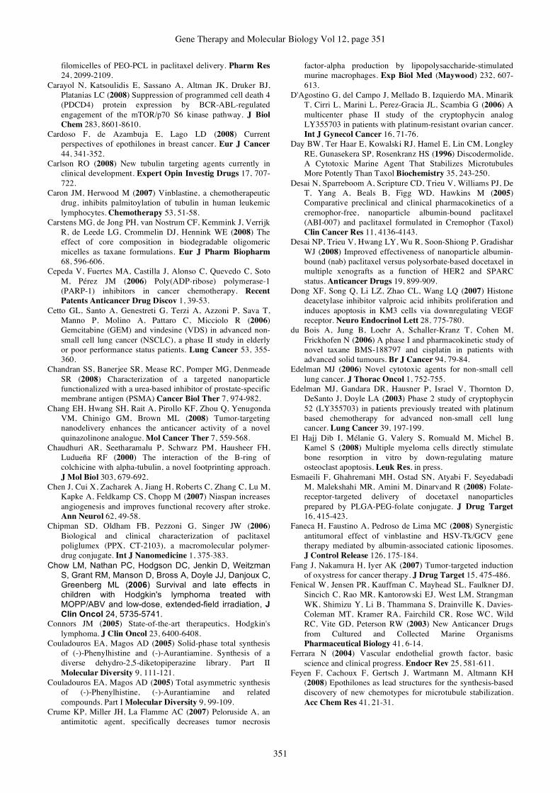
Gene Therapy and Molecular Biology Vol 12, page 351
351
filomicelles of PEO-PCL in paclitaxel delivery. Pharm Res 24, 2099-2109.
Carayol N, Katsoulidis E, Sassano A, Altman JK, Druker BJ, Platanias LC (2008) Suppression of programmed cell death 4 (PDCD4) protein expression by BCR-ABL-regulated engagement of the mTOR/p70 S6 kinase pathway. J Biol Chem 283, 8601-8610.
Cardoso F, de Azambuja E, Lago LD (2008) Current perspectives of epothilones in breast cancer. Eur J Cancer 44, 341-352.
Carlson RO (2008) New tubulin targeting agents currently in clinical development. Expert Opin Investig Drugs 17, 707-722.
Caron JM, Herwood M (2007) Vinblastine, a chemotherapeutic drug, inhibits palmitoylation of tubulin in human leukemic lymphocytes. Chemotherapy 53, 51-58.
Carstens MG, de Jong PH, van Nostrum CF, Kemmink J, Verrijk R, de Leede LG, Crommelin DJ, Hennink WE (2008) The effect of core composition in biodegradable oligomeric micelles as taxane formulations. Eur J Pharm Biopharm 68, 596-606.
Cepeda V, Fuertes MA, Castilla J, Alonso C, Quevedo C, Soto M, Pérez JM (2006) Poly(ADP-ribose) polymerase-1 (PARP-1) inhibitors in cancer chemotherapy. Recent Patents Anticancer Drug Discov 1, 39-53.
Cetto GL, Santo A, Genestreti G, Terzi A, Azzoni P, Sava T, Manno P, Molino A, Pattaro C, Micciolo R (2006) Gemcitabine (GEM) and vindesine (VDS) in advanced non-small cell lung cancer (NSCLC), a phase II study in elderly or poor performance status patients. Lung Cancer 53, 355-360.
Chandran SS, Banerjee SR, Mease RC, Pomper MG, Denmeade SR (2008) Characterization of a targeted nanoparticle functionalized with a urea-based inhibitor of prostate-specific membrane antigen (PSMA) Cancer Biol Ther 7, 974-982.
Chang EH, Hwang SH, Rait A, Pirollo KF, Zhou Q, Yenugonda VM, Chinigo GM, Brown ML (2008) Tumor-targeting nanodelivery enhances the anticancer activity of a novel quinazolinone analogue. Mol Cancer Ther 7, 559-568.
Chaudhuri AR, Seetharamalu P, Schwarz PM, Hausheer FH, Ludueña RF (2000) The interaction of the B-ring of colchicine with alpha-tubulin, a novel footprinting approach. J Mol Biol 303, 679-692.
Chen J, Cui X, Zacharek A, Jiang H, Roberts C, Zhang C, Lu M, Kapke A, Feldkamp CS, Chopp M (2007) Niaspan increases angiogenesis and improves functional recovery after stroke. Ann Neurol 62, 49-58.
Chipman SD, Oldham FB, Pezzoni G, Singer JW (2006) Biological and clinical characterization of paclitaxel poliglumex (PPX, CT-2103), a macromolecular polymer-drug conjugate. Int J Nanomedicine 1, 375-383.
Chow LM, Nathan PC, Hodgson DC, Jenkin D, Weitzman S, Grant RM, Manson D, Bross A, Doyle JJ, Danjoux C, Greenberg ML (2006) Survival and late effects in children with Hodgkin's lymphoma treated with MOPP/ABV and low-dose, extended-field irradiation, J Clin Oncol 24, 5735-5741.
Connors JM (2005) State-of-the-art therapeutics, Hodgkin's lymphoma. J Clin Oncol 23, 6400-6408.
Couladouros EA, Magos AD (2005) Solid-phase total synthesis of (-)-Phenylhistine and (-)-Aurantiamine. Synthesis of a diverse dehydro-2,5-diketopiperazine library. Part II Molecular Diversity 9, 111-121.
Couladouros EA, Magos AD (2005) Total asymmetric synthesis of (-)-Phenylhistine, (-)-Aurantiamine and related compounds. Part I Molecular Diversity 9, 99-109.
Crume KP, Miller JH, La Flamme AC (2007) Peloruside A, an antimitotic agent, specifically decreases tumor necrosis
factor-alpha production by lipopolysaccharide-stimulated murine macrophages. Exp Biol Med (Maywood) 232, 607-613.
D'Agostino G, del Campo J, Mellado B, Izquierdo MA, Minarik T, Cirri L, Marini L, Perez-Gracia JL, Scambia G (2006) A multicenter phase II study of the cryptophycin analog LY355703 in patients with platinum-resistant ovarian cancer. Int J Gynecol Cancer 16, 71-76.
Day BW, Ter Haar E, Kowalski RJ, Hamel E, Lin CM, Longley RE, Gunasekera SP, Rosenkranz HS (1996) Discodermolide, A Cytotoxic Marine Agent That Stabilizes Microtubules More Potently Than Taxol Biochemistry 35, 243-250.
Desai N, Sparreboom A, Scripture CD, Trieu V, Williams PJ, De T, Yang A, Beals B, Figg WD, Hawkins M (2005) Comparative preclinical and clinical pharmacokinetics of a cremophor-free, nanoparticle albumin-bound paclitaxel (ABI-007) and paclitaxel formulated in Cremophor (Taxol) Clin Cancer Res 11, 4136-4143.
Desai NP, Trieu V, Hwang LY, Wu R, Soon-Shiong P, Gradishar WJ (2008) Improved effectiveness of nanoparticle albumin-bound (nab) paclitaxel versus polysorbate-based docetaxel in multiple xenografts as a function of HER2 and SPARC status. Anticancer Drugs 19, 899-909.
Dong XF, Song Q, Li LZ, Zhao CL, Wang LQ (2007) Histone deacetylase inhibitor valproic acid inhibits proliferation and induces apoptosis in KM3 cells via downregulating VEGF receptor. Neuro Endocrinol Lett 28, 775-780.
du Bois A, Jung B, Loehr A, Schaller-Kranz T, Cohen M, Frickhofen N (2006) A phase I and pharmacokinetic study of novel taxane BMS-188797 and cisplatin in patients with advanced solid tumours. Br J Cancer 94, 79-84.
Edelman MJ (2006) Novel cytotoxic agents for non-small cell lung cancer. J Thorac Oncol 1, 752-755.
Edelman MJ, Gandara DR, Hausner P, Israel V, Thornton D, DeSanto J, Doyle LA (2003) Phase 2 study of cryptophycin 52 (LY355703) in patients previously treated with platinum based chemotherapy for advanced non-small cell lung cancer. Lung Cancer 39, 197-199.
El Hajj Dib I, Mélanie G, Valery S, Romuald M, Michel B, Kamel S (2008) Multiple myeloma cells directly stimulate bone resorption in vitro by down-regulating mature osteoclast apoptosis. Leuk Res, in press.
Esmaeili F, Ghahremani MH, Ostad SN, Atyabi F, Seyedabadi M, Malekshahi MR, Amini M, Dinarvand R (2008) Folate-receptor-targeted delivery of docetaxel nanoparticles prepared by PLGA-PEG-folate conjugate. J Drug Target 16, 415-423.
Faneca H, Faustino A, Pedroso de Lima MC (2008) Synergistic antitumoral effect of vinblastine and HSV-Tk/GCV gene therapy mediated by albumin-associated cationic liposomes. J Control Release 126, 175-184.
Fang J, Nakamura H, Iyer AK (2007) Tumor-targeted induction of oxystress for cancer therapy. J Drug Target 15, 475-486.
Fenical W, Jensen PR, Kauffman C, Mayhead SL, Faulkner DJ, Sincich C, Rao MR, Kantorowski EJ, West LM, Strangman WK, Shimizu Y, Li B, Thammana S, Drainville K, Davies-Coleman MT, Kramer RA, Fairchild CR, Rose WC, Wild RC, Vite GD, Peterson RW (2003) New Anticancer Drugs from Cultured and Collected Marine Organisms Pharmaceutical Biology 41, 6-14.
Ferrara N (2004) Vascular endothelial growth factor, basic science and clinical progress. Endocr Rev 25, 581-611.
Feyen F, Cachoux F, Gertsch J, Wartmann M, Altmann KH (2008) Epothilones as lead structures for the synthesis-based discovery of new chemotypes for microtubule stabilization. Acc Chem Res 41, 21-31.
Boulikas and Tsogas: Microtubule-targeting anticancer drugs page
352
Fialho AM, Chakrabarty AM (2007) Recent patents on bacterial proteins as potential anticancer agents. Recent Patents Anticancer Drug Discov 2, 224-234.
Fishman MN, Garrett CR, Simon GR, Chiappori AA, Lush RM, Dinwoodie WR, Mahany JJ, Dellaportas AM, Cantor A, Gollerki A, Cohen MB, Sullivan DM (2006) Phase I study of the taxane BMS-188797 in combination with carboplatin administered every 3 weeks in patients with solid malignancies. Clin Cancer Res 12, 523-528.
Florio T (2008) Molecular mechanisms of the antiproliferative activity of somatostatin receptors (SSTRs) in neuroendocrine tumors. Front Biosci 13, 822-840.
Forrest ML, Yáñez JA, Remsberg CM, Ohgami Y, Kwon GS, Davies NM (2008) Paclitaxel prodrugs with sustained release and high solubility in poly(ethylene glycol)-b-poly(e-caprolactone) micelle nanocarriers, pharmacokinetic disposition, tolerability, and cytotoxicity. Pharm Res 25, 194-206.
Fumoleau P, Coudert B, Isambert N, Ferrant E (2007) Novel tubulin-targeting agents, anticancer activity and pharmacologic profile of epothilones and related analogues. Ann Oncol Suppl 5, 9-15.
Garrett CR, Fishman MN, Rago RR, Williams CC, Dellaportas AM, Mahany JJ, Lush RM, Dalton WS, Gollerkeri A, Cohen MB, Sullivan DM (2005) Phase I study of a novel taxane BMS-188797 in adult patients with solid malignancies. Clin Cancer Res 11, 3335-3341.
Gelmon KA, Tolcher A, Diab AR, Bally MB, Embree L, Hudon N, Dedhar C, Ayers D, Eisen A, Melosky B, Burge C, Logan P, Mayer LD (1999) Phase I study of liposomal vincristine. J Clin Oncol 17, 697-705.
Georgoulias V, Ardavanis A, Tsiafaki X, Agelidou A, Mixalopoulou P, Anagnostopoulou O, Ziotopoulos P, Toubis M, Syrigos K, Samaras N, Polyzos A, Christou A, Kakolyris S, Kouroussis C, Androulakis N, Samonis G, Chatzidaki D (2005) Vinorelbine plus cisplatin versus docetaxel plus gemcitabine in advanced non-small-cell lung cancer, a phase III randomized trial. J Clin Oncol 23, 2937-2945.
Gigant B, Wang C, Ravelli RB, Roussi F, Steinmetz MO, Curmi PA, Sobel A, Knossow M (2005) Structural basis for the regulation of tubulin by vinblastine. Nature. 435, 519-522.
Gobbi PG, Levis A, Chisesi T, Broglia C, Vitolo U, Stelitano C, Pavone V, Cavanna L, Santini G, Merli F, Liberati M, Baldini L, Deliliers GL, Angelucci E, Bordonaro R, Federico M; Intergruppo Italiano Linfomi (2005) ABVD versus modified stanford V versus MOPPEBVCAD with optional and limited radiotherapy in intermediate- and advanced-stage Hodgkin's lymphoma, final results of a multicenter randomized trial by the Intergruppo Italiano Linfomi. J Clin Oncol 23, 9198-9207.
Gómez-Varela D, Zwick-Wallasch E, Knötgen H, Sánchez A, Hettmann T, Ossipov D, Weseloh R, Contreras-Jurado C, Rothe M, Stühmer W, Pardo LA (2007) Monoclonal antibody blockade of the human Eag1 potassium channel function exerts antitumor activity. Cancer Res 67, 7343-7349.
Goodin S, Kane MP, Rubin EH (2004) Epothilones, mechanism of action and biologic activity. J Clin Oncol 22, 2015-2025.
Gradishar WJ (2006) Albumin-bound paclitaxel, a next-generation taxane. Expert Opin Pharmacother 7, 1041-1053.
Gradishar WJ, Tjulandin S, Davidson N, Shaw H, Desai N, Bhar P, Hawkins M, O'Shaughnessy J (2005) Phase III trial of nanoparticle albumin-bound paclitaxel compared with polyethylated castor oil-based paclitaxel in women with breast cancer. J Clin Oncol 23, 7794-7803.
Green MR, Manikhas GM, Orlov S, Afanasyev B, Makhson AM, Bhar P, Hawkins MJ (2006) Abraxane, a novel Cremophor-
free, albumin-bound particle form of paclitaxel for the treatment of advanced non-small-cell lung cancer. Ann Oncol 17, 1263-1268.
Gridelli C, Gallo C, Shepherd FA, Illiano A, Piantedosi F, Robbiati SF, Manzione L, Barbera S, Frontini L, Veltri E, Findlay B, Cigolari S, Myers R, Ianniello GP, Gebbia V, Gasparini G, Fava S, Hirsh V, Bezjak A, Seymour L, Perrone F (2003) Gemcitabine plus vinorelbine compared with cisplatin plus vinorelbine or cisplatin plus gemcitabine for advanced non-small-cell lung cancer, a phase III trial of the Italian GEMVIN Investigators and the National Cancer Institute of Canada Clinical Trials Group. J Clin Oncol 21, 3025-3034.
Gu YT, Zhang H, Xue YX (2007) Dexamethasone enhances adenosine 5'-triphosphate-sensitive potassium channel expression in the blood-brain tumor barrier in a rat brain tumor model. Brain Res 1162, 1-8.
Guo WJ, Tao GJ, Tao WY, Cui FJ, Jin XC, Bi F, Xu ZH, Ao ZH (2007) A myxobacterium strain Sorangium cellulosum AHB125 producing epothilone B and other anticancer substances. Nat Prod Res 21, 1256-1265.
Gupta, Jr. LM, Bode CJ, Georg, GI, Himes RH (2003) Understanding tubulin-Taxol interactions, Mutations that impart Taxol binding to yeast tubulin. Proc Natl Acad Sci USA 100, 6394-6397.
Haley B, Frenkel E (2008) Nanoparticles for drug delivery in cancer treatment. Urol Oncol 26, 57-64.
Hamaguchi T, Kato K, Yasui H, Morizane C, Ikeda M, Ueno H, Muro K, Yamada Y, Okusaka T, Shirao K, Shimada Y, Nakahama H, Matsumura Y (2007) A phase I and pharmacokinetic study of NK105, a paclitaxel-incorporating micellar nanoparticle formulation. Br J Cancer 97, 170-176.
Hamaguchi T, Matsumura Y, Suzuki M, Shimizu K, Goda R, Nakamura I, Nakatomi I, Yokoyama M, Kataoka K, Kakizoe T (2005) NK105, a paclitaxel-incorporating micellar nanoparticle formulation, can extend in vivo antitumour activity and reduce the neurotoxicity of paclitaxel. Br J Cancer 92, 1240-1346.
Hamel E, Day BW, Miller JH, Jung MK, Northcote PT, Ghosh AK, Curran DP, Cushman M, Nicolaou KC, Paterson I, Sorensen EJ (2006) Synergistic effects of peloruside A and laulimalide with taxoid site drugs, but not with each other, on tubulin assembly. Mol Pharmacol 70, 1555-1564.
Han LM, Guo J, Zhang LJ, Wang QS, Fang XL (2006) Pharmacokinetics and biodistribution of polymeric micelles of paclitaxel with Pluronic P123. Acta Pharmacol Sin 27, 747-753.
Hayashi Y, Orikasa S, Tanaka K, Kanoh K, Kiso Y (2000) Total Synthesis of Anti-microtubule Diketopiperazine Derivatives, Phenylahistin and Aurantiamine J Org Chem 65, 8402-8405.
Henderson IC, Bhatia V (2007) Nab-paclitaxel for breast cancer, a new formulation with an improved safety profile and greater efficacy. Expert Rev Anticancer Ther 7, 919-943.
Hicklin DJ, Ellis LM (2005) Role of the vascular endothelial growth factor pathway in tumor growth and angiogenesis. J Clin Oncol 23, 1011-1027.
Hiles JJ, Kolesar JM (2008) Role of sunitinib and sorafenib in the treatment of metastatic renal cell carcinoma. Am J Health Syst Pharm 65, 123-131.
Horti J, Juhasz E, Monostori Z, Maeda K, Eckhardt S, Bodrogi I (2008) Phase I study of TZT-1027, a novel synthetic dolastatin 10 derivative, for the treatment of patients with non-small cell lung cancer. Cancer Chemother Pharmacol 62, 173-180.
Horwitz SB, Klein LE, Freeze BS, Amos BS (2005) The microtubule stabilizing agent discodermolide is a potent

Gene Therapy and Molecular Biology Vol 12, page 353
353
inducer of accelerated cell senescence. Cell Cycle 4, 501-507.
Hsu LC, White RL (1998) BRCA1 is associated with the centrosome during mitosis. Proc Natl Acad Sci U S A 95, 12983-12988.
Huby RDJ, Weiss A, Ley SC (1998) Nocodazole Inhibits Signal Transduction by the T Cell Antigen Receptor J BiolChem 273, 12024 -12031.
Huh KM, Min HS, Lee SC, Lee HJ, Kim S, Park K (2008) A new hydrotropic block copolymer micelle system for aqueous solubilization of paclitaxel. J Control Release 126, 122-129.
Hwang HY, Kim IS, Kwon IC, Kim YH (2008) Tumor targetability and antitumor effect of docetaxel-loaded hydrophobically modified glycol chitosan nanoparticles. J Control Release 128, 23-31.
Ibrahim NK, Samuels B, Page R, Doval D, Patel KM, Rao SC, Nair MK, Bhar P, Desai N, Hortobagyi GN (2005) Multicenter phase II trial of ABI-007, an albumin-bound paclitaxel, in women with metastatic breast cancer. J Clin Oncol 23, 6019-6026.
International Breast Cancer Study Group (2006) Tamoxifen After Adjuvant Chemotherapy for Premenopausal Women With Lymph Node-Positive Breast Cancer, International Breast Cancer Study Group Trial 13-93. J Clin Oncol 24, 1332-1341.
Jain RK (2001) Normalizing tumor vasculature with antiangiogenic therapy, a new paradigm for combination therapy. Nat Med 7, 987-989.
Jiemjit A, Fandy TE, Carraway H, Bailey KA, Baylin S, Herman JG, Gore SD (2008) p21(WAF1/CIP1) induction by 5-azacytosine nucleosides requires DNA damage. Oncogene., in press.
Jordan MA, Kamath K, Manna T, Okouneva T, Miller HP, Davis C, Littlefield BA, Wilson L (2005) The primary antimitotic mechanism of action of the synthetic halichondrin E7389 is suppression of microtubule growth. Mol Cancer Ther 4, 1086-1095.
Kainthan RK, Mugabe C, Burt HM, Brooks DE (2008) Unimolecular micelles based on hydrophobically derivatized hyperbranched polyglycerols, ligand binding properties. Biomacromolecules 9, 886-895.
Kamath K, Jordan MA (2003) Suppression of Microtubule Dynamics by Epothilone B Is Associated with Mitotic Arrest. Cancer Res 63, 6026 - 6031.
Kanai M, Tong WM, Wang ZQ, Miwa M (2007) Haploinsufficiency of poly(ADP-ribose) polymerase-1-mediated poly(ADP-ribosyl)ation for centrosome duplication. Biochem Biophys Res Commun 359, 426-430.
Kanoh K, Kohno S, Katada J, Hayashi Y, Muramatsu M, Uno I (1999) Antitumor Activity of Phenylahistin in Vitro and in Vivo Biosci Biotechnol Biochem 63, 1130-1133.
Karmali PP, Kotamraju VR, Kastantin M, Black M, Missirlis D, Tirrell M, Ruoslahti E (2008) Targeting of albumin-embedded paclitaxel nanoparticles to tumors. Nanomedicine NBM,, in press.
Katada J., Muramatsu M., Kawashima H., Sekiya H., Uno I (1997) (-)-Phenylahistin, A New Mammalian Cell Cycle Inhibitor Produced By Aspergillus Ustus Bioorg Med Chem Lett 22, 2847-2852.
Keating TJ, Peloquin JG, Rodionov VI, Momcilovic D, Borisy GG (1997) Microtubule release from the centrosome. Proc Natl Acad Sci U S A. 94, 5078-5083.
Kenneth, DT (2005) TLK-286, a novel glutathione S-transferase-activated prodrug. Expert Opin Investig Drugs 14, 1047-54.
Khaddor M, Saidi R, Aidoun A, Lamarti A, Tantaoui-Elaraki A, Ezziyyani M, Candela Castillo M-E, Badoc A (2007)
Antibacterial effects and toxigenesis of Penicillium aurantiogriseum and P. viridicatum African Journal of Biotechnology 6 (20), 2314-2318.
Khalil MW, Sasse F, Lünsdorf H, Elnakady YA, Reichenbach H (2006) Mechanism of action of tubulysin, an antimitotic peptide from myxobacteria. Chembiochem 7, 678-683.
Kim KJ, Li B, Winer J, Armanini M, Gillett N, Phillips HS, Ferrara N (1993) Inhibition of vascular endothelial growth factor-induced angiogenesis suppresses tumour growth in vivo. Nature 362, 841-844.
Kim TJ, Lim Y, Kim DW, Kwon JS, Son JH, Jin YR, Son DJ, Jung JC, Avery MA, Hong JT, Yun YP (2007) Epothilone D, a microtubule-stabilizing compound, inhibits neointimal hyperplasia after rat carotid artery injury by cell cycle arrest via regulation of G1-checkpoint proteins. Vascul Pharmacol 47, 229-237.
Kindler HL, Tothy PK, Wolff R, McCormack RA, Abbruzzese JL, Mani S, Wade-Oliver KT, Vokes EE (2005) Phase II trials of dolastatin-10 in advanced pancreaticobiliary cancers. Invest New Drugs 23, 489-493.
Konig H, Holtz M, Modi H, Manley P, Holyoake TL, Forman SJ, Bhatia R (2008) Enhanced BCR-ABL kinase inhibition does not result in increased inhibition of downstream signaling pathways or increased growth suppression in CML progenitors. Leukemia., in press.
Kontoyianni C, Sideratou Z, Theodossiou T, Tziveleka LA, Tsiourvas D, Paleos CM (2008) A novel micellar PEGylated hyperbranched polyester as a prospective drug delivery system for paclitaxel. Macromol Biosci 8, 871-881.
Koshkina NV, Waldrep JC, Roberts LE, Golunski E, Melton S, Knight V (2001) Paclitaxel liposome aerosol treatment induces inhibition of pulmonary metastases in murine renal carcinoma model. Clin Cancer Res 7, 3258-3262.
Koukoulitsa C, Kyrikou I, Demetzos C, Mavromoustakos T (2006) The role of the anticancer drug vinorelbine in lipid bilayers using differential scanning calorimetry and molecular modeling. Chem Phys Lipids 144, 85-95.
Kurata T, Shimada Y, Tamura T, Yamamoto N, Hyodo I, Saeki T, Takashima S, Fujiwara K, Wakasugi H, Kashimura M (2000) Phase I and pharmacokinetic study of a new taxoid, RPR 109881A, given as a 1-hour intravenous infusion in patients with advanced solid tumors. J Clin Oncol 18, 3164-3171.
Lacko AG, Nair M, Prokai L, McConathy WJ (2007) Prospects and challenges of the development of lipoprotein-based formulations for anti-cancer drugs. Expert Opin Drug Deliv 4, 665-675.
Langer C, Li S, Schiller J, Tester W, Rapoport BL, Johnson DH (2007) Eastern Cooperative Oncology Group, Randomized phase II trial of paclitaxel plus carboplatin or gemcitabine plus cisplatin in Eastern Cooperative Oncology Group performance status 2 Non-small-cell lung cancer patients, ECOG 1599. J Clin Oncol 25, 418-423.
Larkin JM, Kaye SB (2006) Epothilones in the treatment of cancer. Expert Opin Investig Drugs 15, 691-702.
Larsen TO, Frisvad JC, Jensen SR (1992) Aurantiamine, A Diketopiperazine from Two Varieties of Penicillium Aurantiogriseum Phytochemistry 31, 1613-1615.
Le Tourneau C, Raymond E, Faivre S (2007) Aplidine, a paradigm of how to handle the activity and toxicity of a novel marine anticancer poison. Curr Pharm Des 13, 3427-3439.
Lee H, Lee K, Park TG (2008) Hyaluronic acid-paclitaxel conjugate micelles, synthesis, characterization, and antitumor activity. Bioconjug Chem 19, 1319-1325.
Lee JJ, Swain SM (2006) Peripheral neuropathy induced by microtubule-stabilizing agents. J Clin Oncol 1, 1633-1642.
Boulikas and Tsogas: Microtubule-targeting anticancer drugs page
354
Lee SC, Huh KM, Lee J, Cho YW, Galinsky RE, Park K (2007) Hydrotropic polymeric micelles for enhanced paclitaxel solubility, in vitro and in vivo characterization. Biomacromolecules 8, 202-208.
Lee SH, Son SM, Son DJ, Kim SM, Kim TJ, Song S, Moon DC, Lee HW, Ryu JC, Yoon DY, Hong JT (2007) Epothilones induce human colon cancer SW620 cell apoptosis via the tubulin polymerization independent activation of the nuclear factor-kappaB/IkappaB kinase signal pathway. Mol Cancer Ther 6, 2786-2797.
Lee Villano J, Mehta D, Radhakrishnan L (2006) Abraxane induced life-threatening toxicities with metastatic breast cancer and hepatic insufficiency. Invest New Drugs 24, 455-456.
Li C, Newman RA, Wu QP, Ke S, Chen W, Hutto T, Kan Z, Brannan MD, Charnsangavej C, Wallace S (2000)
Biodistribution of paclitaxel and poly(l -glutamic acid)-paclitaxel conjugate in mice with ovarian OCa-1 tumor. Cancer Chemother Pharmacol 46, 416-422.
Li J, Chen F, Hu C, He L, Yan K, Zhou L, Pan W (2008) Optimized preparation of in situ forming microparticles for the parenteral delivery of vinpocetine. Chem Pharm Bull (Tokyo) 56, 796-801.
Lim Y, Kim TJ, Jin YR, Kim DW, Kwon JS, Son JH, Jung JC, Avery MA, Son DJ, Hong JT, Yun YP (2007) Epothilone B inhibits neointimal formation after rat carotid injury through the regulation of cell cycle-related proteins. J Pharmacol Exp Ther 321, 648-655.
Lin NU, Parker LM, Come SE, Burstein HJ, Haldoupis M, Ryabin N, Gelman R, Winer EP, Shulman LN (2007) Phase II study of CT-2103 as first- or second-line chemotherapy in patients with metastatic breast cancer, unexpected incidence of hypersensitivity reactions Invest New Drugs 25, 369-375.
Liu B, Yang M, Li X, Qian X, Shen Z, Ding Y, Yu L (2008) Enhanced efficiency of thermally targeted taxanes delivery in a human xenograft model of gastric cancer. J Pharm Sci 97, 3170-3181.
Lobo C, Lopes G, Silva O, Gluck S (2007) Paclitaxel albumin-bound particles (abraxane) in combination with bevacizumab with or without gemcitabine, early experience at the University of Miami/Braman Family Breast Cancer Institute. Biomed Pharmacother 61, 531-533.
Luo L, Xu X, Shi B, Wu J, Hu Y (2007) Polyoxyethylene 40 stearate modulates multidrug resistance and enhances antitumor activity of vinblastine sulfate. AAPS J 9, E329-35.
Luo Y, Chen D, Ren L, Zhao X, Qin J (2006) Solid lipid nanoparticles for enhancing vinpocetine's oral bioavailability. J Control Release 114, 53-59.
Mackler NJ, Pienta KJ (2005) Drug insight, Use of docetaxel in prostate and urothelial cancers. Nat Clin Pract Urol 2, 92-100.
Magnani M, Ortuso F, Soro S, Alcaro S, Tramontano A, Botta M (2006) The βI/βIII-tubulin isoforms and their complexes with antimitotic agents. Docking and molecular dynamics studies. FEBS J 273, 3301-3310.
Makarov AA, Tsvetkov PO, Villard C, Esquieu D, Pourroy B, Fahy J, Braguer D, Peyrot V, Lafitte D (2007) Vinflunine, a novel microtubule inhibitor, suppresses calmodulin interaction with the microtubule-associated protein STOP. Biochemistry. 46, 14899-14906.
Marguerite V, Beri-Dexheimer M, Ortiou S, Guéant JL, Merten M (2007) Cobalamin potentiates vinblastine cytotoxicity through downregulation of mdr-1 gene expression in HepG2 cells. Cell Physiol Biochem 20, 967-976.
McConathy WJ, Nair MP, Paranjape S, Mooberry L, Lacko AG (2008) Evaluation of synthetic/reconstituted high-density lipoproteins as delivery vehicles for paclitaxel. Anticancer Drugs 19, 183-188.
McKeown SR, Cowen RL, Williams KJ (2007) Bioreductive drugs, from concept to clinic. Clin Oncol (R Coll Radiol) 19, 427-442.
Meerum Terwogt JM, ten Bokkel Huinink WW, Schellens JH, Schot M, Mandjes IA, Zurlo MG, Rocchetti M, Rosing H, Koopman FJ, Beijnen JH (2001) Phase I clinical and pharmacokinetic study of PNU166945, a novel water-soluble polymer-conjugated prodrug of paclitaxel. Anticancer Drugs 12, 315-323.
Miller JH, Wilmes A, Bargh K, Kelly C, Northcote PT (2007) Peloruside A Synergizes with Other Microtubule Stabilizing Agents in Cultured Cancer Cell Lines. Mol Pharm. 4, 269-280. Moneo V, Serelde BG, Leal JF, Blanco-Aparicio C, Diaz-Uriarte R, Aracil M, Tercero JC, Jimeno J, Carnero A (2007) Levels of p27(kip1) determine Aplidin sensitivity. Mol Cancer Ther 6, 1310-1316.
Mooberry SL (2007) Strategies for the development of novel Taxol-like agents. Methods Mol Med 137, 289-302.
Moreno-Aspitia A, Perez EA (2005) Nanoparticle albumin-bound paclitaxel (ABI-007), a newer taxane alternative in breast cancer. Future Oncol 1, 755-762.
Morgan MA, Darcy KM, Rose PG, DeGeest K, Bookman MA, Aikins JK, Sill MW, Mannel RS, Allievi C, Egorin MJ (2008) Gynecologic Oncology Group. Paclitaxel poliglumex and carboplatin as first-line therapy in ovarian, peritoneal or fallopian tube cancer, a phase I and feasibility trial of the Gynecologic Oncology Group. Gynecol Oncol 110, 329-335.
Moser C, Lang SA, Mori A, Hellerbrand C, Schlitt HJ, Geissler EK, Fogler WE, Stoeltzing O (2008) ENMD-1198, a novel tubulin-binding agent reduces HIF-1alpha and STAT3 activity in human hepatocellular carcinoma(HCC) cells, and inhibits growth and vascularization in vivo. BMC Cancer 23, 206.
Murata-Hori M, Tatsuka M, Wang YL (2002) Probing the dynamics and functions of aurora B kinase in living cells during mitosis and cytokinesis. Mol Biol Cell 13, 1099-1108.
Nagano S, Li H, Shimizu H, Nishida C, Ogura H, Ortiz de Montellano PR, Poulos TL (2003) Crystal structures of epothilone D-bound, epothilone B-bound, and substrate-free forms of cytochrome P450epoK. J Biol Chem 278, 44886-44893.
Nakano H, Nakamura Y, Soda H, Kamikatahira M, Uchida K, Takasu M, Kitazaki T, Yamaguchi H, Nakatomi K, Yanagihara K, Kohno S, Tsukamoto K (2008) Methylation status of breast cancer resistance protein detected by methylation-specific polymerase chain reaction analysis is correlated inversely with its expression in drug-resistant lung cancer cells. Cancer. 112, 1122-1130.
Negishi T, Koizumi F, Uchino H, Kuroda J, Kawaguchi T, Naito S, Matsumura Y (2006) NK105, a paclitaxel-incorporating micellar nanoparticle, is a more potent radiosensitising agent compared to free paclitaxel. Br J Cancer 95, 601-606.
Nemunaitis J, Cunningham C, Senzer N, Gray M, Oldham F, Pippen J, Mennel R, Eisenfeld A (2005) Phase I study of CT-2103, a polymer-conjugated paclitaxel, and carboplatin in patients with advanced solid tumors. Cancer Invest 23, 671-676.
Nguyen L, Bennouna J, Delord JP, Campone M (2008) Vinflunine, a new microtubule inhibitor agent. Clin Cancer Res 14, 1625-1632.
Nomura M, Inoue Y, Fujita S, Sakao J, Hirota M, Souda S, Ohshima M (2005) Pathological complete response to trastuzumab and paclitaxel in a patient with inflammatory local recurrence following breast conserving surgery. Breast Cancer 12, 226-230.

Gene Therapy and Molecular Biology Vol 12, page 355
355
Nyman DW, Campbell KJ, Hersh E, Long K, Richardson K, Trieu V, Desai N, Hawkins MJ, Von Hoff DD (2005) Phase I and pharmacokinetics trial of ABI-007, a novel nanoparticle formulation of paclitaxel in patients with advanced nonhematologic malignancies. J Clin Oncol 23, 7785-7793.
O'Brien ME, Socinski MA, Popovich AY, Bondarenko IN, Tomova A, Bilynsky BT, Hotko YS, Ganul VL, Kostinsky IY, Eisenfeld AJ, Sandalic L, Oldham FB, Bandstra B, Sandler AB, Singer JW (2008) Randomized phase III trial comparing single-agent paclitaxel Poliglumex (CT-2103, PPX) with single-agent gemcitabine or vinorelbine for the treatment of PS 2 patients with chemotherapy-naïve advanced non-small cell lung cancer. J Thorac Oncol 3, 728-734.
O'Dwyer PJ (2006) The present and future of angiogenesis-directed treatments of colorectal cancer. Oncologist. 1, 992-998.
Oldham EA, Li C, Ke S, Wallace S, Huang P (2000) Comparison of action of paclitaxel and poly(L-glutamic acid)-paclitaxel conjugate in human breast cancer cells. Int J Oncol 16, 125-132.
Panda D, Miller HP, Islam K, Wilson L (1997) Stabilization of microtubule dynamics by estramustine by binding to a novel site in tubulin, a possible mechanistic basis for its antitumor action. Proc Natl Acad Sci U S A 94, 10560-10564.
Panda D, Rathinasamy K, Santra MK, Wilson L (2005) Kinetic suppression of microtubule dynamic instability by griseofulvin, implications for its possible use in the treatment of cancer Proc Natl Acad Sci USA 102, 9878-9883.
Papadopoulou MV, Ji X, Bloomer WD (2006) Potentiation of alkylating agents by NLCQ-1 or TPZ in vitro and in vivo. J Exp Ther Oncol 5, 261-272.
Pappo AS, Lyden E, Breitfeld P, Donaldson SS, Wiener E, Parham D, Crews KR, Houghton P, Meyer WH (2007) Children's Oncology Group, Two consecutive phase II window trials of irinotecan alone or in combination with vincristine for the treatment of metastatic rhabdomyosarcoma, the Children's Oncology Group. J Clin Oncol 25, 362-369.
Park WS, Park SJ, Han SJ, Lee J, Kim DS, Kim JH, Kim BW, Lee J, Sim SJ (2007) Repeated batch production of epothilone B by immobilized Sorangium cellulosum. J Microbiol Biotechnol 17, 1208-1212.
Parker KA, Wang P (2007) Deconstruction-reconstruction strategy for accessing valuable polyketides. Preparation of the C15-C24 stereopentad of discodermolide. Org Lett 9, 4793-4796.
Pasquier E, Kavallaris M (2008) Microtubules, a dynamic target in cancer therapy. IUBMB Life 60, 165-170.
Patel S, Keohan ML, Saif MW, Rushing D, Baez L, Feit K, DeJager R, Anderson S (2006) Phase II study of intravenous TZT-1027 in patients with advanced or metastatic soft-tissue sarcomas with prior exposure to anthracycline-based chemotherapy. Cancer 107, 2881-2887.
Paterson I, Gardner NM, Poullennec KG, Wright AE (2008) Synthesis and Biological Evaluation of 10,11-Dihydrodictyostatin, a Potent Analogue of the Marine Anticancer Agent Dictyostatin. J Nat Prod 71, 364-369.
Paz-Ares L, Ross H, O'Brien M, Riviere A, Gatzemeier U, Von Pawel J, Kaukel E, Freitag L, Digel W, Bischoff H, García-Campelo R, Iannotti N, Reiterer P, Bover I, Prendiville J, Eisenfeld AJ, Oldham FB, Bandstra B, Singer JW, Bonomi P (2008) Phase III trial comparing paclitaxel poliglumex vs docetaxel in the second-line treatment of non-small-cell lung cancer. Br J Cancer 98, 1608-1613.
Perez EA (2007) Novel enhanced delivery taxanes, an update. Semin Oncol 34, suppl 1-5.
Perez EA, Hillman DW, Fishkin PA, Krook JE, Tan WW, Kuriakose PA, Alberts SR, Dakhil SR (2005) Phase II trial of dolastatin-10 in patients with advanced breast cancer. Invest New Drugs 23, 257-261.
Petit I, Karajannis MA, Vincent L, Young L, Butler J, Hooper AT, Shido K, Steller H, Chaplin DJ, Feldman E, Rafii S (2008) The microtubule-targeting agent CA4P regresses leukemic xenografts by disrupting interaction with vascular cells and mitochondrial-dependent cell death. Blood 111, 1951-1961.
Posadas EM, Undevia S, Manchen E, Wade JL, Colevas AD, Karrison T, Vokes EE, Stadler WM (2007) A phase II study of ixabepilone (BMS-247550) in metastatic renal-cell carcinoma Cancer Biol Ther 6, 490-493.
Presta LG, Chen H, O'Connor SJ, Chisholm V, Meng YG, Krummen L, Winkler M, Ferrara N (1997) Humanization of an anti-vascular endothelial growth factor monoclonal antibody for the therapy of solid tumors and other disorders. Cancer Res 57, 4593-4599.
Pronzato P (2008) New therapeutic options for chemotherapy-resistant m etastatic breast cancer , the epothilones. Drugs 68, 139-146.
Puissant A, Grosso S, Jacquel A, Belhacene N, Colosetti P, Cassuto JP, Auberger P (2008) Imatinib mesylate-resistant human chronic myelogenous leukemia cell lines exhibit high sensitivity to the phytoalexin resveratrol. FASEB J, in press.
Pusztai L (2007) Markers predicting clinical benefit in breast cancer from microtubule-targeting agents. Ann Oncol 18 Suppl 12, 15-20.
Pyrko P, Schönthal AH, Hofman FM, Chen TC, Lee AS (2007) The unfolded protein response regulator GRP78/BiP as a novel target for increasing chemosensitivity in malignant gliomas. Cancer Res 67, 9809-9816.
Quaglia F, Ostacolo L, Mazzaglia A, Villari V, Zaccaria D, Sciortino MT (2009) The intracellular effects of non-ionic amphiphilic cyclodextrin nanoparticles in the delivery of anticancer drugs. Biomaterials 30, 374-382.
Rana S, Maples PB, Senzer N, Nemunaitis J (2008) Stathmin 1, a novel therapeutic target for anticancer activity. Expert Rev Anticancer Ther 8, 1461-1470.
Ranieri G, Patruno R, Ruggieri E, Montemurro S, et al (2006) Vascular endothelial growth factor (VEGF) as a target of bevacizumab in cancer, from the biology to the clinic. Curr Med Chem 13, 1845-1857.
Ranson M, Thatcher N (1999) Paclitaxel, a hope for advanced non-small cell lung cancer? Expert Opin Investig Drugs 8, 837-848.
Reichenbach H, Höfle G (2008) Discovery and development of the epothilones, a novel class of antineoplastic drugs. Drugs R D 9, 1-10.
Richards DA, Richards P, Bodkin D, Neubauer MA, Oldham F (2005) Efficacy and safety of paclitaxel poliglumex as first-line chemotherapy in patients at high risk with advanced-stage non-small-cell lung cancer, results of a phase II study. Clin Lung Cancer 7, 215-220.
Riely GJ, Gadgeel S, Rothman I, Saidman B, Sabbath K, Feit K, Kris MG, Rizvi NA (2007) A phase 2 study of TZT-1027, administered weekly to patients with advanced non-small cell lung cancer following treatment with platinum-based chemotherapy. Lung Cancer 55, 181-185.
Rodionov V, Nadezhdina E, Borisy G (1999) Centrosomal control of microtubule dynamics. Proc Natl Acad Sci U S A 96, 115-120.
Roncuzzi L, Marti G, Baiocchi D, Del Coco R, Cocchi S, Gasperi-Campani A (2006) Effect of Vinorelbine on cell growth and apoptosis induction in human osteosarcoma in vitro. Oncol Rep 15, 73-77.

Boulikas and Tsogas: Microtubule-targeting anticancer drugs page
356
Rose WC, Fairchild C, Lee FY (2001) Preclinical antitumor activity of two novel taxanes. Cancer Chemother Pharmacol 47, 97-105.
Rothenberg M, Hande K, Nabulsi A, Berlin J, Meek K, Roth B, Lockhart C, Glad-Anderson S, Eliopoulos H, Roberts P (2002) A phase I trial of ABT-751, a novel microtubulin inhibitor Eur J Cancer 38, S.7, S42.
Rowinsky EK, Calvo E (2006) Novel agents that target tublin and related elements. Semin Oncol 33, 421-435.
Ruffié P, Talbot DC, Margery J, Dabouis G, Dark G, Taylor H, Boussemart H, Cadic V, Pinel MC, Rivière A, Ollivier L (2007) Phase II study of vinflunine in malignant pleural mesothelioma. J Clin Oncol 25, 4751-4756.
Safavy A (2008) Recent developments in taxane drug delivery. Curr Drug Deliv 5, 42-54.
Sarris AH, Hagemeister F, Romaguera J, Rodriguez MA, McLaughlin P, Tsimberidou AM, Medeiros LJ, Samuels B, Pate O, Oholendt M, Kantarjian H, Burge C, Cabanillas F (2000) Liposomal vincristine in relapsed non-Hodgkin's lymphomas, early results of an ongoing phase II trial. Ann Oncol 11, 69-72.
Sato B, Nakajima H, Hori Y, Hino M, Hashimoto S, Terano H (2000) A new antimitotic substance, FR182877. II. The mechanism of action. J Antibiot (Tokyo) 53, 123-130.
Saville M, Lietzau J, Pluda J, Feuerstein I, Odom J, Wilson W, Humphrey R, Feigal E, Steinberg S, Broder S (1995) Treatment of HIV-associated Kaposi's sarcoma with paclitaxel. Lancet 346, 26-28.
Schelman W, Morgan-Meadows S, Bailey H, Holen K, Thomas JP, Eickhoff J, Brandon H, Oliver K, Alberti D, Wilding G (2008) A phase I trial of gemcitabine in combination with patupilone in patients with advanced solid tumors. Cancer Chemother Pharmacol, in press.
Senthilkumar M, Mishra P, Jain NK (2008) Long circulating PEGylated poly(D,L-lactide-co-glycolide) nanoparticulate delivery of Docetaxel to solid tumors. J Drug Target 16, 424-435.
Serafin AM, Bohm L (2005) Influence of p53 and bcl-2 on chemosensitivity in benign and malignant prostatic cell lines. Urol Oncol 23, 123-129.
Sessa C, Cuvier C, Caldiera S, Bauer J, Van Den Bosch S, Monnerat C, Semiond D, Pérard D, Lebecq A, Besenval M, Marty M (2002) Phase I clinical and pharmacokinetic studies of the taxoid derivative RPR 109881A administered as a 1-hour or a 3-hour infusion in patients with advanced solid tumors. Ann Oncol 13, 1140-1150.
Shang D, Ito N, Kamoto T, Ogawa O (2007) Demethylating agent 5-aza-2'-deoxycytidine enhances susceptibility of renal cell carcinoma to paclitaxel. Urology 69, 1007-1012.
Shaw SJ, Menzella HG, Myles DC, Xian M, Smith AB 3rd (2007) Coumarin-derived discodermolide analogues possessing equivalent antiproliferative activity to the natural product--a further simplification of the lactone region. Org Biomol Chem 5, 2753-2755.
Shnyder SD, Hasan J, Cooper PA, Pilarinou E, Jubb E, Jayson GC, Bibby MC (2005) Development of a modified hollow fibre assay for studying agents targeting the tumour neovasculature Anticancer Res 25, 1889-1894.
Simoens C, Lardon F, Pauwels B, De Pooter CM, Lambrechts HA, Pattyn GG, Breillout F, Vermorken JB (2008) Comparative study of the radiosensitizing and cell cycle effects of vinflunine and vinorelbine, in vitro. BMC Cancer 8, 65.
Simoens C, Vermorken JB, Korst AE, Pauwels B, De Pooter CM, Pattyn GG, Lambrechts HA, Breillout F, Lardon F (2006) Cell cycle effects of vinflunine, the most recent promising Vinca alkaloid, and its interaction with radiation, in vitro. Cancer Chemother Pharmacol
58, 210-218. Smedsgaard J., Frisvad, J. C (1996) Using direct
electrospray mass spectrometry in taxonomy and secondary metabolite profiling of crude fungal extracts. J Microbiol Meth 25, 5-17.
Song X, Zhao Y, Hou S, Xu F, Zhao R, He J, Cai Z, Li Y, Chen Q (2008) Dual agents loaded PLGA nanoparticles, systematic study of particle size and drug entrapment efficiency. Eur J Pharm Biopharm 69, 445-453.
Sonpavde G, Galsky MD, Hutson TE (2008) Current optimal chemotherapy for advanced urothelial cancer. Expert Rev Anticancer Ther 8, 51-61.
Spano A, Monaco G, Barni S, Sciola L (2007) Expression of cell kinetics and death during monocyte-macrophage differentiation, effects of Actinomycin D and Vinblastine treatments. Histochem Cell Biol 127, 79-94.
Sparreboom A, Scripture CD, Trieu V, Williams PJ, De T, Yang A, Beals B, Figg WD, Hawkins M, Desai N (2005) Comparative preclinical and clinical pharmacokinetics of a cremophor-free, nanoparticle albumin-bound paclitaxel (ABI-007) and paclitaxel formulated in Cremophor (Taxol) Clin Cancer Res 11, 4136-4143.
Srivastana RK, Srivastana AR, Korsmeyer SJ, Nesterova M, Cho-Chung YS, Longo DL (1998) Involvement of Microtubules in the Regulation of Bcl2 Phosphorylation and Apoptosis through Cyclic AMP-Dependent Protein Kinase Mol Cell Biol 18, 3509-3517.
Stachel SJ, Chappell MD, Lee CB, Danishefsky SJ, Chou TC, He L, Horwitz SB (2000) On the total synthesis and preliminary biological evaluations of 15(R) and 15(S) aza-dEpoB, a Mitsunobu inversion at C15 in pre-epothilone fragments. Org Lett 2, 1637-1639.
Stinchcombe TE, Socinski MA, Walko CM, O'Neil BH, Collichio FA, Ivanova A, Mu H, Hawkins MJ, Goldberg RM, Lindley C, Claire Dees E (2007) Phase I and pharmacokinetic trial of carboplatin and albumin-bound paclitaxel, ABI-007 (Abraxane) on three treatment schedules in patients with solid tumors. Cancer Chemother Pharmacol 60, 759-766.
Strube A, Stepina E, Klar U, Hauff P, Hoffmann J, Käkönen S-M (2008) Sagopilone (ZK-EPO) inhibits bone destruction and tumor burden more efficiently than paclitaxel in a mouse model of established breast cancer bone metastasis Bone 42 Suppl 1, 108.
Sun W, Stevenson JP, Gallagher ML, Vaughn D, Hahn SM, Haller DG, Cohen M, Kopit J, Gallant G, O'Dwyer PJ (2003) Phase I and pharmacokinetic trial of the novel taxane BMS-184476 administered as a 1-hour intravenous infusion in combination with cisplatin every 21 days. Clin Cancer Res 9, 5221-5227.
Sznitowska M, Klunder M, Placzek M (2008) Paclitaxel solubility in aqueous dispersions and mixed micellar solutions of lecithin. Chem Pharm Bull (Tokyo) 56, 70-74.
Talbot DC, Margery J, Dabouis G, Dark G, Taylor H, Boussemart H, Cadic V, Pinel MC, Rivière A, Ollivier L, Ruffié P (2007) Phase II study of vinflunine in malignant pleural mesothelioma. J Clin Oncol 25, 4751-4756.
Tamura K, Nakagawa K, Kurata T, Satoh T, Nogami T, Takeda K, Mitsuoka S, Yoshimura N, Kudoh S, Negoro S, Fukuoka M (2007) Phase I study of TZT-1027, a novel synthetic dolastatin 10 derivative and inhibitor of tubulin polymerization, which was administered to patients with advanced solid tumors on days 1 and 8 in 3-week courses. Cancer Chemother Pharmacol 60, 285-293.
Teicher BA, Forler P, Menon K, Phares V, Amsrud T, Shih C (2000) Cryptophycin 52 and cryptophycin 55 in sequential and simultaneous combination treatment regimens in human tumor xenografts. In Vivo 14, 471-480.

Gene Therapy and Molecular Biology Vol 12, page 357
357
To KK, Polgar O, Huff LM, Morisaki K, Bates SE (2008) Histone Modifications at the ABCG2 Promoter following Treatment with Histone Deacetylase Inhibitor Mirror Those in Multidrug-Resistant Cells. Mol Cancer Res 6, 151-164.
Torres K, Horwitz SB (1998) Mechanisms of Taxol-induced Cell Death Are Concentration Dependent. Cancer Res 58, 3620-3626.
Van Tol BL, Missan S, Crack J, Moser S, Baldridge WH, Linsdell P, Cowley EA (2007) Contribution of KCNQ1 to the regulatory volume decrease in the human mammary epithelial cell line MCF-7 Am J Physiol Cell Physiol 293, C1010-1019.
Vanderwal CD, Vosburg DA, Weiler S, Sorensen EJ (1999) Postulated biogenesis of WS9885B and progress toward an enantioselective synthesis. Org Lett 1, 645-648.
Velcheti V, Viswanathan A, Govindan R (2006) The proportion of patients with metastatic non-small cell lung cancer potentially eligible for treatment with bevacizumab, a single institutional survey J Thorac Oncol 1, 501.
Verbeek B, Southgate TD, Gilham DE, Margison GP (2008) O6-Methylguanine-DNA methyltransferase inactivation and chemotherapy. Br Med Bull 85, 17-33.
Veronese ML, Flaherty K, Kramer A, Konkle BA, Morgan M, Stevenson JP, O'Dwyer PJ (2005) Phase I study of the novel taxane CT-2103 in patients with advanced solid tumors. Cancer Chemother Pharmacol 55, 497-501.
Villalonga N, Ferreres JC, Argilés JM, Condom E, Felipe A (2007) Potassium channels are a new target field in anticancer drug design. Recent Patents Anticancer Drug Discov 2, 212-223.
von Mehren M, Balcerzak SP, Kraft AS, Edmonson JH, Okuno SH, Davey M, McLaughlin S, Beard MT, Rogatko A (2004) Phase II Trial of Dolastatin-10, a Novel Anti-Tubulin Agent, in Metastatic Soft Tissue Sarcomas. Sarcoma 8, 107-111.
Wan F, You J, Sun Y, Zhang XG, Cui FD, Du YZ, Yuan H, Hu FQ (2008) Studies on PEG-modified SLNs loading vinorelbine bitartrate (I), preparation and evaluation in vitro. Int J Pharm 359, 104-10.
Wang C, Cormier A, Gigant B, Knossow M (2007) Insight into the GTPase activity of tubulin from complexes with stathmin-like domains. Biochemistry 46, 10595-10602.
Wang FP, Wang L, Yang JS, Nomura M, Miyamoto K (2005) Reversal of P-glycoprotein-dependent resistance to vinblastine by newly synthesized bisbenzylisoquinoline alkaloids in mouse leukemia P388 cells. Biol Pharm Bull 28, 1979-1982.
Wang Y, Yu L, Han L, Sha X, Fang X (2007) Difunctional Pluronic copolymer micelles for paclitaxel delivery, synergistic effect of folate-mediated targeting and Pluronic-mediated overcoming multidrug resistance in tumor cell lines. Int J Pharm 337(1-2), 63-73.
Wang YC, Liu XQ, Sun TM, Xiong MH, Wang J (2008) Functionalized micelles from block copolymer of polyphosphoester and poly(ε-caprolactone) for receptor-mediated drug delivery. J Control Release. 128, 32-40.
Wang YC, Tang LY, Sun TM, Li CH, Xiong MH, Wang J (2008) Self-assembled micelles of biodegradable triblock copolymers based on poly(ethyl ethylene phosphate) and poly(ε-caprolactone) as drug carriers. Biomacromolecules 9, 388-395.
Wender PA, Verma VA, Paxton TJ, Pillow TH (2008) Function-oriented synthesis, step economy, and drug design. Acc Chem Res 41, 40-49.
Wilmes A, Bargh K, Kelly C, Northcote PT, Miller JH (2007) Peloruside A synergizes with other microtubule stabilizing agents in cultured cancer cell lines. Mol Pharm 4, 269-280.
Wright AE, Isbrucker RA, Cummins J, Pomponi SA, Longley RE (2003) Tubulin polymerizing activity of dictyostatin-1, a
polyketide of marine sponge origin. Biochem Pharmacol 66, 75-82.
Wu J, Meng F, Lu H, Kong L, Bornmann W, Peng Z, Talpaz M, Donato NJ (2008) Lyn regulates BCR-ABL and Gab2 tyrosine phosphorylation and c-Cbl protein stability in imatinib resistant chronic myelogenous leukemia cells. Blood. 111, 3821-3829.
Xia S, Kenesky CS, Rucker PV, Smith AB 3rd, Orr GA, Horwitz SB (2006) A photoaffinity analogue of discodermolide specifically labels a peptide in β-tubulin. Biochemistry 45, 11762-11775.
Xu Z, Chen L, Gu W, Gao Y, Lin L, Zhang Z, Xi Y, Li Y (2009) The performance of docetaxel-loaded solid lipid nanoparticles targeted to hepatocellular carcinoma. Biomaterials 30, 226-232.
Yarosh W, Barrientos T, Esmailpour T, Lin L, Carpenter PM, Osann K, Anton-Culver H, Huang T (2008) TBX3 is overexpressed in breast cancer and represses p14 ARF by interacting with histone deacetylases. Cancer Res 68, 693-699.
Yin LT, Fu YJ, Xu QL, Yang J, Liu ZL, Liang AH, Fan XJ, Xu CG (2007) Potential biochemical therapy of glioma cancer. Biochem Biophys Res Commun 362, 225-229.
Yin S, Cabral F, Veeraraghavan S (2007) Amino acid substitutions at proline 220 of β-tubulin confer resistance to paclitaxel and colcemid. Mol Cancer Ther 6, 2798-2806.
Yoo GH, Tran VR, Lemonnier LA, Ezzat WH, Subramanian G, Piechocki MP, Ensley JF, Lonardo F, Kim H, Lin HS (2007) BMS-275183-induced gene expression patterns in head and neck carcinoma. Am J Otolaryngol 28, 309-315.
You J, Wan F, de Cui F, Sun Y, Du YZ, Hu FQ (2007) Preparation and characteristic of vinorelbine bitartrate-loaded solid lipid nanoparticles. Int J Pharm 343, 270-276.
Zelek L, Barthier S, Riofrio M, Fizazi K, Rixe O, Delord JP, Le Cesne A, Spielmann M (2001) Weekly vinorelbine is an effective palliative regimen after failure with anthracyclines and taxanes in metastatic breast carcinoma. Cancer 92, 2267-2272.
Zhang LH, Wu L, Raymon HK, Chen RS, Corral L, Shirley MA, Narla RK, Gamez J, Muller GW, Stirling DI, Bartlett JB, Schafer PH, Payvandi F (2006) The synthetic compound CC-5079 is a potent inhibitor of tubulin polymerization and tumor necrosis factor-alpha production with antitumor activity. Cancer Res 66, 951-959.
Zhang X, Burt HM, Mangold G, Dexter D, Von Hoff D, Mayer L, Hunter WL (1997) Anti-tumor efficacy and biodistribution of intravenous polymeric micellar paclitaxel. Anticancer Drugs 8, 696-701.
From left to right: Teni Boulikas and Ioannis Tsogas

Boulikas and Tsogas: Microtubule-targeting anticancer drugs page
358


![Targeted antitumor activity of Ginsenoside (Rg1) in paclitaxel ...Ginsenoside in nasopharyngeal carcinoma 2057 JBUON 2019; 24(5): 2057 treatment of cancer [1]. Plants can be considered](https://static.fdocuments.us/doc/165x107/5f8870dd69b94e4fa748fad0/targeted-antitumor-activity-of-ginsenoside-rg1-in-paclitaxel-ginsenoside-in.jpg)