Byte-Sized Potential: Can Compassion & Citizenship Go Viral?
Upload
roxannemae-biradorCategory
view
26download
1
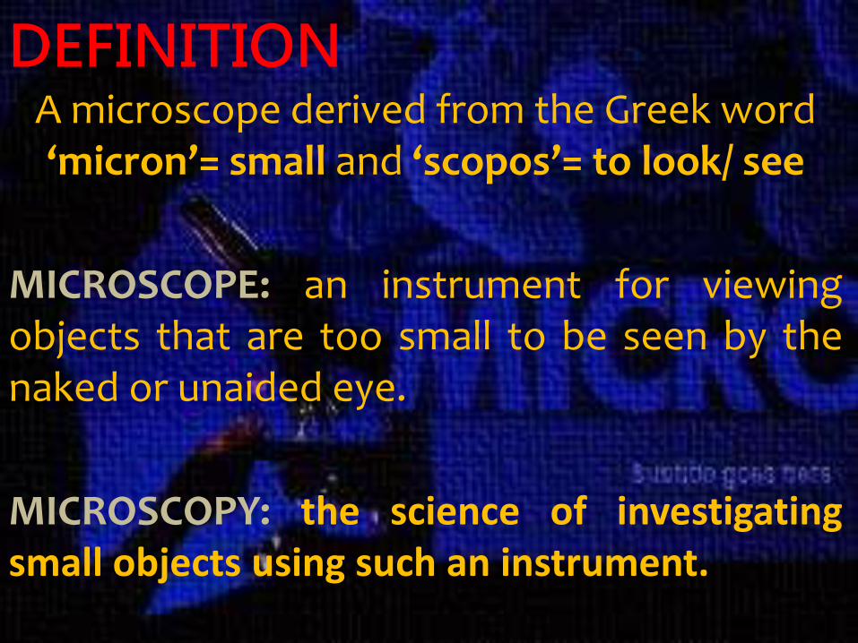
DEFINITIONA microscope derived from the Greek word ‘micron’= small and ‘scopos’= to look/ see
MICROSCOPE: an instrument for viewingobjects that are too small to be seen by thenaked or unaided eye.
MICROSCOPY: the science of investigatingsmall objects using such an instrument.

HISTORICAL BACKGROUND• 1590— Hans Janssen and his son Zacharias
Janssen, invented the first compound microscope.

• 1609—Galileo Galilei developed a compound microscope and he called it the "occhiolino" or "little eye."

• 1620— Christian Huygens, developed a simple2-lens ocular system that was chromaticallycorrected.

ANTON VAN LEEUWENHOEK –
He is commonly known as “THEFATHER OF MICROBIOLOGY", andconsidered to be the first microbiologist. Heis best known for his work on theimprovement of the microscope and for hiscontributions towards the establishment ofmicrobiology.
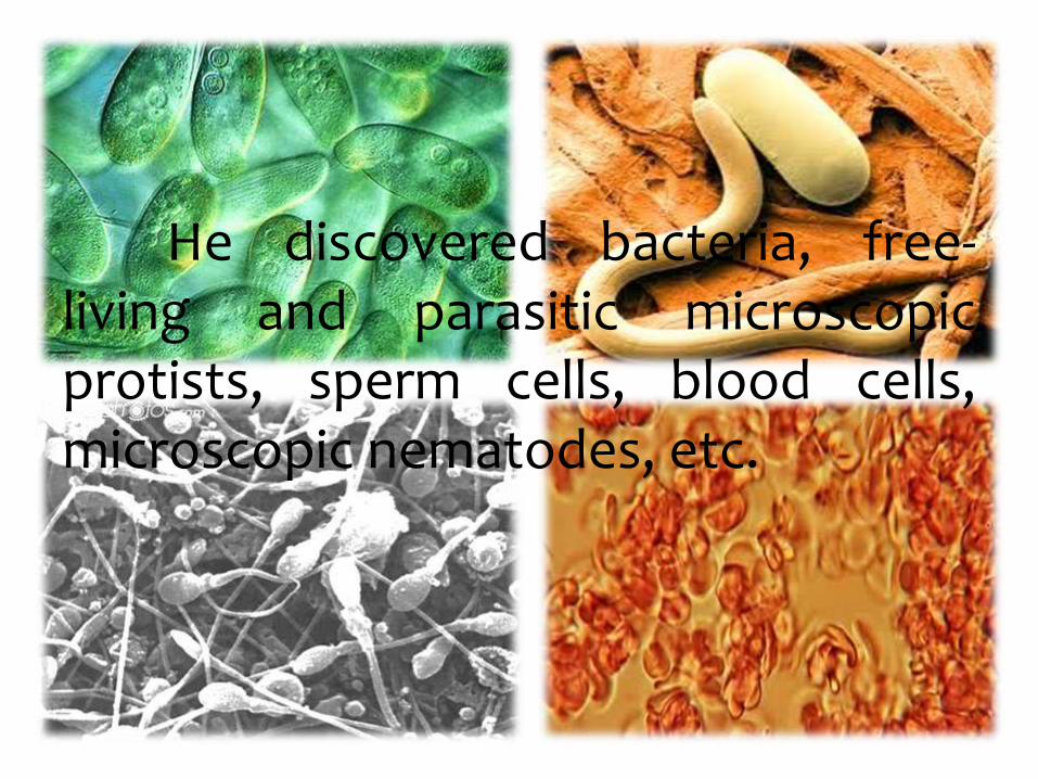
He discovered bacteria, free-living and parasitic microscopicprotists, sperm cells, blood cells,microscopic nematodes, etc.
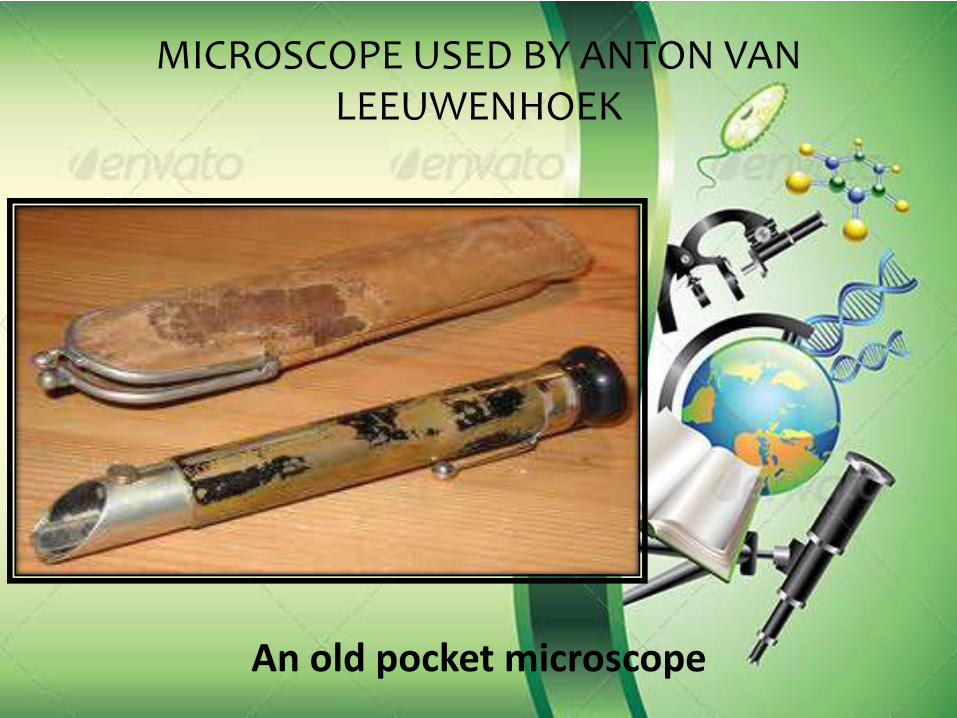
MICROSCOPE USED BY ANTON VAN LEEUWENHOEK
An old pocket microscope

What you can see in the
microscope?
MICROSCOPIC
MONSTERS

When viewed up close beneath the unblinking eye of the microscope, the tiniest mites and most
harmless of insects become terrifying beasts to haunt your dreams."
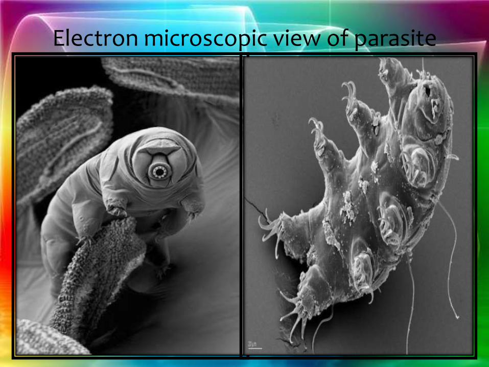
Electron microscopic view of parasite

Mouth of a round worm parasite
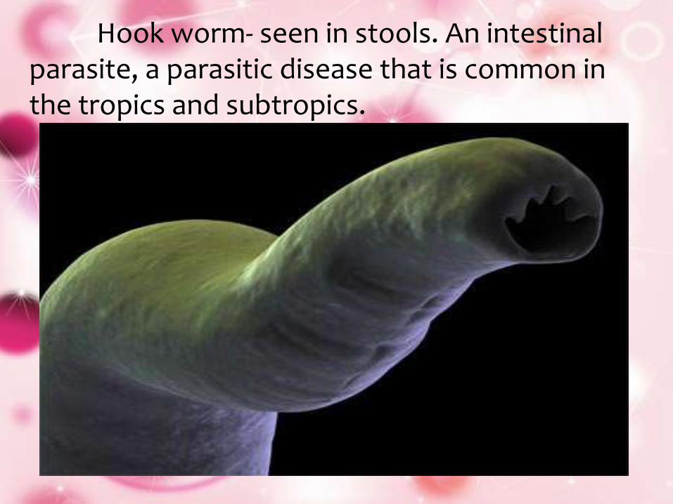
Hook worm- seen in stools. An intestinal parasite, a parasitic disease that is common in the tropics and subtropics.

SCABIES
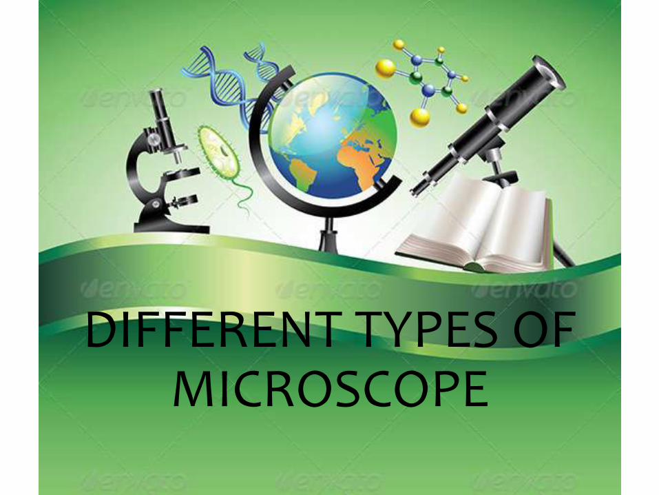
DIFFERENT TYPES OF MICROSCOPE

X-RAY MICROSCOPE

NEUTRON MICROSCOPE

SCANNING HELIUM ION MICROSCOPE (SHIM OR HEIM)
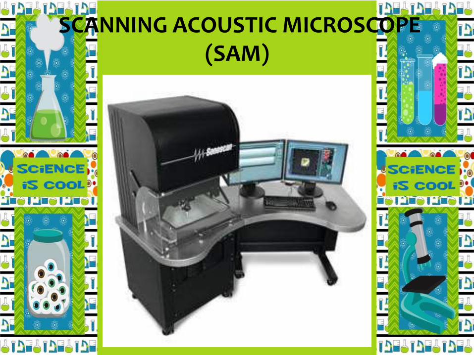
SCANNING ACOUSTIC MICROSCOPE (SAM)

Electron Microscope
High level of technical skill is needed
Specimen preparation often takes
several days
Only dead and dried specimen
can be seen
All lenses are electromagnets
Very costly and heavy running cost

EXAMPLES OF ELECTRON MICROSCOPE

TRANSMISSION ELECTRON MICROSCOPY (TEM)

SCANNING ELECTRON MICROSCOPY (SEM)

SCANNING PROBE MICROSCOPES

Optical/ Light Microscope
–Techniques are simple
–Specimen preparation normally
takes a few minutes to a few hours
–Live and dead specimen
can be seen
–Condenser, objective and eye
piece lenses are made of glass
–Cheap and negligible running cost

EXAMPLES OF OPTICAL MICROSCOPE
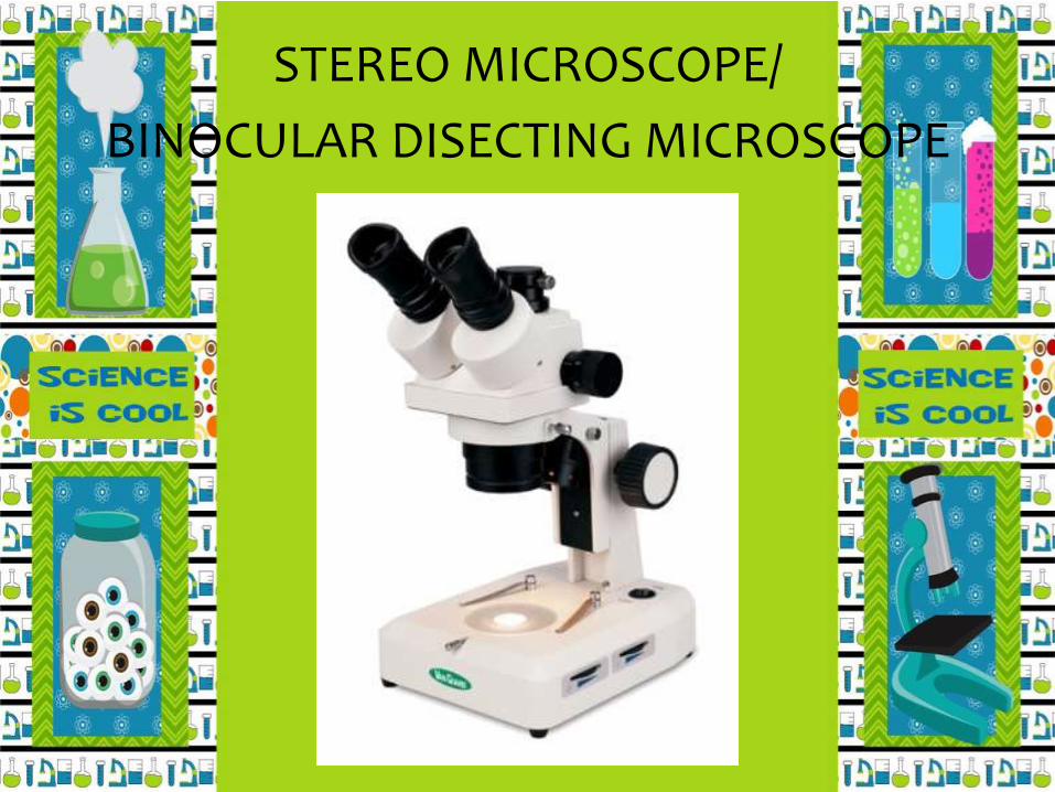
STEREO MICROSCOPE/
BINOCULAR DISECTING MICROSCOPE
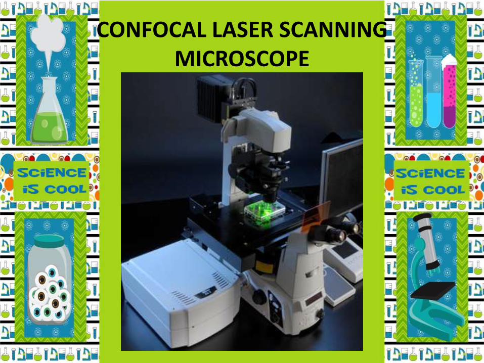
CONFOCAL LASER SCANNING MICROSCOPE

COMPOUND MICROSCOPE— anoptical instrument for formingmagnified images of smallobjects, consisting of an objectivelens with a very short focal lengthand an eyepiece with a longerfocal length, both lensesmounted in the same tube.

SIMPLE MICROSCOPE—
uses a single lens
COMPOUND MICROSCOPE—uses a set of lenses or lens
system

COMPOUND MICROSCOPE

COMPOUND MICROSCOPEO
M
Mechanical Parts— used to support and adjust the parts
Magnifying Parts— used to enlarge the specimen
Illuminating Parts— used to provide light


LET’S HAVE AN EXCERCISE

ANSWERS
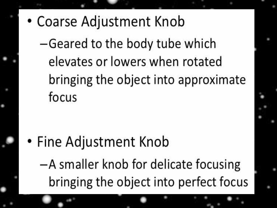
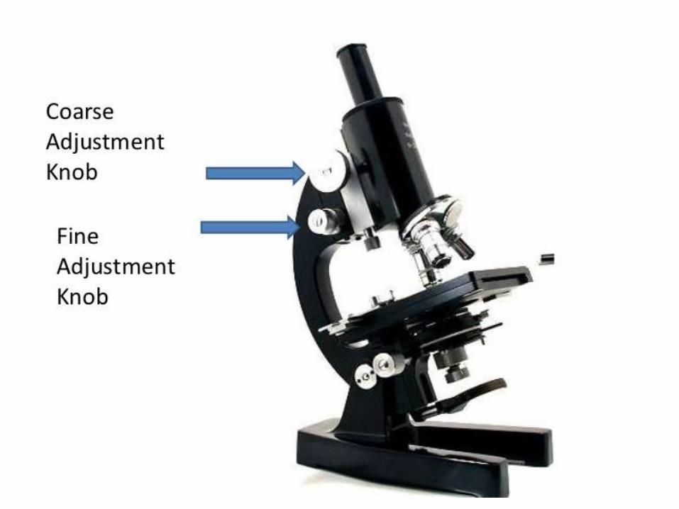
a. COARSE ADJUSTMENT KNOB
b. FINE ADJUSTMENT KNOB
c. ARM OR THE NECK
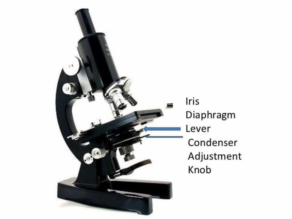
d. IRIS DIAPHRAGM LEVER
e. FINE ADJUSTMENT
f. MICROSCOPE BASE
g. OCULAR or THE EYEPIECE
h. BODY TUBE
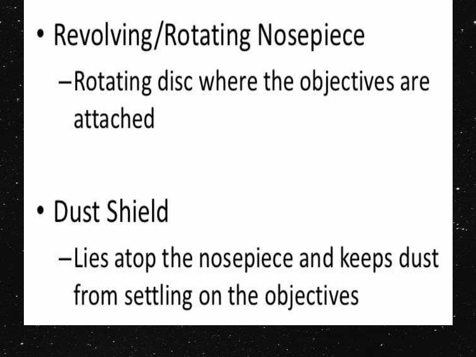
i. DUST SHIELD
J. OBJECTIVE
k. OBJECTIVE
l. STAGE
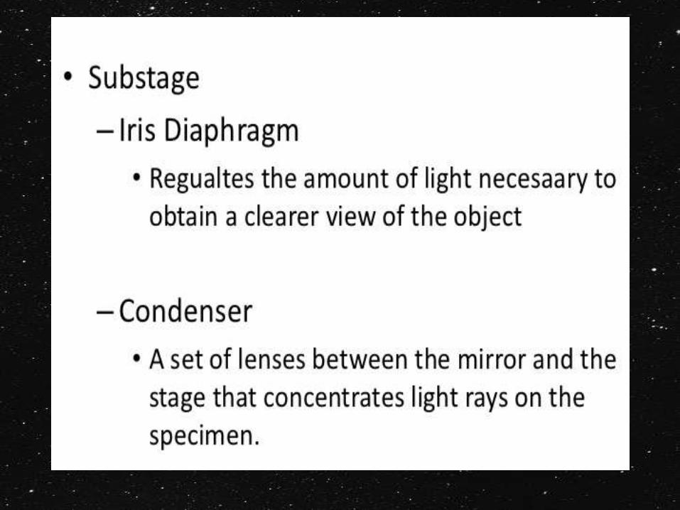
m. CONDENSOR
n. MIRROR OR ELECTRIC LAMP