APPLICATION OF SOLID PHASE MICROEXTRACTION IN GAHARU ESSENTIAL
Pertanika J. Trop. Agric. Sci. 36 (1): 43 - 50 (2013)
ISSN: 1511-3701 © Universiti Putra Malaysia Press
TROPICAL AGRICULTURAL SCIENCEJournal homepage: http://www.pertanika.upm.edu.my/
Article history:Received: 27 April 2011Accepted: 19 October 2011
ARTICLE INFO
E-mail addresses: [email protected] (Mohamed, R.), [email protected] (Wong, M. T.), [email protected] (Halis, R.)* Corresponding author
Microscopic Observation of ‘Gaharu’ Wood from Aquilaria malaccensis
Mohamed, R.1*, Wong, M. T.1 and Halis, R.2 1Forest Biotech Laboratory, Department of Forest Management, Faculty of Forestry, Universiti Putra Malaysia, 43400 Serdang, Selangor, Malaysia2Department of Forest Production, Faculty of Forestry, Universiti Putra Malaysia, 43400 Serdang, Selangor, Malaysia
ABSTRACT
Aquilaria produces fragrant wood known as ‘gaharu’ in its stem and branches, often in mature and damaged trees. In this study, anatomical characteristics in juvenile and mature trees were investigated by comparing their anatomical structures after various staining methods and direct observations under a light microscope. Juvenile and mature wood share similar anatomical structures. No major differences were observed other than the percentage of area covered by included phloem in juvenile was 2.16 times more than that of the mature wood. Microscopic observation revealed that in mature resinous wood, brownish bodies were found in ray and axial parenchyma, included phloem, xylem vessels and fibres, and this finding indicates that these are important elements for ‘gaharu’ depositing. Thus, it was concluded that juvenile tree possess the anatomical features of that of mature wood in producing ‘gaharu’.
Keywords: Gaharu, agarwood, anatomy, Aquilaria, included phloem
INTRODUCTION
Aquilaria is a member of the Thymelaeaceae family from the order Myrtales. Members of this genus are known to produce the prized ‘gaharu’ (which is also known as
‘agarwood’, ‘aloeswood’, ‘eaglewood’, ‘jinkoh’, and ‘agalloch’). ‘Gaharu’ is highly valuable for its fragrance and medicinal values and has been widely used for perfumery, incense and religious purposes, especially in the Middle East and Asian countries (Chakrabarty et al., 1994). ‘Gaharu’ contains resin and its biosynthesis is commonly associated with the tree’s defence system. Mechanical wounding has been shown to be the primary effect to
Mohamed, R., Wong, M. T. and Halis, R.
44 Pertanika J. Trop. Agric. Sci. 36 (1): 44 - 50 (2013)
commence ‘gaharu’ formation (Nobuchi & Mohd. Hamami, 2008; Pojanagaroon & Kaewrak, 2003; Rahman & Khisa, 1984) followed by fungal infection. Many varieties or genetic strains of fungi have been identified from ‘gaharu’-containing stems (Mohamed et al., 2010; Tabata et al., 2003).
The stem and branches of Aquilaria tree are the main organs where ‘gaharu’ is deposited. Meanwhile, sections on the wood reveal the existence of the ‘included phloem’, which is characterized by the strands or layers of the phloem tissue embedded in the
xylem (Esau, 2006). In Aquilaria, included phloem is produced internally by the cambium (Thouvenin, 1892; Van Tieghem, 1892) and the resin is concentrated mainly in the included phloem (Rao & Dayal, 1992). A few have reported the anatomical structures and changes on mature Aquilaria tree due to wounding damages. Among other, parenchyma cells have been shown to take part in resin formation as the cells showed abrupt changes in sapwood after mechanical wounding. Changes include the decrease and disappearance of starch grains in parenchyma cells, vacuolization, and the
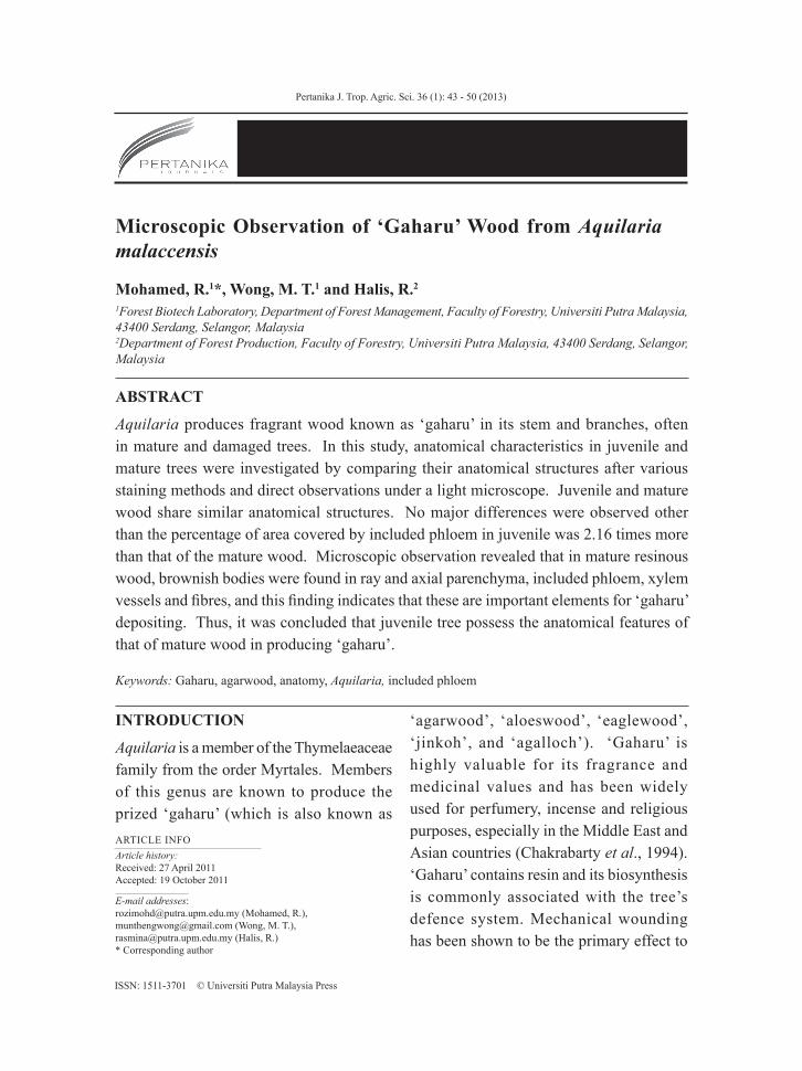
Fig.1: Photographs of Aquilaria malaccensis trees used in the experiments; (a) A three-year old juvenile tree growing in a polybag in the nursery, and (b) a mature tree from the wild
Microscopic Observation of ‘Gaharu’ Wood from Aquilaria malaccensis
45Pertanika J. Trop. Agric. Sci. 36 (1): 45 - 50 (2013)
appearance of brownish droplets following the disappearance of starch grains (Nobuchi & Mohd. Hamami, 2008). However, no report has been made on the anatomy of juvenile wood and its comparison to mature wood. In this work, key anatomical structures in the formation of ‘gaharu’ in A. malaccensis were identified and compared.
MATERIALS AND METHODS
A Comparison between Juvenile and Mature Wood
For the purpose of comparing between juvenile and mature wood, the juvenile wood samples of 3 cm thick (tangential) were taken from a 3-year old sapling (Fig.1a) that grew in a polybag at the nursery of the Faculty of Forestry, Universiti Putra Malaysia (UPM). The sapling was collected from natural population of Jeli, Kelantan. The tree was 2 m tall and had a diameter of 4 cm when measured at 5 cm above ground. Meanwhile, the samples of the mature wood were collected from a wild tree that was growing in Temerloh, Pahang (see Fig.1b). The tree had a diameter at breast height of about 1 m. The wood samples of 3 cm thick (tangential) were collected from parts of the trunk, which had wounding marks. The wood samples were subjected to sectioning and microscopic observations.
Fixation and Sectioning
The samples were fixed in 3% gluteraldehyde. For light microscopy (LM), serial transverse, radial and tangential sections of 20 μm thick were cut using a microtome knife on Leica SM 2000R sliding microtome (Leica,
Germany). Two staining methods were applied on the sections, namely, periodic acid-Shiff’s (PAS) staining and toluidine blue. For the PAS staining, the sections were oxidized with 1% periodic acid for 30 min, washed with three changes of distilled water, and then treated with Schiff’s reagent for 30 min in darkness. After washing with three changes of distilled water, the sections were dehydrated with a series of ethanol (once at 30% and 50%, followed by twice at 70%, and twice at 95%). The sections were then cleared in clove oil and mounted with Di-n-butylPhthalate in Xylene (DPX). For toluidine blue staining, the sections were stained for 3 min with 1% toluidine blue dissolved in distilled water. After washing with three changes of distilled water, the sections were mounted with glycerine. These were observed under the light microscope using 10x and 40x objective lenses and microphotographed using Leitz DMRB research microscope (Leica, Germany).
Measurements of the Included Phloem
The length and width of the included phloem were measured directly from the transverse sections using an image analyzer (Leica, Germany). One hundred measurements were chosen randomly to calculate the mean and standard deviation. Statistical analysis was conducted using the SPSS software version 16.0.2 (IBM Corporation) with significance judge at P < 0.05 in independent sample t-test. The percentage of included phloem coverage (area of included phloem) over the total transverse section area was calculated.
Mohamed, R., Wong, M. T. and Halis, R.
46 Pertanika J. Trop. Agric. Sci. 36 (1): 46 - 50 (2013)
RESULTS AND DISCUSSION
Microscopic Observation of Juvenile and Mature Wood from Aquilaria malaccensis
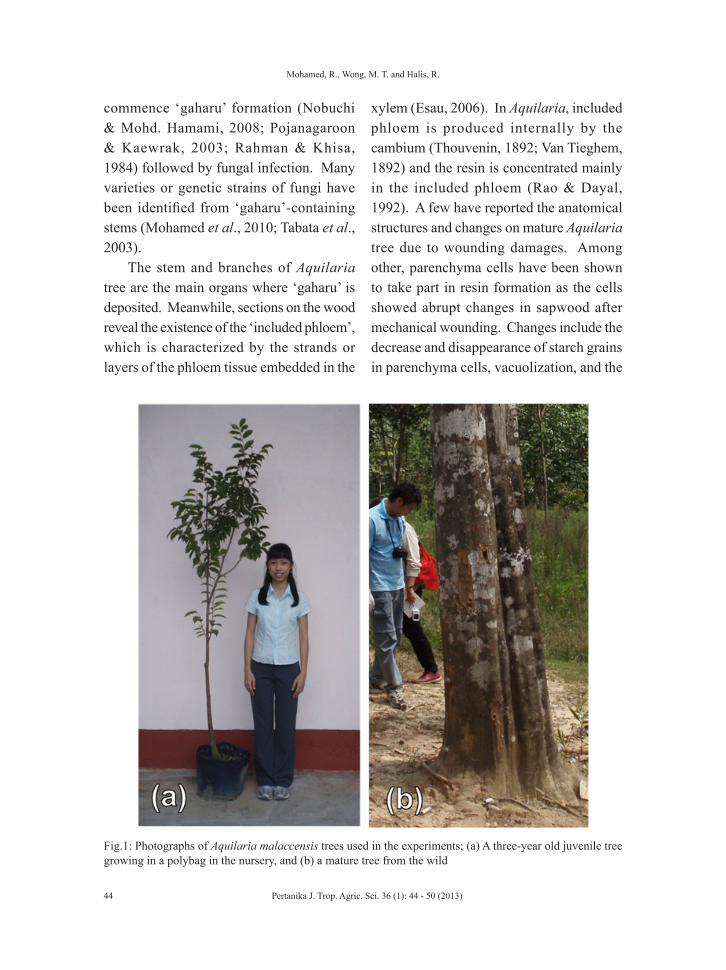
Juvenile wood that was used to characterize the anatomy of A. malaccensis was stained with toluidine blue. Transverse, radial and tangential sections of juvenile A. malaccensis are shown in Fig.2. Sections of the juvenile wood samples revealed the unique characteristic of the genus Aquilaria, which is the existence of included phloem (Fig.2a), when observed under the light microscope. The microscopic survey revealed element constituting xylem parts as
vessel elements, axial and ray parenchyma cells and wood fibres. The distribution of axial parenchyma is scanty paratracheal type and occasionally the parenchyma cells are associated with the vessels or form an incomplete sheath around the vessels (Fig.2b), and the ray parenchyma cells are uniseriate and multiseriate (Fig.2c). Observations described here are similar to that reported on A. crassna (Nobuchi & Siripatanadilok, 1991).
Since in natural forest, ‘gaharu’ is often found in old mature tree, the differences between juvenile and mature wood were
Fig.2: Light micrographs showing the anatomy of unwounded juvenile tree; Top: Transverse sections showing (a) included phloem (Toluidine blue. Scale bar=200 µm), (b) scanty paratracheal axial parenchyma (arrows) (Toluidine blue. Scale bar=30 µm), and (c) tangential section showing uniseriate and multiseriate ray parenchyma cell (arrows) (Toluidine blue. Scale bar=200 µm (arrows). V: vessel, F: fibre, IP: included phloem, AP: axial parenchyma
Microscopic Observation of ‘Gaharu’ Wood from Aquilaria malaccensis
47Pertanika J. Trop. Agric. Sci. 36 (1): 47 - 50 (2013)
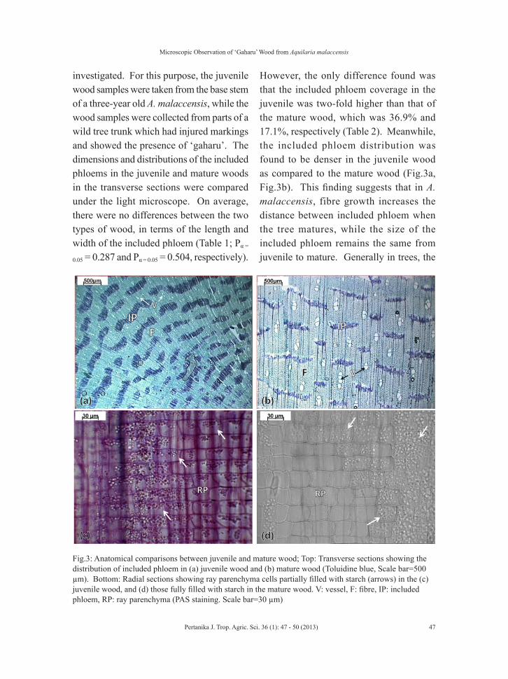
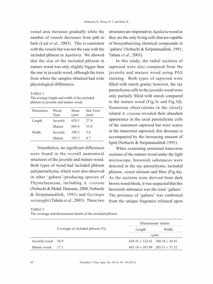
investigated. For this purpose, the juvenile wood samples were taken from the base stem of a three-year old A. malaccensis, while the wood samples were collected from parts of a wild tree trunk which had injured markings and showed the presence of ‘gaharu’. The dimensions and distributions of the included phloems in the juvenile and mature woods in the transverse sections were compared under the light microscope. On average, there were no differences between the two types of wood, in terms of the length and width of the included phloem (Table 1; Pα =
0.05 = 0.287 and Pα = 0.05 = 0.504, respectively).
However, the only difference found was that the included phloem coverage in the juvenile was two-fold higher than that of the mature wood, which was 36.9% and 17.1%, respectively (Table 2). Meanwhile, the included phloem distribution was found to be denser in the juvenile wood as compared to the mature wood (Fig.3a, Fig.3b). This finding suggests that in A. malaccensis, fibre growth increases the distance between included phloem when the tree matures, while the size of the included phloem remains the same from juvenile to mature. Generally in trees, the
Fig.3: Anatomical comparisons between juvenile and mature wood; Top: Transverse sections showing the distribution of included phloem in (a) juvenile wood and (b) mature wood (Toluidine blue, Scale bar=500 µm). Bottom: Radial sections showing ray parenchyma cells partially filled with starch (arrows) in the (c) juvenile wood, and (d) those fully filled with starch in the mature wood. V: vessel, F: fibre, IP: included phloem, RP: ray parenchyma (PAS staining. Scale bar=30 µm)
Mohamed, R., Wong, M. T. and Halis, R.
48 Pertanika J. Trop. Agric. Sci. 36 (1): 48 - 50 (2013)
vessel area increases gradually while the number of vessels decreases from pith to bark (Leal et al., 2003). This is consistent with the vessels but was not the case with the included phloem in Aquilaria. We showed that the size of the included phloem in mature wood was only slightly bigger than the one in juvenile wood, although the trees from where the samples obtained had wide physiological differences.
TABLE 1 The average length and width of the included phloem in juvenile and mature wood.
Dimension Wood Type
Mean(µm)
Std. Error(µm)
Length Juvenile 620.3 27.8
Mature 666.4 33.0
Width Juvenile 188.3 5.6
Mature 193.3 4.7
Nonetheless, no significant differences were found in the overall anatomical structures of the juvenile and mature wood. Both types of wood had included phloem and parenchyma, which were also observed in other ‘gaharu’-producing species of Thymelaeaceae, including A. crassna (Nobuchi & Mohd. Hamami, 2008; Nobuchi & Siripatanadilok, 1991) and Gyrinops versteeghii (Tabata et al., 2003). These two
structures are important in Aquilaria wood as they are the only living cells that are capable of biosynthesizing chemical compounds in ‘gaharu’ (Nobuchi & Siripatanadilok, 1991; Tabata et al., 2003).
In this study, the radial sections of sapwood were also compared from the juvenile and mature wood using PAS staining. Both types of sapwood were filled with starch grains; however, the ray parenchyma cells in the juvenile wood were only partially filled with starch compared to the mature wood (Fig.3c and Fig.3d). Numerous observations in the closely related A. crassna revealed their abundant appearance in the axial parenchyma cells of the outermost sapwood but were scarce in the innermost sapwood; this decrease is accompanied by the increasing amount of lipid (Nobuchi & Siripatanadilok 1991).
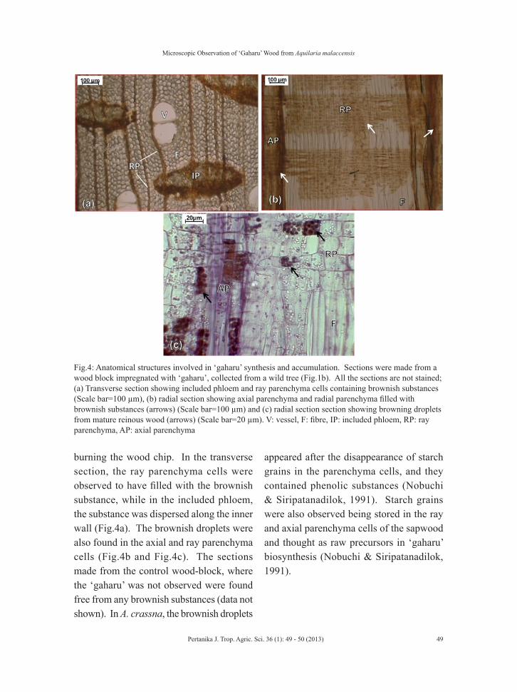
When examining unstained transverse sections of the mature wood under the light microscope, brownish substances were detected in the ray parenchyma, included phloem, vessel element and fibre (Fig.4a). As the sections were derived from dark brown wood-block, it was suspected that this brownish substance was the resin ‘gaharu’. The presence of ‘gaharu’ was confirmed from the unique fragrance released upon
TABLE 2 The coverage and dimensional details of the included phloem.
Coverage of included phloem (%)
Dimensional details
Length Width (µm)
Juvenile wood 36.9 620.35 ± 122.41 188.34 ± 36.41
Mature wood 17.1 663.10 ± 107.09 205.11 ± 37.32
Microscopic Observation of ‘Gaharu’ Wood from Aquilaria malaccensis
49Pertanika J. Trop. Agric. Sci. 36 (1): 49 - 50 (2013)
burning the wood chip. In the transverse section, the ray parenchyma cells were observed to have filled with the brownish substance, while in the included phloem, the substance was dispersed along the inner wall (Fig.4a). The brownish droplets were also found in the axial and ray parenchyma cells (Fig.4b and Fig.4c). The sections made from the control wood-block, where the ‘gaharu’ was not observed were found free from any brownish substances (data not shown). In A. crassna, the brownish droplets
appeared after the disappearance of starch grains in the parenchyma cells, and they contained phenolic substances (Nobuchi & Siripatanadilok, 1991). Starch grains were also observed being stored in the ray and axial parenchyma cells of the sapwood and thought as raw precursors in ‘gaharu’ biosynthesis (Nobuchi & Siripatanadilok, 1991).
Fig.4: Anatomical structures involved in ‘gaharu’ synthesis and accumulation. Sections were made from a wood block impregnated with ‘gaharu’, collected from a wild tree (Fig.1b). All the sections are not stained; (a) Transverse section showing included phloem and ray parenchyma cells containing brownish substances (Scale bar=100 µm), (b) radial section showing axial parenchyma and radial parenchyma filled with brownish substances (arrows) (Scale bar=100 µm) and (c) radial section section showing browning droplets from mature reinous wood (arrows) (Scale bar=20 µm). V: vessel, F: fibre, IP: included phloem, RP: ray parenchyma, AP: axial parenchyma
Mohamed, R., Wong, M. T. and Halis, R.
50 Pertanika J. Trop. Agric. Sci. 36 (1): 50 - 50 (2013)
CONCLUSION
Little variation was found in the structure and cell compositions of the juvenile and mature trees undertaken in the current study. The distribution of the included phloem was twice more compact in the juvenile wood as compared to mature wood, while the dimensions of the included phloem showed no significant difference at all. The juvenile wood appeared to have the anatomical characteristics necessary in producing ‘gaharu’ because of the existence of parenchyma cells, and its included phloem were of comparable sizes to the mature wood. The findings of the current study suggest that ‘gaharu’ can possibly be induced in juvenile trees as young as 3 years old.
ACKNOWLEDGEMENTS
The authors are grateful for the financial support from the Ministry of Higher Education, Malaysia, under the Fundamental Research Grant Scheme (Project No: 01-01-07-069FR). The authors would also like to thank Professor Dr. Tadashi Nobuchi (previously with Kyoto University, Japan, and Universiti Putra Malaysia) for his technical assistance and valuable suggestions.
REFERENCESChakrabarty, K., Kumar, A., & Menon, V. (1994).
Trade In Agarwood. New Delhi: TRAFFIC India and WWF-India.
Esau, K. (2006). Phloem: Cells types and development aspects. In K. Esau (ed.), Esau’s plant anatomy (pp. 358). New Jersey: John Wiley & Sons.
Leal, S., Pereira, H., Grabner, M., & Wimmer, R. (2003). Clonal and site variation of vessels in 7-year-old Eucalyptus globulosus. IAWA Journal, 24, 185-195.
Mohamed, R., Jong, P. L., & Zali, M. S. (2010). Fungal diversity in wounded stems of Aquilaria malaccensis. Fungal Diversity, 43, 67-74.
Nobuchi, T., & Mohd. Hamami, S. (2008). The formation of wood in tropical trees: a challenge from the perspective of functional wood anatomy. Serdang: Penerbit Universiti Putra Malaysia.
Nobuchi, T., & Siripatanadilok, S. (1991). Cytological observations of Aquilaria crassna wood associated with the formation of aloeswood. Bulletin of Kyoto University Forest, 63, 226-235.
Pojanagaroon, S., & Kaewrak, C. (2003). Mechanical methods to stimulate aloeswood formation in Aquilaria crassna Pierre ex H. Lec. (Kristana) trees. Acta Horticulturae, 676, 161-166.
Rahman, M. A., & Khisa, S. K. (1984). Agar production in agar tree by artificial inoculation and wounding II. Bano Biggyan Patrika, 13,57-63.
Rao, K. R., & Dayal, R. (1992.) The secondary xylem of Aquilaria agallocha (Thymelaeaceae) and the formation of agar. IAWA Bulletin, 13(2),163-172.
Tabata, Y., Widjaja, E., Mulyaningsih, T., Parman, I., Wiriadinata, H., Mandang, Y. I., & Itoh, T. (2003.). Wood Research. Bulletin of the Wood Research Institute Kyoto Univiversity, 90,11-12.
Thouvenin, M. (1892). Sur la structure des Aquilaria. Journal de Botanique, 212-215.
Van Tieghem, M. P. (1892). Sur la structure des Aquilaria. Journal de Botanique, 217-219.