Micropropagation, in vitro flowering and cytological studies of...
10
Micropropagation, in vitro flowering and cytological studies of Bacopa chamaedryoides, an ethno-medicinal plant Sk Moquammel Haque, Biswajit Ghosh* Plant Biotechnology Laboratory, Department of Botany, Ramakrishna Mission Vivekananda Centenary College, Rahara, Kolkata – 700118, India *Corresponding author, E-mail: [email protected] Abstract is is the first report of both establishment of micropropagation protocol as well as cytological studies of Bacopa chamaedryoides (Kunth) Wettst., an important Indian ethno-medicinal herb. Shoot-tips and nodal segments explants were inoculated on Murashige and Skoog basal medium containing 0.7% agar, 3.0% sucrose and different concentrations and combinations of cytokinins and auxins. Optimum multiplication was achieved on medium containing 6-benzyl-aminopurine (2.0 mg L –1 ) and indole-3-acetic acid (0.2 mg L –1 ). Shoot-tips proved to be a better explant in having a high rate of shoot multiplication (18.7 ± 0.17) in comparison to nodal segments (15.1 ± 0.18) in the same medium. In vitro rooting of multiplied individual shoots was achieved on half strength Murashige and Skoog medium supplemented with 50% of ‘Aloe vera gel’, with a maximum of 18.3 ± 0.17 roots. Up to 66.7% of these multiplied shoots induced healthy flowers in vitro on Murashige and Skoog medium containing low concentration of 6-benzyl-aminopurine (0.2 mg L –1 ). In vitro produced flowers contained 96.54 % viable pollen, more or less same as the field grown mother plants. Micropropagated plants have shown normal diploid 2n = 22 chromosomes, same as that of the mother plant. ese micropropagated plants were successfully established in soil aſter hardening them in submerged condition, with an 84% survival rate. In total, 88.9% of the survived plants flowered and fruited normally aſter 50 days of field transfer. More than 85% field-grown regenerated plants developed normal fruits and viable seeds. is work presents an efficient micropropagation method from node and shoot-tip culture for mass propagation and in vitro flowering of B. chamaedryoides. Key words: Aloe vera gel, auxins, Bacopa chamaedryoides, cytokinins, cytology, in vitro flowering, pollen viability. Abbreviations: AvG, Aloe vera gel; BAP, 6-benzyl-aminopurine; KIN, kinetin; NAA, α-naphthaleneacetic acid; IAA, indole-3-acetic acid; IBA, indole-3-butyric acid; MS, Murashige and Skoog; PGRs, plant growth regulators. Environmental and Experimental Biology (2013) 11: 59–68 Original Paper Introduction e genus Bacopa is a well known member of the family Scrophulariaceae. Bacopa monnieri L. has use as a natural brain stimulant. Bacopa chamaedryoides (Kunth) Wettst. (Syn. Herpestis chamaedryoides Kunth) also has this use and is considered as an important medicinal plant in India (Prain 1903; Kirtikar, Basu 1975). Historically, tribal people and modern communities, the common people of India, have been using this traditional medicinal herb as a neuro-stimulant and for other purposes. Due to increasing demand of this plant species, it has now been depleted from its primary habitat. is species (B. chamaedryoides) propagates typically through seeds, but seed germination rate is poor and plants of high quality are not readily available due to seed heterozygosity. Micropropagation methods are now being used for the clonal propagation of many medicinal and other economically important plants in large scale for stable supply round the year. In vitro flowering provides (i) an ideal experimental system for studying phase transition from vegetative to floral development (Huang et al. 2009), and (ii) early in vitro flowering of micropropagated plantlets can shorten the breeding cycle to generate better quality of plant varieties that can meet the market demand (Sivanesan, Jeong 2007; Kiełkowska, Havey 2012; Haque, Ghosh 2013). Knowledge of chromosome structure has played a crucial role in the improvement of medicinally important plant species and has far-reaching implications (Samaddar et al. 2012). e results of chromosomal studies may be also useful in plant taxonomy and phylogenetic analysis. However, there are only a few reports of chromosome studies in the family Scrophulariaceae (Sinha 1984; Chandran, Bhavanandan 1986; Chandran, Bhavanandan 1987) probably due to small chromosomes. Recently, a chromosome study of B. monnieri was published by Samaddar et al. (2012), but there is no report on B. chamaedryoides. B. monnieri is the only species of Bacopa that has been studied by many groups for more than four decades in different aspects, including micropropagation as well as genetic transformation (akur et al. 1976; Tiwari et al. 2006; Ceasar et al. 2010; Joshi et al. 2010; Ramesh et al. 2011; Majumdar et al. 2011). So far, to our present knowledge, 59
Transcript of Micropropagation, in vitro flowering and cytological studies of...

Micropropagation, in vitro flowering and cytological studies of Bacopa chamaedryoides, an ethno-medicinal plant
Sk Moquammel Haque, Biswajit Ghosh*
Plant Biotechnology Laboratory, Department of Botany, Ramakrishna Mission Vivekananda Centenary College, Rahara, Kolkata – 700118, India
*Corresponding author, E-mail: [email protected]
Abstract
This is the first report of both establishment of micropropagation protocol as well as cytological studies of Bacopa chamaedryoides (Kunth) Wettst., an important Indian ethno-medicinal herb. Shoot-tips and nodal segments explants were inoculated on Murashige and Skoog basal medium containing 0.7% agar, 3.0% sucrose and different concentrations and combinations of cytokinins and auxins. Optimum multiplication was achieved on medium containing 6-benzyl-aminopurine (2.0 mg L–1) and indole-3-acetic acid (0.2 mg L–1). Shoot-tips proved to be a better explant in having a high rate of shoot multiplication (18.7 ± 0.17) in comparison to nodal segments (15.1 ± 0.18) in the same medium. In vitro rooting of multiplied individual shoots was achieved on half strength Murashige and Skoog medium supplemented with 50% of ‘Aloe vera gel’, with a maximum of 18.3 ± 0.17 roots. Up to 66.7% of these multiplied shoots induced healthy flowers in vitro on Murashige and Skoog medium containing low concentration of 6-benzyl-aminopurine (0.2 mg L–1). In vitro produced flowers contained 96.54 % viable pollen, more or less same as the field grown mother plants. Micropropagated plants have shown normal diploid 2n = 22 chromosomes, same as that of the mother plant. These micropropagated plants were successfully established in soil after hardening them in submerged condition, with an 84% survival rate. In total, 88.9% of the survived plants flowered and fruited normally after 50 days of field transfer. More than 85% field-grown regenerated plants developed normal fruits and viable seeds. This work presents an efficient micropropagation method from node and shoot-tip culture for mass propagation and in vitro flowering of B. chamaedryoides.
Key words: Aloe vera gel, auxins, Bacopa chamaedryoides, cytokinins, cytology, in vitro flowering, pollen viability.Abbreviations: AvG, Aloe vera gel; BAP, 6-benzyl-aminopurine; KIN, kinetin; NAA, α-naphthaleneacetic acid; IAA, indole-3-acetic acid; IBA, indole-3-butyric acid; MS, Murashige and Skoog; PGRs, plant growth regulators.
Environmental and Experimental Biology (2013) 11: 59–68 Original Paper
Introduction
The genus Bacopa is a well known member of the family Scrophulariaceae. Bacopa monnieri L. has use as a natural brain stimulant. Bacopa chamaedryoides (Kunth) Wettst. (Syn. Herpestis chamaedryoides Kunth) also has this use and is considered as an important medicinal plant in India (Prain 1903; Kirtikar, Basu 1975). Historically, tribal people and modern communities, the common people of India, have been using this traditional medicinal herb as a neuro-stimulant and for other purposes. Due to increasing demand of this plant species, it has now been depleted from its primary habitat. This species (B. chamaedryoides) propagates typically through seeds, but seed germination rate is poor and plants of high quality are not readily available due to seed heterozygosity. Micropropagation methods are now being used for the clonal propagation of many medicinal and other economically important plants in large scale for stable supply round the year. In vitro flowering provides (i) an ideal experimental system for studying phase transition from vegetative to floral development (Huang et al. 2009), and (ii) early in vitro
flowering of micropropagated plantlets can shorten the breeding cycle to generate better quality of plant varieties that can meet the market demand (Sivanesan, Jeong 2007; Kiełkowska, Havey 2012; Haque, Ghosh 2013).
Knowledge of chromosome structure has played a crucial role in the improvement of medicinally important plant species and has far-reaching implications (Samaddar et al. 2012). The results of chromosomal studies may be also useful in plant taxonomy and phylogenetic analysis. However, there are only a few reports of chromosome studies in the family Scrophulariaceae (Sinha 1984; Chandran, Bhavanandan 1986; Chandran, Bhavanandan 1987) probably due to small chromosomes. Recently, a chromosome study of B. monnieri was published by Samaddar et al. (2012), but there is no report on B. chamaedryoides.
B. monnieri is the only species of Bacopa that has been studied by many groups for more than four decades in different aspects, including micropropagation as well as genetic transformation (Thakur et al. 1976; Tiwari et al. 2006; Ceasar et al. 2010; Joshi et al. 2010; Ramesh et al. 2011; Majumdar et al. 2011). So far, to our present knowledge,
59
S.M. Haque, B. Ghosh
60
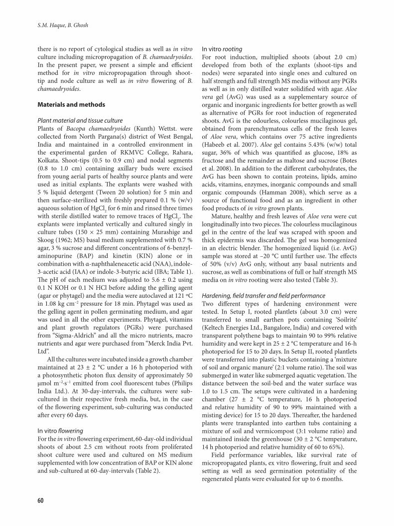
there is no report of cytological studies as well as in vitro culture including micropropagation of B. chamaedryoides. In the present paper, we present a simple and efficient method for in vitro micropropagation through shoot-tip and node culture as well as in vitro flowering of B. chamaedryoides.
Materials and methods
Plant material and tissue culturePlants of Bacopa chamaedryoides (Kunth) Wettst. were collected from North Pargana(s) district of West Bengal, India and maintained in a controlled environment in the experimental garden of RKMVC College, Rahara, Kolkata. Shoot-tips (0.5 to 0.9 cm) and nodal segments (0.8 to 1.0 cm) containing axillary buds were excised from young aerial parts of healthy source plants and were used as initial explants. The explants were washed with 5 % liquid detergent (Tween 20 solution) for 5 min and then surface-sterilized with freshly prepared 0.1 % (w/v) aqueous solution of HgCl2 for 6 min and rinsed three times with sterile distilled water to remove traces of HgCl2. The explants were implanted vertically and cultured singly in culture tubes (150 × 25 mm) containing Murashige and Skoog (1962; MS) basal medium supplemented with 0.7 % agar, 3 % sucrose and different concentrations of 6-benzyl-aminopurine (BAP) and kinetin (KIN) alone or in combination with α-naphthaleneacetic acid (NAA), indole-3-acetic acid (IAA) or indole-3-butyric acid (IBA; Table 1). The pH of each medium was adjusted to 5.6 ± 0.2 using 0.1 N KOH or 0.1 N HCl before adding the gelling agent (agar or phytagel) and the media were autoclaved at 121 ºC in 1.08 kg cm–2 pressure for 18 min. Phytagel was used as the gelling agent in pollen germinating medium, and agar was used in all the other experiments. Phytagel, vitamins and plant growth regulators (PGRs) were purchased from “Sigma-Aldrich” and all the micro nutrients, macro nutrients and agar were purchased from “Merck India Pvt. Ltd”.
All the cultures were incubated inside a growth chamber maintained at 23 ± 2 °C under a 16 h photoperiod with a photosynthetic photon flux density of approximately 50 μmol m–2·s–1 emitted from cool fluorescent tubes (Philips India Ltd.). At 30-day-intervals, the cultures were sub-cultured in their respective fresh media, but, in the case of the flowering experiment, sub-culturing was conducted after every 60 days.
In vitro floweringFor the in vitro flowering experiment, 60-day-old individual shoots of about 2.5 cm without roots from proliferated shoot culture were used and cultured on MS medium supplemented with low concentration of BAP or KIN alone and sub-cultured at 60-day-intervals (Table 2).
In vitro rootingFor root induction, multiplied shoots (about 2.0 cm) developed from both of the explants (shoot-tips and nodes) were separated into single ones and cultured on half strength and full strength MS media without any PGRs as well as in only distilled water solidified with agar. Aloe vera gel (AvG) was used as a supplementary source of organic and inorganic ingredients for better growth as well as alternative of PGRs for root induction of regenerated shoots. AvG is the odourless, colourless mucilaginous gel, obtained from parenchymatous cells of the fresh leaves of Aloe vera, which contains over 75 active ingredients (Habeeb et al. 2007). Aloe gel contains 5.43% (w/w) total sugar, 36% of which was quantified as glucose, 18% as fructose and the remainder as maltose and sucrose (Botes et al. 2008). In addition to the different carbohydrates, the AvG has been shown to contain proteins, lipids, amino acids, vitamins, enzymes, inorganic compounds and small organic compounds (Hamman 2008), which serve as a source of functional food and as an ingredient in other food products of in vitro grown plants.
Mature, healthy and fresh leaves of Aloe vera were cut longitudinally into two pieces. The colourless mucilaginous gel in the centre of the leaf was scraped with spoon and thick epidermis was discarded. The gel was homogenized in an electric blender. The homogenized liquid (i.e. AvG) sample was stored at –20 °C until further use. The effects of 50% (v/v) AvG only, without any basal nutrients and sucrose, as well as combinations of full or half strength MS media on in vitro rooting were also tested (Table 3).
Hardening, field transfer and field performanceTwo different types of hardening environment were tested. In Setup I, rooted plantlets (about 3.0 cm) were transferred to small earthen pots containing ‘Soilrite’ (Keltech Energies Ltd., Bangalore, India) and covered with transparent polythene bags to maintain 90 to 99% relative humidity and were kept in 25 ± 2 °C temperature and 16-h photoperiod for 15 to 20 days. In Setup II, rooted plantlets were transferred into plastic buckets containing a ‘mixture of soil and organic manure’ (2:1 volume ratio). The soil was submerged in water like submerged aquatic vegetation. The distance between the soil-bed and the water surface was 1.0 to 1.5 cm. The setups were cultivated in a hardening chamber (27 ± 2 °C temperature, 16 h photoperiod and relative humidity of 90 to 99% maintained with a misting device) for 15 to 20 days. Thereafter, the hardened plants were transplanted into earthen tubs containing a mixture of soil and vermicompost (3:1 volume ratio) and maintained inside the greenhouse (30 ± 2 °C temperature, 14 h photoperiod and relative humidity of 60 to 65%).
Field performance variables, like survival rate of micropropagated plants, ex vitro flowering, fruit and seed setting as well as seed germination potentiality of the regenerated plants were evaluated for up to 6 months.

In vitro propagation and flowering of Bacopa chamaedryoides
61
Pollen viability testPollen viability was estimated by two different methods: (i) staining of pollen with dyes and (ii) in vitro germination assays (Lyra et al. 2011). In the first method, fresh pollen was stained with 2% acetio-carmine dye and observed under a light microscope. Dark purple coloured pollen grains were scored as viable and the rest of the grains were scored as non-viable. In the second method, pollen grains were germinated in Petri dishes (60 mm diameter) containing 6 mL of medium. The nutrient medium consisted of 15% (w/v) sucrose, 300 mg L–1 calcium nitrate [Ca(NO3)2 4H2O], 200 mg L–1 magnesium sulphate (MgSO4 7H2O), 100 mg L–1 potassium nitrate (KNO3) and 100 mg L–1 boric acid (H3BO3), at pH 7.0 (Rajasekharan et al. 1994) and solidified with 0.2% (w/v) Phytagel. Pollen grains were strewn in a fine layer over the surface of the medium, incubated at 25 °C for 12 h in a dark chamber and then observed under light microscope. For photography, they were stained with 2% acetic carmine dye (Sawidis 2008). Observations were made on 10 flowers from each of the in vivo, in vitro and ex vitro plants and a minimum of 500 pollen grains from each flower.
Seed viability testViability of the seeds produced by field-grown regenerated plants and mother plants was tested through in vitro germination assay. A total of 1000 seeds from 30 randomly selected fruits of regenerated plants as well as 1000 seeds from 30 randomly selected fruits of mother plants were inoculated on half strength MS medium and the cultures were incubated at 23 ± 2 °C in dark.
Cytological observationsFor mitotic chromosome study, root tips of mother plants and 20 randomly selected micropropagated plants were pre-treated with 0.002 M 8-hydroxyquinoline for 5 h at 8 to 10 °C and fixed in Carnoy’s solution (6:3:1, absolute ethanol: chloroform: acetic acid) for overnight and stained with 2% aceto-orcein : 1 N HCl (9:1 v/v) mixture (Ghosh, Sen 1996). In total, 6 to 8 root tips from each plant and a minimum of five metaphase plates from each root tip were analyzed.
Statistical analysisEach treatment contained three replicates with 10 explants per replicate. The data pertaining to the number of shoots or roots per explant, shoot and root length, number of flowers per shoot were subjected to a one-way analysis of variance (ANOVA). The differences among the means were compared by high-range statistical domain using Tukey’s test with the standard software SPSS 16.0 version.
Results
Shoot multiplication from nodal explantsThe effect of different concentrations ranging from 0.5 mg
L–1 to 5.0 mg L–1 of BAP and KIN alone on in vitro shoot multiplication from node cultures was investigated. It was found that initially two to four shoot buds induced from each node after 12 to 15 days of implantation, followed by swelling at the base of the node, and gradually the number increased. A maximum of 9.5 ± 0.17 shoots were induced in MS basal medium containing 2.0 mg L–1 BAP, whereas in presence of 1.0 and 5.0 mg L–1 BAP only 6.3 ± 0.13 and 2.7 ± 0.16 shoots were induced after 60 days of culture, respectively (Table 1). On the other hand, maximum 6.8 ± 0.20 shoots from each node were induced in the presence of 2.0 mg L–1 KIN. In the present study, BAP alone was sufficient for formation of multiple shoots from nodal segments of B. chamaedryoides. But at higher concentrations, it not only reduced the number of shoots induced but also resulted in stunted growth of the multiplied shoots. All the three auxins (NAA, IAA, IBA) in low concentration, along with optimum concentration of BAP (2.0 mg L–1), significantly increased the multiplication rate as well as the elongation of multiplied shoots from nodal explants. MS basal medium supplemented with 2.0 mg L–1 BAP + 0.2 mg L–1 IAA was found to be the most suitable with maximum 15.1 ± 0.18 shoots per node induced after 60 days of culture, whereas 2.0 mg L–1 BAP with 0.2 mg L–1 NAA and with 0.2 mg L–1
IBA induced maximum 12.9 ± 0.17 and 12.7 ± 0.18 shoots per node after 60 days of culture, respectively (Table 1). Although all three auxins increased the multiplication rate, IAA gave a significantly better result, but the difference between IBA and NAA was insignificant. Maximum shoot length of 3.0 ± 0.04 cm was observed with 2.0 mg L–1 BAP + 0.5 mg L–1 IAA after 60 days of culture.
Shoot multiplication from shoot-tip explantsFrom shoot-tip explants, a maximum of 12.2 ± 0.28 shoots were induced in MS basal medium containing 2.0 mg L–1 BAP after 60 days of culture (Fig. 1A), while 8.7 ± 0.20 shoots were induced in the presence of 2.0 mg L–1 KIN; this proved BAP to be more efficient than KIN for shoot proliferation from shoot-tip explants (Table 1).
Similar to nodal explants, all three auxins (NAA, IAA, IBA) in low concentration along with optimum concentration of BAP (2.0 mg L–1) had beneficial effect on multiplication rate and elongation of multiplied shoots from shoot-tip explants of B. chamaedryoides. Maximum 18.7 ± 0.17 shoots from each shoot-tip explant were induced in MS basal medium supplemented with 2.0 mg L–1 BAP with 0.2 mg L–1 IAA after 60 days of culture (Fig. 1B). Of the three auxins used, IAA was most effective followed by NAA and IBA (Table 1, Fig. 2). MS basal medium supplemented with 2.0 mg L–1 BAP + 0.2 mg L–1 NAA and 2.0 mg L–1 BAP + 0.2 mg L–1 IBA induced 16.3 ± 0.19 and 14.2 ± 0.21 shoots from each shoot-tip explant after 60 days of culture, respectively. Maximum elongation of shoots reaching 3.1 ± 0.04 cm was recorded on 2.0 mg L–1 BAP + 0.5 mg L–1 IAA after 60 days of culture (Table 1).

S.M. Haque, B. Ghosh
62
In vitro floweringBoth BAP and KIN alone, in low concentration, induced in vitro flowering of B. chamaedryoides, and BAP was found to be more effective than KIN (Table 2). Maximum number of explants with induced flowering (66.7%) was observed in 0.2 mg L–1 BAP-containing MS medium and maximum number of flowers per plant (4.2 ± 0.24) was maintained in the same medium without any subculturing after 60 days of culture. In total, 43.3 % of explants produced flowers and the highest number of flowers per plant (2.9 ± 0.21) was recorded in 0.2 mg L–1 KIN-containing medium after the same period of culture. In the present study, in vitro rooting was first observed within 15 days of culture. Thereafter, the first flower appeared at the nodal region during 30 to 32 days of culture in the flower inducing media and later, one to five more flowers per plant developed within the next 28 days. Within 60 days of culture, individual shoot formed roots as well as flowers without need for subcultivation. These in vitro induced flowers were yellow and had normal petals and sepals. Few nodes developed a single flower (Fig. 1D, E) and a few developed two flowers from the axil of two opposite decussate leaves (Fig. 1F). It was noted that in vitro induced flowers exhibited the same morphology as the mother plant. The calyx of these flowers persisted for more than eight weeks in in vitro conditions. However, no fruits developed due to lack of pollination.
It was found also that B. chamaedryoides produced flowers spontaneously in MS medium without any PGRs,
but the percentage of explants forming flowers (23.3%) and the number of flowers per plant (1.4 ± 0.20) was low, compared to PGRs-containing medium (Table 2). The subculturing time for flower induction can substantially affect in vitro flowering. A period of 60 days in flower induction medium without subculture yielded the highest percentage of plantlets forming flowers (66.7%), but when subculturing time was decreased to 30 days on flower-induction medium, the frequency of flowering decreased significantly (data not shown).
Root inductionThe excised shoots showed 100% rooting with no callusing at the cut ends within 10 days of subculture in all treatments. In vitro rooting from the basal end of the shoots occurred only on distilled water (without MS nutrients, sucrose) solidified with agar without any PGRs, but growth of these plantlets was stunted (only 2.8 ± 0.02 cm after 15 days from initial height of 2.5 cm). However, in half-strength MS medium, plantlets grew to 3.6 ± 0.02 cm height within 15 days (Table 3). It was noted that inclusion of 50% (v/v) AvG as an organic supplement along with half-strength MS nutrients, triggered rapid growth of plantlets and they attained 4.9 ± 0.02 cm plant height along with maximum number of roots (18.3 ± 0.17). Also, 2.3 ± 0.02 cm root length was achieved in the same media within 15 days of culture (Table 3, Fig. 1C). In our study, only 50% AvG without any MS nutrient and sucrose was found to be
Table 1. Effect of growth regulators on number of shoots, length of the longest shoot in B. chamaedroides (after 60 days of cultivation). Each value represents the mean ± SE, n = 30. Mean followed by the same letters in each column are not significantly different at P < 0.05 according to Tukey’s multiple range tests
Treatment PGRs Concentration Nodal explant Shoot-tip explant (mg L–1) Response Shoot number Shoot Response Shoot number Shoot (%) (explant–1) length (cm) (%) (explant–1) length (cm)Control – 0 86.7 1.8 ± 0.08a 6.2 ±0.06k 90.0 2.2 ± 0.12a 6.8 ± 0.07k
Cytokinin BAP 0.5 83.3 4.1 ± 0.20d 1.2 ± 0.03d 83.3 6.9 ± 0.19d 1.2 ± 0.06de
1.0 76.7 6.3 ± 0.13f 1.0 ± 0.04cd 83.3 9.2 ± 0.18f 1.1 ± 0.05cde
2.0 80.0 9.5 ± 0.17g 0.8 ± 0.03b 76.7 12.2 ± 0.28g 0.9 ± 0.03bc
5.0 70.0 2.7 ± 0.16b 0.5 ± 0.03a 73.3 4.1 ± 0.17b 0.6 ± 0.03a
KIN 0.5 80.0 3.7 ± 0.21cd 1.2 ± 0.04d 83.3 5.2 ± 0.14c 1.3 ± 0.04e
1.0 83.3 5.2 ± 0.13e 1.0 ± 0.04d 83.3 7.8 ± 0.15e 1.2 ± 0.04de
2.0 76.7 6.8 ± 0.20f 0.8 ± 0.03bc 80.0 8.7 ± 0.20ef 1.0 ± 0.03cd
5.0 63.3 3.3 ± 0.17bc 0.6 ± 0.03a 70.0 4.3 ± 0.15b 0.7 ± 0.03ab
Cytokinin + auxin BAP + NAA 2+0.2 76.7 12.9 ± 0.17h 2.6 ± 0.04gh 76.7 16.3 ± 0.19j 2.7 ± 0.04h
2+0.5 76.7 12.6 ± 0.15h 2.9 ± 0.03ij 80.0 15.5 ± 0.22j 2.9 ± 0.04i
2+1.0 66.7 10.3 ± 0.16g 2.0 ± 0.04e 76.7 13.9 ± 0.17i 2.4 ± 0.03g
BAP + IAA 2+0.2 80.0 15.1 ± 0.18i 2.7 ± 0.04gh 86.7 18.7 ± 0.17l 2.9 ± 0.04ij
2+0.5 76.7 14.5 ± 0.15i 3.0 ± 0.04j 80.0 17.3 ± 0.15k 3.1 ± 0.04j
2+1.0 73.3 12.0 ± 0.16h 2.4 ± 0.04f 70.0 15.8 ± 0.28j 2.4 ± 0.03g
BAP + IBA 2+0.2 76.7 12.7 ± 0.18h 2.6 ± 0.04g 80.0 14.2 ± 0.21i 2.7 ± 0.04h
2+0.5 70.0 12.3 ± 0.16h 2.8 ± 0.03hi 66.7 13.3 ± 0.24hi 2.8 ± 0.04hi
2+1.0 63.3 10.0 ± 0.18g 1.9 ± 0.04e 63.3 12.9 ± 0.17gh 2.2 ± 0.04f
In vitro propagation and flowering of Bacopa chamaedryoides
63
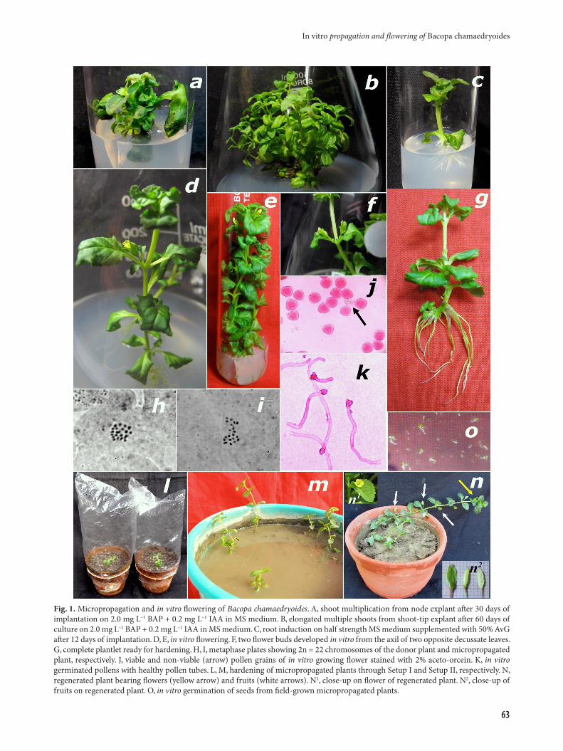
Fig. 1. Micropropagation and in vitro flowering of Bacopa chamaedryoides. A, shoot multiplication from node explant after 30 days of implantation on 2.0 mg L–1 BAP + 0.2 mg L–1 IAA in MS medium. B, elongated multiple shoots from shoot-tip explant after 60 days of culture on 2.0 mg L–1 BAP + 0.2 mg L–1 IAA in MS medium. C, root induction on half strength MS medium supplemented with 50% AvG after 12 days of implantation. D, E, in vitro flowering. F, two flower buds developed in vitro from the axil of two opposite decussate leaves. G, complete plantlet ready for hardening. H, I, metaphase plates showing 2n = 22 chromosomes of the donor plant and micropropagated plant, respectively. J, viable and non-viable (arrow) pollen grains of in vitro growing flower stained with 2% aceto-orcein. K, in vitro germinated pollens with healthy pollen tubes. L, M, hardening of micropropagated plants through Setup I and Setup II, respectively. N, regenerated plant bearing flowers (yellow arrow) and fruits (white arrows). N1, close-up on flower of regenerated plant. N2, close-up of fruits on regenerated plant. O, in vitro germination of seeds from field-grown micropropagated plants.

enough for in vitro rooting of B. chamaedryoides. However, with respect to the growth rate and health of the plantlets (plant height, number of root, number and size of leaf; i.e. overall visual appearance), 50% AvG along with half-strength MS medium was found to be the best. We also found that the number of roots dramatically increased along with 100% response on AvG-containing media (Table 3). According to our findings, ≥ 1.0 mg L–1 of IBA inhibited root induction, while ≤ 0.5 mg L–1 of IBA induced large numbers (up to 29) of roots, further growth of these roots was inhibited by IBA and stopped after reaching 3 to 5 mm.
Hardening and field transferA total of 150 in vitro rooted plantlets (Fig. 1G), 75 plantlets each on Setup I (Fig. 1L) and Setup II (Fig. 1M) were hardened for 15 to 20 days. Rooted plantlets were successfully hardened with 53.3 % survival rate on Setup
I and 84% survival rate on Setup II. Hardened plants were then transferred to earthen tubs filled with mixture of soil and vermicompost at 3:1 ratio and ultimately established in soil. After 40 to 50 days of field transfer, 85.4 to 88.9 % (Fig. 3) of the survived plants flowered and fruited normally (Fig. 1N).
Pollen viabilityThe pollen grains of in vitro, ex vitro and in vivo plants showed 96.54, 94.82 and 97.20% viability, respectively by aceto-carmine staining method (Fig. 1J). In vitro germination assay showed 92.06, 89.28 and 91.52% germination of pollen grains of in vitro, ex vitro and in vivo plants, respectively (Fig. 1K). These results showed that the viability of pollen grains from all the three sources of flowers ranged between 94.82 to 97.20% and the germination rate between 89.28 to 92.06% (Fig. 4). The results did not not significantly differ. In conclusion, all field-grown as well as in vitro-grown plants bore more or less equally viable pollen grains.
Seed viabilityA total of 78.3% seeds of field-grown regenerated plants
S.M. Haque, B. Ghosh
64
Table 2. Effect of BAP and KIN in MS medium on in vitro flower induction of B. chamaedroides (after 60 days of cultivation). Each value represents the mean ± SE, n = 30. Mean followed by the same letters in each column are not significantly different at P < 0.05 according to Tukey’s multiple range tests
Concentration of Plants producing Number of PGRs (mg L–1) flowers (%) flowers plant–1
KIN BAP 0 0 23.3 1.6 ± 0.20a
0.1 0 30.0 2.3 ± 0.37ab
0.2 0 43.3 2.9 ± 0.21bc
0.5 0 26.7 1.4 ± 0.18a
1.0 0 0.0 0.0 0 0.1 53.3 3.6 ± 0.24cd
0 0.2 66.7 4.2 ± 0.24d
0 0.5 30.0 2.1 ± 0.26ab
0 1.0 0.0 0.0
Table 3. Effect of media strength and Aloe vera gel (AvG) on in vitro rooting of B. chamaedroides (after 15 days of cultivation). Each value represents the mean ± SE, n = 30. Mean followed by the same letters in each column are not significantly different at P < 0.05 according to Tukey’s multiple range tests
Medium Response (%) Height of individual shoot (cm) Health of Root number Root length (cm) Day 0 Day 15 plantlets (explant–1)Water + agar 100 2.5 2.8 ± 0.02a + 9.7 ± 0.17a 1.3 ± 0.03a
1/2 MS 100 2.5 3.6 ± 0.02b + + + 11.6 ± 0.17b 1.6 ± 0.02b
Full MS 100 2.5 3.9 ± 0.03c + + + + 9.1 ± 0.15a 2.4 ± 0.03c
50% AvG 100 2.5 4.1 ± 0.03d + + + 13.2 ± 0.20c 1.7 ± 0.02b
1/2 MS + 50% AvG 100 2.5 4.9 ± 0.02f + + + + + 18.3 ± 0.17e 2.3 ± 0.02c
Full MS + 50% AvG 100 2.5 4.3 ± 0.04e + + + + + 16.8 ± 0.14d 1.4 ± 0.03a
Fig. 2. Effect of plant growth regulatorss (PGRs) on shoot multiplication from node and shoot-tip culture of Bacopa chamaedryoides after 60 days of culture.

and 75.1% seeds of mother plants germinated in half-strength MS medium in in vitro conditions after 10 days of inoculation (Fig. 1O). This result indicates that there was no significant difference between the viability of seeds produced by regenerated plants and the mother plant. Hence all the regenerated plants were as genetically fertile as the mother plants.
Cytological obseervationsCytological analysis was conducted for the evaluation of the cytogenetic uniformity among donor and in vitro derived regenerated plants. The chromosome numbers of the donor plants were found to be 2n = 22 (Fig. 1H). A total of randomly selected 15 micropropagated plants were analysed and all of them revealed diploid nature showing 2n = 22 number of chromosomes (Fig. 1I), the same as in donor plants. Thus, all the regenerated plants were cytologically stable and no abnormality was found.
Discussion
In the present study, an attempt had been made to determine most favourable set of physical environment, nutritional composition and PGRs conditions for optimum micropropagation and in vitro flower induction in B. chamaedryoides.
BAP alone was sufficient for formation of multiple shoots from nodal segments and from shoot-tips of B. chamaedryoides. Higher BAP concentrations not only reduced the number of shoot induction but also resulted in stunted growth of the multiplied shoots, a finding
similar to that noted in Gossypium hirsutum (Hazra et al. 2001). BAP was found to be more effective than KIN for shoot proliferation of B. chamaedryoides. Similar results were reported in Salix (Khan et al. 2011), Andrographis (Purkayastha et al. 2008) and Calophyllum (Nair, Seeni 2003), where BAP proved to be the most effective amongst KIN and various other cytokinins tested for node culture (Nair, Seeni 2003; Purkayastha et al. 2008; Khan et al. 2011) and shoot-tip culture (Nair, Seeni 2003).
Our results are similar to those of previous studies, where BAP in combination with auxins proved to be advantageous for shoot-tip culture. Addition of low concentrations of auxin along with optimum concentration of BAP has proven beneficial in increasing shoot length and multiplication rate. Although all three auxins (NAA, IAA, IBA) increased the multiplication rate, BAP in combination with low concentration of IAA was found to be most effective for axillary shoot multiplication from nodal as well as shoot-tip explants of B. chamaedryoides. Similar observations have been found in Scrophularia takesimensis, another species of Scrophulariaceae (Sivanesan et al. 2008), Momordica dioica (Shekhawat et al. 2011) and Cassia angustifolia (Siddique, Anis 2007). Similarly, BAP in combination with NAA has proved to be beneficial in node culture of Vitex (Balaraju et al. 2008), Asparagus (Ghosh, Sen 1994) and shoot-tip culture of Beta vulgaris (Rady 1997) and Coleus blumei (Rani et al. 2006). On the other hand, BAP in combination with IBA proved to be beneficial for multiplication from shoot-tip culture of Liquidambar styraciflua (Ďurkovič, Lux 2010). According to our findings, increasing auxin concentration resulted in
In vitro propagation and flowering of Bacopa chamaedryoides
65
Fig. 4. Comparison of pollen viability and germinating pollen numbers of in vitro, ex vitro, in vivo flowers of Bacopa chamaedryoides.
Fig. 3. Comparison of two different types of environment conditions used (Setup I and Setup II, see Materials and methods for details) for hardening of Bacopa chamaedryoides.

decreased multiplication rate for both nodal and shoot-tip explants, and optimal concentration of cytokinin along with low concentration of auxin proved to be better.
Our findings in B. chamaedryoides are similar to that of Coleus (Rani et al. 2006), where shoot-tips proved to be better explants in producing higher number of shoots, as compared to nodal segments, on all type of media tested (Table 1, Fig. 2).
In vitro flowering has been reported to occur spontaneously or deliberately in several plant species in MS basal medium (Ghosh et al. 1994; Kiełkowska, Havey 2012). The transition from vegetative phase to flowering phase depends on the developmental genetic program, which is triggered and modulated by environmental stimuli (Huijser, Schmid 2011). Flowering was considered to be a complex process regulated by both endogenous and exogenous factors (Galoch et al. 2002). In many plants, in vitro flowering has been induced in high frequency by the application of exogenous PGRs in culture media (Peeters et al. 1991; Kiełkowska, Havey 2012). Cytokinin has been mentioned as a probable component of a multi-factored flowering stimulus (Bernier 1988) and recognized as an important signal in flowering (Bonhomme et al. 2000; Lindsay et al. 2006). BAP has been reported to be effective in floral induction of a number of plant species, such as orchids (Naor et al. 2004), bitter melon (Wang et al. 2001), Perilla frutescens (Zhang 2007), Dioscorea zingiberensis (Huang et al. 2009), Ipomoea quamoclit (Haque, Ghosh 2013) and many others.However, KIN proved to be more effective than other cytokinins for in vitro flowering of Fagopyrum esculentum (Kachonpadungkitti et al. 2001) and Cucumis sativus (Kiełkowska, Havey 2012). According to Vadawale et al. (2006), both BAP and KIN induce flower buds in Vitex negundo, but KIN-induced flower buds did not develop into functional flowers. In Scoparia dulcis, another species of the family Scrophulariaceae, BAP or KIN alone does not induce flowers, but combination of both BAP and KIN induces flowers (Premkumar et al. 2011). However, in the present study, we found that both BAP and KIN alone, in very low concentration, enhanced in vitro flower bud induction and all flower buds ultimately developed into functional flowers. In addition, BAP proved to be much effective than KIN. Similar results were reported for Ipomoea (Haque, Ghosh 2013).
Our findings in B. chamaedryoides corroborate those of Wang et al. (2002), who reported that both decreased as well as increased subculturing time is responsible for the fall of flower bud induction frequency.
In tissue culture, rooting and flowering are known to be antagonistic (Dennin, McDaniel 1985; Kostenyuk et al. 1999; Taylor et al. 2005). This might be attributed to the presence of endogenous phytohormones or compounds in roots inhibitory to flowering. However, we found rooting and flowering to occur together, without inhibiting each other. Although auxin-like IBA favours rooting in many
plants (Rani and Grover 1999; Sreekumar et al. 2000), in our study we found that IBA inhibited root induction of B. chamaedryoides. Like many other plants, in vitro rooting was achieved on half strength MS medium without any PGRs. According to the findings of Das et al. (2010), AvG without sucrose has the potential to induce rooting in Aloe vera plants. We found that the number of roots dramatically increased along with 100% response on AvG containing media (Table 3). With respect to the growth rate and health of the plantlets, 50% AvG along with half-strength MS medium was found to be the best.
Pollen viability was estimated by two different methods: staining of fresh pollen with dye (2% aceto-carmine) and in vitro germination assay. Staining technique aims to determine pollen enzymatic activity and membrane integrity of pollen, whereas in vitro germination assay estimates the actual germination ability of pollen under suitable conditions (Tuinstra, Wedel 2000; Lyra et al. 2011).
The majority of micropropagation protocols do not refer to the acclimatization process or they only mention that the acclimatization was tested with success. In our study, particular attention was given on acclimatization of regenerated plantlets. Two different environments i.e. ‘terrestrial’ (Fig. 1L) as well as ‘submerged’ (Fig. 1M) were tested and second one was found most suitable for successful acclimatization of B. chamaedryoides.
Very few reports are available on the cytology of the family Scrophulariaceae (Sinha 1984; Chandran, Bhavanandan 1986; Chandran, Bhavanandan 1987). The chromosome behaviour of B. monnieri was previously reported by Srivastava et al. (2002) and recently karyotypic analysis was reported by Samaddar et al. (2012). However, the present paper is probably the first report on chromosome counts of B. chamaedryoides.
In conclusion, this work presents the chromosome number of B. chamaedryoides as well as an efficient micropropagation method to produce apparently stable diploids using shoot-tips and nodal segments for mass propagation of this species, where shoot-tips were found to be better explants producing higher number of shoots as compared to the nodal segments. BAP (2.0 mg L–1) with IAA (0.2 mg L–1) was found to be optimal for multiplication of this plant, while BAP alone in low concentration (0.2 mg L–1) was found to be optimal for in vitro flowering, and these in vitro flowers contained 96.54% viable pollen grains. The key component in our experiments was AvG, which not only increased the number of induced roots but also the health of the plantlets. The regenerated plants were genetically fertile and produced fertile seeds. This is the first report of micropropagation as well as in vitro flowering of this important medicinal plant. Further experiments should lead to better understanding of the physiological and molecular events underlying the shift from the vegetative state to flowering state and the specific role of cytokinin on inducing flowers.
S.M. Haque, B. Ghosh
66

Acknowledgements
The authors thank Swami Kamalasthananda (Principal) and Swami Shukadevananda (former Principal), Ramakrishna Mission Vivekananda Centenary College, Rahara, Kolkata (India), for the facilities provided as well as his continuous enthusiastic encouragement for the present study.
References
Balaraju K., Agastian P., Preetamraj J.P., Arokiyaraj S., Ignacimuthu S. 2008. Micropropagation of Vitex agnus-castus, (Verbenaceae) – a valuable medicinal plant. In Vitro Cell Dev. Biol. Plant 44: 436–441.
Bernier G. 1988. The control of floral evocation and morphogenesis. Annu. Rev. Plant Physiol. Mol. Biol. 39: 175–219.
Bonhomme F., Kurz B., Melzer S., Bernier G., Jacqmard A. 2000. Cytokinin and gibberellin activate SaMADSA, a gene apparently involved in regulation of the floral transition in Sinapis alba. Plant J. 24: 103–111.
Botes L., van der Westhuizen F.H., Loots D.T. 2008. Phytochemical contents and antioxidant capacities of two Aloe greatheadii var. davyana extracts. Molecules 13: 2169–2180.
Ceasar S.A., Maxwell S.L., Prasad K.B., Karthigan M., Ignacimuthu S. 2010. Highly efficient shoot regeneration of Bacopa monnieri (L.) using a two-stage culture procedure and assessment of genetic integrity of micropropagated plants by RAPD. Acta Physiol. Plant. 32: 443–452.
Chandran R., Bhavanandan K.V. 1986. Cytological investigation of the family Scrophulariaceae l. Limnophila R. Br. Cytologia 51: 261–270.
Chandran R., Bhavanandan K.V. 1987. Cytological investigation of the family Scrophulariaceae ll. Coenocytism in Digitalis purpurea Linn. Cytologia 52: 81–84.
Ďurkovič J., Lux A. 2010. Micropropagation with a novel pattern of adventitious rooting in American sweetgum (Liquidambar styraciflua L.). Trees 24: 491–497.
Das A., Mukherjee P., Jha T.B. 2010. High frequency micropropagation of Aloe vera L. Burm.f as a low cost option towards commercialization. Plant Tissue Cult. Biotechnol. 20: 29–35.
Dennin K.A., McDaniel C.N. 1985. Floral determination in axitlary buds of Nicotiana silvestris. Dev. Biol. 112: 377–382.
Galoch E., Czaplewska J., Burkacka-Łaukajtys E., Kopcewicz J. 2002. Induction and stimulation of in vitro flowering of Pharbitis nil by cytokinin and gibberellin. Plant Growth Regul. 37: 199–205.
Ghosh B, Sen S. 1996. Suspension culture, somatic embryogenesis and stable regeneration in Asparagus cooperi Baker. Cytobios 87: 189–200.
Ghosh B., Saha N., Mazumder M.K., Sen S. 1994. In vitro regeneration and flowering in Niger [Guizotia absysinica (LF) Cass]. Phytobreedon 10: 11–16.
Ghosh B., Sen S. 1994. Micropropagation of Asparagus cooperi as affected by growth regulators. Biol. Plant. 36: 527–534.
Habeeb F., Shakir E., Bradbury F., Cameron P., Taravati M.R., Drummond A.J., Ferro V.A. 2007. Screening methods used to determine the anti-microbial properties of Aloe vera inner gel. Methods 42: 315–320.
Hamman J.H. 2008. Composition and applications of Aloe vera leaf gel. Molecules. 13: 1599-1616.
Haque S.M., Ghosh B. 2013. In vitro completion of sexual life cycle: production of R1 plants of Ipomoea quamoclit L. Propag. Ornam. Plants 13: 19–24.
Hazra S., Agrawal D.C., Banerjee A.K., Krishnamurthy K.V., Nalawade S.M. 2001. Induction of multiple shoots and plant regeneration from ‘accessory buds’ of nodal segments from field-grown mature cotton plants (Gossypium hirsutum). In Vitro Cell Dev. Biol. Plant 37: 830–834.
Huang X.L., Yang B., Hu C.G., Yao J.L. 2009. In vitro induction of inflorescence in Dioscorea zingiberensis. Plant Cell Tissue Organ Cult. 99: 209–215.
Huijser P., Schmid M. 2011. The control of developmental phase transitions in plants. Development 138: 4117–4129.
Joshi A.G., Pathak A.R., Sharma A.M., Singh S. 2010. High frequency of shoot regeneration on leaf explants of Bacopa monnieri. Env. Exp. Biol. 8: 81–84.
Kachonpadungkitti Y., Romchatngoen S., Hasegawa K., Hisajima S. 2001. Efficient flower induction from cultured buckwheat (Fagopyrum esculentum L.) node segments in vitro. Plant Growth Regul. 35: 37–45.
Khan M.I., Ahmad N., Anis M. 2011. The role of cytokinins on in vitro shoot production in Salix tetrasperma Roxb.: a tree of ecological importance. Trees 25: 577–584.
Kiełkowska A., Havey M.J. 2012. In vitro flowering and production of viable pollen of cucumber. Plant Cell Tissue Organ Cult. 109: 73–82.
Kirtikar K.R., Basu B.D. 1975. Indian Medicinal Plants. Vol. 5. Singh B. and Singh M.P. Publisher, Deradhun, India. 263 p.
Kostenyuk I., Oh B.J., So I.S. 1999. Induction of early flowering in Cymbidium niveo-marginatum Mak in vitro. Plant Cell Rep. 19: 1–5.
Lindsay D.L., Sawhney V.K., Bonham-Smith P.C. 2006. Cytokinin-induced changes in CLAVATA1 and WUSCHEL expression temporally coincide with altered floral development in Arabidopsis. Plant Sci. 170: 1111–1117.
Lyra D.H., Sampaio L.S., Pereira D.A., Silva A.P., Amaral C.L.F. 2011. Pollen viability and germination in Jatropha ribifolia and Jatropha mollissima (Euphorbiaceae): Species with potential for biofuel production. Afr. J. Biotechnol. 10: 368–374.
Majumdar S., Garai S., Jha S. 2011. Genetic transformation of Bacopa monnieri by wild type strains of Agrobacterium rhizogenes stimulates production of bacopa saponins in transformed calli and plants. Plant Cell Rep. 30: 941–954.
Murashige T., Skoog F. 1962. A revised medium for rapid growth and bioassays with tobacco tissue culture. Physiol Plant. 15: 473–497.
Nair L.G., Seeni S. 2003. In vitro multiplication of Calophyllum apetalum (Clusiaceae), an endemic medicinal tree of the Western Ghats. Plant Cell Tissue Organ Cult. 75: 16–174.
Naor V., Kigel K., Ziv M. 2004. Hormonal control of inflorescence development in plantlets of calla lily (Zantedeschia spp.) grown in vitro. Plant Growth Regul. 42: 7–14.
Peeters A.J.M., Gerards W., Barendse G.W.M, Wullems G.J. 1991. In vitro flower bud formation in tobacco: interaction of hormones. Plant Physiol. 97: 402–408.
Prain D. 1903. Bengal Plants. Vol. 2. Singh B. and Singh M.P. Publisher, Deradhun, India. 765 p.
Premkumar G., Sankaranarayanan R., Jeeva S., Rajarathinam K. 2011. Cytokinin induced shoot regeneration and flowering of Scoparia dulcis L. (Scrophulariaceae) – an ethnomedicinal herb. Asian Pacific J. Tropical Biomed. 1: 169–172.
In vitro propagation and flowering of Bacopa chamaedryoides
67
Received 4 February 2013; received in revised form 26 February 2013; accepted 12 March 2013
Purkayastha J., Sugla T., Paul A., Solleti S., Sahoo L. 2008. Rapid in vitro multiplication and plant regeneration from nodal explants of Andrographis paniculata: a valuable medicinal plant. In Vitro Cell Dev. Biol. Plant 44: 442–447.
Rady M.R. 1997. In vitro multiplication of Beta vulgaris L. throughout excised shoot-tips. Biol. Plant. 40: 515–522.
Rajasekharan P.E., Rao T.M., Janakiram T., Ganeshan S. 1994. Freeze preservation of gladiolus pollen. Euphytica 80: 105–109.
Ramesh M., Vijayakumar K.P., Karthikeyan A., Pandian S.K. 2011. RAPD based genetic stability analysis among micropropagated, synthetic seed derived and hardened plants of Bacopa monnieri (L.): a threatened Indian medicinal herb. Acta Physiol. Plant. 33: 163–171.
Rani G., Grover I.S. 1999. In vitro callus induction and regeneration studies in Withania somnifera. Plant Cell Tissue Organ Cult. 57: 23–27.
Rani G., Talwar D., Nagpal A., Virk G.S. 2006. Micropropagation of Coleus blumei from nodal segments and shoot tips. Biol. Plant. 50: 496–500.
Samaddar T., Nath S., Halder M., Sil B., Roychowdhury D., Sen S., Jha S. 2012. Karyotype analysis of three important traditional Indian medicinal plants, Bacopa monnieri, Tylophora indica and Withania somnifera. Nucleus 55:17–20.
Sawidis T. 2008. Effect of cadmium on pollen germination and tube growth in L. longiflorum and N. tabacum. Protoplasma 233: 95–106.
Shekhawat M.S., Shekhawat N.S., Harish R.K., Phulwaria M., Gupta A.K. 2011. High frequency plantlet regeneration from nodal segment culture of female Momordica dioica (Roxb.). J. Crop Sci. Biotechnol. 14: 133–137.
Siddique I., Anis M. 2007. In vitro shoot multiplication and plantlet regeneration from nodal explants of Cassia angustifolia (Vahl.): a medicinal plant. Acta Physiol. Plant. 29: 233–238.
Sinha A.R.P. 1984. New basic chromosome numbers and cytology for some scrophulariacious weeds of Bihar (India). Cytologia
49: 781–787.Sivanesan I., Hwang S.J., Joeng B.R. 2008. Influence of plant
growth regulators on axillary shoot multiplication and iron source on growth of Scrophularia takesimensis Nakai – a rare endemic medicinal plant. Afr. J. Biotechnol. 7: 4484–4490.
Sivanesan I., Jeong B.R. 2007. Micropropagation and in vitro flowering in Pentanema indicum Ling. Plant Biotechnol. 24: 527–532.
Sreekumar S., Seeni S., Pushpangadan P. 2000. Micropropagation of Hemidesmus indicus for cultivation and production of 2-hydroxy 4-methoxy benzaldehyde. Plant Cell Tissue Organ Cult. 62: 211–218.
Srivastava S., Lavania U.C., Mishra N.K., Basu S. 2002. Chromosome behaviour in diploid and its bearing on tetraploid meiosis in Bacopa monnieri (L) Pennell. Nucleus 45: 57–60.
Taylor N.J., Light M.E., Staden J.V. 2005. In vitro flowering of Kniphofia leucocephala: influence of cytokinins. Plant Cell Tissue Organ Cult. 83: 327–333.
Thakur S., Ganpathy P.S., Johri B.N. 1976. Differentiation of abnormal plantlets in Bacopa monnieri. Phytomorphology 26: 422–424.
Tiwari V., Tiwari K.N., Singh B.D. 2006. Shoot bud regeneration from different explants of Bacopa monniera (L.) Wettst. by trimethoprim and bavistin. Plant Cell Rep. 25: 629–635.
Tuinstra M.R., Wedel J. 2000. Estimation of pollen viability in grain sorghum. Crop Sci. 40: 968–970.
Vadawale A.V., Barve D.M., Dave A.M. 2006. In vitro flowering and rapid propagation of Vitex negundo L. – A medicinal plant. Indian J. Biotechnol. 5: 112–116.
Wang G.Y., Yuan M.F., Hong Y. 2002. In vitro flower induction in roses. In Vitro Cell Dev. Biol. Plant 38: 513-–518.
Wang S., Tang L., Chen F. 2001. In vitro flowering of bitter melon. Plant Cell Rep. 20: 393–397.
Zhang T. 2007. In vitro flowering of Perilla frutescens. In Vitro Cell Dev. Biol. Plant 43: 91–94.
S.M. Haque, B. Ghosh
68