Micropropagation: An Important Tool for Conserving …pertanika.upm.edu.my/Pertanika PAPERS/JTAS...
26
Pertanika J. Trop. Agric. Sci. 36 (1): 1 - 26 (2013) ISSN: 0128-7702 © Universiti Putra Malaysia Press TROPICAL AGRICULTURAL SCIENCE Journal homepage: http://www.pertanika.upm.edu.my/ Article history: Received: 26 March 2012 Accepted: 12 June 2012 ARTICLE INFO E-mail address: [email protected] (N. Singh) * Corresponding author Review Article Micropropagation: An Important Tool for Conserving Forest Trees Kataria, N., Yadav, K., Kumari, S. and N. Singh* Department of Botany, Kurukshetra University, Kurukshetra, Haryana, India ABSTRACT Forest trees are renewable sources of food, fodder, fuel wood, timber and other valuable non-timber products. The ever increasing human and livestock populations have put heavy demands for plant products, resulting in over exploitation of forest trees. Therefore, there is an urgent need for conservation of germplasm and also for propagation of a sustainable utilization of forest trees. Micropropagation of tree species offers a rapid means of producing clonal planting stock for afforestation, woody biomass production and conservation of elite and rare germplasm. This review provides an overview of the success achieved on in vitro work done for a number of important forest trees. Keywords: Micropropagation, multipurpose, Albizia lebbeck, Leucaena leucocephala, Prosopis cineraria INTRODUCTION In the past, forests spread over half of the land surface but due to large scale changes in land use, forests covered only 30 per cent of the Earth land area. In particular, the forest and tree cover in India has been reduced by 21 per cent and hence, the forest policy emphasizes on conserving the natural heritage of the country by preserving the remaining natural forests. Forest trees are renewable sources of food, fodder, fuel wood, timber and other valuable non-timber products. Due to the rapid growth in the population and the human desire to progress, there has been a tremendous reduction in the forest and tree cover from the earth’s surface, and thus, the increasing demands for biomass fuel wood, timber and pulp for paper industry can no longer be met from the existing natural resources. Consequently, there is an urgent requirement for a large number of improved
Transcript of Micropropagation: An Important Tool for Conserving …pertanika.upm.edu.my/Pertanika PAPERS/JTAS...
Pertanika J. Trop. Agric. Sci. 36 (1): 1 - 26 (2013)
ISSN: 0128-7702 © Universiti Putra Malaysia Press
TROPICAL AGRICULTURAL SCIENCEJournal homepage: http://www.pertanika.upm.edu.my/
Article history:Received: 26 March 2012Accepted: 12 June 2012
ARTICLE INFO
E-mail address: [email protected] (N. Singh) * Corresponding author
Review Article
Micropropagation: An Important Tool for Conserving Forest Trees
Kataria, N., Yadav, K., Kumari, S. and N. Singh* Department of Botany, Kurukshetra University, Kurukshetra, Haryana, India
ABSTRACT
Forest trees are renewable sources of food, fodder, fuel wood, timber and other valuable non-timber products. The ever increasing human and livestock populations have put heavy demands for plant products, resulting in over exploitation of forest trees. Therefore, there is an urgent need for conservation of germplasm and also for propagation of a sustainable utilization of forest trees. Micropropagation of tree species offers a rapid means of producing clonal planting stock for afforestation, woody biomass production and conservation of elite and rare germplasm. This review provides an overview of the success achieved on in vitro work done for a number of important forest trees.
Keywords: Micropropagation, multipurpose, Albizia lebbeck, Leucaena leucocephala, Prosopis cineraria
INTRODUCTION
In the past, forests spread over half of the land surface but due to large scale changes in land use, forests covered only 30 per cent of the Earth land area. In particular, the forest and tree cover in India has been reduced by 21 per cent and hence, the forest policy emphasizes on conserving
the natural heritage of the country by preserving the remaining natural forests. Forest trees are renewable sources of food, fodder, fuel wood, timber and other valuable non-timber products. Due to the rapid growth in the population and the human desire to progress, there has been a tremendous reduction in the forest and tree cover from the earth’s surface, and thus, the increasing demands for biomass fuel wood, timber and pulp for paper industry can no longer be met from the existing natural resources. Consequently, there is an urgent requirement for a large number of improved
Kataria, N., Yadav, K., Kumari, S. and N. Singh
2 Pertanika J. Trop. Agric. Sci. 36 (1): 2 - 26 (2013)
fast growing trees in shortest duration suitable for agro forestry, fuel, timber, and fodder. In order to maintain and sustain forest vegetation, conventional approaches like grafting, layering and cutting have been used for propagation. Nonetheless, these conventional methods of plant propagation and improvement have limited applicability (Yadav & Singh, 2011a; Yadav et al., 2012). In general, trees are slow growing, long-lived, sexually self-incompatible and highly heterozygous plants, and limit the use of traditional breeding methods (Williams & Savolainen, 1996). The major constraint with the conventional methods of tree breeding is that these methods are slow, often lead to virus infected material and are less productive, and hence, they cannot be used efficiently for the propagation of trees.
Forest tree biotechnology emerged during the 1980s, and it encompasses a developing collection of tools for modifying tree physiology and genetics to aid breeding, propagation and research (Burdon & Libby, 2006). Advanced biotechnological methods of culturing plant cells and tissues should provide new means for conserving and rapidly propagating valuable, rare and endangered forest tree species. Over the past two decades, various components have been established individually, but all are still considered under the large umbrella of forest tree biotechnology (FAO, 2004). Micropropagation offers a rapid means of aforestation, multiplying woody biomass, conservation of elite and rare germplasm (Bajaj, 1986; Karp, 1994), regeneration of plantlets from both callus cultures and
organ cultures (Chalupa, 1987), shortening germination period and developing single cells into callus (Muir et al., 1958). The application of micropropagation techniques as an alternative mean of asexual propagation of important trees has increased the interest of workers in various fields. The technique of cell and tissue culture, under controlled and defined conditions, has contributed in raising new plants, manipulation of plant without conventional breeding mechanism and methods, shortening germination and developmental phase of plants. It thus holds a place of unique importance in today’s world among plant biologists (Batra et al., 2000). Plant tissue culture is also employed in haploid production, production of disease free and resistant plants, elimination of breeding barriers, biosynthesis of secondary metabolites, generation of variability, germplasm conservation and selection of desirable traits (Pijut et al., 1990; Karp, 1994; Roja & Rao, 1998). While the main use of propagation technologies has been for forest establishment or clones, there is also a conservation use for those species that are at risk, rare, endangered or of special cultural, economic or ecological value (Benson, 2003). In general, woody trees are difficult to regenerate under in vitro conditions. The sticking constraint in the propagation of trees under in vitro conditions is the comparatively poor success with mature explants from adult trees. Most of the trees can be propagated by vegetative means during the juvenile phase. As trees grow and attain maturity, the ability of vegetative propagules to root declines. It is well
Micropropagation: An Important Tool for Conserving Forest Trees
3Pertanika J. Trop. Agric. Sci. 36 (1): 3 - 26 (2013)
established that juvenile tissues facilitate propagation of mature trees. Hence, in order to circumvent these impediments, clonal or vegetative propagation has been deployed. Up to 1975, micropropagation involved regeneration of plantlets from callus cultures only, but later organ culture also became quite popular (Chalupa, 1987). The potential benefits of the micropropagation of elite genotypes for production of clonal planting stock for aforestation/reforestation have long been recognized. Plantlets have been regenerated from both juvenile and mature trees (Dunstan, 1988; Thorpe et al., 1991). The current extent of the world’s plantation forest area is about 187 million hectares (mha) with the annual planting of 4.5 mha. India is one of the largest hardwood plantation resources comprising about 32.5 mha, with Eucalyptus, Acacia and Teak as major species. The annual planting target of India is about 3.0 million hac.
C o m m e r c i a l a p p l i c a t i o n s o f micropropagation are however generating increasing interest. The potential is huge although, up to now, only several thousand hectares seem to have been established globally using micropropagated material. An overview of the work carried out earlier on the different woody species of forests is given in the following section.
AN OVERVIEW
During the past decade, major advances have been made in this field and now it has become an industrial technology. Great advances in micropropagation have occurred since Harberlandt’s exploration
of the concept presented in his landmark paper published in 1902. The pioneering experiments were initiated by the father of tissue culture Gottlieb Haberlandt in 1898, in which he chose single cell isolated from the palisade tissue of leaves, epidermis and epidermal hairs of different plants. He grew them on Knop’s (1865) salt solution with sucrose and observed the growth in the palisade cells but could not succeed because of handling with highly differentiated cells and lack of proper techniques. Haberlandt had also perceived the concept of growth hormones, which he called “growth enzymes” and felt that these are released from one type of cells and stimulated growth and developments in other cells. From the time Haberlandt presented his paper in 1902 until about 1934, there was hardly any progress was made in the field of plant tissue culture as conceived by Haberlandt. His pioneering experiments inspired other botanists to conduct further work on the morphogenetic potentialities of the living cells and abilities of tissue and organ to develop into complete plant. Kotte (1922), a student of Haberlandt, in Germany and Robbins (1922) were successful in the establishment of excised plant root tips under in vitro conditions. Meanwhile, success in the continuously growing cultures of tomato root tips using sucrose, inorganic salts and yeast extract was achieved by White (1943).
Gauthere t (1934) observed the proliferation of callus by culturing cambium cells of Salix and Populous on Knops’ solution. Van Overbeek et al. (1942) studied
Kataria, N., Yadav, K., Kumari, S. and N. Singh
4 Pertanika J. Trop. Agric. Sci. 36 (1): 4 - 26 (2013)
the stimulatory effect of coconut milk on the embryo development and callus formation in Datura. These findings set the stage for the large increase in research for tissue culture from this period, advances such as the eradication of viruses through meristem culture (Morel & Martin, 1952), cultivation of single cells and suspension cultures (Muir et al. 1954), auxin and cytokinin basis of organogenesis (Skoog & Miller, 1957), somatic embryogenesis (Reinert, 1959), large scale culture of cells (Tulecke & Nickell, 1960), regeneration of plants from single cell (Vasil & Hildebrandt, 1966), uptake of DNA by cells (Ledoux, 1965) and variability of cells in culture (Lutz, 1969) were made. During in vitro culture, various intrinsic and extrinsic factors like culture medium (carbohydrates, growth regulators, agar concentration, pH, etc.), culture conditions (photoperiod, temperature), type of explants and their interactions affect the successful growth and development of plant.
This technology of plant tissue culture offers advantages over conventional methods of propagation for a rapid and large scale multiplication of important plants under in vitro conditions, irrespective of the season with conservation of space and time (Nehra & Kartha, 1994; Rao et al., 1996). The propagation of some commercial plants, which are difficult to reproduce conventionally by seed or vegetative propagules, is realized by in vitro tissue culture technique. Thus, advances in biotechnological research have opened new avenues for rapid multiplication of
forest trees. Consequently, a large number of horticultural, plantation and forest species, numbers of important fruit trees and medicinal plant are being propagated in vitro on commercial scale (Arumugam & Bhojwani, 1990). A large number of woody trees species have been successfully cultured in vitro (Table 1).
For micropropagation, beside proper techniques and requirements, various experimental conditions have also been maintained; these are briefly reviewed as follows.
Nutrition
Growth of plants under in vitro conditions is largely determined by the composition of the culture medium. The importance of nutrition in plant tissue culture has been reported by Gautheret (1955). The main components of most plants tissue culture media are mineral salts and sugar as carbon source and water. Other components may include organic supplements, growth regulators and gelling agent (Gamborg et al., 1968; Gamborg & Phillips, 1995). Although the amounts of the various ingredients in the medium vary for different stages of culture and plant species, the basic MS (Murashige & Skoog, 1962) and LS (Linsmaier & Skoog, 1965) are most widely used media. Media compositions have been formulated for the specific plants and tissues (Nitsch & Nitsch, 1969). Some tissues respond much better on solid media while others on liquid media. As such, no single medium can be suggested as being entirely satisfactory for all types of plant tissues and organs. Different culture media
Micropropagation: An Important Tool for Conserving Forest Trees
5Pertanika J. Trop. Agric. Sci. 36 (1): 5 - 26 (2013)
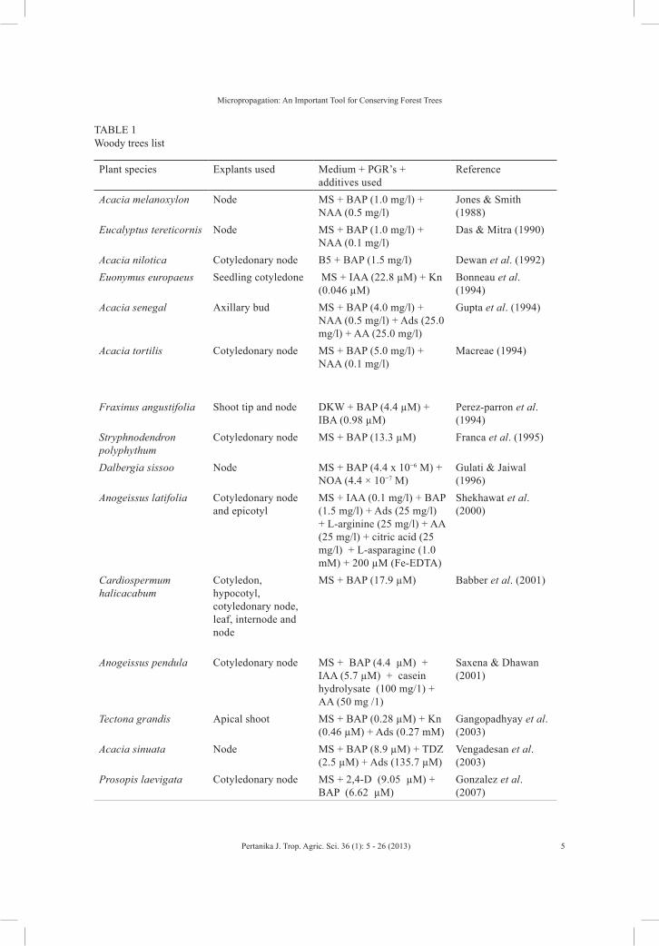
TABLE 1 Woody trees list
Plant species Explants used Medium + PGR’s + additives used
Reference
Acacia melanoxylon Node MS + BAP (1.0 mg/l) + NAA (0.5 mg/l)
Jones & Smith (1988)
Eucalyptus tereticornis Node MS + BAP (1.0 mg/l) + NAA (0.1 mg/l)
Das & Mitra (1990)
Acacia nilotica Cotyledonary node B5 + BAP (1.5 mg/l) Dewan et al. (1992)Euonymus europaeus Seedling cotyledone MS + IAA (22.8 µM) + Kn
(0.046 µM)Bonneau et al. (1994)
Acacia senegal Axillary bud MS + BAP (4.0 mg/l) + NAA (0.5 mg/l) + Ads (25.0 mg/l) + AA (25.0 mg/l)
Gupta et al. (1994)
Acacia tortilis Cotyledonary node MS + BAP (5.0 mg/l) + NAA (0.1 mg/l)
Macreae (1994)
Fraxinus angustifolia Shoot tip and node DKW + BAP (4.4 µM) + IBA (0.98 µM)
Perez-parron et al. (1994)
Stryphnodendron polyphythum
Cotyledonary node MS + BAP (13.3 µM) Franca et al. (1995)
Dalbergia sissoo Node MS + BAP (4.4 x 10⁻⁶ M) + NOA (4.4 × 10⁻⁷ M)
Gulati & Jaiwal (1996)
Anogeissus latifolia Cotyledonary node and epicotyl
MS + IAA (0.1 mg/l) + BAP (1.5 mg/l) + Ads (25 mg/l) + L-arginine (25 mg/l) + AA (25 mg/l) + citric acid (25 mg/l) + L-asparagine (1.0 mM) + 200 µM (Fe-EDTA)
Shekhawat et al. (2000)
Cardiospermum halicacabum
Cotyledon, hypocotyl, cotyledonary node, leaf, internode and node
MS + BAP (17.9 µM) Babber et al. (2001)
Anogeissus pendula Cotyledonary node MS + BAP (4.4 µM) + IAA (5.7 µM) + casein hydrolysate (100 mg/1) + AA (50 mg /1)
Saxena & Dhawan (2001)
Tectona grandis Apical shoot MS + BAP (0.28 µM) + Kn (0.46 µM) + Ads (0.27 mM)
Gangopadhyay et al. (2003)
Acacia sinuata Node MS + BAP (8.9 µM) + TDZ (2.5 µM) + Ads (135.7 µM)
Vengadesan et al. (2003)
Prosopis laevigata Cotyledonary node MS + 2,4-D (9.05 µM) + BAP (6.62 µM)
Gonzalez et al. (2007)

Kataria, N., Yadav, K., Kumari, S. and N. Singh
6 Pertanika J. Trop. Agric. Sci. 36 (1): 6 - 26 (2013)
proposed by the different scientists from time to time vary from each other in terms of their salt concentrations. Some of the earliest plant tissue culture media were developed by White (1943) and Gautheret (1939). All the subsequent media formulations are based on White’s and Gautheret’s media. The pH of the medium is also an important factor for tissue culture. The pH of the medium is usually adjusted to between 5 and 5.8 before autoclaving and extremes of pH are avoided. Each plant species has different optimized conditions both for growth of the cells and for production of useful products, so it is necessary to optimize the conditions in each case. Humidity in the culture vessel and osmotic potential of the medium affects the growth and development of plantlets in vitro in different ways (Brown et al., 1976; Ziv et al., 1983).
The MS medium was used either as
described originally or with little variation and combination of phytohormones and vitamins, such as Dalbergia latifolia (Raghavaswamy et al., 1992), Terminalia arjuna (Kumari et al., 1998), Sapindus mukorossi (Philomina and Rao, 1999), Melia azedarach (Shahzad and Siddique, 2001), Azadirachta indica (Shekhawat et al. 2002). Raghavaswamy et al. (1992) observed that axillary bud initiation in Dalbergia latifolia was better on the MS medium while multiple shoot induction was better on Woody Plant Medium (WPM) or MS (reduced major salts) medium. Bhargava et al. (2003) reported that globular proembryonic mass of callus was formed on the MS medium after 40-50 days of incubation, and then transferred to B5 medium for fragile snowy callus in Phoenix dactylifera. Sharada et al . (2003) used the MS
Acacia senegal Cotyledonary node MS + BAP (1.0 mg /l ) Khalafalla & Daffalla (2008)
Acacia chundra Shoot tip and node MS + BAP (1.5 mg/l) + IAA (0.05 mg/l) + Ads 50 mg/l
Rout et al. (2008)
Wrightia tomentosa Cotyledonary node MS + BAP (5.0 mg /l) Joshi et al. (2009)Melia azedarach Node MS + BAP (5µM) Husain & Anis
(2009)Spondias mangifera Node MS + BAP (1.0 mg /l) Tripathi & Kumari
(2010)Michelia champaca Seedling cotyledone MS + NAA (2.0 mg /l) Armiyanti et al.
(2010)Acacia auriculiformis Axillary bud B5 + coconut milk (10%) +
BAP (10-6M)Girijashankar (2011)
Streblus asper Node MS + Kn (4.60 µM) + BAP (4.44 µM)
Gadidasu et al. (2011)
Terminalia catappa Node MS + BAP (2.0 mg /l) Phulwaria et al. (2012)
Table 1 (continued)
Micropropagation: An Important Tool for Conserving Forest Trees
7Pertanika J. Trop. Agric. Sci. 36 (1): 7 - 26 (2013)
medium for shoot induction and B5 or WPM medium for root development in Celastrus paniculatus.
There are some complex substances like coconut milk (CM), casein hydrolysate (CH), adenine sulphate (Ads), activated charcoal (AC), which are sometimes required in addition to growth hormones for callus induction and regeneration. For instance, the coconut milk of green nut is very effective in providing an undefined mixture of organic nutrients and growth factors (Gamborg & Phillips, 1995). Raghavaswami et al. (1992) reported that the growth adjuvants, like coconut milk, casein hydrolysate and adenine sulphate were also supplemented to the media for direct organogenesis and somatic embryogenesis in Dalbergia latifolia. Deb (2001) used 200 mg/l of casein hydrolysate for the induction of embryogenic callus from 3-4 days imbibed seeds of Melia azedarach.
Fridberg et al. (1978) reported that charcoal had an important role during culture by absorbing toxic compounds released by inoculated explants. Pierik (1987) showed that the addition of AC often has a promoting effect on growth and organogenesis in plant species. Charcoal has been used in regeneration medium for trees like Dalbergia sissoo (Gulati & Jaiwal, 1996) and Areca catechu (Mathew & Philip, 2000) to prevent browning of culture due to phenolic exudation released by the explants. The beneficial effects of act ivated charcoal were also found on multiple shoot induction from nodal explants
of Wattakaka volubilis (Chakradhar & Pullaiah, 2006).
During culture, carbohydrates play an important role and act as an energy source required for growth, maintenance and for synthesis of cell constituents. The most commonly used carbohydrate source is sucrose, but other sugar like glucose, fructose, dextrose, mannitol, sorbitol etc. are also occasionally used. Meanwhile, sucrose also has an important role as it serves as a source of carbon and energy. Sucrose is also required for differentiation of xylem and phloem elements in the cultured cells (Aloni, 1980). Glucose and fructose are also known to support good growth of some tissues and are occasionally used. Sucrose represents the major osmotic component of the medium and is necessary for various metabolic activities. In most plants, 2-3% sucrose is found very effective for optimal growth and morphogenesis. MS medium with 2% sucrose was optimal for culturing of shoot tips in Tamarindus indica (Kopp & Nataraja, 1990). In Eucalyptus sideroxylon, however, it was observed that 4 to 6% sucrose caused more callus formation during culturing of axillary shoots, while 2-6 per cent sucrose in the MS medium supported roots development (Cheng et al., 1992). It was found that 3% sucrose is effective for shoot initiation from cotyledonary node explants in Stryphnodendron polyphythum (Franca et al., 1995). Twenty per cent sucrose concentration is more effective for the development of globular embryos of Terminalia arjuna (Kumari et al., 1998). In
Kataria, N., Yadav, K., Kumari, S. and N. Singh
8 Pertanika J. Trop. Agric. Sci. 36 (1): 8 - 26 (2013)
Alnus nepalensis, 1.5 % sucrose in WPM medium was optimal for shoot proliferation from terminal axillary buds (Thakur et al., 2001). Shahzad and Siddiqui (2001) reported that 3% sucrose was required for callus as well as for shoot proliferation in Melia azedarach. Similarly, Shekhawat et al. (2002) also advocated the use of 2-3% sugar to obtain multiple shoots in Azadirachta indica. Likewise, Chakradhar and Pullaiah (2006) reported that 1.0 per cent of sucrose was necessary for the rooting of regenerated plantlets in Wattakaka volubili.
Agar-agar is used as a solidifying agent and assumed to be that of neutral support for callus growth and multiplication. Normally, 0.8 percent agar is used for culture medium. Pasqualatto et al. (1986) reported a higher concentration of solidifying agent in the medium reduced vitrification, but in certain cases, an increase in agar amount causes adverse effect, as observed by Lal and Singh (1995).
Plant Growth Regulators
Plant growth regulators directly or indirectly affect the growth and differentiation of plant tissues. Different plant growth regulators have different effects and they vary with the type and quantity to be applied. There are five known major classes of compounds with plant growth regulatory activity. These are auxins, cytokinins, gibberellins, abscisic acid and ethylene. Among various growth regulators, auxins (NAA, IAA, IBA and 2, 4-D), cytokinins (BAP, Kinetin, Zeatin), ABA, gibberellins and ethylene are very
important. The nature of organogenic differentiation is determined by the relative concentration of auxins and cytokinins. Higher cytokinins to auxin ratio promote shoot formation, while higher auxins to cytokinins ratio favours root differentiation. Therefore, an auxin/cytokinin ratio plays a critical role in the induction of roots and shoots (Skoog & Miller, 1957). Auxins have an essential role in shoot induction and plant regeneration in most plant species. Auxins also induce somatic embryogenesis from the callus of Citrus sinensis (Kochba & Roy, 1973). Cytokinins alone, or in combination with auxins has been generally used in tissue culture. Cytokinins has been used in range of (0.5-30mg/l) and higher concentrations bring about morphological abnormalities and cause hyper hydration. Among cytokinins, BAP is the most commonly used in a variety of explants for shoot regeneration. Goyal and Arya (1979) observed the regeneration in Prosopis cineraria on MS medium with different concentrations and combinations of Kinetin, IAA, IBA and BAP. Gamborg’s medium was used by Mukhopadhayay and Mohan (1981) for culturing of Dalbergia sissoo. Meanwhile, Rumary and Thorpe (1984) reported that in some cases, mixed cytokinins have beneficial role. Multiple shoots were obtained in Eucalyptus grandis on the MS medium supplemented with additional thiamine (Lakshmi Sita & Shobha Rani, 1985). It has been reported that the decrease in NAA ensures shoots formation (Rao et al., 1984; Sudha Devi & Natreja, 1987). Mittal et al. (1989) obtained multiple shoots from the axillary buds of

Micropropagation: An Important Tool for Conserving Forest Trees
9Pertanika J. Trop. Agric. Sci. 36 (1): 9 - 26 (2013)
Acacia auriculiformis on Gamborg’s (B5) basal medium supplemented with coconut milk and BAP. Kopp and Nataraj (1990) regenerated plantlets by supplementing 2.0 mg/l BAP in Tamarindus indica. Multiple shoots were obtained from cotyledonary nodes of Dalbergia latifolia on MS medium
fortified with (2.0 mg/l) BAP (Lakshmi Sita & Raghavaswamy, 1992). BAP also produced longer shoot as compared to kinetin in Prosopis cineraria and Aegle marmelos (Kumar & Singh, 2009; Yadav & Singh, 2011b), as shown in Fig.1b, 1c, and 1d.
Fig.1: A. Callus induction from internodal segment of Albizia lebbeck on MS medium + BAP (2.0 mg/l) + NAA (0.5 mg/l); B. Shoot bud initiation on MS medium + BAP (2.0 mg/l) in nodal explants of Aegle marmaelos; C. Shoot formation from nodal explants of Aegle marmelos on MS medium with 2.0 mg/l BAP + 1.0 mg/l IAA; D. Callus growth and shoots proliferation from nodal explants of Prosopis cineraria on MS medium supplemented with BAP (2.0mg/l).
Kataria, N., Yadav, K., Kumari, S. and N. Singh
10 Pertanika J. Trop. Agric. Sci. 36 (1): 10 - 26 (2013)
The MS media supplemented with BAP in combination with NAA supported the highest percentage of callus induction in Leucaena leucocephala (Singh & Lal, 2007) and Albizia lebbeck (Yadav & Singh, 2011a) (see Fig.1a). In Eucalyptus camaldulensis, the highest frequency of somatic embryos were produced from the callus obtained on the MS medium supplemented with 0.5 mg/l of BAP and 0.1 mg/l NAA from mature zygotic embryos (Prakash & Gurumurthi, 2010).
Selection of the Explants
The success in micropropagation from mature plants depends upon the careful selection of explants (Murashige, 1974; Sommer & Caldas, 1981; Williams & Maheswaran, 1986). In Albizia lebbeck, stem, root, leaf, rachis, leaflets, hypocotyls and axillary buds were used for regeneration (Arya et al., 1978; Gharyal & Maheshwari, 1990). Several factors influence the behaviour of the inoculum in culture (Murashige, 1974). These factors include:
a. The organ that serves as tissue source;
b. The physiological and ontogenic stage of the organ;
c. Season in which explant is obtained;
d. Size of the explants;
e. Overall quality of plant from which explants are to be taken
Huda et al. (2007) studied the in vitro morphogenic responses of different explants of Corchorus olitorius. The different explants viz. leaf segments, internodes
and nodal segments showed different morphogenic responses. The nodal segments initiated callus earlier than leaf segments and the internode explants and higher amount of callus were obtained from the leaf segments than internodes and nodal segments. It was also reported by many workers that the quality of explants primarily determines the establishment of in vitro culture (Keathley, 1984). The nodal explants from a mature tree of Heavea brasiliensis failed to produce plantlets, while the explants taken from 6 to 8 weeks old plant regenerated plantlets (Rehman et al., 1981). Similarly, the explants from mature trees of Eucalyptus citriodora required pre-treatment for induction of shoot buds but the explants from seedlings did not require any pre-treatment (Gupta et al., 1981). Gulati and Jaiwal (1996) reported that the nodal explants taken from coppied shoots of mature Dalbergia sissoo exhibited the least phenolic exudation and responded better shoot regeneration, but this was not observed in the explants taken from mature trees. Callus formation and regeneration of plantlets from nodal explants was reported by Nandwani and Ramawat (1991) in Prosopis juliflora. Swamy et al. (1992) used the nodal explants of in vitro grown root suckers from the 60-80 years old tree of Dalbergia latifolia for direct organogenesis. In Fraxinus angustifolia, shoot tips and nodal segments were used for micropropagation (Perez-parron et al., 1994). The maximum number of shoots (9 shoots per explant) in the Aegle marmelos from nodal segments were obtained on

Micropropagation: An Important Tool for Conserving Forest Trees
11Pertanika J. Trop. Agric. Sci. 36 (1): 11 - 26 (2013)
the MS medium supplemented with BAP (8.8µM) + IAA (5.7µM) by Pati et al. (2008). In Melia azedarach, multiple shoots were produced from the nodal segments on the MS medium supplemented with 5 µM of BAP (Husain & Anis, 2009). Tripathi and Kumari (2010) obtained an efficient in vitro propagation of Spondias mangifera using nodal explants from seedlings.
In addition, season was also found to affect the shoot proliferation and explants contamination. Seasonal conditions at the time of explants collection may influence the in vitro growth of explants, phenolics exudation and degree of contamination. The nodal segments of Eucalyptus tereticornis collected during July to September were more responsive to micropropagation because of the negligible phenolic exudation from explants as compared to that collected in October-November and May-June due to the high amount of phenolic exudation (Das & Mitra, 1990). Similar effects of season have also been noticed in other plants like Tactona grandis (Gupta et al., 1980) and Eucalyptus tereticornis (Das & Mitra, 1990). Bonneau et al. (1994) observed a higher percentage of embryonic callus production from the zygotic embryo explants in Euonymus europacus taken during May to September. Similarly, Thakur et al. (2001) observed an optimal establishment of axillary and terminal buds of Alnus nepalensis cultured during February and March; thereafter, the percentage establishment showed a declining order. Singh and Goyal (2007) reported that the August to October season was the best for explant collection in
Salvadora oleoides throughout the year. The harvesting time of pods also showed a significant effect on the in vitro germination of seeds. Yadav and Singh (2011a) recorded the highest germination (83.3%) for the seeds extracted from dark-yellow pods in Albizia lebbeck.
Meanwhile, the size of the explants plays a key role in expressing the morphogenetic potentiality. Among other, Okazava et al. (1967) reported that small explants are more likely to form callus while larger explants maintain greater morphogenetic potentiality. This may be due to the available food reserves and growth regulators which have been proven to be useful in the initiation of new growth (Anderson, 1980). The orientation of the explants also plays an important role in giving morphogenic response. The horizontal position of the explants has been reported to promote adventitious shoot formation in many higher plants (Frett & Smagula, 1983; Pierik, 1987).
Cultural Conditions
Light is an important factor for the success of a tissue culture experiment. The intensity, quality and extent of daily exposure of light are the determining factors in the plant tissue culture. Cultures are usually maintained at a constant temperature of 25±2ºC and a photoperiod of 16 hours of light (20 μmol m-2 s-1 photosynthetic photon flux intensity) and 8 hours of darkness.
Gupta et al. (1981) reported multiple shoots production when the terminal buds from twenty years old tree of Eucalyptus

Kataria, N., Yadav, K., Kumari, S. and N. Singh
12 Pertanika J. Trop. Agric. Sci. 36 (1): 12 - 26 (2013)
citridora were cultured on the MS medium at 150C in continuous light, followed by culture at 250C with 16 hours photoperiod. The effects of light and cytokinins interaction on the cultured cotyledon explants of Radiata pine were studied by Victor et al. (1984). In Eucalyptus tereticonis, a high rate of multiplication was achieved on the MS medium at a slightly higher temperature (30-320C) (Das & Mitra, 1990). Calleberg and Johansson (1993) studied that direct regeneration was mostly stimulated when the anther cultured was incubated at 200C. The formation of multiple shoots at 25±20C and 16 hours of light and 8 hours of dark periods under light intensity of 3000-4000 lux has also been reported in Azadirachta indica (Shekhawat et al., 2002).
Organogenesis
It involves the formation of organized structure like shoot and root from pre-existing structure, i.e. unorganized mass of cells known as callus. The controlled organogenesis under in vitro was given by White (1939) who obtained the shoots from the callus of Nicotiana glauto and N. Iongsodorffi hybrid on a agar-agar solidified medium. Later on a number of reports approved, depicting the formation of shoots and roots either directly from the explants or indirectly, i.e. from the callus.
Organogenesis deals through two pathways, i.e. direct pathway and indirect pathway. Direct pathway occurs through the continuous development of shoot meristems activity from lateral or axillary buds. Indirect pathways deal with the shoot formation via
callus formation. Indirect regeneration often results in somaclonal variation making the strategy less desirable for large scale clonal multiplication. Therefore, direct regeneration without a callus phase is a reliable method for clone production.
Direct Organogenesis
Direct organogenesis, i.e. without callus formation, has also been reported in many herbeaceous and tree species. Several limitations, such as low shoot proliferation in forest trees, excessive phenolic exudation (Linington, 1991), basal callusing (Marks & Simpson, 1994), vitrification (Monsalud et al., 1995), and shoot tip necrosis (Bargchi & Alderson, 1996) are pronounced in tree tissue culture. Further, difficulty in rooting (Harada & Murai, 1996) has also had a negative effect on micropropagation of woody forest tree species.
M i c r o p r o p a g a t i o n w i t h o u t a n intervening callus phase is advantageous over conventional vegetative propagation in t e rms of quan t i ty, qua l i ty and economics (Altmann & Loberant, 1998). In general, three modes of in vitro plant regeneration have been in practice, namely, organogenesis, embryogenesis and axillary proliferation. The difference mainly matters when it relates to the genetic stability of the resulting micropropagated plants, and the obvious option would be the axillary and adventitious shoot proliferation. Meanwhile, in vitro micropropagation has been proven in the recent past as a means for supplying planting material for forestry (Ahuja, 1993; Lakshmi Sita & Raghavaswamy,
Micropropagation: An Important Tool for Conserving Forest Trees
13Pertanika J. Trop. Agric. Sci. 36 (1): 13 - 26 (2013)
1998). Multiple shoots could be induced from the nodal segments of Eucalyptus grandis (Cresswell & Nitch, 1975). The use of a protocol to promote axillary and apical shoot bud proliferation in vitro has been used for the propagation of forest tree species. The multiple shoots of Himalayan oaks were induced from intact embryos and cotyledonary nodes of Himalayan oaks (Purohit et al., 2002). A method for adventitious shoot regeneration from leaf explants of micropropagated Peach shoots has been developed (Gentile et al., 2002). This method involves the utilization of shoot tips, lateral buds and small nodal and internodal cutting as explants and it genetically establishes stable culture without any callus formation. Mittal et al. (1989) observed the formation of multiple shoots from the axillary buds from the in vitro grown seedlings of Acacia auriculiformis. Kopp and Natraraja (1990) regenerated plantlets from shoot tip explants of in vitro grown seedlings of Tamarindus indica. Das and Mitra (1990) reported 18-22 shoots per explants in Eucalyptus tereticornis when nodal explants were inoculated on the MS medium with BAP (1.0 mg/l) and NAA (0.1 mg/l). Singh et al. (1993) observed the sprouting of axillary bud in Acacia nilotica on MS and WP medium fortified with BAP (1.0 mg/l).
Kumar and Seeni (1998) achieved a rapid clonal multiplication of Aegle marmelos by enhancing axillary bud proliferation in single node segment of a twenty five years old tree on the MS medium supplemented with BAP (2.5mg/l) in combination with
IAA (1mg/l). Komalavalli and Rao (2000) established the in vitro propagation protocol of Gymnema sylvestre - a multipurpose plant. The MS medium fortified with growth regulators such as BAP (0.5mg/l) in combination with NAA (0.01mg/l) has been reported to give optimum results in Utleria salcifolia (Gangaprasad, 2003). Kumari et al. (2005) reported multiple shoot formation from the nodal and shoot tip explants in Wedelia chilensis under in vitro conditions. Rathore et al. (2005) developed protocol for the in vitro propagation of Maerua oblongifolia using nodal shoots segments on MS medium and achieved a high rate of shoot multiplication. Thidiazuron (TDZ) was used at 1.0 mM and found to be the most effective in inducing bud break and growth, and also in initiating multiple shoot proliferation at the rate of 25 microshoots per nodal explant with axillary buds, after 4 weeks of culture (Ahmad & Anis, 2007). Rathore and Shekhawat (2009) reported 35–40 shoots per culture vessel shoots proliferated on the MS medium supplemented with 4.44 lM BA and 0.57 lM indole acetic acid (IAA) and additives in Pueraria tuberosa. The multiple shoot formation was observed to be the highest in the MS fortified with 2 mg/L Benzyl amino purine (BAP) and 0.1 mg/L Naphthalene acetic acid (NAA) in Acacia auriculiformis (Girijashankar, 2011).
Indirect Organogenesis
Lakshmi Sita and Vaidyanathan (1979) raised plantlets from the cotyledonary c a l l u s o f E u c a l y p t u s c i t r i o d o r a .
Kataria, N., Yadav, K., Kumari, S. and N. Singh
14 Pertanika J. Trop. Agric. Sci. 36 (1): 14 - 26 (2013)
Adventitious shoot regeneration was reported through the callus culture in Eucalyptus camaldulensis taken from the shoot culture of mature tree (Murlidharan & Mascarenhas, 1987). Inamdar et al. (1990) reported the formation of somatic embryos via callusing from culture of shoot apices of adult Crataeva nurvala on MS medium containing 2, 4-D. The callus cultures and plantlet formation in vitro were reported in Prosopis juliflora by Nandwani and Ramawat (1991). Joshi and Dhar (2003) obtained the maximum shoots from epicotyle explants that were cultured on the MS medium supplemented with (0.25 µm) NAA and (1.0 µm) Kinetin in Saussurea obvallata. Thomas and Philip (2005) reported a high frequency shoot organogenesis from the leaf derived callus of Dalbergia sissoo. Similarly, Faisal and Anis (2005) developed a protocol for high frequency shoot regeneration and plant establishment of Tylophora indica from petiole derived callus. Organogenic Callus was developed from the stem explant of Ruta graveolens on the MS medium composed of 2.5 µm BA + 10 µm 2-4-D. Deepa et al. (2006) noticed profuse callusing from cotyledon and shoot tip explants in Pseudarthria viscida on the MS medium supplemented with 2,4-D (1.5-2 mg/l) and BAP (1-1.5 mg/l). Agrawal and Sardar (2006) described a high frequency shoot regeneration through leaflet and cotyledon derived calli in Cassia angustifolia. The MS medium, supplemented with BAP (1.0 mg/l) and IBA (0.5 mg/l), was found to be the
most effective combination for shoot bud differentiation in Jatropha curcus (Rajore & Batra, 2005). Meanwhile, subsequent shoot regeneration was achieved in the MS medium supplemented with BAP (2mg/l).
The occurrence of genetic variation is a matter of great variation concern where commercial success in micro propagation is dependent mainly on the maintenance of clonal uniformity (Bonga, 1987). However, abnormalities in the tissue culture and the plants produced from them often increase in frequency with the increase in the culture passages.
Rooting of In vitro Regenerated Shoots
For the development of a perfect plantlet, it is essential that the regenerated shoots must develop the roots. The culture medium for rooting varies from tissue to tissue, as well as from species to species. The shoots of certain plant species developed root only on the simple MS medium, i.e. hormone free media, as reported in Cardiospermum halicacabum (Babber et al., 2001). Among the auxins, IBA with MS medium is the most commonly used to induce rooting (Raghavaswamy et al., 1992; Nandwani & Ramawat, 1993; Shahazad & Siddqui, 2001). Ahmed et al. (2007) used different concentrations of IBA, NAA and IAA for rooting and the highest rooting percentage (97.66%) was reported on the MS medium with 0.1 mg/l IAA. This shows that IBA is better than NAA and IAA for shoot and root formation. As for the effect of NAA on root formation, data indicated that the percentage of shoots formed roots was 87%
Micropropagation: An Important Tool for Conserving Forest Trees
15Pertanika J. Trop. Agric. Sci. 36 (1): 15 - 26 (2013)
on the MS basal medium without plant growth regulators as compared to the MS basal medium supplemented with NAA at low concentrations of 0.01 and 0.1 mg/l NAA, where the root percentage was 80%, 73% and 53%, respectively. On the other hand, NAA at 1.0 and 1.5 mg/l did not help shoots to form roots. The shoots must be transferred to a rooting medium which is different from the shoot multiplication medium, particularly in its hormonal and salt composition. However, the nutritive medium for rooting varies from tissue to tissue, as well as from species to species. Shoot multiplication was induced on full strength MS medium whereas the salt concentration was reduced to half (Garland & Stoltz, 1981; Zimmerman & Broome, 1981) or a quarter (Skirvin & Chu, 1979) for rooting. Most frequently, IAA, IBA and NAA (0.1-1.0 mg/l) have been used for this purpose; however, IAA and IBA were found to be more effective (George & Sherrington, 1984). In Moringa pterygosperma, half strength MS medium supplemented with GA3 (0.2 mg/l) produced roots within 7 days in 25% cultures. However, a better rooting was developed with 3% sucrose and IBA (0.2 mg/l) only (Mohan et al., 1995). Sharma and Padhya (1996) observed root induction in Crataeva nurvala within 7 days on the MS medium with a low dose of NAA (0.5 µm). Mustafa and Hariharan (1997) got limited root in the hormone free medium whereas NAA alone was better than the combination of NAA and IBA in Alpinia galangal. Sharada et al. (2003) achieved 85% rooting on McCown medium (WPM)
containing IBA (5 x 10⁻⁶M) in Celastrus paniculatus. The high percentage of root regeneration in adventitious shoots was obtained on the MS medium supplemented with 0.1 mg/l IAA in Populus ciliata (Thakur et al., 2005). Sayd et al. (2010) reported that all strengths of MS-medium with 1g/l AC produced rooted shootlet but the quarter strength of MS-medium produced the highest rooting, leaf number and shootlet length. In vitro developed microshoots were rooted on the MS half strength medium supplemented with 2.46 μM IBA in Streblus asper (Gadidasu et al., 2011).
Hardening of Regenerated Plantlets
After rooting, hardening of regenerants prior to transfer in the soil increases the survival rate of transferred plants. So, it is a step which gradually acclimatizes the plant to the harsh natural environment. Spraying, misting and covering with the thin polythene may serve to fulfil the above objective. Various types of substrates have been used during acclimatization such as soil vermiculites mixture sterilized sand and soil (Goyal & Arya, 1981; Gulati & Jaiwal, 1996; Philomina & Rao, 1999; Thakur et al., 2001; Sunaina & Goyal, 2000). The in vitro developed plantlets of Dalbergia latifolia were successfully transferred by Raghavaswamy et al. (1992) by keeping the roots in tap water and high humidity. After this, the plantlets were exposed to fresh air for a few hours daily and after 14 days they were transferred into 1:3 courses sand:soil mixture. The plantlets of

Kataria, N., Yadav, K., Kumari, S. and N. Singh
16 Pertanika J. Trop. Agric. Sci. 36 (1): 16 - 26 (2013)
4-5 cm long with fresh leaves were slowly transferred to field conditions (Chaudhary et al., 2004). Sharma et al. (2006) transferred well-developed plantlets with a complete root system into pot containing sterile vermiculite and covered with transferred polythene bags to ensure high humidity. The serving plants were transplanted to field after 2 months. The most crucial step in the micropropagation is the hardening and acclimatization as it is the process which makes the plantlets capable of tolerating the natural environmental conditions. Complete regenerated plantlets with sufficient roots were taken out from the test tubes and washed several times with sterile distilled water to remove traces of the MS medium by putting the roots under continuous slow running water with the help of fine brush. Then, the in vitro regenerated plantlets were transplanted in small earthen pots containing sterilized soil and sand mixture (3:1). Each pot was covered with polythene bags with small holes to maintain high humidity and they were kept in the culture room to get acclimatized. The plantlets were initially irrigated with half strength (salts only) MS medium without sucrose on alternate days. The plantlets were exposed to the conditions for natural humidity for 3-4 hours daily to after 10 days of transfer. After about 30 days, the plants were transferred to bigger pots in the greenhouse and were maintained under natural conditions of day length, temperature and humidity. Finally, the plants were transferred to the field
conditions. Successful acclimatization and field transfer of in vitro regenerated plantlets have also been reported in Tamarindus indica (Kopp & Nataraja, 1990), Eucalyptus tereticornis (Das & Mitra, 1990), Prosopis juliflora (Nandwani & Ramawat, 1991), Dalbergia latifolia (Raghavaswamy et al., 1992), Thevetia peruviana (Kumar, 1992), Alpinia galangal (Anand & Hariharan, 1997), Terminalia arjuna (Kumari et al., 1998), Sapindus mukorossi (Philomina & Rao, 2000), Salvadora persica (Mathur et al., 2002), Bupleurum disticho-phyllum (Karuppusamy & Pullaiah, 2007), Spondias mangifera (Tripathi & Kumari, 2010), Acacia auriculiformis (Girijashankar, 2011), Streblus asper (Gadidasu et al., 2011). Hardening and acclimatization of plantlets were done because the plantlets raised under in vitro conditions on the synthetic carbohydrate supplemented medium under artificial light failed to abruptly acclimatize to the rigour of the natural environment. So, a careful transfer of the plantlets in the soil after hardening and acclimatization is required.
CONCLUSION
Tissue cul ture offers unparal le led opportunity for forest tree improvement. Micropropagation techniques have been applied to a wide range of tree species. Successful in vitro techniques are dependent upon the strong and intricate interactions between the explant, plant growth regulators, culture conditions, and genotype. The present status of tree tissue culture, however, is adequate to initiate commercialization

Micropropagation: An Important Tool for Conserving Forest Trees
17Pertanika J. Trop. Agric. Sci. 36 (1): 17 - 26 (2013)
programmes. Commercialization has already been proven to be successful in the cases of some trees like Eucalyptus, Rosewood, Poplar and others. In a commercial scale, the improved protocols should be working efficiently with various genotypes, preferably of the mature origin. Meanwhile, the establishment of protocols with reduced steps of developmental pathways may significantly reduce the time and cost. Most of the tree tissue culture research has centred on methods, primarily on the development of culture media and techniques to induce juvenility in trees for micropropagation. The thrust is towards methods, which have commercial use and the potential for patents. Also, unless the cost of plantlet production by tissue culture techniques is brought down considerably to match with or less than the conventional methods of propagation, the efforts are not worthy enough to justify. Nonetheless, clonal fidelity in trees micropropagated by organogenesis has not been adequately tested with many species. Research on micropropagation will stress on the mass propagation of mature trees and reduction of costs. Research on micropropagation by somatic embryogenesis has to be intensified so as to reach the ultimate goal of mass propagation since it also allows automation which will in turn reduce the cost per propagule. Continued research into the use of biotechnology will contribute to improving the vegetative propagation of the species, which will ultimately play an important role in future breeding programmes of forest tree improvement and reforestation programmes.
REFERENCESAgrawal V., & Sardar P. R. (2006). In vitro propagation
of Cassia angustifolia through leaflet and cotyledon derived calli. Biologia Plantarum, 50, 118-122.
Ahmad, N., & Anis, M. (2007). Rapid plant regeneration protocol for cluster bean (Cyamopsis tetragonoloba L. Taub.). Journal of Horticulture Science and Biotechnology, 82, 585-589.
Ahuja, M. R., & Libby W. J. (Eds.) (1993). Clonal Forestry, Vol. 1. Genetics and Biotechnology. Springer verlag, Berlin, Heidelberg.
Aloni, R. (1980). Role of auxin and sucrose in the differentiation of sieve and tracheary elements in Plant tissue cultures. Planta, 150, 255-263.
Altman, A., & Loberant, B. (1998). Micropropagation: Clonal Plant Propagation in vitro. In Altman, A., & Marcel Dekker (Eds.) Agricultural Biotechnology. Inc.New York, USA pp. 19-42.
Anand, P. H. M., & Hariharan, M. (1997). In vitro multiplication of greater galangal [(Alpinia galangal Linn.) Wild] - a medicinal plant. Phytomorphology, 47, 45-50.
Anderson (1980). Tissue culture propagation of red and black raspberries Rubus idaeus and R. occidentalis. Acta Horticulturae, 112, 13-20.
Armiyanti, K. A. M., Kadzimin, S., & Panjaita, B. S. (2010). Plant regeneration of Michelia champaca L., through somatic embryogenesis. African Journal of Biotechnology, 9, 2640-2647.
Arumugam, N., & Bhojwani, S. S. (1990). Somatic embryogenesis in tissue cultures of Podophyllum hexandrum. Canadian Journal of Botany, 68, 487-491.
Arya, H. C., Rawat, K. G., & Suthar, K. C. (1978). Culture and differentiation of plants of economic importance. Albizzia lebbeck. Journal of Indian Botanical Society, 57, 50.
Kataria, N., Yadav, K., Kumari, S. and N. Singh
18 Pertanika J. Trop. Agric. Sci. 36 (1): 18 - 26 (2013)
Babber, S., Mittal, K., Ahlawat, R., & Varghese, T. M. (2001). Micropropagation of Cardiospermum halicacabum. Biologia Plantarum, 44, 603-606.
Bajaj, Y. P. S. (1986). Biotechnology of tree improvement for rapid propagation and biomass energy production. Biotechnology in Agriculture and Forestry, Vol. 1 – Tree. Springer-verlag, pp. 1-23.
Bargchi, M., & Alderson, P. G. (1996). The control of shoot tip necrosis in Pistacia vera L. in vitro. Plant Growth Regulators, 20, 31–35.
Batra, A., Sardana, J. A., Sharma, M., & Ali, D. J. (2000). Tissue Culture: An indispensable component of biotechnology for plant improvement. In I. Khan, and A. Khanum (eds.). Role of biotechnology in medicinal and aromatic plants, Vol. 3, Ukaz Pub. Hyderabad, India.
Benson, E. (2003). Conserving special trees: Integrating biotechnological and traditional approaches (p. 23–24). Forest biotechnology in Europe: impending barriers, policy and implication. Institute of Forest Biotechnology, Edinburgh, UK.
Bhargava, S. C., Saxena, S. N., & Sharma, R. (2003). In vitro multiplication of Phoenix dactylifera (L.). Journal of Plant Biochemistry and Biotechnology, 12, 43-47.
Bonga, J. M. (1987). Clonal propagation of mature trees: Problems and possible solutions. Cell and Tissue Culture in Forestry, 1, 249-271.
Bonneau, L., Beranger, N., & Monin, J. (1994). Somatic embryogenesis and plant regeneration in woody species: the European Spindle Tree (Euonymus europaeus L.) Plant Cell Reports, 13, 135-138.
Brown, S., Wetherell, D. F., & Dougall, D. K. (1976). The potassium requirement for growth and embryogenesis in wild carrot suspension cultures. Physiologia Plantarum, 37, 73-79.
Burdon, R. D., & Libby, W. J. (2006). Genetically Modified Forests: From Stone Age to Modern Biotechnology. Durham, N.C.: Forest History Society 79.
Calleberg, E. K., & Johansson, L. B. (1993). The effect of Starch and incubation temperature in anther culture of potato. Plant Cell, Tissue and Organ Culture, 32, 27-34.
Chakradhar, T., & Pullaiah, T. (2006). Effect of explant source on axillary shoot multiplication during micropropagation of a rare medicinal plant - Wattakaka volubilis (L.F.) stapf. Journal of Plant Biochemistry and Biotechnology, 15, 43-45.
Chalupa, V. (1987). European hardwoods. In J. M. Bonga, D. J. Durzan, & N. Martinus (Eds.), Cell and Tissue Culture in Forestry, Vol. 3 (pp. 224-246). Dordrecht, Boston, Lancaster.
Chaudhary, P., Das, M., Sikdar, S. R., & Pal, A. (2004). Influence of the physiological age and position of the nodal explants on micropropagation of field grown Dendrocalamus strictus nees. Plant Cell Biotechnology and Molecular Biology, 5, 45-50.
Cheng, B. Peterson, C. M., & Mitchell, R. J. (1992). The role of sucrose, auxin and explant source on in vitro rooting of seedling explants of Eucalyptus sideroxylon. Plant Science, 87, 207 – 214.
Cresswell, R., & Nitsch, C. (1975). Organculture of Eucalyptus grandis L. Planta, 125, 87-90.
Das, T., & Mitra, G. C. (1990). Micropropagation of Eucalyptus tereticornis Smith. Plant Cell Tissue and Organ Culture, 22, 95-103.
Deb, C. R. (2001). Somatic embryogenesis Plantlets regeneration of Melia azadarech L. (Ghora neem) from cotyledonary segments. Journal of Plant Biochemistry and Biotechnology, 10, 63-65.

Micropropagation: An Important Tool for Conserving Forest Trees
19Pertanika J. Trop. Agric. Sci. 36 (1): 19 - 26 (2013)
Deepa, M. A., Bhaskar, C., & Narmtha, B. V. N. (2006). Callus induction and organogenesis from seedling explants of Pseudartharia viscida Wight and Arnott. A medicinal legume. Phymorphology, 56, 211-214.
Dewan, A., Nanda, K., & Gupta, S. C. (1992). In vitro micropropagation of Acacia nilotica subsp. Indica Brenan via cotyledonary nodes. Plant Cell Reports, 12, 18-21.
Dunstan, D. I. (1988). Prospects and Progress in conifer biotechnology. Canadian Journal of Forest Research, 18, 1497-1506.
Faisal, M., Singh, S., & Anis, M. (2005). In vitro regeneration and plant establishment of Tylophora indica (Burm. F.) Merrill: petiole callus culture. In Vitro Cellular and Developmental Biology -Plant, 41, 511-515.
FAO. (2004). Preliminary review of biotechnology in forestry, including genetic modification. Forest Genetic Resources Working Paper FGR/59E, Forest Resources Development Service, Forest Resources Division. Rome, Italy.
Franca, S. C., Duarte, I. B., Moraes, R. M., & Peira, A. M. S. (1995). Micropropagation of Stryphnodendron polyphythum (Barbatimão). Plant Cell, Tissue and Organ Culture, 42, 291-293.
Frett, J. J., & Smagula, J. M. (1983). In vitro shoot production of lowbush blueberry. Canadian Journal of Plant Science, 63, 467-472.
Fridberg, G., Pedersin, M., Landstrom, L. E., & Friksson, T. (1978). The effect of activated charcoal on tissue culture: Absorption of metaboli tes inhibi t ing morphogenesis . Physiologia Plantarum, 32, 104-106.
Gadidasu, K., Umate, P., Aileni, M., Kota, R. S., Kokkirala, V. R., Kasula, K., & Abbagani, S. (2011). Micropropagation of a valuable ethnomedicinal plant Streblus asper Lour. Journal of Phytology, 3, 18-23.
Gamborg, O. L., & Phillips, G. C. (1995). Laboratory facilities, operation and management. In O. L. Gamborg, & G. C. Phillips (Eds.), Fundamental Methods of Plant Cell, Tissue and Organ Culture (pp. 3.20). Berlin: Springer-Verlag.
Gamborg, O. L., Miller, R. A., & Ojima, K. (1968). Nutrient requirements of suspension cultures of soybean root cells. Experimental Cell Research, 50, 151-158.
Gangaprasad, A., Lakshminair, G. K., Radhakrishnan, K., Seenis, N. G. M., & Pushpangadan, P. (2003). Micropropagation of Utleria salcifolia endemic and endangered ethnomedicinal plant of the Western Ghats. Journal of Medicinal and Aromatic Plant Science, 25, 19-24.
Gangopadhyay, G., Basu, S., Gangopadhyay, R. P., & Gupta, S. (2003). Micropropagation of Tectona grandis: assessment of genetic fidelity. Biologia plantarum, 46, 459-461.
Garland, P., & Stoltz, L. P. (1981). Micropropagation of Pissrdi plum, Annals of Botany, 48, 387-389.
Gautheret, R. J. (1939). Sur la possibilite da realise la culture indefinite des tissue de tubercules de Carotte. C. R. Hebd. Seanc. Acad. Sci., Paris, 208, 118-121.
Gautheret, R. J. (1955). The nutrition of plant tissue cultures. Annual Review of Plant Physiology, 6, 433-484.
Gautheret, R. J. (1934). Culture du tissu cambial. C. R. Acad. Sci. (Paris), 198, 2195-2196.
Gentile, S. M., & Damiano, C. (2002). Adventitious shoot regeneration in peach [Prunus persica (L.) Batsch]. Plant Cell Reports, 20, 1011–1016.
George, E. F., & Sherrington, P. D. (1984). Plant propagation by tissue culture. Eversley, England: Exegetics Ltd. p. 39-71.
Gharyal, P. K., & Maheshwari, S. C. (1990). Differentiation in explants from mature leguminous trees. Plant Cell Reports, 8, 550–553

Kataria, N., Yadav, K., Kumari, S. and N. Singh
20 Pertanika J. Trop. Agric. Sci. 36 (1): 20 - 26 (2013)
Girijashankar, V. (2011). Micropropagation of m u l t i p u r p o s e m e d i c i n a l t r e e A c a c i a auriculiformis. Journal of Medicinal Plants Research, 5, 462–466.
Gonzalez, L. B., Villafuerte, J. O., Sosa, F. C., Avila, V. M. C., & Carter, E. J. V. (2007). Clonal propagation of mesquite tree (Prosopis laevigata Humb. & Bonpl. Ex Willd. M. C. Johnston). I. via cotyledonary nodes. In Vitro Cellular and Developmental Biology -Plant, 43, 260-266.
Goyal, Y., & Arya, H. C. (1979). Clonal propagation of Prosopis cineraria Linn. Through tissue culture and differentiation. Journal of Indian Botanical Society, 58, 61.
Goyal, Y., & Arya, H. C. (1981). Differentiation in cultures of Prosopis cineraria Linn. Current Sciences, 50, 468-469.
Gulati, A., & Jaiwal, P. K. (1996). Micropropagation of Dalbergia sissoo from nodal explants of mature trees. Biologia Plantarum, 38, 169-175.
Gupta, P. K., Nadgir, A. L., Mascarenhas, A. F., & Jagannathan, V. (1980). Tissue culture of forest trees: Clonal multiplication of Tectona grandis L. (teak) by tissue culture. Plant Science Letters, 17, 259-268.
Gupta, P. K., Mascarenhas, A. F., & Jagannathan, V. (1981). Tissue culture of forest trees: Clonal propagation of mature trees of Eucalyptus citriodora Hook by tissue culture. Plant Science Letters, 20, 195-201.
Harada, H. H., & Murai, Y. (1996). Micropropagation of Prunus mume. Plant Cell, Tissue and Organ Culture, 46, 265-267.
Hildebrandt, A. C. (1958). Stimulation or inhibitionof virus infected & insect-gall tissues & single cell clones. Proceedings of the National Academy of Sciences, 44, 354-363.
Huda, K. M. K., Bhuiyan, M. S. R., Kabir, M. H., Jamal, A. F. M., & Khatun, A. (2007). In vitro plant regeneration protocol of Tossa jute
(Corchorus olitorius). Intl. J. Integrat. Biol, 1, 96-101.
Husain, K. M., & Anis, M. (2009). Rapid in vitro multiplication of Melia azedarach L. (A multipurpose woody tree). Acta Physiologiae Plantarum, 31, 765-772.
Inamdar, J. A., Nataraj, M., Mohan, J. S. S., & Subramanian , R . B . (1990) . Somat ic embryogenesis from callus cultures of Crataeva nurvala Buch. Ham. Phytomorphology, 40, 319-322.
Jones , C. , & Smith , D. (1988) . Effect of 6-benzylaminopurine and 1-naphthlacetic acid on in vitro axillary bud development of mature Acacia melanoxylon. Proc. Int. Plant Prop. Soc., 38, 389-393.
Joshi, M., & Dhar, U. (2003). In vitro propagation of Saussurea obvallata (DC.) Edgew. – an endangered ethnoreligious medicinal herb of Himalaya. Plant Cell Reports, 21, 933-939.
Joshi, P., Trivedi, R., & Purohit, S. D. (2009). Micropropagation of Wrightia tomentosa: Effect of gelling agents, Carbon source and vessel type. Indian Journal of Biotechnology, 8, 115-120.
Karp, A. (1994). Origin, causes and uses of variation in plant tissue cultures. In I. K. Vasil, & T. A. Thorpe (Eds.), Plant Cell and Tissue Culture (pp. 139-150). Dordrecht, Kluwer Academic Publ.
Karuppusamy, S., & Pullaiah, T. (2007). In vitro shoot multiplication of Bupleurum distichophyllum Wight. A native medicinal plant of southern India. Plant Tissue Culture and Biotechnology, 17, 115–124.
Keathley, D. E. (1984). In Proceedings of the International symposium of Recent Advances in Forest Biotechnology, June 10-13, Michigan Biotechnology institute, Michigan, p. 58-63.
Khalafalla, M. M., & Daffalla, H. M. (2008). In Vitro Micropropagation and Micrografting of Gum Arabic Tree [Acacia senegal (L.) Wild].
Micropropagation: An Important Tool for Conserving Forest Trees
21Pertanika J. Trop. Agric. Sci. 36 (1): 21 - 26 (2013)
International Journal of Sustainable Crop Production, 3, 19-27.
Kochba, J., & Spieyel, R. P. (1973). Effect of culture media on embryoid formation from ovule callus of ‘shamouti’ orange (Citrus sinensis) Z. Pflanzan, 69, 156-162.
Komalavalli, N., & Rao, M. V. (2000). In vitro propaga t ion of Gymnema sy lves t re -A multipurpose medicinal plant. Plant Cell, Tissue and Organ Culture, 61, 97-105.
Kopp, M. S., & Nataraja, K. (1990). In vitro plantlet regeneration from shoot tip cultures of Tamarindus indica L. Indian Journal Forestry, 13, 30-33.
Kotte, W. (1922). Wurzelmeristem in Gewebekultur. Ber. Deut. Ges. 40, 269-272.
Kumar, A. (1992). Somatic embryogenesis and high frequency plantlet regeneration in callus culture of Thevetia peruviana L. Plant Cell, Tissue and Organ Culture, 31, 47-50.
Kumar, A. D., & Seeni, S. (1998). Rapid clonal multiplication through in vitro axillary shoots proliferation of Aegle marmelos (L.) Corr., a medicinal tree. Plant Cell Reports, 17, 422-426.
Kumar, S., & Singh, N. (2009). Micropropagation of Prosopis cineraria (L.) Druce-a multipurpose desert tree. Researcher, 1, 28-32.
Kumari, M. M. V., & Shivanna, M. B. (2005). Callus mediated regeneration of Desmodium oojeinense Roxb. Phytomorphology, 55, 171-177.
Kumari, N., Jaiswal, V., & Jaiswal, V. S. (1998). Induction of somatic embryogenesis and plant regeneration from leaf callus of Terminalia arjuna Bedd. Current Science, 75, 1052-1055.
Laksami Sita, G., & Vaidyanathan, C. S. (1979). Rapid multiplication of Eucalyptus citriodora by multiple shoot production. Current Science, 48, 350-352.
Laksami Sita, G., & Raghavaswamy, B. V. (1993). Regeneration of plantlets from leaf disc cultures of rosewood: control of leaf abscission and shoot tip necrosis. Plant Science, 88, 107–112.
Laksami Sita, G., & Raghavaswamy, B. V. (1992). Application of cell and tissue culture technology for mass propagation of elite trees with special reference to rosewood (Dalbergia latifolia). Indian Journal of Forestry, 118, 36-47.
Laksami Sita, G., & Raghavaswamy, B. V. (1998). Application of Biotechnology in forest trees - clonal multiplication of sandal wood, Rose wood, Teak, Eucalyptus and Bamboos by tissue culture in India. In S. Puri (Ed.), Tree Improvement (pp. 233-248). New Delhi: Oxford.
Laksami Sita, G., & Shobha, R. B. (1985). In vitro propagation of Eucalyptus grandis L. by tissue culture. Plant Cell Reports, 4, 63-65.
Lal, N., & Singh, S. B. (1995). Effect of agar concentration on pH of the medium and plantlets formation in vitro grown sugarcane shoot. Indian Journal of Plant Physiology, 38, 301-304.
Ledoux, L. (1965). Uptake of DNA by living cells. Advanced Nucelic Acid Research, 4, 231.
Linington, I. M. (1991). In vitro propagation of Dipterocarpus alatus and Dipterocarpus intricatus. Plant Cell Tissue and Organ Culture, 27, 81-88.
Linsmaier, E. M., & Skoog, F. (1965). Organic growth factor requirements of tobacco tissue cultures. Physiologia Plantarum, 18, 100-127.
Lutz, A. (1969). Etudes des aptitudes morphogenetiques des cultures de tissues. Analyse Par La methode des clones d origine Unicellulaire. Rev. Gen. Bot. 76, 309- 359.
Macrae, S. (1994). The use of Agrobacterium for plant improvement. Doctoral thesis, University of Natal, Pietermaritzburg, 64-136.
Kataria, N., Yadav, K., Kumari, S. and N. Singh
22 Pertanika J. Trop. Agric. Sci. 36 (1): 22 - 26 (2013)
Marks, T. R., & Simpson, S. E. (1994). Factors affecting shoot development in apically dominant Acer Cultivars in vitro. Journal of Horticulture Science and Biotechnology, 69, 543 - 551.
Mathew, M., & Philip, V. J. (2000). In vitro adventitious shoot formation from embryos of Areca catechu L. Phytomorphology, 50, 221–227.
Mathur, N., Ramawat, K. G., & Nandwani, D. (1995). Rapid in vitro multiplication of jujube through mature stem explants. Plant Cell Tissue and Organ Culture, 43, 75-77.
Mathur, S., Shekhawat, G. S., & Batra, A. (2002). An efficient in vitro method for mass propagation of Salvadora persica via apical meristems. Journal of Plant Biochemistry and Biotechnology, 11, 125-127.
Mittal, A., Agarwal, R., & Gupta, S. (1989). In vitro development of plantlets from axillary buds of Acacia auriculiformis - a leguminous tree. Plant Cell Tissue and Organ Culture, 19, 65-70.
Mohan, V., Purohit, M., & Shrivastava, P. S. (1995). In vitro Micropropagation of Moringa pterigosperma. Phytomorphology, 45, 253-256.
Monsalud, M. J., Mathews, H., Litz, R. E., & Gray, D. J. (1995). Control of hyperhydricity of mango somatic embryos. Plant Cell Tissue and Organ Culture, 42, 195–206.
Morel, G., & Martin, C. (1952). Micropropagation of multipurpose medicinal tree Acacia auriculiformis Guerison de dahlias atteints d’une maladie a virus. C.R. Academy of Science, Paris. 235, 1324-1325.
Muir, W. H., Hilderbrandt, A. C., & Riker, A. J. (1958). The preparation, isolation and growth in culture of single cells from higher plants. American Journal of Botany, 45, 589-597.
Muir, W. H., Hilderbrandt, A. C., & Riker, A. J. (1954): Plant tissue cultures produced from single isolated plant cell. Science, 119, 877-887.
Mukhopadhyay, A., & Mohan, R. H. Y. (1981): Regeneration of plantlets from excised roots of Dalbergia sissoo. Indian Journal of Experimental Biology, 19, 1113-1115.
Murashige, T., & Skoog, E. A. (1962). A revised medium for rapid growth and bioassay with tobacco tissue cultures. Physiologia Plantarum, 15, 473–497.
Murashige, T. (1974). Plant propagation through tissue culture. Annual Review of Plant Physiology, 25, 136-166.
Murlidharan, E. M., & Mascarenhas, A. F. (1987). In vitro plantlet formation by organogenesis in Eucalyptus camaldulensis and by somatic embroyogenesis in E. citriodora. Plant Cell Reports, 6, 256-259.
Mustafa, A. P. H., & Hariharan, M. (1997). In vitro multiplication of greater galangal (Alpinia galangal) Linn. Wild. – a medicinal plant. Phytomorphology, 47, 54-50.
Nandwani, D., & Ramawat, K. G. (1991). Callus culture and plantlets formation from nodal explants of Prosopis juliflora (Swartz) DC. Indian Journal of Experimental Biology, 29, 523-527.
Nandwani, D., & Ramawat, K. G. (1993). In vitro plantlets formation through juvenile and nodal explant of Prosopis cineraria. Indian Journal of Experimental Biology, 21, 156-160.
Nehra, S. A., & Kartha, K. K. (1994). Meristem and shoot tip culture, requirements and applications, p. 37-70.
Nitsch, J. P., & Nitsch, C. (1969). Haploid plants from pollen grains. Science, 163, 85-87.
Okazawa, Y. Katsura, N., & Tagawa, T. (1967). Effect of auxin and kinetin on the development and differentiation of potato tissue cultured in vitro. Physiologia Plantarum, 20, 862-869.
Micropropagation: An Important Tool for Conserving Forest Trees
23Pertanika J. Trop. Agric. Sci. 36 (1): 23 - 26 (2013)
Pasqualatto, P. L., Zimmerman, R. H., & Fordham, I. (1986). Gelling agent and growth regulator effects on shoot vitrification of Gela apple in vitro. Journal of the American Society for Horticultural Science, 111, 976-980.
Pati, R., Chandra, R., Chauhan, U. K., Mishra, M., & Srivastava, N. (2008). In vitro clonal propagation of Bael (Aegle marmelos Corr.) CV. CISH-B1 through enhanced axillary branching. Physiology and Molecular Biology of Plants, 14, 337-346.
Perez-parron, M. A., Gonzalez-benito, M. E., & Perez, C. (1994). Micropropagation of Fraxinus angustifolia from mature and juvenile plant material. Plant Cell Tissue and Organ Culture, 37, 297–302.
Philomina, N. S., & Rao, J. V. S. (1999). Multiple shoot production from seed culture of soap nut (Sapindus mukorossi Gaertn.). Phytomorphology, 49, 419-423.
Philomina, N. S. , & Rao, J . V. S. (2000). Micropropagation of Sapindus mukorossi Gaertn. Indian Journal of Experimental Biology, 38, 621-624.
Phulwaria, M., Ram, K., Harish, Gupta, A. K., & Shekhawat, N. S. (2012). Micropropagation of mature Terminalia catappa (Indian Almond), a medicinally important forest tree. Journal of Forest Research, 17, 202–207.
Pierik, R. L. M. (1987). In vitro culture of higher plants. In J. M. Bonga, & J. M Durzon (Eds.), Cell and Tissue Culture in Forestry (p. 34). Dordrecht: Martinus Nijhoff.
Pijut, P. M., Domir, S. C., Lineberger, R. D., & Schreiber, L. R. (1990). Use of culture filterate of Ceratocystis ulmi as a bioassay to screen for disease tolerant Ulmus americana. Plant Science, 70, 191-196.
Prakash, M. G., & Gurumurthi, K. (2010). Effects of type of explant and age, plant growth regulators and medium strength on somatic
embryogenesis and plant regeneration in Eucalyptus Camaldulensis. Plant Cell Tissue and Organ Culture, 100, 13–20.
Purohit, V. K., Palni, L. M., Nandi, S. K., & Rikhari, H. C. (2002). In vitro regeneration of Quercus floribunda Lindl. through cotyledonary nodes: an important tree of central Himalya. Current Science, 83, 312-316.
Raghavaswamy, B. V., Himabindu, K., & Lakshmi, S. G. (1992). In vitro micropropagation of elite rosewood (Dalbergia latifolia Roxb.). Plant Cell Reports, 11, 126–131.
Rajore, S., & Batra, A. (2005). Efficient Plant regeneration via Shoot tip explants in Jatropha curcas. Journal of Plant Biochemistry and Biotechnology, 14, 73-75.
Rao, P. S., Suprasanna, P., & Ganapathi, T. (1996). Plant biotechnology and agriculture: Prospects for crop improvement and increasing productivity. Science Cultivation, 62, 185-191.
Rao, K. S., Lakshmi, S. G., & Vaidyanathan, C. S. (1984). In vitro cloning of Dalbergia latifolia Roxb. (Indian Rose Wood). In R. R. Henke, K. W. Hughes, M. J. Constanin, & A. Hollaender (Eds.), Tissue Culture in Forestry and Agriculture (p. 346). New York: Plenum Press.
Rathore, J. S., Shekhawat, N. S., & Rathore, M . S . ( 2 0 0 5 ) . M i c r o p r o p a g a t i o n o f Maerua ablongifolia- A liana of arid areas. Phytomorphology, 55, 241-247.
Rathore, M. S., Panwar, D., Rathore, J. S., Dagla, H. R., & Shekhawat, N. S. (2009). Pueraria tuberose (Roxb.Ex Wild) and determination of puerarian content in different tissues. Plant Cell Tissue and Organ Culture, 99, 327-334.
Rehman, W. R., Gandhimathi, H., Rohani, O., & Paranjothy, K. (1981). Recent developments in tissue culture of Hevea. In A. N. Rao (Ed.), Tissue Culture of Economically Important Plants (pp. 152 – 158). Singapore.
Kataria, N., Yadav, K., Kumari, S. and N. Singh
24 Pertanika J. Trop. Agric. Sci. 36 (1): 24 - 26 (2013)
Reinert, J. (1959). Tyber die Kontrolle der Morphogenese und die Indukt ion von Adventivembryonen an Gewebekulturen aus Karotten. Planta, 53, 318-333.
Robbins, W. J. (1922). Cultivation of excised root tips under sterile conditions. Bot. Gaz. 73, 376-390.
Roja, G., & Rao, P. S. (1998). Biotechnological investigation in medicinal plants for the production of secondary metabolites. In I. Khan, & A. Khanum (Eds.), Role of biotechnology in medicinal and aromatic plants (pp. 95 – 125). Hyderabad: Ukaaj Publ.
Rout, G. R., Senapati, S. K., & Aparajeta, S. (2008). Micropropagation of Acacia chundra (Roxb.) DC. Horticultural Science, 35, 22-26.
Rumary, C. & Thorpe, T. A. (1984). Plantlet formation inblack and white spruce-1-In vitro techniques. Canadian Journal of Forestry Research, 14 , 10-16.
Saxena, S., & Dhawan, V. (2001). Large-scale production of Anogeissus pendula and A.latifolia by micropropagation. In Vitro Cellular and Developmental Biology -Plant, 37, 586–591.
Sayd, S. S., Hanan, A. A., Taie, H. A. A., & Taha, L. S. (2010). Micropropagation, antioxidant activity, total phenolics and flavonoids content of Gardenia jasminoides ellis as affected by growth regulators. International Journal of Academic Research, 2, 184-191.
S hahzad , A . , & S id d iqu i , S . A . ( 2001 ) . Micropropagation of Melia azedarach L. Phytomorphology, 51, 151-154.
Sharada, M., Ashok, A., & Kaul, M. K. (2003). Regeneration of plantlets via callus cultures in Celastrus paniculatus Willd. – A rare endangered medicinal plant. Journal of Biochemestry and Biotechnology, 12, 65 -69.
Sharma, V. K., Monostori, T., Gobel, C., Hansch, R., Bittner, F., Wasternack, C., Feussner, I.,
Mendel, R. R., Hause, B., & Schulze, J. (2006). Transgenic barley plants overexpressing a 13-lipoxygenase to modify oxylipin signature. Phytochemistry, 67, 264-276.
Sharma, V., & Padhya, M. A. (1996). In vitro rapid Multiplication and propagation of Cratavia nurvala. Indian Journal of Experimental Biology, 34, 243-246.
Shekhawat, G. S., Batra, A., & Mathur, S. (2002). A reliable in vitro protocol for rapid mass propagation of Azadirachta indica Juss. Journal of Plant Biology, 29, 109-112.
Shekhawat, N. S., Yadav, J., Arya, V., & Singh, R. P. (2000). Micropropagation of Anogeissus latifolia (Roxb. Ex DC.) Wall, ex Guill. & Perr. - A Tree of Fragile Ecosystems. Journal of Sustainable Forestry, 11, 83-96.
Singh, H. P., Singh, S., Saxena, R. P., & Singh, R. K. (1993). In vitro bud break in axillary nodal segments of mature trees of Acacia nilotica. Indian Journal of Plant Physiology, 36, 21-24.
Singh, N., & Lal, D. (2007). Growth and organogenetic potential of calli from some explants of Leucaena leucocephala (Lam.) de Wit. International Journal of Tropical Agricultural, 25, 389-399.
Singh, R., & Goyal, S. C. (2007). Micropropagation of Salvadora oleoides Decne from nodal explants of mature tree. Journal of Plant Biology, 34, 1-5.
Skirvin, R. M., & Chu, M. C. (1979). In vitro propagation of ‘‘Forever Yours’’rose, Hort. Science, 14, 608-610.
Skoog, F., & Miller, C. O. (1957). Chemical regulation of growth and organ formation in plant tissue cultures in vitro. Symp. Soc. Exp. Biol., 11, 118–131.
Sommer, H. E., & Caldas, L. S. (1981). In vitro methods applied to forest trees. In T. A. Thorpe (Ed.), Plant tissue culture-Methods and applications in Agriculture (pp. 349-358).
Micropropagation: An Important Tool for Conserving Forest Trees
25Pertanika J. Trop. Agric. Sci. 36 (1): 25 - 26 (2013)
Sudhadevi, A. M., & Nataraja, K. (1987). Establishment of plantlets in stem cultures of Dalbergia latifolia Roxb. Indian Journal of Forestry, 113, 501-506.
Sunaina, & Goyal , S . C. (2000) . In v i tro Micropropagation of Harsinghar (Nyctanthes arbor-tristis Linn.). In National Seminar on Plant Physiology at Interface of Agri. - Horticulture and industry organized by ISPP and Rajasthan Agriculture University, Udaipur. p. 177.
Thakur, A. K., Sharma, S., & Srivastava, D. K. (2005). Plant regeneration and genetic transformation studies in petiole tissue of Himalayan poplar (Populus ciliata Wall.). Current Science, 89, 664–668.
Thakur, M., Sharma, D. R., & Kawar, K. (2001). Mass micropropagation of Alnus nepalensis D. Don. Phytomorphology, 51, 123-127.
Thomas, T. D., & Philip, B. (2005). Thidiazuron induced high frequency plant regeneration via organogenesis from leaf-derived calli of a medicinal climber, Tylophora indica (Burm. f.) Merrill. In Vitro Cellular and Developmental Biology -Plant, 41, 124-128.
Thorpe, T. A., Harry, I. S., & Kumar, P. P. (1991). Application of micropropagation to forestry. In P. C. Debergh, & R. H Zimmerman (Eds.), Micropropagation (pp. 311 – 336). Dordrecht, The Netherland: Kluwer Academic Publishers.
Tripathi, M., & Kumari, N. (2010). Micropropagation of a tropical fruit tree Spondias mangifera Wild through direct organogenesis. Acta Physiologiae Plantarum, 32, 1011–1015.
Tulecke, W., & Nickell, L. G. (1960). Methods, problems, and results of growing plant cells under submerged conditions. Trans. N.Y. Acad. Sci.Ser. II, 22, 196-206.
Van Overbeek, J., Conklin, M. E., & Blakeslee, A. F. (1942). Cultivation in vitro of small Datura embryos. American Journal of Botany, 29, 472-477.
Vasil, I. K., & Hildebrandt, A. C. (1966). Variation of morphogenetic behaviour in plant tissue culture I. Cichorium endivia. American Journal of Botany, 53, 860-869.
Vasil, V., & Hilderbrandt, A. C. (1965). Differentiation of tobacco from single isolated cells in microculture. Science, 150, 889-892.
Vengadesan, G., Ganapathi, A., Pream Anand, R., & Selvaraj, N. (2003). In vitro Propagation of Acacia sinuata (Lour.) Merr. from Nodal Segments of a 10-Year-Old tree. In Vitro Cell. Dev. Biol.-Plant, 39, 409-414.
Victor, M., Villelobes, V. M., Edwar, C., Yeng, E., & Thorpe, T. A. (1984). Origin of adventitious shoots in excised radiate pine cotyledon cultured in vitro. Canadian Journal of Botany, 63, 2172-2176.
White, P. R. (1943). A handbook of plant tissue culture. Lancaster, Pa: The Jacques Cattell Press.
White, P. R. (1939). Potentially unlimited growth of excised plant callus in an artificial medium. American Journal of Botany, 26, 59-64.
Williams, C. G., & Savolainen, O. (1996), Inbreeding depression in conifers: Implication for breeding strategy. Journal of Forest Science, 42, 102-117.
Williams, C. G., & Maheswaran, G. (1986). Somatic Embryogenesis: Factors influencing coordinated behaviour of cells as an embryogenic group. Annals of Botany, 57, 443-462.
Yadav, K., Singh, N., & Verma, S. (2012). Plant tissue culture: a biotechnological tool for solving the problem of propagation of multipurpose endangered medicinal plants in India. Journal of Agricultural Technology, 8, 305-318.
Yadav, K., & Singh, N. (2011a). Effect of seed harvesting season and sterilization treatments on germination and In vitro propagation of Albizia lebbeck (L.) Benth. Analele Universităţii din Oradea - Fascicula Biologie, 18,151-156.

Kataria, N., Yadav, K., Kumari, S. and N. Singh
26 Pertanika J. Trop. Agric. Sci. 36 (1): 26 - 26 (2013)
Yadav, K., & Singh, N. (2011b). In vitro propagation and biochemical analysis of field established Wood apple (Aegle marmelos L.). Analele Universităţii din Oradea – Fascicula Biologie, 18, 23-28.
Zimmerman, R. H., & Broome, O. C. (1981): Phloroglucinol and in vitro rooting of apple cultivar cuttings. Journal of the American Society for Horticultural Science, 106, 648-652.
Ziv, M., Hertz, N., & Biran, Y. (1983). Vegetative reproduction of Allium ampeloprasum L. in vivo and in vitro. Israel Journal of Botany, 32, 1-9.