Microbiota Analysis of Biofilms on Experimental Abutments ...
15
Microbiota Analysis of Biofilms on Experimental Abutments Mimicking Dental Implants: An In Vivo Model Berta Corte ´ s-Acha,* Rui Figueiredo,* † Ramo ´ n Seminago, ‡ Francisco J. Roig, § Carlos Llorens, ‡§ and Eduard Valmaseda-Castello ´n* † Background: The microbiota colonizing dental implants has been said to be similar to the microbiome surrounding teeth. In the absence of inflammation, a biofilm with patho- logic bacteria can cover implant surfaces exposed to the oral cavity, for example, due to a remodeling process. The aim of the present study is to identify microbiota surrounding exposed dental implants in patients with and without a history of periodontitis through a deep-sequencing approach. Methods: An experimental abutment with the same sur- face and structure as a commercially available dental im- plant was used. Bacterial DNA was isolated, and the 16S ribosomal RNA gene was amplified and sequenced. Multi- plexed tag-encoded sequencing of DNA from the samples was performed, and the reads were processed by metage- nomic rapid annotation. Results: A wide variety of bacteria, 96 species, were iden- tified. The most frequently found bacteria were Fusobacte- rium nucleatum and Prevotella denticola. Some species generally associated with periodontitis were found to a great- er extent in patients without a history of periodontitis. Some bacteria that have never been described as part of the oral microbiome were identified in the present sample. Conclusions: Analysis of data suggests that the bacteria surrounding exposed dental implants form a diverse micro- biome regardless of the periodontal profile of patients. Further research is needed to clarify the role of these microorganisms in the oral environment. J Periodontol 2017;88:1090-1104. KEY WORDS Bacteria; biofilms; dental abutments; dental implants; microbiology; microbiota. O sseointegrated dental implants have become an important al- ternative for replacing missing teeth. Despite high survival and success rates of dental implants, biologic com- plications, mainly peri-implant mucosi- tis and peri-implantitis, are a growing concern. It is estimated that 12% to 22% of patients with implants will be diagnosed with peri-implantitis within a short-term follow-up (5 years). 1 Peri-implantitis is considered an in- fectious chronic disease that starts with the formation of a heterogeneous biofilm community. 2 Recent findings suggest a model of pathogenesis in which peri- odontitis is initiated by a broadly based, dysbiotic, synergistic microbiota, 3 as opposed to the traditional view of a con- ventional infectious disease caused by one or more periopathogens such as the ‘‘red complex.’’ 4 This could also be the case with peri-implant diseases. 5 The micro- biota colonizing implants is still poorly known, and its differences from biofilms around teeth need further investigation. In fact, due to the macrostructure and sur- face characteristics of implants, biofilm content can be quite different and can favor the presence of pathologic bacteria even in absence of peri-implant diseases. To the authors’ knowledge, no data have been published on microbiota formed in patients whose implants have become exposed to the oral cavity due to soft tissue recession but who have no inflammation. * Oral Surgery and Implantology, Faculty of Medicine and Health Sciences, University of Barcelona, Barcelona, Spain. † Bellvitge Biomedical Research Institute, Barcelona, Spain. ‡ Unit of Genomics, Scientific and Technological Centers, University of Barcelona. § Biotechvana, Valencia, Spain. doi: 10.1902/jop.2017.170051 Volume 88 • Number 10 1090
Transcript of Microbiota Analysis of Biofilms on Experimental Abutments ...
Microbiota Analysis of Biofilms onExperimental Abutments MimickingDental Implants: An In Vivo ModelBerta Cortes-Acha,* Rui Figueiredo,*† Ramon Seminago,‡ Francisco J. Roig,§ Carlos Llorens,‡§ andEduard Valmaseda-Castellon*†
Background: The microbiota colonizing dental implantshas been said to be similar to the microbiome surroundingteeth. In the absence of inflammation, a biofilm with patho-logic bacteria can cover implant surfaces exposed to theoral cavity, for example, due to a remodeling process. Theaim of the present study is to identify microbiota surroundingexposed dental implants in patients with and without a historyof periodontitis through a deep-sequencing approach.
Methods: An experimental abutment with the same sur-face and structure as a commercially available dental im-plant was used. Bacterial DNA was isolated, and the 16Sribosomal RNA gene was amplified and sequenced. Multi-plexed tag-encoded sequencing of DNA from the sampleswas performed, and the reads were processed by metage-nomic rapid annotation.
Results: A wide variety of bacteria, 96 species, were iden-tified. The most frequently found bacteria were Fusobacte-rium nucleatum and Prevotella denticola. Some speciesgenerally associated with periodontitis were found to a great-er extent in patients without a history of periodontitis. Somebacteria that have never been described as part of the oralmicrobiome were identified in the present sample.
Conclusions: Analysis of data suggests that the bacteriasurrounding exposed dental implants form a diverse micro-biome regardless of the periodontal profile of patients. Furtherresearch is needed to clarify the role of these microorganismsin the oral environment. J Periodontol 2017;88:1090-1104.
KEY WORDS
Bacteria; biofilms; dental abutments; dental implants;microbiology; microbiota.
Osseointegrated dental implantshave become an important al-ternative for replacing missing
teeth. Despite high survival and successrates of dental implants, biologic com-plications, mainly peri-implant mucosi-tis and peri-implantitis, are a growingconcern. It is estimated that 12% to 22% ofpatients with implants will be diagnosedwith peri-implantitis within a short-termfollow-up (5 years).1
Peri-implantitis is considered an in-fectious chronic disease that starts withthe formation of a heterogeneous biofilmcommunity.2 Recent findings suggesta model of pathogenesis in which peri-odontitis is initiated by a broadly based,dysbiotic, synergistic microbiota,3 asopposed to the traditional view of a con-ventional infectious disease caused by oneor more periopathogens such as the ‘‘redcomplex.’’4 This could also be the casewith peri-implant diseases.5 The micro-biota colonizing implants is still poorlyknown, and its differences from biofilmsaround teeth need further investigation. Infact, due to the macrostructure and sur-face characteristics of implants, biofilmcontent can be quite different and canfavor the presence of pathologic bacteriaeven in absence of peri-implant diseases.To the authors’ knowledge, no data havebeen published on microbiota formed inpatients whose implants have becomeexposed to the oral cavity due to soft tissuerecession but who have no inflammation.
* Oral Surgery and Implantology, Faculty of Medicine and Health Sciences, University ofBarcelona, Barcelona, Spain.
† Bellvitge Biomedical Research Institute, Barcelona, Spain.‡ Unit of Genomics, Scientific and Technological Centers, University of Barcelona.§ Biotechvana, Valencia, Spain.
doi: 10.1902/jop.2017.170051
Volume 88 • Number 10
1090
This knowledge is essential to prevent initiation andprogression of peri-implant diseases.
A number of methods have been used to study themicrobiota surrounding dental implants.6-8 Sampleshave been collected by rubbing or scratching im-plants with sterile paper points, curets, or periodontalprobes. These procedures recover some of the bacteriabut may fail to identify microorganisms that remainfirmly attached to the implant surface. Recovering theentire biofilm on exposed implants would be of greatvalue, but this is only possible when the implant isretrieved (i.e., in advanced cases that can only betreated by explantation). A removable abutmentmimicking the macrostructure and microstructure ofan implant would make it possible to recover an intactbiofilm similar to that covering an exposed implant.
Another very important limitation of most studies onthis issue is that their techniques, such as oligonu-cleotide probes, polymerase chain reaction (PCR), andcheckerboard DNA–DNA hybridization, do not allowmassive bacterial sequencing.6,9-11 Pyrosequencingprovides amore complete view of the oralmicrobiome.Using metagenomic techniques and next-generationsequencing technology, the total DNA pool in complexmicrobiologic samples can be analyzed. This methodcan detect most species and identify bacteria thatcannot be cultivated by standard techniques.
The hypothesis that microbiota colonizing thesurface of experimental abutments mimicking ex-posed dental implants without peri-implant diseasescontain a large number of bacterial species, withpredominance of Gram-negative bacteria and withdifferences among patients with and without a previoushistory of periodontitis, is presented. Therefore, thepresent study identifies the microbiome formed onabutments that simulate exposed dental implants usingpyrosequencing and compares the bacteria of healthypatients and patients with a history of periodontitis.
MATERIALS AND METHODS
Patient RecruitmentThe experimental non-randomized study consisted of10 individuals (six males and four females, aged 45to 84 years; mean age: 60.7 years) with at least onehealthy osseointegrated dental implant with a hex-agonal external connection, of whom five had a his-tory of periodontitis (periodontitis group, PG), andfive were periodontally healthy (healthy group, HG).
Patients were defined as periodontally healthywhen they had no attachment loss (AL), no bone loss(BL) measured in periapical radiographs, probingdepth (PD) of <4 mm, and no bleeding on probing(BOP) for at least 70% of sites. They were consideredto have history of periodontitis when they presentedAL ‡1mmat >30% of sites and evidence of BL. It shouldbe pointed out that all patients in PG had been treated,
and the disease was under control when they wereenrolled in the present trial. All patients met the fol-lowing inclusion criteria: 1) aged 18 to 90 years; 2)American Society of Anesthesiologists (ASA) healthstatus score12 £3; 3) osseointegrated implants withhexagonal external connection; 4) not having receivedfinal prosthesis; 5) gingival height of 2mm from implantshoulder to gingivalmargin; and 6) sufficient intellectualcapacity to understand the study. Study protocol wasapproved by the Institutional Review Board (ClinicalResearch Ethics Committee; protocol number 05/14),Dental Hospital, University of Barcelona, Barcelona,Spain, and complied with the Helsinki Declaration of1975 as revised in 2013. All patients gave written in-formed consent to participate in the study.
Patients were excluded in the following situations: 1)generalized gingivitis with BOP at >30% of sites; 2)uncontrolled periodontal disease (PD ‡5 mm withbleeding and/or suppuration); 3) peri-implant disease(implants with bleeding and/or suppuration, and at leastone site with radiographic evidence of BL ‡2 mm); 4)any periodontal treatment in the 30 days prior to en-rollment; or 5) use of antibiotic or antiseptic mouthrinse(bisbiguanides, quaternary ammonium salts, and es-sential oils) in the 30 days prior to enrollment.
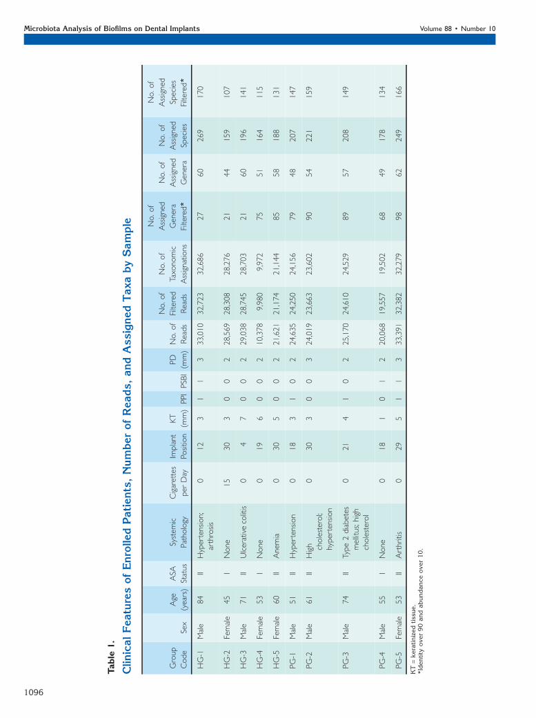
Data SamplingA single researcher (BC-A) recruited the patients fromFebruary to September 2014 at the Dental Hospital ofthe University of Barcelona, Hospitalet de Llobregat(Barcelona, Spain) and examined all clinical records.Data retrieved were age, sex, patient health statusbased on the ASA Physical Status ClassificationSystem,12 systemic pathologies, current medication,smoking habit (number of cigarettes per day), peri-odontal disease (periodontal chart with recessions,PD, BOP, and suppuration), and the following implantvariables: 1) date of implant placement; 2) diameter;3) length; 4) position; 5) distance from any nearbyimplants; 6) width of keratinized mucosa; 7) type ofprosthesis; 8) Mombelli peri-implant plaque index(PPI);13 9) Mombelli peri-implant sulcus bleeding in-dex (PSBI);13 10) suppuration; 11) peri-implant PD;and 12) BL. BL was measured on digital periapicalradiographs using image processing software.i14
Sample Collection and DNA IsolationThe abutments used, specifically fabricated for thispurpose, were designed to have the samemacroscopicand microscopic shape as an implant with a bio-absorbable blast media surface¶ (see supplementaryFig. 1 in online Journal of Periodontology).
After a thorough explanation of the study objectives,a healing abutment was replaced by the experimental
i ImageJ software, US National Institutes of Health, Bethesda, MD.¶ Mozo-Grau dental implants, Mozo-Grau, Valladolid, Spain.
J Periodontol • October 2017 Cortes-Acha, Figueiredo, Seminago, Roig, Llorens, Valmaseda-Castellon
1091
abutment (see supplementary Fig. 1 in online Journalof Periodontology). Patients were instructed to refrainfrom cleaning the abutment and using toothpaste ormouthrinse solutions during the study period.
After 14 days, the experimental abutment wascarefully removed, placed in a sterile snap-cap tube,and transported to the laboratory in <1 hour ina portable refrigerator at 4�C. The abutment wasscrewed to an implant analog placed inside the tube,allowing the biofilm to remain intact without touchingthe tube wall.
All patients were enrolled in a peri-implant main-tenance program, and a final prosthesis was made.
After the abutment was received at the laboratory,it was unscrewed, stored in a 1.5-mL microcentrifugetube, and frozen at -80�C until further analysis. Theabutment was rinsed with phosphate-buffered salineand vortexed for 5 minutes to release the bacteria.Total DNA was purified with a DNA purification kit,#
according to the manufacturer’s protocol for buccalswabs. The amount of DNA extracted was calculatedusing a scientific instrumentation system.**
Statistical AnalysesVariable regions V1, V2, V3, V4, and V5 of the 16Sribosomal RNA (rRNA) gene were amplified witha multiplex PCR system†† and sequenced with a ti-tanium sequencing kit.‡‡
PCRs for V1-V3 and V5-V3 primers were set upwith annealing temperatures of 56�C and 50�C, re-spectively. Two replicate PCRs were performed andpooled for each sample. Amplicon library wascleaned with a PCR purification system,§§ accordingto the manufacturer’s instructions. Amplicon con-centration was estimated using an assay kit.ii
Multiplexed tag-encoded sequencing of DNA fromthe samples was performed on a pyrosequencingplatform.¶¶
Primers used to amplify the 16S rRNA gene and tointroduce multiplex identifiers to identify ampliconsor samples are available on the National Institutes ofHealth Human Microbiome Project website.15
The resulting fast files were preprocessed witha quality control and data preprocessing tool16 bysize (more than 50 bp), quality (minimum quality30), and N content (rejecting reads with >5% of Nsand removing terminal Ns).
The reads were processed through metagenomicrapid annotation using subsystems technology (MG-RAST)17 based on hierarchical classification with theRibosomal Database Project (RDP; release 11). MG-RAST default clustering parameters within the basiclocal alignment search tool–like alignment tool al-gorithm were used.
Each read was taxonomically assigned down tothe genus and species level with 80% confidence
threshold. Reads giving no bacterial hits were ex-cluded. Artificial replicate sequences produced bysequencing artifacts were removed.18
To estimate bacterial diversity, the number ofoperational taxonomic units (OTUs) in the sampleswas determined, and rarefaction analysis was per-formed. Rarefaction curves were obtained by plottingthe number of observed OTUs against the number ofsequences, using the MG-RAST platform17 and theRDP database.19,20
To estimate total diversity, sequences were clus-tered at a standard threshold of 98% nucleotideidentity over a 90% sequence alignment length toallow minimal flexibility and to minimize false posi-tives. Rarefaction curves were obtained using theRDP pyrosequencing pipeline (Fig. 1A). Venn anal-ysis (Fig. 1B) and principal component analysis(PCA) were performed, and heatmaps were gener-ated using a statistical package.21 Venn analysis wasrun on taxonomic diversity data. PCA analysis wasrun on taxonomic diversity and abundance of eachindividual sample and on the average of each group(i.e., PG and HG) (Fig. 1C).
Microbial communities were compared by statis-tical analysis using a distance metric matrix.22 Thisanalysis compares the 16S estimated diversitythrough a phylogenetic approach that takes taxo-nomically assigned and unassigned reads into ac-count (Fig. 2). Heatmap analysis of taxonomicdiversity and abundance was done for each sample(Fig. 3).
RESULTS
After extracting metagenomic data, quality of thereadings was assessed by a quality control tool.## Se-quencing samples were of excellent quality (Phredvalues >28). Unknown reads (not identified as rRNAgenes) varied among samples, ranging from 0.08% to0.31%. Samples used in this study were deposited in theGenBank under accession numbers SAMN06116059to SAMN06116068.
Oral Microbial CommunityThe number of reads (filtered and assigned) andnumber of taxonomic assignations (genus and spe-cies) are listed in Table 1. Althoughmicrobiota-basedrarefaction curves (Fig. 1A) failed to reach saturationphase, the slope of the curves become notably lesspronounced.
# QiAamp DNA minikit, Qiagen, Hilden, Germany.** Qubit system, Thermo Fisher Scientific, Waltham, MA.†† FastStart High Fidelity PCR Systems, Roche, Mannheim, Germany.‡‡ GS Junior titanium sequencing kit, Roche.§§ Agencourt AMPure beads, Beckman Coulter, Brea, CA.ii Qubit dsDNA HS assay kit, Thermo Fisher Scientific.¶¶ GS Junior platform, Roche Applied Science, Indianapolis, IN.## FastQC pipelines, Babraham Bioinformatics, Babraham Institute,
Cambridge, U.K.
Microbiota Analysis of Biofilms on Dental Implants Volume 88 • Number 10
1092
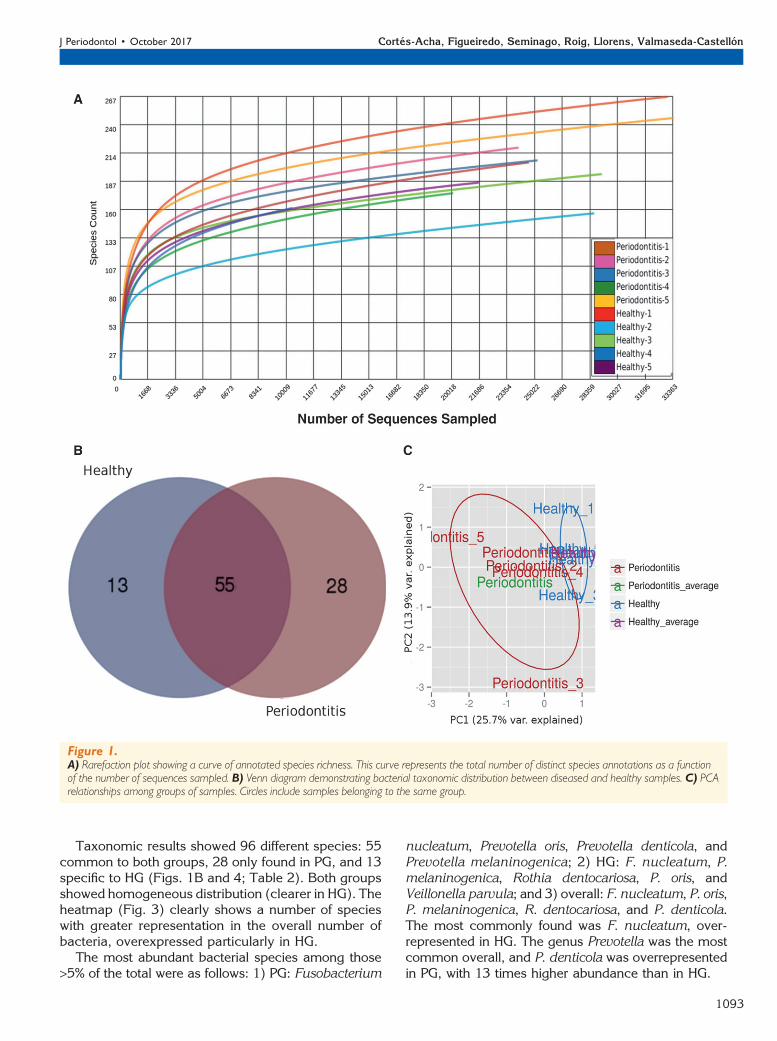
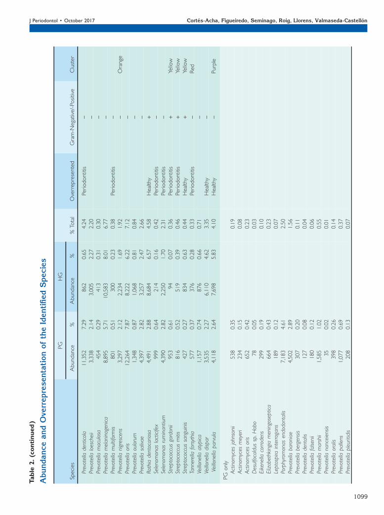
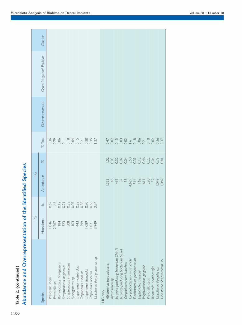
Taxonomic results showed 96 different species: 55common to both groups, 28 only found in PG, and 13specific to HG (Figs. 1B and 4; Table 2). Both groupsshowed homogeneous distribution (clearer in HG). Theheatmap (Fig. 3) clearly shows a number of specieswith greater representation in the overall number ofbacteria, overexpressed particularly in HG.
The most abundant bacterial species among those>5% of the total were as follows: 1) PG: Fusobacterium
nucleatum, Prevotella oris, Prevotella denticola, andPrevotella melaninogenica; 2) HG: F. nucleatum, P.melaninogenica, Rothia dentocariosa, P. oris, andVeillonella parvula; and 3) overall: F. nucleatum, P. oris,P. melaninogenica, R. dentocariosa, and P. denticola.The most commonly found was F. nucleatum, over-represented in HG. The genus Prevotella was the mostcommon overall, and P. denticola was overrepresentedin PG, with 13 times higher abundance than in HG.
Figure 1.A) Rarefaction plot showing a curve of annotated species richness. This curve represents the total number of distinct species annotations as a functionof the number of sequences sampled. B) Venn diagram demonstrating bacterial taxonomic distribution between diseased and healthy samples. C) PCArelationships among groups of samples. Circles include samples belonging to the same group.
J Periodontol • October 2017 Cortes-Acha, Figueiredo, Seminago, Roig, Llorens, Valmaseda-Castellon
1093

Sixteen of the bacteria found have been related tomicrobial complexes of subgingival plaque4 (see sup-plementary Table 1 in online Journal of Periodontology).
Bacteria from the green and purple clustersseemed to be more numerous than others, except forthe above-mentioned bacteria and Campylobactershowae (orange cluster).
Regarding differences between groups (Table 2),periodontopathogens were not more prevalent inPG than in HG. Moreover, Porphyromonas gingivaliswas only found in healthy individuals. In contrast, firstcolonizers such as Streptococcus anginosus andStreptococcus intermedius were only found in PG.
Of the 96 bacteria identified, 19 were not in theHuman Oral Microbiome Database (HOMD)23 or theCORE Microbiome Database.24
DISCUSSION
Implant surfaces are designed to enhance osseointe-gration. When bone remodeling or loss occurs andareas of the implant surface become exposed to theoral environment, saliva biopolymers form a film thatbecomes the interface between the implant surfaceand the first microorganisms. Many characteristics ofthe titanium implant surface, such as roughness, hy-drophobicity, and charge, affect bacterial adhesion.25
Figure 2.Bacteriome cladogram with pyrosequencing datasets of the two groups of samples (PG and HG: pool 1 and pool 2). The RDP database20 was used asthe annotation source, and a minimum identity cutoff of 98% was applied. Colors for the genus branches are indicated in the taxa section of the key.Bars in the external circle indicate abundance of the term in each sample. Colors of the samples are indicated in the samples section of the key.
Microbiota Analysis of Biofilms on Dental Implants Volume 88 • Number 10
1094
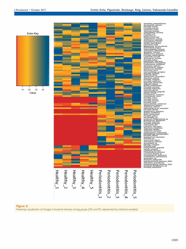
Figure 3.Heatmap visualization of changes in bacterial diversity among groups (HG and PG, represented by individual samples).
J Periodontol • October 2017 Cortes-Acha, Figueiredo, Seminago, Roig, Llorens, Valmaseda-Castellon
1095
Table
1.
Clin
icalFeaturesofEnrolle
dPatients,NumberofReads,andAssignedTaxabySample
Group
Code
Sex
Age
(years)
ASA
Status
System
ic
Patho
logy
Cigarettes
per
Day
Implant
Positio
n
KT
(mm)
PPIPSBI
PD
(mm)
No.of
Reads
No.of
Filtered
Reads
No.of
Taxo
nomic
Assignatio
ns
No.of
Assigned
Genera
Filtered*
No.of
Assigned
Genera
No.of
Assigned
Species
No.of
Assigned
Species
Filtered*
HG-1
Male
84
IIHypertension;
arthrosis
012
31
13
33,010
32,723
32,686
27
60
269
170
HG-2
Female
45
INone
15
30
30
02
28,569
28,308
28,276
21
44
159
107
HG-3
Male
71
IIUlcerativecolitis
04
70
02
29,038
28,745
28,703
21
60
196
141
HG-4
Female
53
INone
019
60
02
10,378
9,980
9,972
75
51
164
115
HG-5
Female
60
IIAnemia
030
50
02
21,621
21,174
21,144
85
58
188
131
PG-1
Male
51
IIHypertension
018
31
02
24,635
24,250
24,156
79
48
207
147
PG-2
Male
61
IIHigh cholesterol;
hypertension
030
30
03
24,019
23,663
23,602
90
54
221
159
PG-3
Male
74
IIType2diabetes
mellitus;high
cholesterol
021
41
02
25,170
24,610
24,529
89
57
208
149
PG-4
Male
55
INone
018
10
12
20,068
19,557
19,502
68
49
178
134
PG-5
Female
53
IIArthritis
029
51
13
33,391
32,382
32,279
98
62
249
166
KT=keratinized
tissue.
*Iden
tity
ove
r90andabundance
ove
r10.
Microbiota Analysis of Biofilms on Dental Implants Volume 88 • Number 10
1096
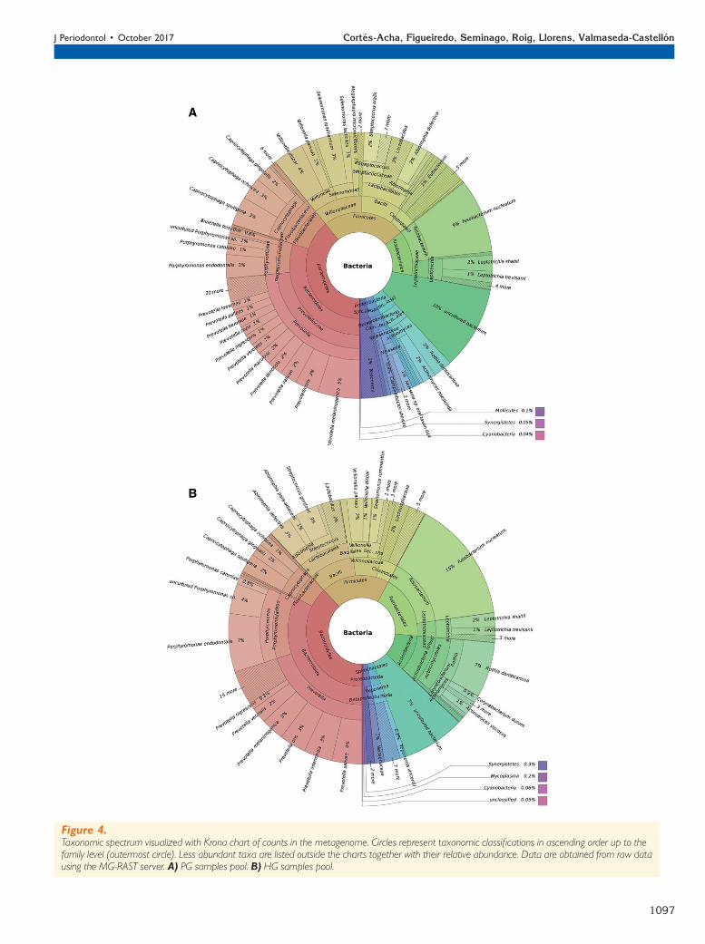
Figure 4.Taxonomic spectrum visualized with Krona chart of counts in the metagenome. Circles represent taxonomic classifications in ascending order up to thefamily level (outermost circle). Less abundant taxa are listed outside the charts together with their relative abundance. Data are obtained from raw datausing the MG-RAST server. A) PG samples pool. B) HG samples pool.
J Periodontol • October 2017 Cortes-Acha, Figueiredo, Seminago, Roig, Llorens, Valmaseda-Castellon
1097

Table
2.
AbundanceandOve
rrepresentationoftheIdentifiedSpecies
Species
PG
HG
%Total
Overrepresented
Gram-N
egative/-Positive
Cluster
Abundance
%Abundance
%
Commonto
PG
andHG
Abiotrophiadefectiva
1,788
1.15
2,112
1.60
1.35
+Actinom
yces
naeslundii
1,943
1.25
2,526
1.91
1.55
+Blue
Actinom
yces
odontolyticus
582
0.37
477
0.36
0.37
+Blue
Actinom
yces
viscosus
1,734
1.11
440
0.33
0.76
Periodontitis
+Blue
Atopobium
minutum
735
0.47
172
0.13
0.32
Periodontitis
+Atopobium
vaginae
720
0.46
947
0.72
0.58
+Butyrivibrio
fibrisolvens
703
0.45
1,815
1.37
0.87
Healthy
Cam
pylobacter
concisus
245
0.16
512
0.39
0.26
Healthy
-Cam
pylobacter
gracilis
847
0.54
1,007
0.76
0.64
-Orange
Cam
pylobacter
rectus
670
0.43
531
0.40
0.42
-Orange
Cam
pylobacter
show
ae1,578
1.01
631
0.48
0.77
Periodontitis
-Orange
Capnocytophaga
canimorsus
322
0.21
42
0.03
0.13
Periodontitis
-Capnocytophaga
gingivalis
4,458
2.86
2,995
2.27
2.59
-Green
Capnocytophaga
ochracea
1,716
1.10
542
0.41
0.78
Periodontitis
-Green
Capnocytophaga
sputigena
5,591
3.59
6,440
4.87
4.18
-Green
Clostridium
aminobutyricum
70
0.04
131
0.10
0.07
Healthy
+Dialisterinvisus
991
0.64
815
0.62
0.63
-Dialisterpneum
osintes
1,300
0.83
1,398
1.06
0.94
-Erysipelothrixrhusiopathiae
113
0.07
198
0.15
0.11
Healthy
Eubacterium
saburreum
412
0.26
325
0.25
0.26
Orange
Fusobacterium
canifelinum
2,146
1.38
2,895
2.19
1.75
-Fusobacterium
nucleatum
15,404
9.89
24,714
18.71
13.94
Healthy
-Gem
ellahaem
olysans
546
0.35
657
0.50
0.42
+Gem
ellamorbillorum
803
0.52
76
0.06
0.31
Periodontitis
+Gem
ellasanguinis
229
0.15
394
0.30
0.22
Healthy
+Granulicatellaadiacens
610
0.39
792
0.60
0.49
+Granulicatellaelegans
141
0.09
158
0.12
0.10
+Leptotrichiabuccalis
921
0.59
922
0.70
0.64
-Leptotrichiashahii
1,637
1.05
1,110
0.84
0.95
-Leptotrichiasp.HKU24
457
0.29
805
0.61
0.44
Healthy
-Leptotrichiawadei
1,688
1.08
830
0.63
0.87
Periodontitis
-Megasphaeramicronuciform
is983
0.63
780
0.59
0.61
-Parvimonas
micra
1,114
0.72
1,085
0.82
0.76
+Peptostreptococcus
anaerobius
230
0.15
142
0.11
0.13
Periodontitis
+Porphyromonas
catoniae
2,276
1.46
1,195
0.90
1.21
Periodontitis
-Prevotellabuccae
2,670
1.71
3,566
2.70
2.17
-
Microbiota Analysis of Biofilms on Dental Implants Volume 88 • Number 10
1098
Table
2.(continued)
AbundanceandOve
rrepresentationoftheIdentifiedSpecies
Species
PG
HG
%Total
Overrepresented
Gram-N
egative/-Positive
Cluster
Abundance
%Abundance
%
Prevotelladenticola
11,352
7.29
862
0.65
4.24
Periodontitis
-Prevotellaloescheii
3,338
2.14
3,005
2.27
2.20
-Prevotellamaculosa
454
0.29
413
0.31
0.30
-Prevotellamelaninogenica
8,895
5.71
10,583
8.01
6.77
-Prevotellamultiformis
801
0.51
300
0.23
0.38
Periodontitis
-Prevotellanigrescens
3,297
2.12
2,234
1.69
1.92
-Orange
Prevotellaoris
12,264
7.87
8,222
6.22
7.12
-Prevotellaoulorum
1,348
0.87
1,068
0.81
0.84
-Prevotellasalivae
4,397
2.82
3,257
2.47
2.66
-Rothiadentocariosa
4,491
2.88
8,684
6.57
4.58
Healthy
+Selenomonas
lacticifex
999
0.64
214
0.16
0.42
Periodontitis
-Selenomonas
ruminantium
4,390
2.82
2,250
1.70
2.31
Periodontitis
-Streptococcus
gordonii
953
0.61
94
0.07
0.36
Periodontitis
+Yellow
Streptococcus
mitis
816
0.52
519
0.39
0.46
Periodontitis
+Yellow
Streptococcus
sanguinis
427
0.27
834
0.63
0.44
Healthy
+Yellow
Tannerellaforsythia
577
0.37
376
0.28
0.33
Periodontitis
-Red
Veillonellaatypica
1,157
0.74
876
0.66
0.71
-Veillonelladispar
3,535
2.27
6,110
4.62
3.35
Healthy
-Veillonellaparvula
4,118
2.64
7,698
5.83
4.10
Healthy
-Purple
PG
only
Actinom
yces
johnsonii
538
0.35
0.19
Actinom
yces
meyeri
234
0.15
0.08
Actinom
yces
oris
652
0.42
0.23
Desulfocaldus
sp.Hobo
78
0.05
0.03
Eikenellacorrodens
299
0.19
0.10
Elizabethkingiameningoseptica
664
0.43
0.23
Leptospira
interrogans
189
0.12
0.07
Porphyromonas
endodontalis
7,183
4.61
2.50
Prevotellabaroniae
4,502
2.89
1.56
Prevotellabergensis
307
0.20
0.11
Prevotelladentalis
127
0.08
0.04
Prevotellafalsenii
180
0.12
0.06
Prevotellamarshii
1,585
1.02
0.55
Prevotellananceiensis
35
0.02
0.01
Prevotellaoralis
398
0.26
0.14
Prevotellapallens
1,077
0.69
0.37
Prevotellapleuritidis
208
0.13
0.07
J Periodontol • October 2017 Cortes-Acha, Figueiredo, Seminago, Roig, Llorens, Valmaseda-Castellon
1099
Table
2.(continued)
AbundanceandOve
rrepresentationoftheIdentifiedSpecies
Species
PG
HG
%Total
Overrepresented
Gram-N
egative/-Positive
Cluster
Abundance
%Abundance
%
Prevotellashahii
1,046
0.67
0.36
Prevotellasp.
2,267
1.46
0.79
Rum
inococcusflavefaciens
184
0.12
0.06
Streptococcus
anginosus
323
0.21
0.11
Streptococcus
interm
edius
508
0.33
0.18
Synergistetessp.
103
0.07
0.04
Treponem
amaltophilum
443
0.28
0.15
Treponem
amedium
599
0.38
0.21
Treponem
asocranskii
1,089
0.70
0.38
Treponem
avincentii
1,021
0.66
0.35
UnculturedPorphyromonas
sp.
3,949
2.54
1.37
HG
only
Abiotrophiaparaadiacens
1,353
1.02
0.47
Azospirillum
sp.
46
0.03
0.02
butyrate-producing
bacterium
SM4/1
419
0.32
0.15
butyrate-producing
bacterium
SS3/4
87
0.07
0.03
Corynebacterium
kutscheri
58
0.04
0.02
Corynebacterium
matruchotii
4,629
3.50
1.61
Fusobacterium
periodonticum
514
0.39
0.18
Leptotrichiatrevisanii
162
0.12
0.06
Porphyromonas
gingivalis
611
0.46
0.21
Prevotellacopri
290
0.22
0.10
Riemerellaanatipestifer
52
0.04
0.02
UnculturedKingella
sp.
1,048
0.79
0.36
UnculturedStreptococcus
sp.
1,069
0.81
0.37
Microbiota Analysis of Biofilms on Dental Implants Volume 88 • Number 10
1100
The present study adds new information regardingmicrobiota formed on implants when they becomeexposed to the oral cavity without associated in-flammation (i.e., in soft tissue recession or bone re-modeling processes). These data can also be usefulwhen peri-implantitis patients are surgically treatedwith a resective approach. In these cases, after surfacedecontamination, an apically repositioned flap ismadeto reduce peri-implant pockets, thus exposing therough surface of the implant.
One of the main limitations of this study is thereduced sample size, which may jeopardize gener-alization of outcomes. Also, the fact that all sampleswere collected after 14 days did not allow a study ofthe evolution of biofilm formation.
Periodontal disease is a known risk factor for peri-implantitis and one of the explanations, apart frompatient susceptibility, is that periodontally involvedteeth may act as a reservoir for periodontal pathogensthat can colonize the implant surface.26 However,a study using an open-ended molecular approachshowed that in 85% of participants, <8% of specieswere shared between teeth and implants, suggestingthat microbiology of peri-implantitis and periodontitismight be quite different.27 Data from the present study,although obtained from healthy sites, seem to supportthese results, as species generally associated withdiseased implants, such as P. gingivalis, are detectedin healthy implants but only in periodontally healthyindividuals. P. gingivalis might have an important rolein peri-implant diseases as it has been described as an‘‘enhancer species’’ that is involved in coaggregationstages during biofilm maturation.28
On the other hand, many identified bacteria suchas Streptococcus sanguinis, Actinomyces naeslun-dii, Campylobacter rectus, Parvimonas micra, andGranulicatella adiacens or the genera Fusobacte-rium,Actinomyces, Veillonella,Atopobium,Gemella,Rothia, and Leptotrichia have been associated withhealthy implants in previous studies.8,28-30 Specialconsideration should be taken with the genera Acti-nomyces and Veillonella as most authors28,31 agreedto finding them more frequently in healthy implants,and none of the revised studies associate them withperi-implantitis.8,28-33 Presence of these bacteria,together with Streptococcus mitis and S. sanguinis,may play a protective role regarding peri-implantdiseases.34,35
Prevotella spp. were found widely in both PG andHG, and P. denticola was one of the most abundantbacteria. It has been associated with periodontaldisease as strongly as the classic red complex,36 but itshould be remembered that patients in PG had PD<4 mm and at least 70% of sites with no BOP.Therefore, presence of this genus in both groups maysuggest that it is only pathogenic when the bacterial
balance is disturbed or when there is host suscepti-bility.
Another most abundant bacterium was F. nucle-atum. It is known to mediate between the first andsubsequent colonizers and interact with host cells,facilitating coaggregation with periodontopathogenssuch as P. gingivalis.37 Some authors suggest thatF. nucleatum infection facilitates attachment of P.gingivalis to the gingival fibroblast, and consequentlythese two bacteria are often found together.38 In-terestingly, F. nucleatum was abundant in both groups(15,404 in PG and 24,714 in HG), whereas P. gingivaliswas not identified in PG but had an abundance of 611in HG.
Previous publications concluded that in peri-odontally healthy individuals, P. gingivalis and Ag-gregatibacter actinomycemcomitans were never39 orrarely40 found on implants. The present study con-tradicts this statement, as P. gingivalis was specifi-cally found in HG. This disparity might be explainedby the different analytic method (pyrosequencing),which in the authors’ opinion affords more completeand detailed data gathering and should be im-plemented more frequently in the future.
The microbiome surrounding teeth has been shownto be significantly more diverse than that around im-plants.41,42 In addition, the rate of traditional pathogensaround implants has been reported to be lower than thataround teeth in both healthy and diseased patients.43
Cortelli et al.43 also pointed out that early colonizers ofrough implant surfaces might constitute a differentbacterial microbiome from that of periodontal diseases.The present report shows that individuals have an im-portant variability regarding the composition of biofilm.This indicates that studies with large samples are re-quired. It would be interesting to analyze whether thisvariability is related with the different progression pat-terns of BL found in peri-implantitis.
Streptococcus, Granulicatella, and Gemella werepresent in both PG and HG, in agreement with a pre-vious study.44 These bacteria are considered symbi-onts, with a high proportion returning to pockets afterperiodontal treatments.45
Regarding differences among groups, specialmention should be made of four bacteria with >1%abundance: Porphyromonas endodontalis, Prevotellabaroniae and an uncultured Porphyromonas sp. in PGand Corynebacterium matruchotii in HG. P. endo-dontalis is found in symptomatic oral infections, suchas endodontic infections and periodontal pockets, butalso in asymptomatic cases. It shows low virulence inexperimental monoinfections but seems to play animportant role in anaerobic mixed infections.46 P.baroniae has been described as a causal agent ofendodontic abscesses.47 C. matruchotii is consideredpart of normal oral microbiota.48
J Periodontol • October 2017 Cortes-Acha, Figueiredo, Seminago, Roig, Llorens, Valmaseda-Castellon
1101
High prevalence of Gram-negative bacteria in HGand Gram-positive bacteria in PG is surprising, asearlier studies demonstrated Gram-negative preva-lence in oral microbioma.2
Although 19 bacteria were not registered in theHOMD or CORE databases, five had been reportedpreviously, including Prevotella bergensis, Leptotrichiasp. HKU24, and Prevotella copri. The remaining 14have been identified in human infections in other areasof the body or found in insects, contaminated water,dogs, cats, or birds (see supplementary Table 1 inonline Journal of Periodontology). Contamination ofsamples during transport was highly unlikely becausethe snap-tubes were sterile, and the abutment did nottouch its walls (it was firmly screwed to a sterile implantreplica). Future research should examine whether thesemicroorganisms play an important role in peri-implantdiseases. This is quite an important finding, and onceagain indicates the importance of using metagenomicanalysis techniques. Other microbiologic methods suchas checkerboard DNA–DNA hybridization are indeedextremely accurate and have high sensitivity (>92.5%)and specificity (100%), but are clearly insufficient todetect composition of the microbiome surroundingimplants.49 Likewise, the sample collection methodmight cause important discrepancies among studies.Biofilms collected with curets can result in lower bac-terial counts due to implant topography,11 and sterilepaper points can be a source of foreign bacteria.50 Thisis an important advantage of the present method torecover biofilm. Other authors have previously reporteduse of abutments with different roughnesses51 for thispurpose, but with no threads. Thus, a study comparingdifferent biofilm sampling methods would be of interest.
CONCLUSIONS
A wide variety of bacteria (96 species) were foundaround abutments simulating exposed dental im-plants without inflammation in 10 individuals. Themost frequently found bacteria were F. nucleatumand P. denticola. Some species generally associatedwith periodontitis were more commonly found inpatients without history of periodontitis.
A large number of bacteria that had never beendescribed as part of the oral microbiome were foundin the present sample. Further research with largersamples is needed to clarify the role of these mi-croorganisms in the oral environment.
ACKNOWLEDGMENTS
The authors thank Dr. Ruben Leon and Dr. VanessaBlanc from the Research and Development and Micro-biology Departments of Dentaid SL (Cerdanyola delValles, Spain) for critical review of the manuscript.The authors also thank Mary Georgina Hardinge (na-tive British freelance translator, Valencia, Spain) for
English language editing of the manuscript. Dr.Figueiredo reports grants from the Faculty of Dentistry,University of Barcelona (Spain) and non-financial sup-port from Mozo-Grau (Valladolid, Spain) during con-duct of the study. Also, he reports grants, personalfees, and non-financial support from Mozo-Grau (Val-ladolid, Spain) and personal fees from BioHorizonsIberica (Madrid, Spain), Inibsa Dental (Llicxa de Vall,Spain), DENTSPLY implants Iberia (Barcelona,Spain), and ADIN implants (Afula, Israel) outside thesubmittedwork. In addition,Dr. Figueiredohasapatent‘‘Biofilm collector abutment’’ pending to Mozo-Grau,Rui Figueiredo, and Eduard Valmaseda-Castellon.Dr. Valmaseda-Castellon reports grants from the Fac-ulty of Dentistry, University of Barcelona (Spain) andnon-financial support from Mozo-Grau (Valladolid,Spain) during conduct of the study. Also, he reportsgrants, personal fees, and non-financial support fromMozo-Grau (Valladolid, Spain) and personal fees fromBioHorizons Iberica (Madrid, Spain), Inibsa Dental(Llicxa de Vall, Spain), and DENTSPLY implants Iberia(Barcelona, Spain) outside the submitted work. In ad-dition, Dr. Valmaseda-Castellon has a patent ‘‘Biofilmcollector abutment’’ pending to Mozo-Grau, RuiFigueiredo, and Eduard Valmaseda-Castellon. Dr.Cortes-Acha reports grants from the Faculty of Den-tistry, University of Barcelona (Spain) and non-financialsupport fromMozo-Grau (Valladolid, Spain) during con-duct of the study. The present research was conduct-ed by the Dental and Maxillofacial Pathology andTherapeutics research group at the IDIBELL Institute(L’Hospitalet de Llobregat, Spain) and fundedby a postgraduate research grant from the Facultyof Dentistry, University of Barcelona (€4.560).
REFERENCES1. Mir-Mari J,Mir-Orfila P, FigueiredoR,Valmaseda-Castellon
E, Gay-Escoda C. Prevalence of peri-implant diseases. Across-sectional study based on a private practice environ-ment. J Clin Periodontol 2012;39:490-494.
2. QuirynenM,De SoeteM, van SteenbergheD. Infectiousrisks for oral implants: A review of the literature. ClinOral Implants Res 2002;13:1-19.
3. Hajishengallis G, Lamont RJ. Beyond the red complexand into more complexity: The polymicrobial synergyand dysbiosis (PSD) model of periodontal diseaseetiology. Mol Oral Microbiol 2012;27:409-419.
4. Socransky SS, Haffajee AD, Cugini MA, Smith C, KentRL Jr. Microbial complexes in subgingival plaque.J Clin Periodontol 1998;25:134-144.
5. Murray JL, Connell JL, StacyA, Turner KH,WhiteleyM.Mechanisms of synergy in polymicrobial infections.J Microbiol 2014;52:188-199.
6. Shibli JA, Melo L, Ferrari DS, Figueiredo LC, Faveri M,Feres M. Composition of supra- and subgingival biofilmof subjects with healthy and diseased implants. ClinOral Implants Res 2008;19:975-982.
7. Kumar PS, Mason MR, Brooker MR, O’Brien K. Pyrose-quencing reveals uniquemicrobial signatures associated
Microbiota Analysis of Biofilms on Dental Implants Volume 88 • Number 10
1102

with healthy and failing dental implants. J Clin Peri-odontol 2012;39:425-433.
8. Zheng H, Xu L, Wang Z, et al. Subgingival microbiomein patients with healthy and ailing dental implants. SciRep 2015;5:10948.
9. Quirynen M, Vogels R, Peeters W, van Steenberghe D,Naert I, Haffajee A. Dynamics of initial subgingivalcolonization of ‘‘pristine’’ peri-implant pockets. ClinOral Implants Res 2006;17:25-37.
10. Renvert S, Roos-Jansaker AM, Lindahl C, Renvert H,Rutger Persson G. Infection at titanium implants with orwithout a clinical diagnosis of inflammation. Clin OralImplants Res 2007;18:509-516.
11. Gerber J, Wenaweser D, Heitz-Mayfield L, Lang NP,Persson GR. Comparison of bacterial plaque samplesfrom titanium implant and tooth surfaces by differentmethods. Clin Oral Implants Res 2006;17:1-7.
12. ASA House of Delegates. American Society ofAnesthesiologists – ASA Physical Status Classifica-tion System. Available at: http://www.asahq.org/resources/clinical-information/asa-physical-status-classification-system. Accessed April 20, 2017.
13. Mombelli A, van Oosten MA, Schurch E Jr., Land NP.The microbiota associated with successful or failingosseointegrated titanium implants. Oral Microbiol Im-munol 1987;2:145-151.
14. Garcıa-Garcıa M, Mir-Mari J, Benic GI, Figueiredo R,Valmaseda-Castellon E. Accuracy of periapical radi-ography in assessing bone level in implants affected byperi-implantitis: A cross-sectional study. J Clin Peri-odontol 2016;43:85-91.
15. Jumpstart Consortium Human Microbiome ProjectData Generation Working Group. 16S 454 SequencingProtocol HMP Consortium 2010. Available at: http://www.hmpdacc.org/doc/16S_Sequencing_SOP_4.2.2.pdf. Accessed January 25, 2016.
16. Schmieder R, Edwards R. Quality control and prepro-cessing of metagenomic datasets. Bioinformatics 2011;27:863-864.
17. Meyer F, Paarmann D, D’Souza M, et al. The metage-nomics RAST server – A public resource for theautomatic phylogenetic and functional analysis ofmetagenomes. BMC Bioinformatics 2008;9:386.
18. Gomez-Alvarez V, Teal TK, Schmidt TM. Systematicartifacts in metagenomes from complex microbialcommunities. ISME J 2009;3:1314-1317.
19. Wang Q, Garrity GM, Tiedje JM, Cole JR. NaiveBayesian classifier for rapid assignment of rRNAsequences into the new bacterial taxonomy. ApplEnviron Microbiol 2007;73:5261-5267.
20. Cole JR, Wang Q, Cardenas E, et al. The RibosomalDatabase Project: Improved alignments and new tools forrRNA analysis. Nucleic Acids Res 2009;37:D141-D145.
21. Futami R, Munoz-Pomer A, Viu JM, et al. GPRO: Theprofessional tool for management, functional analysisand annotation of omic sequences and databases.Biotechvana Bioinforma 2011;1:1-5.
22. Chen J, Bittinger K, Charlson ES, et al. Associatingmicrobiome composition with environmental covariatesusing generalized UniFrac distances. Bioinformatics 2012;28:2106-2113.
23. Chen T, YuW-H, Izard J, BaranovaOV, LakshmananA,Dewhirst FE. TheHumanOral MicrobiomeDatabase: AWeb Accessible Resource for Investigating Oral Mi-crobe Taxonomic and Genomic Information. Database(Oxford) 2010;2010:baq013.
24. OSU CORE Database, Oral Microbiome. Available at:http://microbiome.osu.edu/sequences. Accessed No-vember 21, 2016.
25. TeughelsW, VanAsscheN, Sliepen I, QuirynenM.Effectofmaterial characteristics and/or surface topography onbiofilm development. Clin Oral Implants Res 2006;17(Suppl. 2):68-81.
26. Renvert S, Roos-Jansaker AM, Lindahl C, Renvert H,Rutger Persson G. Infection at titanium implants with orwithout a clinical diagnosis of inflammation. Clin OralImplants Res 2007;18:509-516.
27. Dabdoub SM, Tsigarida AA, Kumar PS. Patient-specificanalysis of periodontal and peri-implant microbiomes.J Dent Res 2013;92(Suppl. 12):168S-175S.
28. Rickard AH, Gilbert P, High NJ, Kolenbrander PE,Handley PS. Bacterial coaggregation: An integral pro-cess in the development of multi-species biofilms.Trends Microbiol 2003;11:94-100.
29. Neilands J, Wickstrom C, Kinnby B, et al. Bacterialprofiles and proteolytic activity in peri-implantitis ver-sus healthy sites. Anaerobe 2015;35(Pt A):28-34.
30. da Silva ES, Feres M, Figueiredo LC, Shibli JA, RamiroFS, FaveriM.Microbiological diversity of peri-implantitisbiofilm by Sanger sequencing. Clin Oral Implants Res2014;25:1192-1199.
31. Tamura N, Ochi M, Miyakawa H, Nakazawa F. Analysisof bacterial flora associated with peri-implantitis usingobligate anaerobic culture technique and 16S rDNAgene sequence. Int J Oral Maxillofac Implants 2013;28:1521-1529.
32. Persson GR, Renvert S. Cluster of bacteria associatedwith peri-implantitis. Clin Implant Dent Relat Res 2014;16:783-793.
33. Eick S, Ramseier CA, Rothenberger K, Bragger U,Buser D, Salvi GE. Microbiota at teeth and implants inpartially edentulous patients. A 10-year retrospectivestudy. Clin Oral Implants Res 2016;27:218-225.
34. Quirynen M, De Soete M, Dierickx K, van SteenbergheD. The intra-oral translocation of periodontopathogensjeopardises the outcome of periodontal therapy. Areview of the literature. J Clin Periodontol 2001;28:499-507.
35. Stingu C-S, Eschrich K, Rodloff AC, Schaumann R,Jentsch H. Periodontitis is associated with a loss ofcolonization by Streptococcus sanguinis. J Med Micro-biol 2008;57:495-499.
36. Vartoukian SR, Palmer RM, Wade WG. Diversity andmorphology ofmembers of the phylum ‘‘synergistetes’’in periodontal health and disease. Appl Environ Micro-biol 2009;75:3777-3786.
37. AngMY,DuttaA,WeeWY,DymockD, Paterson IC, ChooSW. Comparative genome analysis of Fusobacteriumnucleatum. Genome Biol Evol 2016;8:2928-2938.
38. Metzger Z, Lin Y-Y, Dimeo F, Ambrose WW, Trope M,Arnold RR. Synergistic pathogenicity of Porphyromo-nas gingivalis and Fusobacterium nucleatum in themouse subcutaneous chamber model. J Endod 2009;35:86-94.
39. Heuer W, Elter C, Demling A, et al. Analysis of earlybiofilm formation on oral implants in man. J OralRehabil 2007;34:377-382.
40. Cosgarea R, Dannewitz B, Sculean A, et al. Bacterialand inflammatory behavior of implants in the earlyhealing phase of chronic periodontitis. QuintessenceInt 2012;43:491-501.
41. HeuerW, Kettenring A, Stumpp SN, et al. Metagenomicanalysis of the peri-implant and periodontal microflora
J Periodontol • October 2017 Cortes-Acha, Figueiredo, Seminago, Roig, Llorens, Valmaseda-Castellon
1103
in patients with clinical signs of gingivitis or mucositis.Clin Oral Investig 2012;16:843-850.
42. Vered Y, Zini A, Mann J, et al. Teeth and implantsurroundings: Clinical health indices andmicrobiologicparameters. Quintessence Int 2011;42:339-344.
43. Cortelli SC, Cortelli JR, Romeiro RL, et al. Frequency ofperiodontal pathogens in equivalent peri-implant andperiodontal clinical statuses. Arch Oral Biol 2013;58:67-74.
44. Costalonga M, Herzberg MC. The oral microbiome andthe immunobiology of periodontal disease and caries.Immunol Lett 2014;162(2 Pt A):22-38.
45. Yamanaka W, Takeshita T, Shibata Y, et al. Composi-tional stability of a salivary bacterial population againstsupragingival microbiota shift following periodontaltherapy. PLoS One 2012;7:e42806.
46. van Winkelhoff AJ, van Steenbergen TJ, de Graaff J.Porphyromonas (Bacteroides) endodontalis: Its role inendodontal infections. J Endod 1992;18:431-434.
47. Rocxas IN, Siqueira JF Jr. Prevalence of new candidatepathogens Prevotella baroniae, Prevotella multisac-charivorax and as-yet-uncultivatedBacteroidetes cloneX083 in primary endodontic infections. J Endod 2009;35:1359-1362.
48. Wu C. Human microbiome, Actinobacteria. In: NelsonKA, ed. Encyclopedia of Metagenomics. New York:Springer New York; 2013:1-7.
49. Socransky SS, Haffajee AD, Smith C, et al. Use ofcheckerboard DNA-DNA hybridization to study com-plex microbial ecosystems. Oral Microbiol Immunol2004;19:352-362.
50. van der Horst J, Buijs MJ, Laine ML, et al. Sterile paperpoints as a bacterial DNA-contamination source inmicrobiome profiles of clinical samples. J Dent 2013;41:1297-1301.
51. Elter C, Heuer W, Demling A, et al. Supra- andsubgingival biofilm formation on implant abutmentswith different surface characteristics. Int J Oral Max-illofac Implants 2008;23:327-334.
Correspondence: Dr. Rui Figueiredo, Faculty of Medicineand Health Sciences, University of Barcelona, BellvitgeCampus, Pavello de Govern 2a planta, Despatx 2.9,L’Hospitalet de Llobregat, 08907 Barcelona, Spain. E-mail: [email protected].
Submitted January 21, 2017; accepted for publicationApril 28, 2017.
Microbiota Analysis of Biofilms on Dental Implants Volume 88 • Number 10
1104