Microbes in deionized and tap water: Implications for ... · 1 Microbes in deionized and tap water:...
32
1 Microbes in deionized and tap water: Implications for maintenance of laboratory water production system Wenfa Ng Department of Chemical and Biomolecular Engineering, National University of Singapore Email: [email protected] Graphical abstract Short description: Microbes were recovered from deionized and tap water in significant numbers that calls for concern for both deionized and tap water quality. Specifically, greater diversity and relative abundance of microbes were recovered from deionized water compared to tap water on formulated colourless agar (with 0.1 g/L yeast extract) at 30 o C after multiple days of incubation. Possible removal of monochloramine residual disinfectant from tap water via ion exchange resins in deionized water production system might account for the greater recovery of microbes from deionized water compared to tap water. Close proximity of microbes further highlighted that possible exchange of metabolites and signaling molecules might facilitate the growth of more microbial species through a network of collaborative or neutral relationships. This suggested that antagonistic relationships were less prevalent in the consortium of microbes that constituted the microbial flora of deionized and tap water. Furthermore, keystone species could have enabled the growth of neighbouring microbial cells embedded in the agar matrix through the secretion of needed metabolites and signaling molecules, which generated zones with high colony density of microbes as well as others devoid of microbes due to absence of keystone species. Abstract Microbes, with their vast metabolic capabilities and great adaptability, occupy almost every conceivable ecological niche on Earth. Thus, could they survive in oligotrophic deionized (DI) water? Observations of white cauliflower-like lumps and black specks in salt solutions after Tap water Deionized water PeerJ Preprints | https://doi.org/10.7287/peerj.preprints.181v6 | CC BY 3.0 Open Access | rec: 23 Mar 2018, publ: 23 Mar 2018
Transcript of Microbes in deionized and tap water: Implications for ... · 1 Microbes in deionized and tap water:...

1
Microbes in deionized and tap water: Implications for maintenance of
laboratory water production system
Wenfa Ng
Department of Chemical and Biomolecular Engineering, National University of Singapore
Email: [email protected]
Graphical abstract
Short description: Microbes were recovered from deionized and tap water in significant numbers
that calls for concern for both deionized and tap water quality. Specifically, greater diversity and
relative abundance of microbes were recovered from deionized water compared to tap water on
formulated colourless agar (with 0.1 g/L yeast extract) at 30 oC after multiple days of incubation.
Possible removal of monochloramine residual disinfectant from tap water via ion exchange resins
in deionized water production system might account for the greater recovery of microbes from
deionized water compared to tap water. Close proximity of microbes further highlighted that
possible exchange of metabolites and signaling molecules might facilitate the growth of more
microbial species through a network of collaborative or neutral relationships. This suggested that
antagonistic relationships were less prevalent in the consortium of microbes that constituted the
microbial flora of deionized and tap water. Furthermore, keystone species could have enabled the
growth of neighbouring microbial cells embedded in the agar matrix through the secretion of
needed metabolites and signaling molecules, which generated zones with high colony density of
microbes as well as others devoid of microbes due to absence of keystone species.
Abstract
Microbes, with their vast metabolic capabilities and great adaptability, occupy almost every
conceivable ecological niche on Earth. Thus, could they survive in oligotrophic deionized (DI)
water? Observations of white cauliflower-like lumps and black specks in salt solutions after
Tap water Deionized water
PeerJ Preprints | https://doi.org/10.7287/peerj.preprints.181v6 | CC BY 3.0 Open Access | rec: 23 Mar 2018, publ: 23 Mar 2018

2
months of storage in plastic bottles suggested a microbial origin for the “contaminants”. Growth
experiments was conducted to profile the microbial diversity of fresh DI water, produced on a
just-in-time basis by a filter cum ion exchange system with tap water as feed. Inoculation of DI
water on R2A agar and a formulated colourless agar followed by multi-day aerobic incubation
revealed the presence of a large variety of microbes with differing pigmentations, growth rates,
colony sizes and morphologies. Additionally, greater abundance and diversity of microorganisms
was recovered at 30 oC compared to 25 and 37 oC; most probably due to adaptation of microbes
to tropical ambient water temperatures of 25 to 30 oC. Comparative experiments with tap water
as inoculum recovered a significantly smaller number and diversity of microorganisms; thus,
suggesting that monochloramine residual disinfectant in tap water was effective in inhibiting cell
viability. In contrast, possible removal of monochloramine by adsorption onto ion exchange
resins of the DI water production system might explain the observed greater diversity and
abundance of viable microbes in DI water. More importantly, greater diversity and abundance of
microbes from tap water were recovered on R2A agar compared to formulated colourless agar,
which suggested that chelating compounds in R2A agar could have complexed monochloramine
and reduced its toxicity towards microbes. Similar chelating compounds were unlikely to be
present in the formulated colourless agar. Finally, keystone species secreting signaling molecules
and metabolites could induce the growth of neighbouring cells embedded in the agar matrix. This
explained the presence of large clear zones devoid of colonies where there was no keystone
species. Additionally, close proximity of colonies on agar suggested that cooperative and neutral
relationships guided by exchange of metabolite and signaling molecules might be more prevalent
compared to antagonistic relationships in which inhibitory compounds were used. Collectively,
this study confirmed the presence of microbes in fresh DI water and tap water. Propensity of
microbes in forming biofilm on various surfaces suggested that intermittent flow in just-in-time
DI water production provided opportunities for cell attachment and biofilm formation during
water stagnation, and subsequent dislodgement and resuspension of cells upon water flow. Thus,
regular maintenance and cleaning of the production system should help reduce DI water’s
microorganism load.
Keywords: viable but not cultivable (VBNC), biofilm, microbial ecology, tap water, deionized
water, chlorine residual, nutrient poor, monochloramine, disinfection, microbial flora,
Subject areas: microbiology, ecology, biodiversity, biochemistry, freshwater biology,
Significance of the work
Microbes varied in physiology and metabolic preferences and occupy almost every ecological
niche on Earth. Thus, it would not be a surprise that microbes were found in deionized water and
tap water. This study confirmed the presence of large diversity and numbers of microorganisms in
deionized water. Microbes of differing pigmentations, colony size and morphology were also
recovered from tap water. Adsorption and removal of residual disinfectant, monochloramine, by
ion exchange resins of the deionized water production system was postulated to account for the
greater recovery of microorganisms in deionized water compared to tap water. Greater diversity
and numbers of microbes were also recovered at 30 oC compared to 25 and 37 oC, which indicated
that microbes in tap and deionized water had adapted to tropical ambient temperatures of ~30 oC.
Close proximity of many recovered colonies from deionized and tap water revealed that there was
a greater preponderance of neutral and collaborative relationships compared to antagonistic ones
PeerJ Preprints | https://doi.org/10.7287/peerj.preprints.181v6 | CC BY 3.0 Open Access | rec: 23 Mar 2018, publ: 23 Mar 2018
3
amongst microbes. Specifically, secretion and exchange of metabolites and signaling molecules
could have enabled the recovery of many colonies. Finally, keystone species that secrete useful
metabolites and signaling molecules needed by other microbes could have played a critical role in
enabling the recovery of more microbes on both R2A and a formulated colourless agar. Areas on
agar without such keystone species would appear as clear zones devoid of colonies due to the lack
of secreted metabolites or signaling molecules that facilitated the recovery of microorganisms.
Introduction
Microorganisms occupy every conceivable niche on Earth, whether in deep ocean
hydrothermal vents or within the Earth crust.1 2 3 4 5 Hence, microbes, in aggregate, possess
versatile metabolism and adaptability for coping with various nutritional conditions and
environmental stressors on Earth. Individual species of microorganism, however, do not possess
all requisite metabolic pathways for surviving various environmental conditions.6 Thus,
differences in nutritional conditions and environmental stressors at different locales could account
PeerJ Preprints | https://doi.org/10.7287/peerj.preprints.181v6 | CC BY 3.0 Open Access | rec: 23 Mar 2018, publ: 23 Mar 2018

4
for differing metabolic characteristics of microbes present in the habitat. For example, microbes
able to survive in drinking water possess the ability to harness small amounts of nutrients (i.e.,
oligotrophic environment) for survival and growth, while bacteria with a high energy metabolism
requiring constant infusion of nutrients for survival would likely not be able to survive in potable
water.
Observations of cauliflower-like lumps and black specks in prepared salts solutions
contained in plastic bottles suggested deionized water as a possible source of contaminants. As a
low nutrient growth environment, deionized water is not conducive for microbial growth; however,
microbes could remain viable even though they are not in the growth state characterized by rapid
cell division. Many approaches and agar media have been used in inducing the growth of
microorganisms from tap water or other water matrixes with low nutrient content, but they
generally could not recover significant numbers of microbes thought to be present in the water.7
By mimicking the natural low nutritional environment of various environmental matrixes such as
tap water, R2A agar is the gold standard agar medium for the cultivation of microorganisms from
oligotrophic environments.8 Specifically, slow growth rates in a low nutrient environment provides
multiple microbial species with an opportunity to grow given that access to nutrients was not
constrained to species with fast growth rates. Results indicated that R2A agar could recover
significantly more species of microorganisms from tap water compared to other agar media. This
motivated the formulation of a similar low nutrient agar medium, which in being colourless, help
improve the optical transparency of the agar that improves the identification of small colonies.9
Hence, using R2A agar and a formulated colourless agar with deionized and tap water as
inoculum, growth experiments via spread plate inoculation was performed at 25, 30 and 37 oC.
Recovery of a large variety of microorganisms of different pigmentation, morphology, and growth
rates revealed that a diverse microbial flora existed in deionized water, due possibly to the removal
of the residual disinfectant monochloramine from tap water by ion exchange resins of the
deionized water production system. Tap water, in comparison, harboured fewer number and types
of viable microbes relative to deionized water; thereby, indicating that monochloramine was
effective in inhibiting the growth of microbes in tap water. Thus, removal of monochloramine by
adsorption on ion exchange resins in the deionized water production system likely removed an
environmental stressor inhibiting the growth of microbes in tap water. Furthermore, possible poor
maintenance of the filter cum ion exchange column critical to production of deionized water could
provide a concentrator of microorganisms through the growth of biofilms on the surfaces of the
filter membrane of the deionized water production system.
PeerJ Preprints | https://doi.org/10.7287/peerj.preprints.181v6 | CC BY 3.0 Open Access | rec: 23 Mar 2018, publ: 23 Mar 2018
5
Materials and Methods
Materials
R2A and Tryptic Soy agar were purchased from Merck and used as is. Composition of R2A agar
was [g/L]: yeast extract, 0.5; Proteose Peptone, 0.5; Casein hydrolysate, 0.5; Glucose, 0.5; Starch
soluble, 0.5; Sodium pyruvate, 0.3; K2HPO4, 0.3; MgSO4, 0.024; Agar, 15.0. Composition of
formulated colourless agar was [g/L]: D-Glucose, 2.0; NH4Cl, 0.5; K2HPO4, 0.5; KH2PO4, 0.1;
NaCl, 0.5; MgSO4.7H2O, 1.0; Yeast extract, 1.0; Agar, 15.0. Composition of LB Lennox agar was
[g/L]: Tryptone, 10.0; Yeast extract, 5.0; NaCl, 5.0; Agar, 15.0. Composition of M9 agar was
[g/L]: D-Glucose, 4.0; NH4Cl, 1.0; Na2HPO4, 6.78; NaH2PO4, 3.0; NaCl, 0.5; Agar, 15.0.
Composition of Tryptic Soy Agar was [g/L]: Pancreatic digest of casein, 15.0; Papaic digest of
soya bean, 5.0; NaCl, 5.0; Agar, 15.0.
Growth of microbes on solid medium
Tap and deionized water were collected fresh from the tap and deionized water production system
respectively after allowing the water to run for 5 minutes, and was contained in a pre-sterilized 15
ml polypropylene centrifuge tube. The deionized water production system was equipped with
ELGA Stat cation and anion exchange resins system preceded by a filter membrane cartridge. 0.1
ml of tap or deionized water was used as inoculum for either R2A agar or formulated colourless
agar and inoculated via the spread plate method. Inoculated plates were incubated aerobically in a
temperature controlled incubator at the chosen temperature (Yih Der LM570D, Taiwan). Agar
plates were observed daily and at suitable time points, photographs were taken of the agar plates
in a Class II Biosafety cabinet. Three biological replicates were performed in each experiment.
Results and Discussion
Agar plate Close-up
a)
PeerJ Preprints | https://doi.org/10.7287/peerj.preprints.181v6 | CC BY 3.0 Open Access | rec: 23 Mar 2018, publ: 23 Mar 2018
6
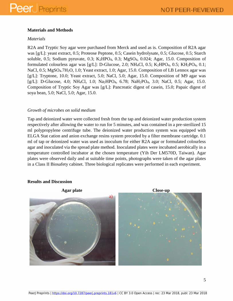
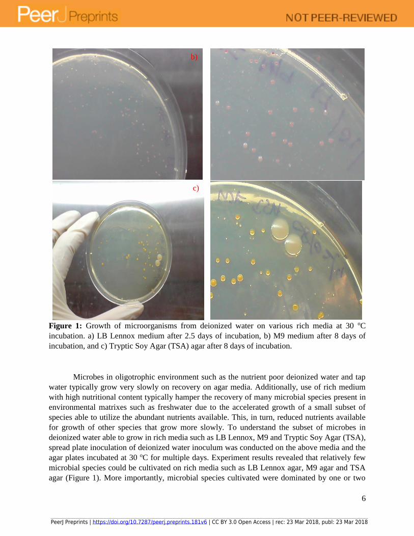
Figure 1: Growth of microorganisms from deionized water on various rich media at 30 oC
incubation. a) LB Lennox medium after 2.5 days of incubation, b) M9 medium after 8 days of
incubation, and c) Tryptic Soy Agar (TSA) agar after 8 days of incubation.
Microbes in oligotrophic environment such as the nutrient poor deionized water and tap
water typically grow very slowly on recovery on agar media. Additionally, use of rich medium
with high nutritional content typically hamper the recovery of many microbial species present in
environmental matrixes such as freshwater due to the accelerated growth of a small subset of
species able to utilize the abundant nutrients available. This, in turn, reduced nutrients available
for growth of other species that grow more slowly. To understand the subset of microbes in
deionized water able to grow in rich media such as LB Lennox, M9 and Tryptic Soy Agar (TSA),
spread plate inoculation of deionized water inoculum was conducted on the above media and the
agar plates incubated at 30 oC for multiple days. Experiment results revealed that relatively few
microbial species could be cultivated on rich media such as LB Lennox agar, M9 agar and TSA
agar (Figure 1). More importantly, microbial species cultivated were dominated by one or two
b)
c)
PeerJ Preprints | https://doi.org/10.7287/peerj.preprints.181v6 | CC BY 3.0 Open Access | rec: 23 Mar 2018, publ: 23 Mar 2018

7
types of colonies on the agar used, which revealed that microbes adapted to a low nutritional
environment could not adapt to a high nutrient environment due possibly to the presence of a
metabolic sensory system able to determine the nutritional and energy state of the environment
surrounding a microbe. Thus, the types of nutrients that form the basis for determining the
nutritional state of the environment with respect to a microbe would be key to understanding the
principles of medium design for enabling the cultivation of the broadest subset of microbes from
environmental samples. Overall, results revealed that rich media such as LB Lennox, M9 and TSA
could not be used in understanding the microbial flora of deionized water.
PeerJ Preprints | https://doi.org/10.7287/peerj.preprints.181v6 | CC BY 3.0 Open Access | rec: 23 Mar 2018, publ: 23 Mar 2018
8
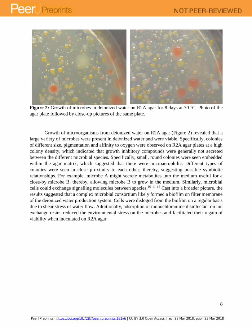
Figure 2: Growth of microbes in deionized water on R2A agar for 8 days at 30 oC. Photo of the
agar plate followed by close-up pictures of the same plate.
Growth of microorganisms from deionized water on R2A agar (Figure 2) revealed that a
large variety of microbes were present in deionized water and were viable. Specifically, colonies
of different size, pigmentation and affinity to oxygen were observed on R2A agar plates at a high
colony density, which indicated that growth inhbitory compounds were generally not secreted
between the different microbial species. Specifically, small, round colonies were seen embedded
within the agar matrix, which suggested that there were microaerophilic. Different types of
colonies were seen in close proximity to each other; thereby, suggesting possible symbiotic
relationships. For example, microbe A might secrete metabolites into the medium useful for a
close-by microbe B; thereby, allowing microbe B to grow in the medium. Similarly, microbial
cells could exchange signalling molecules between species.10 11 12 Cast into a broader picture, the
results suggested that a complex microbial consortium likely formed a biofilm on filter membrane
of the deionized water production system. Cells were disloged from the biofilm on a regular basis
due to shear stress of water flow. Additionally, adsorption of monochloramine disinfectant on ion
exchange resins reduced the environmental stress on the microbes and facilitated their regain of
viability when inoculated on R2A agar.
PeerJ Preprints | https://doi.org/10.7287/peerj.preprints.181v6 | CC BY 3.0 Open Access | rec: 23 Mar 2018, publ: 23 Mar 2018
9
PeerJ Preprints | https://doi.org/10.7287/peerj.preprints.181v6 | CC BY 3.0 Open Access | rec: 23 Mar 2018, publ: 23 Mar 2018

10
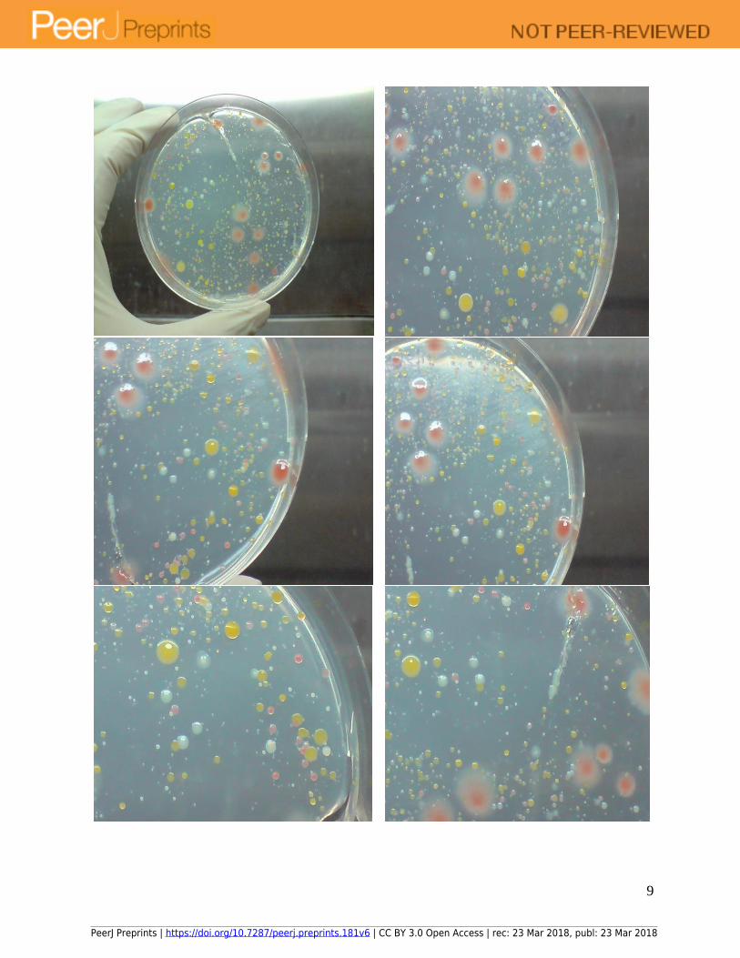
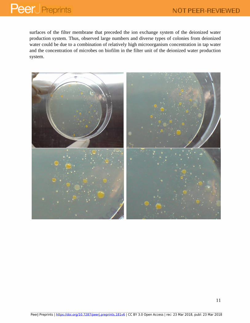
Figure 3: Growth of microbes in deionized water on formulated colourless agar with 1 g/L of
yeast extract at 30 oC for 8 days. Photo of agar plate followed by close-up pictures of the same
plate.
Similar growth experiments conducted using spread plate inoculation of deionized water
inoculum on formulated colourless agar with 1 g/L of yeast extract recovered large diversity of
microbes of different colony morphology, growth rates, pigmentation and sizes (Figure 3). Small,
round colonies were also seen embedded within the agar matrix, which meant that they were likely
microaerophilic. Ability to recover a wide variety of microorganisms meant that the formulated
colourless agar did not harbour growth inihibitory compounds detrimental to growth. Given that
the different colonies were in close proximity to each other, with some in close contact, growth
inhibitory compounds such as antibiotic was likely not secreted by the microbes. This revealed
that the microbial species had adapted to growth in close proximity to each other, where
intercellular signalling could have formed the communication basis of a cooperative or neutral
relationship instead of antagonistic ones.
Furthermore, recovery of the consortium of microbes on agar also revealed that a large
variety of microbes likely constituted the microbial community in deionized water production
system, which holds important implications to our understanding of microbial ecology of water.
Specifically, could antagonistic microbes be induced to form neutral relationships in the face of
severe environmental stressors such as the disinfectant monochloramine and low nutrients? From
another perspective, it is interesting to note that there was no substantial clear circular zones
surrounding individual colonies on both R2A and formulated colourless agar, which indicated that
antibiotic secretion between different microbial species was absent. Absence of antibiotic warfare
between microbial species provided a platform for the concurrent cultivation of many species on
the same agar that aids in understanding the diversity of microbes present. It also speaks of the
potential absence of major antagnoistic relationships between different microbes in deionized
water that lead to an interesting question in microbial ecology: could survival and growth in close
proximity in a biofilm environment change the inter-relationships between microbes from
competitive to cooperation?
Monochloramine, when present, is a source of environmental stress for microbial survival
given its known toxicity towards microbes as well as its ability at inhibiting their viability.13 14 15 16 17 Hence, adsorption of monochloramine by ion exchange resins of the deionized water
production system removed a significant source of environmental stress to the microbes; thereby,
enabling their return to viability, which resulted in the recovery of large diversity of microbes in
deionized water on both R2A and formulated colourless agar medium. Finally, given that
monohloramine was most probably adsorbed by ion exchange resins, the high surface
concentration of the disinfectant on the resins meant that biofilm would likely not form extensively
on the resins’ surfaces. Hence, microorganisms present in deionized water likely reside on the
PeerJ Preprints | https://doi.org/10.7287/peerj.preprints.181v6 | CC BY 3.0 Open Access | rec: 23 Mar 2018, publ: 23 Mar 2018
11
surfaces of the filter membrane that preceded the ion exchange system of the deionized water
production system. Thus, observed large numbers and diverse types of colonies from deionized
water could be due to a combination of relatively high microorganism concentration in tap water
and the concentration of microbes on biofilm in the filter unit of the deionized water production
system.
PeerJ Preprints | https://doi.org/10.7287/peerj.preprints.181v6 | CC BY 3.0 Open Access | rec: 23 Mar 2018, publ: 23 Mar 2018

12
Figure 4: Growth of microorganisms from tap water on R2A agar. Incubation conditions: 30 oC
for 7 days. Photo of agar plate followed by close-up pictures of the same plate.
Comparative growth of microbes in tap water on R2A agar for understanding the
provenance of the microbes in deionized water revealed significant reduction in numbers and types
of colonies recovered (Figure 4). However, significant number of different types of microbes
remain recoverable; thereby, indicating that the microbial load of tap water was likely high. With
the presence of monochloramine as a residual disinfectant, microbial viability in tap water was
likely significantly lower compared to deionized water, which explained the significant reduction
in number of colonies recovered.
More importantly, given the close proximity of different types of colonies from tap water
recovered on R2A agar, antagonistic relationships between microorganisms in tap water were
likely to be few, which suggested that the drinking water biofilm18 19 20 21 that existed in the
drinking water distribution pipeline harbours species of microbes able to form synergistic and non-
competitive relationships with each other. Hence, forced growth in defined community such as a
drinking water biofilm might have engendered communications between different microbial
species that reshape the community structure of the biofilm, lending a less competitive relationship
to ones previously known to be competitive. Such modulation of microbes’ behaviour, if shown
to be true, would be a significant advance in our understanding of microbial cell-cell
communication.
PeerJ Preprints | https://doi.org/10.7287/peerj.preprints.181v6 | CC BY 3.0 Open Access | rec: 23 Mar 2018, publ: 23 Mar 2018

13
Figure 5: Cultivation of microbes in tap water on formulated colourless agar at 30 oC with different
concentrations of yeast extract supplementation: a) 0.1 g/L yeast extract (8 days of incubation), b)
0.5 g/L yeast extract (7 days of incubation), and c) 1.0 g/L yeast extract (6 days of incubation).
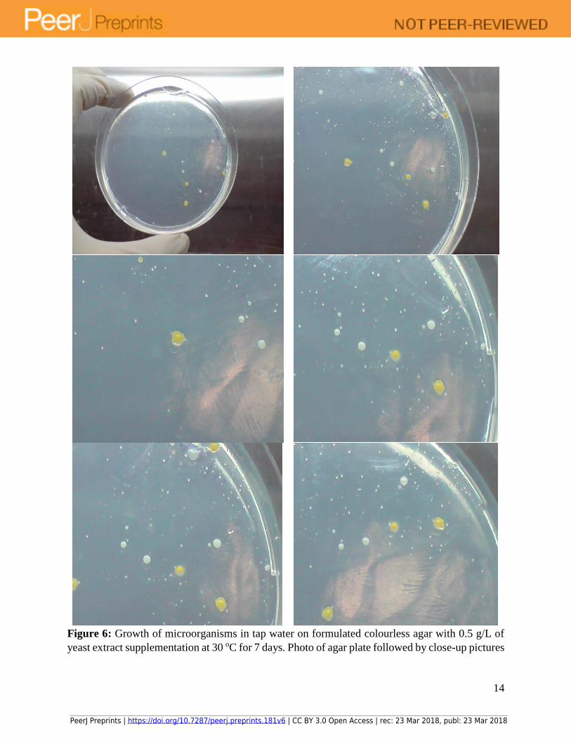
Experiments aimed at understanding the growth behaviour of microbes in tap water on
formulated colourless agar at 30 oC revealed a substantial reduction in the types and numbers of
microbes that could be recovered on the agar medium (Figure 5). Specifically, only a few dominant
types of microbes could be recovered. Hence, amount of yeast extract concentration in the
colourless agar medium was varied for understanding if the amount of vitamins and growth factors
supplemented for growth could affect the recovery rates of microbes in tap water. Results revealed
that yeast extract of 1 g/L was not useful for recovering more types of microbes in tap water. On
the other hand, a single dominant type of microbe of white round colony was recovered at yeast
extract of 0.1 g/L. Finally, 0.5 g/L of yeast extract appeared to be the optimal concentration for
inducing the growth of more types of microbes from tap water (Figure 6). Close proximity of
colonies to each other revealed that cooperative and neutral relationships likely dominate over
antagonistic ones. In addition, inhibitory compounds were likely not secreted by microbes which
would have affected the growth of nearby colonies. Finally, metabolites and signalling molecules
could have been exchanged between nearby colonies.
c) a) b)
PeerJ Preprints | https://doi.org/10.7287/peerj.preprints.181v6 | CC BY 3.0 Open Access | rec: 23 Mar 2018, publ: 23 Mar 2018
14
Figure 6: Growth of microorganisms in tap water on formulated colourless agar with 0.5 g/L of
yeast extract supplementation at 30 oC for 7 days. Photo of agar plate followed by close-up pictures
PeerJ Preprints | https://doi.org/10.7287/peerj.preprints.181v6 | CC BY 3.0 Open Access | rec: 23 Mar 2018, publ: 23 Mar 2018

15
of the same plate revealed many small colonies could be recovered from tap water after extended
incubation of a few days.
Overall, R2A agar was demonstrably able to recover more types and numbers of microbes
from tap water compared to formulated colourless agar with different concentrations of yeast
extract supplementation. One possible reason that could account for this phenomenon might be the
presence of chelating compounds in R2A agar that sequester monochloramine present in tap water.
Specifically, by chelating monochloramine, the residual disinfectant’s ability in effecting toxicity
on microbes in tap water could be drastically reduced. In particular, the new monochloramine-
chelating compound complex would have reduced capability at inhibiting microbial viability in
tap water. This could result in enhanced viability of microbes when tap water was inoculated on
R2A agar. Lack of similar chelating compounds in the formulated colourless agar meant that
monochloramine remains an active agent in the agar and inhibited viability of microbes; thereby,
preventing their growth.
Viewed from a different perspective, microbes in deionized water originated from tap water
feed, but adsorption of microbes to various surfaces of the deionized water production system such
as the filter membrane help concentrate the microorganisms present in deionized water.
Specifically, different microbes exhibited different growth and death cycles in tap and deionized
water. With a filter membrane as surfaces to adhere to, microbes were concentrated in the
deionized water production system in both numbers and types of microbes. Hence, it was natural
to recover more types of microorganisms from deionized water compared to the tap water feed,
given that microbes of low abundance in tap water could adhere to the surfaces of filter membrane,
multiply and grow and constitute part of a sprawling biofilm matrix teeming with microorganisms.
Agar plate Close-up
a)
PeerJ Preprints | https://doi.org/10.7287/peerj.preprints.181v6 | CC BY 3.0 Open Access | rec: 23 Mar 2018, publ: 23 Mar 2018
16
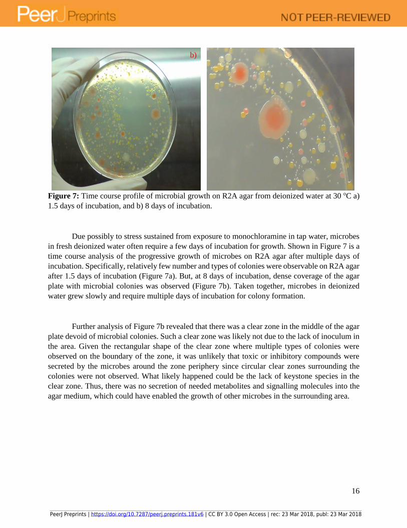
Figure 7: Time course profile of microbial growth on R2A agar from deionized water at 30 oC a)
1.5 days of incubation, and b) 8 days of incubation.
Due possibly to stress sustained from exposure to monochloramine in tap water, microbes
in fresh deionized water often require a few days of incubation for growth. Shown in Figure 7 is a
time course analysis of the progressive growth of microbes on R2A agar after multiple days of
incubation. Specifically, relatively few number and types of colonies were observable on R2A agar
after 1.5 days of incubation (Figure 7a). But, at 8 days of incubation, dense coverage of the agar
plate with microbial colonies was observed (Figure 7b). Taken together, microbes in deionized
water grew slowly and require multiple days of incubation for colony formation.
Further analysis of Figure 7b revealed that there was a clear zone in the middle of the agar
plate devoid of microbial colonies. Such a clear zone was likely not due to the lack of inoculum in
the area. Given the rectangular shape of the clear zone where multiple types of colonies were
observed on the boundary of the zone, it was unlikely that toxic or inhibitory compounds were
secreted by the microbes around the zone periphery since circular clear zones surrounding the
colonies were not observed. What likely happened could be the lack of keystone species in the
clear zone. Thus, there was no secretion of needed metabolites and signalling molecules into the
agar medium, which could have enabled the growth of other microbes in the surrounding area.
b)
PeerJ Preprints | https://doi.org/10.7287/peerj.preprints.181v6 | CC BY 3.0 Open Access | rec: 23 Mar 2018, publ: 23 Mar 2018

17
Agar plate Close-up
a)
b)
c)
PeerJ Preprints | https://doi.org/10.7287/peerj.preprints.181v6 | CC BY 3.0 Open Access | rec: 23 Mar 2018, publ: 23 Mar 2018

18
Figure 8: Growth of microbes from tap water on R2A agar at 30 oC. a) 2 days of incubation, b) 3
days of incubation, c) 4 days of incubation, and d) 7 days of incubation.
Similar cultivation experiments with tap water as inoculum on R2A agar at 30 oC
incubation revealed that microbes in tap water were also slow-growing, which could likely be due
to the effect of monochloramine’s toxicity on the cells as well as a long adaptation period after
microbes’ regain of viability due to the sequestration of monochloramine by chelating compounds
in R2A agar. Specifically, only small colonies were seen on R2A agar after 2 days of incubation
(Figure 8a), which progressively increased in size with time (Figure 8b and 8c). More importantly,
more types of colonies were recovered with increasing time of incubation. Finally, significant
increase in types and numbers of colonies in close proximity to each other was observed after 7
days of incubation (Figure 8d); thereby, indicating that microbes exposed to monochloramine
disinfectant in water required more time for cultivation and recovery on agar.
d)
PeerJ Preprints | https://doi.org/10.7287/peerj.preprints.181v6 | CC BY 3.0 Open Access | rec: 23 Mar 2018, publ: 23 Mar 2018

19
Figure 9: Growth of microorganisms from deionized water on R2A agar at 25 oC after 4 days of
incubation. Photos of agar plate and close-up pictures of the same plate.
Different types of microbes from deionized water were recovered on R2A agar after 4 days
of incubation at 25 oC (Figure 9). However, the types and numbers of microbes recovered were
significantly fewer compared to multiple days of incubation on R2A agar at 30 oC, which suggested
that microbes in tap and deionized water could have adapted to tropical water temperature of
around 30 oC. Lack of clear zones around individual microbial colony as well as close proximity
between different types of colonies suggested that growth inhibitory compounds were not secreted.
More importantly, different types of colonies could be seen in contact with each other, which
indicated that possible synergistic interactions or at least neutral relationships might exist between
them (green arrow in Figure 9). Overall, microbes in deionized water that could be recovered on
R2A agar at 25 oC developed small round colonies not different from those exhibited by microbes
recovered at 30 oC on the same agar. Relative close proximity between colonies on R2A agar after
4 days of incubation at 25 oC suggested that the consortium of microbes in deionized water did not
exhibited significant antagonistic relationships, which suggested possibe selection forces in
PeerJ Preprints | https://doi.org/10.7287/peerj.preprints.181v6 | CC BY 3.0 Open Access | rec: 23 Mar 2018, publ: 23 Mar 2018

20
deionized water environment that selected for neutral, synergistic or mutualistic relationships in
place of antagnoistic ones.
Figure 10: Relatively few microbial colonies were recovered from tap water on R2A agar at 25 oC after 4 days of incubation. Photo of agar plate and close-up pictures of the same plate.
Similar growth experiments for profiling microbes in tap water on R2A agar at 25 oC
revealed few colonies (Figure 10); thereby, highlighting that microbes in tap water could have
adapted to 30 oC temperature in distribution pipelines in a tropical climate. This suggested that
natural selection could exert its effects, in this case, modulating the preferred growth temperatures
of microbes in tap water, over a relatively short time span.
PeerJ Preprints | https://doi.org/10.7287/peerj.preprints.181v6 | CC BY 3.0 Open Access | rec: 23 Mar 2018, publ: 23 Mar 2018
21
PeerJ Preprints | https://doi.org/10.7287/peerj.preprints.181v6 | CC BY 3.0 Open Access | rec: 23 Mar 2018, publ: 23 Mar 2018

22
Figure 11: Growth of microbes in deionized water on R2A agar at 37 oC after 6 days of incubation.
Photos of agar plate and close-up pictures of the same plate.
Multiple types of microbial colonies including fungus were recovered on R2A agar from
deionized water after 6 days of incubation at 37 oC (Figure 11). Thus, these microbes could
potentially colonize human host and were potential pathogens. Close examination of the agar plate
revealed that the irregularly shaped white colony of a fungus likely secreted compounds into the
surrounding medium resulting in a white halo around the colony. Yellow colonies were observed
in close proximity to the fungal colonies; thereby, suggesting that secreted compounds did not
affect the growth of the yellow colonies. Given that many species of fungus secrete antibiotics for
self-defense, observed “immunity” of yellow colonies to secreted compounds of the white
irregularly shaped fungal colonies raised the possibility of presence of antibiotic resistant microbes
in the deionized water biofilm within the deionized water production system. In general, close
proximity of many types of colonies to the fungal colonies revealed that resistance to the
compounds secreted by the white fungal colonies could be prevalent in species of the microbial
biofilm in deionized water production system. Additionally, fewer microbial colonies were
recovered from deionized water on R2A agar at 37 oC incubation compared to 30 oC, which
suggested that microbes in deionized and tap water might have adapted to tropical water
temperatures of ~30 oC.
PeerJ Preprints | https://doi.org/10.7287/peerj.preprints.181v6 | CC BY 3.0 Open Access | rec: 23 Mar 2018, publ: 23 Mar 2018

23
Figure 12: Growth of microbes in deionized water on formulated colourless agar with 1 g/L yeast
extract after 6 days of incubation at 37 oC. Photos of agar plate and close-up pictures of the same
plate.
PeerJ Preprints | https://doi.org/10.7287/peerj.preprints.181v6 | CC BY 3.0 Open Access | rec: 23 Mar 2018, publ: 23 Mar 2018
24
Formulated colourless agar medium with 1 g/L of yeast extract also recovered multiple
types of microbial colonies (including white irregularly shaped fungal colonies) after 6 days of
incubation of deionized water at 37 oC (Figure 12). This reinforced the notion that potential human
pathogens could be present in deionized water. In general, fewer types and numbers of colonies
were recovered from deionized water during cultivation at 37 oC than at 30 oC due possibly to
acclimation of microbes to tropical ambient temperatures of 30 oC. Colonies recovered on the
colourless agar were in close proximity to each other; thereby, indicating that metabolites and
signalling compounds secreted enabled the growth of other species of microbes. Many types of
colonies were in close proximity to the white irregularly shaped fungal colonies, which did not
have a white halo surrounding the colonies. This suggested that the fungal colonies were likely not
antagonistic to other species.
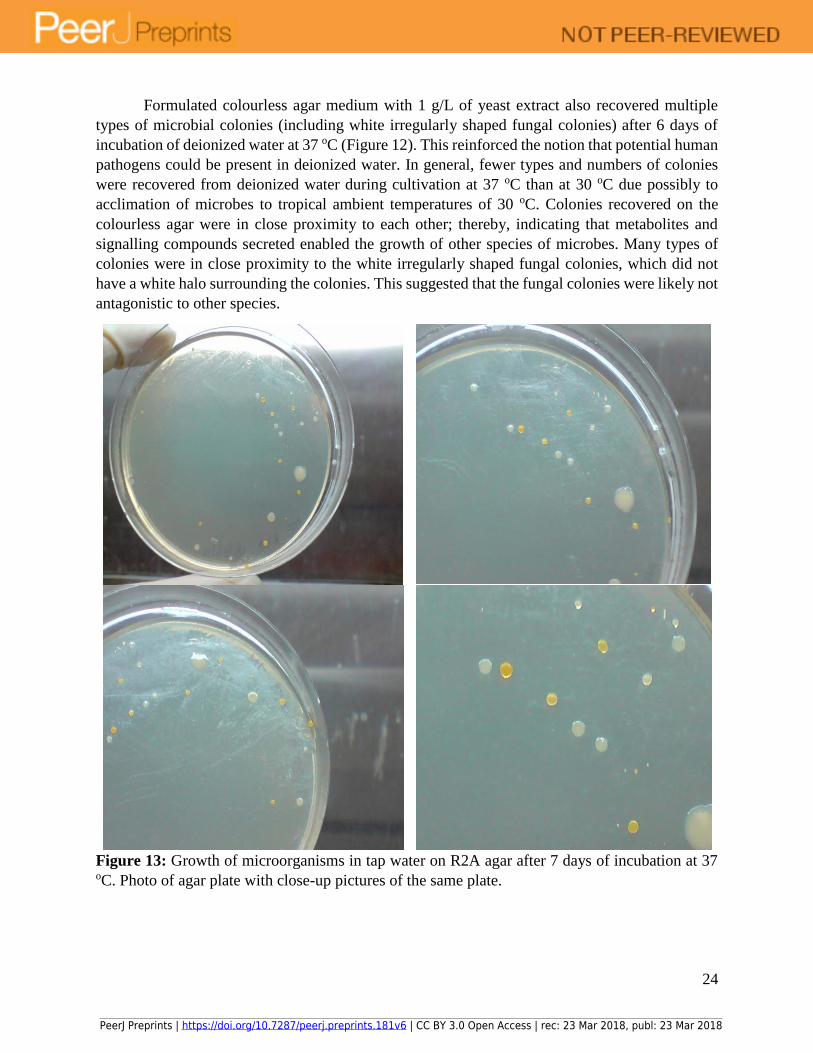
Figure 13: Growth of microorganisms in tap water on R2A agar after 7 days of incubation at 37 oC. Photo of agar plate with close-up pictures of the same plate.
PeerJ Preprints | https://doi.org/10.7287/peerj.preprints.181v6 | CC BY 3.0 Open Access | rec: 23 Mar 2018, publ: 23 Mar 2018

25
Similarly, microbial colonies were also recovered from tap water on R2A agar after 7 days
of incubation at 37 oC (Figure 13), which indicated that potential human pathogens were present
in tap water. In general, fewer colonies were recovered at 37 oC compared to 30 oC on R2A agar,
which provided further evidence that 30 oC might be the preferred temperature of growth for
microbes in tap water, similar to the case for microbes in deionized water. Recovered microbes
were in close proximity to each other, with a large clear zone of the agar plate devoid of any
colony, which suggested that the hypothesis of keystone microbial species secreting metabolite or
signalling molecules that enabled the growth of neighbouring colonies in the agar matrix might be
valid. Specifically, different types of colonies were seen in close proximity to each other on the
agar, which suggested that inhibitory compounds were not present in the agar surrounding the
colonies. Additionally, microbial colonies in close proximity to each other might require secreted
metabolites or signalling molecules from other microbial species for growth. In general, close
proximity of colonies on agar plate meant that neutral and synergistic relationships might be more
prevalent compared to antagonistic ones.
PeerJ Preprints | https://doi.org/10.7287/peerj.preprints.181v6 | CC BY 3.0 Open Access | rec: 23 Mar 2018, publ: 23 Mar 2018

26
Figure 14: Growth of microorganisms from tap water on formulated colourless agar with 1 g/L
yeast extract after 7 days of incubation at 37 oC. Photo of agar plate with close-up pictures of the
same plate.
Microbial colonies recovered from tap water on formulated colourless agar with 1 g/L of
yeast extract after 7 days of incubation at 37 oC (Figure 14) exhibited similar characteristics to
those recovered on R2A agar under identical incubation conditions (Figure 13). Number and types
of colonies recovered was significantly fewer at 37 oC incubation compared to 30 oC. Additionally,
multiple types of microbial colonies were recovered on the formulated colourless agar medium in
close proximity to each other, which suggested that antagonistic relationships were generally less
important compared to neutral, synergistic and mutualistic relationships in the drinking water
biofilm present in tap water distribution network. Additionally, inhibitory compounds were likely
not secreted by the microbes recovered and there could be exchange of metabolites and signalling
molecules between colonies.
PeerJ Preprints | https://doi.org/10.7287/peerj.preprints.181v6 | CC BY 3.0 Open Access | rec: 23 Mar 2018, publ: 23 Mar 2018

27
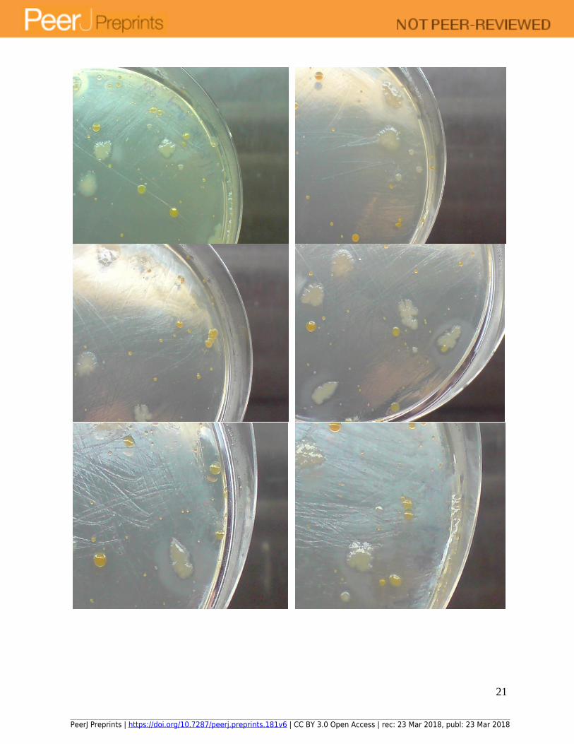
Figure 15: Growth of microorganisms from deionized water on formulated colourless agar
without yeast extract after 8 days of incubation at 30 oC. Photo of agar plate with close-up pictures
of the same plate.
PeerJ Preprints | https://doi.org/10.7287/peerj.preprints.181v6 | CC BY 3.0 Open Access | rec: 23 Mar 2018, publ: 23 Mar 2018

28
Given that ability to grow in minimal salts medium represents a significant growth
advantage over other microbial species, cultivation of microbes from deionized water on
formulated colourless agar without yeast extract was conducted for multiple days at 30 oC to
understand the diversity of microbes able to grow without supplementation of vitamins and growth
factors. Results indicated a rich diversity of different types of microbial species at differing relative
abundance could grow on minimal salt version of formulated colourless agar (Figure 15); thereby,
suggesting that a large consortium of microorganisms could collectively grow without infusion of
vitamins and growth factors from the environment. Specifically, microbes in the community could
share vitamins and growth factors secreted by some members of the consortium; thereby, gaining
the ability to grow on a minimal salts medium. Hence, microbial species able to synthesize
vitamins and growth factors without uptake from the environment play pivotal roles in enabling
the viability of the microbial consortium in a minimal salts medium. For example, large clear zones
devoid of microbial colonies in the centre of the agar plate suggested that microbial species able
to grow in minimal salts medium and which secrete vitamins and growth factors into the
surrounding medium were absent in the zone.
Conclusions
Multiple types of microbial colonies at high cell density was recovered from deionized and
tap water on R2A agar and a formulated colourless agar during multi-day incubation at 25, 30 and
37 oC. Greater diversity of microbial species of higher relative abundance were recovered from
both deionized and tap water during growth at 30 oC compared to 25 and 37 oC; thereby, suggesting
that microbes in deionized and tap water could have adapted to ambient tropical temperature of
~30 oC. This further indicated the relative speed at which natural selection could have exerted its
effect in influencing the optimal growth temperatures of microbes in environmental matrixes such
as freshwater. Recovery of microbes from deionized and tap water during incubation at 37 oC
suggested that potential human pathogens could be in deionized and tap water.
More importantly, more types and higher colony density of microbes could be recovered
on both agars from deionized water compared to tap water, due possibly to the removal of the
residual disinfectant, monochloramine, from tap water through adsorption on ion exchange resins
of the deionized water production system. Removal of monochloramine thus reduced a significant
environmental stressor that impinge on microbial viability. Additionally, during cultivation of
microbes from tap water, more colonies of different types could be recovered on R2A agar
compared to the formulated colourless agar, due probably to the presence of chelating compounds
in R2A agar able to sequester monochloramine; thereby, reducing its toxicity to cells. Similar
chelating compounds were unlikely to be present in the formulated colourless agar medium.
Rich diversity of microbes was recovered at high colony density from deionized water on
both R2A and formulated colourless agar, which could be due to the role of the filter membrane
PeerJ Preprints | https://doi.org/10.7287/peerj.preprints.181v6 | CC BY 3.0 Open Access | rec: 23 Mar 2018, publ: 23 Mar 2018
29
of the deionized water production system serving as surfaces for the adsorption and concentration
of microbes. Thus, biofilms likely formed on these surfaces and constituted a community of
microbes that could harbour microbial species previously not viable in deionized or tap water.
Such microbes could regain viability through the metabolites or signalling factors secreted by other
microbial species. More importantly, different microbial species of low relative abundance could
also be concentrated in such biofilms, which help explained the large diversity of microbes
recovered from deionized water.
Close proximity of microbial colonies on R2A agar and formulated colourless agar at high
colony density revealed that antagonistic relationships were likely less prevalent between species
compared to neutral or cooperative relationships. For example, metabolites or signalling molecules
secreted by a keystone species could enable the return to viability of microbes which previously
chose a “hibernation” cellular differentiation programme due to presence of monochloramine in
tap water. Thus, a consortium of microbes could be recovered in close proximity to the keystone
species through an exchange of metabolites or signalling molecules, resulting in areas of the agar
plate with dense microbial colonies and other areas devoid of microbes where the keystone species
was absent. In essence, need for survival in nutrient-poor deionized water likely selected for
cooperative or neutral relationships between microbes in the biofilm present in deionized water
production system and drinking water distribution pipelines, which explained the relatively lack
of antagonistic relationships between microbial species recovered.
Finally, cultivation on agar plates through the spread plate technique represent a simple
and effective method for assessing the microbiological quality of tap and deionized water, and
which together with conductivity and resistivity measurements, comprise a trio of tests useful for
maintaining the quality of deionized water.
Supplementary information
Photos of agar plates in other experiments could be found in the appended supplementary
information.
References
1. Dick, G. et al. The microbiology of deep-sea hydrothermal vent plumes: ecological and
biogeographic linkages to seafloor and water column habitats. Front. Microbiol. 4, 124
(2013).
PeerJ Preprints | https://doi.org/10.7287/peerj.preprints.181v6 | CC BY 3.0 Open Access | rec: 23 Mar 2018, publ: 23 Mar 2018

30
2. Hasan, N. A. et al. Deep-sea hydrothermal vent bacteria related to human pathogenic Vibrio
species. Proc. Natl. Acad. Sci. 112, E2813 (2015).
3. Zhang, L. et al. Bacterial and archaeal communities in the deep-sea sediments of inactive
hydrothermal vents in the Southwest India Ridge. Sci. Rep. 6, 25982 (2016).
4. He, T., Li, H. & Zhang, X. Deep-sea hydrothermal vent viruses compensate for microbial
metabolism in virus-host interactions. mBio 8, (2017).
5. Drake, H. et al. Isotopic evidence for microbial production and consumption of methane in the
upper continental crust throughout the Phanerozoic eon. Earth Planet. Sci. Lett. 470, 108–118
(2017).
6. Zhuang, W.-Q. et al. Incomplete Wood–Ljungdahl pathway facilitates one-carbon metabolism
in organohalide-respiring Dehalococcoides mccartyi. Proc. Natl. Acad. Sci. 111, 6419 (2014).
7. Massa, S., Caruso, M. & Trovatelli, F. Comparison of plate count agar and R2A medium for
enumeration of heterotrophic bacteria in natural mineral water. World J. Microbiol.
Biotechnol. 14, 727–730
8. Reasoner, D. J. & Geldreich, E. E. A new medium for the enumeration and subculture of
bacteria from potable water. Appl. Environ. Microbiol. 49, 1–7
9. Ng, W. Colourless agar for enhancing colour contrast between microbial colonies and agar.
PeerJ Prepr. 6, e89v6 (2018).
10. Stewart, E. J. Growing Unculturable Bacteria. J. Bacteriol. 194, 4151–4160 (2012).
11. D’Onofrio, A. et al. Siderophores from neighboring organisms promote the growth of
uncultured bacteria. Chem. Biol. 17, 254–264 (2010).
12. van der Meij, A., Worsley, S. F., Hutchings, M. I. & van Wezel, G. P. Chemical ecology of
antibiotic production by actinomycetes. FEMS Microbiol. Rev. 41, 392–416 (2017).
PeerJ Preprints | https://doi.org/10.7287/peerj.preprints.181v6 | CC BY 3.0 Open Access | rec: 23 Mar 2018, publ: 23 Mar 2018
31
13. Lee, W. H., Wahman, D. G., Bishop, P. L. & Pressman, J. G. Free chlorine and
monochloramine application to nitrifying biofilm: comparison of biofilm penetration, activity,
and viability. Environ. Sci. Technol. 45, 1412–1419 (2011).
14. Xue, Z., Lee, W. H., Coburn, K. M. & Seo, Y. Selective reactivity of monochloramine with
extracellular matrix components affects the disinfection of biofilm and detached clusters.
Environ. Sci. Technol. 48, 3832–3839 (2014).
15. Wahman, D. G., Wulfeck-Kleier, K. A. & Pressman, J. G. Monochloramine disinfection
kinetics of Nitrosomonas europaea by propidium monoazide quantitative PCR and Live/Dead
Baclight methods. Appl. Environ. Microbiol. 75, 5555–5562 (2009).
16. Yu, F. P., Pyle, B. H. & McFeters, G. A. A direct viable count method for the enumeration of
attached bacteria and assessment of biofilm disinfection. J. Microbiol. Methods 17, 167–180
(1993).
17. Mi, Z., Dai, Y., Xie, S., Chen, C. & Zhang, X. Impact of disinfection on drinking water
biofilm bacterial community. J. Environ. Sci. 37, 200–205 (2015).
18. Wingender, J. & Flemming, H.-C. Biofilms in drinking water and their role as reservoir for
pathogens. Second Eur. PhD Stud. Workshop Water Health Cannes 2010 214, 417–423
(2011).
19. Douterelo, I., Husband, S., Loza, V. & Boxall, J. Dynamics of biofilm regrowth in drinking
water distribution systems. Appl. Environ. Microbiol. 82, 4155–4168 (2016).
20. Liu, S. et al. Understanding, monitoring, and controlling biofilm growth in drinking water
distribution systems. Environ. Sci. Technol. 50, 8954–8976 (2016).
21. Chao, Y., Mao, Y., Wang, Z. & Zhang, T. Diversity and functions of bacterial community in
drinking water biofilms revealed by high-throughput sequencing. Sci. Rep. 5, 10044 (2015).
PeerJ Preprints | https://doi.org/10.7287/peerj.preprints.181v6 | CC BY 3.0 Open Access | rec: 23 Mar 2018, publ: 23 Mar 2018
32
Conflicts of interest
The author declares no conflicts of interest.
Funding
The author thank the National University of Singapore for financial support.
PeerJ Preprints | https://doi.org/10.7287/peerj.preprints.181v6 | CC BY 3.0 Open Access | rec: 23 Mar 2018, publ: 23 Mar 2018