Michelle-Lee Jones Neurology PGY-3 July 15th, 2009.
35
Michelle-Lee Jones Neurology PGY-3 July 15th, 2009
-
Upload
jonas-holland -
Category
Documents
-
view
213 -
download
0
Transcript of Michelle-Lee Jones Neurology PGY-3 July 15th, 2009.

Michelle-Lee JonesNeurology PGY-3July 15th, 2009

Coma & Brain Death - Outline
• COMA:– Definition– Pathophysiology– Aetiologies– Exam & relevant
investigations– Differential
diagnosis & related conditions
– Prognostication
• BRAIN DEATH:– Definition– Determination,
including the role of confirmatory testing
– Ongoing controversies

COMA
•Definition:–Greek in origin – “deep sleep or trance”
–It refers to an unconscious state characterised by a lack of both arousal and responsiveness

COMA
• Related disorders of consciousness:– Stupor (Latin “ to be stunned”):
aroused by and responsive to only the most vigorous stimuli
– Minimally conscious state: Markedly impaired consciousness with evidence of self or environmental awareness (intermittent rudimentary vocal or motor responses)

COMA
• Related disorders of consciousness:– Vegetative state: No awareness of
self or environment, but basic cycling of arousal states & periodic eye-opening are present
– Persistent vegetative state: Vegetative state persisting for at least 30 days

COMA•Pathophysiology:
– The ascending reticular activating system (ARAS) controls one’s level of arousal or alertness
– The ARAS is comprised of the rostral brainstem tegmentum (cholinergic peribrachial nuclei*), the diencephalon and associated cortical projections
– *pedunculopontine tegmental nucleus & lateral dorsal tegemental nucleus

COMA
•Pathophysiology:– The peribrachial nuclei project via 2
major pathways – dorsal & ventral pathways
– Dorsal: glutaminergic projections from thalamic nuclei to various cortical areas
– Ventral: histaminergic projections from the posterior hypothalamus (hypocretin, orexin also) & cholinergic projections from the basal forebrain to many cortical areas (McGill connection Herbert Jasper 1961 & Barbara Jones 2000)

COMA
•Pathophysiology:– Sleep centre: Preoptic area of
hypothalamus (GABAergic)
– Notably, the ARAS pathways exhibit redundancy that may facilitate recovery of the arousal system (within 2-3 weeks if lesions are more rostral)

COMA
PERIBRACHIAL NUCLEI (http://www.nature.com/nature/journal/v437/n7063/images/nature04284-f2.2.jpg)

COMA
Dorsal & Ventral ARAS pathways (Jones, B.E. 2000)

COMA
•CAVEAT: Damage to the ARAS and associated structures or both hemispheres is usually necessary to cause a comatose state. Occasionally, left hemispheric dysfunction can per se lead to coma...

COMA•Aetiologies
–Structural lesions:•Destructive & compressive•Ischemic stroke, haemorrhage, tumours & inflammation/infection
•Long list including SDH, EDH, SAH, cerebral contusion, pontine hemorrhage, cerebellar hemorrhage/infarction, brain abscess, vasculitis, venous sinus thrombosis, etc.

COMA•Aetiologies
–Herniation Syndromes:•Munro-Kellie doctrine to consider
•Lateral displacement of the diencephalon (e.g. basal ganglia bleed) – monitor via displacement of the calcified pineal gland; need 9 to 13 mm shift to produce coma; related to initial impairment of consciousness

COMA•Aetiologies
–Herniation Syndromes:•Falcine herniation –
–expanding mass causes the cingulate gurus & pericallosal/callosomarginal arteries to be compressed & displaced under the falx
–Medial wall of hemisphere - infarction & edema
–Diencephalic distortion via downward herniation or midline shift

COMA•Aetiologies
–Herniation Syndromes:•Uncal hernation –
–expanding mass causes medial and downward herniation of the medial temporal lobe into the tentorial notch
– ipsilateral fixed & dilated pupil–ocular dysmotility–contralateral (uncus presses on
nearby cerebral peduncle) or ipsilateral hemiparesis (Kernohan’s sign)
–PCA infarction

COMA•Aetiologies
–Herniation Syndromes:•Central transtentorial herniation -
–expanding mass causes downward herniation of the diencephalon and pressure on the midbrain
– Ischemia & infarction as feeder vessels are stretched and compressed
–Diabetes insipidus with pituitary stalk avulsion

COMA•Aetiologies
–Herniation Syndromes:•Tonsillar herniation -
–Cerebellar tonsils forced down through foramen magnum e.g. SAH
–Medullary compression apnea & compensatory HTN
•Rostrocaudal deteriortion – downward displacement of pons/midbrain; Duret h.
•Upward brainstem herniation Posterior fossa lesion expands upward, compresses dorsal midbrain

COMAHerniation Syndromes
http://www.lfhk.cuni.cz/patfyz/Intranet/Figures/88/5.26.jpg

COMA•Aetiologies
– Metabolic disturbances/toxins• Hypoglycemia, hyperglycemic hyperosmolar
state, diabetic ketoacidosis• Hyper/hyponatremia, hyper/hypocalemia,
hypo/hypermagnesemia, hyper/hypothroidism• Uremic or hepatic encephalopathy• Drugs such as alcohol, sympathomimetics,
opioids, antidepressants, salicylates, etc.• Hypothermia, porphyria, mitochondrial
disorders
– Ischemia/Hypoxia, inflammation, infections, seizures

COMA•Physical Examination & Investigations– General inspection: Racoon eyes, Battle sign,
hemotympanum, CSF rhinorrhea or otorrhea basal skull fracture
– Elevated BP: hypertensive encephalopathy (>250/150), intracerebral or subarachnoid hemorrhage; acute ischemic infarct
– Respiratory status: Cheyne-Stokes, apneustic breathing, atactic respiration etc.
– Hypothermia: ethanol or sedative drug intoxication, myxedema, Wernicke encephalopathy, hepatic encephalopathy hypoglycemia

COMA•Physical Examination & Investigations– Hyperthermia: status epilepticus, malignant
hyperthermia, anticholinergic drug intoxication, hypothalamic lesions, pontine hemorrhage, heat stroke
– Meningeal irritation signs for meningitis or subarachnoid hemorrhage
– Fundoscopic exam: papilledema, retinal hemorrhages (chronic or acute HTN); subhyaloid (superficial retinal) hemorrhages for subarachnoid hemorrhage

COMA
•Physical Examination & Investigations Pupil size, location &
reactivityLikely site of pathology
> 7 mm, non-reactive 3rd nerve compression; anticholinergic intox.
Slightly smaller, reactive Early thalamic compression
Fixed midsized pupils ≈ 5 mm
Midbrain injury
Pinpoint, minimally reactive
Opioid overdose; pontine injury, organophosphates, neurosyphilis

COMA•Physical Examination & Investigations– Oculocephalic & oculovestibular
reflexes:• If the brainstem is intact, a comatose patient will
demonstrate full conjugate horizontal eye movements during the oculocephalic testing and tonic conjugate movement of both eyes to the side of the ice-water irrigation during caloric testing.
• Absent oculovestibular responses in a comatose patient pontine injury, sedative drug intoxication (can also see downward deviation of one or both eyes)

COMA• Physical Examination &
Investigations

COMA•Physical Examination & Investigations– Metabolic, infectious, vasculitic, stroke
W/U
– CT, MRI, EEG (mild slowing to burst suppression – mortality rate for the latter?)
– One study found that 8% of patients comatose secondary to brain injury are in NCSE – role for continuous EEG monitoring in the ICU?
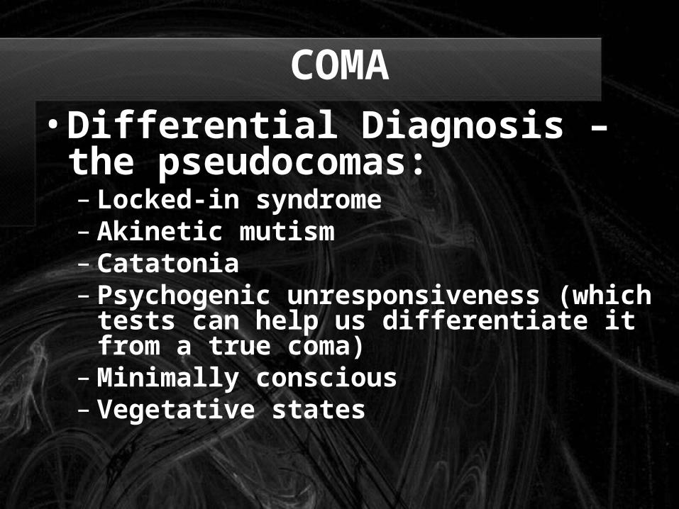
COMA•Differential Diagnosis – the pseudocomas:– Locked-in syndrome– Akinetic mutism– Catatonia– Psychogenic unresponsiveness
(which tests can help us differentiate it from a true coma)
– Minimally conscious– Vegetative states

COMA - Prognostication:
AAN guidelines for coma post CPR

COMA - Prognostication
Prognostic Signs in Coma from Global Cerebral Ischemia. Comparison of the Findings in Two Studies(Clinical Neurology, Aminoff)
Probability of Recovering Independent Function (%)
Time Since onset of Coma (Days)
Sign 0 1 3 7
Data from Levy et al2
No verbal response 13 8 5 6
No eye opening 11 6 4 0
Unreactive pupils 0 0 0 0
No spontaneous eye movements
6 5 2 0
No caloric responses 5 6 6 0
Extensor posturing 18 0 0 0
Flexor posturing 14 3 0 0
Absent motor responses 4 3 0 0
Data from Edgren et al3
No eye opening to pain 31 8 0 0
Unreactive pupils 17 7 0 0

BRAIN DEATH•Definition:
– The irreversible loss of brain function, inclusive of the brainstem
•Determination:– Triad to remember: COMA, ABSENCE OF
BRAINSTEM REFLEXES (pupil, corneal, VOR, pharyngeal & laryngeal) & APNEA
– Exclusion of confounding clinical conditions (see table)

BRAIN DEATH
Morenski et. al , 2003

BRAIN DEATH•Determination continued:

BRAIN DEATH• Spontaneous and reflex movements that may
be seen in brain death:

BRAIN DEATH•Ancillary tests:
–Transcranial doppler US–Conventional angiography–EEG–Technetium-99m brain scan–SSEPs: N20-P22 absence bilaterally with median nerve stimulation

Morenski et. al, 2003

A few good references• AAN Determining Brain Death in
Adults Current guidelines
• Prediction of Outcome in Comatose Survivors after Cardiopulmonary Resuscitation Current guidelines July 2006
• Plum and Posner's Diagnosis of Stupor and Coma, 4th edition (☆☆☆☆☆) – great book!