
MeV+R: using MeV as a graphical user interface for Bioconductor ... · Genome Biology 2008, 9:R118...
11
Genome Biology 2008, 9:R118 Open Access 2008 Chu et al. Volume 9, Issue 7, Article R118 Software MeV+R: using MeV as a graphical user interface for Bioconductor applications in microarray analysis Vu T Chu * , Raphael Gottardo † , Adrian E Raftery ‡ , Roger E Bumgarner * and Ka Yee Yeung * Addresses: * Department of Microbiology, University of Washington, Seattle, WA 98195, USA. † Department of Statistics, University of British Columbia, Vancouver, BC, V6T 1Z2, Canada. ‡ Department of Statistics, University of Washington, Seattle, WA 98195, USA. Correspondence: Ka Yee Yeung. Email: [email protected] © 2008 Chu et al.; licensee BioMed Central Ltd. This is an open access article distributed under the terms of the Creative Commons Attribution License (http://creativecommons.org/licenses/by/2.0), which permits unrestricted use, distribution, and reproduction in any medium, provided the original work is properly cited. MeV+R <p>MeV+R provides users with point-and-click access to traditionally command-line-driven tools written in R.</p> Abstract We present MeV+R, an integration of the JAVA MultiExperiment Viewer program with Bioconductor packages. This integration of MultiExperiment Viewer and R is easily extensible to other R packages and provides users with point and click access to traditionally command line driven tools written in R. We demonstrate the ability to use MultiExperiment Viewer as a graphical user interface for Bioconductor applications in microarray data analysis by incorporating three Bioconductor packages, RAMA, BRIDGE and iterativeBMA. Rationale While microarray technology has given biologists unprece- dented access to gene expression data, reliable and effective data analysis remains a difficult problem. There are many freely or commercially available software packages, but biol- ogists are often faced with trading off power and flexibility for usability and accessibility. In addition to the potentially pro- hibitive costs, researchers using commercial software tools may find themselves waiting for state-of-the-art algorithms to be implemented with the packages. The Bioconductor project [1,2] is an open source software project that provides a wide range of statistical tools primarily based on the R program- ming environment and language [3,4]. Taking advantage of R's powerful statistical and graphical capabilities, developers have created and contributed numerous Bioconductor pack- ages to solve a variety of data analysis needs. The use of these packages, however, requires a basic understanding of the R programming/command language and an understanding of the documentation accompanying each package. The primary users of R and the Bioconductor packages have been compu- tational scientists, statisticians and the more computationally oriented biologists. However, in our experience, many biolo- gists find themselves uncomfortable issuing command lines in a terminal. Hence, there is a need for a graphical user inter- face (GUI) for Bioconductor packages that will allow biolo- gists easy access to data analytical tools without learning the command line syntax. The tcltk package in R adds GUI ele- ments to R by allowing programmers to write GUI-driven modules by embedding Tk commands into the R language [5]. There are also GUIs developed for basic statistical analysis in R, such as the R Commander [6] and windows-based SciViews [7]. However, these GUIs are not designed for microarray analysis. There are Bioconductor packages, such as limmaGUI [8], affylmGUI [9] and OLINgui [10] that are built on the R tcltk package to provide GUIs. LimmaGUI and affylmGUI provide GUIs for the analysis of designed experi- ments and the assessment of differential expression for two- color spotted microarrays and single-color Affymetrix data, respectively. OLINgui provides a GUI for the visualization, normalization and quality testing of two-channel microarray Published: 24 July 2008 Genome Biology 2008, 9:R118 (doi:10.1186/gb-2008-9-7-r118) Received: 10 March 2008 Revised: 1 June 2008 Accepted: 24 July 2008 The electronic version of this article is the complete one and can be found online at http://genomebiology.com/2008/9/7/R118
Transcript of MeV+R: using MeV as a graphical user interface for Bioconductor ... · Genome Biology 2008, 9:R118...

Open Access2008Chuet al.Volume 9, Issue 7, Article R118SoftwareMeV+R: using MeV as a graphical user interface for Bioconductor applications in microarray analysisVu T Chu*, Raphael Gottardo†, Adrian E Raftery‡, Roger E Bumgarner* and Ka Yee Yeung*
Addresses: *Department of Microbiology, University of Washington, Seattle, WA 98195, USA. †Department of Statistics, University of British Columbia, Vancouver, BC, V6T 1Z2, Canada. ‡Department of Statistics, University of Washington, Seattle, WA 98195, USA.
Correspondence: Ka Yee Yeung. Email: [email protected]
© 2008 Chu et al.; licensee BioMed Central Ltd. This is an open access article distributed under the terms of the Creative Commons Attribution License (http://creativecommons.org/licenses/by/2.0), which permits unrestricted use, distribution, and reproduction in any medium, provided the original work is properly cited.MeV+R<p>MeV+R provides users with point-and-click access to traditionally command-line-driven tools written in R.</p>
Abstract
We present MeV+R, an integration of the JAVA MultiExperiment Viewer program withBioconductor packages. This integration of MultiExperiment Viewer and R is easily extensible toother R packages and provides users with point and click access to traditionally command linedriven tools written in R. We demonstrate the ability to use MultiExperiment Viewer as a graphicaluser interface for Bioconductor applications in microarray data analysis by incorporating threeBioconductor packages, RAMA, BRIDGE and iterativeBMA.
RationaleWhile microarray technology has given biologists unprece-dented access to gene expression data, reliable and effectivedata analysis remains a difficult problem. There are manyfreely or commercially available software packages, but biol-ogists are often faced with trading off power and flexibility forusability and accessibility. In addition to the potentially pro-hibitive costs, researchers using commercial software toolsmay find themselves waiting for state-of-the-art algorithms tobe implemented with the packages. The Bioconductor project[1,2] is an open source software project that provides a widerange of statistical tools primarily based on the R program-ming environment and language [3,4]. Taking advantage ofR's powerful statistical and graphical capabilities, developershave created and contributed numerous Bioconductor pack-ages to solve a variety of data analysis needs. The use of thesepackages, however, requires a basic understanding of the Rprogramming/command language and an understanding ofthe documentation accompanying each package. The primaryusers of R and the Bioconductor packages have been compu-
tational scientists, statisticians and the more computationallyoriented biologists. However, in our experience, many biolo-gists find themselves uncomfortable issuing command linesin a terminal. Hence, there is a need for a graphical user inter-face (GUI) for Bioconductor packages that will allow biolo-gists easy access to data analytical tools without learning thecommand line syntax. The tcltk package in R adds GUI ele-ments to R by allowing programmers to write GUI-drivenmodules by embedding Tk commands into the R language [5].There are also GUIs developed for basic statistical analysis inR, such as the R Commander [6] and windows-basedSciViews [7]. However, these GUIs are not designed formicroarray analysis. There are Bioconductor packages, suchas limmaGUI [8], affylmGUI [9] and OLINgui [10] that arebuilt on the R tcltk package to provide GUIs. LimmaGUI andaffylmGUI provide GUIs for the analysis of designed experi-ments and the assessment of differential expression for two-color spotted microarrays and single-color Affymetrix data,respectively. OLINgui provides a GUI for the visualization,normalization and quality testing of two-channel microarray
Published: 24 July 2008
Genome Biology 2008, 9:R118 (doi:10.1186/gb-2008-9-7-r118)
Received: 10 March 2008Revised: 1 June 2008Accepted: 24 July 2008
The electronic version of this article is the complete one and can be found online at http://genomebiology.com/2008/9/7/R118
Genome Biology 2008, 9:R118

http://genomebiology.com/2008/9/7/R118 Genome Biology 2008, Volume 9, Issue 7, Article R118 Chu et al. R118.2
data. However, no such GUIs are available for the majority ofBioconductor packages. In addition, since each Bioconductorpackage is often written by a different research group, there isgenerally no uniformity in the look and feel of the GUIs avail-able for the different packages. Hence, the end user may notbe able to easily transfer experience gained with one analysistool to the use of another.
An alternative microarray data analysis tool is the MultiEx-periment Viewer (MeV), a component of the TM4 suite ofmicroarray analysis tools [11]. MeV has a user-friendly GUIdesigned with the biological community in mind. MeV is anopen source Java application with a simple to learn, easy touse GUI. It comes with many popular microarray analyticalalgorithms for clustering, visualization, classification andbiological theme discovery, such as hierarchical clustering[12] and Expression Analysis Systematic Explorer (EASE)[13]. MeV was carefully designed to provide an applicationprogramming interface (API), thus allowing straightforwardcontributions by the community. MeV is hosted at Source-Forge [14] in a concurrent versions system repository. Assuch, frequent builds of the source code are made possible,greatly reducing the lag time between version releases.
In this paper, we present MeV+R, which is an effort to providemore consistent and well-integrated GUIs for Bioconductorpackages by using MeV as a 'wrapper' application for Biocon-ductor methods. Our work brings the best of both worldstogether: providing state-of-the-art statistical algorithmsfrom Bioconductor through the open source and easy to useMeV graphical interface to the biomedical community.MeV+R has many advantages, including platform independ-ence, a well-defined modular API, and a point and click GUIthat is easy to learn and use. We demonstrate the successful
integration and advantages of three Bioconductor packages(RAMA [15], BRIDGE [16], and iterativeBMA [17]) over exist-ing tools in the MeV environment through case studies. Theunderlying framework that we used to integrate these Biocon-ductor packages with MeV is easily extensible to other analy-sis tools developed in R. The software, documentation and atutorial are publicly available from our project home page[18].
ImplementationOur integration effort is composed of three separate entities(Figure 1). MeV provides the graphical user interface whileRserve serves as the communication layer and R is the lan-guage and environment in which the analysis packages run.Rserve is a TCP/IP server that allows various languages to usethe facilities of R without the need to initialize R or linkagainst an R library [19]. In other words, we use R as the backend to run Bioconductor packages through the use of Rserve.Rserve is open source, freely available [20], and licensedunder GPL.
As such, Java, Rserve, and R must all be installed on the user'scomputer, and we provide an automated installer on ourproject web site. Furthermore, Rserve needs to be running tobe used. However, R does not need to be started. Since Rserveworks through TCP/IP, it can run on the user's own machine,on an internal network or over the internet. By default, ourcode assumes Rserve to be running on the local host, but theuser can change, add and save additional new hosts using apull down menu. Once a connection is established, the Javacode in MeV converts the user's data from the MeV datastructure to the R format and loads it into R. The appropriate
Our integration effort is composed of three separate entities: MeV as the GUI, Rserve as the communication layer, and R as the language and environment in which the analysis packages runFigure 1Our integration effort is composed of three separate entities: MeV as the GUI, Rserve as the communication layer, and R as the language and environment in which the analysis packages run.
Genome Biology 2008, 9:R118

http://genomebiology.com/2008/9/7/R118 Genome Biology 2008, Volume 9, Issue 7, Article R118 Chu et al. R118.3
R libraries are loaded followed by the R commands that arenecessary to initiate the analysis. Upon completion, thereturned data from R are explicitly called back into MeV andpresented to the user.
We have incorporated three Bioconductor packages, RAMA[15], BRIDGE [16], and iterativeBMA [17], into MeV to illus-trate the successful MeV+R integration. The Robust Analysisof MicroArray (RAMA) algorithm computes robust estimatesof expression intensities from two-color microarray data,which typically consist of a few replicates and potential out-liers [15]. RAMA also takes advantage of dye swap experimen-tal designs. Bayesian Robust Inference for Differential GeneExpression (BRIDGE) is a robust algorithm that selects dif-ferentially expressed genes under different experimental con-ditions on both one- and two-color microarray data [16]. BothRAMA and BRIDGE make use of a computationally intensivetechnique called Markov Chain Monte Carlo for parameterestimation, and it is non-trivial to re-implement these algo-rithms in Java. Hence, we took advantage of our previousdevelopment work by simply using MeV as an interface to theBioconductor packages. The iterative BMA algorithm is amultivariate gene selection and classification algorithm,which considers multiple genes simultaneously and typicallyleads to a small number of relevant genes to classify microar-ray data [17]. The iterativeBMA Bioconductor package imple-ments the iterative BMA algorithm as previously described[17] in R, and its implementation is part of our current inte-gration effort. Both RAMA and BRIDGE are included in thelatest release of MeV (version 4.1), and iterativeBMA will beincluded in future releases. The user interfaces, usage andcase studies for RAMA, BRIDGE and iterativeBMA are brieflydescribed below. Detailed documentation is included with thesoftware distribution [21] as well as linked in the MeV appli-cation. Help pages are also available as Help Dialogs accessedvia buttons on the MeV dialog boxes. Our MeV+R implemen-tation is publicly available and runs on Windows, Mac OS Xand Linux.
Integrated Bioconductor packages: description and user interfacesRAMA: Robust Analysis of MicroArraysRAMA uses a Bayesian hierarchical model for the robust esti-mation of cDNA microarray intensities with replicates. This ishighly relevant for replicated microarray experimentsbecause even one outlying replicate (such as due to scratchesor dust) can have a disastrous effect on the estimated signalintensity. Outliers are modeled explicitly using a t-distribu-tion, which is more robust than the usual Gaussian model.Our model borrows strength from all the genes to decide if ameasurement is an outlier, and hence it is better at detectingoutliers based on a small number of replicate measurementsthan other classical robust estimators. Our algorithm usesMarkov Chain Monte Carlo for parameter estimation, andaddresses classical issues such as design effects, normaliza-
tion, transformation, and nonconstant variance. Please referto [15] for a detailed description of the algorithm.
User interfaceThe user can start RAMA by clicking 'Adjust Data' - 'ReplicateAnalysis' - 'RAMA' from the MeV main menu. The RAMA dia-log box is then displayed asking the user to label the arraysthat were loaded into MeV with their appropriate dye color.At this time, the user is asked to make sure that Rserve is run-ning. On a Win32 system, double clicking Rserve.exe accom-plishes this. On a UNIX or Linux or Mac OS X system, theuser issues the command 'R CMD Rserve' at a prompt. Bydefault, RAMA will look on the local machine for an Rserveserver. However, since Rserve is a TCP/IP server, the Rserveserver can be a remote machine. The user is allowed to adjusta few advanced parameters, though suggested values aregiven as defaults. If an Rserve connection is successfullymade, the location of Rserve is written to the user's MeV con-figuration file and will be available in later sessions. Afterclicking 'OK', the input data are sent to R. An indeterminateprogress bar is displayed while RAMA runs - unfortunately,the architecture of RServe and the R Server do not allow foran accurate indication of the time remaining in an ongoinganalysis. Once completed, the user is given a dialog box tosave the results. The returned results will then replace theloaded data in a new Multiple Array Viewer (MAV). The oldMAV is deleted. The user can then choose to continue usingMeV as if the data were loaded through the native loadingmodules.
BRIDGE: Bayesian Robust Inference for Differential Gene ExpressionBRIDGE fits a robust Bayesian hierarchical model to test fordifferentially expressed genes on microarray data. It can beused with both two-color microarrays and single-channelAffymetrix chips. BRIDGE builds on the previous work ofGottardo et al. [15] by allowing each gene to have a differentvariance and the detection of differentially expressed genesunder multiple (up to three in our current implementation)experimental conditions. Robust inference is accomplishedby modeling outliers using a t-distribution, and henceBRIDGE is powerful even with a small number of samples(either biological or technical replicates) under each experi-mental condition. Parameter estimation is carried out using anovel version of Markov Chain Monte Carlo. The currentimplementation of BRIDGE does not handle missing values.Please refer to [16] for a detailed description of the model.
User interfaceBRIDGE starts when a user clicks the 'BRIDGE' button in thetoolbar located on top of the MeV window. The user is onceagain presented with a dialog box similar to that of RAMAasking for the dye labeling identity of each loaded slide. Theuser is offered the option to adjust the advanced parametersand to establish an Rserve connection. After clicking OK, theinput data are sent to R. An indeterminate progress bar is
Genome Biology 2008, 9:R118
http://genomebiology.com/2008/9/7/R118 Genome Biology 2008, Volume 9, Issue 7, Article R118 Chu et al. R118.4
displayed while BRIDGE runs. The results are presented tothe user in three formats: heat maps, expression graphs ortables. In each format, the genes for which there is strong evi-dence of differential expression are identified as 'SignificantGenes', defined by the posterior probability being above 0.5.
IterativeBMA: Iterative Bayesian Model AveragingThe iterativeBMA algorithm is a multivariate technique forgene selection and classification of microarray data. BayesianModel Averaging (BMA) takes model uncertainty into consid-eration by averaging over the predicted probabilities based onmultiple models, weighted by their posterior model probabil-ities [22]. The most commonly used BMA algorithm is limitedto data in which the number of variables is greater than thenumber of responses, and the algorithm is inefficient fordatasets containing more than 30 genes (variables). In thecase of classifying samples using microarray data, there aretypically thousands or tens of thousands of genes (variables)under a few dozen samples (responses). In the iterative BMAalgorithm, we start by ranking the genes using the ratio ofbetween-group to within-group sum of squares (BSS/WSS)[23]. In this initial preprocessing step, genes with large BSS/WSS ratios (that is, genes with relatively large variationbetween classes and relatively small variation within classes)receive high rankings. We then apply the traditional BMAalgorithm to the 30 top ranked genes, and remove genes withlow posterior probabilities. Genes from the rank orderedBSS/WSS ratios are then added to the set of genes to replacegenes with low probabilities. These steps of gene swaps anditerative applications of BMA are repeated until all genes aresubsequently considered. We have previously shown that theiterative BMA algorithm selects small numbers of relevantgenes, achieves high prediction accuracy, and produces pos-terior probabilities for the predictions, selected genes andmodels [17].
The iterativeBMA Bioconductor package implements the iter-ative BMA algorithm described in Yeung et al. [17] (previ-ously implemented in Splus) when there are two classes. It ispart of the original work for this publication. The user docu-mentation (vignette) is included in the package.
User interfaceWe have integrated the iterativeBMA Bioconductor packagein MeV. IterativeBMA starts after the user clicks on the'iBMA' icon on top of the MeV window. The current imple-mentation of the iterativeBMA Bioconductor package is lim-ited to only two classes. After loading the data, the user isasked to label the two classes. The default labels for the twoclasses are 0 and 1, respectively. In the same dialog box, theuser is asked to establish an Rserve connection. The user isalso given the option of specifying advanced parameters forthe analysis. The next dialog box asks the user to assign labelsto each of the samples in the data, either by using a pull-downmenu or loading an assignment file. At this point, if Rserve isnot already running, the user is reminded to start the connec-
tion. Then, the data and the parameters are sent to R, and aprogress bar is shown warning the user that the computationcould take a long time. After the iterativeBMA Bioconductorpackage finishes running, the following analysis results aredisplayed: the predicted probability and class for each testsample; the posterior probabilities of the selected genessorted in descending order; the posterior probabilities of theselected models sorted in descending order; and the heat-maps of the selected genes in both classes.
Case studies illustrating the merits of the integrated Bioconductor packagesIn this section, we compare the performance of the integratedBioconductor packages (RAMA, BRIDGE and iterativeBMA)to existing tools in MeV in order to illustrate the merits of theintegrated packages. In addition, we demonstrate that ourMeV+R modules can be used together with other MeV mod-ules in the integrated analysis of microarray data, hence,extending the capabilities of MeV.
RAMA: Robust Analysis of MicroArraysWe compared the microarray gene intensities estimatedusing RAMA to that of the log ratios over intensities averagedover all the replicates on two microarray datasets and theresults are summarized in Table 1. The first dataset is a subsetof the HIV data [24] consisting of the expression levels of1,028 transcripts, including 13 positive controls and 24 nega-tive controls, in CD4-T-cell lines at time t = 1 hour after infec-tion with HIV virus type 1 hybridized to two-color cDNAarrays. The experimental design consists of four technicalreplicates and balanced dye swap in which two of the four rep-licates were hybridized with Cy3 for the control and Cy5 forthe treatment and then the dyes were reversed on the othertwo replicates. The second dataset is a subset of the like andlike data [15] consisting of 1,000 genes over four experimentsusing the same RNA preparation isolated from a HeLa cellline on four different microarray slides. Since the same RNAwas used in both channels, no genes from these data shouldshow any differential expression. Both sample datasets areavailable on our project web site and are included as part ofour MeV+R package release.
Figure 2 shows the log ratios of all genes sorted in descendingorder after applying RAMA integrated in MeV+R to the HIVdata. As shown in Figure 2, the log ratios (to base 2) computedwith the robust intensities estimated using RAMA for all 13positive controls are all greater than one. The log ratios fromRAMA for all 24 negative controls are smaller than one (datanot shown in Figure 2). On the contrary, computing the logratios by simply averaging the gene intensities over the fourreplicates produces log ratios greater than one for three neg-ative controls. Applying RAMA to the like and like data pro-duces no log ratio greater than one as desired since we do notexpect any differentially expressed genes. On the contrary,the average log ratio of gene intensities yields six genes with
Genome Biology 2008, 9:R118

http://genomebiology.com/2008/9/7/R118 Genome Biology 2008, Volume 9, Issue 7, Article R118 Chu et al. R118.5
log ratios greater than one. Please refer to the supplementarymaterial [18] for the details of our case studies. To summa-rize, RAMA produced the desired results on both datasetswhile the averaged log ratio produced three and six false pos-itives, respectively, on these two datasets.
BRIDGE: Bayesian Robust Inference for Differential Gene ExpressionWe compared the differentially expressed genes identifiedusing BRIDGE, t-test and SAM (Significance Analysis ofMicroarrays) [25] as implemented in MeV on two datasets.Applying BRIDGE to the HIV data described in the previoussection identified all 13 positive controls as 'significant' genes(Figure 3). On the other hand, applying the one-sample t-testas implemented in MeV to the same HIV data identified atotal of 14 significant genes, including all 13 positive controlsand one negative control using a p-value cut-off of 0.01 with-
out any Bonferroni correction. Using a p-value cut-off of 0.05and standard Bonferroni correction, the one-sample t-testidentified only one significant gene (which is one of the 13positive controls) and incorrectly assigned the remaining 12positive controls as 'insignificant'. Similarly, using one-sam-ple SAM as implemented in MeV identified 12 out of 13 posi-tive controls using default parameters.
The second dataset we used comprises the Affymetrix U133spike-in data [26], which consists of three technical replicatesof 14 separate hybridizations of 42 spiked transcripts in acomplex human background at varying concentrations.Thirty of the spikes are isolated from a human cell line, fourspikes are bacterial controls, and eight spikes are artificiallyengineered sequences believed to be unique in the humangenome. The data were preprocessed using GCRMA [27],
Table 1
Comparing the results of RAMA to the averaged log ratios on the HIV data and the like and like data
Data Benchmark RAMA Averaged log ratio
HIV data 13 positive controls All 13 positive controls have log ratios >1
All 13 positive controls have log ratios >1
24 negative controls All 24 negative controls have log ratios <1
3 negative controls have log ratios >1
Like and like data No genes expected to be differentially expressed
All log ratios <1 6 genes with log ratios >1
RAMA produced the desired results on both datasets while the averaged log ratio produced three and six false positives, respectively, on these two datasets.
The results of applying RAMA to the HIV dataFigure 2The results of applying RAMA to the HIV data. The log ratios computed from RAMA are sorted in descending order, and the top 13 genes with log ratios greater than one are the positive controls.
13 positive controls
Genome Biology 2008, 9:R118

http://genomebiology.com/2008/9/7/R118 Genome Biology 2008, Volume 9, Issue 7, Article R118 Chu et al. R118.6
resulting in a dataset of 22,300 genes across 42 samples. Inaddition to the original 42 spiked-in genes, we included anadditional 20 genes that consistently showed significant dif-ferential expression across the array groups and an additionalthree genes containing probe sequences exactly matchingthose for the spiked-in genes [28,29]. As a result, ourexpanded spiked-in gene list contains 65 entries in total. Weused a subset of this spiked-in data consisting of 1,059 genesthat include all 65 spiked-in genes across two samples intriplicate. In our comparison, only the 65 spiked-in genesshould be identified as differentially expressed.
BRIDGE identified 45 differentially expressed genes on thisdata subset. All of these 45 genes identified by BRIDGE arespiked-in genes. On the other hand, the t-test with a p-value
cut-off of 0.01 without any correction for multiple compari-son identified a total of 33 significant genes, of which 31 werespiked-in genes. Using a p-value cut-off of 0.05 and thestandard Bonferroni correction, the t-test identified only foursignificant genes (which are among the spiked-in genes).SAM identified eight spiked-in genes as differentiallyexpressed.
Our comparison results are summarized in Table 2. We haveshown that BRIDGE is the only tool that successfully identi-fied all 13 positive controls as 'significant' on the HIV data. Inaddition, BRIDGE identified the highest number of true pos-itives (spiked-in genes) without any false positives on theAffymetrix spike-in data.
The significant genes identified by applying BRIDGE to the HIV dataFigure 3The significant genes identified by applying BRIDGE to the HIV data.
Genome Biology 2008, 9:R118

http://genomebiology.com/2008/9/7/R118 Genome Biology 2008, Volume 9, Issue 7, Article R118 Chu et al. R118.7
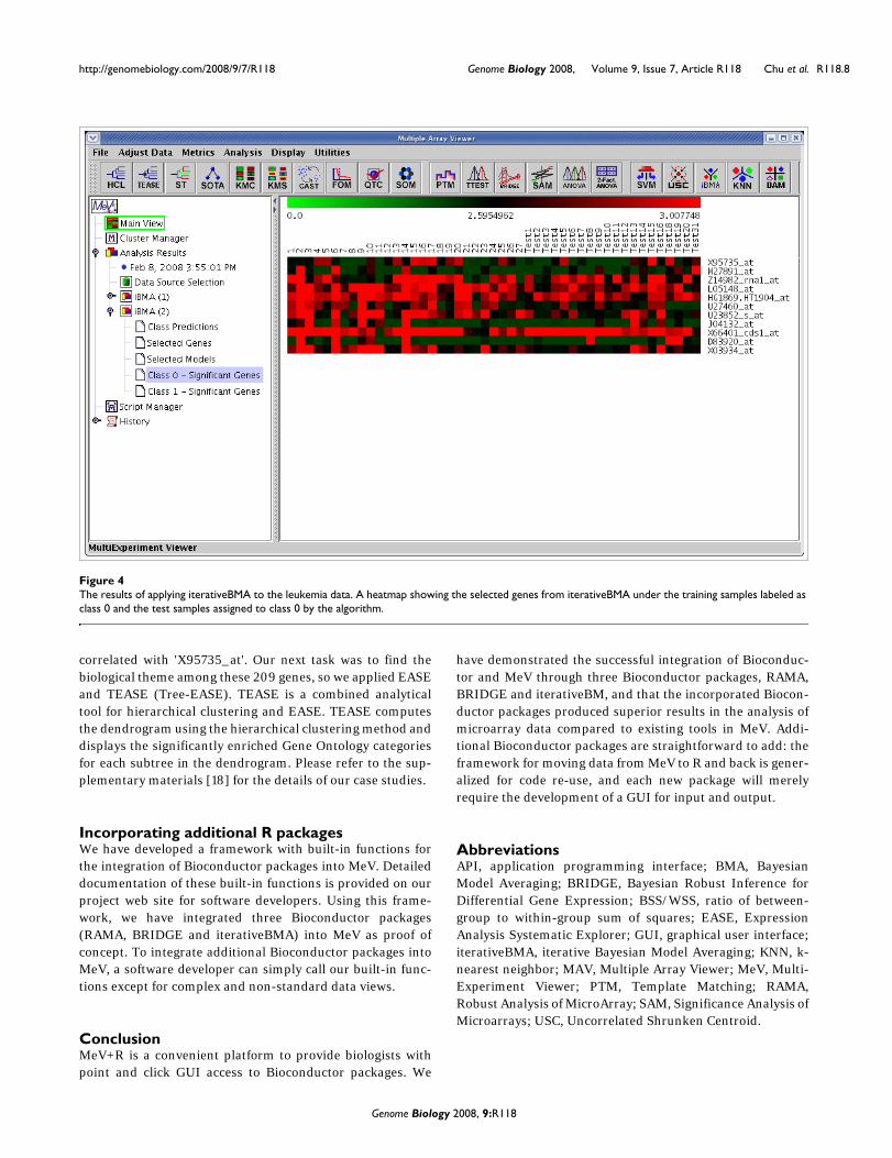
IterativeBMA: Iterative Bayesian Model AveragingWe compared the performance of iterativeBMA (abbreviatedas iBMA in our MeV+R implementation) to KNN (k-nearestneighbor) [30] and USC (Uncorrelated Shrunken Centroid)[31] implemented in MeV using the well-studied leukemiadata [32]. We used the filtered leukemia dataset, which con-sists of 3,051 genes, 38 samples in the training data and 34samples in the test set. The data consist of samples frompatients with either acute lymphoblastic leukemia (ALL) oracute myeloid leukemia (AML). On the leukemia data, itera-tiveBMA produced 2 classification errors using 11 selectedgenes over 11 models (Figures 4 and 5). On the other hand,KNN does not have a gene selection procedure and produced2 classification errors using all 3,051 genes. Similarly, USCproduced 2 classification errors using 51 selected genes.
The second dataset we used is the breast cancer prognosisdataset [33], which consists of 4,919 genes with 76 samples inthe training set, and 19 samples in the test set [17]. Thepatient samples are divided into two categories: the goodprognosis group (patients who remained disease free for atleast five years) and the poor prognosis group (patients whodeveloped distant metastases within five years). The itera-tiveBMA algorithm produced three classification errors usingfour genes averaged over three models. On the other hand,KNN does not have a gene selection procedure and producedfive classification errors using all genes. Similarly, USC pro-duced four classification errors using 662 genes.
Our results are summarized in Table 3. On the breast cancerprognosis data, iterativeBMA produced higher predictionaccuracy using much fewer genes. On the leukaemia data,iterativeBMA produced comparable prediction accuracyusing much fewer genes.
Using other MeV modules in an integrated data analysisThe previous sub-sections showed that our MeV+R modulesachieved superior performance when compared to otherexisting tools implemented in MeV. Here we demonstratehow the R packages that we incorporated into MeV can beused in combination with other existing tools in MeV. Thisillustrates the fact that the MeV+R framework has extendedthe capabilities of MeV, and that using these R packagesthrough the MeV GUI adds value to the integrated analysis ofmicroarray data.
In this case study, we will follow-up on the results from apply-ing the iterativeBMA algorithm to the leukemia data [32]. TheiterativeBMA algorithm is a multivariate gene selectionmethod designed to select a small set of predictive genes forthe classification of microarray data. In the case of the leuke-mia data, the iterativeBMA algorithm selected 11 genes thatproduced two classification errors on the 34-sample test set.It would be interesting to identify the biological theme in this11-gene list. Towards this end, we applied EASE [13] as imple-mented in MeV to determine the over-represented GeneOntology categories in this gene list relative to all the genes onthe microarray. Figure 6 shows the tabular view from theEASE analysis.
Since iterativeBMA identifies a small set of predictive genesfor classification, other genes that exhibit similar expressionpatterns to the selected genes are likely of biological interest.For example, we would like to explore the gene with the high-est posterior probability 'X95735_at' from the iterativeBMAanalysis on the leukemia data [32]. We applied PTM (Tem-plate Matching) [34] as implemented in MeV to identify genesthat are highly correlated with 'X95735_at'. Using a p-valuethreshold of 0.0001, PTM identified 209 genes that are highly
Table 2
Comparing the results of BRIDGE to t-test and SAM on the HIV data and the Affymetrix spike-in data
t-test
Dataset Benchmark BRIDGE p-value cut-off 0.01, no correction
p-value cut-off 0.05, standard Bonferroni correction
SAM
HIV data 13 positive controls, 24 negative controls
DE 13 14 1 12
TP 13 13 1 12
FP 0 1 0 0
Affymetrix spike-in data 65 spike-in genes
DE 45 33 4 8
TP 45 31 4 8
FP 0 2 0 0
For each dataset and each method, the number of differentially expressed (DE) genes, true positives (TP) and false positives (FP) are shown. For each dataset, the maximum TP and the minimum FP across all methods are shown in bold. BRIDGE produced the best results on both datasets in identifying the highest number of true positives without any false positives.
Genome Biology 2008, 9:R118
http://genomebiology.com/2008/9/7/R118 Genome Biology 2008, Volume 9, Issue 7, Article R118 Chu et al. R118.8
correlated with 'X95735_at'. Our next task was to find thebiological theme among these 209 genes, so we applied EASEand TEASE (Tree-EASE). TEASE is a combined analyticaltool for hierarchical clustering and EASE. TEASE computesthe dendrogram using the hierarchical clustering method anddisplays the significantly enriched Gene Ontology categoriesfor each subtree in the dendrogram. Please refer to the sup-plementary materials [18] for the details of our case studies.
Incorporating additional R packagesWe have developed a framework with built-in functions forthe integration of Bioconductor packages into MeV. Detaileddocumentation of these built-in functions is provided on ourproject web site for software developers. Using this frame-work, we have integrated three Bioconductor packages(RAMA, BRIDGE and iterativeBMA) into MeV as proof ofconcept. To integrate additional Bioconductor packages intoMeV, a software developer can simply call our built-in func-tions except for complex and non-standard data views.
ConclusionMeV+R is a convenient platform to provide biologists withpoint and click GUI access to Bioconductor packages. We
have demonstrated the successful integration of Bioconduc-tor and MeV through three Bioconductor packages, RAMA,BRIDGE and iterativeBM, and that the incorporated Biocon-ductor packages produced superior results in the analysis ofmicroarray data compared to existing tools in MeV. Addi-tional Bioconductor packages are straightforward to add: theframework for moving data from MeV to R and back is gener-alized for code re-use, and each new package will merelyrequire the development of a GUI for input and output.
AbbreviationsAPI, application programming interface; BMA, BayesianModel Averaging; BRIDGE, Bayesian Robust Inference forDifferential Gene Expression; BSS/WSS, ratio of between-group to within-group sum of squares; EASE, ExpressionAnalysis Systematic Explorer; GUI, graphical user interface;iterativeBMA, iterative Bayesian Model Averaging; KNN, k-nearest neighbor; MAV, Multiple Array Viewer; MeV, Multi-Experiment Viewer; PTM, Template Matching; RAMA,Robust Analysis of MicroArray; SAM, Significance Analysis ofMicroarrays; USC, Uncorrelated Shrunken Centroid.
The results of applying iterativeBMA to the leukemia dataFigure 4The results of applying iterativeBMA to the leukemia data. A heatmap showing the selected genes from iterativeBMA under the training samples labeled as class 0 and the test samples assigned to class 0 by the algorithm.
Genome Biology 2008, 9:R118

http://genomebiology.com/2008/9/7/R118 Genome Biology 2008, Volume 9, Issue 7, Article R118 Chu et al. R118.9
Authors' contributionsVC carried out the software implementation, and drafted partof the initial manuscript. RG and AER designed and wrote theBioconductor packages RAMA and BRIDGE, and assisted inincorporating these packages into MeV. REB conceived of thestudy, and designed and coordinated the project. KYY partic-ipated in the design and coordination of the study, wrote theiterativeBMA Bioconductor package, carried out the casestudies and prepared the manuscript. All authors read andapproved the final manuscript.
AcknowledgementsWe would like to give special thanks to Dr John Quakenbush and hisresearch group for the original development of MeV and for insightfuldiscussions related to this work. We would also like to thank Drs ChrisVolinsky and Ian Painter for their help on the development of the itera-tiveBMA Bioconductor package. We would also like to thank Dr Renee Ire-ton for editing the manuscript. VTC is supported by NIH-NCI grantK25CA106988 and 5R01HL072370. RG's research is supported by theNatural Sciences and Engineering Research Council of Canada. AER'sresearch was supported by NICHD grant R01 HD054511, NIH grant 8 R01
The results of applying iterativeBMA to the leukemia dataFigure 5The results of applying iterativeBMA to the leukemia data. A heatmap showing the selected genes from iterativeBMA under the training samples labeled as class 1 and the test samples assigned to class 1 by the algorithm.
Table 3
Comparing the results of iterativeBMA to KNN and USC on the leukemia data and the breast cancer prognosis data
Data Size of data iterativeBMA KNN USC
Leukemia data [32] 38 training samples 11 genes 3,051 genes 51 genes
34 test samples 2 errors 2 errors 2 errors
Breast cancer prognosis data [33] 76 training samples 4 genes 4,919 genes 662 genes
19 test samples 3 errors 5 errors 4 errors
The number of selected genes and the number of classification errors are shown for each method. For each dataset, the smallest number of genes and the smallest number of classification errors across all three methods are shown in bold. On the leukemia data, iterativeBMA produced the same number of classification errors using much fewer genes. On the breast cancer prognosis data, iterativeBMA produced fewer errors using much fewer genes.
Genome Biology 2008, 9:R118

http://genomebiology.com/2008/9/7/R118 Genome Biology 2008, Volume 9, Issue 7, Article R118 Chu et al. R118.10
EB002137-02, NSF grant ATM 0724721 and ONR grant N00014-01-10745.REB is funded by NIH-NIAID grants 5P01 AI052106-02, 1R21AI052028-01and 1U54AI057141-01, NIH-NIEHA grant 1U19ES011387-02, NIH-NHLBIgrants 5R01HL072370-02 and 1P50HL073996-01. KYY is supported byNIH-NCI grant K25CA106988.
References1. Gentleman RC, Carey VJ, Bates DM, Bolstad B, Dettling M, Dudoit S,
Ellis B, Gautier L, Ge Y, Gentry J, Hornik K, Hothorn T, Huber W,Iacus S, Irizarry R, Leisch F, Li C, Maechler M, Rossini AJ, Sawitzki G,Smith C, Smyth G, Tierney L, Yang JY, Zhang J: Bioconductor: opensoftware development for computational biology andbioinformatics. Genome Biol 2004, 5:R80.
2. The Bioconductor Project [http://www.bioconductor.org]3. Ihaka R, Gentleman RC: R: a language for data analysis and
graphics. J Computational Graphical Stat 1996, 5:299-314.4. The R Project for Statistical Computing [http://www.r-
project.org]5. Dalgaard P: The R-Tcl/Tk interface. Proceedings of the Second Inter-
national Workshop on Distributed Statistical Computing: 15-17 March2001; Vienna, Austria. [http://www.ci.tuwien.ac.at/Conferences/DSC-2001/Proceedings/Dalgaard.pdf].
6. Fox J: The R commander: a basic-statistics graphical userinterface to R. J Stat Software 2005, 14: [http://www.jstatsoft.org/v14/i09/paper].
7. Grosjean P: SciViews: an object-oriented abstraction layer todesign GUIs on top of various calculation kernels. Proceedingsof the Third International Workshop on Distributed Statistical Computing:
20-22 March 2003; Vienna, Austria. [http://www.ci.tuwien.ac.at/Conferences/DSC-2003/Proceedings/Grosjean.pdf].
8. Wettenhall JM, Smyth GK: limmaGUI: a graphical user interfacefor linear modeling of microarray data. Bioinformatics 2004,20:3705-3706.
9. Wettenhall JM, Simpson KM, Satterley K, Smyth GK: affylmGUI: agraphical user interface for linear modeling of single channelmicroarray data. Bioinformatics 2006, 22:897-899.
10. Futschik ME, Crompton T: OLIN: optimized normalization, vis-ualization and quality testing of two-channel microarraydata. Bioinformatics 2005, 21:1724-1726.
11. Saeed AI, Sharov V, White J, Li J, Liang W, Bhagabati N, Braisted J,Klapa M, Currier T, Thiagarajan M, Sturn A, Snuffin M, Rezantsev A,Popov D, Ryltsov A, Kostukovich E, Borisovsky I, Liu Z, Vinsavich A,Trush V, Quackenbush J: TM4: a free, open-source system formicroarray data management and analysis. Biotechniques 2003,34:374-378.
12. Eisen MB, Spellman PT, Brown PO, Botstein D: Cluster analysisand display of genome-wide expression patterns. Proc NatlAcad Sci USA 1998, 95:14863-14868.
13. Hosack DA, Dennis G Jr, Sherman BT, Lane HC, Lempicki RA: Iden-tifying biological themes within lists of genes with EASE.Genome Biol 2003, 4:R70.
14. SourceForge.net [http://www.sourceforge.net/projects/mev-tm4]15. Gottardo R, Raftery AE, Yeung KY, Bumgarner RE: Quality control
and robust estimation for cDNA microarrays withreplicates. J Am Stat Assoc 2006, 101:30-40.
16. Gottardo R, Raftery AE, Yeung KY, Bumgarner RE: Bayesian robustinference for differential gene expression in cDNA microar-rays with multiple samples. Biometrics 2006, 62:10-18.
The results of applying EASE to the 11 genes selected by iterativeBMA on the leukemia dataFigure 6The results of applying EASE to the 11 genes selected by iterativeBMA on the leukemia data.
Genome Biology 2008, 9:R118
http://www.ncbi.nlm.nih.gov/entrez/query.fcgi?cmd=Retrieve&db=PubMed&dopt=Abstract&list_uids=9843981

http://genomebiology.com/2008/9/7/R118 Genome Biology 2008, Volume 9, Issue 7, Article R118 Chu et al. R118.11
17. Yeung KY, Bumgarner RE, Raftery AE: Bayesian model averaging:development of an improved multi-class, gene selection andclassification tool for microarray data. Bioinformatics 2005,21:2394-2402.
18. MeV+R Supplementary Web Site [http://expression.washington.edu/mevr]
19. Urbanek S: Rserve - a fast way to provide R functionality toapplications. Proceedings of the Third International Workshop on Dis-tributed Statistical Computing: 20-22 March 2003; Vienna, Austria. [http://www.ci.tuwien.ac.at/Conferences/DSC-2003/Proceedings/Urbanek.pdf].
20. Rserve [http://stats.math.uni-augsburg.de/Rserve]21. MeV Manual [http://www.tm4.org/mev.html]22. Raftery AE: Bayesian model selection in social research (with
discussion). Sociol Methodol 1995, 25:111-196.23. Dudoit S, Fridlyand J, Speed TP: Comparison of discrimination
methods for the classification of tumors using gene expres-sion data. J Am Stat Assoc 2002, 97:77-87.
24. van 't Wout AB, Lehrman GK, Mikheeva SA, O'Keeffe GC, Katze MG,Bumgarner RE, Geiss GK, Mullins JI: Cellular gene expressionupon human immunodeficiency virus type 1 infection ofCD4(+)-T-cell lines. J Virol 2003, 77:1392-1402.
25. Tusher VG, Tibshirani R, Chu G: Significance analysis of micro-arrays applied to the ionizing radiation response. Proc NatlAcad Sci USA 2001, 98:5116-5121.
26. Affymetrix U133 Spike-in Data [http://www.affymetrix.com/support/technical/sample_data/datasets.affx]
27. Irizarry RA, Bolstad BM, Collin F, Cope LM, Hobbs B, Speed TP:Summaries of Affymetrix GeneChip probe level data. NucleicAcids Res 2003, 31:e15.
28. Sheffler W, Upfal E, Sedivy J, Noble WS: A learned comparativeexpression measure for affymetrix genechip DNAmicroarrays. Proc IEEE Comput Syst Bioinform Conf 2005:144-154.
29. Lo K, Gottardo R: Flexible empirical Bayes models for differ-ential gene expression. Bioinformatics 2007, 23:328-335.
30. Theilhaber J, Connolly T, Roman-Roman S, Bushnell S, Jackson A, CallK, Garcia T, Baron R: Finding genes in the C2C12 osteogenicpathway by k-nearest-neighbor classification of expressiondata. Genome Res 2002, 12:165-176.
31. Yeung KY, Bumgarner RE: Multiclass classification of microarraydata with repeated measurements: application to cancer.Genome Biol 2003, 4:R83.
32. Golub TR, Slonim DK, Tamayo P, Huard C, Gaasenbeek M, MesirovJP, Coller H, Loh ML, Downing JR, Caligiuri MA, Bloomfield CD,Lander ES: Molecular classification of cancer: class discoveryand class prediction by gene expression monitoring. Science1999, 286:531-537.
33. van 't Veer LJ, Dai H, Vijver MJ van de, He YD, Hart AA, Mao M,Peterse HL, Kooy K van der, Marton MJ, Witteveen AT, Schreiber GJ,Kerkhoven RM, Roberts C, Linsley PS, Bernards R, Friend SH: Geneexpression profiling predicts clinical outcome of breastcancer. Nature 2002, 415:530-536.
34. Pavlidis P, Noble WS: Analysis of strain and regional variationin gene expression in mouse brain. Genome Biol 2001,2:research0042.1-0042.15.
Genome Biology 2008, 9:R118


![111 NIST Calibration of a Neutron Spectrometer ROSPEC · 252Cf sources [4,5], a thermal-neutron beam, and 2.5 MeV and 14 MeV sources. The 2.5 MeV and 14 MeV sources are of known energy,](https://static.fdocuments.us/doc/165x107/5ebad920c3c33b6ef9254a6b/111-nist-calibration-of-a-neutron-spectrometer-rospec-252cf-sources-45-a-thermal-neutron.jpg)


