Methyl-CpG Binding Domain Proteins and Their...
12
Methyl-CpG Binding Domain Proteins and Their Involvement in the Regulation of the MAGE-A1, MAGE-A2, MAGE-A3, and MAGE-A12 Gene Promoters Frank Wischnewski, 1 Olaf Friese, 2 Klaus Pantel, 1 and Heidi Schwarzenbach 1 1 Institute of Tumor Biology and 2 Institute of Biochemistry and Molecular Biology I, University Medical Center Hamburg-Eppendorf, Hamburg, Germany Abstract Promoter hypermethylation is responsible for the restricted expression of the tumor-associated MAGE antigens. In order to elucidate the mechanism underlying methylation-dependent repression, we examined the involvement of methyl-CpG binding proteins, MBD1, MBD2a, and MeCP2, in silencing of MAGE-A1, MAGE-A2, MAGE-A3 , and MAGE-A12 genes. Electrophoretic mobility shift assays displayed binding of MBD1 to the methylated and unmethylated MAGE-A promoters. Using chromatin immunoprecipitation assays, in vivo binding of MBD1 and MeCP2 to the promoters could be observed in MCF-7 and T47D cells. Transient transfection assays of MCF-7 cells were done with the transcriptional repression domains (TRD) of MBD1, MBD2a, and MeCP2, and MAGE-A1, MAGE-A2, MAGE-A3, and MAGE-A12 promoters. Whereas the TRD of MBD1 and MeCP2 repressed the MAGE-A promoters, the TRD of MBD2 had no inhibiting effect on the promoter activity. Furthermore, cotransfections of Mbd1-deficient mouse fibroblasts and MCF-7 cells with MBD2a, MeCP2, and the MBD1 splice variants, 1v1 and 1v3, showed that strong methylation-dependent repression of the MAGE-A promoters could not be further down-regulated by these proteins. However, the two MBD1 splice variants, 1v1 and 1v3, were able to repress the basal activity of unmethylated MAGE-A promoters. Additional cotransfection experiments with both isoforms of MBD1 and the transcription factor Ets-1 showed that Ets-1 could not abrogate the MBD1-mediated suppression. In contrast with the repressive effect mediated by MBD1, MBD2a was found to up-regulate the basal activity of the promoters. In conclusion, these data show, for the first time, the involvement of methyl-CpG binding domain proteins in the regulation of the MAGE-A genes. (Mol Cancer Res 2007;5(7):749 – 59) Introduction The MAGE-A gene family is located on chromosome Xq28 (1) and is comprised of 12 members (MAGE-A1 – 12 ). They encode tumor-associated antigens which are recognized by CTLs in conjunction with MHC class I molecules of various haplotypes on the tumor cell surface (2). Their expression is restricted to different histologic tumors types including melanoma, lung, mamma, bladder, and stomach carcinoma (3-8). In normal adult tissues, they are silent, except for testi- cular germ cells (spermatogonia and primary spermatocytes) and placenta (9). Hypermethylation of CpG-rich MAGE-A promoters causes gene silencing and prevents Ets transcription factors from accessing their binding sites (1, 10, 11), which are responsible for the high transcriptional activation of the MAGE-A1 gene (12). Alterations in DNA methylation occur during the patho- genesis of human tumors, and a global DNA hypomethylation of the genome, including the tumor-associated MAGE-A genes, has been observed in various carcinomas. Recent clinical studies showed that immunotherapy against MAGE-A antigens caused a significant tumor regression in patients with various tumor entities, pointing out that vaccination with a MAGE-A peptide could be a promising target for the treatment of patients with tumors (13-16). Nevertheless, heterogeneous intratumor expression of the MAGE-A genes may hamper the effectiveness of immunotherapy and urgently requires a detailed examination of the regulation of these genes in tumors. The mechanism leading to partial DNA hypomethy- lation of the genes during carcinogenesis remains poorly understood. In our previous article, we addressed this issue and found that besides DNA methylation, histone deacetylation, which leads to a compact and transcriptional inactive chromatin structure (17), is also involved in the partial repression of MAGE-A genes in tumor cells and may impede their activation (18). Whether methyl-CpG binding proteins, which recruit histone deacetylases to methylated DNA, contribute to the silencing of MAGE-A genes has not yet been clarified. To date, five methyl-CpG binding proteins have been identified: MBD1, MBD2, MBD3, MBD4, and MeCP2. All of these proteins are involved in the transcriptional repression Received 10/27/06; revised 3/20/07; accepted 4/13/07. Grant support: Wilhelm-Sander-Stiftung, Mu ¨ nchen, Germany (grant 2004.056.1) and the Deutsche Forschungsgemeinschaft, Bonn, Germany (grant SFB545B2). The costs of publication of this article were defrayed in part by the payment of page charges. This article must therefore be hereby marked advertisement in accordance with 18 U.S.C. Section 1734 solely to indicate this fact. Requests for reprints: Heidi Schwarzenbach, Institute of Tumor Biology, University Medical Center Hamburg-Eppendorf, Martinstrasse 52, 20246 Hamburg, Germany. Phone: 49-4042-803-7494; Fax: 49-4042-803-6546. E-mail: [email protected] Copyright D 2007 American Association for Cancer Research. doi:10.1158/1541-7786.MCR-06-0364 Mol Cancer Res 2007;5(7). July 2007 749 Research. on August 31, 2018. © 2007 American Association for Cancer mcr.aacrjournals.org Downloaded from
Transcript of Methyl-CpG Binding Domain Proteins and Their...

Methyl-CpG Binding Domain Proteins and TheirInvolvement in the Regulation of the MAGE-A1,MAGE-A2, MAGE-A3, and MAGE-A12Gene Promoters
Frank Wischnewski,1 Olaf Friese,2 Klaus Pantel,1 and Heidi Schwarzenbach1
1Institute of Tumor Biology and 2Institute of Biochemistry and Molecular Biology I,University Medical Center Hamburg-Eppendorf, Hamburg, Germany
AbstractPromoter hypermethylation is responsible for the
restricted expression of the tumor-associated MAGE
antigens. In order to elucidate the mechanism
underlying methylation-dependent repression, we
examined the involvement of methyl-CpG binding
proteins, MBD1, MBD2a, and MeCP2, in silencing of
MAGE-A1, MAGE-A2, MAGE-A3 , and MAGE-A12 genes.
Electrophoretic mobility shift assays displayed binding
of MBD1 to the methylated and unmethylated MAGE-A
promoters. Using chromatin immunoprecipitation
assays, in vivo binding of MBD1 and MeCP2 to the
promoters could be observed in MCF-7 and T47D cells.
Transient transfection assays of MCF-7 cells were done
with the transcriptional repression domains (TRD) of
MBD1, MBD2a, and MeCP2, and MAGE-A1, MAGE-A2,
MAGE-A3, and MAGE-A12 promoters. Whereas the TRD
of MBD1 and MeCP2 repressed the MAGE-A promoters,
the TRD of MBD2 had no inhibiting effect on the
promoter activity. Furthermore, cotransfections of
Mbd1-deficient mouse fibroblasts and MCF-7 cells with
MBD2a, MeCP2, and the MBD1 splice variants, 1v1
and 1v3, showed that strong methylation-dependent
repression of the MAGE-A promoters could not be
further down-regulated by these proteins. However,
the two MBD1 splice variants, 1v1 and 1v3, were able to
repress the basal activity of unmethylated MAGE-A
promoters. Additional cotransfection experiments with
both isoforms of MBD1 and the transcription factor
Ets-1 showed that Ets-1 could not abrogate the
MBD1-mediated suppression. In contrast with the
repressive effect mediated by MBD1, MBD2a was found
to up-regulate the basal activity of the promoters.
In conclusion, these data show, for the first time,
the involvement of methyl-CpG binding domain proteins
in the regulation of the MAGE-A genes.
(Mol Cancer Res 2007;5(7):749–59)
IntroductionThe MAGE-A gene family is located on chromosome Xq28
(1) and is comprised of 12 members (MAGE-A1–12). They
encode tumor-associated antigens which are recognized by
CTLs in conjunction with MHC class I molecules of various
haplotypes on the tumor cell surface (2). Their expression is
restricted to different histologic tumors types including
melanoma, lung, mamma, bladder, and stomach carcinoma
(3-8). In normal adult tissues, they are silent, except for testi-
cular germ cells (spermatogonia and primary spermatocytes)
and placenta (9). Hypermethylation of CpG-rich MAGE-A
promoters causes gene silencing and prevents Ets transcription
factors from accessing their binding sites (1, 10, 11), which are
responsible for the high transcriptional activation of the
MAGE-A1 gene (12).
Alterations in DNA methylation occur during the patho-
genesis of human tumors, and a global DNA hypomethylation
of the genome, including the tumor-associated MAGE-A genes,
has been observed in various carcinomas. Recent clinical
studies showed that immunotherapy against MAGE-A antigens
caused a significant tumor regression in patients with various
tumor entities, pointing out that vaccination with a MAGE-A
peptide could be a promising target for the treatment of
patients with tumors (13-16). Nevertheless, heterogeneous
intratumor expression of the MAGE-A genes may hamper the
effectiveness of immunotherapy and urgently requires a
detailed examination of the regulation of these genes in
tumors. The mechanism leading to partial DNA hypomethy-
lation of the genes during carcinogenesis remains poorly
understood. In our previous article, we addressed this issue and
found that besides DNA methylation, histone deacetylation,
which leads to a compact and transcriptional inactive chromatin
structure (17), is also involved in the partial repression of
MAGE-A genes in tumor cells and may impede their activation
(18). Whether methyl-CpG binding proteins, which recruit
histone deacetylases to methylated DNA, contribute to the
silencing of MAGE-A genes has not yet been clarified.
To date, five methyl-CpG binding proteins have been
identified: MBD1, MBD2, MBD3, MBD4, and MeCP2. All
of these proteins are involved in the transcriptional repression
Received 10/27/06; revised 3/20/07; accepted 4/13/07.Grant support:Wilhelm-Sander-Stiftung, Munchen, Germany (grant 2004.056.1)and the Deutsche Forschungsgemeinschaft, Bonn, Germany (grant SFB545B2).The costs of publication of this article were defrayed in part by the payment of pagecharges. This article must therefore be hereby marked advertisement in accordancewith 18 U.S.C. Section 1734 solely to indicate this fact.Requests for reprints: Heidi Schwarzenbach, Institute of Tumor Biology,University Medical Center Hamburg-Eppendorf, Martinstrasse 52, 20246Hamburg, Germany. Phone: 49-4042-803-7494; Fax: 49-4042-803-6546. E-mail:[email protected] D 2007 American Association for Cancer Research.doi:10.1158/1541-7786.MCR-06-0364
Mol Cancer Res 2007;5(7). July 2007 749
Research. on August 31, 2018. © 2007 American Association for Cancermcr.aacrjournals.org Downloaded from

of methylated DNA (19, 20). The methyl-CpG binding domain
protein 2 (MeCP2) can interact with both the corepressor SinA
3 and histone deacetylases (21-23). It can also repress the
transcription of distinct promoters independently of histone
deacetylases, probably through direct interaction with basal
transcription factors (24). MeCP2-deficient mice show neuronal
defects, which likely lead to their short survival time of 2 to
3 months (25). The other members of the family (MBD1,
MBD2, and MBD3) also associate with histone deacetylases.
MBD1 is alternatively spliced to produce five protein isoforms
(PCM1, MBD1v1, MBD1v2, MBD1v3, and MBD1v4) which
differ in the number of cysteine-rich (CXXC) domains and the
carboxyl-terminal sequence. All five MBD1 isoforms repress
transcription from methylated promoters and the variants with
three CXXC domains additionally repress unmethylated
promoters (26, 27). Two isoforms of MBD2 are known:
MBD2a and MBD2b. The shorter form, MBD2b, starting at the
second methionine therefore lacks the NH2-terminal sequence
of MBD2a (28). Recent reports described MBD2a as both an
activator and a repressor of transcription (29-31). Mbd1- and
Mbd2-deficient mice are viable and fertile. However, the
Mbd1�/� mouse shows mild defects in neurogenesis (32),3 andthe Mbd2�/� mouse in nurturing their offspring (33).Based on our findings that the association of DNA
methylation and histone deacetylation may be responsible for
inactivation of the MAGE-A genes (18), we have continued to
examine whether methyl-CpG binding proteins participate in
silencing these genes. Our promoter studies and transient
transfection assays show that both splice variants MBD1v1 and
1v3 were able to repress the unmethylated MAGE-A1, MAGE-
A2, MAGE-A3, and MAGE-A12 promoters, and the trans-
activator Ets-1 could not abrogate the MBD1-mediated gene
repression. Conversely, MBD2a could enhance the promoter
activity of these genes.
ResultsIn vitro Binding of MBD1 to the 5¶-Regions of MAGE-A1,MAGE-A2, MAGE-A3, and MAGE-A12 GenesIn our previous studies, we reported the association of
DNA methylation and histone deacetylation in silencing of
MAGE-A1, MAGE-A2, MAGE-A3 , and MAGE-A12 genes (18).
Because methyl-CpG binding proteins can recruit histone
deacetylases to methylated DNA, we investigated whether
these proteins are able to bind to hypermethylated MAGE-A
promoters. For these analyses, we applied two techniques: the
in vitro electrophoretic mobility shift assay (EMSA) and in vivo
chromatin immunoprecipitation (ChIP) assay.
EMSA was done using the regions extending from �77 to�36 of MAGE-A1 , �158 to �83 of MAGE-A2 , �160 to �102of MAGE-A3 , and �109 to �63 of MAGE-A12 genes
(Fig. 1A). We chose these sequences because they contained
Ets binding motifs which have been shown to be important for
the MAGE-A1 promoter activity (10). There were four, six,
two, and five CpG dinucleotides in the MAGE-A1, MAGE-A2,
MAGE-A3, and MAGE-A12 promoter fragments, respectively
(Fig. 1A). In vitro DNA methylation of the fragments was
carried out by DNA methylases SssI and HpaII, which
methylate each cytosine residue of the CpG dinucleotides and
the second cytosine of the sequence CCGG, respectively. For
EMSA, we used nuclear extracts derived from three breast
cancer cells (MCF-7, T47D, and MDA-MB-231) as well as the
epithelioid cervical carcinoma cell line HeLa. Nuclear extracts
from HeLa cells were used because methyl-CpG binding
proteins were recently found to be components of HeLa cells
and served as control for the binding analyses (30, 34). When
the unmethylated, HpaII- and SssI-methylated probes were
incubated with various nuclear extracts, prominent complexes
were formed as detected by EMSA. Figure 1B and C show
representative examples of such binding reactions between
nuclear extracts derived from MCF-7 or T47D breast cancer
cells and the unmethylated or methylated MAGE-A1 (Fig. 1B,
lanes 2-4) and MAGE-A3 sequences (Fig. 1C, lanes 2-4). A
striking increase in the formation of complexes paralleled with
the methylation status of the MAGE-A1 promoter when nuclear
extract from MCF-7 cells was used (Fig. 1B, lanes 2-4).
Binding was competed by the addition of an excess of cognate,
unlabeled oligonucleotides which were either unmethylated or
methylated indicating that the complexes were specific (Fig. 1B
and C, lanes 5-10). Furthermore, the complexes competed
by SssI-methylated oligonucleotides had a weaker intensity
(Fig. 1B and C, lanes 8-10) than those competed by
unmethylated oligonucleotides (Fig. 1B and C, lanes 5-7),
suggesting that the binding affinity of nuclear extracts was
higher to methylated than unmethylated DNA.
To characterize the nature of the complexes, we did supershift
experiments using antibodies that specifically react with
different methyl-CpG binding domain proteins. When added
to the EMSA reaction, an antibody specific for MBD1 produced
a supershift (SS , Fig. 2), whereas antibodies specific for MBD2a
and MeCP2 had no effect on the binding reaction (data not
shown). Our findings show that MBD1 had a binding affinity to
both methylated and unmethylated MAGE-A1, MAGE-A2,
MAGE-A3, and MAGE-A12 promoters (Fig. 2). Figure 2A
shows such a supershift caused by MBD1 using increasing
amounts of nuclear extract from MCF-7 (lanes 4 and 5),
MDA-MB-231 (lanes 8 and 9), and T47D (lanes 12 and 13)
breast cancer cells and the unmethylated MAGE-A1 probe. As
shown in Fig. 2B and C, an antibody specific for MBD1
generated supershifted complexes containing nuclear extracts
derived from MCF-7 (B) or T47D cells (C) and methylated or
unmethylated MAGE-A2, MAGE-A3, and MAGE-A12 probes
(lanes 5-7).
In vivo Binding of MBD1 and MeCP2 to the 5¶-Regions ofMAGE-A1, MAGE-A2, MAGE-A3, and MAGE-A12 GenesAs EMSA revealed in vitro binding, specifically of MBD1
to methylated and unmethylated MAGE-A promoters, we did
ChIP assays to monitor in vivo binding of methyl-CpG binding
proteins to the MAGE-A promoters in MCF-7 and T47D
cells. Following cross-linking of nuclear proteins to DNA with
formaldehyde and immunoprecipitation of the protein-DNA
complexes with the antibodies specific for MBD1, MBD2, or
MeCP2, the immunoprecipitated DNA fragments were ampli-
fied by PCR using gene-specific primers and visualized by gel3 Our unpublished data.
Wischnewski et al.
Mol Cancer Res 2007;5(7). July 2007
750
Research. on August 31, 2018. © 2007 American Association for Cancermcr.aacrjournals.org Downloaded from
electrophoresis (Fig. 3A). Compared with DNA precipitated
with IgG, DNA precipitated with antibodies against MBD1 and
MeCP2 contained significant increases of MAGE-A1, MAGE-
A2, MAGE-A3, and MAGE-A12 promoter DNA (Fig. 3A).
Densitometric evaluation showed that amplification of the
MBD1- and MeCP2-immunoprecipitated DNA resulted in a
6- and 9-fold, 2- and 5-fold, 4- and 3-fold, and 3- and 8-fold
increase using primers specific for MAGE-A1, MAGE-A2,
MAGE-A3 , and MAGE-A12 genes, respectively, in comparison
with the PCR products of the IgG-immunoprecipitated DNA.
MeCP2 had a higher affinity with the MAGE-A1, MAGE-A2,
MAGE-A3, and MAGE-A12 promoters than MBD1; possibly
caused by the higher precipitation efficiency of the MeCP2-
specific antibody (Fig. 3A).
To verify the data of the standard PCR, we additionally
carried out a quantitative real-time PCR with primers specific
for the MAGE-A1 sequence and the RPLP0 (ribosomal protein,
large protein) housekeeping gene. The primer pairs were chosen
due to the stringent conditions of the real-time PCR. A low
amount of immunoprecipitated RPLP0 DNA could be observed
in the bar chart of Fig. 3B based on the characteristics of a
housekeeping gene, which is constitutively expressed, whereas
its promoter region is unmethylated. In MCF-7 cells which do
not express MAGE-A1 (18), the yield of MAGE-A1 DNA
immunoprecipitated with antibodies against MBD1 and MeCP2
was 2- and 2.5-fold higher, respectively, than the yield of DNA
immunoprecipitated with the antibody against MBD2 or IgG,
which served as a negative control (Fig. 3B). When DNA
originated from T47D cells, there was no difference in the yield
of MAGE-A1 DNA precipitated with antibodies against
MBD1, MBD2, and MeCP2, in comparison to the DNA
precipitated with IgG (Fig. 3B), indicating that these cells
expressed MAGE-A1 (18). Taken together, these findings show
that MBD1, as well as MeCP2, interact in vivo with all four
MAGE-A promoter sequences in MCF-7 cells.
Transcriptional Repression of MAGE-A1, MAGE-A2,MAGE-A3, and MAGE-A12 Genes by the TranscriptionalRepression Domains of MBD1 and MeCP2To functionally test the effect of the methyl-CpG binding
proteins on MAGE-A promoters, we constructed a series of
reporter plasmids in which we inserted sequences derived from
the MAGE-A1, MAGE-A2, MAGE-A3, and MAGE-A12
promoters immediately downstream of the Gal4 DNA-binding
sites. The expression plasmids contained the transcriptional
repression domains (TRD) of MBD1, MBD2, and MeCP2
linked to the heterologous Gal4 DNA-binding domain (28).
Both reporter and expression plasmids were transiently
cotransfected with a reference plasmid into MCF-7 cells; cell
lysates were then assayed for luciferase activity 48 h post-
transfection. Immunoblot analysis using antibodies specific for
MBD1, MBD2, and MeCP2 documented efficient expression of
the fusion proteins in these cells (data not shown). Targeting of
the TRD of MBD1 to Gal4 DNA-binding sites upstream of the
MAGE-A1, MAGE-A2, MAGE-A3, and MAGE-A12 pro-
moters produced a 7-, 25-, 25-, and 8-fold decrease in promoter-
driven luciferase activity, respectively, when compared with the
basal activity of Gal4-MAGE-A constructs (Fig. 4). Although
the Gal4-linked TRD of MeCP2 had a weaker effect, it was
able to repress the MAGE-A1, MAGE-A2, MAGE-A3, and
MAGE-A12 transcription by 2-, 4-, 5- and 2-fold, respectively
(Fig. 4). These data show that the TRDs of MBD1, and to a
lesser extent, MeCP2, but not that of MBD2, are able to inhibit
each of the tested MAGE-A promoter activities.
Transcriptional Repression of Unmethylated MAGE-AGenes by the Splice Variants of MBD1, and Activationby MBD2aOur recent studies on various cell lines indicated that
promoter hypermethylation and histone deacetylation are
associated with silencing of MAGE-A1, MAGE-A2, MAGE-A3 ,
FIGURE 1. Nuclear extract specifically binds to unmethylated andmethylated MAGE-A1, MAGE-A2, MAGE-A3, and MAGE-A12 promotersin vitro . A. Schematic view of MAGE-A1, MAGE-A2, MAGE-A3, andMAGE-A12 promoter fragments. The sequences used for EMSA (openboxes ) are indicated by the nucleotide positions given below; putative Etsmotifs (black boxes ); transcriptional start sites (arrow ); and CpGdinucleotides (vertical lines ). B and C. EMSA was done with unmethy-lated (lanes 2, 5, and 8 ; U ), Hpa II- (lanes 3, 6, and 9 ; H ), Sss I- (lanes4, 7, and 10 ; S) methylated fragments of the MAGE-A promoters asindicated in (A) and nuclear extracts derived from MCF-7 and T47D cells.To perform competition experiments, an excess of cognate, unlabeledoligonucleotides, which were either unmethylated (lanes 5-7 ) or methylated(lanes 8-10 ), was added to the EMSA reaction. The positions of theMAGE-A/NE complexes (C) and free DNA are indicated.
Methyl-CpG Binding Proteins and MAGE Genes
Mol Cancer Res 2007;5(7). July 2007
751
Research. on August 31, 2018. © 2007 American Association for Cancermcr.aacrjournals.org Downloaded from

and MAGE-A12 genes (18). Furthermore, our present findings
suggest that MBD1 and MeCP2 may be able to bind to and
repress MAGE-A promoters (Figs. 1-4). To more precisely
define the role of the methyl-CpG binding proteins in
MAGE-A gene repression, we transiently cotransfected full-
length MBD1, MBD2a, and MeCP2 together with methylated
and unmethylated MAGE-A1, MAGE-A2, MAGE-A3, and
MAGE-A12 reporter constructs into MCF-7 cells, and assessed
their influence on transcription in response to methylation by
SssI and HpaII. We expanded our transfection assay with
embryonic fibroblast cells from a Mbd1-knockout mouse3 to
exclude interference from endogenous MBD1.
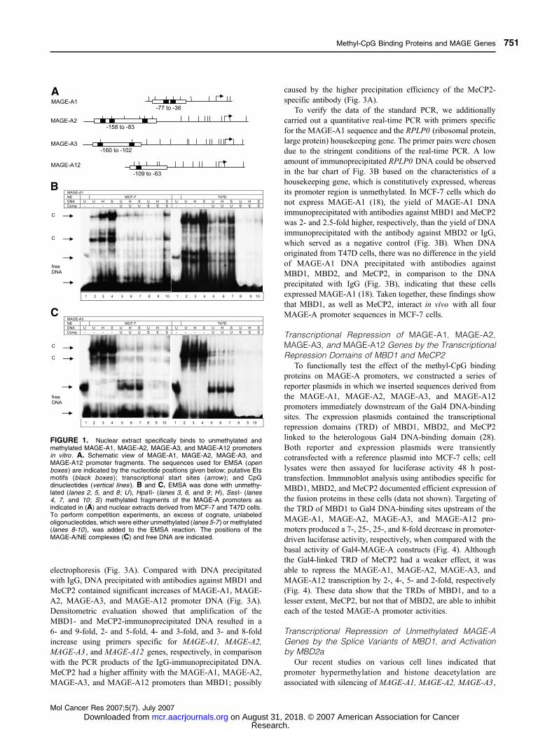
The Mbd1-knockout mouse was analyzed by PCR using
genomic DNA from tail biopsies and a primer set gene-specific
for the wild-type and knockout allele, as illustrated in the
schematic view of Fig. 5A. The primers binding at �695 and�266 bp relative to the start site of MBD1 (ATG) produceda PCR product of 430 bp in size when genomic DNA from
wild-type mice were amplified (Fig. 5B, wt lane). No PCR
product of 430 bp was generated when DNA originated from
homozygous Mbd1-knockout mice (Fig. 5B, Mbd1�/� lane).
The primer pair at �695 bp and +1383 bp amplified a PCRproduct of 2078 bp in size when DNA was derived from wild-
type mice (data not shown), and a 520-bp-long product when
deleted DNA from Mbd1-knockout mice was used (Fig. 5B,
Mbd1�/� lane). Consequently, amplification of DNA from
heterozygous mice formed three PCR products at 430, 520, and
2078 bp (Fig. 5B, Mbd1+/� lane).
The genetic characterization of embryonic mouse fibroblasts
from the Mbd1-knockout mouse was carried out by amplifica-
tion of their genomic DNA using the same primer set. Whereas
amplification with the primers specific for the wild-type allele
did not lead to a PCR product of 430 bp in size (Fig. 5C,
wt lane), amplification with the primers specific for the Mbd1
sequence with the deleted exon resulted in a 520-bp-long PCR
product (Fig. 5C, Mbd1�/� lane). For cotransfection of MCF-7
cells and Mbd1-deficient embryonic fibroblast cells, we used
two isoforms and a mutant form of MBD1. Whereas MBD1v1
FIGURE 2. EMSA supershift experiments with anantibody against MBD1 (Ab ). A. EMSA was done withunmethylated MAGE-A1 promoter fragments and differ-ent amounts of nuclear extracts (NE ) derived from MCF-7(lanes 2-5), MDA-MB-231 (lanes 6-9 ), and T47D (lanes10-13 ) cell lines. B and C. EMSA was done withunmethylated (lanes 2 and 5 ; U ), Hpa II- (lanes 3 and6 ; H), and Sss I-methylated (lanes 4 and 7 ; S) fragmentsof MAGE-A2, MAGE-A3, and MAGE-A12 promoters andnuclear extracts derived from MCF-7 (B) and T47D (C)cell lines as indicated above the polyacrylamide gels. SS,supershifted bands of MAGE-A1/MBD1, MAGE-A2/MBD1, MAGE-A3/MBD1, and MAGE-A12/MBD1 com-plexes (A, lanes 4, 5, 8, 9, 12 , and 13 ; B and C, lanes5-7). The positions of the MAGE-A/NE complexes (C)and free DNA are indicated.
Wischnewski et al.
Mol Cancer Res 2007;5(7). July 2007
752
Research. on August 31, 2018. © 2007 American Association for Cancermcr.aacrjournals.org Downloaded from

contains two CXXC domains, MBD1v3 harbors three CXXC
domains. The mutant form of MBD1 is functionally inactivated
by a 126-bp deletion in the methyl-CpG binding domain and
a mutation in the TRD (amino acids 537-556), and served as
a negative control. Evaluation of promoter-driven luciferase
activities showed that DNA methylation of the reporter
plasmids by SssI caused a nearly complete loss of MAGE-A
promoter activity in mouse Mbd1�/� fibroblasts (Fig. 5D).
Similar results were obtained when transfection assays were
done with SssI- and HpaII-methylated constructs in MCF-7
cells, as described in our recent article (18). Due to the strong
methylation-dependent repression, cotransfected methyl-CpG
binding proteins had no further inhibitory effect on methylated
MAGE-A constructs in Mbd1-deficient mouse cells (Fig. 5D)
and MCF-7 cells (data not shown). In contrast, the transcrip-
tional activity of the unmethylated MAGE-A1, MAGE-A2,
MAGE-A3, and MAGE-A12 promoters in Mbd1-deficient
mouse cells was efficiently reduced 3.5-, 2.5-, 3-, and 8.5-fold
by isoform MBD1v1 and 1.5-, 3-, 2-, and 1.5-fold by isoform
MBD1v3, respectively (Fig. 5D). In MCF-7 cells, MBD1v1
repressed the basal activity of unmethylated MAGE-A1,
MAGE-A2, MAGE-A3, and MAGE-A12 promoters by 8-, 6-,
3-, and 17-fold, respectively, whereas MBD1v3 did not affect
the promoters (Fig. 6). MBD1v1 with its three CXXC domains
usually affected the repression more strongly than MBD1v3
with its two CXXC domains. The mutant version of MBD1
failed to repress the reporter plasmids indicating the specificity
of repression by MBD1v1 and MBD1v3 (Figs. 5D and 6).
FIGURE 3. MBD1 and MeCP2 bind to MAGE-A1, MAGE-A2, MAGE-A3, and MAGE-A12 promoters in vivo . A. PCR amplifications of theimmunoprecipitated DNA derived from MCF-7 cells were carried out with primer sets specific for MAGE-A1, MAGE-A2, MAGE-A3, and MAGE-A12promoters. The intensity of the PCR products derived from the MBD1-, MBD2-, and MeCP2 – immunoprecipated MAGE-A1, MAGE-A2, MAGE-A3, andMAGE-A12 DNA are shown relative to the intensity of the DNA immunoprecipitated with the antibody IgG. The amplification products with and without anasterisk belong to two independent experiments. Both amplifications should therefore be considered independently. Amplification of sonicated genomic DNA(gDNA ) with the gene-specific primers prior immunoprecipitation served as positive control. Lane M, contains marker DNA. B. Quantitative real-time PCRwas done on a realplex4 system with primer sets specific for the MAGE-A1 and RPLP0 promoters and immunoprecipitated DNA derived from MCF-7 andT47D cells. Columns, amounts of IgG-, MBD1-, MBD2-, and MeCP2 – immunoprecipated DNA.
Methyl-CpG Binding Proteins and MAGE Genes
Mol Cancer Res 2007;5(7). July 2007
753
Research. on August 31, 2018. © 2007 American Association for Cancermcr.aacrjournals.org Downloaded from

Contrary to the repressive effect by the MBD1 isoforms,
MBD2a was able to stimulate f1.5-, 3-, 10-, and 1.5-fold the
MAGE-A1, MAGE-A2, MAGE-A3, and MAGE-A12 pro-
moters, respectively, in MCF-7 cells (Fig. 7). A similar
stimulation by MBD2a was observed in Mbd1-deficient mouse
cells (data not shown). Finally, MeCP2 had no effect on the
unmethylated promoters (data not shown).
Ets-1 Did Not Affect MBD1-Mediated Suppression andMBD2-Mediated ActivationThe present data suggests that promoter hypermethylation is
sufficient for silencing of MAGE-A genes. Furthermore, it
shows that MBD1v1 and MBD1v3 are able to bind to the
unmethylated MAGE-A promoters and down-regulate their
basal activity. The MAGE-A1 promoter region contains two
motifs for Ets transcription factors. Promoter demethylation
and binding of the Ets factors to their binding sites have been
shown to drive 90% of the MAGE-A1 transcription (10).
In contrast with the investigated MAGE-A1 promoter, only
little is known of the MAGE-A2, MAGE-A3, and MAGE-
A12 promoters. However, it is assumed that they are regulated
in a similar manner. By DNA sequence analysis and align-
ments, we identified Ets-1 consensus motifs at positions
�103/�90, �143/�134, and �154/�145 of MAGE-A2, at
positions �113/�104 and �151/�141 of MAGE-A3, and atposition �100/�90 of the MAGE-A12 promoter. To examinethe involvement of Ets-1 in MAGE-A2, MAGE-A3, and
MAGE-A12 transcription, we transiently transfected MCF-7
cells and embryonic mouse fibroblasts with an expression
plasmid encoding Ets-1. Ets-1 could stimulate the promoter
activity of MAGE-A1, MAGE-A2, MAGE-A3, and MAGE-
A12 in Mbd1-deficient fibroblasts and MCF-7 cells 2-, 4-, 2-,
and 2-fold (Fig. 5D) and 2-, 4-, 6-, and 3-fold (Fig. 7),
respectively. We conclude that Ets is not only a transactivator
for MAGE-A1 but for the other tested promoters as well.
By additional cotransfection experiments, we examined the
influence of Ets-1 on MBD1-mediated repression and MBD2a-
mediated activation. As shown in Figs. 5D and 7, Ets-1 could
neither alleviate the suppression (Fig. 5D) nor affect the
activation (Fig. 7) of the promoters by MBD1 and MBD2a,
respectively.
DiscussionThe current study shows for the first time the different
regulations for unmethylated MAGE-A1, MAGE-A2, MAGE-
A3, and MAGE-A12 promoters by methyl-CpG binding
proteins. Thus far, these epigenetic factors have been found
to be mainly associated with methylated promoters of tumor
suppressor genes (32-36).
Our transfection assays of MCF-7 cells and embryonic
mouse Mbd1�/� fibroblasts show that promoter hypermethy-
lation seems to be sufficient for silencing of the MAGE-A
genes. However, considering that mice deficient of the different
methyl-CpG binding proteins and the resulting phenotypes
(25, 32, 33), it could be possible that other methyl-CpG binding
proteins, like MeCP2, could compensate for the loss of MBD1
in the repression of methylated MAGE-A promoters. Moreover,
our binding analyses of EMSA show that MBD1 bound to
methylated as well as to unmethylated MAGE-A promoters,
and sustains the transfection data that binding of MBD1 to
unmethylated DNA may lead to repression of the promoters.
The ability of MBD1 to repress unmethylated promoters
in vitro is supported by a recently published study showing
methylation-independent repression (35). Repression of unme-
thylated genes depends on the third CXXC domain, and
repression of methylated genes requires the methyl-CpG
binding domain (26, 36). The isoform MBD1v1 contains the
additional third CXXC domain and could therefore repress
promoters regardless of their methylation status. However,
MBD1v3 lacks the third CXXC domain. Although the
repression by MBD1v3 was weaker than the suppression by
MBD1v1 and only observed in Mbd1-deficient mouse
fibroblasts, our findings indicate that the two other CXXC
domains may also contribute to the repression of unmethylated
promoters, however, with a weaker affinity. The repressive
effect on the examined MAGE-A promoters by MBD1v3 could
also be ascribed to the fact that endogenous DNA methylases
3a and 3b in the transfected cells might methylate de novo the
introduced reporter plasmids containing each of the MAGE-A
promoters (37). Furthermore, the specific repression of MAGE-
A promoters by MBD1v1 and MBD1v3 was confirmed by the
use of mutated MBD1 in our overexpression studies.
MBD1mut, which lacks the methyl-CpG binding domain and
harbors a nonfunctional TRD, congruously showed no effect on
MAGE-A gene expression.
The MAGE-A promoters contain putative Ets motifs and the
transcription factor Ets has been shown to be responsible for the
high transcriptional activation of MAGE-A1 (10). Our results
show that transfection of expression plasmid encoding Ets-1
FIGURE 4. The TRDs of MBD1 and MeCP2 repress transcription ofMAGE-A1, MAGE-A2, MAGE-A3 , and MAGE-A12 genes. MCF-7 cellswere transiently cotransfected with pGL2-Luciferase reporter plasmidscontaining fragments of MAGE-A1, MAGE-A2, MAGE-A3, or MAGE-A12promoters immediately downstream of the Gal4 binding sites andexpression plasmids containing TRDs of MBD1, MBD2, or MeCP2 linkedto the heterologous Gal4 DNA-binding domain. The activities derivedfrom the reference plasmid pCMV LacZ were used to normalize thevariability in transfection efficiency. The relative luciferase activity of thereporter construct containing the Gal4/MAGE-A1 sequence was arbitrarilyset to 100%.
Wischnewski et al.
Mol Cancer Res 2007;5(7). July 2007
754
Research. on August 31, 2018. © 2007 American Association for Cancermcr.aacrjournals.org Downloaded from
resulted in the activation of all tested MAGE-A promoters. It is
noteworthy that cotransfection of MBD1v1 or MBD1v3
together with Ets-1 did not lead to an abrogation of MBD1-
mediated repression. Concerning the potential role of MBD1 as
a repressor of unmethylated MAGE-A promoters and the
mechanisms underlying the transcriptional repression, it is of
considerable interest to investigate the relation between MBD1
and Ets-1. It is unclear whether, in the cotransfection assay
using both MBD1 and Ets-1, the MBD1-mediated repression
depends on basal or Ets-1–mediated activity and whether
MBD1 is able to prevent the binding of Ets-1 to its motif.
Although EMSA and ChIP assays show that MBD2a lacked
binding activity with any of the examined MAGE-A promoters,
transfected MBD2a could stimulate the luciferase activity of
unmethylated reporter plasmids containing MAGE-A promoter
fragments. The stimulatory effect of MBD2a on unmethylated
promoters was recently reported for cyclic AMP–responsive
genes (29). The absence of binding activity of MBD2a to these
FIGURE 5. The splice variants MBD1v1 and MBD1v3 repress basal transcription of unmethylated MAGE-A1, MAGE-A2, MAGE-A3 , and MAGE-A12genes in Mbd1-knockout embryonic mouse fibroblasts. A. Schematic view of the MBD1 gene with the start site (ATG ) and the deleted region from �365 to+1194 bp. Primers used for the characterization of the wild-type and Mbd1-knockout mice (arrows ) lead to PCR products of 430 and 520 bp, respectively.B. Characterization of the Mbd1-knockout mouse was done by PCR using genomic DNA from tail biopsies of homozygous (Mbd1�/�), heterozygous (Mbd1+/�)and wild-type mice and gene-specific primers in A. C. Genetic characterization of embryonic mouse fibroblasts from the Mbd1-deficient mouse was doneby PCR with genomic DNA from these cells and the same primer set in A. D. Embryonic fibroblasts were transfected with unmethylated (unmeth. ) andmethylated (meth. ) reporter plasmids containing fragments of MAGE-A1, MAGE-A2, MAGE-A3, or MAGE-A12 promoters and expression plasmids encodingMBD1mut, MBD1v1, MBD1v3, or the transcription factor Ets-1. The signals derived from the reference plasmid pCMV LacZ were used to normalize thevariability in transfection efficiency. The activities are shown relative to the activity of the reporter construct containing the MAGE-A1 sequence which wasarbitrarily set to 100%.
Methyl-CpG Binding Proteins and MAGE Genes
Mol Cancer Res 2007;5(7). July 2007
755
Research. on August 31, 2018. © 2007 American Association for Cancermcr.aacrjournals.org Downloaded from

genes and their activation by MBD2a were explained by its
interaction with the RNA helicase A and by a hypothetical
model illustrating MBD2a as a factor in the transcriptional
coactivator complex which is associated with the RNA
polymerase II (29). Whether MBD2a forms a link with one
of the components of the basal transcription machinery or even
interacts with Ets-1 will require further investigation. Moreover,
we cannot completely exclude that MBD2a may promote
transcription mediated by other transcription factors, like Sp1
which is also involved in MAGE-A1 promoter activation (10).
Our ChIP assays show that both MBD1 and MeCP2 were
able to bind to each of the four examined MAGE-A promoters
in MCF-7 cells. As we previously reported, MAGE-A
promoters are heavily methylated in this cell line (18). The
cells have been shown to possess a high maintenance of DNA
methyltransferase activity (38). In contrast with our ChIP
results, the in vitro data obtained by EMSA show the binding
activity of MBD1 to the MAGE-A promoter fragments, but no
binding of MeCP2. The reason for this discrepancy seems to
be the use of short DNA fragments extending not more than
80 bp 5¶ from the start site of the promoters in the EMSA
assays. For in vitro studies, longer DNA sequences were
amplified in the PCR reaction following the ChIP assays.
These findings suggest that MBD1 does not bind to the same
site of the promoter as MeCP2 does. Apparently, MBD1 and
MeCP2 have different binding preferences to the MAGE-A
promoters.
In addition, a putative repressive effect on these promoters
was caused by MeCP2 when its TRD was linked with the
heterologous Gal4-binding domain. However, the use of full-
length MeCP2 affected neither methylated nor unmethylated
MAGE-A promoters. The discrepancy of our results could
allude to the availability of endogenous MeCP2 in MCF-7 cells
and Mbd1-deficient mouse fibroblasts. Besides its affinity to
hypermethylated DNA (36, 39, 40), the binding of MeCP2 to
unmethylated promoters was recently described for the metal-
lothionein I gene, and was explained by its TRD which harbors
a DNA binding motif in addition to the NH2-terminal methyl-
CpG binding domain (35).
Despite the global decrease in DNA methylation, hyper-
methylated regions leading to transcriptional silencing of
tumor suppressor and DNA repair genes have also been
detected in malignant cells (41). Loriot et al. proposed a
model that could explain how hypomethylation in tumors is
maintained despite methylation activities. They reported that
during the neoplastic process, cancerous cells undergo a
transient phase of epigenetic instability leading to demethy-
lation of the MAGE-A1 gene. In cells containing factors that
exert a sufficient level of transcriptional activation to protect
the region against remethylation, the promoter of MAGE-A1
will be maintained unmethylated (37). Because MBD1 and
MeCP2 can interact with the MAGE-A promoters, it could be
interesting to investigate whether they are involved in this
process to prevent remethylation by their interaction with this
promoter.
In conclusion, our results refer to substantial aspects of the
MAGE-A gene repression and reveal why promoter demethy-
lation must not result in the activation of the MAGE-A1,
MAGE-A2, MAGE-A3 , and MAGE-A12 genes. It is notewor-
thy that we could show for the first time that binding of
MBD1 to the unmethylated MAGE-A promoters lead to gene
repression which could not be abrogated by the transcription
factor Ets-1. Studies are under way to investigate the interplay
between MBD1 and Ets-1 at unmethylated MAGE-A
promoters.
Materials and MethodsCell CulturesThe human cell lines HeLa derived from an epithelioid
cervical carcinoma and the three breast cancer cell lines,
MCF-7, MDA-MB-231, and T47D, as well as the primary cell
FIGURE 6. The splice variant MBD1v1 represses basal transcription ofthe unmethylated MAGE-A1, MAGE-A2, MAGE-A3 , and MAGE-A12genes in MCF-7 cells. MCF-7 cells were transiently cotransfected withreporter plasmids containing fragments of MAGE-A1, MAGE-A2, MAGE-A3, or MAGE-A12 promoters, and expression plasmids containing thesplice variants MBD1v1 and MBD1v3, or the mutant MBD1mut. Theactivities are shown relative to the activity of the reporter constructcontaining the MAGE-A1 sequence which was arbitrarily set to 100%.
FIGURE 7. Ets-1 and MBD2a activate transcription of MAGE-A1,MAGE-A2, MAGE-A3 , and MAGE-A12 genes in MCF-7 cells. MCF-7 cellswere transiently cotransfected with reporter plasmids containing fragmentsof the MAGE-A1, MAGE-A2, MAGE-A3, or MAGE-A12 promoters, andexpression plasmids encoding the transcription factor Ets-1 or MBD2a.The activities are shown relative to the activity of the reporter constructcontaining the MAGE-A1 sequence which was arbitrarily set to 100%.
Wischnewski et al.
Mol Cancer Res 2007;5(7). July 2007
756
Research. on August 31, 2018. © 2007 American Association for Cancermcr.aacrjournals.org Downloaded from

culture of embryonic fibroblast cells from a Mbd1-deficient
mouse were maintained in DMEM (Invitrogen), supplemented
with 10% FCS, and 2 mmol/L of L-glutamine (Invitrogen).
Preparation of Primary Mouse Embryonic Fibroblastsfrom Mbd1-Knockout MiceFor the construction of the Mbd1-deficient mouse, the
second exon of Mbd1 containing the first ATG of the
translation start site was deleted. A second ATG is located in
the third exon in another reading frame, and is directly followed
by a stop codon. By introducing the deletion of the second
exon, no functional Mbd1 protein can be translated. The
generated Mbd1-knockout mice are, however, viable and
fertile.3 The embryonic fibroblasts were isolated from these
mice at embryonic day 13. Embryos were dissected, and head
and organs were removed. The embryonic tissues were washed
with PBS and mechanically digested following incubation for
2 h on ice with trypsin/EDTA (Invitrogen). The genotypes of
the Mbd1-knockout mouse and primary embryonic fibroblasts
were verified by PCR using the following forward primer and
two reverse primers specific for MBD1: 5¶-CCT CGA GAC
GGG AGA AAT AGT TAG A-3¶ (�695 to �671), 5¶-CCCGGT GTT CTA GCT GCA TTT ATG G-3¶ (�290 to �266),and 5¶-GAC TAA GAG CCA ATG GCT TTA GAG G-3¶(+1359 to +1383). The primer position was relative to the first
ATG in exon 2.
Preparation of Nuclear Extracts and EMSANuclear extracts were isolated from three breast cancer cell
lines (MCF-7, MDA-MB-231, and T47D) and HeLa cells
derived from an epithelioid cervical carcinoma with 300 AL of abuffer containing 10 mmol/L of HEPES (pH 7.9), 10 mmol/L of
KCl, 0.1 mmol/L of EDTA, 0.1 mmol/L of EGTA, 1 mmol/L of
DTT, and 0.5 mmol/L of phenylmethylsulfonyl fluoride. The
suspension was incubated for 15 min on ice and 20 AL of 10%Nonidet P40 was added and subsequently centrifuged for 60 s
at 10,000 � g . The nuclear pellet obtained was resuspended in
100 AL of a buffer containing 20 mmol/L of HEPES (pH 7.9),
25% glycerol, 0.4 mmol/L of NaCl, 1 mmol/L of EDTA,
1 mmol/L of EGTA, 1 mmol/L of DTT, and 0.5 mmol/L of
phenylmethylsulfonyl fluoride, and incubated for 20 min at
4jC. After centrifugation for 5 min, 10,000 � g at 4jC, thesupernatant, corresponding to the nuclear extract, was collected.
For EMSA, we incubated 10 fmol of 32P-end–labeled
oligonucleotides with 2 or 4 Ag of nuclear extract, 1 Ag poly(dI/dC), and 0.4 Ag salmon sperm DNA in 15 AL of a buffer
containing 4% Ficoll, 1 mmol/L of EDTA, 1 mmol/L of DTT,
20 mmol/L of HEPES (pH 7.9), and 50 mmol/L of KCl. After
15 min of incubation at room temperature, the binding reaction
was separated on a native 4% polyacrylamide gel (29:1 cross-
linked) at 10 V/cm for 2 to 3 h at room temperature.
Competition experiments were done by mixing 1,500 fmol of
unlabeled competitor DNA to the binding reaction before
adding the nuclear extract. For supershift assay experiments,
3 AL of the polyclonal antibodies recognizing a peptide of
MBD1 (436-455 amino acids; Abcam), MBD2/3 (Upstate; 152-
262 amino acids), and MeCP2 (Upstate; 465478 amino acids)
were added to the nuclear extract and incubated at room
temperature for 10 min. The following oligonucleotides were
synthesized for EMSA: MAGE-1 (�36 to �77 relative to thestart site), 5¶-TCC CGC CAG GAA ACA TCC GGG TGC
CCG GAT GTG ACG CCA CTG-3¶; MAGE-A2 (�83 to
�157), 5¶-ATC CCC ATC CGG GCA GAA TCC GGT TCC
ACC CTT GCC GTG AAC CCA GGG AAG TCA CGG GCC
CGG ATG TGA CGC CAC-3¶; MAGE-A3 (�102 to �160),5¶-ACC CCC ATC CGA TCC CCA TCC AGG CAG AAT
CCA GTT CCA CCC CTG CCC GGA ACC AAC CCA GG-
3¶; and MAGE-A12 (�47 to �93), 5¶-AAG TCA CGG GGC
CGG ATG TGA CGC CAC TGA CTT GCG CGT TGG AGG
TC-3¶. The putative Ets binding sites are indicated in boldface.All oligonucleotides were purified using a nucleotide removal
kit (Qiagen).
ChIP AssaysMCF-7 and T47D cells at 80% cell confluency were fixed
with 20 mL of 1% formaldehyde in minimal medium for
10 min at room temperature. The DNA/protein cross-linking
reaction was stopped by adding a glycine stop-fix solution. The
cells were washed with ice-cold PBS, scraped and pelleted by
centrifugation for 10 min, 720 � g at 4jC. Cells were lysedwith a hypotonic lysis buffer, and the nuclei were pelleted by
centrifugation for 10 min, 2,400 � g at 4jC. The nuclei pelletwas sheared in 1 mL of shearing buffer by sonication at 25%
power for 4 min on ice (Sonicator UP50H; Dr. Hielscher
GmbH) to chromatin fragment lengths of 200 to 1,000 bp. The
stop-fix solutions, the hypotonic lysis buffer, and the shearing
buffer were obtained from the ChIP-IT kit (Active Motif). The
extract was precleared with protein G beads and an aliquot of
the precleared supernatant was used as a positive control
(gDNA). Aliquots (170 AL) of the supernatant were immuno-precipitated overnight using 4 Ag of the antibodies specific forIgG (Active Motif), MBD1 (Abcam), MBD2a (Abcam), or
MeCP2 (Abcam) at 4jC. The DNA/protein/antibody complexeswere incubated with the blocked protein G beads for 2 h at 4jC.After washing the beads, the immunoprecipitated DNA was
eluted from the beads with 100 AL elution buffer containing 1%SDS and 50 mmol/L of NaHCO3 for 15 min and protein-DNA
crosslinks were reversed with 200 mmol/L of NaCl by
incubation at 65jC for 4 h. Digestion of the proteins was donewith 0.1 mmol/L of EDTA, 20 mmol/L of Tris-HCl (pH 6.5)
and 2 AL of Proteinase K solution (Active Motif) for 2 h at
42jC. The DNA purified by mini-columns (Active Motif) was
amplified by standard PCR or quantitative real-time PCR.
For standard PCR, the following primers were used: MAGE-
A1, 5¶-GTT CCC GCC AGG AAA CAT-3¶ and 5¶-GGG GCT
CTC TAT TTG GAG-3¶ (PCR product of 198 bp); MAGE-A2,5¶-AAA CAG CCA GGA GTG AGG AAG AGG A-3¶ and5¶-TTG GGG GAT GGG ATT GGTAGT GAG GGTA-3¶ (881bp); MAGE-A3, 5¶-TCA CCC AGA CCA CAC CCC TT-3¶ and5¶-GCC GTC GCT CGT TGC TCA G-3¶ (430 bp); MAGE-A12, 5¶-GGTAAA CTTAGG CAATAATGT CAC CC-3¶ and5¶-CAG CAG AGG CAG CGC TGG ATT AT-3¶ (540 bp). Thereaction was done in a final volume of 25 AL containing 5 AL ofpurified ChIP DNA, PCR buffer (Qiagen), 5 mmol/L of each
deoxynucleotide triphosphate (Roche), 10 pmol of each primer
set, and 2.5 units of Taq polymerase (Qiagen). Template DNA
Methyl-CpG Binding Proteins and MAGE Genes
Mol Cancer Res 2007;5(7). July 2007
757
Research. on August 31, 2018. © 2007 American Association for Cancermcr.aacrjournals.org Downloaded from

was amplified in 35 cycles. After agarose gel electrophoresis,
the PCR products were quantified densitometrically using the
software NIH Image 6.2f (NIH).
Quantitative real-time PCR was done on a realplex4 system
(Mastercycler ep gradient S; Eppendorf AG). PCR reactions
contained 8 AL of purified immunoprecipitated DNA, 5 pmol ofeach primer set, 10 AL of DNA QuantiTect SYBR Green PCR
kit (Qiagen), and water to a final volume of 20 AL. Theimmunoprecipitated DNA was amplified during 45 cycles of
95jC for 30 s, 58jC for 30 s, and 72jC for 15 s. The followingprimers were used: MAGE-A1, 5¶-GTT CCC GCC AGG AAA
CAT C-3¶ and 5¶-GAA CTC TAC GCC GTC CCT CAG-3¶;RPLP0 (housekeeping gene), 5¶-TTA GTT TGC TGA GCT
CGC CAG-3¶ and 5¶-CTC TGA GCT GCT GCC ACC TG-3¶.After amplification, melting curve analysis was used to
determine the specificity of the PCR products. For quantifica-
tion, a serial dilution of genomic DNA was used in each run.
Each sample was thermocycled in triplicate and all experiments
were repeated at least thrice.
RT-PCRFor cloning of the different MBD1 isoforms and MBD2a,
total RNA was prepared from HeLa cells using the RNeasy
RNA-Isolation Kit (Qiagen) and were done according to the
manufacturer’s description. cDNA synthesis was carried out
using the SuperScript First-Strand System and priming with the
oligonucleotides dT (Invitrogen). PCR amplification of cDNA
was done with primers specific for MBD1: 5¶-ATG GCT GAG
GAC TGG CTG GAC TGC CCG-3¶ and 5¶-TCT AGT CTG
TAG AAG CCT CCA GTC TAC TGC-3¶ and for MBD1mut:5¶-GAC AGG ATG CGA AGC AAA GTT GAG CTG-3¶ and5¶-TCT AGT CTG TAG AAG CCT CCA GTC TAC TGC-3¶,and for MBD2a: 5¶-ATG CGC GCG CAC CCG GGG-3¶ and5¶-TTA GGC TTC ATC TCC ACT-3¶. The reaction was done ina final volume of 20 AL containing PCR Buffer (Qiagen), 200Amol/L of each deoxynucleotide triphosphate (Roche AppliedScience), 0.5 Amol/L of each primer, and 2.5 units of Pfu turbohot start polymerase (Stratagene). Template DNAwas amplified
in 35 cycles.
Construction of PlasmidsReporter plasmids were constructed by cloning the MAGE-
A1 (�82/+116), MAGE-A2 (�549/+332), MAGE-A3 (�418/+12), and MAGE-A12 (�425/+115) promoter fragments intothe BglII and HindIII sites of the pGL2-Luciferase reporter
plasmid (Promega). Five Gal4 DNA-binding motifs derived
from the plasmid pFR-Luc (Stratagene) were inserted into the
MluI and XhoI sites upstream of the promoter fragments of
the pGL2-Luciferase reporter plasmids. Expression plasmids
were constructed by cloning different isoforms of MBD1
(MBD1v1 and MBD1v3) and mutated MBD1 (MBD1mut) into
the EcoRI and XhoI sites of the pcDNA3.1(+) vector.
MBD1mut has a 126-bp-long deletion of the methyl-CpG
binding domain and a frame-shift mutation at position 1608 to
1668 bp. The expression plasmids containing MBD2a were
inserted into the BamHI and XhoI sites of the pcDNA3.1(+)
vector. All clones were verified by restriction digestion
and DNA sequencing (MWG-Biotech AG). The expression
plasmids containing sequences encoding the Gal4 DNA-
binding domain and the TRD of MeCP2 (amino acids 196-
486, MeCP2-TRD), MBD1 (383-605, MBD1-TRD), or MBD2
(45-262, MBD2-TRD), full length MeCP2, and the transcrip-
tion factor Ets-1 have been previously described (28, 42).
In vitro Methylation of Plasmid DNATwenty micrograms of the reporter plasmid constructs
containing MAGE-A1, MAGE-A2, MAGE-A3, and MAGE-
A12 promoter fragments were methylated by SssI or HpaII
methylase (New England Biolabs) for 4 h. Efficient methylation
of plasmid DNA was confirmed by its resistance to digestion
with the methylation-sensitive restriction enzyme Hpa II
(New England Biolabs). A control digestion was done using
the isoschizomer MspI (New England Biolabs).
Transfection and Luciferase Reporter AssayThe MCF-7 cell line was transiently transfected with 0.5 Ag
of reporter plasmids and expression plasmids using the
FuGENE 6 Reagent (Roche Applied Science). The mouse
embryonic fibroblasts were transfected with 0.4 Ag of the sameplasmids, however, the FuGENE HD Reagent was used. For
efficiency control of transfection assays, 0.2 Ag of a CMV-h-galactosidase vector was additionally transfected as an internal
control. Following 48 h of incubation, the transfected cells were
lysed using the Luciferase Reporter Gene Assay Lysis Buffer
(Roche Applied Science). Promoter-driven luciferase activity
was measured by a 20/20n Luminometer (Turner Biosystems)
and normalized by h-galactosidase activity (Galacto-Light;
Applied Biosystems). Each transfection experiment was carried
out in triplicate wells and repeated at least thrice.
AcknowledgmentsWe thank Prof. W.H. Stratling for the generous gifts of plasmid DNA, his supportfor the construction of the Mbd1-deficient mouse, and critical reading ofthe manuscript; Prof. U. Wienand from the Faculty of Biology (University ofHamburg, Hamburg, Germany) for helpful discussion; and Dirk Kemming fortechnical assistance in performing real-time PCR.
References1. De Smet C, Lurquin C, Lethe B, Martelange V, Boon T. DNA methylation isthe primary silencing mechanism for a set of germ line- and tumor-specific geneswith a CpG-rich promoter. Mol Cell Biol 1999;19:7327– 35.
2. Chomez P, De Backer O, Bertrand M, De Plaen E, Boon T, Lucas S. Anoverview of the MAGE gene family with the identification of all human membersof the family. Cancer Res 2001;61:5544–51.
3. De Smet C, Loriot A, Boon T. Promoter-dependent mechanism leading toselective hypomethylation within the 5¶ region of gene MAGE-A1 in tumor cells.Mol Cell Biol 2004;24:4781 –90.
4. Inoue H, Li J, Honda M, et al. MAGE-1 mRNA expression in gastriccarcinoma. Int J Cancer 1995;64:76 –7.
5. Liang G, Gonzales FA, Jones PA, Orntoft TF, Thykjaer T. Analysis of geneinduction in human fibroblasts and bladder cancer cells exposed to themethylation inhibitor 5-aza-2¶-deoxycytidine. Cancer Res 2002;62:961– 6.
6. Miyashiro I, Kuo C, Huynh K, et al. Molecular strategy for detectingmetastatic cancers with use of multiple tumor-specific MAGE-A genes. ClinChem 2001;47:505 –12.
7. Otte M, Zafrakas M, Riethdorf L, et al. MAGE-A gene expression pattern inprimary breast cancer. Cancer Res 2001;61:6682–7.
8. Kufer P, Zippelius A, Lutterbuse R, et al. Heterogeneous expression ofMAGE-A genes in occult disseminated tumor cells: a novel multimarker reversetranscription-polymerase chain reaction for diagnosis of micrometastatic disease.Cancer Res 2002;62:251–61.
9. Takahashi K, Shichijo S, Noguchi M, Hirohata M, Itoh K. Identification of
Wischnewski et al.
Mol Cancer Res 2007;5(7). July 2007
758
Research. on August 31, 2018. © 2007 American Association for Cancermcr.aacrjournals.org Downloaded from
MAGE-1 and MAGE-4 proteins in spermatogonia and primary spermatocytes oftestis. Cancer Res 1995;55:3478–82.
10. De Smet C, Courtois SJ, Faraoni I, et al. Involvement of two Ets binding sitesin the transcriptional activation of the MAGE1 gene. Immunogenetics 1995;42:282– 90.
11. Sigalotti L, Fratta E, Coral S, et al. Intratumor heterogeneity of cancer/testis antigens expression in human cutaneous melanoma is methylation-regulated and functionally reverted by 5-aza-2¶-deoxycytidine. Cancer Res2004;64:9167–71.
12. De Smet C, De Backer O, Faraoni I, Lurquin C, Brasseur F, Boon T. Theactivation of human gene MAGE-1 in tumor cells is correlated with genome-widedemethylation. Proc Natl Acad Sci U S A 1996;93:7149 –53.
13. Thurner B, Haendle I, Roder C, et al. Vaccination with mage-3A1 peptide-pulsed mature, monocyte-derived dendritic cells expands specific cytotoxicT cells and induces regression of some metastases in advanced stage IVmelanoma. J Exp Med 1999;190:1669 –78.
14. Sadanaga N, Nagashima H, Mashino K, et al. Dendritic cell vaccination withMAGE peptide is a novel therapeutic approach for gastrointestinal carcinomas.Clin Cancer Res 2001;7:2277–84.
15. Marchand M, Punt CJ, Aamdal S, et al. Immunisation of metastatic cancerpatients with MAGE-3 protein combined with adjuvant SBAS-2: a clinical report.Eur J Cancer 2003;39:70 –7.
16. Mischo A, Kubuschok B, Ertan K, et al. Prospective study on the expressionof cancer testis genes and antibody responses in 100 consecutive patients withprimary breast cancer. Int J Cancer 2006;118:696–703.
17. Nan X, Cross S, Bird A. Gene silencing by methyl-CpG-binding proteins.Novartis Found Symp 1998;214:6– 16; discussion 16–21, 46 –50.
18. Wischnewski F, Pantel K, Schwarzenbach H. Promoter demethylation andhistone acetylation mediate gene expression of MAGE-A1, -A2, -A3, and -A12 inhuman cancer cells. Mol Cancer Res 2006;4:339–49.
19. Kondo E, Gu Z, Horii A, Fukushige S. The thymine DNA glycosylaseMBD4 represses transcription and is associated with methylated p16(INK4a)and hMLH1 genes. Mol Cell Biol 2005;25:4388 –96.
20. Hendrich B, Bird A. Identification and characterization of a family ofmammalian methyl-CpG binding proteins. Mol Cell Biol 1998;18:6538–47.
21. Lewis JD, Meehan RR, Henzel WJ, et al. Purification, sequence, and cellularlocalization of a novel chromosomal protein that binds to methylated DNA. Cell1992;69:905–14.
22. Weitzel JM, Buhrmester H, Stratling WH. Chicken MAR-binding proteinARBP is homologous to rat methyl-CpG-binding protein MeCP2. Mol Cell Biol1997;17:5656–66.
23. Kaludov NK, Wolffe AP. MeCP2 driven transcriptional repression in vitro :selectivity for methylated DNA, action at a distance and contacts with the basaltranscription machinery. Nucleic Acids Res 2000;28:1921–8.
24. Yu F, Thiesen J, Stratling WH. Histone deacetylase-independent transcrip-tional repression by methyl-CpG-binding protein 2. Nucleic Acids Res 2000;28:2201–6.
25. Guy J, Hendrich B, Holmes M, Martin JE, Bird A. A mouse Mecp2-nullmutation causes neurological symptoms that mimic Rett syndrome. Nat Genet2001;27:322–6.
26. Jorgensen HF, Ben-Porath I, Bird AP. Mbd1 is recruited to both methylatedand nonmethylated CpGs via distinct DNA binding domains. Mol Cell Biol 2004;24:3387–95.
27. Fujita N, Takebayashi S, Okumura K, et al. Methylation-mediatedtranscriptional silencing in euchromatin by methyl-CpG binding protein MBD1isoforms. Mol Cell Biol 1999;19:6415 –26.
28. Yu F, Zingler N, Schumann G, Stratling WH. Methyl-CpG-binding protein 2represses LINE-1 expression and retrotransposition but not Alu transcription.Nucleic Acids Res 2001;29:4493–501.
29. Fujita H, Fujii R, Aratani S, Amano T, Fukamizu A, Nakajima T. Antitheticeffects of MBD2a on gene regulation. Mol Cell Biol 2003;23:2645– 57.
30. Ng HH, Zhang Y, Hendrich B, et al. MBD2 is a transcriptional repressorbelonging to the MeCP1 histone deacetylase complex. Nat Genet 1999;23:58 –61.
31. Lembo F, Pero R, Angrisano T, et al. MBDin, a novel MBD2-interactingprotein, relieves MBD2 repression potential and reactivates transcription frommethylated promoters. Mol Cell Biol 2003;23:1656– 65.
32. Zhao X, Ueba T, Christie BR, et al. Mice lacking methyl-CpG bindingprotein 1 have deficits in adult neurogenesis and hippocampal function. Proc NatlAcad Sci U S A 2003;100:6777 –82.
33. Hendrich B, Guy J, Ramsahoye B, Wilson VA, Bird A. Closely relatedproteins MBD2 and MBD3 play distinctive but interacting roles in mousedevelopment. Genes Dev 2001;15:710–23.
34. Cross SH, Meehan RR, Nan X, Bird A. A component of the transcriptionalrepressor MeCP1 shares a motif with DNA methyltransferase and HRX proteins.Nat Genet 1997;16:256–9.
35. Majumder S, Kutay H, Datta J, Summers D, Jacob ST, Ghoshal K.Epigenetic regulation of metallothionein-i gene expression: differentialregulation of methylated and unmethylated promoters by DNA methyl-transferases and methyl CpG binding proteins. J Cell Biochem 2006;97:1300– 16.
36. Wade PA. Methyl CpG-binding proteins and transcriptional repression.Bioessays 2001;23:1131 –7.
37. Loriot A, De Plaen E, Boon T, De Smet C. Transient down-regulation ofDNMT1 methyltransferase leads to activation and stable hypomethylation ofMAGE-A1 in melanoma cells. J Biol Chem 2006;281:10118–26.
38. Guo Y, Pakneshan P, Gladu J, Slack A, Szyf M, Rabbani SA. Regulation ofDNA methylation in human breast cancer. Effect on the urokinase-typeplasminogen activator gene production and tumor invasion. J Biol Chem 2002;277:41571–9.
39. Jorgensen HF, Bird A. MeCP2 and other methyl-CpG binding proteins. MentRetard Dev Disabil Res Rev 2002;8:87 – 93.
40. Nan X, Ng HH, Johnson CA, et al. Transcriptional repression by the methyl-CpG-binding protein MeCP2 involves a histone deacetylase complex. Nature1998;393:386 –9.
41. Esteller M, Herman JG. Cancer as an epigenetic disease: DNA methylationand chromatin alterations in human tumours. J Pathol 2002;196:1– 7.
42. Schwarzenbach H, Newell JW, Matthias P. Involvement of the Ets familyfactor PU.1 in the activation of immunoglobulin promoters. J Biol Chem 1995;270:898–907.
Methyl-CpG Binding Proteins and MAGE Genes
Mol Cancer Res 2007;5(7). July 2007
759
Research. on August 31, 2018. © 2007 American Association for Cancermcr.aacrjournals.org Downloaded from

2007;5:749-759. Mol Cancer Res Frank Wischnewski, Olaf Friese, Klaus Pantel, et al. MAGE-A12 Gene Promotersin the Regulation of the MAGE-A1, MAGE-A2, MAGE-A3, and Methyl-CpG Binding Domain Proteins and Their Involvement
Updated version
http://mcr.aacrjournals.org/content/5/7/749
Access the most recent version of this article at:
Cited articles
http://mcr.aacrjournals.org/content/5/7/749.full#ref-list-1
This article cites 42 articles, 25 of which you can access for free at:
Citing articles
http://mcr.aacrjournals.org/content/5/7/749.full#related-urls
This article has been cited by 3 HighWire-hosted articles. Access the articles at:
E-mail alerts related to this article or journal.Sign up to receive free email-alerts
Subscriptions
Reprints and
To order reprints of this article or to subscribe to the journal, contact the AACR Publications
Permissions
Rightslink site. (CCC)Click on "Request Permissions" which will take you to the Copyright Clearance Center's
.http://mcr.aacrjournals.org/content/5/7/749To request permission to re-use all or part of this article, use this link
Research. on August 31, 2018. © 2007 American Association for Cancermcr.aacrjournals.org Downloaded from