METHODS - Shodhganga : a reservoir of Indian theses @...
27
Fabrication Of Coatings That Prevent Microbial Adhesion: Antimicrobial Effect Of Biomedical Products _______________________________________________________________________________________________________________ 39 _________________________________________________________________________________ Materials and methods METHODS The present study was carried out in Microbiology laboratory, CMS College of Science and Commerce, Coimbatore, India. FTIR analysis of the test samples were carried out in Central Research Laboratory, PSG College of Arts and Science, Coimbatore, India. Scanning electron microscopic (SEM) analysis was carried out at SASTRA University, Tanjore, India. Physical parameters of the drug treated and untreated textile materials were tested in SITRA, Coimbatore and NFIT-TEA, Tirupur, India. 3.1. Determining the surface colonizing capability of test bacteria on biomedical materials The surface colonizing capability of test bacteria on biomedical implantable materials (Silicone, Polyurethane, Teflon, Polyester and Silk Suture) and non- implantable materials (Cotton, Cotton-Polyester, Polyamide and Viscose) was determined using standard preliminary (Exit-Site challenge test) and confirmatory tests (Congo red agar biofilm assay and microtitre plate biofilm assay). 3.1.1. Preliminary Exit-site challenge test (Bayston et al., 2009) Exit-site challenge test was performed as preliminary test. This test was used to identify the ability of specific test organism to grow on a type of biomedical materials used in the study. In this method, three-quarter strength Iso-sensitest semi solid Agar (Annexure-1) was poured into a sterile boiling tube and allowed to solidify. The surface of the agar was then inoculated with 18h cultures of test organisms [Staphylococcus epidermidis (ATCC 35984), Staphylococcus aureus (ATCC 29213), Escherichia coli (ATCC 43894), Proteus mirabilis (ATCC 49565), Pseudomonas aeruginosa (ATCC 700829), Bacteroides fragilis (ATCC 25285), Fusobacterium nucleatum (ATCC 10953) and Prevotella intermedia (ATCC 2564)]. Different implantable materials and non-implantable materials were partially inserted into the semi-solid medium through the inoculated area and incubated at 37°C. In the case of anaerobic test bacteria, Wright’s tube method was followed. Migrating ability of the test bacteria from the ‘‘exit site’’ down the material track i.e., outside of the materials were assessed visually up to 24-48 hours.
Transcript of METHODS - Shodhganga : a reservoir of Indian theses @...

Fabrication Of Coatings That Prevent Microbial Adhesion: Antimicrobial Effect Of Biomedical Products
_______________________________________________________________________________________________________________
39 _________________________________________________________________________________
Materials and methods
METHODS
The present study was carried out in Microbiology laboratory, CMS College
of Science and Commerce, Coimbatore, India. FTIR analysis of the test samples
were carried out in Central Research Laboratory, PSG College of Arts and Science,
Coimbatore, India. Scanning electron microscopic (SEM) analysis was carried out
at SASTRA University, Tanjore, India. Physical parameters of the drug treated and
untreated textile materials were tested in SITRA, Coimbatore and NFIT-TEA,
Tirupur, India.
3.1. Determining the surface colonizing capability of test bacteria on
biomedical materials
The surface colonizing capability of test bacteria on biomedical implantable
materials (Silicone, Polyurethane, Teflon, Polyester and Silk Suture) and non-
implantable materials (Cotton, Cotton-Polyester, Polyamide and Viscose) was
determined using standard preliminary (Exit-Site challenge test) and confirmatory
tests (Congo red agar biofilm assay and microtitre plate biofilm assay).
3.1.1. Preliminary Exit-site challenge test (Bayston et al., 2009)
Exit-site challenge test was performed as preliminary test. This test was
used to identify the ability of specific test organism to grow on a type of biomedical
materials used in the study. In this method, three-quarter strength Iso-sensitest
semi solid Agar (Annexure-1) was poured into a sterile boiling tube and allowed to
solidify. The surface of the agar was then inoculated with 18h cultures of test
organisms [Staphylococcus epidermidis (ATCC 35984), Staphylococcus aureus
(ATCC 29213), Escherichia coli (ATCC 43894), Proteus mirabilis (ATCC 49565),
Pseudomonas aeruginosa (ATCC 700829), Bacteroides fragilis (ATCC 25285),
Fusobacterium nucleatum (ATCC 10953) and Prevotella intermedia (ATCC 2564)].
Different implantable materials and non-implantable materials were partially
inserted into the semi-solid medium through the inoculated area and incubated at
37°C. In the case of anaerobic test bacteria, Wright’s tube method was followed.
Migrating ability of the test bacteria from the ‘‘exit site’’ down the material track
i.e., outside of the materials were assessed visually up to 24-48 hours.

Fabrication Of Coatings That Prevent Microbial Adhesion: Antimicrobial Effect Of Biomedical Products
_______________________________________________________________________________________________________________
40 _________________________________________________________________________________
Materials and methods
3.2. Assessing the biofilm forming capability of test bacteria on biomedical
materials using two standard confirmatory test methods
The efficiency of test organisms to form biofilm on the biomedical materials
was confirmed after the preliminary exit-site screening test using two standard
methods, Congo red agar biofilm assay (Freeman et al., 1989) and Microtitre plate
biofilm assay (Christensen et al., 1985).
3.2.1. Biofilm forming ability of test bacteria by Congo red agar biofilm
assay (Freeman et al., 1989)
The biofilm forming ability of test organisms were confirmed by Congo red
agar biofilm assay method. In the method, specially prepared Brain heart infusion
agar (Annexure-2) was used to isolate the colonies of biofilm producing organisms.
Congo red was prepared as concentrated aqueous solution and autoclaved at
121°C for 15 minutes, separately from other medium constituents and was then
added when the agar had cooled to 55°C. Plates were inoculated and incubated
aerobically for 24 to 48 hours at 37°C. In the case of anaerobes, the seeded media
was incubated anaerobically in McIntosh field jar at 37°C for 24 to 48 hours.
Positive result was indicated by black colonies with a dry crystalline consistency.
Weak slime producers usually remain pink, though occasional darkening at the
centres of colonies was observed. A darkening of the colonies with the absence of a
dry crystalline colonial morphology indicated an indeterminate result. The
experiment was carried out in triplicates to confirm the biofilm production.
3.2.2. Confirmatory test on biofilm formation using Microtitre plate biofilm
assay (Christensen et al., 1985)
Bacterial attachment to an abiotic surface is assessed by measuring the
stain taken up by adherent biomass in a 96-well plate format by means of
microtitre biofilm assay. The test organisms were grown in 96-well microtitre plate
for 48 hours. The anaerobes were grown inside McIntosh Field jar simultaneously.
The wells were washed to remove any unbound test bacteria. Cells remaining
adhered to the wells were subsequently stained with a dye that allowed
visualization of the attachment pattern. Each of the test organisms was inoculated

Fabrication Of Coatings That Prevent Microbial Adhesion: Antimicrobial Effect Of Biomedical Products
_______________________________________________________________________________________________________________
41 _________________________________________________________________________________
Materials and methods
in a 5 ml culture broth and grown to stationary phase. Cultures were diluted at
1:100. Following this, 100 μl of each diluted cultures was pipetted into eight wells
in a fresh microtitre plate. The plate was covered and incubated at optimal growth
temperature for 24-48 hours. Four small trays were set up in a series and 1 to 2
inches of tap water was added to the last three. The first tray was used to collect
waste, while the others were used to wash the assay plates. Unbound bacteria if
any were removed from each microtitre dish by briskly shaking the dish out over
the waste tray.
About 125 μl of 0.1% crystal violet solution was added to each well. Staining
was done for 10 min at room temperature. The crystal violet solution was removed
by shaking each microtitre dish out over the waste tray. The dishes were washed
successively in each of the next two water trays and as much liquid as possible
was shaken out after each wash. To remove any excess liquid, each microtitre dish
was inverted and vigorously tapped on paper towels. The plates were allowed to
air-dry. Added 200 μl of 95% ethanol to each stained well. The plates were covered
to allow solubilisation by incubating for 10 to 15 min at room temperature. The
contents of each well were briefly mixed by pipetting. Following this, 125 μl of the
crystal violet-ethanol solution was transferred from each well to a separate well in
an optically clear flat-bottom 96-well plate. The optical density (OD) of each of
these 125μl samples was measured at a wavelength of 500 to 600 nm. Optical
density (OD) of stained adherent bacteria was determined with a micro ELISA auto
reader. The OD values were considered as an index of bacteria adhering to surface
and forming biofilms. Based on the OD value the adherence of organism in the
plate can be classified as below (Table-5).
Table-5: Classification of biofilm formation
Mean OD values Biofilm formation Biofilm index
<0.120 Nil Non / weak
0.120-0.240 Moderately Moderate
>0.240 Strong High
Table adapted from Mathur et al., (2006)

Fabrication Of Coatings That Prevent Microbial Adhesion: Antimicrobial Effect Of Biomedical Products
_______________________________________________________________________________________________________________
42 _________________________________________________________________________________
Materials and methods
3.3. Determining the synergistic activity of fluoroquinolone and
nitroimidazole drugs against test bacteria (Eliopoulos and Moellering, 1991)
The synergistic activity of a fluoroquinolone drug and a nitroimidazole drug
on all the test bacteria was determined by the standard checker board titration
method. To determine the inhibitory concentrations of each drug separately and in
combinations, the minimal inhibitory concentrations (MIC) was simultaneously
identified in this method. The fractional inhibitory concentrations (FIC) of the drugs
were calculated from MIC values to determine the synergism between the
fluoroquinolone drugs (norfloxacin, ofloxacin and ciprofloxacin) and nitroimidazole
drugs (metronidazole, ornidazole and tinidazole).
3.3.1. Assessing the antimicrobial combinations against test bacteria using
standard checker board titration method (Qasiasgar and Kermanshahi, 2009)
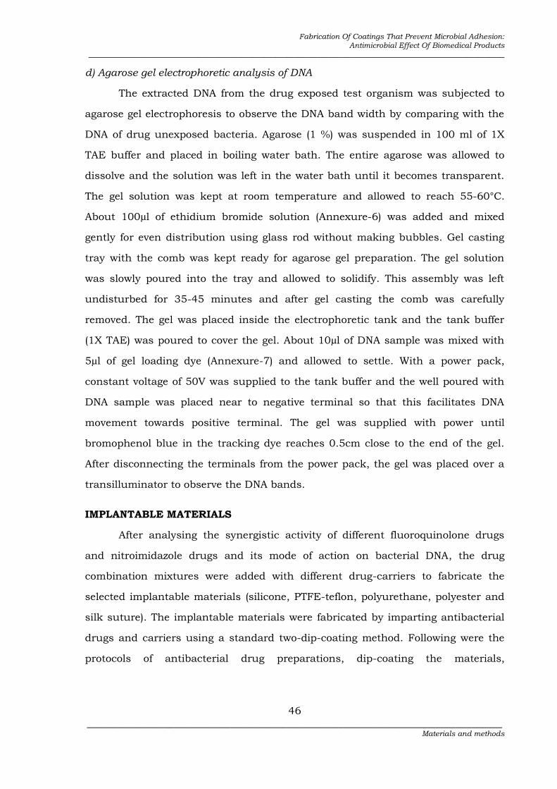
To assess antimicrobial combinations in vitro the checkerboard method was
selected. In this technique by using agar dilution method, the concentrations
tested for each antimicrobial agent were typically ranged from four or five
below the expected MIC to twice the anticipated MIC as in the 45 degree line
in Figure- 2 (each square represents one plate).
Figure-2: Checkerboard model to determine synergism of two drugs
(The picture was adapted from Qaziasgar and Kermanshahi, 2008)
Bottom row: To determine MIC of Drug-A, Left column: to determine MIC of drug-B, Centre: MIC of Drugs A + B

Fabrication Of Coatings That Prevent Microbial Adhesion: Antimicrobial Effect Of Biomedical Products
_______________________________________________________________________________________________________________
43 _________________________________________________________________________________
Materials and methods
The predetermined concentrations (µg/ml) used for this method were 0.015,
0.03, 0.06, 0.12, 0.25, 0.5, 1.0 and 2.0. According to figure-2, the plates in the left
hand column was used for the predetermined concentrations of first drug
(fluoroquinolone), the plates in bottom row was used for second drug
(nitroimidazole) and the plates in the 45 degree line was used for mixed drug
combinations. In all the arranged plates, 1ml of predetermined dilutions of the
antimicrobial agents were added with sterile and molten Muller-Hinton agar.
Then the surface of each plate was inoculated with 1 X 10⁴ CFU/spot of bacteria.
After 16-20 hours incubation at 37 ºC, the plates were examined for evidence of
visible growth. Experimental set up was made for all the drug combinations
(norfloxacin-metronidazole, ofloxacin-ornidazole and ciprofloxacin-tinidazole) in
triplicate.
3.3.2. Evaluating the synergism between fluoroquinolone and nitroimidazole
drugs by fractional inhibitory concentration index (Bharadwaj et al., 2003)
Fractional inhibitory concentration index (FICI) was calculated by using the
following equation.
Formula to determine synergy
FIC index = FICA + FICB
MICA in combination FICA= ................................ MICA
MICB in combination FICB= ............................... MICB
where A was the minimal inhibitory concentration (MIC) of drug A in a plate that
was the lowest inhibitory concentration in its row, and B was the MIC of drug
B in a plate that was the lowest inhibitory concentration in its column. MICAB
was the lowest inhibitory concentration of drug A and B in combination in the 45
degree line. With this method, synergism has traditionally been defined as an FIC
index of 0.5 or less and partial synergy as a FIC index of >0.5 - ≤1.0; antagonism
has been defined as a FIC index of ≥2.0.

Fabrication Of Coatings That Prevent Microbial Adhesion: Antimicrobial Effect Of Biomedical Products
_______________________________________________________________________________________________________________
44 _________________________________________________________________________________
Materials and methods
Interpretation:
Mean FICI ≤ 0.5 → Synergy, (p< 0.5),
Mean FICI >0.5 - ≤1.0→ Partial synergy, (p> 0.5)
Mean FICI ≥ 2.0→ Antagonism
Synergy: Synergistic action of a combination of antibiotics is present if the effect of
the combination exceeds the additive effects of the individual components.
In simple terms, synergism is defined as the ability of a pair of drugs to produce a
more rapid rate of bactericidal action within the first 24 hours of exposure than
either member of the pair alone, and killing of great numbers of bacteria that could
be expected from simple summation of single drug effects.
Partial synergy: The additive effect of combination is one in which the effect of
combination is equal to that of the sum of the effects of the individual components.
3.4. Determining the mode of action of fluoroquinolone and nitroimidazole
drugs on bacterial DNA (Rajendran et al., 2011)
Fluoroquinolone drug and nitroimidazole drug inhibits the DNA replicative
enzymes of anaerobic and facultative anaerobic bacteria. To confirm this mode of
action, the test organisms were exposed to the synergistic drugs under optimum
conditions. Two groups of similar test organisms were selected. One group of the
representative test organisms [Staphylococcus epidermidis (ATCC 35984) and
Escherichia coli (ATCC 43894)] were exposed to the synergistic drugs. 100 ml of 18
hours test cultures were treated with 1 % each of the synergistic drug for 3 hours.
Another group of similar organisms was cultured separately without exposing to
the drugs. Drug exposed test organisms and unexposed test organisms were then
processed to extract DNA. DNA samples of both the samples were electrophoresed
to observe the difference in width of DNA bands under UV light to determine the
mode of action of synergistic drugs. Following were the steps involved in DNA
extraction and electrophoresis.
a) Culturing the test bacteria for DNA extraction
About 50 ml of LB broth was prepared and inoculated each of the
representative test organisms separately (S. epidermidis (ATCC 35984) as gram-

Fabrication Of Coatings That Prevent Microbial Adhesion: Antimicrobial Effect Of Biomedical Products
_______________________________________________________________________________________________________________
45 _________________________________________________________________________________
Materials and methods
positive representative and E. coli (ATCC 43894) as gram-negative representative
organisms). Cultures were incubated in the appropriate condition (37 °C for 12
hours). Dispensed 5 ml of the culture separately and centrifuged at 8,000 rpm for
15 minutes. The pellets were washed in normal saline twice and then suspended
in 2ml normal saline buffer.
b) Exposing the test bacteria with synergistic drugs
Norfloxacin-metronidazole (D1) and ofloxacin-ornidazole (D2) drug
combinations prepared at 1mg/ml were added to the bacterial pellet suspension
and incubated overnight at 37°C. Another set of bacterial pellet suspension was
handled in parallel without the addition of drugs (test organisms unexposed to
drugs)
c) Extraction of DNA from drug exposed and unexposed test bacteria
About 2ml of drug-exposed test bacterial culture was taken after overnight
incubation. Both aerobic and anaerobic culture suspensions were centrifuged at
10,000 rpm for 15 minutes to pelletize the cell mass. The supernatant was
removed and the cell pellet was incubated on ice till next use. To the obtained
pellet 500µl of solution-A (Annexure-3) was added and gently mixed to make a
uniform cell suspension. The cell suspension was incubated on ice for 30 minutes.
To the mixture 100µl of freshly prepared solution-B (Annexure-4) was added and
vortexed well. The suspension was incubated in ice for 5 minutes (cells get lysed
and the solution becomes clear). About 750µl of solution-C (Annexure-5) was
added and vortexed for 2 min. The suspension was kept in ice for 60 minutes
(chromosomal DNA and cell material will precipitate into whole viscous clump).
The tubes were centrifuged at 6000 rpm for 10 minute at 4°C. Transferred the
supernatant to fresh eppendorf tube and centrifuged at 10,000 rpm for 10 minute
at 4°C. The supernatant was discarded and to the pellet, 50 µl of ice cold ethanol
was added along the sides of the tube. The solvent used was removed immediately
without disturbing the pellet. The tubes were blot dried using blotting paper by
inverting the tube over it. The pellets of DNA were stored by adding 100µl of 1X
TAE buffer and placed in a deep freezer. Similar procedure was carried out for
drug-unexposed DNA samples of test organism.
Fabrication Of Coatings That Prevent Microbial Adhesion: Antimicrobial Effect Of Biomedical Products
_______________________________________________________________________________________________________________
46 _________________________________________________________________________________
Materials and methods
d) Agarose gel electrophoretic analysis of DNA
The extracted DNA from the drug exposed test organism was subjected to
agarose gel electrophoresis to observe the DNA band width by comparing with the
DNA of drug unexposed bacteria. Agarose (1 %) was suspended in 100 ml of 1X
TAE buffer and placed in boiling water bath. The entire agarose was allowed to
dissolve and the solution was left in the water bath until it becomes transparent.
The gel solution was kept at room temperature and allowed to reach 55-60°C.
About 100µl of ethidium bromide solution (Annexure-6) was added and mixed
gently for even distribution using glass rod without making bubbles. Gel casting
tray with the comb was kept ready for agarose gel preparation. The gel solution
was slowly poured into the tray and allowed to solidify. This assembly was left
undisturbed for 35-45 minutes and after gel casting the comb was carefully
removed. The gel was placed inside the electrophoretic tank and the tank buffer
(1X TAE) was poured to cover the gel. About 10µl of DNA sample was mixed with
5µl of gel loading dye (Annexure-7) and allowed to settle. With a power pack,
constant voltage of 50V was supplied to the tank buffer and the well poured with
DNA sample was placed near to negative terminal so that this facilitates DNA
movement towards positive terminal. The gel was supplied with power until
bromophenol blue in the tracking dye reaches 0.5cm close to the end of the gel.
After disconnecting the terminals from the power pack, the gel was placed over a
transilluminator to observe the DNA bands.
IMPLANTABLE MATERIALS
After analysing the synergistic activity of different fluoroquinolone drugs
and nitroimidazole drugs and its mode of action on bacterial DNA, the drug
combination mixtures were added with different drug-carriers to fabricate the
selected implantable materials (silicone, PTFE-teflon, polyurethane, polyester and
silk suture). The implantable materials were fabricated by imparting antibacterial
drugs and carriers using a standard two-dip-coating method. Following were the
protocols of antibacterial drug preparations, dip-coating the materials,

Fabrication Of Coatings That Prevent Microbial Adhesion: Antimicrobial Effect Of Biomedical Products
_______________________________________________________________________________________________________________
47 _________________________________________________________________________________
Materials and methods
antibacterial activity of the drug coated materials and its durability
determinations.
3.5. Preparation of antibacterial drugs for coating the implantable
materials (Gollwitzer et al., 2003)
The antibacterial drugs for coating the implantable materials were prepared
using a solvent-casting technique. Each carrier (beta-cyclodextrin, DL-lactic acid,
tocopherol acetate) was mixed with each of the synergistic drugs (a fluoroquinolone
drug and a nitroimidazole drug) by the following procedure. All the carriers were
selected based on their biological properties like; enhancement of antibacterial
activity; provides sustained and constant release of drugs, food and medical grade
polymers.
a) Beta-cyclodextrin + synergistic drugs
Carrier, 650 mg of β-Cyclodextrin was added in 5 ml of 0.1N NaOH at a
concentration of 130 mg/ml. To the solvent carrier mixture, Norfloxacin and
Metronidazole or Ofloxacin and Ornidazole or Ciprofloxacin and Tinidazole were
added to attain a final concentration of 1%.
b) DL-Lactic acid + synergistic drugs
Carrier, 0.78 ml (650mg) (1.21 ml = 1g) of DLLA was added in 4.22 ml of
0.1N sodium hydroxide (NaOH) at a concentration of 130 mg/ml. To the solvent
carrier mixture, Norfloxacin and Metronidazole or Ofloxacin and Ornidazole or
Ciprofloxacin and Tinidazole were added to attain a final concentration of 1%.
c) Tocopherol acetate + synergistic drugs
Carrier, 0.84 ml (650mg) (1.4 ml = 1g) of Tocopherol acetate was added in
4.16 ml of 0.1N NaOH at a concentration of 130 mg/ml. To the solvent carrier
mixture, Norfloxacin and Metronidazole or Ofloxacin and Ornidazole or
Ciprofloxacin and Tinidazole were added to attain a final concentration of 1%.
The test was carried out in such a way that, all the three combinations of
synergistic drugs were mixed individually with all the three types of carriers. Thus
drug and carrier combinations were D1C1, D1C2, D1C3, D2C1, D2C2, D2C3 and D3C1,
D3C2, D3C3. In these combinations, norfloxacin-metronidazole was indicated as

Fabrication Of Coatings That Prevent Microbial Adhesion: Antimicrobial Effect Of Biomedical Products
_______________________________________________________________________________________________________________
48 _________________________________________________________________________________
Materials and methods
(D1), ofloxacin-ornidazole as (D2), ciprofloxacin-tinidazole as (D3) and beta-
cyclodextrin as (C1), DL-Lactic acid as (C2) and tocopherol acetate as (C3).
3.6. Coating the implantable materials with prepared antibacterial drugs
using the standard two-dip-coating method (Matl et al., 2008)
All the implantable materials (Silicone, Polyurethane, PTFE, Polyester and
Silk Suture) were subjected for coatings in 3 different groups (Group-A, B and C).
The materials in Group-A was coated with synergistic drugs and carriers. Materials
in Group-B were coated using synergistic drugs without carriers and the materials
in Group-C were coated using carriers without drugs. The surface
functionalization of the implantable materials was carried out using a standard
two-dip-coating method. The coated materials in Group-A, B and C were referred
below as drug-carrier coated (dcc), drug-coated (dc) and carrier-coated (cc). Uncoated
(uc) implantable materials were also used in parallel to differentiate from other 3
groups of materials.
All the materials were cut according to the type of experimental needs and
also based on the standard references. Silicone was cut in two different sizes (5
mm disc for antibacterial assay and 1 cm disc pieces for FTIR analysis).
Polyurethane of 1 cm in length was selected for antibacterial assay. PTFE and
polyester of 1 cm2 pieces was selected for antibacterial assay and Silk suture with
length of 2.5 cm was cut to determine the antibacterial activity.
All the materials were autoclaved at 121°C for 15 minutes before coating.
Sterile materials were coated separately with drugs, carriers and drug-carrier
conjugates. The dip-coating procedure was carried out in sterile glass beakers on a
shaker (120 rpm) for 3 hours, with a drying period of about 15 minutes between
the two coating procedures. The coated materials were rinsed in phosphate
buffered saline (PBS) to remove surface accumulation of particles, followed by
drying at room temperature. All coating steps were carried out under aseptic
conditions in a laminar airflow hood. The weight of each coating i.e., the amount of
the drug incorporated, was assessed by the difference in the weight of the test
material before and after the coating procedure.

Fabrication Of Coatings That Prevent Microbial Adhesion: Antimicrobial Effect Of Biomedical Products
_______________________________________________________________________________________________________________
49 _________________________________________________________________________________
Materials and methods
3.7. Determining the drug-add on percentage on the implantable materials.
Weight determination of coated materials (Shanmugasundaram et al., 2011)
Add on concentration of antibacterial drugs on each implantable materials
were calculated using a standard formula of drug add on percentage. After coating
with antibacterial drugs all the implantable materials were subjected to determine
the weight. To measure the drug concentration, the materials were weighed using
Schimadzu weighing balance before and after coating. To ensure the coatings on
the implantable surfaces each material was weighed separately. The average
weight of the samples was assessed by weighing each sample three times. The
increase in weight after coating with drugs was determined in percentage.
Drug add on (%) = [(W1 – W2)/W2] x 100
Where,
W1 = weight of the material after coating
W2 = weight of the material before coating
3.8. Assessing the qualitative antibacterial activity of dip-coated implantable
materials (El-rehewy et al., 2009)
The method was performed for analysing the antibacterial activity of
different implant materials after dip-coating with synergistic antimicrobial drugs
and carriers. In this qualitative method the pre-measured size (Section 3.6) of all
sterilized implant materials were tested from each preparation [drug-carrier coated
(dcc), carrier-coated (cc) and uncoated (uc) implantable materials]. The materials
were all rinsed twice in phosphate buffered saline (Annexure-9) before testing to
remove any surface accumulation of drug. All implant materials were placed on the
surface of Mueller-Hinton agar plate which had previously been seeded with an
overnight broth culture of the test organisms and incubated at 37°C for 24 to 48
hours. In the case of anaerobes, the seeded media were incubated anaerobically in
McIntosh field jar at 37°C for 24 to 48 hours. The experiment was carried out in
triplicate. Antibacterial activity was expressed as the diameter of the zone of

Fabrication Of Coatings That Prevent Microbial Adhesion: Antimicrobial Effect Of Biomedical Products
_______________________________________________________________________________________________________________
50 _________________________________________________________________________________
Materials and methods
inhibition. From the qualitative analysis, for each implantable material, atleast
three drug carrier combinations were selected for the in vitro drug release analysis.
3.9. Analysing the drug release profiles from coated implantable materials
using the standard in vitro dissolution method (Mashru and Saikumar, 2010)
The release profile of synergistic drugs from the drug-carrier coated (dcc) and
drug coated (dc) implantable materials was studied by in vitro dissolution method.
This test was performed in triplicate to determine the sustained release of
synergistic antimicrobial drugs from the drug-carrier coated implantable materials.
The concentration of each drug released from the implant materials were
measured using the calibration curves constructed from standards of each drug.
3.9.1. Development of standard calibration curve of antibacterial drugs
A stock solution of Norfloxacin, Metronidazole, Ofloxacin, Ornidazole,
Ciprofloxacin and Tinidazole (100 µg/ml) was prepared by adding 10mg of each
drug in 100ml of phosphate buffer (pH - 6.8). From the stock, series of standards
(10µg to 200µg) were prepared and the absorbance was measured to evaluate the
concentration of drug. The absorbance was plotted against the respective
concentration to obtain the calibration curves. The absorbance of known drug
concentration was measured spectrophotometrically using UV-VIS
spectrophotometer at their respective wavelengths (Norfloxacin - 300 nm,
Metronidazole - 320 nm, Ofloxacin - 293.4 nm, Ornidazole - 319.6 nm,
Ciprofloxacin - 277 nm, Tinidazole - 240 nm).
3.9.2. Determining the drug release concentrations from (drug coated and
drug-carrier coated materials using in vitro dissolution method
Drug release from drug-carrier coated (dcc) and drug coated (dc) implantable
materials was studied in 50 ml phosphate buffer (pH - 6.8) kept at 300 rpm in a
thermomixer. The release profiles of each of the drugs were evaluated
spectrophotometrically using UV-VIS spectrophotometer. Absorbance was
measured for 4 ml of the sample solution at their respective wave lengths against
Phosphate buffered saline as a blank. Fresh medium of the same volume was

Fabrication Of Coatings That Prevent Microbial Adhesion: Antimicrobial Effect Of Biomedical Products
_______________________________________________________________________________________________________________
51 _________________________________________________________________________________
Materials and methods
replaced each time after measuring absorbance. The release profile of the drugs
was determined from 30 minutes to 120 hours (30 min, 1, 2, 4, 8, 12, 24, 48, 72,
96 120 hours) using UV-Vis spectrophotometer. Specific drug-carrier combinations
for each type of implantable materials showing sustained release of the drugs were
alone selected for further parameters like bacterial adherence test and in vitro
challenge test.
3.10. Quantitative antibacterial activity of coated materials using the
standard bacterial Adherence test (El-Rehewy et al., 2009)
Antibacterial activity for each implantable material coated with the selected
drug-carrier combinations were quantitatively analysed using bacterial adherence
test (specific drug-carrier combination for each material was selected after in vitro
dissolution method).
The effect of dip-coated implantable materials (drug-carrier coated - dcc) was
tested against the test organism using a standard bacterial adherence test. The
materials were placed separately in a tube with 5 ml of each of the test bacteria
and incubated at 37 °C for 18 h. During the incubation period the bacterial cells
adhere on the surface of the implantable materials. After incubation, the numbers
of viable adherent cells were determined as follow: Implantable materials were
collected aseptically and washed in a sterile test tube of 10 ml normal saline to
remove the non-adherent cells. The washed pieces were transferred each in 10 ml
fresh sterile saline, and sonicated for 30 seconds to dislodge the sessile adherent
cells using an ultra-sonicator. After sonication, serial dilution of the sonicated
saline was made and the number of sessile bacteria which indicates degree of
adherence was determined by viable count technique. Similar experimental set up
was run in parallel for uncoated (uc) and carrier coated (cc) materials. The
difference in number of adhered cells on dcc, and cc were determined statistically
using chi square analysis.
The percentage reduction of adhered organisms on the drug-carrier coated
materials was determined using a standard percentage reduction formula.

Fabrication Of Coatings That Prevent Microbial Adhesion: Antimicrobial Effect Of Biomedical Products
_______________________________________________________________________________________________________________
52 _________________________________________________________________________________
Materials and methods
Bacterial reduction (%) = A – B/A X 100
Where,
A = number of adhered organisms (in CFU) obtained from the uc materials
B = number of adhered organisms (in CFU) obtained from the dcc or cc materials
3.10.1 Statistical analysis of total viable bacteria on coated materials
Chi-square non parametric test using SPSS-9 for Windows 7 was used as a
statistical tool to determine the effect of antimicrobial drug on bacterial adherence.
The hypothesis selected (H0) was that “There is significant effect of antimicrobial
drug on the test organisms”. The difference in the bacterial reduction percentage
between the drug-carrier coated (dcc) implantable materials and the carrier-coated
(cc) implantable materials were statistically calculated with P<0.05 considered
significant.
3.11. Determining the persistence of drugs on coated materials using the
standard in vitro challenge test (Bayston et al., 2009)
The In vitro challenge (IVC) test was designed to determine the ability of dip-
coated implantable materials to resist bacterial colonization with multiple
challenges in flow conditions. In vitro challenge test was performed in triplicate to
determine the persistence of the most effective antimicrobial drug during the
implantation period. Briefly, the pre-measured size (Section 3.6) of each material
coated with the selected antimicrobial drug combination was used for the study.
The drug-carrier coated (dcc) implantable materials were inserted into a controlled-
environment chamber containing pre-sterilized nutrient broth. The broth
containing dcc implantable material was inoculated with the test organism and
incubated in a shaker incubator (80 rpm) at 37°C. A challenge dose (1 ml
overnight culture) of each test organism was inoculated into the broth. Similar set
up was made for the uncoated implantable materials as control. All the materials
were examined visually each day and samples of the medium were collected
periodically for culture. If no colonisation was detected after 7 days incubation, a

Fabrication Of Coatings That Prevent Microbial Adhesion: Antimicrobial Effect Of Biomedical Products
_______________________________________________________________________________________________________________
53 _________________________________________________________________________________
Materials and methods
second challenge dose was administered. In the case of absence of turbidity after
the second challenge dose, further challenge dose was administered at 7 days
interval until turbidity was observed.
NON-IMPLANTABLE MATERIALS
Plain weave cotton and cotton blend polyester (C/P) woven fabrics (mostly
used in woven surgical gowns, patients, physicians and nurse uniforms);
polyamide and viscose (routinely used in several applications like medical gauze,
diapers, wet wipes, facial mask and incontinence products) were selected as non-
implantable materials in the present study. The specifications of cotton and C/P
fabrics were given in table-6. Polyamide and Viscose with the following
specifications, weight of 50 g/m2 (GSM), white coloured and width of 100-2100
mm was selected.
Table-6: Specification of cotton and C/P fabrics
S. No Particulars Cotton C/P
1 Count – Warp Weft
40s
40s 2/40s
2/40s
2 Ends/Inch (EPI) 60 60
3 Picks/Inch (PPI) 56 56
4 Weave Plain Plain
3.12. Different antibacterial finishing methods on non-implantable textile
materials
Two standard methods used in textile applications were selected in this
research to impart antibacterial drugs on non-implantable materials. In the first
method, standard reactive dye method was used to impart the reactive drugs onto
textile materials. The method was a standard protocol handled in textile industries
to impart reactive dye colours onto textiles. Secondly, microencapsulation method
was used to increase the wash durability properties in finished textile materials.
Following were the protocols of two standard methods used to fabricate the non-
implantable test materials for antibacterial finish and its durability
determinations.

Fabrication Of Coatings That Prevent Microbial Adhesion: Antimicrobial Effect Of Biomedical Products
_______________________________________________________________________________________________________________
54 _________________________________________________________________________________
Materials and methods
3.12.1. Direct attachment of synergistic drugs onto textile materials using
the standard reactive dye method (Chun and Gamble, 2007)
The approach in this study was to modify two well described antibacterial
drugs (fluoroquinolone and nitroimidazole compounds) for direct attachment to
textile fabrics.
a) Synthesis of reactive synergistic drugs
To convert the drug reactive and imparting to non-implantable materials,
the method was performed. Synthesis of reactive fluoroquinolone drug was
accomplished by suspending 2 % each of the drugs (norfloxacin, ofloxacin,
ciprofloxacin) in 20 ml deionized water in an ice bath at 5 °C. To this suspension,
0.04 M cyanuric chloride was added to convert the drug reactive. The suspension
was maintained at 5 °C during the drop-wise addition of 0.04 M NaOH. Synthesis
of reactive nitroimidazole drug was similarly prepared suspending 2 % each of the
drugs (metronidazole, ornidazole, tinidazole) in 20 ml deionized water in an ice
bath at 5 °C. To this suspension, 0.03 M cyanuric chloride was added to convert
nitroimidazole drug reactive. The suspension was maintained at 5 °C during the
drop-wise addition of 0.03 M NaOH.
b) Exhaust dyeing method to bind reactive antibacterial agents to textile
materials
An exhaust dyeing method was used to bind the synthesized reactive
antibiotic to the non-implantable textile fabric. The dye bath was prepared by
adding 0.5 ml of Triton-X-100, 75 g of sodium sulfate, and 6.5 g of the reactive
antibacterial drug, or 3.25 g of each of the two reactive antibacterial drugs (a
fluoroquinolone and a nitroimidazole drug) to 1.2 L of deionized water. To the
suspension each of the carriers (beta-cyclodextrin, DL-lactic acid and tocopherol
acetate) was added at a concentration of 2 %. Three, 20 g squares of the test fabric
(cotton) were submerged in the dye bath heated to 60 °C. After 30 min of
incubation, 12 g NaOH that had been dissolved in 100 ml of deionized water was
added. The temperature was then raised to 80 °C, and the fabrics heated for
another 30 min. The fabric was then rinsed in deionized water and heated for 10

Fabrication Of Coatings That Prevent Microbial Adhesion: Antimicrobial Effect Of Biomedical Products
_______________________________________________________________________________________________________________
55 _________________________________________________________________________________
Materials and methods
min at 80 °C in deionized water, then rinsed and kept in a convection oven at 105
°C until dried. Similar procedure was carried out for all the test fabrics (C/P,
polyamide and viscose).
3.12.2. Determining the durability of drugs using the standard
microencapsulation method (Sathianarayanan et al., 2010)
Fabrics treated with microencapsulated antibacterial agents were reported
to be durable for few number of wash cycles compared to other methods. Hence, in
the present study the test materials were treated with encapsulated drugs by
adding it as core material and acacia powder as wall material.
a) Synthesis of microencapsulated synergistic drugs
About 10 grams of acacia powder was allowed to swell for 15 min in 100ml
of hot water. To this mixture, 50 ml of hot water was added and stirred for 15 min
maintaining the temperature between 40 °C and 50 °C. Around 2% of core-
material (a fluoroquinolone drug and a nitroimidazole drug) was slowly added
under stirring condition. Stirring was continued for another 15 min and then 10
ml of 20% sodium sulphate was added. To this suspension, 2 % of a carrier (beta-
cyclodextrin, DL-lactic acid, or tocopherol acetate) and 2 % of citric acid
(crosslinker) was added. The stirrer was stopped and the mixture was freeze-dried
overnight to develop microcapsules.
b) Characterization of microcapsules
The developed microcapsules were observed under conventional light
microscope to determine its shape and outer structure. The method was also used
to identify the particle size and its homogenous distribution.
c) Binding microencapsulated drugs with textile materials
Each of the fabric samples (cotton, cotton-polyester, polyamide and viscose)
was immersed in the microcapsule solution and padded through a pneumatic
padding mangle at a pressure of 3 psi to get a wet pick up of 100% on weight of
fabric. The treated fabric was dried at 80°C for 5 min and cured at 150°C for 5

Fabrication Of Coatings That Prevent Microbial Adhesion: Antimicrobial Effect Of Biomedical Products
_______________________________________________________________________________________________________________
56 _________________________________________________________________________________
Materials and methods
min. Similar experiment was carried out for each material separately, treated with
synergistic drugs (D1, D2 or D3) coupled with each carriers (C1, C2 or C3).
3.13. Evaluating the antibacterial activity of treated fabrics using the
standard AATCC Test Method 100 – 2004.
The antibacterial properties of materials can be studied by quantitative
(AATCC-100) test methods. Quantitative test is the proper indicator of degree of
antibacterial activity when the antibacterial agents are fixed on to the textile
material or are unable to leach out. The different tests carried out in this study
were based on such consideration.
Assay for antibacterial properties of textile materials (AATCC Method 100-2004)
All the treated fabrics and untreated fabrics by both the methods (reactive
dye and microencapsulation method) were subjected to antibacterial assay. The
assay used for measuring antibacterial properties was based on the AATCC Test
Method 100-2004. Briefly, 1.0 ml of 12 hours challenge bacterial inoculum was
dispersed as droplets over the 3 swatches (test fabrics) using a micropipette. The
swatches were inoculated in pre-sterilized 250 ml Erlenmeyer flasks. After all the
samples were inoculated, the flasks were incubated at 37 ± 2 °C for 18 h before
being assayed for bacterial population density. The bacterial population density
was determined by extracting the bacteria from the fabric by adding 100 ml of
distilled water to each flask and shaken using an orbital shaker for 1 min. Then
aliquots were serially diluted and pour plated to determine the bacterial density.
The difference in number of viable bacteria was evaluated on the basis of the
percentage reduction. Percentage reduction was calculated using the following
formula.
R = (A-B) / A X 100
Where, R is percentage reduction; A is the number of bacteria in the broth
inoculated with treated test fabric sample immediately after inoculation i.e., at zero
contact time and B is the number of bacteria recovered from the broth inoculated
with treated test fabric sample after the desired contact period of 18 hours.

Fabrication Of Coatings That Prevent Microbial Adhesion: Antimicrobial Effect Of Biomedical Products
_______________________________________________________________________________________________________________
57 _________________________________________________________________________________
Materials and methods
3.14. Determining the wash durable properties of treated textile materials
using the standard wash fastness test (AATCC Test Method 124-2010)
Assay for antibacterial properties of after wash materials (AATCC Method 100-2004)
All the treated fabrics and untreated fabrics by both the methods (reactive
dye and microencapsulation method) were subjected to wash fastness test. The
fabric was washed based on the AATCC Test Method 124-1996 laundering
procedure to see if the drug would persist through multiple washings. The treated
samples were washed 2, 5, 10 and 15 times. After each wash, antibacterial activity
of each fabric were quantitatively assayed (AATCC Method 100-2004) using the
method described in Section – 3.13.
3.14.1. Statistical analysis of wash fastness
Statistical analysis of viable bacteria on encapsulated drug treated textiles after 15th
wash
Chi-square non parametric test using SPSS-19 for Windows-7 was selected
as a statistical tool to determine the effect of antimicrobial drug on the number of
viable bacteria at 18th hour after 15th wash. The hypothesis selected (H0) was that
“There is significant effect of antibacterial drug on the test organisms”. The
difference in the number of viable bacteria after ‘0’ contact time and 18 hours
contact time for the encapsulated drug treated fabrics (after 15th wash) were
statistically calculated with P<0.05 considered significant.
3.15. Analysing the physical properties of textile materials after antibacterial
finishing
Different physical properties of reactive drug treated and encapsulated drug
treated textile materials were analysed and compared with the normal untreated
materials. The methods were performed to determine whether the normal physical
properties of the textile materials were unaffected after the treatment of
antibacterial drugs.
3.15.1. Tensile strength test (ASTM D 5035-2006 test method)
Tensile strength is the measure of the resistance of the fabric tensile load or
stress in either warp or weft direction. It is the strength shown by a specimen

Fabrication Of Coatings That Prevent Microbial Adhesion: Antimicrobial Effect Of Biomedical Products
_______________________________________________________________________________________________________________
58 _________________________________________________________________________________
Materials and methods
subjected to tension as distinct from torsion, compression, or shear. Elongation
defines the length to which a fibre may stretch before breaking. A sample of 12” X
2” was taken for the test. The tensile strength of the fabric was determined by
cloth tensile strength tester. Tensile strength is performed using cut strip method.
This test is used for treated or heavily sized fabrics. Three readings for every
sample were taken and the average was calculated.
3.15.2. Abrasion test (AATCC 119-2004 test method)
Abrasion test determined the ability of a fabric to withstand damage by
friction. This is dependent on the fineness of the fiber, the amount of twist of the
yarn and the weave structure of the fabric. Yarns that have a firmer and tighter
twist were generally more resistant to abrasion. The fabric specimen was mounted
over a foam rubber cushion and rubbed multi-directionally against a wire screen
mounted on a weighted head. Abrasion was carried out for 75 revolutions for the
fabrics. The initial and final weight of the fabric was noted. Four readings for every
sample were taken and the average was calculated.
Abrasion resistance % = weight before abrasion – weight after abrasion X 100
Weight before abrasion
3.15.3. Resistance to Pilling
A piece of fabric measuring 5 inch x 5 inch is stitched so as to be a firm fit
when placed round a rubber tube 6 inch long, 1¼ inch outside diameter and 1.8
inch thick. The cut ends of the fabric (9 inch x 9 inch x 9 inches) lined with cork
1.8 inch thick. The instrument, I.C.I. Pill box tester was used for this method. The
pre-measured size of the test fabric was then rotated at 60 rpm for 5 hours. For
fabrics that are normally subjected to repeat washing as well as to wear, washing
may be done prior to sample preparation. After the test was over, the extent of
pilling was assessed visually by comparison with the arbitrary standards 1, 2, 3, 4
and 5. Under the test conditions, fabrics of standard 1 become hairy but do not
pill, fabrics of standard 2 become hairy and pills lightly, while fabrics of standard 3
become hairy and pill more severely, fabrics of standard 4 refers to good and
fabrics of standard 5 refers to excellent.

Fabrication Of Coatings That Prevent Microbial Adhesion: Antimicrobial Effect Of Biomedical Products
_______________________________________________________________________________________________________________
59 _________________________________________________________________________________
Materials and methods
3.15.4. Dimensional stability
Length and width bench marks were made on a woven fabric specimen. The
materials were then washed and dried followed by re-measuring the bench marks
distance. The laundering process was repeated for the indicator cycle and the
dimensional change was calculated from the measurement.
Materials used to determine the dimensional stability of the textile materials
Cylindrical wash wheel of the reversing type, flat-bed press (at a
temperature not less than 135 °C), dryer of the rotary type, hand iron, measuring
device and ATCC detergent.
Method
The samples measuring the size of 60cm X 60cm for each textile materials
were placed in the washing machine with specified level of water. 66 ± 1 gm of
standard reference detergent was added with preselected washing temperature.
The specimen was dried by flat-bed dry method. After washing and drying,
materials were pressed and hand ironed at temperature of 120 0C - 180 0C. For
dimensional stability, the distance between each part of benchmarks to the
nearest millimetre were measured using the following formula
(B –A) Dimensional change = -------------- X 100 A Where,
A - Average of three original measurements for the length wise (or) width wise
direction in the specimen.
B - Average of three measurements after cycle completed for the lengthwise (or)
width wise direction of the specimen.
3.15.5. Air- permeability test (ASTM D 737-96 test method)
Air permeability of a fabric is the volume of air measured in cubic cm
passed per second through 1 sq. cm for the fabric at a pressure of one cm. head of
water. Air permeability can be measured using an instrument called Shirley Air
Permeability Tester. Air permeability was determined in accordance with Test
Method ASTM D-737-96. The conditioned specimens in the standard atmosphere
for testing textiles, 21 ± 1°C and 65 ± 2 % relative humidity was tested unless

Fabrication Of Coatings That Prevent Microbial Adhesion: Antimicrobial Effect Of Biomedical Products
_______________________________________________________________________________________________________________
60 _________________________________________________________________________________
Materials and methods
otherwise specified in a material specification or contract order. The test
specimens were carefully handled to avoid altering the natural state of the
material. Placed each test specimen onto the test head of the test instrument, and
performed the test as specified in the manufacturer’s operating instructions. Read
and recorded the individual test results in SI units as cm3/s/cm2.
3.16. Surface characterization of coated biomedical materials
The surface properties of biomedical materials before and after coating with
antibacterial drugs were characterized using two standard parameters, Fourier
transform infra-red spectroscopy (FTIR) and scanning electron microscopy (SEM).
Silicone and cotton alone were selected as a representative material for
implantable and non-implantable material. Both the material was subjected for
experimentation physically and chemically. Chemical analysis was made using
FTIR and physical characterization was analysed by SEM.
3.16.1. Determining the presence of drugs and its chemical interventions on
coated biomaterials using FTIR analysis (Coates, 2000).
The alteration in the functional groups of test materials (silicone and cotton)
due to the addition of drugs and carriers was determined chemically using FTIR
spectroscopy. The FTIR absorption spectra of the implantable material (silicone),
non-implantable material (cotton), pure drugs (ofloxacin, ornidazole), carriers (DL-
lactic acid and beta-cyclodextrin) and drug-carrier coated silicone and cotton were
recorded in the range of 400-4000 cm-1 by KBr disc method. FTIR spectra of the
samples were determined using Shimadzu FTIR spectrophotometer. All the
samples were prepared in KBr discs with a hydrostatic press at a force of
5.2 τ cm-2 for 3 minutes to reduce the moisture content on the disc surface. Each
disc was dried under radiation to remove excess moisture content.
3.16.2. Examining the homogenous coatings on biomaterials using
topographic analysis – SEM (Matl et al., 2008; Rajendran et al., 2012)
The surface coatings of the drugs and carriers on implantable (silicone) and
non-implantable materials (cotton) were observed using Scanning electron

Fabrication Of Coatings That Prevent Microbial Adhesion: Antimicrobial Effect Of Biomedical Products
_______________________________________________________________________________________________________________
61 _________________________________________________________________________________
Materials and methods
microscopy (SEM). SEM evaluation was also used to know the uniformity of
coating of finishing chemicals over the specimen. The topographic analysis of
coated and uncoated test materials was prepared for SEM using a suitable
accelerating voltage (10 KV), vacuum (below 5 Pa) and magnification (X 3500).
Metal coating was used as the conducting material to analyse the sample.
3.17. Determining the tissue reactions of coated biomedical materials using
the standard HET-CAM test (Luepke, 1985 and Valdes et al., 2002)
Hen´s Egg Test on the Chorioallantoic Membrane (HET-CAM) of chick eggs
To understand the inflammatory tissue reactions of coated biomedical
materials on the live tissues, the materials were placed on the surface of chorio-
allantoic membrane (CAM) of embryonated chick eggs. The inflammatory response
on CAM was evaluated by direct evaluation method and histological method.
a) Fertile eggs used in the study
Freshly laid fertile eggs were collected from the chicken farm and incubated
at 36-37° C for 8 days before implanting the sample materials (drug-carrier coated
and uncoated implantable and non-implantable). During the incubation time, the
eggs were turned twice daily.
b) Egg windowing
On the day of implantation, the eggs were candled to determine the position
of the air sac and the embryo. A square, with sides approximately 18-20 mm, was
marked on the shell where the chorio-allantoic membrane was best developed.
Areas with large blood vessels were avoided to obviate possible haemorrhage.
Using a dental drill fitted with a straight hand-piece the sides of the marked
square were drilled, taking care not to pierce the underlying shell membrane. In
one corner of this large triangle a second smaller square was drilled, with sides of
approximately 5 mm. A small slit was drilled in the shell over the air sac. A
mixture of molten paraffin wax and vaseline was painted over the drilled surfaces
to prevent fragments of shell from falling on to the membrane when it was later
exposed.

Fabrication Of Coatings That Prevent Microbial Adhesion: Antimicrobial Effect Of Biomedical Products
_______________________________________________________________________________________________________________
62 _________________________________________________________________________________
Materials and methods
c) Application of test material, positive and negative control on CAM
Aseptic technique was used for the implantation of the test material. For
dropping the material onto chorio-allantoic membrane the egg was mounted on a
stand, with the drilled area of shell uppermost; a straight Hagedorn's needle was
gently inserted under one corner of the smaller square of shell and this square was
raised and removed. The shell and shell membrane circumscribed by the larger
square were then removed, and the sterile pre-measured size of sample
implantable materials and non-implantable materials (drug-carrier coated
materials placed on CAM of separate eggs) were inserted and carefully lowered on
to the exposed membrane. Inorder to compare the inflammatory and non-
inflammatory patterns of the implanted test material, 0.3 ml of the substance
(positive and negative control) was applied to the surface of the CAM. 0.1 N NaOH
was added on the CAM of separate egg as a positive control and 0.9 % NaCl was
used as an appropriate negative control. After a 20-second exposure period, the
CAM is rinsed with 5 ml of water.
3.17.1 Direct evaluation of CAM (Luepke, 1985)
The CAM was evaluated for development of irritant endpoints like
hyperemia (increased blood flow), hemorrhage (blood from a ruptured vessel), and
coagulation (presence of blood clots)
a) Time to Development of Observed Endpoints after Exposure to the Test
Substance
A procedure used to evaluate the time to development of endpoints after
exposure to the test substance was to continually observe the CAM during the 5-
minute observation period and record (typically in seconds) the time at which each
of the endpoints developed. Therefore, three separate time values were obtained
and recorded for each egg (one time value for each endpoint). Individual values for
the observed endpoints were then used to determine the irritation potential of the
test substance.

Fabrication Of Coatings That Prevent Microbial Adhesion: Antimicrobial Effect Of Biomedical Products
_______________________________________________________________________________________________________________
63 _________________________________________________________________________________
Materials and methods
b) Irritation Scores (IS)
A majority of the test method protocols calculated a score (referred to as
irritation score, irritation index, or irritation potential) that represented the
irritation potential of the test substance based on endpoint development. This
score could be determined by a mathematical model which was discussed below.
c) IS calculation – IS [B] analysis method
The time taken for the development of each endpoints, hyperemia,
hemorrhage and coagulation was substituted in the standard formula as per IS [B]
analysis method. The time values assigned/obtained to each endpoint were
totalled to give an overall IS value for the test substance. An IS score could be
calculated using the following general formula,
Where, Hemorrhage time = time (in seconds) of the first appearance of blood
hemorrhages, Lysis time = time (in seconds) of the first appearance of vessel lysis
Coagulation time = time (in seconds) of first appearance of protein coagulation
d) Relationship of scores with category of irritation
After calculating the IS scores for the developed endpoints, the totalled
score was compared to identify the category of irritation caused by the test
materials on CAM. In Table-6 the final IS value ranged from 0 (for test substances
that do not induce development of any of the observed endpoints) to 21 (for test
substances that induce development of all three endpoints within 30 seconds of
application of the test substance) was presented. The relationship between scores
and category of irritation was tabulated below.
Table-7: Relationship of scores with category of irritation
Scores on HET-CAM Category of irritation
0 – 0.9 No irritation
1 – 4.9 Weak or slight irritation
5 – 8.9 Moderate irritation
9 - 21 Strong or severe irritation
From Cazedey et al., (2009)

Fabrication Of Coatings That Prevent Microbial Adhesion: Antimicrobial Effect Of Biomedical Products
_______________________________________________________________________________________________________________
64 _________________________________________________________________________________
Materials and methods
3.17.2 Histological evaluation of CAM (Valdes et al., 2002)
a) Incubation of eggs with test materials for microscopic analysis
After calculating the initial irritation scores of the test materials by
comparing with the scores of positive and negative controls, another set of similar
test materials were reimplanted on CAM of fresh eggs. The opening in the shell was
sealed with heated adhesive tape and the hole over the air sac region sealed with
molten paraffin wax. The eggs were then incubated at 37°C for 9 days, without
turning.
At the end of this period, the chorio-allantoic membrane was exposed by
cutting around the long circumference of the egg with a pair of curved scissors; the
chorio-allantoic membrane remained in the top half of the shell, together with the
implantable sample material. The sample material and the underlying portion of
thickened membrane were particularly dissected out. The membrane was gently
eased out of the shell using forceps and placed in a dish containing 10 % formal-
saline solution. After 24 hours fixation, implants were teased away from
underlying CAM which were then trimmed, paraffin embedded and prepared for
staining by using haematoxylin and eosin staining method. Prepared specimen
was observed for the inflammatory response on the CAM at the implanted site
under bright field microscopy.
b) Haematoxylin and eosin staining
The paraffin section was cut (4 μm to 8 μm thick) and placed on a labelled
slide. The slides were placed in a manual staining rack and dried in a 60 °C oven
for 30 to 40 minutes. Then the slides were stained manually using the following
reagent bath. Xylene was poured on the specimen and allowed to withstand for 4
min. The step is repeated twice using fresh xylene. 100% alcohol was added and
kept for 3 min (repeated thrice using fresh alcohol). The slide was then rinsed
using tap water. Haematoxylin stain was added and kept for 5 min. Again the slide
was rinsed using tap water. Acid water (0.5% conc HCl aqueous solution) was
added as haematoxylin differentiators and allowed to stand for 10 seconds
(repeated for 5 to 10 times). The slides were then rinsed with tap water and added
5% aqueous eosin for 3 min. Then slides were dipped in 95% and 100% ethanol

Fabrication Of Coatings That Prevent Microbial Adhesion: Antimicrobial Effect Of Biomedical Products
_______________________________________________________________________________________________________________
65 _________________________________________________________________________________
Materials and methods
solution for 2 times (repeated 4 times with fresh ethanol). Finally xylene was added
on to the slides for 10 seconds (repeated thrice with fresh xylene). Stained
specimens were covered with a glass coverslip before obtaining the microscopic
image.
c) Interpretation of the stains on tissue cells (Gamble and Wilson, 2002)
Haematoxylin
Haematoxylin is a very versatile stain and can be used to demonstrate many
different tissue components in a highly selective way. The type of mordant used
alters the specificity and colour of the stain. It stains the nucleus of the cell,
specifically, the chromatin within the nucleus and the nuclear membrane. The
active ingredient in haematoxylin solution is hematein complexed with mordant,
aluminium potassium sulphate (potash alum). In acid solutions, the alum dye
lakes are quite soluble and have a strong red colour. In alkaline conditions, the
dye lakes are less soluble and have a strong blue colour. The dyeing bath is
usually acidified and once staining is complete the section is rinsed in an alkaline
solution. In hard water areas, the tap water is alkaline and simply rinsing in tap
water will ‘blue’ the section.
Eosin
It stains nearly everything that haematoxylin will not stain. Eosin is a very
good cytoplasmic stain as it gives several shades to the tissue. The range of shades
can be extended even further if more than one dye is used in the solution. It
produces three different hues which can be used to differentiate various tissue
elements; like, red blood cells stain dark reddish orange, collagen stains a lighter
pastel pink and smooth muscle stains bright pink.