
Methods for surgical implantation of
87
Transcript of Methods for surgical implantation of
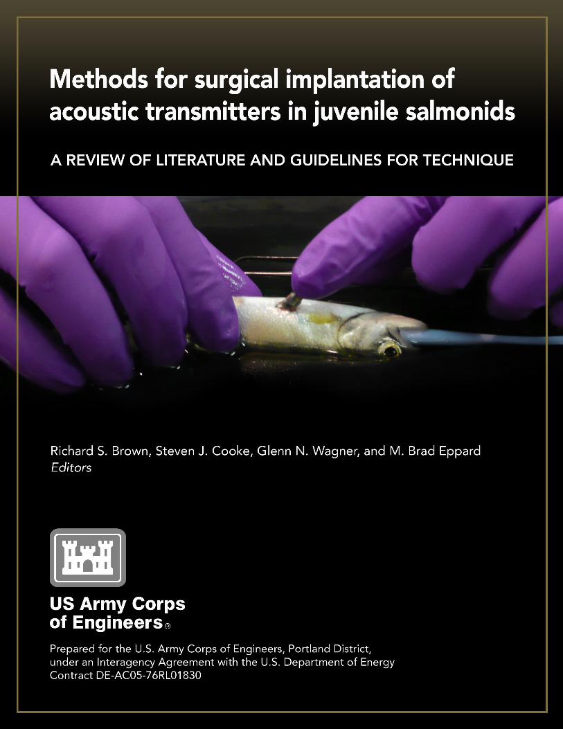
Methods for surgical implantation of acoustic transmitters in juvenile salmonids A review of literature and guidelines for technique
Edited by
Richard S. Brown Ecology Group Pacific Northwest National Laboratory Steven J. Cooke Institute of Environmental Science and Department of Biology Carleton University Glenn N. Wagner Rescan Environmental Services Ltd. M. Brad Eppard Portland District U.S. Army Corps of Engineers Prepared for the U.S. Army Corps of Engineers, Portland District, under an Interagency Agreement with the U.S. Department of Energy Contract DE-AC05-76RL01830

iii
Contents
Contributors ........................................................................................................................... vii Acknowledgments ................................................................................................................. ix 1 Introduction ................................................................................................................... 1 Richard S. Brown and M. Brad Eppard 2 Surgical Training ........................................................................................................... 3 Steven J. Cooke, Glenn N. Wagner, Richard S. Brown, and Katherine A. Deters 3 Pre- and Post-Surgical Holding ..................................................................................... 21 Eric W. Oldenburg and Richard S. Brown 4 Water Conditioners ........................................................................................................ 33 Ryan A Harnish and Richard S. Brown 5 Anesthesia ..................................................................................................................... 39 Kathleen M. Carter, Christa M. Woodley, and Richard S. Brown 6 Incision Closure and Surgery ........................................................................................ 53 Glenn N. Wagner, Steven J. Cooke, Richard S. Brown, and Katherine A. Deters Appendix – Common Errors in Surgical Technique ............................................................. A.1 Steven J. Cooke, Glenn N. Wagner, Richard S. Brown, and Katherine A. Deters

v
Figures
3.1 Upper panel is oxygen consumption of juvenile coho salmon sampled at hourly intervals beginning 1 h before and ending 5 h after the imposition of an acute handling stressor ............................................................................................. 25
3.2 Mean plasma cortisol and glucose concentrations in juvenile Chinook salmon
subjected to one or more 30-s handling stress, spaced 3 h apart ................................... 26 3.3 Plasma cortisol concentration in juvenile Chinook salmon subjected to
moderate confinement in a small live-cage or unconfined .......................................... 26 5.1 The changes in the partial pressure of oxygen and the partial pressure of
carbon dioxide in the blood of rainbow trout while in various stages of anesthesia and recovery using 100 mg/L TMS ............................................................. 44
6.1 Two common types of surgical needle used to suture wounds: a reverse
cutting needle and a tapered needle .............................................................................. 59 6.2 Simple interrupted suture pattern used to close surgical incisions in fish..................... 60 6.3 Two typical knots used to tie sutures ............................................................................ 62 6.4 Two surgeon’s knots used by Deters et al. (in press) .................................................... 62
Tables
2.1 Examples of correlations between surgical skills and surgery outcomes in medicine ........................................................................................................................ 5
2.2 Summary of differences in surgery outcomes between novice and expert
surgeons for largemouth bass tagged with dummy radio transmitters .......................... 6 2.3 Methods of learning surgical procedures on fish identified as most effective
for training future fish surgeons .................................................................................... 9 2.4 Essential competences of the fish surgeon .................................................................... 12 2.5 Sample schedule for proposed 3-day surgical training ................................................. 15 3.1 Time until complete gastric evacuation at varying water temperatures ........................ 23 3.2 Surgical holding-related stressors and associated recovery times ................................ 27 4.1 Partial list of commercially available water additives that claim to provide
mucus layer protection .................................................................................................. 35

vi
5.1 Stages of anesthesia ...................................................................................................... 41
6.1 Suture properties and needle types used to close abdominal incisions in salmonid parr and smolts. ............................................................................................. 58
vii
Contributors
Richard S. Brown: Pacific Northwest National Laboratory, Ecology Group, MSIN K6-85,
Richland, Washington 99352, USA; [email protected]
Kathleen M. Carter: Pacific Northwest National Laboratory, Ecology Group, MSIN K6-85,
Richland, Washington 99352, USA; [email protected]
Steven J. Cooke: Carleton University, Institute of Environmental Science and Department of
Biology, 1125 Colonel By Drive, Ottawa, Ontario K1S 5B6, Canada; [email protected]
Katherine A. Deters: Pacific Northwest National Laboratory, Ecology Group, MSIN K6-85,
Richland, Washington 99352, USA; [email protected]
M. Brad Eppard: U.S. Army Corps of Engineers, Portland District, 333 S.W. First Avenue,
Portland, Oregon 97204, USA; [email protected]
Ryan A Harnish: Pacific Northwest National Laboratory, Ecology Group, MSIN K6-85,
Richland, Washington 99352, USA; [email protected]
Eric W. Oldenburg: Pacific Northwest National Laboratory, Ecology Group, MSIN K6-85,
Richland, Washington 99352, USA; [email protected]
Glenn N. Wagner: Rescan Environmental Services Ltd., 1111 West Hastings Street, Vancouver,
British Columbia V6E 2J3, Canada; [email protected]
Christa M. Woodley: Pacific Northwest National Laboratory, Ecology Group, MSIN K6-85,
Richland, Washington 99352, USA; [email protected]


ix
Acknowledgments
The idea for this document emerged from a meeting held in 2007 to discuss standardization of protocols among fisheries researchers doing work in the Columbia Basin. We would like to acknowledge the participants in this meeting, including Geoff McMichael, Michelle Rub, James Hughes, Josh Murauskas, Carl Schilt, Dave Robichaud, Anita Blakley, Leah Brown, Kelly Kiyohara, Theresa Liedke, Noah Adams, Eric Hockersmith, Gordon Axel, and Lynn McComas.

1
1 Introduction
Richard S. Brown and M. Brad Eppard
Telemetry has been used for decades to gain a better understanding of the behavior and survival of free-swimming fish. To optimize research using this technology, investigators should have a thorough understanding of the techniques used to implant transmitters and all of the activities associated with implantation. Hart and Summerfelt (1975) provided the first detailed account of surgical techniques for the implantation of telemetry transmitters into the peritoneal cavity of fish. Since this seminal report, numerous research studies have been conducted, dedicated to improving techniques used to implant transmitters in fish (for reviews, see Summerfelt and Smith 1990: Harms and Lewbart 2000; Jepsen et al. 2002; Mulcahy 2003; Cooke and Wagner 2004; Wagner and Cooke 2005).
Despite the extent of research that has been performed on surgical implantation of transmitters into fish, there still are differences among researchers in the techniques used. This is the case even among groups conducting research on juvenile salmonids within the Columbia Basin. A lack of consistency in implantation techniques among studies can make the comparison of results between studies more difficult. A single set of guidelines based on scientific research currently is lacking but necessary to standardize the surgical techniques and improve the comparison of results for studies in the Columbia Basin. Therefore, the purpose of this document is to provide background and methodology on the surgical implantation of acoustic transmitters into juvenile salmon. The goal of the U.S. Army Corps of Engineers (USACE) is to have a standard protocol for surgical implantation of acoustic transmitters to be followed for all USACE research projects.
A thorough review of the literature has been completed in an effort to provide a scientific background on surgical implantation of acoustic transmitters. The goal of this review is to supply researchers with the reasons why certain procedures should be used instead of simply providing a list of procedures. However, research performed in several areas of fish surgical implantation has been insufficient to allow researchers to base their methodology on empirical science. Therefore, some techniques used for implantation of acoustic transmitters often are based on recommendations from veterinarians or from years of experience by fisheries researchers.
This document clearly delineates the difference between methods based on these two different sources. When a scientific basis is not available for a certain surgical method, suggestions are provided for future research that could provide one. Where possible, data for juvenile salmonids are used. In most cases, however, we relied on data from studies on a wide range of marine and freshwater fish species.
2
References
Cooke SJ, Wagner GN (2004) Training, experience, and opinions of researchers who use surgical techniques to implant telemetry devices into fish. Fisheries 29(12):10-18
Harms CA, Lewbart GA (2000) Surgery in fish. In: Bennett RA (ed) Veterinary clinics of North America: exotic animal practice. Saunders, New York, pp 759-774
Hart LG, Summerfelt RC (1975) Surgical procedures for implanting ultrasonic transmitters into flathead catfish (Pylodictis olivaris). Transactions of the American Fisheries Society 104:56-59
Jepsen N, Koed A, Thorstad EB, Baras E (2002) Surgical implantation of telemetry transmitters in fish: how much have we learned? Hydrobiologia 483:239-248
Mulcahy DM (2003) Surgical implantation of transmitters into fish. ILAR Journal 44:295-306
Summerfelt RC, Smith L (1990) Anesthesia, surgery, and related techniques. In: Schreck CB, Moyle PB (eds) Methods for fish biology. American Fisheries Society, Bethesda, MD, pp 213-272
Wagner GN, Cooke SJ (2005) Methodological approaches and opinions of researchers involved in the surgical implantation of telemetry transmitters in fish. Journal of Aquatic Animal Health 17:160–169.
3
2 Surgical Training
Steven J. Cooke, Glenn N. Wagner, Richard S. Brown, and Katherine A. Deters
Background
Biotelemetry has quickly become a key tool for fisheries professionals in recent years. Each year, tens of thousands of radio and acoustic transmitters are surgically implanted in the intraperitoneal cavity of fish for remote telemetry purposes. Nowhere are more transmitters deployed than in the Columbia Basin of the Pacific Northwest. Among some studies in the Columbia Basin, usually those associated with hydropower infrastructure and operations, thousands of telemetry transmitters per study are deployed over a several-week period (e.g., Keefer et al. 2004; Schreck et al. 2006; Caudill et al. 2007; Naughton et al. 2007). With multiple research organizations (e.g., U.S. Fish and Wildlife Service, Pacific Northwest National Laboratory, U.S. Geological Survey, National Marine Fisheries Service, state natural resource agencies), academic institutions, tribal organizations, and environmental consultants each implanting telemetry devices into fish, there is a great need for consistency in terms of the surgical procedures. Given that many of these studies are conducted to comply with regulatory requirements, the data they generate can undergo intense scrutiny, including legal challenges. Therefore, there is a long-standing interest in ensuring that surgical procedures and the presence of the telemetry device do not alter fish condition, behavior, or survival. Consistent surgical procedures also would improve comparisons among years of the same study and between different studies. Currently, these comparisons are unreliable.
Here we discuss the topic of training individuals to become qualified to surgically implant intraperitoneal acoustic telemetry transmitters into salmon smolts. However, the material is equally applicable to other fish species and life stages as well as to different biotelemetry or biologging devices. The importance of training
Training is a fundamental part of all scientific and technical disciplines. This is particularly true for surgeons, whether working on humans or other animals. For surgical procedures, a number of skills are necessary to reduce the likelihood for mistakes. Trainees must be provided with the most extensive yet standardized set of problem-solving skills and technical skills to deal with challenges that can arise. In medical and veterinary contexts, surgeons also must operate under a legal framework. Failure to achieve a specific outcome for a patient, particularly if based on negligence, can result in malpractice litigation. When dealing with fish surgery to implant biotelemetry transmitters, however, the context is somewhat different.
In fisheries-related research, the primary legislative bodies responsible for outcomes and approving procedures are the institutional animal care and use committees (often referred to as IACUCs in the United States). Although once limited to higher vertebrates in academic settings,
4
IACUCs now cover all vertebrates (including fish) and extend toward many state and federal government agencies, reflecting increased interest and concern for animal welfare (DeTolla et al. 1995; see discussion in Mulcahy 2003a; Huntingford et al. 2006). Although IACUCs wield little true legal power, they can withhold research funding and levy academic misconduct charges to ensure compliance with their guidelines or decisions. For some agencies and organizations, project funding cannot be released until approval has been granted by an IACUC (note that this varies between agencies). Prior to being granted IACUC permission to conduct a procedure such as transmitter implantation on a fish, the applicant (or those who will be involved in the procedures) must be able to document proficiency. However, compliance typically is obtained by simply participating in generic animal care training delivered by the IACUC on topics such as ethics and animal welfare. Rarely are there training materials or performance evaluations specific to fish surgery. In some cases, individual IACUCs have attempted to regulate or standardize procedures for fisheries research (Borski and Hodson 2003). However, because most fish telemetry research is not performed by veterinarians and occurs under field conditions, it has been difficult to develop guidelines that are useful and appropriate for fisheries scientists (Mulcahy 2003a, 2003b). Many of the existing documents have been written by veterinarians (e.g., Stoskopf 1993a, 1993b; Harms and Lewbart 2000; Mulcahy 2003a). These sources provide important veterinary rigor, but that rigor is not always transferable to wild fish in field environments (Stoskopf 2003).
Furthermore, there seems to be much variation among the standards employed by different agencies, jurisdictions, and employers. Professional fisheries societies, including the American Fisheries Society (AFS), American Institute of Fisheries Research Biologists (AIFRB), the American Society of Ichthyologists and Herpetologists (ASIH), and the Fisheries Society of the British Isles (FSBI), have developed guidelines intended to improve the welfare of fish used in research (ASIH et al. 1987, 1988; FSBI 2002; AFS et al. 2004). These general guidelines include sections on surgical implantation of telemetry transmitters, but, as Mulcahy (2003a) noted, there is very little detailed or standardized information concerning the development of guidelines for training and regulation of fish surgery. This paucity of information is particularly surprising, considering documented cases of negative consequences arising from surgery on fish (see Bridger and Booth 2003; Mulcahy 2003b; Welch et al. 2007) and the presence of information from studies having empirically tested fish surgical techniques (see reviews by Jepsen et al. 2002; Mulcahy 2003b; Wagner and Cooke 2005). The relationship among training, experience, and outcome: perspectives from medical and veterinary sciences
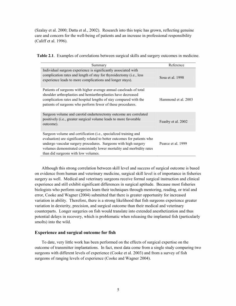
In medical and veterinary science, training of the surgeon and the volume of procedures conducted have been deemed important in the outcome of surgical procedures (Califf et al. 1996). An expanding body of literature suggests that despite receiving formal surgical instruction and clinical experience, veterinarians and physicians still exhibit significant differences in surgical aptitude (Sosa et al. 1998; Freund et al. 1999). Research in medical and veterinary science has shown that novice medical surgeons have reduced dexterity compared to more experienced surgeons. The range of dexterity affects not only the accuracy of the suture placement and the degree of suture holding but also the time required to complete the surgery (Annett 1971; Engelhorn 1997). It should not be surprising that surgical skills, including manual dexterity, have been shown to be strongly correlated to the outcome of medical procedures (see Table 2.1)
5
(Szalay et al. 2000; Datta et al., 2002). Research into this topic has grown, reflecting genuine care and concern for the well-being of patients and an increase in professional responsibility (Califf et al. 1996).
Table 2.1. Examples of correlations between surgical skills and surgery outcomes in medicine.
Summary Reference Individual surgeon experience is significantly associated with complication rates and length of stay for thyroidectomy (i.e., less experience leads to more complications and longer stays).
Sosa et al. 1998
Patients of surgeons with higher average annual caseloads of total shoulder arthroplasties and hemiarthroplasties have decreased complication rates and hospital lengths of stay compared with the patients of surgeons who perform fewer of these procedures.
Hammond et al. 2003
Surgeon volume and carotid endarterectomy outcome are correlated positively (i.e., greater surgical volume leads to more favorable outcome).
Feasby et al. 2002
Surgeon volume and certification (i.e., specialized training and evaluation) are significantly related to better outcomes for patients who undergo vascular surgery procedures. Surgeons with high surgery volumes demonstrated consistently lower mortality and morbidity rates than did surgeons with low volumes.
Pearce et al. 1999
Although this strong correlation between skill level and success of surgical outcome is based on evidence from human and veterinary medicine, surgical skill level is of importance in fisheries surgery as well. Medical and veterinary surgeons receive formal surgical instruction and clinical experience and still exhibit significant differences in surgical aptitude. Because most fisheries biologists who perform surgeries learn their techniques through mentoring, reading, or trial and error, Cooke and Wagner (2004) submitted that there is greater opportunity for increased variation in ability. Therefore, there is a strong likelihood that fish surgeons experience greater variation in dexterity, precision, and surgical outcome than their medical and veterinary counterparts. Longer surgeries on fish would translate into extended anesthetization and thus potential delays in recovery, which is problematic when releasing the implanted fish (particularly smolts) into the wild. Experience and surgical outcome for fish
To date, very little work has been performed on the effects of surgical expertise on the outcome of transmitter implantations. In fact, most data come from a single study comparing two surgeons with different levels of experience (Cooke et al. 2003) and from a survey of fish surgeons of ranging levels of experience (Cooke and Wagner 2004).
6
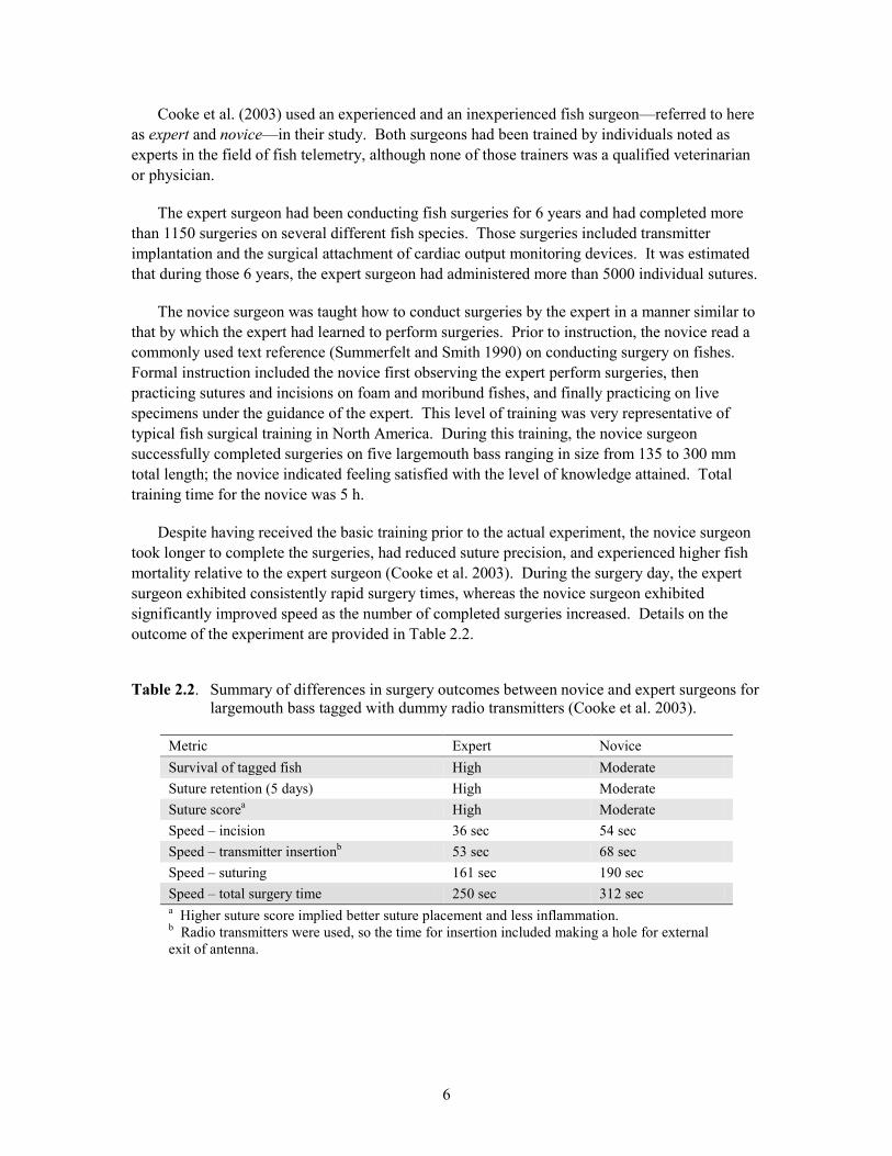
Cooke et al. (2003) used an experienced and an inexperienced fish surgeon—referred to here as expert and novice—in their study. Both surgeons had been trained by individuals noted as experts in the field of fish telemetry, although none of those trainers was a qualified veterinarian or physician.
The expert surgeon had been conducting fish surgeries for 6 years and had completed more than 1150 surgeries on several different fish species. Those surgeries included transmitter implantation and the surgical attachment of cardiac output monitoring devices. It was estimated that during those 6 years, the expert surgeon had administered more than 5000 individual sutures.
The novice surgeon was taught how to conduct surgeries by the expert in a manner similar to that by which the expert had learned to perform surgeries. Prior to instruction, the novice read a commonly used text reference (Summerfelt and Smith 1990) on conducting surgery on fishes. Formal instruction included the novice first observing the expert perform surgeries, then practicing sutures and incisions on foam and moribund fishes, and finally practicing on live specimens under the guidance of the expert. This level of training was very representative of typical fish surgical training in North America. During this training, the novice surgeon successfully completed surgeries on five largemouth bass ranging in size from 135 to 300 mm total length; the novice indicated feeling satisfied with the level of knowledge attained. Total training time for the novice was 5 h.
Despite having received the basic training prior to the actual experiment, the novice surgeon took longer to complete the surgeries, had reduced suture precision, and experienced higher fish mortality relative to the expert surgeon (Cooke et al. 2003). During the surgery day, the expert surgeon exhibited consistently rapid surgery times, whereas the novice surgeon exhibited significantly improved speed as the number of completed surgeries increased. Details on the outcome of the experiment are provided in Table 2.2.
Table 2.2. Summary of differences in surgery outcomes between novice and expert surgeons for largemouth bass tagged with dummy radio transmitters (Cooke et al. 2003).
Metric Expert Novice Survival of tagged fish High Moderate Suture retention (5 days) High Moderate Suture scorea High Moderate Speed – incision 36 sec 54 sec Speed – transmitter insertionb 53 sec 68 sec Speed – suturing 161 sec 190 sec Speed – total surgery time 250 sec 312 sec a Higher suture score implied better suture placement and less inflammation. b Radio transmitters were used, so the time for insertion included making a hole for external exit of antenna.
7
The importance of feedback in surgical outcome
The ability of surgeons to observe the surgical sites on their fish in the days and weeks following surgery provides immediate feedback, enabling surgeons to modify their procedures and conduct improved surgeries (Deters et al. in press). While evaluating different suture types for implanting acoustic transmitters into juvenile Chinook salmon, Deters and colleagues found differences in the surgical outcomes among four experienced surgeons (all who previously had implanted transmitters into hundreds or thousands of fish). Deters et al. (in press) concluded that the opportunity of two of those surgeons to monitor the healing progression of their surgeries over time during laboratory-based studies was beneficial to the surgical outcome of their fish. Surgeons who obtained feedback had higher suture retention, lower incision openness scores, and higher transmitter retention. This finding indicates that having a large quantity of surgical experience may be important, but a feedback process also is important so surgeons can see the outcomes of their surgeries and improve their techniques. Surgeons should receive feedback and observe the surgical sites externally (e.g., suture retention, incision condition, and tag loss). However, it may be more valuable to have the trainees perform necropsies on each of their practice fish to examine the internal condition (e.g., wound healing/annealing from inside-out, nicking/snagging or cutting of organs, depth of sutures). The state of training and evaluation in medical and veterinary science
Each medical and veterinary school seems to use different techniques for surgical training and evaluation. These differences may reflect the fact that surgical training has undergone many changes in the last decade (Bradley 2006). In particular, one trend, not yet uniformly accepted, is the development of surgical skills through different simulation techniques. Torkington et al. (2000) suggest that practicing surgical procedures on simulated human (or other animal) tissue, if perfect, could enable complete transfer of techniques learned in a skills laboratory directly to the operating theater. Simulation techniques currently used in human medicine include artificial tissues, animal models, and virtual reality computer simulation.
In fish surgery, one of the more technical aspects of the procedure is the suturing. This skill is important also in medical and veterinary practices. In a microsurgical department in Spain, students are advised to perform 1000 to 1500 microsurgical stitches (which represents around 40 to 50 h of practice) to attain expertise in microsuturing (Uson and Calles 2002). Although this high number initially was viewed as excessive, the instructors (through student feedback) agreed that the techniques could be mastered only after much practice. Uson and Calles (2002) surveyed their students, and 98% favored training first on nonliving models prior to switching to live animal models.
Starkes et al. (1998) also assessed suturing performance for 13 novice microsurgeons throughout a 4- to 5-day microsurgical training course. At the beginning and end of each training day, time to complete a suture (from needle insertion to completion of tie-off) was assessed on a standardized suture task using simulated tissue as well as actual tissue. An average learning curve for suturing performance on the standardized test was developed and demonstrated significant performance improvement in the suturing of actual tissue. Thus, the use of standardized tests appears to reflect actual suturing performance and to be sensitive to improvements in suturing skill that result from practice.
8
In a survey of all 31 veterinary schools in the United States and Canada, Bauer (1993) revealed that models were frequently used to teach suturing, general psychomotor skills, knot tying, and hemostasis. Indeed, globally there is a trend toward no longer using live animals for surgical technique training classes in veterinary science (Silva et al. 2007). Ideally, training would include a combination of core skills that are initiated on models and subsequently applied to living organisms. This training method is particularly relevant for fish research in which the surgeon is responsible also for handling, anesthetization, and recovery of the patient. The state of training in fish science
Currently, there are no standardized or official training methods or guides for the implantation of transmitters into fish; the majority of fish surgeons learn their craft from direct observation, mentoring, and the literature (Cooke and Wagner 2004). Conversely, Bauer (1993) reported that 37% of veterinary schools in North America used a standardized process to evaluate the surgical skills of students in the laboratory training environment prior to their transitioning to work on live animals. In a survey of fish surgeons, Cooke and Wagner (2004) revealed a lack of clear consensus on the need for international standards for fish telemetry surgery within the fish surgery community. However, many of the survey respondents stated that some minimum standards are needed to ensure that fish exposed to surgery have a reasonable chance of recovery and survival. The majority of respondents also stated they work in a jurisdiction or for an employer that does not require a minimal level of training or proficiency prior to conducting fish surgery. In the instances in which training was required, a veterinarian or other appropriate official within an IACUC often simply observed the surgical approaches used on fish, providing guidance and eventual approval to work independently. Some individuals surveyed by Cooke and Wagner (2004) indicated they were required to demonstrate competence in surgical technique and speed on nonliving specimens coupled with survival of trial organisms in the laboratory. However, government agencies in Europe (e.g., the UK Home Office) regulate surgical activity on all vertebrates, including fish, and in Iceland, the Fish Disease Officer must approve all fish surgeons. To our knowledge, similar levels of training or certification are not required in any jurisdiction in North America. Cooke and Wagner (2004) asked whether respondents had taken any university or college courses for credit that included instruction on surgical techniques. Only 12% of respondents participated in university-level course work that included such instruction, and of those, only one-half included experience focused specifically on fishes. The majority (88%) of respondents had not participated in any academic credit-based courses that included instruction on surgical techniques. Some schools with veterinary programs now include graduate courses involving surgery for research; these courses are relatively new and are unlikely to include modules or content on fishes. If there is a component on fish surgery, it would likely focus on tumors and general health assessment rather than transmitter implantation.
When Cooke and Wagner (2004) queried as to the potential of an Internet portal to serve as a resource for training surgeons, the results were mixed. Overall, more respondents disagreed (41.0%) than agreed (16.3%) that an Internet portal could provide enough information to train fish surgeons. Another 35.5% were neutral to the idea. Few respondents indicated they strongly disagreed or strongly agreed. Although the survey results reflected general apprehension to the idea, the Internet still could serve as a resource for communication among fish surgeons. An Internet portal could provide a venue for exchange of information on surgical techniques and
9
species-specific insights, as well as provide opportunities for less-experienced surgeons to identify and connect with potential mentors.
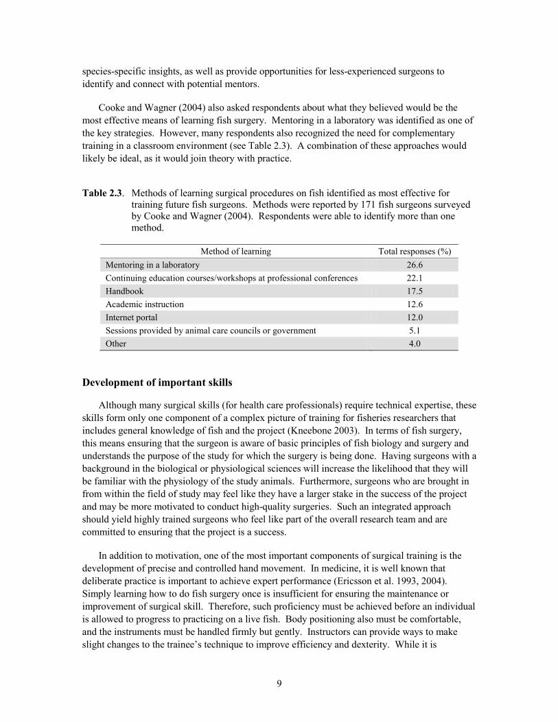
Cooke and Wagner (2004) also asked respondents about what they believed would be the most effective means of learning fish surgery. Mentoring in a laboratory was identified as one of the key strategies. However, many respondents also recognized the need for complementary training in a classroom environment (see Table 2.3). A combination of these approaches would likely be ideal, as it would join theory with practice.
Table 2.3. Methods of learning surgical procedures on fish identified as most effective for training future fish surgeons. Methods were reported by 171 fish surgeons surveyed by Cooke and Wagner (2004). Respondents were able to identify more than one method.
Method of learning Total responses (%) Mentoring in a laboratory 26.6 Continuing education courses/workshops at professional conferences 22.1 Handbook 17.5 Academic instruction 12.6 Internet portal 12.0 Sessions provided by animal care councils or government 5.1 Other 4.0
Development of important skills
Although many surgical skills (for health care professionals) require technical expertise, these skills form only one component of a complex picture of training for fisheries researchers that includes general knowledge of fish and the project (Kneebone 2003). In terms of fish surgery, this means ensuring that the surgeon is aware of basic principles of fish biology and surgery and understands the purpose of the study for which the surgery is being done. Having surgeons with a background in the biological or physiological sciences will increase the likelihood that they will be familiar with the physiology of the study animals. Furthermore, surgeons who are brought in from within the field of study may feel like they have a larger stake in the success of the project and may be more motivated to conduct high-quality surgeries. Such an integrated approach should yield highly trained surgeons who feel like part of the overall research team and are committed to ensuring that the project is a success.
In addition to motivation, one of the most important components of surgical training is the development of precise and controlled hand movement. In medicine, it is well known that deliberate practice is important to achieve expert performance (Ericsson et al. 1993, 2004). Simply learning how to do fish surgery once is insufficient for ensuring the maintenance or improvement of surgical skill. Therefore, such proficiency must be achieved before an individual is allowed to progress to practicing on a live fish. Body positioning also must be comfortable, and the instruments must be handled firmly but gently. Instructors can provide ways to make slight changes to the trainee’s technique to improve efficiency and dexterity. While it is
10
important to note that the surgery speed is not a competition, it is imperative that the surgery be done in the least amount of time possible to enable the fish to recover quickly. These steps are important for surgical procedures and to maintain the comfort of the surgeon across multiple surgeries often lasting hours to days. Evaluation of surgical experience
In medical and veterinary education, assessment of surgical skills is incorporated into the training program. Such assessment is rarely incorporated into fish surgery training, with the exception of a few countries. The medical professional competence standards developed by Epstein and Hundert (2002) state that evaluation of surgical ability and outcomes is important for several reasons. From the perspective of the trainee, evaluation provides useful feedback about individual strengths and weaknesses—feedback that guides future learning, fosters habits of self-reflection and self-remediation, and promotes access to advanced training. From the perspective of the curriculum and the training program, evaluation enables instructors to respond to lack of demonstrated competence, fosters course or curricular change, and certifies the competence of graduates. As well, there is the opportunity for self assessment of the need for additional practice, which can be relevant when switching between organisms or adapting to a change to the surgical procedure, or after long periods of surgical inactivity.
Suture practice in veterinary education is used to strengthen motor skills and increase confidence and efficiency (Smeak 1999), but it is difficult to determine at what point a fish surgeon has gained enough experience. Although an experienced fish surgeon has been shown to be consistently quicker, with smaller incisions and better suture placement than a novice (Cooke et al. 2003), the accuracy of suture placement in human subjects has been shown to improve with experience among already experienced surgeons (Seki 1987). Unfortunately, the majority (93%) of fish surgeons have not been formally tested or evaluated to determine their level of surgical proficiency (Cooke and Wagner 2004). Clearly this pattern needs to change in order to elevate the quality of fish surgery for the sake of fish welfare and the integrity of scientific research.
Feedback, or the ability to monitor the progression of surgical incision healing, can improve surgical outcome (Deters et al. in press). Surgeons who previously took part in research projects that involved post-surgery examination of study animals were associated with better surgical outcomes, such as higher suture and transmitter retention. To provide feedback within a surgery training program, we suggest that surgeons first be allowed to view examples of poor techniques that can manifest into significant problems several days or weeks later (examples are shown in the Appendix). As part of the surgeon evaluation, images of practice fish should be taken on the day of surgery and at other times within approximately 2 weeks of surgery, before the incision location begins to heal considerably. Images taken after 2 to 3 weeks post-surgery may not provide the surgeon with optimal feedback because problems associated with poor technique may be concealed by the advancement in healing, possibly leading to the false impression that no negative issues were associated with the surgical technique. For this reason, it is suggested that images be taken both immediately following surgery and during the first few weeks of the healing process (7 and 14 days after surgery, for example).
11
Delivery of fish surgery training
It is crucial that the individuals delivering the training are themselves experts in surgical procedures and fish care. Therefore, veterinary professionals should be involved in at least some aspects of training to ensure that basic surgical principles (related to tissue and tool handling and cleaning) are observed. This may be as simple as having the surgical instructor conduct a surgery in the presence of a veterinarian while asking for guidance on technique. Often, the IACUC panels that approve animal care permits include veterinarians who are happy to provide advice. However, it should be noted that most veterinarians are familiar with terrestrial animals, not fish. This difference in patient experience may lead to large variations in animal-handling and surgical techniques.
Veterinary involvement in training may help to avoid situations where incorrect surgical procedures are taught, such as using hands rather than surgical tools to drive suture needles, doing surgery with the entire fish out of water, and suggesting the use of surgical gloves is unnecessary. Such information learned at surgical workshops and seminars is dangerous because it can be further disseminated through word of mouth. Along with veterinary consultation, a benefit to the instruction team would be the presence of experienced fish surgeons who have worked on a number of species and are familiar with the latest advances in fish (and other wildlife) surgery. Defining attributes for fish surgeons
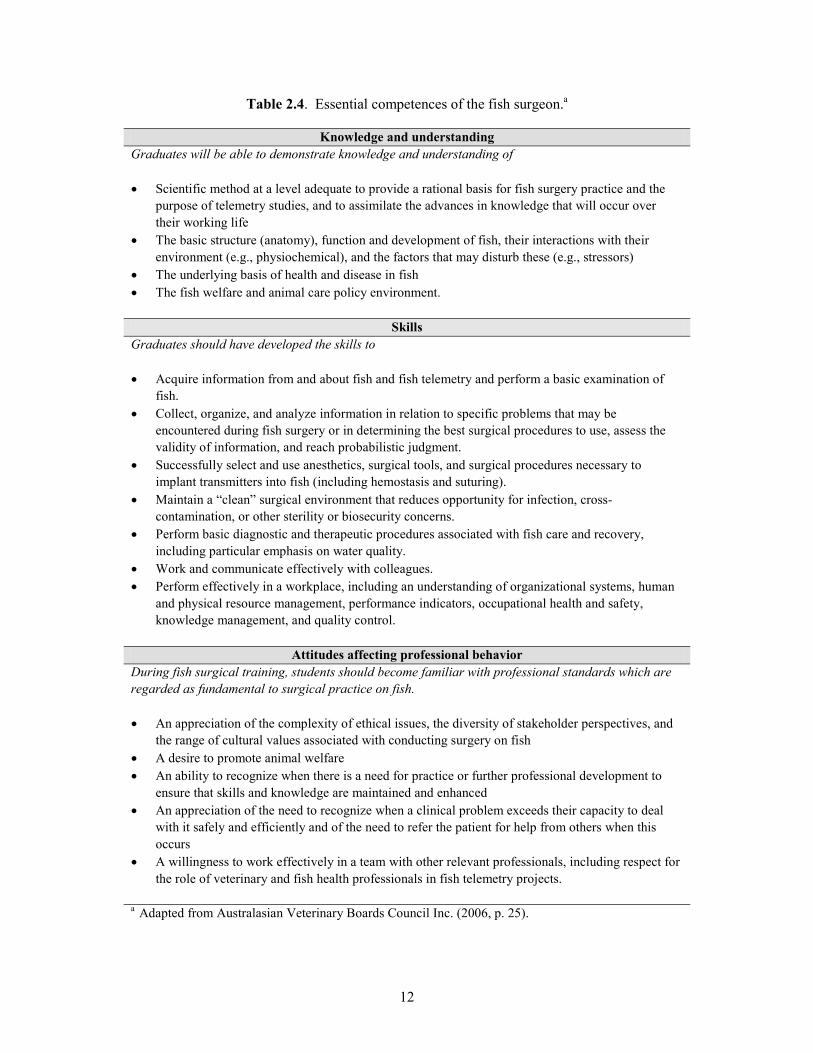
One way forward in fish surgical training is to identify a set of attributes related to knowledge, understanding, and skill that surgeons must demonstrate prior to engaging in fish surgery. Indeed, such an approach has been used to define a series of attributes that are expected of graduating veterinarians (Walsh et al. 2001; Zemljic 2004). Such an approach can be used to develop an outcomes assessment to evaluate whether fish surgeons are meeting these expectations (Walsh et al. 2002). Typically these outcomes assessments are performed by surveying recent graduates (Tinga et al. 2001), which could easily be adapted for fish surgeons. The three sets of attributes listed in Table 2.4 provide an example of the competences required of a surgeon implanting telemetry transmitters into fish. Proposed curriculum for surgical training related to fish
To date, we are unaware of any published curricula developed specifically for training surgeons to conduct intraperitoneal implantation of telemetry transmitters in fish. Here we provide a proposed 3-day curriculum for fish surgical training as a guide for instructors. Our hope is that this curriculum will be used to advance the area of surgical training for fish biologists. Included is a combination of classroom instruction with hands-on trials and mentoring using models and real animals.
To ensure that knowledge is maintained, each individual should be subjected to routine evaluation and provided with occasional refresher training. Furthermore, there should be some expectation that each trainee will engage in deliberate practice throughout his or her career.
12
Table 2.4. Essential competences of the fish surgeon.a
Knowledge and understanding Graduates will be able to demonstrate knowledge and understanding of • Scientific method at a level adequate to provide a rational basis for fish surgery practice and the
purpose of telemetry studies, and to assimilate the advances in knowledge that will occur over their working life
• The basic structure (anatomy), function and development of fish, their interactions with their environment (e.g., physiochemical), and the factors that may disturb these (e.g., stressors)
• The underlying basis of health and disease in fish • The fish welfare and animal care policy environment.
Skills Graduates should have developed the skills to • Acquire information from and about fish and fish telemetry and perform a basic examination of
fish. • Collect, organize, and analyze information in relation to specific problems that may be
encountered during fish surgery or in determining the best surgical procedures to use, assess the validity of information, and reach probabilistic judgment.
• Successfully select and use anesthetics, surgical tools, and surgical procedures necessary to implant transmitters into fish (including hemostasis and suturing).
• Maintain a “clean” surgical environment that reduces opportunity for infection, cross-contamination, or other sterility or biosecurity concerns.
• Perform basic diagnostic and therapeutic procedures associated with fish care and recovery, including particular emphasis on water quality.
• Work and communicate effectively with colleagues. • Perform effectively in a workplace, including an understanding of organizational systems, human
and physical resource management, performance indicators, occupational health and safety, knowledge management, and quality control.
Attitudes affecting professional behavior
During fish surgical training, students should become familiar with professional standards which are regarded as fundamental to surgical practice on fish. • An appreciation of the complexity of ethical issues, the diversity of stakeholder perspectives, and
the range of cultural values associated with conducting surgery on fish • A desire to promote animal welfare • An ability to recognize when there is a need for practice or further professional development to
ensure that skills and knowledge are maintained and enhanced • An appreciation of the need to recognize when a clinical problem exceeds their capacity to deal
with it safely and efficiently and of the need to refer the patient for help from others when this occurs
• A willingness to work effectively in a team with other relevant professionals, including respect for the role of veterinary and fish health professionals in fish telemetry projects.
a Adapted from Australasian Veterinary Boards Council Inc. (2006, p. 25).
13
Guiding principles
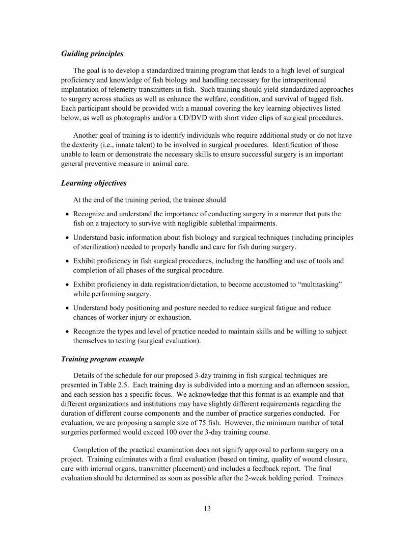
The goal is to develop a standardized training program that leads to a high level of surgical proficiency and knowledge of fish biology and handling necessary for the intraperitoneal implantation of telemetry transmitters in fish. Such training should yield standardized approaches to surgery across studies as well as enhance the welfare, condition, and survival of tagged fish. Each participant should be provided with a manual covering the key learning objectives listed below, as well as photographs and/or a CD/DVD with short video clips of surgical procedures.
Another goal of training is to identify individuals who require additional study or do not have the dexterity (i.e., innate talent) to be involved in surgical procedures. Identification of those unable to learn or demonstrate the necessary skills to ensure successful surgery is an important general preventive measure in animal care. Learning objectives
At the end of the training period, the trainee should
• Recognize and understand the importance of conducting surgery in a manner that puts the fish on a trajectory to survive with negligible sublethal impairments.
• Understand basic information about fish biology and surgical techniques (including principles of sterilization) needed to properly handle and care for fish during surgery.
• Exhibit proficiency in fish surgical procedures, including the handling and use of tools and completion of all phases of the surgical procedure.
• Exhibit proficiency in data registration/dictation, to become accustomed to “multitasking” while performing surgery.
• Understand body positioning and posture needed to reduce surgical fatigue and reduce chances of worker injury or exhaustion.
• Recognize the types and level of practice needed to maintain skills and be willing to subject themselves to testing (surgical evaluation).
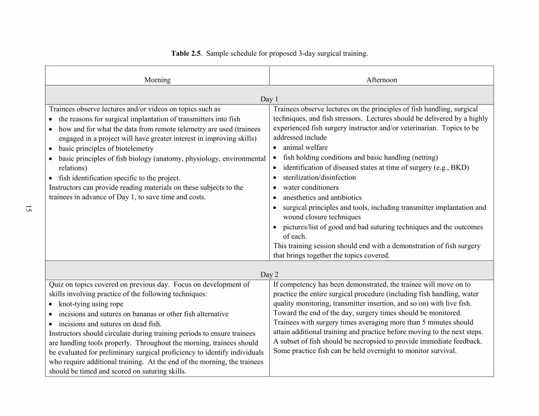
Training program example
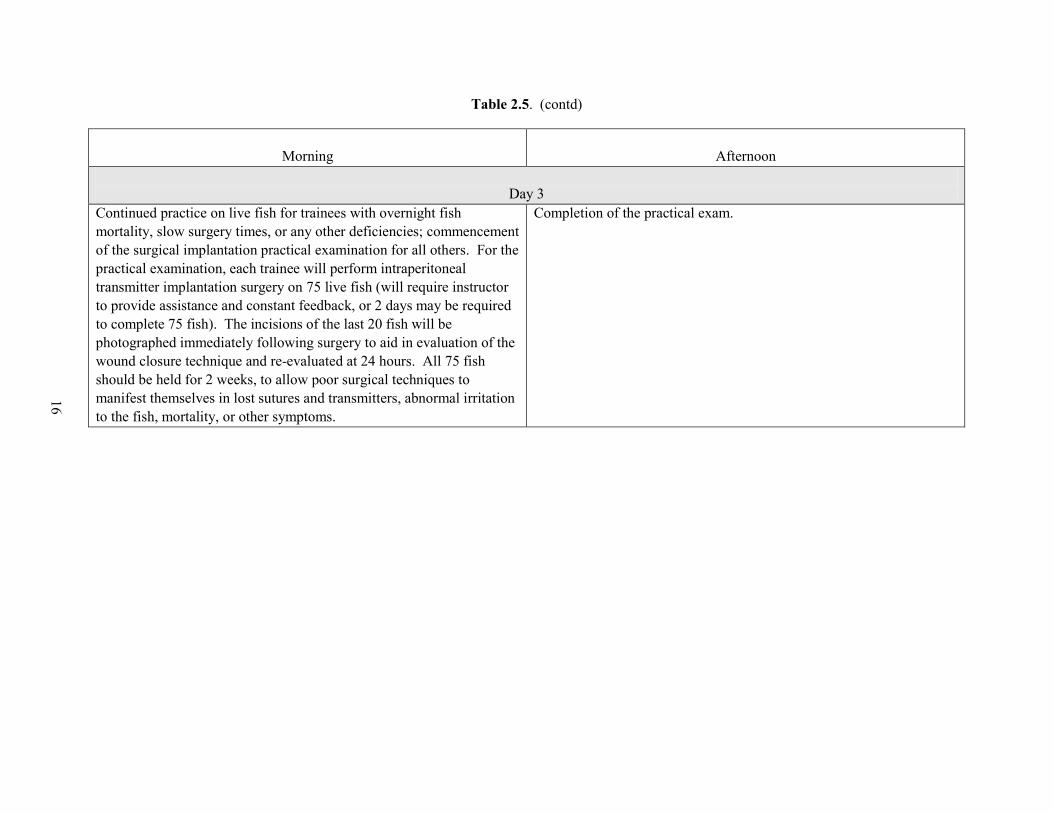
Details of the schedule for our proposed 3-day training in fish surgical techniques are presented in Table 2.5. Each training day is subdivided into a morning and an afternoon session, and each session has a specific focus. We acknowledge that this format is an example and that different organizations and institutions may have slightly different requirements regarding the duration of different course components and the number of practice surgeries conducted. For evaluation, we are proposing a sample size of 75 fish. However, the minimum number of total surgeries performed would exceed 100 over the 3-day training course.
Completion of the practical examination does not signify approval to perform surgery on a project. Training culminates with a final evaluation (based on timing, quality of wound closure, care with internal organs, transmitter placement) and includes a feedback report. The final evaluation should be determined as soon as possible after the 2-week holding period. Trainees
14
should not be scheduled to perform surgeries for a project until they have been approved as a qualified surgeon in the final evaluation. For this reason, funding agencies should be made aware of this time requirement, to allow potential surgeons to be trained before the project starts. Additional time should be allocated in case trainees do not pass the final evaluation. This time provides the opportunity to retrain and retake the exam. However, instructors should identify individuals with an obvious lack of skill because it may be more cost-effective to train a new surgeon with more innate ability.
Beyond this type of 3-day training program, novice fish surgeons should further practice their learned surgical skills to continue to improve timing, incision and suture placement, and fish recovery. More experienced and expert surgeons should retrain with a small number of fish when working with a new species, when the procedure has been modified, or after a length of surgical inactivity.
Because the success of telemetry studies depends on the retention of transmitters and high survival of study fish, it is important that the final evaluation of the trainee include a collection of data not limited to percentages of fish mortality, transmitter retention, suture retention, or wound healing and appearance. Evaluations of photos taken a few times post-surgery (24 hours, 1 week, and 2 weeks are commonly used) provide the information necessary to assess the surgical proficiency of the trainees. By taking photographs of the incision at a number of days post-surgery, instructors and trainees will be able to observe which aspects of the surgery may be causing detriment to the fish. Techniques can then be modified to improve the surgical outcome. Retraining could include surgeries on live fish only, with subsequent feedback on the surgeries.
15
Table 2.5. Sample schedule for proposed 3-day surgical training.
Morning
Afternoon
Day 1
Trainees observe lectures and/or videos on topics such as • the reasons for surgical implantation of transmitters into fish • how and for what the data from remote telemetry are used (trainees
engaged in a project will have greater interest in improving skills) • basic principles of biotelemetry • basic principles of fish biology (anatomy, physiology, environmental
relations) • fish identification specific to the project. Instructors can provide reading materials on these subjects to the trainees in advance of Day 1, to save time and costs.
Trainees observe lectures on the principles of fish handling, surgical techniques, and fish stressors. Lectures should be delivered by a highly experienced fish surgery instructor and/or veterinarian. Topics to be addressed include • animal welfare • fish holding conditions and basic handling (netting) • identification of diseased states at time of surgery (e.g., BKD) • sterilization/disinfection • water conditioners • anesthetics and antibiotics • surgical principles and tools, including transmitter implantation and
wound closure techniques • pictures/list of good and bad suturing techniques and the outcomes
of each. This training session should end with a demonstration of fish surgery that brings together the topics covered.
Day 2
Quiz on topics covered on previous day. Focus on development of skills involving practice of the following techniques: • knot-tying using rope • incisions and sutures on bananas or other fish alternative • incisions and sutures on dead fish. Instructors should circulate during training periods to ensure trainees are handling tools properly. Throughout the morning, trainees should be evaluated for preliminary surgical proficiency to identify individuals who require additional training. At the end of the morning, the trainees should be timed and scored on suturing skills.
If competency has been demonstrated, the trainee will move on to practice the entire surgical procedure (including fish handling, water quality monitoring, transmitter insertion, and so on) with live fish. Toward the end of the day, surgery times should be monitored. Trainees with surgery times averaging more than 5 minutes should attain additional training and practice before moving to the next steps. A subset of fish should be necropsied to provide immediate feedback. Some practice fish can be held overnight to monitor survival.
16
Table 2.5. (contd)
Morning
Afternoon
Day 3
Continued practice on live fish for trainees with overnight fish mortality, slow surgery times, or any other deficiencies; commencement of the surgical implantation practical examination for all others. For the practical examination, each trainee will perform intraperitoneal transmitter implantation surgery on 75 live fish (will require instructor to provide assistance and constant feedback, or 2 days may be required to complete 75 fish). The incisions of the last 20 fish will be photographed immediately following surgery to aid in evaluation of the wound closure technique and re-evaluated at 24 hours. All 75 fish should be held for 2 weeks, to allow poor surgical techniques to manifest themselves in lost sutures and transmitters, abnormal irritation to the fish, mortality, or other symptoms.
Completion of the practical exam.
17
References
AFS (American Fisheries Society), American Institute of Fishery Research Biologists, and American Society of Ichthyologists and Herpetologists (2004) Guidelines for the use of fishes in research. American Fisheries Society, Bethesda, MD. Available at www.fisheries.org/afs/ docs/policy_guidelines2004.pdf. Accessed April 2009
Annett J (1971) Acquisition of skill. British Medical Bulletin 27:266–271
ASIH (American Society of Ichthyologists and Herpetologists), American Fisheries Society, and American Institute of Fishery Research Biologists (1987) Guidelines for use of fishes in field research. Copeia Supplement:1–12.
ASIH (American Society of Ichthyologists and Herpetologists), American Fisheries Society, and American Institute of Fishery Research Biologists (1988) Guidelines for use of fishes in field research. Fisheries 13(2):16–23
Australasian Veterinary Boards Council Inc. (2006) Annex 2, Essential competences required of the veterinary surgeon. In: Policies, procedures and standards – Veterinary Schools Accreditation Advisory Committee. Australasian Veterinary Boards Council Inc, Melbourne, Australia, p. 25
Bauer MS (1993) A survey of the use of live animals, cadavers, inanimate models, and computers in teaching veterinary surgery. Journal of the American Veterinary Medical Association 203:1047–1051
Borski RJ, Hodson RG (2003) Fish research and the institutional animal care and use committee. ILAR Journal 44:286–294
Bradley P (2006) The history of simulation in medical education and possible future directions. Medical Education 40:3254–262
Bridger CJ, Booth RK (2003) The effects of biotelemetry transmitter presence and attachment procedures on fish physiology and behavior. Reviews in Fisheries Science 11:13–34
Califf RM, Jollis JG, Peterson ED (1996) Operator-specific outcomes: a call to professional responsibility. Circulation 93:403–406
Caudill CC, Daigle WR, Keefer ML, Boggs CT, Jepson MA, Burke BJ, Zabel RW, Bjornn TC, Peery CA (2007) Slow dam passage in adult Columbia River salmonids associated with unsuccessful migration: delayed negative effects of passage obstacles or condition-dependent mortality? Canadian Journal of Fisheries and Aquatic Sciences 64:979–995
Cooke SJ, Wagner GN (2004) Training, experience, and opinions of researchers who use surgical techniques to implant telemetry devices into fish. Fisheries 29(12):10–18
18
Cooke SJ, Graeb BDS, Suski CD, Ostrand KG (2003) Effects of suture material on incision healing, growth and survival of juvenile largemouth bass implanted with miniature radio transmitters: case study of a novice and experienced fish surgeon. Journal of Fish Biology 62:1366–1380
Cooke SJ, Hinch SG, Wikelski M, Andrews RD, Wolcott TG, Butler PJ (2004) Biotelemetry: a mechanistic approach to ecology. Trends in Ecology and Evolution 19:334–343
Deters KA, Brown RS, Carter KM, Boyd JW, Eppard MB, Seaburg, AG (in press) Performance assessment of suture type, water temperature, and surgeon skill in juvenile Chinook salmon surgically implanted with acoustic transmitters. Transactions of the American Fisheries Society
DeTolla LJ, Srinivas S, Whitaker BR, Andrews B, Hecker B, Kane AS, Reimschuessel R (1995) Guidelines for the care and use of fish in research. ILAR Journal 37:159–173
Engelhorn R (1997) Speed and accuracy in the learning of a complex motor skill. Perceptual and Motor Skills 85:1011–1017
Epstein RM, Hundert EM (2002) Defining and assessing professional competence. Journal of the American Medical Association 287:226–235
Ericsson KA (2004) Deliberate practice and the acquisition and maintenance of expert performance in medicine and related domains. Academic Medicine 79:S70–S81
Ericsson KA, Krampe RT, Tesch-Roemer C (1993) The role of deliberate practice in the acquisition of expert performance. Psychological Review 100:363–406
Feasby TE, Quan H, Ghali WA (2002) Hospital and surgeon determinants of carotid endarterectomy outcomes. Archives of Neurology 59(12):1877–1881
FSBI (Fisheries Society of the British Isles) (2002) Fish welfare. Briefing Paper 2, Fisheries Society of the British Isles, Granta Information Systems, Sawston, Cambridge, UK
Freund D, Lave J, Clancy C, Hawker G, Hasselblad V, Keller R, Shneiter E, Wright J (1999) Patient outcomes research teams: contribution to outcomes and effectiveness research. Annual Review of Public Health 20:337–359
Hammond JW, Queale WS, Kim TK, McFarland EG (2003) Surgeon experience and clinical and economic outcomes for shoulder arthroplasty. Journal of Bone and Joint Surgery – American Version A(12):2318–2324
Harms CA, Lewbart GA (2000) Surgery in fish. In: Bennett RA (ed) Veterinary clinics of North America: exotic animal practice. W. B. Saunders, New York, pp 759–774
Jepsen N, Koed A, Thorstad EB, Baras E (2002) Surgical implantation of telemetry transmitters in fish: how much have we learned? Hydrobiologia 483:239-248
19
Keefer ML, Peery CA, Bjornn TC, Jepson MA, Stuehrenberg LC (2004) Hydrosystem, dam, and reservoir passage rates of adult Chinook salmon and steelhead in the Columbia and Snake Rivers. Transactions of the American Fisheries Society 133:1413–1439
Kneebone R (2003) Simulation in surgical training: educational issues and practical implications. Medical Education 37: 267–277
Mulcahy DM (2003a) Does the Animal Welfare Act apply to free-ranging animals? ILAR Journal 44:252–258
Mulcahy DM (2003b) Surgical implantation of transmitters into fish. ILAR Journal 44:295–306
Naughton GP, Caudill CC, Peery CA, Clabough TS, Jepson MA, Bjorn TC, Stuehrenberg LC (2007) Experimental evaluation of fishway modifications on the passage behaviour of adult Chinook salmon and steelhead at Lower Granite Dam, Snake River, USA. River Research and Applications 23:99–111
Pearce WH, Parker MA, Feinglass J, Ujiki M, Manheim LM (1999) The importance of surgeon volume and training in outcomes for vascular surgical procedures. Journal of Vascular Surgery 29(5):768–76; discussion 777–778
Schreck CB, Stahl TP, Davis LE, Roby DD, Clemens BJ (2006) Mortality estimates of juvenile spring-summer Chinook salmon in the Lower Columbia River and estuary. Transactions of the American Fisheries Society 135:457–475
Seki S (1987) Accuracy of suture techniques of surgeons with different surgical experience. Japanese Journal of Surgery 17:465–469
Silva RM, Matera JM, Ribeiro AA (2007) New alternative methods to teach surgical techniques for veterinary medicine students despite the absence of living animals. Is that an academic paradox? Anatomia Histologia Embryologia 36:220–224
Smeak DD (1999) Accent on an alternative: skin and suture pattern simulator. Alternatives in Veterinary Medical Education Newsletter 10:2–3
Sosa JA, Bowman HM, Tielsch JM, Powe NR, Gordon TA, Udelsman R (1998) The importance of surgeon experience for clinical and economic outcomes from thyroidectomy. Annals of Surgery 228(3):320–330
Starkes JL, Payk I, Hodges NJ (1998) Developing a standardized test for the assessment of suturing skill in novice microsurgeons. Microsurgery 18:19–22
Stoskopf MK (1993a) Clinical examination and procedures. In: Stoskopf MK (ed) Fish medicine. W. B. Saunders Inc., New York, pp 62–78
Stoskopf MK (1993a) Surgery. In: Stoskopf MK (ed) Fish medicine. W. B. Saunders Inc., New York, pp 91–97
20
Stoskopf MK (2003) All of the world is a laboratory. ILAR Journal 44:249–251
Summerfelt RC, Smith L (1990) Anesthesia, surgery, and related techniques. In: Schreck CB, Moyle PB (eds) Methods for fish biology. American Fisheries Society, Bethesda, MD, pp 213–272
Szalay D, MacRae H, Regehr G, Reznik R (2000) Using operative outcome to assess technical skill. American Journal of Surgery 180: 234–237
Tinga CE, Adams CL, Bonnett BN, Ribble CS (2001) Survey of veterinary technical and professional skills in students and recent graduates of a veterinary college. Journal of the American Veterinary Medical Association 219:924–931
Torkington J, Smith SG, Rees BI, Darzi A (2000) The role of simulation in surgical training. Annals of the Royal College of Surgeons of England 82(2):88–94
Uson J, Calles MC (2002) Design of a new suture practice card for microsurgical training. Microsurgery 22:324–328
Wagner GN, Cooke SJ (2005) Methodological approaches and opinions of researchers involved in the surgical implantation of telemetry transmitters in fish. Journal of Aquatic Animal Health 17:160–169.
Walsh DA, Osburn BI, Christopher MM (2001) Defining the attributes expected of graduating veterinary medical students. Journal of the American Veterinary Medical Association 219:1358–1365
Walsh DA, Osburn BI, Schumacher RL (2002) Defining the attributes expected of graduating veterinary medical students, part 2: external evaluation and outcomes assessment. Journal of Veterinary Medical Education 29(1):36–42
Welch DW, Batten SD, Ward BR (2007) Growth, survival, tag retention of steelhead trout (O. mykiss) surgically implanted with dummy acoustic tags. Hydrobiologia 582:289–299
Zemljic B (2004) Standards of veterinary education for the future: Preparing the profession for the new century. Journal of Veterinary Medical Education 31:13–14
21
3 Pre- and Post-Surgical Holding
Eric W. Oldenburg and Richard S. Brown
Holding of fish prior to and after surgical implantation of transmitters is an important aspect of telemetry studies, yet one that is often overlooked or considered of low importance. Essentially, such surgical holding is analogous to pre-operative and post-operative care in human and veterinary medicine, in which the goal is to restore the patient to as near the normal physiological state as rapidly as possible. A primary assumption of telemetry studies is that the surgically implanted fish are representative of the population of inference. However, the process of surgical implantation has the potential to introduce bias to the sample and alter aspects of fish swimming ability (Adams et al. 1998a; Wagner and Stevens 2000; Brown et al. 2006), growth (Martin et al. 1995; Adams et al. 1998b), physiology (Jepsen et al. 2001; Close et al. 2003), and survival (Adams et al. 1998a; Jepsen et al. 1998). Therefore, it is desirable to minimize bias created by the surgical implantation process to the greatest feasible extent so that inferences can be made regarding the population of interest.
Pre-surgical holding often occurs to facilitate logistical needs of research projects, as an attempt to minimize negative physiological effects due to capture and handling stress (Waring et al. 1992; Brobbel et al. 1996; Barton 2000; Chandroo et al. 2005), or to ensure that fish are in a post-absorptive state (i.e., all food has been digested and assimilated). Pre-surgical holding factors that should be considered include holding time and conditions, water quality (e.g., supplemental oxygen and water circulation), whether fish should be fed, fish density within the holding tank, and fish handling.
Pre-surgical holding times range from 12 to 48 h among the multiple entities conducting fish research in the Columbia Basin (e.g., Chelan County Public Utility District, Public Utility District No. 2 of Grant County, National Oceanic and Atmospheric Administration Fisheries, Pacific Northwest National Laboratory, U.S. Geological Survey). There currently is no standard holding time for field-based telemetry projects examining fish migration within the Columbia Basin. Logistical discrepancies among studies and dissimilar protocols among agencies prevent standardization of pre-surgery holding time to a single value. However, it is desirable to standardize to a range of time that is biologically meaningful (e.g., reduces stress) while accommodating for these discrepancies.
The interrelationships between stress and metabolism are important aspects of surgical holding. Acute or chronic stress increases metabolic rates in juvenile salmonids (Barton and Schreck 1987) and can impair reproduction, immune function, growth, and survival (Wedemeyer et al. 1990). Stress can alter fish behavior, such as social hierarchies (Ejike and Schreck 1980; Connors et al. 2002), leading to increases in predation risk, passive displacement, impingement, or entrainment. Further, stress can lead to mortality if the energy required to mitigate the stress exceeds the scope for activity (difference between the maximum metabolic rate and the standard
22
or resting metabolic rate) of the individual, causing metabolic rate-dependent mortality (Priede 1977) through cardiovascular collapse (Farrell 2002). Thus, it is important to minimize stress and keep the energetic demands of fish within their scope for activity.
Handling, confinement, and air exposure are stressors among salmonids (Strange et al. 1977, 1978; Barton et al. 1988; Ferguson and Tufts 1992; Davis and Schreck 1997; Arends et al. 1999) to which individuals may be subjected throughout surgical holding. Minor physical disturbances (i.e., stressors) have elicited greater than two-fold increases in metabolic rates of juvenile salmonids and may consume one-quarter of the scope for activity of an individual (Barton and Schreck 1987). Further, stress can be cumulative in juvenile fish (Barton et al. 1986), which increases the potential for stress-induced mortality (Armstrong et al. 1992). Effects of multiple acute or chronic stress events such as handling, confinement, and surgical procedures may amplify stress levels and metabolic demand in juvenile salmonids. Therefore, it is desirable to reduce the number and magnitude of stressors to which fish are subjected, ultimately decreasing metabolic demand and increasing survival among individuals. Specifically, fish should be held long enough to enable collection-related handling stress levels to subside.
Holding density, conditions, and water quality also affect the physiological stress response and metabolic scope in salmonids. In long-term studies, increasing rearing density can result in increased mortality and decreased final weight, length, condition factor, and food conversion efficiency among salmonids (Fagerlund et al. 1981; Poston and Williams 1988; Procarione et al. 1999). A standardization of maximum holding density is difficult due to discrepancies in tank size, water quality, and available flow capabilities among surgical holding sites and species-specific water quality requirements. When taking into account fish health and economic yield, the optimal stocking density of rainbow trout has been shown to be between 10 and 25 kg/m3 (Trzebiatowski et al. 1981; Baker and Ayles 1990). However, this density of fish is based on adult growth rates and does not discern minimal stress levels for fish.
It has been suggested that high rearing density alone is likely not a chronic stressor in salmonids (Procarione et al. 1999) and that high water exchange rates can permit high rearing densities with little negative physiological effect (Westers 2001). Therefore, when planning for short-term holding (e.g., pre- and post-surgical holding), water quality is likely a more important factor to consider than fish density. Maintaining the intricate balance among density, loading (flow), and water exchange rate has been reviewed by Westers (2001), who found dissolved oxygen to be the first limiting water quality factor, with a desirable goal to have levels near saturation. For this reason, supplemental oxygen should be applied to surgical holding tanks whenever necessary in order to keep dissolved oxygen levels between 80% and 110%.
Covering the fish-holding tanks with a lid or other type of cover has not been well studied. However, some salmonids demonstrate an affinity for covered areas in holding tanks (Gibson 1978); thus, it is possible that exposing fish to intense light or startling may be stressors to fishes. Therefore, we suggest that holding tanks be covered throughout the surgical process.
The postprandial metabolic response also affects metabolic scope in salmonids. Specific dynamic action (SDA) is the portion of the metabolic scope allocated to processing and assimilating food. Studies on salmonids have demonstrated that SDA remains remarkably fixed during exercise and that the energetic demands of swimming are met only after the energetic
23
demands of SDA are met (Alsop and Wood 1997; Thorarensen and Farrell 2006). Thus, it is possible that energetic needs of other processes (e.g., compensatory stress responses) may also be met only after the energetic needs of SDA have been met. Therefore, fish should be held long enough to allow SDA to cease and should not be fed during surgical holding because SDA may usurp large portions of metabolic scope that could otherwise be allocated to compensatory stress responses.
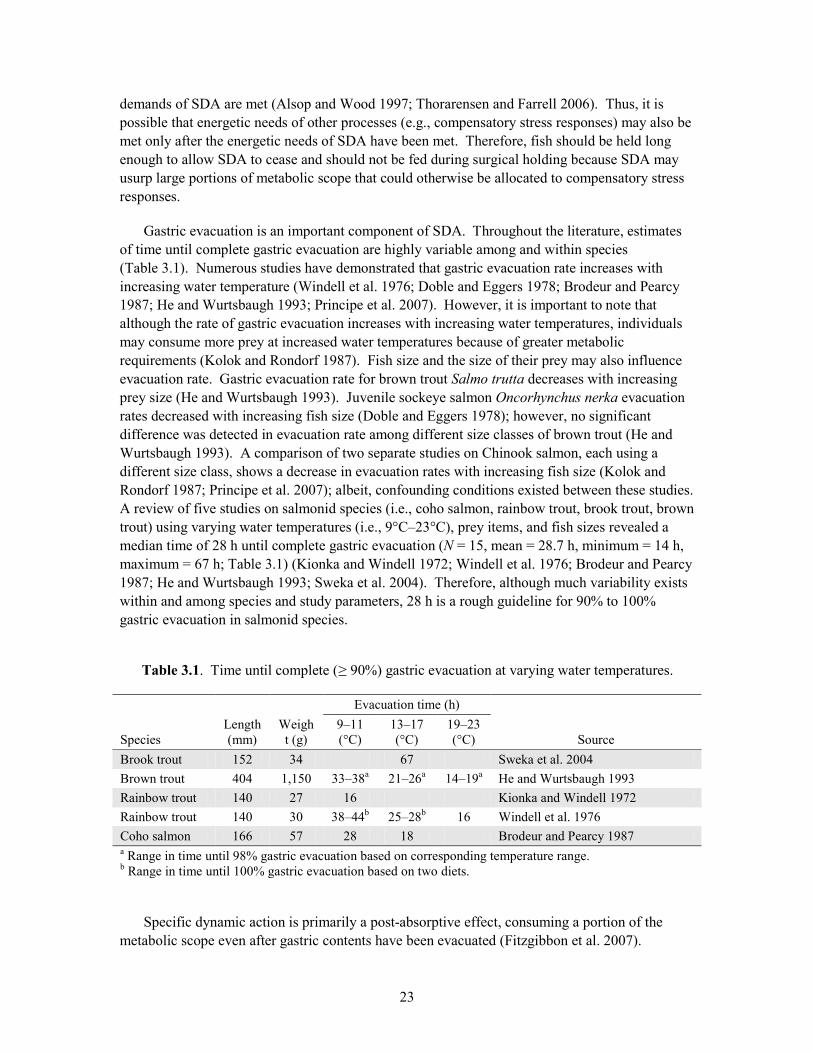
Gastric evacuation is an important component of SDA. Throughout the literature, estimates of time until complete gastric evacuation are highly variable among and within species (Table 3.1). Numerous studies have demonstrated that gastric evacuation rate increases with increasing water temperature (Windell et al. 1976; Doble and Eggers 1978; Brodeur and Pearcy 1987; He and Wurtsbaugh 1993; Principe et al. 2007). However, it is important to note that although the rate of gastric evacuation increases with increasing water temperatures, individuals may consume more prey at increased water temperatures because of greater metabolic requirements (Kolok and Rondorf 1987). Fish size and the size of their prey may also influence evacuation rate. Gastric evacuation rate for brown trout Salmo trutta decreases with increasing prey size (He and Wurtsbaugh 1993). Juvenile sockeye salmon Oncorhynchus nerka evacuation rates decreased with increasing fish size (Doble and Eggers 1978); however, no significant difference was detected in evacuation rate among different size classes of brown trout (He and Wurtsbaugh 1993). A comparison of two separate studies on Chinook salmon, each using a different size class, shows a decrease in evacuation rates with increasing fish size (Kolok and Rondorf 1987; Principe et al. 2007); albeit, confounding conditions existed between these studies. A review of five studies on salmonid species (i.e., coho salmon, rainbow trout, brook trout, brown trout) using varying water temperatures (i.e., 9°C–23°C), prey items, and fish sizes revealed a median time of 28 h until complete gastric evacuation (N = 15, mean = 28.7 h, minimum = 14 h, maximum = 67 h; Table 3.1) (Kionka and Windell 1972; Windell et al. 1976; Brodeur and Pearcy 1987; He and Wurtsbaugh 1993; Sweka et al. 2004). Therefore, although much variability exists within and among species and study parameters, 28 h is a rough guideline for 90% to 100% gastric evacuation in salmonid species.
Table 3.1. Time until complete (≥ 90%) gastric evacuation at varying water temperatures.
Evacuation time (h) Species
Length (mm)
Weight (g)
9–11 (°C)
13–17 (°C)
19–23 (°C)
Source
Brook trout 152 34 67 Sweka et al. 2004 Brown trout 404 1,150 33–38a 21–26a 14–19a He and Wurtsbaugh 1993 Rainbow trout 140 27 16 Kionka and Windell 1972 Rainbow trout 140 30 38–44b 25–28b 16 Windell et al. 1976 Coho salmon 166 57 28 18 Brodeur and Pearcy 1987 a Range in time until 98% gastric evacuation based on corresponding temperature range. b Range in time until 100% gastric evacuation based on two diets.
Specific dynamic action is primarily a post-absorptive effect, consuming a portion of the metabolic scope even after gastric contents have been evacuated (Fitzgibbon et al. 2007).
24
Consequently, time until gastric evacuation alone may not provide the best estimate for pre-surgical holding time. In a study on rainbow trout (mean weight [± SE] = 808 ± 47 g), postprandial gastrointestinal blood flow peaked at 136% above baseline 11 h after feeding, postprandial heart rate peaked at 110% above baseline after 14 h, and postprandial oxygen consumption peaked at 96% above baseline after 27 h (Eliason et al. 2008).
The importance of holding fish prior to surgery to create a post-absorptive condition has been demonstrated; however, it is equally essential to minimize this required time, to reduce the impacts of cumulative and holding-related stressors such as confinement and crowding. These variables must be balanced so that post-surgery energy demands do not exceed the metabolic scope of the fish. Therefore, we suggest that pre-surgical holding be standardized to the range of time from 18–36 h after the cessation of fish collection.
Post-surgical holding, another aspect of the surgical process, is important not only because of the aforementioned holding factors (e.g., metabolic rate-dependent mortality), but also because post-surgical holding time and conditions greatly influence the physiological state of fish prior to being returned to the river. The physiological state of fish upon release may affect survival and behavior of individuals following release and ultimately bias a study. For example, individuals released into the river under stress may be affected by elevated plasma cortisol concentrations (Wendelaar Bonga 1997). Increased plasma cortisol concentrations can affect endocrine processes, such as suppression of reproductive hormones in upriver migrating adult salmonids (Hinch et al. 2005). Although these types of endocrine cascades have not been well studied in juvenile salmonids, similar effects may occur in this life stage. Further, increases in stress are an important consideration for downstream migrating salmon smolts because cortisol appears to play a role in acclimation to saline environments (Redding et al. 1984; McCormick 1996). Therefore, efforts should be made to reduce stress levels in fish during surgical holding and prior to release.
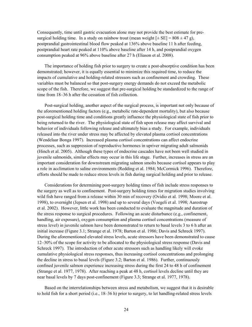
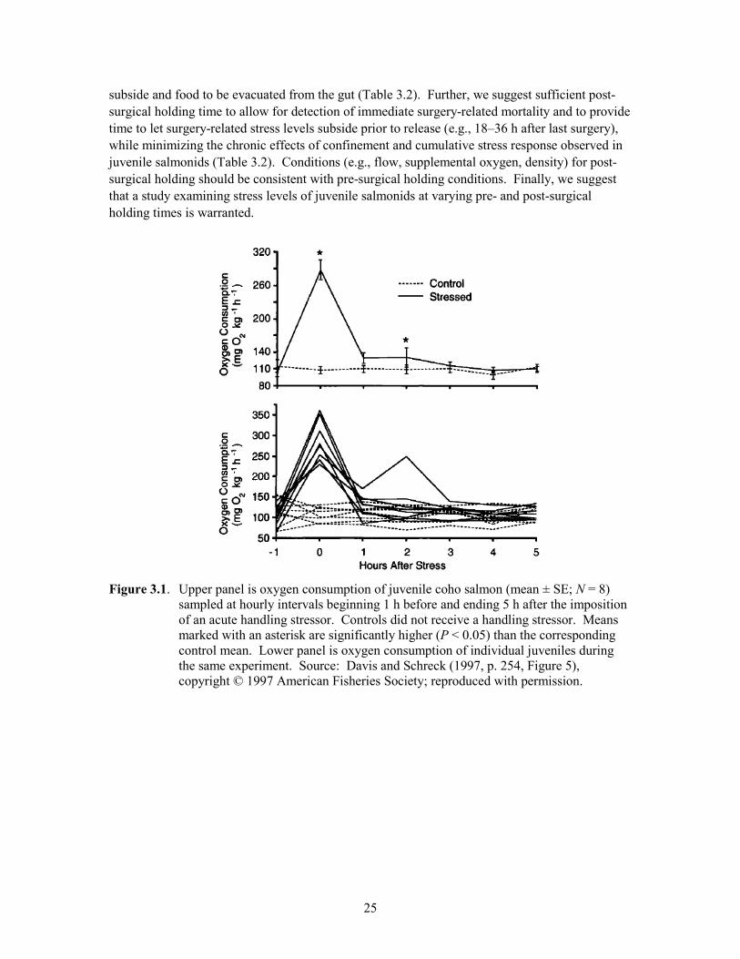
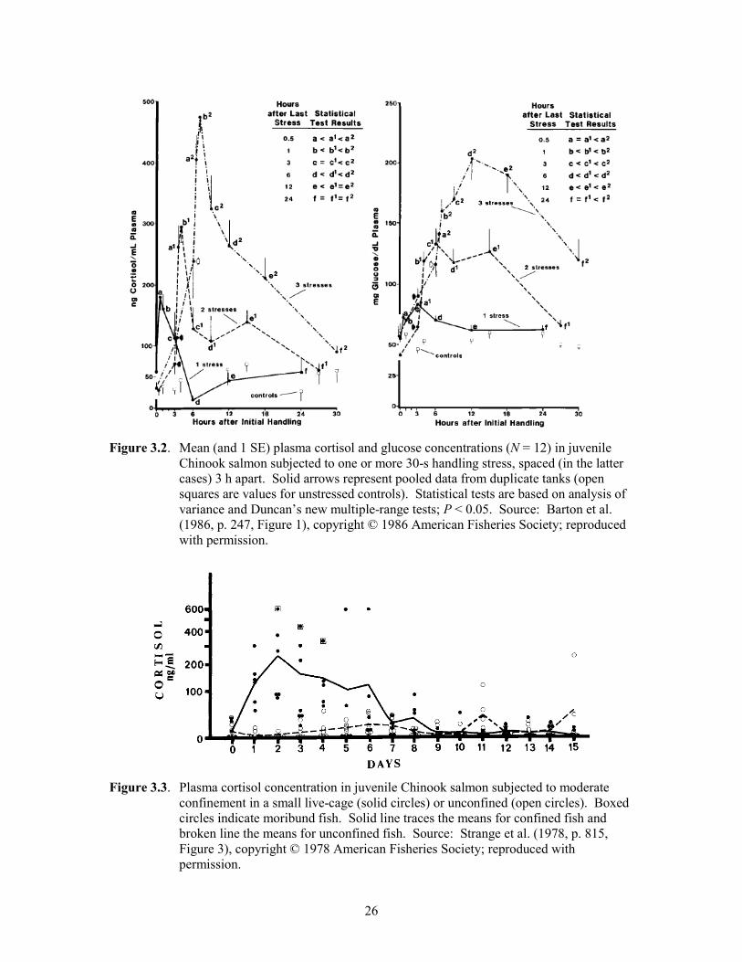
Considerations for determining post-surgery holding times of fish include stress responses to the surgery as well as to confinement. Post-surgery holding times for migration studies involving wild fish have ranged from a release within 30 min of recovery (Ovidio et al. 1998; Moore et al. 1998), to overnight (Jepsen et al. 1998) and up to several days (Voegeli et al. 1998; Aarestrup et al. 2002). However, little work has been conducted to evaluate the magnitude and duration of the stress response to surgical procedures. Following an acute disturbance (e.g., confinement, handling, air exposure), oxygen consumption and plasma cortisol concentrations (measure of stress level) in juvenile salmon have been demonstrated to return to basal levels 3 to 6 h after an initial increase (Figure 3.1; Strange et al. 1978; Barton et al. 1986; Davis and Schreck 1997). During the aforementioned elevated stress levels, acute stressors have been demonstrated to cause 12–30% of the scope for activity to be allocated to the physiological stress response (Davis and Schreck 1997). The introduction of other acute stressors such as handling likely will evoke cumulative physiological stress responses, thus increasing cortisol concentrations and prolonging the decline in stress to basal levels (Figure 3.2; Barton et al. 1986). Further, continuously confined juvenile salmon experience increasing stress during the first 24 to 48 h of confinement (Strange et al. 1977, 1978). After reaching a peak at 48 h, cortisol levels decline until they are near basal levels by 7 days post-confinement (Figure 3.3; Strange et al. 1977, 1978).
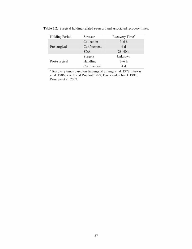
Based on the interrelationships between stress and metabolism, we suggest that it is desirable to hold fish for a short period (i.e., 18–36 h) prior to surgery, to let handling-related stress levels
25
subside and food to be evacuated from the gut (Table 3.2). Further, we suggest sufficient post-surgical holding time to allow for detection of immediate surgery-related mortality and to provide time to let surgery-related stress levels subside prior to release (e.g., 18–36 h after last surgery), while minimizing the chronic effects of confinement and cumulative stress response observed in juvenile salmonids (Table 3.2). Conditions (e.g., flow, supplemental oxygen, density) for post-surgical holding should be consistent with pre-surgical holding conditions. Finally, we suggest that a study examining stress levels of juvenile salmonids at varying pre- and post-surgical holding times is warranted.
Figure 3.1. Upper panel is oxygen consumption of juvenile coho salmon (mean ± SE; N = 8)
sampled at hourly intervals beginning 1 h before and ending 5 h after the imposition of an acute handling stressor. Controls did not receive a handling stressor. Means marked with an asterisk are significantly higher (P < 0.05) than the corresponding control mean. Lower panel is oxygen consumption of individual juveniles during the same experiment. Source: Davis and Schreck (1997, p. 254, Figure 5), copyright © 1997 American Fisheries Society; reproduced with permission.
26
Figure 3.2. Mean (and 1 SE) plasma cortisol and glucose concentrations (N = 12) in juvenile
Chinook salmon subjected to one or more 30-s handling stress, spaced (in the latter cases) 3 h apart. Solid arrows represent pooled data from duplicate tanks (open squares are values for unstressed controls). Statistical tests are based on analysis of variance and Duncan’s new multiple-range tests; P < 0.05. Source: Barton et al. (1986, p. 247, Figure 1), copyright © 1986 American Fisheries Society; reproduced with permission.
Figure 3.3. Plasma cortisol concentration in juvenile Chinook salmon subjected to moderate
confinement in a small live-cage (solid circles) or unconfined (open circles). Boxed circles indicate moribund fish. Solid line traces the means for confined fish and broken line the means for unconfined fish. Source: Strange et al. (1978, p. 815, Figure 3), copyright © 1978 American Fisheries Society; reproduced with permission.
27
Table 3.2. Surgical holding-related stressors and associated recovery times.
Holding Period Stressor Recovery Timea
Collection 3–6 h Pre-surgical Confinement 4 d SDA 28–40 h Surgery Unknown Post-surgical Handling 3–6 h
Confinement 4 d a Recovery times based on findings of Strange et al. 1978; Barton et al. 1986; Kolok and Rondorf 1987; Davis and Schreck 1997; Principe et al. 2007.
28
References
Aarestrup K, Nielsen C, Koed A (2002) Net ground speed of downstream migrating radio-tagged Atlantic salmon (Salmo salar L.) and brown trout (Salmo trutta L.) smolts in relation to environmental factors. Hydrobiologia 483:95–102
Adams NS, Rondorf DW, Evans SD, Kelly JE, Perry RW (1998a) Effects of surgically and
gastrically implanted radio transmitters on swimming performance and predator avoidance of juvenile Chinook salmon (Oncorhynchus tshawytscha). Canadian Journal of Fisheries and Aquatic Sciences 55:781–787
Adams NS, Rondorf DW, Evans SD, Kelly JE (1998b) Effects of surgically and gastrically
implanted radio transmitters on growth and feeding behavior of Chinook salmon. Transactions of the American Fisheries Society 127:128–136
Alsop DH, Wood CM (1997) The interactive effects of feeding and exercise on oxygen
consumption, swimming performance and protein usage in juvenile rainbow trout (Oncorhynchus mykiss). Journal of Experimental Biology 200:2337–2346
Arends RJ, Mancera JM, Munoz JL, Wendelaar Bonga SE, Flik G (1999) The stress response of
the gilthead sea bream (Sparus aurata L.) to air exposure and confinement. Journal of Endocrinology 163:149–157
Armstrong JD, Priede IG, Lucas MC (1992) The link between respiratory capacity and changing
metabolic demands during growth of northern pike, Esox lucius L. Journal of Fish Biology 41(Supplement B):65–75
Baker RF, Ayles GB (1990) The effects or varying density and loading level on the growth of
Arctic charr(Salvelinus alpinus) and rainbow trout (Oncorhynchus mykiss). World Aquaculture 21:58–62
Barton BA (2000) Salmonid fishes differ in their cortisol and glucose responses to handling and
transport stress. North American Journal of Aquaculture 62:12–18 Barton BA, Schreck CB (1987) Metabolic cost of acute physical stress in juvenile steelhead.
Transactions of the American Fisheries Society 116:257–263 Barton BA, Schreck CB, Sigismondi LA (1986) Multiple acute disturbances evoke cumulative
physiological stress responses in juvenile Chinook salmon. Transactions of the American Fisheries Society 115:245–251
Barton BA, Schreck CB, Fowler LG (1988) Fasting and diet content affect stress-induced
changes in plasma glucose and cortisol in juvenile Chinook salmon. The Progressive Fish-Culturist 50:16–22
29
Brobbel MA, Wilkie MP, Davidson K, Kieffer JD, Bielak AT, Tufts B (1996) Physiological effects of catch and release angling in Atlantic salmon (Salmo salar) at different stages of freshwater migration. Canadian Journal of Fisheries and Aquatic Sciences 53:2036–2043
Brodeur RD, Pearcy WG (1987) Diel feeding chronology, gastric evacuation and estimated daily
ration of juvenile coho salmon, Oncorhynchus kisutch (Walbaum), in the coastal marine environment. Journal of Fish Biology 31:465–477
Brown RS, Geist DR, Deters KA, Grassell A (2006) Effects of surgically implanted acoustic
transmitters >2% of body mass on the swimming performance, survival and growth of juvenile sockeye and Chinook salmon. Journal of Fish Biology 69:1626–1638
Chandroo KP, Cooke SJ, McKinley RS, Moccia R (2005) Use of electromyogram telemetry to
assess the behavioural and energetic responses of rainbow trout, Oncorhynchus mykiss (Walbaum) to transportation stress. Aquaculture Research 36:1226–1238
Close DA, Fitzpatrick MS, Lorion CM, Li HW, Schreck CB (2003) Effects of intraperitoneally
implanted radio transmitters on the swimming performance and physiology of pacific lamprey. North American Journal of Fisheries Management 23:1184–1192
Connors KB, Scruton D, Brown JA, McKinley RS (2002) The effects of surgically-implanted
dummy radio transmitters on the behaviour of wild Atlantic salmon smolts. Hydrobiologia 483:231–237
Davis LE, Schreck CB (1997) The energetic response to handling stress in juvenile coho salmon.
Transactions of the American Fisheries Society 126:248–258 Doble BD, Eggers DM (1978) Diel feeding chronology, rate of gastric evacuation, and prey
selectivity in Lake Washington juvenile sockeye salmon (Oncorhynchus nerka). Transactions of the American Fisheries Society 107(1):36–45
Ejike C, Schreck CB (1980) Stress and social hierarchy in coho salmon. Transactions of the
American Fisheries Society 109:423–426 Eliason EJ, Higgs DA, Farrell AP (2008) Postprandial gastrointestinal blood flow, oxygen
consumption and heart rate in rainbow trout (Oncorhynchus mykiss). Comparative Biochemistry and Physiology A 144:381–394
Fagerlund UHM, McBride JR, Stone ET (1981) Stress-related effects of hatchery rearing density
on coho salmon. Transactions of the American Fisheries Society 110:644–649 Farrell AP (2002) Cardiorespiratory performance in salmonids during exercise at high
temperature: Insights into cardiovascular design limitations in fishes. Comparative Biochemistry and Physiology A 132: 797–810
30
Ferguson RA, Tufts BL (1992) Pyhsiological effects of brief air exposure in exercised rainbow trout (Oncorhynchus mykiss): implications for catch and release fisheries. Canadian Journal of Fisheries and Aquatic Sciences 49(6):1157–1162
Fitzgibbon QP, Seymour RS, Ellis D, Buchanan J (2007) The energetic consequence of specific
dynamic action in southern bluefin tuna Thunnus maccoyii. The Journal of Experimental Biology 210:290–298
Gibson, RJ (1978) The behaviour of juvenile Atlantic salmon (Salmo salar) and brook trout
(Salvelinus fontinalis) with regard to temperature and water velocity. Transaction of the American Fisheries Society 107:703-712
He E, Wurtsbaugh WA (1993) An empirical model of gastric evacuation rates for fish and an
analysis of digestion in piscivorous brown trout. Transactions of the American Fisheries Society 122:717–730
Hinch SG, Cooke S, Healey MC, Farrell AP (2005) Behavioural physiology of fish migrations:
salmon as a model approach. In: Sloman K, Balshine S, Wilson R (eds) Fish physiology, volume 24: Behaviour and physiology of fish. Elsevier Press, pp 239–295
Jepsen N, Aarestrup K, Økland F, Rasmussen G (1998) Survival of radio-tagged Atlantic salmon
(Salmo salar L.) and trout (Salmo trutta L.) smolts passing a reservoir during seaward migration. Hydrobiologia 371/372:347–353
Jepsen N, Davis LE, Schreck CB, Siddens B (2001) The physiological response of salmon smolts
to two methods of radio-tagging. Transactions of the American Fisheries Society 130:495–500
Kionka BC, Windell JT (1972) Differential movement of digestible and indigestible food
fractions in rainbow trout, Salmo gairdneri. Transactions of the American Fisheries Society 101:112–115
Kolok AS, Rondorf DW (1987) Effect of differential gastric evacuation and multispecies prey
items on estimates of daily energy intake in juvenile Chinook salmon. Environmental Biology of Fishes 19(2):131–137
Martin SW, Long JA, Pearsons TN (1995) Comparisons of survival, gonad development, and
growth between rainbow trout with and without surgically implanted dummy radio transmitters. North American Journal of Fisheries Management 15:494–498
McCormick SD (1996) Effects of growth hormone and insulin-like growth factor I on salinity
tolerance and gill Na+, K+-ATPase in Atlantic salmon (Salmo salar): interaction with cortisol. General and Comparative Endocrinology 101:3–11
31
Moore A, Ives S, Mead TA, Talks L (1998) The migratory behaviour of wild Atlantic salmon (Salmo salar L.) smolts in the River Test and Southampton Water, southern England. Hyrobiologia 371/372:295–304
Ovidio M, Baras E, Goffaux D, Birtles C, Philippart JC (1998) Environmental unpredictability
rules the autumn migration of brown trout (Salmo trutta L.) in the Belgian Ardennes. Hydrobilogia 371/372:263–274
Poston HA, Williams RC (1988) Interrelations of oxygen concentration, fish density, and
performance of Atlantic salmon in an ozonated water reuse system. The Progressive Fish-Culturist 50(2):69–76
Priede IG (1977) Natural selection for energetic efficiency and relationship between activity level
and mortality. Nature 267:610–611 Principe ND, Kraft CE, Mills EL (2007) Gastric evacuation and daily ration of naturally produced
age-0 Chinook salmon in Lake Ontario. Transactions of the American Fisheries Society 136:1206–1215
Procarione LS, Barry TP, Malison JA (1999) Effects of high rearing densities and loading rates
on the growth and stress responses of juvenile rainbow trout. North American Journal of Aquaculture 61:91–96
Redding JM, Schreck CB, Birks EK, Ewing RD (1984) Cortisol and its effects on plasma thyroid
hormone and electrolyte concentrations in fresh water and during seawater acclimation in yearling coho salmon, Oncorhynchus kisutch. General and Comparative Endocrinology 56:146–155
Strange RJ, Schreck CB, Golden JT (1977) Corticoid stress responses to handling and
temperature in salmonids. Transactions of the American Fisheries Society 106(3):213–218 Strange RJ, Schreck CB, Ewing RD (1978) Cortisol concentrations in confined juvenile Chinook
salmon (Oncorhynchus tshawytscha). Transactions of the American Fisheries Society 107(6):812–819
Sweka JA, Cox MK, Hartman KJ (2004) Gastric evacuation rates of brook trout. Transactions of
the American Fisheries Society 133:204–210 Thorarensen H, Farrell AP (2006) Postprandial intestinal blood flow, metabolic rates, and
exercise in Chinook salmon (Oncorhynchus tshawytscha). Physiological and Biochemical Zoology 79(4):688–697
Trzebiatowski R, Fillipiak J, Jakubowski J (1981) Effect of stock density on growth and survival
of rainbow trout (Salmo gairdneri Rich). Aquaculture 22:289–295
32
Voegeli FA, Lacroix GL, Anderson JM (1998) Development of miniature pingers for tracking Atlantic salmon smolts at sea. Hydrobiologia 371/372:35–46
Wagner GN, Stevens ED (2000) Effects of different surgical techniques, suture material and
location of incision site on the behaviour of rainbow trout (Oncorhynchus mykiss). Marine and Freshwater Behaviour and Physiology 33:103–114
Waring CP, Stagg RM, Poxton MG (1992) The effects of handling on flounder (Platichthys
flesus L.) and Atlantic salmon (Salmo salar L.). Journal of Fish Biology 41:131–144 Wedemeyer GA, Barton BA, McLeay DJ (1990) Stress and acclimation. In: Schreck CB, Moyle
PB (eds) Methods for fish biology. American Fisheries Society, Bethesda, MD, pp 451–489 Wendelaar Bonga, SE (1997) The stress response of fish. Physiological Reviews 77:591-625. Westers H (2001) Production. In: Wedemeyer GA (ed) Fish hatchery management, 2nd edn.
American Fisheries Society, Bethesda, MD, pp 31–89 Windell JT, Kitchell JF, Norris DO, Norris JS, Folts JW (1976) Temperature and rate of gastric
evacuation by rainbow trout, Salmo gairdneri. Transactions of the American Fisheries Society 105:712–717
33
4 Water Conditioners
Ryan A. Harnish and Richard S. Brown
The epidermal layer of fishes is abundant with goblet, Malpighian, and other secretory cells that serve as the primary biological interface between teleost fish and their environment by producing a layer of protective mucus (Shephard 1994; Ottesen and Olafsen 1997). Mucus consists mainly of water along with high molecular weight, gel-forming macromolecules that are predominated by glycoproteins known as mucins (Shephard 1994). The mucus layer serves as the primary barrier against infection (Pickering 1974; Pickering and Macey 1977; Ingram 1980; Alexander and Ingram 1992; Nagashima et al. 2001), protects against injury (Pickering and Richards 1980), reduces friction (Rosen and Conford 1971; Pickering 1974), and plays a role in ionic and osmotic regulation (Handy et al. 1989; Shephard 1994).
The mucus layer is easily disturbed when fish are netted, handled, transported, stressed, or subjected to adverse water conditions such as high particulates (Roberts and Bullock 1980; Buermann et al. 1997) and contaminants (Muniz and Leivestad 1980; Eddy and Fraser 1982). Disturbances to the mucus layer may alter the ionic and osmoregulatory abilities of fish while also making them vulnerable to scale loss, skin abrasions, and a variety of bacterial, fungal, and parasitic diseases (Wedemeyer 1996). These disturbances can be particularly harmful to juvenile salmonids undergoing the parr–smolt transformation because they can alter the developing hypoosmoregulatory ability that pre-adapts the fish to life in seawater (Wedemeyer 1996). The smoltification process requires large amounts of energy reserves and is stressful to fish, as indicated by outbreaks of disease upon seawater entry (Stoskopf 1993). Although the mucus layer can be regenerated relatively quickly, the compensatory energy required to do so can compromise the survival of fish as they enter seawater (Wedemeyer 1996).
For these reasons, it is desirable to prevent or minimize the effects of any disturbance to the mucus layer that may occur during the netting, handling (including surgical tag implantation), and transporting of fish. The tropical fish industry has successfully used water additives containing polyvinylpyrrolidone (PVP) or proprietary polymers to prevent the deleterious effects of mucus layer disturbances that can occur during the transportation of aquarium fish (Wedemeyer 1996). When abrasions and scale loss do occur, these polymers temporarily bond to proteins on the exposed tissue, forming a protective coating that is displaced as healing proceeds and the mucus layer is regenerated (Wedemeyer 1996). These polymer formulations are being used increasingly by the aquaculture industry and state and federal conservation hatcheries as a water additive for transporting juvenile salmonids (Wedemeyer 1996).
Relatively few studies have been published in peer-reviewed fisheries journals regarding the usefulness of these polymer formulations. Some of the studies conducted have shown potential benefits of water additives containing synthetic polymers for minimizing handling and transport
34
mortality, although the results are equivocal. Much of this research was conducted using black bass (Micropterus species) that were held in live wells containing water conditioners or other additives. For example, survival of angled largemouth bass Micropterus salmoides held in water that contained an unspecified commercially available water conditioner was significantly higher than survival of angled fish held in unconditioned water (Plumb et al. 1988). However, there was no significant difference in survival between angled fish that were released immediately and those that were held in the conditioned water for 3–9 h. Gilliland (2002) explored the effectiveness of different live-well operating procedures in reducing mortality of black bass and found that the live-well additives significantly improved the survival of tournament catches in Oklahoma. However, Cooke et al. (2002) found increased cardiac recovery times for smallmouth bass Micropterus dolomieui held in live wells with an unspecified commercially available water conditioner that was gradually flushed with lake water compared to fish held in live wells that were flushed with only lake water, suggesting that conditioners may be detrimental to fish recovery. Prolonged recovery following stress (e.g., handling and surgery) could potentially increase the likelihood of mortality or behavioral alterations. One of the challenges associated with using water conditioners is that many are proprietary with claims of effectiveness that often are not validated (Cooke et al. 2002).
Several studies have indicated that water conditioners were used effectively in aquaculture facilities. For example, mortality was reduced by 23%–43% when Polyaqua, a commercially available water conditioner, was added to holding tanks while adult steelhead Oncorhynchus mykiss were examined repeatedly for spawning ripeness over a 3-month period (Wedemeyer 1996). A concentration of 100 ppm Polyaqua used during transport significantly reduced the prespawning mortality of adult fall Chinook salmon Oncorhynchus tshawytscha and steelhead caused by the freshwater fungus Saprolegnia (Wedemeyer 1996). Addition of NovAqua, another commercially available conditioner, to transport water significantly increased survival of delta smelt Hypomesus transpacificus by about 27% over that of the control (Swanson et al. 1996). The improved survival was attributed to the polymers, which may have reduced physiological stress responses, such as osmotic imbalances (Swanson et al. 1996).
Although no known studies have been conducted, the survival of tagged fish may be improved by using a polymer-containing water additive prior to and during surgical tag implantations. The manufacturer-recommended dose of conditioner can be applied to the transport water, anesthetic solution, and surgery table to prevent the harmful effects of mucus layer disturbances that may occur during the tagging process.
Many water conditioners are commercially available, but some are formulated only to dechlorinate water and/or bind heavy metals. Water additives that form a protective “slime layer” will contain a polymer (often PVP or carboxymethyl cellulose [CMC]) or colloid (Table 4.1). Some additives contain aloe extract from leaves of the Aloe vera plant. Manufacturers of these products claim that the Aloe vera extract promotes healing of damaged tissue. One potential drawback to water additives that contain Aloe vera extract or CMC is the addition of organic waste load that can reduce the water quality and oxygen levels in a closed system. This may not be an issue, depending on the density of fish, length of time fish are held, and oxygen content of the water. However, the effects of these substances on gill tissue are unclear. Other common ingredients of water conditioners include dechlorinating agents such as sodium thiosulfate and
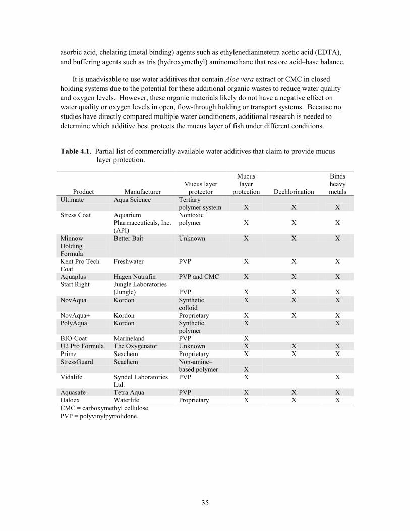
35
asorbic acid, chelating (metal binding) agents such as ethylenedianinetetra acetic acid (EDTA), and buffering agents such as tris (hydroxymethyl) aminomethane that restore acid–base balance.
It is unadvisable to use water additives that contain Aloe vera extract or CMC in closed holding systems due to the potential for these additional organic wastes to reduce water quality and oxygen levels. However, these organic materials likely do not have a negative effect on water quality or oxygen levels in open, flow-through holding or transport systems. Because no studies have directly compared multiple water conditioners, additional research is needed to determine which additive best protects the mucus layer of fish under different conditions.
Table 4.1. Partial list of commercially available water additives that claim to provide mucus layer protection.
Product
Manufacturer
Mucus layer
protector
Mucus layer
protection
Dechlorination
Binds heavy metals
Ultimate Aqua Science Tertiary polymer system
X
X
X
Stress Coat Aquarium Pharmaceuticals, Inc. (API)
Nontoxic polymer
X
X
X
Minnow Holding Formula
Better Bait Unknown X X X
Kent Pro Tech Coat
Freshwater PVP X X X
Aquaplus Hagen Nutrafin PVP and CMC X X X Start Right Jungle Laboratories
(Jungle) PVP
X
X
X
NovAqua Kordon Synthetic colloid
X X X
NovAqua+ Kordon Proprietary X X X PolyAqua Kordon Synthetic
polymer X X
BIO-Coat Marineland PVP X U2 Pro Formula The Oxygenator Unknown X X X Prime Seachem Proprietary X X X StressGuard Seachem Non-amine–
based polymer
X
Vidalife Syndel Laboratories Ltd.
PVP X X
Aquasafe Tetra Aqua PVP X X X Haloex Waterlife Proprietary X X X CMC = carboxymethyl cellulose. PVP = polyvinylpyrrolidone.
36
References Alexander JB, Ingram GA (1992) Noncellular nonspecific defence mechanisms of fish. Annual
Review of Fish Diseases (incorporated into Aquaculture) 2:249–279 Buermann Y, Du Preez HH, Steyn GJ, Smit L (1997) Tolerance levels of redbreast tilapia,
Tilapia rendalli (Boulenger, 1896) to natural suspended silt. Hydrobiologia 344:11–18 Cooke SJ, Schreer JF, Wahl DH, Phillip DP (2002) Physiological impacts of catch-and-release
angling practices on largemouth bass and smallmouth bass. American Fisheries Society Symposium 31:489–512
Eddy AE, Fraser JE (1982) Sialic acid and mucus production in rainbow trout (Salmo gairdneri)
in response to zinc in seawater. Comparative Biochemistry and Physiology 73C:357–359 Gilliland ER (2002) Livewell operating procedures to reduce mortality of black bass during
summer tournaments. American Fisheries Society Symposium 31:477–487 Handy RD, Eddy FB, Romain G (1989) In vitro evidence for the ionoregulatory role of rainbow
trout mucus in acid, acid/aluminum and zinc toxicity. Journal of Fish Biology 35:737–747 Ingram GA (1980) Substances involved in the natural resistance of fish to infection – a review.
Journal of Fish Biology 16:23–60 Muniz AH, Leivestad H (1980) Toxic effects of aluminum on the brown trout (Salmo trutta). In:
Drablos D, Tollan A (eds) Ecological impact of acid precipitation. SNSF Project, Oslo, Norway, pp 320–321
Nagashima Y, Sendo A, Shimakura K, Shiomi K, Kobayashi T, Kimura B, Fujii T (2001)
Antibacterial factors in skin mucus of rabbitfishes. Journal of Fish Biology 58:1761–1765 Ottesen OH, Olafsen JA (1997) Ontogenetic development and composition of the mucous cells
and the occurrence of saccular cells in the epidermis of Atlantic halibut. Journal of Fish Biology 50:620–633
Pickering AD (1974) The distribution of mucous cells in the epidermis of the brown trout Salmo
trutta (L.) and the char Salvelinus alpinus (L.). Journal of Fish Biology 6:111–118 Pickering AD, Macey DJ (1977) Structure, histochemistry and the effect of handling on the
mucous cells of the epidermis of the char Salvelinus alpinus (L.). Journal of Fish Biology 10:505–512
Pickering AD, Richards RH (1980) Factors influencing the structure, function and biota of the
salmonid epidermis. Proceedings of the Royal Society of Edinburgh 79B:93–104
37
Plumb JA, Grizzle JM, Rogers WA (1988) Survival of caught and released largemouth bass after containment in live wells. North American Journal of Fisheries Management 8:325–328
Roberts RJ, Bullock AM (1980) The skin surface ecosystem of teleost fishes. Proceedings of the
Royal Society of Edinburgh 79B:87–91 Rosen MW, Cornford NE (1971) Fluid friction of fish slimes. Nature 234:49–51 Shephard KL (1994) Functions for fish mucus. Reviews in Fish Biology and Fisheries 4:401–429 Stoskopf MK (1993) Fish medicine. W.B. Saunders Company, Philadelphia, PA Swanson C, Mager RC, Doroshov SI, and Cech Jr JJ (1996) Use of salts, anesthetics, and
polymers to minimize handling and transport mortality in delta smelt. Transactions of the American Fisheries Society 125:326–329
Wedemeyer GA (1996) Physiology of fish in intensive culture systems. Chapman & Hall,
International Thompson Publishing, New York

39
5 Anesthesia
Kathleen M. Carter, Christa M. Woodley, and Richard S. Brown
Anesthetics are used to immobilize fish during transport, handling, and surgical procedures by depressing their central and peripheral nervous systems (Summerfelt and Smith 1990). This nervous system depression renders fish immobile and reduces sensory perception during the procedure (Spath and Schweickert 1977; Hensel et al. 1975; Arnolds et al. 2002; Sneddon 2002). Although the capability of fish to feel pain is a highly controversial topic (for counterarguments, see Rose 2002, Sneddon et al. 2003, and Braithwaite and Boulcott 2007), considerable evidence suggests that using anesthetics increases the wellbeing of fish. Prolonged and/or cumulative stressors associated with fish husbandry and research, such as repeated handling, blood sampling, or surgery, can lead to several physiological changes that can ultimately leave fish susceptible to disease or predation (Wedemeyer et al. 1990; Barton 2002). The use of anesthetic agents has been shown to reduce physiological indicators of stress resulting from blood sampling (Iwama and Ackerman 1994; Wagner et al. 2003) and to reduce mortality when exposed to severe and repeated stressors (Strange and Schreck 1978). It is now considered standard practice to use anesthetic agents for humane handling of fish during stressful or invasive procedures (Olfert et al. 1993; AFS 2004). For this reason, it is essential for handlers to understand its proper use.
Our discussion focuses on the anesthetic tricaine methanesulfonate (TMS), known also as MS-222, ethyl 3-aminobenzoate methanesulfonic acid, metacaine, methanesulfonate, FinquelTM (Argent Chemical laboratories, Redmond, WA), or Tricaine-STM (Western Chemical, Inc., Ferndale, WA). TMS is a white odorless crystalline powder with a high solubility in water (1 g/0.8 mL at 20°C; 11%; Merck and Company 1983; Argent 2008; Sigma 2008). Storage areas for the powder form should be dark, dry, well-ventilated, and cool (Merck and Company 1989; Argent 2008; Sigma 2008). Reasons for this type of storage are discussed below. In the United States, TMS is the only legal anesthetic for use on a limited number of foodfish (FDA 2006) and is used widely in Columbia Basin fish-related projects. In the future, additional anesthetics may be legalized for use, so we emphasize that it is necessary to monitor progress and developments in the testing and regulation of other fish anesthetics.
The main purpose of this chapter is to instruct future fish surgeons on the use of TMS for anesthetizing fish. Within, we discuss important aspects and proper use of TMS as it relates to research in the Columbia Basin, mechanisms of action, and intended and unintended physiological side effects in fish.
TMS belongs to the barbiturate family, which has been widely studied but not entirely understood. The limited use of TMS in human and veterinary applications has resulted in
40
relatively little research into properties and physiological effects specific to TMS when compared to other barbiturates. Therefore, we also highlight areas of ambiguity in the literature regarding the use or effects of TMS.
Designated uses for tricaine methanesulfonate
TMS is classified as an ester-type synthetic local anesthetic commonly used in the fisheries industry (Sato et al. 2000). The limitations on TMS use with food fish include a 21-day waiting period before harvesting, and use is restricted to the families Ictaluridae (catfishes), Salmonidae (salmon, trout, char, whitefish, and grayling), Esocidae (pike and pickerel), and Percidae (perch, walleye, ruffe, and darters) (FDA 2006). For other species, the drug should be limited to hatchery or laboratory applications in which fish will not be released into the wild. According to the U.S. Food and Drug Administration (FDA) and manufacturers, water temperatures during the 21-day waiting period should exceed 10°C (50°F; FDA 2006; Argent 2008; Western 2008). However, a Freedom of Information Act factsheet (FDA 1997) stated that water should not exceed 10°C (50°F) during the 21-day period. The reason for this discrepancy is not clear and possibly a typographical error. The authors were not successful at confirming this suspicion. Storage and preparation
Although there is evidence that TMS is not mutagenic (Yoshimura et al. 1981), humans should be aware that TMS is retinotoxic and an irritant to mucous membranes, including the upper respiratory tract (Berstein et al. 1997; Argent 2008; Sigma 2008). Prior to handling or using, one should read manufacturer-specific material safety data sheets (MSDSs) and avoid contact with the powder or solution by using accepted personal protective equipment. These can include gloves, eye protection, and general skin protection when dealing with all forms of the chemical; and masks and/or fume hoods when dealing with the powdered form (Argent 2008; Sigma 2008). Additionally, hand washing after handling TMS is recommended.
TMS in powder form easily can become airborne and could become hazardous to the handler. Due to the difficulty of handling powdered TMS in situations in which proper personal protective equipment may not be readily available (e.g., field settings), some researchers pre-mix and store TMS stock solutions in dark bottles. Although this practice is widely used, there is some question as to how long TMS solutions can stand without degradation. Based on Bové (1962) and fact sheets on Argent Laboratories Finquel (2008) and Western Chemical Tricaine-S (2008), no significant degradation of TMS stock solution occurred after 3 d of standing. However, a 5% activity decrease was observed within 10 d. In contrast, the Sigma-Aldrich MSDS (2008) stated TMS solutions degrade quickly and therefore recommended solutions be made immediately prior to use. Further diverging from Bové (1962), ALPHARMA (2001) reported that stock solutions were stable for 1 month when stored in dark or opaque bottles in cool environments. Ross and Ross (2008) concur these solutions are stable but indicate that solutions should be stored for only 3 months in dark, sealed containers that are cooled. Neither ALPHARMA (2001) nor Ross and Ross (2008) provided a TMS activity degradation curve at a specific temperature.
Some manufacturers agree that TMS is photosensitive (its color changes with light exposure), although the color change does not indicate significant activity decrease (Bové 1962; Argent 2008; Western 2008) and cannot be used to gauge degradation. In contrast, Bell (1987) stated
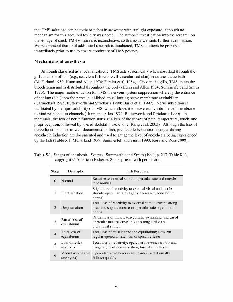
41
that TMS solutions can be toxic to fishes in seawater with sunlight exposure, although no mechanism for this acquired toxicity was noted. The authors’ investigation into the research on the storage of stock TMS solutions is inconclusive, so this issue warrants further examination. We recommend that until additional research is conducted, TMS solutions be prepared immediately prior to use to ensure continuity of TMS potency.
Mechanisms of anesthesia
Although classified as a local anesthetic, TMS acts systemically when absorbed through the gills and skin of fish (e.g., scaleless fish with well-vascularised skin) in an anesthetic bath (McFarland 1959; Hunn and Allen 1974; Fereira et al. 1984). Once in the gills, TMS enters the bloodstream and is distributed throughout the body (Hunn and Allen 1974; Summerfelt and Smith 1990). The major mode of action for TMS is nervous system suppression whereby the entrance of sodium (Na+) into the nerve is inhibited, thus limiting nerve membrane excitability (Carmichael 1985; Butterworth and Strichartz 1990; Burka et al. 1997). Nerve inhibition is facilitated by the lipid solubility of TMS, which allows it to move easily into the cell membrane to bind with sodium channels (Hunn and Allen 1974; Butterworth and Strichartz 1990). In mammals, the loss of nerve function starts as a loss of the senses of pain, temperature, touch, and proprioception, followed by loss of skeletal muscle tone (Rang et al. 2003). Although the loss of nerve function is not as well documented in fish, predictable behavioral changes during anesthesia induction are documented and used to gauge the level of anesthesia being experienced by the fish (Table 5.1; McFarland 1959; Summerfelt and Smith 1990; Ross and Ross 2008).
Table 5.1. Stages of anesthesia. Source: Summerfelt and Smith (1990, p. 217, Table 8.1), copyright © American Fisheries Society; used with permission.
Stage Descriptor Fish Response
0 Normal Reactive to external stimuli; opercular rate and muscle tone normal
1 Light sedation Slight loss of reactivity to external visual and tactile stimuli; opercular rate slightly decreased; equilibrium normal
2 Deep sedation Total loss of reactivity to external stimuli except strong pressure; slight decrease in opercular rate; equilibrium normal
3 Partial loss of equilibrium
Partial loss of muscle tone; erratic swimming; increased opercular rate; reactive only to strong tactile and vibrational stimuli
4 Total loss of equilibrium
Total loss of muscle tone and equilibrium; slow but regular opercular rate; loss of spinal reflexes
5 Loss of reflex reactivity
Total loss of reactivity; opercular movements slow and irregular; heart rate very slow; loss of all reflexes
6 Medullary collapse (asphyxia)
Opercular movements cease; cardiac arrest usually follows quickly
42
Once inside the body, TMS concentrations are metabolized rapidly by acetylation reactions and excreted (Burka et al. 1997). The major excretion route for TMS and its non-polar metabolites is through the gills (Wayson et al. 1976). Unmetabolized TMS and its more polar metabolites are excreted via the kidneys (Hunn and Allen 1974; Wayson et al. 1976; Burka et al. 1997). The estimated plasma half-life of TMS is 1.5 to 4 h (Hunn and Allen 1974). After 8 and 24 h, TMS is undetectable in whole blood and urine, respectively (Hunn and Allen 1974; Ohr 1976; Wayson et al. 1976; Burka et al. 1997). Local TMS injections are ineffective because the drug is eliminated too quickly to induce anesthesia (Allen and Hunn 1986; Burka et al. 1997). Therefore, for the vast majority of procedures involving fish, TMS is administered by immersion in an anesthetic bath and, when appropriate, followed by continuous irrigation of the gills with anesthetic solution. These TMS administration approaches allow for a continual uptake of TMS by the gills for the period during which the state of anesthesia is needed.
The potency of anesthetic agents similar to TMS, such as 2-phenoxyethanol, benzocaine, tetracaine, and lidocaine, can increase dramatically with cooler temperatures (Sehdev et al. 1963; Butterworth and Strichartz 1990). Although pharmacokinetics have not been directly studied in fish, changes in pharmacokinetics at cooler temperatures and slowed diffusion rates decrease the clearance rate from the nerve site (Ohr 1976; Butterworth and Strichartz 1990). Thus, at 10°C, a lower dose may more readily block nerve function than at 22°C. Administration Induction of anesthesia
Differential exposure time and dosage of TMS can induce stages of anesthesia in fish corresponding to differing states of narcosis and neural functioning. As neural function decreases, fish exhibit predictable changes in behavior that can be used to gauge the current level of anesthesia (as reviewed in Table 5.1; McFarland 1959). The rate of decline for neural function, as well as the level to which it declines, varies primarily with the anesthetic dosage due to the rapid diffusion of TMS across the gill (Hunn and Allen 1974). For minor handling procedures (e.g., measurements, blood samples) or transport, lower dosages (15–30 mg of TMS/L of water for Salmonidae) result in tranquilization and light sedation (Stages 1–2), which can be held for long periods (Schoettger and Julin 1967). Major procedures require higher doses (60–100 mg/L) to quickly (within 4 min) induce deep anesthesia levels (Stages 4–5; Schoettger and Julian 1967; Hunn and Allen 1974; Summerfelt and Smith 1990).
For invasive procedures such as transmitter implantation into the peritoneal cavity, the fish must be in a deep level of anesthesia (≥Stage 4) to be rendered completely immobile. Several authors have suggested an ideal anesthetic should induce Stage 4 anesthesia quickly (in under 3 min) while allowing for quick recovery (less than 5 min; Marking and Meyer 1985; Bell 1987; Iwama and Ackerman 1994). Because TMS moves across the gills rapidly and is metabolized readily, blood concentrations may diminish if bath concentrations are not high enough to overcome the speed at which the active form of the drug leaves the body (Hunn and Allen 1974). TMS moves quickly into the blood to be dispersed throughout the body and can peak in 1 to 3 min (Hunn and Allen 1974). Higher doses will induce and maintain Stage 4 anesthesia quickly. However, the risk for side effects would increase if fish were not able to be processed in a timely manner (Marking and Meyer 1985; Bell 1987; Iwama and Ackerman 1994).
43
Exact dosages for salmonids vary with anesthesia stage targeted, species, body size, health, age and life stage, and water quality (Summerfelt and Smith 1990; Ross and Ross 2008). In addition, the tolerance of salmonids for specific dosages varies between stocks and/or sexes (Marking 1967; Schoettger and Julian 1967; Houston and Corlett 1976; Burka et al. 1997). The optimal TMS dosage to induce anesthesia varies between 60 mg/L and 100 mg/L (Iwama and Ackerman 1994). Induction and recovery times are inversely correlated with body weight, especially for salmon (Burka et al. 1997; Houston and Corlett 1976). Water quality parameters, such as temperature, pH, salinity, and hardness, can affect metabolic rate, acid–base regulation, and osmoregulation and ion regulation (Schoettger and Julin 1967; Heisler 1988; Iwama et al. 1989; Perry and Gilmour 2006). These factors can also affect the pharmacodynamics of TMS (Marking 1967; Ohr 1976). Before a full study and fish-handling are initiated, or if environmental conditions have changed, it is recommended that sample fish be tested by anesthetizing at the desired TMS dosage (Schoettger and Julian 1967; Iwama and Ackerman 1994; Argent 2008; Western Chemical 2008). When possible, fish should be monitored for 24 to 48 h after anesthetic administration and the associated procedure to ensure full recovery.
Anesthetic baths (whether induction, surgery table, or recovery) should be changed when the temperature varies from ambient water temperature by more than 2°C, when the bath becomes noticeably frothy or cloudy, or when reduced water quality is suspected due to blood, fish excretion, and/or mucus buildup (Summerfelt and Smith 1990; Portz et al. 2006). Anesthetic baths also should be changed when induction or recovery times are noticed to increase or decrease, respectively. Changes in induction and recovery times occur due to the decay of TMS within the induction bath as it becomes metabolized or diluted with the physical movement of fish into and out of the induction bath (Schoettger and Julin 1967; Burka et al. 1997; Summerfelt and Smith 1990).
TMS has side effects, such as changes to the cardiovascular and endocrine systems as well as to osmoregulation and ion regulation. During deeper levels of anesthesia when opercular movements are slowed and respiration is depressed, severe hypoxia and respiratory and metabolic acidosis can develop (Iwama et al. 1989). Reduced blood flow through the gills and the concomitant increased anatomical and physiological dead spaces significantly slow the exchange of gas (oxygen and carbon dioxide) across the gills. Declined exchange results in a consequent reduction of blood oxygen tension (Figure 5.1A), an increase in blood carbon dioxide tension, and a concomitant decrease in blood pH (Figure 5.1B; Iwama et al. 1989; Iwama and Ackerman 1994; Hill and Forster 2004). If allowed to continue, the condition results in hypoxemia. Depressed respiration combined with the change in blood oxygen and carbon dioxide levels can cause hypotension and changes to heart rate and cardiac output (Randall 1962; Houston et al. 1971; Hill and Forster 2004). Therefore, it is important to aerate both induction and recovery baths as well as the anesthetic solution being used for gill irrigation during any surgical procedures. Aeration will increase passive gas exchange at the gills (Summerfelt and Smith 1990; Ross and Ross 2008).
In addition to elevating catecholamine levels, TMS exposure increases the level of circulating cortisol (Iwama et al 1989; Molinera and Gonzalez 1995; Mommsen et al. 1999). Cortisol, a principal corticosteroid that plays a role in intermediary metabolism, ion regulation and osmoregulation, and immune function, has been shown to cause swelling of erythrocytes (red blood cells) (Soivio et al. 1977; Iwama et al. 1989; Mommsen et al. 1999). This swelling can
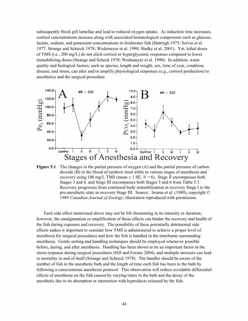
44
subsequently block gill lamellae and lead to reduced oxygen uptake. As induction time increases, cortisol concentrations increase along with associated hematological components such as glucose, lactate, sodium, and potassium concentrations in freshwater fish (Hattingh 1975; Soivio et al. 1977; Strange and Schreck 1978; Wedemeyer et al. 1990; Sladky et al. 2001). Yet, lethal doses of TMS (i.e., 200 mg/L) do not elicit cortisol or hyperglycemic responses compared to lower immobilizing doses (Strange and Scheck 1978; Wedemeyer et al. 1990). In addition, water quality and biological factors, such as species, length and weight, sex, time of year, condition, disease, and stress, can alter and/or amplify physiological responses (e.g., cortisol production) to anesthetics and the surgical procedure.
Figure 5.1. The changes in the partial pressure of oxygen (A) and the partial pressure of carbon
dioxide (B) in the blood of rainbow trout while in various stages of anesthesia and recovery using 100 mg/L TMS (mean ± 1 SE. N = 6). Stage II encompasses both Stages 3 and 4, and Stage III encompasses both Stages 5 and 6 from Table 5.1. Recovery progresses from continued body immobilization in recovery Stage I to the pre-anesthetic state in recovery Stage III. Source: Iwama et al. (1989), copyright © 1989 Canadian Journal of Zoology; illustration reproduced with permission.
Each side effect mentioned above may not be life threatening in its intensity or duration;
however, the amalgamation or amplification of these effects can hinder the recovery and health of the fish during exposure and recovery. The possibility of these potentially detrimental side effects makes it important to consider how TMS is administered to achieve a proper level of anesthesia for surgical procedures and how the fish is handled in the timeframe surrounding anesthesia. Gentle netting and handling techniques should be employed whenever possible before, during, and after anesthesia. Handling has been shown to be an important factor in the stress response during surgical procedures (Hill and Forster 2004), and multiple stressors can lead to mortality in and of itself (Strange and Schreck 1978). The handler should be aware of the number of fish in the anesthetic bath and the length of time each fish has been in the bath by following a conscientious anesthesia protocol. This observation will reduce avoidable differential effects of anesthesia on the fish caused by varying times in the bath and the decay of the anesthetic due to its absorption or interaction with byproducts released by the fish.
45
To limit hypoxemia and acute stress responses, induction and handling times should be minimized. It is recommended that stages required for surgery, like Stage 4, should be achieved in less than 3 min (Wedemeyer et al 1990; Wedemeyer 1997; Mommsen et al. 1999; Wagner et al. 2003). Focused research into physiological differences between longer and shorter induction times is somewhat scarce. However, many of the negative physiological effects occur in deeper stages of anesthesia and as fish approach and reach medullary collapse (Stage 3 and higher; Schoettger and Julin 1967; Iwama et al. 1989; Burka et al. 1997). Therefore, decreasing time spent in these stages by increasing the dosage of TMS is warranted. When large numbers of fish are expected to be implanted, higher dosages of anesthetic could improve fish induction time in the bath, thus reducing numbers of fish in the anesthetic bath at any one time. This method would not only reduce the time each fish is in the anesthetic solution but also allow for more flexibility when managing anesthetized fish in case of processing delays. Maintenance of anesthesia during surgery
Once fish are placed on the surgery table, an anesthetic maintenance dose (a lower dose than the induction anesthetic bath) is necessary to maintain Stage 4 anesthesia for a short (<10-min) surgical procedure. A well-oxygenated maintenance dose can be delivered via a flexible hose that is placed in the mouth to irrigate the fish’s gills. The flow rate should be adequate to ensure sufficient gas exchange across the gills. Published guidelines, although vague, can be used to estimate a sufficient maintenance dose to maintain a fish at the appropriate level of anesthesia (Schoettger and Julin 1967; Ross and Ross 2008). Therefore, a maintenance water system that incorporates two water sources—one containing anesthetic and one with fresh water—and mixes them just before reaching the gills is preferred for anesthesia maintenance, fish stress minimization, and surgeon ease. In the field, this method can be easily set up using two buckets (one containing TMS water and one containing fresh water) and a hose from each that converges at or before the fish’s mouth. This method allows the surgeon to control the level of anesthesia during the surgical procedure. After the surgical procedure is completed, the fish should be transferred immediately to a recovery bath containing fresh, aerated water and monitored to ensure recovery. Buffering the anesthetic bath
In general, salmonids should be maintained in waters with a pH range of 6.5 to 9.0 to avoid experiencing detrimental changes in physiological functions (Piper et al. 2001). Dissolving TMS in freshwater can reduce the anesthetic bath pH due to the hydrolysis of the sulfonate radical (Ohr 1976). If the water in the bath does not have adequate buffering capacity, the subsequent pH drop from the TMS additions could alter the water pH below the ideal physiological range of a given species. Fish held in acidic environments (pH ≤ 5) have difficulty maintaining many physiological functions (Iwama et al. 1989; Burka et al. 1997). In low-pH conditions, disturbances in ionic and osmotic balance can lead to haemoconcentration (Iwama et al. 1989; Burka et al. 1997), increased blood pressure (Milligan and Wood 1982), and suppressed metabolic rate (Packer 1979; Pelster and Randall 1998).
The potency of TMS decreases with decreasing pH (Ohr 1976). In acidic environments with a pH of 5.5 or less, the anesthetic portion of triciane is positively charged, which inhibits its diffusion across the gill surface (Ohr 1976; Ross and Ross 2008). Therefore, induction time
46
increases inversely with pH at these low values, increasing the time in the anesthetic bath. Reduced pH after the addition of TMS can be counteracted by adding buffers, such as sodium bicarbonate (NaHCO3), tris (tris [hydroxymethyl] aminomethane; [HOCH2]3CNH2), or sodium hydroxide (NaOH), to the water bath. The need for additional buffers to maintain an acceptable pH (6.5 to 9) will depend on the dose of TMS used and the alkalinity of the local water source (Summerfelt and Smith 1990). Saltwater and freshwater with higher alkalinity contain enough buffers to maintain an acceptable pH (Piper et al 2000). However, waters without sufficient alkalinity can experience wide shifts in pH if the addition of TMS overcomes the buffering capacity. Therefore, it is important to check the pH of the anesthetic bath before its use at each new location or when a change in local water chemistry is suspected (Summerfelt and Smith 1990; Iwama and Ackerman 1994). Areas in which future research is needed
The long-term effects of TMS exposure on salmonids and other fish species after surgical implantation of transmitters have not been thoroughly elucidated. Therefore, more research is needed to determine these effects. In addition, research exploring ideal storage protocols for stock TMS solutions and the physiological effects of various induction and recovery times needs to be conducted to increase the safety and efficacy of this anesthetic.
There is a need for additional laboratory research on other anesthetics because some aspects of TMS are undesirable. The required withdrawal period for potential food fish, the need for personal protective equipment (sometimes difficult in a field setting), and the ease of accidental overdose and sublethal negative physiological effects make TMS less than ideal as an anesthetic for fish. Considerations for use of tricaine methanesulfonate in Columbia Basin fish research
Guidelines for using TMS for implanting acoustic transmitters have been formulated based on review of the available literature on anesthetic use in fish. Before a study begins, fish management strategies should be designed to decrease handling stress. This approach would include minimizing tank transfers from the time the fish are captured through their release and using sanctuary nets or buckets to avoid stress elicited from air exposure (Strange and Schreck 1978; Hill and Forster 2004). The pH of the anesthetic bath should be monitored before and during the anesthesia process to ensure a neutral range. Stage 4 anesthesia should be induced quickly (within 3 to 4 min). For juvenile Chinook salmon, this process often requires TMS doses of 80 to 100 mg/L (based on recommendations from Wedemeyer et al 1990; Wedemeyer 1997; Mommsen et al. 1999; Wagner et al. 2003). The effects of TMS can vary with temperature, species, fish size, and physiological state. Fish should be monitored closely while in the induction bath to ensure that Stage 4 anesthesia is met within this timeframe.
After induction, fish can be maintained with a lower dose of TMS. Previous experience with Chinook salmon in the Columbia Basin has shown a dose of 40 mg/L to be adequate for anesthetic maintenance. Surgeons can manipulate the exact dose to each fish by having access to freshwater and water containing TMS. Maintenance anesthesia (typically 40 mg/L) and freshwater (no anesthetic) can be gravity-fed or pumped through hoses that converge before reaching the fish. This method allows the surgeon to manipulate the composition of the
47
maintenance anesthetic from purely fresh water to water containing 40 mg TMS/L to supply the lowest dosage needed (Schoettger and Julin 1967; Ross and Ross 2008). Surgeons should observe the stages of anesthesia and maintain the fish in Stage 4 during the entire surgery.
The water quality of the anesthetic and maintenance baths should be monitored. Buffers should be added if the induction anesthetic bath pH falls below 7 after the addition of TMS. These buffers should be used to maintain the bath pH at the same level as that of river water at the study site. Due to possible fluctuations, the pH should be checked daily during the study period (Iwama and Ackerman 1994; Piper et al 2000). Baths should be aerated or oxygenated to prevent hypoxia and to maintain consistent concentrations of oxygen. A bath should be changed when the water temperature varies more than 2°C from the fish’s acclimated temperature, as well as when it becomes frothy or fouled from excess mucus or other contaminants. If the maintenance anesthetic bath is pumped and recirculated over several surgeries, care should be taken to avoid contaminating the maintenance anesthetic bath with blood. At any sign of blood or excess mucus, the maintenance anesthetic bath should be changed. Following these guidelines will minimize stress to the fish and maximize the success of the study due to increased general health and survival rates of the fish.
48
References AFS (American Fisheries Society) (2004) Guidelines for the use of fishes in research. American
Fisheries Society Press, Bethesda, MD Argent (Argent Laboratories) (2008) Anesthetics and tranquilizers – Finquel (MS-222 or
tricaine). Available via http://www.argent-labs.com/argentwebsite/trandrug.htm. Accessed 07 April 2009
ALPHARMA (Animal Health Limited) (2001) MS222 (Tricaine methane sulphonate). Technical
Bulletin. Available via www.europharma.no/fiskehelse/datablad/bedovelse/MS222.pdf. Accessed 10 April 2009
Arnolds DE, Zottoli SJ, Adams CE, Dineen SM, Fevrier S, Guo Y, Pascal AJ (2002)
Physiological effects of tricaine on the supramedullary/dorsal neurons of the cunner, Tautogolabrus adspersus. The Biological Bulletin 203:188–189
Barton BA (2002) Stress in fishes: a diversity of responses with particular reference to changes in
circulating corticosteroids. Integrative and Comparative Biology 42(3):517–525 Bell GR (1987) An outline of anesthetic and anesthesia for salmonids, a guide for fish culturists
in British Columbia. Canadian Technical Report of Fisheries and Aquatic Sciences No. 1534 Berstein PS, Digre KB, Creel DJ (1997) Retinal toxicity associated with occupational exposure to
the fish anesthetic MS-222. American Journal of Ophthalmology 124:834–843 Bové FJ (1962) MS-222 Sandoz. The anesthetic of choice for fish and other cold-blooded
organisms. Sandoz News 3. Braithwaite VA, Boulcott P (2007) Pain perception and fear in fish. Diseases in Aquatic
Organisms 75:131–138 Burka JF, Hammel KL, Horsberg TE, Johnson GR, Rainnie DJ, Spears DJ (1997) Drugs in
salmonid aquaculture – a review. Journal of Veterinary Pharmacology and Therapeutics 20:333–349
Butterworth JF, Strichartz GR (1990) Molecular mechanisms of local anesthesia: a review.
Anesthesiology 72(4):711–734 Carmichael FJ (1985) General anaesthetics and local anaesthetics. In: Kalant H, Roschlau WHE,
Sellers EM (eds) Principles of medical pharmacology, 4th edn. Department of Pharmacology, University of Toronto, Toronto, Ontario, pp 265–289
Ferreira JT, Schoonbee HG, Smit GL (1984) The uptake of the anaesthetic benzocaine
hydrochloride by the gills and skin of three freshwater fish species. Journal of Fish Biology 25:35–41
49
FDA (U.S. Food and Drug Administration) (1997) Freedom of Information Summary for NADA 200-226. Available via http://www.fda.gov/cvm/FOI/1635.htm. Accessed 07 April 2009
FDA (U.S. Food and Drug Administration) (2006) Database of approved animal drug products.
U.S. Department of Health and Human Services, Food and Drug Administration, Center for Veterinary Medicine. VMRCVM Drug Information Laboratory. Available via http://www.accessdata.fda.gov/scripts/AnimalDrugsAtFDA/. Accessed 10 December 2009
Hattingh J (1977) The effect of tricaine methanesulphonate (MS-222) on the microhaematocrit of
fish blood. Journal of Fish Biology 10(5):453–455 Heisler N (1988) Acid-base regulation. In: Shuttleworth TJ (ed) Physiology of elasmobranch
fishes. Springer-Verlag, Berlin, pp 215–252 Hensel H, Bromm B, Nier K (1975) Effect of ethyl m-aminobenzoate (MS-222) on ampullae of
lorenzini and lateral-line organs. Experientia 31:958–960 Hill JV, Forster ME (2004) Cardiovascular responses of Chinook salmon (Oncorhynchus
tshawytscha) during rapid anaesthetic induction and recovery. Comparative Biochemistry and Physiology C–Toxicology and Pharmacology 137(2):167–177
Houston AH, Corlett JT (1976) Specimen weight and MS-222. Journal of the Fisheries Research
Board of Canada 33(6):1403–1407 Houston AH, Madden JA, Woods RJ, Miles HM (1971) Some physiological effects of handling
and tricaine methane-sulphonate anesthetization upon the brook trout, Salvelinus fontanalis. Journal of the Fisheries Research Board of Canada 28:625–633
Hunn JB, Allen JL (1974) Movement of drugs across the gills of fishes. Annual Review of
Pharmacology 14:47–55 Iwama GK, Ackerman PA (1994) Anaesthetics. In: Hochachka P, Mommsen T (eds)
Biochemistry and molecular biology of fishes, vol 3 – Analytical Techniques. Elsevier Science, Amsterdam, pp 1–15
Iwama GK, McGeer JC, Pawluk MP (1989) The effects of five fish anaesthetics on acid–base
balance, hematocrit, cortisol, and adrenaline in rainbow trout. Canadian Journal of Zoology 67:2065–2073
Marking LL (1967) Toxicity of MS-222 to selected fishes. In: Investigations in fish control.
Resource Publication 18, U.S. Department of the Interior, Bureau of Sport Fisheries and Wildlife, Washington, DC, pp 3–10
Marking LL, Meyer FP (1985) Are better anesthetics needed in fisheries? Fisheries 10:2–5
50
McFarland WN (1959) A study of the effects of anesthetics on the behavior and physiology of fishes. Publication of the Institute of Marine Science, University of Texas at Austin 6:23–55
Merck and Company (1989) The Merck index, 11th edn. Merck and Company, Inc., Rahway, NJ Milligan CL, Wood CM (1982) Disturbances in haematology, fluid volume distribution and
circulatory function associated with low environmental pH in the rainbow trout, Salmo gairdneri. Journal of Experimental Biology 99:397–415
Molinero A, Gonzalez J (1995) Comparative effects of MS-222 and 2-phenoxyethanol on
gilthead sea bream (Sparus aurata L.) during confinement. Comparative Biochemistry and Physiology A–Molecular and Integrative Physiology 111(3):405–414
Mommsen TP, Vijayan MM, Moon TW (1999) Cortisol in teleosts: dynamics, mechanisms of
action, and metabolic regulation. Reviews in Fish Biology and Fisheries 9:211–268 Ohr EA (1976) Tricane methanesulphonate – l, pH and its effects on anaesthetic potency.
Comparative Biochemistry and Physiology C–Toxicology and Pharmacology 54:13–17 Olfert DE, Cross BM, McWilliam AA (1993) Guide to the care and use of experimental animals,
2nd edn, vols 1 and 2. Canadian Council on Animal Care, Ottawa, Ontario, Canada Packer RK (1979) Acid-base balance and gas exchange in brook trout (Salvelinus fontainalis)
exposed to acidic environments. Journal of Experimental Biology 79:127–134 Pelster B, Randall D (1998) The physiology of the root effect. In: Perry SF, Tufts B (eds) Fish
physiology, vol 17. Academic Press, New York, pp 113–139 Perry SF, Gilmour KM (2006) Acid-base balance and CO2 excretion in fish: unanswered
questions and emerging models. Respiratory Physiology & Neurobiology 154:199–215 Piper RG, McElwain IB, Orme LE, McCraren JP, Fowler LG, Leonard JR (eds) (2000)
Chapter 1: Hatchery requirements. In: Wedemeyer GA (ed) Fish hatchery management. American Fisheries Society, Bethesda, MD, pp 11–12
Portz DE, Woodley CM, Cech Jr CC (2006) Stress-associated impacts of short-term holding on
fishes. Reviews in Fish Biology and Fisheries 16(2):125–170 Randall DJ (1962) Effect of an anaesthetic on the heart and respiration of teleost fish. Nature
195:506–508 Rang HP, Dale MM, Ritter JM, Moore PK (2003) Chapter 43: Local anesthetics. In: Rang HP,
Dale MM, Ritter JM, Moore PK (eds) Pharmacology, 5th edn. Churchill Livingstone, Edinburgh, Scotland
51
Rose JD (2002) The neurobehavioral nature of fishes and the question of awareness and pain. Reviews in Fisheries Science 10:1–38
Ross LG, Ross BR (2008) Anaesthetic and sedative techniques for aquatic animals. Blackwell
Science, Oxford, United Kingdom Sato T, Kitayama S, Mitsuhata C, Ikeda T, Morita K, Dohi T (2000) Selective inhibition of
monoamine neurotransmitter transport by synthetic local anesthetics. Naunyn-Schmiedeberg’s Archives of Pharmacology 361:214–220
Schoettger RA, Julin AM (1967) Efficacy of MS-222 as an anaesthetic on four salmonids. In:
Investigations in fish control, Resource Publication 19. U.S. Department of the Interior, Bureau of Sport Fisheries and Wildlife, Washington, DC, pp 3–15
Sehdev HS, McBride JR, Fagerlund UHM (1963) 2-phenoxyethanol as a general anesthetic for
sockeye salmon. Journal of the Fisheries Research Board of Canada 20:1435–1440 Sigma (2008) Material safety data sheet: ethyl 3-aminobenzoate methanesulfonate, E10521.
Sigma-Aldrich Corporation. Available via http://www.sigmaaldrich.com/site-level/msds.html. Accessed 10 April 2009
Sladky KK, Swanson CR, Stoskopf MK, Loomis MR, Lewbart GA (2001) Comparative efficacy
of tricaine methanesulfonate and clove oil for use as anesthetics in red pacu (Piaractus brachypomis). American Journal of Veterinary Research 62(3):337–342
Sneddon LU (2002) Anatomical and electrophysiological analysis of the trigeminal nerve in a
teleost fish, Oncorhynchus mykiss. Neuroscience Letters 319:167–171 Sneddon LU, Braithwaite VA, Gentle MJ (2003) Do fish have nociceptors? Evidence for the
evolution of a vertebrate sensory system. Proceedings of the Royal Society B–Biological Sciences 270:1115–1121
Sovio A, Nyholm K, Huhti M (1977) Effects of anesthesia with MS222, neutralized MS222 and
benzocaine on the blood constituents of rainbow trout, Salmo gairdneri. Journal of Fish Biology 10:91–101
Spath M, Schweickert W (1977) The effect of metacaine (MS-222) on the activity of the efferent
and afferent nerves in the teleost lateral-line system. Naunyn-Schmiedeberg’s Archives of Pharmacology 297:9–16
Strange RJ, Schreck CB (1978) Anesthetic and handling stress on survival and cortisol
concentration in yearling Chinook salmon (Oncorhynchus tshawytscha). Journal of the Fisheries Research Board of Canada 35:345–349
52
Summerfelt RC, Smith LS (1990) Anesthesia, surgery and related techniques. In: Schreck CB, Moyle PB (eds) Methods for fish biology. American Fisheries Society, Bethesda, MD, pp 213–272
Wagner GN, Singer TD, McKinley RS (2003) The ability of clove oil and MS-222 to minimize
handling stress in rainbow trout (Oncorhynchus mykiss Walbaum). Aquaculture Research 34:1139–1146
Wayson KA, Downes H, Lynn RK, Gerber N (1976) Studies on the comparative pharmacology
and selective toxicity of tricaine methanesulfonate: metabolism as a basis of the selective toxicity in poikilotherms. Journal of Pharmacology and Experimental Therapeutics 198(3):695–708
Wedemeyer GA, Barton BA, McLeay D (1990) Stress and acclimation. In: Schreck CB, Moyle
PB (eds) Methods for fish biology. American Fisheries Society, Bethesda, MD, pp 451–489 Wedemeyer GA (1997) Effects of rearing conditions on the health and physiological quality of
fish in intensive culture. In: Iwama GK, Pickering AD, Sumpter JP, Schreck CB (eds) Fish stress and health in aquaculture. Cambridge University Press, Cambridge, pp 35–72
Western (Western Chemical, Inc.) (2008a) Fact sheet: Tricaine-S. Available via
http://www.wchemical.com/Assets/File/tricaineS_instructions.pdf. Accessed 07 April 2009 Western (Western Chemical, Inc.) (2008b) Material safety data sheet: Tricaine-S. Available via
http://www.wchemical.com/Assets/File/tricaineS_MSDS.pdf. Accessed 07 April 2009 Yoshimura H, Nakamura M, Koeda T (1981) Mutagenicity screening of anesthetics for fishes.
Mutation Research 90:119–124
53
6 Incision Closure and Surgery
Glenn N. Wagner, Steven J. Cooke, Richard S. Brown, and Katherine A. Deters
The general guidelines for surgical procedures on fish are to know the anatomy, minimize tissue damage and surgery times, and to not cut unknown tissues (Murray 2002). Adherence to these guidelines minimizes discomfort to the fish because small, clean wounds that are sutured properly heal quickly with minimal complications (Mulcahy 2003). Therefore, the size of the incision, suture material, and needles to be used are dependent largely on the size of fish involved and the subsequent size of transmitter to be implanted. Along with minimizing the amount of tissue damage, it is important to use general aseptic techniques to minimize the risk of infections (Mulcahy 2003; CCAC 2005). These techniques help to maximize healing rates, retain the transmitter in the body cavity of the fish, and reduce the chance of introducing pathogens into the body.
This chapter reviews the literature available related to making incisions, incision closure, and other related surgery techniques. In some cases, the only available information pertains to studies performed on mammals. The protocols developed from these reviews are intended to provide the best available scientifically based methods for implantation of acoustic transmitters in juvenile salmonids. In many cases, the optimal procedures are unknown, so suggestions for future research are provided as well. Surgical tools
Implantation of acoustic transmitters requires the use of surgical tools to create and close the incision. Although many tool options are available and personal preference may differ among surgeons, it is important to use tools that are appropriate for the application (Dunn and Philips 2004). For implantation of juvenile salmon, the tools should be smaller and more delicate than those used on adults because the fish and transmitters are smaller.
Typically, hundreds of juvenile salmon smolts are tagged daily on USACE projects; thus, choosing ergonomic and efficient tools is important. A scalpel, forceps, and needle holder are needed for creating and closing the incision. Scalpel blades that remain sharp after repeated use are ideal for both cost and time efficiency purposes. Due to the thin body wall of juvenile salmon and the small length of incision needed, the use of large surgical blades (e.g., size 10) is not suggested. A smaller blade, such as a size 15, is preferred, while a micro blade, such as the Becton-Dickinson Micro-Sharp Blade or Micro-Unitome Knife by BD (~1.5–3.0 mm cutting tip), is a good alternative. Some surgeons prefer to use the micro blade to start the incision (i.e., make a cut through the skin), followed by a surgical blade (e.g., size 15) to finish the incision through to the abdominal cavity. Surgeons implanting transmitters in juvenile salmonids typically use small forceps (~10 cm long) with blunt curved tips that are approximately 0.5 mm wide. In addition, relatively small (~12 cm long) needle holders with a small tip width (~1.5 mm) and
54
clamping length (~8.5 mm) commonly are used. Having needle holders with integrated scissors (i.e., Olsen-Hegar needle holders) also reduces the need for additional tools.
It is important to properly clean and dry surgical tools used in the aquatic environment to avoid corrosion and other physical damage. Also recommended is the veterinary practice of surgical instrument sterilization prior to and between surgical procedures on fish (CCAC 2005). Typically, the need for sterilization and disinfection is considered on a scale that depends on the risk of infection to the patient, based on use of a given instrument (Spaulding 1968). In the case of fish surgery, the risk of infection is considered high for any instrument used in the surgical procedure. As such, these tools are considered critical items and require the most stringent of sterilization procedures because any microbial contamination could result in disease transmission (Spaulding 1968). Sterilization is relevant not only to the surgical instruments but also to the transmitters being implanted (Mulcahy 2003) and possibly the surgical gloves (Korniewicz et al. 1991). Unlike human physicians and veterinarians, the majority of fish surgeons use nonsterile latex gloves, likely because these surgeries are performed in a nonsterile environment.
Heat and chemical sterilization can completely eliminate all microorganisms, including bacterial endospores and all viruses and bacteria that are resistant to disinfectants. The various sterilization and disinfection techniques, their limitations, and their efficacy are summarized in several reviews focused on human medicine (e.g., Rutala 1990, 1996; Favero and Bond 1991; Jacobs et al. 1998; Rutala and Weber 2004). Although it is impossible to conduct surgical procedures on fish under sterile conditions (AFS 2004), these precautions will help to minimize bacterial contamination of the incision and body cavity. Completely sterile conditions are not possible because of the aquatic environment in which surgeries are performed and the fact that incisions are made through a mucus layer containing high numbers of colonizing bacteria (Austin and Austin 1987).
Heat sterilization (e.g., using an autoclave) is the most effective way to kill 100% of all microorganisms. Typically, autoclaves should maintain a temperature of at least 246°C (475°F) for 30 min using either dry or steam heat. Dry heat autoclaves are used for surgical products and instruments that are sensitive to the moisture created in steam heat autoclaves that use heated water vapors. This process of sterilization is effective but energy-intensive and time-consuming, and may be impractical under field conditions when electricity is not available (e.g., when hiking into a remote site). Some large autoclaves are not easily moved, whereas others (such as those developed for piercing and tattoo parlors) can be easily transported to the field. Therefore, decisions regarding which type of autoclave to use must reflect the logistics of a given study. Another option involves sterilizing tools in the laboratory in sterile packages that can be opened in the field. This approach assumes multiple sets of surgical tools are available. A fundamental problem with heat, however, is the potential to damage telemetry devices that are made primarily of plastics and resins that can melt. Also, internal circuits and sensors can be damaged by extreme heat, and internal batteries have the potential to explode.
An alternative to heat is the chemical sterilization of surgical instruments. This process can be performed by soaking instruments in a chemical sterilant such as glutaraldehyde (Mulcahy 2003). Chemical sterilants kill all microorganisms by denaturing their proteins and dissolving their lipids (McDonnell and Russell 1999). Other chemicals, such as ethanol, benzalkonium chloride, and chlorhexidine, have been used to disinfect surgical tools, although very few
55
disinfectants can truly sterilize (i.e., completely eliminate all microorganisms). Bacterial endospores are most resistant to disinfectants; however, some viruses and bacteria also possess some tolerance. Proper use of chemical sterilants and disinfectants involves submerging surgical instruments in a full concentration bath for 10 min (Burger et al. 1994; CCAC 2005). Soaking tools in this manner must be followed by a thorough rinsing with sterile water or saline because these chemicals can be toxic to tissues. For multiple surgeries, several sets of instruments can be rotated among the chemical bath, the rinsing bath, and use in the surgical procedure. Disposal of such chemicals often is regulated, and appropriate personal protective equipment is required during their use (see discussion in Rutala and Weber 2004). Chemical disinfection is the only option for telemetry transmitters. However, it is necessary to ensure the chemicals to be used do not degrade or destroy the sensors or encapsulating material on the transmitters. Surgical incisions Incision placement
Implantation of a transmitter within the abdominal cavity of a juvenile salmon requires an initial incision approximately 5–10 mm in length, depending on the size of the transmitter relative to the fish. This incision usually is made on the ventral surface of the fish, anterior to the pelvic girdle. Two locations most often are used for incisions—on the ventral midline along the linea alba (dense fibrous structure composed mostly of collagenous connective tissue) or lateral and parallel to this region. Currently, it is accepted practice for USACE projects to make the incision parallel to, and slightly off, the linea alba. However, midline incisions may reduce the amount of tissue damage and are a traditional approach for abdominal surgeries on both humans (Rath et al. 1996) and fish (Murray 2002).
Various pros and cons are associated with these two commonly used incision locations. A possible negative consequence of making incisions on the linea alba of fish is the increased likelihood of the post-surgical fish coming into contact with objects, substrate, or debris on the bottom of a body of water, possibly leading to physical damage at the incision site (CCAC 2005). However, this placement has been shown to have the strongest suture-holding power and wound closure strength for abdominal incisions in humans (Tera and Åberg 1977). Fish are similar in anatomy to humans at the midline of the abdomen where bilaterally symmetrical muscle groups are joined together by the connective tissue of the linea alba. An incision parallel to the midline would avoid the problem of direct contact with substrate, but in humans this location causes damage to muscles and has weaker holding power (Tera and Åberg 1977). Along with muscles not tolerating suturing well, cutting through muscle tissue can increase blood loss (Nygaard and Squatrito 1996) and interfere with important nerve function in comparison to cutting through the relatively avascular linea alba (Burger et al. 2002). Each abdominal muscle fiber in fish is innervated by multiple axons (i.e., polyneuronal) (Hudson 1969), which do not always heal as quickly as connective tissue that has a high prevalence of collagen and fibroblasts (Mutsaers et al. 1997). Although myofibrillar regeneration of muscles can act as fast as fibroblast and collagen reformation of connective tissue, muscle healing often is slower due to necrotic muscle tissue and inflammation (Anderson and Roberts 1975).
Although it has been well documented that the linea alba is the location of choice for abdominal surgeries in humans, research still is lacking as to which incision location is better for
56
juvenile salmonids. Currently, there is widespread use of these two locations in fish telemetry surgeries, but there has been only one documented study comparing the effects of the two incision locations. Wagner and Stevens (2000) saw no difference between the two incision locations in terms of inflammation or the behavior of freely swimming rainbow trout (average 306 mm and 338 g). However, fish with midline incisions swam farther than fish with off-midline incisions, indicating that incisions along the linea alba minimize behavioral changes in trout. Further research is needed, however, because the fish in this study had a tag burden of 1%, and it is typical for Chinook salmon smolts to have a tag burden up to and over 7–10% when implanted with acoustic microtransmitters. Because it is unknown how this heightened tag burden affects incisions on the linea alba, more research is needed.
Investigations into incision placement are currently being conducted on juvenile salmonids at the Pacific Northwest National Laboratory. This research has found further evidence of incision healing differences for juvenile Chinook salmon implanted with acoustic microtransmitters. Preliminary results from this research indicate that both inflammation and apposition (i.e., alignment of the tissue layers) of incisions was better for fish with incisions on the linea alba than those with incisions off the mid-line (R. S. Brown, unpublished). However, thorough research must be completed before incisions on the linea alba replace off-linea alba incisions as the accepted practice on USACE projects. Soft tissue protection
While making the incision and during insertion of the transmitter, it is important to protect the soft tissues and organs inside the body cavity, such as the pyloric cecae and the spleen. In juveniles, perforation of the swim bladder is also an issue, as it occasionally is positioned immediately below the incision site. The viscera can be protected during the initial incision by proceeding slowly with the scalpel to extend a small shallow cut through the tissue layers. Use of good lighting (e.g., fiber optic or LED white light so that heat is not generated and directed toward the fish) makes it possible to discern when the peritoneum has been cut. Curved, blunt forceps can be inserted into the body cavity through this small opening, lifting the skin gently with the forceps to create space between the body wall and viscera while the incision is lengthened to the desired size. Insertion of the transmitter involves passing one end through the incision into the abdominal cavity, then decreasing the angle of insertion while gently pushing the transmitter slightly anterior until the entire tag is within the abdominal cavity. In smaller fish, placement of the transmitter immediately below the incision can provide a barrier between the viscera and the needle when suturing. After closure, the transmitter can then be gently repositioned away from the incision by stroking the body of the fish. This repositioning may help prevent excess pressure on the incision site that can lead to pressure necrosis and transmitter expulsion (Lucas 1989; Knights and Lasee 1996). With larger juvenile salmon, the transmitter can be placed anterior and to one side of the incision. For USACE projects, fish are typically implanted with a passive integrated transponder (PIT) tag in addition to the acoustic transmitter. In this case, the PIT tag should be placed anteriorly in the abdominal cavity prior to the insertion of the transmitter. Care should be taken to not accidently drive the PIT tag into any of the organs, especially the liver, when pushing the transmitter into the abdominal cavity.
57
Wound closure Suture material
Several types of suture material are available for closing incisions, including synthetic monofilament and braided sutures (absorbable and nonabsorbable), silk (nonabsorbable), natural gut (absorbable), stainless steel suture and staples (nonabsorbable), and tissue adhesives (nonabsorbable). The most common suture material currently used for surgeries involving fish is synthetic monofilament (Wagner and Cooke 2005), which has been shown to minimize tissue inflammation compared to other suture types in several fish species. These suture comparison studies have included rainbow trout Oncorhynchus mykiss (range of 394–618 mm and 700–2980 g, Kaseloo et al. 1992; mean 306 mm and 338 g, Wagner et al. 2000), tilapia Oreochromis aureus (range of 574–1033 g, Thoreau and Baras 1997), and koi Cyprinus carpio (average 94 g, Hurty et al. 2002).
However, it is possible some species variation may occur with respect to the effects of suture material on wound healing. Recently, absorbable braided suture has been shown to reduce transmitter loss and improve healing in brown trout Salmo trutta compared to nonabsorbable monofilament (two group averages—78 g and 194 g, Jepsen et al. 2008). This result is contrary to those of Deters et al. (in press), who studied seven suture types used to close incisions in juvenile Chinook salmon Oncorhynchus tschawytscha (range of 95–121 mm, 10–24 g) at two water temperatures (12°C and 17°C). The two monofilaments (absorbable and nonabsorbable) generally had higher tag and suture retention and lower inflammation and ulceration than most of the braided suture types through 14 d post-surgery. Given the weight of evidence in their favor, monofilament sutures (specifically absorbable-type) are currently the accepted suture for use on USACE projects implanting transmitters in juvenile salmonids.
When choosing the type of monofilament to use, it is important to consider the temperature of water in the study area because absorbable sutures may lose tensile strength and be absorbed more quickly at temperatures that are typically higher than those present for most USACE projects. Walsh et al. (2000) found absorbable suture losses in fish held at high temperatures (range of 22–29°C) began at 7 d post-surgery and were reduced to two-thirds of their original number by 30 d. The absorbable sutures of fish held at lower temperatures (range 12–18 °C) were not affected.
Although the use of quickly placed materials such as staples can decrease surgery times, proper placement in fish skin is difficult (Murray 2002; Wagner and Cooke 2005). Additionally, there have been no reports of researchers using staples to close incisions on fish less than 150 mm long, which is typical of juvenile salmon smolts. As well, the wound closure success and healing rates associated with staples have been variable and sometimes negative. For example, Haeseker et al. (1996) reported high mortality rates (40%) in striped bass Morone saxatilis (505–847 mm, 2000–3000 g average), and Starr et al. (2000) reported increased transmitter loss in rockfishes (350–580 mm) when staples were initially used instead of synthetic sutures to close incisions. Similarly, the use of cyanoacrylate adhesives for wound closure is not recommended due to the
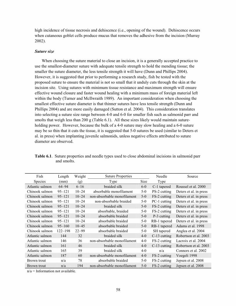
58
high incidence of tissue necrosis and dehiscence (i.e., opening of the wound). Dehiscence occurs when cutaneous goblet cells produce mucus that removes the adhesive from the incision (Murray 2002). Suture size
When choosing the suture material to close an incision, it is a generally accepted practice to use the smallest-diameter suture with adequate tensile strength to hold the mending tissue; the smaller the suture diameter, the less tensile strength it will have (Dunn and Phillips 2004). However, it is suggested that prior to performing a research study, fish be tested with the proposed suture to ensure the material is not so small that it unduly cuts through the skin at the incision site. Using sutures with minimum tissue resistance and maximum strength will ensure effective wound closure and faster wound healing with a minimum mass of foreign material left within the body (Turner and McIlwraith 1989). An important consideration when choosing the smallest effective suture diameter is that thinner sutures have less tensile strength (Dunn and Phillips 2004) and are more easily damaged (Sutton et al. 2004). This consideration translates into selecting a suture size range between 4-0 and 6-0 for smaller fish such as salmonid parr and smolts that weigh less than 200 g (Table 6.1). All these sizes likely would maintain suture-holding power. However, because the bulk of a 4-0 suture may slow healing and a 6-0 suture may be so thin that it cuts the tissue, it is suggested that 5-0 sutures be used (similar to Deters et al. in press) when implanting juvenile salmonids, unless negative effects attributed to suture diameter are observed.
Table 6.1. Suture properties and needle types used to close abdominal incisions in salmonid parr and smolts.
Fish Length Weight Suture Properties Needle Source
Species (mm) (g) Type Size Type Atlantic salmon 64–94 6–16 braided silk 6-0 C-1 tapered Roussel et al. 2000 Chinook salmon 95–121 10–24 absorbable monofilament 5-0 PS-2 cutting Deters et al. in press Chinook salmon 95–121 10–24 non-absorbable monofilament 5-0 FS-2 cutting Deters et al. in press Chinook salmon 95–121 10–24 non-absorbable braided 5-0 PC-1 cutting Deters et al. in press Chinook salmon 95–121 10–24 braided silk 5-0 FS-2 cutting Deters et al. in press Chinook salmon 95–121 10–24 absorbable, braided 5-0 FS-2 cutting Deters et al. in press Chinook salmon 95–121 10–24 absorbable braided 5-0 P-3 cutting Deters et al. in press Chinook salmon 95–121 10–24 absorbable braided 5-0 RB-1 tapered Deters et al. in press Chinook salmon 95–160 10–45 absorbable braided 5-0 RB-1 tapered Adams et al. 1998 Chinook salmon 122–198 22–99 absorbable braided 5-0 SH tapered Anglea et al. 2004 Atlantic salmon 144 32 braided silk 4-0 C-13 cutting Robertson et al. 2003 Atlantic salmon 146 36 non-absorbable monofilament 4-0 FS-2 cutting Lacroix et al. 2004 Atlantic salmon 161 46 braided silk 4-0 C-13 cutting Robertson et al. 2003 Atlantic salmon 165 39 braided silk 4-0 n/a Connors et al. 2002 Atlantic salmon 187 60 non-absorbable monofilament 4-0 FS-2 cutting Voegeli 1998 Brown trout n/a 78 absorbable braided 5-0 FS-2 cutting Jepsen et al. 2008 Brown trout n/a 194 non-absorbable monofilament 5-0 FS-2 cutting Jepsen et al. 2008 n/a = Information not available.
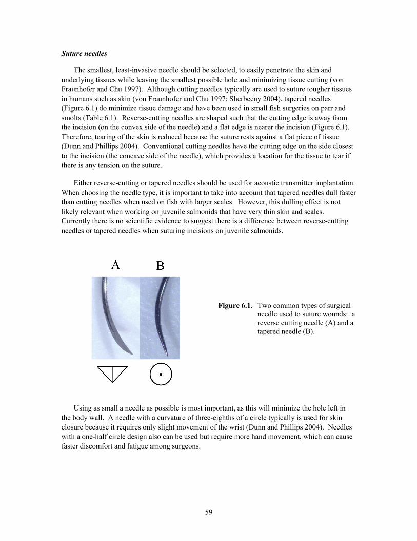
59
Suture needles
The smallest, least-invasive needle should be selected, to easily penetrate the skin and underlying tissues while leaving the smallest possible hole and minimizing tissue cutting (von Fraunhofer and Chu 1997). Although cutting needles typically are used to suture tougher tissues in humans such as skin (von Fraunhofer and Chu 1997; Sherbeeny 2004), tapered needles (Figure 6.1) do minimize tissue damage and have been used in small fish surgeries on parr and smolts (Table 6.1). Reverse-cutting needles are shaped such that the cutting edge is away from the incision (on the convex side of the needle) and a flat edge is nearer the incision (Figure 6.1). Therefore, tearing of the skin is reduced because the suture rests against a flat piece of tissue (Dunn and Phillips 2004). Conventional cutting needles have the cutting edge on the side closest to the incision (the concave side of the needle), which provides a location for the tissue to tear if there is any tension on the suture.
Either reverse-cutting or tapered needles should be used for acoustic transmitter implantation. When choosing the needle type, it is important to take into account that tapered needles dull faster than cutting needles when used on fish with larger scales. However, this dulling effect is not likely relevant when working on juvenile salmonids that have very thin skin and scales. Currently there is no scientific evidence to suggest there is a difference between reverse-cutting needles or tapered needles when suturing incisions on juvenile salmonids.
Figure 6.1. Two common types of surgical needle used to suture wounds: a reverse cutting needle (A) and a tapered needle (B).
Using as small a needle as possible is most important, as this will minimize the hole left in the body wall. A needle with a curvature of three-eighths of a circle typically is used for skin closure because it requires only slight movement of the wrist (Dunn and Phillips 2004). Needles with a one-half circle design also can be used but require more hand movement, which can cause faster discomfort and fatigue among surgeons.
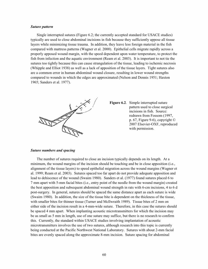
60
Suture pattern
Single interrupted sutures (Figure 6.2; the currently accepted standard for USACE studies) typically are used to close abdominal incisions in fish because they sufficiently appose all tissue layers while minimizing tissue trauma. In addition, they leave less foreign material in the fish compared with mattress patterns (Wagner et al. 2000). Epithelial cells migrate rapidly across a properly apposed wound margin, with the speed dependent upon water temperature, to protect the fish from infection and the aquatic environment (Ream et al. 2003). It is important to not tie the sutures too tightly because this can cause strangulation of the tissue, leading to ischemic necrosis (Whipple and Elliot 1938) as well as a lack of apposition of the tissue layers. Tight sutures also are a common error in human abdominal wound closure, resulting in lower wound strengths compared to wounds in which the edges are approximated (Nelson and Dennis 1951; Haxton 1965; Sanders et al. 1977).
Figure 6.2. Simple interrupted suture pattern used to close surgical incisions in fish. Source: redrawn from Fossom (1997, p. 67, Figure 9-6), copyright © 2007 Elsevier-OXF, reproduced with permission.
Suture numbers and spacing
The number of sutures required to close an incision typically depends on its length. At a minimum, the wound margins of the incision should be touching and be in close apposition (i.e., alignment of the tissue layers) to speed epithelial migration across the wound margins (Wagner et al. 1999; Ream et al. 2003). Sutures spaced too far apart do not provide adequate apposition and lead to dehiscence of the wound (Swaim 1980). Sanders et al. (1977) found sutures placed 6 to 7 mm apart with 5-mm facial bites (i.e., entry point of the needle from the wound margin) created the best apposition and subsequent abdominal wound strength in rats with 6-cm incisions, 4 to 6 d post-surgery. In general, sutures should be spaced the same distance apart as each suture is wide (Swaim 1980). In addition, the size of the tissue bite is dependent on the thickness of the tissue, with smaller bites for thinner tissue (Turner and McIlwraith 1989). Tissue bites of 2 mm on either side of the incision result in a 4-mm-wide suture. Therefore, in this case the sutures should be spaced 4 mm apart. When implanting acoustic microtransmitters for which the incision may be as small as 5 mm in length, use of one suture may suffice, but there is no research to confirm this. Currently, the standard within USACE studies involving implantation of acoustic microtransmitters involves the use of two sutures, although research into this topic is currently being conducted at the Pacific Northwest National Laboratory. Sutures with about 2-mm facial bites are evenly spaced along the approximate 8-mm incision. Suture spacing for abdominal
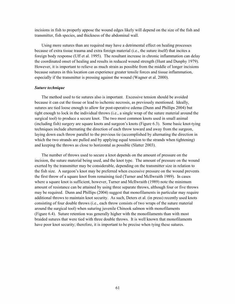
61
incisions in fish to properly appose the wound edges likely will depend on the size of the fish and transmitter, fish species, and thickness of the abdominal wall.
Using more sutures than are required may have a detrimental effect on healing processes because of extra tissue trauma and extra foreign material (i.e., the suture itself) that incites a foreign body response (Uff et al. 1995). The resultant increase in chronic inflammation can delay the coordinated onset of healing and results in reduced wound strength (Hunt and Dunphy 1979). However, it is important to relieve as much strain as possible from the middle of longer incisions because sutures in this location can experience greater tensile forces and tissue inflammation, especially if the transmitter is pressing against the wound (Wagner et al. 2000). Suture technique
The method used to tie sutures also is important. Excessive tension should be avoided because it can cut the tissue or lead to ischemic necrosis, as previously mentioned. Ideally, sutures are tied loose enough to allow for post-operative edema (Dunn and Phillips 2004) but tight enough to lock in the individual throws (i.e., a single wrap of the suture material around the surgical tool) to produce a secure knot. The two most common knots used in small animal (including fish) surgery are square knots and surgeon’s knots (Figure 6.3). Some basic knot-tying techniques include alternating the direction of each throw toward and away from the surgeon, laying down each throw parallel to the previous tie (accomplished by alternating the direction in which the two strands are pulled and by applying equal tension to the strands when tightening) and keeping the throws as close to horizontal as possible (Slatter 2003).
The number of throws used to secure a knot depends on the amount of pressure on the incision, the suture material being used, and the knot type. The amount of pressure on the wound exerted by the transmitter may be considerable, depending on the transmitter size in relation to the fish size. A surgeon’s knot may be preferred when excessive pressure on the wound prevents the first throw of a square knot from remaining tied (Turner and McIlwraith 1989). In cases where a square knot is sufficient, however, Turner and McIlwraith (1989) note the minimum amount of resistance can be attained by using three separate throws, although four or five throws may be required. Dunn and Phillips (2004) suggest that monofilaments in particular may require additional throws to maintain knot security. As such, Deters et al. (in press) recently used knots consisting of four double throws (i.e., each throw consists of two wraps of the suture material around the surgical tool) when suturing juvenile Chinook salmon with monofilaments (Figure 6.4). Suture retention was generally higher with the monofilaments than with most braided sutures that were tied with three double throws. It is well known that monofilaments have poor knot security; therefore, it is important to be precise when tying these sutures.
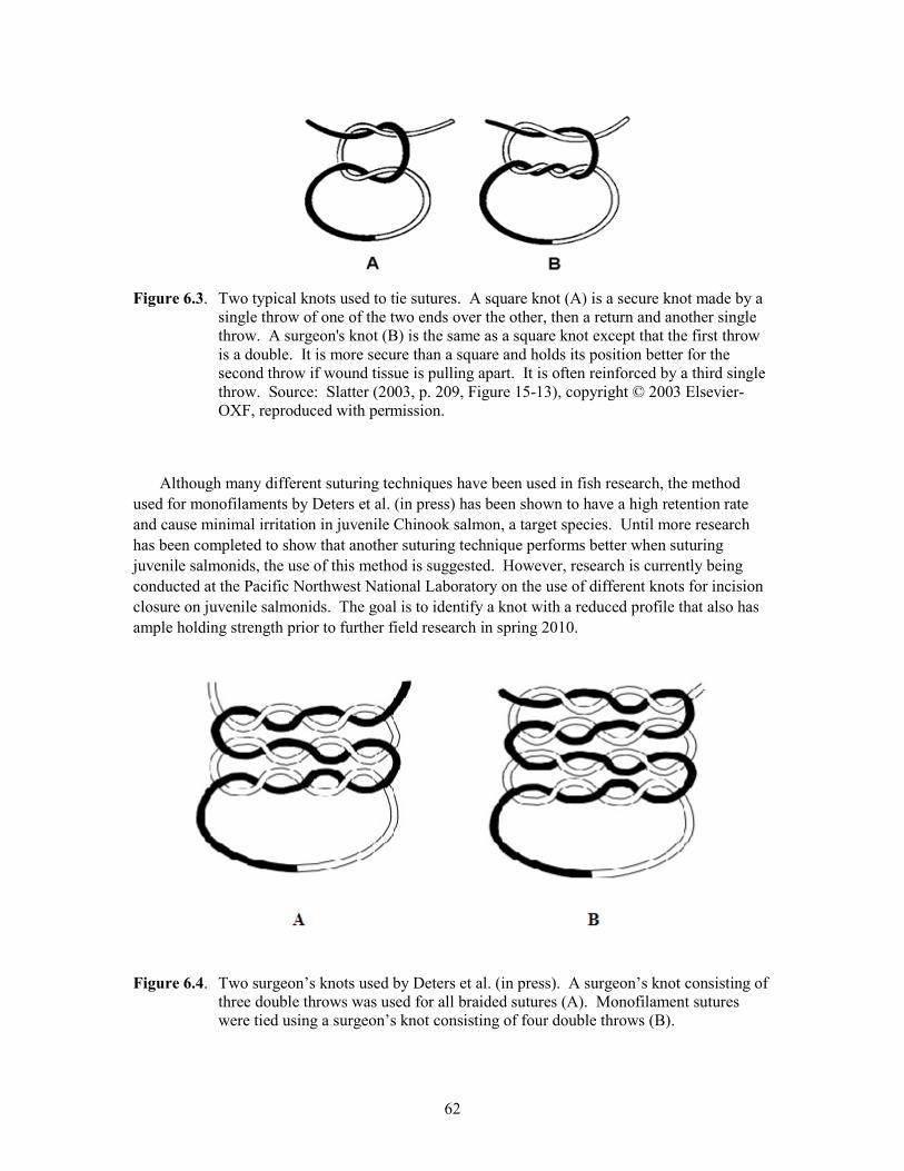
62
Figure 6.3. Two typical knots used to tie sutures. A square knot (A) is a secure knot made by a
single throw of one of the two ends over the other, then a return and another single throw. A surgeon's knot (B) is the same as a square knot except that the first throw is a double. It is more secure than a square and holds its position better for the second throw if wound tissue is pulling apart. It is often reinforced by a third single throw. Source: Slatter (2003, p. 209, Figure 15-13), copyright © 2003 Elsevier-OXF, reproduced with permission.
Although many different suturing techniques have been used in fish research, the method used for monofilaments by Deters et al. (in press) has been shown to have a high retention rate and cause minimal irritation in juvenile Chinook salmon, a target species. Until more research has been completed to show that another suturing technique performs better when suturing juvenile salmonids, the use of this method is suggested. However, research is currently being conducted at the Pacific Northwest National Laboratory on the use of different knots for incision closure on juvenile salmonids. The goal is to identify a knot with a reduced profile that also has ample holding strength prior to further field research in spring 2010.
Figure 6.4. Two surgeon’s knots used by Deters et al. (in press). A surgeon’s knot consisting of three double throws was used for all braided sutures (A). Monofilament sutures were tied using a surgeon’s knot consisting of four double throws (B).
63
Summary
The use of clean surgical instruments properly selected for a given size of fish will minimize tissue damage, minimize infection rates, and speed the wound healing of fish implanted with transmitters. For the implantation of transmitters into small fish, the optimal surgical methods include making an incision lateral and parallel to the midline, protecting the viscera (by lifting the skin with forceps while creating the incision), using 5-0 absorbable monofilament suture with a small suaged tapered or reverse-cutting needle. Standardizing the implantation techniques to be used in a study involving particular species and age classes of fish will improve the survivorship and transmitter retention results while allowing for comparisons to be made among studies and across multiple years. Ongoing and future research will provide a basis for refining the surgical methods outlined above.
64
References Adams NS, Rondorf DW, Evans SD, Kelly JE (1998). Effects of surgically and gastrically
implanted radio transmitters on growth and feeding behavior of juvenile Chinook salmon. Transactions of the American Fisheries Society 127:128-136
AFS (American Fisheries Society) (2004) Guidelines for the use of fishes in research. American
Fisheries Society Press, Bethesda, MD Anderson CD, Roberts RJ (1975) A comparison of the effects of temperature on the wound
healing in a tropical and a temperate teleost. Journal of Fish Biology 7:173–182 Anglea SM, Geist DR, Brown RS, Deters KA (2004) Effects of acoustic transmitters on
swimming performance and predator avoidance of juvenile Chinook salmon. North American Journal of Fisheries Management 24:162–170
Austin B, Austin DA (1987) Bacterial fish pathogens: disease in farmed and wild fish. Ellis
Horwood, New York Burger B, DeYoung D, Hunter D (1994) Sterlization of implantable transmitters. Telonics
Quarterly 7(3):3–4. Available via http://www.telonics.com/quarterly/index.php. Accessed 07 April 2009
Burger JWA, van ‘t Riet M, Jeekel J (2002) Abdominal incisions: techniques and postoperative
complications. Scandinavian Journal of Surgery 91:315–321 CCAC (Canadian Council on Animal Care) (2005) CCAC guidelines on: the care and use of fish
in research, teaching and testing. Canadian Council on Animal Care, Ottawa, Ontario, Canada. Available via www.ccac.ca/en/CCAC_Programs/Guidelines_Policies/ GDLINES/Guidelis.htm. Accessed 07 April 2009
Connors KB, Scruton D, Brown JA, McKinley RS (2002) The effects of surgically-implanted
dummy radio transmitters on the behaviour of wild Atlantic salmon smolts. Hydrobiologia 483:231–237
Deters KA, Brown RS, Carter KM, Boyd JW, Eppard MB, Seaburg, AG (in press) Performance
assessment of suture type, water temperature, and surgeon skill in juvenile Chinook salmon surgically implanted with acoustic transmitters. Transactions of the American Fisheries Society
Dunn DL, Phillips J (2004) Ethicon wound closure manual. Ethicon, Inc., Somerville, NJ
Available via http://www.jnjgateway.com/public/NLDUT/ Wound_Closure_Manual1.pdf Accessed 07 April 2009
65
Favero MS, Bond WW (1991) Sterilization, disinfection, and antisepsis in the hospital. In: Balows A, Hausler WJ, Hermann KL, Isenberg HD, Shadomy HJ (eds) Manual of clinical microbiology. American Society for Microbiology, Washington, DC, pp 183–200
Fossom TW (1997) Small animal surgery. Mosby, St. Louis, MO Haeseker SL, Carmichael JT, Hightower JE (1996) Summer distribution and condition of striped
bass within Albemarle Sound, North Carolina. Transactions of the American Fisheries Society 125:690–704
Haxton H (1965) The influence of suture materials and methods on the healing of abdominal
wounds. British Journal of Surgery 1965:372–375 Hudson RCL (1969) Polyneural innervation of the fast muscles of the marine teleost Cottus
scorpius L. Journal of Experimental Biology 50:47–67 Hunt T, Dunphy JE (1979) Fundamentals of wound management. Appleton-Century-Crofts,
New York Hurty CA, Brazik DC, McHugh Law J, Sakamoto K, Lewbart GA (2002) Evaluation of the tissue
reactions in the skin and body wall of koi (Cyprinus carpio) to five suture materials. Veterinary Record 151:324–328
Jacobs PT, Wang JH, Gorhan RA et al. (1998) Cleaning: principles, methods and benefits. In:
Rutala WA (ed) Disinfection, sterilization and antisepsis in health care. Association for Professionals in Infection Control and Epidemiology, Inc., and Polyscience Publications, Inc., Washington, DC, pp 165–181
Jepsen N, Mikkelsen JS, Koed A (2008) Effects of tag and suture type on survival and growth of
brown trout with surgically implanted telemetry tags in the wild. Journal of Fish Biology 72:594–602
Kaseloo PA, Weatherley PH, Lotimer J, Farina MD (1992) A biotelemetry system recording fish
activity. Journal of Fish Biology 40:165–179 Korniewicz DM, Kirwin M, Larson E (1991) Do your gloves fit the task? American Journal of
Nursing 91(6):38–40 Lacroix GL, Knox D, McCurdy P (2004) Effects of implanted dummy acoustic transmitters on
juvenile Atlantic salmon. Transactions of the American Fisheries Society 133:211–220 Lucas MC (1989) Effects of implanted dummy transmitters on mortality, growth and tissue
reaction in rainbow trout, Salmo gairdneri Richardson. Journal of Fish Biology 35:577–587 Knights BC, Lasee BA (1996) Effects of transmitters on adult bluegills at two temperatures.
Transactions of the American Fisheries Society 125:440–449
66
McDonnell G, Russell AD (1999) Antiseptics and disinfectants: activity, action, and resistance. Clinical Microbiological Review 12:147–79
Mulcahy DM (2003) Surgical implantation of transmitters into fish. ILAR Journal 44:295–306 Murray MJ (2002) Fish surgery. Seminars in Avian and Exotic Pet Medicine 11:246–257 Mutsaers SE, Bishop JE, McGrouther G. Laurent JG (1997) Mechanisms of tissue repair: from
wound healing to fibrosis. International Journal of Biochemistry and Cell Biology 29:5–17 Nelson CA, Dennis C (1951) Wound healing: technical factors in the gain of strength in sutured
abdominal wounds in rabbits. Surgery, Gynecology and Obstetrics 93:461–467 Nygaard IE, Squatrito RC (1996) Abdominal incisions from creation to closure. Obsterical &
Gynecological Survey 51:429–436 Rath AM, Attali P, Dumas JL, Goldlust D, Zhang J, Chevrel JP (1996) The abdominal linea alba:
an anatomo-radiologic and biomechanical study. Surgical and Radiological Anatomy 18:281–288
Ream RA, Theriot JA, Somero GN (2003) Influences of thermal acclimation and acute
temperature change on the motility of epithelial wound-healing cells (keratocytes) of tropical, temperate and Antarctic fish. The Journal of Experimental Biology 206:4539–4551
Robertson MJ, Clarke KD, Scruton DA, Brown JA (2003) Interhabitat and instream movements
of large Atlantic salmon parr in a Newfoundland watershed in winter. Journal of Fish Biology 63:1208–1218
Roussel J-M, Haro A, Cunjak RA (2000) Field test of a new method for tracking small fishes in
shallow rivers using passive integrated transponder (PIT) technology. Canadian Journal of Fisheries and Aquatic Sciences 47:1326–1329
Rutala WA (1990) APIC guidelines for selection and use of disinfectants. American Journal of
Infection Control 18:99–117 Rutala WA (1996) Selection and use of disinfectants in health care. In: Mayhall CG (ed) Hospital
epidemiology and infection control. Williams & Wilkins, Baltimore, MD, pp 913–936 Rutala WA, Weber DJ (2004) Disinfection and sterilization in health care facilities: What
clinicians need to know. Clinical Infectious Diseases 39:702–709 Sanders RJ, DiClementi D, Ireland K (1977) Principles of abdominal wound closure: I. Animal
studies. Archives of Surgery 112:1184–1187 Sherbeeny A (2004) Instruments and techniques. Needles, sutures, and knots. Part I: needles. Ain
Shams Journal of Obstetrics and Gynecology 1:29–30
67
Slatter D (2003) Textbook of small animal surgery - Volume 1, 3rd edn. W. B. Saunders, Philadelphia, PA
Spaulding EH (1968) Chemical disinfection of medical and surgical materials. In: Lawrence C,
Block SS (eds) Disinfection, sterilization, and preservation. Lea & Febiger, Philadelphia, PA, pp 517–531
Starr RM, Haine JN, Johnson KA (2000) Techniques for tagging and tracking deepwater
rockfishes. North American Journal of Fisheries Management 20:597–609 Sutton TM, Volkman ET, Pangle KL, Rajchel DA, Duehr JP (2004) Effects of absorbable suture
strand diameter on retention of external radio transmitters by juvenile lake sturgeon. North American Journal of Fisheries Management 24:1404–1408
Swaim S (1980) Surgery of traumatized skin: management and reconstruction in the dog and cat.
W. B. Saunders, Philadelphia, PA Tera H, Åberg C (1977) Strength of knots in surgery in relation to type of knot, type of suture
material and dimension of suture thread. Acta Chirurgica Scandinavica 143(2):75–83 Thoreau EB, Baras E (1997) Evaluation of surgical procedures for implanting telemetry
transmitters into the body cavity of tilapia Oreochromis aureus. Aquatic Living Resources 10:207–211
Turner AS, McIlwraith CW (1989) Techniques in large animal surgery. Lea & Febiger,
Philadelphia, PA Uff CR, Scott AD, Pockley G, Phillips RKS (1995) Influence of soluble suture factors on in vitro
macrophage function. Biomaterials 16:355–360 Voegeli FA, Lacroix GL, Anderson JM (1998) Development of miniature pingers for tracking
Atlantic salmon smolts at sea. Hydrobiologia 371/372:35–46 von Fraunhofer JA, Chu CC (1997) Surgical needles. In: Chu CC, von Fraunhofer JA,
Greisler HP (eds) Wound closure biomaterials and devices. CRC Press, Boca Raton, FL, pp 25–38
Walsh MG, Bjorgo KA, Isely JJ (2000) Effects of implantation method and temperature on
mortality and loss of simulated transmitters in hybrid striped bass. Transactions of the American Fisheries Society 129:539–544
Wagner GN, Cooke SJ (2005) Methodological approaches and opinions of researchers involved
in the surgical implantation of telemetry transmitters in fish. Journal of Aquatic Animal Health 17:160–169
68
Wagner GN, Stevens ED (2000) Effects of different surgical techniques, suture material and location of incision site on the behavior of rainbow trout (Oncorhynchus mykiss). Marine and Freshwater Behaviour and Physiology 33:103–114
Wagner GN, Stevens ED, Harvey-Clark C (1999) Wound healing in rainbow trout following
surgical site preparation with a povidone-iodine antiseptic. Journal of Aquatic Animal Health 11:373–382
Wagner GN, Stevens ED, Byrne P (2000) The effects of suture type and patterns on surgical
wound healing in rainbow trout. Transactions of the American Fisheries Society 129:1196–1205
Whipple AO, Elliott Jr RHE (1938) The repair of abdominal incisions. Annals of Surgery
108:741–756
Appendix Common Errors in Surgical Technique

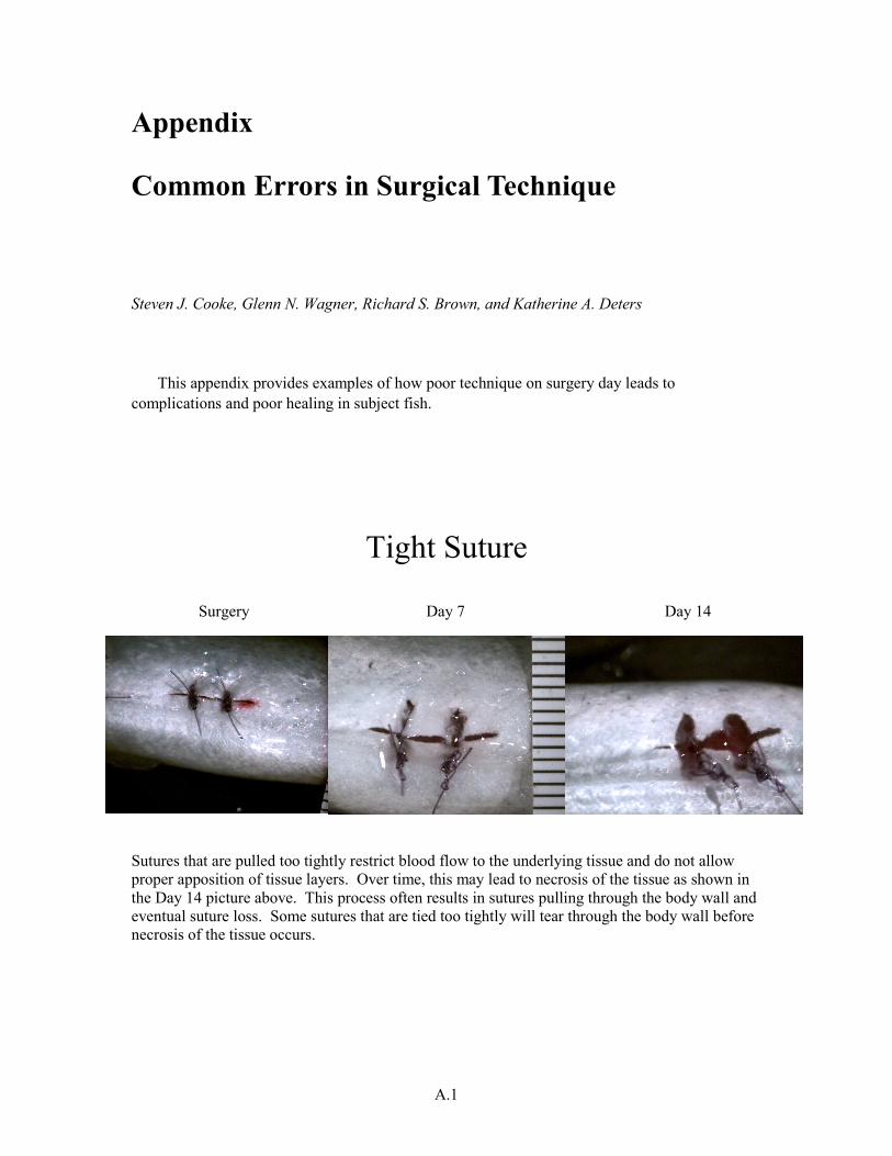
A.1
Appendix
Common Errors in Surgical Technique
Steven J. Cooke, Glenn N. Wagner, Richard S. Brown, and Katherine A. Deters
This appendix provides examples of how poor technique on surgery day leads to complications and poor healing in subject fish.
Tight Suture
Surgery Day 7 Day 14
Sutures that are pulled too tightly restrict blood flow to the underlying tissue and do not allow proper apposition of tissue layers. Over time, this may lead to necrosis of the tissue as shown in the Day 14 picture above. This process often results in sutures pulling through the body wall and eventual suture loss. Some sutures that are tied too tightly will tear through the body wall before necrosis of the tissue occurs.
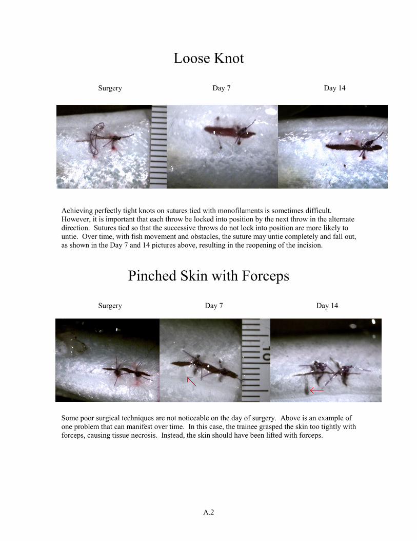
A.2
Loose Knot
Surgery Day 7 Day 14
Achieving perfectly tight knots on sutures tied with monofilaments is sometimes difficult. However, it is important that each throw be locked into position by the next throw in the alternate direction. Sutures tied so that the successive throws do not lock into position are more likely to untie. Over time, with fish movement and obstacles, the suture may untie completely and fall out, as shown in the Day 7 and 14 pictures above, resulting in the reopening of the incision.
Pinched Skin with Forceps
Surgery Day 7 Day 14 Some poor surgical techniques are not noticeable on the day of surgery. Above is an example of one problem that can manifest over time. In this case, the trainee grasped the skin too tightly with forceps, causing tissue necrosis. Instead, the skin should have been lifted with forceps.
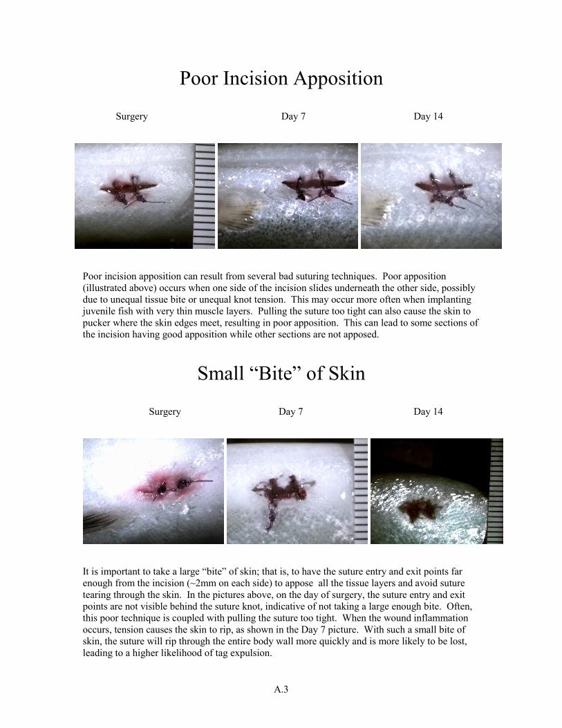
A.3
Poor Incision Apposition Surgery Day 7 Day 14
Poor incision apposition can result from several bad suturing techniques. Poor apposition (illustrated above) occurs when one side of the incision slides underneath the other side, possibly due to unequal tissue bite or unequal knot tension. This may occur more often when implanting juvenile fish with very thin muscle layers. Pulling the suture too tight can also cause the skin to pucker where the skin edges meet, resulting in poor apposition. This can lead to some sections of the incision having good apposition while other sections are not apposed.
Small “Bite” of Skin
Surgery Day 7 Day 14
It is important to take a large “bite” of skin; that is, to have the suture entry and exit points far enough from the incision (~2mm on each side) to appose all the tissue layers and avoid suture tearing through the skin. In the pictures above, on the day of surgery, the suture entry and exit points are not visible behind the suture knot, indicative of not taking a large enough bite. Often, this poor technique is coupled with pulling the suture too tight. When the wound inflammation occurs, tension causes the skin to rip, as shown in the Day 7 picture. With such a small bite of skin, the suture will rip through the entire body wall more quickly and is more likely to be lost, leading to a higher likelihood of tag expulsion.


![3H]Thymidine-Labeled Cells in the Rat Utricular Maculadepts.washington.edu/rubelab/personnel/popoff.pdf · Surgical procedure for minipump implantation Surgical procedures described](https://static.fdocuments.us/doc/165x107/5eb64936c4181619b96005e9/3hthymidine-labeled-cells-in-the-rat-utricular-surgical-procedure-for-minipump.jpg)


