Methods for determining protein structure - UCLArebecca/153A/W11/Lectures/153A_W11_Lec15... ·...
23
Methods for determining protein structure • Sequence: – Edman degradation – Mass spectrometry • Secondary structure: – Circular Dichroism – FTIR • Tertiary, quaternary structure: – NMR – X-ray crystallography
Transcript of Methods for determining protein structure - UCLArebecca/153A/W11/Lectures/153A_W11_Lec15... ·...

Methods for determining protein structure
• Sequence:– Edman degradation
– Mass spectrometry
• Secondary structure:– Circular Dichroism
– FTIR
• Tertiary, quaternary structure:– NMR
– X-ray crystallography

Protein sequencing approaches depend on
what is known and what is the goal
• Protein is unknown, from organism with no DNA sequence information – starting from scratch– Purify protein & separate chains (if multimer)
– Fragment and sequence each chain
– Fragment differently and sequence
– Reassemble sequence based on overlapping fragments
• Protein is unknown or known, and comes from an organism with known DNA sequence– Purify protein (& separate chains)
– Fragment chain(s) and sequence or measure mass
– Use sequence database to reassemble sequence

Protein sequencing from scratch
• Step 0: Purify the protein
• Step 1: Separate the chains (if multimeric)
– If needed, reduce disulfides and block free thiols

Protein sequencing from scratch
• Step 0: Purify the protein
• Step 1: Separate the chains (if multimeric)
• Step 2: Fragment each polypeptide
– Enzymatically, with endopeptidase, chemically (e.g.
with cyanogen bromide), or physically (e.g. through
collision in MS)

Step 2: Fragment each polypeptide
Cyanogen bromide (CNBr): Rn-1 = Met

Protein sequencing from scratch
• Step 0: Purify the protein
• Step 1: Separate the chains (if multimeric)
• Step 2: Fragment each polypeptide
• Step 3: Sequence the fragments
– Via, e.g., Edman degradation or Mass spectrometry

Sequence peptides with mass spectrometry
(MS/MS)

MS cleavage occurs mainly at peptide
bonds, and charge is retained in one product

Protein sequencing from scratch
• Step 0: Purify the protein
• Step 1: Separate the chains (if multimeric)
• Step 2: Fragment each polypeptide
• Step 3: Sequence the fragments
• Step 4: Reconstruct the sequence

Protein sequencing approaches depend on
what is known and what is the goal
• Protein is unknown, from organism with no DNA sequence information – starting from scratch– Purify protein & separate chains (if multimer)
– Fragment and sequence each chain
– Fragment differently and sequence
– Reassemble sequence based on overlapping fragments
• Protein is unknown or known, and comes from an organism with known DNA sequence– Purify protein (& separate chains)
– Fragment chain(s) and sequence or measure mass
– Use sequence database to reassemble sequence

There are different approaches for using
mass spectrometry to sequence a protein
Bottom-Up Proteomics
• Fragment protein (e.g. enzymatically) and separate fragments
• Ionize fragments, trap in the spectrometer, and measure m/z
• Select one m/z peak and fragment (e.g. by collision)
• Measure m/z of the smaller fragments and use a database to match
the peaks to known sequences

There are different approaches for using
mass spectrometry to sequence a protein
Top-Down Proteomics
• Ionize whole protein(s), trap in the spectrometer, and measure m/z
• Use the instrument to select one m/z peak and fragment the protein
(e.g. by collision)
• Measure m/z ratios of the fragments and use a database to match
the peaks to known sequences
• OR Select a peak and fragment again, then match to sequence
(Selection and fragmentation may be repeated over and over)
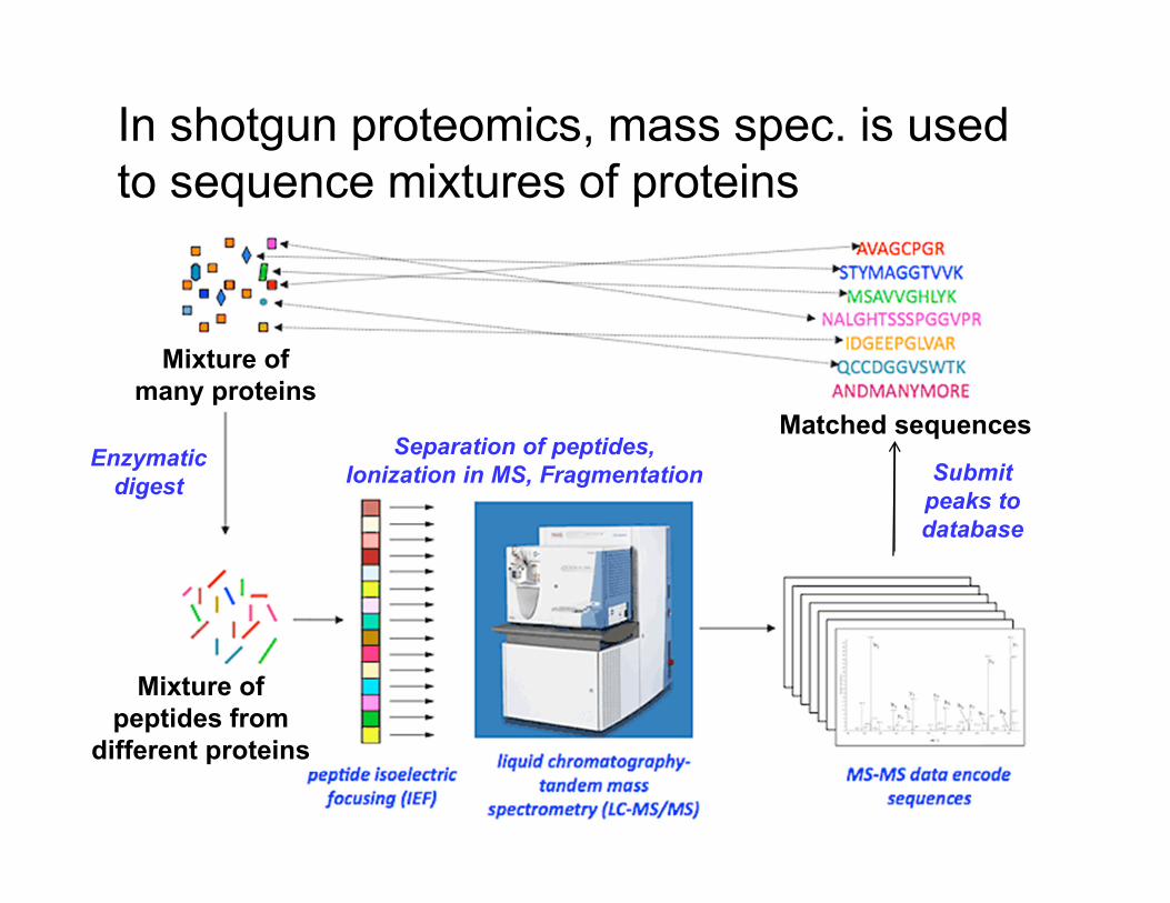
In shotgun proteomics, mass spec. is used
to sequence mixtures of proteins
Mixture of
many proteins
Mixture of
peptides from
different proteins
Enzymatic
digest
Separation of peptides,
Ionization in MS, Fragmentation
Matched sequences
Submit
peaks to
database

Methods for determining protein structure
• Sequence:– Edman degradation
– Mass spectrometry
• Secondary structure:– Circular Dichroism
– FTIR
• Tertiary, quaternary structure:– NMR
– X-ray crystallography

Circular dichroism (CD) measures amide
absorption of circularly polarized UV light
• Ellipticity (∆ε) is the difference in absorption of left-handed and right-handed circularly polarized light
• Different secondary structures show different patterns of ellipticity
• Protein’s CD spectrum is ‘deconvoluted’ to estimate fractional contribution of helix, sheet, turn, and coil

Proteins with different compositions of 2°
structure give different CD spectra

Fourier transform infrared (FTIR) spectra
show amide absorption of infrared light
• Peak frequencies show
bond stretching and
bending, which vary with
protein conformation
• C=O stretching
frequency of amide I
band correlates with
secondary structure
• Protein’s FTIR spectrum
is ‘deconvoluted’ to
estimate fractional
contribution of helix,
sheet, and coil

Methods for determining protein structure
• Sequence:– Edman degradation
– Mass spectrometry
• Secondary structure:– Circular Dichroism
– FTIR
• Tertiary, quaternary structure:– NMR
– X-ray crystallography

Proteins have too many protons to be
resolved by one-dimensional NMR

2D NMR separates proton peaks and can reveal
approximate distances between nearby atoms
a
b
c
d
Cross-peaks indicate protons are within 5Å of each other

NMR-derived distance constraints are used
to calculate likely protein conformations

X-ray crystallography reveals the layout of
repeating electron density
X-rays
Protein crystal
Diffraction pattern
Data processingDiffracted
X-rays
Electron
density
map
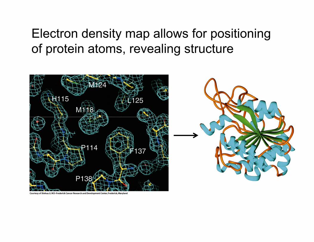
Electron density map allows for positioning
of protein atoms, revealing structure


![Determining protein structure by tyrosine bioconjugation · INTRODUCTION Protein structure is driven by the interactions of the 20 amino acids with solvent and other amino acids[1].](https://static.fdocuments.us/doc/165x107/5f2af327b5a59d74a66e7b0a/determining-protein-structure-by-tyrosine-bioconjugation-introduction-protein-structure.jpg)