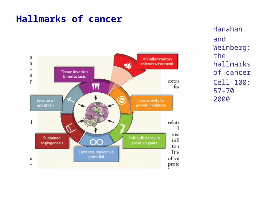
Metastasis. Hanahan and Weinberg: the hallmarks of cancer Cell 100: 57- 70 2000 Hallmarks of cancer.
67
Metastasis
-
Upload
jeremy-chapman -
Category
Documents
-
view
235 -
download
1
Transcript of Metastasis. Hanahan and Weinberg: the hallmarks of cancer Cell 100: 57- 70 2000 Hallmarks of cancer.
Metastasis
Hanahan
and Weinberg: the hallmarks of cancer
Cell 100: 57-70 2000
Hallmarks of cancer
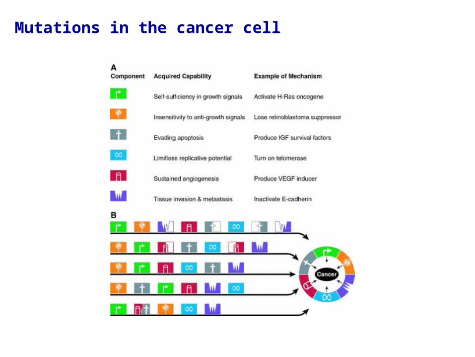
Mutations in the cancer cell
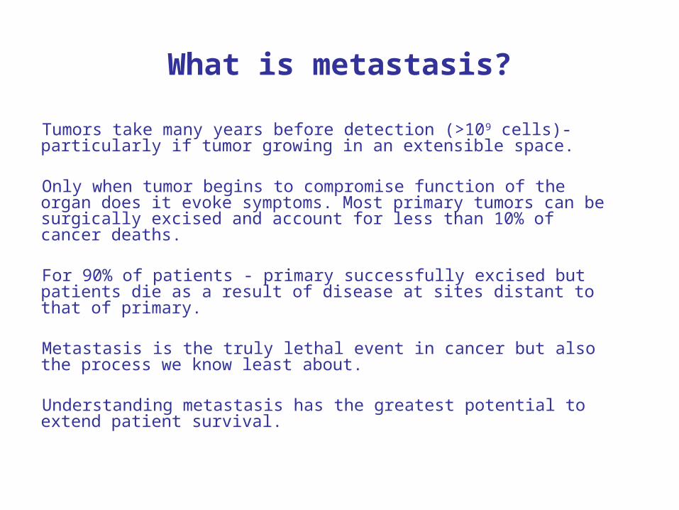
What is metastasis?
Tumors take many years before detection (>109 cells)- particularly if tumor growing in an extensible space.
Only when tumor begins to compromise function of the organ does it evoke symptoms. Most primary tumors can be surgically excised and account for less than 10% of cancer deaths.
For 90% of patients - primary successfully excised but patients die as a result of disease at sites distant to that of primary.
Metastasis is the truly lethal event in cancer but also the process we know least about.
Understanding metastasis has the greatest potential to extend patient survival.
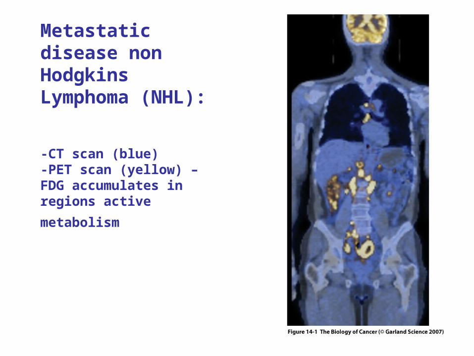
Metastatic disease non Hodgkins Lymphoma (NHL):
-CT scan (blue) -PET scan (yellow) – FDG accumulates in regions active
metabolism
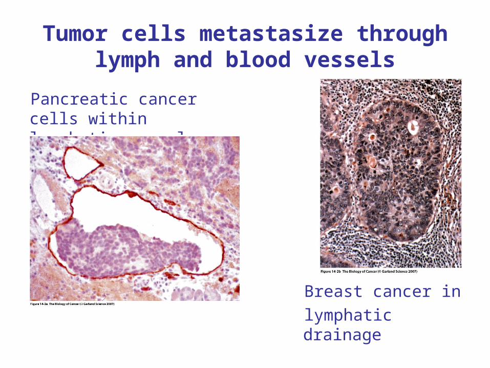
Tumor cells metastasize through lymph and blood vessels
Pancreatic cancer cells within lymphatic vessel
Breast cancer in
lymphatic drainage
Metastases disrupt organ functions
Breast cancer metastasizes to bone (pain and skeletal collapse); brain, lungs, liver (function).
Other cancers spread to different organs.
Some cancers have very high rates of metastasis-while others hardly metastasize at all.
Understanding the basis for these differences could lead to control over tumor spread.
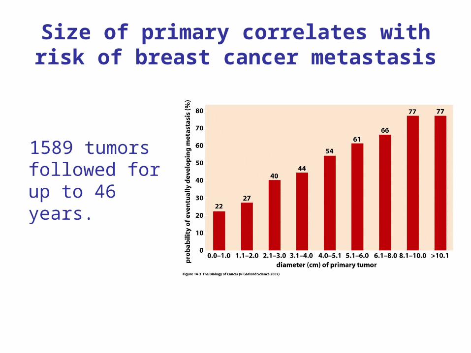
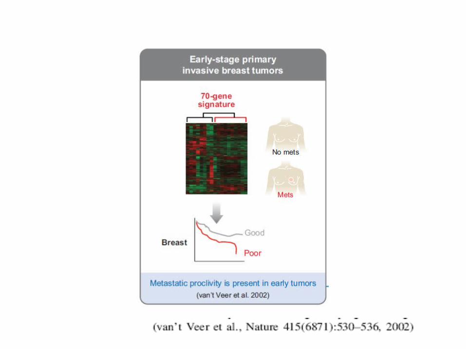
Size of primary correlates with risk of breast cancer metastasis
1589 tumors followed for up to 46 years.
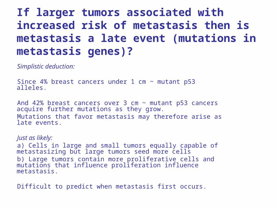
If larger tumors associated with increased risk of metastasis then is metastasis a late event (mutations in metastasis genes)?
Simplistic deduction:
Since 4% breast cancers under 1 cm ~ mutant p53 alleles.
And 42% breast cancers over 3 cm ~ mutant p53 cancers acquire further mutations as they grow. Mutations that favor metastasis may therefore arise as late events.
Just as likely:a) Cells in large and small tumors equally capable of metastasizing but large tumors seed more cellsb) Large tumors contain more proliferative cells and mutations that influence proliferation influence metastasis.
Difficult to predict when metastasis first occurs.
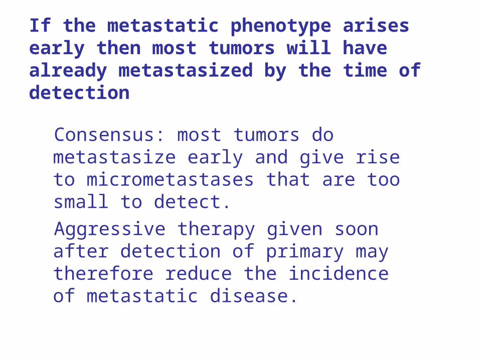
If the metastatic phenotype arises early then most tumors will have already metastasized by the time of detection
Consensus: most tumors do metastasize early and give rise to micrometastases that are too small to detect.
Aggressive therapy given soon after detection of primary may therefore reduce the incidence of metastatic disease.
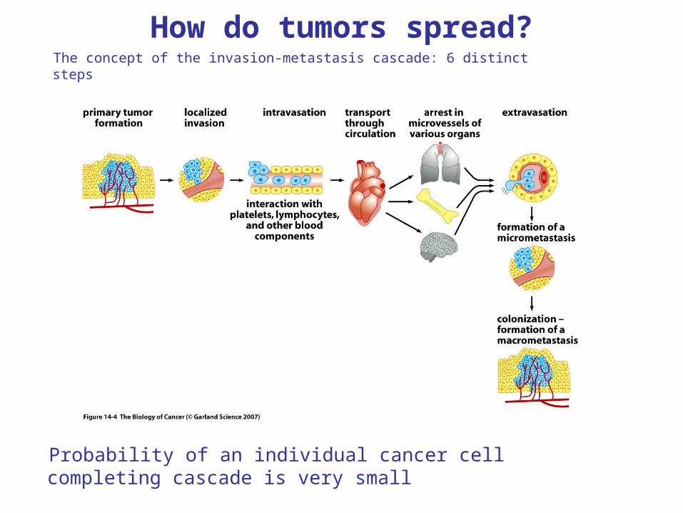
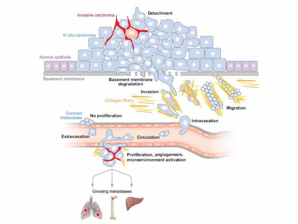
How do tumors spread?The concept of the invasion-metastasis cascade: 6 distinct steps
Probability of an individual cancer cell completing cascade is very small
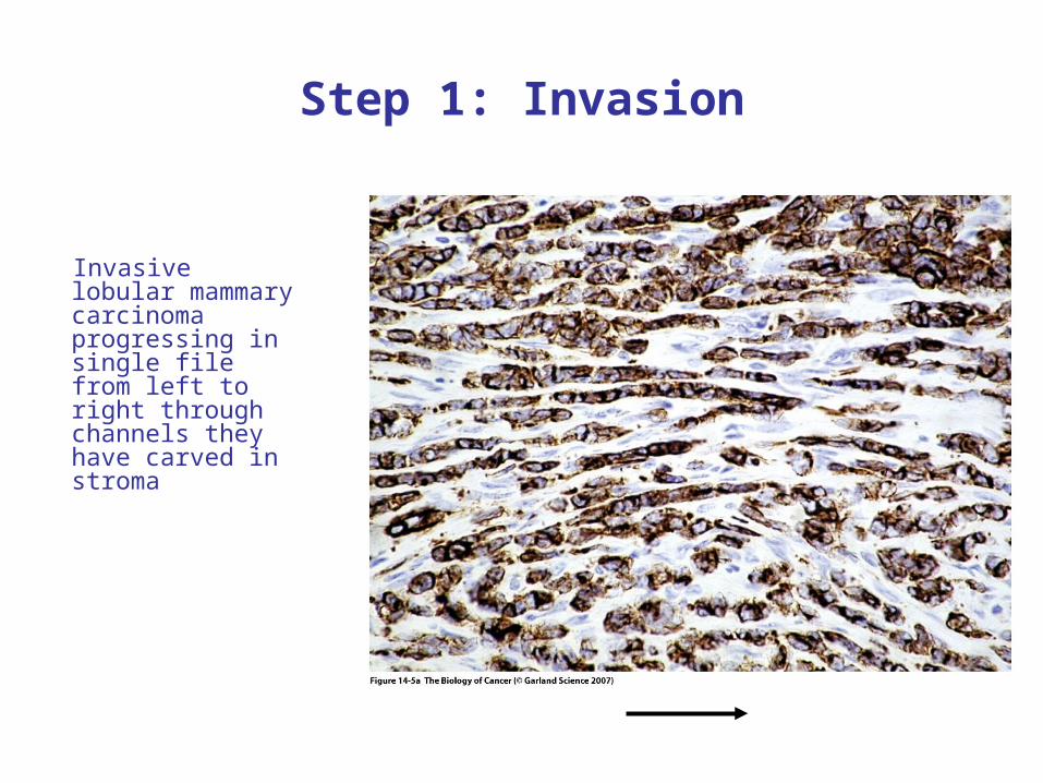
Step 1: Invasion
Invasive lobular mammary carcinoma progressing in single file from left to right through channels they have carved in stroma
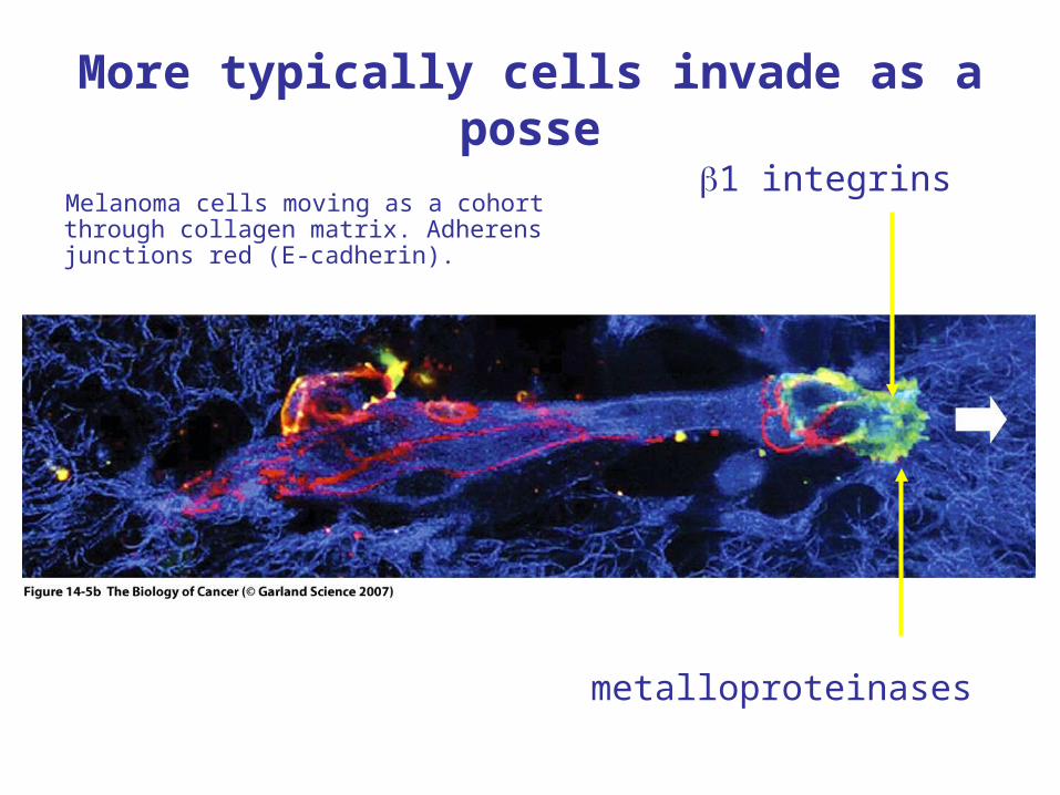
More typically cells invade as a posse
Melanoma cells moving as a cohort through collagen matrix. Adherens junctions red (E-cadherin).
1 integrins
metalloproteinases
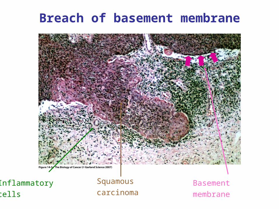
Breach of basement membrane
Inflammatory
cells
Basement
membrane
Squamous
carcinoma
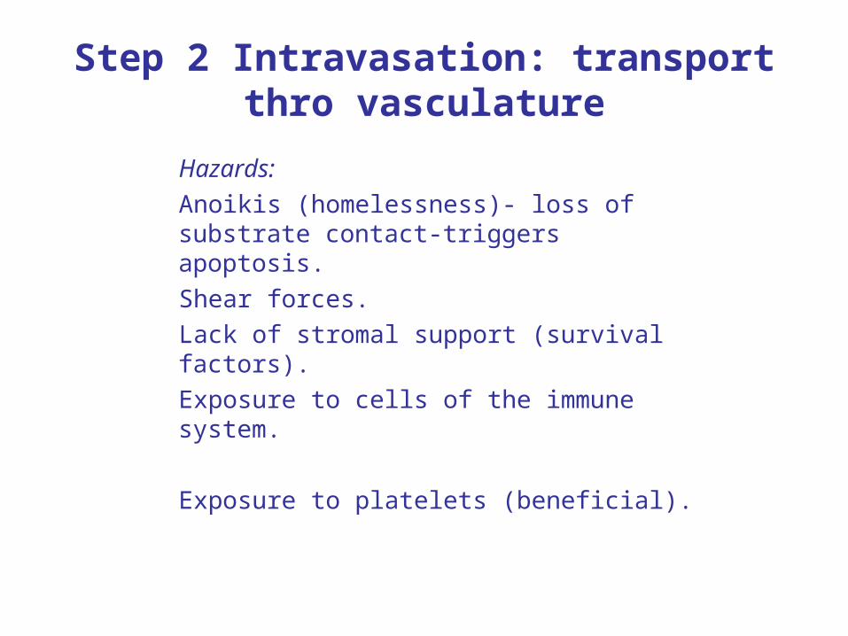
Step 2 Intravasation: transport thro vasculature
Hazards:
Anoikis (homelessness)- loss of substrate contact-triggers apoptosis.
Shear forces.
Lack of stromal support (survival factors).
Exposure to cells of the immune system.
Exposure to platelets (beneficial).
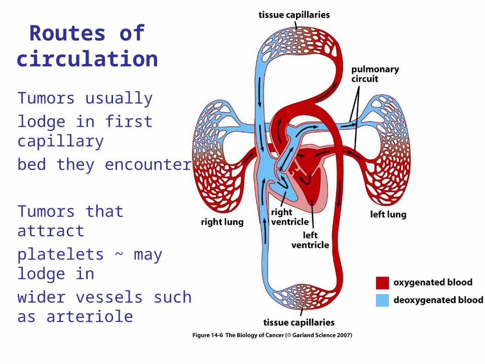
Routes of circulation
Tumors usually
lodge in first capillary
bed they encounter
Tumors that attract
platelets ~ may lodge in
wider vessels such as arteriole
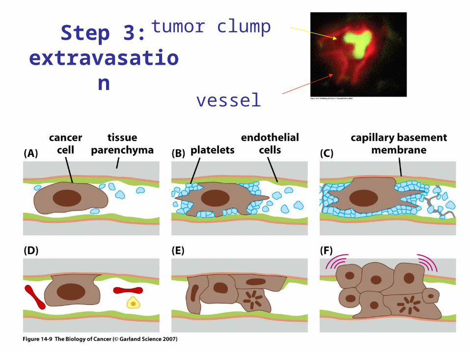
Step 3: extravasation
vessel
tumor clump
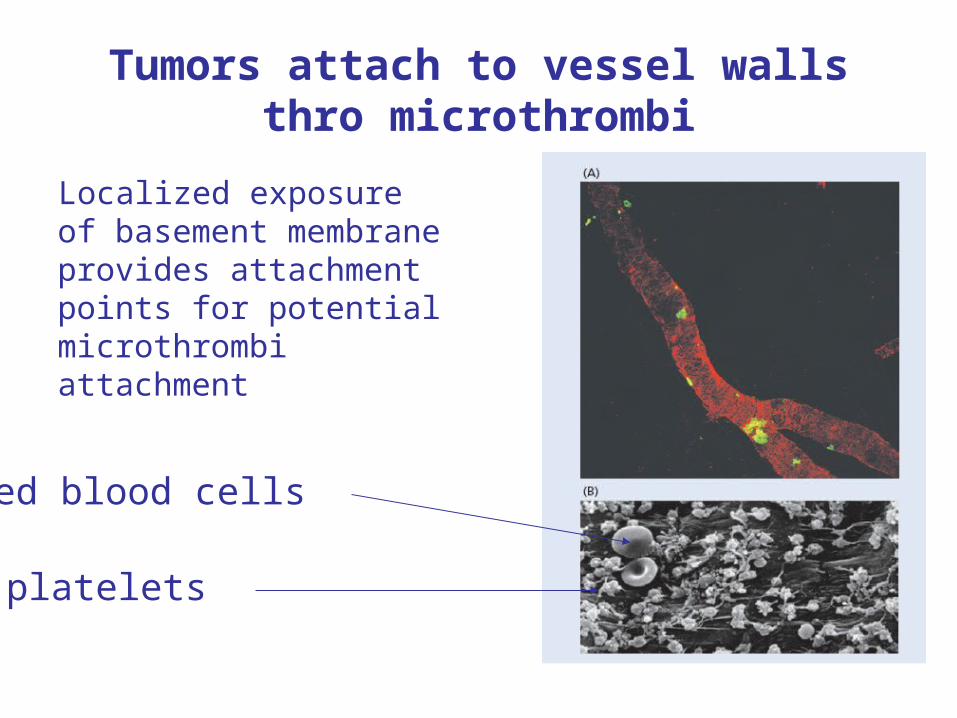
Tumors attach to vessel walls thro microthrombi
Localized exposure of basement membrane provides attachment points for potential microthrombi attachment
red blood cells
platelets
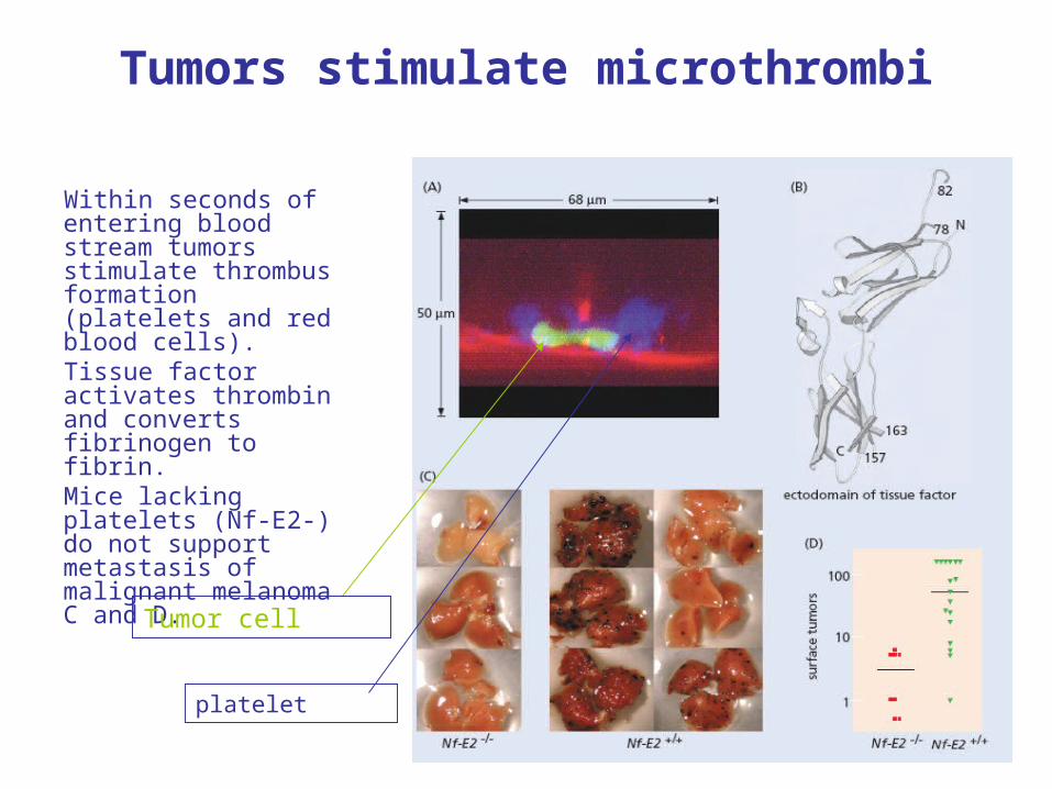
Tumors stimulate microthrombi
Within seconds of entering blood stream tumors stimulate thrombus formation (platelets and red blood cells).Tissue factor activates thrombin and converts fibrinogen to fibrin.Mice lacking platelets (Nf-E2-) do not support metastasis of malignant melanoma C and D.
Tumor cell
platelet
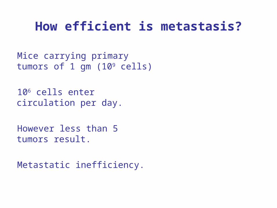
How efficient is metastasis?
Mice carrying primary tumors of 1 gm (109 cells)
106 cells enter circulation per day.
However less than 5 tumors result.
Metastatic inefficiency.
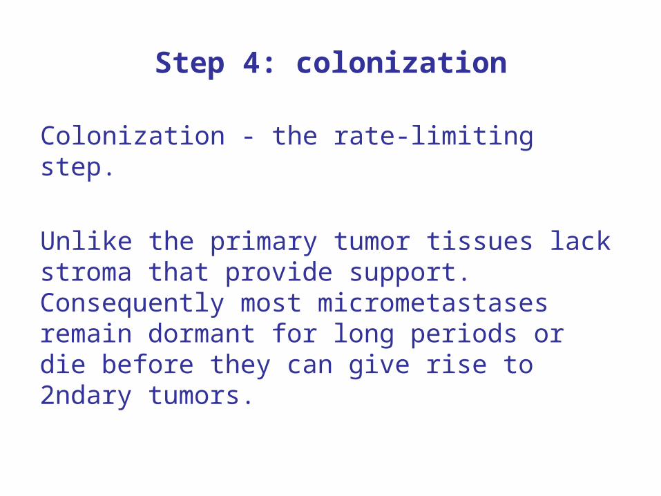
Step 4: colonization
Colonization - the rate-limiting step.
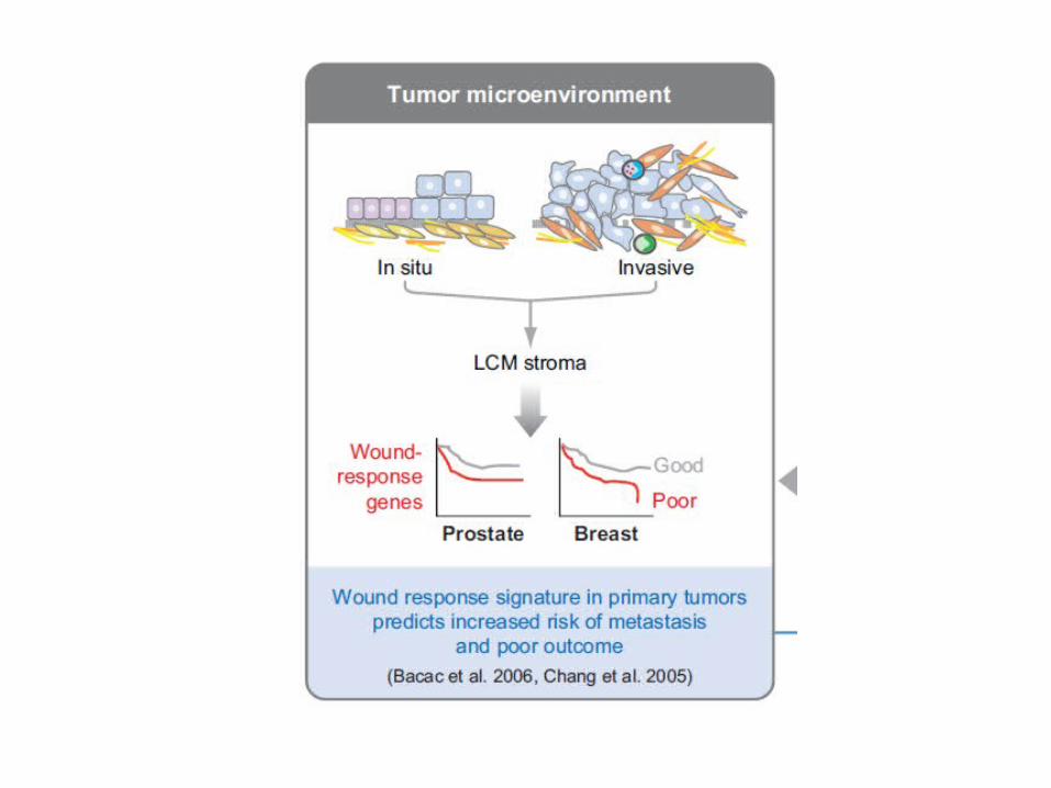
Unlike the primary tumor tissues lack stroma that provide support. Consequently most micrometastases remain dormant for long periods or die before they can give rise to 2ndary tumors.
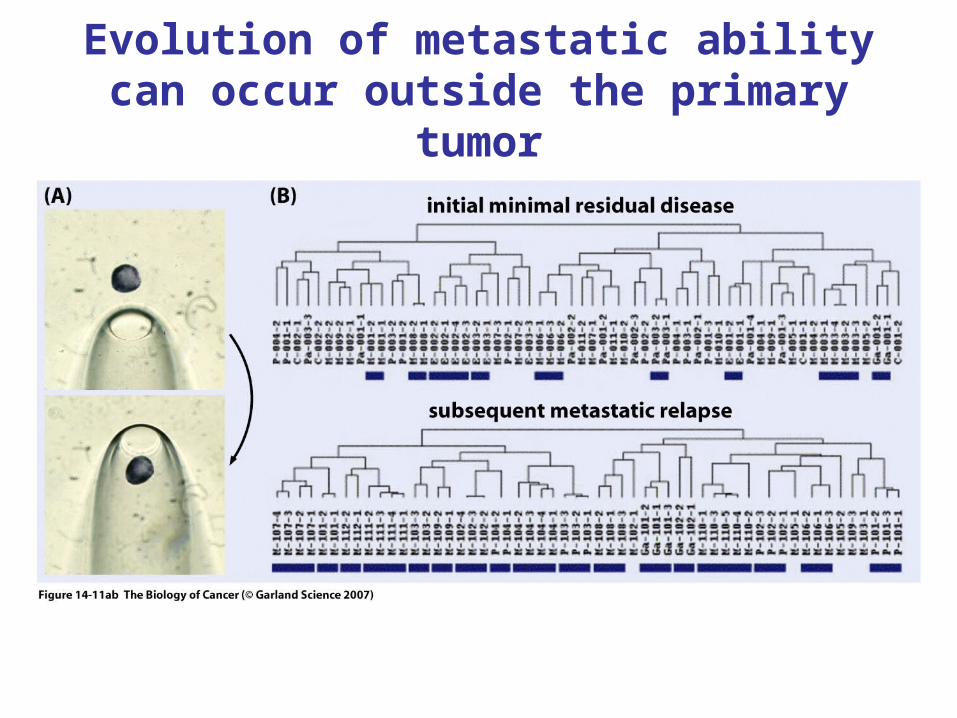
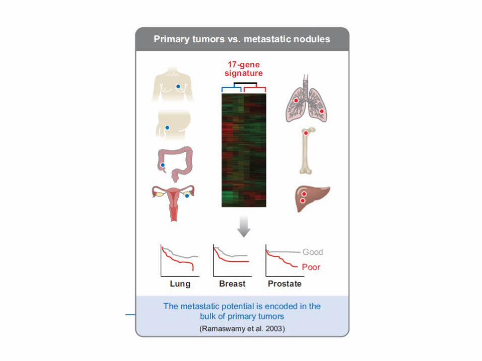
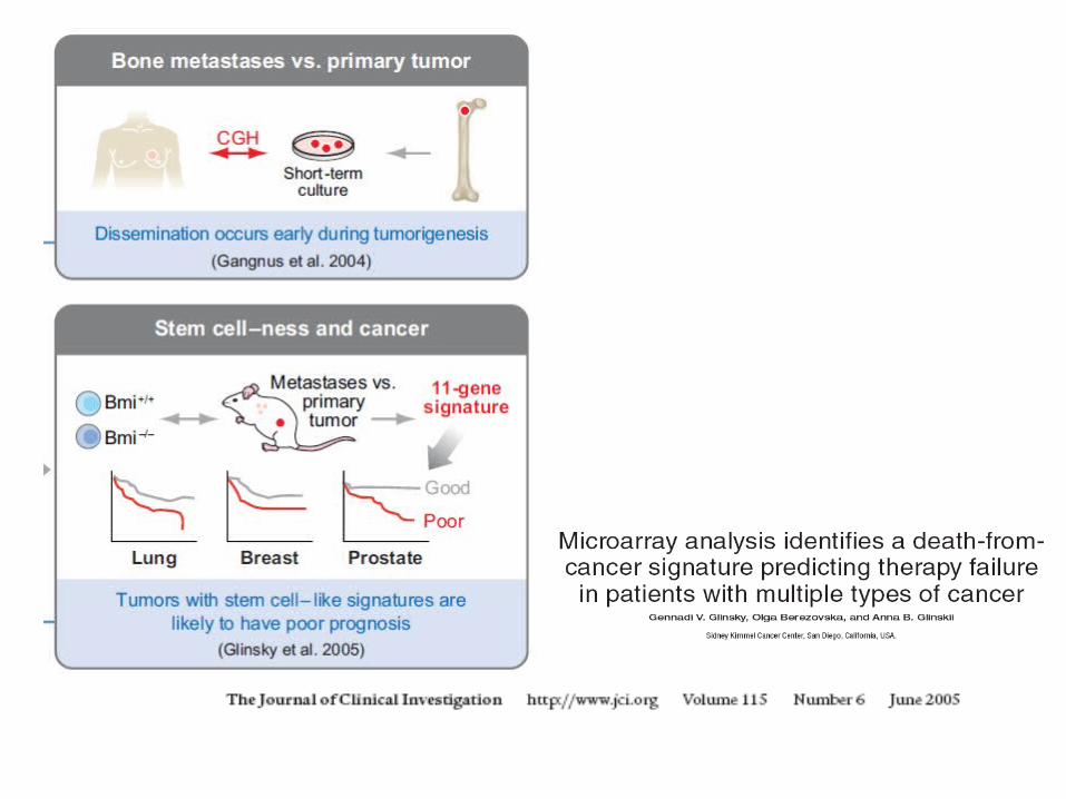
Evolution of metastatic ability can occur outside the primary tumor
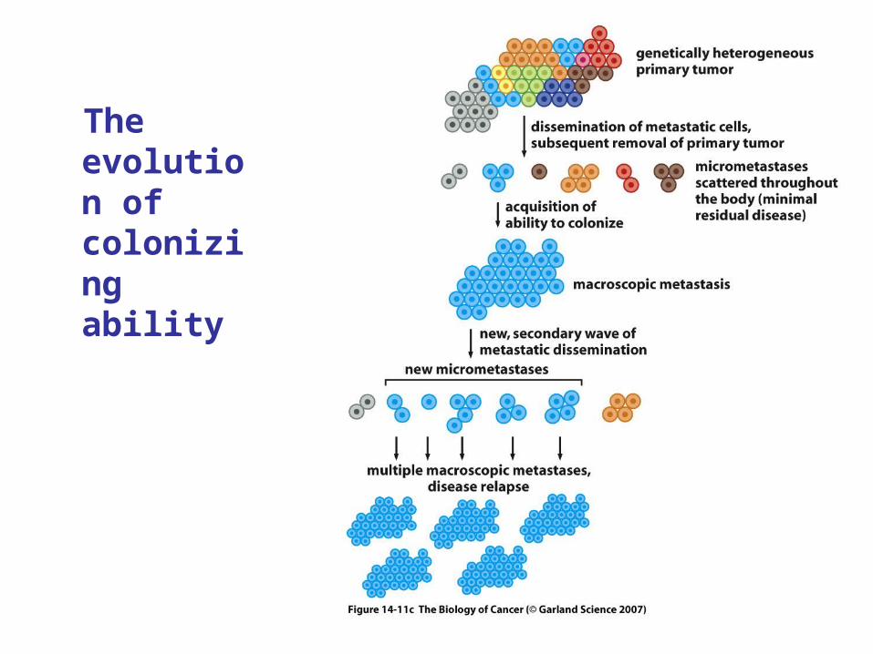
The evolution of colonizing ability
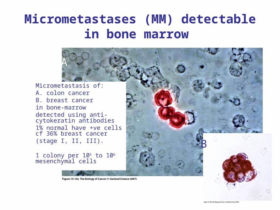
Micrometastases (MM) detectable in bone marrow
Micrometastasis of:A. colon cancerB. breast cancer in bone-marrowdetected using anti-cytokeratin antibodies1% normal have +ve cells cf 36% breast cancer(stage I, II, III).
1 colony per 105 to 106 mesenchymal cells
A
B
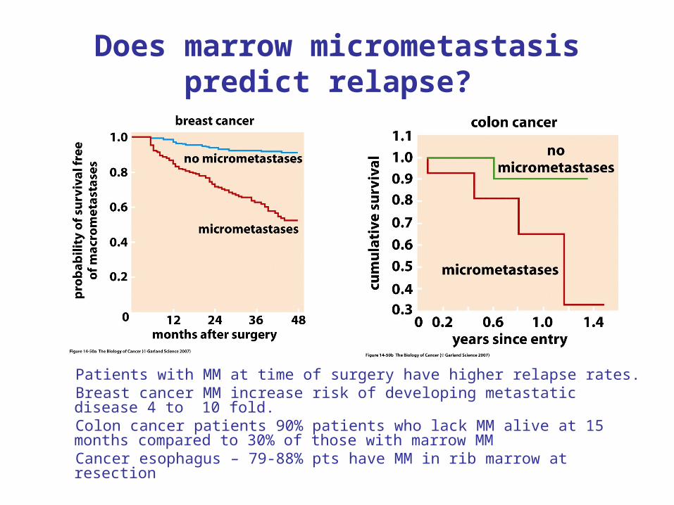
Does marrow micrometastasis predict relapse?
Patients with MM at time of surgery have higher relapse rates.Breast cancer MM increase risk of developing metastatic disease 4 to 10 fold.Colon cancer patients 90% patients who lack MM alive at 15 months compared to 30% of those with marrow MMCancer esophagus – 79-88% pts have MM in rib marrow at resection
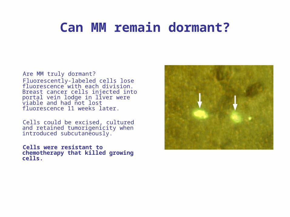
Can MM remain dormant?
Are MM truly dormant? Fluorescently-labeled cells lose fluorescence with each division. Breast cancer cells injected into portal vein lodge in liver were viable and had not lost fluorescence 11 weeks later. Cells could be excised, cultured and retained tumorigenicity when introduced subcutaneously.
Cells were resistant to chemotherapy that killed growing cells.
What we have learned so far
Invasion-metastasis cascade has multiple steps.
As many changes in phenotype involved as precede inititial tumor formation.
Cells readily enter circulation and seed distant sites. However, only a small fraction grow.
Nevertheless vast numbers of cells seeded by growing tumors ensure that some of these will eventually grow.
Clinically these pose a major threat to survival of patient.
Important question:
To what extent do the steps represent changes in gene expression. Are there metastasis specific genes and their suppressors?
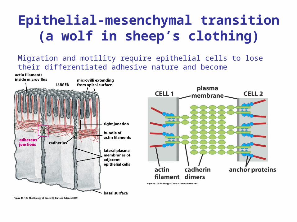
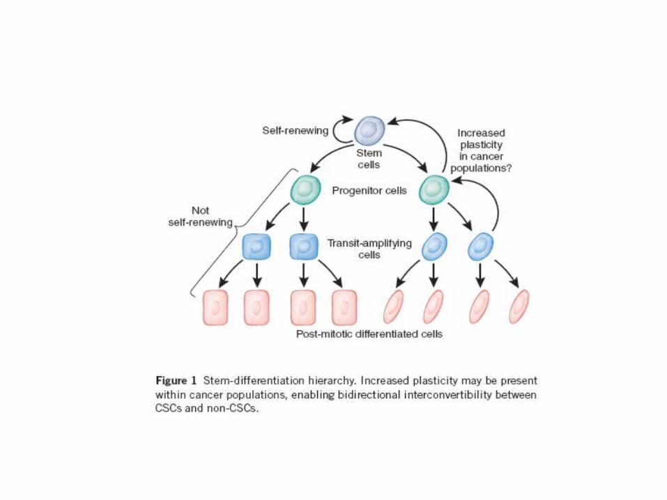
Epithelial-mesenchymal transition(a wolf in sheep’s clothing)
Migration and motility require epithelial cells to lose their differentiated adhesive nature and become mesenchyme-like.
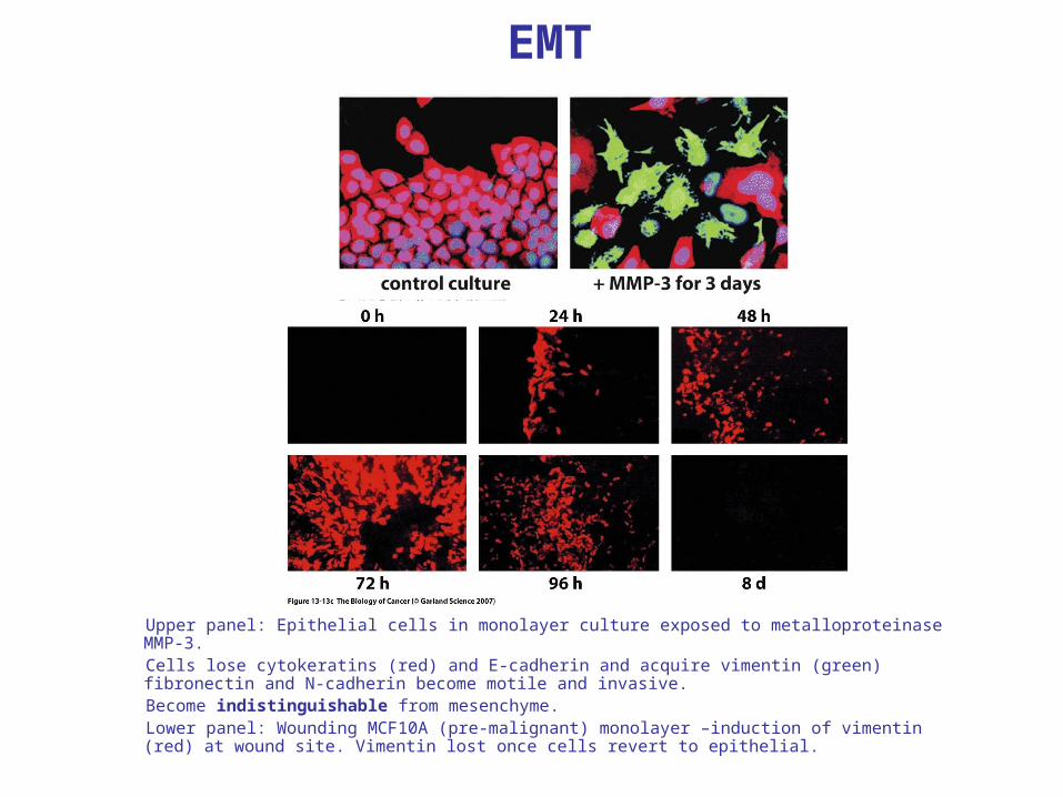
EMT
Upper panel: Epithelial cells in monolayer culture exposed to metalloproteinase MMP-3.Cells lose cytokeratins (red) and E-cadherin and acquire vimentin (green) fibronectin and N-cadherin become motile and invasive.Become indistinguishable from mesenchyme. Lower panel: Wounding MCF10A (pre-malignant) monolayer –induction of vimentin (red) at wound site. Vimentin lost once cells revert to epithelial.
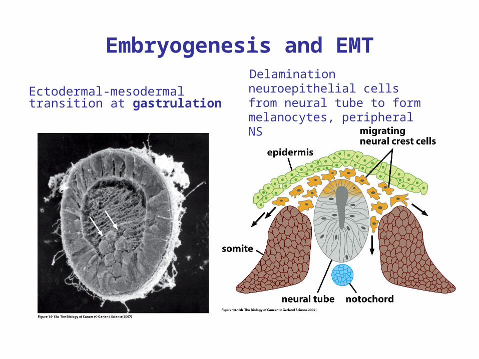
Embryogenesis and EMT
Ectodermal-mesodermal transition at gastrulation
Delamination neuroepithelial cells from neural tube to form melanocytes, peripheral NS
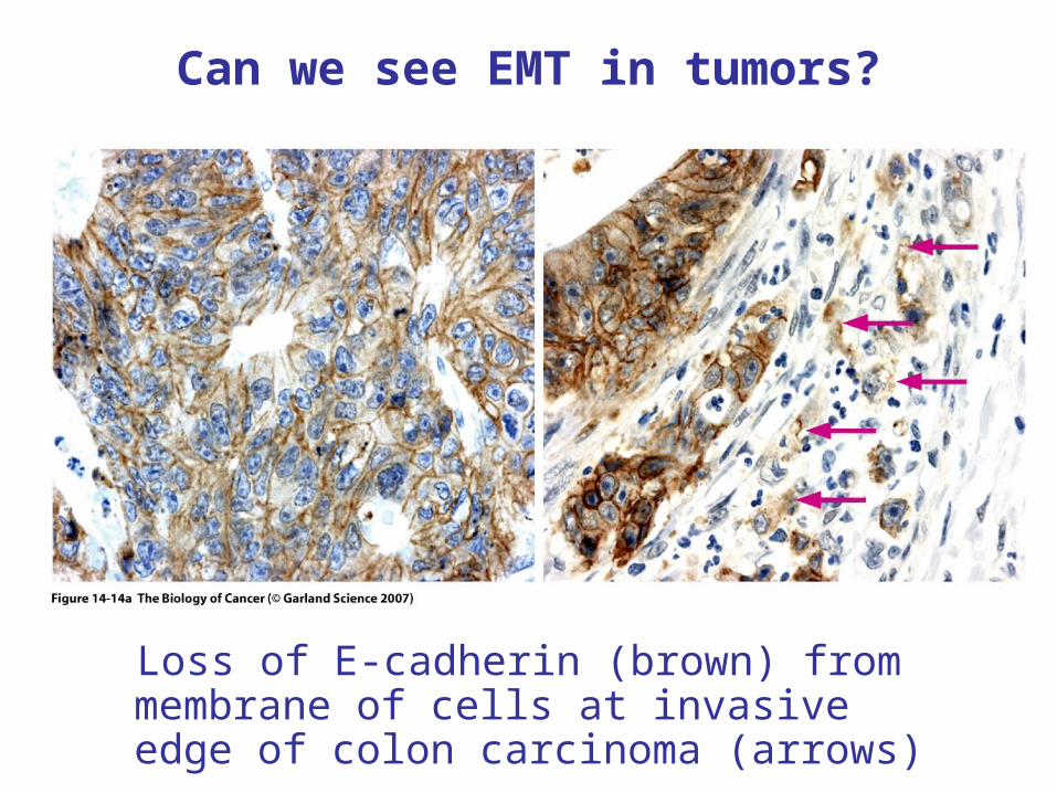
Can we see EMT in tumors?
Loss of E-cadherin (brown) from membrane of cells at invasive edge of colon carcinoma (arrows)
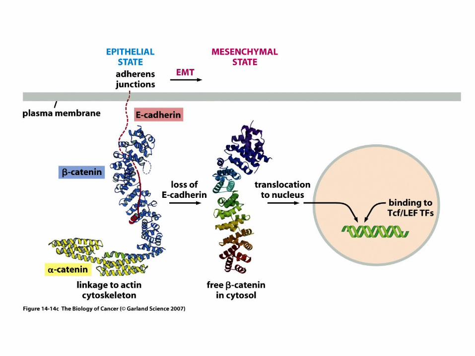
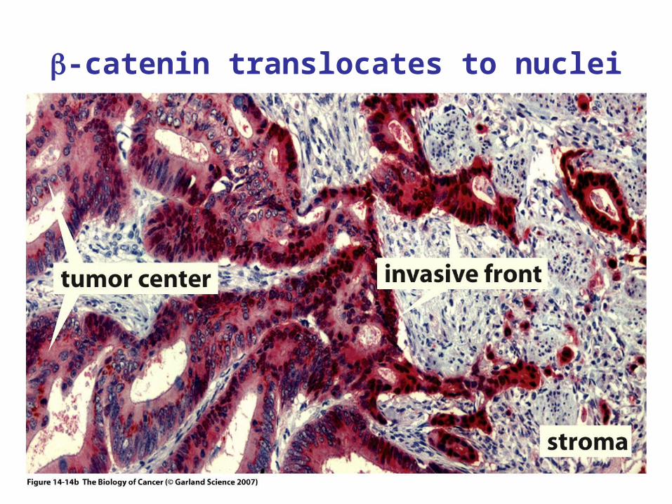
-catenin translocates to nuclei
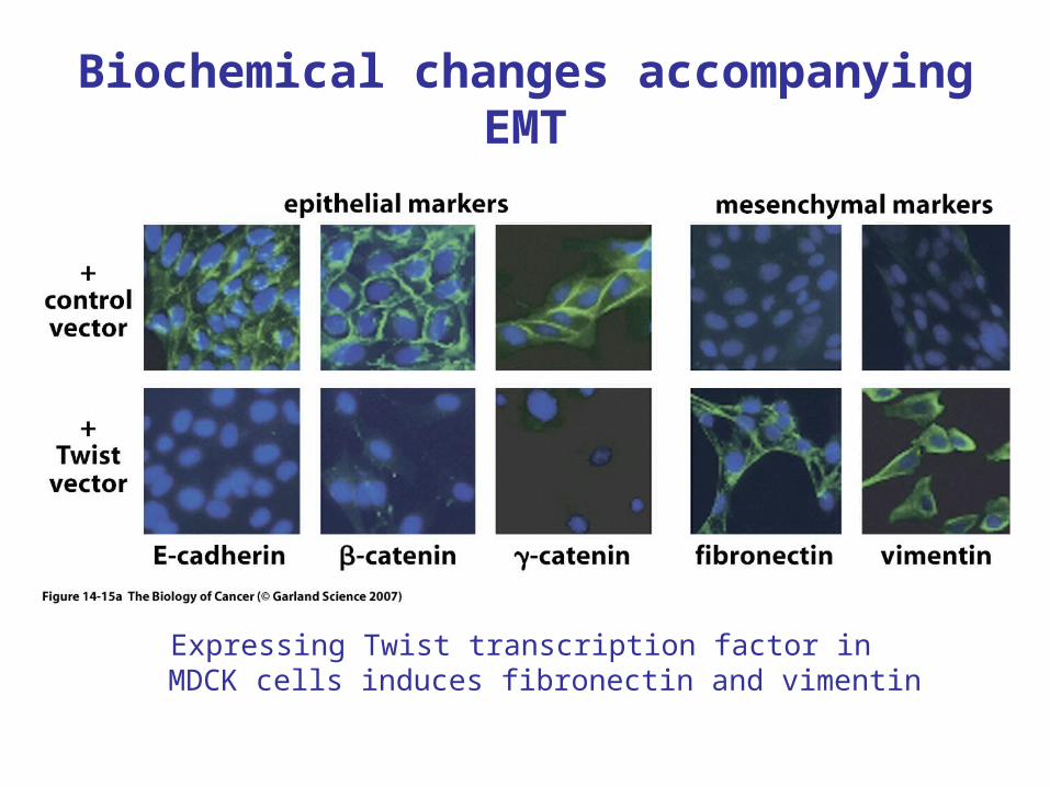
Biochemical changes accompanying EMT
Expressing Twist transcription factor in MDCK cells induces fibronectin and vimentin
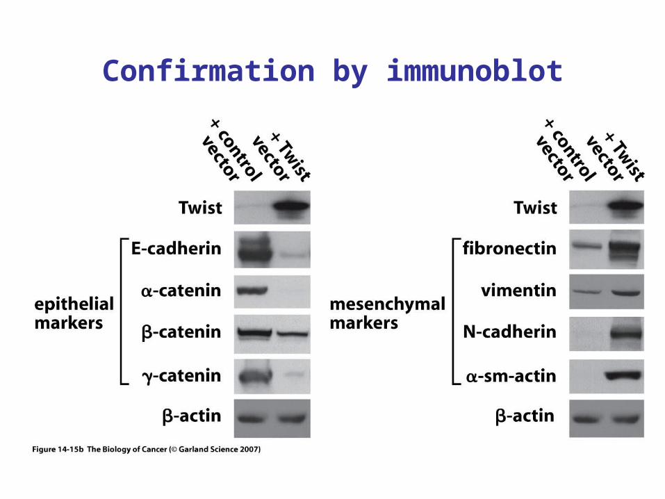
Confirmation by immunoblot
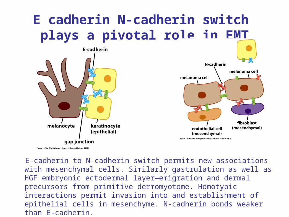
E cadherin N-cadherin switch plays a pivotal role in EMT
E-cadherin to N-cadherin switch permits new associations with mesenchymal cells. Similarly gastrulation as well as HGF embryonic ectodermal layer—emigration and dermal precursors from primitive dermomyotome. Homotypic interactions permit invasion into and establishment of epithelial cells in mesenchyme. N-cadherin bonds weaker than E-cadherin.
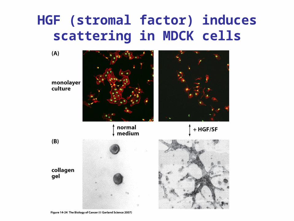
HGF (stromal factor) induces scattering in MDCK cells
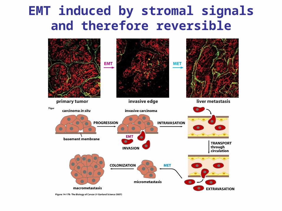
EMT induced by stromal signals and therefore reversible
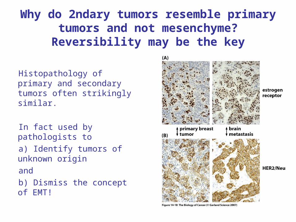
Why do 2ndary tumors resemble primary tumors and not mesenchyme?
Reversibility may be the key
Histopathology of primary and secondary tumors often strikingly similar.
In fact used by pathologists to
a) Identify tumors of unknown origin
and
b) Dismiss the concept of EMT!
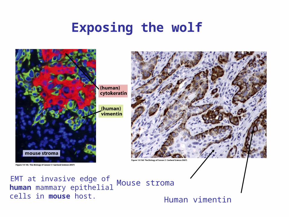
Exposing the wolf
Mouse stroma
Human vimentin
EMT at invasive edge of human mammary epithelial cells in mouse host.
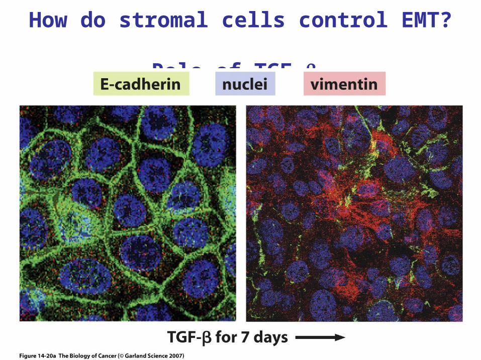
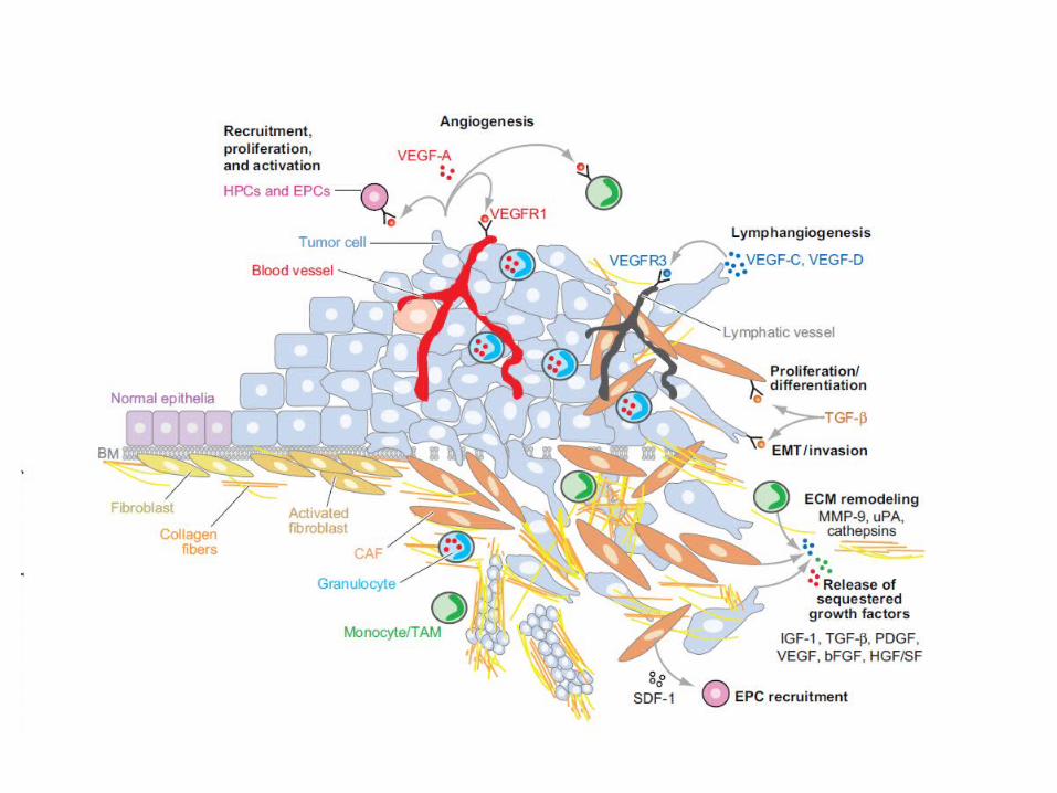
How do stromal cells control EMT? Role of TGF-
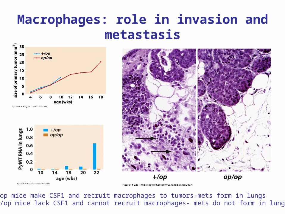
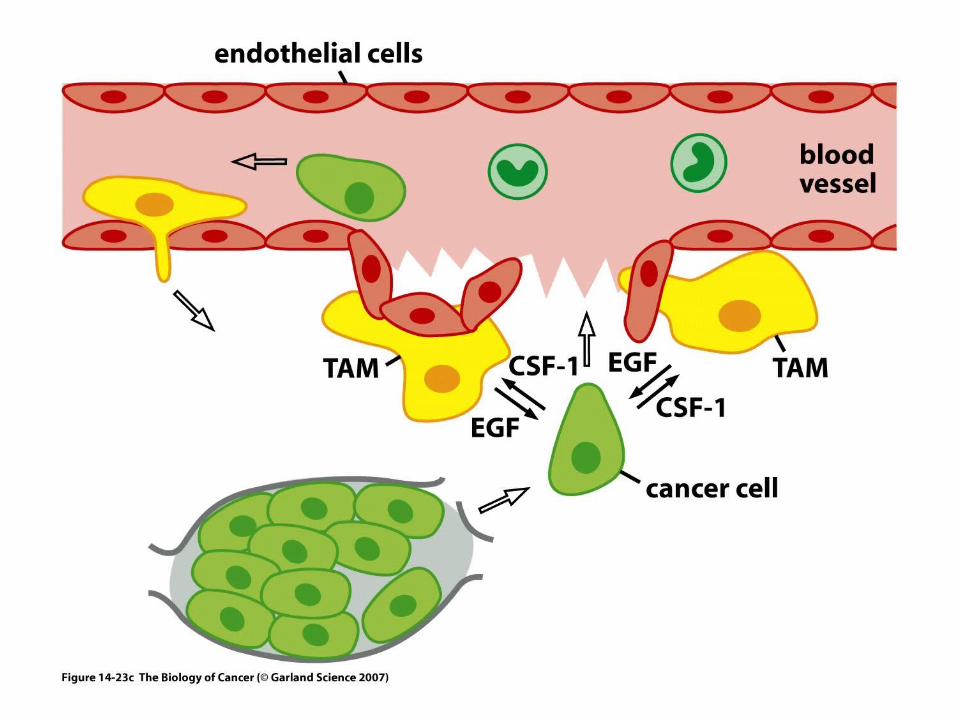
Macrophages: role in invasion and metastasis
+/op mice make CSF1 and recruit macrophages to tumors-mets form in lungsop/op mice lack CSF1 and cannot recruit macrophages- mets do not form in lungs
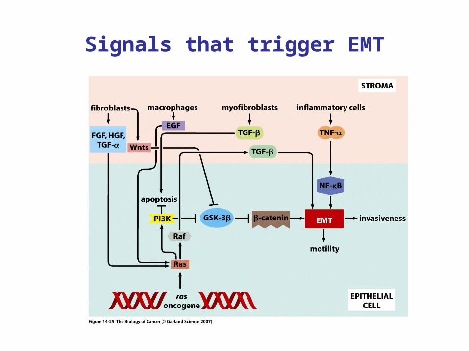
Signals that trigger EMT

Embryonic transcription factors programming EMT
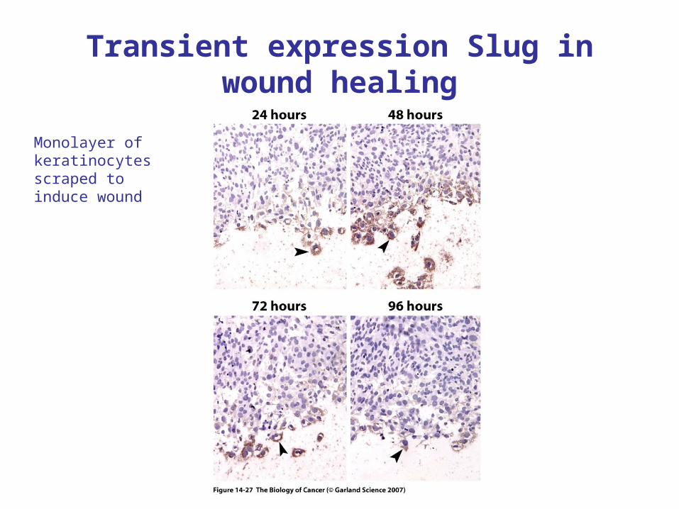
Transient expression Slug in wound healing
Monolayer of keratinocytesscraped to induce wound
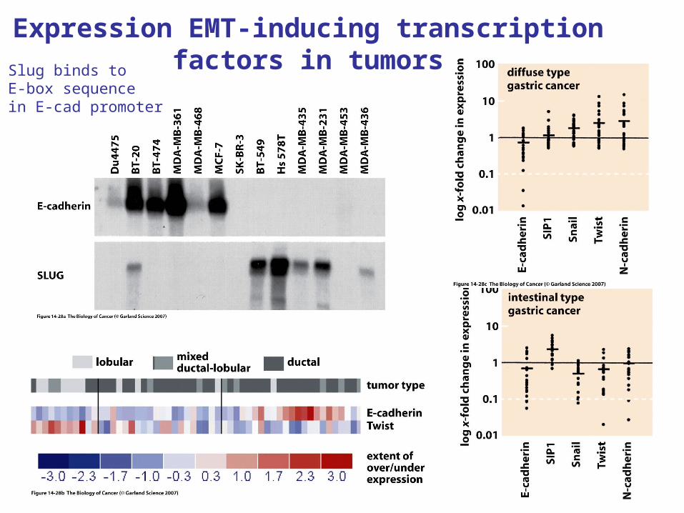
Expression EMT-inducing transcription factors in tumorsSlug binds to
E-box sequencein E-cad promoter
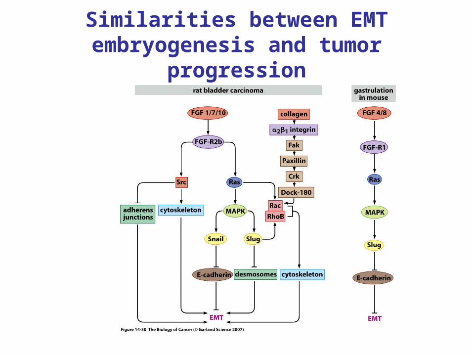
Similarities between EMT embryogenesis and tumor progression
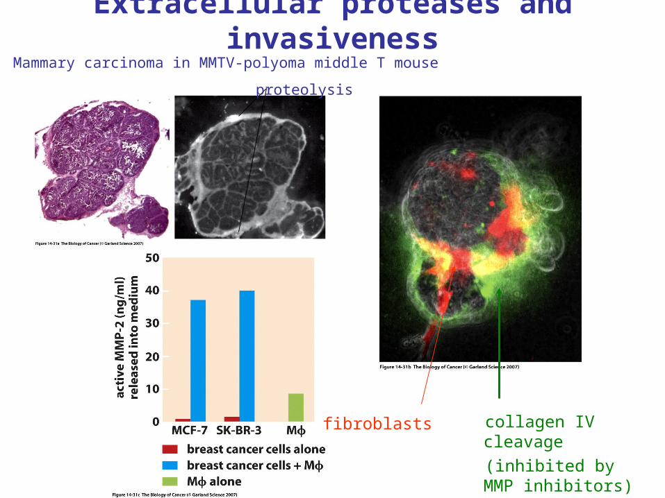
Extracellular proteases and invasiveness
collagen IV cleavage
(inhibited by MMP inhibitors)
fibroblasts
Mammary carcinoma in MMTV-polyoma middle T mouse
proteolysis
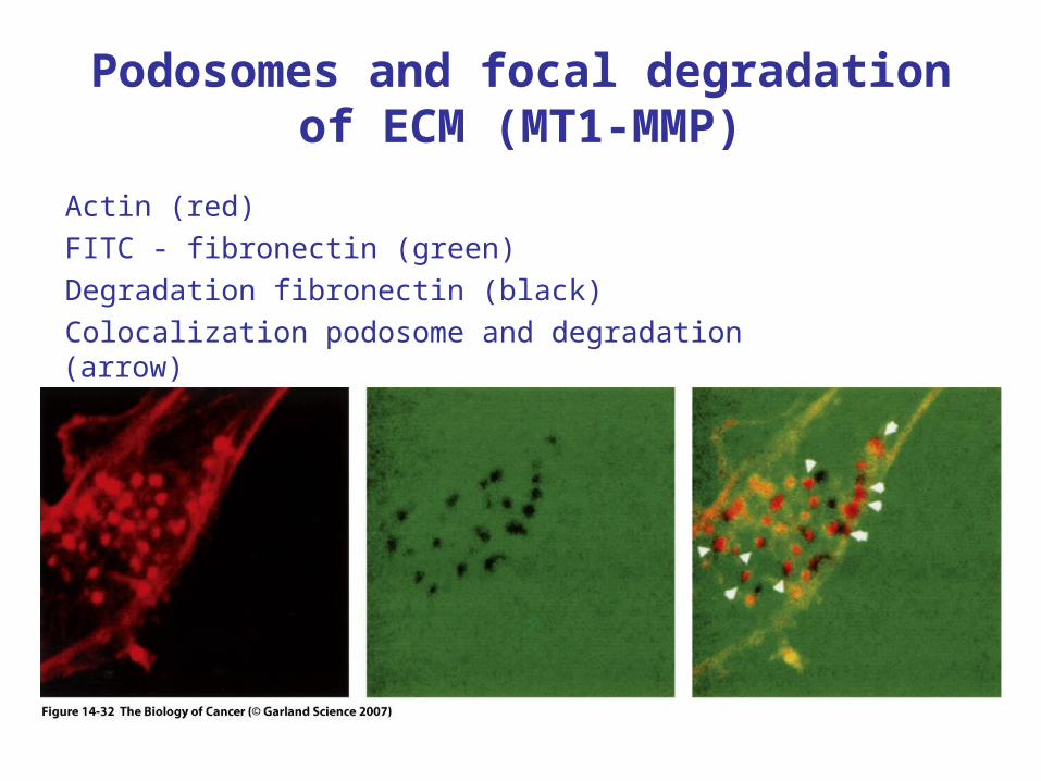
Podosomes and focal degradation of ECM (MT1-MMP)
Actin (red)
FITC - fibronectin (green)
Degradation fibronectin (black)
Colocalization podosome and degradation (arrow)
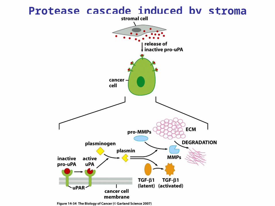
Protease cascade induced by stroma
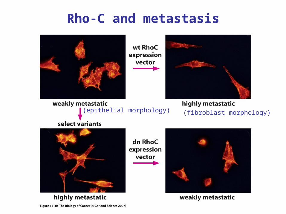
Rho-C and metastasis
(epithelial morphology) (fibroblast morphology)
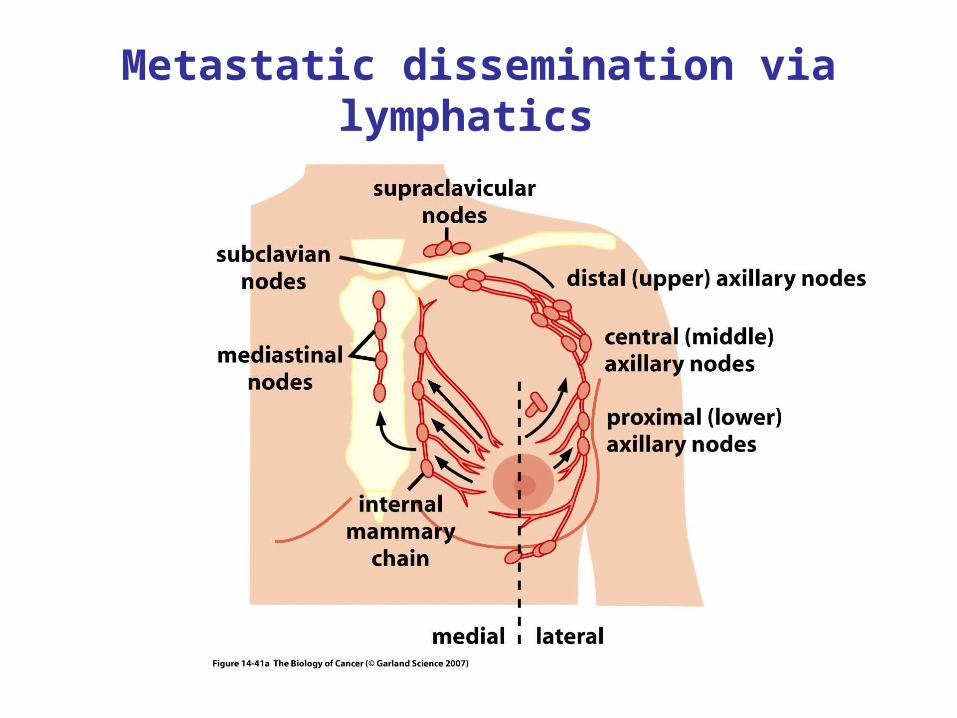
Metastatic dissemination via lymphatics
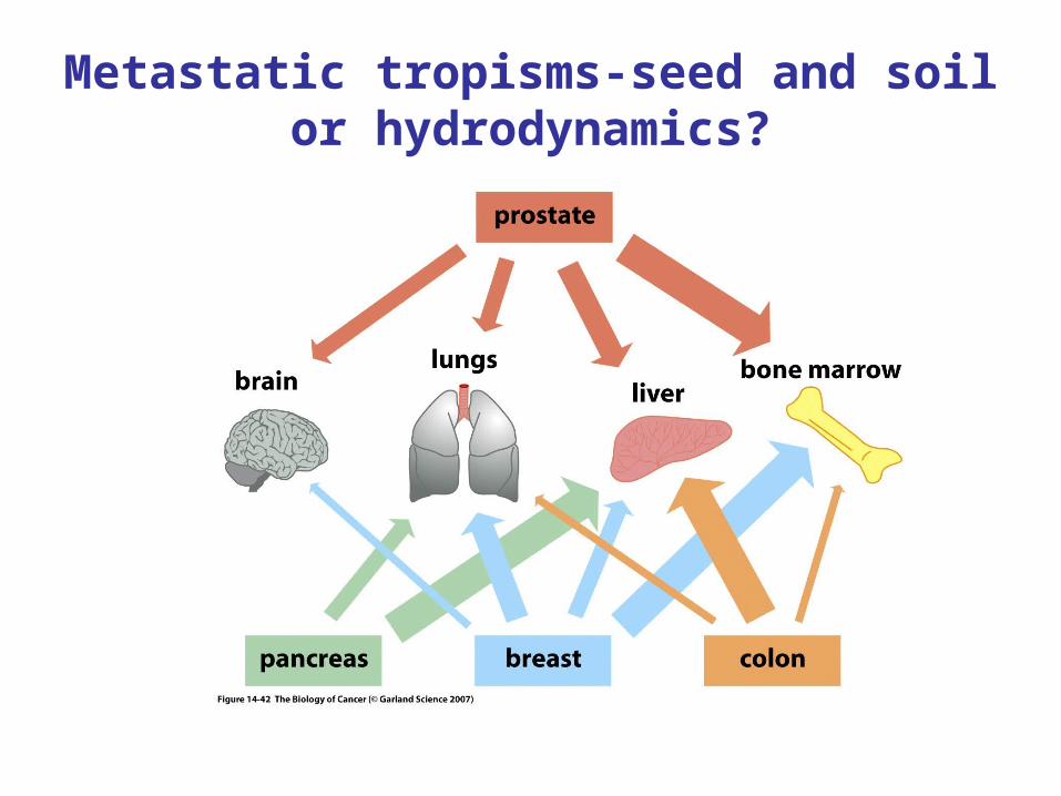
Metastatic tropisms-seed and soil or hydrodynamics?
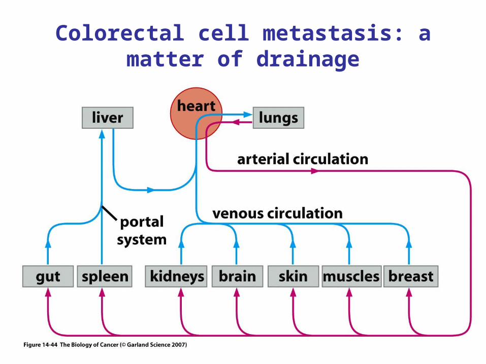
Colorectal cell metastasis: a matter of drainage
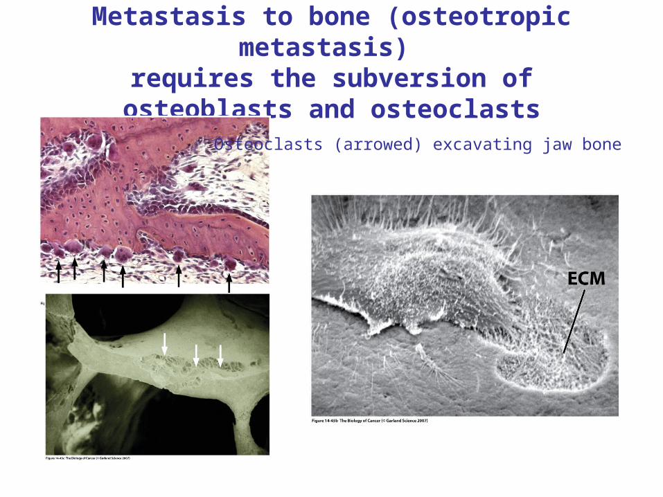
Metastasis to bone (osteotropic metastasis) requires the subversion of osteoblasts and osteoclasts
Osteoclasts (arrowed) excavating jaw bone
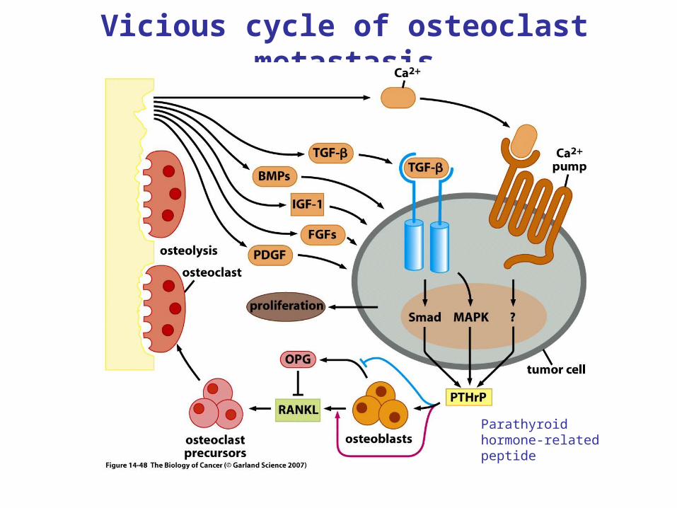
Vicious cycle of osteoclast metastasis
Parathyroid hormone-related peptide
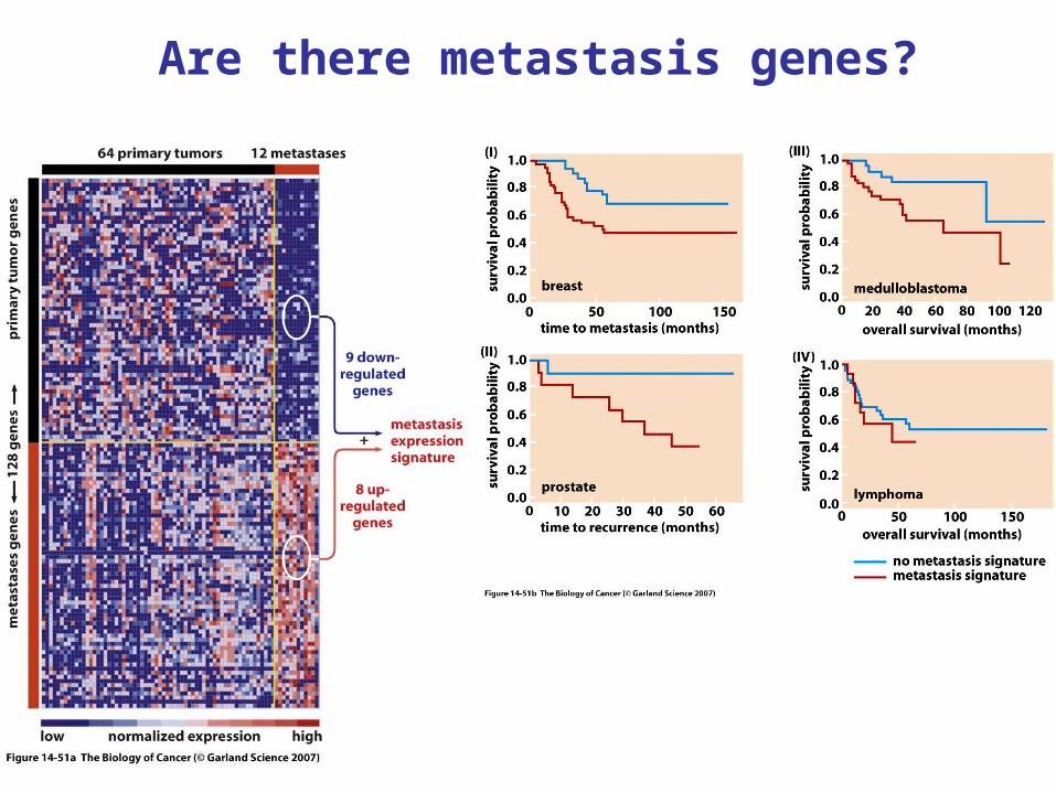
Are there metastasis genes?