Metaphorical sentences are more emotionally …adele/Princeton_Construction_Site/...Metaphorical...
22
To appear, Journal of Cognitive Neuroscience Metaphorical sentences are more emotionally engaging than their literal counterparts Francesca M.M. Citron 1,2* , Adele E. Goldberg 2,1 1 Cluster of Excellence “Languages of Emotion”, Freie Universität Berlin, Berlin, Germany 2 Humanities Council, Princeton University, Princeton, NJ, USA Address correspondence to: Francesca Citron Cluster of Excellence “Languages of Emotion” Freie Universität Berlin Habelschwerdter Allee 45 D-14195 Berlin Tel.: +49 (0) 3083857869 Fax: +49 (0) 3083852887 E-mail: [email protected] Running head: Metaphors evoke emotions
Transcript of Metaphorical sentences are more emotionally …adele/Princeton_Construction_Site/...Metaphorical...
To appear, Journal of Cognitive Neuroscience
Metaphorical sentences are more emotionally engaging than their literal counterparts
Francesca M.M. Citron1,2* , Adele E. Goldberg2,1
1 Cluster of Excellence “Languages of Emotion”, Freie Universität Berlin, Berlin, Germany
2 Humanities Council, Princeton University, Princeton, NJ, USA Address correspondence to: Francesca Citron
Cluster of Excellence “Languages of Emotion” Freie Universität Berlin Habelschwerdter Allee 45 D-14195 Berlin Tel.: +49 (0) 3083857869 Fax: +49 (0) 3083852887
E-mail: [email protected]
Running head: Metaphors evoke emotions
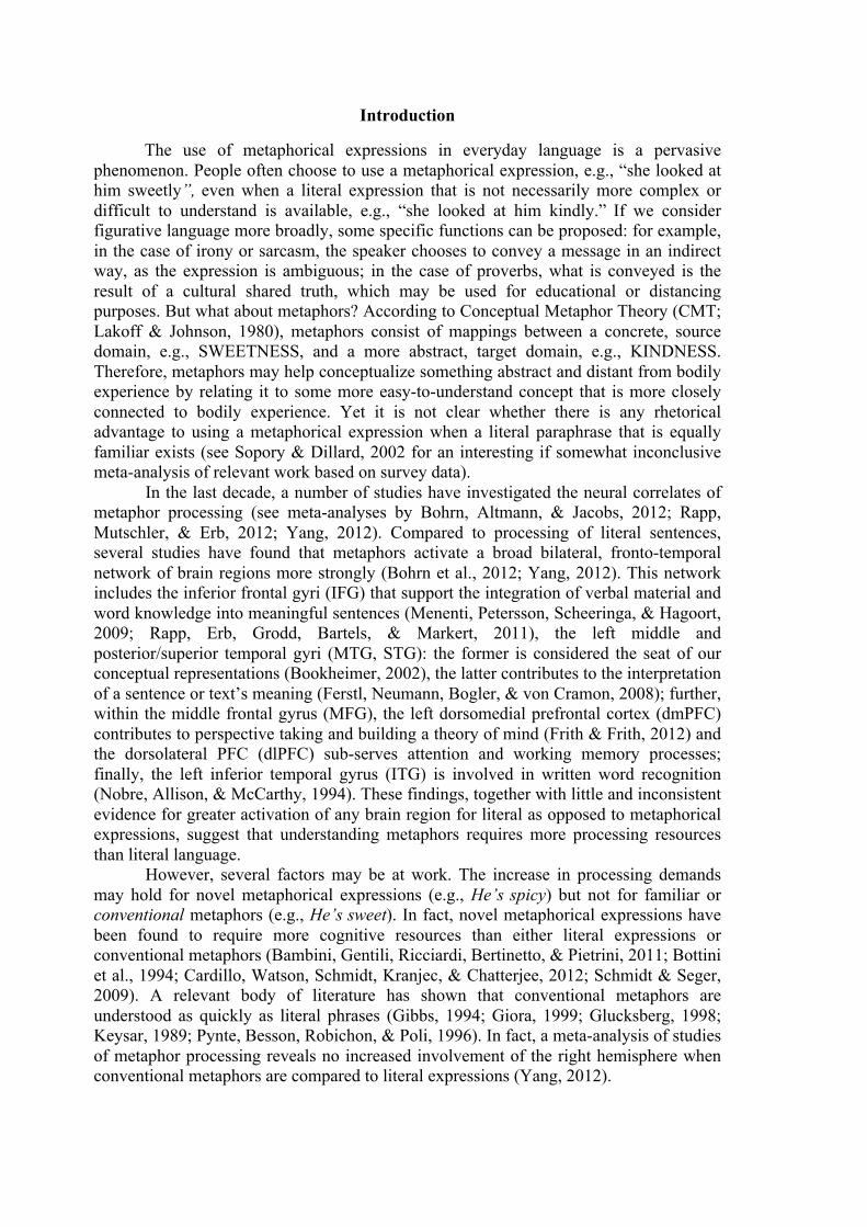
Abstract Why do people so often use metaphorical expressions when literal paraphrases
are readily available? The present study focuses on a comparison of metaphorical statements involving the source domain of taste (e.g., “She looked at him sweetly”), and their literal paraphrases (e.g., “She looked at him kindly”). Metaphorical and literal sentences differed only in one word and were normed for length, familiarity, imageability, emotional valence, and arousal. Our findings indicate that conventional metaphorical expressions are more emotionally evocative than literal expressions, as the amygdala and the anterior portion of the hippocampus were more active in the metaphorical sentences. They also support the idea that even conventional metaphors can be grounded in sensory-motor and perceptual representations in that primary and secondary gustatory areas (lateral orbitofrontal cortex, frontal operculum, anterior insula) were more active as well. A comparison of the individual words that distinguished the metaphorical and literal sentences revealed greater activation in the lateral orbitofrontal cortex and the frontal operculum for the taste-related words, supporting the claim that these areas are relevant to taste.
Keywords: metaphor, fMRI, emotion, taste, amygdala, hippocampus
Introduction
The use of metaphorical expressions in everyday language is a pervasive phenomenon. People often choose to use a metaphorical expression, e.g., “she looked at him sweetly”, even when a literal expression that is not necessarily more complex or difficult to understand is available, e.g., “she looked at him kindly.” If we consider figurative language more broadly, some specific functions can be proposed: for example, in the case of irony or sarcasm, the speaker chooses to convey a message in an indirect way, as the expression is ambiguous; in the case of proverbs, what is conveyed is the result of a cultural shared truth, which may be used for educational or distancing purposes. But what about metaphors? According to Conceptual Metaphor Theory (CMT; Lakoff & Johnson, 1980), metaphors consist of mappings between a concrete, source domain, e.g., SWEETNESS, and a more abstract, target domain, e.g., KINDNESS. Therefore, metaphors may help conceptualize something abstract and distant from bodily experience by relating it to some more easy-to-understand concept that is more closely connected to bodily experience. Yet it is not clear whether there is any rhetorical advantage to using a metaphorical expression when a literal paraphrase that is equally familiar exists (see Sopory & Dillard, 2002 for an interesting if somewhat inconclusive meta-analysis of relevant work based on survey data).
In the last decade, a number of studies have investigated the neural correlates of metaphor processing (see meta-analyses by Bohrn, Altmann, & Jacobs, 2012; Rapp, Mutschler, & Erb, 2012; Yang, 2012). Compared to processing of literal sentences, several studies have found that metaphors activate a broad bilateral, fronto-temporal network of brain regions more strongly (Bohrn et al., 2012; Yang, 2012). This network includes the inferior frontal gyri (IFG) that support the integration of verbal material and word knowledge into meaningful sentences (Menenti, Petersson, Scheeringa, & Hagoort, 2009; Rapp, Erb, Grodd, Bartels, & Markert, 2011), the left middle and posterior/superior temporal gyri (MTG, STG): the former is considered the seat of our conceptual representations (Bookheimer, 2002), the latter contributes to the interpretation of a sentence or text’s meaning (Ferstl, Neumann, Bogler, & von Cramon, 2008); further, within the middle frontal gyrus (MFG), the left dorsomedial prefrontal cortex (dmPFC) contributes to perspective taking and building a theory of mind (Frith & Frith, 2012) and the dorsolateral PFC (dlPFC) sub-serves attention and working memory processes; finally, the left inferior temporal gyrus (ITG) is involved in written word recognition (Nobre, Allison, & McCarthy, 1994). These findings, together with little and inconsistent evidence for greater activation of any brain region for literal as opposed to metaphorical expressions, suggest that understanding metaphors requires more processing resources than literal language.
However, several factors may be at work. The increase in processing demands may hold for novel metaphorical expressions (e.g., He’s spicy) but not for familiar or conventional metaphors (e.g., He’s sweet). In fact, novel metaphorical expressions have been found to require more cognitive resources than either literal expressions or conventional metaphors (Bambini, Gentili, Ricciardi, Bertinetto, & Pietrini, 2011; Bottini et al., 1994; Cardillo, Watson, Schmidt, Kranjec, & Chatterjee, 2012; Schmidt & Seger, 2009). A relevant body of literature has shown that conventional metaphors are understood as quickly as literal phrases (Gibbs, 1994; Giora, 1999; Glucksberg, 1998; Keysar, 1989; Pynte, Besson, Robichon, & Poli, 1996). In fact, a meta-analysis of studies of metaphor processing reveals no increased involvement of the right hemisphere when conventional metaphors are compared to literal expressions (Yang, 2012).
Specific tasks may elicit different processing strategies for metaphors than other types of sentences. These include judgments about plausibility (Bottini et al., 1994), since metaphorical sentences are typically literally false and therefore arguably less plausible; judgments of semantic relatedness of an expression with a target word (Bambini et al., 2011; Chen, Widick, & Chatterjee, 2008; Stringaris et al., 2006), since metaphors commonly use more concrete language than literal expressions; or judgments about whether two words are literally related vs. metaphorically related vs. unrelated (Mashal, Faust, & Hendler, 2005; Mashal, Faust, Hendler, & Jung-Beeman, 2007), since the task requires explicit judgments of metaphoricity. Further, reading certain metaphorical compounds e.g., broken heart, or predicative nominal metaphors such as my surgeon is a butcher might feel unnatural and require more integrative processes than reading the same expressions embedded in situational/discourse contexts, whereas similar literal phrases might sound perfectly acceptable even without a context (e.g., sunny day; my son is a butcher).
Finally, and most importantly, only a few studies have matched metaphorical and literal sentences on important psycholinguistic variables, such as familiarity, length, syntactic complexity, imageability, meaning, emotional valence, and arousal (e.g., Cardillo, Schmidt, Kranjec, & Chatterjee, 2010; Cardillo et al., 2012). A recent study by Desai, Binder, Conant, Mano and Seidenberg (2011) did match their stimuli for familiarity and processing difficulty, and while they found more activation of left temporal areas as well as cingulate cortex for metaphorical than literal sentences, they found no greater involvement of the IFG, unlike the meta-analyses cited above. Another study focused on texture metaphors (Lacey, Stilla, & Sathian, 2012) employed commonly used metaphorical expressions such as she had a rough day (conventional and natural), and compared them with identical literal sentences that differed only in a single word, i.e., she had a bad day. The authors were able to show more activation of the texture-related areas for texture metaphors than literal sentences, but no other brain region was significantly more active. The lack of difference, however, was possibly due to the fact that their metaphors were more imageable and frequent than the literal sentences and therefore possibly easier to process on those dimensions. Therefore the question remains, are conventional metaphors processed distinctly from literal sentences, when relevant variables are carefully controlled for?
In order to explore this question, we devised an experiment focused on expressions involving conventional taste metaphors, identical but for one word to their literal counterparts. We normed our stimuli and matched them on a range of psycholinguistic variables including length in letters, number of words, familiarity, imageability, emotional valence and arousal. Our metaphors consist of expressions that are commonly used in everyday conversations and were rated as “relatively common/familiar.” Metaphorical sentences were also judged to be highly similar in meaning to their corresponding literal sentences. Participants were simply asked to read silently for comprehension.
We also explore whether highly conventional metaphors are indeed grounded in embodied representations. Evidence to date has been mixed. Some behavioral evidence suggests that we only access concrete representations when processing novel metaphors, not conventional, “dead” metaphors (Keysar, Shen, Glucksberg, & Horton 2000; but cf. Gibbs, Lima and Francozo (2004). Certain neuroimaging studies have shown recruitment of sensory-motor representations in response to conventional figurative expressions (e.g., Boulenger, Hauk, & Pulvermueller, 2009; Lacey et al., 2012), while others have found motor regions active in response to action words in isolation or embedded in literal sentences, but not in response to action idioms (Raposo, Moss, Stamatakis, & Tyler,
2009). Finally, Desai et al. (2011) has shown the engagement of motor regions to be inversely correlated with metaphor familiarity; hence the more familiar the metaphor, the less likely the access to embodied representations.
Our first goal is exploratory: if conventional metaphors are understood in the same way as literal sentences, we should find no additional areas of activation; if metaphors are processed differently than literal sentences, we expect to find additional areas of activation. The fronto-temporal network reported in the literature might or might not be involved, since our task and verbal material should not elicit differential processing strategies or difficulty levels for metaphors vs. literal sentences.
A second hypothesis predicts that the comprehension of taste metaphors may activate primary and secondary gustatory areas, i.e., lateral orbitofrontal cortex (lOFC) frontal operculum, and anterior insula (AIC) (De Araujo, Rolls, Kringelbach, McGlone, & Phillips, 2003; Small et al., 2007; Veldhuizen et al., 2011). In order to determine that these areas are active because of taste-reference and not metaphoricity (which might also recruit inferior frontal regions), we extracted the single words that distinguished the metaphorical and literal sentences, and presented them in isolation at the end of the experimental session. We anticipate that taste-referential words (e.g., “sweet” in its literal meaning) will activate primary and secondary gustatory areas more strongly than non-taste-referential words (e.g., “kind”) and these same areas will not be active in the opposite contrast.
Method
Participants Twenty-six native German speakers from the Berlin area took part in the experiment (mostly students; M = 27 years, SD = 4.9; 19 women). They were all right-handed, with normal or corrected-to-normal vision, and had no learning disabilities or neurological diseases. Participants either received course credit or were paid 10€ for their participation. They all gave written informed consent before participating. Materials Thirty-seven conventional metaphorical sentences (MS) were created in German by including a taste-referential word (e.g., süß “sweet”) into a sentential context where the word would be interpreted metaphorically (e.g., Sie bekam ein süßes Kompliment “She received a sweet compliment”). Each taste word was then replaced with its literal counterpart (e.g., nettes “nice”) in order to create 37 literal sentences (LS) that were matched in overall meaning with the metaphors. All sentences were then rated by an independent group of participants (N = 34, 13 women, M = 31 years, SD = 11) for metaphoricity, taste-reference, emotional valence, emotional arousal, imageability, and familiarity (i.e., subjective frequency of use). The descriptive statistics are reported in Table 1. As intended, MS were significantly more metaphorical (t(60.74) = 12.50, p < .0001, r = 0.85) and taste-referential than LS (ts(53.21) = 14.75, p < .0001, r = 0.90), with extremely large effect sizes (i.e., r > 0.50), while being equally valenced, arousing, and imageable (ts(72) < 0.61, ns); Ratings of semantic similarity between MS and corresponding LS were quite high (M = 5.84 on a scale of 1-7; SEM = 0.13). Even though MS consisted of commonly used expressions (conventional metaphors), they were slightly less familiar than LS (t(72) = 2.29, p = .025, r = 0.26), with medium-to-low effect size (i.e., r < 0.30); familiarity is therefore used as a regressor in the analyses. Thirty-seven hash marks strings (HMS) were created as a baseline, similar to the sentences in length and number of continuous sequences (e.g., ## ###### #### ###### ###). MS, LS and HMS were matched for length in words/sequences and letters/hash
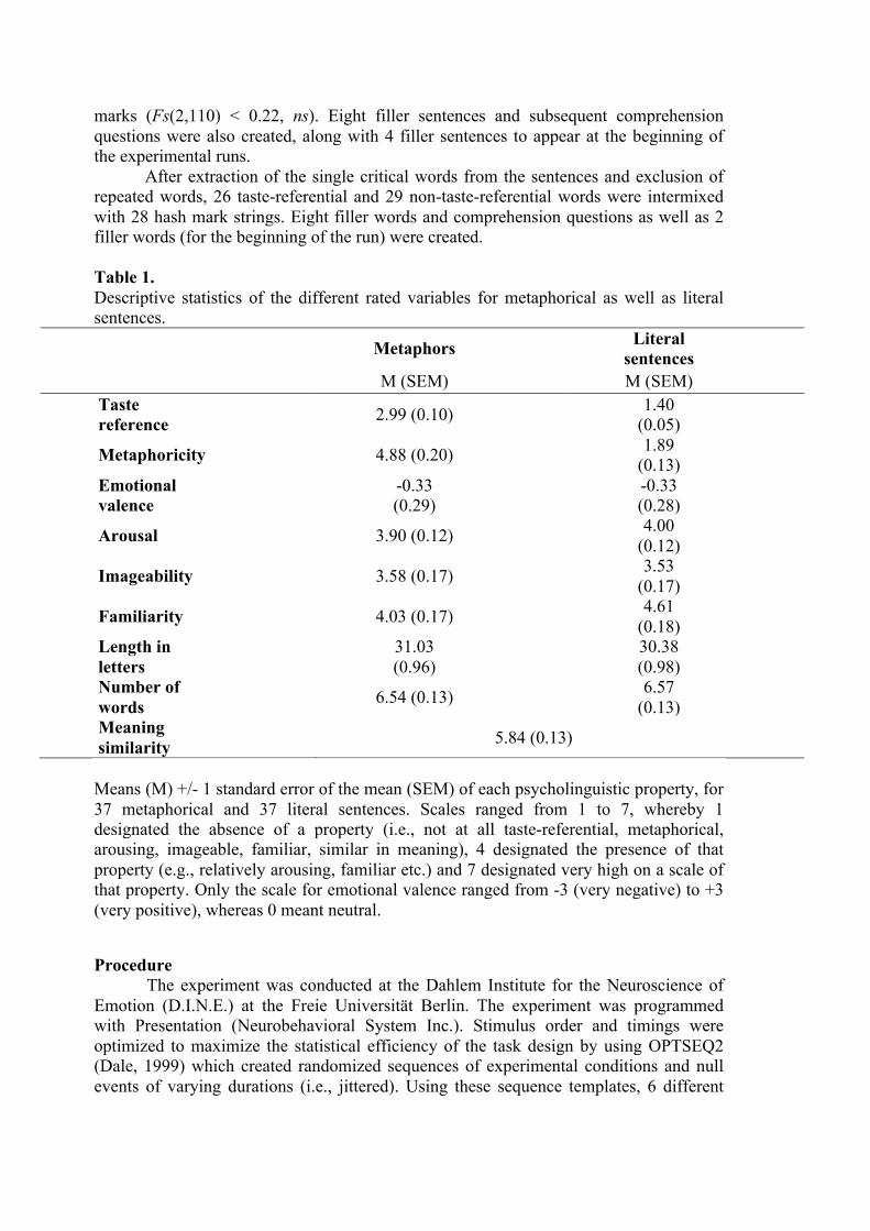
marks (Fs(2,110) < 0.22, ns). Eight filler sentences and subsequent comprehension questions were also created, along with 4 filler sentences to appear at the beginning of the experimental runs. After extraction of the single critical words from the sentences and exclusion of repeated words, 26 taste-referential and 29 non-taste-referential words were intermixed with 28 hash mark strings. Eight filler words and comprehension questions as well as 2 filler words (for the beginning of the run) were created. Table 1. Descriptive statistics of the different rated variables for metaphorical as well as literal sentences.
Metaphors Literal sentences
M (SEM) M (SEM) Taste reference 2.99 (0.10) 1.40
(0.05)
Metaphoricity 4.88 (0.20) 1.89 (0.13)
Emotional valence
-0.33 (0.29)
-0.33 (0.28)
Arousal 3.90 (0.12) 4.00 (0.12)
Imageability 3.58 (0.17) 3.53 (0.17)
Familiarity 4.03 (0.17) 4.61 (0.18)
Length in letters
31.03 (0.96)
30.38 (0.98)
Number of words 6.54 (0.13) 6.57
(0.13) Meaning similarity 5.84 (0.13)
Means (M) +/- 1 standard error of the mean (SEM) of each psycholinguistic property, for 37 metaphorical and 37 literal sentences. Scales ranged from 1 to 7, whereby 1 designated the absence of a property (i.e., not at all taste-referential, metaphorical, arousing, imageable, familiar, similar in meaning), 4 designated the presence of that property (e.g., relatively arousing, familiar etc.) and 7 designated very high on a scale of that property. Only the scale for emotional valence ranged from -3 (very negative) to +3 (very positive), whereas 0 meant neutral.
Procedure
The experiment was conducted at the Dahlem Institute for the Neuroscience of Emotion (D.I.N.E.) at the Freie Universität Berlin. The experiment was programmed with Presentation (Neurobehavioral System Inc.). Stimulus order and timings were optimized to maximize the statistical efficiency of the task design by using OPTSEQ2 (Dale, 1999) which created randomized sequences of experimental conditions and null events of varying durations (i.e., jittered). Using these sequence templates, 6 different
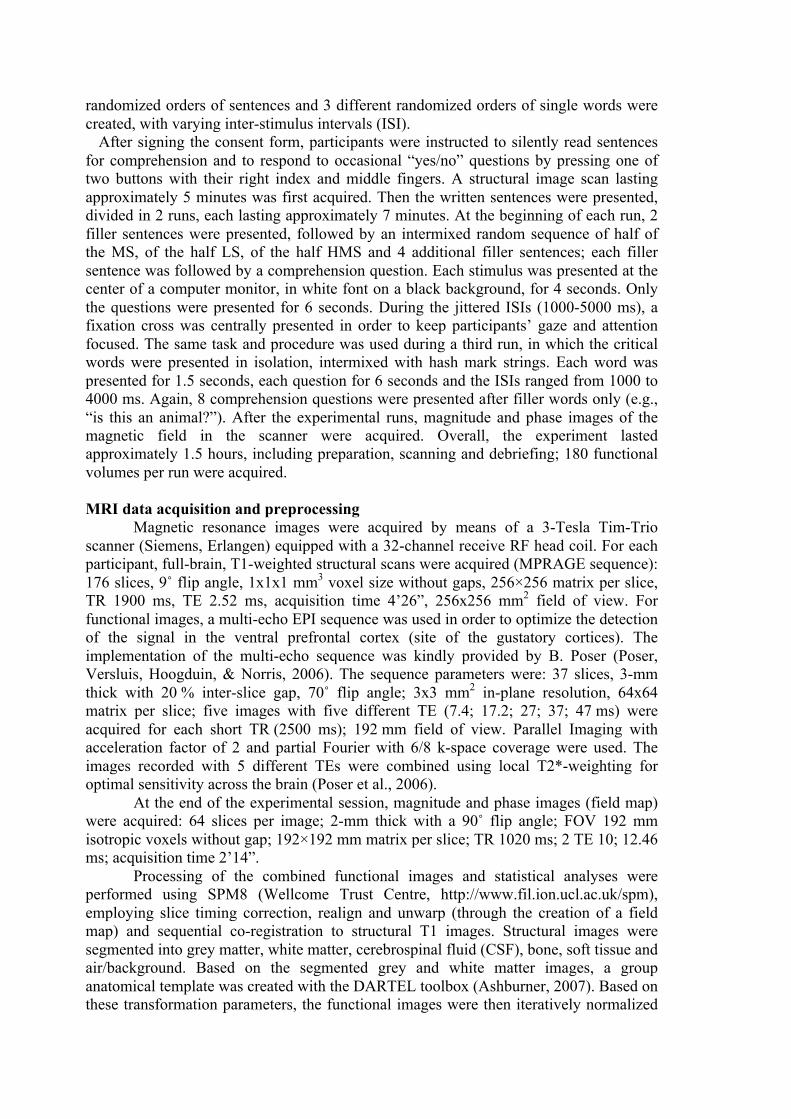
randomized orders of sentences and 3 different randomized orders of single words were created, with varying inter-stimulus intervals (ISI). After signing the consent form, participants were instructed to silently read sentences for comprehension and to respond to occasional “yes/no” questions by pressing one of two buttons with their right index and middle fingers. A structural image scan lasting approximately 5 minutes was first acquired. Then the written sentences were presented, divided in 2 runs, each lasting approximately 7 minutes. At the beginning of each run, 2 filler sentences were presented, followed by an intermixed random sequence of half of the MS, of the half LS, of the half HMS and 4 additional filler sentences; each filler sentence was followed by a comprehension question. Each stimulus was presented at the center of a computer monitor, in white font on a black background, for 4 seconds. Only the questions were presented for 6 seconds. During the jittered ISIs (1000-5000 ms), a fixation cross was centrally presented in order to keep participants’ gaze and attention focused. The same task and procedure was used during a third run, in which the critical words were presented in isolation, intermixed with hash mark strings. Each word was presented for 1.5 seconds, each question for 6 seconds and the ISIs ranged from 1000 to 4000 ms. Again, 8 comprehension questions were presented after filler words only (e.g., “is this an animal?”). After the experimental runs, magnitude and phase images of the magnetic field in the scanner were acquired. Overall, the experiment lasted approximately 1.5 hours, including preparation, scanning and debriefing; 180 functional volumes per run were acquired. MRI data acquisition and preprocessing
Magnetic resonance images were acquired by means of a 3-Tesla Tim-Trio scanner (Siemens, Erlangen) equipped with a 32-channel receive RF head coil. For each participant, full-brain, T1-weighted structural scans were acquired (MPRAGE sequence): 176 slices, 9˚ flip angle, 1x1x1 mm3 voxel size without gaps, 256×256 matrix per slice, TR 1900 ms, TE 2.52 ms, acquisition time 4’26”, 256x256 mm2 field of view. For functional images, a multi-echo EPI sequence was used in order to optimize the detection of the signal in the ventral prefrontal cortex (site of the gustatory cortices). The implementation of the multi-echo sequence was kindly provided by B. Poser (Poser, Versluis, Hoogduin, & Norris, 2006). The sequence parameters were: 37 slices, 3-mm thick with 20 % inter-slice gap, 70˚ flip angle; 3x3 mm2 in-plane resolution, 64x64 matrix per slice; five images with five different TE (7.4; 17.2; 27; 37; 47 ms) were acquired for each short TR (2500 ms); 192 mm field of view. Parallel Imaging with acceleration factor of 2 and partial Fourier with 6/8 k-space coverage were used. The images recorded with 5 different TEs were combined using local T2*-weighting for optimal sensitivity across the brain (Poser et al., 2006).
At the end of the experimental session, magnitude and phase images (field map) were acquired: 64 slices per image; 2-mm thick with a 90˚ flip angle; FOV 192 mm isotropic voxels without gap; 192×192 mm matrix per slice; TR 1020 ms; 2 TE 10; 12.46 ms; acquisition time 2’14”.
Processing of the combined functional images and statistical analyses were performed using SPM8 (Wellcome Trust Centre, http://www.fil.ion.ucl.ac.uk/spm), employing slice timing correction, realign and unwarp (through the creation of a field map) and sequential co-registration to structural T1 images. Structural images were segmented into grey matter, white matter, cerebrospinal fluid (CSF), bone, soft tissue and air/background. Based on the segmented grey and white matter images, a group anatomical template was created with the DARTEL toolbox (Ashburner, 2007). Based on these transformation parameters, the functional images were then iteratively normalized
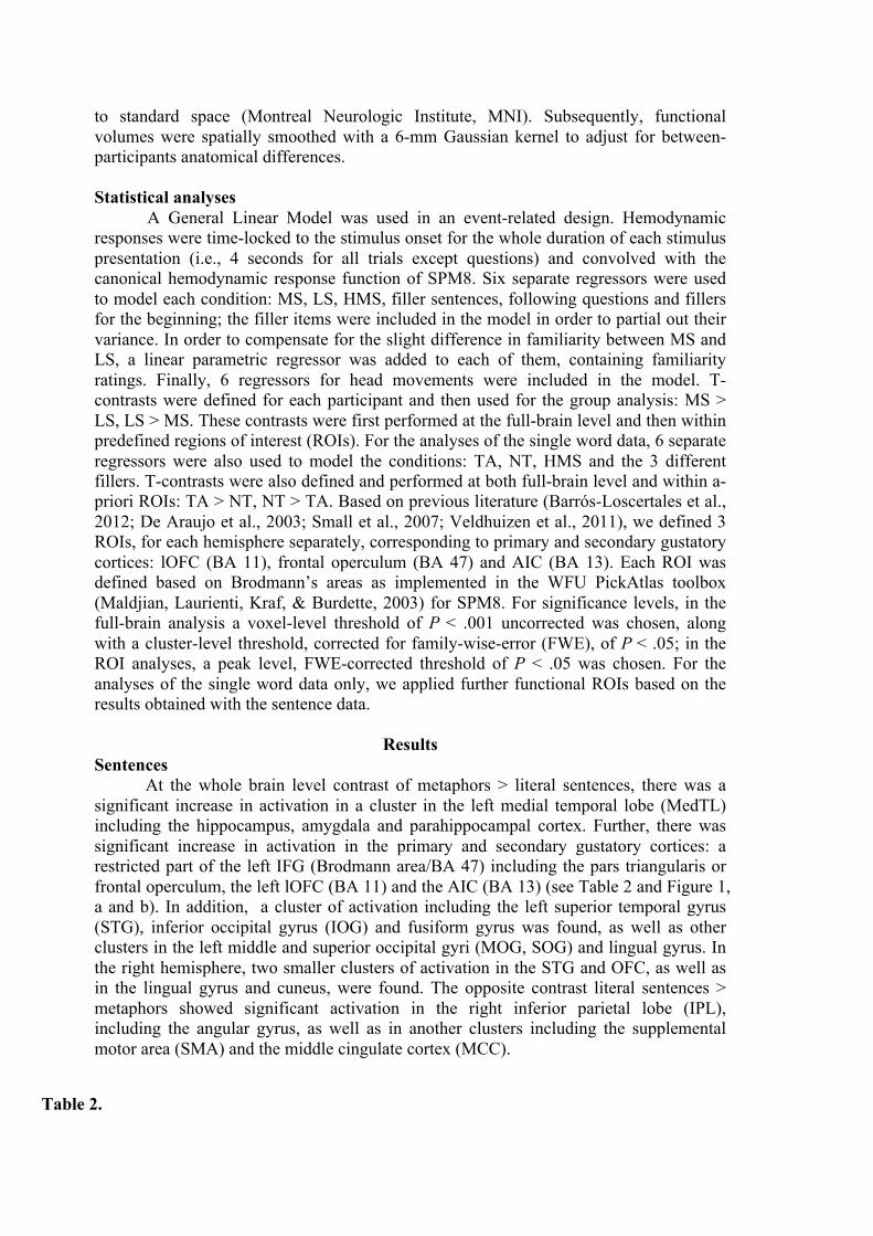
to standard space (Montreal Neurologic Institute, MNI). Subsequently, functional volumes were spatially smoothed with a 6-mm Gaussian kernel to adjust for between-participants anatomical differences. Statistical analyses
A General Linear Model was used in an event-related design. Hemodynamic responses were time-locked to the stimulus onset for the whole duration of each stimulus presentation (i.e., 4 seconds for all trials except questions) and convolved with the canonical hemodynamic response function of SPM8. Six separate regressors were used to model each condition: MS, LS, HMS, filler sentences, following questions and fillers for the beginning; the filler items were included in the model in order to partial out their variance. In order to compensate for the slight difference in familiarity between MS and LS, a linear parametric regressor was added to each of them, containing familiarity ratings. Finally, 6 regressors for head movements were included in the model. T-contrasts were defined for each participant and then used for the group analysis: MS > LS, LS > MS. These contrasts were first performed at the full-brain level and then within predefined regions of interest (ROIs). For the analyses of the single word data, 6 separate regressors were also used to model the conditions: TA, NT, HMS and the 3 different fillers. T-contrasts were also defined and performed at both full-brain level and within a-priori ROIs: TA > NT, NT > TA. Based on previous literature (Barrós-Loscertales et al., 2012; De Araujo et al., 2003; Small et al., 2007; Veldhuizen et al., 2011), we defined 3 ROIs, for each hemisphere separately, corresponding to primary and secondary gustatory cortices: lOFC (BA 11), frontal operculum (BA 47) and AIC (BA 13). Each ROI was defined based on Brodmann’s areas as implemented in the WFU PickAtlas toolbox (Maldjian, Laurienti, Kraf, & Burdette, 2003) for SPM8. For significance levels, in the full-brain analysis a voxel-level threshold of P < .001 uncorrected was chosen, along with a cluster-level threshold, corrected for family-wise-error (FWE), of P < .05; in the ROI analyses, a peak level, FWE-corrected threshold of P < .05 was chosen. For the analyses of the single word data only, we applied further functional ROIs based on the results obtained with the sentence data.
Results Sentences At the whole brain level contrast of metaphors > literal sentences, there was a significant increase in activation in a cluster in the left medial temporal lobe (MedTL) including the hippocampus, amygdala and parahippocampal cortex. Further, there was significant increase in activation in the primary and secondary gustatory cortices: a restricted part of the left IFG (Brodmann area/BA 47) including the pars triangularis or frontal operculum, the left lOFC (BA 11) and the AIC (BA 13) (see Table 2 and Figure 1, a and b). In addition, a cluster of activation including the left superior temporal gyrus (STG), inferior occipital gyrus (IOG) and fusiform gyrus was found, as well as other clusters in the left middle and superior occipital gyri (MOG, SOG) and lingual gyrus. In the right hemisphere, two smaller clusters of activation in the STG and OFC, as well as in the lingual gyrus and cuneus, were found. The opposite contrast literal sentences > metaphors showed significant activation in the right inferior parietal lobe (IPL), including the angular gyrus, as well as in another clusters including the supplemental motor area (SMA) and the middle cingulate cortex (MCC).
Table 2.
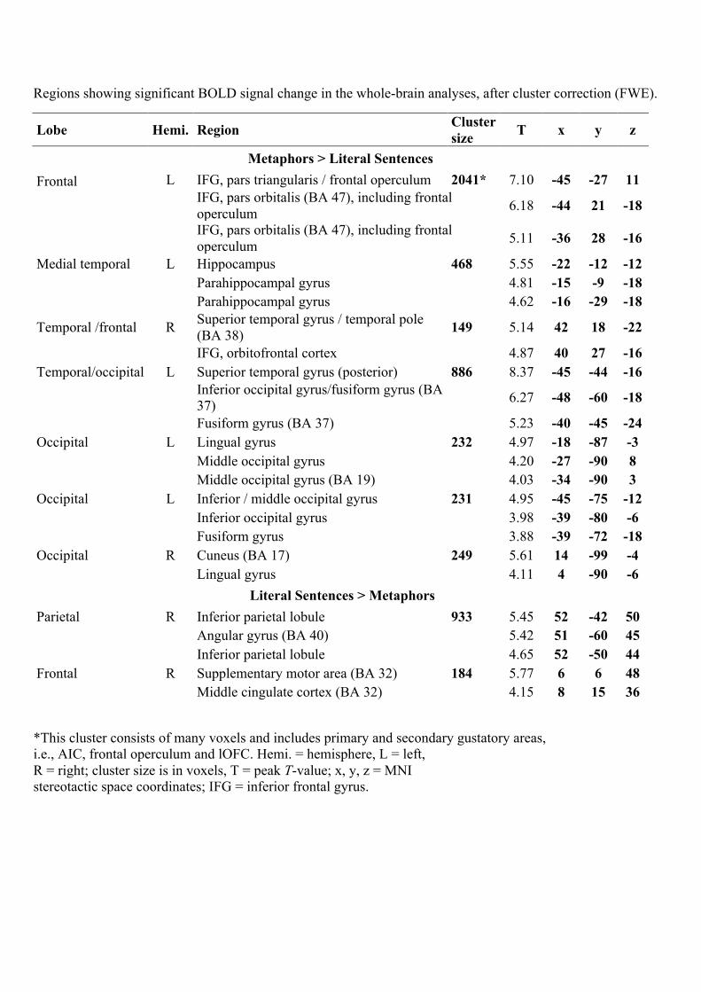
Regions showing significant BOLD signal change in the whole-brain analyses, after cluster correction (FWE).
Lobe Hemi. Region Cluster size T x y z
Metaphors > Literal Sentences Frontal L IFG, pars triangularis / frontal operculum 2041* 7.10 -45 -27 11
IFG, pars orbitalis (BA 47), including frontal operculum 6.18 -44 21 -18
IFG, pars orbitalis (BA 47), including frontal operculum 5.11 -36 28 -16
Medial temporal L Hippocampus 468 5.55 -22 -12 -12 Parahippocampal gyrus 4.81 -15 -9 -18 Parahippocampal gyrus 4.62 -16 -29 -18
Temporal /frontal R Superior temporal gyrus / temporal pole (BA 38) 149 5.14 42 18 -22
IFG, orbitofrontal cortex 4.87 40 27 -16 Temporal/occipital L Superior temporal gyrus (posterior) 886 8.37 -45 -44 -16
Inferior occipital gyrus/fusiform gyrus (BA 37) 6.27 -48 -60 -18
Fusiform gyrus (BA 37) 5.23 -40 -45 -24 Occipital L Lingual gyrus 232 4.97 -18 -87 -3 Middle occipital gyrus 4.20 -27 -90 8 Middle occipital gyrus (BA 19) 4.03 -34 -90 3 Occipital L Inferior / middle occipital gyrus 231 4.95 -45 -75 -12 Inferior occipital gyrus 3.98 -39 -80 -6 Fusiform gyrus 3.88 -39 -72 -18 Occipital R Cuneus (BA 17) 249 5.61 14 -99 -4 Lingual gyrus 4.11 4 -90 -6
Literal Sentences > Metaphors Parietal R Inferior parietal lobule 933 5.45 52 -42 50 Angular gyrus (BA 40) 5.42 51 -60 45 Inferior parietal lobule 4.65 52 -50 44 Frontal R Supplementary motor area (BA 32) 184 5.77 6 6 48 Middle cingulate cortex (BA 32) 4.15 8 15 36
*This cluster consists of many voxels and includes primary and secondary gustatory areas, i.e., AIC, frontal operculum and lOFC. Hemi. = hemisphere, L = left, R = right; cluster size is in voxels, T = peak T-value; x, y, z = MNI stereotactic space coordinates; IFG = inferior frontal gyrus.

Metaphors evoke emotions
11
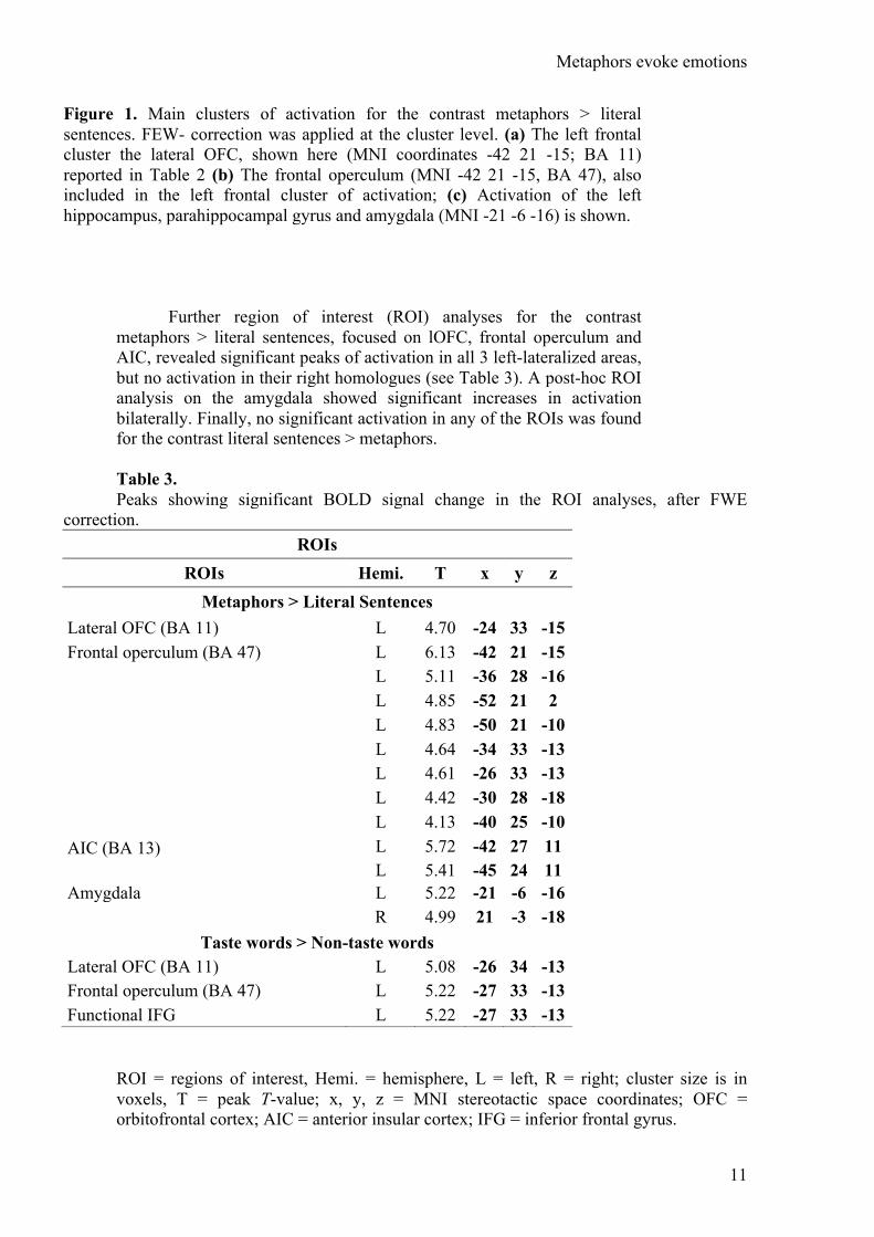
Figure 1. Main clusters of activation for the contrast metaphors > literal sentences. FEW- correction was applied at the cluster level. (a) The left frontal cluster the lateral OFC, shown here (MNI coordinates -42 21 -15; BA 11) reported in Table 2 (b) The frontal operculum (MNI -42 21 -15, BA 47), also included in the left frontal cluster of activation; (c) Activation of the left hippocampus, parahippocampal gyrus and amygdala (MNI -21 -6 -16) is shown.
Further region of interest (ROI) analyses for the contrast metaphors > literal sentences, focused on lOFC, frontal operculum and AIC, revealed significant peaks of activation in all 3 left-lateralized areas, but no activation in their right homologues (see Table 3). A post-hoc ROI analysis on the amygdala showed significant increases in activation bilaterally. Finally, no significant activation in any of the ROIs was found for the contrast literal sentences > metaphors. Table 3. Peaks showing significant BOLD signal change in the ROI analyses, after FWE
correction. ROIs
ROIs Hemi. T x y z Metaphors > Literal Sentences
Lateral OFC (BA 11) L 4.70 -24 33 -15 Frontal operculum (BA 47) L 6.13 -42 21 -15 L 5.11 -36 28 -16 L 4.85 -52 21 2 L 4.83 -50 21 -10 L 4.64 -34 33 -13 L 4.61 -26 33 -13 L 4.42 -30 28 -18 L 4.13 -40 25 -10 AIC (BA 13) L 5.72 -42 27 11 L 5.41 -45 24 11 Amygdala L 5.22 -21 -6 -16 R 4.99 21 -3 -18
Taste words > Non-taste words Lateral OFC (BA 11) L 5.08 -26 34 -13 Frontal operculum (BA 47) L 5.22 -27 33 -13 Functional IFG L 5.22 -27 33 -13
ROI = regions of interest, Hemi. = hemisphere, L = left, R = right; cluster size is in voxels, T = peak T-value; x, y, z = MNI stereotactic space coordinates; OFC = orbitofrontal cortex; AIC = anterior insular cortex; IFG = inferior frontal gyrus.

Metaphors evoke emotions
12
Single words In the whole brain analysis, no significant clusters of activation were found for the contrast between taste words (TA) and non-taste words (NT), in either direction. A-priori ROIs showed significant peaks of activation for the contrast TA > NT in the left frontal operculum and lOFC although not in the AIC. Functional ROIs based on the first 4 clusters of activations found for metaphors > literal sentences (cf. Table 2) showed a significant peak of activation in the left inferior frontal cluster, specifically in the lOFC, confirming the involvement of gustatory areas during processing of taste-referential, non-metaphorical words; functional ROIs using the left hippocampal-parahippocampal-amygdaloid cluster, the right fronto-temporal and the left temporo-occipital clusters revealed no significant clusters of activation. Further, an ROI analysis on left and right amygdala revealed no significant peaks of activation. The contrast NT > TA revealed no significant peaks of activation in any of the ROIs.
Discussion The present study explored the hypothesis that conventional
metaphorical expressions require more processing resources or are processed differently than their literal counterparts, when various psycholinguistic variables are carefully controlled for. In order to test this, a specific perceptual domain was targeted: taste. Commonly used taste metaphors, e.g., “she looked at him sweetly” were read silently for comprehension, along with nearly identical literal sentences, that differed only in a single word (i.e., “she looked at him kindly”). Further, we aimed to test whether processing of highly conventional metaphors would rely on sensory-motor representations, therefore activating gustatory cortices, since previous behavioral and neuroimaging work has been divided on this issue.
Our results showed significantly greater activation of a left inferior frontal cluster including primary and secondary gustatory cortices in response to taste metaphors compared to their literal counterparts. This finding supports the idea that even very common, metaphorical expressions are grounded in sensory-motor and perceptual representations (Barsalou, 1999; Lakoff & Johnson, 1980; Pulvermueller, 1999) and is in line with similar results on action and texture metaphors (Aziz-Zadeh & Damasio, 2008; see also Boulenger et al. (2009) for idioms; Desai et al., 2011; Lacey et al., 2012), while extending the result to an additional domain, namely taste. Our activations were left-lateralized; studies on taste perception and visual perception of food usually (but not always) report bilateral activation of gustatory areas (e.g., Small et al., 2003; van der Laan, de Ridder, Viergever, & Smeets, 2011; Veldhuizen et al., 2011), whereas the only study using written taste words reports a left-lateralized pattern. Hence, our activations could reflect the use of linguistic material.
The role of the IFG activation in this context is potentially ambiguous, since it could reflect the integration of verbal material and world knowledge into meaningful sentences (e.g., Menenti et al., 2009). Nevertheless, while the activation includes BA 47 (which involves the frontal operculum), it does not include the language-related BA 44 and 45. Further, the activation extends beyond BA 47 to BA 11 (lOFC) and part
Metaphors evoke emotions
13
of BA 13 (AIC). Since these three regions combined represent the primary and secondary gustatory cortices, we propose that their activation reflects the recruitment of domain-specific sensory representations related to taste. The results of our control comparison involving single words further support this claim. Like the metaphorical sentences, taste-referential words presented in isolation (e.g., sweet, bitter, cheese) also elicit enhanced activation in the frontal operculum and lateral OFC compared to non-taste referential words (e.g., kind, sad, nonsense). This demonstrates that the comprehension of concrete taste words also relies on gustatory representations. At the same time, it remains possible that the slightly larger involvement of the prefrontal cortex, beyond the frontal operculum, serves a different or additional role in the present study.
Since our taste metaphors were contrasted with literal sentences that do not contain taste-related words, the activation of gustatory cortices may be due to the taste words per se, which would not be a novel finding if words used in familiar metaphors within sentences reliably activated the same regions that they do when presented in isolation with a presumably literal interpetation. However, as previous literature suggests, the sentential context can change the way in which we process, for example, action words (Raposo et al., 2009), and thus our study demonstrates that even highly conventional metaphorical sentences, rated as low in taste-relatedness (2.99 out of 7 points), nonetheless evoke embodied representations.
An unexpected but intriguing finding is the involvement of the left hippocampus and parahippocampal gyrus, including the amygdala. The amygdala is associated with automatic processing of intense emotional stimuli (Adolphs, Russel, & Tranel, 1999; Citron, 2012; Garavan, Pendergrass, Ross, Stein, & Risinger, 2001; Hamann & Mao, 2002; Wager, Phan, Liberzon, & Taylor, 2003); left amygdala activation facilitates successful encoding of emotional verbal material in the hippocampus (Kensinger & Corkin, 2004; Phelps, 2004; Richardson, Strange, & Dolan, 2004) and concurrent activation of these two regions has been associated with the successful retrieval of emotional memories (Dolcos, LaBar, & Cabeza, 2005). While the hippocampus is typically involved in learning and memory (e.g., Moscovitch, Nadel, Winocur, Gilboa, & Rosenbaum, 2006), its activation along with the parahippocampal gyrus has also been shown in studies employing emotional stimuli such as single emotion words (see Citron, 2012 for a review; Kuchinke et al., 2005) and music (Mitterschiffthaler, Fu, Dalton, Andrew, & Williams, 2007), in which no memory encoding or retrieval task is involved. In fact, the hippocampus, parahippocampal gyrus and amygdala are more generally part of the limbic system (or Papez circuit), that is involved in emotion processing (Papez, 1995).
We propose that the cluster of activation found indicates that metaphorical expressions are implicitly more emotionally engaging than literal expressions. That is, even though the explicit ratings collected prior to the study revealed no explicit awareness of a difference in emotional arousal between metaphorical and literal sentences, it appears that implicit processing of the sentences’ meanings evoked stronger emotional responses in the case of metaphors.
Metaphors evoke emotions
14
Although no previous empirical study has invoked emotion as playing a key role in the comprehension of metaphorical expressions, the current finding receives support from a meta-analysis of 23 studies by Bohrn et al. (2012), which also revealed left amygdala activation in contrasts of figurative > literal verbal material. The present study adds needed support to the meta-analysis since in the present study, relevant variables were either controlled for (meaning, length in letters and number of words, emotional valence, emotional arousal, imageability), or were used as a regressor in the analysis (familiarity). Attention to all of these variables is not possible in meta-analyses. Moreover, the otherwise well-controlled Desai et al. (2011) and Lacey et al. (2012) studies mentioned in the introduction did not control for emotion-related variables such as valence and arousal, since these studies were not aimed at investigating possible emotional correlates of metaphorical processing. The attention to affective variables as well as other psycholinguistically relevant variables in the present study, together with the use of a naturalistic task in which participants simply read the critical stimuli for comprehension, allows us to bring the role of emotion in metaphorical processing to the fore.
Further evidence is needed to determine the root cause of the increase in emotion-related activation for metaphorical sentences, as it may be due to metaphoricity or due to the embodied representations (here, taste). The lack of significant peaks of activation for taste words as compared with non-taste words in the hippocampus or amygdala suggests that it appears to be metaphoricity and not taste-reference per se that evokes implicit emotional responses. This possibility is consistent with a finding from Kousta, Vigliocco, Vinson, Andrews and Del Campo (2011), who investigated the processing of abstract words. By tightly controlling for a range of psycholinguistic variables, Kousta et al. demonstrated that the representations of abstract concepts rely more strongly on affective experiential information when compared to concrete concepts. The latter rely more on sensory-motor information instead. In our study, when taste words were intended metaphorically, i.e., more abstractedly, the sentences elicited emotion-related brain activations; as Kousta et al. (2011) would predict, the effect was not evident when the taste words were presented in isolation and understood concretely.
However, we cannot rule out the possibility that the lack of activation of emotion-related areas when taste words were compared to non-taste words was due to a lack of power. It remains possible that embodied representations related to taste, are more emotionally engaging. If this is in fact the case, it would provide a link between the main two findings of the present paper: it may be that grounding in physical experience leads to greater emotional engagement. In future work, either a block design or repeated presentation of the single words would ensure higher statistical power. Alternatively, in order to investigate whether sentences containing taste words evoke activation in areas associated with emotion, one could compare “She looked at him sweetly” with “She looked at his sweet strawberries” or “She looked at the sweet melon”. However, this comparison cannot control for the content of the sentences; moreover, the sentences necessarily vary by more than one single word. We chose to use controls for our metaphorical stimuli, following Lacey et

Metaphors evoke emotions
15
al. (2012), that conveyed the same semantic content but differed by a single word, and then we compared the distinct taste words and literal counterparts separately.
It is possible that the activation of other areas may also highlight a greater role for emotion in the processing of conventional metaphorical sentences when compared with their literal, “non-embodied” counterparts. For example, activation of the OFC, or ventromedial prefrontal cortex (vmPFC), is associated with decision making (Bechara, Damasio, & Damasio, 2000) but also with processing of emotion and reward (e.g., Kringelbach, 2005). Stronger activation of striate and extrastriate regions for metaphors is not a novel finding (e.g., Bohrn et al., 2012), and perhaps relatedly, emotionally relevant linguistic material has been shown to increase visual processing, possibly because of re-entrant projections from the amygdala (cf. Herbert et al., 2009).
While we do not find the widespread temporal activation that had been found in the studies discussed in the introduction, we do find more activation for metaphorical sentences in two small clusters, namely the anterior portion of the STG, or temporal pole, which is associated with increasing integration demand while reading a text (Ferstl, Rinck, & von Cramon, 2005; Yarkoni, Speer, & Zacks, 2008), as well as the posterior portion of the STG, also associated with text and discourse processing (Ferstl, 2010).
In order to be able to exclude that the regions of enhanced activation for metaphors reflect lower familiarity of these stimuli than their literal counterparts, we partialled out this variable in our original analyses. In addition, we regressed the BOLD signal on familiarity ratings for all sentences post-hoc; both whole-brain and ROI analyses yielded no significant clusters of activation, likely because there was not enough variability in familiarity. We had aimed to make all of the sentences familiar, and the null result supports the idea that our findings are not attributable to differences in familiarity. Moreover, if the slightly lower familiarity of the metaphorical sentences were leading to higher processing demand, we would expect to find significantly greater activations in brain regions constituting the multiple-demand system, which responds to increasing cognitive demand in a range of different tasks (verbal, arithmetic, spatial) requiring a range of cognitive processes (e.g., working memory, updating of information, inhibition of a prepotent response) (Cabeza & Nyberg, 2000; Fedorenko, Duncan, & Kanwisher, 2013). The regions found in our key contrast between metaphorical and literal stimuli, however, are not those of the multiple-demand system. Thus we can conclude that the regions that are more active in response to metaphorical sentences are neither attributable to familiarity nor to higher processing demand.
It should be borne in mind that the present experiment was not designed to test whether metaphors are more emotionally engaging and it is therefore exploratory at this stage. Future work is needed to see whether metaphors with source domains other than taste show the same effects. Only then can we conclude that the recruitment of the amygdala and hippocampus is relevant beyond the specific somato-sensory representations related to the metaphorical interpretation of taste. Another suggestion for future research concerns the ecological validity of the
Metaphors evoke emotions
16
linguistic material and the task used. The present study combined the use of metaphors consisting of common, natural expressions with a simple silent reading task that was fairly naturalistic. In a future study, we aim to increase the ecological validity of the task by employing full stories containing figurative expressions vs. literal language, rather than single sentences in isolation.
To conclude, the present results provide evidence that even very common metaphorical expressions require somewhat more processing resources than literal ones, although the differences found were markedly curtailed as compared to previous studies, likely because our stimuli were carefully matched and the task was implicit. Our findings support the idea that even highly conventional, abstract metaphorical concepts are grounded in sensory-motor and perceptual representations by extending existing evidence to the domain of taste.
The present findings provide initial evidence that conventional metaphorical expressions are more emotionally engaging than literal expressions. Thus metaphorical expressions may be chosen over literal expressions in part because they are more emotionally evocative; this would go some way to explaining the ubiquitous use of metaphors by speakers and writers, even when literal counterparts exist. We remain agnostic about whether the recruitment of emotion-related areas is due to embodied representations of taste-related concepts, or to metaphoricity per se. It is possible that the more evocative nature of metaphors may be due to the fact that metaphorical expressions are more closely tied to physical sensations. In this way, the finding that taste-related metaphors activate gustatory cortices may give rise to the finding that taste-related metaphorical sentences activate areas associated with emotional engagement.
Metaphors evoke emotions
17
Acknowledgements We would like to thank Benedikt Poser for kindly providing us
with muti-echo EPI pulse sequence, and Evgeniya Kirilina for setting up an MR protocol of the study. We further thank Michael Kucharski and Luna Beck for their help with material preparation and data collection, and Chun-Ting Hsu for his advice on the analyses. The authors are funded by an Einstein Visiting Fellowship awarded to AEG, in conjunction with the Cluster of Excellence “Languages of Emotion.”
Metaphors evoke emotions
18
References Adolphs, R., Russel, J. A., & Tranel, D. (1999). A role for the human amygdala
in recognizing emotional arousal from unpleasant stimuli. Psychological Science, 10(2), 167-171.
Aziz-Zadeh, L., & Damasio, A. (2008). Embodied semantics for actions: Findings from functional brain imaging. Journal of Physiology - Paris, 102, 35-39. doi: 10.1016/j.jphysparis.2008.03.012
Bambini, V., Gentili, C., Ricciardi, E., Bertinetto, P. M., & Pietrini, P. (2011). Decomposing metaphor processing at the cognitive and neural level through functional magnetic resonance imaging. Brain Research Bulletin, 86, 203-216. doi: 10.1016/brainresbull.2011.07.015
Barrós-Loscertales, A., González, J., Pulvermueller, F., Ventura-Campos, N., Bustamente, J. C., Costumero, V., . . . Ávila, C. (2012). Reading sald activates gustatory brain regions: fMRI evidence for semantic grounding in a novel sensory modality. Cerebral Cortex, 22, 2554-2563. doi: 10.1093/cercor/bhr324
Barsalou, L. W. (1999). Perceptual symbol systems. Behavioral and Brain Sciences, 22, 577-660.
Bechara, A., Damasio, H., & Damasio, A. R. (2000). Emotion, decision making and the orbitofrontal cortex. Cerebral Cortex, 10, 295-307.
Bohrn, I., Altmann, U., & Jacobs, A. M. (2012). Looking at the brains behind figurative language - A quantitative meta-analysis of neuroimaging studies on metaphor, idiom, and irony processing. Neuropsychologia, 50, 2669-2683. doi: 10.1016/j.neuropsychologia.2012.07.021
Bookheimer, S. Y. (2002). Functional MRI of language: New approaches to understanding the cortical organisation of semantic processing. Annual Review of Neuroscience, 25, 151-188. doi: 10.1146/annurev.neuro.25.112701.142946
Bottini, G., Corcoran, R., Sterzi, R., Paulesu, E., Schenone, P., Scarpa, P., . . . Frith, D. (1994). The role of the right hemisphere in the interpretation of figurative aspects of language. A positron emission tomography activation study. Brain, 117, 1241-1253. doi: 10.1093/brain/117.6.1241
Boulenger, V., Hauk, O., & Pulvermueller, F. (2009). Grasping ideas with the motor system: Semantic somatotopy in idiom comprehension. Cerebral Cortex, 19, 1905-1914. doi: 10-1093/cercor/bhn217
Cabeza, R., & Nyberg, L. (2000). Imaging cognition II: an empirical review of 275 PET and fMRI studies. Journal of Cognitive Neuroscience, 12, 1-47.
Cardillo, E. R., Schmidt, G. L., Kranjec, A., & Chatterjee, A. (2010). Stimulus design in and obstacle course: 560 matched literal and metaphorical sentences for testing neural hypotheses about metaphor. Behavior Research Methods, 42, 651-664. doi: 10.3758/BRM.42.3.651
Metaphors evoke emotions
19
Cardillo, E. R., Watson, C. E., Schmidt, G. L., Kranjec, A., & Chatterjee, A. (2012). From novel to familiar: Tuning the brain for metaphors. NeuroImage, 59, 3212-3221. doi: 10.1016/j.neuroimage.2011.11.079
Chen, E., Widick, P., & Chatterjee, A. (2008). Functional-anatomical organization of predicate metaphor processing. Brain and Language, 107, 194-202.
Citron, F. M. M. (2012). Neural correlates of written emotion word processing: A review of recent electrophysiological and hemodynamic neuroimaging studies. Brain and Language, 122, 211-226. doi: 10.1016/j.bandl.2011.12.007
Dale, A. M. (1999). Optimal experimental design for event-related fMRI. Human Brain Mapping, 8, 109-114.
De Araujo, I. E. T., Rolls, E. T., Kringelbach, M. L., McGlone, F., & Phillips, N. (2003). Taste-olfactory convergence, and the representation of the pleasantness of flavour, in the human brain. European Journal of Neuroscience, 18, 2059-2068. doi: 10.1046/j.1460-9568.2003.02915.x
Desai, R. H., Binder, J. R., Conant, L. L., Mano, Q. R., & Seidenberg, M. S. (2011). The neural career of sensory-motor metaphors. Journal of Cognitive Neuroscience, 23, 2376-2386. doi: 10.1162/jocn.2010.21596
Dolcos, F., LaBar, K. S., & Cabeza, R. (2005). Remembering one year later: Role of the amygdala and the medial temporal lobe memory system in retrieving emotional memories. Proceedings of the National Academy of Sciences of the USA, 102, 2626-2631. doi: 10.1073/pnas.0409848102
Fedorenko, E., Duncan, J., & Kanwisher, N. (2013). Broad domain generality in focal regions of frontal and parietal cortex. Proceedings of the National Academy of Sciences of the USA, 110, 16616-16621. doi: 10.1073/pnas.1315235110
Ferstl, E. C. (2010). Neuroimaging of text comprehension: Where are we now? Italian Journal of Linguistics, 22, 61-88.
Ferstl, E. C., Neumann, J., Bogler, C., & von Cramon, D. Y. (2008). The extended language network: A meta-analysis of neuroimaging studies on text comprehension. Human Brain Mapping, 29, 581-593.
Ferstl, E. C., Rinck, M., & von Cramon, D. Y. (2005). Emotional and temporal aspects of situation model processing during text comprehension: An event-related fMRI study. Journal of Cognitive Neuroscience, 17, 724-739.
Frith, C. D., & Frith, U. (2012). Mechanisms of social cognition. Annual Review of Psychology, 63, 287-313. doi: 10.1146/annurev-psych-120710-100449
Garavan, H., Pendergrass, J. C., Ross, T. J., Stein, E. A., & Risinger, R. C. (2001). Amygdala response to both positively and negatively valenced stimuli. NeuroReport, 12, 2779-2783.
Metaphors evoke emotions
20
Gibbs, R. W. (1994). The poetics of mind: Figurative thought, language, and understanding. New York: Cambridge University Press.
Gibbs, R. W., Lima, P. L. C., & Francozo, E. (2004). Metaphor is grounded in embodied experience. Journal of Pragmatics, 36, 1189-1210. doi: 10.1016/j.pragma.2003.10.009
Giora, R. (1999). On the priority of salient meanings: studies of literal and figurative language. Journal of Pragmatics, 31, 919-929.
Glucksberg, S. (1998). Understanding metaphor. Current Directions in Psychological Science, 7, 39-43.
Hamann, S., & Mao, H. (2002). Positive and negative emotional verbal stimuli elicit activity in the left amygdala. NeuroReport, 13, 15-19.
Herbert, C., Ethofer, T., Anders, S., Junghofer, M., Wildgruber, D., Grodd, W., & Kissler, J. (2009). Amygdala activation during reading of emotional adjectives - an advantage for pleasant content. Social Cognitive and Affective Neuroscience, 4, 35-49. doi: 10.1093/scan/nsn027
Kensinger, E. A., & Corkin, S. (2004). Two routes to emotional memory: Distinct neural processes for valence and arousal. Paper presented at the National Academy of Science, St. Luis, MO.
Keysar, B. (1989). On the functional equivalence of literal and metaphorical interpretations in discourse. Journal of Memory and Language, 28, 375-385.
Keysar, B., Shen, Y., Glucksberg, S., & Horton, W. S. (2000). Conventional language: How metaphorical is it? Journal of Memory and Language, 43, 576-593. doi: 10.1006/jmla.2000.2711
Kousta, S.-T., Vigliocco, G., Vinson, D. P., Andrews, M., & Del Campo, E. (2011). The representation of abstract words: Why emotion matters. Journal of Experimental Psychology: General, 1, 14-34. doi: 10.1037/a0021446
Kringelbach, M. L. (2005). The human orbitofrontal cortex: Linking reward to hedonic experience. Nature Reviews Neuroscience, 6, 691-702.
Kuchinke, L., Jacobs, A. M., Gubrich, C., Võ, M. L.-H., Conrad, M., & Herrmann, M. (2005). Incidental effects of emotional valence in single word processing: An fMRI study. NeuroImage, 28, 1022-1032. doi: 10.1016/j.neuroimage.2005.06.050
Lacey, S., Stilla, R., & Sathian, K. (2012). Metaphorically feeling: Comprehending textural metaphors anctivates somatosensory cortex. Brain and Language, 120, 416-421. doi: 10.1016/j.bandl.2011.12.016
Lakoff, G., & Johnson, M. (1980). Metaphors we live by. Chicago: University of Chicago.
Metaphors evoke emotions
21
Maldjian, J. A., Laurienti, P. J., Kraf, R. A., & Burdette, J. B. (2003). An automated method for neuro-anatomic and cytoarchitectonic atlas-based interrogation of fMRI data sets. NeuroImage, 19, 1233-1239.
Mashal, N., Faust, M., & Hendler, T. (2005). The role of the right hemisphere in processing nonsalient metaphorical meanings: Application of principal components analysis to fMRI data. Neuropsychologia, 43, 2084-2100.
Mashal, N., Faust, M., Hendler, T., & Jung-Beeman, M. (2007). An fMRI investigation of the neural correlates underlying the processing of novel metaphoric expressions. Brain and Language, 100, 115-126. doi: 10.1016/j.bandl.2005.10.005
Menenti, L., Petersson, K. M., Scheeringa, R., & Hagoort, P. (2009). When elephants fly: differential sensitivity of right and left inferior frontal gyri to discourse and world knowledge. Journal of Cognitive Neuroscience, 21(2358-2368).
Mitterschiffthaler, M. T., Fu, C. H. Y., Dalton, J. A., Andrew, C. M., & Williams, S. C. R. (2007). A functional MRI study of happy and sad affective states evoked by classical music. Human Brain Mapping, 28, 1150-1162. doi: 10.1002/hbm.20337
Moscovitch, M., Nadel, L., Winocur, G., Gilboa, A., & Rosenbaum, R. S. (2006). The cognitive neuroscience of remote episodic, semantic and spatial memory. Current Opinion in Neurobiology, 16, 179-190. doi: 10.1016/j.conb.2006.03.013
Nobre, A. C., Allison, T., & McCarthy, G. (1994). Word recognition in the human inferior temporal lobe. Nature, 372, 260-263.
Papez, J. (1995). A proposed mechanism of emotion. Journal of Neuropsychiatry and Clinical Neuroscience, 7, 103-112.
Phelps, E. A. (2004). Human emotion and memory: interactions of the amygdala and hippocampal complex. Current Opinion in Neurobiology, 14, 198-202.
Poser, B. A., Versluis, M. J., Hoogduin, J. M., & Norris, D. G. (2006). BOLD contrast sensitivity enhancement and artifact reduction with multiecho EPI: Parallel acquired inhomogeneity desensitized fMRI. Magnetic Resonance in Medicine, 55, 1227-1235.
Pulvermueller, F. (1999). Words in the brain's language. Behavioral and Brain Sciences, 22, 253-336.
Pynte, J., Besson, M., Robichon, F. H., & Poli, J. (1996). The time-course of metaphor comprehension: An event-related potential study. Brain and Language, 55, 293-316.
Raposo, A., Moss, H. E., Stamatakis, E. A., & Tyler, L. K. (2009). Modulation of motor and premotor cortices by actions, action words and action sentences. Neuropsychologia, 47, 388-396.
Metaphors evoke emotions
22
Rapp, A. M., Erb, M., Grodd, W., Bartels, M., & Markert, K. (2011). Neural correlates of metonymy resolution. Brain and Language, 119, 196-205.
Rapp, A. M., Mutschler, D. E., & Erb, M. (2012). Where in the brain is nonliteral language? A coordinate-based meta-analysis of functional magnetic resonance imaging studies. NeuroImage, 63, 600-610. doi: 10.1016/j.neuroimage.2012.06.022
Richardson, M. P., Strange, B. A., & Dolan, R. J. (2004). Encoding of emotional memories depends on amygdala and hippocampus and their interactions. Nature Neuroscience, 7, 278-285.
Schmidt, G. L., & Seger, C. A. (2009). Neural correlates of metaphor processing: The roles of figurativeness, familiarity and difficulty. Brain and Cognition, 71, 375-386. doi: 10.1016/j.bandc.2009.06.001
Small, D. M., Bender, G., Veldhuizen, M. G., Rudenga, K., Nachtigal, D., & Felsted, J. (2007). The role of the human orbitofrontal cortex in taste and flavor processing. Annals of the New York Academy of Sciences, 1121, 136-151. doi: 10.1196/annals.1401.002
Small, D. M., Gregory, M. D., Mak, Y. E., Gitelman, D., Mesulam, M. M., & Parrish, T. (2003). Dissociation of neural representation of intensity and affective valuation in human gustation. Neuron, 39, 701-711.
Sopory, P., & Dillard, J. P. (2002). The persuasive effects of metaphor: A meta-analysis. Human Communication Research, 28, 382-419.
Stringaris, A. K., Medford, N. C., Giora, R., Giampietro, V., Brammer, M. J., & Davis, A. S. (2006). How metaphors influence semantic relatednes judgments: The role of the right frontal cortex. NeuroImage, 33, 784-793.
van der Laan, L. N., de Ridder, D. T. D., Viergever, M. A., & Smeets, P. A. M. (2011). The first taste is always with the eyes: A meta-analysis on the neural correlates of processing visual food cues. NeuroImage, 55, 296-303. doi: 10.1016/j.neuroimage.2010.11.055
Veldhuizen, M. G., Albrecht, J., Zelano, C., Boesveldt, S., Breslin, P., & Lundström, J. N. (2011). Identification of human gustatory cortex by activation likelihood estimation. Human Brain Mapping, 32, 2256-2266. doi: 10.1002/hbm.21188
Wager, T. D., Phan, K. L., Liberzon, I., & Taylor, S. F. (2003). Valence, gender, and lateralization of functional brain anatomy in emotion: a meta-analysis of findings from neuroimaging. NeuroImage, 19, 513-531.
Yang, J. (2012). The role of the right hemisphere in metaphor comprehension: A meta-analysis of functional magnetic resonance imaging studies. Human Brain Mapping. doi: 10.1002/hbm.22160
Yarkoni, T., Speer, N. K., & Zacks, J. M. (2008). Neural substrates of narrative comprehension and memory. NeuroImage, 41, 1408-1425.