METABOLISM AND NUTRITION - · PDF fileMETABOLISM AND NUTRITION AND HOMEOSTASIS ... * Energy...
36
Chapter 25 BIOLOGY 2402 Anatomy and Physiology lecture Chapter 25 METABOLISM AND NUTRITION
-
Upload
truongkhanh -
Category
Documents
-
view
226 -
download
2
Transcript of METABOLISM AND NUTRITION - · PDF fileMETABOLISM AND NUTRITION AND HOMEOSTASIS ... * Energy...
Chapter 25
BIOLOGY 2402 Anatomy and Physiology lecture
Chapter 25
METABOLISM AND NUTRITION
Chapter 25
2
METABOLISM AND NUTRITION AND HOMEOSTASIS
*Unlike plants, we cannot harness the energy of sunlight to provide energy for our own needs. Food is our only source of energy for performing biological work such as: � Synthesis of protein � Contraction of muscle � Mitosis � Active transport Three Fates of Food Molecules Absorbed by the Gastrointestinal (GI) Tract: 1. Most are used to supply energy for sustaining life processes, for
example, active transport, DNA replication, synthesis of proteins and other molecules, and muscle contraction.
2. Some are used as building block during synthesis of structural or
functional molecules such as muscle proteins, hormones, and enzymes.
3. Others are stored for future use, for example, glycogen stored in liver
cells and triglycerides stored in adipose cells. NUTRITION Nutrition – Is the process by which certain components of food are obtained and used by the body. The process includes:
- Digestion - Absorption - Transportation - Cell metabolism
Chapter 25
3
NUTRIENTS Nutrients – Are the chemicals taken into the body that are used to produce energy, provide building blocks for new molecules, or function in other chemical reactions. Three Functions Of Nutrients: 1. Most are used to supply energy for sustaining life processes, such as
active transport, DNA replication, synthesis of proteins and other molecules, muscle contraction, and nerve impulse conduction.
2. Some are used to synthesize structural or functional molecules such as muscle proteins, hormones, and enzymes.
3. Some are stored for future use, for example, glycogen stored in liver cells.
Six Principal Classes of Nutrients (1) Carbohydrates, (2) Lipids, and (3) Proteins Digested by enzymes in the gastrointestinal tract (GI). End products that reach body cells are Amino Acids, fatty acids, glycerol, monoglycerides and monosaccharides. Uses: 1) synthesize new structural molecules; 2) produce energy for active transport, DNA replication, muscle contraction, etc. * Energy is stored in ATP (Adenosine triphosphate) until needed. 4) Minerals, and 5) Vitamins Part of enzyme systems that catalyze the reactions undergone by carbohydrates, proteins, and lipids.

Chapter 25
4
6) Water � Excellent solvent and suspending medium � Participates in hydrolysis reaction � Acts as a coolant � Lubricates � Helps maintain a constant body temperature by its ability to release
and absorb heat slowly How does each group of nutrients contribute to the body's growth, repair, and energy needs?
REGULATION OF FOOD INTAKE *When the energy content of our body balances the energy needs of all the cells of our body, we stay at the same weight. How is food intake regulated? Within the hypothalamus are nuclei that play key roles in regulating food intake. a) Feeding (Hunger) Center - a cluster of neurons (nerve cells) in the
lateral hypothalamus nucei. When stimulated in animals, they begin to eat heartily, even if they are already full.
b) Satiety Center - a cluster of neurons in the ventromedial nuclei of the
hypothalamus.
It is the opposite of appetite; it is a sensation of fullness with lack of desire to eat

Chapter 25
5
Stimuli that affect the Feeding and Satiety Centers are: If neurons in the hypothalamus regulate food intake, how do they sense whether a person is well-fed or in need of food? 1. Glucose - Glucostatic theory states that when blood glucose levels
are low, feeding increases.
- High glucose stimulates satiety center, and feeding is depressed. 2. Lipid - Lipostatic theory - as the amount of adipose tissue
increases in the body, the rate of feeding decreases. 3. Body temperature - a cold environment enhances eating, whereas a
warm environment depresses it. 4. Distension of the Gastrointestinal Tract - particularly Stomach
and Duodenum - when these organs are stretched, a reflex is initiated that activates the satiety center and depresses the feeding center.
5. Cholecystokinin (CCK) -- a hormone - secreted when fat
(triglycerides) enters the small intestine, inhibits eating.
*Psychological factors, eg. Obesity, anorexia nervosa, and Bulimia. May override the usual intake mechanisms.
METABOLISM Metabolism – Is the total of all the chemical changes that occur in the body. May be thought of as energy-balance between catabolic (degradative) reactions and anabolic (synthesis) reactions.
Chapter 25
6
(Often, catabolic reactions occur in one compartment of a cell, for example, the mitochondria). (Synthetic reaction takes place in another location such as the cytosol or endoplasmic reticulum). Which reaction occur depend on which enzymes are active in a particular cell at a particular time. ATP (adenosine triphosphate) is the molecule that participates most often in energy exchange of living cells. The two Phases of Metabolism: 1. Catabolsim
Catabolism - Chemical reactions that break down complex molecules and polymers into simpler ones.
-Does not require energy but releases energy.
Chimcal energy released is captured in an easy-to-use form by phosphorylating ADP (adenosine diphosphate) to create ATP (adenosine triphosphate)
Important Sets of Catabolism Reactions: (1) Glycolysis (2) Krebs Cycle (3) Electron transport chain Other Examples of Catabolism: (1) Breakdown of glycogen into glucose (2) Breakdown of triglycerides into fatty acids and glycerol

Chapter 25
7
2. Anabolism
Anabolism - chemical reactions that combines simple molecules and monomers to make comples ones that form the body's structural and functional components.
-Requires energy.
Examples: (a) Formation of a peptide bonds between amino acids, thereby
building the amino acids into protein.
(b) Fatty acids can be built into the phospholipids that form plasma membrane bilayer.
(c) Glucose monomers can be put together to form glycogen.
Coupling of Catabolism and Anabolism by ATP Chemical reactions of living system depend on efficiency transferring manageable amounts of energy from one molecule to another. ATP, the "energy currency" of a living cell, is the molecule that accomplishes this transfer of manageable energy. A typical cell has about a billion molecules of ATP, which usually lasts for less than a minuet before being used. It is therefore not a long-term storage form of currency, but rather convenient cash for moment-to-moment transactions. When the terminal phosphate group is split from ATP, adenosine diphosphate (ADP) and a phosphate group (symbolized (p)) are formed). (The energy released is used to drive anabolic reactions such as formation

Chapter 25
8
of glycogen from glucose). (Then the energy from catabolic reactions is used to combine ADP and phosphate group to resynthesize ATP: (ADP + (P) + energy → ATP) About 40% of the energy released in catabolism is available for cellular function. The rest of energy is converted to heat, which helps maintain normal body temperature. Excess heat is lost to the environment. (Compared with man-made machines, which typically operate at 10-20% efficiency)
METABOLISM AND BODY TEMPERATURE
More or less heat is produced, depending on the rates of metabolic reactions. Homeostasis of body temperature can be maintained only if the rate of body heat loss equals the rate of body heat production. Basal Metabolic Rate Metabolic rate is the overall rate at which heat is produced. Basa state is the condition of the body, in which it is measured under standard conditions designed to reduce or eliminate those factors that can affect metabolic rate. Basal Metabolic Rate (BMR) is a measure of the rate at which the quiet, resting, fasting body breaks down nutrients to liberate energy.

Chapter 25
9
Heat is a form of kinetic energy that can be measured as temperature, and expressed in units called calories. A calorie (spelled with little c) is the amount of heat required to raise the temperature of 1 gram of water from 14oC to 15oC. (Since the calorie is a small unit relative to the large amount of energy stored in food, the kilocalorie (kcal) or Calorie (Cal) (spelled with a big C) is used). A Kilocalorie (kcal) or Calorie is equal to 1000 calories. It is unit used to express the heating value of food and to measure the body's metabolic rate. Basal metabolic rate is most often measure indirectly by measuring oxygen consumption using a spirometer. To release a given amount of heat energy when it is oxidized, a nutrient must combine with a given amount of oxygen. Thus by measuring the amount of oxygen needed for the metabolism of foods, we can determine how many kilocalories are produced. Note: The amount of heat energy released when: (1) 1 liter of oxygen combines with carbohydrates = 5.05 kcal (2) 1 liter of oxygen combines with triglycerides = 4.70 kcal (3) 1 liter of oxygen combines with proteins = 4.60 kcal (Heat produced on a typical diet, uptake of 1 liter of oxygen = 4.9 kcal) Metabolic rate is expressed in kilocalories per square meter of body surface area per hour (kcal/m2/hr). Suppose you sue 1.8 liters in 6 minutes as recorded on a spirometer. Your oxygen consumption in an hour would be calculated as follows; (1.8 liters/6min) x 60 min/hr = 18 liters/hr

Chapter 25
10
Your basal metabolic rate would calculated as follows: 18liters/hr x 4.9 kcal/liter = 88 kcal/hr To express the kilocalories per square meter of body surface, a standardized chart is used. Shows square meters of body surface relative to height in centemeter and weight in kilograms. If your weight = 75 kg (165 ib) If your height = 109 cm (75 in) Your surface area = 2 m2 (from chart) Your basal metabolic rate = 88 kcal ÷ 2m2 = 44 kcal/m2/hr Values 15% above or below the standard may indicate an excess or deficiency of thyroid hormone. Homeostasis of Body Temperature If your heat production equals heat loss, you maintain a constant core temperature near 37oC (98.6oF). Core Temperature refers to the body's temperature in body structures below the skin and subcutaneous tissue, Shell Temperature refers to the body's temperature at the surface, that is, the skin and subcutaneous tissue. Core temperature is usually a little higher than shell temperature. Too high a core temperature kills by denaturing body proteins, while too low a core temperature causes cardiac arrhythmias (irregular heart rhythm) that results to death.

Chapter 25
11
Hypothalamic Thermostat Body temperature is regulated by mechanisms that attempt to keep heat production and heat loss in balance. A center of control for those mechanisms that are reflex in nature is found in the hypothalamus. Preoptic area , a group of neurons in the anterior portion of the hypothalamus, receives input from temperature receptors in the skin and mucous membrane (peripheral thermoreceptors) and in internal structures (central thermoreceptors), including the hypothalamus. If blood temperature increases, the neurons of the preoptic area fire nerve impulses more rapidly.
If something causes the blood’s temperature to decrease, these neurons fire nerve impulses less rapidly.
The preoptic area maintains normal body temperature and thus serves as your thermostat.
Nerve impulses from the preoptic area are sent to other portions of the hypothalamus known as:
(a) Heat-losing center, when stimulated by
ENERGY PRODUCTION
Nutrrient molecules, like all molecules, have energy stored in the bonds between their atoms. Two important aspects of energy production -oxidation-reduction reactions
Chapter 25
12
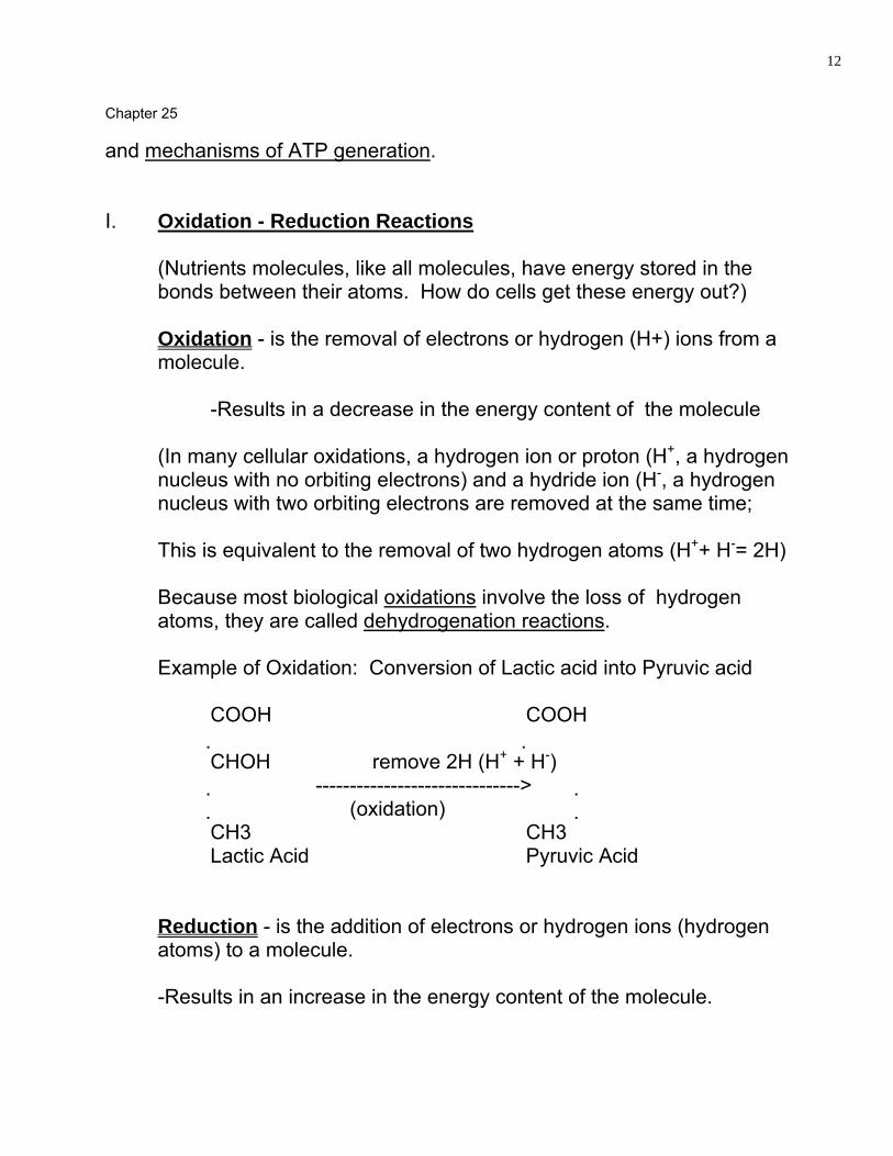
and mechanisms of ATP generation.
I. Oxidation - Reduction Reactions
(Nutrients molecules, like all molecules, have energy stored in the bonds between their atoms. How do cells get these energy out?)
Oxidation - is the removal of electrons or hydrogen (H+) ions from a molecule.
-Results in a decrease in the energy content of the molecule
(In many cellular oxidations, a hydrogen ion or proton (H+, a hydrogen nucleus with no orbiting electrons) and a hydride ion (H-, a hydrogen nucleus with two orbiting electrons are removed at the same time;
This is equivalent to the removal of two hydrogen atoms (H++ H-= 2H)
Because most biological oxidations involve the loss of hydrogen atoms, they are called dehydrogenation reactions.
Example of Oxidation: Conversion of Lactic acid into Pyruvic acid COOH COOH CHOH remove 2H (H+ + H-) ------------------------------> (oxidation) CH3 CH3 Lactic Acid Pyruvic Acid Reduction - is the addition of electrons or hydrogen ions (hydrogen
atoms) to a molecule.
-Results in an increase in the energy content of the molecule.

Chapter 25
13
*Reduction is the opposite of oxidation. Example of Reduction: conversion of Pyruvic acid into Lactic acid COOH COOH C = O add 2H (2H+ + 2E-) CHOH ------------------------------> Reduction CH3 CH3 Lactic Acid Pyruvic Acid *Oxidation-Reduction (Redox) Reactions - is referred to the coupled reactions of oxidation of oxidation and reduction. (Note: when a substance is oxidized, the liberated hydride ions do not remain free in the cell but are transferred immediately by coenzymes to another compound.) Oxidation-Reduction Reactions: Oxidation-Reduction (Rexon Reaction) -Are electron transfers. Oxidation is the loss of electrons from one substance during a redox reaction. Reduction is the addition of electrons to another substance during a redox reaction. ---------------------------Oxidation----------------� Na + Cl � Na+ + Cl- ---------------------Reduction-----------�

Chapter 25
14
Na is the electron donor, and is called the Reducing agent. Cl is the electron acceptor, and is called the oxidizing agent. -------------oxidation-------------� C6H12O6 + 6CO2 ----------> 6CO2 + 6H2O --------------------Reduction----------------� *When a molecule is oxidized, it loses hydrogen atoms (electrons and hydrogen ions). *When a molecule is reduced, it gains hydrogen atoms (electrons and hydrogen ions). Two Coenzymes commonly used by Animal Cells to carry Hydrogen Atoms i Nicotinamide Adenine Dinucleotide (NAD), a derivative of B
vitamin Niacin
+2H (H= + H-) NAD+ � NADH + H+
-2H (H= + H-) Oxidized Reduced ii Flavin Adenine Dinucleotide (FAD), a derivative of Vitamin B2
(Riboflavin) +2H (H= + H-) FAD � FADH2 -2H (H= + H-) Oxidized Reduced

Chapter 25
15
When lactic acid is oxidized to form pyruvic acid , the two hydrogen atoms removed in the reaction are used to reduces NAD. This coupled oxidation-reduction may be written as follows: Diagram: An important point to remember about oxidation-reduction reactions is that oxidation is usually an energy-producing reaction. Generation of ATP (Some of the energy released during oxidation reactions is trapped within a cell as ATP is formed.) A phosphate group (P) is added to ADP, with an input of energy, to form ATP.
(Adenosine-P~P + Energy + P->P~P) ADP ATP
Phosphorylation is the addition of phosphate group to a chemical compound. Kinases are enzymes that catalyze phosphorylation reactions. Three Mechanism of Phosphorylation to Generate ATP 1) Substrate-Level Phosphorylation - ATP is generated when a high-
energy phosphate group is transferred directly from an intermediate phosphorylated metabolic compound to ADP.
Occurs in the cytosol (Occurs twice in glycolysis)

Chapter 25
16
2. Oxidative Phosphorylation - electrons are removed from organic
compounds (usually by NAD+) and passed through a series of electron acceptors to molecules of oxygen (O2) or other inorganic molecules.
-Occurs in the inner mitochondrial membrane of human cells.
Electron Transport Chain - the series of electron acceptors used in oxidative phosphorylation. Transfer of electrons from one electron acceptor to the next releases energy, which is used to generate ATP from ADP and P.
3. Photophosphorylation - occurs only in photosynthetic cells, which
contain light-trapping pigments such as the chlorophylls.
CARBOHYDRATE METABOLISM
(Re-Call: Before glucose can be used by body cells, it must first pass through the plasma membrane and then enter the cytoplasm.) (During the process of digestion, polysaccharides and disaccharides are hydrolyzed into the monosaccharides glucose, fructose, and galactose.) (Glucose represents about 80% of the monosaccharides. Some fructose is converted into glucose as it is absorbed through the intestinal epithelial cells.) *(The three monosaccharides are absorbed into the capillaries of the Villi of the small intestine, and are carried through the Hepatic Portal vein to the liver, where much of the remaining fructose and practically all the galactose are converted to glucose.) *Thus, the story of carbohydrate metabolism is really the story of Glucose Metabolism.

Chapter 25
17
Fate of Carbohydrates (Since glucose is the body's preferred source for synthesizing ATP, the fate of absorbed glucose depends on the energy needs of body cells.) 1. ATP production. If the cells require immediate energy, glucose is
oxidized by the cells. 2. Amino Acid synthesis. Glucose can be used to form amino acids,
which then can be incorporated into proteins. 3. Glycogenesis. The liver can store a small amount of excess glucose
by converting it to glycogen.
Glycogenolysis. Hepatic cells can convert glycogen back to glucose and release it into the blood, when blood glucose starts to decrease.
*Skeletal muscle fibers (cells) can also store glycogen and oxidize if to provide ATP for their use. However, they lack the enzyme needed to release glucose back into the blood.
4. Lipogenesis. If the glycogen storage are filled up, liver cells and fat
cells can transform the glucose to glycerol and fatty acids that can be used for synthesis of triglycerides. (natural fats)
-Triglycerides (natural fats) then are deposited in adipose tissue, which has virtually unlimited storage capacity.
5. Excretion in Urine. Excess glucose occasionally is excreted in the
urine.
(Normally, this happens only when a meal containing mostly carbohydrate and no triglycerides is eaten. Without the inhibiting effect of triglycerides, the stomach empties its contents quickly, and the carbohydrates are all digested at the same time. As a result, a large amount of glucose suddenly floods into the bloodstream.)

Chapter 25
18
(At high blood glucose levels, the kidneys may not recover all glucose molecules that are filtered and some glucose may spill over into the urine.)
*Diabetic who experience high blood glucose levels may often spill glucose into the urine.
Glucose Movement Into Cells (Before glucose can be used by body cells, it must first pass through the plasma membrane and then enter the cytosol.) *Glucose absorption in the Gastrointestinal (GI) tract (and kidney tubules) is accomplished by secondary Active Transport. *Glucose movement from blood into body cells occurs by facilitated diffusion. *Insulin increases the rate of glucose facilitated diffusion into most cells, except neurons and liver cells. *The rate of glucose transport into cells is greatly increased by Insulin. Immediately upon entry into cells, glucose is phosphorylated. It combines with a phosphate group, produced by the breakdown of ATP, to form glucose 6-phosphate. Phosporylation traps glucose in the cell so that it cannot diffuse back out. Hepatocytes, kidney tubule cells, and intestinal epithelial cells have the necessary enzymes (phosphataes) to remove the phosphate group, which enables glucose to diffuse out of these cells into the bloodstream.

Chapter 25
19
Glucose Catabolism Oxidation of Glucose is also known as Cellules Respiration. Involves: -Glycolysis - Anaerobic cellular respiration (without oxygen) -Formation of Acetyl Coenzyme A -The Krebs cycle - aerobic cellular respiration (with oxygen). -Electron transport chain reactions. GLYCOLYSIS
Glycolysis - refers to ten chemical reactions in the cytosol of a cell that splits a six-carbon molecule of glucose into two three carbon molecules of pyruvic acid.
Two Phases of the ten steps of Glycolysis:
1. Energy-investment phase (1-5) 2 ATP used semigluid portion of cytoplasm 2. Energy-yielding phase (6-10) 4 ATP 2NADP production (Intracellular fluid)
Net Gain 2 ATP and 2 NADH
Steps of Glycolysis -Each of the 10 reactions is catalyzed by a specific enzyme.
Steps 1,2, & 3:
First three reactions involve the addition of a phosphate group (phosphorylation) to glucose, its conversion to fructose, and the addition of another phosphate group to fructose.
Chapter 25
20
*Phosphofructokinase (enzyme that catalyzes step 3) is the key regulator of the rate of glycolysis. When ADP concentration is high, this enzyme has a high activity. Then both pyruvic acid and ATP are produced rapidly. When ATP is plentiful, on the other hand, the enzyme activity is low, and most glucose 6 - phosphate is converted to glycogen for storage rather than catabolized to produce ATP.)
Steps 4 & 5: The double phosphorylated molecule of fructose splits into two three-carbon compounds, glyceraldehyde 3-phosphate (G3-P) and dihydroxyacetone phosphate. These two compounds are interconvertible, but it is G3-P that undergoes further reactions to pyruvic acid. Step 6: Oxidation occurs here as two molecules of NAD+ accept two pairs of electrons and hydrogen ions from two molecules of G 3-P, forming two molecules each of NADH and 1,3-Diphosphoglyceric acid (DPG) - (now called 1,3-Bisphophoglyceric acid (BPG)). Steps 7-10: These reactions generate four molecules of ATP and produce pyruvic acid
(pyruvate). Fate of Pyruvic Acid Fate of pyruvic acid produced during glycolysis depends on the availability of oxygen. 1. Without oxygen (anaerobic conditions)

Chapter 25
21
In skeletal muscle fibers, for example, during strenuous exercise, pyruvate acid is reduced by the addition of two hydrogen atoms to form lactic acid (lactate).
2 Pyruvic Acid + 2 NADH + 2 H+ → Lactic Acid + 2 NAD+
2. With oxygen (aerobic conditions)
Most cells convert pyruvic acid to acetyl coenzyme A.
Acetyl coenzyme A links glycolysis, which occurs in the cytosol, with the Krebs cycle, which occurs in the matrix of mitochondria.
Pyruvic acid enters the mitochondrial matrix with the help of a special transport protein. Red blood cells can produce ATP only through glycolysis, since they lack mitochondria. Formation of Acetyl Coenzyme A
(During the transitional step between glycolysis and the Krebs cycle, pyruvic acid is prepared for entrance into the cycle. Pyruvates Carbonyl group, which has little chemical energy, is removed, given off as molecule of CO2. -Pyruvic acid is converted to two-carbon compound by the loss of carbon dioxide. Decarboxylation is the loss of a molecule of CO2 by a substance. *This two-carbon fragment, called an Acetyl group attaches itself to coenzyme A, and the whole complex is called Acetyl Coenzyme A.
Chapter 25
22
(Remember that the oxidation of one glucose molecule produces two molecules of pyruvic acid, so for each molecule of glucose two molecules of carbon dioxide are lost, and two NADH + H+ are produced.) (Once the pyruvic acid has undergone decarboxylation and its derivative (the acetyl group) has attached to coenzyme A (CoA), the resulting compound (Acetyl Coenzyme A) is ready to enter the Krebs cycle.) Decarboxylation Reactions: *In preparation for entering the cycle, pyruvic acid is decarboxylated to form an acetyl group. (In the cycle, isocitric acid, a six-carbon compound, loses a molecule of CO2 to form a five-carbon compound called -ketoglutaric acid. In the next step, -ketoglutaric acid is decarboxylated (and picks up a molecule of CoA) to form succinyl CoA, a four-carbon compound.) *Thus, each time pyruvic acid enters the Krebs cycle, three molecules of CO2 are liberated by decarboxylation. THE KREBS CYCLE (Citric acid cycle, or tricarboxyl acid cycle) (Pages 958-959) It is a series of nine biochemical reactions that occur in the matrix of mitochondria of cells. *The Pyruvic Acid derivatives are oxidized, whereas the coenzymes are reduced. - Coenzyme A carries an Acetyl group to a mitochondrion, detaches itself, and goes back into the cytoplasm to pick up another fragment. *Krebs cycle consists mainly of a series of decarboxylation and oxidation-

Chapter 25
23
reduction reactions, each controlled by a different enzyme. 1. In the conversion of pyruvic acid to Acetyl CoA, Pyruvic acid loses
two hydrogen atoms; that is, it is oxidized.
(The hydrogen atoms are picked up by a coenzyme called Nicotinamide Adenine Dinucleotide (NAD+).
-Since NAD+ picks up the hydrogen atoms, it is Reduced and represented as NADH + H+.
2. Isocitric acid is oxidized to form -ketoglutaric acid. NAD+ is reduced
to NADH + H+. 3. -Ketoglutaric acid is oxidized in the conversion to succinyl CoA.
NAD+ is reduced to NADH + H+. 4. Succinic acid is oxidized to Fumaric Acid. In this case, the coenzyme
that picks up the two hydrogen atoms is Flavin Adenine Dinucleotide (FAD).
5. Malic Acid is oxidized to Oxalocetic Acid. NAD is reduced to NADH +
H+. *This sequence of chemical reactions is called a "cycle" because the starting substance (Oxaloacetic Acid) is formed again at the end. -The Acetyl group combines with Oxaloacetic acid to form citric acid. *(The reduced coenzymes (NADH + H+ and FADH2) are the most important outcome of the Krebs cycle because they contain the energy originally stored in glucose and then stored in pyruvic acid.) Looking at Krebs Cycle as a whole: �For every two molecules of Acetyl CoA that enter the cycle four molecules of carbon dioxide are liberated by decarboxylation;

Chapter 25
24
�6 NADH + 6H+ and 2 FADH2 are produced by oxidation- reduction reaction; �2 molecules of guanosine triphosphate (G TP, the equivalent of ATP) are generated by substrate-level phosphorylation. ELECTRON TRANSPOET CHAIN REACTIONS -Involves a sequence of electron carrier molecules on the Inner Mitochondria membrane that are capable of oxidation and reduction. -As electrons pass through the chain, there is a stepwise release of energy for generation of ATP. -In aerobic cellular respiration, the last electron acceptor of the chain is molecular oxygen (O2). Chemosmosis - process by which energy released is used to generate ATP when a substance moves along a gradient. (Energy is released as electrons are passed from one carrier to the next and is used to pump H+ (protons) from the matrix of the mitochon into the space between the inner and outer mitoch membranes- (ATP synthesis then occurs as H+ diffuse back into the Mitochon. Matrix through a special type of H+ channel (pore) in the inner membrane) H+ pumped out but comes back in; follow concentration gradient, releasing energy in the process. Electron Carriers: 1. Flavin Mononucleotide (FMN) 2. Cytochromes 3. Iron-sulfur (Fe-S)
Chapter 25
25
4. Copper Atoms (Cu) 5. Ubiquinones or Coenzyme Q (Q) Chemosmotic Mechanism of ATP Generation 1. NADH dehydrogenase complex 2. Cytochrome b-c1, Complex 3. Cytochrome oxidase complex *Each complex act as a protein pump that expels H+ from the mitochon-matrix and helps create an electrochemical gradient of H+. Electron Transport Chain -Is a series of oxidation-reduction in which the energy in NADH + H+ and FADH2 is liberated and transferred to ATP for storage. -The carrier molecules involved include Flavin Mononucleotide (FMN); Ubiquinones (Coenzyme Q), and Cytochromes. -The electron transport chain yields 34 molecules of ATP and H2O. *-The complete oxidation of glucose can be represented as follows: �*C6 H12 O6 + 6O2 → 38 ATP + 6 CO2 + 6H2O Summary of ATP Production during Aerobic Respiration of one Glucose Molecule Source ATP Yield (Method) Glycolysis 1. Oxidation of glucose to 2ATP (Substrate-level Phosphorylation) Pyruvic acid 2. Production of 2NADH + 2H+ 6ATP (oxidative phosphorylation in electron transport chain). Formation of Acetyl CoA
Chapter 25
26
2 NADH + 2H+ 6ATP (oxidative phosphorylation in electron transport chain). Krebs Cycle 1. Oxidation of succinyl CoA 2ATP (equivalent of ATP, substrate to Succinic Acid level phosphorylation)
2. Production of 6 NADH + 6H+ 18 ATP (oxidative phosphorylation in electron transport chain).
3. Production of 2 FADH2 4 ATP (oxidative phosphorylation in electron transport chain). TOTAL: 36 or 38 ATP *The overall reaction for Aerobic Respiration is as follows: *C6H12)6 + 6O2 + 38 ADP + 38P → 6CO2 + 6H2O + 38 ATP Glucose Oxygen Carbon dioxide Water *(Glycolysis, the Krebs Cycle, and especially the Electron Transport Chain provide all the ATP for cellular activities. And because the Krebs cycle and electron transport chain are aerobic processes, the cells cannot carry on their activities for long without sufficient oxygen.) Glucose Anabolism Glucose storage: 1. Glycogenesis - The conversion of glucose to glycogen for storage in
the liver and skeletal muscle.
-Occurs in the live and is stimulated by Insulin. -The body can store about 500 g of glycogen.
2. Glycogenolysis - The conversion of glycogen back to glucose.

Chapter 25
27
-Occurs between meals and is stimulated by glucagen and epinephrine.
Formation of Glucose from Proteins and Fats: 3. Gluconeogenesis - The conversion of protein molecules into
glucose. (Process by which new glucose is formed from noncarbohydrate sources.)
- Stimulated by Cortisol, thyroxine, epimephrine, glucagen, and lumen growth hormone (LGH).
4. Glycerol may be converted to Glyceraldehyde -3- phosphate, and
some amino acids may be converted to pyruvic acid. LIPID METABOLISM
1. During digestion, fats are ultimately broken down into fatty acids and
monoglycerides.
(When triglycerides are eaten, they are digested into fatty acids and monoglycerides.)
2. Long-chain fatty acids and monoglycerides are carried in micelles for
entrance into villi, digested to glycerol and fatty acids in epithelial cells, recombined to form triglycerides, and transported by chylomicrons through the lacteals of villi into the thoracic duct.
Fate of Lipids -Lipid, like carbohydrates, may be oxidized to produce ATP. -Each gram of Triglyceride produces = 9 Kilocalories. 1. Some fats may be oxidized to produce ATP.

Chapter 25
28
2. Some fats are stored in adipose tissue (if the body has no immediate need to use the lipid in ATP production).
3. Other lipids are used as structural molecules or to synthesize essential molecules.
Examples: Include phospholipids of plasma membranes, lipoproteins that
transport cholesterol, thromboplastin for blood clotting, and cholestrol used to synthesize bile salts and steroid hormones.
Fat Storage 1. Fats are stored in adipose tissue, mostly in the subcutaneous layer
until they are needed for ATP production in other parts of the body. 2. Adipose cells contain liposes that catalyze the deposition of fats from
chylomicrons and hydrolyze fats into fatty acids and glycerol. Lipid Catabolism: Lipolysis Triglycerides stored in adipose tissue constitute 98% of all energy reserves. 1. Fat is split into fatty acids and glycerol under the influence of
hormones such as epinephrine, norepinephrine, and glycocorticoids and released from fat depots.
2. Glycerol can be converted into glucose by conversion into
glyceraldehyde 3-phosphate. 3. In beta oxidation, carbon atoms are removed in pairs from fatty acid
chains; the resulting molecules of Acetyl coenzyme A enter the Krebs cycle.
4. The formation of Ketone bodies by the liver is a normal phase of fatty

Chapter 25
29
acid catabolism, but an excess of Ketone bodies, called ketosis, may cause acidosis.
Lipid Anabolism: Lipogenesis 1. Lipogenesis - the conversion of glucose or amino acids into lipids.
- The process is stimulated by insulin 2. The intermediary links in lipogenesis are glyceraldhyd-3- phosphate
and Acetyl coenzyme A.
(Excess dietary carbohydrates, proteins, and fats all have the same fate: they are converted into Triglycerides.)
PROTEIN METABOLISM 1. During digestion, proteins are hydrolyzed into amino acids. 2. Amino acids are absorbed by the capillaries of villi and enter the liver
via the hepatic portal vein. *Unlike carbohydrates and triglycerides, which are stored, proteins are not warehoused for future use. Amino acids may be oxidized to produce ATP or used to synthesize new proteins for body growth and repair. Excess dietary amino acids are not excreted in the urine or feces but rather are converted into glucose (gluconeogenesis) or triglycerides (lipogenesis). Fate of Proteins 1. Amino acids, under the influence of human growth hormone (hGH)
and insulin, enter body cells by active transport.

Chapter 25
30
2. Inside cells, amino acids are synthesized into proteins that function
as enzymes, hormones, structural elements, and so forth; stored as fat or glycogen; or used for energy.
Protein Catabolism 1. Before amino acids can be catabolized, they must be converted to
substances that can enter the Krebs cycle; these conversions involve:
Deamination - Removing the amino group (NH2) from Amino Acid. Decarboxylation - Removal of carbon dioxide from an organic acid (amino acid). Hydrogenation - Addition of hydrogen
2. Amino acids may also be converted into glucose, fatty acids, and
ketone bodies. 3. Proteins are extracted from worn-out cells, such as red blood cells,
and broken down into free amino acids. Protein Anabolism Protein anabolism involves the formation of peptide bonds between amino acids to produce new proteins. 1. Protein synthesis is stimulated by human growth (hGH), thyroxine,
and insulin. 2. The process is directed by DNA and RNA and carried out on the
ribosomes of cells.
Chapter 25
31
SUMMARY OF KEY MOLECULES IN METABOLISM Three molecules that play key roles in metabolism are:
1. Glucose 6-Phosphate (G6-P) 2. Pyruvic Acid (Pyruvate) 3. Acetyl Coenzyme A (acetyl CoA)
1. Glucose 6-Phosphate (G6-P)
- Glucose is phosphorylated to glucose 6-phosphate, upon entering any cell into the body.
2. Pyruvic Acid (Pyruvate)
- Marks decision point in Anaerobic versus aerobic cellular respiration.
(Goes into Krebs cycle producing ATP if oxygen is present, changes to lactic acid of oxygen is in short supply.)
3. Acetyl CoA
-A gateway into the Krebs cycle - Used to synthesize fatty acid, ketine bodies, and cholesterol.
*Mammals including humans, cannot reconvert Acetyl CoA to Pyruvic Acid, so Lipids cannot be used to generate glucose or other carbohydrates. Summary of Metabolism Metabolic Processes During the Absorptive (FED) State
ABSORPTIVE AND POSTABSORPTIVE STATE

Chapter 25
32
1. During the absorptive state, ingested nutrients enter the blood and
lymph from the GI tract. Metabolic reactions depend on how recently you have eaten.
2. During the absorptive state, most blood glucose is used by body cells for oxidation.
- Glucose transported to the liver is converted to glycogen or fat. - Most fat is stored in adipose tissue. - Amino acids in liver cells are converted to carbohydrates, fats and proteins.
3. During the postabsorptive (fasting) state, absorption is complete and
the energy needs of the body are satisfied by nutrients already present in the body.
4. The major concern of the body during the postabsorptive state is to
maintain normal blood glucose level.
- Involves conversion of liver and skeletal muscle glycogen into glucose; - Conversion of glycerol into glucose; - Conversion of Amino acids into glucose. - The body also switches from glucose oxidation to fatty acid oxidation.
REGULATION OF METABOLISM
1. Absorbed nutrients may be oxidized, stored, or converted, based on the needs of the body.
2. The pathway taken by a particular nutrient is enzymatically controlled and is regulated hormones.
Minerals

Chapter 25
33
1. Minerals are inorganic substances (containing no carbon with few exception not a part of animal origin) that help regulate body processes.
2. Minerals known to perform essential functions are calcium, phosphorus,
sodium, chlorine, potassium, magnesium, iron, sulfur, iodine, manganese, cobalt, copper, zinc, selenium, and chromium.
Calcium and phosphorus form part of the structure of bone. Vitamins 1. Vitamins are organic (containing carbon derived from a living
organism) nutrients that maintain growth and normal metabolism. - May function in enzyme systems.
2. Fat-soluble vitamins are absorbed with fats and include A, D, E, and
K. 3. Water-soluble vitamins are absorbed with water and include the B
vitamins and vitamin C. Metabolism and Body Heat Measuring Heat Heat is a form of kinetic energy that can be measured as temperature. 1. A kilocalorie (kcal) is the amount of energy required to raise the
temperature of 1000g of water 1� C from 14� to 15� C. 2. The kilocalorie is the unit of heat used to express the caloric value of
Chapter 25
34
foods and to measure the body's metabolic rate. 3. The apparatus used to determine the calorie value of foods is called a
calorimeter. Production of Body Heat 1. Most body heat is a result of oxidation of the food we eat.
- The rate at which this heat is produced is known as the metabolic rate.
2. Metabolic rate is affected by exercise, the nervous system,
hormones, body temperature, ingestion of food, age, sex (lower in females except during pregnancy and lactation), climate, sleep, and malnutrition.
Basal Metabolic Rate (BMR) 1. Measurement of the metabolic rate under basal conditions is called
the Basal Metabolic rate (BMR). 2. BMR is expressed in kilocalories per square meters of body area per
hour (kcal/M2/hr). Loss of body Heat 1. Radiation is the transfer of heat as infrared heat rays from one object
to another without physical contact. 2. Conduction is the transfer of body heat to a substance or object in
contact with the body. 3. Convection is the transfer of body heat by the movement of air that
has been warmed by the body.
Chapter 25
35
4. Evaporation is the conversion of a liquid to a vapor.
HOMEOSTASIS OF BODY TEMPERATURE REGULATION
1. A normal body temperature is maintained by a delicate balance between heat-producing and heat-losing mechanisms.
2. The hypothalamus thermostat is the preoptic area. 3. Mechanisms that produce or retain heat are vasoconstriction,
sympathetic stimulation, skeletal muscle contraction, and thyroxine production.
4. Mechanisms of heat loss include vasodilation, decreased metabolic
rate, decreased skeletal muscle contraction, and perspiration.
BODY TEMPERATURE ABNORMALITIES
1. Fever is an abnormally high body temperature most commonly caused by protaglandius, with interleukin-1 and mediators.
2. Heat cramps are painful skeletal muscle contractions due to loss of salt and water.
3. Heat exhaustion results in a normal or below normal body
temperature, profuse perspiration, nausea, cramps, and dizziness. Rest and salt tablets are indicated.
4. Heatstroke results in decreased blood flow to skin reduced
perspiration, and high body temperature: fluid therapy and body cooling are indicated.
5. Hypothermia refers to a lowering of body temperature.

Chapter 25
36
DISORDERS: HOMEOSTATIC IMBALANCES
1. Obesity is defined as a body weight 10 to 20% above desirable standard as the result of excessive accumulation of fat.
a. Hypertrophic (adult-onset) obesity - there is an increase in the
amount of fat in adipocytes but no increase in the number of fat cells.
b. Hyperplastic (lifelong) obesity - in which there is an increase in
the number of adipocytes, as well as an increase in the amount of fat within them.
3. Malnutrition refers to a state of bad or poor nutrition. 4. Phenylketonuria (PKU) - is a genetic error of metabolism
characterized by an elevation of phenylalanine in the blood. 5. Cystic fibrosis (CF) is a metabolic disease of the exocrine glands in
which absorption of vitamins A, D, and K and Calcium is inadequate. 6. Celiac disease is a condition in which the ingestion of gluten causes
morphological changes in the small intestinal mucosa resulting in malabsorption.