MESOZOIC ANURANS FROM LIAONING PROVINCE ... ANURANS FROM LIAONING PROVINCE, CHINA, AND PHYLOGENETIC...
17
460 Journal of Vertebrate Paleontology 21(3):460–476, September 2001 q 2001 by the Society of Vertebrate Paleontology MESOZOIC ANURANS FROM LIAONING PROVINCE, CHINA, AND PHYLOGENETIC RELATIONSHIPS OF ARCHAEOBATRACHIAN ANURAN CLADES KE-QIN GAO 1 and YUAN WANG 2 1 Division of Paleontology, American Museum of Natural History, Central Park West at 79th Street, New York, New York 10024, USA; 2 Institute of Vertebrate Paleontology and Paleoanthropology, Chinese Academy of Sciences, Beijing 100044, People’s Republic of China ABSTRACT—Two Jurassic–Cretaceous anurans are described based on well-preserved specimens from the lower part of the Yixian Formation, western Liaoning Province, northeastern China. One specimen, from the Heitizigou site, documents a new genus and species, and the second, from the Sihetun site, is the holotype and only known specimen for the recently named Callobatrachus sanyanensis. Phylogenetic relationships of the major archaeobatrachian anuran clades are investigated with incorporation into the analysis of selected (well-established) early fossil taxa. The new taxon named and described in this paper is placed as the representative of a distinct archaic anuran clade, and Callo- batrachus is considered to be an ingroup member of the Discoglossidae, constituting the earliest record of the family from Asia. The oldest known fossil anuran, Prosalirus from the Early Jurassic of Arizona, is grouped with Notobatra- chus as sister taxa, and the two together form the most basal clade of Anura. Contradicting the widely accepted Leiopelmatidae–Discoglossidae sistergroup relationship, new evidence places the Leiopelmatidae as the most basal extant familial group and the sister group to other archaeobatrachian clades. The relationships and classification of the major archaic anuran clades are discussed, based on the phylogenetic results of this study. INTRODUCTION Frogs and toads are lissamphibians that are classified in the order Anura, superorder Salientia (Duellman and Trueb, 1986; see also Trueb, 1993 for comments). The superorder Salientia, as recently defined, includes the Early Triassic Proanura (Tria- dobatrachus from Madagascar) and the taxonomically diverse Anura (Duellman and Trueb, 1986; Milner, 1988; but see also Evans and Borsuk-Bialynicka, 1998 for a different definition of Anura). The recent discovery of Czatkobatrachus from Poland (Evans and Borsuk-Bialynicka, 1998) has added a second Tri- assic salientian, which is more derived than Triadobatrachus (see phylogenetic discussion below). Taxonomically, the Order Anura includes some 4,400 extant species in more than 300 genera among 24 families, and approximately 100 fossil species in 50 genera that are known from the Early Jurassic through Neogene (data from Duellman and Trueb, 1986; Glaw and Ko ¨h- ler, 1998; Sanchiz, 1998; Ko ¨hler et al., 2000). As a major lissamphibian group, the order Anura holds an important position in vertebrate history, although the fossil re- cords of many anuran clades are poorly documented (Trueb and Cloutier, 1991a, b; Sanchiz, 1998). The evolutionary history of anurans can be traced back to the Early Jurassic, documented by two genera and species: Prosalirus bitis Shubin and Jenkins, 1995 from the Kayenta Formation of Arizona (see also Jenkins and Shubin, 1998); and Vieraella herbstii Reig, 1961 from the Roca Blanca Formation of Argentina. The former represents the oldest anuran recognized, and was assigned to the monotypic family Prosaliridae with uncertain relationships to other fami- lies (Shubin and Jenkins, 1995). The latter taxon was classified in different groups by different authors (Casamiquela, 1965, Notobatrachidae; Estes and Reig, 1973, possibly Ascaphidae; Ba ´ez and Basso, 1996, a basal member of Salientia; Sanchiz, 1998, family incertae sedis; but see phylogenetic discussion in this paper). Besides these, only two other Jurassic frogs are represented by fairly well preserved and articulated specimens: Notobatrachus degiustoi Reig, 1955 from the Middle–Late Ju- rassic La Matilde Formation of Argentina (Reig, 1957; Estes and Reig, 1973; Ba ´ez and Basso, 1996), and the pipoid Rhad- inosteus parvus Henrici, 1998 from the Late Jurassic Morrison Formation of Utah (Henrici, 1998). Following the Jurassic, there is a great shortage of anuran fossils of various familial groups during the Early Cretaceous (see Sanchiz, 1998; Roc ˇek, 2000). Eodiscoglossus santonjae from the late Berriasian or ear- ly Valanginian beds of Spain (formerly reported as Late Juras- sic; see Villalta, 1957; Hecht, 1970; Estes and Sanchiz, 1982; Evans et al., 1995) and three genera of pipids from Israel (see Sanchiz, 1998) are the only Early Cretaceous frogs that are known from articulated material. Discovery of additional fossils of this age would greatly clarify early anuran evolution. Recent discoveries from western Liaoning Province, China, include two articulated anuran specimens from this important interval. The Jurassic–Cretaceous fossil beds, from which these specimens originate, are located in the Sihetun area, near Bei- piao (Fig. 1). The age of the fossil beds was dated as 124.6 Ma by Swisher et al. (1999) and 147.1 Ma by Lo et al. (1999). Herein, we describe the two specimens and provide an assess- ment of their relationships with other archaeobatrachians. One specimen represents a new genus and species belonging to a distinct archaic anuran clade, and the other documents an early discoglossid recently named and briefly described as Calloba- trachus sanyanensis (Wang and Gao, 1999). Both specimens were collected from the lower part of the Yixian Formation, in association with the well-known feathered dinosaur and primi- tive bird fossils (Wang et al., 1998). The age of the formation has been regarded as either Late Jurassic or Early Cretaceous (Jin, 1996; Ren et al., 1997; Ji et al., 1999; Wang, 1990, 1998) based on biostratigraphic correlation, but recent radiometric dating of rock samples of the formation has yielded conflicting results (Swisher et al., 1999; Lo et al., 1999). Historically, the early reports of vertebrate fossils from western Liaoning Prov- ince begin in the early 1940s, and Endo (1940) published a faunal list that included the anuran ‘‘Jeholobatrachus interme- diuns Endo (M. S.).’’ Although this represents the first literature record of fossil amphibians from western Liaoning, the name never was validated by publication of the manuscript, and the specimen seems to have been lost during the World War II, together with other important fossils, including the holotypes of the diapsid Monjurosuchus and the mammal Endotherium. Nonetheless, the two new specimens described here from Liaoning are so far the best-preserved Mesozoic anuran fossils
Transcript of MESOZOIC ANURANS FROM LIAONING PROVINCE ... ANURANS FROM LIAONING PROVINCE, CHINA, AND PHYLOGENETIC...

460
Journal of Vertebrate Paleontology 21(3):460–476, September 2001q 2001 by the Society of Vertebrate Paleontology
MESOZOIC ANURANS FROM LIAONING PROVINCE, CHINA, AND PHYLOGENETICRELATIONSHIPS OF ARCHAEOBATRACHIAN ANURAN CLADES
KE-QIN GAO1 and YUAN WANG2
1Division of Paleontology, American Museum of Natural History, Central Park West at 79th Street,New York, New York 10024, USA;
2Institute of Vertebrate Paleontology and Paleoanthropology, Chinese Academy of Sciences, Beijing 100044,People’s Republic of China
ABSTRACT—Two Jurassic–Cretaceous anurans are described based on well-preserved specimens from the lower partof the Yixian Formation, western Liaoning Province, northeastern China. One specimen, from the Heitizigou site,documents a new genus and species, and the second, from the Sihetun site, is the holotype and only known specimenfor the recently named Callobatrachus sanyanensis. Phylogenetic relationships of the major archaeobatrachian anuranclades are investigated with incorporation into the analysis of selected (well-established) early fossil taxa. The newtaxon named and described in this paper is placed as the representative of a distinct archaic anuran clade, and Callo-batrachus is considered to be an ingroup member of the Discoglossidae, constituting the earliest record of the familyfrom Asia. The oldest known fossil anuran, Prosalirus from the Early Jurassic of Arizona, is grouped with Notobatra-chus as sister taxa, and the two together form the most basal clade of Anura. Contradicting the widely acceptedLeiopelmatidae–Discoglossidae sistergroup relationship, new evidence places the Leiopelmatidae as the most basalextant familial group and the sister group to other archaeobatrachian clades. The relationships and classification of themajor archaic anuran clades are discussed, based on the phylogenetic results of this study.
INTRODUCTION
Frogs and toads are lissamphibians that are classified in theorder Anura, superorder Salientia (Duellman and Trueb, 1986;see also Trueb, 1993 for comments). The superorder Salientia,as recently defined, includes the Early Triassic Proanura (Tria-dobatrachus from Madagascar) and the taxonomically diverseAnura (Duellman and Trueb, 1986; Milner, 1988; but see alsoEvans and Borsuk-Bialynicka, 1998 for a different definition ofAnura). The recent discovery of Czatkobatrachus from Poland(Evans and Borsuk-Bialynicka, 1998) has added a second Tri-assic salientian, which is more derived than Triadobatrachus(see phylogenetic discussion below). Taxonomically, the OrderAnura includes some 4,400 extant species in more than 300genera among 24 families, and approximately 100 fossil speciesin 50 genera that are known from the Early Jurassic throughNeogene (data from Duellman and Trueb, 1986; Glaw and Koh-ler, 1998; Sanchiz, 1998; Kohler et al., 2000).
As a major lissamphibian group, the order Anura holds animportant position in vertebrate history, although the fossil re-cords of many anuran clades are poorly documented (Trueb andCloutier, 1991a, b; Sanchiz, 1998). The evolutionary history ofanurans can be traced back to the Early Jurassic, documentedby two genera and species: Prosalirus bitis Shubin and Jenkins,1995 from the Kayenta Formation of Arizona (see also Jenkinsand Shubin, 1998); and Vieraella herbstii Reig, 1961 from theRoca Blanca Formation of Argentina. The former represents theoldest anuran recognized, and was assigned to the monotypicfamily Prosaliridae with uncertain relationships to other fami-lies (Shubin and Jenkins, 1995). The latter taxon was classifiedin different groups by different authors (Casamiquela, 1965,Notobatrachidae; Estes and Reig, 1973, possibly Ascaphidae;Baez and Basso, 1996, a basal member of Salientia; Sanchiz,1998, family incertae sedis; but see phylogenetic discussion inthis paper). Besides these, only two other Jurassic frogs arerepresented by fairly well preserved and articulated specimens:Notobatrachus degiustoi Reig, 1955 from the Middle–Late Ju-rassic La Matilde Formation of Argentina (Reig, 1957; Estesand Reig, 1973; Baez and Basso, 1996), and the pipoid Rhad-inosteus parvus Henrici, 1998 from the Late Jurassic MorrisonFormation of Utah (Henrici, 1998). Following the Jurassic,
there is a great shortage of anuran fossils of various familialgroups during the Early Cretaceous (see Sanchiz, 1998; Rocek,2000). Eodiscoglossus santonjae from the late Berriasian or ear-ly Valanginian beds of Spain (formerly reported as Late Juras-sic; see Villalta, 1957; Hecht, 1970; Estes and Sanchiz, 1982;Evans et al., 1995) and three genera of pipids from Israel (seeSanchiz, 1998) are the only Early Cretaceous frogs that areknown from articulated material. Discovery of additional fossilsof this age would greatly clarify early anuran evolution.
Recent discoveries from western Liaoning Province, China,include two articulated anuran specimens from this importantinterval. The Jurassic–Cretaceous fossil beds, from which thesespecimens originate, are located in the Sihetun area, near Bei-piao (Fig. 1). The age of the fossil beds was dated as 124.6 Maby Swisher et al. (1999) and 147.1 Ma by Lo et al. (1999).Herein, we describe the two specimens and provide an assess-ment of their relationships with other archaeobatrachians. Onespecimen represents a new genus and species belonging to adistinct archaic anuran clade, and the other documents an earlydiscoglossid recently named and briefly described as Calloba-trachus sanyanensis (Wang and Gao, 1999). Both specimenswere collected from the lower part of the Yixian Formation, inassociation with the well-known feathered dinosaur and primi-tive bird fossils (Wang et al., 1998). The age of the formationhas been regarded as either Late Jurassic or Early Cretaceous(Jin, 1996; Ren et al., 1997; Ji et al., 1999; Wang, 1990, 1998)based on biostratigraphic correlation, but recent radiometricdating of rock samples of the formation has yielded conflictingresults (Swisher et al., 1999; Lo et al., 1999). Historically, theearly reports of vertebrate fossils from western Liaoning Prov-ince begin in the early 1940s, and Endo (1940) published afaunal list that included the anuran ‘‘Jeholobatrachus interme-diuns Endo (M. S.).’’ Although this represents the first literaturerecord of fossil amphibians from western Liaoning, the namenever was validated by publication of the manuscript, and thespecimen seems to have been lost during the World War II,together with other important fossils, including the holotypesof the diapsid Monjurosuchus and the mammal Endotherium.Nonetheless, the two new specimens described here fromLiaoning are so far the best-preserved Mesozoic anuran fossils

461GAO AND WANG—MESOZOIC ANURANS FROM CHINA
FIGURE 1. Area map showing the frog fossil localities in westernLiaoning Province, northeast China.
from China, and study of them has significant phylogenetic andbiogeographic implications.
Institutional Abbreviations AMNH, American Museumof Natural History, New York; GMV, vertebrate fossils in thecollections of the National Geological Museum of China, Bei-jing, China; IVPP, Institute of Vertebrate Paleontology and Pa-leoanthropology, Chinese Academy of Sciences, Beijing, Chi-na; ZPAL, Institute of Paleobiology, Polish Academy of Sci-ences, Warsaw, Poland.
Anatomical Abbreviations angs, angulosplenial; carp,carpal; cla, clavicle; cle, cleithrum; col, columella; cora, cor-acoid; den, dentary; fem, femur; fibl, fibulare; frpa, frontopa-rietal; hum, humerus; il, ilium; im, intermedium; isch, ischium;max, maxilla; na, nasal; otc, otic capsule; pmx, premaxilla; pp,prepollex; pro, prootic; psph, parasphenoid; ptg, pterygoid; qj,quadratojugal; rad, radiale; radu, radioulna; sacd, sacral di-apophysis; scap, scapula; sph, sphenethmoid; sq, squamosal;supsc, suprascapula; tibf, tibiofibula; tibl, tibiale; ul, ulnare;uro, urostyle; vom, vomer; Y, Element Y.
SYSTEMATIC PALEONTOLOGY
Class AMPHIBIA Linnaeus, 1758Subclass LISSAMPHIBIA Haeckel, 1866Superorder SALIENTIA Laurenti, 1768
Order ANURA Rafinesque, 1815
Taxonomic Remarks The Anura are considered to be amajor salientian group that includes some 24 extant and oneextinct tailless amphibian families (e.g., Kohler et al., 2000;Sanchiz, 1998). Ford and Cannatella (1993:99) provided anode-based definition of the Anura: ‘‘the ancestor of livingfrogs and all of its descendants.’’ The stem-based name Anuraused in this paper refers to a higher group that includes notonly all living anurans, but also several fossil taxa that are moreclosely related to living frogs than to proanurans (see phylo-genetic discussion below).
Family incertae sedisGenus MESOPHRYNE, gen. nov.
Etymology Mesos and phryne (Gr.), meaning ‘‘middletoad,’’ in reference to the Mesozoic age of the anuran from the‘‘Middle Kingdom,’’ China.
Type Species Mesophryne beipiaoensis, sp. nov.Diagnosis As for the type and only known species.Known Distribution Known only from the type locality
and horizon.
MESOPHRYNE BEIPIAOENSIS, sp. nov.(Figs. 2–4)
Etymology Beipiao (place name), referring to the city ap-proximately 25 km north of the type locality.
Holotype IVPP V11721, nearly complete skeleton with im-pressions exposed as part and counterpart on shale slabs.
Type Locality and Horizon Heitizigou, approximately 25km south of Beipiao City, Liaoning Province; lower part of theYixian Formation (Wang et al., 1998).
Diagnosis A primitive anuran differing from other anuransin having the following combination of character states: Skulllarge in relation to size of body; squamosal/maxillary contactpresent; vertebral column greatly shortened, but having ninepresacrals; free ribs present in association with Presacrals II–IV; urostyle short, only slightly longer than half length of fe-mur; prepollex strongly expanded.
This taxon differs from Callobatrachus in having squamosal/maxillary contact; procoelous vertebral centra; short urostyleapproximately 60% of length of femur; transverse processes onposterior presacrals laterally oriented; ilial crest absent; prepol-lex enlarged; tibiofibula shorter than femur; and Digit IV in pesproportionally much longer (91.7%) in relation to the length ofthe tibiofibula.
Description
The holotype and only known specimen (IVPP V11721) is anearly complete skeleton exposed as part and counterpart (Figs.2, 4). The skeleton has a snout–vent length of 69 mm, and isdorsoventrally compressed, as commonly seen in other verte-brate specimens from the same horizon. Most elements of theskeleton remain in their original positions, but the skull isslightly distorted and some elements of the pectoral girdle areslightly displaced.
Skull The skull is proportionally short and wide, having themaximum width (across the otic capsules) approximately 142%of the total length (34 mm vs. 24 mm). No dermal sculpturecan be seen on the lateral surface of the maxillae or on thesquamosals.
The premaxillae are incompletely preserved, and due tocrushing, are medially separated by the anterior end of the leftdentary. No palatine process of the premaxilla is developed,and the posterior aspect of the element bears a simple shelf asshown on V11721A. The distal end of the premaxilla fits intothe notch of the maxilla, and no posterolateral process is de-veloped. Each premaxilla is about 6 mm wide, and bears slen-der and conical teeth. Sixteen teeth are present on the incom-plete left element, and six are exposed on the right element.
Only the left nasal is preserved, but it is incomplete and thecondition of its anterolateral margin (concave or straight) isuncertain. The medial contact of the nasals, the nasal-premax-illary contact, and the condition of the rostral process of thenasal are also uncertain due to poor preservation of the area.The frontoparietal is exposed lateral to the cultriform processof the parasphenoid on the ventral face (Fig. 4). The exposedanterior portion shows no dermal sculpture on the dorsal sur-face, resembling in that respect the lateral surfaces of the max-

462 JOURNAL OF VERTEBRATE PALEONTOLOGY, VOL. 21, NO. 3, 2001
FIGURE 2. Mesophryne beipiaoensis, gen. et sp. nov. (IVPP V11721A, holotype), nearly complete skeleton and impressions in dorsal view.
illa and the squamosal. Whether the frontoparietal is paired orazygous cannot be determined, nor can the condition of thedorsal exposure of the frontoparietal fontanelle.
The maxillae, exposed in dorsolateral view, are proportion-ally short and thick in relation to the overall dimensions of theskull. In life, the posterior end of the maxilla would have artic-ulated medially with the anterolateral surface of the quadrato-jugal, but the latter element (exposed on the right side onV11721A slab) as preserved has been slightly detached fromthe maxilla. A small but well-defined triangular postorbital pro-cess of the maxilla is exposed posterodorsally and is still inarticulation with the zygomatic ramus of the squamosal(V11721B slab; Fig. 4).
The prootic and exoccipital probably are fused. The left col-umella is identifiable, although it has been slightly displaced. Ithas a circular footplate and a short, slightly bent shaft. Thesquamosal is triradiate with a clearly defined zygomatic ramus(5 mm long) contacting the maxilla. The ventral ramus is slight-ly longer (7 mm) and extends to meet the quadratojugal. Al-though the tip of the otic ramus is broken, it is obviously shortand slenderly built, as observed from the right squamosal. Me-dially, the squamosal has likely lost its contact with the skulltable as in other anurans, although the skull table is not exposedon the specimen.
The palatal aspect of the skull is best shown on the V11721Bslab (Fig. 4). Both vomers are mostly broken, and are difficultto restore to their original configuration. Medially, two smallpieces of bone may represent the broken tips of the prechoanalprocesses of the vomers; the conditions of the other parts of thethese bones (position of the anterior process of vomer, vomer-
choana relationship, postchoanal process of vomer, dentigerousprocess of vomer) as well as the vomerine dentition cannot bedetermined. The presence or absence of a palatine bone cannotbe ascertained. The sphenethmoid is a single element with anotched posterior border, where both the dorsal and ventralparts of the element are exposed on the specimen.
The triradiate pterygoids are well preserved on both sides,but have been slightly displaced from their original positions.The anterior ramus (7 mm long) extends to contact the pos-teromedial part of the maxilla. The medial ramus (5 mm long)extends medially along the anterior surface of the otic capsule.The relationships between the medial ramus of the pterygoidand the parasphenoid is uncertain owing to the distortion of thispart of the skull. The posterior ramus (6 mm long) is thinnerthan the other two, and extends posterolaterally to the quadra-tojugal.
The parasphenoid has its anterior tip and the right ala broken;otherwise it is complete. This bone has an inverted ‘‘T’’ con-figuration, as commonly seen in other anurans, with the cross-bar of the ‘‘T’’ in a posterior position. The ventral surface ofthe parasphenoid is generally smooth and ventrally convex, butthe distal end of the left ala is weakly striated. The ala is lat-erally oriented, crossing the width of the otic capsule. The an-teroposterior width of the ala is narrower than one-third of thedistance between the lateral ends, and no posterolateral notchis developed on the parasphenoid alae. Because of breakage,neither the anterior extent of the cultriform process (to or be-hind the level of the vomers) nor the morphology of the pos-teromedial process of the parasphenoid can be determined.
Mandible The mandibles are mostly preserved on the

463GAO AND WANG—MESOZOIC ANURANS FROM CHINA
FIGURE 3. Mesophryne beipiaoensis, gen. et sp. nov., line drawing of IVPP V11721A (holotype).
FIGURE 4. Mesophryne beipiaoensis, gen. et sp. nov., line drawingof skull and pectoral girdle in ventral view (IVPP V11721B, holotype).
V11721A slab, and are exposed in dorsal view (Figs. 2, 3).Although nearly covered by the maxillary arch, the slightly ex-panded anterior end of the mandible represents a well-ossifiedmentomeckelian bone near the mandibular symphysis. Becauseof crushing of the skull, the two mandibles are disarticulated at
the symphysis and separated by the right premaxilla. The den-tary has a thin crest that is edentate. The angulosplenial isstrong and thick, with its anterior part attaching to the medialsurface of the dentary. Posteriorly, the angulosplenial is slightlyexpanded, and the craniomandibular joint is located lateral tothe otic capsule.
Vertebral Column The vertebral column is composed ofat least nine presacral vertebrae, a single sacral vertebra, and aurostyle. The presacral vertebral series is proportionally short(about 23 mm long) in relation to the snout–vent length of thefrog. Eight presacrals are exposed on the specimen. The anter-iormost presacral exposed, however, obviously is not the atlas,because it lacks cotyles but bears a pair of free ribs. This prob-ably is the Presacral II, which is unfused to the atlas as evi-denced by the smooth anterior surface of the centrum. The atlasitself probably was broken off or crushed into the skull. Thepresacral vertebrae are procoelous, and in this feature, Meso-phryne differs from Callobatrachus (opisthocoelous) from thesame area and horizon. The neural arches are imbricated androof the spinal canal, as seen in other primitive anurans exceptAlytes and leiopelmatids.
The transverse processes of Presacral II are short and later-ally oriented, whereas those on Presacrals III–VII are directedposterolaterally. The longest pair of processes is associated withthe Presacral V. The last two presacrals have spikelike trans-verse processes extending horizontally (the processes on theright side of the specimen are anterolaterally directed due todistortion). Three pairs of free ribs are present in associationwith Presacrals II–IV (Figs. 2, 3). The first pair of ribs is hatch-

464 JOURNAL OF VERTEBRATE PALEONTOLOGY, VOL. 21, NO. 3, 2001
et-shaped, best shown on the right side of the specimen. Thesecond pair is relatively long and bifurcated, with a small andposterolaterally extending branch (best shown on the left side).The third pair is short and robust, having a slightly expandedlateral end. The sacral diapophysis is broadly expended, the‘‘butterfly-wing’’ type of Clarke (1988). The angles of the lead-ing and trailing edges of the diapophyses to the midline areapproximately 458 and 608, respectively.
The urostyle is mostly preserved on the V11721A slab, buta small posterior portion is preserved on the V11721B slab.The urostyle is proportionally short, having a length of 18 mm,which is approximately 60% of the length of the femur. Thecondylar pattern of the urostyle cannot be observed as the uro-style is still in articulation with the sacrum. However, a vestigialpostsacral transverse process can be observed on the right sideof the proximal end of the urostyle, and a foramen for a spinalnerve opens dorsally at the base of the transverse process.
Pectoral Girdle and Forelimb The pectoral girdle is ofarciferal type, as indicated by the curved right clavicle that ispreserved on the V11721B slab (Fig. 4). The lateral end of theclavicle is slightly expanded for articulation with the scapula,but the remaining portion shows the curved nature of the ele-ment. A slender bone (unidentified) obliquely crosses the clav-icle lateral to the first free rib. With an expanded medial end,the left coracoid is incompletely preserved on V11721A slab.The element is preserved as a horizontal bone, but it likely hasbeen slightly rotated from its original oblique position in keep-ing with the slight dislocation of the left forelimb (Figs. 2, 3).
The scapula is preserved on both part and counterpart slabs.It has a concave posterior border, while the leading edge isobscured by other bones. The scapula is approximately 9 mmlong and is 6 mm wide at the distal end. The right cleithrum ispreserved on the V11721A slab, anterior to the right scapula(Fig. 3). It is compressed, together with the suprascapula, ontothe dorsal plane, and hence the dorsolateral surface of the boneis exposed. The cleithrum is elongated, having a straight ante-rior border; however, whether or not it is distally bifurcatedcannot be determined. The right suprascapula is partly exposedbeneath the right cleithrum. It is well ossified, but its generalshape cannot be restored because the bone is badly broken.
Both humeri are well preserved, and each shows a robustcrista ventralis proximally. Distally, the humeral condyle is wellossified, although the actual size and morphological details can-not be observed on either side. The fused radioulna is approx-imately two-thirds of the length of the humerus (15 mm vs. 22mm). The olecranon process is well defined.
There are seven ossified carpal elements preserved in thewrist of the left forelimb. Following the terminology of Fabreziand Alberch (1996), we identified these as: a free intermedium,element Y, radiale, ulnare, and carpals 2, 3, and 5. Two digitsand two prepollical elements are preserved in the wrist of theleft forelimb (Figs. 2, 3). Identification of the prepollex is basedon its position medial to Digit II and its association with a smalldistal segment. The prepollex is broadly expanded, with a smalldistal prepollical segment. Each of the two digits preserved onthe left side has two phalanges in the complete finger, and theseapparently represent Digits II and III as evidenced by their pha-langeal formula and positions. The tips of the terminal phalan-ges are pointed and claw-shaped.
Pelvic Girdle and Hind Limb The pelvis is preserved inarticulation with the vertebral column and the hind limbs, with-out significant distortion. The length of the ilial shaft (20 mm)is slightly shorter than that of the preserved eight presacrals (21mm). The ilial shaft lacks a dorsal crest, but having an extreme-ly weakly developed dorsal protuberance close to the acetabu-lum. The acetabular region is well exposed on the right side onV11721B. A well-defined supra-acetabular fossa is present dor-sal to the acetabulum. The preacetabular angle (i.e., the angle
between the ilial shaft and the preacetabular margin) is about568. The dorsal acetabular expansion is weakly developed, buta ventral acetabular expansion is best shown by the impressionsof the right ilium. In dorsal view, the ischia are posteriorlyfused to form an inverted triangle, but are anteriorly bifurcatedto form two narrowly separated bars (Figs. 2, 3). No ossifiedpubis or epipubis is identifiable.
The hind limb (best preserved on the V11721A slab) in itsentirety is longer than that in Callobatrachus sanyanensis, butthe tibiofibula (30 mm long) is slightly shorter than the femur(31 mm long). The fibulare (18 mm) is slightly longer than thetibiale (17 mm), and hence, the proximal tarsal segment of thisnew form is 60% of the length of the tibiofibula. The tibialeand the fibulare are fused at their proximal and distal ends,leaving an extremely narrow gap between the two elements.The pes has the typical anuran phalangeal formula of 2-2-3-4-3, and the relative length of the digits is I , II , V , III ,IV. The longest Digit IV is approximately 91.7% of the lengthof the tibiofibula. No ossified tarsal or prehallical elements canbe identified on either limb, and these elements were probablyabsent as bony structures.
Comparison and Discussion
As described above, the new taxon Mesophryne beipiaoensisdiffers from Callobatrachus sanyanensis (from a nearby site;see below) primarily in having procoelous vertebral centra, anurostyle of the same length as the proximal tarsal segment, atibiofibula shorter than the femur, and proportionally a muchlonger fourth pedal digit (91.7%) in relation to the length of thetibiofibula. Compared to other known anurans, the new taxonis morphologically distinct in having: a large skull in relationto body size, greatly shortened vertebral column (although stillconsisting of nine presacrals), strongly expanded prepollex, anda proportionally short urostyle that is only 60% of length offemur.
In terms of the evolution of anurans, one interesting featureof this early frog is the presence of free ribs in association withPresacrals II–IV. Free ribs are only present in several primitiveanuran clades, including extant leiopelmatids and discoglossids.In addition, free ribs occur in extant pipids and extinct palaeo-batrachids at subadult stages but ankylose to the transverse pro-cesses in adults (see Spinar, 1972; Duellman and Trueb, 1986).The ossification of the Mentomeckelian and columella bonesindicates that the holotype of Mesophryne is a fully developedadult that bears free ribs. The distribution pattern of this char-acter among anurans, in keeping with the ontogenetic evidencefrom pipids and palaeobatrachids, shows a strong evolutionarytrend towards reduction in the number, and eventually loss, ofribs (see Lynch, 1973; Trueb, 1973).
Family DISCOGLOSSIDAE Gunther, 1859
Taxonomic Remarks The Discoglossidae are a group ofprimitive frogs that include 17 living species in five genera anda number of fossil taxa (Duellman and Trueb, 1986; Duellman,1999). The familial status of the group is recognized by mostauthors (e.g., Estes and Sanchiz, 1982; Frost, 1985; Duellmanand Trueb, 1986; Clarke, 1988; Baez and Basso, 1996; Sanchiz,1998), but is rejected by others (e.g., Slabbert and Maree, 1945;Cannatella, 1985; Ford and Cannatella, 1993). Dubois (1987)discussed the nomenclatural problems of this taxonomic group,and the monophyly of the group is discussed below.
Genus CALLOBATRACHUS Wang and Gao, 1999
Type Species Callobatrachus sanyanensis Wang and Gao,1999.
Diagnosis As for the type and only known species.

465GAO AND WANG—MESOZOIC ANURANS FROM CHINA
FIGURE 5. Callobatrachus sanyanensis (IVPP V11525, holotype), nearly complete skeleton and impressions of bones exposed in dorsal view.
Known Distribution Known only from the type localityand horizon.
CALLOBATRACHUS SANYANENSIS Wang and Gao, 1999(Fig. 5)
Holotype IVPP V11525, nearly complete skeleton and im-pressions of bones exposed in dorsal view on a shale slab.
Type Locality and Horizon Site near Sihetun, BeipiaoCity, Liaoning Province, northeastern China; lower part of theYixian Formation (Wang et al., 1998).
Diagnosis (Revised from Wang and Gao, 1999) Sharingwith extant discoglossids derived character states including:presacral vertebrae stegochordal and opisthocoelous; leading
edge of scapula straight; anterior margin of scapula overlain byclavicle; and coracoid elongate, with rounded sternal end. Dif-fering from other discoglossids in having a combination of thefollowing character states: frontoparietal having parallel lateralborders; presacral vertebrae nine in number; cleithrum nonbi-furcate; proximal tarsal segment longer than one-half length oftibiofibula.
Description
Only a brief description of Callobatrachus sanyanensis wasprovided by Wang and Gao (1999) in order to validate the tax-onomic name. Herein, an explicit description of the holotypeand the only known specimen for the taxon is provided.

466 JOURNAL OF VERTEBRATE PALEONTOLOGY, VOL. 21, NO. 3, 2001
Exposed in dorsal view, the specimen as preserved includesthe cranial and postcranial skeleton, with impressions of themissing parts (Fig. 5). Most of the skeletal elements are pre-served in their original positions, but the pelvic girdle and thehind limbs are shifted to the right of the rest of the skeleton.The total body length (snout–vent) is estimated as approxi-mately 94 mm (100 mm as preserved with slight dislocation ofthe pelvis and hind limbs), and the specimen obviously repre-sents a fully developed, adult individual as indicated by its os-sification of the Mentomeckelian and columella bones (see be-low).
Skull The skull is short and wide, with a broadly roundedsnout. Compressed dorsoventrally, the maximum width of theskull across the otic capsule region is 125% of the total length(35 mm vs. 28 mm) from the tip of the snout to the occipitalcondyles. All of the cranial elements lack dermal sculpture,differing in this respect from some pelobatids and many otheranurans (Trueb, 1973), including certain fossil taxa that havebeen referred to the Discoglossidae (Sanchiz, 1998).
Both premaxillae are preserved, and each laterally articulateswith the notched anterior end of the maxilla. The alary pro-cesses of the premaxillae are broken off. The pars dentalis ofeach premaxilla bears 18–20 slender and conical teeth, whichare slightly expanded linguolabially. The medial aspect of thepremaxilla is not exposed; therefore, whether a palatal processis present cannot be determined.
The maxillae are incompletely preserved on both sides, andthe dorsal process of each is largely missing. Each maxilla an-teriorly has a well-defined notch for articulation with the pre-maxilla, and posteriorly becomes narrow and pointed, articu-lating with the quadratojugal. Like the premaxilla, the maxillabears fine pedicellate teeth (approximately 40–50) that have thepedicels slightly expanded linguolabially and compressed alongthe tooth row. The quadratojugal (preserved on both sides) ar-ticulates with the maxilla to form a complete maxillary arcade.By comparison, in Leiopelmatidae and Pipidae, the quadrato-jugal is missing and the arcade is incomplete (Trueb, 1993).The posterior end of the quadratojugal is hooked anteromediallyin IVPP V11525 to form the articulation with the pterygoid andthe squamosal.
The nasals (preserved as impressions) are paired elementswith a midline contact, and each has a short rostral processanteromedially. The configuration of the anterior border of thenasal is unknown owing to poor preservation. The posterolat-eral process of the nasal does not contact the maxilla, but itdoes form most of the anterior margin of the orbit.
The frontoparietals are probably paired as indicated by thestraight medial border of the right element. Although incom-pletely preserved, the parallel lateral borders give a rectangularconfiguration to this part of the skull. As in other discoglossids,the frontoparietal lacks a supraorbital flange. The frontoparietalfontanelle is present, but the extent of its exposure is uncertain(contra Wang and Gao, 1999). Also, the condition of the pos-terolateral process of the frontoparietal cannot be determinedbecause of breakage.
Preserved on the right side, the squamosal is T-shaped andhas no contact with the maxilla or the skull table. The ventralramus is thin, meets the quadratojugal ventrally. The otic andzygomatic rami of the squamosal are short, each about 2.5 mmlong or about half the length of the ventral ramus.
The otic capsule is incompletely preserved on both sides, butthe prootic and exoccipital are apparently fused. The capsulearticulates anteriorly with the well-developed posteromedialprocess of the pterygoid. The columella (or the stapes of otherauthors) is well exposed on the left side, while the right elementis partially exposed (Fig. 5). This element is present in mostfrog taxa but is absent in leiopelmatids and rhinophrynids(Trueb, 1993). The columella of Callobatrachus is short, rod-
like, laterally extends from the otic capsule (fenestra ovale) tothe posterior of the squamosal where the tympanum was posi-tioned in life.
Because the skull roof is broken, some palatal and neurocra-nial elements are exposed in dorsal view. The vomer is incom-pletely preserved on both sides, with the anterior process lyingposterior to the premaxilla-maxillary articulation. The vomerlacks contact with the maxillary arch, and has well-defined pre-choanal and postchoanal processes; the latter forms a narrowangle with the anterior part of the vomer. Vomerine teeth cannotbe observed, because the element is exposed in dorsal view.No palatines are identifiable, and they are probably absent, asin discoglossids and many other anuran groups.
The sphenethmoid is single and has a triangular anterior endbetween the vomers. The posterior part of the element is cov-ered by the frontoparietals. The optic foramen, which normallyopens between the sphenethmoid and the prootic, cannot beobserved, as this part of the skull is exposed in dorsal view.Posterior to the triangular portion of the sphenethmoid, the an-terior tip of the parasphenoid is exposed. The remainder of thecultriform process and the posterolateral extensions (alae orwings) are covered by other cranial elements.
The pterygoid is triradiate, with its anterior ramus attachesto the medial aspect of the maxilla at a point slightly posteriorto the midlevel of the maxillary tooth row. This contact is bestshown on the right side of the specimen, while the left ptery-goid has been separated from the maxilla by the slightly dis-located dentary (Fig. 5). The medial ramus is about half thelength of the anterior ramus, and articulates with the anteriorwall of the otic capsule; its relationship to the parasphenoidcannot be determined. The posterolateral ramus of the pterygoidis slightly shorter than the medial ramus, but it extends pos-terolaterally and contacts the squamosal.
Mandible The mandibular elements are mostly obscuredby the maxillary arch, but the symphysis and some other partsare exposed in dorsal view. The edentate dentary has a thindorsal crest as commonly seen in other anurans. The mento-meckelian bone is a small, rectangular element, which is slight-ly expanded at the symphysis. The anterior part of the angu-losplenial as preserved is slightly detached from the dentary onboth sides of the specimen, and is exposed as a slender spikemedial to the dentary. The craniomandibular joint is locatedlateral to the otic capsule (see Baez and Pugener, 1998 for eval-uation of this character).
Vertebral Column The vertebral column consists of ninepresacral vertebrae, a single sacral vertebra, and a free urostylethat has a bicondylar articulation with the sacrum. The first twopresacrals are unfused, typical of other discoglossids. The neu-ral arch is incompletely preserved on each of the first threevertebrae, while on the others it is mostly broken off. The arch-es on the first three presacrals are strongly imbricated. The cen-tra of last two presacrals (VIII and IX) are opisthocoelous.Three pairs of free ribs, preserved as bony elements or impres-sions, are present in association with Presacrals II–IV. The firstpair of ribs is hatchet-shaped, the second pair has a well-defineduncinate process, and the third pair is simply a short bar. Thetransverse processes are consistently short and slant stronglyanterolaterally on the Presacrals VII–IX, slant less strongly an-terolaterally on the Presacrals II and VI, and are oriented lat-erally on Presacrals III and V.
The sacral vertebra is badly broken. The left diapophysis(preserved only as an impression) is broadly expanded antero-posteriorly, the shape of which is more of a ‘‘butterfly-wing’’type than ‘‘hatchet-shaped’’ (terms of Clarke, 1988). Anteriorly,the diapophysis contacts the transverse process of Presacral IX,which is also preserved as an impression. The right diapophysisis broken at the base with its distal part displaced lateral to theright ilium (Fig. 5).

467GAO AND WANG—MESOZOIC ANURANS FROM CHINA
The urostyle (27 mm long) is approximately 80% of thelength of the femur. The anterior end of the urostyle has twosmall cotyles aligned horizontally, indicating a bicondylar ar-ticulation with the sacrum. A transverse process of the urostyle,or postsacral transverse process, is preserved as a clear impres-sion on the left side and is oriented posterolaterally. The op-posite process cannot be observed, and may be still imbeddedin the matrix. A pair of small foramina for spinal nerves ispresent anteriorly on the dorsolateral side of the urostyle.
Pectoral Girdle and Forelimb The pectoral girdle is ofthe arciferal type as indicated by the curved clavicle and thestrongly oblique coracoid. The scapula is slightly shorter thanone half of the length of the humerus, and has a straight leadingedge as in extant discoglossids. Anteriorly, the scapula is over-lain by the clavicle. The latter feature is considered to be adiagnostic feature of discoglossids (Duellman and Trueb, 1986;but see discussion below). Best shown on the left side, both thecleithrum and the suprascapula are well ossified (Fig. 5). Dif-fering from the condition in extant discoglossids, the cleithrumis nonbifurcate, having a bladelike distal border. The coracoidis moderately elongated (approximately 7 mm long) and has amedial end that is narrower than the lateral end. The curvedclavicle is at least 9 mm long, and has a grooved posteriormargin for articulation with the epicoracoid cartilage.
The left forelimb is folded anteriorly, with the forefoot closeto the snout. The right forelimb is folded posteriorly, and theforefoot lies underneath the vertebral column (Presacrals V–VII). The humerus is approximately 20 mm long, substantiallylonger than the more distal segment of the forelimb, which is14 mm long. The humeral condyle is not exposed on eitherside, and its actual condition (single or double condyles) cannotbe determined. Proximally, the radioulna bears a short and well-ossified olecranon process, and distally it is expanded to aslightly greater width than the proximal end (5 mm vs. 4 mm).
The mesopodials of the forelimb exhibit the so-called ‘‘Mor-phology A’’ (Fabrezi, 1992) pattern, having a moderate-sizedradiale and ulnare proximally, a relatively large Element Y andcarpal 5, and much smaller carpals 2–4 distally. No free inter-medium is present. A triangular prepollex lies distolateral toElement Y. Although slightly overlapping one another as pre-served, the metacarpals are better shown on the right side thanon the left. The Metacarpal IV is the longest (5 mm) and V theshortest (3 mm). Because only two digits are exposed, the rel-ative length of the digits cannot be determined except that DigitIV is longer than Digit V. The phalangeal formula of the fore-foot as observed is ?-?-3-3.
Pelvic Girdle and Hind Limb As preserved, the pelvicgirdle is shifted to the right side of the urostyle, but is still inarticulation with the hind limbs. The ilium is laterally com-pressed and bears a weakly developed iliac crest dorsally. Itlacks a conspicuous dorsal protuberance, a feature commonlyseen in extant discoglossids and many other anurans. The pubisis probably unossified as in other anurans generally. The rightischium is exposed in articulation with the ilium, while the leftelement is probably obscured underneath.
Both hind limbs are nearly completely preserved, with theleft femur broken distally. The hind limb is slenderly built andhas an estimated length of approximately 116 mm. The nearlystraight femur has a slender shaft. Both ends of the femur areslightly expanded, with the distal end being slightly wider thanthe proximal end (6 mm vs. 5 mm). The tibiofibula has a sulcusat the proximal and distal ends marking the fusion of the tibiawith the fibula. The total length of the femur (34 mm) plus thetibiofibula (35 mm) is about 69 mm, which are about 73% ofthe snout–vent length. The proximal tarsal segment is about 20mm long (tibiale 19 mm; fibulare 20 mm). The tibiale and thefibulare are fused at both ends, leaving a narrow space betweenthe two elements. The tarsal elements were probably unossified,
but the prehallux on the right hind foot is indicated by clearimpressions. The hind foot has a phalangeal formula of 2-2-3-4-3 (best shown on the right side), with the longest Digit IV(27 mm), being approximately 77% of the length of the tibio-fibula.
Comparison and Discussion
The referral of Callobatrachus sanyanensis to the Discoglos-sidae requires a stem-based definition of the family Discoglos-sidae. This early fossil taxon shares with other discoglossidsfour unequivocal character states: (1) postchoanal process ofthe vomer present, forming a narrow angle with anterior portionof vomer; (2) the leading edge of the scapula straight; (3) pres-ence of anterior overlap of clavicle on scapula; and (4) thecoracoid slenderly built with little expansion at its medial end.Two other character states also support a monophyletic Discog-lossidae, but cannot be scored for Callobatrachus: the singlehumeral condyle is enlarged, with diameter greater than 60%of distal width; and the trigeminal and facial foramina separatedby prefacial commissure (see below). Callobatrachus differsfrom all other known discoglossids in retaining primitive char-acter states such as possession of nine rather than eight presa-crals, and a nonbifurcated cleithrum.
The discovery of the early discoglossid from the Liaoningbeds is biogeographically significant. Besides Europe and NorthAfrica, extant discoglossids have a fairly wide distribution inAsia (East and Southeast Asia, and the Middle East); however,no definite fossils of the group have been known that corre-spond to their Recent distribution in Asia. Early fossil discog-lossids are known by fragmentary material from the Middle andLate Jurassic beds in Europe and North America (Evans et al.,1990; Evans and Milner, 1993), but an unanswered questioncontinued to be when did the group invade Asia as they had noAsian record. With the discovery of Callobatrachus sanyanen-sis from Liaoning, it is now clear that the historical distributionof discoglossids in Asia can be traced back to Late Jurassic orEarly Cretaceous, 124.6–147.1 Ma (Swisher et al., 1999; Lo etal., 1999) before the present.
PHYLOGENETIC ANALYSIS AND RESULTS
Construction of Character-Taxon Matrix
To resolve the phylogenetic relationships of the two frogsfrom the western Liaoning fossil beds with other archaic anuranclades, a data matrix containing 65 characters across 23 taxawas constructed (Appendices 1, 2). Most of the characters weretaken from Baez and Basso (1996), with modifications ex-plained in Appendix 1. New characters added to the originaldata set are denoted with an asterisk in the character list (Ap-pendix 1).
Among the ingroup taxa in our data matrix, several families(Rhinophrynidae, Palaeobatrachidae, Pelodytidae) are each rep-resented by a single genus, while other families are representedby more than one selected genera. Because the monophyly ofDiscoglossidae has been questioned (e.g., Ford and Cannatella,1993), we included the major representative genera of the groupto test the monophyly of the family. As explained below, sev-eral purported members of the Discoglossidae are excludedfrom the analysis because of their doubtful taxonomic status ormorphological ambiguity. Until the taxonomy and morphologyof these problematic taxa is clarified, they should not be in-cluded in the data set to avoid generating misleading phyloge-netic hypotheses.
Gobiates Spinar and Tatarinov, 1986 is highly problematictaxonomically. It includes as many as 11 nominal species (seeSanchiz, 1998 for species list) based on extremely fragmentarymaterial from the Kyzylkum Desert and slightly better pre-

468 JOURNAL OF VERTEBRATE PALEONTOLOGY, VOL. 21, NO. 3, 2001
served material from the Gobi Desert. Many of these speciesare based on anatomically noncomparable parts (e.g., Gobiatesasiaticus based on a squamosal fragment, Gobiates parvus ona fragment of right maxilla), and hence, have been recognizedas ‘‘nomina inquirenda’’ (Sanchiz, 1998). Some other conge-neric species are named based on inadequate material, and thesefall in the ‘‘nomen dubium’’ category (Sanchiz, 1998). Twonominal species (Gobiates khermeentsavi and Gobiates lepto-colaptus) are based on slightly better preserved skull materialfrom the same locality and horizon in the Gobi (KhermeenTsav); however, in reviewing these specimens, Sanchiz (1998:20) concluded that ‘‘a phylogenetic relationship of the Mon-golian Gobiates with the pelobatoids is a possibility that de-serves future studies.’’ Gubin (1999) reviewed the taxonomy ofthe Gobiatidae and noted the similarities of some gobiatids toPalaeobatrachus in morphology and possible mode of life.
Wealdenbatrachus Fey, 1988, based on fragmentary material,is another problematic taxon that was referred to the Discog-lossidae. In Sanchiz’s (1998) review, it is ambigously describedas ‘‘very similar to Eodiscoglossus’’ but having an ‘‘elongatedLeiopelma-like scapula,’’ and the morphology of the vertebralcentra (amphicoelous or opisthocoelous) cannot be confirmed.From the available information, we see no evidence to supporta referral of this problematic taxon to the Discoglossidae, andto include such taxon in our data set can only render confusion.
Latonia von Meyer, 1843 is a large anuran known from theTertiary of Europe. The type species (Latonia seyfriedi) is basedon articulated skeletons that were only exposed in ventral view,while the other two nominal species (L. gigantea and L. ragei)were based on disarticulated material that morphologically can-not be compared with the holotype of the type species; there-fore, the taxonomic status of the latter two species is still anopen question (Rocek, 1994). The taxonomic position of La-tonia has been shifted from Ranidae, to Pelobatidae, Latoniidae,and Discoglossidae (see Rocek, 1994 for citations). This Ter-tiary anuran has eight opisthocoelous presacrals, and three pairsof free ribs in association with Presacrals II–IV. It is probablybased on these features that some authors (e.g., Rocek, 1994;Sanchiz, 1998) placed this taxon in Discoglossidae. However,it must be pointed out that these are actually the plesiomorphicfeatures that Latonia shares not only with discoglossids but withpipoids as well (Appendix 1). The skull of Latonia is bizarrein having heavy osteodermal incrustations on its dorsal roofingelements, and the azygous frontoparietal with a well-developedlateral flange shows no resemblance to any known discoglos-sids.
Liaobatrachus grabaui Ji and Ji, 1998 from the Liaoningfossil beds is known from an incomplete skeleton (GMV 2126).The basic morphology of this frog is poorly understood, andthe referral of it to the Pelobatidae is not supported by theavailable evidence from the specimen. For example, the as-sumed frontoparietal is not an actual structure or impressionsof a structure but an artifact; the vertebral centra are describedas procoelous, but this morphology cannot be verified; it is de-scribed as lacking free ribs, but several ribs are indicated byimpressions on the specimen. Because of these anatomical un-certainties, such a nominal taxon should not be included in aphylogenetic analysis before its morphology and taxonomic sta-tus are clarified.
Analysis of the Data Set
The data set was analyzed using PAUP version 3.1.1 (Swof-ford, 1993) on a Macintosh PowerBook 3400c, using theBranch-and-Bound search option. Tracing of character evolu-tion was performed using MacClade version 3.01 (Maddisonand Maddison, 1992). All characters were unordered and equal-ly weighted, and ACCTRAN tree optimization was used to min-
imize possibilities of parallelisms. Besides using a differentcomputer program, our analysis differs from Baez and Basso’s(1996) in several other aspects:
1. Uninformative characters. Baez and Basso (1996) includedall uninformative characters, in which the derived state is au-tapomorphic for a single taxon in their matrix. In this study,three uninformative characters (6, 34, 56; see Appendix 1) wereexcluded from the actual analysis (see Yeates, 1992; Bryant,1995 for discussion).
2. Outgroup rooting. Baez and Basso (1996) included twonon-lissamphibian dissorophoids (Apateon and Doleserpeton)as more basal outgroups than Caudata. Because it is well un-derstood that anurans are lissamphibians within the Salientia,we choose to use the basal salientian Triadobatrachus and Cau-data as successive outgroups.
3. Character coding. Where subsets of certain characters areobviously related to the same anatomical structure and the fea-tures can be combined, we have chosen to use fused coding ofsingle, multistate characters to avoid risks of homoplasy bias(but see Pleijel, 1995; Lee and Bryant, 1999 for discussion).
Phylogenetic Position of Czatkobatrachus
Described by Evans and Borsuk-Bialynicka (1998), Czatko-batrachus is the second Triassic salientian besides the Mada-gascan Triadobatrachus. The taxon is known from disarticulat-ed material, including the holotype ilium (ZPAL Ab. IV/7) andother referred specimens from Early Triassic (Scythian) fissuredeposits at the Czatkowice locality in Poland. The original au-thors discussed character evolution, and placed Czatkobatra-chus as the second most basal salientian clade. The results ofthis study concur with those of Evans and Borsuk-Bialynicka(1998) in that Czatkobatrachus holds a more derived positionthan Triadobatrachus, as the sister group with the stem-basedtaxon Anura (Fig. 6).
Characterization of the Stem-based Anura
Diagnosis or characterization of the Anura has been providedby previous authors (e.g., Duellman and Trueb, 1986; Milner,1988; Trueb and Cloutier, 1991a). Among the osteological char-acters that were included in the data set, the following characterstates characterize (diagnose) the stem-based Anura (Clade A):otic-occipital region composed of prootic and exoccipital (11-1; see Duellman and Trueb, 1986; Baez and Basso, 1996; phy-logenetic discussion below); prefrontal lost (16-1); squamosaltriradiate with anteriorly projecting zygomatic ramus (18-1);squamosal not in contact with dermal bones of the skull table(21-1); postchoanal processes of vomer present, forming wideor narrow angles with anterior portion of vomer (24-1); palatineabsent owing to fusion with vomer (25-1; reversal in Peloba-toidea); vertebral column shortened, with no more than ten pre-sacral vertebrae (35-1); free ribs greatly reduced in number andeventually lost on all vertebrae (40-1-4); presence of rodlikeurostyle with or without free postsacral vertebrae (44-1-2);scapula shortened to less than half length of humerus (47-1);epipodial elements fused to form compound bones (radioulnaand tibiofibula) (54-1); dorsal tubercle of ilium low or absent(58-1; reversal in Pipidae); and hind limbs lengthened with sig-nificant elongation of proximal tarsal elements (59-1).
Phylogenetic Positions of Several Basal Anuran Clades
Prosalirus and Notobatrachus The earliest known anuranProsalirus bitis is represented by disarticulated material fromthe Early Jurassic Kayenta Formation in northeastern Arizona(Shubin and Jenkins, 1995). The family Prosaliridae was pro-posed by Shubin and Jenkins (1995), whereas Sanchiz (1998)listed Prosalirus as family incertae sedis together with Vieraella

469GAO AND WANG—MESOZOIC ANURANS FROM CHINA
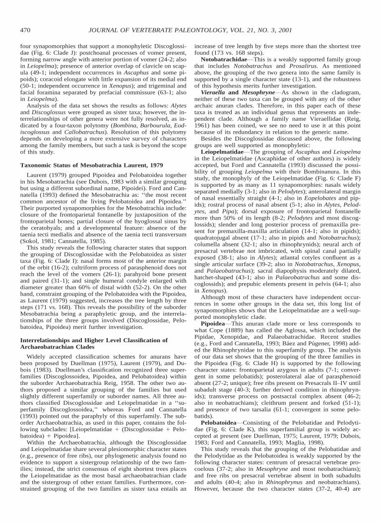
FIGURE 6. Phylogenetic hypotheses based on analysis of 65 characters across 23 taxa (see Appendices 1, 2). Strict consensus of eight shortesttrees (TL 5 168, CI 5 0.577, RI 5 0.718): A, Anura (stem-based taxon); B, Notobatrachidae (weakly supported taxon); C, unnamed clade 1; D,unnamed clade 2; E, Archaeobatrachia; F, Leiopelmatidae; G, unnamed clade 3; H, Pipoidea; I, unnamed clade 4; J, Discoglossidae; K, Pelobatoidea.
and several other taxa. In this study, strict consensus of the fiveequally parsimonious trees found suggests a Prosalirus–Noto-batrachus sistergroup relationship, and tracing character evo-lution shows that this group (Clade B: Notobatrachidae) isweakly supported by a single character state: palatine processof premaxilla well developed (13-1). Such a palatal process ofthe premaxilla has independent occurrences within the Discog-lossidae (Alytes, Bombina, and Discoglossus), in pelobatids, andin some neobatrachians.
Vieraella Reig (1961) suggested the possibility of naminga monotypic family Vieraellidae, whereas Casamiquela (1965)placed Vieraella in the family Notobatrachidae. Estes and Reig(1973) discussed similarities of Vieraella with Ascaphidae (nowLeiopelmatidae) and Discoglossidae, and referred both Vierael-la and Notobatrachus to their Ascaphidae (see also Duellmanand Trueb, 1986). More recently, Baez and Basso (1996:131)viewed Vieraella as ‘‘the most basal known member of Salien-tia, except the Early Triassic Triadobatrachus from Madagas-car.’’
The phylogenetic results of this study revealed that Vieraellais an ingroup member of the Anura. It shares with other anuransa large number of derived character states: Formation of pro-otic-occipital region by prootic-exoccipital (11-1); squamosaltriradiate and T-shaped (18-1); squamosal not in contact withdermal bones of skull table (21-1); postchoanal processes ofvomer present, forming wide angle with anterior portion of vo-mer (24-1); palatine absent owing to fusion with vomer or max-illa (25-1); epipodial elements fused to form single element (54-1); hind limb proportionally long with great elongation of prox-imal tarsals (59-1). These character states are among those sup-porting the Clade A in the resulting cladogram (Fig. 6).
Within the Anura, Vieraella holds a more derived positionthan the Prosalirus–Notobatrachus clade, and is placed as thesister group of a more derived clade that includes Mesophryneand other known anuran groups. This placement is supportedby four character states: Premaxilla-nasal contact lost with sep-aration of the two elements (15-1); position of anterior processof vomer lying near premaxilla-maxilla articulation (23-1); pos-
terolateral notch of parasphenoid alae absent (28-1). Thesecharacter states support the Clade C in the resulting cladogram(Fig. 6). Vieraella is more primitive than other anurans in hav-ing a greater number of presacral vertebrae (35-1); however,congruence with other characters does not place this taxon asthe most basal member of the stem-based Anura.
Mesophryne As shown in the cladogram (Fig. 6), this newtaxon represents a distinct archaic anuran clade, which is in amore derived position than Vieraella. It shares with the ar-chaeobatrachian and more derived anurans several characterstates, including: Sculpture on dermal skull roof absent (2-1);formation of prootic-occipital region by fused prootic-exoccip-ital (11-2); sacral diapophysis widely expanded as butterflywing-shaped structure (43-2); postsacral vertebrae uniformlymodified into a single urostyle (44-2); humeral condyle wellossified (53-1); proximal tarsals fused at proximal and distalends (60-1). However, Mesophryne lacks several other derivedcharacter states of the Archaeobatrachia: e.g., transverse pro-cess of posterior presacral vertebrae anterolaterally oriented(41-1), free intermedium in carpus absent (55-1), and prehalluxpresent (62-1). Lack of these derived character states, in con-gruence with other characters in the data set, places Mesophry-ne as the sister group rather than an ingroup member of theArchaeobatrachia (Fig. 6: Clade D).
Monophyly of Discoglossidae
The diagnosis of the family provided by previous authors(Duellman and Trueb, 1986; Clarke, 1988; Rocek, 1994) wasbased on a combination of plesiomorphic and derived characterstates. The most often used osteological character states in-clude: eight stegochordal, opisthocoelous presacral vertebrae;Presacrals I and II unfused; presence of free ribs on PresacralsII–IV; and sacrum having expanded diapophyses and bicondylararticulation with urostyle (e.g., Duellman and Trueb, 1986).
Because these are not discoglossid synapomorphies, Ford andCannatella (1993) suggested dropping the family name Discog-lossidae. However, the phylogenetic analysis in this study found
470 JOURNAL OF VERTEBRATE PALEONTOLOGY, VOL. 21, NO. 3, 2001
four synapomorphies that support a monophyletic Discoglossi-dae (Fig. 6: Clade J): postchoanal processes of vomer present,forming narrow angle with anterior portion of vomer (24-2; alsoin Leiopelma); presence of anterior overlap of clavicle on scap-ula (49-1; independent occurrences in Ascaphus and some pi-poids); coracoid elongate with little expansion of its medial end(50-1; independent occurrence in Xenopus); and trigeminal andfacial foramina separated by prefacial commissure (63-1; alsoin Leiopelma).
Analysis of the data set shows the results as follows: Alytesand Discoglossus were grouped as sister taxa; however, the in-terrelationships of other genera were not fully resolved, as in-dicated by a four-taxon polytomy (Bombina, Barbourula, Eod-iscoglossus and Callobatrachus). Resolution of this polytomydepends on developing a more extensive survey of charactersamong the family members, but such a task is beyond the scopeof this study.
Taxonomic Status of Mesobatrachia Laurent, 1979
Laurent (1979) grouped Pipoidea and Pelobatoidea togetherin his Mesobatrachia (see Dubois, 1983 with a similar groupingbut using a different subordinal name, Pipoidei). Ford and Can-natella (1993) defined the Mesobatrachia as: ‘‘the most recentcommon ancestor of the living Pelobatoidea and Pipoidea.’’Their purported synapomorphies for the Mesobatrachia include:closure of the frontoparietal fontanelle by juxtaposition of thefrontoparietal bones; partial closure of the hyoglossal sinus bythe ceratohyals; and a developmental feature: absence of thetaenia tecti medialis and absence of the taenia tecti transversum(Sokol, 1981; Cannatella, 1985).
This study reveals the following character states that supportthe grouping of Discoglossidae with the Pelobatoidea as sistertaxa (Fig. 6: Clade I): nasal forms most of the anterior marginof the orbit (16-2); cultriform process of parasphenoid does notreach the level of the vomers (26-1); parahyoid bone presentand paired (31-1); and single humeral condyle enlarged withdiameter greater than 60% of distal width (52-2). On the otherhand, constraint grouping of the Pelobatoidea with the Pipoidea,as Laurent (1979) suggested, increases the tree length by threesteps (171 vs. 168). This reveals the possibility of the suborderMesobatrachia being a paraphyletic group, and the interrela-tionships of the three groups involved (Discoglossidae, Pelo-batoidea, Pipoidea) merit further investigation.
Interrelationships and Higher Level Classification ofArchaeobatrachian Clades
Widely accepted classification schemes for anurans havebeen proposed by Duellman (1975), Laurent (1979), and Du-bois (1983). Duellman’s classification recognized three super-families (Discoglossoidea, Pipoidea, and Pelobatoidea) withinthe suborder Archaeobatrachia Reig, 1958. The other two au-thors proposed a similar grouping of the families but usedslightly different superfamily or suborder names. All three au-thors classified Discoglossidae and Leiopelmatidae in a ‘‘su-perfamily Discoglossoidea,’’ whereas Ford and Cannatella(1993) pointed out the paraphyly of this superfamily. The sub-order Archaeobatrachia, as used in this paper, contains the fol-lowing subclades: [Leiopelmatidae 1 (Discoglossidae 1 Pelo-batoidea) 1 Pipoidea].
Within the Archaeobatrachia, although the Discoglossidaeand Leiopelmatidae share several plesiomorphic character states(e.g., presence of free ribs), our phylogenetic analysis found noevidence to support a sistergroup relationship of the two fam-ilies; instead, the strict consensus of eight shortest trees placesthe Leiopelmatidae as the most basal archaeobatrachian cladeand the sistergroup of other extant families. Furthermore, con-strained grouping of the two families as sister taxa entails an
increase of tree length by five steps more than the shortest treefound (173 vs. 168 steps).
Notobatrachidae This is a weakly supported family groupthat includes Notobatrachus and Prosalirus. As mentionedabove, the grouping of the two genera into the same family issupported by a single character state (13-1), and the robustnessof this hypothesis merits further investigation.
Vieraella and Mesophryne As shown in the cladogram,neither of these two taxa can be grouped with any of the otherarchaic anuran clades. Therefore, in this paper each of thesetaxa is treated as an individual genus that represents an inde-pendent clade. Although a family name Vieraellidae (Reig,1961) has been coined, we see no need to use it at this pointbecause of its redundancy in relation to the generic name.
Besides the Discoglossidae discussed above, the followinggroups are well supported as monophyletic:
Leiopelmatidae The grouping of Ascaphus and Leiopelmain the Leiopelmatidae (Ascaphidae of other authors) is widelyaccepted, but Ford and Cannatella (1993) discussed the possi-bility of grouping Leiopelma with their Bombinanura. In thisstudy, the monophyly of the Leiopelmatidae (Fig. 6: Clade F)is supported by as many as 11 synapomorphies: nasals widelyseparated medially (3-1; also in Pelodytes); anterolateral marginof nasal essentially straight (4-1; also in Eopelobates and pip-ids); rostral process of nasal absent (5-1; also in Alytes, Pelod-ytes, and Pipa); dorsal exposure of frontoparietal fontanellemore than 50% of its length (8-2; Pelodytes and most discog-lossids); slender and long posterior process of premaxilla pre-sent for premaxilla-maxilla articulation (14-1; also in pipids);quadratojugal absent (17-1; also in pipids and Notobatrachus);columella absent (32-1; also in rhinophrynids); neural arch ofpresacral vertebrae not imbricated, with spinal canal partiallyexposed (38-1; also in Alytes); atlantal cotyles confluent as asingle articular surface (39-2; also in Notobatrachus, Xenopus,and Palaeobatrachus); sacral diapophysis moderately dilated,hatchet-shaped (43-1; also in Palaeobatrachus and some dis-coglossids); and prepubic elements present in pelvis (64-1; alsoin Xenopus).
Although most of these characters have independent occur-rences in some other groups in the data set, this long list ofsynapomorphies shows that the Leiopelmatidae are a well-sup-ported monophyletic clade.
Pipoidea This anuran clade more or less corresponds towhat Cope (1889) has called the Aglossa, which included thePipidae, Xenopidae, and Palaeobatrachidae. Recent studies(e.g., Ford and Cannatella, 1993; Baez and Pugener, 1998) add-ed the Rhinophrynidae to this superfamily group. The analysisof our data set shows that the grouping of the three families inthe Pipoidea (Fig. 6: Clade H) is supported by the followingcharacter states: frontoparietal azygous in adults (7-1; conver-gent in some pelobatids); posterolateral alae of parasphenoidabsent (27-2; unique); free ribs present on Presacrals II–IV untilsubadult stage (40-3; further derived condition in rhinophryn-ids); transverse process on postsacral complex absent (46-2;also in neobatrachians); cleithrum present and forked (51-1);and presence of two tarsalia (61-1; convergent in some pelo-batids).
Pelobatoidea Consisting of the Pelobatidae and Pelodyti-dae (Fig. 6: Clade K), this superfamilial group is widely ac-cepted at present (see Duellman, 1975; Laurent, 1979; Dubois,1983; Ford and Cannatella, 1993; Maglia, 1998).
This study reveals that the grouping of the Pelobatidae andthe Pelodytidae as the Pelobatoidea is weakly supported by thefollowing character states: centrum of presacral vertebrae pro-coelous (37-2; also in Mesophryne and most neobatrachians);and free ribs on presacral vertebrae absent in both subadultsand adults (40-4; also in Rhinophrynus and neobatrachians).However, because the two character states (37-2, 40-4) are

471GAO AND WANG—MESOZOIC ANURANS FROM CHINA
shared with neobatrachians, the homology of these characterstates and the monophyly of the Pelobatoidea need to be furthertested by developing a more global analysis based on moreextensive data.
CONCLUSIONS
This study comes to the following conclusions:1. The new fossil material from the Jurassic–Cretaceous de-
posits in western Liaoning Province, northeastern China, doc-uments two anuran taxa: a new taxon, Mesophryne, representsa distinct archaic anuran clade, and a second taxon, Calloba-trachus, represents the earliest fossil record of discoglossidsfrom Asia.
2. Phylogenetic analysis of 65 characters across 23 taxa pro-vides important insights into the interrelationships of the majorarchaic anuran clades. All of the three Jurassic forms (Prosa-lirus, Vieraella, and Notobatrachus) are ingroup members ofthe stem-based Anura, and Prosalirus and Notobatrachus canbe grouped in the same family, Notobatrachidae.
3. A revised classification scheme is proposed for Archaeo-batrachia on the basis of the results of the phylogenetic analysis.Recognized monophyletic groups include a weakly supportedNotobatrachidae, Leiopelmatidae, Pipoidea, Discoglossidae,and Pelobatoidea.
4. The Discoglossidae and Leiopelmatidae are not sistergroups, and the Discoglossoidea as previously defined is prob-ably an artificial taxon. New evidence supports the grouping ofthe Discoglossidae with the Pelobatoidea, and the suborder Me-sobatrachia as previously defined is probably a paraphyleticgroup.
ACKNOWLEDGMENTS
Field collection of the specimens used in this study was sup-ported by the Chinese Academy of Sciences (grant KZ951-B1-410 and KZCX3-J-03) and the Special Funds for Major StateBasic Research Projects of China (grant no. G2000077700).Gao’s work was supported by the Frick Fund from the Amer-ican Museum of Natural History, and by the National ScienceFoundation (NSF DEB-9806811). Photographs of the speci-mens were taken by Mick Ellison (AMNH), and the outlinedrawings were prepared by Yang Mingwan (IVPP). Thanks aregiven to Susan E. Evans (University College London), RichardC. Fox (University of Alberta), and Julian Faivovich (AMNH)for their careful reading of the manuscript, and to Jim Gardner(Royal Tyrrell Museum of Palaeontology) for providing rele-vant literature material.
LITERATURE CITED
Baez, A. M., and N. G. Basso. 1996. The earliest known frogs of theJurassic of South America: review and cladistic appraisal of theirrelationships. Munchner Geowissenschaftliche Abhandlungen, A(Geologie und Palaontologie) 30:131–158.
———, and L. A. Pugener. 1998. A new Paleogene pipid frog fromnorthwestern Patagonia. Journal of Vertebrate Paleontology 18:511–524.
Bryant, H. N. 1995. Why autapomorphies should be removed: a replyto Yeates. Cladistics 11:381–384.
Cannatella, D. C. 1985. A phylogeny of primitive frogs (archaeobatra-chians), Ph.D. dissertation, The University of Kansas, Lawrence,404 pp.
Casamiquela, R. M. 1965. Nuevo material de Vieraella herbstii Reig.Revista del Museo de La Plata, Paleontologia (Nueva Serie) 4:265–317.
Clarke, B. T. 1987. A description of the skeletal morphology of Bar-bourula (Anura: Discoglossidae) comments on its relationships.Journal of Natural History 21:879–891.
——— 1988. Evolutionary relationships of the discoglossoid frogs—
osteological evidence, Ph.D. dissertation, British Museum (NaturalHistory) and City of London Polytechnic, London, 891 pp.
Cope, E. D. 1889. The Batrachia of North America. Bulletin of theUnited States National Museum 34:1–525.
Dubois, A. 1983. Classification et nomenclature supragenerique des am-phibiens anoures. Bulletin Mensuel de la Societe Linneenne deLyon 52:270–276.
——— 1987. Discoglossidae Gunther, 1858 (Amphibia, Anura): Pro-posed conservation. Alytes 6:56–68.
Duellman, W. E. 1975. On the classification of frogs. Occasional Papers,Museum of Natural History, The University of Kansas 42:1–14.
——— 1999. Global distribution of amphibians: patterns, conservation,and future challenges; pp. 1–30 in W. E. Duellman (ed.), Patternsof Distribution of Amphibians: A Global Perspective. The JohnsHopkins University Press, Baltimore.
———, and L. Trueb. 1986. Biology of Amphibians. McGraw-HillBook Company, New York, 670 pp.
Endo, R. 1940. A new genus of Thecodontia from the Lycoptera bedsin Manchoukuo. Bulletin of the Central National Museum of Man-choukuo 2:1–14.
Estes, R. 1970. New fossil pelobatid frogs and a review of the genusEopelobates. Bulletin of the Museum of Comparative Zoology 139:293–339.
———, and O. A. Reig. 1973. The early fossil record of frogs: a reviewof the evidence; pp. 11–63 in J. L. Vial (ed.), Evolutionary Biologyof the Anurans: Contemporary Research on Major Problems. Uni-versity of Missouri Press, Columbia.
———, and F. B. Sanchiz. 1982. New discoglossid and palaeobatrachidfrogs from the Late Cretaceous of Wyoming and Montana, and areview of other frogs from the Lance and Hell Creek formations.Journal of Vertebrate Paleontology 2:9–20.
Evans, S. E., and M. Borsuk-Bialynicka. 1998. A stem-group frog fromthe Early Triassic of Poland. Acta Palaeontologica Polonica 43:573–580.
———, and A. R. Milner. 1993. Frogs and salamanders from the UpperJurassic Morrison Formation (Quarry Nine, Como Bluff) of NorthAmerica. Journal of Vertebrate Paleontology 13:24–30.
———, ———, and F. Mussett. 1990. A discoglossid frog (Amphibia:Anura) from the Middle Jurassic of England. Palaeontology 33:299–311.
———, G. Mcgowan, A. R. Milner, and B. Sanchiz. 1995. Amphibians;pp. 51–53 in N. Melendez (ed.), Las Hoyas: A Lacustrine Konser-vat-Lagerstatte, Cuenca, Spain. Universidad Complutense de Ma-drid, Spain.
Fabrezi, M. 1992. El carpo de los anuros. Alytes 10:1–29.———, and P. Alberch. 1996. The carpal elements of anurans. Herpe-
tologica 52:188–204.Fey, B. 1988. Die Anurenfauna aus der Unterkreide von Una (Ostspan-
ien). Berliner Geowissenschaftliche Abhandlungen, Reihe A 103:1–99.
Ford, L. S., and D. C. Cannatella. 1993. The major clades of frogs.Herpetological Monographs 7:94–117.
Frost, D. R. (ed.). 1985. Amphibian Species of the World: A Taxonomicand Geographic Reference. Allen Press and the Association of Sys-tematics Collections, Lawrence, Kansas.
Glaw, F., and J. Kohler. 1998. Amphibian species diversity exceeds thatof mammals. Herpetological Review 29:11–12.
Gubin, Y. M. 1999. Gobiatids (Anura) from the Upper Cretaceous lo-cality Khermeen-Tsav (Gobi Desert, Mongolia). PaleontologicalJournal 33:77–87.
Hecht, M. K. 1970. The morphology of Eodiscoglossus, a completeJurassic frog. American Museum Novitates 2424:1–17.
Henrici, A. C. 1994. Tephrodytes brassicarvalis, new genus and species(Anura: Pelodytidae), from the Arikareean Cabbage Patch beds ofMontana, USA, and pelodytid–pelobatid relationships. Annals ofCarnegie Museum 63:155–183.
——— 1998. A new pipoid anuran from the Late Jurassic MorrisonFormation at Dinosaur National Monument, Utah. Journal of Ver-tebrate Paleontology 18:321–332.
Ivachnenko, M. F. 1978. Urodelans from the Triassic and Jurassic ofSoviet Central Asia. Paleontological Journal 12:362–368.
Jenkins, F. A., Jr., and N. H. Shubin. 1998. Prosalirus bitis and theanuran caudopelvic mechanism. Journal of Vertebrate Paleontology18:495–510.

472 JOURNAL OF VERTEBRATE PALEONTOLOGY, VOL. 21, NO. 3, 2001
Ji, S., and Q. Ji. 1998. The first Mesozoic frog fossil from China (Am-phibia: Anura). Chinese Geology 250:39–42.
Ji, Q., S.-A. Ji, D. Ren, L.-W. Lu, X.-S. Fang, and Z.-G. Guo. 1999.On the sequence and age of the protobird-bearing deposits in theSihetun-Jianshangou area, Beipiao, western Liaoning. ProfessionalPapers of Stratigraphy and Paleontology 27:74–80.
Jin, F. 1996. New advances in the Late Mesozoic stratigraphic researchof western Liaoning, China. Vertebrata PalAsiatica 34:102–122.
Jurgens, J. D. 1971. The morphology of the nasal region of Amphibiaand its bearing on the phylogeny of the group. Annals of the Uni-versity of Stellenbosch 46:1–146.
Kohler, J., F. Glaw, and F. Bohme. 2000. Diversity in amphibians; pp.32–35 in R. Hofrichter (ed.), Amphibians: The World of Frogs,Toads, Salamanders and Newts. Firefly Books, New York.
Laurent, R. F. 1979. Esquisse d’une phylogenese des anoures. BulletinSociete Zoologique France 104:397–422.
Lee, D.-C., and H. N. Bryant. 1999. A reconsideration of the coding ofinapplicable characters: assumptions and problems. Cladistics 15:373–378.
Lo, C.-H., P.-J. Chen, T.-Y. Tsou, S.-S. Sun, and C.-Y. Lee. 1999. 40Ar/39Arlaser single-grain and K-Ar dating of the Yixian Formation, NE China;pp. 328–340 in P.-J. Chen and F. Jin (eds.), Jehol Biota (Palaeoworldspecial issue 11). Press of University of Science and Technology ofChina, Hefei, China.
Lynch, J. D. 1969. Evolutionary relationships and osteology of the frogfamily Leptodactylidae, Ph.D. dissertation, The University of Kan-sas, Lawrence, 801 pp.
——— 1973. The transition from archaic to advanced frogs; pp. 133–182 in J. L. Vial (ed.), Evolutionary Biology of the Anurans: Con-temporary Research on Major Problems. University of MissouriPress, Columbia.
Maddison, W. P., and D. R. Maddison. 1992. MacClade (version 3):Analysis of Phylogeny and Character Evolution. Sinauer Associ-ates, Inc. Sunderland, Massachusetts, 398 pp.
Maglia, A. M. 1998. Phylogenetic relationships of extant pelobatoidfrogs (Anura: Pelobatoidea): evidence from adult morphology. Sci-entific Papers, Natural History Museum, The University of Kansas10:1–19.
Meyer, v. H. 1843. Summarische Ubersicht der fossilen Wirbelthieredes Mainzer Tertiar-Beckens, mit besonderer Rucksicht auf Wies-ernau. Neues Jahrbuch fur Mineralogie, Geognosie, Geologie undPetrefactenkunde 1843:379–410.
Milner, A. R. 1988. The relationships and origin of living amphibians;pp. 59–102 in M. J. Benton (ed.), The Phylogeny and Classificationof the Tetrapods. 1. Amphibians, Reptiles, Birds. Oxford UniversityPress, Oxford.
Paterson, N. F. 1960. The inner ear of some members of the Pipidae(Amphibia). Proceedings of the Zoological Society of London 34:509–546.
Piveteau, J. 1937. Paleontologie de Madgascar XXIII. Un amphibien duTrias Inferieur. Annales Paleontologie 26:135–177.
Pleijel, F. 1995. On character coding for phylogeny reconstruction. Cla-distics 11:309–315.
Rage, J.-C., and Z. Rocek. 1989. Redescription of Triadobatrachusmassinoti (Piveteau, 1936) an anuran amphibian from the EarlyTriassic. Palaeontographica Abteilung A 206:1–16.
Reig, O. A. 1957. Los anuros del Matildense; pp. 231–297 in P. N. Sti-panicic and O. A. Reig (eds.), El complejo porfirico de la Patagoniaextraandina y su fanuna de anuros. Acta Geologica Lilloana 2.
——— 1958. Proposiciones para una nueva macrosistematica de losanuros. Nota Preliminar, Physis 21:109–118.
——— 1961. Noticia sobre un nuevo anuro fosil del Jurasico de SantaCruz (Patagonia). Ameghiniana 2:73–78.
Ren, D., Z. Guo, L. Lu, S. Ji, F. Tang, Y. Jin, X. Fang, and Q. Ji. 1997.A further contribution to the knowledge of the Upper Jurassic Yix-ian Formation in western Liaoning. Geological Review 43:449–459.
Ritland, R. M. 1955. Studies on the post-cranial morphology of Asca-phus truei. I. Skeleton and spinal nerves. Journal of Morphology97:119–178.
Rocek, Z. 1981. Cranial anatomy of the frogs of the family PelobatidaeStannius, 1856, with outlines of their phylogeny and systematics.Acta Universitatis Carolinae Biologica 1980:1–164.
——— 1994. Taxonomy and distribution of Tertiary discoglossids (An-ura) of the genus Latonia v. Meyer, 1843. Geobios 27:717–751.
——— 2000. Mesozoic anurans; pp. 1295–1331 in H. E. Heatwole andR. L. Carroll (eds.), Amphibian Biology, Vol. 4—Palaeontology.Surrey Beatty & Sons, Australia.
———, and J.-C. Rage. 2000. Proanuran states (Triadobatrachus, Czat-kobatrachus); pp. 1283–1294 in H. E. Heatwole and R. L. Carroll(eds.), Amphibian Biology, Vol. 4—Palaeontology. Surrey Beatty& Sons, Australia.
Sanchiz, B. 1998. Salientia. Handbuch der Palaoherpetologie, Teil 4.Verlag Dr. Friedrich Pferl, Munchen, 275 pp.
Shubin, N. H., and F. A. Jenkins, Jr. 1995. An Early Jurassic jumpingfrog. Nature 377:49–52.
Slabbert, G. K., and W. A. Maree. 1945. The cranial morphology of theDiscoglossidae and its bearing upon the phylogeny of the primitiveAnura. Annals of the University of Stellenbosch 23A(2–6):91–97.
Sokol, O. M. 1981. The larval chondrocranium of Pelodytes punctatus,with a review of tadpole chondrocrania. Journal of Morphology169:161–183.
Spinar, Z. V. 1972. Tertiary Frogs from Central Europe. CzechoslovakAcademy of Sciences, Prague, 286 pp.
———, L. P. Tatarinov. 1986. A new genus and species of discoglossidfrog from the Upper Cretaceous of the Gobi Desert. Journal ofVertebrate Paleontology 6:113–122.
Stephenson, E. M. 1951. The anatomy of the head of the New Zealandfrog, Leiopelma. Transactions of the Zoological Society of London27:255–305.
——— 1952. The vertebral column and appendicular skeleton of Leio-pelma hochstetteri Fitzinger. Transactions of the Royal Society ofNew Zealand 79:601–613.
——— 1960. The skeletal characters of Leiopelma hamiltoni Mc-Culloch, with particular reference to the effects of heterochrony onthe genus. Transactions of the Royal Society of New Zealand 88:473–488.
Swisher, C. C., Y. Wang, X. Wang, X. Xu, and Y. Wang. 1999. Creta-ceous age for the feathered dinosaurs of Liaoning, China. Nature400:58–61.
Swofford, D. L. 1993. PAUP: Phylogenetic Analysis Using Parsimony.Version 3.1. Illinois Natural History Survey.
Trueb, L. 1973. Bones, frogs, and evolution; pp. 65–132 in J. L. Vial(ed.), Evolutionary Biology of the Aurans: Contemporary Researchon Major Problems. University of Missouri Press, Columbia.
——— 1993. Patterns of cranial diversity among the Lissamphibia; pp.255–343 in J. Hanken and B. K. Hall (eds.), The Skull, Vol. 2.Patterns of Structural and Systematic Diversity. The University ofChicago Press, Chicago, 566 pp.
———, and R. Cloutier. 1991a. A phylogenetic investigation of theinter- and intrarelationships of the Lissamphibia (Amphibia: Tem-nospondyli); pp. 175–188 in H.-P. Schultze and L. Trueb (eds.),Origins of the Higher Groups of Tetrapods: Controversy and Con-sensus. Cornell University Press, Ithaca.
———, and ——— 1991b. Toward an understanding of the amphibi-ans: two centuries of systematic history; pp. 223–313 in H.-P.Schultze and L. Trueb (eds.), Origins of the Higher Groups of Tet-rapods: Controversy and Consensus. Cornell University Press, Ith-aca.
Van Eeden, J. A. 1951. The development of the chondrocranium ofAscaphus truei Stejneger with special reference to the relations ofthe palatoquadrate to the neurocranium. Acta Zoologica 32:41–176.
Villalta, C. J. F., de. 1957. Eodiscoglossus santonjae Vill., la Rana masantigua conocida, que ha sido encontrada en el Jurasico superiorde Santa Maria de Meya (Lerida); p. 146 in B. Melendez, La ev-olucion biologica. Madrid, Ediciones Fax. [Adaptation of Leonardi,Piero, 1950. L’evoluzione dei viventi. Brescia, Morcelliana, 360pp.]
Wang, S. 1990. Origin, evolution and mechanism of the Jehol fauna.Acta Geologica Sinica 1990:350–360.
——— 1998. Correlation of the continental Jurassic in the north ofChina to the paralic Jurassic in northwest Scotland, United King-dom—with a discussion of the stratigraphic subdivision and cor-relation of the Jurassic in the north of China. Acta Geologica Sinica72:11–21.
Wang, Y., and K. Gao. 1999. Earliest Asian discoglossid frog fossilfrom western Liaoning. Chinese Science Bulletin 44:636–641.
Wang, X. L., Y. Q. Wang, Y. Wang, X. Xu, Z. L. Tang, F. C. Zhang, Y.M. Hu, G. Gu, and Z. L. Hao. 1998. Stratigraphic sequence andvertebrate-bearing beds of the lower part of the Yixian Formation

473GAO AND WANG—MESOZOIC ANURANS FROM CHINA
in Sihetun and neighboring area, western Liaoning, China. Verte-brata PalAsiatica 36:81–101.
Worthy, T. H. 1987. Osteology of Leiopelma (Amphibia, Leiopelmati-dae) and description of three new subfossil Leiopelma species.Journal of the Royal Society New Zealand 17:201–251.
Yeates, D. 1992. Why remove autapomorphies? Cladistics 8:387–389.
Received 30 June 2000; accepted 1 March 2001.
APPENDIX 1
Characters used in phylogenetic analysis (modified from Baez andBasso, 1996), with new characters from other sources denoted with anasterisk.
1. General shape of the skull: skull wider than long (0); or roughlyas long as wide (1). In Triadobatrachus and the remaining taxa studied,the skull is wider than long. The skull is as long as wide in Vieraella,Rhinophrynus, and Palaeobatrachus.
*2. Sculpture on dermal skull roof: present (0); or absent (1). Mod-ified from Henrici (1994). Triadobatrachus has low rogosities on skullroof (Rage and Rocek, 1989). Outgroup comparison indicates that pres-ence of dermal sculpture is a primitive condition among anurans.
3. Medial contact of nasals: contact present (0); nasals widely sepa-rated medially (1); or nasals fused medially (2). Among the taxa in-cluded in the analysis, the (1) condition occurs in leiopelmatids andpelodytids independently and the (2) condition is unique for Xenopus.
*4. Anterolateral margin of nasal: concave (0); or essentially straight(1). See Trueb (1993) and Henrici (1994) for discussion. Considerablevariation in size and shape of the nasals can be seen in different anurangroups, but primitive anurans generally have the nasal with a concaveanterior margin, while most ‘‘mesobatrachians’’ and neobatrachianshave the nasal with a more or less straight anterior margin.
5. Rostral process of nasal: present (0); or absent (1). In caudates,the nasals project anteriorly to contact the alary processes of the pre-maxillae. The nasals of Vieraella, Notobatrachus, and all discoglossidsbut Alytes bear rostral processes extending forward along the cranialmidline. In Ascaphus and Leiopelma, the nasals lack a distinct rostralprocess (Stephenson, 1951).
6. Fusion of frontal and parietal: frontal and parietal remain separate(0); or fused to form frontoparietal (1). The frontals and parietals arediscrete dermal roofing bones in caudates. In Triadobatrachus and othersalientians, the two elements are fused to form the frontoparietals eitherpaired or azygous. The derived state is a salientian synapomorphy (Mil-ner, 1988).
7. Frontoparietal in adults: paired (0); or azygous (1). Although thiscan be combined with character 6, we treat this character as separatebecause of the uncertain condition in Czatkobatrachus, Prosalirus andMesophryne. Coding the paired condition for Triadobatrachus followsRocek and Rage (2000). Some Barbourula have the frontoparietals par-tially fused posteriorly (Clarke, 1987), but we have coded the primitivepaired condition for the taxon.
8. Dorsal exposure of frontoparietal fontanelle: fontanelle not ex-posed (0); exposed 50% of its length or less (1); fontanelle exposedmore than 50% of its length (2). The absence of exposure of the fron-toparietal fontanelle in adults occurs in caudates and Triadobatrachusas a primitive condition. See Baez and Basso (1996) for comments.
9. Posterolateral process of frontoparietal: well developed and wing-like (0); poorly developed or absent (1). The primitive state occurs inbasal caudates, Triadobatrachus (Rage and Rocek, 1989), and Prosa-lirus (Jenkins and Shubin, 1998). Coding of Notobatrachus and Vi-eraella is based on Baez and Basso (1996:135, 139). Pelobates andScaphiopus have a small posterolateral process (Rocek, 1981), but def-initely not wing-like.
10. Frontoparietal shelf: absent (0); or present (1). The lateral borderof the frontoparietal is elaborated to form a narrow orbital shelf inNotobatrachus. Such a shelf is absent in caudates, in which the frontaland parietal are separate bones, but it occurs also in rhinophrynids andpelobatids.
11. Formation of prootic-occipital region: by prootic-exoccipital-op-isthotic complex (0); by prootic-exoccipital without fusion (1); or byfused prootic-exoccipital (2). The prootic and exoccipital are fused dur-ing ontogeny in most living anurans. A separate exoccipital has beendescribed in Triadobatrachus (Rage and Rocek, 1989). Discrete exoc-cipitals occur in Vieraella, Notobatrachus, Ascaphus (Van Eeden,1951), and Leiopelma, although in adults of the latter genus these bones
may eventually fuse to the prootics (Worthy, 1987). The exoccipital isindistinguishably fused to the prootic in Bombina, Alytes, and Discog-lossus. Coding (1) for Eodiscoglossus is according to Hecht (1970).
12. Perilymphatic foramen: double foramina open on medial capsularwall (0); double foramina present on posterior wall of otic capsule (1);only superior perilymphatic foramen present (2); or only inferior fora-men present (3). In urodeles, the perilymphatic foramina are absent onthe posterior wall of the otic capsule (open on the medial wall, instead).A similar condition is seen in Ascaphus and Leiopelma (Van Eeden,1951). A single foramen in Notobatrachus is interpreted as the jugularforamen (Baez and Basso, 1996). Coding this character for Pipa concurswith Baez and Pugener’s (1998:appendix 3) data matrix but contradictstheir incorrect description that both foramina are absent in pipines. Pat-erson (1960) identified a single foramen (superior perilymphatic fora-men) in pipids.
13. Palatine process of premaxilla: absent or barely present (0); orwell developed (1). The pars palatina of the premaxilla forms a distinctpalatine process in Notobatrachus and most discoglossids (Baez andBasso, 1996). Such a process is barely present in Ascaphus and Leio-pelma (Jurgens, 1971), and is lacking in the premaxilla in caudates. Theactual condition of this process is unknown for Vieraella and Triadob-atrachus because of poor preservation.
14. Premaxilla-maxilla articulation: posterior process of premaxillaabsent (0); or present (1). In Leiopelma and Ascaphus (Trueb, 1993:fig.6.2 and 6.18), the pars palatina of the premaxilla projects posteriorlyinto an elongate and pointed ‘‘process’’ for the articulation with themaxilla. In Triadobatrachus, the frontal region of the skull is not pre-served and the character is unknown. This elongate shape of the pos-terior (or lateral) end of the pars palatina of the premaxilla also occursin Pipa and Xenopus among the other taxa studied.
15. Premaxilla-nasal contact: articulation or contact present (0); orcontact lost with separation of the two elements (1). The contact con-dition is known for Notobatrachus (Baez and Basso, 1996), but theactual condition of the appropriate region is unknown for Triadobatra-chus, Czatkobatrachus, and Prosalirus. The derived condition occurs inall other anurans but Palaeobatrachus that represents a character re-versal.
16. Prefrontal and anterior margin of the orbit: prefrontal present,maxilla and nasal excluded from the anterior margin of the orbit (0);prefrontal lost with maxilla and nasal forming the anterior margin ofthe orbit (1); nasal forming most of the anterior margin of the orbit (2);or anterior ramus of pterygoid excludes maxilla from the anterior mar-gin of the orbit (3). A prefrontal is plesiomorphically retained in cau-dates and Triadobatrachus, while it is absent in all the anuran taxa thatcan be scored. Pending knowledge of the actual condition in Czatko-batrachus, Prosalirus, and Vieraella, loss of the prefrontal could be asynapomorphy for the anurans (e.g., Trueb, 1993; Baez and Basso,1996).
17. Quadratojugal: present (0); or absent (1). A quadratojugal is prob-ably present in Triadobatrachus (Piveteau, 1937; Estes and Reig, 1973;but see also Rage and Rocek, 1989). Among anurans, the derived con-dition occurs in Notobatrachus, Ascaphus, Leiopelma, and pipids(Trueb, 1993). As the condition is uncertain in Vieraella (see Estes andReig, 1973; Baez and Basso, 1996), we have coded it as unknown.
18. Shape of squamosal: as a simple horizontal bar (0); triradiate andT-shaped (1); nontriradiate with loss of zygomatic ramus (2); or funnel-shaped and in fusion with tympanic annulus (3). Both caudates andTriadobatrachus have a simple squamosal that is not distinctly triradi-ate. Most anurans have a typically triradiate squamosal with variationsinvolving expansion/reduction of the zygomatic or otic processes, orcurving of the quadrate process (see Trueb, 1993 for discussion).
19. Squamosal-maxilla contact: absent (0); or contact present (1). Asthe presence of a squamosal-maxilla contact is directly related to thesize of the zygomatic ramus of the squamosal, this character can bepolarized by comparison with the character above. The derived state isprobably a pelobatine synapomorphy with independent occurrences inDiscoglossus, Mesophryne, and some neobatrachians.
20. Expansion of otic ramus of squamosal in lateral view: not ex-panded (0); or otic ramus expanded and deep (1). Among all of the taxaincluded in the matrix, the derived state of this character diagnoses thePelobatidae.
21. Medial articulation of squamosal: squamosal medially in contactwith dermal skull roof (0); or squamosal not in contact with dermalskull roof (1). Although stated ‘‘both squamosals lost their contact withthe frontoparietals,’’ Rage and Rocek (1989:text-fig. 4) restored a con-

474 JOURNAL OF VERTEBRATE PALEONTOLOGY, VOL. 21, NO. 3, 2001
tact for Triadobatrachus, and this restoration was followed by Sanchiz(1998).
22. Sphenethmoid: bilaterally paired (0); or single (1); or fused toparasphenoid ventrally (2). Modified from Baez and Basso (1996:char-acter 17). The sphenethmoid ossification is bilaterally paired in theadults of Notobatrachus (Baez and Basso, 1996). A similar conditionoccurs in leiopelmatids and most microhylids (Van Eeden, 1951; Trueb,1993); however, Trueb (1993:313) explained that ‘‘the apparent divisionis the result of incomplete medial replacement of cartilage by bone.’’The sphenethmoid is single in most anurans, but is fused to the paras-phenoid in pipids.
23. Position of anterior process of vomer: anterior process of vomerlying immediately behind premaxilla (0); or lying near premaxilla-max-illa articulation (1). In the primitive caudate Karaurus, the anterior partof the vomer lies adjacent to the premaxilla anteriorly, whereas in uro-deles it broadly contacts the premaxilla and maxilla. The condition inTriadobatrachus is unknown because the appropriate region is not pre-served. In Notobatrachus, the anterior plate-like portion of the vomer(anterior process) lies close to the premaxilla, and is more or less par-allel to the cranial midline. In all of the other ingroup taxa, the anteriorportion of the vomer, if well developed, lies adjacent to the premaxilla-maxilla articulation.
24. Postchoanal processes of vomer: absent (0); or present, formingwide angle with anterior portion of vomer (1); present, forming narrowangle with anterior portion of vomer (2). Modified from Baez and Basso(1996:character 21). The postchoanal process of the vomer forms arather wide angle (about 90–1108) with the anterior process in Vieraellaand Notobatrachus. In Leiopelma, Bombina, Alytes, Barbourula, andDiscoglossus, this angle is significantly narrower (about 458). Ascaphuslacks anterior and postchoanal processes on the vomer, which is reducedto the dentigerous portion (Van Eeden, 1951). In Triadobatrachus, thecondition of this region is unknown.
*25. Palatine: present as separate element (0); or absent or fused withvomer or maxilla (1). The palatine is retained as a separate element inmany urodele taxa (Trueb, 1993), in Triadobatrachus (Rage and Rocek,1989), but is absent in Vieraella and Notobatrachus (Estes and Reig,1973; Baez and Basso, 1996). The condition of this character is un-known for Czatkobatrachus and Prosalirus (Evans and Borsuk-Bialyn-icka, 1998; Jenkins and Shubin, 1998).
26. Cultriform process of parasphenoid: extending anteriorly to thelevel of the vomers (0); or not reaching the level of the vomers (1).The anterior terminus of the cultriform process of the parasphenoidextends forward to reach the level of the posteromedial portion of thevomers in Notobatrachus, Ascaphus, and Leiopelma (Jurgens, 1971).The cultriform process does not reach so far forward in other taxa,including Bombina, Alytes, and Discoglossus. Baez and Basso (1996)ambiguously stated ‘‘it is impossible to assess the anterior extent of theprocess’’ but coded it as (1) for Vieraella; we coded the character asunknown for the taxon. The condition in Triadobatrachus cannot bedetermined as preserved, but the character can be polarized, as the prim-itive state is known for caudates.
27. Posterolateral alae of parasphenoid: anteroposterior width of alaeequal or greater than one-third distance between lateral ends (0); widthnarrower than one-third distance between lateral ends (1); or alae absent(2). The primitive state is known for Triadobatrachus, Vieraella, No-tobatrachus, and Prosalirus. All other taxa show the (1) condition, withthe exception of pipoids, which lack posterolateral alae of the paras-phenoid.
28. Posterolateral notch of parasphenoid alae: present (0); or absent(1). The posterolateral corner of the parasphenoid alae is deeply in-dented in both Triadobatrachus (Rage and Rocek, 1989) and Notoba-trachus (Baez and Basso, 1996). The latter authors considered the sim-ilarity as superficial. As in both taxa, the notch is functionally relatedto the auditory capsule, we choose to code them (0).
29. Posteromedial process of parasphenoid: absent (0); or present (1).A distinct, narrow process on the posterior margin of the parasphenoidis absent in Triadobatrachus, Vieraella, Notobatrachus, and Prosalirus.Among other ingroup taxa, it occurs in all groups that can be scoredwith the exception of Palaeobatrachus and Rhinophrynus.
30. Medial ramus of pterygoid: not contacting parasphenoid (0); orcontacting parasphenoid (1); or medial ramus absent (2). Modified fromBaez and Basso (1996:character 28). The medial process of the ptery-goid does not contact or overlap the parasphenoid ala in Triadobatra-chus (Rage and Rocek, 1989). We coded unknown for Vieraella becauseof the uncertain condition of this taxon (Baez and Basso, 1996). Rhin-
ophrynids are unique in having no medial ramus of the pterygoid(Trueb, 1993).
*31. Parahyoid bone: present and single (0); or present and paired(1); or absent (2). Single parahyoid is present in Triadobatrachus (Rageand Rocek, 1989), Notobatrachus (Baez and Basso, 1996), leiopelma-tids, rhinophrynids (Trueb, 1993), and palaeobatrachids (Estes and Reig,1973); paired in discoglossids including Bombina and Barbourula(Trueb, 1993; Clarke, 1987), and pelodytids; absent in pipids, peloba-tids, and neobatrachians. The ‘‘Y’’-shaped parahyoid in Alytes is con-sidered to be a modified paired condition.
*32. Columella: well-ossified columella present (0); or absent (1).Trueb (1993:315) discussed the uncertain polarity of this character;however, given the distribution of the character among extant and fossilanurans in comparison to Triadobatrachus and Caudata, it is clear thatthe absent condition is a derived condition. The derived state is knownfor leiopelmatids (Ascaphus and Leiopelma), rhinophrynids, and someneobatrachians (Trueb, 1993).
*33. Mentomeckelian bone: present (0); or absent (1). An ossified men-tomeckelian bone is present in caudates and most anurans, but the derivedcondition is known for pipids and rhinophrynids (see Trueb, 1993).
34. Marginal teeth: both upper and lower dentition present (0); orlower dentition absent (1); or both upper and lower dentition absent (2).The dentary is edentulous in Triadobatrachus and this condition is con-sidered a synapomorphy of Salientia (Milner, 1988). Prosalirus has anupper dentition, while the condition of its lower dentition is unknown;therefore, we coded unknown for this taxon.
35. Number of presacral vertebrae: 14 or more (0); ten presacralvertebrae (1); normally nine presacral vertebrae (2); normally eight orfewer (3). See Lynch (1973) for discussion on extant taxa. Triadoba-trachus has 14 presacral vertebrae (Rage and Rocek, 1989). The numberof presacrals is ten in Vieraella and nine in Notobatrachus (Baez andBasso, 1996).
*36. Fusion of Presacrals I and II: fusion absent (0); or fusion present(1). From Duellman and Trueb (1986). The derived condition occurs inthe Pelodytidae and several other taxa that are included in the datamatrix: Pipa, Palaeobatrachus, some neobatrachians including somebufonids, and some ranids (Duellman and Trueb, 1986).
37. Centrum of presacral vertebrae: vertebral centra amphicoelous ornotochordal (0); or opisthocoelous (1); or procoelous (2). The primitivestate is known for Triadobatrachus, Czatkobatrachus, Prosalirus, Vi-eraella, Notobatrachus, Ascaphus, and Leiopelma (Rage and Rocek,1989; Baez and Basso, 1996; Evans and Borsuk-Bialynicka, 1998; Jen-kins and Shubin, 1998). Coding of Eodiscoglossus based on the originalphotographs of the holotype, in which the last three presacrals have anopisthocoelous centrum.
38. Neural arch of presacral vertebrae: completely or weakly imbri-cated roofing of spinal canal (0); or not imbricated with spinal canalpartially exposed (1). The derived state is known for Ascaphus, Leio-pelma, and Alytes. Czatkobatrachus and Prosalirus, based on disartic-ulated material, are coded as unknown, although at least the latter taxonmay have the imbricated condition (Jenkins and Shubin, 1998). Thederived state also occurs in some neobatrachians (Trueb, 1973).
39. Morphology of atlantal cotyles: cotyles mostly ventral and nar-rowly separated by notochordal fossa (0); cup-like cotyles displacedlaterally and widely separated from one another (1); cotyles confluentas a single articular surface (2). See discussion in Trueb (1973). In Baezand Basso’s (1996) original data set, two states (ventral and lateral po-sitions of the cotyles) were recognized. Since the ventrally located co-tyles can be either apart from one another or confluent, we choose toseparate the two states following Lynch (1969).
40. Free ribs on presacral vertebrae: free ribs present on all presacralvertebrae (0); ribs present on Presacrals II–V or II–VI (1); or ribs re-stricted to Presacrals II–IV (2); present at least on Presacrals II–IV tillsubadult stage (3); free ribs absent in both subadults and adults (4).Vieraella is coded as unknown because of the uncertain condition onposterior presacrals (Baez and Basso, 1996). In Prosalirus, free ribs aredefinitely present on some presacral vertebrae, but the actual numbercannot be determined (Jenkins and Shubin, 1998). Palaeobatrachidshave free ribs on Presacrals II–VI in subadults, but these ribs are an-kylosed to the transverse processes in adults; similarly, young pipidshave free ribs on Presacrals II–IV but they are ankylosed to the trans-verse processes in adults.
41. Transverse process of posterior presacral vertebrae: laterally ori-ented (0); or anterolaterally oriented (1). The derived condition is seenin all archaeobatrachians with the exception of the palaeobatrachids.

475GAO AND WANG—MESOZOIC ANURANS FROM CHINA
*42. Fusion of sacral ribs: remain free from sacral vertebra (0); or fusedto transverse process of sacral (1). The primitive state is known for Tria-dobatrachus (Rage and Rocek, 1989), while all other taxa including Czat-kobatrachus and Prosalirus have the sacral ribs fused to the vertebra(Evans and Borsuk-Bialynicka, 1998; Jenkins and Shubin, 1998).
43. Dilation of sacral diapophysis: diapophysis slender and no dila-tion (0); or moderately dilated and hatchet-shaped (1); or widely ex-panded as butterfly wing-shaped (2). Description of the transformationseries follows Clarke (1988). Coding the (2) condition for Eodiscoglos-sus santonjae is based on observation of the original photographs ofthe type specimen, which clearly show the impressions that indicateexpanded diapophyses on both sides (contra Hecht, 1970). Among ex-tant discoglossids, state (2) is seen in Barbourula and Bombina, whilethe moderately expanded ‘‘hatchet-shaped’’ condition is seen in Alytesand Discoglossus (Clarke, 1988).
44. Postsacral vertebrae: caudal vertebrae remain unfused (0); or uro-style present in association with discrete caudal between sacrum andurostyle (1); or all postsacral vertebrae uniformly modified into singleurostyle (2). Triadobatrachus has at least six caudals bearing transverseprocesses. Notobatrachus normally has a discrete caudal between thesacrum and urostyle, but all other anurans have a highly modified uro-style only. Prosalirus has a urostyle, which could be either the (1) or(2) condition (Jenkins and Shubin, 1998).
45. Sacrococcygeal articulation: cartilaginous joint (0); bicondylar(1); monocondylar (2); or simply fused (3). In Notobatrachus, Ascaphus(Ritland, 1955), and Leiopelma (Stephenson, 1952, 1960), there is acartilaginous joint between the sacrum and urostyle, similar to that be-tween successive presacral vertebrae. A bony monocondylar joint isformed between the sacrum and the urostyle in Bombina and Barbou-rula, whereas a bicondylar joint is present in Alytes and Discoglossus.A cartilaginous joint between the sacral vertebra and the following ver-tebra is present in Triadobatrachus.
*46. Transverse process on postsacral complex: present (0); or fusedto a bony web of sacral diapophysis (1); or absent (2). See Lynch (1973)for discussion. The postsacral complex refers to either the discrete ver-tebrae as in Triadobatrachus or the urostyle as in other salientians.Czatkobatrachus may have caudal vertebrae that bear no transverse pro-cesses (Evans and Borsuk-Bialynicka, 1998); we code the character (?)as the urostyle is unknown for the taxon.
47. Length of scapula: at least half length of humerus (0); or less thanhalf length of humerus (1). The primitive state is known for Triadoba-trachus (Rage and Rocek, 1989). Prosalirus has a scapula that is slightlyshorter than one-half length of the humerus (Jenkins and Shubin, 1998).Although described as having a relatively long scapula and coded (0) forVieraella (Baez and Basso, 1996), the actual condition is uncertain asboth humeri are incomplete on the holotype and only known specimen;therefore, we coded the character as unknown for the taxon.
48. Leading edge of scapula: leading edge concave (0); or straight(1). The leading edge of the scapula is anteriorly concave in Triadob-atrachus, Prosalirus, Vieraella, Notobatrachus, and Leiopelma. Theedge is straight in discoglossids, and a similar condition is also seen inAscaphus, Xenopus, and Palaeobatrachus.
*49. Anterior overlap of clavicle on scapula: overlap absent (0); oroverlap present (1); or two elements fused (2). Overlapping occurs inAscaphus, discoglossids, palaeobatrachids, pipids, and rhinophrynids(see Lynch, 1973). Coding of pelodytids is based on Henrici (1994).The condition in Triadobatrachus, Czatkobatrachus, and Prosalirus isunknown, but this character can be polarized by the primitive conditionobserved in caudates.
50. Coracoid shape: medial end of coracoid expanded anteriorly (0);or coracoid elongate, medial end little expanded anteriorly (1). Themedial (sternal) end of the coracoid is expanded anteriorly in Vieraellaand Notobatrachus. A similar anterior expansion of the medial end ofthis bone also occurs in Leiopelma and Ascaphus, whereas in Bombina,Alytes and Discoglossus, the coracoid has a rounded sternal end. Theshape of the coracoid in Triadobatrachus cannot be determined becauseof incomplete preservation (Rage and Rocek, 1989). The medial end ofthe coracoid in Prosalirus is strongly expanded (Jenkins and Shubin,1998:fig. 4C), while the structure in Czatkobatrachus is unknown(Evans and Borsuk-Bialynicka, 1998).
*51. Cleithrum: present and unforked (0); present and forked (1); orcleithrum fused to suprascapula (2). The unforked condition of thecleithrum is known for Triadobatrachus (Rage and Rocek, 1989), Vi-eraella, and several extant groups, including leiopelmatids and pelo-batids. The forked condition is seen in pipoids, and all discoglossids
but Callobatrachus. The fused condition is known for rhinophrynidsand brachycephalids (Trueb, 1973).
52. Humeral condyle: double humeral condyles present (0); or singlecondyle with small diameter less than 60% of distal width (1); or singlecondyle enlarged with diameter greater than 60% of distal width (2).The condyle has a diameter of 54% of the distal width in Czatkobatra-chus; the condition cannot be determined in Prosalirus, Mesophryne,and Callobatrachus.
53. Ossification of humeral condyle: condyle unossified (0); or con-dyle ossified (1). The derived condition is known for Ascaphus and allother archaeobatrachians; cartilaginous condition is known for Triadob-atrachus, Prosalirus, Vieraella, and Notobatrachus.
54. Epipodial elements: remaining as separate elements (0); or fusedto form single element (1). The derived condition is an unambiguoussynapomorphy for the Anura (see Milner, 1988). The primitive state isknown for Triadobatrachus (Rage and Rocek, 1989) and Czatkobatra-chus (Evans and Borsuk-Bialynicak, 1998). Prosalirus has the derivedcondition as in other anurans (Jenkins and Shubin, 1998).
55. Free intermedium in carpus: present (0); or absent (1). The prim-itive state is known for Triadobatrachus, Notobatrachus, and the newtaxon Mesophryne. The condition is unknown for Prosalirus and Vi-eraella because of preservation. The derived condition is known for allmodern anuran groups (Trueb, 1973).
56. Length and orientation of ilium: short ilium essentially dorsallydirected (0); or elongate shaft of ilium anteriorly directed (1). The de-rived state is known for Triadobatrachus, Czatkobatrachus, Prosalirus,and Vieraella as in other salientians. This is an unambiguous synapo-morphy for Salientia (Milner, 1988).
57. Dorsal acetabular expansion of ilium: not extending to dorsallimit of ischium (0); or extending to dorsal limit of ischium (1). Thederived state is known for Alytes and Discoglossus (Clarke, 1988), Rhin-ophrynus, and pelobatoids except for Eopelobates (Estes, 1970).
*58. Dorsal prominence of ilium: strongly developed as a high tu-bercle (0); or tubercle low or absent (1). Degree of development of thisstructure is highly variable among anurans, and description of the dorsalprominence (dorsal protuberance of other authors) in the literature isconfusing (e.g., Estes and Sanchiz, 1982; Evans and Milner, 1993).Triadobatrachus and Czatkobatrachus clearly has a conspicuous tuber-cle (Rage and Rocek, 1989:text-fig. 3; Evans and Borsuk-Bialynicka,1998:fig. 1), while in Prosalirus and Notobatrachus the dorsal promi-nence is barely developed (Jenkins and Shubin, 1998:fig. 2).
*59. Hind limb proportions: normal length (0); or proportionally longwith great elongation of proximal tarsals (1). The derived state is pos-itively known for all anurans except Czatkobatrachus. The hind limbof Vieraella is incompletely preserved, but an elongated femur givespositive evidence for hind limb elongation as in other anurans.
*60. Fusion of proximal tarsals: fusion absent (0); or fused at prox-imal and distal ends (1); or completely fused to form a single bone (2).Modified from Henrici (1994:character 22). The unfused condition isknown for primitive anurans including Prosalirus and Notobatrachus,while the condition is unknown for Vieraella. Most other anurans havethe (1) condition, but complete fusion of the tibiale and fibulare isknown for pelodytids and centrolenids (Duellman and Trueb, 1986).
*61. Number of tarsalia: three or more free elements (0); or only twoelements present (1). For discussion of the character, see Lynch (1973),Duellman and Trueb (1986), and Henrici (1994).
*62. Prehallux: absent (0); or present as small hind foot element (1);or modified as bony spade (2). Modified from Henrici (1994:character24). The primitive condition is seen in Mesophryne, and the derivedcondition is scored for all the modern anuran groups according to Trueb(1973).
*63. Consolidation of Cranial Nerve V and VII: three separate fo-ramina occur (0); or trigeminal and facial foramina separated by pre-facial commissure (1); or commissure absent, nerves exit via singleprootic foramen (2). From Trueb (1973:84): (0) condition in primitiveanuran Ascaphus and salamanders; (1) condition in Leiopelma and dis-coglossids; and (2) condition in all other anurans.
*64. Prepubic elements: absent (0); or present (1). The derived con-dition is known for leiopelmatids, Xenopus, and Pseudhymenochirus(Duellman and Trueb, 1986).
*65. Posture of manus: medial inturning of manus absent (0); orinturning present (1). This character corresponds to the condition thathas been termed ‘‘carpal torsion’’ by Estes and Reig (1973), and ‘‘bra-chiocarpal rotation’’ by Baez and Basso (1996); see also discussion inJenkins and Shubin (1998).

476 JOURNAL OF VERTEBRATE PALEONTOLOGY, VOL. 21, NO. 3, 2001