Mesoporous Silica Nanoparticles: Synthesis, Modification...
10
Nanomedicine & Nanotechnology Open Access ISSN: 2574-187X Mesoporous Silica Nanoparticles: Synthesis, Modification and Applications Nanomed Nanotechnol Mesoporous Silica Nanoparticles: Synthesis, Modification and Applications Manavitehrani I 1,2 , Schindeler A 3,4,5 and Parviz M 6 * 1 Kids Heart Research, Heart Centre for Children, The Children’s Hospital at Westmead, Australia 2 Sydney Medical School, University of Sydney, Australia 3 School of Chemical and Biomolecular Engineering, The University of Sydney, Australia, 4 Orthopaedic Research & Biotechnology Unit, The Children’s Hospital at Westmead, Australia 5 Discipline of Paediatrics & Child Health, University of Sydney, Australia 6 Institute for Biomedical Materials and Devices and ARC Research Hub for Integrated Device for End-user Analysis at Low-levels, University of Technology Sydney, Australia *Corresponding author: Maryam Parviz, Institute for Biomedical Materials and Devices and ARC Research Hub for Integrated Device for End-user Analysis at Low-levels, Faculty of Science, University of Technology Sydney, Sydney, NSW, Australia, Tel: (+61) 406179704; Email: [email protected] Abstract Considerable technological success has been recently achieved in nanomedicine. Mesoporous silica nanoparticles (MSNs) are one of the most versatile and successful particles for biomedical applications. The large surface area, the aspect ratio between pore size and porosity, and tunability of these characteristics give MSNs advantages over other nanoparticles in the biomedical space. In this review, we outline the conventional synthesis methods for MSNs. In addition, the biocompatibility of MSNs is discussed with respect to the size, shape and surface properties. Emphasis has been placed on physical and chemical modifications that are utilized to enhance the biocompatibility and extend the biomedical applications of MSNs. Layer by layer self-assembly and chemical surface functionalization have been entirely discussed. Lastly, we demonstrate the potential of silica nanoparticles in biomedical applications including drug delivery, tissue regeneration, and bioimaging. The development of multifunctional MSNs able to exert the optimal therapeutic and/or diagnostic actions constitutes a significant challenge in nanomedicine. Recent advanced MSN-based platforms open new avenues in the application of nanoparticles in nanomedicine. Keywords: Nanomedicine; Mesoporous Silica Nanoparticles; Drug Delivery; Biomedical Applications; Surface Modification Mini Review Volume 3 Issue 2 Received Date: March 19, 2018 Published Date: April 26, 2018 DOI: 10.23880/nnoa-16000136
Transcript of Mesoporous Silica Nanoparticles: Synthesis, Modification...

Nanomedicine & Nanotechnology Open Access
ISSN: 2574-187X
Mesoporous Silica Nanoparticles: Synthesis, Modification and Applications Nanomed Nanotechnol
Mesoporous Silica Nanoparticles: Synthesis, Modification and
Applications
Manavitehrani I1,2, Schindeler A3,4,5 and Parviz M 6*
1Kids Heart Research, Heart Centre for Children, The Children’s Hospital at Westmead,
Australia
2Sydney Medical School, University of Sydney, Australia
3School of Chemical and Biomolecular Engineering, The University of Sydney, Australia,
4Orthopaedic Research & Biotechnology Unit, The Children’s Hospital at Westmead, Australia
5Discipline of Paediatrics & Child Health, University of Sydney, Australia
6Institute for Biomedical Materials and Devices and ARC Research Hub for Integrated Device for End-user Analysis at
Low-levels, University of Technology Sydney, Australia
*Corresponding author: Maryam Parviz, Institute for Biomedical Materials and Devices and ARC Research Hub for
Integrated Device for End-user Analysis at Low-levels, Faculty of Science, University of Technology Sydney, Sydney, NSW,
Australia, Tel: (+61) 406179704; Email: [email protected]
Abstract
Considerable technological success has been recently achieved in nanomedicine. Mesoporous silica nanoparticles (MSNs)
are one of the most versatile and successful particles for biomedical applications. The large surface area, the aspect ratio
between pore size and porosity, and tunability of these characteristics give MSNs advantages over other nanoparticles in
the biomedical space. In this review, we outline the conventional synthesis methods for MSNs. In addition, the
biocompatibility of MSNs is discussed with respect to the size, shape and surface properties. Emphasis has been placed on
physical and chemical modifications that are utilized to enhance the biocompatibility and extend the biomedical
applications of MSNs. Layer by layer self-assembly and chemical surface functionalization have been entirely discussed.
Lastly, we demonstrate the potential of silica nanoparticles in biomedical applications including drug delivery, tissue
regeneration, and bioimaging. The development of multifunctional MSNs able to exert the optimal therapeutic and/or
diagnostic actions constitutes a significant challenge in nanomedicine. Recent advanced MSN-based platforms open new
avenues in the application of nanoparticles in nanomedicine.
Keywords: Nanomedicine; Mesoporous Silica Nanoparticles; Drug Delivery; Biomedical Applications; Surface
Modification
Mini Review
Volume 3 Issue 2
Received Date: March 19, 2018
Published Date: April 26, 2018 DOI: 10.23880/nnoa-16000136

Nanomedicine & Nanotechnology Open Access
Parviz M. Mesoporous Silica Nanoparticles: Synthesis, Modification and Applications. Nanomed Nanotechnol 2018, 3(2): 000136.
Copyright© Parviz M.
2
Introduction
Over the last decades, nanomedicine opened a vast field of biomedical research in improving human health due to the interaction with biological molecules at the nanoscale level [1]. These interactions can be monitored and tuned both in the extracellular medium and intracellular environment. The unique physical features of the nanoparticles such as the volume/surface ratio yield to several advancements in the fields of drug delivery, tissue regeneration, and bioimaging [2,3]. However, there are a number of risks associated with the biomedical application of nanoparticles including poor biocompatibility [4], colloidal instability [5], premature degradation of the nanoparticle before it reaches its site of action or loss of nanoparticles outside the tissue of interest [6]. In the case of drug delivery, tumor penetration, endosomal escape, and controlled drug release are yet to be addressed [7]. Mesoporous silica nanoparticles (MSNs) possess superior features compared with organic and inorganic nanostructures such as their tuneable porosity, pore size, particle size, excellent biocompatibility, and high specific surface area [8]. These unique characteristics have resulted in a broad range of applications of MSNs in different sub-areas of medicine such as diagnostics, therapy, and monitoring [9]. In bone regenerative medicine, MSNs exhibit superior osteo-conductivity and osteo-inductivity in comparison to the solid microparticle, and improved bioactivity versus conventional bioglass particles due to a faster release of Si ions [10]. The ease of synthesis using the sol-gel method and template removal has made MSNs accessible and favoured for biomedical applications. In this review, we discuss the synthesis, biocompatibility, modification, and applications of MSNs. We specifically cover the physical and chemical modification methods that expand the use of MSNs in extracellular and intracellular delivery, dental and orthopedic regeneration, and bioimaging.
Synthesis
The most common silica source for the synthesis of MSNs is tetraethyl orthosilicate (TEOS) using the sol-gel method. Other tetraalkoxy silanes, particularly tetramethyl orthosilicate (TMOS) are rarely used because its reaction is fast and difficult to control [11]. The latter is
usually utilized in the large-scale production of silica where the rapidreaction of TMOS enables the use of smaller-scale equipment [12]. Templates such as hexadecyltrimethyl ammonium bromide (CTAB) and n-octane are commonly utilized to form porous structures, initially reported in 1988 by Kuroda et al. [13]. Addition of these surfactants during the synthesis generates a structure with many small pores (mesoporous structure); these ranges between 2 and 50 nm, according to IUPAC notation [14]. More recently, it has been recommended to use templates such as chitosan due to its inherent amino and hydroxyl functional groups [15]. These functional groups facilitate further surface modification of MSNs, for instance with bioactive molecules, significantly increasing their range of applications.
Biocompatibility and Bioresorbability
In the context of biomedical applications, mesoporous silica nanoparticles are designed to perform a specific function, with minimal non-specific or adverse effects. Silica is generally considered as a biocompatible substance. However, MSNs biocompatibility needs to be assessed with regards to individual size, shape, and surface chemistries. Based on previous studies, the biocompatibility of MSNs remains inconclusive [16,17]. Here, we discuss the current advances in assessing the effects of size, shape, and surface properties on MSNs interactions with live cells.
Effect of Size
Controversy has arisen regarding the impact of particle size on the biocompatibility of MSNs [18]. Particle size can manipulate biological factors such as in vivo distribution, duration of blood-circulation, and excretion rate. With systemic (intravenous) delivery, MSNs were found to be mainly distributed in the liver and spleen, with a minority of them in the lung, and a few in the kidney and heart. A longer blood circulation lifetime was observed for particles with smaller size [19]. The excretion of MSNs from urine increased by the elevation of particle size may affect the degradation rate and therefore biocompatibility. In vitro assays have suggested a degree of toxicity for spherical MSNs at the particle size of 1220 nm at >25 mg/ml concentration [20], while another study demonstrated the size-dependent hemolytic activity [21]. However, Hudson et al. showed no size-independent toxicity using an in vivo mouse model [17].

Nanomedicine & Nanotechnology Open Access
Parviz M. Mesoporous Silica Nanoparticles: Synthesis, Modification and Applications. Nanomed Nanotechnol 2018, 3(2): 000136.
Copyright© Parviz M.
3
Effect of Shape
The morphology of MSNs influences the biocompatibility, biodistribution, and clearance. Huang et. al. have shown that Short-rod MSNs distribute mainly in the liver, while long-rod MSNs are easily trapped in the spleen and display a slower clearance rate than sort-rod ones using animal models [22]. The shape of MSNs also affects cellular uptake, which has been a major recent research focus. In vitro studies reported independency of shapeon endocytosis rates and dependency to endocytotic rate [23]. The large aspect ratio of MSN can result in a more extended circulation time and therefore, different biocompatibility.
Effect of Surface Chemistry
In addition to size and shape of nanoparticles, surface properties such as charge, functional groups and the presence of antifouling molecules can influence the biocompatibility of MSNs. Nanostructures with cationic charges on the surface induce more significant immune response and cytotoxicity than the neutral and anion counterparts [24,25]. However, they are beneficial for transvascular transport in tumors. MSNs with negative zeta potential can be associated with serum opsonin. They are rapidly removed from the extracellular or intracellular environments by macrophages in the reticuloendothelial system (RES). Another critical surface chemistry that might impact on the biocompatibility of MSNs is the number of silanol groups at the outer layer. These functional groups can negatively interact with biological molecules, such as cellular membrane lipids and proteins, and destroy the structure of these molecules [26]. Therefore, surface modification is an essential step in altering surface reactivity to enhance biocompatibility and further broaden the biomedical applications of MSNs.
Surface Modification
Physical and chemical surface modifications have been employed to expand the range of biomedical applications for MSNs. These can enhance biocompatibility, prevent non-specific adsorption, and provide functional groups for further biomolecule conjugation purposes. Layer by layer self-assembly (LSA) and chemical surface functionalization are the most common physical and
chemical methods to modify the surface of MSNs, respectively.
Layer by Layer Self-Assembly (LSA)
Introducing functional moieties after the synthesis of the MSNs to the particles commonly occurs through the electrostatic interactions. The negative charges from the free SiO- groups on the particle’s surface can be triggered in the LSA [9]. For instance, cationic polymers such as polyethyleneimine functionalize the surface of MSNs to provide nucleic acid binding properties [27,28]. More recently, this technique has been utilized by Chen et al. to deposit negatively charged per-O-methyl-b-cyclodextrin-grafted-hyaluronic acid (HA-CD),and 5,10,15,20-Tetrakis(4- sulfonatophenyl)-porphyrin (TPPS4) were alternatively onto the surface of MSNs [29]. This nanocomposite was developed to fabricate a versatile tumor-specific theranostic nanoplatform based on self-assembled MSNs. The premature drug release from MSNs can be prevented by protecting the pore gates using biodegradable self-assembled layers of polymers [30] or stimuli-responsive polymers [31,32]. For instance, bis-aminated poly (glycerol methacrylate)s (BA-PGOHMAs) polymeric layers were successfully added to MSNs to control the release of anticancer drugs such as doxorubicin (DOX) [33]. In contrast, several studies showed significant clinical complications such as uncontrolled and excessive biocompound resorption due to the physical modifications of MSNs. Therefore there is a need to investigate the chemical modification methods to improve the biocompatibility of MSNs.
Chemical Surface Functionalization
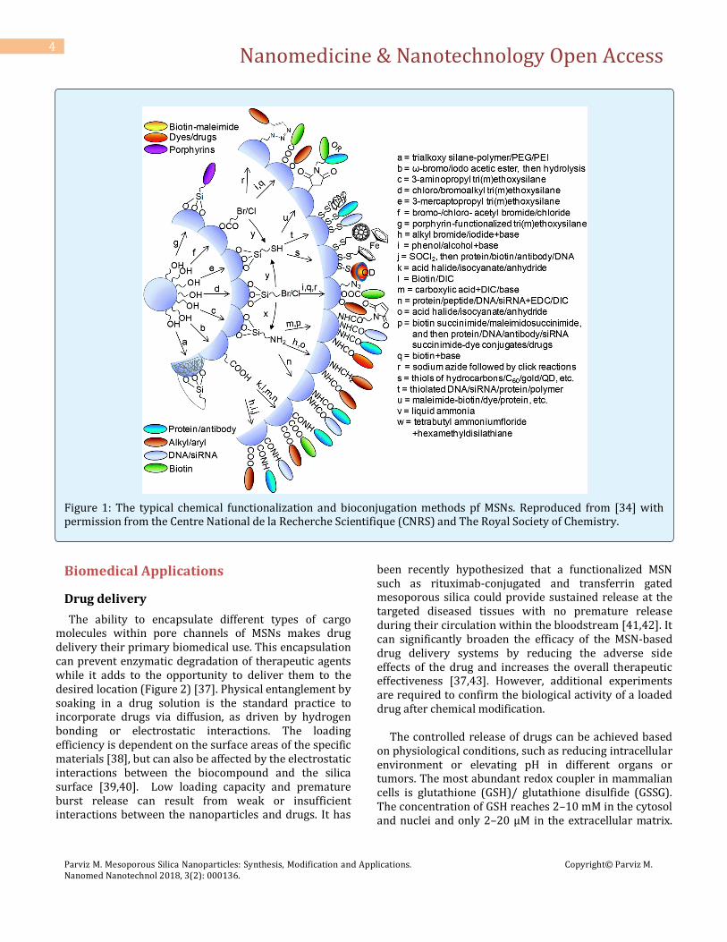
The presence of silanol groups on the surface of MSNs provides the ability to chemically modify the nanoparticles surface. Common biological and chemical groups that have been conjugated to MSNs are listed in Figure 1 [34]. The modified surface of nanoparticles plays a vital role in drug delivery applications. That surface modification of MSNs has been found to improve the loading of drugs, DNA, and siRNAs [35]. Surface modification using long-chain organic molecules such as oligomers and polymers can enhance the selectivity of nanoparticles. This can lead to beneficial or detrimental decreases in the pore size, the wettability of the pore surface by aqueous solutions, and biological activity of the encapsulated biocompound [36].
Nanomedicine & Nanotechnology Open Access
Parviz M. Mesoporous Silica Nanoparticles: Synthesis, Modification and Applications. Nanomed Nanotechnol 2018, 3(2): 000136.
Copyright© Parviz M.
4
Figure 1: The typical chemical functionalization and bioconjugation methods pf MSNs. Reproduced from [34] with permission from the Centre National de la Recherche Scientifique (CNRS) and The Royal Society of Chemistry.
Biomedical Applications
Drug delivery
The ability to encapsulate different types of cargo molecules within pore channels of MSNs makes drug delivery their primary biomedical use. This encapsulation can prevent enzymatic degradation of therapeutic agents while it adds to the opportunity to deliver them to the desired location (Figure 2) [37]. Physical entanglement by soaking in a drug solution is the standard practice to incorporate drugs via diffusion, as driven by hydrogen bonding or electrostatic interactions. The loading efficiency is dependent on the surface areas of the specific materials [38], but can also be affected by the electrostatic interactions between the biocompound and the silica surface [39,40]. Low loading capacity and premature burst release can result from weak or insufficient interactions between the nanoparticles and drugs. It has
been recently hypothesized that a functionalized MSN such as rituximab-conjugated and transferrin gated mesoporous silica could provide sustained release at the targeted diseased tissues with no premature release during their circulation within the bloodstream [41,42]. It can significantly broaden the efficacy of the MSN-based drug delivery systems by reducing the adverse side effects of the drug and increases the overall therapeutic effectiveness [37,43]. However, additional experiments are required to confirm the biological activity of a loaded drug after chemical modification. The controlled release of drugs can be achieved based on physiological conditions, such as reducing intracellular environment or elevating pH in different organs or tumors. The most abundant redox coupler in mammalian cells is glutathione (GSH)/ glutathione disulfide (GSSG). The concentration of GSH reaches 2–10 mM in the cytosol and nuclei and only 2–20 μM in the extracellular matrix.

Nanomedicine & Nanotechnology Open Access
Parviz M. Mesoporous Silica Nanoparticles: Synthesis, Modification and Applications. Nanomed Nanotechnol 2018, 3(2): 000136.
Copyright© Parviz M.
5
In vivo studies have indicated that tumors possess a GSH concentration of at least 4-fold higher than normal tissues. Small redox molecules like GSH or dithiothreitol (DTT) cleave disulfide bonds through thiol-disulfide exchange reactions. Based on that, reduction-sensitive materials containing disulfide groups have been used for controlled release of drugs [44]. Furthermore, the pH is considerably lower in organelles such as endosomes and lysosomes (pH 5.0–5.5) compared with blood and normal tissues (pH 7.4) [45]. Therefore, pH-sensitive bonds such as hydrolyzable Schiff base linkage can be designed to
cleave between drug and nanoparticles specifically in the acidic environment of endosome and lysosome, resulting in site-specific drug release [46]. The pH-sensitive organic layer can also protect the bioactive compound in digestive system with different pH values [47]. These polymers can be employed to coat MSNs pores to act as gatekeepers to avoid premature drug exposure (Figure 2). Ultimately, surface functionalization of MSNs will have many applications for targeted drug delivery, enabling customization of release, and tissue targeting, but these have yet to be fully optimized.
Figure 2: Schematic of prevention of enzymatic degradation and release of the drug in the desired place. Reproduced from [37] with permission from the Centre National de la Recherche Scientifique (CNRS) and The Royal Society of Chemistry.
Tissue Regeneration
Biomedical applications of MSNs are mainly focused on drug delivery and controlled release. However, the practical use of these nanoparticles in regenerative
medicine is still in its infancy [48]. MSNs have the potential to serve as vehicles to carry growth factors, peptides, or stem cells to a tissue engineered scaffold to enhance tissue regeneration. The mechanical and nanotopographical features of the scaffolds can also be
Nanomedicine & Nanotechnology Open Access
Parviz M. Mesoporous Silica Nanoparticles: Synthesis, Modification and Applications. Nanomed Nanotechnol 2018, 3(2): 000136.
Copyright© Parviz M.
6
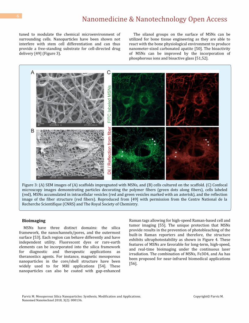
tuned to modulate the chemical microenvironment of surrounding cells. Nanoparticles have been shown not interfere with stem cell differentiation and can thus provide a free-standing substrate for cell-directed drug delivery [49] (Figure 3).
The silanol groups on the surface of MSNs can be utilized for bone tissue engineering as they are able to react with the bone physiological environment to produce nanometer-sized carbonated apatite [50]. The bioactivity of MSNs can be improved by the incorporation of phosphorous ions and bioactive glass [51,52].
Figure 3: (A) SEM images of (A) scaffolds impregnated with MSNs, and (B) cells cultured on the scaffold. (C) Confocal microscopy images demonstrating particles decorating the polymer fibers (green dots along fibers), cells labeled (red), MSNs accumulated in intracellular vesicles (red and green vesicles marked with an asterisk), and the reflection image of the fiber structure (red fibers). Reproduced from [49] with permission from the Centre National de la Recherche Scientifique (CNRS) and The Royal Society of Chemistry.
Bioimaging
MSNs have three distinct domains: the silica framework, the nanochannels/pores, and the outermost surface [53]. Each region can behave differently and have independent utility. Fluorescent dyes or rare-earth elements can be incorporated into the silica framework for diagnostic and therapeutic applications as theranostics agents. For instance, magnetic mesoporous nanoparticles in the core/shell structure have been widely used to for MRI applications [54]. These nanoparticles can also be coated with gap-enhanced
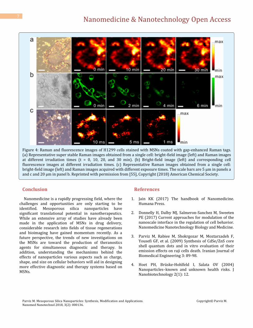
Raman tags allowing for high-speed Raman-based cell and tumor imaging [55]. The unique protection that MSNs provide results in the prevention of photobleaching of the built-in Raman reporters and therefore, the structure exhibits ultraphotostability as shown in Figure 4. These features of MSNs are favorable for long-term, high-speed, and real-time bioimaging under the continuous laser irradiation. The combination of MSNs, Fe3O4, and Au has been proposed for near-infrared biomedical applications [56].
Nanomedicine & Nanotechnology Open Access
Parviz M. Mesoporous Silica Nanoparticles: Synthesis, Modification and Applications. Nanomed Nanotechnol 2018, 3(2): 000136.
Copyright© Parviz M.
7
Figure 4: Raman and fluorescence images of H1299 cells stained with MSNs coated with gap-enhanced Raman tags. (a) Representative super stable Raman images obtained from a single cell: bright-field image (left) and Raman images at different irradiation times (t = 0, 10, 20, and 30 min). (b) Bright-field image (left) and corresponding cell fluorescence images at different irradiation times. (c) Representative Raman images obtained from a single cell: bright-field image (left) and Raman images acquired with different exposure times. The scale bars are 5 μm in panels a and c and 20 μm in panel b. Reprinted with permission from [55], Copyright (2018) American Chemical Society.
Conclusion
Nanomedicine is a rapidly progressing field, where the challenges and opportunities are only starting to be identified. Mesoporous silica nanoparticles have significant translational potential in nanotherapeutics. While an extensive array of studies have already been made in the application of MSNs in drug delivery, considerable research into fields of tissue regenerations and bioimaging have gained momentum recently. As a future perspective, the trends of new investigations on the MSNs are toward the production of theranostics agents for simultaneous diagnostic and therapy. In addition, understanding the mechanisms behind the effects of nanoparticles various aspects such as charge, shape, and size on cellular behaviors will aid in designing more effective diagnostic and therapy systems based on MSNs.
References
1. Jain KK (2017) The handbook of Nanomedicine. Humana Press.
2. Donnelly H, Dalby MJ, Salmeron-Sanchez M, Sweeten PE (2017) Current approaches for modulation of the nanoscale interface in the regulation of cell behavior. Nanomedicine Nanotechnology Biology and Medicine.
3. Parviz M, Rabiee M, Shokrgozar M, Moztarzadeh F, Yousefi GF, et al. (2009) Synthesis of CdSe/ZnS core shell quantum dots and in vitro evaluation of their emission effects on raji cells death. Iranian Journal of Biomedical Engineering 3: 89-98.
4. Hoet PH, Brüske-Hohlfeld I, Salata OV (2004) Nanoparticles–known and unknown health risks. J Nanobiotechnology 2(1): 12.
Nanomedicine & Nanotechnology Open Access
Parviz M. Mesoporous Silica Nanoparticles: Synthesis, Modification and Applications. Nanomed Nanotechnol 2018, 3(2): 000136.
Copyright© Parviz M.
8
5. Duong H, Chen Y, Tawfik SA, Wen S, Parviz M, et al. (2018) Systematic Investigation of Functional Ligands for Colloidal Stable Upconversion Nanoparticles. RSC Adv 8: 4842-4849.
6. Watermann A, Brieger J (2017) Mesoporous Silica Nanoparticles as Drug Delivery Vehicles in Cancer. Nanomaterials 7(7): 189.
7. Wang Z, Deng X, Ding J, Zhou W, Zheng X, et al. (2017) mechanisms of drug release in ph-sensitive micelles for tumour targeted drug delivery system: A review. Int J pharm 535(1-2): 253-260.
8. Wang Y, Zhao Q, Han N, Bai L, Li J, et al. (2015) Mesoporous silica nanoparticles in drug delivery and biomedical applications. Nanomedicine 11(2): 313-327.
9. Li Z, Barnes JC, Bosoy A, Stoddart JF, Zink JI (2012) Mesoporous silica nanoparticles in biomedical applications. Chem Soc Rev 41(7): 2590-2605.
10. Arcos D, Vallet-Regí M (2010) Sol–gel silica-based biomaterials and bone tissue regeneration. Acta Biomater 6(8): 2874-2888.
11. Ernawati L, Balgis R, Ogi T, Okuyama K (2017) Tunable Synthesis of Mesoporous Silica Particles with Unique Radially Oriented Pore Structures from Tetramethyl Orthosilicate via Oil–Water Emulsion Process. Langmuir 33(3): 783-790.
12. Ernawati L, Ogi T, Balgis R, Okuyama K, Stucki M, et al. (2015) Hollow silica as an optically transparent and thermally insulating polymer additive. Langmuir 32(1): 338-345.
13. Mekaru H, Lu J, Tamanoi F (2015) Development of mesoporous silica-based nanoparticles with controlled release capability for cancer therapy. Adv Drug Deliv Rev 95: 40-49.
14. Assefa D, Zera E, Campostrini R, Soraru GD, Vakifahmetoglu C (2016) Polymer-derived SiOC aerogel with hierarchical porosity through HF etching. Ceramics International 42(10): 11805-11809.
15. Tiwari D, Lee SM (2017) Chitosan templated synthesis of mesoporous silica and its application in the treatment of aqueous solutions contaminated with cadmium (II) and lead (II). Chemical Engineering Journal 328: 434-444.
16. Tang F, Li L, Chen D (2012) Mesoporous silica nanoparticles: synthesis, biocompatibility and drug delivery. Advanced materials 24(12): 1504-1534.
17. Hudson SP, Padera RF, Langer R, Kohane DS (2008) The biocompatibility of mesoporous silicates. Biomaterials 29(30): 4045-4055.
18. Feliu N, Fadeel B (2010) Nanotoxicology: no small matter. Nanoscale 2(12): 2514-2520.
19. He Q, Zhang Z, Gao F, Li Y, Shi J (2011) In vivo Biodistribution and Urinary Excretion of Mesoporous Silica Nanoparticles: Effects of Particle Size and PEGylation. Small 7(2): 271-280.
20. He Q, Zhang Z, Gao Y, Shi J, Li Y (2009) Intracellular Localization and Cytotoxicity of Spherical Mesoporous Silica Nano- and Microparticles. Small 5(23): 2722-2729.
21. Lin YS, Haynes CL (2010) Impacts of Mesoporous Silica Nanoparticle Size, Pore Ordering, and Pore Integrity on Hemolytic Activity. Journal of the American Chemical Society 132(13): 4834-4842.
22. Huang X, Li L, Liu T, Hao N, Liu H, et al. (2011) The shape effect of mesoporous silica nanoparticles on biodistribution, clearance, and biocompatibility in vivo. ACS nano 5(7): 5390-5399.
23. Trewyn BG, Nieweg JA, Zhao Y, Lin VSY (2008) Biocompatible mesoporous silica nanoparticles with different morphologies for animal cell membrane penetration. Chemical Engineering Journal 137(1): 23-29.
24. Nel AE, Mädler L, Velegol D, Xia T, Hoek EMV, et al. (2009) Understanding biophysicochemical interactions at the nano–bio interface. Nat Mater 8(7): 543-557.
25. Verma A, Stellacci F (2010) Effect of surface properties on nanoparticle–cell interactions. Small 6(1): 12-21.
26. Slowing II, Wu CW, Vivero‐Escoto JL, Lin VSY (2009) Mesoporous silica nanoparticles for reducing hemolytic activity towards mammalian red blood cells. Small 5(1): 57-62.
27. Meng H, Liong M, Xia T, Li Z, Ji Z, et al. (2010) Engineered design of mesoporous silica nanoparticles to deliver doxorubicin and P-glycoprotein siRNA to

Nanomedicine & Nanotechnology Open Access
Parviz M. Mesoporous Silica Nanoparticles: Synthesis, Modification and Applications. Nanomed Nanotechnol 2018, 3(2): 000136.
Copyright© Parviz M.
9
overcome drug resistance in a cancer cell line. ACS nano 4(8): 4539-4550.
28. Hom C, Lu J, Liong M, Luo H, Li Z, et al. (2010) Mesoporous silica nanoparticles facilitate delivery of siRNA to shutdown signaling pathways in mammalian cells. Small 6(11): 1185-1190.
29. Chen WH, Luo GF, Qiu WX, Lei Q, Liu LH, et al. (2017) Mesoporous silica-based versatile theranostic nanoplatform constructed by layer-by-layer assembly for excellent photodynamic/chemo therapy. Biomaterials 117: 54-65.
30. Manavitehrani I, Fathi A, Badr H, Daly S, Negahi Shirazi A, et al. (2016) Biomedical applications of biodegradable polyesters. Polymers 8(1): 20.
31. Xing L, Zheng H, Cao Y, Che S (2012) Coordination polymer coated mesoporous silica nanoparticles for pH‐responsive drug release. Adv Mater 24(48): 6433-6437.
32. Manavi‐Tehrani I, Rabiee M, Parviz M, Tahriri MR, Fahimi Z (2010) Preparation, Characterization and Controlled Release Investigation of Biocompatible pH‐Sensitive PVA/PAA Hydrogels in Macromolecular symposia. Wiley Online Library 296(1): 457-465.
33. Li QL, Sun Y, Sun YL, Wen J, Zhou Y, et al. (2014) Mesoporous silica nanoparticles coated by layer-by-layer self-assembly using cucurbit [7] uril for in vitro and in vivo anticancer drug release. Chemistry of Materials 26(22): 6418-6431.
34. Biju V (2014) Chemical modifications and bioconjugate reactions of nanomaterials for sensing, imaging, drug delivery and therapy. Chem Soc Rev 43(3): 744-764.
35. Blumen SR, Cheng K, Ramos-Nino ME, Taatjes DJ, Weiss DJ, et al. (2007) Unique uptake of acid-prepared mesoporous spheres by lung epithelial and mesothelioma cells. Am J Resp cell Mol Biol 36(3): 333-342.
36. Natarajan SK, Selvaraj S (2014) Mesoporous silica nanoparticles: importance of surface modifications and its role in drug delivery. RSC advances 4(28): 14328-14334.
37. Wen J, Yang K, Liu F, Li H, Xu Y, et al. (2017) Diverse gatekeepers for mesoporous silica nanoparticle based
drug delivery systems. Chem Soc Rev 46(19): 6024-6045.
38. Andersson J, Rosenholm J, Areva S, Lindén M (2004) Influences of Material Characteristics on Ibuprofen Drug Loading and Release Profiles from Ordered Micro- and Mesoporous Silica Matrices. Chem Mater 16(21): 4160-4167.
39. Meng H, Xue M, Xia T, Zhao YL, Tamanoi F, et al. (2010) Autonomous in Vitro Anticancer Drug Release from Mesoporous Silica Nanoparticles by pH-Sensitive Nanovalves. J Am Chem Soc 132(36): 12690-12697.
40. Chung TH, Wu SH, Yao M, Lu CW, Lin YS, et al. (2007) The effect of surface charge on the uptake and biological function of mesoporous silica nanoparticles in 3T3-L1 cells and human mesenchymal stem cells. Biomaterials 28(19): 2959-2966.
41. Zhou S, Wu D, Yin X, Jin X, Zhang X, et al. (2017) Intracellular pH-responsive and rituximab-conjugated mesoporous silica nanoparticles for targeted drug delivery to lymphoma B cells. Journal of Experimental & Clinical Cancer Research 36(1): 24.
42. Chen X, Sun H, Hu J, Han X, Liu H, et al. (2017) Transferrin gated mesoporous silica nanoparticles for redox-responsive and targeted drug delivery. Colloids Surf B Biointerfaces 152: 77-84.
43. Tian B, Liu S, Wu S, Lu W, Wang D, et al. (2017) pH-responsive poly (acrylic acid)-gated mesoporous silica and its application in oral colon targeted drug delivery for doxorubicin. Colloids Surf B Biointerfaces 154: 287-296.
44. Saito G, Swanson JA, Lee KD (2003) Drug delivery strategy utilizing conjugation via reversible disulfide linkages: role and site of cellular reducing activities. Advanced drug delivery reviews 55(2): 199-215.
45. Liu L, Liu P (2015) Synthesis strategies for disulfide bond-containing polymer-based drug delivery system for reduction-responsive controlled release. Front Mater Sci 9(3): 211-226.
46. Jang J, Cha C (2018) Multivalent Polyaspartamide Cross-Linker for Engineering Cell-Responsive Hydrogels with Degradation Behavior and Tunable Physical Properties. Biomacromolecules 19(2): 691-700.
Nanomedicine & Nanotechnology Open Access
Parviz M. Mesoporous Silica Nanoparticles: Synthesis, Modification and Applications. Nanomed Nanotechnol 2018, 3(2): 000136.
Copyright© Parviz M.
10
47. Fahimi Z, Parviz M, Orang F, Bonakdar S (2008) Synthesis of pH Sensitive Hydrogels Based on Poly Vinyl Alcohol and Poly Acrylic Acid. Iranian Journal of Pharmaceutical Sciences 4(4): 275-280.
48. Rosenholm JM, Zhang J, Linden M, Sahlgren C (2016) Mesoporous silica nanoparticles in tissue engineering – a perspective. Nanomedicine 11(4): 391-402.
49. Böcking D, Wiltschka O, Niinimäki J, Shokry H, Brenner R, et al. (2014) Mesoporous silica nanoparticle-based substrates for cell directed delivery of Notch signalling modulators to control myoblast differentiation. Nanoscale 6(3): 1490-1498.
50. Vallet‐Regí M (2006) Ordered mesoporous materials in the context of drug delivery systems and bone tissue engineering. Chemistry-A European Journal 12(23): 5934-5943.
51. Vallet-Regí M, Izquierdo-Barba I, Rámila A, Pérez-Pariente J, Babonneau F, et al. (2005) Phosphorous-doped MCM-41 as bioactive material. Solid State Sciences 7(2): 233-237.
52. Horcajada P, Rámila A, Boulahya K, González-Calbet J, Vallet-Regí M (2004) Bioactivity in ordered
mesoporous materials. Solid State Sciences 6(11): 1295-1300.
53. Chen NT, Cheng SH, Souris JS, Chen CT, Mou CY, et al. (2013) Theranostic applications of mesoporous silica nanoparticles and their organic/inorganic hybrids. Journal Mater Chem B 1: 3128-3135.
54. Wang Y, Gu H (2015) Core–Shell‐Type Magnetic Mesoporous Silica Nanocomposites for Bioimaging and Therapeutic Agent Delivery. Advanced materials 27(3): 576-585.
55. Zhang Y, Qiu Y, Lin L, Gu H, Xiao Z, et al. (2017) Ultraphotostable Mesoporous Silica-Coated Gap-Enhanced Raman Tags (GERTs) for High-Speed Bioimaging. ACS Appl Mater Interfaces 9(4): 3995-4005.
56. Khosroshahi M, Tehrani IM, Nouri A (2018) Fabrication and Characterization of Multilayer mSiO2@Fe3O4@Au Mesoporous Nanocomposite For Near-Infrared Biomedical Applications. Adv Nano Bio M&D 2(1): 230-246.