Merosin-negative congenital muscular dystrophy: magnetic resonance spectroscopy findings
3
Case report Merosin-negative congenital muscular dystrophy: magnetic resonance spectroscopy findings Mehmet Aslan a, * , Alpay Alkan b , Cengiz Yakinci a , Ergu ¨n Sonmezgoz a , Ug ˘ur Bicak a , Suzan Zorludemir c a Department of Pediatrics, Inonu University, Turgut O ¨ zal Medical Center, School of Medicine, Malatya 44069, Turkey b Department of Radiology, Inonu University, Turgut O ¨ zal Medical Center, Malatya, Turkey c Department of Pathology, C ¸ ukurova University School of Medicine, Adana, Turkey Received 28 July 2003; received in revised form 9 July 2004; accepted 3 August 2004 Abstract Congenital muscular dystrophies (CMD) are heterogenous group of muscle disorders with autosomal recessive inheritance. Merosin deficiency has been identified in some patients with CMD all of whom also had white matter abnormalities on MRI. In postmortem studies, the brain showed extensive myelin pallor with a spongy appearance of white matter and moderate astrocytosis or demyelination. Direct assessment of neuropathologic aspects of MN-CMD such as demyelination is possible with MR spectroscopy (MRS). Although previous reports have described several neuro-imaging findings of this disease, MRS findings have not been reported in literature. In this case, we report MRS features of a 4-year old girl with MN-CMD. MRS of brain demonstrated that N-acetylaspartate (NAA)/Creatine (Cr) ratio was normal. Increased Choline (Cho)/Cr and Myo-inositol (MI)/Cr ratios were obtained. These findings were interpreted as demyelination and gliosis of white matter. q 2004 Elsevier B.V. All rights reserved. Keywords: Magnetic resonance spectroscopy; Congenital muscular dystrophy 1. Introduction A primary deficiency of merosin caused by mutations of the laminin a2 chain gene produces MN-CMD. Conventional MRI findings of the white matter changes have been interpreted as leukodystrophy-like demyelina- tion, arrest in myelination, or dysfunction of blood brain barrier [1,2]. Therefore, MRS study seems to be important to clarify the nature of this abnormality. Demyelination and astrocytosis are the most important features of neurological involvement of this disease [3–5]. MRS provides valuable information regarding in vivo brain metabolism and neuronal functions. It is a promising method that may illustrate the pathophysiologic events for MN-CMD. Although it is not specific or diagnostic for MN-CMD, MRS could show substantial metabolic impairments in the white matter, especially in the early stages. We performed single voxel proton MRS in a 4-year old girl with proven diagnosis of MN-CMD and white matter involvement. To our knowledge, this is the first report of proton MRS features of MN-CMD. 2. Case report A 4-year old girl was admitted to our clinic due to muscle weakness. Her first admission to the hospital was at 23 months at which time hypotonia and delay in develop- ment were present. On physical examination, her weight was 10 kg (!3 percentile) and length was 88 cm (!3 percentile). Motor activity was decreased and sym- metric muscular weakness was detected. Deep tendon Brain & Development 27 (2005) 308–310 www.elsevier.com/locate/braindev 0387-7604/$ - see front matter q 2004 Elsevier B.V. All rights reserved. doi:10.1016/j.braindev.2004.08.004 * Corresponding author. Tel.: C90 422 341 06 60x5304; fax: C90 422 3410 728 29. E-mail address: [email protected] (M. Aslan).
-
Upload
mehmet-aslan -
Category
Documents
-
view
217 -
download
1
Transcript of Merosin-negative congenital muscular dystrophy: magnetic resonance spectroscopy findings
Case report
Merosin-negative congenital muscular dystrophy: magnetic
resonance spectroscopy findings
Mehmet Aslana,*, Alpay Alkanb, Cengiz Yakincia, Ergun Sonmezgoza,Ugur Bicaka, Suzan Zorludemirc
aDepartment of Pediatrics, Inonu University, Turgut Ozal Medical Center, School of Medicine, Malatya 44069, TurkeybDepartment of Radiology, Inonu University, Turgut Ozal Medical Center, Malatya, Turkey
cDepartment of Pathology, Cukurova University School of Medicine, Adana, Turkey
Received 28 July 2003; received in revised form 9 July 2004; accepted 3 August 2004
Abstract
Congenital muscular dystrophies (CMD) are heterogenous group of muscle disorders with autosomal recessive inheritance. Merosin
deficiency has been identified in some patients with CMD all of whom also had white matter abnormalities on MRI. In postmortem studies,
the brain showed extensive myelin pallor with a spongy appearance of white matter and moderate astrocytosis or demyelination. Direct
assessment of neuropathologic aspects of MN-CMD such as demyelination is possible with MR spectroscopy (MRS). Although previous
reports have described several neuro-imaging findings of this disease, MRS findings have not been reported in literature. In this case, we
report MRS features of a 4-year old girl with MN-CMD. MRS of brain demonstrated that N-acetylaspartate (NAA)/Creatine (Cr) ratio was
normal. Increased Choline (Cho)/Cr and Myo-inositol (MI)/Cr ratios were obtained. These findings were interpreted as demyelination and
gliosis of white matter.
q 2004 Elsevier B.V. All rights reserved.
Keywords: Magnetic resonance spectroscopy; Congenital muscular dystrophy
1. Introduction
A primary deficiency of merosin caused by mutations of
the laminin a2 chain gene produces MN-CMD.
Conventional MRI findings of the white matter changes
have been interpreted as leukodystrophy-like demyelina-
tion, arrest in myelination, or dysfunction of blood brain
barrier [1,2]. Therefore, MRS study seems to be important
to clarify the nature of this abnormality. Demyelination and
astrocytosis are the most important features of neurological
involvement of this disease [3–5]. MRS provides valuable
information regarding in vivo brain metabolism and
neuronal functions. It is a promising method that may
illustrate the pathophysiologic events for MN-CMD.
0387-7604/$ - see front matter q 2004 Elsevier B.V. All rights reserved.
doi:10.1016/j.braindev.2004.08.004
* Corresponding author. Tel.: C90 422 341 06 60x5304; fax: C90 422
3410 728 29.
E-mail address: [email protected] (M. Aslan).
Although it is not specific or diagnostic for MN-CMD,
MRS could show substantial metabolic impairments in the
white matter, especially in the early stages. We performed
single voxel proton MRS in a 4-year old girl with proven
diagnosis of MN-CMD and white matter involvement. To
our knowledge, this is the first report of proton MRS
features of MN-CMD.
2. Case report
A 4-year old girl was admitted to our clinic due to muscle
weakness. Her first admission to the hospital was at
23 months at which time hypotonia and delay in develop-
ment were present. On physical examination, her
weight was 10 kg (!3 percentile) and length was 88 cm
(!3 percentile). Motor activity was decreased and sym-
metric muscular weakness was detected. Deep tendon
Brain & Development 27 (2005) 308–310
www.elsevier.com/locate/braindev
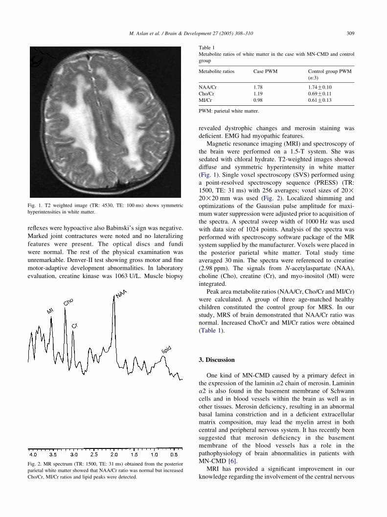
Fig. 1. T2 weighted image (TR: 4530, TE: 100 ms) shows symmetric
hyperintensities in white matter.
Table 1
Metabolite ratios of white matter in the case with MN-CMD and control
group
Metabolite ratios Case PWM Control group PWM
(n:3)
NAA/Cr 1.78 1.74G0.10
Cho/Cr 1.19 0.69G0.11
MI/Cr 0.98 0.61G0.13
PWM: parietal white matter.
M. Aslan et al. / Brain & Development 27 (2005) 308–310 309
reflexes were hypoactive also Babinski’s sign was negative.
Marked joint contractures were noted and no lateralizing
features were present. The optical discs and fundi
were normal. The rest of the physical examination was
unremarkable. Denver-II test showing gross motor and fine
motor-adaptive development abnormalities. In laboratory
evaluation, creatine kinase was 1063 U/L. Muscle biopsy
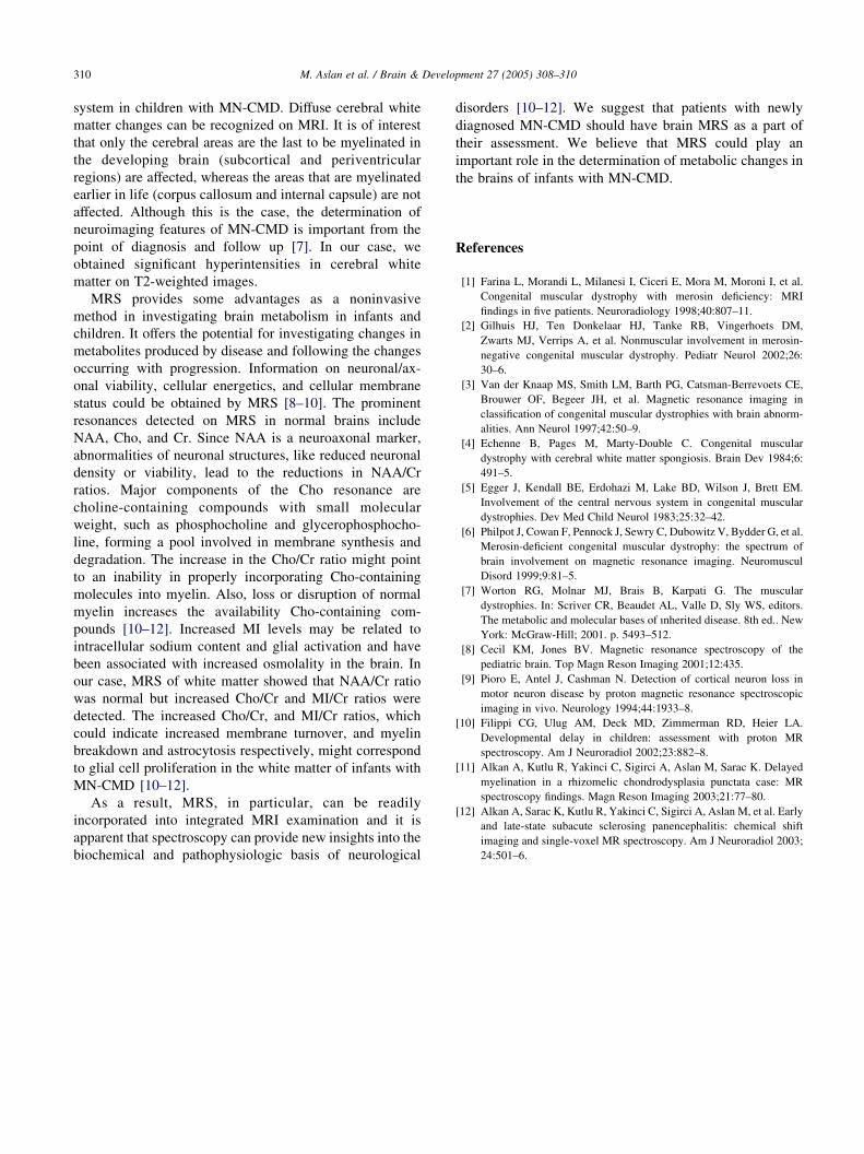
Fig. 2. MR spectrum (TR: 1500, TE: 31 ms) obtained from the posterior
parietal white matter showed that NAA/Cr ratio was normal but increased
Cho/Cr, MI/Cr ratios and lipid peaks were detected.
revealed dystrophic changes and merosin staining was
deficient. EMG had myopathic features.
Magnetic resonance imaging (MRI) and spectroscopy of
the brain were performed on a 1.5-T system. She was
sedated with chloral hydrate. T2-weighted images showed
diffuse and symmetric hyperintensity in white matter
(Fig. 1). Single voxel spectroscopy (SVS) performed using
a point-resolved spectroscopy sequence (PRESS) (TR:
1500, TE: 31 ms) with 256 averages; voxel sizes of 20!20!20 mm was used (Fig. 2). Localized shimming and
optimizations of the Gaussian pulse amplitude for maxi-
mum water suppression were adjusted prior to acquisition of
the spectra. A spectral sweep width of 1000 Hz was used
with data size of 1024 points. Analysis of the spectra was
performed with spectroscopy software package of the MR
system supplied by the manufacturer. Voxels were placed in
the posterior parietal white matter. Total study time
averaged 30 min. The spectra were referenced to creatine
(2.98 ppm). The signals from N-acetylaspartate (NAA),
choline (Cho), creatine (Cr), and myo-inositol (MI) were
integrated.
Peak area metabolite ratios (NAA/Cr, Cho/Cr and MI/Cr)
were calculated. A group of three age-matched healthy
children constituted the control group for MRS. In our
study, MRS of brain demonstrated that NAA/Cr ratio was
normal. Increased Cho/Cr and MI/Cr ratios were obtained
(Table 1).
3. Discussion
One kind of MN-CMD caused by a primary defect in
the expression of the laminin a2 chain of merosin. Laminin
a2 is also found in the basement membrane of Schwann
cells and in blood vessels within the brain as well as in
other tissues. Merosin deficiency, resulting in an abnormal
basal lamina constriction and in a deficient extracellular
matrix composition, may lead the myelin arrest in both
central and peripheral nervous system. It has recently been
suggested that merosin deficiency in the basement
membrane of the blood vessels has a role in the
pathophysiology of brain abnormalities in patients with
MN-CMD [6].
MRI has provided a significant improvement in our
knowledge regarding the involvement of the central nervous
M. Aslan et al. / Brain & Development 27 (2005) 308–310310
system in children with MN-CMD. Diffuse cerebral white
matter changes can be recognized on MRI. It is of interest
that only the cerebral areas are the last to be myelinated in
the developing brain (subcortical and periventricular
regions) are affected, whereas the areas that are myelinated
earlier in life (corpus callosum and internal capsule) are not
affected. Although this is the case, the determination of
neuroimaging features of MN-CMD is important from the
point of diagnosis and follow up [7]. In our case, we
obtained significant hyperintensities in cerebral white
matter on T2-weighted images.
MRS provides some advantages as a noninvasive
method in investigating brain metabolism in infants and
children. It offers the potential for investigating changes in
metabolites produced by disease and following the changes
occurring with progression. Information on neuronal/ax-
onal viability, cellular energetics, and cellular membrane
status could be obtained by MRS [8–10]. The prominent
resonances detected on MRS in normal brains include
NAA, Cho, and Cr. Since NAA is a neuroaxonal marker,
abnormalities of neuronal structures, like reduced neuronal
density or viability, lead to the reductions in NAA/Cr
ratios. Major components of the Cho resonance are
choline-containing compounds with small molecular
weight, such as phosphocholine and glycerophosphocho-
line, forming a pool involved in membrane synthesis and
degradation. The increase in the Cho/Cr ratio might point
to an inability in properly incorporating Cho-containing
molecules into myelin. Also, loss or disruption of normal
myelin increases the availability Cho-containing com-
pounds [10–12]. Increased MI levels may be related to
intracellular sodium content and glial activation and have
been associated with increased osmolality in the brain. In
our case, MRS of white matter showed that NAA/Cr ratio
was normal but increased Cho/Cr and MI/Cr ratios were
detected. The increased Cho/Cr, and MI/Cr ratios, which
could indicate increased membrane turnover, and myelin
breakdown and astrocytosis respectively, might correspond
to glial cell proliferation in the white matter of infants with
MN-CMD [10–12].
As a result, MRS, in particular, can be readily
incorporated into integrated MRI examination and it is
apparent that spectroscopy can provide new insights into the
biochemical and pathophysiologic basis of neurological
disorders [10–12]. We suggest that patients with newly
diagnosed MN-CMD should have brain MRS as a part of
their assessment. We believe that MRS could play an
important role in the determination of metabolic changes in
the brains of infants with MN-CMD.
References
[1] Farina L, Morandi L, Milanesi I, Ciceri E, Mora M, Moroni I, et al.
Congenital muscular dystrophy with merosin deficiency: MRI
findings in five patients. Neuroradiology 1998;40:807–11.
[2] Gilhuis HJ, Ten Donkelaar HJ, Tanke RB, Vingerhoets DM,
Zwarts MJ, Verrips A, et al. Nonmuscular involvement in merosin-
negative congenital muscular dystrophy. Pediatr Neurol 2002;26:
30–6.
[3] Van der Knaap MS, Smith LM, Barth PG, Catsman-Berrevoets CE,
Brouwer OF, Begeer JH, et al. Magnetic resonance imaging in
classification of congenital muscular dystrophies with brain abnorm-
alities. Ann Neurol 1997;42:50–9.
[4] Echenne B, Pages M, Marty-Double C. Congenital muscular
dystrophy with cerebral white matter spongiosis. Brain Dev 1984;6:
491–5.
[5] Egger J, Kendall BE, Erdohazi M, Lake BD, Wilson J, Brett EM.
Involvement of the central nervous system in congenital muscular
dystrophies. Dev Med Child Neurol 1983;25:32–42.
[6] Philpot J, Cowan F, Pennock J, Sewry C, Dubowitz V, Bydder G, et al.
Merosin-deficient congenital muscular dystrophy: the spectrum of
brain involvement on magnetic resonance imaging. Neuromuscul
Disord 1999;9:81–5.
[7] Worton RG, Molnar MJ, Brais B, Karpati G. The muscular
dystrophies. In: Scriver CR, Beaudet AL, Valle D, Sly WS, editors.
The metabolic and molecular bases of ınherited disease. 8th ed.. New
York: McGraw-Hill; 2001. p. 5493–512.
[8] Cecil KM, Jones BV. Magnetic resonance spectroscopy of the
pediatric brain. Top Magn Reson Imaging 2001;12:435.
[9] Pioro E, Antel J, Cashman N. Detection of cortical neuron loss in
motor neuron disease by proton magnetic resonance spectroscopic
imaging in vivo. Neurology 1994;44:1933–8.
[10] Filippi CG, Ulug AM, Deck MD, Zimmerman RD, Heier LA.
Developmental delay in children: assessment with proton MR
spectroscopy. Am J Neuroradiol 2002;23:882–8.
[11] Alkan A, Kutlu R, Yakinci C, Sigirci A, Aslan M, Sarac K. Delayed
myelination in a rhizomelic chondrodysplasia punctata case: MR
spectroscopy findings. Magn Reson Imaging 2003;21:77–80.
[12] Alkan A, Sarac K, Kutlu R, Yakinci C, Sigirci A, Aslan M, et al. Early
and late-state subacute sclerosing panencephalitis: chemical shift
imaging and single-voxel MR spectroscopy. Am J Neuroradiol 2003;
24:501–6.