Menadione effect on l-cysteine desulfuration in U373 cells · Menadione effect on l-cysteine...
5
Communication Menadione effect on l-cysteine desulfuration in U373 cells Maria Wróbel and Halina Jurkowska Chair of Medical Biochemistry, Jagiellonian University Medical College, Kraków, Poland Received: 13 March, 2007; revised: 30 March, 2007; accepted: 16 April, 2007 available on-line: 23 May, 2007 The non-cytotoxic concentration (20 µM) of menadione (2-methyl-1,4-naphthoquinone), aſter 1 h of incubation, leads to loss of the activity of rhodanese by 33%, 3-mercaptopyruvate sulfurtrans- ferase by 20%, as well as the level of sulfane sulfur by about 23% and glutathione by 12%, in the culture of U373 cells, in comparison with the control culture. Reactive oxygen species generated by menadione oxidize sulydryl groups in active centers of the investigated enzymes, inhibiting them and saving cysteine for glutathione synthesis. A decreased sulfane sulfur level can be cor- related with an oxidative stress. Keywords: astrocytoma U373, menadione, 3-mercaptopyruvate sulfurtransferase, reactive oxygen species, rhodanese, sulfane sulfur INTRODUCTION 3-Mercaptopyruvate sulfurtransferase (MPST, EC 2.8.1.2), one of two enzymes generating sul- fane sulfur-containing compounds from l-cysteine (Scheme 1), plays the main role in astrocytoma U373 cells because of the very low levels of γ-cys- tathionase (cystathionine γ-lyase, EC 4.4.1.1) activity (Jurkowska & Wróbel, 2007). Rhodanese (thiosulfate sulfurtransferase, EC 2.8.1.1) is a well-known enzyme participating in transferring labile sulfur atoms from various sulfane sulfur-containing compounds to var- ious acceptors (Westley et al., 1983). Sulfane sulfur atoms are provided for the synthesis of iron sulfur proteins and for detoxification of such compounds as, for example, cyanide (Westley, 1973; 1980; Bein- ert, 2000; Liew & Shaw, 2005). The antioxidant po- tential of sulfane sulfur and rhodanese has also been demonstrated (Ogasawara et al., 1999; Nandi et al., 2000; Wróbel et al., 2004). MPST and rhodanese are members of the same family (Nagahara, 1995). The catalytic site of both enzymes contains a Cys247 residue, which is proposed to form persulfide in the process of cataly- sis (Nagahara & Nishino, 1996). Enzymes contain- ing catalytic cysteines are generally inhibited by hy- drogen peroxide (Cannella & Berni, 1983; Lee et al., 1998; Skorey et al., 1997). Under these oxidizing con- ditions, formation of a sulfenate (–SO – ), a sulfinate (–SO 2 – ), a sulfonate (–SO 3 – ), a sulfenyl compound (–S-R), or a disulfide bond has been characterized (Poole & Claiborne 1989; Claiborne et al., 1999). Mild oxidation of MPST was found to result in formation of a stable sulfenate (-SO – ) at Cys247, which exhibits lower redox potential than that of glutathione (Na- gahara & Katayama, 2005). Oxidative stress decreas- es the MPST activity so as to increase the amount of cysteine (Scheme 1), a precursor of glutathione, and furthermore, these cellular reductants restore this ac- tivity. Thus, the redox state regulates the MPST ac- tivity at the enzymatic level, and on the other hand, MPST helps maintain cellular redox homeostasis. The cytotoxic effects of menadione (vitamin K-3, 2-methyl-1,4-naphthoquinone) are thought to be mediated through its one-electron reduction to semi- quinone radicals, which subsequently enter redox Corresponding author: M. Wróbel, Jagiellonian University Medical College, Chair of Medical Biochemistry, M. Koperni- ka 7, 31-034 Kraków, Poland; phone: (48 12) 424 7229; fax: (48 12) 422 3272; e-mail: [email protected] Abbreviations: DMEM, Dulbecco’s modified eagle medium; DTNB, 5,5’-dithio-bis-(2-nitrobenzoic acid); DTT, dl-dithio- threitol; LDH, lactate dehydrogenase; MPST, 3-mercaptopyruvate sulfurtransferase; NEM, N-ethylmaleimide; PBS, potas- sium phosphate buffer containing 150 mM NaCl; PLP, pyridoxal phosphate; ROS, reactive oxygen species. Vol. 54 No. 2/2007, 407–411 on-line at: www.actabp.pl
Transcript of Menadione effect on l-cysteine desulfuration in U373 cells · Menadione effect on l-cysteine...
Communication
Menadione effect on l-cysteine desulfuration in U373 cells
Maria Wróbel and Halina Jurkowska
Chair of Medical Biochemistry, Jagiellonian University Medical College, Kraków, Poland
Received: 13 March, 2007; revised: 30 March, 2007; accepted: 16 April, 2007 available on-line: 23 May, 2007
The non-cytotoxic concentration (20 µM) of menadione (2-methyl-1,4-naphthoquinone), after 1 h of incubation, leads to loss of the activity of rhodanese by 33%, 3-mercaptopyruvate sulfurtrans-ferase by 20%, as well as the level of sulfane sulfur by about 23% and glutathione by 12%, in the culture of U373 cells, in comparison with the control culture. Reactive oxygen species generated by menadione oxidize sulfhydryl groups in active centers of the investigated enzymes, inhibiting them and saving cysteine for glutathione synthesis. A decreased sulfane sulfur level can be cor-
related with an oxidative stress.
Keywords: astrocytoma U373, menadione, 3-mercaptopyruvate sulfurtransferase, reactive oxygen species, rhodanese, sulfane sulfur
INTRODUCTION
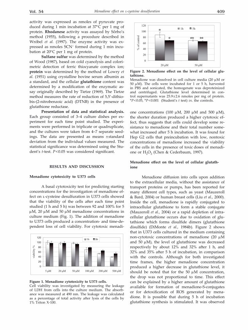
3-Mercaptopyruvate sulfurtransferase (MPST, EC 2.8.1.2), one of two enzymes generating sul-fane sulfur-containing compounds from l-cysteine (Scheme 1), plays the main role in astrocytoma U373 cells because of the very low levels of γ-cys-tathionase (cystathionine γ-lyase, EC 4.4.1.1) activity (Jurkowska & Wróbel, 2007). Rhodanese (thiosulfate sulfurtransferase, EC 2.8.1.1) is a well-known enzyme participating in transferring labile sulfur atoms from various sulfane sulfur-containing compounds to var-ious acceptors (Westley et al., 1983). Sulfane sulfur atoms are provided for the synthesis of iron sulfur proteins and for detoxification of such compounds as, for example, cyanide (Westley, 1973; 1980; Bein-ert, 2000; Liew & Shaw, 2005). The antioxidant po-tential of sulfane sulfur and rhodanese has also been demonstrated (Ogasawara et al., 1999; Nandi et al., 2000; Wróbel et al., 2004).
MPST and rhodanese are members of the same family (Nagahara, 1995). The catalytic site of both enzymes contains a Cys247 residue, which is
proposed to form persulfide in the process of cataly-sis (Nagahara & Nishino, 1996). Enzymes contain-ing catalytic cysteines are generally inhibited by hy-drogen peroxide (Cannella & Berni, 1983; Lee et al., 1998; Skorey et al., 1997). Under these oxidizing con-ditions, formation of a sulfenate (–SO–), a sulfinate (–SO2
–), a sulfonate (–SO3–), a sulfenyl compound
(–S-R), or a disulfide bond has been characterized (Poole & Claiborne 1989; Claiborne et al., 1999). Mild oxidation of MPST was found to result in formation of a stable sulfenate (-SO–) at Cys247, which exhibits lower redox potential than that of glutathione (Na-gahara & Katayama, 2005). Oxidative stress decreas-es the MPST activity so as to increase the amount of cysteine (Scheme 1), a precursor of glutathione, and furthermore, these cellular reductants restore this ac-tivity. Thus, the redox state regulates the MPST ac-tivity at the enzymatic level, and on the other hand, MPST helps maintain cellular redox homeostasis.
The cytotoxic effects of menadione (vitamin K-3, 2-methyl-1,4-naphthoquinone) are thought to be mediated through its one-electron reduction to semi-quinone radicals, which subsequently enter redox
Corresponding author: M. Wróbel, Jagiellonian University Medical College, Chair of Medical Biochemistry, M. Koperni-ka 7, 31-034 Kraków, Poland; phone: (48 12) 424 7229; fax: (48 12) 422 3272; e-mail: [email protected]: DMEM, Dulbecco’s modified eagle medium; DTNB, 5,5’-dithio-bis-(2-nitrobenzoic acid); DTT, dl-dithio-threitol; LDH, lactate dehydrogenase; MPST, 3-mercaptopyruvate sulfurtransferase; NEM, N-ethylmaleimide; PBS, potas-sium phosphate buffer containing 150 mM NaCl; PLP, pyridoxal phosphate; ROS, reactive oxygen species.
Vol. 54 No. 2/2007, 407–411
on-line at: www.actabp.pl
408 2007M. Wróbel and H. Jurkowska
cycles with molecular oxygen to produce reactive oxygen species and oxidative stress. The cytotoxicity of menadione seems to be associated with superox-ide generation, protein thiol oxidation, and altera-tion in the Ca2+ homeostasis (DiMonte et al., 1984a; 1984b; Niemczyk et al., 2004). Menadione may cause toxicity due to depletion of glutathione by conjugate formation (2-methyl-3-glutathionyl-1,4-naphthoqui-none is a product of direct reaction of glutathione
with menadione) (Wefers & Sies, 1983; Mauzeroll & Bard, 2004).
In this study, we provide evidence that both investigated sulfurtransferases, rhodanese and MPST, have lower activity in U373 cells in the pres-ence of menadione in the culture medium, probably due to oxidation of the –SH group of their catalytic cysteines. In effect, cysteine conversion to sulfane sulfur compounds is slower and the amount of cysteine available for the synthesis of glutathione is higher (Scheme 1).
MATERIALS AND METHODS
Sources of chemicals. Folin-Ciocalteau rea-gent, NADPH (Na4), NADH, 5,5’-dithio-bis-(2-ni-trobenzoic acid) (DTNB), glutathione reductase, lactate dehydrogenase (LDH), pyridoxal phosphate (PLP), dl-dithiothreitol (DTT), N-ethylmaleimide (NEM), trypsin, menadione sodium bisulfite, DMEM (Dulbecco’s modified Eagle medium) were obtained from Sigma Chemical Company (St. Louis, MO, USA). Fetal bovine serum was obtained from Gibco Laboratories (Grand Island, NY, USA), potassium cy-anide from Merck (Darmstadt, Germany), sodium 3-mercaptopyruvate from Flucka Chemie GmbH. The Cytotoxicity Detection Kit (LDH) and Cell Prolifera-
tion ELISA, BrdU (colorimetric) test were obtained from Roche Applied Science. All other chemicals were of reagent grade and purchased from common commercial suppliers.
Cell culture. All experiments were performed using the human astrocytoma U373 cell line. U373 cells were grown in monolayer in DMEM supple-mented with 10% fetal bovine serum and antibiotics
(100 U/ml penicillin and 100 µg/ml streptomycin), in plastic culture dishes (100 mm in diameter), at 37oC, in a humidified atmosphere containing 5% CO2. Af-ter trypsinization (0.25% trypsin/EDTA), the cells were diluted with complete medium (DMEM with 10% FBS) (Ponten & Macintyre, 1968).
Treatment of cells with menadione. The cells were seeded at 1 mln/100 mm dish the day before treatment. Menadione (5–50 µM) was dissolved in cell culture media and filtered through a 0.22-μm filter for sterilization. The cells were incubated for 1 or 5 h and then washed three times with 3 ml of PBS (10 mM potassium phosphate buffer, pH 7.4, containing 150 mM NaCl). Then the cells were har-vested in cold PBS, centrifuged at 5 000 r.p.m. at 4°C during 10 min and solubilized in phosphate buffer, pH 7.5 for homogenization.
Determination of cell viability. Cell viability was investigated by measuring the leakage of LDH from dead or dying cells into the culture medium using the Cytotoxicity Detection Kit. Briefly, the cells were cultured in 96-well plates at a density of 1.5–2 × 10³ U373 cells per well. After treatment, LDH was determined spectrophotometrically by measur-ing its activity in an aliquot of cell-free medium. One hundred microliters of the culture medium collected from the dishes after the incubation of the cells were added to 100 µl of the incubation mixture and incu-bated for 30 min at room temperature, in the dark. After the incubation, 50 μl of 1 M HCl was added and the absorbance was measured with a microcul-ture plate reader at 490 nm. The leakage was calcu-lated as the percentage of total activity after lysis of the cells by 1% Triton X-100.
Cell homogenization. U373 cells (2–6 × 10⁶) were suspended in 0.1 M phosphate buffer pH 7.5, in the proportion 1 mln cells/0.04 ml of the buff-er, sonicated 3 × 5 s at 4°C (Bandelin Sonoplus GM 70). After centrifugation at 3 000 r.p.m. for 10 min, the supernatant was used for the determination of protein concentration, sulfane sulfur levels and the activity of rhodanese and MPST. For glutathione de-terminations, the homogenate was deproteinized by adding 5% trichloroacetic acid in the proportion 1:1 and centrifuged for 10 min at 3 000 r.p.m.
Enzyme assays. MPST activity was assayed according to the method of Valentine and Fran-kelfeld (1974), following a procedure described in our earlier paper (Wróbel et al., 1997). The enzyme
CYSTEINE GSH
Cystine
sulfane sulfur
3-mercaptopyruvate
Pyruvate
Protein
H2S CN-
SCN-MPST
aminotransferase
acceptorsulfurated acceptor
Rhodanese
cystathionase
Scheme 1. Sulfane sulfur formation from l-cysteine.In U373 cells, MPST playes the main role in generating sulfane sulfur-containing compounds from l-cysteine be-cause of trace activity of γ-cystathionase (dotted line).
Vol. 54 409Menadione effect on l-cysteine desulfuration
activity was expressed as nmoles of pyruvate pro-duced during 1 min incubation at 37°C per 1 mg of protein. Rhodanese activity was assayed by Sőrbo’s method (1955), following a procedure described in Wróbel et al. (1997). The enzyme activity was ex-pressed as nmoles SCN– formed during 1 min incu-bation at 20°C per 1 mg of protein.
Sulfane sulfur was determined by the method of Wood (1987), based on cold cyanolysis and colori-metric detection of ferric thiocyanate complex ion; protein was determined by the method of Lowry et al. (1951) using crystalline bovine serum albumin as a standard, and the cellular glutathione content was determined by a modification of the enzymatic as-say originally described by Tietze (1969). The Tietze method measures the rate of reduction of 5,5’-dithio-bis-(2-nitrobenzoic acid) (DTNB) in the presence of glutathione reductase.
Presentation of data and statistical analysis. Each group consisted of 3–4 culture dishes per ex-periment for each time point studied. The experi-ments were performed in triplicate or quadruplicate, and the cultures were taken from 4–7 separate seed-ings. The data are presented as means ± standard deviation from the individual values measured. The statistical significance was determined using the Stu-dent’s t-test. P < 0.05 was considered significant.
RESULTS AND DISCUSSION
Menadione cytotoxicity to U373 cells
A basal cytotoxicity test for predicting starting concentrations for the investigation of menadione ef-fect on l-cysteine desulfuration in U373 cells showed that the viability of the cells after each time point studied (1 h and 5 h) was between 92 and 100% for 5 µM, 20 µM and 50 µM menadione concentrations in culture medium (Fig. 1). The addition of menadione to U373 cells produced a concentration- and time-de-pendent loss of cell viability. For cytotoxic menadi-
one concentrations (100 µM, 200 µM and 500 µM), the shorter duration produced a higher cytotoxic ef-fect, thus suggests that cells could develop some re-sistance to menadione and their total number some-what increased after 5 h incubation. It was found for Hep G2 cells that preincubation with low, nontoxic concentrations of menadione increased the viability of the cells in the presence of toxic doses of menadi-one or H2O2 (Chen & Cederbaum, 1997).
Menadione effect on the level of cellular glutath-ione
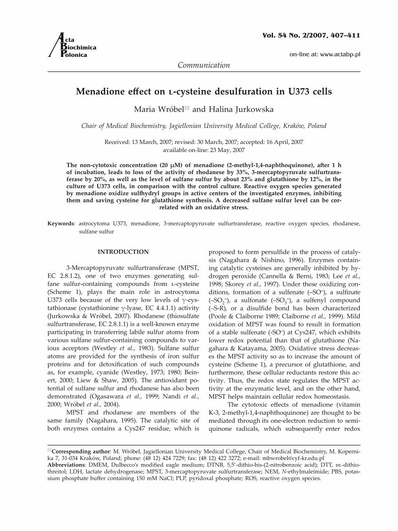
Menadione diffusion into cells upon addition to the extracellular media, without the assistance of transport proteins or pumps, has been reported for many different cell types, such as yeast (Mauzeroll & Bard, 2004) or human breast cells (Liu et al., 2000). Inside the cell, menadione is rapidly conjugated to intracellular glutathione to form a stable conjugate (Mauzeroll et al., 2004) or a rapid depletion of intra-cellular glutathione occurs due to oxidation of glu-tathione which forms disulfide dimers (glutathione disulfide) (DiMonte et al., 1984b). Figure 2 shows that in U373 cells cultured in the medium containing non-cytotoxic concentrations of menadione (20 µM and 50 µM), the level of glutathione was decreased respectively by about 12% and 32% after 1 h, and 32% and 35% after 5 h of incubation, in comparison with the controls. Although for both investigated time frames, the higher menadione concentration produced a higher decrease in glutathione level, it should be noted that for the 50 µM concentration, the drop was not proportional to time. This effect can be explained by a higher amount of glutathione available for formation of menadione-S-conjugates or for detoxification of ROS generated by mena-dione. It is possible that during 5 h of incubation glutathione synthesis is stimulated. It was observed
0
20
40
60
80
100
120
5 µM 20 µM 50 µM 100 µM 200 µM 500 µM
viab
ility
%
1h 5h
Figure 1. Menadione cytotoxicity to U373 cells.Cell viability was investigated by measuring the leakage of LDH from cells into the culture medium. The absorb-ance was measured at 490 nm. The leakage was calculated as a percentage of total activity after lysis of the cells by 1% Triton X-100.
0
20
40
60
80
100
120
20 µM 50 µM
% o
f con
trol
1h 5h
Figure 2. Menadione effect on the level of cellular glu-tathione. Menadione was dissolved in cell culture media (20 µM or 50 µM). The cells were incubated for 1 or 5 h, harvested in PBS and sonicated, the homogenate was deproteinized and centrifuged. Glutathione level determined in con-trol supernatants was 25.9 ± 2.6 nmoles per mg of protein. *P < 0.05, *P < 0.001 (Student’s t test) vs. the controls.
410 2007M. Wróbel and H. Jurkowska
that 2,3-dimethoxy-1,4-naphthoquinone and menadi-one, redox cycling quinones, increased the activity
of γ-glutamylcysteine synthase in bovine pulmonary artery endothelial cells and Chinese hamster lung fi-broblast V79 cells (Shi et al., 1994; Ochi, 1996). An increase of glutathione concentration in cells can be also achieved by the inhibition of cysteine-consum-ing pathways, which are alternatives to glutath-ione synthesis, e.g., sulfane sulfur formation from cysteine (Scheme 1).
Menadione effect on rhodanese and MPST activity and the level of sulfane sulfur
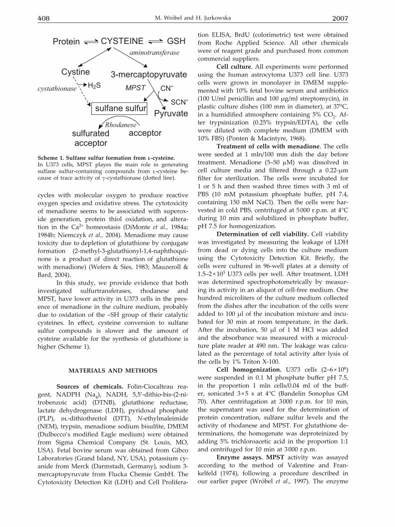
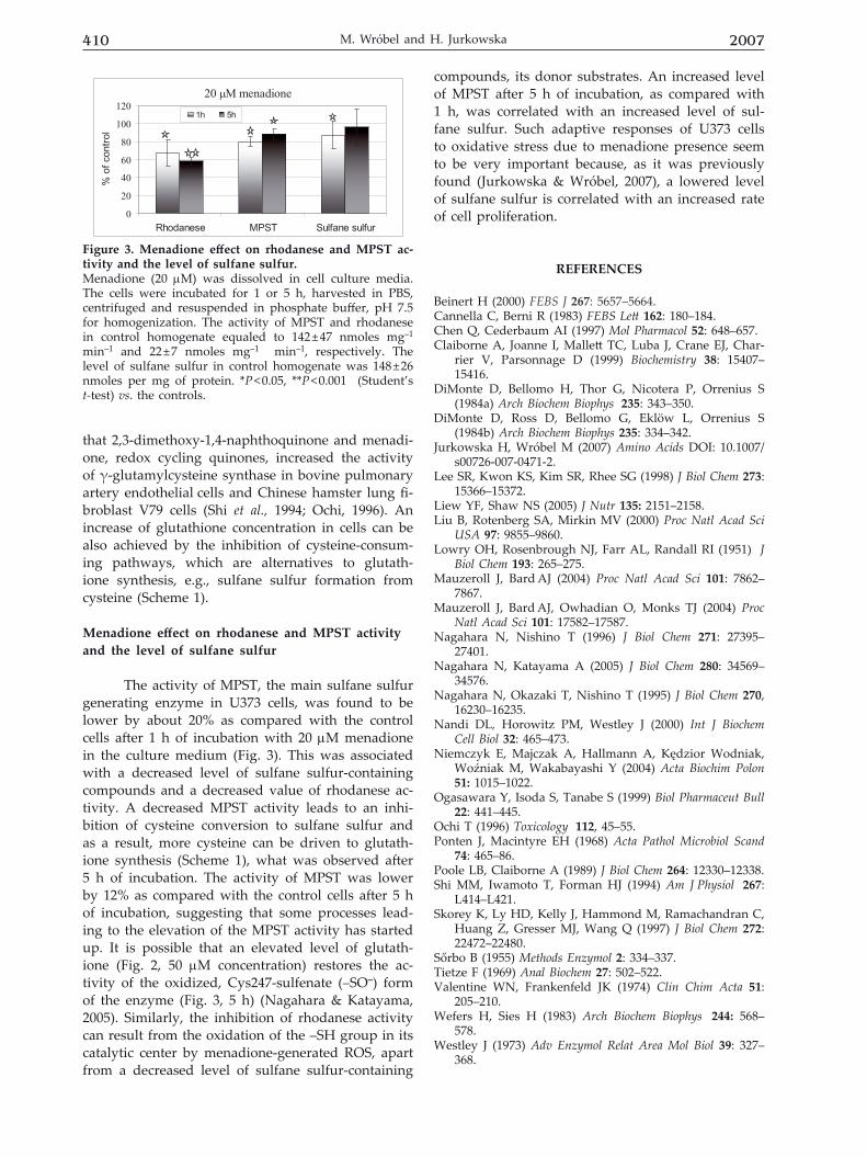
The activity of MPST, the main sulfane sulfur generating enzyme in U373 cells, was found to be lower by about 20% as compared with the control cells after 1 h of incubation with 20 µM menadione in the culture medium (Fig. 3). This was associated with a decreased level of sulfane sulfur-containing compounds and a decreased value of rhodanese ac-tivity. A decreased MPST activity leads to an inhi-bition of cysteine conversion to sulfane sulfur and as a result, more cysteine can be driven to glutath-ione synthesis (Scheme 1), what was observed after 5 h of incubation. The activity of MPST was lower by 12% as compared with the control cells after 5 h of incubation, suggesting that some processes lead-ing to the elevation of the MPST activity has started up. It is possible that an elevated level of glutath-ione (Fig. 2, 50 µM concentration) restores the ac-tivity of the oxidized, Cys247-sulfenate (–SO–) form of the enzyme (Fig. 3, 5 h) (Nagahara & Katayama, 2005). Similarly, the inhibition of rhodanese activity can result from the oxidation of the –SH group in its catalytic center by menadione-generated ROS, apart from a decreased level of sulfane sulfur-containing
compounds, its donor substrates. An increased level of MPST after 5 h of incubation, as compared with 1 h, was correlated with an increased level of sul-fane sulfur. Such adaptive responses of U373 cells to oxidative stress due to menadione presence seem to be very important because, as it was previously found (Jurkowska & Wróbel, 2007), a lowered level of sulfane sulfur is correlated with an increased rate of cell proliferation.
REFERENCES
Beinert H (2000) FEBS J 267: 5657–5664. Cannella C, Berni R (1983) FEBS Lett 162: 180–184.Chen Q, Cederbaum AI (1997) Mol Pharmacol 52: 648–657.Claiborne A, Joanne I, Mallett TC, Luba J, Crane EJ, Char-
rier V, Parsonnage D (1999) Biochemistry 38: 15407–15416.
DiMonte D, Bellomo H, Thor G, Nicotera P, Orrenius S (1984a) Arch Biochem Biophys 235: 343–350.
DiMonte D, Ross D, Bellomo G, Eklöw L, Orrenius S (1984b) Arch Biochem Biophys 235: 334–342.
Jurkowska H, Wróbel M (2007) Amino Acids DOI: 10.1007/s00726-007-0471-2.
Lee SR, Kwon KS, Kim SR, Rhee SG (1998) J Biol Chem 273: 15366–15372.
Liew YF, Shaw NS (2005) J Nutr 135: 2151–2158.Liu B, Rotenberg SA, Mirkin MV (2000) Proc Natl Acad Sci
USA 97: 9855–9860.Lowry OH, Rosenbrough NJ, Farr AL, Randall RI (1951) J
Biol Chem 193: 265–275.Mauzeroll J, Bard AJ (2004) Proc Natl Acad Sci 101: 7862–
7867.Mauzeroll J, Bard AJ, Owhadian O, Monks TJ (2004) Proc
Natl Acad Sci 101: 17582–17587. Nagahara N, Nishino T (1996) J Biol Chem 271: 27395–
27401.Nagahara N, Katayama A (2005) J Biol Chem 280: 34569–
34576. Nagahara N, Okazaki T, Nishino T (1995) J Biol Chem 270,
16230–16235.Nandi DL, Horowitz PM, Westley J (2000) Int J Biochem
Cell Biol 32: 465–473.Niemczyk E, Majczak A, Hallmann A, Kędzior Wodniak,
Woźniak M, Wakabayashi Y (2004) Acta Biochim Polon 51: 1015–1022.
Ogasawara Y, Isoda S, Tanabe S (1999) Biol Pharmaceut Bull 22: 441–445.
Ochi T (1996) Toxicology 112, 45–55. Ponten J, Macintyre EH (1968) Acta Pathol Microbiol Scand
74: 465–86.Poole LB, Claiborne A (1989) J Biol Chem 264: 12330–12338.Shi MM, Iwamoto T, Forman HJ (1994) Am J Physiol 267:
L414–L421.Skorey K, Ly HD, Kelly J, Hammond M, Ramachandran C,
Huang Z, Gresser MJ, Wang Q (1997) J Biol Chem 272: 22472–22480.
Sőrbo B (1955) Methods Enzymol 2: 334–337.Tietze F (1969) Anal Biochem 27: 502–522.Valentine WN, Frankenfeld JK (1974) Clin Chim Acta 51:
205–210.Wefers H, Sies H (1983) Arch Biochem Biophys 244: 568–
578.Westley J (1973) Adv Enzymol Relat Area Mol Biol 39: 327–
368.
20 µM menadione
0
20
40
60
80
100
120
Rhodanese MPST Sulfane sulfur
% o
f con
trol
1h 5h
Figure 3. Menadione effect on rhodanese and MPST ac-tivity and the level of sulfane sulfur.Menadione (20 µM) was dissolved in cell culture media. The cells were incubated for 1 or 5 h, harvested in PBS, centrifuged and resuspended in phosphate buffer, pH 7.5 for homogenization. The activity of MPST and rhodanese in control homogenate equaled to 142 ± 47 nmoles mg–1 min–1 and 22 ± 7 nmoles mg–1 min–1, respectively. The level of sulfane sulfur in control homogenate was 148 ± 26 nmoles per mg of protein. *P < 0.05, **P < 0.001 (Student’s t-test) vs. the controls.

Vol. 54 411Menadione effect on l-cysteine desulfuration
Westley J (1980) In Enzymatic Basis of Detoxification II (Ja-koby WB, ed) pp 245–262. Academic Press, New York,
Westley J, Adler H, Westley L, Nishida C (1983) Fundam App Toxicol 3: 377–382.
Wood IL (1987) Methods Enzymol 43: 25–29.
Wróbel M, Ubuka T, Yao WB, Abe T (1997) Physiol Chem Phys Med NMR 29: 11–14.
Wróbel M, Jurkowska H, Śliwa L, Srebro Z (2004) Toxicol Mech Meth 14: 331–337.