Membrane topology and function of dengue virus NS2A protein
14
Membrane Topology and Function of Dengue Virus NS2A Protein Xuping Xie, a,b Shovanlal Gayen, c CongBao Kang, c Zhiming Yuan, a Pei-Yong Shi b State Key Laboratory of Virology, Wuhan Institute of Virology, Chinese Academy of Science, China a ; Novartis Institute for Tropical Diseases, Singapore b ; Experimental Therapeutics Centre, Agency for Science, Technology and Research, Singapore c Flavivirus nonstructural protein 2A (NS2A) is a component of the viral replication complex that functions in virion assembly and antagonizes the host immune response. Although flavivirus NS2A is known to associate with the endoplasmic reticulum (ER) membrane, the detailed topology of this protein has not been determined. Here we report the first topology model of flavi- virus NS2A on the ER membrane. Using dengue virus (DENV) NS2A as a model, we show that (i) the N-terminal 68 amino acids are located in the ER lumen, with one segment (amino acids 30 to 52) that interacts with ER membrane without traversing the lipid bilayer; (ii) amino acids 69 to 209 form five transmembrane segments, each of which integrally spans the ER membrane; and (iii) the C-terminal tail (amino acids 210 to 218) is located in the cytosol. Nuclear magnetic resonance (NMR) structural analysis showed that the first membrane-spanning segment (amino acids 69 to 93) consists of two helices separated by a “helix breaker.” The helix breaker is formed by amino acid P85 and one positively charged residue, R84. Functional analysis using rep- licon and genome-length RNAs of DENV-2 indicates that P85 is not important for viral replication. However, when R84 was re- placed with E, the mutation attenuated both viral RNA synthesis and virus production. Remarkably, an R84A mutation did not affect viral RNA synthesis but blocked intracellular formation of infectious virions. Collectively, the mutagenesis results demon- strate that NS2A functions in both DENV RNA synthesis and virion assembly/maturation. The topology model of DENV NS2A provides a good starting point for studying how flavivirus NS2A modulates viral replication and evasion of host immune response. D engue virus (DENV) is a mosquito-borne human pathogen that belongs to the genus Flavivirus, family Flaviviridae. Be- sides the four serotypes of DENV, the genus Flavivirus also in- cludes other mosquito- and tick-borne viruses of public health importance, including West Nile virus (WNV), Japanese enceph- alitis virus (JEV), yellow fever virus (YFV), and tick-borne en- cephalitis virus (TBEV). DENV is prevalent in the tropical and subtropical regions of the world. DENV-infected patients develop dengue fever (DF); some infected individuals develop life-threat- ing dengue hemorrhagic fever (DHF) or dengue shock syndrome (DSS). There are annually 50 to 100 million DENV infections, leading to 500,000 DHF cases and 22,000 deaths around the world (1). Due to the increase in urbanization and transportation, the global burden of DENV has grown dramatically, with over 2.5 bil- lion people now at risk (http://www.who.int/csr/disease/dengue /en/). Unfortunately, neither a vaccine nor an antiviral is clinically available for prevention and treatment of DENV infection. It is therefore urgent to develop effective antivirals for DENV and other flaviviruses. Flaviviruses are enveloped viruses containing a single-strand, plus-sense RNA genome 11 kb in length. The genomic RNA contains a 5= untranslated region (UTR) with a type I cap (m 7 GpppAm) (2), a single open-reading frame (ORF), and a 3= UTR. The ORF encodes a long polyprotein, which is co- and post- translationally processed by a combination of viral and cellular proteases into three structural proteins (capsid [C], premembrane [prM], and envelope [E]) and seven nonstructural proteins (NS1, NS2A, NS2B, NS3, NS4A, NS4B, and NS5). The structural pro- teins are essential components of the virion and function in viral entry, fusion, and assembly. The nonstructural proteins are com- ponents of viral replication complexes (3). Among them, glyco- protein NS1 plays an essential role in viral RNA replication (4) and facilitates immune complex formation (5). NS3 contains serine protease (using NS2B as a cofactor), RNA helicase, and nucleotide triphosphatase activities (6, 7); in addition, NS3 functions in viral assembly (8, 9). The N-terminal one-third of NS5 contains a methyltransferase activity, responsible for viral RNA cap forma- tion and internal RNA methylation (10–12). The methyltrans- ferase domain was also proposed to have a weak guanylyltrans- ferase activity (13). The C-terminal two-thirds of NS5 is an RNA-dependent RNA polymerase (RdRp) (14). NS5 also plays a role in evasion of innate immune response (15, 16). Limited in- formation is known about the functions of hydrophobic mem- brane proteins NS2A, NS4A, and NS4B. These proteins serve as a scaffold for the replication complex (17). The integral membrane protein NS4A induces membrane rearrangement (18); NS4B co- locates with double-stranded RNA (dsRNA) and plays a critical role in viral replication (19). NS4B also suppresses interferon / response (20, 21). Flavivirus NS2A is a 22-kDa hydrophobic protein (22). Its N terminus is generated in the endoplasmic reticulum (ER) lumen by an unknown host protease (23); its C terminus is generated in cytoplasm by viral protease. NS2A was previously shown to be important for viral replication and pathogenesis. (i) NS2A func- tions in viral RNA synthesis. For example, Kunjin virus (KUNV) NS2A colocalizes with double-stranded viral RNA (dsRNA) and interacts with the 3= UTR, NS3, and NS5 in the form of a replica- tion complex (3). (ii) NS2A is important for viral assembly (see Received 6 September 2012 Accepted 4 February 2013 Published ahead of print 13 February 2013 Address correspondence to Pei-Yong Shi, [email protected]. Copyright © 2013, American Society for Microbiology. All Rights Reserved. doi:10.1128/JVI.02424-12 April 2013 Volume 87 Number 8 Journal of Virology p. 4609 – 4622 jvi.asm.org 4609 Downloaded from https://journals.asm.org/journal/jvi on 31 January 2022 by 218.51.123.182.
Transcript of Membrane topology and function of dengue virus NS2A protein

Membrane Topology and Function of Dengue Virus NS2A Protein
Xuping Xie,a,b Shovanlal Gayen,c CongBao Kang,c Zhiming Yuan,a Pei-Yong Shib
State Key Laboratory of Virology, Wuhan Institute of Virology, Chinese Academy of Science, Chinaa; Novartis Institute for Tropical Diseases, Singaporeb; ExperimentalTherapeutics Centre, Agency for Science, Technology and Research, Singaporec
Flavivirus nonstructural protein 2A (NS2A) is a component of the viral replication complex that functions in virion assemblyand antagonizes the host immune response. Although flavivirus NS2A is known to associate with the endoplasmic reticulum(ER) membrane, the detailed topology of this protein has not been determined. Here we report the first topology model of flavi-virus NS2A on the ER membrane. Using dengue virus (DENV) NS2A as a model, we show that (i) the N-terminal 68 amino acidsare located in the ER lumen, with one segment (amino acids 30 to 52) that interacts with ER membrane without traversing thelipid bilayer; (ii) amino acids 69 to 209 form five transmembrane segments, each of which integrally spans the ER membrane;and (iii) the C-terminal tail (amino acids 210 to 218) is located in the cytosol. Nuclear magnetic resonance (NMR) structuralanalysis showed that the first membrane-spanning segment (amino acids 69 to 93) consists of two helices separated by a “helixbreaker.” The helix breaker is formed by amino acid P85 and one positively charged residue, R84. Functional analysis using rep-licon and genome-length RNAs of DENV-2 indicates that P85 is not important for viral replication. However, when R84 was re-placed with E, the mutation attenuated both viral RNA synthesis and virus production. Remarkably, an R84A mutation did notaffect viral RNA synthesis but blocked intracellular formation of infectious virions. Collectively, the mutagenesis results demon-strate that NS2A functions in both DENV RNA synthesis and virion assembly/maturation. The topology model of DENV NS2Aprovides a good starting point for studying how flavivirus NS2A modulates viral replication and evasion of host immuneresponse.
Dengue virus (DENV) is a mosquito-borne human pathogenthat belongs to the genus Flavivirus, family Flaviviridae. Be-
sides the four serotypes of DENV, the genus Flavivirus also in-cludes other mosquito- and tick-borne viruses of public healthimportance, including West Nile virus (WNV), Japanese enceph-alitis virus (JEV), yellow fever virus (YFV), and tick-borne en-cephalitis virus (TBEV). DENV is prevalent in the tropical andsubtropical regions of the world. DENV-infected patients developdengue fever (DF); some infected individuals develop life-threat-ing dengue hemorrhagic fever (DHF) or dengue shock syndrome(DSS). There are annually 50 to 100 million DENV infections,leading to 500,000 DHF cases and 22,000 deaths around the world(1). Due to the increase in urbanization and transportation, theglobal burden of DENV has grown dramatically, with over 2.5 bil-lion people now at risk (http://www.who.int/csr/disease/dengue/en/). Unfortunately, neither a vaccine nor an antiviral is clinicallyavailable for prevention and treatment of DENV infection. It istherefore urgent to develop effective antivirals for DENV andother flaviviruses.
Flaviviruses are enveloped viruses containing a single-strand,plus-sense RNA genome 11 kb in length. The genomic RNAcontains a 5= untranslated region (UTR) with a type I cap(m7GpppAm) (2), a single open-reading frame (ORF), and a 3=UTR. The ORF encodes a long polyprotein, which is co- and post-translationally processed by a combination of viral and cellularproteases into three structural proteins (capsid [C], premembrane[prM], and envelope [E]) and seven nonstructural proteins (NS1,NS2A, NS2B, NS3, NS4A, NS4B, and NS5). The structural pro-teins are essential components of the virion and function in viralentry, fusion, and assembly. The nonstructural proteins are com-ponents of viral replication complexes (3). Among them, glyco-protein NS1 plays an essential role in viral RNA replication (4) andfacilitates immune complex formation (5). NS3 contains serine
protease (using NS2B as a cofactor), RNA helicase, and nucleotidetriphosphatase activities (6, 7); in addition, NS3 functions in viralassembly (8, 9). The N-terminal one-third of NS5 contains amethyltransferase activity, responsible for viral RNA cap forma-tion and internal RNA methylation (10–12). The methyltrans-ferase domain was also proposed to have a weak guanylyltrans-ferase activity (13). The C-terminal two-thirds of NS5 is anRNA-dependent RNA polymerase (RdRp) (14). NS5 also plays arole in evasion of innate immune response (15, 16). Limited in-formation is known about the functions of hydrophobic mem-brane proteins NS2A, NS4A, and NS4B. These proteins serve as ascaffold for the replication complex (17). The integral membraneprotein NS4A induces membrane rearrangement (18); NS4B co-locates with double-stranded RNA (dsRNA) and plays a criticalrole in viral replication (19). NS4B also suppresses interferon �/�response (20, 21).
Flavivirus NS2A is a 22-kDa hydrophobic protein (22). Its Nterminus is generated in the endoplasmic reticulum (ER) lumenby an unknown host protease (23); its C terminus is generated incytoplasm by viral protease. NS2A was previously shown to beimportant for viral replication and pathogenesis. (i) NS2A func-tions in viral RNA synthesis. For example, Kunjin virus (KUNV)NS2A colocalizes with double-stranded viral RNA (dsRNA) andinteracts with the 3= UTR, NS3, and NS5 in the form of a replica-tion complex (3). (ii) NS2A is important for viral assembly (see
Received 6 September 2012 Accepted 4 February 2013
Published ahead of print 13 February 2013
Address correspondence to Pei-Yong Shi, [email protected].
Copyright © 2013, American Society for Microbiology. All Rights Reserved.
doi:10.1128/JVI.02424-12
April 2013 Volume 87 Number 8 Journal of Virology p. 4609–4622 jvi.asm.org 4609
Dow
nloa
ded
from
http
s://j
ourn
als.
asm
.org
/jour
nal/j
vi o
n 31
Jan
uary
202
2 by
218
.51.
123.
182.

details in Discussion) (8, 24). (iii) Expression of DENV-2 NS2Aalone inhibits interferon �/� response (21). An A30P substitutionreduced the effect KUNV NS2A on antagonism of interferon re-sponse, leading to virulence attenuation in mice (25, 26). JEVNS2A was recently shown to block dsRNA-activated protein ki-nase PKR (27). (iv) A conserved slippery heptanucleotide motif,located at the beginning of the NS2A gene, contributes to theproduction of NS1= in the JEV serogroup through a ribosomalframeshift mechanism (28). (v) NS2A participates in virus-in-duced membrane formation (24). Despite the above functions,the topology of flavivirus NS2A on the ER membrane has not beenexperimentally determined.
In this study, we used a number of biochemical approaches toestablish the topology of DENV-2 NS2A on the ER membrane.Our results indicate that DENV-2 NS2A contains five integraltransmembrane segments (amino acids 69 to 209) that span thelipid bilayer of the ER membrane; in addition, one membrane-associated segment (amino acids 32 to 51) interacts with the ERmembrane without traversing the lipid bilayer. The nuclear mag-netic resonance (NMR) structure of the first transmembrane seg-ment (amino acids 69 to 93) showed two helices connected by aP85-mediated “helix breaker.” Functional analysis showed thatamino acid P85 is not important for viral replication. Instead, theadjacent positively charged residue, R84, is critical for both viralRNA synthesis and intracellular virion assembly/maturation.
MATERIALS AND METHODSBioinformatics. The amino acid sequence of DENV-2 NS2A (strain NGC)was used for membrane topology prediction using the following webserv-ers: HMMTOP (http://www.enzim.hu/hmmtop/index.php), TMHMM2(http://www.cbs.dtu.dk/services/TMHMM/), DAS (http://www.sbc.su.se/�miklos/DAS/maindas.html), TOPCONS (http://topcons.cbr.su.se/),Split (http://split.pmfst.hr/split/4), and MEMSAT3 (http://bioinf.cs.ucl.ac.uk/psipred/). The amphipathic helix prediction was performed usingthe HeliQuest program (http://heliquest.ipmc.cnrs.fr/).
Cell culture, viruses, and antibodies. Baby hamster kidney (BHK-21)cells were maintained in high-glucose Dulbecco modified Eagle medium(DMEM) (Invitrogen, Carlsbad, CA) supplemented with 4 mM L-glu-tamine, 10% fetal bovine serum (FBS) (HyClone Laboratories, Logan,UT), and 1% penicillin/streptomycin (Invitrogen, Carlsbad, CA).HEK293T (human embryo kidney 293T) cells were grown in low-glucoseDMEM (Invitrogen) with 10% FBS and 1% penicillin/streptomycin. Allcells were incubated at 37°C with 5% CO2. Recombinant DENV-2 (NGCstrain) was produced from plasmid pACYC NGC FL, a full-length (FL)infectious cDNA clone of DENV-2 (see details below). A mouse mono-clonal antibody against enhanced green fluorescence protein (eGFP) waspurchased from Roche. The mouse monoclonal antibody 4G2 againstDENV envelope protein was prepared from hybridoma cell lines obtainedfrom the American Type Culture Collection (ATCC). Goat anti-mouseantibodies conjugated to horseradish peroxidase (HRP) were purchasedfrom Sigma-Aldrich. Alexa Fluor 488 goat anti-mouse IgG (MolecularProbes) was bought from Life Technologies.
Plasmid construction. Standard molecular biology methods wereused in all plasmid constructions. Our experience suggests that the low-copy-number plasmid pACYC177 (Thermo Fisher Scientific Inc.) is anideal vector for construction of stable infectious cDNA clone of flavivirus(29–31). Therefore, we swapped the full-length (FL) cDNA of DENV-2(NGC strain) from a previous infectious clone plasmid (32) into vectorpACYC177. The resulting plasmid, pACYC-NGC FL, contained a longDNA fragment covering a T7 promoter, the DENV-2 NGC genome, andthe hepatitis delta virus ribozyme sequence (HDVr). The DNA fragmentwas engineered into pACYC177 via two unique restriction sites: SacII(upstream of T7 promoter) and ClaI (downstream of HDVr).
Replicon pACYC-NGC was constructed by replacing the viral struc-tural genes (C-prM-E) with a Renilla luciferase gene followed by a foot-and-mouth disease virus (FMDV) 2A peptide. In the replicon clone, thefirst 38 amino acids of C protein and the last 31 amino acids of E proteinwere retained. To facilitate the construction, a SacI restriction site wasintroduced into both pACYC NGC FL and the pACYC-NGC repliconclone by silent mutation (A4338G, A4342T, and G4343C) using aQuikChange II XL site-directed mutagenesis kit (Agilent Technologies,Inc.) with the primer pair NGC_SacI_F and NGC_SacI_R (Table 1). Forsite-directed mutagenesis, subclone TA-D was constructed by amplifyingviral genome from nucleotide position 2,544 to 4,344 using the primerpair NGC_NheI_F and NGC_SacI_R (Table 1), the PCR DNA was di-rectly cloned into vector pCR2.1-TOPO (Invitrogen, Carlsbad, CA).NS2A mutations (R84A, R84E, R84S, and P85A) were individually intro-duced into the subclone TA-D by site-directed mutagenesis using thecorresponding primers (Table 1); the DNA containing each mutation wasinserted into pACYC NGC FL or the pACYC NGC replicon clone at theNheI and SacI restriction sites.
A mammalian expression vector, pXJ, driven by a cytomegalovirus(CMV) promoter was used to express various NS2A fusion proteins fortopology study. Multiple restriction enzyme sites (EcoRI, NotI, BamHI,XhoI, and KpnI) of vector pXJ were used for cloning different NS2Afragments; each NS2A fragment was C-terminally fused in frame withenhanced green fluorescence protein (eGFP). pACYC NGC FL was usedas a template for amplification of different NS2A fragments. The eGFPwas amplified from pEGFPluc vector (Clontech). For construction ofpXJ-TM-eGFP (TM represents various transmembrane fragments ofNS2A), different NS2A fragments were fused with eGFP and inserted intovector pXJ at the BamHI and XhoI sites. For construction of pXJ-leader-TM-eGFP-Glyc, an N-glycosylation (Glyc) acceptor site (Asn-Ser-Thr-Ser-Ala) (18) was fused to the C terminus of eGFP by PCR using theprimer pair 2A-p1-F and 2A-p3-R (Table 1); the eGFP-Glyc fragment wasinserted into vector pXJ at BamHI and XhoI sites, resulting in plasmidpXJ-eGFP-Glyc. To construct the “leader” (containing viral proteins up-stream of NS2A to form the right topology of NS2A; see below), we per-formed an overlap PCR using two primer pairs (2A-p4-F1/2A-p4-R1 and2A-p4-F2/2A-p4-R2) to fuse a signal peptide comprising the last 24 aminoacids of E protein (E24) with a fragment containing the first 50 amino acidsof NS1 protein (N50), an NotI restriction site, and the last 50 amino acidsof NS1 (C50). The leader was inserted into plasmid pXJ-eGFP-Glyc (de-
TABLE 1 Sequences of DNA primers used in this study
Primer name Sequence (5= to 3=)NGC_SacI_F CAGGCAGAGATATCAGGGAGCTCTCCAATCCTGTCAATAACNGC_SacI_R GTTATTGACAGGATTGGAGAGCTCCCTGATATCTCTGCCTGNGC_NheI_F CCTTCAAAGCTAGCTTCAGCTN2A_R84A_F CTAGCAGCCTTCAAAGTCGCACCAACTTTTGCAGCTGGN2A_R84A_R CCAGCTGCAAAAGTTGGTGCGACTTTGAAGGCTGCTAGN2A_R84E_F CTAGCAGCCTTCAAAGTCGAACCAACTTTTGCAGCTGGN2A_R84E_R CCAGCTGCAAAAGTTGGTTCGACTTTGAAGGCTGCTAGN2A_R84S_F CTAGCAGCCTTCAAAGTCTCCCCAACTTTTGCAGCTGGACN2A_R84S_R GTCCAGCTGCAAAAGTTGGGGAGACTTTGAAGGCTGCTAGN2A_P85A_F CAGCCTTCAAAGTCAGAGCAACTTTTGCAGCTGGN2A_P85A_R CCAGCTGCAAAAGTTGCTCTGACTTTGAAGGCTG2A-p1-F CGCGGATCCACCATGGTGAGCAAGGGCGAGGAGC2A-p1-R ACCCTCGAGTTACTTGTACAGCTCGTCCATGCCG2A-p3-R ACCCTCGAGTTAGGCGGAGGTGGAGTTCTTGTACAGCTCGTC
CATGCCGAGAG2A-p4-F1 GGCGAATTCACCATGAGCACCTCACTGTCTGTGT2A-p4-R1 TCTGTTATGAGTTTTCCAGAGGCGGCCGCTTTCTGGATAGCTG2A-p4-F2 CAGCTATCCAGAAAGCGGCCGCCTCTGGAAAACTCATAACAGA2A-p4-R2 GGTGGATCCGGCTGTGACCAAGGAGTTGACNGC 7764V CGTCGAGAGAAATATGGTCACACCNGC 7844C CCACAATAGTATGACCAGCCTM_GAPDH-F AGGTCGGTGTGAACGGATTTGM_GAPDH-R TGTAGACCATGTAGTTGAGGTCA
Xie et al.
4610 jvi.asm.org Journal of Virology
Dow
nloa
ded
from
http
s://j
ourn
als.
asm
.org
/jour
nal/j
vi o
n 31
Jan
uary
202
2 by
218
.51.
123.
182.

scribed above) at the EcoRI and BamHI sites, resulting in construct pXJ-leader-eGFP-Glyc. Plasmid pXJ-leader-eGFP-Glyc was used for insertionof various NS2A fragments using the NotI and BamHI sites, resulting invarious pXJ-leader-TM-eGFP-Glyc constructs.
For constructs used for in situ fluorescence protease protection assay,the eGFP sequence, amplified from the pEGFPluc vector with the primerpair 2A-p1-F1 and 2A-p1-R (Table 1), was engineered into vector pXJ atBamHI and XhoI restriction sites, resulting in plasmid pXJ-eGFP. A mod-ified pACYC-B plasmid (using pACYC177 as a vector and containing a T7promoter followed by nucleotides 1 to 5434 of the DENV-2 NGC ge-nome) was initially constructed by silent mutation (T2874C and T2343C)to knock out two internal EcoRI sites. PCR fragments containing E24,full-length NS1, and different truncated versions of NS2A were amplifiedfrom the modified pACYC-B plasmid, digested with EcoRI and BamHI,and inserted into plasmid pXJ-eGFP, resulting in constructs pXJ-E24-NS1-TM-eGFP. All constructs were validated by restriction enzymes di-gestion and DNA sequencing.
Membrane flotation assay. A membrane flotation assay was per-formed according to a previously described procedure with some modi-fications (19). Briefly, 293T cells were seeded in a 10-cm dish (Nunc) andtransfected with 10 �g plasmids encoding different TM-eGFP fusion pro-teins using X-tremeGENE 9 DNA transfection reagent (Roche) followingthe manufacturer’s instructions. At 10 to 16 h posttransfection (p.t.), thecells were washed once with phosphate-buffered saline (PBS), detachedinto PBS by gentle pipetting, and centrifuged at 1,000 � g for 5 min. Cellpellets were resuspended in prechilled hypotonic lysis buffer (10 mMTris-HCl [pH 7.5], 2 mM MgCl2), incubated on ice for 10 min, and dis-rupted with 15 strokes of a Dounce homogenizer (Sigma). Cell lysateswere centrifuged at 1,000 � g at 4°C for 5 min to remove nuclei, unlysedcells, and debris. Postnuclear lysates were mixed with an equal volume of75% (wt/vol) Nycodenz (Axis Shield, Oslo, Norway) solution in PBS. Thelysates (0.96 ml) were loaded at the bottom of a 3.5-ml thick-walled poly-carbonate ultracentrifugation tube (Beckman). The tube was sequentiallyoverlaid with 2.16 ml of 35% Nycodenz in PBS (first later) and 0.24 ml of5% Nycodenz in PBS (second layer). Equilibrium centrifugation was per-formed in a Beckman SW 55 Ti rotor at 30,000 rpm for 20 h. After cen-trifugation, an equal volume (30 �l) of sample from each fraction (0.42 mlper fraction) was analyzed by sodium dodecyl sulfate-polyacrylamide gelelectrophoresis (SDS-PAGE), and proteins were detected by Westernblotting (described below).
In situ fluorescence protease protection assay. The in situ fluores-cence protease protection assay was performed using a previously de-scribed protocol (33). BHK-21 cells were seeded in a Lab-Tek chamber(1.2 � 104 cells/chamber). After 16 to 24 h of incubation, cells were trans-fected with 1 �g plasmids encoding different NS2A-eGFP fusion proteins.At 24 h p.t., the cells were washed twice in KHM buffer (110 mM potas-sium acetate [pH 7.2], 20 mM HEPES, and 2 mM MgCl2). Plasma mem-brane was selectively permeabilized in KHM buffer supplemented with 50�M digitonin (Merck) at room temperature for 5 min. After being washedonce with KHM buffer, the cells were treated with 50 �g/ml protease K(New England BioLabs) in KHM buffer and immediately quantified forfluorescence intensities at 8-s intervals using a Zeiss LSM 5 DUO laserscanning confocal microscope. Signals from 5 to 7 fields (each field con-tained about 4 to 6 positive cells) were averaged and normalized. Immu-nofluorescence staining images were captured using Zeiss LSM 5 METAsystem and processed using Adobe Photoshop CS3 (Adobe Systems, SanJose, CA). The mean value of fluorescence intensities of the whole timeseries (5 min) is presented. Figures were plotted using Adobe illustratorCS3 software. The following formulae were used to calculate the relativefluorescence intensity and the initial rate of fluorescence degradation:relative fluorescence intensity � florescence intensity collected at 300s/florescence intensity collected at time zero � 100; initial rate of fluores-cence degradation (%/min) � (100% � retained relative fluorescenceintensity at 8 s) � 60/8.
In vitro deglycosylation assay. Deglycosylation assay was performedby following a previously reported method (34). HEK293T cells weretransfected in a 6-well plate (4 � 105 cells/well and 4 �g DNA) usingX-tremeGENE 9 DNA transfection reagent (Roche). At 24 h p.t., the cellsexpressing GFP-tagged NS2A were washed twice in PBS and detached bygently pipetting. The cells were pelleted by centrifuging at 500 � g for 5min and resuspended in a solubilization buffer comprising 20 mM Tris-HCl (pH 7.5), 50 mM NaCl, 10 mM magnesium acetate, 1% Triton X-100(Sigma), and an EDTA-free protease inhibitor cocktail (Roche). After thecells had been solubilized by rotating the tube at 4°C for 2 h, the cell lysateswere centrifuged at 15,000 rpm and 4°C for 1 h. The clarified supernatantswere treated with 50,000 units/ml PNGase F (New England Biolabs) orPBS at 37°C for 2 h according to the manufacturer’s instructions, exceptthat the denaturation procedure was omitted. The samples were heated at65°C for 15 min and analyzed by 10% SDS-PAGE followed by Westernblotting.
In vitro protease K protection assay. HEK293T cells were transfectedwith various NS2A expression constructs, as described in the precedingsection. At 20 h p.t., cell lysates were prepared according to a previouslydescribed protocol (19), with some modifications. Briefly, the transfectedcells were washed twice with PBS, detached in cold PBS, and centrifuged at1,000 � g for 5 min. Cells were resuspended in a hypotonic lysis buffer (10mM Tris-HCl, pH 7.5, and 2 mM MgCl2). After incubation on ice for 10min, the cells were disrupted with 15 strokes of a homogenizer (Sigma).The lysates were clarified by centrifugation at 1,000 � g for 5 min to pelletnuclei, unlysed cells, and debris. The supernatant (postnuclear lysates)were centrifuged at 15,000 rpm for 30 min to pellet membranes and theirassociated proteins. Pellets were resuspended in PBS and treated with 50�g/ml protease K (New England BioLabs) for 1 h on ice in the absence orpresence of 0.5% Triton X-100. The digestion reaction was stopped byadding 20 mM phenylmethylsulfonyl fluoride (PMSF; Sigma), and thereaction mixture was further incubated on ice for 15 min. The sampleswere then mixed with a preheated (95°C) 4� lithium dodecyl sulfate(LDS) sample buffer (Invitrogen) supplemented with 100 mM dithiothre-itol (DTT) (Sigma), heated at 95°C for 15 min, and analyzed by Westernblotting.
SDS-PAGE and Western blotting. Samples were analyzed on a 10%polyacrylamide–SDS gel. Proteins were transferred onto a nitrocelluloseor polyvinylidene difluoride (PVDF) membrane using a Trans-Blot appa-ratus (Bio-Rad), followed by incubation in blocking buffer (comprising5% skim milk, 20 mM Tris-HCl [pH 7.5], 137 mM NaCl, and 0.1% Tween20) for 1 h. The blots were incubated in blocking buffer containing amouse monoclonal antibody against eGFP (1:1,000 dilution) overnight at4°C. After being washed three times (10 min each time) with TBST (Tris-buffered saline–Tween) buffer, the blots were further incubated with agoat anti-mouse antibody conjugated to horseradish peroxidase (1:5,000dilution) in blocking buffer for 1 h, followed by three washes with TBSTbuffer. The antibody-protein complexes were detected using the ECLWestern blotting system (GE Healthcare).
RNA transcription, electroporation, and replicon transient-trans-fection assay. FL DENV-2 and replicon RNAs were in vitro transcribedusing a T7 mMessage mMachine kit (Ambion, Austin, TX) from cDNAplasmids prelinearized by XbaI. The RNA transcripts (10 �g) were elec-troporated into BHK-21 cells following a protocol described previously(35). The cells transfected with replicon RNA were seeded in a 12-wellplate (2 � 105 cells/well). At various time points, the cells were washedonce with phosphate-buffered saline (PBS) and lysed using lysis buffer(Promega). The plates containing the lysis buffer were sealed with Para-film and stored at �80°C. Once samples for all time points had beencollected, luciferase signals were measured in a Clarity luminescence mi-croplate reader (BioTek) according to the manufacturer’s protocol.
Immunofluorescence assay (IFA). BHK-21 cells transfected with viralRNA were grown in an 8-well Lab-Tek chamber slide (Thermo FisherScientific). At various time points, the cells were fixed in PBS supple-mented with 4% paraformaldehyde at room temperature for 20 to 30 min
Membrane Topology and Function of Dengue Virus NS2A
April 2013 Volume 87 Number 8 jvi.asm.org 4611
Dow
nloa
ded
from
http
s://j
ourn
als.
asm
.org
/jour
nal/j
vi o
n 31
Jan
uary
202
2 by
218
.51.
123.
182.

and permeabilized with 0.1% Triton X-100 in PBS at room temperaturefor 10 min. After 1 h of incubation in a blocking buffer containing 1% FBSin PBS, the cells were treated with mouse monoclonal antibody 4G2 for 1h and washed three times for 5 min with PBS. The cells were then incu-bated with Alexa Fluor 488 goat anti-mouse IgG for 1 h in blocking buffer,after which the cells were washed as described above. The cells weremounted in a mounting medium with DAPI (4=,6-diamidino-2-phe-nylindole; Vector Laboratories, Inc.). Fluorescence images were takenwith a Leica DM4000 B system. Images were merged using Adobe Photo-shop CS3 software.
Virus production and reverse transcriptase PCR (RT-PCR) assay.BHK-21 cells were transfected with genome-length RNA as descriedabove. The transfected cells were seeded in one T-75 flask (8 � 106 cells in15 ml DMEM supplemented with 10% FBS). After incubation at 37°C for24 h, the cells were cultured in DMEM with 2% FBS at 30°C for another 4days. At every 24 h p.t., 200 �l culture fluids was collected and stored at�80°C. On day 5 p.t., all culture fluids were centrifuged at 4°C and 500 �g for 5 min, aliquoted, and stored at �80°C. For each sample, viral titerwas quantified by plaque assay using BHK-21 cells (30).
Quantification of extra- and intracellular viral RNA and infectiousvirions. After electroporation, BHK-21 cells were seeded in a 6-well plate(about 4.5 � 105 cells per well) in DMEM with 10% FBS and incubated at37% with 5% CO2. At 5 h p.t., the transfected cells were washed once withDMEM to remove residual in vitro-transcribed RNA in the culture fluidsand maintained in DMEM containing 2% FBS. At 12, 24, and 48 p.t.,culture fluids were harvested, clarified by centrifugation at 500 � g at 4°Cfor 5 min, and stored at �80°C. Cell samples were harvested by using apreviously described method, with some modifications (36). Briefly, atvarious time points, cells were washed once with cold PBS and incubatedfor 3 min at 4°C in a cold alkaline– high-salt solution (1 M NaCl and 50mM sodium bicarbonate, pH 9.5) to remove cell surface-associated virus.The cells were then washed twice with cold PBS, treated with 0.25% Tryp-sin-EDTA (Gibco) to detach them from the plate surface, and collected bycentrifugation at 1,000 � g for 5 min. The cell pellets were resuspended in250 �l RPMI 1640 medium with 2% FBS. One hundred microliters of thecell suspensions was centrifuged to pellet the cells; the pelleted cells werethen quantified for intracellular viral RNA (see below). The remaining 150�l of cell suspensions was lysed using a single freeze-thaw cycle (frozen at�80°C and thawed at 37°C), cellular debris was removed by centrifuga-tion at 3,200 � g for 5 min at 4°C, and the supernatant was quantified forintracellular infectious virus using a plaque assay.
Viral RNAs in culture fluids were extracted using a QIAamp viral RNAminikit (Qiagen), and intracellular total RNAs were isolated using anRNeasy minikit (Qiagen). Extracted RNAs were eluted in 50 �l RNase-free water. Two microliters of RNA elution was used for real-time RT-PCR. Viral RNAs were measured using an iScript one-Step RT-PCR kitwith SYBR green (Bio-Rad) and the primer pair NGC 7764V/7844C (Ta-ble 1). The mRNA level of the housekeeping gene glyceraldehyde-3-phophate dehydrogenase (GAPDH) was measured in parallel using theprimer pair M_GAPDH-F/M_GAPDH-R (Table 1). The intracellular vi-ral RNAs were normalized according to the mRNA level of GAPDH.
Peptide synthesis, purification, and NMR spectroscopy. A peptiderepresenting the first transmembrane segment, corresponding to residuesD67 to R94 of DENV-2 NS2A (acetyl-DIGMGVTYLALLAAFKVRPTFAAGLLLR), was synthesized and purified by reverse-phase high-pressureliquid chromatography (RP-HPLC) to a purity of �95% (GL BiochemLtd., Shanghai, China). The sequence of the peptide was confirmed bymass spectroscopy. The purified peptide was dissolved to 3 mg/ml in 250�l of the deuterated solvent (Cambridge isotope) containing 4:4:1 ofCDCl3, CD3OH, and H2O (pH � 7.0) (37). The NMR sample was trans-ferred to a 3-mm NMR tube for data collection. All NMR data were col-lected at 25°C on a Bruker Avance II700 MHz spectrometer equipped witha 5-mm z-gradient TXI cryoprobe. A total correlation spectroscopy(TOCSY) experiment was recorded with a mixing time of 80 ms. Two-dimensional (2D) nuclear Overhauser effect spectroscopy (NOESY) ex-
periments were recorded using mixing times of 200 ms and 300 ms. Theproton chemical shifts were directly referenced to 4,4-dimethyl-4-silapentane-1-sulfonic acid (DSS). The nitrogen and carbon chemicalshifts were referenced indirectly to DSS. The 1H-13C and 1H-15N hetero-nuclear single quantum correlation spectroscopy (HSQC) in 13C and 15Nnatural abundance were also acquired to facilitate resonance assignment.All NMR spectra were processed using Topspin (Bruker, version 2.1) andanalyzed using Sparky (http://www.cgl.ucsf.edu/home/sparky/).
Peptide structure determination. The peptide assignment in solventwas accomplished using the previously reported procedures, in whichspin systems were identified by a TOCSY spectrum and sequential con-nectivity was determined using a NOESY spectrum (37, 38). Cross-peaksfrom the NOESY spectrum with a mixing time of 200 ms were selected,assigned, and integrated in Sparky. The NOEs were calibrated withCYANA and transferred to distance constrains for structural determina-tion (39). The structure was determined using torsion angle dynamics-simulated annealing as implemented in CYANA using NOE restraints.One hundred structures were calculated using CYANA. Fifteen structureswith the lowest energy function were selected and viewed using PyMOL(www.pymol.org).
RESULTSBioinformatic analysis of DENV NS2A membrane topology.Approximately 42% of DENV NS2A amino acids are hydrophobicresidues. Flavivirus NS2A was previously shown to associate withER membrane in the replication complex (3). To examine whichregions of NS2A are responsible for membrane association, weinitially analyzed the membrane topology using several bioinfor-matics algorisms. As shown in Fig. 1A, different algorithms pre-dicted different numbers and positions of transmembrane seg-ments (pTMS). To avoid discounting any pTMS, we combined allthe predicted topologies into a reference model (Fig. 1B). Based onthis reference model, we experimentally analyzed individual seg-ments as either membrane associated (integrally or peripherallymembrane associated) or non-membrane associated (see below).Each NS2A segment was C-terminally fused with an eGFP re-porter (Fig. 1B). The length of each selected segment was slightlydifferent from that of the predicted pTMS to balance the intactflexibility between pTMS and eGFP according to charged residuesaround the C terminus of each pTMS.
Cellular localization of NS2A fragments. To define the re-gions of NS2A that has membrane-associated activity, we trans-fected BHK-21 cells with plasmids that express various NS2A frag-ment-eGFP fusion proteins (Fig. 1B). At 20 h p.t., the expressionand intracellular localization of each fusion protein were moni-tored by IFA. A fusion protein containing a real membrane-tar-geting segment would present an ER staining pattern, whereas aprotein without any membrane-targeting segment would displayan even distribution of intracellular staining pattern. As shown inFig. 1C, expression of eGFP alone (without any NS2A fragment)exhibited an even distribution of fluorescence throughout the cell,whereas expression of the full-length NS2A-eGFP (construct1–218) displayed fluorescence in the perinuclear region, confirm-ing that NS2A is a membrane-associated protein. Expression ofNS2A fragments 1–24, 50 –71, 118 –140, and 209 –218 fused witheGFP showed fluorescence patterns similar to that of expression ofeGFP alone, suggesting that these NS2A fragments do not havemembrane-associated activity. In contrast to the above result, ex-pression of NS2A fragments 30 –52, 69 –95, 96 –118, 142–163,165–186, and 186 –209 fused with eGFP exhibited a reticular flu-orescence pattern, indicating that these NS2A fragments havemembrane-associated activity. It should be noted that these re-
Xie et al.
4612 jvi.asm.org Journal of Virology
Dow
nloa
ded
from
http
s://j
ourn
als.
asm
.org
/jour
nal/j
vi o
n 31
Jan
uary
202
2 by
218
.51.
123.
182.

gions that showed ER membrane-interacting activity do not nec-essarily span the entire lipid bilayer of ER membrane; some ofthese fragments could peripherally interact with the membranewithout traversing the complete lipid bilayer.
Membrane flotation analysis. To validate the above mem-brane-associated activities of NS2A fragments, we performed amembrane flotation assay (Fig. 1D). As controls, we found thatsoluble eGFP alone was exclusively distributed in the high-density(HD) fractions, whereas calnexin, a well-known ER integral mem-brane protein, completely floated to low-density (LD) fractions.For the NS2A fragment-eGFP fusion proteins, more than 99% ofthe expressed proteins containing NS2A fragments 1–24, 50 –71,118 –140, and 209 –218 were found in the HD fractions, indicatingthat these segments are dissociated with membrane. In contrast,more than 55% of the proteins containing fragments 30 –52, 69 –95, 96 –118, 142–163, 165–186, and 1–218 (full-length NS2A)were detected in the LD fractions, indicating that these fragmentshave membrane-associated activity. The above membrane flota-tion results are in good agreement with the cellular localizationresults (Fig. 1C).
For fragment 186 –209, the correlation between the membraneflotation result and the cellular localization data is not strong: only10.6% of this fragment-eGFP fusion protein was detected in theLD fraction (Fig. 1D), whereas the same protein exhibited a retic-ular fluorescent pattern inside the cell (Fig. 1C). However, itshould be noted that, in the membrane flotation assay, all non-membrane-associated fragments (1–24, 50 –71, 118 –140, and209 –218) exhibited �0.8% protein distribution in the LD frac-tions. The 10.6% distribution of fragment 186 –209 in the LDfractions suggests that this fragment has a weak membrane-asso-ciated activity.
Next, we used three additional methods to further investigate thetopology of NS2A which have been well established for the study ofthe topology of membrane proteins: (i) the in situ fluorescence pro-tease protection assay (34), (ii) the in vitro deglycosylation assay (18,19), and (iii) the in vitro protease protection assay (19).
In situ fluorescence protease protection analysis. We probedthe membrane topology of NS2A in the presence of full-lengthNS1 using an in situ fluorescence protease protection assay. Toensure the correct topology of NS2A on the ER membrane, weconstructed a panel of plasmids in which E24 (the last 24 aminoacids of E protein, which serves as a leader signal for NS1 localiza-
FIG 1 Prediction of membrane topology and analysis of membrane-associ-ated activity of DENV-2 NS2A. (A) Schematic representation of DENV-2NS2A transmembrane segments predicted by HMMTOP, TMHMM2, SOSUI,DAS, TOPCONS, Split, and MEMSAT3. The gray boxes indicate predictedtransmembrane segments (pTMS). The positions of the first and last aminoacid of pTMS are indicated. (B) A reference model of DENV-2 NS2A topology.Different fragments covering the entire NS2A were C-terminally fused witheGFP. The amino acid positions of each NS2A fragment are indicated on theleft. (C) IFA analysis of BHK-21 cells transfected with various NS2A-eGFPconstructs. At 24 h p.t., the expression of eGFP was monitored by a mousemonoclonal antibody against eGFP and a goat anti-mouse IgG conjugatedwith Alexa Fluor 488. The eGFP signal is in white. (D) Membrane flotationanalysis of 293T cells transfected with plasmids expressing NS2A fragment-eGFP fusion proteins. NS2A fragment-eGFP proteins in each fraction weredetected using an antibody against eGFP. Calnexin, probed with rabbit IgGagainst calnexin (Sigma), was used as an integral membrane protein control.The percentages of signals detected in the low-density (LD) fractions (1 to 4)and high-density (HD) fractions (5 to 8) were calculated by ImageJ softwareand are indicated below the panels.
Membrane Topology and Function of Dengue Virus NS2A
April 2013 Volume 87 Number 8 jvi.asm.org 4613
Dow
nloa
ded
from
http
s://j
ourn
als.
asm
.org
/jour
nal/j
vi o
n 31
Jan
uary
202
2 by
218
.51.
123.
182.
tion into the ER lumen) and the full-length NS1 were fused tovarious C-terminally truncated NS2A-eGFP proteins (Fig. 2A).Individual plasmids were transfected into BHK-21 cells. At 24 hp.t., the cells were treated with digitonin, which selectively per-meabilizes the plasma membrane without affecting the ER mem-brane. After the digitonin treatment, the cells were incubated withprotease K, and the intensity of eGFP fluorescence was quantified.If the construct expresses eGFP on the cytosolic side of ER, eGFPwill be efficiently digested by protease K. If the construct expresseseGFP on the lumen side of ER, eGFP will be less accessible toprotease K digestion. The initial rate of fluorescence degradationand the relative intensity of fluorescence at the end of proteasedigestion (5 min) could be used to differentiate the luminal orcytosolic localization of eGFP. We first determined the optimalconcentration of digitonin to be 50 �M (for selective permeabili-zation of plasma membrane). Under this condition, the cytosolicfluorescence in cells expressing eGFP alone disappeared within 5min after addition of protease K (data not shown).
Next, we used the above conditions to analyze various con-structs consisting of E24, NS1, and truncated NS2A proteins con-jugated to eGFP. As expected, in cells expressing E24-NS1-NS2A(complete)-eGFP (construct 1–218 in Fig. 2A and B), the fluores-cence intensity reduced rapidly upon protease K addition with aninitial rate of 269.3% per minute (see Materials and Methods forcalculation); only 2.8% of the fluorescence intensity was retainedat 5 min postdigestion, indicating that eGFP is located on thecytosolic side. In contrast, in cells expressing E24-NS1-eGFP (i.e.,without NS2A; construct 0 in Fig. 2), the fluorescence signal de-creased slowly at an initial rate of 24.2% per minute; about 50% offluorescence intensity was retained at 5 min after protease diges-tion, indicating that eGFP resides in the ER lumen. These resultsare in agreement with the current understanding that the N and Ctermini of NS2A are generated by a lumen-resident cellular pro-tease and the cytosolic viral protease, respectively (23). The resultsalso validate the in situ fluorescence protease assay for membraneprotein analysis.
Figure 2B shows the in situ fluorescence protease digestion ofvarious E24-NS1-NS2A fragment-eGFP constructs. The results re-vealed two distinct patterns of fluorescence degradation. Pattern Iconsisted of a high initial rate of fluorescence degradation (50%per minute) and a low fluorescence intensity (�20%) at 5 minpostdigestion. Pattern I was observed for constructs 1–94, 1–100,1–165, 1–209, and 1–218 (Fig. 2B and C), indicating that the Ctermini of these NS2A constructs are located in cytosol. Pattern IIconsisted of a lower initial rate of fluorescence degradation and ahigher fluorescence upon protease K digestion. Pattern II was de-tected for constructs 0, 1–30, 1–55, 1– 66, 1–70, 1–118, 1–135,1–142, and 1–186 (Fig. 2B and C), indicating that the C termini ofthese NS2A truncates reside in the ER lumen. Statistical analysisshowed that the differences in relative fluorescence intensity be-tween pattern I and pattern II constructs are statistically signifi-cant (two-way ANOVA, P � 0.0001). Notably, among the con-structs exhibiting pattern II, the fluorescence intensities of cellsexpressing E24-NS1-NS2A(0)-eGFP and E24-NS1-NS2A(1–30)-eGFP decreased slightly more than those of other constructs. Thisis likely because constructs 0 and 1–30, respectively, generated theproteins NS1-eGFP and NS1-NS2A(1–30)-eGFP, which have lowaffinity for the ER membrane; these proteins were partially se-creted (like NS1) and became accessible for protease K digestion.Overall, the switch of fluorescence behavior between patterns I
and II among various NS2A constructs (Fig. 2B) allowed us toestimate the topology of NS2A on the ER membrane (see below).
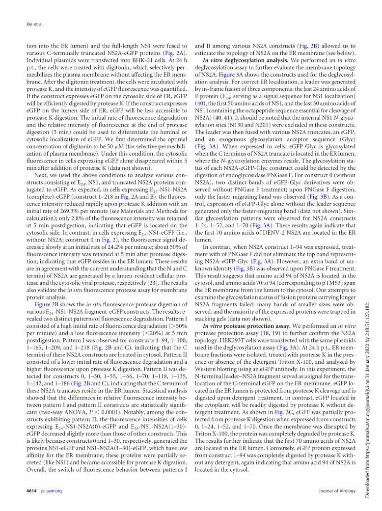
In vitro deglycosylation analysis. We performed an in vitrodeglycosylation assay to further evaluate the membrane topologyof NS2A. Figure 3A shows the constructs used for the deglycosyl-ation analysis. For correct ER localization, a leader was generatedby in-frame fusion of three components: the last 24 amino acids ofE protein (E24, serving as a signal sequence for NS1 localization)(40), the first 50 amino acids of NS1, and the last 50 amino acids ofNS1 (containing the octapeptide sequence essential for cleavage ofNS2A) (40, 41). It should be noted that the internal NS1 N-glyco-sylation sites (N130 and N201) were excluded in these constructs.The leader was then fused with various NS2A truncates, an eGFP,and an exogenous glycosylation acceptor sequence (Glyc)(Fig. 3A). When expressed in cells, eGFP-Glyc is glycosylatedwhen the C terminus of NS2A truncate is located in the ER lumen,where the N-glycosylation enzymes reside. The glycosylation sta-tus of each NS2A-eGFP-Glyc construct could be detected by thedigestion of endoglycosidase PNGase F. For construct 0 (withoutNS2A), two distinct bands of eGFP-Glyc derivatives were ob-served without PNGase F treatment; upon PNGase F digestion,only the faster-migrating band was observed (Fig. 3B). As a con-trol, expression of eGFP-Glyc alone without the leader sequencegenerated only the faster-migrating band (data not shown). Sim-ilar glycosylation patterns were observed for NS2A constructs1–24, 1–52, and 1–70 (Fig. 3A). These results again indicate thatthe first 70 amino acids of DENV-2 NS2A are located in the ERlumen.
In contrast, when NS2A construct 1–94 was expressed, treat-ment with of PNGase F did not eliminate the top band represent-ing NS2A-eGFP-Glyc (Fig. 3A). However, an extra band of un-known identity (Fig. 3B) was observed upon PNGase F treatment.This result suggests that amino acid 94 of NS2A is located in thecytosol, and amino acids 70 to 94 (corresponding to pTMS3) spanthe ER membrane from the lumen to the cytosol. Our attempts toexamine the glycosylation status of fusion proteins carrying longerNS2A fragments failed: many bands of smaller sizes were ob-served, and the majority of the expressed proteins were trapped instacking gels (data not shown).
In vitro protease protection assay. We performed an in vitroprotease protection assay (18, 19) to further confirm the NS2Atopology. HEK293T cells were transfected with the same plasmidsused in the deglycosylation assay (Fig. 3A). At 24 h p.t., ER mem-brane fractions were isolated, treated with protease K in the pres-ence or absence of the detergent Triton X-100, and analyzed byWestern blotting using an eGFP antibody. In this experiment, theN-terminal leader–NS2A fragment served as a signal for the trans-location of the C-terminal eGFP on the ER membrane. eGFP lo-cated in the ER lumen is protected from protease K cleavage and isdigested upon detergent treatment. In contrast, eGFP located inthe cytoplasm will be readily digested by protease K without de-tergent treatment. As shown in Fig. 3C, eGFP was partially pro-tected from protease K digestion when expressed from constructs0, 1–24, 1–52, and 1–70. Once the membrane was disrupted byTriton X-100, the protein was completely degraded by protease K.The results further indicate that the first 70 amino acids of NS2Aare located in the ER lumen. Conversely, eGFP protein expressedfrom construct 1–94 was completely digested by protease K with-out any detergent, again indicating that amino acid 94 of NS2A islocated in the cytosol.
Xie et al.
4614 jvi.asm.org Journal of Virology
Dow
nloa
ded
from
http
s://j
ourn
als.
asm
.org
/jour
nal/j
vi o
n 31
Jan
uary
202
2 by
218
.51.
123.
182.

FIG 2 In situ fluorescence protease protection assay. (A) E24-NS1-NS2A (truncated)-eGFP fusion constructs. Each NS2A fragment was N-terminally fused withE24-NS1 (representing a signal peptide derived from the last 24 residues of E protein [E24] and NS1 protein), and C-terminally fused with eGFP. pTMS are shownas gray boxes. (B) In situ fluorescence protease protection assay. BHK-21 cells were transfected with the indicated NS2A constructs. At 24 h p.t., the cells werepermeabilized with digitonin followed by protease K treatment. Once the protease K was added, the fluorescence intensities were continuously quantified for 300s (see Materials and Methods). Relative intensities were calculated using the fluorescence intensity at every 8 s divided by that at time zero when protease K wasadded. The means of relative intensities derived from 5 to 7 fields (each field containing 4 to 6 positive cells) are presented. The positions of NS2A truncates areindicated in the corresponding panels. (C) Summary of relative fluorescence intensity and initial rate of fluorescence degradation. Average results from at leasttwo independent experiments are presented. See Materials and Methods for calculations.
April 2013 Volume 87 Number 8 jvi.asm.org 4615
Dow
nloa
ded
from
http
s://j
ourn
als.
asm
.org
/jour
nal/j
vi o
n 31
Jan
uary
202
2 by
218
.51.
123.
182.

Our attempt to use the in vitro protease assay to analyze theconstructs carrying longer NS2A fragments failed (data notshown) for the reason described above. It should be noted that inour deglycosylation and in vitro protease protection assays, theN-terminal leader was not cleaved from the NS2A fragments (Fig.3B and C). The result demonstrates that the N-terminal 90 resi-dues of NS2A is not sufficient for the cleavage at the NS1-NS2Ajunction. This observation agrees with a previous report that mostof the N-terminal NS2A (70%) is required for the cleavage at theNS1-NS2A junction (23).
A topology model of DENV NS2A on the ER membrane. Toconsolidate the above results, we propose a topology model ofDENV-2 NS2A on the ER membrane (Fig. 4A). The N-terminal 68amino acids are located in the ER lumen; within this region,pTMS1 does not have membrane-associated activity, whereaspTMS2 is likely to peripherally associate with ER membrane with-out spanning the lipid bilayer. Amino acids 69 to 209 form five realTMS, each of which integrally spans the ER membrane. pTMS3,pTMS6, and pTMS8 span the membrane from lumen to cytosol;pTMS4 and pTMS7 transverse the membrane from cytosol to lu-men. pTMS5 does not have membrane-associated activity. TheC-terminal amino acids 210 to 218 are located in cytosol.
Sequence analysis of DENV-2 NS2A using the HeliQuest pro-gram suggests that amino acids 8 to 25 (overlapping pTMS1), 49to 66 (overlapping three residues with pTMS2), and 123 to 140(within pTMS5) could form amphipathic helices (pAH1-3)(Fig. 4B). The charged residues on one side of the helices couldpotentially form transient interactions with the negatively chargedphosphates on the lipid membrane.
NMR structure of pTMS3. The data above clearly showed thatpTMS3 is a real transmembrane domain. This result prompted usto solve the NMR structure of pTMS3 (Protein Data Bank [PDB]identification number 2M0S). A peptide corresponding to aminoacids D67-R94 was synthesized and purified. Due to its hydropho-bic nature, we solubilized the peptide in deuterated detergents,including SDS and dodecylphosphocholine (DPC) (42). How-ever, poorly resolved NMR spectra were obtained under theseconditions (data not shown). Interestingly, when solubilized in anorganic solvent (see Materials and Methods), the peptide yielded awell-resolved spectrum that allowed us to complete the resonanceassignment for the HSQC and NOESY spectra (Fig. 5A and B).Next, chemical shift index (CSI) analysis (43) was performed us-ing the deviation of 1H� and 13C� chemical shifts from the ran-dom coil values (Fig. 5C). The negative variation of 1H� chemicalshifts and the positive variation of the 13C� chemical shifts wereobserved for residues G69-K82 and T86-L91, indicating that theseamino acids are folded as � helices. There was a “break” presentbetween the two helices due to the presence of P85. The NOEassignment for this peptide further supported the CSI analysis(Fig. 5C). The NOE connectivity of a typical �-helix was observedfor these two helical fragments containing residues G69 to K82and T86 to L91, in which strong dNN (i, i 1) (NOESY connec-tion observed between amide protons) and weak d�N (i, i 4)(NOESY connections observed between alpha and amide pro-tons) NOE connections were observed (Fig. 5D).
The NMR data suggest that pTMS3 consists of two �-helicesseparated by a helix “breaker” (Fig. 5). The three-dimensional(3D) structure was determined with 239 distance constraints, in-
FIG 3 Deglycosylation and in vitro protease protection assays. (A) Leader-NS2A truncate-eGFP-Glyc constructs. The leader sequence contains a signalpeptide derived from the last 24 residues of E protein (E24; black box), the first 50 residues of NS1 (striped box), and the last 50 residues of NS1 (white box).pTMS are shown as gray boxes. Each NS2A fragment was N-terminally fused with the leader sequence and C-terminally fused with eGFP-Glyc (aglycosylation sequence). The amino acid positions of each NS2A constructs are indicated on the left. (B) In vitro deglycosylation assay. HEK293T cellswere transfected with various constructs depicted in (A). An extra band of unknown identity is indicated with an asterisk. At 24 h p.t., the expressed fusionproteins were extracted and treated with PNGase F () or PBS (�) . A mouse monoclonal antibody against GFP was used to probe eGFP-tagged proteins(see Materials and Methods). Molecular mass markers are shown on the left. (C) In vitro protease protection assay. Cell membranes of the transfected cellsas described for panel B were extracted and digested with protease K in the presence or absence of 0.5% Trion X-100. The samples were analyzed byWestern blotting as described for panel B.
Xie et al.
4616 jvi.asm.org Journal of Virology
Dow
nloa
ded
from
http
s://j
ourn
als.
asm
.org
/jour
nal/j
vi o
n 31
Jan
uary
202
2 by
218
.51.
123.
182.

cluding 86 intraresidue, 85 sequential, and 68 medium-rangeNOEs (Table 2). No hydrogen bond constraints were applied dur-ing the structural determination. Figure 6A shows the set of 15structures with the lowest energy function out of 100 calculatedstructures with C� traces. The two �-helical structures were welldefined, with root mean square deviations (RMSD) of 0.05 Å forthe backbone and 0.78 Å for the heavy atoms (Fig. 6A and B). Thestructures of the N- and C-terminal residues are flexible due to alack of distance restraints; this result is consistent with the CSIanalysis (Fig. 5). Figure 6C is a ribbon representation of pTMS3showing two positively charged residues (K82 and R84) and P85;these three residues make the two helices flexible. A surface rep-resentation of the pTMS3 displays a hydrophobic surface at oneside of this domain (Fig. 6D) and two positively charged patcheson the other side of this domain, which arise from the presence ofK82 and R84 (Fig. 6E).
Functional analysis of NS2A R84 and P85. To examine thebiological relevance of the helix breaker identified in pTMS3, weperformed mutagenesis of R84 and P85 in the context of repliconand genome-length RNA of DENV-2. R84 was replaced by theamino acid A, E, or S. P85 was mutated to A to eliminate the helixbreaker. In the transient transfection assay using a luciferase rep-licon (Fig. 7), none of the mutations affected luciferase signals at 2,4, and 6 h p.t. (representing input RNA translation). Only themutation R84E reduced luciferase activity by 20 to 59% at 24, 30,and 42 h p.t. (indicating RNA synthesis); all other mutations did
not change the luciferase signals compared with the wild-type(WT) replicon. The replicon results indicate that, unlike the mu-tation R84E (which attenuates viral RNA synthesis), the substitu-tions R84S and P85A do not affect viral translation or RNA syn-thesis.
Using an infectious cDNA clone of DENV-2, we examined theabove mutations (R84A, R84E, R84S, and P85A) in a completeviral replication cycle. Equal amounts of WT and mutant genome-length RNAs were transfected into BHK-21 cells. The transfectedcells were monitored for viral E protein expression using an IFAassay (Fig. 8A). Similar levels of E-positive cells were observed forthe WT and P85A mutant from day 1 to 4 p.t.; fewer E-positivecells were detected for mutants R84A and R84S; almost no in-crease in E-positive cells was observed for mutant R84E. Since thereplicon results showed that, except for the R84E substitution,other mutations had no effect on viral RNA synthesis (Fig. 7), thedecrease in the number of E-positive cells suggests that R84A andR84S mutants were defective in virus assembly/release. In agree-ment with the IFA result, mutant virus P85A displayed plaquemorphology similar to that of the WT virus, whereas the R84A,R84E, and R84S mutants exhibited plaques smaller than those ofthe WT virus (Fig. 8B). In addition, cells transfected with mutantR84S, R84A, or R84E RNAs did not yield any infectious virus at 24and 48 h p.t., and generated much less virus than those transfectedwith the WT RNA at 72 to 120 h posttransfection. In contrast,mutant P85A and WT RNAs generated similar amounts of infec-tious viruses (Fig. 8C). Sequencing of the recombinant virusesconfirmed that the engineered mutations were retained in the re-covered viruses without any other changes (data not shown).
The NS2A R84A mutation abolishes the formation of intra-cellular infectious virus. To further examine the role of R84 invirion assembly/release, we measured intra- and extracellularamounts of viral RNA as well as infectious viruses after BHK-21cells were transfected with the WT or R84A genomic RNA. Asshown in Fig. 9A, increasing amounts of intra- and extracellularinfectious viruses were observed for the WT RNA at 24 and 48 hp.t., whereas neither intra- nor extracellular infectious viruseswere detected for the R84A RNA. In contrast, at 0 and 12 h p.t., nostatistical significant difference in intra- or extracellular levels ofviral RNA was observed between the WT and the R84A mutant(Fig. 9B), indicating that the two RNAs were transfected into cellswith similar efficiencies. At 24 and 48 h p.t., the WT RNA gener-ated significantly more intra- and extracellular viral RNA than theR84A RNA (Fig. 9B). Since the replicon results showed thatthe R84A mutation does not affect viral RNA synthesis (Fig. 7), thehigher intracellular levels of WT viral RNA at 24 and 48 h p.t. weremost likely due to the reinfection of the newly produced virus.Collectively, the results indicate that the NS2A R84A mutationabolishes the formation of intracellular infectious virus.
Revertant analysis. Since R84A and R84E exhibited the mostdefective phenotype among the analyzed mutants (Fig. 8A to C),we chose these two mutants for revertant analysis. Both mutantviruses were continuously cultured on Vero cells and monitoredfor improved viral replication. After five rounds of passaging (4days per round), the R84E virus produced larger plaques than theunpassaged virus (Fig. 8D) and generated viral titers equivalent tothose of the WT virus (2.2 � 106 PFU/ml); sequencing of theresulting virus showed that the R84E mutation had been changedto R84K. Similarly, after five rounds of passaging, the R84A viruswas able to generate higher viral titers (1.7 � 105 PFU/ml). How-
FIG 4 Membrane topology of DENV NS2A. (A) A topology model ofDENV-2 NS2A on the ER membrane. The N-terminal 68 residues are locatedin the ER lumen; pTMS1 does not associate with the membrane, whereaspTMS2 is likely to peripherally associate with ER membrane. pTMS3, pTMS6,and pTMS8 span the membrane from lumen to cytosol, whereas pTMS4 andpTMS7 span the membrane from cytosol to lumen. The C-terminal 9 residuesare located in cytosol. It should be noted that the topology of pTMS4-8 issupported only by results from Fig. 1 and 2; due to technical difficulties (seedetails in the text), we were not able to validate the topology of pTMS4-8 by thedeglycosylation and in vitro protease protection assays (Fig. 3). (B) Three am-phipathic helices (pAH1, pAH2, and pAH3) predicted by HeliQuest. EachpAH construct is an �-helix composed of 18 amino acids. The numbers rep-resent amino acid positions of NS2A for each pAH. Red, blue, purple, gray, andyellow circles represent negatively charged, positively charged, polar, glycine/alanine, and hydrophobic residues, respectively.
Membrane Topology and Function of Dengue Virus NS2A
April 2013 Volume 87 Number 8 jvi.asm.org 4617
Dow
nloa
ded
from
http
s://j
ourn
als.
asm
.org
/jour
nal/j
vi o
n 31
Jan
uary
202
2 by
218
.51.
123.
182.

ever, the plaque morphology of the resulting virus remained small(Fig. 8D), and sequencing of the resulting virus revealed that theR84A mutation had been changed to R84T. For both revertantviruses, no other mutation was found throughout the genomicRNAs.
DISCUSSION
Flavivirus NS2A colocalizes with dsRNA, NS1, NS3, and NS5within the replication complex (3). Besides viral RNA synthesis,NS2A also functions in flavivirus assembly/release (24). The goalof this study was to determine the topology of flavivirus NS2A onthe ER membrane. Using a panel of biochemical methods, weestablished DENV-2 NS2A as an ER membrane protein with fivemembrane-spanning segments. NMR structure showed that thefirst transmembrane segment (pTMS3) of NS2A consists of twohelices separated by a helix breaker mediated by amino acids R84and P85. Functional analysis of the breaker residues showed that
only R84, but not P85, is critical for viral RNA synthesis as well asvirion assembly/release.
We initially used multiple bioinformatics algorithms to predictthe topology of NS2A that could be experimentally tested. Variousalgorithms predicted different topology models with only onecommon TMS from amino acids 30 to 57 (Fig. 1A). To avoiddiscounting any potential TMS, we consolidated all eight pre-dicted TMS into a test model (Fig. 1B). When each predicted TMSwas individually fused to GFP, six of the eight predicted pTMS(pTMS2, -3, -4, -6, -7, and -8) could anchor GFP to the ER mem-brane, whereas the other two predicted pTMS (pTMS1 and -5)could not (Fig. 1C). The IFA-based cellular localization results(Fig. 1C) are in general agreement with the membrane flotationdata (Fig. 1D), with the exception that the correlation of the re-sults between the two methods is weak for pTMS8 (equivalent tofragment 186 –209). The membrane-associated activity of pTMS8was strengthened by the in situ fluorescence protease protection
FIG 5 Resonance assignment of pTMS3. (A) 1H-15N-HSQC of pTMS3 in 15N natural abundance. The assigned cross-peaks are labeled with amino acid andresidue position. (B) Assignment of the 2D NOESY spectrum of pTMS3 in the H� region. Connections between residues are shown with lines. (C) Secondarystructure analysis of pTMS3. Deviations of C� (top) and H� (bottom) from random coil values are shown. (D) Summary of NOE connectivity. The NOE plotwas made from CYANA.
Xie et al.
4618 jvi.asm.org Journal of Virology
Dow
nloa
ded
from
http
s://j
ourn
als.
asm
.org
/jour
nal/j
vi o
n 31
Jan
uary
202
2 by
218
.51.
123.
182.

result (Fig. 2B), which clearly showed that pTMS8 spans from theER lumen to the cytosol. In the in situ fluorescence protease pro-tection assay, the membrane-associated activity of pTMS8 couldbe enhanced through its interaction with upstream NS2A se-quence; no such interaction occurred in the cellular localizationand membrane flotation assays when only individual NS2A frag-ments were examined in the absence of upstream NS2A sequence.
It should be noted that pTMS that showed ER membrane-interacting activity do not necessarily span the entire lipid bilayerof ER membrane; some of the pTMS (such as pTMS2) could pe-ripherally interact with the membrane without traversing thecomplete lipid bilayer. Sequence analysis of DENV-2 NS2A usingHeliQuest program suggests that amino acids 8 to 25 (overlappingpTMS1), 49 to 66 (overlapping three residues with pTMS2), and123 to 140 (within pTMS5) could form amphipathic helices(pAH1-3) (Fig. 4B). The hydrophilic sides of these amphipathichelices may peripherally or transiently interact with ER mem-brane on the lumen side. However, the results presented in Fig. 1C
and D demonstrate that none of these three fragments are associ-ated with ER membrane when expressed individually, thus argu-ing against the possibility of their membrane-associated activity.
We used an in situ fluorescence protease protection assay toexamine a panel of C-terminally truncated forms of NS2A fusedwith a GFP reporter (Fig. 2). This assay allowed us to differentiatebetween the cytosolic and noncytosolic residues in cells expressingthe various NS2A-GFP fusion proteins. The results indicate that,among the six pTMS that showed membrane-interacting activities(Fig. 1C and D), pTMS2 is located in the ER lumen, whereaspTMS3, pTMS4, pTMS6, pTMS7, and pTMS8 span the ER mem-brane (Fig. 2B). Based on these results, we propose a topologymodel of DENV-2 NS2A on the ER membrane (Fig. 4A). In thismodel, the first 69 amino acids of NS2A are located in the ER
TABLE 2 Statistics for the structural models of pTMS3
Parameter Value
No. of:Restraints 239Intraresidue NOEs 86Sequential NOEs (i to i 1) 85Medium range NOEs (i to i 2,3,4) 68Dihedral angle restraints 0NOE violations 0.5 Å 0
Ramachandran plot (%)Residues in most favored regions 82Residues in additional allowed regions 17.5Residues in generously allowed regions 0.2Residues in disallowed regions 0.2
RMSD (Å) for:Backbone atoms (4–16) 0.05Heavy atoms (4–16) 0.78Backbone atoms (20–24) 0.05Heavy atoms (4–16) 0.74
FIG 6 Structures of NS2A pTMS3. (A) Stereo view of the ensemble of 15 NMR structures from CYANA. Only the C� trace is shown with PyMOL. (B) Cartoonrepresentation of the pTMS3 structure. Two �-helices are shown with labels. (C) Positions of K82, R84, and P85 in pTMS3. In the cartoon representation, the sidechains of K82, R84, and P85 are shown in stick mode. (D) Surface representation of pTMS3. The charge analysis was conducted in Pymol. The positively chargedresidues, negatively charged residues, and hydrophobic residues are shown in blue, red, and white, respectively. (E) Surface representation of pTMS3 with a 180°rotation from panel D. All images were prepared using Pymol (www.pymol.org).
FIG 7 Replicon analysis of NS2A R84 and P85. (A) Schematic diagram of aluciferase replicon of DENV-2. Rluc2A, Renilla luciferase gene followed by thefoot-and-mouth disease virus 2A peptide; C38, nucleotides encoding the first38 amino acids of C protein; E31, nucleotides encoding the last 31 amino acidsof E protein; HDVr, hepatitis delta virus ribozyme sequence. (B) Transientreplicon assay. Equal amounts of replicon RNA (WT or mutant R84A, R84E,R84S, or P85A) were electroporated into BHK-21 cells. At the indicated timepoints, the transfected cells were lysed and assayed for luciferase activities. They axis shows the log10 value of Renilla luciferase activity (RLU). Each data pointis the average for three replicates, and error bars show the standard deviations.
Membrane Topology and Function of Dengue Virus NS2A
April 2013 Volume 87 Number 8 jvi.asm.org 4619
Dow
nloa
ded
from
http
s://j
ourn
als.
asm
.org
/jour
nal/j
vi o
n 31
Jan
uary
202
2 by
218
.51.
123.
182.

lumen. This conclusion is supported by three lines of evidence. (i)Both the N and C termini of pTMS2 were only weakly accessible toprotease digestion in situ (Fig. 2B). (ii) Fragments representingvarious portions of the first 70 amino acids of NS2A were pro-tected from protease cleavage in vitro (Fig. 3C). (iii) When thesefragments were C-terminally fused with a glycosylation signal, the
resulting peptides were all glycosylated (Fig. 3B). As discussedabove, when pTMS2 was individually fused with GFP, it targetedGFP to the ER membrane (Fig. 1C and D). To consolidate all theseresults, we surmise that pTMS2 may peripherally interact with theER membrane on the lumen side (Fig. 4A).
Our model contradicts a previous report that YFV NS2A resi-
FIG 8 Analysis of NS2A R84 and P85 in DENV-2 genome-length RNA. (A) IFA analysis. Equal amounts of WT or mutant genome-length RNA wereelectroporated into BHK-21 cells. The transfected cells were monitored for E protein expression at indicated time points. An anti-E monoclonal antibody 4G2and a goat anti-mouse IgG conjugated with Alexa Fluor 488 were used as primary and secondary antibodies, respectively. Green and blue indicate E protein andnucleus staining, respectively. (B) Plaque morphology of WT and mutant viruses. The viruses were derived from the media collected on day 5 posttransfection.(C) Virus production. Culture media from cells transfected with genome-length RNAs from (A) were collected at the indicated time points; viral titers werequantified by plaque assay. Average results with standard deviations are presented. The dashed line indicates the limit of detection (L.O.D; 10 PFU/ml). (D)Plaque morphology of revertant viruses after 5 rounds of passaging on Vero cells. Amino acid substitutions identified at position 84 of NS2A are indicated.
Xie et al.
4620 jvi.asm.org Journal of Virology
Dow
nloa
ded
from
http
s://j
ourn
als.
asm
.org
/jour
nal/j
vi o
n 31
Jan
uary
202
2 by
218
.51.
123.
182.

dues 189 to 191 (corresponding to DENV-2 NS2A residues 186 to188) are cytosolic. These residues form an internal cleavage site forYFV protease NS2B/NS3, leading to the production of NS2A� (8,44). This discrepancy may imply a variation in the NS2A topologyamong flaviviruses.
We solved the NMR structure of pTMS3 (Fig. 6). pTMS3 con-sists of two helices separated by a helix breaker mediated by resi-due P85. Two charged residues, K82 and R84, are located up-stream of the breaker within this transmembrane region. Weexplored the function of this unique breaker structure by mu-tagenesis analysis. In both replicon (Fig. 7) and genome-length(Fig. 8) RNA, substitution of P85A did not show any defects inviral infection cycle, indicating that the breaker structure itself isnot important for DENV-2 replication. However, the R84E mu-tation reduced viral RNA synthesis and the production of infec-tious virus. The decrease in virus production could be caused bythe reduction of viral RNA synthesis; alternatively, this mutationcould directly reduce the efficiency of virion assembly/release. In-terestingly, when the same residue was changed to amino acid A orS (R84A or R84S), the mutation showed no effect on viral RNAsynthesis, as indicated by the replicon results (Fig. 7); remarkably,both substitutions generated fewer virus particles (Fig. 8 and 9).Further analysis showed that no infectious virion was producedinside the cells transfected with the mutant R84A RNA, whereashigh levels of intracellular infectious virion were generated in theWT RNA-transfected cells (Fig. 9). Collectively, these results dem-onstrate that residue R84 functions in both RNA synthesis (as
demonstrated by using the R84E mutation) and virion assembly/maturation (as demonstrated by using R84A).
In agreement with our results, the Rice group and the Khro-mykh group previously showed that NS2A plays a role in virionassembly/release in YFV (8) and KUNV (24), respectively. In thecase of KUNV, the compensatory mutation T149P in NS2A (cor-responding to amino acid 154 of DENV-2 NS2A) was able torescue the defects in virus assembly caused by the I59N mutation(corresponding to residue 58 of DENV-2 NS2A) (24). If the cur-rent topology model of DENV-2 NS2A is applicable to KUNV, thegenetic data imply that I59 with in the predicted pAH2 (Fig. 4B)may interact with pTMS6 to regulate virus assembly. In additionto the intramolecular interactions reported in KUNV NS2A, anintermolecular interaction between YFV NS2A residue K190S(corresponding to residue 188 in DENV-2 NS2A) and NS3 D343(within helicase domain) was reported to be important for YFVassembly/release (8). It remains to be determined how these non-structural protein interactions modulate flavivirus assembly. Inthe present study, we also performed revertant analysis usingR84A and R84E mutants. Reversions of R84A to R84K and R84Eto R84T were recovered; both revertants were found to enhanceviral replication (Fig. 8D); unfortunately, no intramolecular orintermolecular interaction was obtained in our study.
In summary, the present study identified the DENV-2 NS2A asa protein with five membrane-spanning segments. The proposedtopology model could serve as a starting point to investigate themechanism of flavivirus NS2A in viral replication and its modu-lation of host immune response. Structural and functional analy-sis of the first ER membrane-spanning region (pTMS3) ofDENV-2 NS2A showed its function in both viral RNA synthesisand virion assembly. Studies are ongoing to understand how fla-vivirus NS2A modulates the two events of viral RNA synthesis andvirion assembly.
ACKNOWLEDGMENTS
We thank Jing Zou and other colleagues at Novartis Institute for TropicalDiseases for technical support and helpful discussions during the courseof this study.
This work was partially supported by the TCR flagship “STOP Den-gue” program of the National Medical Research Council in Singapore.C.K. is supported by an A*STAR Investigatorship. Z.Y.’s group is sup-ported by Important National Science & Technology Specific Project(2009ZX10004-504) and National Natural Scientific Fund of China(81072675).
REFERENCES1. Lindenbach BD, Thiel H-J, Rice CM. 2007. Flaviviridae: the viruses and
their replication, p 1101–1152. In Knipe DM, Howley PM (ed), Fieldsvirology, 5th ed, vol 1. Lippincott William & Wilkins, Philadelphia, PA.
2. Cleaves GR, Dubin DT. 1979. Methylation status of intracellular denguetype 2 40 S RNA. Virology 96:159 –165.
3. Mackenzie JM, Khromykh AA, Jones MK, Westaway EG. 1998. Subcel-lular localization and some biochemical properties of the flavivirus Kunjinnonstructural proteins NS2A and NS4A. Virology 245:203–215.
4. Lindenbach BD, Rice CM. 1997. Trans-complementation of yellow fevervirus NS1 reveals a role in early RNA replication. J. Virol. 71:9608 –9617.
5. Avirutnan P, Punyadee N, Noisakran S, Komoltri C, Thiemmeca S, etal. 2006. Vascular leakage in severe dengue virus infections: a potentialrole for the nonstructural viral protein NS1 and complement. J. Infect.Dis. 193:1078 –1088.
6. Warrener P, Tamura JK, Collett MS. 1993. RNA-stimulated NTPaseactivity associated with yellow fever virus NS3 protein expressed in bacte-ria. J. Virol. 67:989 –996.
7. Wengler G, Czaya G, Farber PM, Hegemann JH. 1991. In vitro synthesis
FIG 9 Comparison of intra- and extracellular infectious viral particles andviral RNA between the WT and the R84A mutant. BHK-21 cells were trans-fected with equal amounts of WT and mutant genome-length RNA. At theindicated time points, intra- and extracellular infectious viral particles werequantified by plaque assay (A); viral RNAs were measured by quantitativeRT-PCR (B) (for details, see Materials and Methods). The intracellular viralparticles were normalized to the cell number and presented as PFU per 106
cells. The relative RNA was calculated using formula 100% � 2(CTi � CTo),where CTi is the CT value for the individual virus sample and CTo is the CT
value derived from the intracellular WT viral RNA collected at 0 h p.t. forintracellular RNA calculation or at 12 h p.t. for extracellular RNA calculation.The intracellular viral RNAs were normalized to the CT values derived fromGAPDH mRNA. An asterisk indicates statistical significance based on Stu-dent’s t test (P � 0.05). L.O.D, limit of detection. Each data point is an average;error bars indicate the standard deviations derived from three independentexperiments (n � 3).
Membrane Topology and Function of Dengue Virus NS2A
April 2013 Volume 87 Number 8 jvi.asm.org 4621
Dow
nloa
ded
from
http
s://j
ourn
als.
asm
.org
/jour
nal/j
vi o
n 31
Jan
uary
202
2 by
218
.51.
123.
182.

of West Nile virus proteins indicates that the amino-terminal segment ofthe NS3 protein contains the active centre of the protease which cleavesthe viral polyprotein after multiple basic amino acids. J. Gen. Virol. 72:851– 858.
8. Kummerer BM, Rice CM. 2002. Mutations in the yellow fever virusnonstructural protein NS2A selectively block production of infectiousparticles. J. Virol. 76:4773– 4784.
9. Patkar CG, Kuhn RJ. 2008. Yellow fever virus NS3 plays an essential rolein virus assembly independent of its known enzymatic functions. J. Virol.82:3342–3352.
10. Dong H, Chang DC, Hua MH, Lim SP, Chionh YH, Hia F, Lee YH,Kukkaro P, Lok SM, Dedon PC, Shi PY. 2012. 2=-O methylation ofinternal adenosine by flavivirus NS5 methyltransferase. PLoS Pathog.8:e1002642. doi:10.1371/journal.ppat.1002642.
11. Egloff MP, Benarroch D, Selisko B, Romette JL, Canard B. 2002. AnRNA cap (nucleoside-2=-O-)-methyltransferase in the flavivirus RNApolymerase NS5: crystal structure and functional characterization. EMBOJ. 21:2757–2768.
12. Ray D, Shah A, Tilgner M, Guo Y, Zhao Y, Dong H, Deas TS, Zhou Y,Li H, Shi PY. 2006. West Nile virus 5=-cap structure is formed by sequen-tial guanine N-7 and ribose 2=-O methylations by nonstructural protein 5.J. Virol. 80:8362– 8370.
13. Issur M, Geiss BJ, Bougie I, Picard-Jean F, Despins S, Mayette J,Hobdey SE, Bisaillon M. 2009. The flavivirus NS5 protein is a true RNAguanylyltransferase that catalyzes a two-step reaction to form the RNA capstructure. RNA 15:2340 –2350.
14. Ackermann M, Padmanabhan R. 2001. De novo synthesis of RNA by thedengue virus RNA-dependent RNA polymerase exhibits temperature de-pendence at the initiation but not elongation phase. J. Biol. Chem. 276:39926 –39937.
15. Ashour J, Laurent-Rolle M, Shi PY, Garcia-Sastre A. 2009. NS5 ofdengue virus mediates STAT2 binding and degradation. J. Virol. 83:5408 –5418.
16. Laurent-Rolle M, Boer EF, Lubick KJ, Wolfinbarger JB, Carmody AB,Rockx B, Liu W, Ashour J, Shupert WL, Holbrook MR, Barrett AD,Mason PW, Bloom ME, Garcia-Sastre A, Khromykh AA, Best SM. 2010.The NS5 protein of the virulent West Nile virus NY99 strain is a potentantagonist of type I interferon-mediated JAK-STAT signaling. J. Virol.84:3503–3515.
17. Welsch S, Miller S, Romero-Brey I, Merz A, Bleck CK, Walther P, FullerSD, Antony C, Krijnse-Locker J, Bartenschlager R. 2009. Compositionand three-dimensional architecture of the dengue virus replication andassembly sites. Cell Host Microbe 5:365–375.
18. Miller S, Kastner S, Krijnse-Locker J, Buhler S, Bartenschlager R. 2007.The non-structural protein 4A of dengue virus is an integral membraneprotein inducing membrane alterations in a 2K-regulated manner. J. Biol.Chem. 282:8873– 8882.
19. Miller S, Sparacio S, Bartenschlager R. 2006. Subcellular localization andmembrane topology of the Dengue virus type 2 non-structural protein 4B.J. Biol. Chem. 281:8854 – 8863.
20. Munoz-Jordan JL, Laurent-Rolle M, Ashour J, Martinez-Sobrido L,Ashok M, Lipkin WI, Garcia-Sastre A. 2005. Inhibition of alpha/betainterferon signaling by the NS4B protein of flaviviruses. J. Virol. 79:8004 –8013.
21. Munoz-Jordan JL, Sanchez-Burgos GG, Laurent-Rolle M, Garcia-SastreA. 2003. Inhibition of interferon signaling by dengue virus. Proc. Natl.Acad. Sci. U. S. A. 100:14333–14338.
22. Chambers TJ, McCourt DW, Rice CM. 1989. Yellow fever virus proteinsNS2A, NS2B, and NS4B: identification and partial N-terminal amino acidsequence analysis. Virology 169:100 –109.
23. Falgout B, Markoff L. 1995. Evidence that flavivirus NS1-NS2A cleavageis mediated by a membrane-bound host protease in the endoplasmic re-ticulum. J. Virol. 69:7232–7243.
24. Leung JY, Pijlman GP, Kondratieva N, Hyde J, Mackenzie JM, Khro-mykh AA. 2008. Role of nonstructural protein NS2A in flavivirus assem-bly. J. Virol. 82:4731– 4741.
25. Liu WJ, Chen HB, Wang XJ, Huang H, Khromykh AA. 2004. Analysisof adaptive mutations in Kunjin virus replicon RNA reveals a novel rolefor the flavivirus nonstructural protein NS2A in inhibition of beta inter-feron promoter-driven transcription. J. Virol. 78:12225–12235.
26. Liu WJ, Wang XJ, Clark DC, Lobigs M, Hall RA, Khromykh AA. 2006.A single amino acid substitution in the West Nile virus nonstructuralprotein NS2A disables its ability to inhibit alpha/beta interferon inductionand attenuates virus virulence in mice. J. Virol. 80:2396 –2404.
27. Tu YC, Yu CY, Liang JJ, Lin E, Liao CL, Lin YL. 2012. Blockingdouble-stranded RNA-activated protein kinase PKR by Japanese enceph-alitis virus nonstructural protein 2A. J. Virol. 86:10347–10358.
28. Melian EB, Hinzman E, Nagasaki T, Firth AE, Wills NM, Nouwens AS,Blitvich BJ, Leung J, Funk A, Atkins JF, Hall R, Khromykh AA. 2010.NS1= of flaviviruses in the Japanese encephalitis virus serogroup is a prod-uct of ribosomal frameshifting and plays a role in viral neuroinvasiveness.J. Virol. 84:1641–1647.
29. Zhou Y, Ray D, Zhao Y, Dong H, Ren S, Li Z, Guo Y, Bernard KA, ShiPY, Li H. 2007. Structure and function of flavivirus NS5 methyltrans-ferase. J. Virol. 81:3891–3903.
30. Zou G, Chen YL, Dong H, Lim CC, Yap LJ, Yau YH, Shochat SG, LescarJ, Shi PY. 2011. Functional analysis of two cavities in flavivirus NS5polymerase. J. Biol. Chem. 286:14362–14372.
31. Zou G, Xu HY, Qing M, Wang QY, Shi PY. 2011. Development andcharacterization of a stable luciferase dengue virus for high-throughputscreening. Antiviral Res. 91:11–19.
32. Gualano RC, Pryor MJ, Cauchi MR, Wright PJ, Davidson AD. 1998.Identification of a major determinant of mouse neurovirulence of denguevirus type 2 using stably cloned genomic-length cDNA. J. Gen. Virol.79:437– 446.
33. Lorenz H, Hailey DW, Wunder C, Lippincott-Schwartz J. 2006. Thefluorescence protease protection (FPP) assay to determine protein local-ization and membrane topology. Nat. Protoc. 1:276 –279.
34. Hooper R, Churamani D, Brailoiu E, Taylor CW, Patel S. 2011. Mem-brane topology of NAADP-sensitive two-pore channels and their regula-tion by N-linked glycosylation. J. Biol. Chem. 286:9141–9149.
35. Xie X, Wang QY, Xu HY, Qing M, Kramer L, Yuan Z, Shi PY. 2011.Inhibition of dengue virus by targeting viral NS4B protein. J. Virol. 85:11183–11195.
36. Groat-Carmona AM, Orozco S, Friebe P, Payne A, Kramer L, Harris E.2012. A novel coding-region RNA element modulates infectious denguevirus particle production in both mammalian and mosquito cells andregulates viral replication in Aedes aegypti mosquitoes. Virology 432:511–526.
37. Gayen S, Kang C. 2011. Solution structure of a human minimembraneprotein Ost4, a subunit of the oligosaccharyltransferase complex.Biochem. Biophys. Res. Commun. 409:572–576.
38. Wuthrich K. 1986. NMR of proteins and nucleic acids. Wiley, New York, NY.39. Guntert P. 2004. Automated NMR structure calculation with CYANA.
Methods Mol. Biol. 278:353–378.40. Somnuke P, Hauhart RE, Atkinson JP, Diamond MS, Avirutnan P.
2011. N-linked glycosylation of dengue virus NS1 protein modulates se-cretion, cell-surface expression, hexamer stability, and interactions withhuman complement. Virology 413:253–264.
41. Hori H, Lai CJ. 1990. Cleavage of dengue virus NS1-NS2A requires anoctapeptide sequence at the C terminus of NS1. J. Virol. 64:4573– 4577.
42. Kang C, Li Q. 2011. Solution NMR study of integral membrane proteins.Curr. Opin. Chem. Biol. 15:560 –569.
43. Mielke SP, Krishnan VV. 2004. An evaluation of chemical shift index-based secondary structure determination in proteins: influence of randomcoil chemical shifts. J. Biomol. NMR 30:143–153.
44. Nestorowicz A, Chambers TJ, Rice CM. 1994. Mutagenesis of the yellowfever virus NS2A/2B cleavage site: effects on proteolytic processing, viralreplication, and evidence for alternative processing of the NS2A protein.Virology 199:114 –123.
Xie et al.
4622 jvi.asm.org Journal of Virology
Dow
nloa
ded
from
http
s://j
ourn
als.
asm
.org
/jour
nal/j
vi o
n 31
Jan
uary
202
2 by
218
.51.
123.
182.