Membrane Sculpting by F-BAR Domains Studied by Molecular ...
15
Membrane Sculpting by F-BAR Domains Studied by Molecular Dynamics Simulations Hang Yu 1,2 , Klaus Schulten 1,2,3 * 1 Beckman Institute, University of Illinois, Urbana, Illinois, United States of America, 2 Center of Biophysics and Computational Biology, University of Illinois, Urbana, Illinois, United States of America, 3 Department of Physics, University of Illinois, Urbana, Illinois, United States of America Abstract Interplay between cellular membranes and their peripheral proteins drives many processes in eukaryotic cells. Proteins of the Bin/Amphiphysin/Rvs (BAR) domain family, in particular, play a role in cellular morphogenesis, for example curving planar membranes into tubular membranes. However, it is still unclear how F-BAR domain proteins act on membranes. Electron microscopy revealed that, in vitro, F-BAR proteins form regular lattices on cylindrically deformed membrane surfaces. Using all-atom and coarse-grained (CG) molecular dynamics simulations, we show that such lattices, indeed, induce tubes of observed radii. A 250 ns all-atom simulation reveals that F-BAR domain curves membranes via the so-called scaffolding mechanism. Plasticity of the F-BAR domain permits conformational change in response to membrane interaction, via partial unwinding of the domains 3-helix bundle structure. A CG simulation covering more than 350 ms provides a dynamic picture of membrane tubulation by lattices of F-BAR domains. A series of CG simulations identified the optimal lattice type for membrane sculpting, which matches closely the lattices seen through cryo-electron microscopy. Citation: Yu H, Schulten K (2013) Membrane Sculpting by F-BAR Domains Studied by Molecular Dynamics Simulations. PLoS Comput Biol 9(1): e1002892. doi:10.1371/journal.pcbi.1002892 Editor: Helmut Grubmuller, Max Planck Institute for Biophysical Chemistry, Gottingen, Germany Received August 17, 2012; Accepted December 2, 2012; Published January 31, 2013 Copyright: ß 2013 Yu, Schulten. This is an open-access article distributed under the terms of the Creative Commons Attribution License, which permits unrestricted use, distribution, and reproduction in any medium, provided the original author and source are credited. Funding: This work has been supported by the National Institutes of Health grants R01-GM067887, P41-RR005969 and U54-GM087519. The authors gratefully acknowledge computer time provided under the Extreme Science and Engineering Discovery Environment (XSEDE), which is supported by National Science Foundation grant number OCI-1053575. This research also used resources of the Argonne Leadership Computing Facility at Argonne National Laboratory, which is supported by the Office of Science of the U.S. Department of Energy under contract DE-AC02-06CH11357. The authors acknowledge, furthermore, the use of the parallel computing resource provided by the Computational Science and Engineering Program at the University of Illinois. The funders had no role in study design, data collection and analysis, decision to publish, or preparation of the manuscript. Competing Interests: The authors have declared that no competing interests exist. * E-mail: [email protected] Introduction Interplay between cellular membranes and their peripheral proteins drives many cellular processes, including cell division, growth, movement and cell-cell communication [1–6]. During their lifetime and often with the help of membrane peripheral proteins, eukaryotic cells dynamically sculpt their various types of compartments [2,5,7–12]. Recently, increasing attention has been paid to these proteins [11–23]. Proteins of the Bin/Amphiphysin/Rvs (BAR) domain family play an important role in membrane remodeling, by inducing and stabilizing membrane curvature [13,24–26]. For example, BAR domain deficiency is related to a wide range of cancers and blood disorders [27]. Resolved structures show that BAR domains form crescent-shaped homodimers, the monomers being composed of coiled-coil association of a 3-helix bundle structure [13,28–31]. Three sub-families of BAR domains, namely N-BAR domains, FCH-BAR (F-BAR) domains and Inverse-BAR (I-BAR) domains, differ from each other in their structure and physiological function [7,32–36]. In contrast to N-BAR domains that form a banana shaped dimer, F-BAR domains are elongated and only gently curved [37,38]. A high density of positive charge is found on the part of the protein that is destined to interact with negatively-charged membranes [2,30,39,40]. While N-BAR do- mains stabilize highly curved membrane structures, F-BAR domains stabilize membrane structures of small degree of curvature [13,30,32,38,41]. N-BAR domains also have an N-terminal amphipathic helix, which aids membrane curvature stabilization by membrane insertion. Such helix is lacking in the case of F-BAR domains [37,38]. Both N-BAR domains and F- BAR domains are found to induce formation of tubules in vitro [17,18,37]. Two mechanisms of membrane curvature generation by BAR domain proteins have been proposed [7,13,35,36,38]. According to the scaffolding mechanism, BAR domains bend membranes by attracting negatively-charged lipid headgroups to their positively- charged curved surface [7,13,36,38]. During the scaffolding process, a BAR domain is considered to act as a rigid body, to which lipids are attracted via electrostatic interaction, transferring membrane binding energy into membrane bending energy [36,42]. According to the membrane insertion mechanism, a BAR domain inserts its amphipathic groups like wedges into one leaflet of the membrane and, thereby, curves the membrane [2,35,38]. N-BAR proteins use their N-helix as an amphipathic wedge, while for the F-BAR domain it is suspected that residue Phe117 inserts its bulky side chain into the membrane [7,30,38,43–46]. Either mechanism involves strong membrane- protein interactions. BAR domains are found to shape low-curvature liposomes into high-curvature tubules in vitro [7,38,47]. Such extensive membrane remodeling requires collective action of multiple BAR domains. Striations observed on the surface of BAR domain-induced tubules suggest that the tubules are covered by an ordered arrangement of the proteins [7,19,38,47]. Recent observations revealed that PLOS Computational Biology | www.ploscompbiol.org 1 January 2013 | Volume 9 | Issue 1 | e1002892
Transcript of Membrane Sculpting by F-BAR Domains Studied by Molecular ...
Membrane Sculpting by F-BAR Domains Studied byMolecular Dynamics SimulationsHang Yu1,2, Klaus Schulten1,2,3*
1 Beckman Institute, University of Illinois, Urbana, Illinois, United States of America, 2 Center of Biophysics and Computational Biology, University of Illinois, Urbana, Illinois,
United States of America, 3 Department of Physics, University of Illinois, Urbana, Illinois, United States of America
Abstract
Interplay between cellular membranes and their peripheral proteins drives many processes in eukaryotic cells. Proteins ofthe Bin/Amphiphysin/Rvs (BAR) domain family, in particular, play a role in cellular morphogenesis, for example curvingplanar membranes into tubular membranes. However, it is still unclear how F-BAR domain proteins act on membranes.Electron microscopy revealed that, in vitro, F-BAR proteins form regular lattices on cylindrically deformed membranesurfaces. Using all-atom and coarse-grained (CG) molecular dynamics simulations, we show that such lattices, indeed,induce tubes of observed radii. A 250 ns all-atom simulation reveals that F-BAR domain curves membranes via the so-calledscaffolding mechanism. Plasticity of the F-BAR domain permits conformational change in response to membraneinteraction, via partial unwinding of the domains 3-helix bundle structure. A CG simulation covering more than 350 msprovides a dynamic picture of membrane tubulation by lattices of F-BAR domains. A series of CG simulations identified theoptimal lattice type for membrane sculpting, which matches closely the lattices seen through cryo-electron microscopy.
Citation: Yu H, Schulten K (2013) Membrane Sculpting by F-BAR Domains Studied by Molecular Dynamics Simulations. PLoS Comput Biol 9(1): e1002892.doi:10.1371/journal.pcbi.1002892
Editor: Helmut Grubmuller, Max Planck Institute for Biophysical Chemistry, Gottingen, Germany
Received August 17, 2012; Accepted December 2, 2012; Published January 31, 2013
Copyright: � 2013 Yu, Schulten. This is an open-access article distributed under the terms of the Creative Commons Attribution License, which permitsunrestricted use, distribution, and reproduction in any medium, provided the original author and source are credited.
Funding: This work has been supported by the National Institutes of Health grants R01-GM067887, P41-RR005969 and U54-GM087519. The authors gratefullyacknowledge computer time provided under the Extreme Science and Engineering Discovery Environment (XSEDE), which is supported by National ScienceFoundation grant number OCI-1053575. This research also used resources of the Argonne Leadership Computing Facility at Argonne National Laboratory, which issupported by the Office of Science of the U.S. Department of Energy under contract DE-AC02-06CH11357. The authors acknowledge, furthermore, the use of theparallel computing resource provided by the Computational Science and Engineering Program at the University of Illinois. The funders had no role in studydesign, data collection and analysis, decision to publish, or preparation of the manuscript.
Competing Interests: The authors have declared that no competing interests exist.
* E-mail: [email protected]
Introduction
Interplay between cellular membranes and their peripheral
proteins drives many cellular processes, including cell division,
growth, movement and cell-cell communication [1–6]. During
their lifetime and often with the help of membrane peripheral
proteins, eukaryotic cells dynamically sculpt their various types of
compartments [2,5,7–12]. Recently, increasing attention has been
paid to these proteins [11–23].
Proteins of the Bin/Amphiphysin/Rvs (BAR) domain family
play an important role in membrane remodeling, by inducing
and stabilizing membrane curvature [13,24–26]. For example,
BAR domain deficiency is related to a wide range of cancers
and blood disorders [27]. Resolved structures show that BAR
domains form crescent-shaped homodimers, the monomers
being composed of coiled-coil association of a 3-helix bundle
structure [13,28–31]. Three sub-families of BAR domains, namely
N-BAR domains, FCH-BAR (F-BAR) domains and Inverse-BAR
(I-BAR) domains, differ from each other in their structure and
physiological function [7,32–36]. In contrast to N-BAR domains
that form a banana shaped dimer, F-BAR domains are elongated
and only gently curved [37,38]. A high density of positive charge is
found on the part of the protein that is destined to interact with
negatively-charged membranes [2,30,39,40]. While N-BAR do-
mains stabilize highly curved membrane structures, F-BAR
domains stabilize membrane structures of small degree of
curvature [13,30,32,38,41]. N-BAR domains also have an
N-terminal amphipathic helix, which aids membrane curvature
stabilization by membrane insertion. Such helix is lacking in the
case of F-BAR domains [37,38]. Both N-BAR domains and F-
BAR domains are found to induce formation of tubules in vitro
[17,18,37].
Two mechanisms of membrane curvature generation by BAR
domain proteins have been proposed [7,13,35,36,38]. According
to the scaffolding mechanism, BAR domains bend membranes by
attracting negatively-charged lipid headgroups to their positively-
charged curved surface [7,13,36,38]. During the scaffolding
process, a BAR domain is considered to act as a rigid body, to
which lipids are attracted via electrostatic interaction, transferring
membrane binding energy into membrane bending energy
[36,42]. According to the membrane insertion mechanism, a
BAR domain inserts its amphipathic groups like wedges into one
leaflet of the membrane and, thereby, curves the membrane
[2,35,38]. N-BAR proteins use their N-helix as an amphipathic
wedge, while for the F-BAR domain it is suspected that residue
Phe117 inserts its bulky side chain into the membrane
[7,30,38,43–46]. Either mechanism involves strong membrane-
protein interactions.
BAR domains are found to shape low-curvature liposomes into
high-curvature tubules in vitro [7,38,47]. Such extensive membrane
remodeling requires collective action of multiple BAR domains.
Striations observed on the surface of BAR domain-induced tubules
suggest that the tubules are covered by an ordered arrangement
of the proteins [7,19,38,47]. Recent observations revealed that
PLOS Computational Biology | www.ploscompbiol.org 1 January 2013 | Volume 9 | Issue 1 | e1002892
well-organized spirals of BAR domains form on the surface of
membrane tubules [19,38]. Differences in lattices formed by BAR
domains may result in variations of membrane curvature and
structure [38]. However, it remains unclear how membrane
curvature depends on the type of F-BAR domain lattice
arrangement. Two further open questions are: How do individual
F-BAR domains interact with a membrane to form local
curvature? What dynamics is involved in membrane curvature
formation by F-BAR domain lattices?
Computational approaches, especially molecular dynamics
(MD) simulation, are proven tools for the study of membrane-
protein interactions [48–58]. Recent studies on membrane
deformation by BAR domain proteins include the study of local
deformation of membranes by single N-BAR domains
[12,33,46,59], of large-scale membrane structure deformation by
multiple N-BAR domains described by coarse-grained models
[24,25,60–62] and of large scale membrane deformation under the
influence of BAR domains [63–66].
Extending previous studies [24,25,60,61], we present here the
first all-atom molecular dynamics simulations of F-BAR domains
acting on a lipid bilayer in a fully solvated system. We explore the
system in an equilibrated state without restraints and seek to reveal
how F-BAR domains produce membrane curvature by confor-
mational change of their coiled-coil 3-helix bundle structure. We
also test the mechanism underlying membrane bending by
mutating key positively-charged residues of the F-BAR domain.
We then employ a shape-based coarse-grained (SBCG) model
developed in our group [60] to examine the effect of the F-BAR
domain lattice arrangement on membrane sculpting; variations of
the lattice are found to form a wide range of membrane
curvatures. Finally, we demonstrate through simulations how F-
BAR domain lattices form a complete membrane tubule.
Results/Discussion
F-BAR domains are known to bind to membrane surfaces and
generate membrane vesicles as well as tubules with radii in the
range 25–100 nm [7,19,38,47,67]. To reveal the mechanism of
membrane curvature generation by F-BAR domains, we employ
all-atom and coarse-grained molecular dynamics simulations to
characterize the effect of F-BAR domains on membrane
curvature. Table 1 lists size and timescale of the simulations
carried out and demonstrates the multiscale nature of the present
study.
The F-BAR domain binds and curves a membrane viascaffolding
The results of two separate all-atom equilibrium simulations of
single F-BAR domains (WT1 and NC) binding to negatively
charged lipid bilayers, consisting of 33% DOPS and 67% DOPC,
are shown in Fig. 1A and Videos S1 and S2. An F-BAR domain
dimer was placed on top of the resulting patch with no initial
contacts to the membrane. In simulation WT1, the wild type F-
BAR domain was employed; in simulation NC, the positive
charges of selected residues (see Methods) along the inner surface
of the wild type F-BAR domain were neutralized without changing
residue structure.
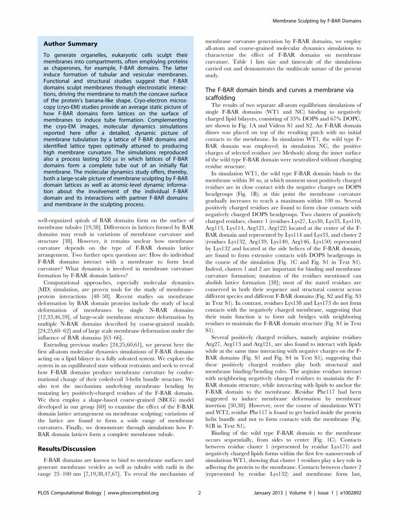
In simulation WT1, the wild type F-BAR domain binds to the
membrane within 30 ns, at which moment most positively charged
residues are in close contact with the negative charges on DOPS
headgroups (Fig. 1B); at this point the membrane curvature
gradually increases to reach a maximum within 100 ns. Several
positively charged residues are found to form close contacts with
negatively charged DOPS headgroups. Two clusters of positively
charged residues, cluster 1 (residues Lys27, Lys30, Lys33, Lys110,
Arg113, Lys114, Arg121, Arg122) located at the center of the F-
BAR domain and represented by Lys114 and Lys33, and cluster 2
(residues Lys132, Arg139, Lys140, Arg146, Lys150) represented
by Lys132 and located at the side helices of the F-BAR domain,
are found to form extensive contacts with DOPS headgroups in
the course of the simulation (Fig. 1C and Fig. S1 in Text S1).
Indeed, clusters 1 and 2 are important for binding and membrane
curvature formation; mutation of the residues mentioned can
abolish lattice formation [38]; most of the stated residues are
conserved in both their sequence and structural context across
different species and different F-BAR domains (Fig. S2 and Fig. S3
in Text S1). In contrast, residues Lys138 and Lys173 do not form
contacts with the negatively charged membrane, suggesting that
their main function is to form salt bridges with neighboring
residues to maintain the F-BAR domain structure (Fig. S1 in Text
S1).
Several positively charged residues, namely arginine residues
Arg27, Arg113 and Arg121, are also found to interact with lipids
while at the same time interacting with negative charges on the F-
BAR domains (Fig. S1 and Fig. S4 in Text S1), suggesting that
these positively charged residues play both structural and
membrane binding/bending roles. The arginine residues interact
with neighboring negatively charged residues to maintain the F-
BAR domain structure, while interacting with lipids to anchor the
F-BAR domain to the membrane. Residue Phe117 had been
suggested to induce membrane deformation by membrane
insertion [30,38]. However, over the course of simulations WT1
and WT2, residue Phe117 is found to get buried inside the protein
helix bundle and not to form contacts with the membrane (Fig.
S1B in Text S1).
Binding of the wild type F-BAR domain to the membrane
occurs sequentially, from sides to center (Fig. 1C). Contacts
between residue cluster 1 (represented by residue Lys171) and
negatively charged lipids forms within the first few nanoseconds of
simulations WT1, showing that cluster 1 residues play a key role in
adhering the protein to the membrane. Contacts between cluster 2
(represented by residue Lys132) and membrane form last,
Author Summary
To generate organelles, eukaryotic cells sculpt theirmembranes into compartments, often employing proteinsas chaperones, for example, F-BAR domains. The latterinduce formation of tubular and vesicular membranes.Functional and structural studies suggest that F-BARdomains sculpt membranes through electrostatic interac-tions, driving the membrane to match the concave surfaceof the protein’s banana-like shape. Cryo-electron micros-copy (cryo-EM) studies provide an average static picture ofhow F-BAR domains form lattices on the surface ofmembranes to induce tube formation. Complementingthe cryo-EM images, molecular dynamics simulationsreported here offer a detailed, dynamic picture ofmembrane tubulation by a lattice of F-BAR domains andidentified lattice types optimally attuned to producinghigh membrane curvature. The simulations reproducedalso a process lasting 350 ms in which lattices of F-BARdomains form a complete tube out of an initially flatmembrane. The molecular dynamics study offers, thereby,both a large-scale picture of membrane sculpting by F-BARdomain lattices as well as atomic-level dynamic informa-tion about the involvement of the individual F-BARdomain and its interactions with partner F-BAR domainsand membrane in the sculpting process.
Membrane Sculpting by F-BAR Domains
PLOS Computational Biology | www.ploscompbiol.org 2 January 2013 | Volume 9 | Issue 1 | e1002892
suggesting that cluster 2 residues are important for curvature
generation, by attracting lipid to the protein. All contacts between
positively charged residues and membrane formed within 40 ns of
simulation WT1.
Side loops formed by residues 56 to 60 maintain the F-BAR domain in an upright orientation
As shown in Fig. 1C, residue Arg57 forms a long lasting contact
with the membrane. This residue is located on a short loop formed
by residues 56 to 60. This loop contains dense positive charges
(Lys56, Arg57, Lys60) and partially inserts Pro58 into the
membrane. However, the insertion did not occur until 80 ns in
simulation WT1, i.e., after the protein is fully bound to the
membrane. The absence of loop insertion during the early stage of
protein-membrane interaction suggests that the 56–60 loop does
not contribute directly to membrane binding or initial curvature
development. The area of the membrane taken up by the loop is
0:6 nm2, which is much smaller than the membrane area taken by
the N-helix of the N-BAR domain. According to [63], to
effectively deform a membrane of 1000 nm2 with loop insertion,
at least 120 nm2 membrane area needs to be taken up by the
protein insertions, corresponding to 120 nm2=0:6 nm2~200loops, i.e., 100 F-BAR dimers. However, the area of membrane
plane taken by an F-BAR domain is 27:4 nm2 per dimer and for
100 F-BAR dimers, a lipid area of 27:4 nm2|100~2740 nm2 is
required. It is impossible to place 100 F-BAR dimers onto a
1000 nm2 membrane in an orientation that both loops of each
dimer contact the membrane. Therefore, it is unlikely that the
loop is involved in a major way in membrane bending. Indeed,
removing residues 56 to 60 showed no significant change in
membrane curvature during a 40 ns simulation (WT1DEL, see
Table 1), strengthening further the conclusion that membrane
insertion by the short loop does not contribute significantly to
membrane curvature formation (Fig. S5 in Text S1). However, the
F-BAR domain turning from an upright orientation to a side-
laying orientation was observed from 40 ns onwards and the
membrane curvature was found to decrease at the same time (Fig.
S5 in Text S1). In experiments, side-laying states are observed at
low BAR domain density and induce tubules of low curvatures
[9,38]. Therefore, the function of the 56–60 loop is likely a
structural one, namely maintaining the F-BAR domain in an
upright orientation and forming contacts with the membrane; the
function of the F-BAR domain loop is similar to the function of N-
helices in case of N-BAR domains.
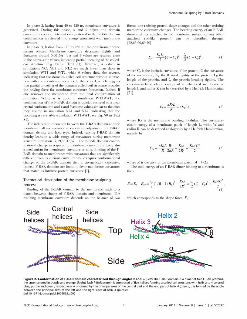
The F-BAR domain undergoes conformational changeduring membrane curvature generation
During the process of curvature generation, the F-BAR
domain interacts with the membrane and undergoes a large
conformational change involving its side helices (helices 3 and 4,
see Fig. 2). To represent the change we employ angle h and a. h is
formed by the principal axes of the central helix 4 (green, residues
241 to 257) and side helix 4 (green, residues 182 to 204); a
decrease of the h value corresponds to a straightening of the
domain. a is formed by the angle between the principal axes of
the left and right sides of helix 3 (purple, residue 120 to 166); a
decrease of the a value corresponds to an increase of overall
domain curvature.
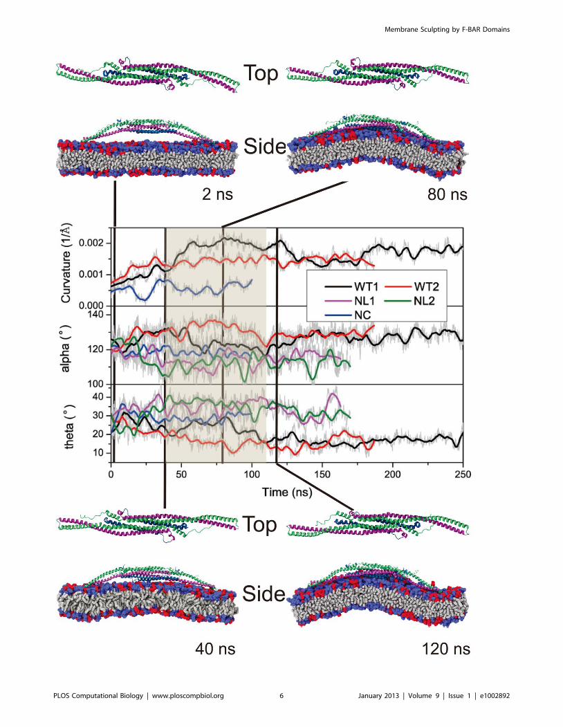
As shown in Fig. 3, both a and h of WT1 change significantly
upon interaction with the membrane; a increases up to 1400, then
decreases to 1200, fluctuating finally around 1300; h decreases to
200, then increases back to 300, fluctuating finally around 200. In
control systems NL1, NL2 and NC, a and h do not show such
changes and fluctuate around different average angles.
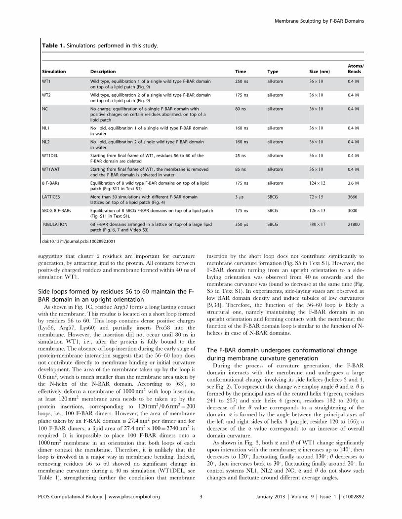
Table 1. Simulations performed in this study.
Simulation Description Time Type Size (nm)Atoms/Beads
WT1 Wild type, equilibration 1 of a single wild type F-BAR domainon top of a lipid patch (Fig. 9)
250 ns all-atom 36|10 0.4 M
WT2 Wild type, equilibration 2 of a single wild type F-BAR domainon top of a lipid patch (Fig. 9)
175 ns all-atom 36|10 0.4 M
NC No charge, equilibration of a single F-BAR domain withpositive charges on certain residues abolished, on top of alipid patch
80 ns all-atom 36|10 0.4 M
NL1 No lipid, equilibration 1 of a single wild type F-BAR domainin water
160 ns all-atom 36|10 0.4 M
NL2 No lipid, equilibration 2 of single wild type F-BAR domainin water
160 ns all-atom 36|10 0.4 M
WT1DEL Starting from final frame of WT1, residues 56 to 60 of theF-BAR domain are deleted
25 ns all-atom 36|10 0.4 M
WT1WAT Starting from final frame of WT1, the membrane is removedand the F-BAR domain is solvated in water
85 ns all-atom 36|10 0.4 M
8 F-BARs Equilibration of 8 wild type F-BAR domains on top of a lipidpatch (Fig. S11 in Text S1)
175 ns all-atom 124|12 3.6 M
LATTICES More than 30 simulations with different F-BAR domainlattices on top of a lipid patch (Fig. 4)
3 ms SBCG 72|15 3666
SBCG 8 F-BARs Equilibration of 8 SBCG F-BAR domains on top of a lipid patch(Fig. S11 in Text S1).
175 ns SBCG 126|13 3000
TUBULATION 68 F-BAR domains arranged in a lattice on top of a large lipidpatch (Fig. 6, 7 and Video S3)
350 ms SBCG 380|17 21800
doi:10.1371/journal.pcbi.1002892.t001
Membrane Sculpting by F-BAR Domains
PLOS Computational Biology | www.ploscompbiol.org 3 January 2013 | Volume 9 | Issue 1 | e1002892
a and h represent the conformational change of the F-BAR
domain in the horizontal and vertical direction. A high anti-
correlation is found between the change of a and h (Pearson
correlation coefficient = 20.5), corresponding to a synchronized
change of F-BAR domain side helices movement and protein
curvature. Visual inspection of the simulation reveals that the anti-
correlation of a and h changes correspond to a partial uncoiling
movement of the coiled-coil structure formed by side helices 3 and
4 (Fig. 3 and Fig. S6 in Text S1). An increase in a accompanied by
a decrease in h corresponds to the F-BAR domain forming a
shallow concave surface; little movement is observed for the
central helices (Fig. S4 in Text S1) and all helices retain their
helical structures during interaction between the F-BAR domain
and the membrane (Fig. S7 in Text S1).
As expected, when the F-BAR domain assumes a concave
shape, the attached membrane undergoes induced-fit bending.
Unlike N-BAR domains, which act like rigid bodies attracted to a
membrane [9,66,68], the F-BAR domain and the membrane
influence each others shape. Indeed, the bending energy of the F-
BAR domain is much lower than that of the N-BAR domain,
suggesting that the F-BAR domain is not as rigid as the N-BAR
domain [30,37,38]. Based on the conformation of the F-BAR
domain and membrane curvature, the curvature generation
process by the F-BAR domain can be separated into three phases.
The curvature generation, in fact, is an induced-fit process, during
which membrane binding energy is transfered into membrane
bending energy through protein conformational change.
In phase 1, lasting from 0 to 40 ns, the F-BAR domain binds to
the membrane and membrane curvature increases slowly, while aincreases and h decreases. During this phase, the side helices of the
F-BAR domain straighten up and the domain adopts a shallow
inner surface, to allow all positively charged residues along the
concave surface to contact the negatively charged membrane
(Fig. 1C); water molecules between the F-BAR domain and
membrane are squeezed out; potential energy is stored in the
newly formed F-BAR domain conformation.
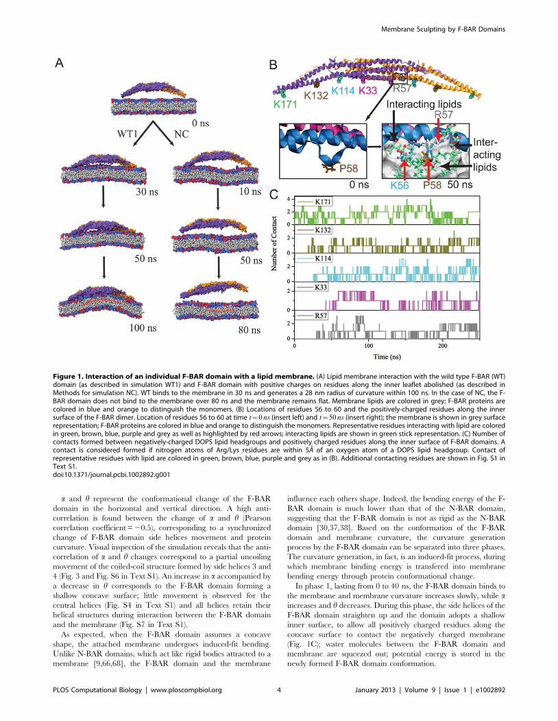
Figure 1. Interaction of an individual F-BAR domain with a lipid membrane. (A) Lipid membrane interaction with the wild type F-BAR (WT)domain (as described in simulation WT1) and F-BAR domain with positive charges on residues along the inner leaflet abolished (as described inMethods for simulation NC). WT binds to the membrane in 30 ns and generates a 28 nm radius of curvature within 100 ns. In the case of NC, the F-BAR domain does not bind to the membrane over 80 ns and the membrane remains flat. Membrane lipids are colored in grey; F-BAR proteins arecolored in blue and orange to distinguish the monomers. (B) Locations of residues 56 to 60 and the positively-charged residues along the innersurface of the F-BAR dimer. Location of residues 56 to 60 at time t~0 ns (insert left) and t~50 ns (insert right); the membrane is shown in grey surfacerepresentation; F-BAR proteins are colored in blue and orange to distinguish the monomers. Representative residues interacting with lipid are coloredin green, brown, blue, purple and grey as well as highlighted by red arrows; interacting lipids are shown in green stick representation. (C) Number ofcontacts formed between negatively-charged DOPS lipid headgroups and positively charged residues along the inner surface of F-BAR domains. Acontact is considered formed if nitrogen atoms of Arg/Lys residues are within 5A of an oxygen atom of a DOPS lipid headgroup. Contact ofrepresentative residues with lipid are colored in green, brown, blue, purple and grey as in (B). Additional contacting residues are shown in Fig. S1 inText S1.doi:10.1371/journal.pcbi.1002892.g001
Membrane Sculpting by F-BAR Domains
PLOS Computational Biology | www.ploscompbiol.org 4 January 2013 | Volume 9 | Issue 1 | e1002892
In phase 2, lasting from 40 to 120 ns, membrane curvature is
generated. During this phase, a and h adjust and domain
curvature increases. Potential energy stored in the F-BAR domain
conformation is released into energy associated with membrane
curvature.
In phase 3, lasting from 120 to 250 ns, the protein-membrane
system relaxes. Membrane curvature decreases slightly and
fluctuates around 0.0015A21; a and h values are restored close
to the native state values, indicating partial uncoiling of the coiled-
coil structure (Fig. S6 in Text S1). However, a values in
simulations NC, NL1 and NL2 are much lower than those in
simulation WT1 and WT2, while h values show the reverse,
indicating that the domains coiled-coil structure without interac-
tion with the membrane becomes further coiled, which suggests
that partial uncoiling of the domains coiled-coil structure provides
the driving force for membrane curvature formation. Indeed, if
one removes the membrane from the final conformation of
simulation WT1, as is done in simulation WT1WAT, the
conformation of the F-BAR domain is quickly restored to a near
crystal conformation and a and h assume values similar to the ones
they assume in simulation NL1 and NL2, indicating that the
uncoiling is reversible (simulation WT1WAT, see Fig. S8 in Text
S1).
The induced-fit interaction between the F-BAR domain and the
membrane allows membrane curvature adjustment to F-BAR
domain density and lipid type. Indeed, varying F-BAR domain
density leads to a wide range of curvatures during membrane
structure formation [7,19,38,47,67]. The F-BAR domain confor-
mational change in response to membrane curvature is likely also
a mechanism for membrane curvature sensing. Binding of the F-
BAR domain to membranes with curvatures that are significantly
different from its intrinsic curvature would require conformational
change of the F-BAR domain that is energetically expensive.
Indeed, F-BAR domains are found to favor membrane curvatures
that match its intrinsic protein curvature [7].
Theoretical description of the membrane sculptingprocess
Binding of the F-BAR domain to the membrane leads to a
match between shapes of F-BAR domain and membrane. The
resulting membrane curvature depends on the balance of two
forces, one resisting protein shape changes and the other resisting
membrane curvature changes. The bending energy of an F-BAR
domain dimer attached to the membrane surface (or any other
attached rod-like protein) can be described through
[22,65,66,69,70]
Ep~KpL0
2(C{Cp)2~
fp
2(C{Cp)2, ð1Þ
where Cp is the intrinsic curvature of the protein, C the curvature
of the membrane, Kp the flexural rigidity of the protein, L0 the
length of the protein, and fp the protein bending rigidity. The
curvature-related elastic energy of a cylindrical membrane of
length L and radius R can be described by a Helfrich Hamiltonian
[71]
El~pKlL
R~pKlLC, ð2Þ
where Kl is the membrane bending modulus. The curvature-
elastic energy of a membrane patch of length L, width W and
radius R can be described analogously by a Helfrich Hamiltonian,
namely by
Elw~pKlL
R
W
2pR~
KlA
2R2~
KlAC2
2, ð3Þ
where A is the area of the membrane patch (A~WL).
The total energy of an F-BAR dimer binding to a membrane is
then
E~EpzElw~fp
2(1=R{1=Rp)2z
KlA
2R2~
fp
2(C{Cp)2z
KlAC2
2,
ð4Þ
which corresponds to the shape force, F ,
Figure 2. Conformation of F-BAR domain characterized through angles h and a. (Left) The F-BAR domain is a dimer of two F-BAR proteins,the latter colored in purple and orange. (Right) Each F-BAR protein is composed of five helices forming a coiled-coil structure, with helix 2 to 4 coloredblue, purple and green, respectively. h is formed by the principal axes of the central part and the end part of helix 4 (green); a is formed by the anglebetween the principal axes of the left and the right sides of helix 3 (purple).doi:10.1371/journal.pcbi.1002892.g002
Membrane Sculpting by F-BAR Domains
PLOS Computational Biology | www.ploscompbiol.org 5 January 2013 | Volume 9 | Issue 1 | e1002892
Membrane Sculpting by F-BAR Domains
PLOS Computational Biology | www.ploscompbiol.org 6 January 2013 | Volume 9 | Issue 1 | e1002892
F~LE
LR~{
fp
R2(1=R{1=Rp){
KlA
R3~{fpC2(C{Cp){KlAC3,
ð5Þ
At equilibrium holds F = 0 and, hence,
C~Cpfp
KlAzfp
: ð6Þ
According to the equipartition theorem of thermodynamics holds
fp
2DC2
p~1
2kBT , ð7Þ
or
fp~kBT
DC2p
, ð8Þ
where DCp is the curvature fluctuation of the protein, kB the
Boltzmann constant and T the temperature.
The curvature of the protein was monitored during the last
100 ns of simulation NL1 and is presented in Fig. S9 in Text S1.
The intrinsic curvature of the protein was determined as the mean
curvature of the protein, namely Cp~0:0283 nm{1, correspond-
ing to a radius of curvature of 35.3 nm. The root-mean square
fluctuation of the curvature of the protein was determined from its
standard deviation from the average protein curvature and was
found to be DCp~0:0062 nm{1. The membrane bending
modulus Kl had been measured, through experiments and
simulations, to be 20kBT [60,65,72–75]. According to Eq. 6,
the radius of curvature of an F-BAR dimer on top of a lipid patch
is then estimated to be 45.1 nm. This value compares well with the
radius of curvature monitored during the last 100 ns of simulation
WT1, which is 48:1+5:3 nm.
With the parameters stated above, one can estimate the total
binding energy of WT1 F-BAR dimer and membrane patch at
equilibrium to be 2:30kBT , with the bending energy of F-BAR
dimer and of membrane patch contributing 0:74kBT and
1:56kBT , respectively. The average membrane curvature during
the early (i.e., phase 1) period 38{40 ns is 0:12 nm{1 and
amounts to the highest membrane curvature during the binding
phase. During this period the total energy of the F-BAR-
membrane system, the bending energy of the F-BAR dimer and
of the membrane patch are 3:99kBT , 3:47kBT and 0:52kBT ,
respectively. During the later (i.e., phase 2) period 78{80 ns the
average membrane curvature is 0:20 nm{1 and amounts to the
highest membrane curvature during the membrane bending
phase. During this period the total energy of the F-BAR-
membrane system, the bending energy of the F-BAR dimer and
of the membrane patch are 2:34kBT , 0:90kBT and 1:44kBT ,
respectively. Therefore, the total energy that is stored in the
protein conformational change during membrane binding and
membrane bending phases is (3:47{0:90)kBT~2:57kBT . The
binding energy can be estimated by the single molecule
experiment proposed in [66], in which an F-BAR dimer molecule
is pulled away from the membrane at one end.
Binding and close adhesion of the F-BAR domain to the
membrane require shape complementarity between protein and
membrane. In case that both protein and membrane shapes are
radially symmetric, i.e., the centerline of either one obeys in the
x, z-plane the equation x2zz2~R2, shape complementarity leads
to membrane curvature 1=R. If the F-BAR domains are forming
on top of the initially planar membrane a lattice oriented (with the
protein major axes) along the x-axis then the planar membrane
coils into a tube with its long axes pointing along the y-axis.
However, in case that the F-BAR domain does not assume a
radial shape, shape complementarity results in an interesting
variation. To demonstrate this we assume that the F-BAR domain
prefers either intrinsically or through the effect of adhesion to the
membrane an ellipsoidal shape governed by the equation
(x=a)2z(y=b)2~1 where a and b are the major and minor axis
of the ellipse. In this case a membrane tube along the y-axis does
not permit close adhesion as the radially symmetric membrane
and the ellipsoidal F-BAR domain don’t match exactly. However,
a tube tilted by an angle b relative to the y-axis permits a perfect
match of protein and membrane shape. To see this we note that,
according to a well known result of geometry, the tilted tube is cut
by the x, z-plane along an ellipsoid. One can convince oneself
readily that this ellipse has a short axis b~R and a long axis
a~R=cos b. One can then conclude that for the assumed
ellipsoidally shaped F-BAR domains (characterized by long axis
a and short axis b), forming a lattice oriented along the x-axis on
an initially planar membrane, a tube of curvature 1=R results with
direction along an angle b relative to the y-axis, where b is given
by
b~arccos(R=a): ð9Þ
This description assumes binding of the F-BAR domain leading to
strong adhesion such that protein and membrane shape match
very closely. In any case, a circular membrane tube can
accommodate non-circular F-BAR domain shapes by rotating
the tube axis, but only shapes that are nearly ellipsoidal. As stated
already, such shapes can result from a combination of an intrinsic
and an induced shape of the F-BAR domain dimer adhesion
surface.
Membrane curvature generated by F-BAR domain latticesAs stated already, tubules and liposomes with wide range of
curvatures are found to be generated by the F-BAR domain
[7,19,38,47,67]. Apparently, the variation stems from the collec-
tive action of the domains as visualized, for example, in cryo-EM
images [38]. To investigate how F-BAR domains curve mem-
branes collectively, we built a series of F-BAR domain lattices
adopting the SBCG simulation model (see Methods). We
performed, for this purpose, four series of simulations with F-
BAR domain lattices of varying type. The lattices studied and the
resulting curvatures are depicted in Fig. 4.
In a series of SBCG simulations, LATTICES (Table 1), we
examined how the F-BAR domain density affects membrane
curvature. As Fig. 4 shows, of the F-BAR domain lattices with five
Figure 3. Conformational change of F-BAR domain during interaction with the membrane. Change of membrane curvature and of anglesa, h during simulations WT1, WT2, NC, NL1 and NL2 (see Table 1). Original data are shown in gray and running averages over 10 ns in color.Conformations of the F-BAR domain and interaction with the membrane are shown at 0, 40, 80 and 120 ns for simulation WT1. Helices 2 to 4 arecolored blue, purple and green, respectively; tails of membrane lipids are colored grey; the neutral DOPC head groups are colored blue and thenegatively charged DOPS head groups red.doi:10.1371/journal.pcbi.1002892.g003
Membrane Sculpting by F-BAR Domains
PLOS Computational Biology | www.ploscompbiol.org 7 January 2013 | Volume 9 | Issue 1 | e1002892
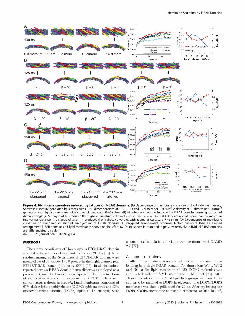
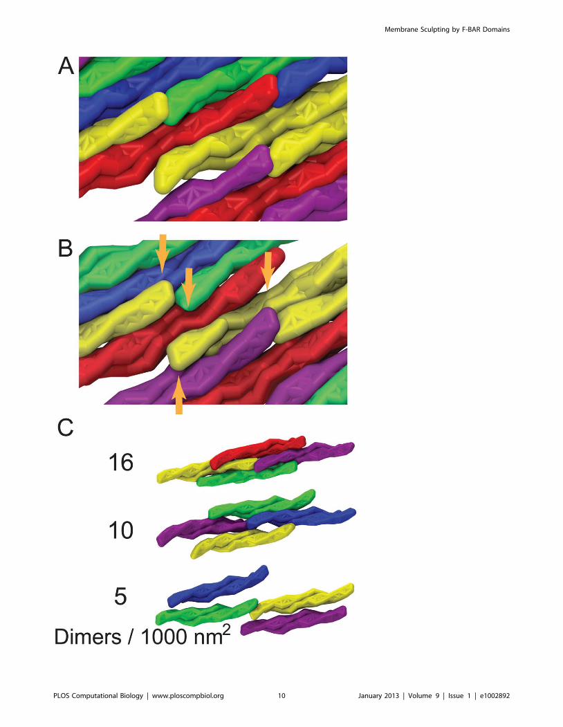
different densities, the one with 10 dimers per 1000 nm2 achieves
highest curvature; lattices with lower densities achieve much lower
curvature. This result is expected since the denser the lattices are,
the more the F-BAR domains can act on the same area of lipid.
However, membrane curvature becomes also reduced when the F-
BAR domain density gets too high, due to neighboring F-BAR
domains hindering each others access to the membrane as shown
in Fig. 5A. This hinderance of neighboring domains increases as
domain density increases (Fig. 5B). The F-BAR domain density
generating the narrowest tubules, as seen in cryo-EM [38], is 8 to
10 dimers per 1000 nm2.
Fig. 4 shows the relationship between membrane curvature and
lattice geometry. Rather diverse curvatures (radii of curvature
range from 25 to 100 nm) are seen to be generated by lattices with
different parameters [7,19,38,47,67]. High curvatures are gener-
ated by lattices with b values in the range of 50–90. An inter-
domain distance of 21.5 nm with the F-BAR domains being
staggered in an end-to-shoulder arrangement yields the highest
curvature. The results in Fig. 4 are consistent with recent cryo-
electron microscopy images of F-BAR domain lattices on
membrane tubules [38].
The observed tilt angle b~80 between y-axis and tube axis
suggests, according to Eq. 9, that the actual shape of the F-BAR
domain membrane adhesion surface is ellipsoidal with axes
a~1:01 R and b~R, i.e., the widening of the F-BAR domain
shape is very small, but significant enough to induce an observable
reorientation of the tube axis. To understand how a deviation
from circular shape as reflected by a~1:01 R can be significant
one should note that the lattice of F-BAR domains averages over
the shape effect of many proteins such that even minor effects add
up to the tube axis tilt.
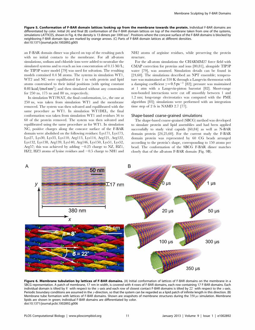
Membrane tubulation by F-BAR domain latticesTo investigate how a complete tubule is formed by a lattice of F-
BAR domains, the best (highest curvature induced) performing
lattice was placed on a 380 nm wide planar membrane (Fig. 6).
Periodic boundary conditions in the y-direction imply that the
lattice acts on an infinitely long membrane patch. Membrane
curvature in simulation TUBULATION (see Table 1 and
Methods) developed within hundreds of microseconds from the
edges (curving first) to the center (curving last). After 350 ms, a
tubular structure with local radius of curvature R = 60–90 nm was
formed, with the edges being separated by only 28 nm. In lieu of
using more computer time (the simulation stretched over 10
months), we applied a weak radial force until the edges met, fusing
the membrane into a complete tubular structure (Fig. 7A). After
the tube was closed, we removed all F-BAR domains and carried
out 30 ms of further equilibrium simulation, during which the tube
remained closed. Tubules formed by the F-BAR domain lattices in
vivo range from 25 to 100 nm in radius [7,19,38,47,67]. In a
second simulation we observed a tube fusing event in which one
edge of a tube met the other edge in a T-like junction. Removing
all F-BAR domains and continuing the simulation for 30 ms
revealed again a stable structure (Fig. 7B).
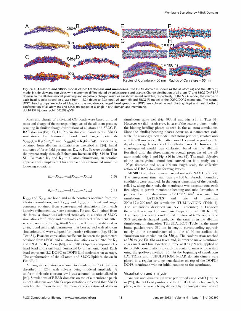
To study the interactions between F-BAR domains in a tube-
forming lattice at all-atom resolution, we aligned all-atom
structures of the F-BAR domains with the SBCG model on the
surface of the fully formed tubule structure (Fig. 8) employing the
method reported in [24]. Analysis of the structure showed that
side-to-side contacts are maintained between most pairs of
neighboring F-BAR domains, due to a large number of charged
residues at the lateral contact points, e.g., Lys66, Asp161 of one
dimer and Glu285, Arg47 of another. Indeed, mutation of these
residues into neutral amino acids abolishes tubule formation by the
F-BAR domains [13,76], which suggests that the contacts are
important for lattice formation and hence, membrane tubulation.
Further analysis of the lattice structure revealed that end-to-end
contacts are not maintained. This observation is consistent with
the cryo-EM images, in which end-to-end contacts are seen not to
be strong and are found absent in the narrowest tubule observed
[38].
F-BAR domain lattices allow defectsWhile most F-BAR domains in our simulations retain their
original degree of tilting with b~80, some F-BAR domains exhibit
degrees of tilting in the range of 5{120. In experiment, F-BAR
domain lattices induce membrane tubules not in a manner
ordered enough to produce high-quality cryo-EM structures [38].
Instead, cryo-EM structures require several rounds of annealing.
Therefore, it is likely that in cells F-BAR domains form lattices
considerably more random than seen in cryo-EM. Indeed, N-BAR
protein coats on tubule surfaces are found to be dynamic and with
a tendency to scramble [21,38].
Additionally, one out of the 167 F-BAR domains was found in
our simulation to assume a side-laying orientation, where the F-
BAR domain turns 900 around its principal axis. In the side-laying
orientation the F-BAR domain forms membrane contact with its
side surface, rather than the concave surface, and the side-to-side
contacts between neighboring F-BAR domains are abolished. The
side-laying orientation is only observed at large defects of the F-
BAR domain lattices, where local F-BAR domain concentration is
low. The side-laying state has also been observed in the all-atom
simulations WT1DEL (Table 1). Indeed, in experiment the side-
laying state has been observed to induce tubules with low
curvatures and at low BAR domain density [9,38]. It is likely
that in cells, both the upright and the side-laying orientation arise
in the F-BAR domain lattice. Both side-to-side contacts between
the F-BAR domains and the short loop of residues 56 to 60 are
important in maintaining orientation in the F-BAR domain
lattices.
ConclusionsIn summary, our study on membrane sculpting by F-BAR
domains resolves in atomic detail how F-BAR domains sculpt
curved membranes. All-atom MD simulations show F-BAR
domains dynamically interacting with a membrane, revealing that
F-BAR domains sculpt membranes according to the scaffolding
mechanism. F-BAR domains act in three steps, namely binding to
the membrane, bending the membrane and equilibration.
Positively charged residues along the concave surface of the F-
BAR domain play a key role in attracting negatively charged
membrane lipids towards the F-BAR domain concave side, though
F-BAR domains do not act as rigid templates.
We also performed a 350 ms CG simulation providing a
detailed, dynamic picture of membrane tubulation by an F-BAR
domain lattice. Depending on the F-BAR domain arrangement
within lattices, a wide range of membrane curvatures can be
generated. Lattices that generate the greatest curvature (radius of
curvature R = 28 nm) involve an F-BAR domain density of 8 to 13
dimers per 1000 nm2, a tilting angle b of 80, an inter-dimer
distance of 21.5 nm and end-to-shoulder contacts. Both side-to-
side contacts between F-BAR domains and, in particular, a short
loop of residues 56 to 60 are important in maintaining the F-BAR
domain in the upright conformation. Our approach combined all-
atom and SBCG simulations and revealed how strikingly beautiful
F-BAR domain lattices generate large scale membrane shapes in
living cells.
Membrane Sculpting by F-BAR Domains
PLOS Computational Biology | www.ploscompbiol.org 8 January 2013 | Volume 9 | Issue 1 | e1002892
Methods
The atomic coordinates of Homo sapiens EFC/F-BAR domain
were taken from Protein Data Bank (pdb code: 2EFK) [13]. Nine
residues missing at the N-terminus of EFC/F-BAR domain were
modeled based on residue 1 to 9 present in the highly homologous
FBP17/F-BAR domain (pdb code: 2EFL) [13]. In all simulations
reported here an F-BAR domain homo-dimer was employed as a
protein unit, since the homodimer is expected to be the active form
of the protein as shown in experiments [7,13,38]. The dimer
conformation is shown in Fig. 9A. Lipid membranes composed of
67% dieleoylphosphatidylcholine (DOPC) lipids (neutral) and 33%
dioleoylphosphatidylserine (DOPS) lipids ({1e charged) were
assumed in all simulations; the latter were performed with NAMD
2.7 [77].
All-atom simulationsAll-atom simulations were carried out to study membrane
bending by a single F-BAR domain. For simulations WT1, WT2
and NC, a flat lipid membrane of 150 DOPC molecules was
constructed with the VMD membrane builder tool [78]. After
10 ns of equilibration, 33% of lipid headgroups were randomly
chosen to be mutated to DOPS headgroups. The DOPC/DOPS
membrane was then equilibrated for 20 ns. After replicating the
DOPC/DOPS membrane to reach a dimension of 36|10 nm2,
Figure 4. Membrane curvature induced by lattices of F-BAR domains. (A) Dependence of membrane curvature on F-BAR domain density.Shown is curvature generated by lattices with F-BAR dimer densities of 5, 8, 10, 13 and 15 dimers per 1000 nm2. A density of 10 dimers per 1000 nm2
generates the highest curvature, with radius of curvature R~38:5 nm. (B) Membrane curvature induced by F-BAR domains forming lattices ofdifferent angle b. An angle of 80 produces the highest curvature, with radius of curvature R~33 nm. (C) Dependence of membrane curvature oninter-dimer distance. A distance of 21.5 nm produces the highest curvature, with radius of curvature R = 33 nm. (D) Dependence of membranecurvature on staggered or aligned arrangement of F-BAR domains. A staggered arrangement produces higher curvature than an alignedarrangement. F-BAR domains and lipid membranes shown on the left of (A–D) are shown in color and in grey, respectively; individual F-BAR domainsare differentiated by color.doi:10.1371/journal.pcbi.1002892.g004
Membrane Sculpting by F-BAR Domains
PLOS Computational Biology | www.ploscompbiol.org 9 January 2013 | Volume 9 | Issue 1 | e1002892
Membrane Sculpting by F-BAR Domains
PLOS Computational Biology | www.ploscompbiol.org 10 January 2013 | Volume 9 | Issue 1 | e1002892
an F-BAR domain dimer was placed on top of the resulting patch
with no initial contacts to the membrane. For all all-atom
simulations, sodium and chloride ions were added to neutralize the
simulated systems and to reach an ion concentration of 0.15 M/L;
the TIP3P water model [79] was used for solvation. The resulting
models contained 0.4 M atoms. The systems in simulation WT1,
WT2 and NC were equilibrated for 1 ns with protein and lipid
atoms constrained to their initial positions (with spring constant
0:01 kcal=(mol nm2)) and then simulated without any constraints
for 250 ns, 175 ns and 80 ns, respectively.
In simulation WT1WAT, the final conformation, i.e., the one at
250 ns, was taken from simulation WT1 and the membrane
removed. The system was then solvated and equilibrated with the
same procedure as WT1. In simulation WT1DEL, the final
conformation was taken from simulation WT1 and residues 56 to
60 of the protein removed. The system was then solvated and
equilibrated using the same procedure as for WT1. In simulation
NC, positive charges along the concave surface of the F-BAR
domain were abolished on the following residues: Lys171, Lys173,
Lys27, Lys30, Lys33, Lys110, Arg113, Lys114, Arg121, Arg122,
Lys132, Lys138, Arg139, Lys140, Arg146, Lys150, Lys51, Lys52,
Arg57; this was achieved by adding 20.25 charge to NZ, HZ1,
HZ2, HZ3 atoms of lysine residues and 20.5 charge to NH1 and
NH2 atoms of arginine residues, while preserving the protein
structure.
For the all-atom simulations the CHARMM27 force field with
CMAP correction for proteins and ions [80,81], alongside TIP3P
water [79], was assumed. Simulation details can be found in
[24,60]. The simulations described an NPT ensemble; tempera-
ture was maintained at 310 K through a Langevin thermostat with
a damping coefficient c~0:5 ps{1 [82]; pressure was maintained
at 1 atm with a Langevin-piston barostat [82]. Short-range
non-bonded interactions were cut off smoothly between 1 and
1.2 nm; long-range electrostatics was computed with the PME
algorithm [83]; simulations were performed with an integration
time step of 2 fs in NAMD 2.7 [77].
Shape-based coarse-grained simulationsThe shape-based coarse-grained (SBCG) method was developed
to simulate protein and lipid assemblies and had been applied
successfully to study viral capsids [60,84] as well as N-BAR
domain protein [24,25,60]. For the current study the F-BAR
domain protein was represented by 60 CG beads arranged
according to the protein’s shape, corresponding to 150 atoms per
bead. The conformation of the SBCG F-BAR dimer matches
closely that of the all-atom F-BAR domain (Fig. 9B).
Figure 5. Conformation of F-BAR domain lattices looking up from the membrane towards the protein. Individual F-BAR domains aredifferentiated by color. Initial (A) and final (B) conformation of the F-BAR domain lattices on top of the membrane taken from one of the systems,simulations LATTICES, shown in Fig. 4; the density is 13 dimers per 1000 nm2 . Positions where the concave surface of the F-BAR domains is blocked byneighboring F-BAR domain tips are marked by orange arrows. (C) Parts of F-BAR domain lattices at different densities.doi:10.1371/journal.pcbi.1002892.g005
Figure 6. Membrane tubulation by lattices of F-BAR domains. (A) Initial conformation of lattices of F-BAR domains on the membrane in aSBCG representation. A patch of membrane, 17 nm in width, is covered with 4 rows of F-BAR domains, each row containing 17 F-BAR domains. Eachindividual domain is tilted by 80 with respect to the x-axis and each row of closest contact F-BAR domains is tilted by 220 with respect to the x-axis.Periodic boundary conditions are assumed in the y-direction, so that the system can be regarded as a lipid patch of infinite length in this direction. (B)Membrane tube formation with lattices of F-BAR domains. Shown are snapshots of membrane structures during the 350 ms simulation. Membranelipids are shown in green; individual F-BAR domains are differentiated by color.doi:10.1371/journal.pcbi.1002892.g006
Membrane Sculpting by F-BAR Domains
PLOS Computational Biology | www.ploscompbiol.org 11 January 2013 | Volume 9 | Issue 1 | e1002892
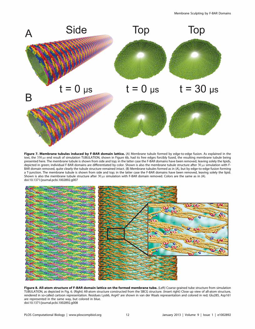
Figure 7. Membrane tubules induced by F-BAR domain lattice. (A) Membrane tubule formed by edge-to-edge fusion. As explained in thetext, the 350 ms end result of simulation TUBULATION, shown in Figure 6b, had its free edges forcibly fused, the resulting membrane tubule beingpresented here. The membrane tubule is shown from side and top; in the latter case the F-BAR domains have been removed, leaving solely the lipids,depicted in green; individual F-BAR domains are differentiated by color. Shown is also the membrane tubule structure after 30 ms simulation with F-BAR domain removed, quite clearly the tubule structure remained intact. (B) Membrane tubules formed as in (A), but by edge-to-edge fusion forminga T-junction. The membrane tubule is shown from side and top; in the latter case the F-BAR domains have been removed, leaving solely the lipid.Shown is also the membrane tubule structure after 30 ms simulation with F-BAR domain removed. Colors are the same as in (A).doi:10.1371/journal.pcbi.1002892.g007
Figure 8. All-atom structure of F-BAR domain lattice on the formed membrane tube. (Left) Coarse-grained tube structure from simulationTUBULATION, as depicted in Fig. 6. (Right) All-atom structure constructed from the SBCG structure. (Insert right) Close up view of all-atom structure,rendered in so-called cartoon representation. Residues Lys66, Arg47 are shown in van der Waals representation and colored in red; Glu285, Asp161are represented in the same way, but colored in blue.doi:10.1371/journal.pcbi.1002892.g008
Membrane Sculpting by F-BAR Domains
PLOS Computational Biology | www.ploscompbiol.org 12 January 2013 | Volume 9 | Issue 1 | e1002892
Mass and charge of individual CG beads were based on total
mass and charge of the corresponding part of the all-atom protein,
resulting in similar charge distributions of all-atom and SBCG F-
BAR domain (Fig. 9C, D). Protein shape is maintained in SBCG
simulations by harmonic bond and angle potentials
Vbond (r)~Kb(r{r0)2 and Vangle(h)~Ka(h{h0)2, respectively,
obtained from all-atom simulations as described in [24]. Initial
estimates of force field parameters Kb, r0, Ka, h0 were obtained in
the present study through Boltzmann inversion (Fig. S10 in Text
S1). To match Kb and Ka to all-atom simulations, an iterative
approach was employed. This approach was automated using the
following equations.
Kb~Kb,obs{m(Kb,obs{Kb,aa) ð10Þ
Ka~Ka,obs{m(Ka,obs{Ka,aa) ð11Þ
Kb, aa and Ka, aa are bond and angle constants obtained from the
all-atom simulation, and Kb, obs and Ka, obs are bond and angle
constants obtained from coarse-grained simulations from each
iterative refinement; m, n are constants. Kb and Ka obtained from
the formula above was adopted iteratively in a series of SBCG
simulations for further and eventually converged refinement. After
several rounds of testing, m~0:5 and n~0:3 were established as
giving bond and angle parameters that best agreed with all-atom
simulations and were adopted for iterative refinement (Fig. S10 in
Text S1). Pearsons correlation coefficients between the parameters
obtained from SBCG and all-atom simulations were 0.965 for Kb
and 0.964 for Ka. As in [60], each SBCG lipid is composed of a
head bead and a tail bead, connected by a harmonic bond. Each
bead represents 2.2 DOPC or DOPS lipid molecules on average.
The conformation of the all-atom and SBCG lipids is shown in
Fig. 9E, F.
A Langevin equation was used to simulate the CG beads as
described in [24], with solvent being modeled implicitly. A
uniform dielectric constant e~1 was assumed as rationalized in
[24]. Simulations of F-BAR domains on top of a membrane patch
in both all-atom and SBCG representations indicated that SBCG
matches the time-scale and the membrane curvature of all-atom
simulations quite well (Fig. 9G, H and Fig. S11 in Text S1).
However we did not observe, in case of the coarse-grained model,
the binding-bending phases as seen in the all-atom simulations.
Since the binding-bending phases occur on a nanometer scale,
while the coarse-grained model (150 atoms per bead) resolves only
a 10-to-50 nm scale, the latter model cannot reproduce the
detailed energy landscape of the all-atom model. However, the
coarse-grained model was calibrated based on the all-atom
forcefield and, therefore, matches overall properties of the all-
atom model (Fig. 9 and Fig. S10 in Text S1). The main objective
of the coarse-grained simulations carried out is to study, on a
100 ms timescale and on a 100 nm length scale, the collective
action of F-BAR domains forming lattices.
All SBCG simulations were carried out with NAMD 2.7 [77].
The integration time step was t~100 fs. Periodic boundary
conditions were assumed. In the longer dimension of the periodic
cell, i.e., along the x-axis, the membrane was discontinuous (with
free edges) to permit membrane bending and tube formation. A
periodic box of dimension 75|15|50 nm3 was used for
simulations LATTICES and one of dimension
200|17|200 nm3 for simulation TUBULATION (Table 1).
The simulations described an NVT ensemble; a Langevin
thermostat was used to maintain temperature at 310 K [82].
The membrane was a randomized mixture of 67% neutral and
33% negatively-charged lipids, i.e., the same as in the all-atom
simulations. In simulation TUBULATION (Table 1), the mem-
brane patches were 380 nm in length, corresponding approxi-
mately to the circumference of a tube of 60 nm radius; the
simulation was carried out for 350 ms. The conformation reached
at 350 ms (see Fig. 6b) was taken and, in order to make membrane
edges meet and fuse together, a force of 0.67 pN was applied to
the F-BAR domain atoms towards the center of mass of the system
using the gridforce method [85]. At the beginning of simulations
LATTICES and TUBULATION, F-BAR domain dimers were
placed in a regular arrangement (lattice) on top of the DOPC/
DOPS membrane without initial contacts to the membrane.
Visualization and analysisAnalysis and visualization were performed using VMD [78]. As
in [24], the tail bead positions of the SBCG lipids define an x, z-
plane, with the x-axis being defined by the longest dimension of
Figure 9. All-atom and SBCG model of F-BAR domain and membrane. The F-BAR domain is shown as the all-atom (A) and the SBCG (B)model in side-view and top-view, with monomers differentiated by colors purple and orange. Charge distribution of all-atom (C) and SBCG (D) F-BARdomain. In the all-atom model, positively and negatively charged residues are shown in red and blue, respectively. In the SBCG model, the charge oneach bead is color-coded on a scale from {2:2e (blue) to 2:2e (red). All-atom (E) and SBCG (F) model of the DOPC/DOPS membrane. The neutralDOPC head groups are colored blue, and the negatively charged head groups on DOPS are colored in red. Starting (top) and final (bottom)conformation of all-atom (G) and SBCG (H) model of a single F-BAR domain and membrane.doi:10.1371/journal.pcbi.1002892.g009
Membrane Sculpting by F-BAR Domains
PLOS Computational Biology | www.ploscompbiol.org 13 January 2013 | Volume 9 | Issue 1 | e1002892
the unit cell membrane patch at time t~0 and the z-axis being
perpendicular to the membrane patch at time t~0. The radius of
curvature of the membrane was calculated by least-squared fitting
of a circle to the obtained membrane profile in the x, z-plane. No
significant membrane curvature developed in the y-direction.
Sequence and structural conservation analysis was performed with
the multiseq plugin of VMD [86]; secondary structure analysis of
F-BAR domains was performed using the timeline plugin of VMD
[78].
Supporting Information
Text S1 Supplementary Figures S1–S11 on structural features of
the F-BAR domain, on the behavior of key residues and on
simulation parameters.
(PDF)
Video S1 Video of simulation WT1 trajectory, corresponding to
Fig. 1.
(WMV)
Video S2 Video of simulation NC trajectory, corresponding to
Fig. 1.
(WMV)
Video S3 Video of simulation TUBULATION trajectory,
corresponding to Figs. 6 and 7.
(WMV)
Acknowledgments
The authors thank Anton Arkhipov and Ying Yin for assistance in getting
the project initiated and for help with the manuscript. The authors thank
also Wei Han, Yanxin Liu, Xueqing Zou, Wen Ma and Jen Hsin for
insightful discussions and assistance.
Author Contributions
Conceived and designed the experiments: HY KS. Performed the
experiments: HY KS. Analyzed the data: HY KS. Contributed reagents/
materials/analysis tools: HY KS. Wrote the paper: HY KS.
References
1. Marsh M, McMahon HT (1999) The structural era of endocytosis. Science 285:215–220.
2. McMahon HT, Gallop JL (2005) Membrane curvature and mechanisms of
dynamic cell membrane remodeling. Nature 438: 590–596.
3. Lecuit T, Pilot F (2003) Developmental control of cell morphogenesis: a focus on
membrane growth. Nature Cell Biology 5: 103–108.
4. Cho W, Stahelin RV (2005) Membrane-protein interactions in cell signaling andmembrane trafficking. Annual Review of Biophysics and Biomolecular Structure
34: 119–151.
5. Kirchhausen T (2000) Clathrin. Annual Review of Biochemistry 69: 699–
727.
6. McMahon HT, Hills IG (2004) COP and clathrin-coated vesicle budding:different pathways, common approaches. Current Opinion in Cell Biology 16:
379–391.
7. Peter BJ, Kent HM, Mills IG, Vallis Y, Butler PJG, et al. (2004) BAR domains as
sensors of membrane curvature: The amphiphysin BAR structure. Science 303:495–499.
8. Wiggins P, Phillips R (2005) Membrane-protein interactions in mechanosensitive
channels. Biophysical Journal 88: 880–902.
9. Blood PD, Voth GA (2006) Direct observation of Bin/amphiphysin/Rvs (BAR)
domain-induced membrane curvature by means of molecular dynamicssimulations. Proceedings of the National Academy of Sciences USA 103:
15068–15072.
10. Cho W, Stahelin RV (2006) Membrane binding and subcellular targeting of C2domains. Biochimica et Biophysica Acta 1761: 838–849.
11. Chandler D, Hsin J, Harrison CB, Gumbart J, Schulten K (2008) Intrinsic
curvature properties of photosynthetic proteins in chromatophores. Biophysical
Journal 95: 2822–2836.
12. Blood PD, Swenson RD, Voth GA (2008) Factors influencing local membranecurvature induction by N-BAR domains as revealed by molecular dynamics
simulations. Biophysical Journal 95: 1866–1876.
13. Shimada A, Niwa H, Tsujita K, Suetsugu S, Nitta K, et al. (2007) Curved EFC/
F-BAR-domain dimers are joined end to end into a filament for membraneinvagination in endocytosis. Cell 129: 761–772.
14. Wang Q, Navarro MVAS, Peng G, Molinelli E, Lin Goh S, et al. (2009)
Molecular mechanism of membrane constriction and tubulation mediated by thef-bar protein pacsin/syndapin. Proceedings of the National Academy of Sciences
106: 12700–12705.
15. Kozlov MM (2010) Biophysics: Joint effort bends membrane. Nature 463: 439–
440.
16. McMahon HT, Kozlov MM, Martens S (2010) Membrane curvature in synapticvesicle fusion and beyond. Cell 140: 601–605.
17. Roberts-Galbraith RH, Gould KL (2010) Setting the F-BAR: functions and
regulation of the F-BAR protein family. Cell Cycle 9: 4091–7.
18. Uezu A, Umeda K, Tsujita K, Suetsugu S, Takenawa T, et al. (2011)
Characterization of the EFC/F-BAR domain protein, FCHO2. Genes Cells 16:868–78.
19. Karotki L, Huiskonen JT, Stefan CJ, Ziolkowska NE, Roth R, et al. (2011)
Eisosome proteins assemble into a membrane scaffold. J Cell Biol 195: 889–902.
20. Chen Y, Sheng R, Kllberg M, Silkov A, Tun M, et al. (2012) Genome-wide
functional annotation of dual-specificity protein- and lipid-binding modules thatregulate protein interactions. Molecular Cell 46: 226–237.
21. Mim C, Cui H, Gawronski-Salerno JA, Frost A, Lyman E, et al. (2012)
Structural basis of membrane bending by the N-BAR protein endophilin. Cell
149: 137–145.
22. Boucrot E, Pick A, Camdere G, Liska N, Evergren E, et al. (2012) Membranefission is promoted by insertion of amphipathic helices and is restricted by
crescent BAR domains. Cell 149: 124–136.
23. Mim C, Unger VM (2012) Membrane curvature and its generation by BAR
proteins. Trends in Biochemical Sciences 37: 526–33.
24. Yin Y, Arkhipov A, Schulten K (2009) Simulations of membrane tubulation bylattices of amphiphysin N-BAR domains. Structure 17: 882–892.
25. Yin Y, Arkhipov A, Schulten K (2010) Multi-scale simulations of membrane
sculpting by N-BAR domains. In: Biggin P, Sansom M, editors. Molecular
Simulations and Biomembranes: From Biophysics to Function. Cambridge:Royal Society of Chemistry. pp. 146–176.
26. Wu M, Huang B, Graham M, Raimondi A, Heuser JE, et al. (2010) Coupling
between clathrin-dependent endocytic budding and F-BAR-dependent tubula-tion in a cell-free system. Nat Cell Biol 12: 902–908.
27. Chen Y, Aardema J, Misra A, Corey SJ (2012) Bar proteins in cancer and blood
disorders. Int J Biochem Mol Biol 3: 198–208.
28. Casal E, Federici L, Zhang W, Fernandez-Recio J, Priego EM, et al. (2006) The
crystal structure of the BAR domain from human Bin1/Amphiphysin II and itsimplications for molecular recognition. Biochemistry 45: 12917–12928.
29. Lee E, Marcucci M, Daniell L, Pypaert M, Weisz OA, et al. (2007) Amphiphysin
2 (Bin1) and T-tubule biogenesis in muscle. Science 297: 1193–1196.
30. Henne WM, Kent HM, Ford MGJ, Hegde BG, Daumke O, et al. (2007)
Structure and Analysis of FCHo2 F-BAR Domain: A Dimerizing andMembrane Recruitment Module that Effects Membrane Curvature. Structure
15: 1–14.
31. Ahmed S, Bu W, Lee RTC, Maurer-Stroh S, Goh WI (2010) F-BAR domain
proteins: Families and function. Communicative & Integrative Biology 3: 116–121.
32. Itoh T, Erdmann KS, Roux A, Habermann B, Werner H, et al. (2005) Dynamin
and the actin cytoskeleton cooperatively regulate plasma membrane invagina-tion by BAR and F-BAR proteins. Developmental Cell 9: 791–804.
33. He Y, Liwo A, Weinstein H, Scheraga HA (2011) PDZ binding to the BAR
domain of PICK1 is elucidated by coarse-grained molecular dynamics. J Mol
Biol 405: 298–314.
34. Ren G, Vajjhala P, Lee JS, Winsor B, Munn AL (2006) The BAR domainproteins: Molding membranes in fission, fusion, and phagy. Microbiology and
molecular biology reviews 70: 37–120.
35. Zimmerberg J, Kozlov MM (2006) How proteins produce cellular membrane
curvature. Nat Rev Mol Cell Biol 7: 9–19.
36. Mattila PK, Pykalainen A, Saarikangas J, Paavilainen VO, Vihinen H, et al.(2007) Missing-in-metastasis and IRSp53 deform PI(4,5)P2-rich membranes by
an inverse BAR domain-like mechanism. Journal of Cell Biology 176: 953–964.
37. Frost A, De Camilli P, Unger VM (2007) F-BAR proteins join the BAR family
fold. Structure 15: 751–3.
38. Frost A, Perera R, Roux A, Spasov K, Destaing O, et al. (2008) Structural basisof membrane invagination by F-BAR domains. Cell 132: 807–817.
39. Weissenhorn W (2005) Crystal structure of the endophilin-A1 BAR domain.
Journal of Molecular Biology 351: 653–661.
40. Millard TH, Bompard G, Heung MY, Dafforn TR, Scott DJ, et al. (2005)
Structural basis of filopodia formation induced by the IRSp53/MIM homologydomain of human IRSp53. EMBO Journal 24: 240–250.
41. Farsad K, Camilli PD (2003) Mechanisms of membrane deformation. Current
Opinion in Cell Biology 15: 372–381.
42. Habermann B (2004) The BAR-domain family of proteins: a case of bending
and binding? EMBO reports 5: 250–255.
Membrane Sculpting by F-BAR Domains
PLOS Computational Biology | www.ploscompbiol.org 14 January 2013 | Volume 9 | Issue 1 | e1002892
43. Farsad K, Ringstad N, Takei K, Floyd SR, Rose K, et al. (2001) Generation of
high curvature membranes mediated by direct endophilin bilayer interactions.Journal of Cell Biology 155: 193–200.
44. Ford MG, Mills IG, Peter BJ, Vallis Y, Praefcke GJ, et al. (2002) Curvature of
clathrin-coated pits driven by epsin. Nature 419: 361–366.45. Gallop JL, Jao CC, Kent HM, Butler PJ, Evans PR, et al. (2006) Mechanism of
endophilin N-BAR domain-mediated membrane curvature. EMBO Journal 25:2898–2910.
46. Cui H, Ayton GS, Voth GA (2009) Membrane binding by the endophilin N-
BAR domain. Biophysical Journal 97: 2746–2753.47. Takei K, Slepnev VI, Haucke V, De Camilli P (1999) Functional partnership
between amphiphysin and dynamin in clathrin-mediated endocytosis. Nat CellBiol 1: 33–39.
48. Klein ML, Shinoda W (2008) Large-scale molecular dynamics simulations ofself-assembling systems. Science 321: 798–800.
49. Reynwar BJ, Illya G, Harmandaris VA, Muller MM, Kremer K, et al. (2007)
Aggregation and vesiculation of membrane proteins by curvature-mediatedinteractions. Nature 447: 461–464.
50. Han DS, Golebiewska U, Stolzenberg S, Scarlata SF, Weinstein H (2011) Adynamic model of membrane-bound phospholipase c2 activation by g subunits.
Molecular Pharmacology 80: 434–445.
51. Mondal J, Zhu X, Cui Q, Yethiraj A (2010) Sequence-dependent interaction ofb-peptides with membranes. Journal of Physical Chemistry B 114: 13585–
13592.52. Yoo J, Cui Q (2009) Curvature generation and pressure profile modulation in
membrane by lysolipids: Insights from coarse-grained simulations. Biophys J 97:2267–2276.
53. Mondal S, Khelashvili G, Shan J, Andersen OS, Weinstein H (2011)
Quantitative modeling of membrane deformations by multihelical membraneproteins: application to G-protein coupled receptors. Biophysical Journal 101:
2092–2101.54. Miyashita N, Straub JE, Thirumalai D (2009) Structures of -amyloid peptide
140, 142, and 155the 672726 fragment of appin a membrane environment with
implications for interactions with -secretase. J Am Chem Soc 131: 17843–17852.55. Reynwar BJ, Deserno M (2011) Membrane-mediated interactions between
circular particles in the strongly curved regime. Soft Matter 7: 8567–8575.56. Illya G, Deserno M (2008) Coarse-grained simulation studies of peptide-induced
pore formation. Biophys J 95: 4163–4173.57. Loverde SM, Pantano DA, Christian DA, Mahmud A, Klein ML, et al. (2011)
Curvature, rigidity, and pattern formation in functional polymer micelles and
vesicles from dynamic visualization to molecular simulation. Current Opinion inSolid State and Materials Science 15: 277–284.
58. Ohkubo YZ, Pogorelov TV, Arcario MJ, Christensen GA, Tajkhorshid E (2012)Accelerating membrane insertion of peripheral proteins with a novel membrane
mimetic model. Biophysical Journal 102: 2130–2139.
59. Lyman E, Cui H, Voth GA (2010) Water under the BAR. Biophysical Journal99: 1783–1790.
60. Arkhipov A, Yin Y, Schulten K (2008) Four-scale description of membranesculpting by BAR domains. Biophysical Journal 95: 2806–2821.
61. Arkhipov A, Yin Y, Schulten K (2009) Membrane-bending mechanism ofamphiphysin N-BAR domains. Biophysical Journal 97: 2727–2735.
62. Lyman E, Cui H, Voth GA (2011) Reconstructing protein remodeled
membranes in molecular detail from mesoscopic models. Phys Chem ChemPhys 13: 10430–6.
63. Campelo F, McMahon HT, Kozlov MM (2008) The hydrophobic insertionmechanism of membrane curvature generation by proteins. Biophysical Journal
95: 2325–2339.
64. Khelashvili G, Harries D, Weinstein H (2009) Modeling membrane deforma-tions and lipid demixing upon protein-membrane interaction: The bar dimer
adsorption. Biophys J 97: 1626–1635.
65. Perutkova S, Kralj-Iglic V, Frank M, Iglic A (2010) Mechanical stability ofmembrane nanotubular protrusions influenced by attachment of flexible rod-like
proteins. Journal of Biomechanics 43: 1612–1617.
66. Kaback H, Smirnova I, Kasho V, Nie Y, Zhou Y (2011) The alternating accesstransport mechanism in LacY. Journal of Molecular Biology 239: 85–93.
67. Sorre B, Callan-Jones A, Manzi J, Goud B, Prost J, et al. (2012) Nature of
curvature coupling of amphiphysin with membranes depends on its bound
density. Proc Natl Acad Sci U S A 109: 173–8.
68. Martens S, McMahon HT (2008) Mechanisms of membrane fusion: disparateplayers and common principles. Nature Reviews Molecular Cell Biology 9: 543–
556.
69. Kralj-Iglic V, Babnik B, Gauger D, May S, Iglic A (2006) Quadrupolar ordering
of phospholipid molecules in narrow necks of phospholipid vesicles. Journal ofStatistical Physics 125: 727–752.
70. Iglic A, Slivnik T, Kralj-Iglic V (2007) Elastic properties of biological membranes
influenced by attached proteins. Journal of Biomechanics 40: 2492–2500.
71. Helfrich W (1973) Elastic properties of lipid bilayers: theory and possible
experiments. Z Naturforsch 28: 693–703.
72. Duwe H, Kaes J, Sackmann E (1990) Bending elastic moduli of lipid bilayers :modulation by solutes. J Phys France 51: 945–961.
73. Goetz R, Gompper G, Lipowsky R (1999) Mobility and elasticity of self-
assembled membranes. Phys Rev Lett 82: 221–224.
74. Lindahl E, Edholm O (2000) Mesoscopic undulations and thickness fluctuations
in lipid bilayers from molecular dynamics simulations. Biophysical Journal 79:426–433.
75. Harmandaris VA, Deserno M (2006) A novel method for measuring the bending
rigidity of model lipid membranes by simulating tethers. Journal of Chemical
Physics 125: 204905.
76. Tsujita K, Suetsugu S, Sasaki N, Furutani M, Oikawa T, et al. (2006)Coordination between the actin cytoskeleton and membrane deformation by a
novel membrane tubulation domain of PCH proteins is involved in endocytosis.J Cell Biol 172: 269–79.
77. Phillips JC, Braun R, Wang W, Gumbart J, Tajkhorshid E, et al. (2005) Scalablemolecular dynamics with NAMD. Journal of Computational Chemistry 26:
1781–1802.
78. Humphrey W, Dalke A, Schulten K (1996) VMD – Visual Molecular Dynamics.Journal of Molecular Graphics 14: 33–38.
79. Jorgensen WL, Chandrasekhar J, Madura JD, Impey RW, Klein ML (1983)Comparison of simple potential functions for simulating liquid water. Journal of
Chemical Physics 79: 926–935.
80. MacKerell AD Jr, Bashford D, Bellott M, Dunbrack JRL, Evanseck J, et al.(1992) Self-consistent parameterization of biomolecules for molecular modeling
and condensed phase simulations. FASEB Journal 6: A143–A143.
81. MacKerell AD Jr, Bashford D, Bellott M, Dunbrack RL Jr, Evanseck JD, et al.
(1998) All-atom empirical potential for molecular modeling and dynamicsstudies of proteins. Journal of Physical Chemistry B 102: 3586–3616.
82. Martyna GJ, Tobias DJ, Klein ML (1994) Constant pressure molecular
dynamics algorithms. Journal of Chemical Physics 101: 4177–4189.
83. Darden TA, Toukmaji A, Pedersen LG (1997) Long-range electrostatic effects in
biomolecular simulations. Journal de Chimie Physique et de Physico-ChimieBiologique 94: 1346–1364.
84. Arkhipov A, Freddolino PL, Schulten K (2006) Stability and dynamics of virus
capsids described by coarse-grained modeling. Structure 14: 1767–1777.
85. Wells D, Abramkina V, Aksimentiev A (2007) Exploring transmembrane
transport through alpha-hemolysin with grid-steered molecular dynamics.Journal of Chemical Physics 127: 125101–125101-10.
86. Roberts E, Eargle J, Wright D, Luthey-Schulten Z (2006) MultiSeq: Unifying
sequence and structure data for evolutionary analysis. BMC Bioinformatics 7:
382.
Membrane Sculpting by F-BAR Domains
PLOS Computational Biology | www.ploscompbiol.org 15 January 2013 | Volume 9 | Issue 1 | e1002892