
Membrane Biology: Auxiliary KChIP4a Suppresses A-type...
16
Wang Yanxin Lu, Lei Lei, Xiling Bian and KeWei Yi-Quan Tang, Ping Liang, Jingheng Zhou, Inactivation of Kv4 Channels (ER) Retention and Promoting Closed-state Current through Endoplasmic Reticulum + Auxiliary KChIP4a Suppresses A-type K Membrane Biology: doi: 10.1074/jbc.M113.466052 originally published online April 10, 2013 2013, 288:14727-14741. J. Biol. Chem. 10.1074/jbc.M113.466052 Access the most updated version of this article at doi: . JBC Affinity Sites Find articles, minireviews, Reflections and Classics on similar topics on the Alerts: When a correction for this article is posted • When this article is cited • to choose from all of JBC's e-mail alerts Click here http://www.jbc.org/content/288/21/14727.full.html#ref-list-1 This article cites 91 references, 43 of which can be accessed free at at PEKING UNIV HEALTH SCIENCE LIBRARY on February 19, 2014 http://www.jbc.org/ Downloaded from at PEKING UNIV HEALTH SCIENCE LIBRARY on February 19, 2014 http://www.jbc.org/ Downloaded from
Transcript of Membrane Biology: Auxiliary KChIP4a Suppresses A-type...

WangYanxin Lu, Lei Lei, Xiling Bian and KeWei Yi-Quan Tang, Ping Liang, Jingheng Zhou, Inactivation of Kv4 Channels(ER) Retention and Promoting Closed-stateCurrent through Endoplasmic Reticulum
+Auxiliary KChIP4a Suppresses A-type KMembrane Biology:
doi: 10.1074/jbc.M113.466052 originally published online April 10, 20132013, 288:14727-14741.J. Biol. Chem.
10.1074/jbc.M113.466052Access the most updated version of this article at doi:
.JBC Affinity SitesFind articles, minireviews, Reflections and Classics on similar topics on the
Alerts:
When a correction for this article is posted•
When this article is cited•
to choose from all of JBC's e-mail alertsClick here
http://www.jbc.org/content/288/21/14727.full.html#ref-list-1
This article cites 91 references, 43 of which can be accessed free at
at PEK
ING
UN
IV H
EA
LT
H SC
IEN
CE
LIB
RA
RY
on February 19, 2014http://w
ww
.jbc.org/D
ownloaded from
at PE
KIN
G U
NIV
HE
AL
TH
SCIE
NC
E L
IBR
AR
Y on February 19, 2014
http://ww
w.jbc.org/
Dow
nloaded from

Auxiliary KChIP4a Suppresses A-type K� Current throughEndoplasmic Reticulum (ER) Retention and PromotingClosed-state Inactivation of Kv4 Channels*
Received for publication, March 1, 2013, and in revised form, April 2, 2013 Published, JBC Papers in Press, April 10, 2013, DOI 10.1074/jbc.M113.466052
Yi-Quan Tang‡, Ping Liang‡, Jingheng Zhou‡, Yanxin Lu‡, Lei Lei‡, Xiling Bian‡, and KeWei Wang‡§¶1
From the ‡Department of Neurobiology, Neuroscience Research Institute, Peking University Health Science Center, and the§Department of Molecular and Cellular Pharmacology, State Key Laboratory of Natural and Biomimetic Drugs, Peking UniversitySchool of Pharmaceutical Sciences, Beijing 100191, China and the ¶Peking University-International Data Group/McGovernInstitute for Brain Research, Peking University, Beijing 100871, China
Background: Compared with other auxiliary KChIPs that enhance Kv4 current, KChIP4a inhibits Kv4 function.Results: We identified an ER retention motif and an adjacent VKL motif within the KChIP4a N terminus that reduces Kv4.3surface expression and promotes closed-state inactivation (CSI), respectively.Conclusion: ER retention and CSI enhancement are two distinct mechanisms by which the N terminus of KChIP4a suppressesKv4 function.Significance: This study provides mechanistic insight into auxiliary KChIP4a-induced inhibition of A-type Kv4 current.
In thebrain andheart, auxiliaryKv channel-interactingproteins(KChIPs)co-assemblewithpore-formingKv4�-subunits to formanativeK� channel complex and regulate the expression and gatingproperties of Kv4 currents. Among the KChIP1–4 members,KChIP4a exhibits a uniqueN terminus that is known to suppressKv4 function, but the underlying mechanism of Kv4 inhibitionremains unknown. Using a combination of confocal imaging,surface biotinylation, and electrophysiological recordings, weidentified a novel endoplasmic reticulum (ER) retention motif,consisting of six hydrophobic and aliphatic residues, 12–17(LIVIVL), within the KChIP4a N-terminal KID, that functionsto reduce surface expression of Kv4-KChIP complexes. This ERretention capacity is transferable and depends on its flankinglocation. In addition, adjacent to the ER retention motif, theresidues 19–21 (VKLmotif) directly promote closed-state inac-tivation of Kv4.3, thus leading to an inhibition of channel cur-rent. Taken together, our findings demonstrate that KChIP4asuppresses A-type Kv4 current via ER retention and enhance-ment of Kv4 closed-state inactivation.
Surface expression and dynamic gating are fundamentalproperties of rapidly inactivating (A-type) Kv4 potassium chan-nels that play a critical role in regulating firing frequency andshaping action potential waveforms (1, 2). Binding of auxiliary�-subunits can change the expression of Kv4 channels at themembrane surface as well as their intracellular trafficking andgating kinetics (3–7). Cytosolic Kv channel-interacting pro-teins (KChIPs),2 a class of �-subunits, co-assemble with pore-
forming Kv4 �-subunits to encode the somatodendritic A-typeK� current (ISA) in neurons (3, 8–10) and the transient outwardcurrent (Ito) in cardiac myocytes (11, 12). Both ISA and Ito acti-vate at subthreshold membrane potentials, inactivate rapidly,and recover quickly from inactivation (1). Neuronal ISA plays acritical role in regulating dendritic excitability, somatoden-dritic signal integration, and long term potentiation (13–17),whereas cardiac Ito is the major current for fast phase repolar-ization of the cardiac action potential (18, 19). Genetic disrup-tion of human Kv4 function causes temporal lobe epilepsy andspinocerebellar ataxias as well as cardiac disorders, such asatrial fibrillation and Brugada syndrome (20–25), demonstrat-ing the essential role of Kv4 channels in physiology andpathology.Auxiliary KChIP1 to -4 subunits, belonging to the neuronal
calcium sensor superfamily, consist of a conserved C-terminalcore domain with four EF-hand-like calcium binding motifs(26) and a variable N-terminal domain that has been proposedto mediate diverse modulation on Kv4 function (27–30). Alter-native splicing of the four KChIP genes generates a large num-ber of variants with distinct N-terminal domains, making theKChIP class themost diverse among the neuronal calcium sen-sor protein family (31). Co-expression of different KChIPs withKv4 in heterologous expression system leads to different effectson Kv4 channel function. The majority of the KChIP isoformsdramatically increase Kv4 surface expression and peak current,slow down fast inactivation, and accelerate the recovery frominactivation (3, 32–34). Consistently, many physiological stud-ies have shown that the current amplitudes and gating kineticsof native Kv4 channels (both ISA and Ito) are primarily deter-mined by the expression levels of KChIPs (8, 10, 11, 35, 36). It isof interest that the KChIP4 splice variant KChIP4a, whichshares a conserved C-terminal core domainwith other KChIPs,
* This work was supported by Ministry of Science and Technology of ChinaGrant 2013CB531300 and National Science Foundation of China Grants81221002 and 30970919 (to K. W. W.).
1 To whom correspondence should be addressed. Tel.: 8010-82805065; Fax:8010-82805065; E-mail: [email protected].
2 The abbreviations used are: KChIP, Kv channel-interacting protein; KID, Kv4channel inhibitory domain; ER, endoplasmic reticulum; CSI, closed-stateinactivation; PM, plasma membrane; EGFP, enhanced green fluorescent
protein; SSI, steady-state inactivation; CAAX, motif in which C representscysteine, A represents an aliphatic residue, and X represents any aminoacid residue.
THE JOURNAL OF BIOLOGICAL CHEMISTRY VOL. 288, NO. 21, pp. 14727–14741, May 24, 2013© 2013 by The American Society for Biochemistry and Molecular Biology, Inc. Published in the U.S.A.
MAY 24, 2013 • VOLUME 288 • NUMBER 21 JOURNAL OF BIOLOGICAL CHEMISTRY 14727
at PEK
ING
UN
IV H
EA
LT
H SC
IEN
CE
LIB
RA
RY
on February 19, 2014http://w
ww
.jbc.org/D
ownloaded from
functions as an inhibitory subunit and exhibits a distinct mod-ulation on Kv4 channel surface expression and gating kineticsvia its uniqueN-terminal domain (4, 37–39). The first N-termi-nal 34 residues of KChIP4a, previously known as the KIS (Kvchannel inactivation suppressor) domain, slow Kv4 channelactivation, disrupt fast inactivation of opened channels, andabolish its core-mediated enhancement of Kv4 current andacceleration of recovery from inactivation (4). However, themechanisms by which the KChIP4a N terminus functions as aspecific Kv4 channel inhibitory domain (KID) to suppressA-type potassium current remains unknown.In the present study, we show that auxiliary KChIP4a sup-
presses Kv4.3 function via an alternative N-terminal KID,which can overcome the positive modulatory effect on Kv4.3channel function by KChIP1. Using confocal imaging, we findthat the KID alone is sufficient to retain proteins in the endo-plasmic reticulum (ER), and its retention ability is transferableand prominently dependent on its flanking location within theprotein sequence. A novel ER retention motif (N-terminal res-idues 12–17, LIVIVL) within the KID overrides the KChIP4acore domain-mediated enhancement of Kv4.3 surface expres-sion, leading to the reduction of peak current. Further experi-ments reveal a VKL motif (N-terminal residues 19–21), adja-cent to the LIVIVL motif, that can also suppress Kv4.3 currentby facilitating Kv4.3 closed-state inactivation (CSI). Takentogether, our findings demonstrate that ER retention and CSIenhancement are two distinct mechanisms by which KChIP4asuppresses Kv4 channel function.
EXPERIMENTAL PROCEDURES
Plasmid Construction—For confocal imaging experiments,cDNAs of KChIP4a and KChIP4amutants were subcloned intoa pEGFP-N1 or pEGFP-C2 vector. DsRed2-ER is a red fluores-cent protein tagged with the signal sequence from calreticulinat its N terminus and a KDEL sequence at its C terminus (Clon-tech). TheC-terminal CAAX signal ofH-Ras (GCMSCKCVLS),which targets Ras proteins to the plasma membrane (PM), wasadded to the C terminus of EGFP to construct a PM-targetingEGFP vector (pEGFP-CAAX-N1). For biochemistry experi-ments, cDNAs of Kv4.3, KChIP1, KChIP4a, and KChIP4amutants were cloned into a pcDNA3.1 vector. For electrophysi-ological experiments, wild-type (WT) or mutant cDNA con-structs of either Kv4.3 or KChIPs were transferred into a pBlue-script KSM vector.Confocal Microscopy and Imaging Analysis—For confocal
imaging experiments, HEK 293 cells were reseeded on glasscoverslips coated with poly-D-lysine for detection and trans-fected using Lipofectamine 2000 (Invitrogen) following themanufacturer’s instructions. After 24–48 h of transfection,cells were washed twice with phosphate-buffered saline (PBS)containing 1 mM MgCl2 and 0.1 mM CaCl2 and fixed in 4%paraformaldehyde at 4 °C for 15 min. Images were obtainedusing a confocal microscope (FV1000, Olympus). For stainingof localization in the ER, HEK 293 cells were co-transfectedwith KChIP4a-EGFP or KChIP4a mutants-EGFP and DsRed2-ER. FV1000 Viewer software (Olympus) was used to calculatePearson’s correlation coefficient values for the co-localizationof the fluorescence signals.
Electrophysiological Recordings—For whole-cell patch clamprecording in HEK293 cells, currents were recorded at roomtemperature using the EPC 10UBS amplifier with PatchMastersoftware (HEKA Electronics). Patch pipettes were pulled fromborosilicate glass and fire-polished to a resistance of 2–4megaohms. The bath solution contained 135 mMNaCl, 2.5 mM
KCl, 10 mM HEPES, 1 mM MgCl2, 1.5 mM CaCl2, and 10mM glucose at pH 7.4, and the pipette solution contained 135mM potassium gluconate, 10 mM KCl, 10 mM HEPES, 1 mM
CaCl2, 1 mM MgCl2, and 10 mM EGTA at pH 7.3.For two-electrode voltage clamp recordings in oocytes, all
cRNAs were transcribed in vitro from linearized plasmids inpBluescript KSM vectors by using the T3 mMESSAGEMachine Kit (Ambion). Xenopus laevis oocytes (stage V-VI)were selected and injected with 46 nl of cRNA solution, con-taining 0.5–5.0 ng of the selected cRNA, using a microinjector(Drummond Scientific). Oocytes were then kept at 17 °C inND96 solution (96 mM NaCl, 2 mM KCl, 1.8 mM CaCl2, 1 mM
MgCl2, 5 mM HEPES, pH 7.4, adjusted with NaOH). 24 h afterinjection, oocyteswere impaledwith twomicroelectrodes (0.5–1.0 megaohms) filled with 3 M KCl in a 40-�l recording cham-ber. Currents were recorded in ND-96 solution at room tem-perature (22 � 1 °C) using a GeneClamp 500B amplifier (AxonInstruments).Data were acquired using PatchMaster software (HEKA
Electronics) and digitized at 1 kHzwith an LIH 8 � 8 computerinterface (HEKA Electronics). OriginPro version 8.6 (Origin-Lab) was used to analyze the data. Tomeasure the peak currentamplitudes and the kinetics of open-state inactivation, the cur-rents were evoked by a 2-s depolarizing pulse to �40 mV froma holding potential of �100 mV. The time constants of macro-scopic inactivation were obtained by curve fitting with a singleexponential function. The peak conductance-voltage (G-V)relationship was derived from peak current amplitudes evokedby depolarizing steps from �100 to �60 mV at 10-mV incre-ments, and the calculation was based on the equation, G �I/(V � Vrev), where I is the peak current amplitude at the testpotentialV, andVrev is the reversal potential. Steady-state inac-tivation (SSI) was assessed by determining the peak currentamplitudes at �40 mV after 1-, 5-, or 10-s prepulses rangingfrom �120 to 0 mV. The voltage dependence of steady-stateactivation (G/Gmax) and inactivation (I/Imax) were fitted to thefollowing single Boltzmann relationship, y � 1/(1 � exp((V �V1⁄2)/k)), where V is the test potential, V1⁄2 is the potential forhalf-maximal activation or inactivation, and k is the corre-sponding slope factor. CSI was monitored with a double-pulseprotocol in which the current was evoked by a test pulse of�40mV from �100 mV (Ipre) and from subsequent �50-mV pre-pulses of variable durations from 5ms to 10.4 s (Ipost). Normal-ized currents forCSIwere determined as the ratio of Ipost to Ipre.The kinetics of CSI were obtained by fitting the normalizedcurrent amplitudes as a function of prepulse duration. All hold-ing potentials were �100 mV in this study unless specified.Cell Surface Biotinylation and Western Blotting Assay—
Transfected HEK 293 cells were washed three times with ice-cold PBS and incubated for 1 h at 4 °Cwith EZ-Link Sulfo-NHS-SS-biotin (0.5 mg/ml; Pierce) to biotinylate cell surfaceproteins. Excess biotinwas quenched by incubating the cells for
ER Retention and Gating Motifs in KChIP4a N terminus
14728 JOURNAL OF BIOLOGICAL CHEMISTRY VOLUME 288 • NUMBER 21 • MAY 24, 2013
at PEK
ING
UN
IV H
EA
LT
H SC
IEN
CE
LIB
RA
RY
on February 19, 2014http://w
ww
.jbc.org/D
ownloaded from

an additional 10minwithTBS containing 100mMglycine. Cellswere lysed with lysis buffer (150 mM NaCl, 20 mM Tris, 1%Triton X-100, 1% sodium deoxycholate, 0.1% SDS, 10 mM
EDTA, and proteinase inhibitor mixture (Roche Applied Sci-ence), pH 8.0) at 4 °C for 30 min. The cell lysates were thencentrifuged at 13,000� g for 15min to yield the protein extractin the supernatant. One fraction containing 200 �g of proteinwas incubatedwith 20�l of neutravidin beads (Pierce) for 4 h at4 °C, and the other fraction was prepared as total protein. Afterincubation, the bead-binding surface proteins were washedfour times with cell lysis buffer and eluted with loading buffercontaining DTT. The total and biotinylated proteins were bothsubjected to Western blot analysis.For Western blot assays, protein samples were loaded on 8%
SDS-PAGE and transferred using gel electrophoresis to nitro-cellulosemembranes (Millipore). After blocking, nitrocellulosemembranes were incubated with mouse monoclonal anti-Kv4.3 (1:2000; Abcam), rabbit polyclonal anti-actin (1:500;Santa Cruz Biotechnology, Inc., Santa Cruz, CA), or mousemonoclonal anti-transferrin receptor (1:500; Invitrogen) anti-bodies at 4 °C overnight. For comparing expression levels ofKChIP4a-3xFLAG and KChIP4a core-3xFLAG, mouse mono-clonal anti-FLAG (1:4000; Sigma) antibody was used. The
membranes were then incubated with their corresponding sec-ondary HRP-conjugated antibodies and detected using an ECLWestern blotting detection system (Millipore). Detection ofsignals was calculated using Quantity One software (Bio-Rad).For quantification, the signals from anti-Kv4.3 (or anti-FLAG)over the anti-actin (for total Kv4.3, KChIP4a-3xFLAG, orKChIP4a�2–34) or the anti-transferrin receptor (for surfaceKv4.3) were normalized. The data are expressed as the mean �S.E.; statistical differences between different groups wereassessed using Student’s t test.
RESULTS
KChIP4a Suppresses Kv4.3 Function via Its N-terminal KID—To investigate the underlying mechanism by which auxiliaryKChIP4a inhibits Kv4 function, we started by examining theeffect of KChIP4a coexpressed with Kv4.3 on the channel cur-rent in both mammalian cells and Xenopus laevis oocytes. Bysequence alignment, KChIP4a reveals a unique N terminus,consisting of the first 34 amino acids, that has been shown tofunction as a KID to inhibit the channel activity (Fig. 1A).Whole-cell patch clamp recordings in HEK 293 cells and two-electrode voltage clamp recordings in X. laevis oocytes bothshowed that KChIP4a co-expression resulted in a reduction of
FIGURE 1. The N-terminal KID of KChIP4a suppresses Kv4.3 function. A, a schematic representation of the general domain structure of KChIPs. KChIPsconsist of an NH2-terminal variable domain (multicolored box) and a conserved COOH-terminal core domain (light blue boxes) containing four EF-hands (lightgreen boxes). The N-terminal domain of KChIP4a (designated KID in this study) is represented with the single-letter amino acid code in the protein sequence.The LIVIVL motif is marked in red, and the VKL motif is marked in black. B, representative current traces of Kv4.3 alone, Kv4.3-KChIP4a, and Kv4.3-KChIP4a�2–34recorded from HEK 293 cells. The currents were evoked by a 1-s depolarizing pulse to �40 mV from a holding potential of �100 mV. C, quantitative analysis ofpeak current densities recorded from HEK 293 cells expressing the indicated channels or channel complexes. Values are mean � S.E. (error bars); n � 6 –10 cells;***, p � 0.001. D, quantitative analysis of peak current amplitudes recorded from Xenopus oocytes expressing the indicated channels or channel complexes.Values are mean � S.E.; n � 10 –26 oocytes; ***, p � 0.001. E, the fusion of the KID to the C-terminal end of Kv4.3 (Kv4.3-KID) or the N terminus of Kv4.3 N-terminaltruncation mutant (KID-Kv4.3�2–24) significantly suppressed the channel current. Values are mean � S.E.; n � 5–10 oocytes. Statistical significance wasassessed using Student’s t test, and statistical significance was considered as p � 0.001 (***).
ER Retention and Gating Motifs in KChIP4a N terminus
MAY 24, 2013 • VOLUME 288 • NUMBER 21 JOURNAL OF BIOLOGICAL CHEMISTRY 14729
at PEK
ING
UN
IV H
EA
LT
H SC
IEN
CE
LIB
RA
RY
on February 19, 2014http://w
ww
.jbc.org/D
ownloaded from
Kv4.3 current (Fig. 1, B–D). In contrast, deleting the first 34residues of the KChIP4a N terminus (KChIP4a�2–34, alsonamed KChIP4a core) led to a dramatic increase in the currentamplitude (Fig. 1, B–D). The inhibitory effect of KChIP4a KIDon Kv4 current was further confirmed by fusing the KID toeither the C terminus of WT Kv4.3 (Kv4.3-KID) or the N ter-minus of Kv4.3 N-terminal truncation mutant (KID-Kv4.3�2–24) (Fig. 1E). These results demonstrate that the inhibitoryeffect of KChIP4a on Kv4 is indeed mediated by its N-terminalKID.In neurons, KChIP4a co-exists with other KChIPs (10, 32),
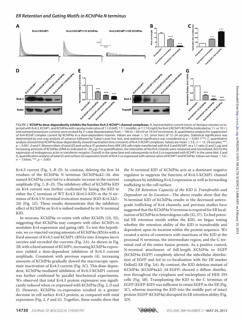
suggesting that KChIP4a may compete with other KChIPs tomodulate Kv4 expression and gating (40). To test this hypoth-esis, we co-injected varying amounts of KChIP4a cRNAs with afixed amount of Kv4.3 and KChIP1 cRNAs into Xenopus laevisoocytes and recorded the currents (Fig. 2A). As shown in Fig.2B, with a fixed amount ofKChIP1, increasingKChIP4a expres-sion yielded a dose-dependent inhibition of Kv4.3 currentamplitude. Consistent with previous reports (4), increasingamounts of KChIP4a gradually slowed the macroscopic open-state inactivation of Kv4.3 current (Fig. 2C). This dose-depen-dent, KChIP4a-mediated inhibition of Kv4.3-KChIP1 currentwas further confirmed by parallel biochemical experiments.We observed that total Kv4.3 protein expression was signifi-cantly reduced when co-expressed with KChIP4a (Fig. 2,D andE). However, KChIP4a co-expression resulted in a greaterdecrease in cell surface Kv4.3 protein, as compared with totalexpression (Fig. 2, F and G). Together, these results show that
the N-terminal KID of KChIP4a acts as a dominant negativeregulator to suppress the function of Kv4.3-KChIP1 channelcomplexes by inhibiting Kv4.3 expression as well as forwardingtrafficking to the cell surface.The ER Retention Capacity of the KID Is Transferable and
Dependent on Its Location—The above results show that theN-terminal KID of KChIP4a results in the decreased antero-grade trafficking of Kv4 channels, and previous studies havesuggested that theKChIP4aN terminus is required for ER local-ization ofKChIP4a in heterologous cells (32, 37). To find poten-tial ER retention motifs within the KID, we began testingwhether the retention ability of the KID is transferable anddependent upon its location within the protein sequence. Wecreated a series of constructs with insertions of the KID at theproximal N terminus, the intermediate region, and the C-ter-minal end of the entire fusion protein. As a positive control,N-terminal attachment of full-length KChIP4a to EGFP(KChIP4a-EGFP) completely altered the subcellular distribu-tion of EGFP and led to co-localization with the ER markerDsRed2-ER (Fig. 3A). By contrast, the KID deletion mutant ofKChIP4a (KChIP4a�2–34-EGFP) showed a diffuse distribu-tion throughout the cytoplasm and nucleoplasm of HEK 293cells (Fig. 3B). Transplanting the KID to the C terminus ofEGFP (EGFP-KID) was sufficient to retain EGFP in the ER (Fig.3C), whereas inserting the KID into the middle part of intactprotein (EGFP-KChIP4a) disrupted its ER retention ability (Fig.3D).
FIGURE 2. KChIP4a dose-dependently inhibits the function Kv4.3-KChIP1 channel complexes. A, representative current traces of Xenopus oocytes co-in-jected with Kv4.3, KChIP1, and KChIP4a with varying mole ratios of 1:1:0 (left), 1:1:1 (middle), or 1:1:10 (right) for Kv4.3/KChIP1/KChIP4a (indicated as 1� or 10�),and outward potassium currents were evoked by 2-s step depolarization from �100 to �60 mV at 10-mV increments. B, quantitative analysis for suppressionof Kv4-KChIP complex current by KChIP4a in a dose-dependent manner. Values are mean � S.E. (error bars) of 12–24 oocytes. Statistical significance wasdetermined by one-way analysis of variance followed by Tukey’s post hoc test, and statistical significance was considered as p � 0.001 (***). C, quantitativeanalysis showed that KChIP4a dose-dependently slowed inactivation time constants of Kv4.3-KChIP complexes. Values are mean � S.E.; n � 12–24 oocytes; ***,p � 0.001. D and F, Western blots of total (D) and surface (F) proteins from HEK 293 cells triple-transfected with Kv4.3 and KChIP1 at a 1:1 ratio (2 and 2 �g) andincreasing amounts of KChIP4a cDNA as indicated (0 –20 �g). For quantification, the intensities of the Kv4.3 bands were measured and normalized, first to theexpression of endogenous actin or transferrin receptor (TransR) in the same lane and subsequently to Kv4.3 co-expressed with KChIP1 in the same blot. E andG, quantification analysis of total (E) and surface (G) expression levels of Kv4.3 co-expressed with various ratios of KChIP1 and KChIP4a. Values are mean � S.E.;n � 3 blots; ***, p � 0.001.
ER Retention and Gating Motifs in KChIP4a N terminus
14730 JOURNAL OF BIOLOGICAL CHEMISTRY VOLUME 288 • NUMBER 21 • MAY 24, 2013
at PEK
ING
UN
IV H
EA
LT
H SC
IEN
CE
LIB
RA
RY
on February 19, 2014http://w
ww
.jbc.org/D
ownloaded from

To establish whether the KID could redirect other mem-brane proteins to the ER, we fused the KID with EGFP to the Cterminus of CD4 (CD4-EGFP-KID). Our results showed thatCD4 fused with the KID caused localization of the chimericproteins to the ER (Fig. 3E). However, fusing CD4 to the Nterminus of KChIP4a (CD4-KChIP4a-EGFP, with equivalentinsertion of the KID into the intermediate region of the intactprotein) had no effect on trafficking and maintained the abilityof CD4 proteins targetting to the PM instead of being retainedin the ER (Fig. 3F). Quantitative analysis of fluorescence inten-sity for co-variance of WT KChIP4a or KChIP4a mutantsagainst the ER marker DsRed2-ER further confirmed the effectof ER retention mediated by its KID (Fig. 3G). These resultsindicate that the KID functions as an ER retention signal whenlocated at either the N or C terminus of a protein but not whenburied in the intermediate region.Identification of a Hydrophobic and Aliphatic ER Retention
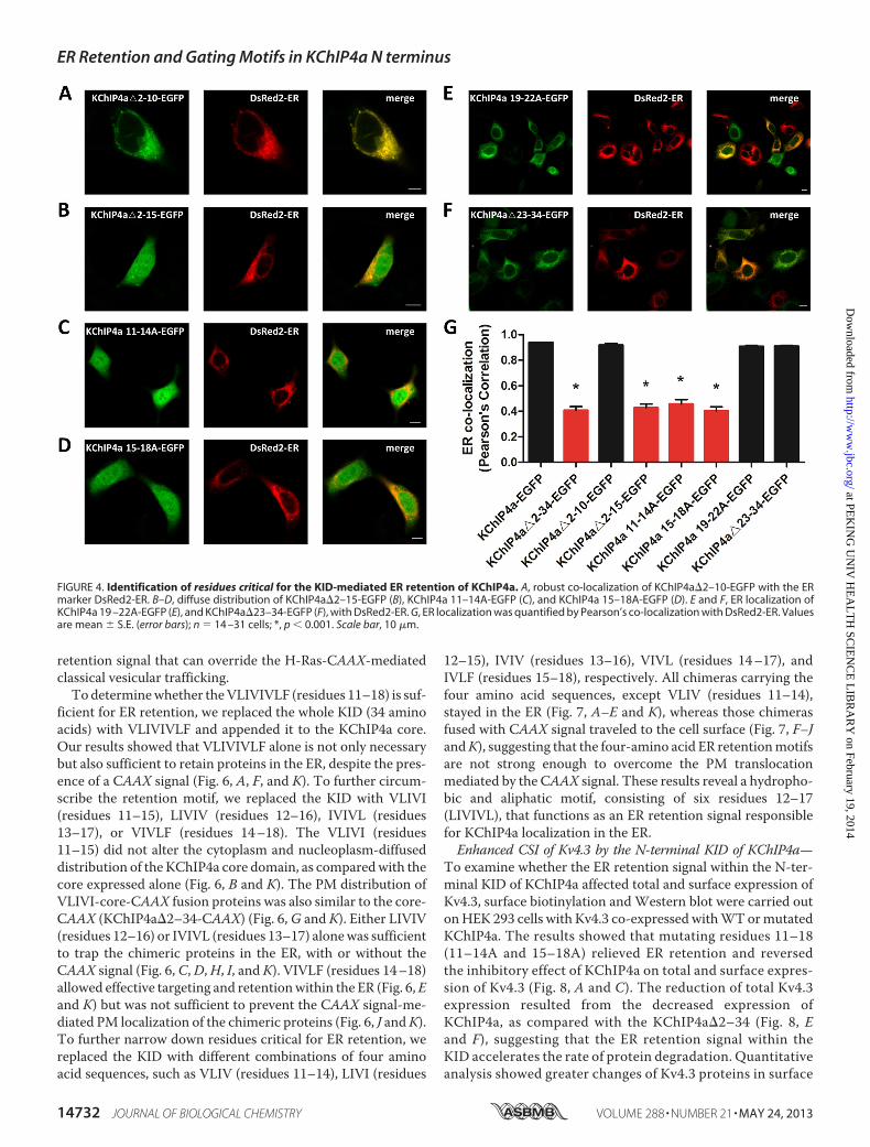
Motif, LIVIVL, within the N-terminal KID of KChIP4a—Be-cause the N-terminal KID does not contain any canonical ERretention motif, the question then arises as to how the KIDcauses ER localization of KChIP4a. To identify specific residuescritical for ER retention of KChIP4a, N-terminal truncations ormutations were introduced into the KID, and the fluorescenceintensity of co-variance versus DsRed2-ER was quantified.With N-terminal deletions of residues 2–10 (KChIP4a�2–10-EGFP) or 23–34 (KChIP4a�23–34-EGFP) and alanine substi-
tutions for residues 19–22 of the KID (KChIP4a 19–22A-EGFP), all of the proteins still co-localized with DsRed2-ER(Fig. 4,A, E, F, andG). In contrast, deletingN-terminal residues2–15 (KChIP4a�2–15-EGFP) or substituting alanine for resi-dues 11–14 (KChIP4a 11–14A-EGFP) or 15–18 (KChIP4a15–18A-EGFP) eliminated ER retention (Fig. 4, B,C,D, andG).These data indicate that residues 11–18 of the KID are criticalfor ER retention.Tounderstand themechanismunderlying theKID-mediated
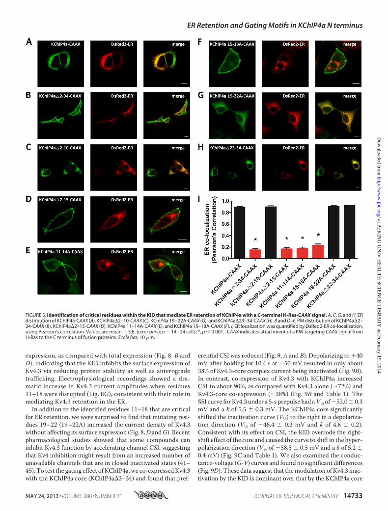
ER retention, we utilized a CAAX signal from the H-Ras pro-teins that functions as a membrane anchor to evaluate theretention ability of KChIP4a and its mutants. The CAAX signalwas appended to the C terminus of KChIP4a-EGFP fusion pro-teins. We then tested the effect of the KID on CAAX signal-mediated forward trafficking to the cell surface. The resultshowed that the intact KID overcame the CAAX signal-medi-ated PM translocation (Fig. 5A). Consistent with our previousresults shown in Figs. 3B and 4, the mutants KChIP4a�2–34-CAAX, KChIP4a�2–15-CAAX, KChIP4a 11–14A-CAAX, andKChIP4a 15–18A-CAAX lost their ability to stay in the ER asillustrated by their distribution at the PM (Fig. 5, B,D–F, and I),whereas the mutants KChIP4a�2–10-CAAX, KChIP4a 19-22A-CAAX, and KChIP4a�23–34-CAAX retained their ERretention ability (Fig. 5, C andG–I). These results demonstratethat residues 11–18 (VLIVIVLF) of the KID function as an ER
FIGURE 3. The ER retention capacity of the KID is transferable and dependent on its location. A, robust co-localization of full-length KChIP4a with the ERmarker DsRed2-ER. B, the N-terminal KID deletion mutant of KChIP4a (KChIP4a�2–34) showed a diffuse distribution. C, appending the KID to the C terminus ofEGFP (EGFP-KID) is sufficient to retain EGFP in the ER. D, disruption of the KID-induced ER retention when the KID was inserted into the middle of the chimericconstruct (EGFP-KChIP4a). E, ER retention of CD4 proteins by the KID fused to the C terminus of the chimeric proteins (CD4-EGFP-KID). F, adding CD4 to the Nterminus of KChIP4a (CD4-KChIP4a-EGFP) disrupted the ER retention caused by the KID, resulting in the localization of the chimeric proteins to the PM. G, ERlocalization was quantified by co-localization with the ER marker DsRed2-ER, using Pearson’s correlation. Values are mean � S.E. (error bars); n � 12–31 cells;*, p � 0.001. Red squares, KID; yellow rectangles, KChIP4a�2–34 (the core domain of KChIP4a); green rectangles, EGFP; blue rectangles, CD4. Scale bar, 10 �m.
ER Retention and Gating Motifs in KChIP4a N terminus
MAY 24, 2013 • VOLUME 288 • NUMBER 21 JOURNAL OF BIOLOGICAL CHEMISTRY 14731
at PEK
ING
UN
IV H
EA
LT
H SC
IEN
CE
LIB
RA
RY
on February 19, 2014http://w
ww
.jbc.org/D
ownloaded from
retention signal that can override the H-Ras-CAAX-mediatedclassical vesicular trafficking.Todeterminewhether theVLIVIVLF (residues 11–18) is suf-
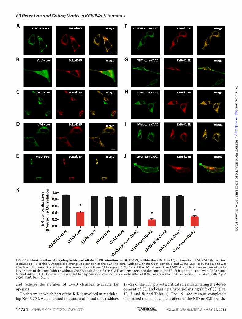
ficient for ER retention, we replaced the whole KID (34 aminoacids) with VLIVIVLF and appended it to the KChIP4a core.Our results showed that VLIVIVLF alone is not only necessarybut also sufficient to retain proteins in the ER, despite the pres-ence of a CAAX signal (Fig. 6, A, F, and K). To further circum-scribe the retention motif, we replaced the KID with VLIVI(residues 11–15), LIVIV (residues 12–16), IVIVL (residues13–17), or VIVLF (residues 14–18). The VLIVI (residues11–15) did not alter the cytoplasm and nucleoplasm-diffuseddistribution of the KChIP4a core domain, as comparedwith thecore expressed alone (Fig. 6, B and K). The PM distribution ofVLIVI-core-CAAX fusion proteins was also similar to the core-CAAX (KChIP4a�2–34-CAAX) (Fig. 6,G and K). Either LIVIV(residues 12–16) or IVIVL (residues 13–17) alonewas sufficientto trap the chimeric proteins in the ER, with or without theCAAX signal (Fig. 6, C,D,H, I, and K). VIVLF (residues 14–18)allowed effective targeting and retentionwithin the ER (Fig. 6,Eand K) but was not sufficient to prevent the CAAX signal-me-diated PM localization of the chimeric proteins (Fig. 6, J andK).To further narrow down residues critical for ER retention, wereplaced the KID with different combinations of four aminoacid sequences, such as VLIV (residues 11–14), LIVI (residues
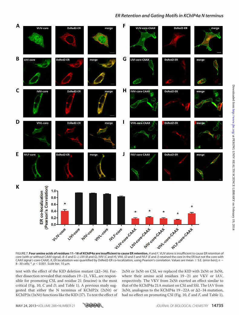
12–15), IVIV (residues 13–16), VIVL (residues 14–17), andIVLF (residues 15–18), respectively. All chimeras carrying thefour amino acid sequences, except VLIV (residues 11–14),stayed in the ER (Fig. 7, A–E and K), whereas those chimerasfused with CAAX signal traveled to the cell surface (Fig. 7, F–JandK), suggesting that the four-amino acid ER retentionmotifsare not strong enough to overcome the PM translocationmediated by the CAAX signal. These results reveal a hydropho-bic and aliphatic motif, consisting of six residues 12–17(LIVIVL), that functions as an ER retention signal responsiblefor KChIP4a localization in the ER.Enhanced CSI of Kv4.3 by the N-terminal KID of KChIP4a—
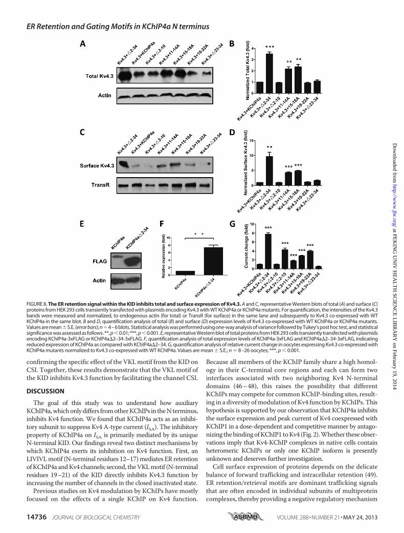
To examine whether the ER retention signal within the N-ter-minal KID of KChIP4a affected total and surface expression ofKv4.3, surface biotinylation andWestern blot were carried outonHEK 293 cells with Kv4.3 co-expressed withWTormutatedKChIP4a. The results showed that mutating residues 11–18(11–14A and 15–18A) relieved ER retention and reversedthe inhibitory effect of KChIP4a on total and surface expres-sion of Kv4.3 (Fig. 8, A and C). The reduction of total Kv4.3expression resulted from the decreased expression ofKChIP4a, as compared with the KChIP4a�2–34 (Fig. 8, Eand F), suggesting that the ER retention signal within theKID accelerates the rate of protein degradation. Quantitativeanalysis showed greater changes of Kv4.3 proteins in surface
FIGURE 4. Identification of residues critical for the KID-mediated ER retention of KChIP4a. A, robust co-localization of KChIP4a�2–10-EGFP with the ERmarker DsRed2-ER. B–D, diffuse distribution of KChIP4a�2–15-EGFP (B), KChIP4a 11–14A-EGFP (C), and KChIP4a 15–18A-EGFP (D). E and F, ER localization ofKChIP4a 19 –22A-EGFP (E), and KChIP4a�23–34-EGFP (F), with DsRed2-ER. G, ER localization was quantified by Pearson’s co-localization with DsRed2-ER. Valuesare mean � S.E. (error bars); n � 14 –31 cells; *, p � 0.001. Scale bar, 10 �m.
ER Retention and Gating Motifs in KChIP4a N terminus
14732 JOURNAL OF BIOLOGICAL CHEMISTRY VOLUME 288 • NUMBER 21 • MAY 24, 2013
at PEK
ING
UN
IV H
EA
LT
H SC
IEN
CE
LIB
RA
RY
on February 19, 2014http://w
ww
.jbc.org/D
ownloaded from
expression, as compared with total expression (Fig. 8, B andD), indicating that the KID inhibits the surface expression ofKv4.3 via reducing protein stability as well as anterogradetrafficking. Electrophysiological recordings showed a dra-matic increase in Kv4.3 current amplitudes when residues11–18 were disrupted (Fig. 8G), consistent with their role inmediating Kv4.3 retention in the ER.In addition to the identified residues 11–18 that are critical
for ER retention, we were surprised to find that mutating resi-dues 19–22 (19–22A) increased the current density of Kv4.3without affecting its surface expression (Fig. 8,D andG). Recentpharmacological studies showed that some compounds caninhibit Kv4.3 function by accelerating channel CSI, suggestingthat Kv4 inhibition might result from an increased number ofunavailable channels that are in closed inactivated states (41–45). To test the gating effect of KChIP4a, we co-expressedKv4.3with the KChIP4a core (KChIP4a�2–34) and found that pref-
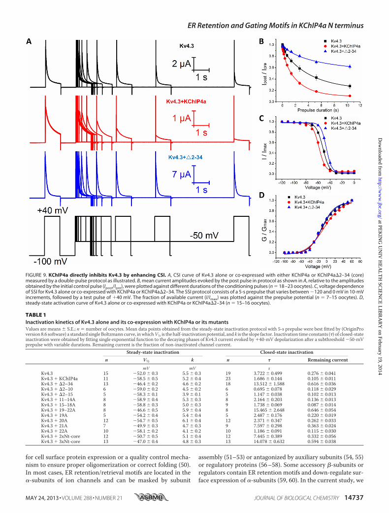
erential CSI was reduced (Fig. 9,A and B). Depolarizing to �40mV after holding for 10.4 s at �50 mV resulted in only about38% of Kv4.3-core complex current being inactivated (Fig. 9B).In contrast, co-expression of Kv4.3 with KChIP4a increasedCSI to about 90%, as compared with Kv4.3 alone (�72%) andKv4.3-core co-expression (�38%) (Fig. 9B and Table 1). TheSSI curve forKv4.3 under a 5-s prepulse had aV1⁄2 of�52.0� 0.3mV and a k of 5.5 � 0.3 mV. The KChIP4a core significantlyshifted the inactivation curve (V1⁄2) to the right in a depolariza-tion direction (V1⁄2 of �46.4 � 0.2 mV and k of 4.6 � 0.2).Consistent with its effect on CSI, the KID overrode the right-shift effect of the core and caused the curve to shift in the hyper-polarization direction (V1⁄2 of �58.5 � 0.5 mV and a k of 5.2 �0.4 mV) (Fig. 9C and Table 1). We also examined the conduc-tance-voltage (G-V) curves and found no significant differences(Fig. 9D). These data suggest that themodulation of Kv4.3 inac-tivation by the KID is dominant over that by the KChIP4a core
FIGURE 5. Identification of critical residues within the KID that mediate ER retention of KChIP4a with a C-terminal H-Ras-CAAX signal. A, C, G, and H, ERdistribution of KChIP4a-CAAX (A), KChIP4a�2–10-CAAX (C), KChIP4a 19 –22A-CAAX (G), and KChIP4a�23–34-CAAX (H). B and D–F, PM distribution of KChIP4a�2–34-CAAX (B), KChIP4a�2–15-CAAX (D), KChIP4a 11–14A-CAAX (E), and KChIP4a 15–18A-CAAX (F). I, ER localization was quantified by DsRed2-ER co-localization,using Pearson’s correlation. Values are mean � S.E. (error bars); n � 14 –24 cells; *, p � 0.001. -CAAX indicates attachment of a PM-targeting CAAX signal fromH-Ras to the C terminus of fusion proteins. Scale bar, 10 �m.
ER Retention and Gating Motifs in KChIP4a N terminus
MAY 24, 2013 • VOLUME 288 • NUMBER 21 JOURNAL OF BIOLOGICAL CHEMISTRY 14733
at PEK
ING
UN
IV H
EA
LT
H SC
IEN
CE
LIB
RA
RY
on February 19, 2014http://w
ww
.jbc.org/D
ownloaded from
and reduces the number of Kv4.3 channels available foropening.To determine which part of the KID is involved in modulat-
ing Kv4.3 CSI, we generated mutants and found that residues
19–22 of the KID played a critical role in facilitating the devel-opment of CSI and causing a hyperpolarizing shift of SSI (Fig.10, A and B, and Table 1). The 19–22A mutant completelyeliminated the enhancement effect of the KID on CSI, consis-
FIGURE 6. Identification of a hydrophobic and aliphatic ER retention motif, LIVIVL, within the KID. A and F, an insertion of VLIVIVLF (N-terminalresidues 11–18 of the KID) caused a strong ER retention of the KChIP4a core (with or without CAAX signal). B and G, the VLIVI sequence alone wasinsufficient to cause ER retention of the core (with or without CAAX signal). C, D, H, and I, the LIVIV (C and H) and IVIVL (D and I) sequences caused the ERlocalization of the core (with or without CAAX signal). E and J, the VIVLF sequence retained the core in the ER (E) but not the core with CAAX signal(-core-CAAX) (J). K, ER localization was quantified by Pearson’s co-localization with DsRed2-ER. Values are mean � S.E. (error bars); n � 14 –20 cells; *, p �0.001. Scale bar, 10 �m.
ER Retention and Gating Motifs in KChIP4a N terminus
14734 JOURNAL OF BIOLOGICAL CHEMISTRY VOLUME 288 • NUMBER 21 • MAY 24, 2013
at PEK
ING
UN
IV H
EA
LT
H SC
IEN
CE
LIB
RA
RY
on February 19, 2014http://w
ww
.jbc.org/D
ownloaded from
tent with the effect of the KID deletion mutant (�2–34). Fur-ther dissection revealed that residues 19–21, VKL, are respon-sible for promoting CSI, and residue 21 (leucine) is the mostcritical (Fig. 10, C and D, and Table 1). A previous study sug-gested that either the N terminus of KChIP2x (2xNt) orKChIP3x (3xNt) functions like theKID (37). To test the effect of
2xNt or 3xNt on CSI, we replaced the KID with 2xNt or 3xNt,where their amino acid residues 19–21 are VKV or IAV,respectively. The VKV from 2xNt exerted an effect similar tothat of the KChIP4a 21Amutant on CSI and SSI. The IAV from3xNt, analogous to the KChIP4a 19–22A or �2–34 mutation,had no effect on promoting CSI (Fig. 10, E and F, and Table 1),
FIGURE 7. Four amino acids of residues 11–18 of KChIP4a are insufficient to cause ER retention. A and F, VLIV alone is insufficient to cause ER retention ofcore (with or without CAAX signal). B–E and G–J, LIVI (B and G), IVIV (C and H), VIVL (D and I) and IVLF (E and J) retained the core in the ER but not the core withCAAX signal (-core-CAAX). K, ER localization was quantified by DsRed2-ER co-localization, using Pearson’s correlation. Values are mean � S.E. (error bars); n �8 –30 cells; *, p � 0.001. Scale bar, 10 �m.
ER Retention and Gating Motifs in KChIP4a N terminus
MAY 24, 2013 • VOLUME 288 • NUMBER 21 JOURNAL OF BIOLOGICAL CHEMISTRY 14735
at PEK
ING
UN
IV H
EA
LT
H SC
IEN
CE
LIB
RA
RY
on February 19, 2014http://w
ww
.jbc.org/D
ownloaded from
confirming the specific effect of the VKLmotif from theKID onCSI. Together, these results demonstrate that the VKLmotif ofthe KID inhibits Kv4.3 function by facilitating the channel CSI.
DISCUSSION
The goal of this study was to understand how auxiliaryKChIP4a,whichonlydiffers fromotherKChIPs in theNterminus,inhibits Kv4 function. We found that KChIP4a acts as an inhibi-tory subunit to suppress Kv4 A-type current (ISA). The inhibitoryproperty of KChIP4a on ISA is primarily mediated by its uniqueN-terminal KID. Our findings reveal two distinct mechanisms bywhich KChIP4a exerts its inhibition on Kv4 function. First, anLIVIVLmotif (N-terminal residues 12–17)mediates ER retentionofKChIP4a andKv4 channels; second, theVKLmotif (N-terminalresidues 19–21) of the KID directly inhibits Kv4.3 function byincreasing the number of channels in the closed inactivated state.Previous studies on Kv4 modulation by KChIPs have mostly
focused on the effects of a single KChIP on Kv4 function.
Because all members of the KChIP family share a high homol-ogy in their C-terminal core regions and each can form twointerfaces associated with two neighboring Kv4 N-terminaldomains (46–48), this raises the possibility that differentKChIPsmay compete for commonKChIP-binding sites, result-ing in a diversity ofmodulation of Kv4 function byKChIPs. Thishypothesis is supported by our observation that KChIP4a inhibitsthe surface expression and peak current of Kv4 coexpressed withKChIP1 in a dose-dependent and competitive manner by antago-nizing thebindingofKChIP1 toKv4 (Fig. 2).Whether theseobser-vations imply that Kv4-KChIP complexes in native cells containheteromeric KChIPs or only one KChIP isoform is presentlyunknown and deserves further investigation.Cell surface expression of proteins depends on the delicate
balance of forward trafficking and intracellular retention (49).ER retention/retrieval motifs are dominant trafficking signalsthat are often encoded in individual subunits of multiproteincomplexes, thereby providing a negative regulatorymechanism
FIGURE 8. The ER retention signal within the KID inhibits total and surface expression of Kv4.3. A and C, representative Western blots of total (A) and surface (C)proteins from HEK 293 cells transiently transfected with plasmids encoding Kv4.3 with WT KChIP4a or KChIP4a mutants. For quantification, the intensities of the Kv4.3bands were measured and normalized, to endogenous actin (for total) or TransR (for surface) in the same lane and subsequently to Kv4.3 co-expressed with WTKChIP4a in the same blot. B and D, quantification analysis of total (B) and surface (D) expression levels of Kv4.3 co-expressed with WT KChIP4a or KChIP4a mutants.Values are mean�S.E. (error bars); n�4–6 blots. Statistical analysis was performed using one-way analysis of variance followed by Tukey’s post hoc test, and statisticalsignificance was assessed as follows. **, p �0.01; ***, p �0.001. E, representative Western blot of total proteins from HEK 293 cells transiently transfected with plasmidsencoding KChIP4a-3xFLAG or KChIP4a�2–34-3xFLAG. F, quantification analysis of total expression levels of KChIP4a-3xFLAG and KChIP4a�2–34-3xFLAG, indicatingreduced expression of KChIP4a as compared with KChIP4a�2–34. G, quantification analysis of relative current change in oocytes expressing Kv4.3 co-expressed withKChIP4a mutants normalized to Kv4.3 co-expressed with WT KChIP4a. Values are mean � S.E.; n � 8–26 oocytes; ***, p � 0.001.
ER Retention and Gating Motifs in KChIP4a N terminus
14736 JOURNAL OF BIOLOGICAL CHEMISTRY VOLUME 288 • NUMBER 21 • MAY 24, 2013
at PEK
ING
UN
IV H
EA
LT
H SC
IEN
CE
LIB
RA
RY
on February 19, 2014http://w
ww
.jbc.org/D
ownloaded from
for cell surface protein expression or a quality control mecha-nism to ensure proper oligomerization or correct folding (50).In most cases, ER retention/retrieval motifs are located in the�-subunits of ion channels and can be masked by subunit
assembly (51–53) or antagonized by auxiliary subunits (54, 55)or regulatory proteins (56–58). Some accessory �-subunits orregulators contain ER retention motifs and down-regulate sur-face expression of �-subunits (59, 60). In the current study, we
FIGURE 9. KChIP4a directly inhibits Kv4.3 by enhancing CSI. A, CSI curve of Kv4.3 alone or co-expressed with either KChIP4a or KChIP4a�2–34 (core)measured by a double-pulse protocol as illustrated. B, mean current amplitudes evoked by the post pulse in protocol as shown in A, relative to the amplitudesobtained by the initial control pulse (Ipost/Ipre), were plotted against different durations of the conditioning pulses (n � 18 –23 oocytes). C, voltage dependenceof SSI for Kv4.3 alone or co-expressed with KChIP4a or KChIP4a�2–34. The SSI protocol consists of a 5-s prepulse that varies between �120 and 0 mV in 10-mVincrements, followed by a test pulse of �40 mV. The fraction of available current (I/Imax) was plotted against the prepulse potential (n � 7–15 oocytes). D,steady-state activation curve of Kv4.3 alone or co-expressed with KChIP4a or KChIP4a�2–34 (n � 15–16 oocytes).
TABLE 1Inactivation kinetics of Kv4.3 alone and its co-expression with KChIP4a or its mutantsValues are means � S.E.; n � number of oocytes. Mean data points obtained from the steady-state inactivation protocol with 5-s prepulse were best fitted by (OriginProversion 8.6 software) a standard single Boltzmann curve, inwhichV1⁄2 is the half-inactivation potential, and k is the slope factor. Inactivation time constants (�) of closed-stateinactivation were obtained by fitting single exponential function to the decaying phases of Kv4.3 current evoked by �40-mV depolarization after a subthreshold �50-mVprepulse with variable durations. Remaining current is the fraction of non-inactivated channel current.
Steady-state inactivation Closed-state inactivationn V1⁄2 k n � Remaining current
mV mV sKv4.3 15 �52.0 � 0.3 5.5 � 0.3 19 3.722 � 0.499 0.276 � 0.041Kv4.3 � KChIP4a 11 �58.5 � 0.5 5.2 � 0.4 23 1.686 � 0.144 0.105 � 0.011Kv4.3 � �2–34 13 �46.4 � 0.2 4.6 � 0.2 18 13.512 � 1.588 0.616 � 0.036Kv4.3 � �2–10 6 �59.0 � 0.2 4.5 � 0.2 6 0.695 � 0.078 0.118 � 0.029Kv4.3 � �2–15 5 �58.3 � 0.1 3.9 � 0.1 5 1.147 � 0.038 0.102 � 0.013Kv4.3 � 11–14A 8 �58.9 � 0.4 5.3 � 0.3 8 2.164 � 0.203 0.136 � 0.013Kv4.3 � 15–18A 8 �58.8 � 0.3 5.0 � 0.3 9 1.738 � 0.069 0.087 � 0.014Kv4.3 � 19–22A 8 �46.6 � 0.5 5.9 � 0.4 8 15.465 � 2.648 0.646 � 0.054Kv4.3 � 19A 5 �54.2 � 0.4 5.4 � 0.4 5 2.487 � 0.176 0.220 � 0.019Kv4.3 � 20A 12 �54.7 � 0.5 6.1 � 0.4 12 2.371 � 0.347 0.262 � 0.033Kv4.3 � 21A 7 �49.9 � 0.3 4.7 � 0.3 9 7.597 � 0.298 0.363 � 0.024Kv4.3 � 22A 10 �58.1 � 0.2 4.1 � 0.2 10 1.186 � 0.091 0.115 � 0.030Kv4.3 � 2xNt-core 12 �50.7 � 0.5 5.1 � 0.4 12 7.445 � 0.389 0.332 � 0.056Kv4.3 � 3xNt-core 13 �47.0 � 0.4 4.8 � 0.3 13 14.078 � 0.632 0.594 � 0.038
ER Retention and Gating Motifs in KChIP4a N terminus
MAY 24, 2013 • VOLUME 288 • NUMBER 21 JOURNAL OF BIOLOGICAL CHEMISTRY 14737
at PEK
ING
UN
IV H
EA
LT
H SC
IEN
CE
LIB
RA
RY
on February 19, 2014http://w
ww
.jbc.org/D
ownloaded from

identified an LIVIVL sequence within the N-terminal KID ofKChIP4a that functions as an ER retention motif to suppressKv4 forward vesicular trafficking from theER to theGolgi appa-ratus, which is different from the inhibition of Kv4 endocytosisinduced by other KChIPs, such as KChIP2 (34). In addition, weutilized the CAAX signal from H-Ras that functions as a PMtargeting signal to evaluate the retention ability of this new-foundmotif. The H-Ras-CAAX signal uses the exocytosis path-way in a similar fashion to the trafficking pathway ofmembraneproteins (61). Therefore, our results support that the ER reten-tionmotif identified in this study plays a dominant negative rolein classical vesicular trafficking pathway, thus leading to thereduction of Kv4 surface expression.Due to its hydrophobic nature, the LIVIVL motif identified
and reported here differs from canonical motifs consisting ofbasic amino acids. A series of hydrophobic ER retention signalshave been identified in different parts of proteins, including theN terminus, C terminus, transmembrane segments, and cyto-plasmic linker of transmembrane segments (53, 62–68). Unlikewell characterized basicmotifs that consist of RXR, RR, andKK,the molecular identity of the hydrophobic motifs still remainsto be further explored. The hydrophobic ER retention motifsvary in their location and composition, and the only commonfeature is membrane adjacency or association. Another inter-esting observation is that the retention ability of the LIVIVLmotif is transferable and dependent on its flanking locationwithin the protein sequence. This feature is similar to theH/KDEL motif that is often located in the C-terminal end ofsoluble ER-resident proteins, such as Bip (Grp78), calreticulin,endoplasmin (Grp94), protein-disulfide isomerase, etc. (50).
The addition of the KDEL to the C terminus of various proteinscan also lead to their ER retention (69–72). The H/KDELmotifis recognized by the KDEL receptor ERD2, which is mainlylocalized to the cis-Golgi and targets the KDEL-containing pro-teins into the retrograde COPI-mediated transport pathway(73, 74).However, how the LIVIVLmotif causes ER retention ofLIVIVL-containing proteins requires further investigation.Previously, we proposed that the core of KChIPs increases
total expression level of Kv4.3 by stabilizing Kv4.3 tetrameriza-tion via a clamping action (48). In the current study we showthat the N-terminal KID of KChIP4a, a non-core region, exertsan inhibitory effect on total Kv4.3 and KChIP4a by reducingtheir protein stability (Fig. 8). Considering the ER retentioneffect of the KID, it is likely that the ER retention motif withinthe KID causes ER-associated protein degradation of KChIP4aitself and its associated proteins, such as Kv4.3. Kv4 expressionand functional properties are regulated by KChIPs as well asdipeptidyl peptidase-like proteins containing DPP6 and DPP10(5, 6). Considerable evidence suggests that native neuronal ISAchannels function in macromolecular protein complexes com-posed of Kv4 pore-forming subunits together with accessoryKChIPs and DPP6/10 subunits (75, 76). Different from cytoso-lic KChIPs, the accessory DPP6/10 subunits are transmem-brane proteins and selectively increase surface expression ofKv4.2 but not total expression (77). In this study, we demon-strate that the ER retention of the KID is dominant over surfacetrafficking of multitransmembrane protein Kv4.3, single trans-membrane protein CD4, and the secretory CAAX signal fromH-Ras protein, so the DPP-mediated augmentation effect on
FIGURE 10. The VKL motif within the N-terminal KID of KChIP4a promotes CSI of Kv4.3. A, analysis for CSI of Kv4.3 co-expressed with KChIP4a or KChIP4amutants (n � 8 –13 oocytes). B, voltage dependence of SSI of Kv4.3 co-expressed with KChIP4a or KChIP4a mutants. C, CSI of Kv4.3 co-expressed with KChIP4aor KChIP4a 19 –22 mutants (n � 5–23 oocytes). D, voltage dependence of SSI of Kv4.3 co-expressed with KChIP4a or KChIP4a 19 –22 mutants (n � 5–12 oocytes).E, CSI of Kv4.3 co-expressed with KChIP4a, core (KChIP4a�2–34), 2xNt-core, or 3xNt-core (n � 12–23 oocytes). F, voltage dependence of SSI of Kv4.3 co-ex-pressed with KChIP4a, core (KChIP4a�2–34), 2xNt-core, or 3xNt-core (n � 11–13 oocytes).
ER Retention and Gating Motifs in KChIP4a N terminus
14738 JOURNAL OF BIOLOGICAL CHEMISTRY VOLUME 288 • NUMBER 21 • MAY 24, 2013
at PEK
ING
UN
IV H
EA
LT
H SC
IEN
CE
LIB
RA
RY
on February 19, 2014http://w
ww
.jbc.org/D
ownloaded from

Kv4 surface expression is likely to be inhibited when co-assem-bled with KChIP4a.The molecular mechanism of Kv4 fast inactivation differs
significantly from other rapidly inactivating Kv channels, suchas Shaker and Kv1.4 (78–82). Kv4 channels exhibit a preferen-tial CSI, implying that upon voltage-dependent activation,inactivation occurs preferentially from partially activatedclosed states that precede the open state (82, 83). In contrast toN-type and C-type inactivation, the molecular basis of CSIremains to be determined. The KID has been previously char-acterized as an open-state inactivation suppressor (so-calledKIS domain) of Kv4 in heterologous cells and neurons (4, 84).Surprisingly, in this study, we found that the KID promotesCSI and shifts the voltage dependence of SSI in a hyperpo-larization direction, whereas the core of KChIP4a-mediatedKv4 modulation on CSI and SSI kinetics is opposite to that ofthe KID (Fig. 9). Moreover, we demonstrated that the exis-tence of the VKL motif (N-terminal residues 19–21) isresponsible for the KID modulation on Kv4 CSI. Therefore,identification of a putative receptor site for the VKL motifbinding to Kv4 channels may provide structural insights intothe mechanism of Kv4 CSI.Similar to Kv4 channels, Kv2 channels (Kv2.1 andKv2.2) also
inactivate preferentially from partially activated closed states,and its steady-state inactivation curve exhibits a U-type shape(85). The functional properties of Kv2 are regulated by form-ing heteromers with the otherwise “silent” �-subunits of theKv5, Kv6, Kv8, or Kv9 subfamily. The modulatory effects ofthese “silent” �-subunits on Kv2 are analogous to those ofKChIP4a on Kv4, such as a reduction in peak current ampli-tude, inhibition of open-state inactivation, acceleration ofCSI, and promotion of hyperpolarized shifts in the voltagedependence of SSI (86–89). Altogether, these findings sug-gest that bidirectional modulation of Kv4 open-state andclosed-state inactivation mediated by the KID may be ahighly conserved function among various ion channels andancillary subunits.The channel subunit composition is a determinant in shap-
ing the repertoire of A-type K� channels present at the surfaceof neurons. Differential regulation of auxiliary KChIPs canmodify the subunit composition of native Kv4 channel com-plexes, altering their biophysical andphysiological properties aswell as neuronal excitability. Recently, an interesting study hasdemonstrated that activation of noncoding RNA 38A inducedby inflammatory stimuli causes a gene-specific alternativesplicing shift from KChIP4bl to KChIP4a, leading to an inhibi-tion of Kv4 function as well as altered amyloid production asso-ciated with neurodegenerative etiology (90). Therefore, it ispossible that the expression level of KChIP4amay vary underpathological conditions that are affected by inflammation,pain, meningitis, multiple sclerosis, Alzheimer disease, Par-kinson disease, or stroke-mediated neuronal dysfunction(91), resulting in perturbation of ISA and neuronal excitabil-ity. Furthermore, because KChIP4a can directly inhibit Kv4function by promoting channel CSI, a specific disruption ofthe interaction between the KChIP4a KID and Kv4 channelsaimed at enhancing ISA may provide a new strategy for treat-
ing hyperexcitable neurological disorders, such as pain andepilepsy.
Acknowledgments—We thank laboratory members Xu Cao, Jun Su,Yuanyuan Cui, and Hao Chen for discussion. We also thank ArunSharma (StanfordUniversity School ofMedicine) for careful and crit-ical reading of the manuscript. K. W. W. thanks J. M. Wang for con-sistent support during this research.
REFERENCES1. Birnbaum, S. G., Varga, A. W., Yuan, L. L., Anderson, A. E., Sweatt, J. D.,
and Schrader, L. A. (2004) Structure and function of Kv4-family transientpotassium channels. Physiol. Rev. 84, 803–833
2. Covarrubias, M., Bhattacharji, A., De Santiago-Castillo, J. A., Dougherty,K., Kaulin, Y. A., Na-Phuket, T. R., andWang, G. (2008) The neuronal Kv4channel complex. Neurochem. Res. 33, 1558–1567
3. An, W. F., Bowlby, M. R., Betty, M., Cao, J., Ling, H. P., Mendoza, G.,Hinson, J. W., Mattsson, K. I., Strassle, B. W., Trimmer, J. S., and Rhodes,K. J. (2000) Modulation of A-type potassium channels by a family of cal-cium sensors. Nature 403, 553–556
4. Holmqvist, M. H., Cao, J., Hernandez-Pineda, R., Jacobson,M. D., Carroll,K. I., Sung, M. A., Betty, M., Ge, P., Gilbride, K. J., Brown, M. E., Jurman,M. E., Lawson, D., Silos-Santiago, I., Xie, Y., Covarrubias,M., Rhodes, K. J.,Distefano, P. S., and An, W. F. (2002) Elimination of fast inactivation inKv4 A-type potassium channels by an auxiliary subunit domain. Proc.Natl. Acad. Sci. U.S.A. 99, 1035–1040
5. Nadal,M. S., Ozaita, A., Amarillo, Y., Vega-Saenz deMiera, E.,Ma, Y.,Mo,W., Goldberg, E. M., Misumi, Y., Ikehara, Y., Neubert, T. A., and Rudy, B.(2003) The CD26-related dipeptidyl aminopeptidase-like protein DPPX isa critical component of neuronal A-type K� channels. Neuron 37,449–461
6. Jerng, H. H., Qian, Y., and Pfaffinger, P. J. (2004) Modulation of Kv4.2channel expression and gating by dipeptidyl peptidase 10 (DPP10). Bio-phys. J. 87, 2380–2396
7. Wang, K. (2008) Modulation by clamping. Kv4 and KChIP interactions.Neurochem. Res. 33, 1964–1969
8. Liss, B., Franz, O., Sewing, S., Bruns, R., Neuhoff, H., and Roeper, J. (2001)Tuning pacemaker frequency of individual dopaminergic neurons byKv4.3L and KChip3.1 transcription. EMBO J. 20, 5715–5724
9. Rhodes, K. J., Carroll, K. I., Sung,M. A., Doliveira, L. C.,Monaghan,M.M.,Burke, S. L., Strassle, B. W., Buchwalder, L., Menegola, M., Cao, J., An,W. F., and Trimmer, J. S. (2004) KChIPs and Kv4 � subunits as integralcomponents of A-type potassium channels in mammalian brain. J. Neu-rosci. 24, 7903–7915
10. Norris, A. J., Foeger, N. C., and Nerbonne, J. M. (2010) Interdependentroles for accessory KChIP2, KChIP3, and KChIP4 subunits in the genera-tion of Kv4-encoded IA channels in cortical pyramidal neurons. J. Neuro-sci. 30, 13644–13655
11. Kuo, H. C., Cheng, C. F., Clark, R. B., Lin, J. J., Lin, J. L., Hoshijima, M.,Nguyen-Tran, V. T., Gu, Y., Ikeda, Y., Chu, P. H., Ross, J., Giles,W. R., andChien, K. R. (2001) A defect in the Kv channel-interacting protein 2(KChIP2) gene leads to a complete loss of Ito and confers susceptibility toventricular tachycardia. Cell 107, 801–813
12. Guo,W., Li, H., Aimond, F., Johns, D. C., Rhodes, K. J., Trimmer, J. S., andNerbonne, J. M. (2002) Role of heteromultimers in the generation ofmyo-cardial transient outward K� currents. Circ. Res. 90, 586–593
13. Hoffman, D. A., Magee, J. C., Colbert, C. M., and Johnston, D. (1997) K�
channel regulation of signal propagation in dendrites of hippocampal py-ramidal neurons. Nature 387, 869–875
14. Bernard, C., Anderson, A., Becker, A., Poolos, N. P., Beck, H., and John-ston, D. (2004) Acquired dendritic channelopathy in temporal lobe epi-lepsy. Science 305, 532–535
15. Cai, X., Liang, C. W., Muralidharan, S., Muralidharan, S., Kao, J. P., Tang,C. M., and Thompson, S. M. (2004) Unique roles of SK and Kv4.2 potas-sium channels in dendritic integration. Neuron 44, 351–364
16. Chen, X., Yuan, L. L., Zhao, C., Birnbaum, S. G., Frick, A., Jung, W. E.,
ER Retention and Gating Motifs in KChIP4a N terminus
MAY 24, 2013 • VOLUME 288 • NUMBER 21 JOURNAL OF BIOLOGICAL CHEMISTRY 14739
at PEK
ING
UN
IV H
EA
LT
H SC
IEN
CE
LIB
RA
RY
on February 19, 2014http://w
ww
.jbc.org/D
ownloaded from

Schwarz, T. L., Sweatt, J. D., and Johnston, D. (2006) Deletion of Kv4.2gene eliminates dendritic A-type K� current and enhances induction oflong-term potentiation in hippocampal CA1 pyramidal neurons. J. Neu-rosci. 26, 12143–12151
17. Kim, J., Jung, S. C., Clemens, A. M., Petralia, R. S., and Hoffman, D. A.(2007) Regulation of dendritic excitability by activity-dependent traffick-ing of the A-type K� channel subunit Kv4.2 in hippocampal neurons.Neuron 54, 933–947
18. Barry, D. M., Xu, H., Schuessler, R. B., and Nerbonne, J. M. (1998) Func-tional knockout of the transient outward current, long-QT syndrome, andcardiac remodeling in mice expressing a dominant-negative Kv4 � sub-unit. Circ. Res. 83, 560–567
19. Hoppe, U. C., Marban, E., and Johns, D. C. (2000) Molecular dissection ofcardiac repolarization by in vivo Kv4.3 gene transfer. J. Clin. Invest. 105,1077–1084
20. Singh, B., Ogiwara, I., Kaneda, M., Tokonami, N., Mazaki, E., Baba, K.,Matsuda, K., Inoue, Y., and Yamakawa, K. (2006) A Kv4.2 truncation mu-tation in a patient with temporal lobe epilepsy. Neurobiol. Dis. 24,245–253
21. Giudicessi, J. R., Ye, D., Tester, D. J., Crotti, L., Mugione, A., Nesterenko,V. V., Albertson, R. M., Antzelevitch, C., Schwartz, P. J., and Ackerman,M. J. (2011) Transient outward current (Ito) gain-of-functionmutations inthe KCND3-encoded Kv4.3 potassium channel and Brugada syndrome.Heart Rhythm 8, 1024–1032
22. Giudicessi, J. R., Ye, D., Kritzberger, C. J., Nesterenko, V. V., Tester, D. J.,Antzelevitch, C., and Ackerman, M. J. (2012) Novel mutations in theKCND3-encoded Kv4.3 K� channel associated with autopsy-negativesudden unexplained death. Hum. Mutat. 33, 989–997
23. Duarri, A., Jezierska, J., Fokkens, M., Meijer, M., Schelhaas, H. J., denDunnen, W. F., van Dijk, F., Verschuuren-Bemelmans, C., Hageman, G.,van de Vlies, P., Kusters, B., van de Warrenburg, B. P., Kremer, B., Wij-menga, C., Sinke, R. J., Swertz, M. A., Kampinga, H. H., Boddeke, E., andVerbeek, D. S. (2012)Mutations in potassium channel kcnd3 cause spino-cerebellar ataxia type 19. Ann. Neurol. 72, 870–880
24. Olesen, M. S., Refsgaard, L., Holst, A. G., Larsen, A. P., Grubb, S., Haunso,S., Svendsen, J. H., Olesen, S. P., Schmitt, N., and Calloe, K. (2013)Cardio-vasc. Res. doi: 10.1093/cvr/cvt028
25. Lee, Y. C., Durr, A., Majczenko, K., Huang, Y. H., Liu, Y. C., Lien, C. C.,Tsai, P. C., Ichikawa, Y., Goto, J., Monin, M. L., Li, J. Z., Chung, M. Y.,Mundwiller, E., Shakkottai, V., Liu, T. T., Tesson, C., Lu, Y. C., Brice, A.,Tsuji, S., Burmeister, M., Stevanin, G., and Soong, B.W. (2012)MutationsinKCND3 cause spinocerebellar ataxia type 22.Ann.Neurol. 72, 859–869
26. Burgoyne, R. D. (2007) Neuronal calcium sensor proteins. Generatingdiversity in neuronal Ca2� signalling. Nat. Rev. Neurosci. 8, 182–193
27. Takimoto, K., Yang, E. K., andConforti, L. (2002) Palmitoylation of KChIPsplicing variants is required for efficient cell surface expression of Kv4.3channels. J. Biol. Chem. 277, 26904–26911
28. O’Callaghan, D. W., Hasdemir, B., Leighton, M., and Burgoyne, R. D.(2003) Residues within the myristoylation motif determine intracellulartargeting of the neuronal Ca2� sensor protein KChIP1 to post-ER trans-port vesicles and traffic of Kv4 K� channels. J. Cell Sci. 116, 4833–4845
29. Decher, N., Barth, A. S., Gonzalez, T., Steinmeyer, K., and Sanguinetti,M. C. (2004) Novel KChIP2 isoforms increase functional diversity of tran-sient outward potassium currents. J. Physiol. 557, 761–772
30. VanHoorick, D., Raes, A., and Snyders, D. J. (2007)The aromatic cluster inKCHIP1b affects Kv4 inactivation gating. J. Physiol. 583, 959–969
31. Pruunsild, P., and Timmusk, T. (2005) Structure, alternative splicing, andexpression of the human and mouse KCNIP gene family. Genomics 86,581–593
32. Shibata, R., Misonou, H., Campomanes, C. R., Anderson, A. E., Schrader,L. A., Doliveira, L. C., Carroll, K. I., Sweatt, J. D., Rhodes, K. J., and Trim-mer, J. S. (2003) A fundamental role for KChIPs in determining the mo-lecular properties and trafficking of Kv4.2 potassium channels. J. Biol.Chem. 278, 36445–36454
33. Cui, Y. Y., Liang, P., and Wang, K. W. (2008) Enhanced trafficking oftetrameric Kv4.3 channels by KChIP1 clamping. Neurochem. Res. 33,2078–2084
34. Foeger, N. C.,Marionneau, C., andNerbonne, J.M. (2010) Co-assembly of
Kv4 � subunits with K� channel-interacting protein 2 stabilizes proteinexpression and promotes surface retention of channel complexes. J. Biol.Chem. 285, 33413–33422
35. Panama, B. K., Latour-Villamil, D., Farman, G. P., Zhao, D., Bolz, S. S.,Kirshenbaum, L. A., and Backx, P. H. (2011) Nuclear factor �B downregu-lates the transient outward potassium current Ito,f through control ofKChIP2 expression. Circ. Res. 108, 537–543
36. Jeyaraj, D., Haldar, S. M., Wan, X., McCauley, M. D., Ripperger, J. A., Hu,K., Lu, Y., Eapen, B. L., Sharma, N., Ficker, E., Cutler, M. J., Gulick, J.,Sanbe, A., Robbins, J., Demolombe, S., Kondratov, R. V., Shea, S. A., Al-brecht, U., Wehrens, X. H., Rosenbaum, D. S., and Jain, M. K. (2012)Circadian rhythms govern cardiac repolarization and arrhythmogenesis.Nature 483, 96–99
37. Jerng, H. H., and Pfaffinger, P. J. (2008) Multiple Kv channel-interactingproteins contain an N-terminal transmembrane domain that regulatesKv4 channel trafficking and gating. J. Biol. Chem. 283, 36046–36059
38. Schwenk, J., Zolles, G., Kandias, N. G., Neubauer, I., Kalbacher, H., Cova-rrubias, M., Fakler, B., and Bentrop, D. (2008) NMR analysis of KChIP4areveals structural basis for control of surface expression of Kv4 channelcomplexes. J. Biol. Chem. 283, 18937–18946
39. Liang, P.,Wang, H., Chen, H., Cui, Y., Gu, L., Chai, J., andWang, K. (2009)Structural insights into KChIP4amodulation of Kv4.3 inactivation. J. Biol.Chem. 284, 4960–4967
40. Liang, P., Chen, H., Cui, Y., Lei, L., andWang, K. (2010) Functional rescueof Kv4.3 channel tetramerization mutants by KChIP4a. Biophys. J. 98,2867–2876
41. Kim,H. J., Ahn,H. S., Choi, B. H., andHahn, S. J. (2011) Inhibition of Kv4.3by genistein via a tyrosine phosphorylation-independent mechanism.Am. J. Physiol. Cell Physiol. 300, C567–C575
42. Kim, H. J., Ahn, H. S., Choi, J. S., Choi, B. H., and Hahn, S. J. (2011) Effectsof ranolazine on cloned cardiac kv4.3 potassium channels. J. Pharmacol.Exp. Ther. 339, 952–958
43. Jeong, I., Choi, B. H., and Hahn, S. J. (2011) Rosiglitazone inhibits Kv4.3potassium channels by open-channel block and acceleration of closed-state inactivation. Br. J. Pharmacol. 163, 510–520
44. Jeong, I., Kim, S. W., Yoon, S. H., and Hahn, S. J. (2012) Block of clonedKv4.3 potassium channels by dapoxetine. Neuropharmacology 62,2261–2266
45. Choi, J. S., and Hahn, S. J. (2012) Duloxetine blocks cloned Kv4.3 potas-sium channels. Brain Res. 1466, 15–23
46. Scannevin, R. H., Wang, K., Jow, F., Megules, J., Kopsco, D. C., Edris, W.,Carroll, K. C., Lu, Q., Xu,W., Xu, Z., Katz, A. H., Olland, S., Lin, L., Taylor,M., Stahl, M., Malakian, K., Somers, W., Mosyak, L., Bowlby, M. R.,Chanda, P., and Rhodes, K. J. (2004) Two N-terminal domains of Kv4 K�
channels regulate binding to and modulation by KChIP1. Neuron 41,587–598
47. Pioletti, M., Findeisen, F., Hura, G. L., and Minor, D. L., Jr. (2006) Three-dimensional structure of the KChIP1-Kv4.3 T1 complex reveals a cross-shaped octamer. Nat. Struct. Mol. Biol. 13, 987–995
48. Wang, H., Yan, Y., Liu, Q., Huang, Y., Shen, Y., Chen, L., Chen, Y., Yang,Q., Hao, Q., Wang, K., and Chai, J. (2007) Structural basis for modulationof Kv4 K� channels by auxiliary KChIP subunits.Nat. Neurosci. 10, 32–39
49. Ma, D., and Jan, L. Y. (2002) ER transport signals and trafficking of potas-sium channels and receptors. Curr. Opin. Neurobiol. 12, 287–292
50. Ellgaard, L., and Helenius, A. (2003) Quality control in the endoplasmicreticulum. Nat. Rev. Mol. Cell Biol. 4, 181–191
51. Zerangue, N., Schwappach, B., Jan, Y. N., and Jan, L. Y. (1999) A new ERtrafficking signal regulates the subunit stoichiometry of plasma mem-brane KATP channels. Neuron 22, 537–548
52. Margeta-Mitrovic,M., Jan, Y. N., and Jan, L. Y. (2000) A trafficking check-point controls GABAB receptor heterodimerization. Neuron 27, 97–106
53. Wang, J. M., Zhang, L., Yao, Y., Viroonchatapan, N., Rothe, E., andWang,Z. Z. (2002) A transmembrane motif governs the surface trafficking ofnicotinic acetylcholine receptors. Nat. Neurosci. 5, 963–970
54. Bichet, D., Cornet, V., Geib, S., Carlier, E., Volsen, S., Hoshi, T., Mori, Y.,and De Waard, M. (2000) The I-II loop of the Ca2� channel �1 subunitcontains an endoplasmic reticulum retention signal antagonized by the �
subunit. Neuron 25, 177–190
ER Retention and Gating Motifs in KChIP4a N terminus
14740 JOURNAL OF BIOLOGICAL CHEMISTRY VOLUME 288 • NUMBER 21 • MAY 24, 2013
at PEK
ING
UN
IV H
EA
LT
H SC
IEN
CE
LIB
RA
RY
on February 19, 2014http://w
ww
.jbc.org/D
ownloaded from

55. Zhang, Z. N., Li, Q., Liu, C.,Wang, H. B.,Wang, Q., and Bao, L. (2008) Thevoltage-gatedNa� channel Nav1.8 contains an ER-retention/retrieval sig-nal antagonized by the �3 subunit. J. Cell Sci. 121, 3243–3252
56. Standley, S., Roche, K. W., McCallum, J., Sans, N., and Wenthold, R. J.(2000) PDZ domain suppression of an ER retention signal in NMDA re-ceptor NR1 splice variants. Neuron 28, 887–898
57. Girard, C., Tinel, N., Terrenoire, C., Romey, G., Lazdunski, M., and Bor-sotto,M. (2002) p11, an annexin II subunit, an auxiliary protein associatedwith the background K� channel, TASK-1. EMBO J. 21, 4439–4448
58. O’Kelly, I., Butler, M. H., Zilberberg, N., and Goldstein, S. A. (2002) For-ward transport. 14-3-3 binding overcomes retention in endoplasmic re-ticulum by dibasic signals. Cell 111, 577–588
59. Renigunta, V., Yuan, H., Zuzarte, M., Rinne, S., Koch, A., Wischmeyer, E.,Schlichthorl, G., Gao, Y., Karschin, A., Jacob, R., Schwappach, B., Daut, J.,and Preisig-Muller, R. (2006) The retention factor p11 confers an endo-plasmic reticulum-localization signal to the potassium channel TASK-1.Traffic 7, 168–181
60. Shruti, S., Urban-Ciecko, J., Fitzpatrick, J. A., Brenner, R., Bruchez, M. P.,and Barth, A. L. (2012) The brain-specific �4 subunit downregulates BKchannel cell surface expression. PLoS One 7, e33429
61. Hancock, J. F. (2003) Ras proteins. Different signals from different loca-tions. Nat. Rev. Mol. Cell Biol. 4, 373–384
62. Szczesna-Skorupa, E., and Kemper, B. (2000) Endoplasmic reticulum re-tention determinants in the transmembrane and linker domains of cyto-chrome P450 2C1. J. Biol. Chem. 275, 19409–19415
63. Kaether, C., Capell, A., Edbauer, D.,Winkler, E., Novak, B., Steiner, H., andHaass, C. (2004) The presenilin C terminus is required for ER-retention,nicastrin-binding and �-secretase activity. EMBO J. 23, 4738–4748
64. Parker, A. K., Gergely, F. V., and Taylor, C.W. (2004) Targeting of inositol1,4,5-trisphosphate receptors to the endoplasmic reticulum by multiplesignals within their transmembrane domains. J. Biol. Chem. 279,23797–23805
65. Zarei, M. M., Eghbali, M., Alioua, A., Song, M., Knaus, H. G., Stefani, E.,and Toro, L. (2004) An endoplasmic reticulum trafficking signal preventssurface expression of a voltage- and Ca2�-activated K� channel splicevariant. Proc. Natl. Acad. Sci. U.S.A. 101, 10072–10077
66. Meur, G., Parker, A. K., Gergely, F. V., and Taylor, C.W. (2007) Targetingand retention of type 1 ryanodine receptors to the endoplasmic reticulum.J. Biol. Chem. 282, 23096–23103
67. Ciczora, Y., Callens, N., Seron, K., Rouille, Y., and Dubuisson, J. (2010)Identification of a dominant endoplasmic reticulum-retention signal inyellow fever virus pre-membrane protein. J. Gen. Virol. 91, 404–414
68. Angelotti, T., Daunt, D., Shcherbakova, O. G., Kobilka, B., andHurt, C.M.(2010) Regulation ofG-protein coupled receptor traffic by an evolutionaryconserved hydrophobic signal. Traffic 11, 560–578
69. Dayel, M. J., Hom, E. F., and Verkman, A. S. (1999) Diffusion of greenfluorescent protein in the aqueous-phase lumen of endoplasmic reticu-lum. Biophys. J. 76, 2843–2851
70. Frigerio, L., Pastres, A., Prada, A., and Vitale, A. (2001) Influence of KDELon the fate of trimeric or assembly-defective phaseolin. Selective use of analternative route to vacuoles. Plant Cell 13, 1109–1126
71. Raykhel, I., Alanen, H., Salo, K., Jurvansuu, J., Nguyen, V. D., Latva-Ranta,M., and Ruddock, L. (2007) A molecular specificity code for the threemammalian KDEL receptors. J. Cell Biol. 179, 1193–1204
72. Matsukawa, S., Moriyama, Y., Hayata, T., Sasaki, H., Ito, Y., Asashima,M.,and Kuroda, H. (2012) KDEL tagging. Amethod for generating dominant-negative inhibitors of the secretion of TGF-� superfamily proteins. Int. J.Dev. Biol. 56, 351–356
73. Griffiths, G., Ericsson,M., Krijnse-Locker, J., Nilsson, T., Goud, B., Soling,
H. D., Tang, B. L., Wong, S. H., and Hong, W. (1994) Localization of theLys, Asp, Glu, Leu tetrapeptide receptor to the Golgi complex and theintermediate compartment in mammalian cells. J. Cell Biol. 127,1557–1574
74. Semenza, J. C., Hardwick, K. G., Dean, N., and Pelham, H. R. (1990) ERD2,a yeast gene required for the receptor-mediated retrieval of luminal ERproteins from the secretory pathway. Cell 61, 1349–1357
75. Jerng, H. H., Kunjilwar, K., and Pfaffinger, P. J. (2005)Multiprotein assem-bly of Kv4.2, KChIP3, and DPP10 produces ternary channel complexeswith ISA-like properties. J. Physiol. 568, 767–788
76. Amarillo, Y., De Santiago-Castillo, J. A., Dougherty, K.,Maffie, J., Kwon, E.,Covarrubias, M., and Rudy, B. (2008) Ternary Kv4.2 channels recapitulatevoltage-dependent inactivation kinetics of A-type K� channels in cerebel-lar granule neurons. J. Physiol. 586, 2093–2106
77. Foeger, N. C., Norris, A. J., Wren, L. M., and Nerbonne, J. M. (2012)Augmentation of Kv4.2-encoded currents by accessory dipeptidyl pepti-dase 6 and 10 subunits reflects selective cell surface Kv4.2 protein stabili-zation. J. Biol. Chem. 287, 9640–9650
78. Hoshi, T., Zagotta, W. N., and Aldrich, R. W. (1990) Biophysical andmolecular mechanisms of Shaker potassium channel inactivation. Science250, 533–538
79. Choi, K. L., Aldrich, R. W., and Yellen, G. (1991) Tetraethylammoniumblockade distinguishes two inactivation mechanisms in voltage-activatedK� channels. Proc. Natl. Acad. Sci. U.S.A. 88, 5092–5095
80. Baukrowitz, T., and Yellen, G. (1995) Modulation of K� current by fre-quency and external [K�]. A tale of two inactivationmechanisms.Neuron15, 951–960
81. Jerng, H. H., and Covarrubias, M. (1997) K� channel inactivation medi-ated by the concerted action of the cytoplasmic N- and C-terminal do-mains. Biophys. J. 72, 163–174
82. Bahring, R., and Covarrubias, M. (2011)Mechanisms of closed-state inac-tivation in voltage-gated ion channels. J. Physiol. 589, 461–479
83. Bahring, R., Boland, L. M., Varghese, A., Gebauer, M., and Pongs, O.(2001) Kinetic analysis of open- and closed-state inactivation transitionsin human Kv4.2 A-type potassium channels. J. Physiol. 535, 65–81
84. Baranauskas, G. (2004) Cell-type-specific splicing of KChIP4 mRNA cor-relates with slower kinetics of A-type current. Eur. J. Neurosci. 20,385–391
85. Klemic, K. G., Shieh, C. C., Kirsch, G. E., and Jones, S. W. (1998) Inactiva-tion of Kv2.1 potassium channels. Biophys. J. 74, 1779–1789
86. Salinas, M., de Weille, J., Guillemare, E., Lazdunski, M., and Hugnot, J. P.(1997) Modes of regulation of Shab K� channel activity by the Kv8.1subunit. J. Biol. Chem. 272, 8774–8780
87. Salinas, M., Duprat, F., Heurteaux, C., Hugnot, J. P., and Lazdunski, M.(1997) New modulatory � subunits for mammalian Shab K� channels.J. Biol. Chem. 272, 24371–24379
88. Kramer, J. W., Post, M. A., Brown, A. M., and Kirsch, G. E. (1998) Modu-lation of potassium channel gating by coexpression of Kv2.1 with regula-tory Kv5.1 or Kv6.1 �-subunits. Am. J. Physiol. 274, C1501–C1510
89. Kerschensteiner, D., and Stocker, M. (1999) Heteromeric assembly ofKv2.1 with Kv9.3. Effect on the state dependence of inactivation. Biophys.J. 77, 248–257
90. Massone, S., Vassallo, I., Castelnuovo, M., Fiorino, G., Gatta, E., Robello,M., Borghi, R., Tabaton, M., Russo, C., Dieci, G., Cancedda, R., and Pa-gano, A. (2011) RNA polymerase III drives alternative splicing of the po-tassium channel-interacting protein contributing to brain complexity andneurodegeneration. J. Cell Biol. 193, 851–866
91. Aktas, O., Ullrich, O., Infante-Duarte, C., Nitsch, R., and Zipp, F. (2007)Neuronal damage in brain inflammation. Arch. Neurol. 64, 185–189
ER Retention and Gating Motifs in KChIP4a N terminus
MAY 24, 2013 • VOLUME 288 • NUMBER 21 JOURNAL OF BIOLOGICAL CHEMISTRY 14741
at PEK
ING
UN
IV H
EA
LT
H SC
IEN
CE
LIB
RA
RY
on February 19, 2014http://w
ww
.jbc.org/D
ownloaded from