Melanoma and Other Skin Cancers
4
Melanoma Develops in the melanocytic cells located in the epidermis. The melanocytes produce melanin, the pigment that provides the skin with its colour. • Is the most dangerous form of skin cancer and the most likely to cause death. • The lifetime risk of developing melanoma is 1 in 25 for men and 1 in 34 for women. • Over 8800 people are diagnosed and more than 1000 people die in Australia every year. • Around 2% of the total number of skin cancers diagnosed are melanoma. • Over 70% of melanoma related deaths in WA occur in people aged over 55 years. • Over 50% of deaths from melanoma in WA occur in men aged over 50 years. Non-melanocytic skin cancer (NMSC) • Squamous cell carcinoma (SCC) develops from the squamous cells in the epidermis. SCC accounts for approximately 30% of NMSC diagnosed. • Basal cell carcinoma (BCC) develops from the basal cells in the epidermis. BCC accounts for approximately 70% of NMSC diagnosed. • Over 370 000 new cases of BCC and SCC are diagnosed in Australia every year resulting in 400 deaths. References Australian Institute of Health and Welfare (AIHW) and Australian Association of Cancer Registries (AACR) (2004) Cancer in Australia 2001. AIHW Cat. no. CAN 23, Canberra. Threlfall, T.J. and Thompson, J.R. (2005) Cancer in Western Australia: Incidence and mortality 2003 and Mesothelioma 1960-2003. Department of Health, Western Australia, Perth. Skin cancer is divided into 2 main types: Screening Population based or mass screening for melanoma and other skin cancers is not recommended. Screening is recommended: • For patients identified with risk factors for melanoma and NMSC, including patients with a previous diagnosis of melanoma. • On an opportunistic or case finding basis, offered as part of a routine medical check-up of patients presenting with risk factors. Skin self-examination (SSE) There is no specific SSE technique or recommended frequency of self-examination that has been shown to reduce morbidity or mortality from skin cancer. Up to 70% of melanomas are initially detected by people themselves or a family member. Regular skin examination increases the probability of detecting skin cancer at an early and highly treatable stage. • The Australasian College of Dermatologists recommends that people examine their skin four times a year or as often as recommended by their medical practitioner. • Patients with risk factors should be encouraged to undergo a total body skin examination with a medical practitioner at least once a year. Australia has the highest rate of skin cancer in the world. One in two people who spend their life in Australia will develop some form of skin cancer. Melanoma and other skin cancers: a guide for medical practitioners Causes of melanoma and other skin cancers • Unprotected exposure to ultraviolet radiation (UVR) remains the single most important risk factor for melanoma and other skin cancers. • Both UVB and UVA contribute to skin damage, premature aging and skin cancer. • Melanoma and BCC are associated with intermittent, high intensity exposure to UVR, especially exposure resulting in sunburn. • SCC is associated with cumulative or large amounts of exposure to UVR over long periods of time. • Other risk factors for NMSC are rarer but can include exposure to some chemicals (arsenic), arc welding, radiation therapy, some psoriasis treatment, reduced immunity and some genetic conditions predisposing to skin cancer. Risk factors for melanoma and other skin cancers • Age. • Experience of sunburn in the past, especially in childhood. • Sporadic, intense exposure to UVR. • Fair skin that burns easily, freckles and does not tan. • Presence of dysplastic naevi. • Presence of a large number of dysplastic naevi (>200). • Having fair or red hair and blue or green eyes. • Having a family history of melanoma. • Personal history of NMSC. Diagnosed and treated early, 95% of melanoma and 99% of NMSC can be cured
-
Upload
rohini-selvarajah -
Category
Documents
-
view
216 -
download
0
Transcript of Melanoma and Other Skin Cancers

Melanoma
Develops in the melanocytic cells located inthe epidermis. The melanocytes producemelanin, the pigment that provides the skinwith its colour.
• Is the most dangerous form of skin cancerand the most likely to cause death.
• The lifetime risk of developing melanomais 1 in 25 for men and 1 in 34 for women.
• Over 8800 people are diagnosed and morethan 1000 people die in Australia everyyear.
• Around 2% of the total number of skincancers diagnosed are melanoma.
• Over 70% of melanoma related deaths inWA occur in people aged over 55 years.
• Over 50% of deaths from melanoma inWA occur in men aged over 50 years.
Non-melanocytic skin cancer(NMSC)
• Squamous cell carcinoma (SCC)develops from the squamous cells in theepidermis. SCC accounts for approximately30% of NMSC diagnosed.
• Basal cell carcinoma (BCC) developsfrom the basal cells in the epidermis. BCCaccounts for approximately 70% of NMSCdiagnosed.
• Over 370 000 new cases of BCC and SCCare diagnosed in Australia every yearresulting in 400 deaths.
ReferencesAustralian Institute of Health and Welfare (AIHW) andAustralian Association of Cancer Registries (AACR) (2004)Cancer in Australia 2001. AIHW Cat. no. CAN 23,Canberra.
Threlfall, T.J. and Thompson, J.R. (2005) Cancer inWestern Australia: Incidence and mortality 2003 andMesothelioma 1960-2003. Department of Health,Western Australia, Perth.
Skin cancer is divided into 2 main types:
Screening
Population based or mass screeningfor melanoma and other skin cancersis not recommended.
Screening is recommended:• For patients identified with risk factors for
melanoma and NMSC, including patientswith a previous diagnosis of melanoma.
• On an opportunistic or case finding basis,offered as part of a routine medical check-up of patients presenting with riskfactors.
Skin self-examination (SSE)
There is no specific SSE technique or recommended frequency of self-examinationthat has been shown to reduce morbidity ormortality from skin cancer. Up to 70% ofmelanomas are initially detected by peoplethemselves or a family member. Regular skinexamination increases the probability ofdetecting skin cancer at an early and highlytreatable stage. • The Australasian College of Dermatologists
recommends that people examine their skinfour times a year or as often as recommended by their medical practitioner.
• Patients with risk factors should beencouraged to undergo a total body skin examination with a medical practitioner at least once a year.
Australia has the highestrate of skin cancer in theworld. One in two peoplewho spend their life inAustralia will develop someform of skin cancer.
Melanoma and other skin cancers: a guide for medical practitioners
Causes of melanoma and other skin cancers
• Unprotected exposure to ultraviolet radiation (UVR) remains the single mostimportant risk factor for melanoma andother skin cancers.
• Both UVB and UVA contribute to skin damage, premature aging and skin cancer.
• Melanoma and BCC are associated withintermittent, high intensity exposure to UVR,especially exposure resulting in sunburn.
• SCC is associated with cumulative or largeamounts of exposure to UVR over longperiods of time.
• Other risk factors for NMSC are rarer butcan include exposure to some chemicals(arsenic), arc welding, radiation therapy,some psoriasis treatment, reducedimmunity and some genetic conditionspredisposing to skin cancer.
Risk factors for melanoma andother skin cancers
• Age.• Experience of sunburn in the past,
especially in childhood.• Sporadic, intense exposure to UVR.• Fair skin that burns easily, freckles and does
not tan.• Presence of dysplastic naevi. • Presence of a large number of
dysplastic naevi (>200).• Having fair or red hair and blue or
green eyes.• Having a family history of melanoma.• Personal history of NMSC.
Diagnosed and treated early,95% of melanoma and 99%
of NMSC can be cured
GP Education version.qxd 20/06/2006 11:27 AM Page 1

Superficial Spreading Melanoma(SSM)
Melanoma can develop in pre-existing moles in theskin, or in the melanocytic cells found in the epidermis.
• SSM is the most common form of melanoma.
• SSM can appear as a new spot, or an existing spot,freckle or mole that changes size, colour or shape.
• SSM can develop on any part of the body, includingparts not heavily exposed to UVR. SSM is commonlyfound on the head, neck and trunk on men and lowerextremities on women.
• A patient diagnosed with melanoma is twice as likely as the average person of the same age todevelop another.
• Survival from melanoma is largely dependent ontumour thickness at the time of diagnosis. Tumoursless than 1mm thick have a cure rate of over 90%,tumours thicker than 4mm, less than 55%.
The ABCD can help distinguish a superficial spreading melanoma from a normal mole:
Asymmetry: a lesion that is irregular in shape.
Border: the border or outline of a melanoma isusually irregular.
Colour: there is variation in colour within the lesion.
Diameter: the lesion is usually greater than 6mm across. However smaller suspiciouslesions should also be investigated.
Melanoma diagnosis
A
B
C
D
E
F
G
If NM is suspected, diagnosis should not be delayed and urgent referral to a dermatologist is recommended.
Biopsy and Excision for melanoma
• Complete excision biopsy with a 2mm margin is recommended.
• Punch biopsies and shave excisions are not recommended as they can interfere with pathology analysis.
• If a thick melanoma or NM is suspected, refer patient to a dermatologist.
Nodular Melanoma (NM)
A highly dangerous form of melanoma that penetratesvertically into the epidermis and grows quickly.
NM differs from SSM in appearance. NM is more likely to be symmetrical and uniform in colour (red,pink, brown or black) and feels firm to the touch. Overtime it develops a crusty surface that bleeds easily.
• NM can become life threatening in 6 - 8 weeks.
• Less than 15% of total melanomas diagnosed areNM but 70% of these lesions are thicker than 3mm.
• NM does not necessarily arise from a pre-existingmole, it can develop on any surface of the body.NM is often found on the back and the scalp.
The ABCD cannot be used to aid diagnosis of nodular melanoma however the following can be of help:
Elevated – can appear as a small, round andraised lump on the skin. Colour is uniformthroughout the lesion.
Feels firm to the touch.
Grows quickly, the lesion being deeper thanappears on the surface.
GP Education version.qxd 20/06/2006 11:27 AM Page 2
NMSC diagnosis
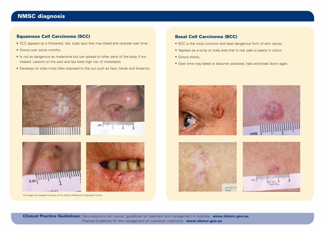
Squamous Cell Carcinoma (SCC)
• SCC appears as a thickened, red, scaly spot that may bleed and ulcerate over time.
• Grows over some months.
• Is not as dangerous as melanoma but can spread to other parts of the body if nottreated. Lesions on the ears and lips have high risk of metastasis.
• Develops on sites most often exposed to the sun such as face, hands and forearms.
Basal Cell Carcinoma (BCC)
• BCC is the most common and least dangerous form of skin cancer.
• Appears as a lump or scaly area that is red, pale or pearly in colour.
• Grows slowly.
• Over time may bleed or become ulcerated, heal and break down again.
All images are supplied courtesy of the Sydney Melanoma Diagnostic Centre
Clinical Practice Guidelines: Non-melanoma skin cancer: guidelines for treatment and management in Australia. www.nhmrc.gov.auPractice Guidelines for the management of cutaneous melanoma. www.nhmrc.gov.au
GP Education version.qxd 20/06/2006 11:27 AM Page 3

Treatment for melanoma
04/0
6
Selecting appropriate primary treatment will depend on theBreslow Thickness (vertical depth) of the tumour. BreslowThickness is measured using the following system:
Tumour – the abnormal cells are found only in the in-situ uppermost layer of the skin and have not (pTis) penetrated into deeper tissue.
pT1 – the melanoma cells reach the upper part of thedermis. The melanoma is less than 1mm thick.
pT2 – the melanoma cells reach the upper part of thedermis. The melanoma is 1mm to 2mm thick.
pT3 – the melanoma cells reach deeper into thedermis. The melanoma is between 2mm and4mm thick.
pT4 – the melanoma is more than 4mm thick or it hasinvaded through the dermis and into theunderlying fat.
Treatment is based on the T1-T4 classification. The surgicalremoval of the tumour with recommended margins of excisionfor each of the T classification groups are:
(pTis) 5mm clearance
(pT1, pT2) 1cm clearance
(pT3) minimum margin 1cm, maximum margin 2cm
(pT4) minimum margin 2cm, maximum margin 3cm
Other treatment options:• Radiotherapy: used if the melanoma has spread to an
internal organ or as follow up prevention treatment after thetumour has been removed.
• Chemotherapy: used to treat cancer that has spread tointernal organs. If a cure is not possible chemotherapy canhelp relieve symptoms caused by the growth of the cancer.
Follow-upAll patients diagnosed with melanoma require follow-up. Thefrequency will depend on the stage of the primary tumour attime of diagnosis.
The reoccurrence of melanoma may be high. Patients shouldbe encouraged to remain vigilant about any changes in theirskin, have a professional full skin examination as deemedappropriate and further testing as required.
WA Melanoma Advisory ServiceThe Western Australian Melanoma Advisory Service (WAMAS)provides advice on diagnosis, management and treatment ofmelanoma through a multidisciplinary panel includingspecialists in anatomical pathology, dermatology, plasticsurgery, medical and radiation oncology and psychologicalcounselling. Information on clinical trials is also available.
The Service is FREE for all West Australians with melanoma.Patients may be referred to WAMAS by their generalpractitioner or specialist. Following consultation, WAMAS willprovide the referring doctor with a suggested plan ofmanagement.
The nurse coordinator for the Service can be contacted on(08) 9382 9445, or fax (08) 9382 9446email: [email protected]
Advice you can give your patients
Ask your patient to check their skin regularly and to see youstraight away if they notice:• A skin spot that is different from other spots around it.• A mole or freckle that has changed in size, shape or colour.• A suspicious spot that is new or has changed over weeks or
months in size, shape or colour.• An inflamed sore that has not healed within 3 weeks.
Skin cancers need not be painful and are much morefrequently seen than felt. Suggest to them that a friend orpartner check areas of the body that are hard to see.
A range of affordable sun protection merchandise is availablefrom the Cancer Council Shop at 334 Rokeby Rd, SubiacoWA 6008. You can contact the shop on (08) 9381 5810 or shoponline at www.cancerwa.asn.au/shop.
This resource was reproduced and modified with the kind permission of The Cancer Council NSW.
FOR MORE INFORMATION FOR YOUR PATIENTSThe Cancer Council Helpline 13 11 20 statewide, for the cost of a local call. Weekdays 8 am – 8 pm,
Saturdays 9 am – 3 pm. TTY (08) 9381 6562www.cancerwa.asn.au
GP Education version.qxd 20/06/2006 11:27 AM Page 4