Me~ical Inositol: history of an effective therapy for ...
71
· "'" European Review for and Pharmacological Sciences 2014; 18:1896-1903 Inositol: history of an effective therapy for Polycystic Ovary Syndrome M. BIZZARRJI, G. CARLOMAGN02 I Department of Experimental Medicine, "Sapienza" University of Rome, Rome, Italy 2LoLiPharma sri, Medical Department, Rome, Italy Abstract. - Inositol is a physiological com- pound belonging to the sugar family. The two inositol stereoisomers, myo-inositol and D-chi- roinositol are the two main stereisomers pre- sent in our body. Myo-inositol is the precursor of inositol triphosphate, a second messenger regulating many hormones such as TSH, FSH and insulin. D-chiroinositol is synthetized by an insulin de- pendent epimerase that converts myo-inositol into D-chiro-inositol. Polycistic Ovary Syndrome (PCOS) is a metabolic and hormonal disorder and a common cause of infertility. Insulin resis- tance and the consequent hyperinsulinaemia contribute to hyperandrogenism development, typical marker of peos. In these patients myo and/or D-chiro-inositol administration improves insulin sensivity while only myo-inositol is a quality marker for oocytes evaluation. Myo-inositol produces second messengers for FSH and glucose uptake, while D-chiroinosi- tol provides second messengers promoting glucose uptake and glycogen synthesis. The physiological ratio of these two isomers is 40:1 (MIIDCI) and seems to be an optimal approach for the treatment of PCOS disorders. Key Words: Inositol, Polycistic ovary syndrome, peas. Introduction The history that has led to the widespread use of inositol compounds in the clinical gynecologic practice is a fascinating and complex tale. In I 850 10hanes loseph Scherer ( 1814-1 8691 2 isolated from the muscle a hexahydroxycyc\o- hexane that he named Inositol [from Ancient Greek stem of (is, in-, "sinew, fiber"), -ose (in- dicating a carbohydrate), -ite ("ester"), -of ("an alcohol")], as it formally belongs to the sugar family3. The structure of this hexahydroxycyclo- hexane allows the formation of 9 different stereoisomers. Among them, myo-inositol is by far the most distributed in biological systems and represents the most interesting form from a meta- bolic and functional point of view. Indeed, myo- inositol is currently thought as a prebiotic mole- cule 4 • given the prominent functions inositol and inositol-derivatives support in several biological systems. Later, in 1850, inositol was found the main component of phytates, i.e. salts of the inositol hexaphosphoric acid. The discovery of phytate dates from 1855 to 1856 when Hartig S first re- ported small round particles in various plant seeds similar in size to potato starch grains. Those pmtic1es were rich in phosphorous, calci- um and magnesium, but without proteins or lipids. As that substances have been not detected neither in meat or dairy products, they were named 'phytin', in order to outline its plant ori- gin. To explain the high phosphorous, calcium and magnesium contents of phytin, several mole- cular structures were under controversial debate for many years. Eventually, in 1914. Anderson 6 presented the molecular structure of myo-inosi- tol-I,2,3,4,5,6-hexakis dihydrogen phosphate, al- so called phytic acid, which was confirmed by various modern analytical methods 7 • s . Inositol (and its derivatives: salts. phosphates and associated lipids) are found in many foods (especially fruits and beans)9. In plants, inositol is generally represented in the rorm of hexaphos- phate. and phytic acid or its salts (phytates). Myo-Inositol was once considered as a mem- ber of the vitamin B complex; however, it cannot be considered a 'true' essential nutrient. given that il can be synlhesiz.ed by the human body. However, it is still a matler of controversy if such biosynthesis may provide amounts considered adequate for good health from glucose. Myo-inositol is synthesized by both prokary- otes and eukaryoles celIs. Myo-inositol is hasi- cally incorporated into ccll membranes as phos- phatidyl-myo-inositol. the precursor of inositol Corresponding Author: Gianfranco Carlomagno, MD; e-mail: g.carlomagno®lolipharma.it
Transcript of Me~ical Inositol: history of an effective therapy for ...

· "'"
European Review for Me~ical and Pharmacological Sciences 2014; 18:1896-1903
Inositol: history of an effective therapy for Polycystic Ovary Syndrome
M. BIZZARRJI, G. CARLOMAGN02
I Department of Experimental Medicine, "Sapienza" University of Rome, Rome, Italy 2LoLiPharma sri, Medical Department, Rome, Italy
Abstract. - Inositol is a physiological compound belonging to the sugar family. The two inositol stereoisomers, myo-inositol and D-chiroinositol are the two main stereisomers present in our body.
Myo-inositol is the precursor of inositol triphosphate, a second messenger regulating many hormones such as TSH, FSH and insulin. D-chiroinositol is synthetized by an insulin dependent epimerase that converts myo-inositol into D-chiro-inositol. Polycistic Ovary Syndrome (PCOS) is a metabolic and hormonal disorder and a common cause of infertility. Insulin resistance and the consequent hyperinsulinaemia contribute to hyperandrogenism development, typical marker of peos. In these patients myo and/or D-chiro-inositol administration improves insulin sensivity while only myo-inositol is a quality marker for oocytes evaluation.
Myo-inositol produces second messengers for FSH and glucose uptake, while D-chiroinositol provides second messengers promoting glucose uptake and glycogen synthesis. The physiological ratio of these two isomers is 40:1 (MIIDCI) and seems to be an optimal approach for the treatment of PCOS disorders.
Key Words: Inositol, Polycistic ovary syndrome, peas.
Introduction
The history that has led to the widespread use of inositol compounds in the clinical gynecologic practice is a fascinating and complex tale.
In I 850 10hanes loseph Scherer ( 1814-1 86912
isolated from the muscle a hexahydroxycyc\ohexane that he named Inositol [from Ancient Greek stem of i~ (is, in-, "sinew, fiber"), -ose (indicating a carbohydrate), -ite ("ester"), -of ("an alcohol")], as it formally belongs to the sugar family3. The structure of this hexahydroxycyclohexane allows the formation of 9 different stereoisomers. Among them, myo-inositol is by
far the most distributed in biological systems and represents the most interesting form from a metabolic and functional point of view. Indeed, myoinositol is currently thought as a prebiotic molecule4
• given the prominent functions inositol and inositol-derivatives support in several biological systems.
Later, in 1850, inositol was found the main component of phytates, i.e. salts of the inositol hexaphosphoric acid. The discovery of phytate dates from 1855 to 1856 when HartigS first reported small round particles in various plant seeds similar in size to potato starch grains. Those pmtic1es were rich in phosphorous, calcium and magnesium, but without proteins or lipids. As that substances have been not detected neither in meat or dairy products, they were named 'phytin', in order to outline its plant origin. To explain the high phosphorous, calcium and magnesium contents of phytin, several molecular structures were under controversial debate for many years. Eventually, in 1914. Anderson6
presented the molecular structure of myo-inositol-I,2,3,4,5,6-hexakis dihydrogen phosphate, also called phytic acid, which was confirmed by various modern analytical methods7
•s.
Inositol (and its derivatives: salts. phosphates and associated lipids) are found in many foods (especially fruits and beans)9. In plants, inositol is generally represented in the rorm of hexaphosphate. and phytic acid or its salts (phytates).
Myo-Inositol was once considered as a member of the vitamin B complex; however, it cannot be considered a 'true' essential nutrient. given that il can be synlhesiz.ed by the human body. However, it is still a matler of controversy if such biosynthesis may provide amounts considered adequate for good health from glucose.
Myo-inositol is synthesized by both prokaryotes and eukaryoles celIs. Myo-inositol is hasically incorporated into ccll membranes as phosphatidyl-myo-inositol. the precursor of inositol
Corresponding Author: Gianfranco Carlomagno, MD; e-mail: g.carlomagno®lolipharma.it

Inositol: history of an effective therapy for Polycystic Ovary Syndrome
triphosphate that acts as second messenger regulating the activities of several hormones such as FSH, TSH and insulin. In addition, inositol is an important component of the structural lipids specifically phosphatidyl-inositol (PI) and its various phosphates. including the phosphatidyl-inositol phosphate (PIP) lipids lO•
After the original discovery by Scherer, many researchers have started to study the role of inositol in different organs and tissues, namely highlighting its relevant role in ensuring a proper cell shape and oocyte feltility.
In 1964. Eisenberg et aP 1, and Eisenberg and Boldenl~ reported that testes are rich of free inositol; few years later. Voglmayr and Amann l3, Lcwin and Beerl4, and Ghafoorunissa l5 showed that the prostate. the epididymis and seminal vesicles contain a large amount of myo-inositol. The seminal fluid is one of the richest sources of inositol, since concentration of inositol in seminal fluid is almost three times higher than that found in plasma I6.17 • These preliminary findings provided the first indirect proof relating inositolbased molecules to germ-cell (spermatozoa and oocytes) physiology.
Over the last six decades. Joseph Lamer has tirelessly pursued scientific studies on insulin action mechanisms, providing new insights into the cause, diagnosis, and cure of non-insulindependent diabetes mellitus IS. In 1974. Lamer proposed the existence of different intracellular chemical mediators of insulin, and hypothesized that, after the binding of insulin to its receptor, different intracellular pathways could he selectively triggered according to the specific mediator involved lR. In 1988 Lamer et al 19 came to the conclusion that the two inositol stereoisomers, myoinositol and D-chiro-inositol, are chemical mediators of insulin, acting through different mechanisms. Both D-chiro-inositol and myo-inositol have similar structures. differing in the stereochemistry of only one hydroxyl group21l. Natural sources for these inositols are endogenous biosynthesis and dietary intake. Myo-inositol is synthesized from glucose-6-phosphate in two steps. First, glucose-6-phosphate is isomerised to myo-inositol-1-phosphate. which is then dephosphorylated by an inositol monophosphatase enzyme giving free myo-inositoPI. 111 vivo, D-chiroinositol is synthetized by an epimerase that converts myo-inositol into D-chiro-inositol. Lamer first demonstrated a decreased D-chiro-inositol content in urine as well as tissues of human subjects and animals with type 2 diabetes2o.22. Uri-
nary decrease in D-chiro-inositol was accompanied by an increase in myo-inositol content~O.22. Additional investigations demonstrated that the altered inositol excretion patterns in human23 and monkey urine~O.2~ were specifically related to the underlying insulin resistance, rather than to the diabetes type. To explain the altered pattern of urine inositol excretion observed under insulin resistance. i.e., increased myo-inositol whereas Dchiro-inositol decreases, Lamer postulated a defect in the epimerization process, that physiologically enacts the conversion of myo-inositol to Dchiro-inositol. He showed that l3HJmyo-inositol is converted to [3H]chiro-inositol ill vivo in rats24, and ill vitro in fibroblasts25, and that this process is stimulated by insulin. Lamer also demonstrated that the conversion of [(3JH]myo-inositol to [(3)H]chiro-inositol ill vivo was markedly decreased in insulin sensitive tissues (liver. muscle, and fat) of Goto-Kakizaki (GK) rats, (inbreeding Wi star rats selected for insulin resistance). compared to Wistar controls16• In a follow-up study, Sun et al27 (Lamer's group) analyzed GK and Wistar rat tissues for total myo-inositol and Dchiro-inositol content. They also developed an epimerase enzyme assay to measure myo-inositol conversion to D-chiro-inositol, as well as a bioassay to measure epimerase activity in cytosolic extracts of tissues of GK and Wistar control rats. Their results showed a consistent decreased total D-chiro-inositollmyo-inositol in kidney. liver, and muscle in GK rats compared to controls; additionally, Sun et aIl7 provided evidence for a myo-inositol to D-chiro-inositol epimerase activity in rat liver cytosol. Importantly, they demonstrated that epimerase bioactivity was significantly decreased in cytosolic extracts of muscle, liver, and fat from GK type 2 diabetic rats, versus Wistar controls. On the basis of these data, Lamer's group hypothesized that a decreased myo-inositol to D-chiroinositol epimerase activity may playa role in explaining the decreased D-chiro-inositollmyoinositol ratio observed in urine and tissues of both animals and humans.
Around the same period when Lamer was publishing those results. a gynecological disorder of wide clinical and social interest, the Polycystic Ovary Syndrome (PCOS), was for the first time linked to insulin resistance, and namely to hyperinsulinemia.
PCOS, the most common cause of infertility. is associated to ovarian dysfunction. metabolic and hormonal impairments, and menstrual irregularity. affecting up to 10% of the total female

M. Bizzarri, G. Carlomagno
population in reproductive age. In 2003. the European Society of Human Reproduction and Embryology (ESHRE), and the American Society for Reproductive Medicine (ASRM) sponsored a Consensus Meeting in Rotterdam, in order to reach a general agreement on the diagnostic criteria for that syndrome. The current definition requires at least two of the following clinical manifestations: chronic ovulatory disorder (oligo-ovulation to anovulation, and amenoO"hea), presence of polycystic ovaries on ultrasound examination, and hyperandrogenism, either clinically established or confirmed by laboratory tests28
•
The pathogenesis of pcas is still largely unknown, although various etiological factors are suspected to be involved. In the past decade. increasing compelling evidence has been accumulated supporting the central role of insulin resistance and/or compensatory hyperinsulinemia in the pcas pathogenesis29.30• Indeed, hyperinsulinaemia, secondary to insulin resistance, is very common in pcas patients, occurring in approximately 80% of women with pcas and central obesity, as well as in 30%-40% of lean women diagnosed with pcas3132 • Insulin resistance and subsequent hyperinsulinemia contribute both directly and indirectly to hyperandrogenism development33.34. Insulin directly stimulates the ovary theca cells to produce greater amount of androgens, and to inhibits hepatic synthesis of sex hormone-binding protein (SHBG), thus indirectly increasing the levels of circulating free androgens. Moreover, theca cells in pcas patients present a greater sensitivity to insulin action on androgen secretion. Noteworthy, the insulin resistance observed in pcas patients predisposes to the development of type 2 diabetes mellitus, especially when a family history of diabetes mellitus is recorded, and if patients are obese35• The importance of insulin resistance in pcas is further underscored by the fact that insulin-sensitizing compounds such as metformin, pioglitazone and troglitazone, have been proposed as treatment for PCaS-associated hyperinsulinemia30.36.37. It is worth noting that metformin may antagonize some hyperandrogenic signs, by reducing total and free testosterone concentrations38
.39
. However. commonly used insulin-sensitizing drugs, by inducing gastrointestinal side effects. could likely reduce patients' compliance40
• and therefore it is unlikely they can be used in routine clinical practice.
The discovery that the impairment in the insulin signalling could be due to a defect in the in-
ositolphosphoglycans (IPGs) second messenger pathway20,41 opened a new horizon in the clinical management of pcas. IPGs are known to have a role in activating enzymes that control glucose metabolism42.43. In pcas women, a defect in tissue availability or altered metabolism of inositol or IPGs mediators may contribute to insulin resistance44•
In 1998, the Insmed Pharmaceuticals Company took out a patent claiming the effectiveness of D-chiro-inositol for the treatment of pcas. That patent originated from the promising data provided by the first clinical trial which assessed the effectiveness of D-chiro-inositol in the treatment of pcas, published in The New England Journal of Medicine4~. In particular, this clinical study mcasured steroids in semm and performed oral glucose-tolerance tests before and after the oral administration of 1200 mg of D-chiro-inositol or placebo once daily for six to eight weeks in 44 obese pcas patients. The results showed that Dchiro-inositol administration to pcas patients was able to improve insulin sensitivity and to reduce serum free testosterone levels compared to the placebo group. Additionally, diastolic and systolic blood pressure, and plasma triglyceride concentrations were decreased in patients treated with D-chiro-inositol. avulation occurred in 19 out of 22 women (86%) who received D-chiroinositol, as compared to 6 out of 22 (27%) in the placebo group45. These promising results laid the foundations for follow-up studies. In 2002. Nestler and Allan published an additional clinical study in which they tested whether administration of D-chiro-inositol would affect the concentration of circulating insulin and androgens, and the frequency of ovulation in lean pcas patients. Those results extended and confirmed earlier findings by showing that, in lean pcas women, D-chiro-inositol reduced serum insulin and androgens, and improved some PCaS-associated metabolic abnormalities (increased blood pressure and hypertriglyceridemia)46. However, when higher doses of D-chiro-inositol were used. the earlier results were not confirmed. Namely, no improvement in insulin sensitivity was reported in women who received a high dose of D-chiro-inositol. Furthermore, the release of D-chiroinositol-containing inositolphosphoglycan (IPG) did not improve in several women in the high dose D-chiro-inositol group, suggesting that these women had a functional defect in D-chiroinositol-containing inositolphosphoglycan release, rather than a simple nutritional deficiency

Inositol: history of an effective therapy for Polycystic Ovary Syndrome
of D-chiro-inositol~7. According to those data, the Insmed Pharmaceutical company decided to stop utterly clinical trials with D-chiro-inositol. Yet, these achievements highlighted the crucial difference emerging when different D-chiro-inositol dosages were used. Indeed, as stressed by the study of Cheang et al-H , clinical efficacy was achieved when treating patients with 2400 mg of D-chiro-inositol, thus leaving open the possibility that this high dose could be responsible of the paradoxical lack of efficacy.
Thankfully. the interest of the scientific community for the potential use of inositols in the clinical practice was not restricted to the D-chiro stereoisomer. In 1992, Chiu et al4R published a study about the role of myo-inositol in vitro human fertilization (lVF). That study had a threefold aim: (I) correlate the embryotrophic properties, assayed by post-implantation embryo culture, and the inositol levels of sera of IVF patients with different pregnancy outcomes following TVF; (2) the monitoring of between-cycle variations in embryotrophic properties and inositol levels of serum samples obtained from patients during normal and treated cycles. and (3) the investigation of the effects of replenishing myo-inositol in serum samples which have previously been found to be non-supportive of mouse embryogenesis4R. The study reported an elevated level of inositol in serum samples of patients having successful IVF pregnancies, thus indicating a possible involvement of inositol in both the early in vitro phase of IVF and the maintenance of normal embryonic development. These findings are consistent with the observation of the teratogenic effect of diabetic patients' serum containing low myo-inositollevels, which causes dysmorphogenesis in cultured rodent embryos49.5o. Furthermore, using the preimplantation mouse embryo assay to determine the trophic activity of the culture media, the authors4R showed that the serum of patients having successful IVF pregnancies and containing high concentrations of myo-inositol, allowed the development of embryos with a greater number of somites. Ten years later, another work51 from the same group examined whether the myo-inositol content in human follicular fluid was associated with better oocyte quality. A total of 53 patients treated with IVF was recmited. Follicular fluid and serum samples were collected and divided into two groups: group A consisted of follicular fluid associated with matured and felt ilized oocytes, while group B was from follicles with immature and unfertilized oocytes. As ex-
pected, a statistically significant correlation between myo-inositol concentration in the follicular fluid and the quality of oocytcs retrieved was found, thus suggesting that higher follicular concentrations of myo-inositol plays a role in follicular maturity, providing, inter alia, a 'quality' marker for oocytes evaluation"l.
Meanwhile, an Italian research group headed by Unfer-'i2 concluded a clinical study on the use of myo-inositol in peDS patients. Twenty-five PCDS patients were enrolled in this study and continuously administered with myo-inositol combined with folic acid twice a day. During an observation period of 6 months, ovulatory activity was monitored with ultrasound scan and honnonal profile, and the numbers of spontaneous menstrual cycles and eventually pregnancies were assessed. Dn the basis of the obtained results, the authorsSl proposed the effectiveness of myo-inositol in restoring spontaneous ovarian activity, and consequently fertility in peDS patients.
These results were later confirmed during follow-up investigations53•s6, highlighting how daily supplementation with myo-inositol, besides improving hormonal profile and restoring ovulation, induces regular menses in both lean and obese PCDS patients. Interestingly, when the effect of myo-inositol on oocyte quality in PCDS patients undergoing intracytoplasmic sperm injection (ICSI) cycles was evaluated, the amount of recombinant FSH (rFSH) administered and the number of days of stimulation were found to be significantly reduced in the myo-inositol group compared to the placebo group. Furthermore, in PCDS patients treated with myo-inositol and folic acid, but not folic acid alone, reduced germinal vesicles and degenerated oocytes at ovum pick-up were observed"7.
Additionally, Rizzo et al58 evaluated the efficacy of a treatment with myo-inositol plus folic acid plus melatonin compared with myo-inositol plus folic acid alone on oocyte quality in pcas women who underwent IVF cycles. Their results further supported the beneficial efficacy of myoinositol and folic acid in improving fertility and suggested that the concomitant supplementation of melatonin can ameliorate oocyte quality and pregnancy outcomes in women with poor oocyte quality history58.
Bearing in mind the positive relationship between follicular myo-inositol levels and better oocyte quality reported by Chiu et a151 , these findings conflicted with Nestler's theory that hypothesized a decreased myo-inositol to D-chiro-inositol

M. Bizzarri, G. Carlomagno
epimerase activity as a crucial factor in PCOS pathogenesis. Indeed. on the basis of Nestler's theory, the administration of myo-inositol would be expected to be ineffective in PCOS patients. Furthermore, it was still unclear why D-chiro-inositol, apparently so effective in preliminary studies45.40,
resulted to be ineffective at higher doses47 . To solve this paradox. the effects of myo-inositol and D-chiro-inositol on oocyte quality in euglycemic pcas patients were compared. Results showed that the total number of oocytes retrieved did not differ in the two treatments groups. However, the number of mature oocytes was significantly increased and the number of immature oocytes decreased in the myo-inositol compared to the Dchiro-inositol group. Furthermore, myo-inositoltreated patients showed an increase in the mean number of top quality embryos and in the total number of pregnancies compared to D-chiroinositol-treated patients59
• These findings can be explained by the fact that, unlike tissues such as muscle and liver, ovaries never become insulin resistant6l).f>2. Therefore. the authors speculated that pcas patients with hyperinsulinemia likely present an enhanced myo-inositol to D-chiro-inositol epimerization in the ovary63; this would result in an increased D-chiro-inositoVmyo-inositol ratio (i.e., overproduction of D-chiro-inositol), which in tum would lead to myo-inositol deliciency in the ovary"3. This myo-inositol depletion could eventually be responsible for the poor oocyte quality observed in PCOS patients6<!. Furthermore, it is likely that the putative myo-inositol deficiency in the ovary would also impair the FSH signaling. resulting in an increased risk of ovarian hyperstimulation syndrome for PCOS patients63 . There is ample indirect evidence supporting this theory: it is well known that patients with elevated levels of insulin need a higher number of FSH IU when undergoing ovary stimulation protocols65 ; moreover. it has been found that myo-inositol supplementation in pcas patients (preferably 3 months before ovary stimulation) reduces the amount of rFSH administrated during IVF cycles57.59.66, with a direct impact on the possibility to achieve pregnancy67. Further support is provided by the data collected by Isabella and Raffone6K. who showed that increasing doses of D-chiro-inositol produce "ovary toxicity", characterized by a negative impact on oocyte quality, and a progressive reduction in the ovary response to FSH and negatively impacting oocyte quality. Interestingly. this negative effect was observcd at the same dose of D-chiro-inositol as the one tested by Cheang et a147
•
A convincing proof arrived from the dosage of myo-inositol and D-chiro-inositol in the follicular fluid of PCOS patients vs. healthy subjects. The study demonstrated that follicular fluid from spontaneous cycles of healthy patients contains high concentrations of myo-inositol and low concentrations of D-chiro-inositol while in PCOS patients, the ratio of the two molecules is completely opposite. Therefore, such findings supported the "DCI paradox", accordingly to which "ovaries in PCOS patients likely present an enhanced myo-inositol to Dchiro-inositol epimerization that leads to a myoinositol tissue depletion that could eventually be responsible for the poor oocyte quality characteristic of these patients'·6'l.
These findings explain the link between myoinositol and FSH. Moreover, it is now clear why Nestler's results were not confirmed when higher doses of D-chiro-inositol were tested47
: ovary of PC OS patient. very rich in D-chiro-inositol, does not longer require that molecule. On the contrary, by using D-chiro-inositol it is possible to counteract insulin-resistance (i.e., the ovary is never insulin-resistant) by reducing insulin levels which may also indirectly benefit the ovary.
One has to ask what should be the proper dose needed to ensure clinical efficacy without compromising ovarian function. To address this question, the plasma physiologic ratio of MyolD-chiroinositol was tirstly identified, and then the clinical effectiveness of a product specifically designed according to those premises has been verified70. The physiological plasma ratio of the two isomers resulted approximatcIy 40:1, and since the therapeutic dosage of myo-inositol ranges between 2 and 4 grams/die, LO.Ll Pharma produced a new product containing 2 grams of myo-inositol and 50 mg of D-chiro-inositol. Modern technologies enabled manufacturing the product as soft gel capsules, which allow a comparable pharmacokinetics proIile, even reducing the dose to a third of the original powder-base drug (i.e. 550 mg of myo-inositol and l3.g mg of D-chiro-inositol, patented)1°. From this innovative formulation scientists expected to obtain a two-fold effect: (1) an action on liver. mainly exerted by D-chiro-inositol, aimed at reducing insulincmic levels; (2) a selective effect on the ovary, where myo-inositol is thought to counteract the increased D-chiro-inositol levels, and hence re-establishing FSH sensitivity. Those results were later confirmed by Nordio et aFI, so far providing a further milestone in the promising story of inositol.

Inositol: history of an effective therapy for Polycystic Ovary Syndrome
References 18) BROMBERG WJ, EDLICH RF. Joseph Larner's personal odyssey: search for the cause and cure of non-in-
1) /(OMPANJE EJ, JANSEN TC VAN DER HOVEN B, BAKKER J. sulin-dependent diabetes mellitus. J Emerg Med
The first demonstration of lactic acid in human 1994; 12: 681-684.
blood in shock by Johann Joseph Scherer (1814- 19) I.ARNER J, HUANG Lc, TANG G, SUZUKI S, SCHWARlZ 0; 1869) in January 1843. Intensive Care Med 2007; ROMERO G, ROULIDIS Z, ZElLER K, SHEN Tv. OSWALD As, 33: 1967-1971. et al. Insulin mediators: structure and formation.
2) BUTTNERJ. [Johann Joseph von Scherer (1814-69). Cold Spring Harb Symp Quant Bioi 1988; 53 Pt 2: The early history of clinical chemistry). J Clin 965-971. Chern Clin Biochem 1978; 16: 478-483. 20) KENNINGTON As, Hill CR, CRAIG J, BOGARDUS C, RAz I,
3) The cell biology of inositol lipids and phosphates. ORTMEYER HK, HANSEN Bc, ROMERO G, lARNER J. Low Proceedings of the 2006 Biochemical Society An- urinary chiro-inositol excretion in non-insulin-nual Symposium. Birmingham, United Kingdom. dependent diabetes mellitus. N Engl J Med 1990; March 29-30, 2006. Biochemical Society sympo- 323: 373-378. sium. 2007(74): pp. 1-271. 21) LOEWUS Mw, WRIGHT Rw; JR., BONDIOLI KR, BEDGAR
4) AGRANOFF BW. Turtles All the Way: Reflections on Dl, KARL A. Activity of myo-inositol-1-phosphate myo-Inositol. J Bioi Chern 2009; 284: 21121- synthase in the epididymal spermatozoa of rams. 21126. J Reprod Fertil 1983; 69: 215-220.
5) HARTIG T. Ueber das Klebermehl. Botanische 22) o ORTMEYER HK, BODKIN Nl, LillEY K, LARNER J, Zeitung 1855; 13: 881. HANSEN Be. Chiroinositol deficiency and insulin re-
6) ANDERSON RJ. A Contribution to the chemistry of sistance. I. Urinary excretion rate of chiroinositol
phytin: i. Composition of barium phytate and phyt- is directly associated with insulin resistance in
ic acid. ii. A study of the properties of phytic acid spontaneously diabetic rhesus monkeys. En-
and its decomposition products. Eighth paper on docrinology 1993; 132: 640-645.
phytin. J Bioi Chern 1914; 17: 171-190. 23) SUZUKI S, KAWASAKI H, SATOH Y. OHTOMO M, HIRAI
7) JOHNSON LF. TATE ME. The structure of myo-inositol M, HIRAI A. HIRAI S, ONODA M, MATSUMOTO M, HI-
pentaphosphates. Ann N Y Acad Sci 1969; 165: NOKIO y. ET AL Urinary chiro-inositol excretion is
526-532. an index marker of insulin sensitivity in Japan-
8) RABOY \I. Myo-lnositol-1,2,3,4,5,6-hexakisphosphate. ese type II diabetes. Diabetes Care 1994; 17: 1465-1468.
Phytochemistry 2003; 64: 1033-1043. 24) PAK Y. HUANG Lc, LIllEY K.i, I.ARNER J. In vivo conver-
9) CLEMENTS Rs, JR., DARNEll B. Myo-inositol content of sion of [3H)myoinositol to [3H)chiroinositol in rat common foods: development of a high-myo- tissues. J Bioi Chern 1992; 267: 16904-16910. inositol diet. Am J Clin Nutr 1980; 33: 1954-1967.
25) PAK Y. PAULE CR, BAo YO, HUANG LC LARNER J. In-10) MAJUMDER Al, BISWAS BB. Biology of inositols and sulin stimulates the biosynthesis of chiro-inositol-
phoshoinositides. Subcellular Biochemistry. containing phospholipids in a rat fibroblast line ex-Springer, 2006; p. 39. pressing the human insulin receptor. Proc Natl
11) EISENBERG F. BOLDEN AH, LaEWUS FA. Inositol formation Acad Sci USA 1993; 90: 7759-7763. by cyclization of glucose chain in rat testis. Biochem 26) PAK Y. HONG Y. KIM S, PICCARIEllO T. FARESE R\I. LARN-Biophys Res Commun 1964; 14: 419-424. ER J. In vivo chiro-inositol metabolism in the rat: a
12) EISENBERG F. JR., BOlDEN AH. Reproductive tract as defect in chiro-inositol synthesis from myo-inositol site of synthesis and secretion of inositol in the and an increased incorporation of chiro-[3H)inosi-male rat. Nature 1964: 202: 599-600. tol into phospholipid in the Goto-Kakizaki (G.K)
13) VOGlMAYR JK. AMANN Rp. The distribution of free rat. Mol Cells 1998; 8: 301-309. myo-inositol in fluids, spermatozoa, and tissues of 27) SUN TH, HEIMARK DB, NGUYGEN T. NADLER JL, I.ARNER the bull genital tract and observations on its up- J. Both myo-inositol to chiro-inositol epimerase take by the rabbit epididymis. Bioi Reprod 1973; activities and chiro-inositol to myo-inositol ratios 8: 504-513. are decreased in tissues of GK type 2 diabetic
14) LEWIN LM, BEER R. Prostatic secretion as the rats compared to Wistar controls. Biochem Bio-source of myo-inositol in human seminal fluid. phys Res Commun 2002; 293: 1092-1098. Ferti! Steril 1973; 24: 666-670. 28) KOIVUNEN R, LMTIKAINEN T. TOMAS C HUHTANIEMI I,
15) GHAFOORUNISSA. Effect of dietary protein & inositol TAPANAINEN J. MARTIKAINEN H. The prevalence of on sperm metabolism & fructose content of male polycystic ovaries in healthy women. Acta Obstet accessory sex organs of rat. Indian J Exp Bioi Gynecol Scand 1999; 78: 137-141. 1976; 14: 564-566. 29) Nestler JE. Role of hyperinsulinemia in the patho-
16) SPECTOR R. LORENZO A\I. The origin of myo-inositol genesis of the polycystic ovary syndrome, and its in brain, cerebrospinal fluid and choroid plexus. J clinical implications. Semin Reprod Endocrinol Neurochem 1975; 25: 353-354. 1997; 15: 111-122.
17) SPECTOR R, LORENZO AV. Myo-inositol transport in 30) BAillARGEON Jp. IUORNO MJ, NESTLER JE. Insulin sen-the central nervous system. Am J Physiol 1975; sitizers for polycystic ovary syndrome. Clin Obstet 228: 1510-1518. Gynecol 2003; 46: 325-340.
119011

M. Bizzarri. G. Car/omagno
31) GENAZZANI AD. BATTAGLIA C. MAulVASI B. 5mUCCHI C. 45) NESTLER JE, JAKUBOWICZ DJ, REAMER P. GUNN RD, AL-TORTOLANI F. GAMBA O. Metformin administration LAN G. Ovulatory and metabolic effects of D-chiro-modulates and restores luteinizing hormone inositol in the polycystic ovary syndrome. N Engl spontaneous episodic secretion and ovarian func- J Med 1999; 340: 1314-1320. tion in nonobese patients with polycystic ovary 46) /UORNO MJ, JAKUBOWICZ DJ, BAlUARGEON JP. DILLON P. syndrome. Fertil Steri12004; 81: 114-119. GUNN RD, AlLAN G, NESTLER JE. Effects of d-chiro-
32) OAMPELLI M, FULGHESU AM, CUCINEllI F. PAVONE V. inositol in lean women with the polycystic ovary RONSISVALLE E, GUIDO M, CARuso A. \.ANZONE A. Im- syndrome. Endocr Pract 2002; 8: 417-423. pact of insulin and body mass index on metabolic 47) CHEANG KI, BAILLARGEON Jp, ESSAH PA, OSTLUND RE, and endocrine variables in polycystic ovary syn- JR., APRIDONIZE 1; ISLAM L NESTlER JE. Insulin-stimu-drome. Metabolism 1999; 48: 167-172. lated release of D-chiro-inositol-containing inosi-
33) UN FER V. CARLOMAGNO G, DANTE G, FACCHINETTI F. tolphosphoglycan mediator correlates with insulin Effects of myo-inositol in women with PCOS: a sensitivity in women with polycystic ovary syn-systematic review of randomized controlled trials. drome. Metabolism 2008; 57: 1390-1397. Gynecol Endocrinol2012; 28: 509-515. 48) CHIU IT, TAM PP. A correlation of the outcome of
34) DUNAIF A. Insulin resistance in women with poly- clinical in vitro fertilization with the inositol content cystic ovary syndrome. Fertil Steril 2006; 86(Sup- and embryotrophic properties of human serum. J pI1): S13-14. Assist Reprod Genet 1992; 9: 524-530.
35) PASOUAl.! R. PElUSI C, RAGAzZINI C, HASANAJ R. GAM- 49) SUSSMAN I. MATSCHINSKY FM. Diabetes affects sor-BINERI A. Glucose tolerance, insulin secretion and bitol and myo-inositol levels of neuroectodermal insulin sensitivity in polycystic ovary syndrome. tissue during embryogenesis in rat. Diabetes JOP J Pancreas (Online) 2002; 3: 1-7. 1988; 37: 974-981.
36) GENAZZANI AD, LANZONI C, RICCHIERI F. BARALDI E, 50) WEIGENSBERG MJ, GARCIA-PALMER FJ, FREINKEL N. Up-CASAROSA E, JASONNI VM. Metformin administration take of myo-inositol by early-somite rat concep-is more effective when non-obese patients with tus. Transport kinetics and effects of hyper-polycystic ovary syndrome show both hyperan- glycemia. Diabetes 1990; 39: 575-582. drogenism and hyperinsulinemia. Gynecol En- 51) CHIU IT, ROGERS MS, LAw EL BRITON-JONES CM, CHE-docrinol 2007; 23: 146-152. UNG LP. HAINES CJ. Follicular fluid and serum con-
37) PASOUAU R. GAMBINERI A. Insulin-sensitizing agents centrations of myo-inositol in patients undergoing in polycystic ovary syndrome. Eur J Endocrinol IVF: relationship with oocyte quality. Hum Reprod 2006; 154: 763-775. 2002; 17: 1591-1596.
38) NESTLER JE, JAKUBOWICZ DJ. Lean women with poly- 52) PAPAlEO E, UNFER V. BAILlARGEON JP, DE SAmIS L, FUSI cystic ovary syndrome respond to insulin reduc- F. BRIGANTE C, MARELU G, CiNO I. REDAELU A, FERRARI tion with decreases in ovarian P450c17 alpha ac- A. Myo-inositol in patients with polycystic ovary tivity and serum androgens. J Clin Endocrinol syndrome: a novel method for ovulation induction. Metab 1997; 82: 4075-4079. Gynecol Endocrinol2007; 23: 700-703.
39) NESTLER JE. Metformin for the treatment of the 53) GERLI S. PAPALEO E, FERRARI A. DI RENZO Gc. Ran-polycystic ovary syndrome. N Engl J Med 2008; domized, double blind placebo-controlled trial: ef-358: 47-54. fects of myo-inositol on ovarian function and
40) LORD JM, FLIGHT IH, NORMAN RJ. Metformin in poly- metabolic factors in women with PCOS. Eur Rev cystic ovary syndrome: systematic review and Med Pharmacol Sci 2007; 11: 347-354. meta-analysis. Br Med J 2003; 327: 951-953. 54) GENAZZANI AD, LANZONI C, RICCHIERI F. JASONNI VM.
41) AsPLIN I. GAlASKO G. lARNER J. chiro-inositol deficien- Myo-inositol administration positively affects hy-cy and insulin resistance: a comparison of the chi- perinsulinemia and hormonal parameters in over-ro-inositol- and the myo-inositol-containing insulin weight patients with polycystic ovary syndrome. mediators isolated from urine, hemodialysate, and Gynecol Endocrinol 2008; 24: 139-144. muscle of control and type II diabetic subjects. 55) ZACCHE MM, CAPUTO L, FlliPPIS S, ZACCHE G, DINDELU Proc Nail Acad Sci USA 1993; 90: 5924-5928. M, FERRARI A. Efficacy of myo-inositol in the treat-
42) COHEN P. The twentieth century struggle to deci- ment of cutaneous disorders in young women pher insulin signalling. Nat Rev Mol Cell Bioi with polycystic ovary syndrome. Gynecol En-2006; 7: 867-873. docrinol2009; 25: 508-513.
43) BAILLARGEON JP. NESTlER JE, OSTLUND RE, APRIDONIDZE 56) COSTANTINO D. MINOZZI G, MINozzl E. GlIARALDI C. T, DIAMANTI-KANDARAKIS E. Greek hyperinsulinemic Metabolic and hormonal effects of myo-inositol in women, with or without polycystic ovary syn- women with polycystic ovary syndrome: a double-drome. display altered inositols metabolism. Hum blind trial. Eur Rev Med Pharmacol Sci 2009; 13: Reprod 2008; 23: 1439-1446. 105-110.
44) BAILlARGEON JP. DIAMANTI-KANDARAKIS E, OSTLUND RE. 57) PAPALEO E, UNFER V. BAlUARGEON JP, FUSI F. OCCHI F. JR., APRIDONIDZE 1; IUORNO MJ, NESTLER JE. Altered DE SANTIS L. Myo-inositol may improve oocyte D-chiro-inositol urinary clearance in women with quality in intracytoplasmic sperm injection cycles. polycystic ovary syndrome. Diabetes Care 2006; A prospective, controlled, randomized trial. Fertil 29: 300-305. Steril 2009; 91: 1750-1754.
119021

Inositol: history of an effective therapy for Polycystic Ovary Syndrome
58) RIZZO P. RAFFONE E. BENEDETTO V. Effect of the treat- 65) HOMBURG R. ORVIETO R. BAR-HAVA I. BEN-RAFAEL Z. ment with myo-inositol plus folic acid plus melatonin Serum levels of insulin-like growth factor-1, IGF in comparison with a treatment with myo-inositol binding protein-1 and insulin and the response to plus folic acid on oocyte quality and pregnancy out- human menopausal gonadotrophins in women come in IVF cycles. A prospective, clinical trial. Eur with polycystic ovary syndrome. Hum Reprod Rev Med Pharmacol Sci 2010; 14: 555-561. 1996; 11: 716-719.
59) UNFER V. CARLOMAGNO G. RIZZO P. RAFFONE E, ROSEFF 66) OOTTA L. STRACaUADANIO M. PAGANO I. CARBONARO A, S. Myo-inositol rather than D-chiro-inositol is able PALUMBO M, GULINO F. Effects of myo-inositol sup-to improve oocyte quality in intracytoplasmic plementation on oocyte's quality in PCOS pa-sperm injection cycles. A prospective, controlled, tients: a double blind trial. Eur Rev Med Pharma-randomized trial. Eur Rev Med Pharmacol Sci col Sci 2011; 15: 509-514. 2011; 15:452-457. 67) PAL L. JINDAl S. WITT BR. SANTORO N. Less is more:
60) HARWOOD K. VUGUIN P. DIMARTINo-NARDI J. Current increased gonadotropin use for ovarian stimula-approaches to the diagnosis and treatment of tion adversely influences clinical pregnancy and polycystic ovarian syndrome in youth. Horm Res live birth after in vitro fertilization. Fertil Steril 2007; 68: 209-217. 2008; 89: 1694-1701.
61) MATALlIOTAKIS I. KOURTIS A. KOUKOURA 0, PANIDIS D. 68) ISABELLA R. RAFFONE E. Does ovary need D-chiro-Polycystic ovary syndrome: etiology and pathogen- inositol? J Ovarian Res 2012; 5: 14. esis. Arch Gynecol Obstet 2006; 274: 187-197. 69) GALLETTA M. GRASSO S. VAIARELlI A, ROSEFF SJ. Bye-
62) RICE S, CHRISTO FORI DIS N, GADD C. NIKOLAOU D, bye chiro-inositol-myo-inositol: true progress in SEYANI L, DONAlDSON A. MARGARA R. HARDY K. the treatment of polycystic ovary syndrome and FRANKS S. Impaired insulin-dependent glucose ovulation induction. Eur Rev Med Pharmacol Sci metabolism in granulosa-lutein cells from anovu- 2011; 15: 1212-1214. latory women with polycystic ovaries. Hum Re- 70) CARLOMAGNO G. DE GRAZIA S. UNFER V. MANNA F. prod 2005; 20: 373-381. Myo-inositol in a new pharmaceutical form: a step
63) CARLOMAGNO G. UNFER V. ROSEFF S. The D-chiro- forward to a broader clinical use. Expert Opin inositol paradox in the ovary. Fertil Steril 2011; 95: Drug Deliv 2012; 9: 267-271. 2515-2516. 71) NORDIO M, PROIETTI E. The combined therapy with
64) CHATTOPADHAYAY R. GANESH A. SAMANTA J, JANA SK. myo-inositol and D-chiro-inositol reduces the risk CHAKRAVARTY BN, CHAUDHURY K. Effect of follicular of metabolic disease in PCOS overweight pa-fluid oxidative stress on meiotic spindle formation tients compared to myo-inositol supplementation in infertile women with polycystic ovarian syn- alone. Eur Rev Med Pharmacal Sci 2012; 16: drome. Gynecol Obstet Invest 2010; 69: 197-202. 575-581.
Gynecological Endocrinology, December 2007; 23(12): 700-703 informa healthcare
peos
Myo-inositol in patients with polycystic ovary syndrome: A novel method for ovulation induction
ENRICO PAPALE0 1, VITTORIO UNFER2
, JEAN-PATRICE BAILlARGEON3,
LUCIA DE SANTIS\ FRANCESCO PUSI1, CLAUDIO BRIGANTE\ GUIDO MARELLII,
ILARIA CIN0 1, ANNA REDAELLII, & AUGUSTO FERRARII
lIVF Unit, Gynaecological-Obstetric Department, IRCCS San Raffaele Hospital, Vita-Salute University, Milan, Italy, 2AGUNCO Obstetrics and Gynecology Centre, Rome, Italy, and 3Department of Medicine, University of Sherbrooke, Sherbrooke, Quebec, Canada
(Received 11 June 2007; revised 6 September 2007; accepted 10 September 2007)
Abstract Background. Polycystic ovary syndrome (peOS) is often characterized by chronic oligo- or anovulation (usually manifested as oligo- or amenorrhea), and hyperandrogenism. In addition, 30--40% of peos women have impaired glucose tolerance, and a defect in the insulin signaling pathway (inositol-containing phosphoglycan mediators) seems to be implicated in the pathogenesis of insulin resistance. peos patients are subfertile as a consequence of such ovulatory disorders and often need drugs, such as clomiphene citrate or follicle-stimulating hormone, for ovulation induction, which increases the risk of multiple pregnancy and ovarian hyperstimulation syndrome. We hypothesized that the administration of an isoform of inositol (myo-inositol), belonging to the vitamin B complex, would improve the insulin-receptor activity, restoring normal ovulatory function. Materials alld methods. Twenty-five peos women of childbearing age with oligo- or amenorrhea were enrolled in the study. Ovulatory disorder due to peos was apparently the only cause of infertility; no tubal defect or deficiency of male semen parameters was found. Myo-inositol combined with folic acid (Inofolic®) 2 g twice a day was administered continuously. During an observation period of 6 months, ovulatory activity was monitored with ultrasound scan and hormonal profile, and the numbers of spontaneous menstrual cycles and eventually pregnancies were assessed. Results. Twenty-two out of the 25 (88%) patients restored at least one spontaneous menstrual cycle during treatment, of whom 18 (72%) maintained normal ovulatory activity during the follow-up period. A total of 10 singleton pregnancies (40% of patients) were obtained. Nine clinical pregnancies were assessed with fetal heart beat at ultrasound scan. Two pregnancies evolved in spontaneous abortion. Conclusion. Myo-inositol is a simple and safe treatment that is capable of restoring spontaneous ovarian activity and consequently fertility in most patients with peos. This therapy did not cause multiple pregnancy.
Keywords: Myo-inositol, polycystic ovary syndrome, ovulation induction
Introduction
Polycystic ovary syndrome (peOS) is a medical condition that causes irregular menstrual cycles, chronic anovulation most often manifested as oligoor amenorrhea, and androgen excess, with the typical ovarian ultrasound features [1]. It is the most common cause of ovulatory disorders and female infertility, and affects approximately 6-10% of women in childbearing age [2]. However, its pathogenesis is still poorly understood.
Recently, many investigators have focused on the impaired glucose tolerance that affects 30-40% of patients with peos [3]. Insulin plays a direct role in the pathogenesis of hyperandrogenemia in peos, acting synergistically with luteinizing hormone to enhance the androgen production of theca cells [4]. An inositol phosphoglycan molecule containing D-chiro-inositol (Del) is known to have a role in activating enzymes that control glucose metabolism [5]. Indeed, a defect in tissue availability or altered metabolism of Del or inositol phosphoglycan
Correspondence: E. Papaleo, IVF Unit, Gynaecological-Obstetric Deparnnent, IRCCS San Raffaele, via Olgettina 60, 1-20132 Milan, Italy. Tel: 39 02 26432202. Fax: 39 02 26434311. E-mail: [email protected]
ISSN 0951-3590 printllSSN 1473-0766 onhne © 2007 Informa UK Ltd. DOl: 10.1080/09513590701672405

c o o o
mediators has been found in PCOS women and may contribute to their insulin resistance [6,7].
Isoforms of inositol belong to the vitamin B complex. Epimerization of the six hydroxyl groups of inositol results in the formation of up to nine stereoisomers, including myo-inositol (MI) and DCI. MI is widely distributed in nature whereas DCI, the product of epimerization of the C 1 hydroxyl group of MI, is relatively rare [8].
Elevated concentration of MI in human follicular fluid appears to play a role in follicular maturity and provides a marker of good-quality oocytes [9]. Furthermore, experiments on mouse oocytes showed that supplementation of MI in the culture medium increased meiotic progression of germinal vesicles by enhancing the intracellular Ca2+ oscillation [10].
Thus we hypothesized that the administration of MI, a precursor of DCI, would improve insulin activity and restore ovulatory function and fertility in amenorrheic women with PCOS.
Materials and methods
A total of 25 women, 28 to 38 age years of age, with PCOS defined by oligo- or amenorrhea (six or fewer menstrual cycles during a period of 1 year), hyperandrogenism (hirsutism, acne or alopecia) or hyperandogenemia (elevated levels of total or free testosterone) and typical ovarian features on ultrasound scan, were enrolled in the study.
All patients attended our IVF Unit for infertility that had lasted for more than 14-16 months. Other medical conditions causing ovulatory dysfunction, such as hyperprolactinemia or hypothyroidism, or androgen excess, such as adrenal hyperplasia or Cushing's syndrome, were excluded by hormonal tests. All women underwent assessment of tubal patency and all male partners were evaluated with two different semen sample analyses, without finding any defect. Anovulation was ascertained by weekly plasma progesterone concentration < 2.5 ng/ml. Thus, at the end of diagnostic procedures, it was determined that the most likely cause of the couple's subfertility was ovulation dysfunction only.
PCOS women were treated orally with MI 2 g plus folic acid 200 Jlg (lnofolic®; Loli Pharma, Rome, Italy) as soluble powder, twice daily, continuously, until the end of the study or a positive pregnancy test. Patients were instructed to register their menstrual bleeding throughout the follow-up period of 6 months. Furthermore, in order to evaluate the restoration of spontaneous ovarian activity, weekly determination of serum progesterone and testosterone levels, as well as transvaginal ultrasound scan documenting the presence of follicular growth or luteal cyst, were performed after the first menstrual cycle. Pre- and post-treatment hormone
Myo-inositol for ovulation induction in PeOS 701
concentrations were statistically compared using the two-tailed t test.
Moreover, eventual pregnancies were confirmed by a positive test for plasma fJ-human chorionic gonadotropin and ascertainment of a fetal heart beat on ultrasound scan.
Results
Baseline clinical and biochemical features of the PCOS patients are reported in Table I. The outcome of treatment is shown in Tables I and II.
After a mean of 34.6 ± 5.5 days of MI administration, 22 out of the 25 women (88%) had a first menstrual cycle. Eighteen of these 22 patients presented monthly menstruations during the followup period. All of them maintained spontaneous ovulation activity, documented by follicular growth and increased serum progesterone concentrations in the luteal phase (mean 10.5 ± 1.8 ng/ml). Furthermore, after treatment with MI, these women showed significantly decreased concentrations of serum total testosterone (95.6 ± 8.5 vs. 45.2 ± 6.7 ngldl; p = 0.003) and free testosterone (1.0 ± 0.8 vs. 0.38 ± 0.1 ng/dl; p = 0.005). The length of successive cycles was improved to 31.7 ± 3.2 days.
Two out of the 22 women showed only a follicular development on ultrasound without progesterone elevation during weekly blood sampling, while two women did not have any further ovarian activity after the first cycle.
During the observational period of 6 months a total of ten biochemical pregnancies occurred. Nine of the ten were singleton pregnancies documented at ultrasound scan, while one of them was a biochemical abortion. One out of the nine pregnancies
Table 1. Clinical and biochemical features of the patients.
Baseline After myo-inositol
Age (years) 32 ± 4 Body mass index (kg/m2
) 28.5 ± 2.4 Follicle-stimulating 4.5 ± 2.8
hormone (mUIIml) Luteinizing hormone 6.3 ± 3.1
TSH (mUl/ml) Prolactin (ng/ml) 19.1 ± 2.7 Thyroid-stimulating 1.78 ± 0.85
hormone Serum progesterone 1.8 ± 0.7 10.5 ± 1.8
(ng/ml) Serum total testosterone 95.6 ± 8.5 45.2 ± 6.7*
(ng/dl) Serum free testosterone 1.0 ± 0.8 0.38 ± O.lt
(ng/dl) Serum androstenedione 230 ± 35 205 ± 28
(ng/dl)
Significant difference compared with baseline: .p = 0.003; tp=0.005.
A I G H 'T S L , "",~ ".i)

E o
:!:
-0 u
" c: ;.,
"
702 E. Papaleo et af.
Table II. Outcome of treatment with myo-inositol.
No. of patients treated No. of patients with menstrual cycle after
treatment (% of patients) No. of patients with restored monthly ovulation
(% of patients) No. of pregnancies No. of pregnancies/no. of treated patients (%) No. of pregnancies/no. patients with restored
monthly ovulation (%) No. of abortions (% of pregnancies) Multiple pregnancy
25 22 (88)
18 (72)
10 40 55
2 (20) o
evolved in a spontaneous abortion at 7 weeks of gestation. No multiple pregnancy was noted.
Discussion
PCOS is one of the most common endocrine disorders affecting women. Insulin resistance and hyperinsulinemia are strictly inherent to the phenotype of a high proportion of women with PCOS. A defect in insulin action has been suspected, particularly as consequence of a deficiency of DCI, a component of inositol phosphoglycan.
Chronic anovulation is often the main cause of infertility in patients of reproductive age. It is well known that ovulation induction is a complex issue owing to the increased risk of ovarian hyperstimulation syndrome and multiple pregnancy [11,12]. Clomiphene citrate, an antiestrogen, is the common first-choice drug in women with newly diagnosed PCOS, while insulin-lowering medications represent novel therapies for restoring spontaneous ovulation [13,14]. The efficacy of metformin is still debated, both alone and in association with clomiphene citrate [15,16]. Metformin treatment is associated with a higher incidence of side-effects such as nausea, vomiting and other gastrointestinal disturbances [17] .
DCI administration increases the action of insulin in patients with PCOS, thereby improving ovulatory function and decreasing serum testosterone concentration [6,18,19]. MI, a precursor of DCI, is widely distributed in nature whereas DCI is relatively rare [7]. MI is present in human follicular fluid, where elevated concentrations appear to playa positive role in follicular maturity and provide a marker of goodquality oocytes [9]. Supplementation of MI in culture medium increased meiotic progression of germinal vesicles in mouse oocytes by enhancing the intracellular Ca2+ oscillation [10]. However, no data exist on therapy with MI in anovulatory women of reproductive age.
Our study demonstrated that MI oral supplementation restores spontaneous ovulation and menstrual cycles, and increases progesterone secretion in the
luteal phase, in most infertile patients with PCOS. The present results are in line with other studies evaluating insulin-sensitizing agents in monotherapy or in association with clomiphene citrate [7,13,14, 16-18], suggesting the positive effect that MI plays on spontaneous ovarian activity. Furthermore, we found that MI therapy is able to reduce serum testosterone, both total and free, as already demonstrated with DCI. All pregnancies obtained in the follow-up period were singleton, and there was no increased incidence of abortion.
In conclusion, MI is a simple and safe treatment that is able to restore spontaneous fertility in most patients with PCOS.
References
I. Ehrmann DA. Polycystic ovary syndrome. N Engl J Med 2005;352: 1223-1236.
2. Franks S. Polycystic ovary syndrome. N Engl J Med 1995; 333:853-861.
3. Ehrmann DA, Barnes RB, Rosenfield RL, Cavaghan MK, Imperial J. Prevalence of impaired glucose tolerance and diabetes in women with polycystic ovary syndrome. Diabetes Care 1999;22: 141-146.
4. Baillargeon JP, Nescler JE. Polycystic ovary syndrome: a syndrome of ovarian hypersensitivity to insulin? J Clin Endocrinol Metab 2006;91 :22-24.
5. Lamer J. Multiple pathways in insulin signaling - fitting the covalent and allosteric puzzle pieces together. Endocr J 1994; 2:167-171.
6. Baillargeon JP, Diamanti-Kandarakis E, Ostlund RE Jr, Apridonidze T, Iuorno MJ, Nestler JE. Altered o-chiroinositol urinary clearance in women with polycystic ovary syndrome. Diabetes Care 2006;29:300-305.
7. Iuorno MJ, Jakubowicz DJ, Baillargeon JP, Dillon P, Gunn RD, Allan G, Nescler JE. Effect of D-chiro-inositol in lean women with the polycystic ovary syndrome. Endocr Pract 2002;8:417-423.
8. Yoshida K, Yamaguchi M, Morinaga T, Ikeuchi M, Kinehara M, Ashida H. Genetic modification of Bacillus subtilis for production of o-chiro-inositol, an investigation drug candidate for treatment of type 2 diabetes and polycystic ovary syndrome. Appl Environ Microbiol 2006;72:1310-1315.
9. Chiu TT, Rogers MS, Law EL, Briton-Jones CM, Cheung LP, Haines CJ. Follicular fluid and serum concentrations of myo-inositol in patients undergoing IVF: relationship with oocyte quality. Human Reprod 2002;17:1591-1596.
10. Chiu TT, Rogers MS, Briton-Jones C, Haines C. Effect of myo-inositol on the in-vitro maturation and subsequent development of mouse oocytcs. Human Reprod 2003;18: 408-416.
II. Tummon I, Gavrilova-Jordan L, Allemand MC, Session D. Polycystic ovaries and ovarian hyperstimulation syndrome: a systematic review. Acta Obstet Gynecol Scand 2005;84: 611-616.
12. Battaglia C, Mancini F, Persico N, Zaccaria V, de Aloysio D. Ultrasound evaluation of PCO, PCOS and OHSS. Reprod Biomed Online 2004;9:614-619.
13. Cheang KI, Nestler JE. Should insulin-sensitizing drugs be used in the treatment of polycystic ovary syndrome? Reprod Biomed Online 2004;8:440-447.
14. Baillargeon JP. Use of insulin sensitizers in polycystic ovarian syndrome. Curr Opin Investig Drugs 2005;6: 1012-1022.
RIG H T S 1.. i ~.' ,....4)

o
~ s
15. Legro RS, Barnhan HX, Schlaff WD, Carr BR, Diamond MP, Carson SA, Steinkampf MP, Coutifaris C, McGovern PG, Cataldo NA, et al.; Cooperative Multicenter Reproductive Medicine Network. Clomiphene, metformin, or both for infertility in the polycystic ovary syndrome. N Engl J Med 2007;356:551-566.
16. Pesant MH, Baillargeon JP. Ovulation Induction in polycystic ovary syndrome - how do metformin and c10mifene citrate compare? Nat Clin Pract Endocr Metab 2007;3:512-513.
17. Lord JM, Flight IH, Norman RJ. Metformin in polycystic ovary syndrome: systematic review and meta-analysis. B Med J 2003;327:951-957.
Myo-inositol for ovulation induction in PCOS 703
18. Nestler ]E, Jakubowicz DJ, Reamer P, Gunn RD, Allan G. Ovulatory and metabolic effects of D-chiro-inositol in the polycystic ovary syndrome. N Engl J Med 1999;340:1314-1320.
19. Gerli S, Mignosa M, Di Renzo GC. Effects of inositol on ovarian function and metabolic factors in women with PCOS: a randomized double blind placebo-controlled trial. Eur Rev Med Pharmacol Sci 2003;7:151-159.
lbis paper was first published online on iFirst on 10 October 2007
RIG H T S L I; "'4)

European Review for Medical and Pharmacological Sciences 2007; 11: 347-354
Randomized, double blind placebo-controlled trial: effects of Myo-inositol on ovarian function and metabolic factors in women with peas S. GERLl 1, E. PAPALE02, A. FERRARF, G.c. OJ RENZ0 1
I Department of Obstetrics and Gynecology, Monteluce Hospital, University of Perugia (Italy) 21VF unit, Gynaecological-Obstetric Department, IRCCS San Raffaele Hospital, Vita-Salute University, Milan (Italy)
Abstract. - Oligomenorrhea and polycystic ovaries in women are one of the most important causes of the high incidence of ovulation failure. This is linked, perhaps, to insulin resistance and related metabolic features. A small number of reports show that myo-inositol improves ovarian function, but in these trials the quality of evidence supporting ovulation is suboptimal. Furthermore, few of them have been placebo-controlled. The aim of our study was to use a doUble-blind, placebo-controlled approach with detailed assessment of ovarian activity (two blood samples per week) to assess the validity of this therapeutic approach in this group of women. Of the 92 patients randomized, 47 received 400 mcg folic acid as placebo, and 45 received myo-inositol plus folic acid (4 g myo-inositol plus 400 mcg folic aCid). The ovulation frequency assessed by the ratio of luteal phase weeks to observa-tion weeks was significantly (P < 0.01) higher in the treated group (25%) compared with the placebo (15%), and the time to first ovulation was significantly (P < 0.05) shorter [24.5 d; 95% confidence interval (CI), 18, 31; compared with 40.5 d; 95% CI, 27, 54]. The number of patients failing to ovulate during the placebo-treatment period was higher (P < 0.05) in the placebo group, and the majority of ovulations were characterized by normal progesterone concentrations in both groups. The effect of myo-inositol on follicular maturation was rapid, because the E2 circulating concentration increased over the first week of treatment only in the myo-inositol group. A significant increase in circulating high-density lipoprotein was observed only in the myo-inositol-treated group. Metabolic risk factor benefits of myo-inositol treatment were not observed in the morbidly obese subgroup of patients (body mass index> 37). After 14-wk myo-inositol or placebo therapy, no change in fasting glucose concentrations, fasting insulin, or insulin responses to glucose challenge was
recorded. There was an inverse relationship between body mass and treatment efficacy. In fact a significant weight loss (and leptin reduction) (P < 0.01) was recorded in the myo-inositol group, whereas the placebo group actually increased weight (P < 0.05).
These data support a beneficial effect of myo-inositol in women with oligomenorrhea and polycystic ovaries in improving ovarian function.
Key Words:
Myo-inositol. peas. Ovarian function.
Introduction
Polycystic ovary syndrome (PCaS) is shared by many women like a common premenopausal disorder, characterized by hyperandrogenism and chronic anovulation 1.2. Its etiology remains unsolved in spite of the fact that there have been no specific population-based studies, but probably only a 5-10% prevalence of this kind of disorder in women of reproductive age is a reasonable moderate value. This early is based to get the upper hand of any studies prevalency on polycystic ovaries which detected that a 20% of self-selected normal women had polycystic ovary morphology on ovarian ultrasound3• The most of them had a slight endocrine abnormality3. The lower amount is based on the reported 3% prevalence rate of secondary amenorrhea for 3 or more months4: an available datum shows that the 75% of women with secondary amenOlThea will fulfill diagnostic criteria for pcas5• pcas women can
Corresponding Author: Sandra Gerli. MD; e-mail: gerber®unipg.it

s. Gerli, E. Papaleo, A. Ferrari, G.c. Di Renzo
also have less profound disturbances in menstrual functionI.3.6• Burghen et al.7 in 1980 affirmed that peos was in association with hyperinsulinemia, and then become clear that the syndrome has major metabolic as well as reproductive morbidities. The recognition of this association stired up the relationship between insulin and gonadal function l •8• Therefore, women with peos were undergoing a treatment with insulin sensitizing agents such as troglitazone7, metforminH and myo-inositoI9• 1I • A number of small randomized and non randomized study groups have shown that women with peos respond to this therapy increasing ovarian activity and menstrual frequency. The relationships between treatment outcome, anthropomeu·ic changes, glycemic, metabolic, and lipid profile adjustments, at any rate, are less comprehensively studied and is able to be argued about. Perhaps some differences in published results, may be in patient selection. In fact patient profiles can differ between infertility and endocrinology clinics and probably also in racial and socioeconomic training. Furthermore, some published ~tudies employing myo-inositol are not double blind, placebo-controlled in design and the greater number having approximately 20 patients. A direct assessment of follicular development, ovulation or progesterone elevations is going too far away to be comprehensive. The latter point is relevant because a number of the ovulations in women with peos show subnormal progesterone concentrations l5 , which may be a sign for a suboptimal follicular maturation and ovulation. The aim of this study was to search into the effects of myo-inositol on detailed ovarian function in women with oligomenorrhea and polycystic ovaries (peOs) who were treated using a randomized, double blind placebo-controlled trial of 16-wk treatment duration.
Patients and Methods
Patients Ninety-two women with oligomenorrhea (cy
cle length 41 d; 8 cycles for year) or amenorrhea and peos, aged less than 35 years old, were recruited from gynecology, endocrine, and infertility outpatient clinics. There's not considered any patients with significant hyperprolactinemia, abnormal thyroid function tests, and congenital adrenal hyperplasia. By using transvaginal ultrasound, effected by a single observer (Z.E.H.), were undertaken to estimate ovarian appearance,
and ovaries were described as polycystic (peOs) about the criteria of Adams et al. 16• None of the patients was taking medications likely to influence hormonal profiles. This diagnosis was used on the understanding that the great part of patients defined on this basis would show elevated androgen activity, symptoms of hyperandrogenism or bothl7.
Protocol Ovarian activity was established throughout the
study, using two blood samples per week for assessment of reproductive hormone concentrations. Before randomization, all patients underwent a 4-wk period of investigation to confirm abnormal ovarian function. The same assessment schedule was maintained through a subsequent 16-wk treatment period after randomization to Inofolic® (LO.LI. Pharma, Rome, Italy) or matching folic acid as placebo. Anthropometric, endocrine, and ovarian ultrasound assessments were effected before and after 14-wk treatment (between 12-16 wk).
The last time window was used to take the measurements outside a luteal phase. The tests were performed only after confirmation that the circulating progesterone concentration was less than 6 nmollliter.
Randomization and Study Power Randomization was effected in a double blind
fashion; patients received either Myo-inositol combined with folic acid (Inofolic®) or only folic acid as placebo, according to the code provided by computer-generated randomization. The study power was based upon predicted changes in the ovulation rate and circulating lipoprotein concentrations, using data derived from the literaturel8 •
The calculation was adapted to account for the fact that 70-80% of the cases would have classical peos, a significant dropout rate (15%), and a failure to attain normal menstrual frequency in another 15% of cases. It was estimated that 13 patients in each arm would detect changes in high-density lipoprotein (HDL) cholesterol with more than 90% power with a type 1 error (a) 0.05. It was predicted that the study required 35 cases in each arm to achieve the stated aim. Before randomization and during the ovarian function assessment, all patients were evaluated for endocrine factors while outside the luteal phase (progesterone concentration, 6 nmol/liter) when they attend the hospital after an overnight fast. Blood samples were taken for assays of E2, T, androstenedione, LH, FSH, triglycerides, choles-
Effects of Myo-inositol on ovarian function and metabolic factors in women with peas
terol, low-density lipoprotein (LDL) cholesterol, and HDL cholesterol. Then, a standardized 75-g oral glucose tolerance test (GTT) was undertaken with blood samples collected at 0, 60, and 120 min for determination of serum glucose and insulin concentrations. This process was repeated at the 14-wk assessment point.
Ovarian Activity Ovulation and the Luteal Ratio
Ovarian activity was monitored using serum E2 rapid (same day) measurements; where follicular activity was diagnosed (E2 > 300 pmol/liter), progesterone and LH concentrations were determined to diagnose ovulation and the luteal phase. Ovulation frequency was calculated using the ratio of luteal phase weeks to observation weeks (the luteal ratio), such that an individual with normal menstrual rhythm would show two luteal weeks in four observation weeks, yielding a ratio of 0.5, expressed as a luteal ratio of 50%. One patient conceived within a week of the end of her treatment schedule, and her data were included in the completed trial analyses, because all samples and tests had been undertaken for the treatment period.
Anthropometric and Lifestyle Parameters Anthropometric data were collected (weight,
height, waist and hip measurements) before and at the 14th week of treatment or placebo by a single trained observer (Z.E.H.) using standardized techniques 19
• The body mass index (BMI) was calculated using the standard formula. Each volunteer completed a questionnaire of medical and social history (desiring pregnancy, smoking habits), from which subjective information about menstrual patterns, skin oiliness, acne, and hirsutism were recorded. Ovarian ultrasound assessments were also effected before treatment and at 14 wk by the same observer.
Assay Methods The reproductive hormones, E2 and proges
terone, were assayed routinely using the semi automated Immulite technology (Diagnostic Products, Los Angeles, CA). The analytes T, LH, FSH, and human chorionic gonadotrophin were assayed retrospectively in batches using the same system. Inhibin-B was measured using the specific two-site immuno-assay (Serotec Ltd., Oxford, UK). Plasma total cholesterol, triglyceride, HDL cholesterol, and LDL cholesterol measurements were performed by a modification of the
standard Lipid Research Clinics protocopo. Serum leptin concentrations were measured by a validated in-house RIA21. Plasma glucose was measured using the glucose oxidase method (Glucose Reagent Kit, Bayer, Newbury, UK), whereas insulin was measured using a competitive RIA (Coat-A-Count I, Diagnostic Products).
The intra- and inter-assay coefficients of variation were less than 7 and 10%, respectively, over the sample concentration range. The detection limit of the assay was 0.5 ng/ml.
Data Analyses and Statistics Fasting and postglucose insulin [area under
curve (AUC)], SHBG, waist to hip ratio (WHR), triglyceride, and the ovulatory function were compared between treatment and placebo groups. Hormone and comparative data were introduced with confidence limits at 95%. Statistical information was prepared using the SPSS for Windows software (SPSS, Inc., Chicago, IL). Hormone data were compared using t test after log transformation if distributions were normalized.
Ethical Approval Ethical committee approval was obtained be
fore the study, and written informed consent was given by each patient.
Results
Recruitment, Randomization, and Pretreatment Assessments
A total of 92 patients proceeded to randomization having either Myo-inositol combined with folic acid (Inofolic®) 2 g twice a day was administrated continuously and controls received folic acid only as placebo.
Infertility was an ailment in only about half of the patients in each group. There was no difference in the proportions of infertile women within the groups (Table I). Although patient selection was based on the more wide-ranging definition often used in Europe (i.e. ultrasound-diagnosed PCOS and oligomenorrhea), 90% had biochemicalor clinical evidence of hyperandrogenism. Table 1 also shows that the Inofolic® and placebo groups were matched for menstrual frequency in the preceding year, age, BMT, T, SHBG, fasting glucose, hemoglobin A I c, and circulating lipid fractions before treatment. The proportions of

s. Gerli, E. Papaleo, A. Ferrari, G.c. Oi Renzo
Table I. Characteristics of the patients randomized to receive myo-inositol or placebo treatment.
Age (yr) 29.7 28.5-30.9 29.0 27.1-30.9 Menses per year 4.1 3.2-4.9 4.7 3.6-5.7 BMI (kg/ml) 34.8 32.4-37.1 34.0 31.5-36.5 WHR 0.90 0.87-0.92 0.89 0.87-0.91 LH (IUlliter) 10.1 8.4-11.7 8.3 6.9-9.7 T (nmoilliter) 4.0 3.8-4.2 2.8 2.4-3.2 SHBG (nmoilliter) 27.8 23.1-32.5 29.3 24.8-33.8 Free androgen index 13.6 11.3-15.9 10.6 9.3-11.8 Fasting insulin (~U/ml) 18.4 15.0-21.8 16.3 13.2-19.3 Insulin AUC (GrT) 229 180-278 191 160-222 Fasting glucose (nmoilliter) 4.86 4.78-4.93 4.99 4.77-5.21 Leptin (nglml) 39.3 32.9-45.6 40.1 33.0-47.2 Inhibin-B (pglml) 80 65-95 99 89-109
No. ofpatient~: placebo-treated, 47 (infertile, 19; hirsutism, 22); myo-inositol-treated, 45 (infertile, 23; hirsutism, 13). P values are NS. CIs, Confidence intervals (95%).
patients seeking fertility treatment were also similar in each group.
All women showed a classical picture of peas on vaginal ultrasound scan.
conception During Treatment There were eight conceptions in eight patients
during the study, and one miscarried in the first trimester. However, only 42 of the patients declared before the study that they wished to conceive. Of these, the distribution of pregnancies was: placebo, 1 of 19 patients; and myo-inositol4 of 23 patients.
The results are not significantly different (P = 0.23).
Ovarian Function: Ovulation An intention to treat analysis revealed that 8 of
45 myo-inositol-treated patients failed to ovulate
during treatment, compared with 17 of 47 placebo-treated. This difference was statistically significant (Fisher's exact test; P = 0.04; Odd's Ratio, 0.38).
Table II shows the data from all cases in which ovulation data (over any length of time) were available. The myo-inositol-treated group had a significantly increased frequency of ovulation compared with the placebo group, defined by the luteal ratio. The distributions show that the placebo group was dominant at low ovulation rate (zero and one ovulations), whereas the myoinositol group was dominant in the high ovulation rate (two to four ovulations).
Table II also shows the frequency of ovulations with deficient luteal phases assessed by the maximum progesterone concentration less than 7 nglml.
Table II. Details of ovulations during placebo and myo-inositol treatment.
Observation weeks Luteal weeks [luteal ratio (%)1 Luteal phases with POI'" 7 nglml (%) Days to first ovulation. mean (CIs, 95%)
P mox' Maximum progesterone concentration.
497 74 (15) 6 (14) 40.5 (27,54)
352 88 (25) 2 (9) 24.5 (18,31)
<0.001 NS 0.02

Effects of Myo-inositol on ovarian function and metabolic factors in women with peas
According to these data, the concentrations of progesterone recorded during monitoring of ovarian function indicated that most of the ovulations showed normal endocrine profiles during both myo-inositol and placebo treatment. All patients started treatment outside the luteal phase, and the delay to the first ovulation after starting the program (Table II) was significantly shorter in the myo-inositol-treated group.
Initial Responses to Treatment: Follicular Development
Inhibin-B is a marker of early follicular granulosa cell activity, and circulating E2 represents follicular maturation. Table III shows the E2, inhibin-B, and T concentrations on the first and eighth days of treatment, showing that the Myo-inositol-treated group had a significant (P = 0.03, paired data) increase in mean E2, whereas the control group showed no change. There was no change in the circulating inhibin-B or T concentrations. These profiles suggest that although improved follicular maturation was detected, there appeared to be no change in the remainder of the ovarian metabolism (total immature granulosa cell activity and stromal androgen biosynthesis).
Metabolic and Anthropometric Assessments
Table IV shows that after) 4-wk treatment, the BMI decreased significantly in the myo-inositol group, whereas it increased in the placebo group. There was no change seen in the WHR in either group. The circulating leptin concen-
tration declined in the myo-inositol-treated group, in contrast to the control group, but there was no change recorded in the fasting glucose, fasting insulin, or insulin AVC in response to the glucose challenge in either group. Circulating very LDL (VLDL) showed little change during the treatment period, but the LDL showed a trend toward reduction, and HDL increased significantly in the myo-inositol-group. It is possible that the reduction in HDL was related to the weight loss achieved in the myo-inositol-treated patients, although the ANOVA (r > 0.34; P > 0.07) did not reach conventional levels of significance.
Subgroup Analyses Characteristics of the Group That Responded to Myo-Inositol With Normal Ovulation Frequency
A total of 12 patients who responded to myoinositol by establishing normal ovulation frequency (n = 6) and/or pregnancy (n = 6) were compared with those patients who did not respond with establishment of normal ovarian function (less than three ovulations in 16 wk; n = 9). The two groups showed similar BMI, WHR, and circulating E2 and inhibin-B concentrations. However, responders to myo-inositol treatment showed significantly lower T (2.3 nmol/liter vs. 3.4 nmol/liter; 95% CI = 0.07 and 2.1, respectively; P > 0.04), higher SHBG (35.9 nmollliter vs. 25.8 nmol/liter; 95% CI, 20.6 and 0.13; P < 0.05), and thus lower free androgen index (6.9 vs. 11.6; 95% CI, 1.2 and 8.1; P = 0.01). Fasting insulin or glucose concentrations or responses to the GTT were not significantly different.
Table J/J. The reproductive hormone changes over the first week of myo-inositol treatment.
Placebo E2 (pmoVliter) 159 108-209 177 119-235 NS Inhibin-B (pglml) 82 69-95 88 72-103 NS T (nmolniter) 4.2 3.6-4.7 4.1 3.4-4.8 NS
Myo-inositol E2 (pmol/liter) 141 122-159 224 147-300 <0.03 Inhibin-B (pglml) 99 89-109 96 87-105 NS T (nmollliter) 2.9 2.3-3.5 3.3 2.5-4.0 NS
[]ill

s. Gerli. E. Papaleo, A. Ferrari, G.c. Di Renzo
Table IV. Changes in metabolic parameters during placebo or myo-inositol treatment.
BMI (SO) 35.2 35.5 WHR 0.90 0.90 Leptin (nglml) (SO) 40.5 39.0 Fasting insulin (IlUiml) 18.1 17.3 GTI insulin AVC 218 220 Fasting glucose (nmoUliter) 4.9 5.0 Total cholesterol (nmoUliter) 4.85 4.92 Triglycerides (mmollliter) 1.39 1.43 VLOL cholesterol (mmollliter) 0.40 0.52 LOL cholesterol (mmol/liter) 3.25 3.32 HOL cholesterol (mmol/liter) 1.I5 1.I5
Statistical probability by t test for paired data.
Metabolic Responses and Obesity It was observed that morbidly obese women
(BMI > 37; n = 10) showed a similar number of ovulations (mean, 1.5) during 16-wk myo-inositol treatment to the leaner women (mean, 2.2), but they showed no indication of changes in either BMl (pretreatment, 42.6 kg/m2; week 14, 42.4 kg/m2) or HDL cholesterol (pretreatment, 0.94 mmolliiter; week 14, 0.94 mmollliter). The leaner women (BMI < 37 kg/m2) showed distinct changes during treatment as follows: BMI, pretreatment, 29.2 kg/m2; week 14,28.3 kglm2 (P = 0.01); or HDL cholesterol, pretreatment, 1.19 mmolniter; week 14, 1.30 mmoi/liter (P = 0.02).
Discussion
This study is the first to give a comprehensive, detailed endocrinological assessment of ovarian function in the context of a large randomized placebo-controlled trial of myo-inositol in women with abnormal ovarian function. Our data show clear beneficial effect of myo-inositol treatment upon ovarian function, anthropometric measures, and lipid profiles in women with oligomenorrhea and peos. We observed that more than 70% of the patients established normal ovarian rhythm (three or more ovulations) through the 16-wk treatment period. This contrasted with 13% for the placebo group. The luteal phases had normal progesterone concentration profiles in a high frequency of the cycles,
0.04 35.0 34.4 0.03 NS 0.89 0.89 NS NS 41.3 37.5 0.05 NS 16.6 16.8 NS NS 190 202 NS NS 5.0 5.1 NS NS 4.53 4.42 NS NS 1.59 1.60 NS NS 0.50 0.55 NS NS 3.05 2.89 0.09 NS I.IO 1.I6 0.03
showing that these were fertile cycles. The mean time until the first ovulation was significantly shorter in the myo-inositol-treated group (25 d) than in the placebo-treated group (41 d).
This suggests a relatively rapid effect of treatment upon ovarian function, which is further supported by the significant increase in E2 concentrations during the first week of treatment.
At week 14 assessment, the myo-inositol patients showed significant reductions in weight, in contrast to patients in the placebo group who actually increased their BM!. Associated with the weight loss were significant reductions in circulating leptin and increased HDL cholesterol concentrations in the myo-inositol-treated group. LDL cholesterol showed a trend toward reduction, and overall the LDL cholesterol to HDL cholesterol ratio improved significantly in the myo-inositol group.
For all increased ovulation frequency, there were no changes in circulating androgen concentrations, glycemic indices, basal or provoked insulin levels, or circulating VLDL cholesterol concentrations. Our data on HDL cholesterol are important, because no previous study has addressed this important issue.
Subgroup analyses comparing those patients who showed a high ovulation rate during myoin-ositol treatment with those who were resistant to it, indicated that the least androgenic patients were more likely to respond with establishment of normal menstrual rhythm. Furthermore, the morbidly obese patients (BMI > 37) showed no cardiovascular risk factor (BMI and HDL cholesterol) benefit.

Effects of Myo-inositol on ovarian function and metabolic factors in women with peas
Taken together, these data suggest that either higher doses of myo-inositol may prove to be more beneficial in the morbidly obese patient or such patients may be resistant to this form of therapy. These asse11ions remain to be tested in future studies. A number of reports have indicated that insulin sensitizing agents improve ovulation rates in women with PCOS, and they have shown conflicting results with respect to changes in ovulation rate and also changes in endocrinology during myo-inositol treatment
On the other hand, a number of studies have shown decreases in hyperandrogenism and markers of insulin resistance with myo-inositol in PCOS9-14. A recent comprehensive multicenter, multidose study using the peroxisome proliferator-activated receptor (PPAR) agonist troglitazone7 showed improvements in hyperan drogenism, mediated through circulating free androgens rather than total androgen concentrations, and also in glycemic indices_ These changes were dose-related, as were improvements in ovulation rates. It is possible that patient selection criteria may have an impact on the potential for beneficial effects of myo-inositol on surrogate markers of insulin resistance and hyperandrogenism_
The principal inclusion criteria in our study was disturbances of ovarian function, whereas in other studies the emphasis may have been on more profound metabolic derangements, including clinical manifestations of hyperandrogenism. It is considerable that the higher doses of troglitazone treatment (300 and 600 mg) were associated with weight increase in women who were generally overweight at the time of starting7 •
Weight loss achieved in the myo-inositol-treated patients would be considered a beneficial effect of treatment. The increase in ovulation rate seen in the myo-inositol-treated patients appeared to take place rapidly, as evidenced by significant increases in circulating E2 concentrations, representing follicular maturation, within the first 8 d of treatment and also the shorter mean time to first ovulation. This effect is likely to have taken place before significant weight loss or changes in the lipid profiles, and also in the absence of changes in glycemic indices. This leads to the possibility of direct gonadal effects of myo-inositol as has been demonstrated for the PPAR agonist troglitazone29.3o•
These should be dose-determining and aimed to define patient characteristics that best predict beneficial response to myo-inositol treatment.
Furthermore, we also suggest that the problems of maternal obesity be carefully considered with such treatment, and that weight loss may be the better approach31 in many circumstances.
Finally, the high dropout rate in the myo-inositol arm (more than 30%) is notable. Clinically, this observation is important and indicates that significant side effects on the dosage regime we used are common. Most of the discontinuation cases occurred at the early part of treatment, suggesting that women prescribed myo-inositol should be adequately counseled and perhaps actively supported through this stage.
In conclusion, using a comprehensive, detailed endocrinological assessment of ovarian function, we have shown that myo-inositol treatment increases ovulation rates by a significant degree in women with oligomenorrhea and PCOS. Continued treatment also resulted in significant weight loss (and leptin reduction) and an associated change in HDL cholesterol even if many different factors may contribute to the metabolic syndrome in PCOS patients. These beneficial effects of myo-inositol support a future therapeutic role in women with PCOS.
References
1) HOMBURG R. Polycystic ovary syndrome. Best Pract Res Clin Obstet Gynaecol 2007 Sep 4, in press.
2) FRANKS S. Polycystic ovary syndrome. N Engl J Med 1995; 333: 853-861.
3) POLSON DW. WADSWORTH J, ADAMS J, FRANKS S. Polycystic ovaries-a common finding in normal women. Lancet 1998; 1: 870-872.
4) PETTERSSON F. FRIES H, NILUUS SJ. Epidemiology of secondary amenorrhea. Am J Obstet Gynecol 1973; 117:80-86.
5) HUll MGR. Epidemiology of infertility and polycystic ovarian disease: endocrinological and demographic studies. Gynecol Endocrinol 1987; 1: 235-245.
6) GOLDZIEHER Jw. GREEN JA The polycystic ovary. I. Clinical and histologic features. J Clin Endocrinol Metab 1962; 22: 325-338.
7) BURGHEN GA. GIVENS JR, KrrABCHI AE. Correlation of hyperandrogenism with hyperinsulinism in polycystic ovarian disease. J Clin Endocrinol Metab 1980; 50: 113-116.
8) DUNAIF A HOFFMAN AR. Insulin resistance and hyperandrogenism: clinical syndromes and possible

· .
s. Gerli, E. Papaleo, A. Ferrari, G.c. Oi Renzo
mechanisms. In: Pancheri P, Zichella L (eds). Bio- concentrations of total, free and bound leptin re-rhythms and Stress in the Physiopathology of Re- late to gender and body composition in adult hu-production. Hemisphere Publish in Co, Washing- mans. Ann Clin Biochem 2000; 37: 717-723. ton, DC, pp 293-317,1988.
22) MOGHETTI P. CAsTELLO R. NEGRI C, Tosl F. PERRONE F. 9) PUG EAT M. Genetics of the polycystic ovarian syn- CAPUTO M. lANOLIN E. MUGGEO M. Metformin ef-
drome and therapeutic perspectives. Rev Med fects on clinical features, endocrine and meta-Chir Soc Med Nat lasi 2000; 104: 11-19. bolic profrles, and insulin sensitivity in polycystic
10) LABOUREAU-SOARES BARBOSA S. RODIEN P. ROHMER V. ovary syndrome: a randomized, double-blind, placebo-controlled 6-month trial, followed by Polycystic ovary syndrome: treatment with in-open, long-term clinical evaluation. J Clin En-sulin-sensitizing agents. Ann Endocrinol (Paris) docrinol Metab 2000; 85: 139-146. 2002; 63: 31-35.
11) IUORNO MJ. NESTLER JE. Insulin-lowering drugs in 23) EHRMANN DA. CAVAGHAN MK. IMPERIAL J. STURIS J. ROSENFIELD RL. POLONSKY KS. Effects of metformin polycystic ovary syndrome. Obstet Gynecol Clin on insulin secretion, insulin action, and ovarian North Am 2001; 28: 153-164. steroidogenesis in women with polycystic ovary
12) NESTLER JE. JAKUBOWICZ DJ, IUORNO MJ. Role of syndrome. J Clin Endocrinol Metab 1997; 82: myo-inositolphosphoglycan mediators of insulin 524-530. action in the polycystic ovary syndrome. J Pediatr
24) MORIN-PAPUNEN LC. KOIVUNEN RM. RUOKONEN A. Endocrinol Metab 2000; 13(Suppl 5): 1295-1298. MARTlKAINEN HK. Metformin therapy improves the
13) NESTLER JE. Myo-inositolphosphoglycans (IPGs) as menstrual pattern with minimal endocrine and mediators of insulin's steroidogenic actions. J Ba- metabolic effects in women with polycystic ovary sic Clin Physiol Pharmacol1998; 9: 197-204. syndrome. Fertil Steri11998; 69: 691-696
14) LARNER J. ALLAN G. KESSLER C. REAMER P. GUNN R. 25) NESTI.ER JE. JAKUBOWICZ DJ. Lean women with poly-HUANG LC. Phosphomyo-inositol glycan derived cystic ovary syndrome respond to insulin reduc-mediators and insulin resistance. Prospects for tion with decreases in ovarian P450c17 alpha ac-diagnosis and therapy. J Basic Clin Physiol Phar- tivity and serum androgens. J Clin Endocrinol maco11998; 9: 127-137. Metab 1997; 82:4075-4079.
15) FLEMING R, MCQUEEN D. YATES RWS. COUTTS JRT. 26) MOLL E. VAN DER VEEN F. VAN WELY M. The role of Spontaneous follicular and luteal function in infer- metformin in polycystic ovary syndrome: a sys-tile women with oligomenorrhoea: role of luteiniz- tematic review. Hum Reprod Update 2007; Sep 1, ing hormone. Clin Endocrinol1995; 43: 735-739. in press.
16) ADAMS J. POLSON JW, FRANKS S. Prevalence of poly- 27) SALEH A. MORRIS D. TAN SL TULANOI T. Effects of la-cystic ovaries in women with anovulation and id- paroscopic ovarian drilling on adrenal steroids in iopathic hirsutism. Br Med J 1986; 293: 355-359. polycystic ovary syndrome patients with and with-
17) WHITE DM, POLSON Ow. KIDDY D. SAGLE P. WATSON out hyperinsulinaemia. Fertil Steril 2001; 75: 501-H. GILLING-SMITH C. HAMILTON-FAIRLEY D. FRANKS S. In- 504. duction of ovulation with low-dose gonadotropins 28) TROLLE B. FLYVRJERG A. KESMODEL U. LAuszus FF. Effi-in polycystic ovary syndrome: an analysis of 109 cacy of metformin in obese and non-obese pregnancies in 225 women. J Clin Endocrinol women with polycystic ovary syndrome: a ran-Metab 1996; 81: 3821-3824. domized, double-blinded, placebo-controlled
18) PIRWANY IR. YATES RW. CAMERON IT. FLEMING R. Ef- crossover tria/. Hum Reprod 2007; Aug 31, in fects of the insulin sensitizing drug metformin on press. ovarian function, follicular growth and ovulation 29) GASIC A. BODEN BURG Y. NAGAMI M. GREEN A. URBAN rate in obese women with oligomenorrhoea. Hum RJ. Troglitazone inhibits progesterone production Reprod 1999; 14: 2963-2968. in porcine granulosa cells. Endocrinology 1998;
19) WORLD HEALTH ORGANIZATION. Measuring obesity: 139:4962-4966. classification and distribution of anthropometric 30) KrM L. TAYLOR AE. BARBIERI RL Insulin sensitizers and data. Copenhagen: WHO (Nutr UD, EURIICP/ polycystic ovary syndrome: can a diabetes med-NUT 125),1989. ication treat infertility? Fertil Steril2000; 73: 1097-
20) UPID RESEARCH CLINICS PROGRAM. Lipid and lipopro- 1099. tein analysis, manual of laboratory operations, 1.
31) CLARK AM. THORNLEY B. TOMLINSON L GALLETI.EY C. Bethesda, MD: National Institutes of Health, NORMAN RJ. Weight loss in obese infertile women DHEW Publications 1975; 75.
results in improvement in the reproductive out-21) MCCONWAY MG. JOHNSON D. KELLY A. GRIFFIN D. come for all forms of fertility treatment. Hum Re-
SMITH J. WALLACE AM. Differences in circulating prod 1998; 13: 1502-1505.

ResearchGate
See discussions, stats, and author profiles for this publication at: 'I\q' ,,' ", ~',<.:" _n,,:.,) :~' ::'.'l ~'_:) '. ,'(i.)'1 2:~~T: 31
Myo-inositol in a new pharmaceutical form: a step forward to a broader clinical use
ARTICLE, -! EXPERT OPINION ON DRUG DELIVERY· FEBRUARY 2012
Impact Fdctor: 4.12 ' 001: 10.1517;174252472011 662953· Source: PuuMec!
CITATIONS
5
4 AUTHORS:
~ A.G.Un.Co.
35 PUBLICATIONS 138 CITATIONS
SEE PROFILE
agunco
71 PUBLICATIONS 834 CITATIONS
• SEE PROFILE I
;tJ-' 52:.: .1" ~'.L!7.:.1 >
'>~, :,' A.G.Un.Co.· Obstetrics & Gynecology center
15 PUBLICATIONS 169 CITATIONS
SEE PROFILE ~ '1........... .......... _._.~
Sapienza University of Rome
84 PUBLICATIONS 777 CITATIONS
•• y .....
I SEE PROFILE ",v .. _
Available from: Gianfranco Carlomagno
Retrieved on: 22 August 2015

t: OJ Co
" l!.l
,
EXPERT OPINION
1. Introduction
2. Materials and methods
3. Results
4. Discussion
5. Conclusions
informa healthcare
Original Research
Myo-inositol in a new pharmaceutical form: a step forward to a broader clinical use Gianfranco Carlomagno', Sara De Grazia, Vittorio Unfer & Fedele Manna tAo G: UN. CO. Obstetrics alld Gynecology Center. viaG. Cassinni. Rome. Itnl)'
Objective: Dose-dependent side effects related to myo-inositol (MI) oral administration represent a significant shortcoming for its clinical use. Aiming to search for a pharmaceutical form able to be better absorbed, the pharmacokinetic (PK) profile of the new manufactured MI soft gelatin capsule form was evaluated and compared with the commercially available MI powder. Research design and methods: A single-dose relative.tr.ial, consisting of four phases, was performed on 20 healthy volunteers wlib rec~,g different doses of MI powder and MI soft gelatin capsules. P.K- profiles relat'ed to the two
.t:ii'- ,~'(!-l' " pharmaceutical forms were obtained by ~Iysis ~f:MI plasm!l con'''tntration, and the respective MI bioavailability w~sKomp'~.r~~. , \j Results: The administration of M,W(g'wder:::t~nd ,.r¥iiY ft gelatir{icapsules resulted in a different bioavaila ity. MI~.~tt' g~!~jfn~c~psule :Ferm showed similar PK parameters compa With·"1hrer~}jmt~ftli.gher doses of MI in powderform. /:'" ,;.< ~~ ~f' ~,' ~ { . Conclusions: MI soft gelatin cansLi~'; di;"'p~y.eci· ar:l~impr.o~red bioavailability,
~ , !:":-- ";f'~;' ,';;" ;.,. allowing to substant,!;Jly re~~c:~th~~m!..p.rst~red d?se and to minimize the dose-dependent side effeG.ts~Gonsideri.!:!~ tl}e'number of conditions in which MI supplementation is re(oinmended;'~1:hi~'evidence could support a broader ::< C'> ... "."..~ •. ? use of MI in clinical pr~~ic~ V :~"1o<;J "~/' -
if' :~ . • '!;~ - '<8:'.... _.
Keywo~~;'bioava!.I~bj)i(y, ~lt.~ii~~i~1. PC:OS . pllarmacokillerics. PMDD. psychiatric
d· d ' . ft \"" " ;f> \' • Isor crs, ~o gc aqn ~psl'.cs '.' '. , . ,~~ • ......... 0' ,<'j>' • !l;,:1b. ... ,.,'W:7 :'. • ~~~
Expm Opil1~DrIIg Deliv, [Enr/y"Onlill i" . ,I>;'
-.";i.'.-' • '," 1. Int~od.uction ',.'
,..... .."", .. ~ -Myo-inositol (MI) is a precursor for many inositol-containing compounds that play critical and diverse roles in signal transduction, membrane biogenesis, vesicle trafficking and chromatin remodeling [I].
Indeed, many studies support the notion that MI is one of the precursors for the synthesis of phosphatidylinositol polyphosphates (PIPs) rhat are a source of several second messengers including diacylglycerol, which regulates some members of the protein kinase C family, inositol-1,4,5-triphosphate, which modifies intracellular calcium levels, and phosphatidylinositol-3,4,5-phosphate, involved in the signal transduction [1-3J.
MI is a component of cell membranes and plays an important role in cell morphogenesis and cytogenesis, lipid synthesis, structure of cell membranes and cell growth. Related to all of these signaling parhways, MI regulates a variety of cellular processes including gametogenesis, fertilization, cell proliferation, cell development, secretion, contraction and neural activity \451.
Several trials highlighted that the administration of MI at high dose could result in mild gastrointestinal side effect~ such as diarrhea and nausea, which led to reduced patients' compliance [61. Dose-dependent side effects are a strong shortcoming for MI clinicalll~e, and in literature an incidence of about 5% ga.mointestinal side effects
10.1517/17425247.2012.662953 © 20121niorma UK, Ltd. ISSN 1742-5247 All nghts reserved' reproduction In whole or In part not permitted
267
R • 0 H T B L t N "'.) ..... t N ... , • '1'''' M ••

N
~ ~ N o
" o 10
'" oC
A new myo-inositol pharmaceutical form: a step forward to a broader clinical use
Table 1. Pharmacokinetic parameters after oral administration of 0.6 9 or 1.2 myo-inositol (M!) in soft gels and 2 9 or 4 9 of MI in powder.
Parameter
Cmax ("molll) T,n .. (min) AUC\O-1440)
MI soft gels 0.6 9
31.5 (3.2) 180 (6) 34809.5 (5509.3)
Data are tabled as mean ± SD.
MI powder 2 9 p-Value
36.3 (3.2) NS 183(10) NS 37452.2 (5219.9) NS
Mean (SD)
MI soft gels 1.2 9 MI powder 4 9 p-Value
41.5 (3.5) 45 (3.5) NS 120 (7) 122 (12) NS 40702.4 (6089.2) 44325.6 (6529.9) NS
AUC(O _ ' .... 01: (flmol·mln/l) area under the plasma conCE'ntratlon-t,me curve to the last measured concentration; (m.,: (pmoVl) maximum observed plasma concentration dUring the 0 - 1440 min dosmg Interval; SD: Standard deviation; Ton.,: Time to peak concentration
is reported for doses higher than J 2 g. Therefore, the development of different MI pharmaceutical forms that arc able to improve the absorption rate and reduce side effects represents one of the main challenges in the clinical practice.
It is well known that oral absorption can be influenced by a variety of factors such as the physiochemical properties, formulation and dose of the compound, as well as the physiology and pathology of the gastrointestinal tract (GIT).
Nowadays, common approaches used to enhance oral absorption are based on the chemical modification of the molecule (salt, amorphous form and prodrug) and/or on the use of a vehicle in which the compound is soluble and remains soluble upon conract with the GIT environment.
More recently, the development of soft gelatin (soft gel) capsule form offered several advantages such as improving swallowabiliry, masking odors and unpleasant taste and protecting the encapsulated compound against oxygen and light.
A soft gel is a single unit dosage form, consisting of a liquid or a semi-solid fill enveloped hy a hermetically sealed shell. Its use is ideal to deliver compounds with sufficient solubility in a pharmaceutically acceptable non-aqueous vehicle, thereby removing any dissolution-rate-limiting steps and yielding to a fastet, uniform and enhanced absorption [7J.
Indeed, several smdies were reported that highlight the advantages of this pharmaceutical form in terms ofbioavailabiliry and compliance above all.
In a recent published paper, the authors compared the clinical effectiveness of MI in premenstrual dysphoric disorder; in particular, they compared in a double blind randomized placebo controlled trial two different MI pharmaceutical forms: 12 g of MI in powder and 3.6 g of MI in soft gel capsules. After a placebo wash-out phase, the authors reported no difference in the effectiveness of these two pharmaceutical forms concerning GI side effects: indeed, no GI events were reported in the soft gel-treated group while one event was described in the powder-treated group [8J.
Based on this evidence, aiming to minimize the dosedependent side effects occurred after MI administration, we rested a new MI soft gelatin capsule formulation and we compared its bioavailabiliry with the commercially
available powder form. A single-dose relative bioavailability trial was conducted and the absorption parameters were evaluated.
2. Materials and methods
2.1 Patients and methods The study involved 20 volunteers, 8 men and 12 women, enrolled at the AGUNCO Obstetrics and Gynecology Center (Rome, Italy). Suhjects were evaluated on the basis of medical history, physical examination and laboratory screenings, and subjects who were found in poor general health were excluded. Volunteers were aged hetween 18 and 35 years, with a body mass index (BMI) ranging between 21 and 25 kg/m2
;
additional demographic data were weight in the range of 55 - 86 kg and height 165 - 187 cm.
Before entering the trial a written informed consent was obtained from the volunteers, and the study was apptoved by the ethical committee.
Based on previously reported clinical studies, subjects received different doses of MI in two different pharmaceutical forms: soft gel capsule (patent pending) or powder. Pharmacokinetic (PK) parameters were evaluated based on the analysis of the MI plasma concemration. Blood samples were collected by venous puncture at pre-dose (0), and at 30, 60, 90, 120, J 80, 300, 420, 540 and 1440 min post administration.
The study consisted of four different phases: Phase I: 0.6 g of MI soft gel capsules was administrated Phase II: 2 g of Ml powder was administrated Phase III: 1.2 g of MI soft gel capsules was administrated Phase IV: 4 g of MI powder was administrated Each phase was separated by a washout period of 15 days. MI quantification was performed by Chelab Pharma
Division using gas chromatography-mass spectrometry (GCMS) analysis after extraction with organic solvents and derivatization.
Injection (1.0 ~tl) was performed in a split-less mode at 2700 e and a capillary column Agilent 122-5532 DB-5 ms (0.25 mm x 30 m x 0.25 pm) was used. Total run-time was 15 min: oven at 70ce from 0 to 1 min; 20°C/min to
150°C; lOoC/min to 240°C; 4 min at 320°C post run. The
268 Expert Opin Drug Deliv. {Early Onlmel

c c.. o
A.
S 0 E .5 S 'iii 0 .S b >-:E
B.
S-0 E -= ;g ., 0 c b >-:=;
50
40
30
20
10
0
80
60
40
20
T
I:)
T T
..... Powder
--- Softgel
-e- Powder
....... Softgel
o r ;--------r------~------~I hnn <:)
~~~ Minutes
Figure 1. A. Comparison of the plasma concentration-time data profile for Phase I and Phase II (myo-inositol (MI) soft gel capsules 0.6 9 and MI powder 2 g). B. Comparison of the plasma concentration-time data profile for Phase III and Phase IV (MI soft gel capsules 1.2 9 and MI powder 4 g).
flow rate:: was fixed ro 1.2 mllmin and results were analyzed by an MS 5973 Network Series detector in sim mode.
2.2 PK parameters C l1ldX and Tl1IdX were obtained directly from the plasma concentration, while area under the curve (AUC(O-1440l) was calculated by trapezoidal method between 0 and 1440 min (see Table 1).
Data set were compared using one-way ANOVA with Bonferroni multi-comparison correction test.
3. Results
All the enrolled subjects completed the rrial. The analysis of MI plasma concentration allowed us to obtain interesting results about the PK parameters of the twO formulations. Data showed MI plasma concentration related to the four phases of the study.
Carlomagno. De Grazia. Unfer & Manna
In particular, in Figure IA MI plasma concentrations after administration of 0.6 g of MI soft gel capsules or 2 g of MI powder were comparable; indeed, both pharmacological forms showed very similar PK parameters (Table 1). A similar result was observed after administration of 1.2 g and 4 g of MI soft gelatin capsules or MI powder, respectively (Figure IB
and Table 1). Tn both cases the new MI form (sofr gel capsules) was
promptly absorbed showing a similar MI plasma concentrarion curve compared wirh rhe higher dose of MI powder.
Furthermore, rhere was no difference in the PK paramerers between genders (dara not shown).
Subjects rhat received MI soft capsules did not report any adverse effecrs while one subject reported a mild GI discomfort after administration of 4 g of MI powder.
4. Discussion
In rhis present study we have shown thar the administration of MI in soft gelatin capsule form results in similar PK parameters as with a three times higher dose of MI in powder form.
MI is widely used in clinical practice due to its role in different cellular processes including cell proliferation, fertilization and neural activity.
Indeed, several clinical trials have already shown that MI plays a crucial role in treating PCOS symptoms such a~ hyperandrogenism, hyperinsulinaemia and oligo-anovulation (fur review see [9]). Furthermore. a crucial role of MI in preventing the ovaty hyperstimulation syndrome, during assisted reproductive technology protocol, has also been proposed [10.11].
A number of clinical trials also have suggested that MI administration provides effective results in the treatment of psychiatric disorders such a~ panic [121, depression [131 and more recently PMDD [8]. In order for MI to be effective. high doses (12 - 30 g) were administered occurring in mild GI discomforts, which reduced the patient's compliance [81.
The clinical use of several pharmacologically active compounds is tempered by their PK shortcomings and the dissolution in a vehicle of soft gelatin capsules often resulted in an enhanced bioavailability.
It is reported that many compounds encapsulated in soft gels are better absorbed compared with other conventional oral pharmaceutical forms. Among all, diclofenac potassium liquid-filled soft gelatin capsules were demonstrated to be more rapidly and consistenrly absorbed than the commercially available diclofenac potassium tablets, resulting in a shorter and more consistent time to onset of analgesia [141. Similarly, ibuprofen in soft gel is absorbed faster than both film-coated tablet and liquid (prepared from effervescent ibuprofen tablet) [1St. In addition, cyclosporine, when administrated in soft gel, reaches therapeutic blood levels that are not achievable from the oral solution form [16]. Another example is the soft gelatin capsule containing levothyroxine dissolved in glycerin, that showed the most consistent dissolution pattern when compared with two different tablet formulations {the
Expert Opm. Drug Dellv. (Early Online] 269
A t G H T S l) f.\i ~~) ... , ..... I ... '$'''' • _ 'N' •

N
2i ~ o c o
'" ." oC
c '0. o '5 c.
'" tlJ
A new myo-inositol pharmaceutical form: a step forward to a broader clinical use
generic levothyroxine sodium by Sandoz, Inc. and Synthroid® by Abbott) [17].
Based on these evidences, MI was recently manufactured in a new soft gel capsule form.
In the present study. the PK profile of two MI pharmaceutical forms (powder and soft gel capsules) was evaluated. The concentration of the investigated molecule was measured in hlood. followed by a single-dose administration to 20 healthy volunteers. The study was divided into four phases in which the subjects received different doses of MI soft gel capsules or MI powder: each phase was analyzed and the main PK parameters were calculated and compared.
Results clearly showed that despite the difference in MI dosage in the two forms, the PK of one capsule containing 0.6 g of MI was equivalent to the PK of 2 g of MI in powder. These results were confirmed also when two capsules and a double dose of MI in powder were administrated.
Therefore, the new MI form in soft gel capsule results in an improved GI absorption that allows to substantially reduce by one-third the administered dose compared to the powder form. overcoming the dose-dependent GI side effects previously described and thus improving the patients' compliance.
Nevertheless, study limitations are present. Indeed, the present results need to be confirmed on a larger and homogeneous population and a direct dosage comparison needs to be performed. However, the strength of the present paper is that we provide a possible explanation and a rationale for the clinical evidence that two different doses administered in the two
different pharmacological forms have the same clinical effectiveness [8].
5. Conclusions
Development of soft gelatin (soft gel) capsules is of growing interest and several studies report the ability to perform a uniform, faster and enhanced absorption compared to other oral forms [14-17].
Dose-dependent side effects are an effective shortcoming fot the oral administration of MI and the use of different pharmaceutical forms to improve the oral absorption represents one of the main challenges in the clinical practice.
Indeed, Ml was recently manufactured in a new soft gelatin capsule form and in order to compare its absorption characteristics with the commercially available MI powder, a single-dose relative trial was conducted.
Ml son gelatin capsules showed an improved bioavailability that substantially reduces the administered dose, thereby representing a clinical advantage for the treatment of psychiatric disorders and other conditions in which the MI supplementation is recommended.
Declaration of interest
LOLl Pharma provided the medication for this study. V Unfer is a consultant for LOLl Pharma. All other authors declare no conflict of interest.
270 Expert Opm Drug Dehv {Early Onlme]

Carlomagno. De Grazia. Unfer & Manna
Bibliography Papers of special note h.lye been highlIghted as Hum Psychopharmacol 14. Lissy M. Scallion R. Stiff 00. et al.
either of imere.'t (.) or of consider-.ble inte«st 2011::'.6(7):526-30 Pharmacokinetic comparison of an oral
( •• 1 to readers. This manuscript was the first to report diclofenac potassium liquid-filled soft
1. Agr.nofF BW. Turtl"" all the way: the equivalence of the two MI gelatin capsule with a didolen.c
reflections on myo-Inositol. J Bioi Chem pharmaceutical forms concerning the potassium tablet.
2009;284(3:>.):21121-6 clinical effectiveness in treating Expert Opin PharmacOlher
This paper oudines the biological role premenstrual dysphoric disorder. 2010:1 H5):701-S
of MI in a clear language, also 9. Unler V. Carlomagno G. dante G. et al. 15. S.ano V. Paronen P.
accessible to a wide-readership. EfFecL< of myo-ino.itOl in women with Peura p. et al. Relative pbarmacokinetia
2. Balla T. Phosphuinositide-deriv<d PCOS: a systematic review of of three oral 400 mg ibuprofen dosage
mesliengers in endocrine t;tgnaling. randomIzed controlled <rial.. form. in healthy volunteer<. 1m JOin
J Endocrinol 2006:1116(21:135-53 Gynecol EndoClinol Pharmacol Ther T oxicol
2012. Il:.pub ahead of priml 1991;29(10):381-5 3. Balla T. Inosltnl-lipid binding
Papaleo E. Unfer V. Baillargeon Jp. et .1. 16. Curtis JJ. B.ltbeitO R. ..... 10 .
25 motifs: signal integrators through Ivlyo-inositOl may improve oocyte quality Pirsch J. et al. DifFeronces in
~ protein-lipid and protein-protein ..... in intracytoplasmic sperm injection bio.vailability between oral cyclosporine 0 interactions. J Cell Sci g
2005;118(Pt 10):2093-104 cycles. A prospective. controlled. formulations in maintenance renal
Ie randomized tri.1. Perri I Steril transplant patiems. Am J Kidney Dis .,., oC 4. Halet G. Tunwell R. 2009:91 (5): 1750-4 1999:34(5):869-74 N Balla T. et al. The dynamics of plasma
II. Ciona L. Stracquadanio M. Pagano I. 17. Pabla D. Akhlaghi F. Zia H. 0 ..... mem" .... ne l'tdlns(4.;)1'(2) at fertilization oC et .t!. Effects of myo-inosirol A comparative pH-dissolution prolile DC of mouse eggs. J Cell Sci >. supplcmenmtinn Oil nOLyte's qualIty in study of selected commercial .0 2002: 115(l't 10):2139-49 E PCOS p.uems: a double blind trial. 1evothyroxine products using inductively 0 u 5. Pap,tieo E. Unfer V. Eur Rev Med Pharmacol Sci coupled plasma m •• , ,pectrometry. Eur J ~ '" Baillargeon JI'. et al. Contribmion of 2011 :15(5):509-14 Ph.rm Biopbarm 2009:72(1):105-10 u:>, =c myo-inositol to reproduction. Eur J ~o 12. Benjamin J. Levine J. Fux M. et al. ~~ Ob~tet Gynewl Reprod Bioi " ::: Double-blind. placebo-controlled. Affiliation E- 2009: 117(2):120-3 ... '" crossover trial of" inositol treatment lor Gianfrallco Carlomagno· t PhD. £§ c ::r. 6. Carlomagno G. Unler V. Inositol safety: panic disorder. Am J Psychiatry Sara Dc Gra7ia l PhD. Vittorio lInfer t MO & "6 R 0 ... dink.1 evidences. Eur Rev Med 1995: 152(7): 1 OS4-6 Fedele Manna' ~o '0'" Pharmacol Sci 2011:15:931-6 This paper reports the results of trials T Author for corre.pondence
" '0 7. Gullapalli IU'. Soft gelatin cap<ules performed with higb doses of MI in IA.G.UN.CO. Obstetrics and Gynecology
'" 0 "2 (softgels). J ph,lI m Sci wbich several GI side-effects occurred. Center. Gennam Ca .. iani 15. 00155 Rome. Italy ~ 0 20 I 0:99( I 0):1 I 07 -48 Fax: +:19 062244207:>.:
0 13. Levine J. Barak Y. Gon"Lalves ~1. et al. .~ This paper outlines tbe biological role Double-blind. controlled trial of inu.irol
E-mail: [email protected] ;; of MI in a clear language, alliO 'Professor. 0 treatment of depression. Am J Psychiatry OIl accessible to a wide-reader~hip. University of Rume Sapienza. Dip. Chimica c 2 1995: 152(5):792-4
0 8. Carlumagno G. Unfer V. BulTo S. et al. This paper reports the results of trials Tecnologia delle Sostanze Biologic.mente Attive.
c:: Ital), '0. M),n-inosirol in the treatment of performed with bigb doses of MI in 0 t: premenstrual dysphoric disorder. whicb several Gl side-effects occurred.
" 0-
" tIl
Expert Opm. Drug Deliv [Early Onlmel 271
~,~.~ ~,:!,~,~ ~.t.i,~.>

European Review for Medical and Pharmacological Sciences 2009; 13: 105-110
Metabolic and hormonal effects of myo-inositol in women with polycystic ovary syndrome; a double-blind trial
D. COSTANTINO, G. MINOZZI*, F. MINOZZI*, C. GUARALDI**
Centro Salute della Donna, Azienda USL, Ferrara (Italy) *Genetique et Diversite Animales, Istitut National de la Recherche Agronomique/AgroParisTech, F-78352 Jouy en Josas (France) * *Department of Obstetrics & Gynecology, Hospital of Valdagno, Vicenza (Italy)
Abstract. - To investigate the effects of treatment with Myo-inositol (an insulin sensitizing drug), on circulating insulin, glucose tolerance, ovulation and serum androgens concentrations in women with the Polycystic Ovary Syndrome (peOS). Forty-two women with peos were treated in a double-blind trial with Myo-inositol plus folic acid or folic acid alone as placebo. In the group treated with Myo-inositol the serum total testosterone decreased from 99.5±7 to 34.8±4.3 ngldl (placebo group: from 116.8±15 to 109±7.5 ng/dl; P=0.003), and serum free testosterone from 0.85±0.1 to 0.24±0.33 ng/dl (placebo group: from 0.89±0.12 to 0.85±0.13 ng/dl; P=0.01). Plasma triglycerides decreased from 195±20 to 95±17 mg/dl (placebo group: from 166±21 to 148±19 mg/dl; P=0.001). Systolic blood pressure decreased from 131 ±2 to 127:t2 mmHg (placebo group: from 128±1 to 130±1 mmHg; P=0.002). Diastolic blood pressure decreased from 88±1 to 82:1:3 mmHg (placebo group: from 86±1 to 90±1 mmHg; P=0.001). The area under the plasma insulin curve after oral administration of glucose decreased from 8.54±1.149 to 5.535±1.792 ,.,Ulmllmin (placebo group: from 8.903:t1.276 to 9.1 :t1.162 ,.,Ulml/min; P=0.03). The index of composite whole body insulin sensitivity (ISlcomp) increased from 2.80±O.35 to 5.05:t0.59 mg-21dl-2 (placebo group: from 3.23±O.48 to 2.81±O.54 mg-21dl-2; P<0.002). 16 out of 23 women of Myo-inositol group ovulated (4 out of 19 in placebo group). Treatment of peos patients with Myo-inositol provided a decreasing of circulating insulin and serum total testosterone as well as an improvement in metabolic factors.
Key Words:
Polycystic Ovary Syndrome, peas, Myo-inositol. Metabolic syndrome.
Introduction
Polycystic ovary syndrome (PCOS) is one of the most I.:ommon endocrine disorders in women of reproductive age l-3 ; its aetiology remains unknown4• Ovulatory disorders represent a major cause of infertility, and the oligoovulation and anovulation with polycystic ovary syndrome tPeOS) are common cause of infertility, which is the most common endocrinopathy of reproductive aged women affecting 6-10% of the population. 41 -.f)
The current definition of PCOS requires the presence of two of the following three conditions: (i) oligo- and/or anovulation: (ii) clinical and/or biochemical signs of hyperandrogenism that may be associated with hirsutism; and (iii) polycystic ovaries - and the exclusion of other aetiologies. Other features of PCOS are acne, seborrhea, obesity, insulin resistance, impaired glucose tolerance and type 2 diabetes mellitus, dyslipidaemia, cardiovascular disease and infertility. Furthermore, endocrine and metabolic alterations as elevated serum concentrations of testosterone, insulin, luteinizing hormone (LH) and prolactin are prevalent in PCOS population. These last may have a profound implications for the longterm health of patients.
In young women with PCOS, insulin resistance may occur with high frequency. In fact many studies revealed that it is intrinsic to the syndrome and affects 30 to 40% of patients with PCOS5• Some studies showed that insulin resistance in the PCOS may be linked to abnormal ovarian steroidogenesis by means of altered insulin signal transduction6.
7•
Inositol phosphoglycan molecule is known to have a role in activating enzymes that control
Corresponding Author. Demetrio Costantino, MD; e-mail: [email protected]
D. Costantino, G. Minozzi, F. Minozzi, C. Guaraldi
glucose metabolism8- lo• A defect in tissue availability or altered metabolism of inositol or inositol phosphoglycan mediators, as in PCOS women, may contribute to insulin resistance 1 1.12.
Isoform of inositol belongs to the vitamin B complex. Epimerization of the six hydroxylgroups of inositol leads to the formation of up to nine stereo isomers. including Myo-inositol (MYO) and D-chiro-inositol (DCl). Elevated concentrations of MYO in follicular fluid appear to playa role in follicular maturity and provide a marker of good quality oocytes I3-15• Furthermore, experiments on mouse oocytes showed that an adding of MI to the culture increases the meiotic progression of gemlinal vesicle by enhancing the intracellular Ca2+ oscillation 16.
Women with the PCOS could be respond favourably to treatment with insulin-sensitizing drugs. Previous studies have shown that the use of metformin, troglitazone or Myo-inositol reduces serum androgens, and improves ovulation in women with the PCOS. In those studies, administration of metformin to patients showed a reduction in circulating and a decrement serum total and free testosterone concentrations l7
•
A recent study outlines a deficiency of Myoinositol in insulin resistance in women with the PCOS and the administration of Myo-inositol reduces serum insulin, decreases serum testosterone, and enhances ovulation lH •
The aim of this study was to investigate the metabolic and hormonal effects of MI in PCOS patients.
Material and Methods
This study was a double-blind trial (subjects and investigators).
42 patients, I R to 40 years of age, were selected to study. They were PCO~ affected with oligomenorrhea, high serum free testosterone level and/or hirsutism.
Women were observed by pelvic, ultrasonography and PCOS was found l9
•
13 of the 42 women were laking some drugs (oral contraceptives, insulin-sensitizing agents and others) during two months before the study.
After randomization, 23 women received 4 gr of Myo-inositol plus 400 mcg of folic acid (lnofolic®) and 19 women received 400 mcg folic acid (Fcrtifol®) alone as placebo. The treatment was made for 12-16 weeks.
Seven women had impaired glucose tolerance (plasma glucose concentration> 140 mg/dl. <200 mg/dl two hours after oral ingestion of 75 g of dextrose). Four of them was assigned to receive Myo-inositol (Inofolic®) and three were assigned to receive placebo (Folic acid only).
The study was approved by the Institutional Review Boards and each woman gave written informed consent.
When we started the study, rhe patients were in the follicular phase of the menstrual cycle (serum progesterone concentration lower than 2.5 ng/ml). On the first day blood pressure. weight, height, waist to hip ratio were measured. Tn the morning, 8:30, 8:45 and 9:00 am., sex hormone binding globulin and serum steroids were obtained. At 9:00 a.m., 75 g of dextrose were administered and plasma glucose and insulin were measured after 30, 60. 90, 120 minutes.
How to take drugs (Myo-inositol or placebo orally once a day) was explained to the patients as well as not to change usual habits both for food, sport and lifestyle.
The serum progesterone was measured weekly and if the relevant results were over 8 nglml the ovulation was supposed
After 6 weeks of drugs (day 49), women in the follicular phase (serum progesterone concentration <2.5 ng/ml) repeated all the baseline measurement.
Statistical Analysis The results are reported as mean values ± SE. The areas under the response curves by the
trapezoidal rule were used to evaluate the plasma glucose and insulin concentration after the oral administration of glucose.
The oral glucose tolerance test (OGTT), by the use of the index of composite whole-body insulin sensitivity (ISIcomp), was used to determine the insulin sensitivity. This methodology was developed by Matsuda and De Fronz0 2o : ISIcomp=10.000/square root of ([fasting glucose x fasting insulinJ x lmean glucose x mean during OGTT]).
To analyze the difference in ovulation rates between the women who received the Myo-inositol and those who received placebo the Fisher exact test was used. The results of the other variables were obtained by comparing the changes from to baseline to the end of the study in both the groups. The distribution of the changes in the two groups was first tested for normality with use of the Wilks-Shapiro test, and then the distri-

Myo-inositol and peas
Table J. Baseline characteristics.
Age Waist to hip ratio BMI (kg/m2) Menstrual periodlyr Free tesloslt:rone (ng/dl) Androstenedione (ng/dl) DHEAS (Ilg/dl) Total testosterone (ng/dl) 17 beta estradiol (pg/ml) Sex hormone binding globulin (nmol/L) Total cholesterol (mg/dl) T riglycerides (mg/dl) ISIcomp (mg-2/dl-2) Gluco~e AUC (mg/dllmin) Insulin AUC (f1U/ml/min) Fasting insulin (f1U/ml) Fasting glucose (mg/dl) Systolic blood pressure (mmHg) Diastolic blood pressure (mmHg)
:!8.8 ± 1.5 0.88 ± 0.02 22.8 ±O.3
3 ± I 0.85 ± 0.11 267 ± 19 366±47
99.5 ± 6.9 45 ± 2.5
1444 ± 18.6 210 ± 10.4 195 ± 20.2
:!.80± 0.35 12.409 ± 686 8.549 ± 1.149
32.5 ±4.1 87.6± 3.5
131 ± 2.3 88 ± 1.0
27.1 ± 1.4 0.87 ± (>.02 22.5 ±0.3
3±1 0.89 ± 0.12 271 ±21 384±63
116.8 ± 14.7 70± 6.7
147 ± 14.5 195 ±7.35 166± 20.6
3.23 ±0.48 12.970 ±802 8.903 ± 1.276 30.8 ± 7.3 84.9 ± 5.8
128 ± 1.3 86± 7.0
DHEAS = dehydroepiandrosterone; AUe = area under the curve during 2 hours. 75 g oral glucose tolerance test: ISlcomp = index of composite whole body insulin sensitivity.
butions were compared with each other by using the Student to-tailed unpaired or the Wilcoxon rank sum test.
P values <0.05 were considered signHicant.
Results
The women of the two groups were similar for baseline characteristics (age, BMI, waist to hip ratio, plasma lipids, and other) (Table I). No significant differences were recorded in the two
Table II. Anthropomorphic and lipid characteIistics.
Systolic blood pressure 131 ±2 127±2 (mmHg)
Diastolic blood pre~sure (mmHg) 88± I 82± 3 Triglycerides (mg/dl) 195 ± 20 95 ± 17 Total cholesterol (mg/dl) 2IO± 10 171 ± II BMI (kg/m2) 22.8 ±0.3 22.9 ± 0.3 Waist 10 hip ratio 0.88 ± 0.02 0.87 ± 0.02
NS: nol significant.
groups for fasting plasma insulin. plasma glucose, areas under the curve for insulin and glucose during the OGTT, and frequency of glucose tolerance.
There was a slight change in BMI in both study groups (Table II).
There was not a statistically significant modification of waist to hip ratio in both groups.
There was a decrement in systolic pressure in Myo-inositol group (from 131±2 to 127±2 mmHg) while an increment in placebo group (from 128± I to 130± I mmHg; P=0.002); similarly about the diastolic blood pressure, with
128 ± I 130± I 0.002
86±7 90± I 0.001 166 ± 21 148± 19 0.001 195 ± 7 204±9 0.001
22.5 ± 0.3 22.4 ± 0.1 NS 0.87 ±0.02 0.89 ±0.01 NS
[iliJ

"
D. Costantino, G. Minolli, F. Minolli, C. Guaraldi
Table III. Plasma glucose and insulin sensitivity index measurements (for 6 to 8 weeks).
Fasting insulin <IlUlml) 32±4 26±8 30.8 ±7 38 ±7 0.20 Fasting gluco~e (mg/dl) 87.6 ±4 81.6 ± 4 84.9 ±6 88±4 0.12 Glucose AVe (mg/dl/min) 12.409 ± 686 1O.452±414 12.970 ± 802 12.992 ± 793 0.04 Insulin AVe (Ilg/ml/min) 8.54 ± 1.149 5.535 ± 1.792 8.903 ± 1.276 9.1 ± 1.162 0.03 ISlcomp (mg-'/dl·') 2.80 ± 0.35 5.05 ±0.59 3.23 ± 0.48 2.81 ± 0.54 <0.002
Ave = Area under the curve during 2 hours. 75 g oral glucose tolerance test; ISIcomp = index of composite whole body insulin sensitivity.
decrement (from R8:!:: I to R2±3 mmHg) in Myoinositol group and incrcmcnL (from 86± 7 to 90± 1 mmHg) in placebo group respectively (P=O.OOl).
In the Myo-inositol group plasma triglycerides decreased by 52% (from 195±20 to 95± 17 mg/dl) and total cholesterol decreased significantly (from 210±IO to 171±11 mg/dl).
The fasting plasma insulin concentration did not change significantly in either study group (Table III). The area under the plasma insulin curve decreased by 36% (from 8.54± 1.149 to
5.535±1.792 IJU/mllmin) while the same was not in the placebo group (from 8.903±1.276 to 9.1±1.162IJVlmllmin; P=O.03).
Likely was for the fasting plasma glucose concentration (from 87.6±4 to 81.6±4 mg/dl). The area under the plasma glucose curve during OGTT decreased in Myo-inositol group (from 12.409±686 to I0.452±414 mg/dllmin) while a slight increment was in placebo group (from 12.970±802 to 12.992±793 mg/dllmin; P=O.04).
The composite whole body insulin sensitivity index (lSIcomp) increased by 84% (from 2.80±O.35 to 5.05±0.59 mg-2/dn in the Myo-in-
Table IV. Serum sex honnone (for 6 to 8 weeks).
Total testosterone lng/dl) 99.5 ± 7 34.8 ± 4.3 Free testosterone (ng/dl) 0.85 ± 0.11 0.24± 0.03 DHEAS If.lg/dl) 366 ± 47 188 ± 24 SHBG (nmoVI) 144.4± 19 198 ±24 Androstenedione (ng/dl) 267 ± 19 196 ± 26 Progesterone peak value 15.1 ±2.2
Ing/ml)*
ositol group and did not change in the placebo group (rrom 3.23± 0.48 to 2.81 ±0.54 mg-!/dl·!) (Table III). The change between two groups was significant (P<O.002).
Sixteen (69,5%) and four (21%) women ovulated in the Myo-inositol group and the placebo group respectively. The different is statistically significant (P=O.OO I).
The progesterone peak value was higher in the Myo-inositol group (l5.1±2.2 ng/ml).
In the Myo-inositol group there was a decrement of serum total testosterone (from 99.5±7 to 34.8±4.3 ngldl) and free testosterone concentrations (from O.85±O.11 to O.24±O.03 ngldl) (Table IV).
An increase of serum sex hormone binding globulin was revealed for each groups (P=0.40).
There was an important decrement of the serum dehydroepiandrosterone sulphate in the Myo-inositol group (from 366±47 to ] 88±24 IJg/dl; P=O.003) while it wasn't significant in the placebo group (from 384± 63 to 320±35 Ilg/dl; P=O.06).
The other serum sex steroid concentration did not change between two groups.
116.8± 15 109 ± 7.5 0.003 0.89 ± 0.12 0.85 ±O.13 0.01 384 ± 63 320± 35 0.06 147 ±4 163 ± 26 0.40 271 :t2l 306 ±41 0.09
6.6 ± l.J 0.003
DHEAS= Dehydroepiandro~erone; SHBG= Sex Hormone binding globulin; *the highest progesterone concentration measured for an individual subject during the study.

Myo-inositol and peas
Discussion
Insulin-sensitizing agents have been recently suggested as the therapy of choice for polycystic ovary syndrome (PCOS), since insulin resistance and associated hyperinsulinemia are recognized as important pathogenetic factors of the syndrome. In fact, almost all obese PCOS women and more than half of those of normal weight are insulin resistant, and therefore present some degree of hyperinsuiinemia. For this reason the use of insulin sensitizers had been suggested in most patients with PCOS, as a treatment useful in the reduction of serum androgen levels and gonadotropins, and in the improvement in serum lipids. and prothrombotic factor plasminogen-activator inhibitor type I. These therapies have also been associated with a decrease in hirsutism and acne, and with a regulation of menses and an improvement of ovulation and fertility.
Recently a defect in the insulin signal pathway (inositol-containing phosphoglycan mediators) had been discovered to be implicated in the pathogenesis of insulin resistance8.l2 • As consequence, the administration of different isoforms of inositol as D-Chiro-inositol (DCI) or myo-inosi1ol (MYO) is newly demonstrated improving the physiological insulin-receptor activity, restoring spontaneous ovulatory function in most of PCOS womenI4.15.18.21.
Aim of our study was to better focus on metabolic implication of a chronic treatment with MYO in PCOS patients.
We analyzed a total of 42 patients treated by Myo-inositol (N° 23) or placebo (N° 19). Myoinositol increased insulin sensitivity, improved glucose tolerance and decreased glucose stimulated insulin release. In these patients there was a 66% decrement of serum total testosterone and 73% decrement of serum free testosterone concentrations. In addition there was a decrement in systolic and diastolic blood pressure. Plasma triglycerides and total cholesterol concentration decreased.
In women with the peos, insulin resistance may be related to a deficiency in Myo-inositol containing mediator of insulin action and the administration of the Myo-inositol improves insulin sensitivity.
In conclusion, Myo-inositol decreases serum androgen concentrations, r~duces circulating insulin and improves glucose tolerance and other
metabolic values altered associated with insulin resistance in women affected by Polycystic ovary syndrome.
References
1) AzZIZ R. pcas: a diagnostic challenge. Reprod Biomed Online 2004; 8: 644-648.
2) GOUDAS VT, DUMESIC DA. Polycystic ovary syndrome. Endocrinol Metab Clin North Am 1997; 26: 893-912.
3) OVALLE F. Awz R. Insulin resistance, polycystic ovary syndrome, and type 2 diabetes mellitus. Fertil Steri12002; 77: 1095-1105.
4) STRAUSS JF. Some new thoughts on the pathophysiology and genetics of polycystic ovary syndrome. Ann N Y Acad Sci 2003; 997: 42-48.
5) EHRMANN DA BARNES RB, ROSENFIELD RL CAVAGHAN MK, IMPERIAL J. Prevalence of impaired glucose tolerance and diabetes in women with polycystic ovary syndrome. Diabetes Care 1999; 22: 141-146.
6) DUNAIFAXIA J, BOOK CB, SCHENKER E, TANG Z. Excessive insulin receptor serine phosphorylation in cultured fibroblasts and in skeletal muscle: a potential mechanism for insulin resistance in the polycystic ovary syndrome. J Clin Invest 1995; 96: 801-810.
7) NESTlER JE, ROMERO G, HUANG LC, ZHANG CG, LARNER J. Insulin mediators are the signal transduction for insulin's action on human placental sterOidogenesis. Endocrinology 1991; 129: 2951-2956.
8) BAILlARGEON Jp, NESTLER JE, OSTLUND RE, Al'RIDONIDZE T. DIAMANTI-KANDARAKIS E. Greek hyperinsulinemic women, with or without polycystic ovary syndrome, display altered inositol metabolism. Hum Reprod 2008; 23: 1439-1446.
9) GENAZZANI AD, LANZONI C, RICCHIERI F. JASONNI VM. Myo-inositol administration positively affects hyperinsulinemia and hormonal parameters in overweight patients with polycystic ovary syndrome. Gynecol Endocrinol2008; 24: 139-144.
10) LARNET J. Multiple pathways in insulin signalling-fitting the covalent and allosteric puzzle pieces together. Endocrinology 1994; 2: 167-171.
11) BAlUARGEON JP, DIAMANTI-KANDARAKIS E, OmUND RE JR, Al'RIDONIDZE T. IUORNO MJ, NESTLER JE. Altered D-chiro-inositol urinary clearance in women with polycystic ovary syndrome. Diabetes Care 2006; 29: 300-305.
12) luoRNo MJ, JAKUBOWICZ DJ, BAILLARGEON JP. DILLON P, GUNN RD, ALlAN G, NEmER JE. Effects of d-chiroinositol in lean women with the polycystic ovary syndrome. Endocr Pract 2002; 8: 417-423.

D. Costantino, G. Minozzi, F. Minozzi, C. Guaraldi
13) CHIU IT. ROGERS MS, LAw EL, BRITON-JONES CM, CHEUNG LP, HAINES 0. Follicular fluid and serum concentrations of myo-inositol in patients undergoing IVF: relationship with oocyte quality. Hum Reprod 2002; 17: 1591-1596.
14) GERU S, PAPALEO E. FERRARI A. 01 RENZO GC Randomized, double-blind placebo-contro"ed trial: effects of myo-inositol on ovarian function and metabolic factors in women with PCOS. Eur Rev Med Pharmacol Sci 2007; 11: 347-354.
15) PAPALEO E. UNFER V. BAILLARGEON JP, FUSI F. OCCHI F. DE SANTIS L. Myo-inositol may improve oocyte quality in intracytoplasmic sperm injection cycles. A prospective. controlled, randomized trial. Fertil Steril 2008: May 5 (Epub ahead of print).
16) (HIU IT. ROGERS MS. BRITON-JONES C. HAINES C Effects of myo-inositol on the in-vitro maturation and subsequent development of mouse oocytes. Hum Reprod 2003; 18: 408-416.
17) NESTLER JE. JACUBOWICZ OJ. Lean women with polycystic ovary syndrome respond to insulin reduc-
tion with decreases in ovarian P450c17 alpha activity and serum androgens. J Clin Endocrinol Metab 1997; 82: 4075-4079.
18) NESTLER JE. JAKUBOWICZ OJ. REAMER P. GUNN RD. ALLAN G. Ovulatory and metabolic effect of D-Chiro inositol in the polycystic ovary syndrome. N Engl J Med 1999; 304: 1341-1320.
19) YEH HC. FUTTERWEIT W. THORTON JC Polycystic ovarian disease; US features in 104 patients. Radiology 1987; 163: 111-116.
20} MATSUDA M. DE FRONZO RA. Insulin sensitivity indices obtained from oral glucose tolerance testing: comparison with the euglycemic insulin clamp. Diabetes Care 1999; 22: 1462-1470.
21) PAPALEO E. UNFER V. BAILLARGEON Jp. DE SANTIS L. Fusl F. BRIGANTE C. MARELLI G. ONO I. REDAElLl A. FERRARI A. Myo-inositol in patients with polycystic ovary syndrome: a novel method for ovulation induction. Gynecol Endocrinol 2007; 23: 700-703.

I
U
Chinthana Govindarajan, Revathi Pitchaipiliai*, Bhuvaneswari Shanmugasundaram,
Sudhananthini Thangam, Jegan Arokiasamy and Manickavasagam Subramania Pillai
Department of Pharmacology, Chennai Medical College Hospital and Research Centre
(SRM Group), Tiruchirapalli - 621 lOS, India.
Article Received on 04 April 2015,
*Correspondence for
Author
Revathi Pitchaipillai
Department of
Pharmacology, Chennai
'M~di'~~i' ~oU~g~ Ho~pitat ",' ", ~ . '"
ABSTRACT
Polycystic ovarian syndrome (PCOS) or stein - leventhal syndrome is
one of the most common endocrine disorders affecting women of
reproductive age. The diagnostic criteria of PCOS are Oligo ovulation
and/or anovulation, excess androgen activity, polycystic ovaries on
ultra sonogram. The estimated prevalence is 36% of women in India
are suffering from PCOS and the Global prevalence is 2.2% to 26%.
Recently, Myoinositol (MI) - a novel insulin sensitizer has been
marketed for PCOS with infertility. Treatment with MI improves the
ovarian function, oocyte quality, metabolic and hormonal parameters
in PCOS. MI promotes weight loss in PCOS. It has been shown to
reduce the systolic and diastolic blood pressure. MI is safe and
effective drug and as such there are no side effects and drug interaction
at clinically used doses.
KEYWORDS: Polycystic ovarian syndrome, Myoinositol, Ovulation and Oocyte quality,
Metabolic dysfunction.
INTRODUCTION
Polycystic ovarian syndrome (PCOS) or stein - leventhal syndrome is one of the most
common endocrine disorders, affecting up to 20% of women of reproductive age.[ll The exact
prevalence of PCOS is not known as the syndrome is not defined precisely. The estimated
prevalence in women of reproductive age is 5-10%. According to the new criteria of
Rotterdam, the prevalence among the general female population will raise up to 10%Yl
Global prevalence -2.2% to 26% roughly I in 15 women Worldwide, 36%ofwomen in India
are suffering from PCOS. [3]
ETIOLOGY AND PATHOPHYSIOLOGY
The increased ovarian androgen production seen in PCOS is a result of a series of complex
biochemical processes which begins with disordered activity in the enzyme cytochrome
P450c 17a, which catalyses 17-hydroxylase and 17/20 lyase activities.[4] the rate limiting step
in androgen biosynthesis. [5] PCOS develops due to excessive luteinizing hormones (LH) by
the anterior pituitary gland with increase in LHiFSH ratio and through high levels of insulin
in blood and insulin resistance. Persistently high levels ofLH will produce excessive amounts
of androstenedione by causing increased cytochrome P450 activity. Insulin like Growth
Factor-l (lGF-l) potentiates the expression of LH receptors and stimulates LH induced
androgen production and the accumulation of androgens in the ovary.[6] IGF-l also acts as an
amplifier of the action of FSH. [7] There is a strong implication of gene sequences in the
etio logy of PCOS. [8] It is possible that a gene (CYP II A I ,CYPI7 AI) may render the ovary
susceptible to insulin stimulation of androgen secretion while blocking follicular
maturation. [9] In male genetic predisposition is expressed as premature balding.
SYMPTOMS AND DIAGNOSTIC CRITERIA
PCOS is one of the leading causes of female infertility. The most common immediate
symptoms are anovulation-oligomenorrhea, amenorrhea and ultrasound polycystic ovaries,
excess androgenic hormones (Hirsutism, Acne, Alopecia and Seborrhea) and insulin
resistance. Mood disorders including depression, anxiety, bipolar disorder and binge eating
disorder also occur more frequently in women with PCOS. [10] Diagnostic criteria for PCOS
have been offered by three groups.
A. The National Institutes of HealthlNational Institute of Child Health and Human Disease
(NIH/NICHD) 199218; based on exclusion of other androgen excess or related disorders
include all the following
1. Clinical and/or biochemical hyperandrogenism, 2. Menstrual dysfunction, B. The
European Society for Human Reproduction and Embryology/American Society for
Reproductive Medicine (ESHRE/ ASRM)200419; based on exclusion of other androgen
excess or related disorders includes two of the following I. Clinical and/or biochemical
hyperandrogenism, 2. Oligo-ovulation or anovulation, 3. Polycystic ovaries and C. The
Androgen Excess and PCOS Society 200620: based on exclusion of other androgen excess or
. Vol
• related disorders includes all the following I.Clinical and/or biochemical hyper-androgen ism,
2. Ovarian dysfunction and/or polycystic ovaries. [II] The diagnostic criteria of PCOS is
based on the 2003 Rotterdams Criteria, a. Oligo ovulation and/or anovulation, b.Excess
androgen activity, c. Polycystic ovaries on ultrasonogram, after excluding other causes like
congenital adrenal hyperplasia, cushings syndrome, hyperprolactinemia, thyroid disease,
acromegaly and androgen secreting tumours of the ovaryY2]
PCOS among adolescents is an emerging problem that needs careful assessment, timely
intervention, and appropriate treatment.[13,14] The first urban community-based study
diagnosing PCOS and phenotypes among adolescent and young girls in India, showed the
prevalence of PCOS among them was 22.5% by Rotterdam and 10.7% by Androgen Excess
Society criteria. Non-obese were comprised 71.8% of PCOS diagnosed by Rotterdam criteria.
Hyperinsulinemia was present among 19.2% of diagnosed PCOS cases. Obese girls with
PCOS were more hirsute, hypertensive, and had significantly higher mean insulin and 2 hr
post 75g glucose levels compared with non-obese PCOS. Moreover this prevalence is
relatively higher than that reported by most studies, mainly due to use of different diagnostic
criteria, study settings, age groups of the sample studied and hence cannot be compared.(l3]
ASSOCIATION OF PCOS WITH OTHER CO-MORBIDITIES
Risk factors for PCOS in adults includes type I diabetes, type 2 diabetes and gestational
diabetes. Insulin resistance affects 50%-70% of women with PCOS leading to a number of
co-morbidities including metabolic syndrome, hypertension, dyslipidemia, glucose
intolerance, and diabetes.[ll,15,16,17] Impaired Glucose Tolerance (IGT) has been found to
increase the risk of cardio vascular diseases(CVD), mortality and progression to diabetes
mellitus in general population. Recent population based data noted a mortality rate of 5.5%
over 5years for those with IGT versus 1.9% with normal glucose tolerance. Furthermore,
lifestyle intervention, drugs like metformin and glitazones can prevent IGT progression to
diabetes mellitus, strengthening the argument for early treatment of PCOS women with
insulin sensitizers. Gestational diabetes have been associated with an increased prevalence of
PCOS.[lI]
MANAGEMENT
Treatment for PCOD includes diet control, weight loss, oral contraceptives, topical
eflornithine hydrochloride for hirsutism, anti androgens, insulin sensitizing agents -
Metformin, Rosiglitazone and pioglitazone and surgical drilling of ovarian cyst. Women with

PCOS could respond favourably to insulin sensitizing drugs. Previous studies have shown
that the use of metformin, Pioglitazone or myoinositol reduces serum androgens, serum total
and free testosterone concentrations and improves ovulation in women with PCOS. [18]
Efficacy of metformin is still debated, either alone or in association with clomiphene citrate.
Furthermore metformin treatment is associated with a higher incidence of side effects, such
as nausea or vomiting and other gastrointestinal disturbanceY9] while the use of
thiazolidinediones has been related to weight gain and more recently to cardiovascular
events, fragility fractures and bladder cancer.[20]
Recently, Myoinositol (MI) - a novel insulin sensitizer has been marketed for PCOS with
infertility cases. MI could be proposed as an alternative to metformin treatment because the
former can affect insulin target tissues and cells and potentiate insulin effects without the side
effects of metformin. Several studies have demonstrated treatment with MI is effective in
reducing hormonal, metabolic and oxidative abnormalities in PCOS patients by improving
Insulin Resistance.[21]
MYO-INOSITOL
Chemistry and source
01-1
Hi.l", •••. ~'.lH
~ H~.l i !)H ;: ,')l.j
MI in 1850 Johanes Joseph Scherer (1814-1869) iso lated from the muscle a
hexahydroxycyclo- hexane that he named Inositol [from Ancient Greek stem ofiC; (is, in-,
"sinew, fiber"), -ose (in- dicating a carbohydrate), -ite ("ester"), -01 ("an alcohol")], as it
formally belongs to the sugar family.[22] Inositol was found the main component ofphytates,
i.e. salts of the inositol hexaphosphoric acid. The discovery of phytate dates from 1855 to
1856 when Hartig first reported small round particles in various plant seeds similar in size to
potato starch grains.[23] Those particles were rich in phosphorous, calcium and magnesium
but without proteins or lipids. Inositol or cyclohexane-I,2,3,4,5,6-hexol is a chemical
compound with formula C6H\206, a sixfold alcohol (polyol) of cyc1ohexane. MI is one of
nine stereoisometric of a C6 sugar alcohol that belongs to vitamin B-Complex group.[24,25]
Inositol is a carbohydrate, assayed at half the sweetness of table sugar (sucrose).[26] MI is the
precursor of inositol triphosphate, a second messenger regulating many hormones such as
. Vol , ,'~

Thyroid Stimulating Hormone, Follicle Stimulating Hormone(FSH) and insulin.[27]
In 1988, Larner et al came to the conclusion that the two inositol stereoisomers.[28,20] MI and
D-chiro-inositol (DCI), are chemical mediators of insulin, acting through different
mechanisms. MI produces second messengers for FSH and glucose uptake, while D
chiroinositol provides second messengers for promoting glucose uptake and glycogen
synthesis.[20,24] Both D-chiro-inositol and MI have similar structures, differing in the stereo
chemistry of only one hydroxyl group. [29] MI is synthesized from glucose-6-phosphate (G-6-
P) in two steps. First, G-6-P is isomerised by an inositol-3-phosphate synthase enzyme to
myoinositol I-phosphate, which is then dephosphorylated by an inositol mono phosphatase
enzyme (called IMPase I) to give free MI.[24] MI is a component of cell membrane and is an
essential nutrient required by the human cells for the growth and survival in the cultureY7] In
healthy adults, the serum concentration of MI remains within a range of 20-70 ~moVL .
However, in new-born infants and fetuses, serum concentrations of MI are several times
higher than in adultsYO] In humans most inositol is synthesized in the kidneys, in typical
amounts of a few grams per dayYI,32] Certain studies were reported that testes, the prostate,
the epididymis and seminal vesicles contain a large amount of Ml.[30] The seminal fluid is one
of the richest sources of inositol. In females MI is rich in ovarian fluid. MI is naturally
present in a variety of plant foods. It is present in high concentration in fruits, beans, grains,
and nuts. [20,24,33]
MECHANISM OF ACTION
MI is a precursor of D-chiro inositol. D-chiroinositol is synthetized by an insulin dependent
epimerase that converts MI into D-chiro-inositol.

Deficiency of Inosito' decreases PI3 kinase activity
~ t • • • GllI(OI.e
.1'11( • ;;=:1 III ~ kml!!\ill'.'IIvitv J __ 1aiI of
l- l~J .+ · IRS
GUll"
Insulin binds to its receptor forms a complex called Insulin Receptor Substrate (IRS). IRS
Stimulates messenger called PI3 kinase. Activated PI3 kinase activates GLUT 4. Glucose is
then taken up by GLUT4 through glucose channel for utilizing energy. Then IRS Complex
breaks down releasing the receptor to go back to its original site. MI helps in both production
and activation of PI3 y4]
USES
Inositol has been found to be an effective in treatment of PCOS, including insulin resistance,
hyperandrogenism, and 0Iigo-amenorrheaY5,36] It has been implicated in insulin signal
transductionp7,38] Additionally MI supplementation improves features of dysmetabolic
syndrome in post-menopausal women, including triglycerides, HDL cholesterol and diastolic
blood pressure.[39] Other uses in panic disorder, Obsessive Compulsive Disorder(OCD),
unipolar and bipolar depression, acute respiratory distress syndrome in premature infants and
lithium induced psoriasis.[40,41] It is possibly safe for most adults sometimes it may cause
nausea, tiredness, head ache, dizziness. Inositol can boost the effects of selective Serotonin
Reuptake Inhibitors. [42]
DOSE, ADMINISTRATION AND DURATION
Dose is 200- 4000 mg once a day before breakfast for PCOS. MI is available in two types of
dosage form one in powder form and another newer pharmaceutical preparation -soft gelatin
capsule. A study of administration of MI powder and MI soft gelatin capsules resulted in a
different bioavailability. Soft gelatin capsule form of MI showed similar pharmacokinetic
parameters compared with three times higher doses ofMI in powder form.[43] Many clinical
trials underline the association between insulin tissue sensitivity increment and DCI or Ml
oral administration for at least 3 months.[44]
, .,;
CLINICAL OUTCOMES OF THE MI ON PCOS
MI and Infertility/Ovulation and Oozyte quality
Several studies showed MI improves oocyte and embryo quality in pcas. The prevalence of
infertility caused mainly by anovulation in pcas women varies between 35-
94%.[4S]Treatment with MI on pcas with oligo, high testosterone, hirsutism cases showed,
improves ovarian function and metabolic and hormonal parametersY9,12] Another study
showed in women with pcas, 69.5% ovulated in MI group compared to 21 % ovulated in
placebo. Calcium signalling in oocytes has been extensively studied in various species
because of inositols putative role in oocyte maturation and the early stages of
fertilization.[46,47,48] and quality of the oocytesY4,49] The presence of high levels of MI can
indicate the well being of the follicle. The comparative study between MI and DCI showed,
MI improved the oocyte and embryo quality than DCI in pcas.[SO] Another study showed
though Myo and/or D-chiroinositol administration improves insulin sensitivity only MI is a
quality marker for oocytes evaluation.[SI] A study showed MI improved oocyte quality in
patients undergoing Intra Cytoplasmic Sperm Insemination(ICSI), or with prior failed
attempts at ICSI or diagnosed with pcas or as poor responders.[S2] Another study on patients
undergoing ovulation induction for ICSI showed that, insulin lowering medications,
particularly different isoforms of inositol, represent novel therapies for restoring spontaneous
ovulation, with a potential positive effect also on human oocyte meiotic maturation.[S3] The
effects of MI in women with pcas are well studied in a systematic review of randomized
controlled trials study showed an overview on the clinical outcomes of the MI use as a
treatment to improve ovarian function and metabolic and hormonal parameters in women
with pcas. [S4]
In women with pcas many ovulations are accompanied by elevated E2 and subnormal P
concentrations, which may indicate a suboptimal follicular maturation and ovulation with a
collection of high numbers of germinal vesicles and degenerated oocytes at ovum pick-up. [S3]
Its increased frequency of ovulation defined by luteal ratio, increase in ovulation rate
evidenced by increase in E2 concentrations over the first week of treatment and shorter mean
time to first ovulation. [SS] Another study also confirmed that the association between
concentration of MI with follicular volume, E2 and better developmental of the oocytes
suggests that higher level ofMI in ovarian follicular fluid may be related to the wellbeing of
the follicle and the quality of oocyte.[S6] Study on pcas with chronic anovulation and
infertility undergoing assisted reproduction techniques showed number of follicles with a

diameter of more than 15 mm visible at ultra sound scan during stimulation and the number
of oocytes retrieved at the pickup resulted significantly higher in MI groupY9)
Additionally, another study on PCOS women who underwent in vitro Fertilization (lVF)
cycles, the efficacy ofa treatment with MI + folic acid + melatonin compared with MI + folic
acid alone on oocyte quality showed the beneficial efficacy of MI + folic acid in improving
fertility and suggested that the concomitant supplementation of melatonin can ameliorate
oocyte quality and pregnancy outcomes in women with poor oocyte quality historyY4)
Furthermore, recently it was proposed as a preventing agent for folate resistant neural tube
defects (NTDs)Yl)
Melatonin is present In both male and female reproductive system. Increased level of
melatonin in the ovarian follicular fluid and seminal fluid maintains the reproductive
function. It plays role as an antioxidant and free radical scavanger which protects follicles
from oxidative stress, rescuing them from atresia, leading to complete follicular maturation
and ovulation and also in human seminal fluid contains melatonin, and spermatozoa express
melatonin receptors; melatonin is able to stimulate flagellar motility of spermatozoa.
Furthermore, it has been demonstrated that there is a direct correlation between melatonin
concentrations in follicular fluid and oocyte quality. [57,58)
The dosage ofMI and D-chiroinositol in the follicular fluid of PC OS patients verses healthy
subjects, follicular fluid from spontaneous cycles of healthy patients contains high
concentrations of MI and low concentrations of D-chiroinositol while in PCOS patients, the
ratio of the two molecules is completely opposite. Therefore, such findings supported the
"DCI paradox", accordingly to which "ovaries in PCOS patients likely present an enhanced
MI to D-chiroinositol epimerization that leads to a MI tissue depletion that could eventually
be responsible for the poor oocyte quality characteristic of these patients. Indeed, increasing
DCI dosage progressively worsens oocyte quality and ovarian response. [59,60) Further support
is provided by the data collected by Isabella and Raffone, who showed that increasing doses
of D-chiroinositol produce "ovary toxicity", characterized by a negative impact on oocyte
quality, and a progressive reduction in the ovary response to FSH and negatively impacting
oocyte quality.[24)
Patients have abnormal menstruation patterns attributed to chronic anovulation. Some women
have oligomenorrhea or secondary amenorrhea. Oligomenorrhea has been observed in 85-

Revathi et aL , W.Qrtd Journal of Pharmacy and Pharmace!ltical Sciences _. "'~' ... ~'1r;;!;"""f.'('. ,_. ~.' t :''¥;.~ ' ...... ~.~t';. . .. . .. :" . 'i... : .--::;.::,~,"'Jt··:f;~ ..... ~. ", :~~ "V, ,rA,',. ~~4t '1\,' ~t
90% of women with peos and as many as 30-40% of amenorrheic patients have peos. Dysfunctional uterine bleeding and infertility are the other consequences of anovulatory
menstrual cycles. The menstrual irregularities in peos usually manifest around the time of
menarche.f61 ] Several studies have reported MI capable of restoring spontaneous ovarian
activity and consequent fertility[53] and improves ovulatory function. [35,62,53] A Study on
peos less than 35years showed both metformin and Ml can be considered as first line
treatment for restoring normal menstrual cycles.f63]
MI and Pregnancy
Generally peos patients are subfertile as a consequence of ovulatory disorders and often
need drugs, such as clomiphene citrate or follicle stimulating hormone, for ovulation
induction, which increases the risk of multiple pregnancy and ovarian hyperstimulation
syndrome. But treatment with MI therapy did not cause multiple pregnancy.f62]
A 2-year, prospective, randomized, open-label, placebo-controlled study was carried out in
pregnant women with a family history of type 2 diabetes who were treated with 2 g MI plus
200 ~g folic acid twice a day may reduce the incidence of Gestational Diabetes Mellitus and
the delivery of macrosomia fetuses.f64]
MI and Insulin resistance
Several studies have reported that insulin resistance is common in peos women, regardless
of the body mass index.[65] The prevalence of insulin resistance in peos ranges from 50%-
70% and occurs Independent of obesity. The effect of obesity on insulin resistance is additive
to that of PCOS.[66,67] MI increases whole body insulin sensitivity index[46] and a 12weeks
study on peos showed improved glucose to insulin ratio and Homeostatic Model
Assessment (HOMA) index[68] decreased serum free testosterone concentration,
Dehydroepiandrosterone-Sulfate (DHEA-S) and increased Sex hormone-binding globulin(
SHBG)Y5,46,68] and reduced LHI FSH ratio.[68] Some leading researchers conclude,
"Myoinositol administration is a simple and safe treatment that ameliorates the metabolic
profile of patients with peos, reducing hirsutism and acne,,[69] through its actions of
decreased testosterone and insulin levels, the participants who supplemented with MI
experienced a reduction in hirsutism, and improvements in skin appearance. A 12 weeks
study with 2 grams of MI + 200 mcg folic acid showed improved insulin sensitivity,
androgen levels [70] and loss of weight.[55] A study on insulin resistant women with the peos showed oral administration ofD-chiroinositol would improve insulin sensitivityY5]
. www.winps.coOl .. . .. Yol.4, Issue 06, 2015. . . ~: ..... :~:'''' :.:).~t;~~ .. I: .~. ~~~:;'7:~.¥ ~ ";'I~ ,:'. ~~{ •• ~'~"' •• ~~ J "J. \;~f!'~'~~ ~.'. [~:~~~~:~n:".',~·."'~ '(

Recently a scientific literature mentioned a new molecule: the N-acetyl- cysteine (NAC) - a
mucolytic drug acting as insulin sensitizer, represents an effective and safe strategy in the
treatment of PCOS patients. This molecule appears to exert its beneficial effect both by
increasing the insulin secretion by the beta cells of the pancreas and by inducing an increased
sensitivity to the organism itself. A study conducted to evaluate the efficacy of NAC +
Inositol + folic acid on ovulation rate and menstrual regularity in PCOS patients with and
without insulin resistance suggested inositol and NAC may have additional non insulin-related
mechanisms of action that allow achieving benefits also in those patients with negative
HOMA-index. [71]
Another study showed administration of MI on PCOS with micro polycvstic ovaries at usa showed LH, prolactin, insulin levels and LH/FSH were reduced, insulin sensitivity results
improved and menstrual cyclicity restored yO]
MI on Obesity
Obesity is common in women who have PCOS. Obesity is present in nearly half of all
women with PCOS, ranging from 30% to 60%.[72] Presence of obesity is a risk factor to
amplify the consequences of PCOS and metabolic dysfunction like insulin resistanceY] An
increased early clinical and subclinical markers of atherosclerosis like endothelial
dysfunction, impaired pulse wave velocity, increased carotid intima media wall thickness,
presence of carotid plaque and increased coronary artery calcification observed in PCOS
women are further exacerbated by obesity. [73] Previous studies showed that MI reduces
plasma triglycerides, total cholesterol.[46,35] LDL cholesterol and increased HDL
cho lestero I. [55] Women ischemia Evaluation Study(WISE) highlighted that PCOS women
undergo through an increased number of cardiovascular events. The combined therapy MI
plus D-chiro-inositol improves the metabolic profile of PCOS women by significantly
reducing total cholesterol, LDL, triglycerides, fasting insulin, fasting glucose and HOMA
index and significantly increased HDL level, therefore reducing the cardiovascular risk.[73]
and weight loss in MI group by reducing circulating leptin level.[55] But in another
randomized controlled trial combined therapy with Ml and D-chiro-inositol was more
effective to reduce the risk of metabolic disease in PCOS in overweight patients compared to
MI supplementation alone, after three months of treatmentY4] Treating women with MI has
been shown to reduce systolic and diastolic blood pressure.[46,35]

et !l4
Reactive oxygen species (ROS) has been considered to playa critical role in the success of
different artificial reproduction techniques. ROS are produced within the follicle, especially
during the ovulatory process, and it is believed that oxidative stress may be a cause of poor
oocyte quality. High levels of oxidants, as H202, as been found in fragmented embryosp5]
Oxidative stress is involved in the pathogenesis and future complications of peos. This
condition occurs when reactive oxygen species (ROS), which are intermediaries of a normal
oxygen metabolism, are produced faster than the endogenous antioxidant defense systems can
neutralize. Oxidation can lead to inter- and/or intramolecular cross-linking, thus inducing
protein degradation, clustering and enzyme inactivation. A study on MI administration
reduces oxidative stress in erythrocytes of patients with polycystic ovary syndrome. Previous
studies have demonstrated that hyperglycemia increases ROS generation from peripheral
blood leukocytes. The resulting oxidative stress may contribute to a pro-inflammatory state
that induces IR and hyperandrogenism in women with this disorder and also increases the risk
of cardiovascular disease.[21]
Safety
Several previous Studies suggested MI is a safe and effective drug. The safety data of the MI
trials report mild side effects such as, nausea and one of flatus and mild insomnia only at 12
g/day or higher. Notably the dosage of 4 g/day of inositol commonly used in clinics is
completely free of side effectsYl] Another study on pharmacokinetics and safety ofa single
intravenous dose ofMI in preterm infants suggest supplemental inositol is safe and beneficial
for preterm infants with respiratory distress. So for there is no evidence for MI drug
interaction. [76]
A study on pharmacokinetics of inositol phosphates are synthesized from the parent molecule
inositol, with daily dietary consumption of inositol estimated at one gram. Once inositol
reaches the cells of the intestinal tract, it is phosphorylated to create inositol hexaphosphate,
and then subsequently dephosphorylated to its lower forms (IPI-5), which play important
roles in signal transduction. Independent of the route of administration, MI has been found to
be absorbed almost instantaneously, transported intracellular and dephosphorylated into
lower inositol phosphates. MI can reach targeted tumor tissue as early as one hour post
administration. [32]
Agents that affect the PI3KJAKT/mTOR pathway have potential as chemotherapeutic agents.
Evidence shows that metformin and derivatives of MI also inhibits PI3KJ AKT/mTOR

signaling. In vitro studies have confirmed that MI specifically inhibits this pathway in lung
cancer cell lines. In addition, the activity of AKT and PI3K has been evaluated in the patients
who had received MI, and regression of dysplasia correlates with decreased PI3K activity.
Combining inositol with budesonide, dexamethasone, N-acetyl cystine and/indole 3 carbinol
increased efficacy even further. In several studies, oral inositol inhibited lung tumorigensis in
mice exposed to carcinogenP7]
MI deficiency in the ovary would impair the FSH signaling, resulting in an increased risk of
ovarian hyperstimulation syndrome in PCOS patients. It is well known the patients with
elevated levels of insulin need a higher number of FSH IU when undergoing ovary
stimulation protocols. The physiological ratio of these two isomers is 40:1 (MI/DCI) and
seems to be an optimal approach for the treatment of PCOS disorders. In order to ensure the
proper dose and clinical efficacy without compromising ovarian function, certain modern
technologies enabled manufacturing the product as soft gel capsules, by reducing the dose to
a third of the original powder- base drug. From the above innovative formulation scientists
expected to obtain a two-fold effect: I. An action on liver, mainly exerted by D-chiroinositol,
aimed at reducing insulinemic levels; 2. A selective effect on the ovary, where MI is thought
to counteract the increased D-chiroinositol levels, and hence reestablishing FSH
sensitivity. [24]
Development of soft gelatin capsules is of growing interest and several studies report the
ability to perform a uniform, faster and enhanced absorption compared to other oral
forms. [78,79]
CONCLUSION
In conclusion, by analyzing various studies on MI supplementation and combination with
other drugs in the management of PCOS and insulin resistance cases suggested, MI is a safe
and effective drug. It gives positive effect on the reproductive axis, other metabolic and
hormonal parameters in women with PCOS. Limited studies are available regarding
prevalence of PCOS in India, on MI- a novel insulin sensitizing agent. No study had been
made till now pertaining to supplementation of inositol on PCOS treatment. -It is clear that
the underlying pathophysiology of PCOS is not fully understood. Further, as per the
statement given by our Indian authors' PCOS is an emerging disorder during adolescence,
screening and early intervention is necessary to improve the reproductive health of
adolescents and prevent future morbidities. Hence, further studies are needed to explore the
prevalence, etiology, pathophysiology of PCOS, drug efficacy, safety, mechanism of action
other than improving insulin sensitivity of target tissues in PCOS and risk associated PCOS
cases.
With the present review, we aim to provide an overview on the clinical outcomes of the MI
use as a treatment to improve ovarian function and metabolic and hormonal parameters in
women with PCOS.
REFERENCES
1. Legro RS, Finegood 0, Dunaif A. A fasting glucose to insulin ratio is a useful measure of
insulin sensitivity in women with polycystic ovary syndrome. J Clin Endocrinol Metab,
1998; 83(8): 2694-8.
2. Sunita JR, Balasaheb BG, Jaiprakash BR, Milind HP, Ravi RG, Suyog SJ. Clinical
characteristics of polycystic ovary syndrome in Indian women. Ind J Endocrinol Metab,
2013; 17(1): 138-45.
3. Nair MKC, Princ1y P, Sheila B, Leena ML, Babu G, Paul SR. Menstrual irregularity and
poly cystic ovarian syndrome among adolescent girl- - a 2 year follow-up study. Ind J
Pediatr, 2012; 79(1): 69-73.
4. Rosenfield RL, Barnes RB. Dysregulation of cytochrome P450c 17 alpha as the cause of
PCOS. Fertil Steril, 1990; 53: 785-91.
5. Barnes RB, Rosenfield RL. Pituitary-ovarian responses to nafarelin testing in the PCOS.
N Engl J Med, 1989; 320: 559-65.
6. Kovacs N. Polycystic ovary syndrome. second edition, Cambridge University: 2013, pp.
13.
7. Adashi. IGF-l as an amplifier ofFSH studies on mechanism and site of action in cultured
rat granulose cells. Endocrinol, 1988; 122: 1582-91.
8. Hague WM. familial poly cystic ovaries a genetic disease. Clin Endocrinol, 1988; 29:
593-605.
9. Nestler JE. Insulin regulation of human ovarian androgens. Hum. Reprod, 1997; 12:
52-62.
10. Susan MS, Kristen AP. Epidemiology diagnosis and management of polycystic ovary
syndrome. Clin Epidemiol, 2014; 6: 1-13.
11. Sirmans SM, Pate KA. Epidemiology diagnosis and management of polycystic ovary
syndrome. Clin Epidemiol, 2013; 18(6): 1-13.
15.

12. Costantino 0, Minozzi G , Minozzi F, Guaraldi C. Metabolic and Hormonal Effects of
Myo-Inositol in Women with Polycystic Ovary Syndrome. Eur Rev Med Pharmacol Sci,
2009; 13: 105-10.
13. Sanjay C, Beena J, Srabani M, Anushree P, Ameya P, Rama V. A cross-sectional study of
polycystic ovarian syndrome among adolescent and young girls in Mumbai India. Ind J
Endocrinol Metabol, 2014; 18: 317-24.
14. Kalavathi DB, Amrita NS. A descriptive study of polycystic ovarian syndrome in
adolescent girls among a tertiary care hospital of Bangalore. Ind J Basic Appl Med Res,
2015; 4(2): 453-57.
15. Vrbikova J, Hainer V. Obesity and polycystic ovary syndrome. Obes Facts, 2009; 2(1):
26-35.
16. Legro RS, Kunselman AR, Dodson WC, Dunaif A. Prevalence and predictors of risk for
type 2 diabetes mellitus and impaired glucose tolerance in polycystic ovary syndrome a
prospective controlled study in 254 affected women. J Clin Endocrinol Metab, 1999;
84( 1): 165-69.
17. Cattrall FR, Healy DL. Long-term metabolic, cardiovascular and neoplastic risks with
polycystic ovary syndrome. Best Pract Res Clin Obstet Gynaecol, 2004; 18(5): 803-12.
18. Nestler JE, Jakubowicz DJ. Lean women with polycystic ovary syndrome respond to
insulin reduction with decreases in ovarian P450c 17 alpha activity and serum androgens.
J Clin Endocrinol Metab, 1997; 82: 4075-79.
19. Lord JM, Flight IH, Norman RJ. Metformin in polycystic ovary syndrome systematic
review and meta-analysis. BMJ, 2003; 327: 951-53.
20. Vittorio U, Gianfranco C, Enrico P, Simona V, Massimo C, Jean-Patrice B.
Hyperinsulinemia Alters Myoinositol to D-chiroinositol Ratio in the Follicular Fluid of
Patients With PCOS. Reprod Sci, 2014; 21 (7): 854-8.
21. Gabriella 0, Chiara S, Cristina F, Marcantonio B, Francesco LG, Eugenio R, Giulio C,
Luciana 8, Decio A. Inositol administration reduces oxidative stress in erythrocytes of
patients with polycystic ovary syndrome. Eur J Endocrinol, 2012; 166: 703-10.
22. Karen A, Richard AA, Stephen BS. Phosphatidylinositol and inositol phosphate
metabolism. Cell Sci, 2001; 114: 2207-8.
23. Cowieson AJ, Wilcock P, Bedford MR. Super dosing effects of phytase in poultry and
other monogastrics. WId Pult Sci J, 2011; 67(2): 225-36.
24. Bizzarri M, Carlomagno G. Inositol: history of an effective therapy for Polycystic Ovary
Syndrome. EurRevMed Pharmacol Sci, 2014; 18(\3): 1896-903.

25. Condorelli RA, La Yignera S, Oi Bari F, Unfer Y, Calogero AE. Effects ofmyoinositol
on sperm mitochondrial function in-vitro. Eur Rev Med Pharmacol Sci, 2011; 15(2):
129-34.
26. Levis WH, David MP, Roger FM. Relationship between galactinol synthase activity and
sugar composition of leaves and seeds of several crop species. J Amer Soc Hort Sci,
1983; 1 08(4): 600-5.
27. Maurizio N, Raffaella P. Combined Treatment with Myo-Inositol and Selenium Ensures
Euthyroidism in Subclinical Hypothyroidism Patients with Autoimmune Thyroiditis. J
Thyroid Res, 2013; Article 10 424163.
28. Megan C, Jacob S. Inositol and Polycystic Ovarian Syndrome Myo-inositol and d-chiro
inositol show promise as a safe PCOS treatment. Nat Med J, 2014; 6(12): 234-9.
29. Kennington AS, Hill CR, Craig J, Bogardus C, Raz I, Ortmeyer HK, Hansen BC, Romero
G, Larner J. Low urinary chiro-inositol excretion in non-insulindependent diabetes
mellitus. N Engl J Med, 1990; 323: 373-8.
30. Greene NO. Genetics of human neural tube defects. Hum Mol Genet, 2009; 18: 113-29.
31. Carlo magna G, Unfer Y. Inositol safety Clinical evidences. Eur Rev Med Pharmacol Sci,
2011; 15: 931-6.
32. Ciotta L, Stracquadanio M, Pagnao I, Carnonaro A, Palimbo M, Gulino F. Effect ofmuo
inositol supplementation on oocytes quality in PCOS patients: a double blind trial. Eur
Rev Med Pharmacol Sci, 2011; 15: 509-14.
33. Marine C, Roxane Y, Nicolas P, H'edi S, Lilas H, Michel G, Christophe S. Chronic
treatment with myo-inositol reduces white adipose tissue accretion and improves insulin
sensitivity in female mice. J Nutr Biochem, 2013; 24(2): 457-66.
34. Robert TW, Jeffrey EP. Bridging the GAP between insulin signaling and GLUT4
translocation. Trends Biochem Sci, 2006; 31: 215-22.
35. Nestler JE, Jakubowicz OJ, Reamer P, Gunn RO, Allan G. Ovulatory and metabolic
effects of O-chiro-inositol in the polycystic ovary syndrome. N Engl J Med, 1999;
340(17): 1314-20.
36. Iuorno MJ, Jakubowicz OJ, Baillargeon JP, Dillon P, Gunn RO, Allan G, Nestler JE.
Effects of d-chiro-inositol in lean women with the polycystic ovary syndrome. Endocr
Pract, 2002; 8(6): 417-23.
37. Nestler JE, Jakubowicz OJ, Iuorno MJ. Role of inositol phosphoglycan mediators of
insulin action in the polycystic ovary syndrome. J Pediatr Endocrinol Metab, 2000; 13(5):
1295-8.

38. Larner J. D-chiro-inositol-its functional role in insulin action and its deficit in insulin
resistance. Int J Exp Diabetes, 2002; 3(1): 47-60.
39. Ciotta L, Stracquadanio M, Pagano I. Effect of Myo-Inositol Supplementation on
Oocyte's quality in PCOS patients. Eur Rev Med Pharmacol Sci, 2011; 15: 509-14.
40. Palatnik A, Frolov K, Fux M, Benjamin J. Double-blind, controlled, crossover trial of
inositol versus fluvoxamine for the treatment of panic disorder. J Clin Psychopharmacol,
2001; 21(3): 335-9.
41. Levine J, Barak Y, Gonzalves M, Szor H, Elizur A, Kofinan 0, Belmaker RH. Double
blind controlled trial of inositol treatment of depression. Amer J Psychiatr, 1995; 152(5):
792-4.
42. Carlomagno G, Unfer V, Buffo S, D'Ambrosio FG. Cassiani. Myo-inositol in the
treatment of premenstrual dysphoric disorder. Psychopharmacol Clin Exp, 2011; 26:
526-30.
43. Carlomagno G, De Grazia S, Unfer V, Manna F. Myo-inositol in a new pharmaceutical
form: a step forward to a broader clinical use. Expert Opin Drug Deliv, 2012; 9(3): 267-
71.
44. Macolsei. Schillaci R, Mangione D, Lo Monte G, Vassiliadis A. Inositol supplementation
and IVF outcome: preliminary data. It. J. Gynrecol. Obstet., 2012; 24: 38-44
45. Allahbadia GN, Merchant R. Polycystic ovary syndrome in the Indian Subcontinent.
Semin Reprod Med, 2008; 26: 22-34.
46. Carrol J, Jones Kt, Whittingham DG. Ca2+ release and the development of ca2+ release
mechanism during oocyte maturation: a prelude to fertilization. Rev Reprod, 1996; I:
137-43.
47. Pesty A, Lefevre B, Kubiak J, Geraud G, Tesarik J, Maro B. Mouse oocyte maturation is
affected by lithium via the polyphosphoinositide metabolism and the microtubule
network. Mol Reprod Dev, 1994; 38: 187-199.
48. DeLisle S, Blonde I 0, Longo FJ, Schnabel WE, Bell GI, Welsh MJ. Expression of
inosito I 1, 4, 5-trisphosphate receptors changes the Ca2+ signal of Xenopus oocytes. Am
J Physiol, 1996; 270: 1255-61.
49. Chiu TT, Rogers MS, Law EL, Briton-Jones CM, Cheung LP, Haines CJ. Follicular fluid
and serum concentrations of myo-inositol in patients undergoing IVF relationship with
oocyte quality. Hum Reprod, 2002; 17: 1591-6.

50. Unfer V, Carlomagno G, Rizzo B, Raffone E, Rossef S.Myoinositol rather than D-chiro
inosito I is able to improve oocyte quality in intracytoplasmic sperm injection cycles.
European Review for Medical and Pharmacological Sciences, 2011; 15(4): 452-7.
51. Galletta M, Grasso S, Vaiarelli A, Scott JR. Bye-Bye chiroinositol- myoinositol: true
progress in the treatment of Polycystic ovary syndrome and ovulation induction. Eur Rev
Med Pharmacol Sci, 2011; 15: 1212-14.
52. Brusco GF, Mariani M. Inositol effects on oocyte quality in patients undergoing ICSI.
Eur Rev Med Pharmacol Sci, 2013; 17: 3095-102.
53. Papaleo E, Unfer V, Baillargeon JP, Fusi F, Occhi F, De Santis L. Myo-inositol may
improve oocyte quality in intracytoplasmic sperm injection cycles. A prospective,
controlled, randomized trial. Fertil Steril2009; 91: 1750-4.
54. Unfer V, Carlomagno G, Dante G, Facchinetti F. Effects ofmyo-inositol in women with
PCOS. Gynecol Endocrinol, 2012; 28(7): 509-15.
55. Gerli S, Mignosa M, Di Renzo Gc. Effects of inositol on ovarian function and metabolic
factors in women with PCOS. Eur Rev Med Pharmacol Sci, 2003; 7: 151-9.
56. Tony TYC, Michael SR, Eric LKL, Christine MB, Cheung LP, Christopher JH. Follicular
fluid and serum concentrations of myoinositol in patient undergoing IVF relationship
with oocyte quality. Hum Rep, 2002; 17(6): 1591-6.
57. Priyanka J, Madhu J, Chandan a H, Tej BS, Shuchi J. Melatonin and its correlation with
testosterone in polycystic ovarian syndrome. J Hum Reprod Sci, 2013; 6(4): 253-8.
58. Carlomagno G, Nordio M, Chiu TT, Unfer V. Contribution ofmyoinositol and melatonin
to human reproduction. Eur J Obstet Gynaecol Rep BioI, 2011; 159(2): 267-72.
59. Rosalbino I, Emanuela R. Does ovary need D-chiro-inositol? J Ovar Res, 2012; 5(1):
14-9.
60. Carlomagno G, Unfer V, Roseff S. The D-chiro-inositol paradox in the ovary. Fertil
Steril. 2011; 95(8): 2515-6.
61. Walter F, Evanthia DK, Ricardo A. Clinical Features of the Polycystic Ovary Syndrome.
Contemporary Endocrinology: Androgen Excess Disorders in Women: Polycystic Ovary
Syndrome and Other Disorders. And Exc Dis Worn Contemp Endocrinol 2007; 13(4):
155-67.
62. Papaleo E, Unfer V, Baillargeon JP, De Santis L, Fusi F, Brigante C, Marelli G. Myo
inositol in patients with polycystic ovary syndrome a novel method for ovulation
induction. Gynecol Endocrinol, 2007; 23: 700-3.

63. Raffone E, Rizzo P, Benedetto V. Insulin sensitiser agents alone and in co-treatment with
r-FSH for ovulation induction in PC OS women. Gynecol Endocrinol, 2010; 26: 275-80.
64. D'Anna R, Scilipoti A, Giordano D, Caruso C, Cannata ML, Interdonato ML, Corrado F,
Di Benedetto A. Myo-Inositol supplementation and onset of gestational diabetes mellitus
in pregnant women with a family history oftype 2 diabetes. E Pub, 2013; 36(4): 854-7.
65. Unfer V, Carlomagno G, Dante G, Facchinetti F. Effects of myo-inositol in women with
PCOS: a systematic review of randomized controlled trials. Gynecol Endocrinol, 2012;
28(7): 509-15.
66. Dunaif A. Insulin resistance and the polycystic ovary syndrome: mechanism and
implications for pathogenesis. Endocrin Rev, 1997; 18: 774-800.
67. Dunaif A, Sega KR, Futterweit W, Dobrjansky A. Profound peripheral insulin resistance
independent of obesity in polycystic ovary syndrome. Diab, 1989; 38: 1165-74.
68. Artini PG, Berardino OMD. Endocrine and Clinical Effects of Myo-Inositol
administration in polycystic ovary syndrome. Gynecol Endocrinol, 2013; 29(4): 375-9.
69. Martino MZ, Luigi C, Susanna F, Gabrio Z, Moreno D, Augusto F. Efficacy of myo
inositol in the treatment of cutaneous disorders in young women with polycystic ovary
syndrome. Gynecol Endocrinol, 2009; 25(8): 508-13.
70. Genazzani AD, Lanzoni C, Ricchieri F, Jasonni VM. Myo-inositol administration
positively affects hyperinsulinemia and hormonal parameters in overweight patients with
polycystic ovary syndrome. Gynecol Endocrinol, 2008; 24(3): 139-44.
71. Angela S, Roberta V, Daniela L, Annalisa DC, Antonella L, Erika R, Roberto C, Fulvio
Z. The Efficacy of Inositol and N-Acetyl Cysteine Administration (Ovaric HP) in
Improving the Ovarian Function in Infertile Women with PCOS with or without Insulin
Resistance. Obstet Gynecol Intern, 2014; 2014: Article ID 141020.
72. Setareh T. Myo inositol in polycystic ovarian syndrome. Nat Med J, 2013; 5(8): 762-8.
73. Minozzi M, Nordio M, Pajalich R. The combined therapy myo-inositol plus D-Chiro
inositol, in a physiological ratio, reduces the cardiovascular risk by improving the lipid
profile in PCOS patients. Eur Rev Med Pharmacol Sci, 2013; 17: 537-40.
74. Nordio M, Proietti E. The combined therapy with myoinositol and D-Chiro-inositol
reduces the risk of metabolic disease in PCOS over weight patients compared to
myoinositol supplementation alone. Eur Rev Med Pharmacol Sci, 2012; 16(5): 575-81.
75. Rizzo P, Raffone E, Benedetto V. Effect of the treatment with myo-inositol plus folic acid
plus melatonin in comparison with a treatment with myo-inositol plus folic acid on oocyte

quality and pregnancy outcome in IYF cycles. Eur Rev Med Pharmacol Sci, 2010; 14:
555-61.
76. Dale PL, Robert MW, Rick L W, Kristi L W, Abbot RL, Lisa A W, Tracy LN, Timothy
RF, Richard AE, Brenda BP, Michael C, Mikko KH, Ivan OF, Roger GF, Kristin MZB,
Abhik 0, Bethany MB, Michael TS, Conra BL, Michele CW, Seetha S, Pablo JS, Edward
FB, Rosemary DH. Pharmacokinetics and Safety of a Single Intravenous Dose of myo
Inositol in Preterm Infants of23 to 29 weeks. Pediatr Res, 2013; 74(6): 72 1-9.
77. Alissa KG, Jun CT, Kam M, Tchou W, Anna Jorgensen and William N. Rom.
Chemoprevention of Lung Cancer: Prospects and Disappointments in Human Clinical
Trials. Canc, 2013; 5: 131-48.
78. Lissy M, Scallion R, Stiff DO. Pharmacokinetic comparison of an oral diclofenac
potassium liquid-filled soft gelatin capsule with a diclofenac potassium tablet. Expert
Opin Pharmacother, 2010; 11(5): 701-8.
79. Saano Y, Paronen P, Peura P. Relative pharmacokinetics of three oral 400 mg ibuprofen
dosage forms in healthy volunteers. Int J Clin Pharmacol Ther Toxicol, 1991; 29(10):
381-5.

ResearchGate
Se~ discussions, stats, and author profiles for this publication at: bttp:. WW\\·.r('~c)·~I1;;;at('.n(·: pl!tJiic,ltlun, 263B!>$4~!)
The rationale of the Myo-Inositol and D-chiroInositol combined treatment for Polycystic Ovary Syndrome.
ARTICLE:' THE JOURNAL OF CLINICAL PHARMACOLOGY· OCTOBER 2014
Impact Factor: 2.4!· [)Ol: IO.l002ijcph.3&7
CITATIONS
3
5 AUTHORS, INCLUDING:
agunco
71 PUBLICATIONS 834 CITATIONS
, , SEE PROFILE i '-_. . ... _- . " •. "
Sapienza University of Rome
75 PUBLICATIONS 639 CITATIONS
! SEE PROFILE ! :"'H . __ ... ~
.. ':~;~: ': A.G.Un.Co.
35 PUBLICATIONS 138 CITATIONS
'~~~"~~~F;~E . 1
Available from: Gianfranco Carlomagno
Retrieved on: 22 August 2015

Review
The Rationale of the Myo-Inositol and D-Chiro-Inositol Combined Treatment for Polycystic Ovary Syndrome
The Journal of Clinical Pharmacology XX(XX) 1-14 © 2014. The American College of Clinical Pharmacology DOl: 10.1 002ljcph.362
Simona Dinicola, PhDI, Tony T. Y. Chiu, PhD2, Vittorio Unfer, MD3,
Gianfranco Carlomagno, PhD3, and Mariano Bizzarri, PhD, MDI
Abstract PCOS is one of the most common endocrine disorders affecting women and it is characterized by a combination of hyper-androgenism. chronic anovulation. and insulin resistance. While a significant progress has recendy been made in the diagnosis for PC OS. the optimal infertility treatment remains to be determined. Two inositol isomers. myo-inositol (MI) and D-chiro-inositol (DCI) have been proven to be effective in PC OS treatment. by improving insulin resistance. serum androgen levels and many features of the metabolic syndrome. However. DCI alone. mostly when it is administered at high dosage. negatively affects oocyte quality. whereas the association MIIDCI. in a combination reproducing the plasma physiological ratio (40: I). represents a promising alternative in achieving better clinical results. by counteracting PCOS at both systemic and ovary level.
Keywords embryo. infertility. inositol. oocyte. polycystic ovarian syndrome
Inositols and their derivatives (salts, phosphates, and associated lipids) are found in many foods, especially fruits and beans. In plants, inositol (INS) is generally represented in the form ofhexaphosphate, and phytic acid or its salts (phytates).1 Inositol is a hexahydroxycyclohexane, chemically represented by a stereo isomeric family of 9 inositols, among which myo-inositol (MI) is the most widely distributed in nature.
INS was once considered as a member of the vitamin B complex, however, it cannot be considered a "true" essential nutrient, in order that the human body can synthesized it from glucose.2 Indeed, INS is synthesized by both prokaryotic and eukaryotic cells, even if in mammals it is mainly obtained from dietary sources, as well inositol-6-phosphate. Within the cells INS is put in its free form or as phosphatidylinositol (phosphoinositides, PtdIns). Phosphatidylinositol can be phosphorylated to form phosphatidylinositol phosphate (PIP) and biphosphate (PIP2), which fulfill several relevant physiological roles.3
INS is basically incorporated into cell membranes as phosphatidyl-myo-inositol, the precursor of inositol triphosphate (Ins-I,4,5P3, InsP3), which acts as second messenger, regulating the activities of several hormones such as FSH, TSH, and insulin.4 In addition, INS serves as an important component of the structural lipids phosphatidyl-inositol and its various phosphorylated derivatives, the phosphatidyl-inositol phosphate lipids.5 The INS
derivative inositol-3-phosphate is a second messenger formed by phospholipase-C (PLP-C) mediated cleavage ofphosphatidyl-inositoI-4,5-phosphate (PI-4,5-P2, PIP2), when cells are stimulated by growth factors or other hormones.6 Following interaction ofIns-I,4,5P3 with its mitochondria-coupled receptors, INS derivatives participate in calcium regulation and further activate several protein phosphorylation processes via protein kinase C (PKC) (Figure I).
In addition, membrane-bound phosphoinositides (glycosyl-phosphatidylinositol, GPI) anchors various proteins to the plasma membrane. Approximately 150 different GPI-anchored proteins have been identified, they belong to different molecules' families. Two
I Dept of Experimental Medicine. Systems Biology Group. University La Sapienza. Roma. Italy 2Department of Obstetrics and Gynecology, Faculty of Medicine. The Chinese University of Hong Kong. Shatin. Hong Kong SAR. China 3Lo.li Pharma Medical Department. Rome. Italy
Submitted for publication 24 February 2014; accepted 10 July 2014.
Corresponding Author: Gianfranco Carlomagno. Lo.li. Pharma sri. Via dei Luxardo. 33-00156 Rome. Italy Email: [email protected]

2
~c:;.,==~ Cell membrane ~
c::-rf)
The Journal of Clinical Pharmacology I Vol XX No XX (2014)
Kinases
Ins-1,4,5-P3 I~ + DAG ' ; ,.~,'
Figure I. Intracellular inositol (INS) biochemical pathways. Ins-6-P, inositol exaphosphate (phytic acid); MYO, myo-inositol; DCI, D-chiro-inositol; Ptdlns, phosphoinositides, phosphatidylinositol; PIP, Ptdlns4P, phosphatidylinositol-4-phospate; PIP2. Ptdlns4,SP2. phosphatidylinositol-4,Sbiphospate; PIP3. Ptdlns3.4.SP3. phosphatidylinositol-3.4.S-biphospate; GPI. glycosyl-phosphatidylinositol; PLP-C. phospholipase-C; CSK. cytoskeleton; InsP3. Ins-I.3.4-P3, inositol-I.3.4-phosphate; DAG. Di-acyl-glycerol; InsP4. inositol-I,3.4.S-phosphate; InsP2, inositol-I.S-phosphate and inositol-3,4-phosphate; Ins-3-P. inositol-3-phosphate; Ins-I-P. inositol-I-phosphate; IMP, inositol monophosphate phosphatise; PKC, protein kinase C.
distinct inositol phosphoglycan (lPG) putative mediators of insulin action have been separated and purified from GPI-proteins anchored on the extracellular side of cell membrane:7,s IPG-A (inositol phosphoglycan-AMP kinase inhibitor) containing MI and glucosamine, and IPG-P(Inositol phosphoglycan-phosphatase simulator) are constituted by D-chiro-inositol (DCI) and galactosamine.9 Convincingly data obtained from different experiment suggest that IPGs are released in the extracellular space 10 and they hence actively re-enter the cell membrane through an A TP-dependent transport. II
Whereas intracellular INS pool is almost (>99%) constituted by MI in most tissues, significant differences have been noticed in the concentration of MI and DCI in fat, muscle and liver. 12 This different distribution reflects the distinct function, the two isomers are likely to play in those tissues, and their respective proportions are actively maintained as MI is enzymatically transformed into Del through a NAD, NADH-dependent epimerase, accordingly to tissue requirement.
Biological Function of Inositol and Its Derivatives in Oocyte Biology
Increasing evidence indicates that INS, by itself or through its derivatives, plays a relevant role in several critical biochemical pathway including morphogenesis, cytoskeleton rearrangement, glucose metabolism, regulation of cell proliferation and fertility. 13.14
MI is thought to exert a pivotal role namely in oocyte and spermatozoa development, as well as during fertilityrelated processes. IS
The organs of the male reproductive tract are particularly rich in free MI. 16 High concentrations have been confirmed in the testis, epididymal, vesicular, and prostatic fluids of the rat. 17,IS Rat studies reveal that the MI concentration of the uterus and ovaries are under hormonal control. The INS concentration of female reproductive organs is much higher than in blood serum owing to their ability to concentrate MI from the blood stream. 19 Interestingly, the INS concentration in the uterine fluid was much lower than in seminal fluid of the

Dinicola et al.
rat; it means that the surrounding MI concentration for the sperm is lowered when entering the uterus. These high MI concentrations in the male and female reproductive tracts suggest that inositol concentration in physiological fluids may significantly influence fertility. As far as results obtained on animal experimental models can hardly be transferred to human beings. Such data have been partially observed also in human studies and a clear-cut correlation has been found between MI serum levels and the outcome of pregnancy in vitro fertilization (lVF) treated patients: MI levels increased significantly during in IVF treatment when they are compared to natural cycle20; additionally, in the same setting, the embryotrophic properties of the sera were examined by a postimplantation mouse embryo culture showing a strong correlation with IVF outcome.
Indeed, MI is essential in ensuring proper oocyte maturation. MI and PLP-C mediated InsP3 release and LHiFSH activiti1.22; furthermore, through specific Ins-1,4,5P3 receptors (IP3-R), inositol-3-phosphate participates in modulating intracellular Ca2+ release from mitochondria. In oocytes that mechanism involves a specific receptor subtype (IP3-R I ),23 and it seems to playa pivotal role in oocyte maturation, namely during the final stages of oogenesis, when oocyte sensitivity to calcium fluctuations reaches the maximal value. Indeed, calcium release from rat oocytes is triggered by Ins-I,4,5P3 injection, leading then to oocyte maturation.24 Moreover, a supplementation with MI can promote meiotic progression into fertilization-competent eggs, whereas depletion ofMI intracellular stores desensitizes inositolrelated pathways, reducing InsP3 and releasing25 proper calcium. Seemingly to what happen in the oocyte, in the zygote as well Ca++ oscillations may playa relevant physiologic role.
MI absorption into embryos is an A TP-dependent process,26 leading to incorporation of INS into PtdIns and inositol phosphates27; as such, MI and its derivatives enhance bovine blastocyst development from in vitro culture with medium supplemented with MI28 and actively participate in embryogenesis.29 Unfortunately, it is not possible to drawn firm clinical conclusions, since the culture media were not the one used in the clinical settings. Yet, in "Colazingari et ai," a new published paper, more relevant evidence is provided on the effect of MI in improving IVF outcomes. Indeed, by employing media routinely used into clinic and applying the protocol for embryo culture used for batch release, authors were able to demonstrate that culturing embryos in media enriched with MI, embryos have a more physiological cleavage rate and an increased number of expanded blastocyst formed by an higher number of blastomeres.3o
InsP3 receptors are indeed over expressed during the early stages of zygote development, and calcium
3
fluctuations occurring in the cleavage stage of the embryo are likely to influence the pre-implantation embryo development.31 Furthermore, it has been demonstrated that the proportion of fertilized oocytes with 2PN, the number of 2-cell stage embryos developed and the percentage of normality of the post-implantation embryos were significantly higher when germinal vesicles were cultured in a maturation medium containing MI compared with control medium. Moreover, phosphorylated derivatives of INS (lns-I,4,5P3) participate in cytoskeleton regulation32.33 and are required to accelerate oviductal transport of oocytes34 (Figure 2). Moreover, a very preliminary report indicates that MI modulates the antimullerian hormone (AMH) serum levels.35 AMH belongs to the Tgf-(3 superfamily and it is releases after FSH stimulation by the granulosa cells. In turn AMH decrease oocyte sensitivity to FSH and participates in regulating follicle maturation.36 Indeed, poor serum AMH levels are considered a marker of diminished ovarian reserve (DOR).37 It is worth of noting that MI supplementation significantly enhances AMH serum levels in patients affected by DOR, and then increase the likelihood of pregnancy.
Inositol is also needed to afford normal developmental processes. Fetuses require INS during gestation and have been proven to be able in concentrating it from maternal blood. At midgestation the MI concentration in mixedumbilical cord serum was fivefold higher than the maternal serum concentration. At term, the serum MI concentration of the neonates had decreased, but it was still two- to threefold higher than in maternal blood.38 Yet, MI promotes differentiation of the fetal lung39 and prevents neural tube defects.4o Given that MI uptake from embryonic cells is competitively inhibited by glucose, it has been suggested that congenital malformations, especially of the central nervous system and the heart, observed with high frequency in infants of diabetic mothers,41 couId be ascribed to hyperglycemia-induced tissue specific shortage of MI.42,43 On the contrary, MI supplementation has been shown to reduce the birth prevalence of neural tube defects in streptozotocin induced diabetic Sprague-Dawley rats and Curly tail mice. A similar effect could be expected in human subjects.44
Inositol and Glucose Metabolism in peos Patients
Overall such results advocate a relevant physiological role sustained by INS and its metabolites in human reproduction, as claimed by "Beemster et al" seminal paper.45 Namely, during the last decades, INS supplementation has been proposed as a novel treatment in women affected by polycystic ovary syndrome (PCOS).
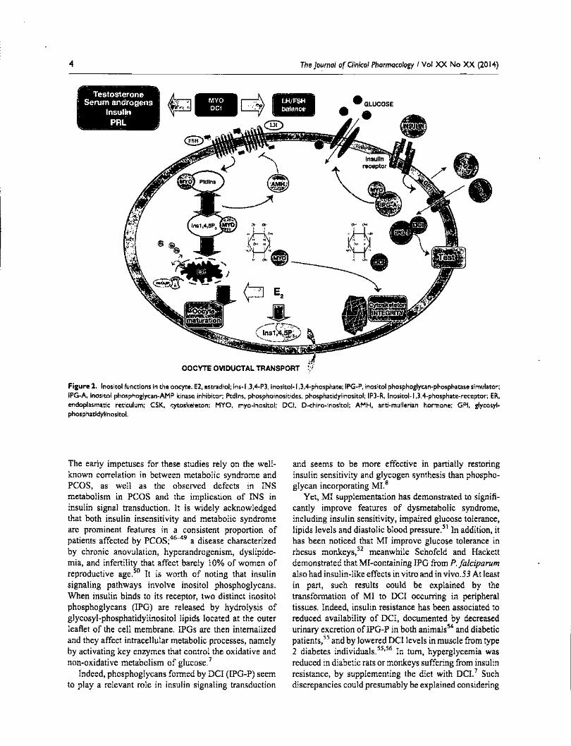
4 The Journal of Clinical Pharmacology I Vol XX No XX (2014)
. - ~-~ ~ -- ... ~ "
:' Testosterone ~ ~ Serum androgens • Insul~n c$' _ .. :"~ ....
PRI.:: j "'_ "J. ,)-
OOCYTEO~DUCTALTRANSPORT
Figure 2. Inositol functions in the oocyte. E2. estradiol; Ins-I.3.4-P3. inositol-I.3.4-phosphate; IPG-P. inositol phosphoglycan-phosphatase simulator; lPG-A. inositol phosphoglycan-AMP kinase inhibitor; Ptdlns. phosphoinositides. phosphatidylinositol; IP3-R. Inositol-I.3.4-phosphate-receptor; ER. endoplasmatic reticulum; CSK. cytoskeleton; MYO. myo-inositol; DCI. D-chiro-inositol; AMH. anti-mullerian hormone; GPI. glycosylphosphatidylinositol.
The early impetuses for these studies rely on the wellknown correlation in between metabolic syndrome and PCDS, as well as the observed defects in INS metabolism in PCDS and the implication of INS in insulin signal transduction. It is widely acknowledged that both insulin insensitivity and metabolic syndrome are prominent features in a consistent proportion of patients affected by PCDS;46-49 a disease characterized by chronic anovulation, hyperandrogenism, dyslipidemia, and infertility that affect barely 10% of women of reproductive age. 50 It is worth of noting that insulin signaling pathways involve inositol phosphoglycans. When insulin binds to its receptor, two distinct inositol phosphoglycans (lPG) are released by hydrolysis of glycosyl-phosphatidylinositol lipids located at the outer leaflet of the cell membrane. IPGs are then internalized and they affect intracellular metabolic processes, namely by activating key enzymes that control the oxidative and non-oxidative metabolism of glucose.7
Indeed, phosphoglycans formed by DCI (lPG-P) seem to playa relevant role in insulin signaling transduction
and seems to be more effective in partially restoring insulin sensitivity and glycogen synthesis than phosphoglycan incorporating MI.8
Yet, MI supplementation has demonstrated to significantly improve features of dysmetabolic syndrome, including insulin sensitivity, impaired glucose tolerance, lipids levels and diastolic blood pressure.51 In addition, it has been noticed that MI improve glucose tolerance in rhesus monkeys,52 meanwhile Schofeld and Hackett demonstrated that MI -containing IPG from P. Jalciparum also had insulin-like effects in vitro and in vivo. 53 At least in part, such results could be explained by the transformation of MI to DCI occurring in peripheral tissues. Indeed, insulin resistance has been associated to reduced availability of DCI, documented by decreased urinary excretion ofIPG-P in both animals54 and diabetic patients,55 and by lowered DCI levels in muscle from type 2 diabetes individuals.55
•56 In turn, hyperglycemia was
reduced in diabetic rats or monkeys suffering from insulin resistance, by supplementing the diet with DCe Such discrepancies could presumably be explained considering

Dinicola et al.
that while DCI is crucial for glycogen synthesis, MI increases glucose cellular uptake. 57
On the other hand, it is well recognized that increased insulin sensitivity in PCOS patients by means of conventional antidiabetic drugs promotes a significant improvement in the ovulatory function and decreases serum androgen concentrations.58--60 It is worth noting that Metformin increases the insulin stimulated release of DCI-phosphoglycans, thus evidencing the antidiabetic drug may enhance insulin sensitivity by restoring an inositol-based signaling.6o In support of this hypothesis it was further documented that PCOS patients show increased DCI urinary clearance and consistent DCI urinary loss, presumably leading to a tissue depletion of DCI-phosphoglycans.61
Once more, these data provided hints in hypothesizing a direct correlation between the availability of inositol phosphoglycans and insulin resistance, even if further studies are warranted in order to fully elucidate the mechanistic pathways linking inositol-derivatives to glucose metabolism.
Inositols and Ovary Function
Observations relating INS to glucose metabolism provided impetus to ascertain the usefulness, whether any, of DCI supplementation in PCOS clinical management. In his seminal paper, Nestler et al62 demonstrated that in women with PCOS, DCI treatment at a dose of 1,200 mg/ daily significantly reduced serum testosterone levels and improved both ovulation rate and metabolic parameters, such as blood pressure and triglycerides. Unfortunately that study provided no information on menstrual cycle regularity. Similar results have been subsequently recorded in lean PCOS women by the same groUp.63 Those preliminary data prompted Nestler and colleagues to advance the results they obtained during the first trials, by increasing up to 2,400 mg the daily dose of DCI administered to PCOS patients.64 That study was able to find a direct correlation between insulin-stimulated release of DCI-containing phosphoglycans and insulin sensitivity. Unexpectedly, that investigation was incapable in confirming the previous beneficial effects orDCI on ovary function, and the Authors did not offer any valuable hint to explain such a paradoxical outcome. A recent investigation performed on PCOS patients treated with increasing DCI doses (from 300 to 2,400mglday) has provided a compelling confirmation that by increasing DCI dosage paradoxically worsens oocyte quality and ovarian response in non-obese and non-insulin resistant PCOS women.65 Namely, total r-FSH units increased significantly in the two groups receiving the higher doses of DCI, while the number of immature oocytes was significantly increased in the three groups treated with the highest doses of DC I. Additionally, the number of grade I
5
embryos was significantly reduced by DCI supplementation.
It is intriguing that those disappointing data are in some way mirrored by results obtained by treating PCOS patients with Metformin: the antidiabetic drug decreases the follicles number and worsens their quality,66 even if Metformin significantly increases the insulin-stimulated release of DCI-phosphoglycans.61
Such contradictory results could likely be explained by considering the different function in each INS isomer plays in distinct tissues. Indeed, a specific MIIDCI ratio has been observed within each tissue: high DCI (even if always lower than MI concentration) is generally observed in glycogen storage tissues (fat, liver, muscle), whereas tissues characterized by high consumption rate of glucose (brain, heart) present low DCI levels.67 Oocytes are characterized by high glucose consumption along the oxidative pathway: thus, by impairing sugar availability oocyte quality would likely be compromised . Indeed, reduced availability of glucose in both oocytes and follicular cells caused by defective transportation of glucose it is suspected to occur in PCOS.68 In tum, energy impairment promotes alternative pathways to utilize fatty acid and amino acids for energy as a compensatory mechanism to deal with energy requirement.69 Moreover, in PCOS women, genes involved in the glucose uptake pathway are down-regulated at ovarian leveeo.71 and energy supplementation is required in achieving higher oocyte quality and better outcome after IVF in PCOS patients.7o Those data highlight how is important to maintain a proper glucose metabolism for oocyte development. Undoubtedly, both DCI and MI are required to fulfill such function in cooperating with insulin. Yet, MI seems to play a more important role in oocyte, as suggested by the fact that almost the 99% of intracellular INS pool is constituted by MI.72 DCI, instead, is produced from MI through a NAD-dependent epimerase whenever it is required. The epimerase conversion of MI to DCI is under insulin control: in type 2 diabetes patients, the reduced tissue insulin sensitivity leads to reduced epimerase activity and hence DCI synthesis.55 However, ovary never displays insulin resistance, unlike other tissues. 73 Thus, increased insulin levels as those recorded in insulin-resistant patients, are likely to increase the activity of ovary epimerase, raising in that way the DCI intracellular production, meanwhile MI levels were progressively reduced. Thereby, in hyperinsulinemic PCOS patients DCI levels paradoxically increase in the ovary. In tum, DCI increase may promote glycogen and testosterone synthesis, impairing the oocyte maturation. Eventually, that mechanism would lower oocyte's MI content, and the MI depletion will in tum negatively affect oocyte quality.
Such imbalance may shed light in the pathogenesis of PCOS at cellular level and helps elucidate the theory

6
known as "the DCI paradox in the ovary.,,74 That hypothesis has recently received a preliminary confirmation by two independent investigations. A study authored by Lamer and coworkers has found a significant increase in the epimerase activity in the theca cells obtained from ovary of PC OS women, leading to a dramatic reduction in the MIIDCI ratio.75 Another research, by investigating the INS concentration in follicular fluid obtained from PCOS women has observed a significant decrease in the MIIDCI ratio: while normal MIIDCI ratio is nearly 100: I, in follicular fluid of PC OS women that value is only 0.2: 1.72 The remarkable abatement in MI levels may thus likely contribute in explaining at least some of the observed dysfunction of ovary in PCOS.
Indeed, whereas DCI administration has been demonstrated to improve the systemic consequences of insulin resistance, namely by modulating insulin effects of endocrinology balance in non-ovarian tissues, DCI has probably only a marginal effect on oocyte function. In turn, a DCI overload, as such obtained by administering 600 mg/day or more, may have likely worsen INS imbalance into ovary cells, determining a dramatic decrease in the MIIDCI intracellular ratio. Additionally, high release of DCT-phosphoglycans, under insulin stimulation, enhances de novo testosterone biosynthesis from ovarian theca cells, thus raising serum androgen levels.76 Those effects would explain why the promising results obtained by Nestler and coworkers during the first study have not been confirmed in the second one.
Indeed, it is of outmost importance to ensure the proper MI concentration at the ovary level. Ml-based phosphoglycans (lPG-A) are required to facilitate oocyte glucose uptake.7 Furthermore MI improves oocyte response to FSH, as indicated by the reduced requirement in rFSH IV administered during IVF cycles.77,78 MI supplementation restores spontaneous ovulation and increases progesterone release during the luteal phase in all but few PCOS patients. 79 Therefore, it is worth of noting that a MI deficiency in the ovary would likely impair the FSH signal, resulting in an increased risk of ovarian hyperstimulation syndrome for PCOS patients. MI exerts in addition other appreciable systemic effects, by improving the reproductive axis functioning in PCOS patients through the reduction of the hyper-insulinemic state which affects LH secretion.8o In that study, following 12 weeks of MI treatment, serum hormone levels were normalized and menstrual cycle was restored in amenorrheic patients. Moreover, PCOS patients submitted to ICSI, the associated treatment of MI and folic acid, but not folic acid alone, significantly decreases the cancellation rate, it reduces oocyte degeneration and germinal vesicles at ovum pickup, thus increasing the success probability of the therapy.77 Indeed, serum and follicular fluid concentration ofMI has been proven to be
The Journal of Qinical Pharmacology I Vol XX No XX (2014)
directly associated with oocyte maturation and fertility outcome in IVF-treated patients.81
Inositols and peos Treatment Usefulness of MI supplementation has since been assessed by several reports. Morgante et al have evidenced that MI supplementation in insulin resistantPCOS patients produces significant results, given the significant reduction in cancellation rate (0% vs. 40%) and the consequent improvement in clinical pregnancy rate (33.3% vs. 13.3%) obtained with INS treatment.82
Gerli et al83 conducted a randomized, double-blind, placebo-controlled trial of 283 PCOS patients treated with MI. In that study, frequency of ovulation (40%) was increased by almost twofold in women who received MI, versus the control group. Moreover, aiming to elucidate the systemic beneficial effects associated to MI therapy, additional studies were able to show that MI treatment lowered lipids,84 insulin and androgen levels, increased insulin sensitivity, reduced blood diastolic pressure, and was effective in treating acne21 and hirsutism.85
Overall, those data demonstrated that MI is equally effective than DCT in normalizing metabolic and endocrine features commonly associated to insulin resistance and PCOS.
Yet, normalizing insulin resistance is not enough for restoring a proper ovulatory function, as suggested by a recent study comparing MI supplementation versus Metformin.86 Sixty PCOS patients were treated with MT 4 g plus Folic acid, and 60 PCOS patients with Metformin 1500 mg/day. Among the patients treated with Metformin, 50% restored spontaneous ovulation activity. Pregnancy occurred spontaneously in 11 (36.6%) of these patients. In the MI group, 65% of patients restored spontaneous ovulation activity, ovulation occurred after a mean of 14.8 days from the day 1 of the menstrual cycle; in this latter group pregnancy occurred in 18 (48.4%) patients, showing a positive trend in increase.
Overall, those data evidence that MI supplementation provides significant benefit in PCOS management (Table 1), even most papers suffer for the lack of proper randomization and/or are flawed by few statistical inconsistencies.
However, by considering both the systemic and the ovary hallmarks of PCOS, INS supplementation should preferably include both the isomers: MI and DCI. Given that physiological values of the MIIDCI ratio, evaluated both in the plasma as well as in the follicular fluid, range from40:1 to 100:1, it seems reasonable that INS should be administered jointly respecting a proportion that should reflect the natural balance among the two stereoisomers. Therefore, as proposed by a recent paper,87 the combined administration ofMI and DCI in the physiological plasma ratio (40: I), could be considered as a first line approach in

Table I. Eligible RCTs Where MI Have Been Evaluated for the Treatment of PCOS Patients
Ref.
Genazzani et also
Morgante
et alB2
Gerli et alBl
Study Design
Randomized.
controlled vs.
folic acid
Randomized.
controlled
trial vs.
placebo
Double-blind.
placebo
controlled vs.
placebo
Duration
12 weeks
4 weeks
16 weeks
Intervation
MY02g
FA 200mgl
day
Inositol
I.SOOmg
Inositol
200mg/day
No. of Subjects
N=20. Treatment: 10.
Placebo: 10
N=30.
Treatment: 15.
Placebo: 15
N=283. Treatment: 136.
Placebo: 147
Inclusion Criteria
Presence of micro polycystic
ovaries at ultrasound; mild to
severe hirsutism andlor acne;
oligomenorrhea or
amenorrhea; absence of
enzymatic adrenal deficiency
andlor other endocrine
disease; normal PRL levels
(range 5-25 nglmL); no
hormonal treatment for at
least 6 months before the
study
Clomiphene-failure patients
with PCOS. with insulin
resistance evaluated by
Homeostatic Model
Assessment index
Age: <35 years women with
oligomenorrhea and PCOS
ovaries. Ovaries were
described as polycystic
(PCOs) about the criteria of Adams et alb
Exclusion Criteria
Not described
Not described
Patients with Significant
hyperprolactinemia.
abnormal thyroid
function tests and
congenital adrenal
hyperplasia
Assessment of the Response Results
LH. FSH. PRL. E2. A. 170HP. LH. PRL. T. insulin levels. LHI
T. insulin. cortisol. OGTT" for FSH resulted Significantly
insulin. glucose. C-peptide de- reduced. Insulin sensitivity
terminations. vaginal ultra- resulted significantly improved.
sound examination Feriman- Menstrual cyclicity was
Gallway score. 8MI: HOMA restored in all amenorrheic
and oligomenorrheic subjects
HOMA. FSH (lUlL). LH (lUlL). The total number of follicles
PRL(nglmL).17-0HP. >ISmm and >18mm in
DHEAS. number of follicles diameter. and the peak E21evels
> 15 mm in diameter. were significantly lower in the
number of follicles > 18 mm inositol group compared with
in diameter. E2 levels on day
of hCG administration (pgl
mL). cancellation rate (%).
clinical PR (%)
Ovarian aaivlty was
monitored using serum E2.
P and LH. Ovulation
frequency was calculated
using the ratio of luteal
phase weeks to observation
weeks. Inibin-b. fasting
glucose. fasting insulin. or
insulin AUC. VLDL. LDL.
HDL. total cholesterol.
triglycerides. 8MI
the control group. In addition.
although the cancellation rate
was significantly lower in the
inositol group. the clinical PR
was not significantly higher.
compared with the control
group
Ovulation frequency
Significantly higher in the
treated group vs. the
placebo. follicular
maturation was rapid: the
circulating concentration of
E2 increased only in the
inositol group during the
first week of treatment.
Significant weight loss was
recorded in the inositol
group. whereas in the
placebo group was recorded
an increase of the weight. A
significant increase in
circulating high-density
(Continued) ......

Table I. Continued IX)
Ref. Study Design Duration Intervation No. of Subjects Inclusion Criteria Exclusion Criteria Assessment of the Response Results
lipoprotein was observed
only in the inositol-treated
group Minozzi et ala. Open-label 12 months MYO 4rJ N= ISS PCOS Women with secondary Clinical and anthropometric A higher reduction of FG score
clinical study day endocrine disorder. those measurement included: age. in OCP plus MI therapy
wishing to conceive during (BMI). serum levels of total group vs OCP alone therapy
the next 12 months and cholesterol. HDl cholesterol. group. OCP plus MI
those with contradictions lDl cholesterol. triglycerides. significantly decreased
to oral contraceptive use apolipoprotein B (apoB). hyperinsulinemia vs the
lipoprotein(a) [lp(a)]. fasting OCP group. Androgens
glucose level. fasting insulin serum levels decreased in
level. insulin resistance. mea- both groups. but significantly
sured by homeostasis model more in OCP plus MI group.
assessment (HOMA-IR). tes- The lipid profile was
tosterone (T). sex hormone- improved in the OCP plus
binding globulin (SHBG). D4- MI group. by reducing low-
androstenedione (A). dehy- density lipoprotein
droepiandrosterone (DHEAS). cholesterol levels and
leutenizing hormone (lH). and enhancing high density
hirsutism score. evaluated by lipoprotein cholesterol
using a modification of the levels :;l Ferriman-Gallwey (FG) score <II
"-M inozzi et alas Open-label 6 months MYO 2rJ N=46 Women with mild to Hyperprolactinemia. Hirsutism scores Serum con- No changes In BMI were 0
c: clinical study twice a day moderate hirsutism. evaluated hypothyroidism. adrenal centrations of total cholester- observed. The hirsutism 3 e..
using a modification of the hyperplasia and Cushing's 01. HDl cholesterol. low- decreased after therapy. Total 0 -. Ferriman-Gallwey score syndrome. Patients who density lipoprotein (lDl) cho- androgens. FSH and lH
~ had taken hormonal lesterol. triglycerides. apolipo- concentrations decreased. [ medications. including protein B. lipoprotein(a). oestradiol concentrations
contraceptive pills. for glucose. insulin. testosterone. increased. There was a slight ~ 0
the past 6 months sex hormone-binding globulin non-significant decrease in total 3 (SHBG). ~4-androstenedione. cholesterol concentrations. an
Q
S cortisol. dehydroepiandroster- increase in HDl cholesterol ~ one sulphate (DHEA-S). lH. concentrations and a decrease
FSH and E2 were measured in lDl cholesterol < Q..
within the first 5 days of the concentrations. No significant
~ menstrual cycle changes were observed in
serum triglyceride. Z 0
apolipoprotein Band X lipoproteln(a) concentrations. X
N (Continued) 0
~

Table I. Continued 0 :i" s·
Ref. Study Design Duration Intervation No. of Subjects Inclusion Criteria Exclusion Criteria Assessment of the Response Results Q
~
Insulin resistance, analysed by Q ,.... homeostasis model
assessment, was reduced
significantly after therapy
Raffone et als6 Randomized Until the MY04g N= 120, PCOS <35 years defined by Other medical condition Restoration of spontaneous Among the patients treated
controlled vs. end of the FA 400mg Treatment: 60, Rotterdam Criteria causing ovulatory ovulation and menstrual cycles with metformin, 50% restored
metformin study or Placebo: 60 dysfunction, tubal and increasing rate pregnancy, spontaneous ovulation activity.
positive defects, semen ovarian activity, serum proges- Pregnancy occurred
pregnancy parameters defects terone dosage, progesterone spontaneously in II (36.6%) of
test levels, j3-hCG plasma level these patients. In the MI group,
6S % of patients restored
spontaneous ovulation activity,
ovulation occurred after a
mean of 14.8 days from the
day I of the menstrual cycle; in
this latter group pregnancy
occurred in 18 (48.4%)
patients, showing a positive
trend in increase
Nordio and Randomized 6 months 2gMYO N=50, Overweight women with Diabetic subjects, Plasma glucose and insulin The combined administration
ProiettiS7 controlled powder Treatment: 24, PCOS smokers and alcohol concentrations Serum proges- of MI and DCI reduces the
550mg of Combined users were excluded terone Ovulation function metabolic and clinical alteration
Myo plus therapy: 26 from the study of PCOS and reduces the risk
13.8mg of of metabolic syndrome
D-chiro-
inositol in
soft gel
capsule
Costantino Double-blind, 12-16 MY04g N=42, Presence of oligomenorrhea, Not described Systohc/dlastolic blood MI increased insuhn senSitiVity,
et al 'Ol randomized weeks FA 400mrJ Treatment: 23, high serum-free testosterone pressure, triglycerldes, improved glucose tolerance controlled vs. day Placebo: 19 level and/or hirsutism cholesterol, 8MI, plasma and decreased glucose
folic acid presence of micropolycystic glucose and insulin stimulated insulin release. ovaries at ultrasound sensitiVity, total/free T, There was a decrement in
DHEAS, SH8G, A, serum total T and serum-free T
progesterone peak value concentrations. In addition,
there was a decrement in
systolic and diastolic blood
pressure. Plasma triglycerides
and total cholesterol
( Continued)
'"

Table I. Continued
Ref. Study Design Duration Intervation No. of Subjects Inclusion Criteria Exclusion Criteria Assessment of the Response Results
concentration decreased Papaleo Prospective. During MY04g N=60. Age: <40 years PCOS women Other medical conditions Number of morphologically Total r-FSH units and number
et alia' randomized. ovulation FA 200 Treatment: 30. diagnosed by causing ovulatory mawre oocytes retrieved. em- of days of stimulation were
controlled vs. induction mglday Placebo: 30 oligoamenorrhea. disorders: bryo quality. pregnancy and significantly reduced in the
folic acid for ICSI hyperandrogenism hyperinsulinemia. implantation rates. Total num- myo-inositol group. Peak E2
orhyperandrogenemia and hyperprolactinemia. ber of days of FSH stimulation. levels at hCG administration
typical features of ovaries on hypothyroidism. or total dose of gonadotropin were significantly lower in
ultrasound scan androgen excess. such as administered. E2 level on the patients receiving myo-
adrenal hyperplasia or day of hCG administration. inositol. The mean number
Cushing syndrome fertilization rate per number of of oocytes retrieved did not
retrieved oocytes. embryo differ in the two groups.
cleavage rate. live birth and whereas in the group
miscarriage rate. severe ovarian cotreated with myo-inositol
hyperstimulation syndrome the mean number of
germinal vesicles and
degenerated oocytes was
significantly reduced. with a
trend for increased
percentage of oocytes in
metaphase II Gerli et aliOS Double-blind. 16 weeks MY04g N=92. Age: <35 years Women with Patients with significant Ovarian actiVity was moni- Beneficial effect of MYO
randomized. FA 200mgi Treatment: 45. oligomenorrhea. hyperprolactinemia. tored using serum E2. p. LH. treatment upon ovarian
controlled vs. day Placebo: 47 amenorrhea and PCOS abnormal thyrOid ovulation frequency was calcu- function. anthropometric
folic acid ovaries. Ovaries were function tests. and lated using the ratio of luteal measures and lipid profiles
described as polycystic congenital adrenal phase weeks to observation
(PCOs) about the criteria hyperplasia weeks. Inibin-b. fasting glucose. of Adams et alb fasting insulin. or insulin AUC.
VLDL. LDL, HDL. total choles-
terol. triglycerides. BMI
LFA. folic acid; PRL, prolactin; E2. estradiol; A. androstenedione; 170HP. 17-hydroxy-progesterone; T. testosterone; P. progesterone; OGTT. oral glucose tolerance; BMI. body mass index; LH. luteinizing hormone; FSH.
follicle stimulatig hormone; DHEAS. dehydroepiandrosterone; SHBG. sex hormone binding globulin; EAUC. area under the curve of OGTT; VLDL. very low density lipoprotein; LDL. low density lipoprotein; HDL. high
density lipoprotein.
'OGTT was performed sampling 15 minutes before. and 30. 60. 90. 120. and 240 minutes after the oral assumption of 75 g of glucose.
bAdams J. Polson JW. Franks S. Prevalence of polycystic ovaries in women With anovulation and idiopathic hirsutism. Br Med J 1986; 293: 355-359.
o
~ Z o
~

Dinicola et al.
pcas overweight patients, being able to reduce the metabolic. hormonal and clinical alteration of pcas.
Conclusion
pcas is one of the most common endocrine disorders affecting women. It represents the most common cause of female infertility and it is characterized by a combination of hyperandrogenism, chronic anovulation and irregular menstrual cycle.88 A significant number of patients suffers also from metabolic syndrome and insulin resistance, even if such features are currently not entirely understood.89,90
While a significant progress has recently been made in the diagnosis for pcas,91-93 the optimal infertility treatment in such cases remains to be determined.94
Recently, several clinical studies have highlighted the usefulness of INS supplementation in pcas treatment. MI is readily taken up following oral ingestion95 and it is has been proven to be safe even after high dose consumption.96
However, despite the relatively high number of reports, only few of them fulfill the criteria of randomized clinical trial. Those studies have been extensively reviewed elsewhere.97,98 Among 70 studies focusing on pcas treatment by means of different pharmaceutical composition incorporating INS, 21 were considered eligible as they involve MI.97 Yet, only six of them were randomized controlled clinical trials (level of evidence Ib), involving more than 300 pcas patients. Remarkably, in all the studies analyzed, no side effects were reported at the doses of both 2 and 4 g/day, thus resulting in a high patient compliance. The 4 glday treatment regimen is useful to treat the symptom spectrum, resulting in a more complete and effective treatment. averall, those studies indicate that MI supplementation improves several of the hormonal disturbances of PC as, providing so far a level Ia evidence of MI effectiveness. MI mechanisms of action appear to be mainly based on improving insulin sensitivity of target tissues, resulting in a positive effect on the reproductive axis (MI restores ovulation and improves oocyte quality) and hormonal functions (MI reduces clinical and biochemical hyperandrogenism and dyslipidemia) through the reduction of insulin plasma levels.
These systemic hallmarks of pcas are significantly affected by both DCI and MI supplementation. However, DCI treatment, mostly when it is administered at high dosage (ie, 600 mg or more), exerts disappointing effects on ovary functions.65 aocyte physiology, among other factors, is likely to be dependent on a fairy balance in between MI and DCI. Indeed, MI is an important constituent of follicular microenvironment, playing a determinant role in both nuclear and cytoplasmic oocyte development. Perhaps, the content of MI in follicular
II
fluids may represent a more appropriate physiological indicator than follicular volume for monitoring the status of the developing follicles. Follicles that containing good quality oocytes have higher concentrations of MI in foIIicular fluids, probably due to the intricate relationship between MI and inositol phosphates in the PtdIns cycle activation for oocyte maturation.
AdditionaIIy, MI by improving glucose uptake may improve both oocyte energy status and oocyte quality. Moreover, during ovarian stimulation MI reduces FSHIU necessary for ovarian stimulation. Altogether, this evidence hints that MI exerts several beneficial effects that improve the pregnancy chance.99
A further confirmation of the link among MI and oocyte quality has been provided by two recent studies performed on both pcas and non-PCaS women. 100 In a prospective randomized open-label, pilot clinical trial involving one hundred non-PCaS women undergoing mUltiple follicular stimulation for in vitro fertilization, the addition of MI promotes the oocyte's meiotic maturation and reduces the number of gonadotropin cycles of treatment, while maintaining clinical pregnancy rate. Even if this study is underpowered to evaluate IVF outcomes like implantation and clinical pregnancy, a trend in favor of increased incidence of implantation in the group pre-treated with MI was observed. Another clinical investigation, lOl focused on one hundred pcas women undergoing IVF-ET. Patients were treated with MI combined with DCI in the physiological ratio (40: I), or with DCI alone (500mg). Significant better results were observed in the MI + DCI treated group, given that patients treated with this combination required lower dosages of FSH for a shorter period of time and showed an improvement in both oocyte quality and pregnancy rate.
Therefore, since the attention of the scientific community has recently been drawn on the relevance of the MIIDCI ratio for a proper ovary function, a treatment based on the association MIIDCI, in a physiological plasma range (ie, 40:1) seems to be the most effective approach, as recently stated by an international Consensus Conference held in Florence (December 2013).102 Moreover, the recent data by Lamer's group provided a molecular mechanistic basis supporting that statement, highlighting the "utility of both MI and D-chiro-inositol as effective agents in treatment. Certainly, a balance between the two inositols is required for normal physiological function and regulation of the MI to chiro-inositol epimerase opens a new avenue for upcoming studies.,,75
Undoubtedly, further studies are warranted to fully elucidate the molecular pathways triggered by myo- and D-chiro-inositol underlining their beneficial effects and to provide a well-grounded rationale for INS supplementation in pcas patients.103-105

12
Declaration of Conflicting Interests
G.C. and V.U. are Lo.Li. Pharrna employees.
References I. Clements RS, Jr, Darnell B. Myo-inositol content of common
foods: development of a high-myo-inositol dIet. Am} Clin Nutr. 1980;33(9): 1954-1967.
2. Hooper NM. Glycosyl-phosphatidylinositol anchored membrane enzymes. Clin Chim Acla. 1997;266(1):3-12.
3. Di Paolo G, De Camilli P. Phosphoinositides in cell regulation and membrane dynamics. Nalure. 2006;443(7112):651-657.
4. Chen IW, Charalampous CF. Biochemical studies on inositol. IX. D-Inositol I-phosphate as intermediate in the biosynthesis of inositol from glucose 6-phosphate. and characteristics of two reactions in this biosynthesis.} Bioi Chem. 1966;241(10):2194-2199.
5. Carman GM, Henry SA. Phospholipid biosynthesis in yeast. Ann Rev Biochem. J 989;58:635-669.
6. Berridge MJ. Inositol trisphosphate and calcium signalling. Nalllre. 1993;361(6410):315-325.
7. Huang LC. Fonteles MC, Houston DB, Zhang C, Lamer J. Chiroinositol deficiency and insulin resistance. Ill. Acute glycogenic and hypoglycemic effects of two inositol phosphoglycan insulin mediators in normal and streptozotocin-diabetie rats in vivo. Endocrinology. 1993; 132(2):652-657.
8. Lamer J. D-chiro-inositol-its functional role in insulin action and its deficit in insulin resistance. Inl } Exp Diabetes Res. 2002;3 (1):47-60.
9. Lamer J, Huang LC, Schwartz CF, et al. Rat liver insulin mediator which stimulates pyruvate dehydrogenase phosphate contains galactosamine and D-chiroinositol. Biochem Biophys Res Commun. 1988;151(3):1416-1426.
10. Romero G, Gamez G, Huang LC, Lilley K, Luttrell L. AntiInositol glycan antibodies selectively block some ofthe actions of insulin in intact BC3Hl cells. Proc Natl Acad Sci USA. 1990;87 (4):1476-1480.
II. Alvarez JF, Sanchez-Arias JA, Guadano A, et al. Transport in isolated rat hepatocytes of the phospho-oligosaccharide that mimics insulin action. Effects of adrenalectomy and glucocorticoid treatment. Biochem). 1991;274(Pt 2):369-374.
12. Sun TH, Heimark DB, Nguygen T, Nadler JL, Lamer J. Bothmyoinositol to chiro-inositol epimerase activities and chiro-inositol to myo-inosltol ratios are decreased in tissues of GK type 2 diabetic rats compared to Wistar controls. Biochem Biophys Res Commlln. 2002;293(3): 1092-1098.
13. Downes CPo The cellular functions ofmyo-inositol. Biochem Soc Trans. 1989; 17(2):259-268.
14. Downes CP, Macphee CH. Myo-inositol metabolites as cellular signals. Ellr} Biochem. 1990;193(1):1-18.
15. Diaz JR, de las Cagigas A, Rodriguez R. Micronutrient deficiencies in developing and affluent countries. Eur } Clin NUlr. 2003;57(Suppl 1):S70-S72.
16. Eisenberg F, Jr., Bolden AH. Reproductive tract as site of synthesis and secretion of inositol in the male rat. Nature. 1964;202:599-600.
17. Ghafoorunissa. Effect of dietary protein on the biosynthesis of inositol in rat testes. } Reprod Ferli!. 1975;42(2):233-238.
18. Lewin LM, Beer R. Prostatic secretion as the source of myoinositol in human seminal fluid. Ferli! Steri!. 1973;24(9):666-670.
19. Lewin LM, Yannai Y, Me1med S, Weiss M. Myo-inosltol in the reproductive tract of the female rat. Int } Biochem. 1982;14 (2):147-150.
20. Chiu TT, Tam PP. A correlation ofthe outcome of clinical in vitro fertilization with the inositol content and embryotrophic properties of human serum.) Ass(vl Reprod Genet. 1992;9(6):524-530.
The Journal of Clinical Pharmacology I Vol XX No XX (2014)
21. Zacche MM, Caputo L, Filippis S, Zacche G, Dindelli M, Ferrari A. Efficacy ofmyo-inositol in the treatment of cutaneous disorders in young women with polycystic ovary syndrome. Gynecol Endocrinol. 2009;25(8):508-513.
22. Matsuda M, Tsutsumi K. Kanematsu T, et al. Involvement of phospholipase C-related inactive protein in the mouse reproductive system through the regulation of gonadotropin levels. Bioi Reprod.2009;81(4):681-689.
23. Goud PT, Goud AP. Van Oostveldt P, Dhont M. Presence and dynamic redistribution of type I inositol 1,4,5-trisphosphate receptors in human oocytes and embryos during in-vitro maturation, fertilization and early cleavage divisions. Mol Hum Reprod. 1999;5(5):441-451.
24. Lowther KM, Weitzman VN, Maier D, Mehlmann LM. Maturation, fertilization, and the structure and function of the endoplasmic reticulum in cryopreserved mouse ooeytes. Bioi Reprod. 2009;81(1): 147-154.
25. Chiu TT, Rogers MS, Briton-Jones C, Haines C. Effects ofmyoinositol on the in-vitro maturation and subsequent development of mousc oocytcs. Hum Reprod. 2003; 18(2):408-416.
26. Kane MT, Norris M, Harrison RA. Uptake and incorporation of inositol by preimplantation mouse embryos. } Reprod Ferti!. 1992;96(2):617-625.
27. Fahy MM, Kane MT. Incorporation of [3H]inositol into phosphoinositides and inositol phosphates by rabbit blastocysts. Mol Reprod Dev. 1993;34(4):391-395.
28. Holm P, Booth Pl, Schmidt MH, Greve T, Callesen H. High bovine blastocyst development in a static in vitro production system using SOFaa medium supplemented with sodium citrate and myoinositol with or without serum-proteins. Theriogenology. 1999;52 (4):683-700.
29. Holub BJ. Metabolism and function of myo-inositol and inositol phospholipids. Anllu Rev Nutr. 1986;6:563-597.
30. Colazingari S, Fiorenza MT, Carlomagno G, Najjar R, Bevilacqua A. Improvement of mouse embryo quality by myo-inositol supplementation of IVF media.) Assist Reprod Genet. 2014;31 (4):463-469.
31. Stachecki JJ, Armant DR. Transient release of calcium from inositol I ,4,5-trisphosphate-specific stores regulates mouse preimplantation development. Development. 1996; 122(8):2485-2496.
32. Huang C, Liang NC. Increase in cytoskeletal actin induced by inositol 1,4-bisphosphate in saponin-permeated pig platelets. Cell Biollnl. 1994; 18(8):797-804.
33. Ducibella T, Kurasawa S, DuffY P, Kopf GS, Schultz RM. Regulation of the polyspermy block in the mouse egg: maturationdependent differences in cortical granule exocytosis and zona pellucida modifications induced by Inositol 1,4,5-trisphosphate and an activator of protein kinase C. Bioi Reprod. 1993;48 (6):1251-1257.
34. Orihuela PA, Parada-Bustamante A, Zuniga LM, Croxatto HB. Inositol triphosphate participates in an oestradiol nongenomic signalling pathway involved in accelerated oviductal transport in cycling rats. ) Endocrinol. 2006; 188(3):579-588.
35. Carlomagno G, Roseff S, Harter S, Murphy Cohen S, Unfer V. A novel approach for treating infertile patients with diminished ovarian reserve (DaR). 14th World Congress on Controversies in Obstetrics, Gynecology & Infertility (COGI), Monduzzi Eds, 20 II, Paris, France.
36. Visser JA, Themmen AP. Anti-Mullerian hormone and folliculogenesis. Mol Cell Endocrinol. 2005;234(1-2):81-86.
37. Visser JA, de Jong FH, Laven JS, Themmen AP. Anti-Mullerian hormone: a new marker for ovarian function. Reproduclion. 2006;131(1): 1-9.
38. Quirk JG, Jr., Bleasdale JE. Myo-Inositol homeostasis in the human fetus. Obstet GYllecol. 1983;62(1):41-44.

Dinicola et 01.
39. Hallman M, Arjomaa P, Hoppu K. Inositol supplementation in respiratory distress syndrome: relationship between serum concentration, renal excretion, and lung effluent phospholipids. J Pediatr. 1987;110(4):604-610.
40. Greene NO. Copp AJ. Inositol prevents folate-resistant neural tube defects in the mouse. Nat Med. 1997;3(1):60-66.
41. Hendricks KA, Nuno OM, Suarez L, Larsen R. Effects of hyperinsulinemia and obesity on risk of neural tube defects among Mexican Americans. Epidemiology. 2001;12(6):630-635.
42. Reece EA, Khandelwal M, Wu YK, Borenstein M. Dietary intake of myo-inositol and neural tube defects in offspring of diabetic rats. Am J Obstet Gynecol. 1997; 176(3):536-539.
43. Goldman AS, Goto MP. Biochemical basis of the diabetic embryopathy. lsr J Med Sci. 1991 ;27(8-9):469-477.
44. Cavalli P, Copp AJ. Inositol and folate resistant neural tube defects. J Med Genet. 2002;39(2):E5.
45. Beemster P, Groenen P, Steegers-Theunissen R. Involvement of inositol in reproduction. Nutr Rev. 2002:60(3):80-87.
46. Dunaif A, Segal KR, Futterweit W, Dobrjansky A. Profound peripheral insulin resistancc, independent of obesity, in polycystic ovary syndrome. Diabetes. 1989;38(9): 1165-1174.
47. Ehrmann DA, Barnes RB, Rosenfield RL, Cavaghan MK,lmperial J. Prevalence of impaired glucose tolerance and diabetes in women with polycystic ovary syndrome. Diabetes Care. I 999;22( I): 141-146.
48. Legro RS, Finegood 0, Dunaif A. A fasting glucose to insulin ratio is a useful measure of insulin sensitivity in women with polycystic ovary syndrome. J Clill Elldocrinol Metab. 1998;83(8):2694-2698.
49. Legro RS. Blanche P, Krauss RM, Lobo RA. Alterations in lowdensity lipoprotein and high-density lipoprotein subclasses among Hispanic women with polycystic ovary syndrome: influence of insulin and genetic factors. Fertil Steril. 1999;72(6):990-995.
50. Franks S. Polycystic ovary syndrome. N Engl J Med. 1995;333 (13):853-861.
51. Giordano D, Corrado F, Santamaria A. et al. Effects of myoinositol supplementation in postmenopausal women with metabolic syndrome: a perspective, randomized, placebo-controlled study. Menopause. 2011;18(1): 102-104.
52. Ortmeyer HK. Dietary myoinositol results in lower urine glucose and in lower postprandial plasma glucose in obese insulin resistant rhesus monkeys. Obes Res. 1996;4(6):569-575.
53. Schofield L, Hackett F. Signal transduction in host cells by a glycosylphosphatidylinositol toxin of malaria parasites. J Exp Med. 1993;177(1): 145-153.
54. Ortmeyer HK. Bodkin NL, Lilley K, Lamer J, Hansen BC. Chiroinositol deficiency and insulin resistance. I. Unnary excretion rate of chiroinositol is directly associated with insulin resistance in spontaneously diabetic rhesus monkeys. Endocrinology. 1993; 132(2):640-645.
55. Asplin I, Galasko G, Lamer J. Chiro-inositol deficiency and insulin resistance: a comparison of the chiro-inositol- and the myoinositol-containing insulin mediators isolated from urine, hemodialysate, and muscle of control and type 11 diabetic subjects. Proc Natl Acad Sci USA. 1993;90(13):5924-5928.
56. Ortmeyer HK, Huang LC, Zhang L, Hansen BC, Lamer J. Chiroinositol deficiency and insulin resistance. II. Acute effects of D-chiroinositol administration in streptozotocin-diabetic rats, normal rats given a glucose load, and spontaneously insulinresistant rhesus monkeys. Endocrinology. 1993;132(2):646-651.
57. Ehrmann DA. Insulin-lowering therapeutic modalities for polycystic ovary syndrome. Endocrinol Mefab Clin North Am. 1999;28 (2):423-438.
58. Hasegawa I, Murakawa H, Suzuki M, Yamamoto Y, Kurabayashi T, Tanaka K. Effect of troglitazone on endocrine and ovulatory
13
performance in women with insulin resistance-related polycystic ovary syndrome. Fertil Steril. 1999;71(2):323-327.
59. Nestlcr JE, Jakubowicz OJ, Evans WS, Pasquali R. Effects of metformin on spontaneous and clomiphene-induced ovulation in the polycystic ovary syndrome. N Engl J Med. 1998;338 (26):1876-1880.
60. Baillargeon JP, Iuomo MJ, Jakubowicz OJ, Apridonidze T, He N, Nestler JE. Metformin therapy increases insulin-stimulated release of D-chiro-inositol-containing inositolphosphoglycan mediator in women with polycystic ovary syndrome. J Clin Endocrinol Metab. 2004;89( I ):242-249.
61. Baillargeon JP, Diamanti-Kandarakis E, Ostlund RE, Jr, Apridonidze T, luomo MJ. Nestler JE. Altered D-chiro-inositol urinary clearance in women with polycystic ovary syndrome. Diabetes Care. 2006;29(2):300-305.
62. Nestler JE, Jakubowicz OJ, Reamer P, Gunn RD, Allan G. Ovulatory and metabolic effects of D-chiro-inositol in the polycystic ovary syndrome. N Ellgl J Med. 1999;340(17):1314-1320.
63. luomo MJ, Jakubowicz OJ, Baillargeon JP, et al. Effects of dchiro-inositol in lean women with the polycystic ovary syndrome. Endocr Pract. 2002;8(6):417-423.
64. Cheang KI, Baillargeon JP, Essah PA, et al. Insulin-stimulated release of D-chiro-inositol-containing inositolphosphoglycan mediator correlates with insulin sensitivity in women with polycystic ovary syndrome. Metabolism. 2008;57( I 0): 1390-1397.
65. Isabella R, Raffone E. Does ovary need D-chiro-inositol. J Ovarian Res. 2012;5(1):14.
66. Palomba S, Falbo A, Di Cello A, Cappiello F, Tolino A, Zullo F. Does metformin affect the ovarian response to gonadotropins for in vitro fertilization treatment in patients with polycystic ovary syndrome and reduced ovarian reserve? A randomized controlled trial. Fertil Steril. 96(5): 1128-1133.
67. Pak Y, Huang LC, Lilley KJ, Lamer J. In vivo conversion of[3H] myoinositoI to [3H]chiroinositol in rat tissues. J Bioi Chem. 1992;267(24): 16904-16910.
68. Chaudhary K, Babu KN, Joshi VN, Srivastava S, Chakravarty BN. NMR-based metabolomics reveals differently expressed metabolites in follicular fluid of pcos women: potential biomarkers for good quality oocyte? Hum Reprod. 2011;26: i226-i246.
69. Pinero-Sagredo E, Nunes S, de Los Santos MJ, Celda B, Esteve V. NMR metabolic profile of human follicular fluid. NMR Biomed. 20 I 0;23(5):485-495.
70. Arya BK, Haq AU, Chaudhury K. Oocyte quality reflected by follicular fluid analysis in poly cystic ovary syndrome (PCOS): A hypothesis based on intermediates of energy metabolism. Med Hypotheses. 2012;78(4):475-478.
71. Ma X, Fan L, Meng Y, et al. Proteomic analysis of human ovaries from normal and polycystic ovarian syndrome. Mol Hum Reprod. 2007; 13(8):527-535.
72. Unfer V. Carlomagno G, Papaleo E, Vailati S, Candiani M, Baillargeon JP. Hyperinsulinemia alters myoinositol to Dchiroinositol ratio in the follicular fluid of patients with PCOS. Published on line 4 February. Reprod Sci. 2014:001: 10.11771
1933719113518985. 73. Matalliotakis I, Kourtis A, Koukoura 0, Panidis D. Polycystic
ovary syndrome: etiology and pathogenesis. Arch Gynecol Obstet. 2006;274(4): 187-197.
74. Carlomagno G, Unfer V, RoseffS. The D-chiro-inositol paradox in the ovary. Ferfil Steril. 20 II ;95(8):2515-2516.
75. Heimark D, McAllister J, Lamer J. Decreased myo-inositol to chiro-inositol (m/c) ratios and increascd mlc epimcrasc activity in pcos theca cells demonstrate increased insulin sensitivity compared to controls. EndocrJ. 2013;61(2):11 I-I 17.
14
76. Nestler JE, Jakubowicz OJ, de Vargas AF, Brik C, Quintero N, Medina F. Insulin stimulates testosterone biosynthesis by human thecal cells ITom women with polycystic ovary syndrome by activating its own receptor and using inositolglycan mediators as the signal transduction system. J Clin Endocrinol Metab. 1998;83 (6):2001-2005.
77. Papaleo E, Unfer V, Baillargeon JP, Fusi F, Occhi F, De Santis L. Myo-inositol may improve oocyte quality in intracytoplasmic sperm injection cycles. A prospective, controlled, randomized trial. Fertil Steril. 2009;91 (5): 1750-1754.
78. Unfer V, Carlomagno G, Rizzo P, Raffone E, Roseff S. Myoinositol rather than D-chiro-inositol is able to improve oocyte quality in intracytoplasmic sperm injection cycles. A prospective, controlled, randomized trial. Eur Rev Med Pharmacol Sci. 20 II; 15 (4):452-457.
79. Papaleo E, Unfer V, Baillargeon JP, et al. Myo-inositol in patients with polycystic ovary syndrome: a novel method for ovulation induction. Gynecol Endocrinol. 2007;23(12):700-703.
80. Genazzani AD, Lanzoni C, Ricchieri F, Jasonni VM. Myo-inositol administration positively affects hypcrinsulincmia and hormonal parameters in overweight patients with polycystic ovary syndrome. Gynecol Endocrinol. 2008;24(3):139-144.
81. Chiu IT, Rogers MS, Law EL, Briton-Jones CM, Cheung LP, Haines CJ. Follicular fluid and scrum conccntrations of myoinositol in patients undergoing IVF: relationship with oocyte quality. Hum Reprod .. 2002;17(6):1591-1596.
82. Morgante G, Orvieto R, Di Sabatino A, Musacchio MC, De Leo V. The role of inositol supplementation in patients with polycystic ovary syndrome, with insulin resistance, undergoing the low-dose gonadotropin ovulation induction regimen. Fertil Steril. 20 II :95 (8):2642-2644.
83. Gerli S, Mignosa M, Di Renzo GC. Effects of inositol on ovarian function and metabolic factors in women with PCOS: a randomized double blind placebo-controlled trial. Eur Rev Med Pharmacol Sci. 2003;7(6):151-159.
84. Minozzi M, Costantino D, Guaraldi C, Unfer V. The effect of a combination therapy with myo-inositol and a combined oral contraceptive pill versus a combined oral contraceptive pill alone on metabolic, endocrine, and clinical parameters in polycystic ovary syndrome. Gynecol Endocrinol. 2011;27(11):920-924. DOl: 10.3109109513590.2011.564685.
85. Minozzi M, D'Andrea G, Unfer V. Treatment of hirsutism with myo-inositol: a prospective clinical study. Reprod Biomed Online. 2008; 17(4):579-582.
86. Raffone E, Rizzo P, Benedetto V. Insulin sensitiser agents alone and in co-treatment with r-FSH for ovulation induction in PCOS women. Gynecol Endocrinol. 2010;26(4):275-280.
87. Nordio M, Proietti E. The combined therapy with myo-inositol and D-chiro-inositol reduces the risk of metabolic disease in PCOS overweight patients compared to myo-inositol supplementation alone. Eur Rev Med Pharmacol Sci. 2012;16(5):575-581.
88. Homburg R, Polycystic ovary, syndrome -, from gynaecological, curiosity to, multisystem endocrinopathy. Hum Reprod. 1996; II (1):29-39.
The Journal of Clinical Pharmacology I Vol XX No XX (2014)
89. Dunaif A, Segal KR, Shelley DR, Green G, Dobrjansky A, Licholai T. Evidence for distinctive and intrinsic defects in insulin action in polycystic ovary syndrome. Diabetes. 1992;41 (10):1257-1266.
90. Nestler JE. Insulin resistance and the polycystic ovary syndrome: recent advances. Cllrr Opin Endocrillol. 2000;7(6):345-349.
91. Lobo RA, Carmina E. The importance of diagnosing the polycystic ovary syndrome. Ann Intern Med. 2000:132(12):989-993.
92. Revised. 2003 consensus on diagnostic criteria and long-term health risks related to polycystic ovary syndrome. Fertif Steril. 2004;81(1): 19-25.
93. Revised. 2003 consensus on diagnostic criteria and long-term health risks related to polycystic ovary syndrome (PCOS). Hum Reprod.2004;19(I):41-47.
94. Akpinar Z, Tokgoz S, Gokbel H, Okudan N, Uguz F, Yilmaz G. The association of nocturnal serum melatonin levels with major depression in patients with acute multiple sclerosis. Psychiatry Res. 2008;161(2):253-257.
95. Groenen PM, Merkus HM, Sweep FC, Wevers RA, Janssen FS, Stccgcrs-Thcunisscn RP. Kincticsofmyo-inositolloading in women of reproductive age. Anll Clin Biochem. 2003;40(Pt 1):79-85.
96. Carlomagno G, Unfer V. Inositol safety: clinical evidences. Eur Rev Med Pharmacol Sci. 2011;15:931-936.
97. Unfcr V, Carlomagno G, Dante G, Facchinctti F. Effccts ofmyoinositol in women with PCOS: a systematic review of randomized controlled trials. Gynecol Endocrinol. 2012;28(7):509-515.
98. Papaleo E, Unfer V, Baillargeon JP, Chiu IT. Contribution of myo-inositol to reproduction. Ear J Obslet Gynecol Reprod BioI. 2009;147(2): 120-123.
99. Pal L, Jindal S, Witt BR, Santoro N. Less is more: increased gonadotropin use for ovarian stimulation adversely influences clinical pregnancy and live birth after in vitro fertilization. Ferlif Sterif. 2008;89(6): 1694-170 I.
100. Lisi F, Carfagna P, Oliva MM, Rago R, Lisi R, et aI. Pretreatment with myo-inositol in non polycystic ovary syndrome patients undergoing multiple follicular stimulation for IVF: a pilot study. Reprod Bioi Endocrinol. 2012;10:52.
101. Colazingari S, Treglia M, Najjar R, Bevilacqua A. The combined therapy myo-inositol plus D-chiro-inositol, rather than D-chiroinositol, is able to improve IVF outcomes: results from a randomized controlled trial. Arch GynecolObstet. 2013;288(6):1405-1411.
102. Personal communication at international Consensus Conference held in Florence December. 2013.
103. Costantino D, Minozzi G, Minozzi E, et al. Metabolic and hormonal effects ofmyo-inositol in women with polycystic ovary syndrome: a double-blind trial. Eur Rev Med Pharmacol Sci. 2009;13:105-110.
104. Papaleo E, Unfer V, Baillargeon JP, et al. Myo-inositol in patients with polycystic ovary syndrome: a novel method for ovulation induction. Gynecol Endocrinol. 2007;23(12):700-703.
105. Gerli S, Papaleo E, Ferrari A, et al. Randomized, double blind placebo-controlled trial: effects of myo-inositol on ovarian function and metabolic factors in women with PCOS. Ear Rev Med Pharmacal Sci. 2007;11(5):347-354.

European Review for Medical and Pharmacological Sciences 2010; 14: 1101-1105
,,·,~b,.,.·t ,/'.... ." ,,,,,,: ... , ,.I,y
Polycystic ovary syndrome: a vitamin deficiency? Floating a new pathogenesis hypothesis
Dear Editor, Polycystic ovary syndrome (PCO'S) is a medical condition that causes irregular menstrual cy
cle, chronic anovulation most often manifested as oligo-amenorrhea, and androgen excess with the typical ovarian ultrasound features1• It is the most common cause of ovulatory disorders and female infertility and affects approximately 6-10% of women in childbearing age2. However, its pathogenesis is still poorly understood.
Recently many investigators focused both on impaired glucose tolerance, that affects 30 to 40% of patients with PCO'S3, and insulin resistance, that is manifested in a significant proportion of women with PCO'S.
Insulin plays a direct role in the pathogenesis of hyperandrogenemia in the polycystic ovary syndrome, acting synergistically with luteinizing hormone to enhance the androgen production of theca cell4. Since the report by Burghen et al5 in 1980 where PC OS was associated with hyperinsulinemia, it has become clear that this syndrome has major metabolic as well as reproductive morbidities. The recognition of this association has also instigated extensive investigation on the relationship between insulin and gonadal function. An inositol phosphoglycan molecule containing D-chiro-inositol is known to have a role in activating enzymes that control glucose metabolisms. A defect in tissue availability or utilisation of D-chiro-inositol (DCI) or inositolphosphoglycan (IPG) mediators may contribute to insulin resistance7.8•
In turn, this association has led to the treatment of women with PCOS with insulin sensitizing agents such as troglitazone9, inositoI8,10.11 and metformin12.
Myionositol) (MI) and D-chiro-inositol (DCI), are isoforms of inositol and belong to the vitamin B complex. MI is widely distributed in nature whereas DCI, the product of epimerization of C1 hydroxyl group of MI, is relatively rare13, We speculate that PCOS should be a clinic manifestation of a genetically determined vitamin (myionositol) deficiency. If that pathogenesis hypothesis would be true, the simply supplementation of myionositol should be the first line treatment of PCOS patients, Currently, in support of our hypothesis, clinical data show that all metabolic, endocrinology and ovarian changes observed in PCOS patients can be reversed by oraly supplement of myionositol (Table I).
References
1) EHRMANN DA. Polycystic Ovary Syndrome. N Engl J Med 2005; 352: 1223-1236.
2) FRANKS S. Polycystic Ovary Syndrome. N Engl J Med 1995; 333: 853-861.
3) EHRMANN DA, BARNES RB, ROSENFIELD RL. CAVAGHAN MK, IMPERIAL J. Prevalence of impaired glucose tolerance and diabetes in women with polycystic ovary syndrome. Diabetes Care 1999; 22: 141-146.
4) BAILLARGEON JP AND NESTLER JE. Commentary: polycystic ovary syndrome: a syndrome of ovarian hypersensitivity to insulin? J Clin Endocrinol Metab 2006; 91: 22-24.
(References continue on page 1105)
Corresponding Author: Vittorio Unfer, MD; e-mail: vunfer®gmail.com

~ Table I.
"Efficacy of Myo-inositol in the 50 2 g/day 3+6 months 200 mcg folic acid Aftcr 3 months of Myo administration. Myo-inosito! admini~lralion treatment of cutancous disordcrs plasma LH, testostcrone. free testosterone, is a ~imple and safe in young women with polycystic insulin and HOMA index resultcd treatment that amclioratcs ovary syndromc" Martino M. signitieantly reduced. No signilicant the metabolic profile of Zacehe et aI, Gynecol Endocrinol changes were observed in pla!oma FSH patients with PCOS, 2009; 25: 508-513 and androstenedione leveh. Both hirsutism reducing hir!.utism and acne.
and acne decrea~ed after 6 months of therapy.
"Myo-inositol administration 20 2 g/day 12 wceks 200 mcg folic acid After 12 weeks of Myo administration MYll administration pusitively affects hyperinsulinemia plasma LH, PRL, T, insulin levels and improves reproduclivc axis and hormonal pllramcters in LHIFSH resulted significantly reduced. functioning in PCOS patients overweight patients with Insulin sensitivily, expressed as glucose- reducing the hyperinsulinemic polycyMic ovary syndrome" to-insulin ratio and HOMA index resulted state that affect~ LH Genazzani ct aI, significantly improved aftcr 12 weeks of secretion. Gynecol Endocrinol, treatment. MenMmal cyclicity was restored 2008; 24: 139-144 in all amenorrheic and oligomenorrheic :< subjecls. Nu changes occurrcd in the
patients treated willI fulic acid. C ::J
"Myu-inositol in patients with 25 2 g/day 6 munths 200 mcg fulic acid Twenty-twu out of the 25 (88%) patients Myu-inusitul is a simple nr ~
polycystic ovary syndrome: (twice a day) twicc a day reslored atlcast onc spontancuus menstrual and safe treatmcnt that is a novel mcthod for ovulation cyclc during treatment, ufwhom 18 (72%) capable of restoring induction" Papaleo E ct ai, maintaincd non1lal ovulatory acti vity spontancous ovarian activity
Gynecol Endocrinol 2007; during the ti.lllow-up pcriod. A total of 10 and consequcntly fcrtility in 23: 700-703 singleton pregnancies (40'70 of paticnts) most patients with PCOS.
wcre obtaincd. Nine clinical pregnancics This therapy did not cause were asscs~cd with fetal hea11 beat at multiple pregnancy. ultrasound scan. Two pregnancies evolved in spontaneous abortion.
"Myo-inositol is an cffcctivc 51 2 glday 6 months 200 mcg folic acid F0l1y-two out of 51 patients rt:storcd at Companng results with data first-line approach for inducing (twice a day) twice a day least onc spontaneous menstrual cycle presented in literature. ovulation in women with during treatmcnt while nine patients Myo-inositol is a simple and polycystic ovary syndrome" showed Myo-inositol rcsistancc. safe treatment that is capablc Papalco et aI, Gynecol Thirty-four out of 42 women maintained to restore a sponumeous Endocrinol 2008: monthly ovulatory activity during the ovarian acti vity and 24 (Suppl I): Issn: follow-up period corresponding at 66.6% consequcntly fertility in most 0951-3590 OP 4 of patients with PS~. A total of 18 patients with polycystic ovary
pregnancies were obtained. syndrome. This therapy did not causc multiple pregnancy.

Table I. (Continue)
"Myo-inositol in the treatment of obesity and insulin resistance in PCOS adolescent girls: a randomized. controlled trial" Nigro et ai, Gynccol Endocrinol 2008: 24 (Suppl I): Issn: 0951-3590 01' 6
"The effects of Myo-inositol on ovarian stimulation and in vitro fertilization: a pilot study" Gerli ct al. Gynecol Endocrinol 2008; 24 (Suppl I): Issn: 0951-3590 OP 281
"Metabolie and hormonal effects of Myo-inositol in women with polycystic ovary syndrome: a double-blind trial" D. Costantino et ai, Eur Rev Med Pharmacol Sci 2009; 13: 105-110
49
104
42
2 g/day (twice a day)
4 g/day
4 g/day
6 months
3 wceks
200 mcg folic acid twice a day
400 mcg folic acid thcn recombinant FSH
12-16 weeks 400 mcg folic acid
Myo-inositol had a greater treatment effect over placebo for weight. body mass index, waist circumference. sc abdominal adipose tissue and fasting insulin. Si improved in 55% of subjects while on Myo-inositol and 17% of subjects while on placebo (P=0.21).
Bayesian <lIlalysis showed probabilities of 0.05 that Myo-inositol rcduccs FHS requircment by at least 12'X: and of that 10% oocytes are collected after Myo-inositol co-treatment.
There was a decrement in systolic pressure in Myo-inositol group (from 131 ±2 to 127±2 mmHg) while an increment in placebo group (from 128±1 to 130±1 mmHg: P=0.002); similarly about the diastolic blood pressure, with decrement (from 88±1 to 82±3 mmHg) in Myoinositol group and increment (from 86±7 to 90±1 mmHg) in placebo group respectively (P=O.OOI). In the Myo-inositol group plasmatriglycerides decreased by 52% (from I 95±20 to 95±17 mg/dl) and total cholesterol decreased significantly (from210±IOto 171±llmgldl).Sixteen (69,5'*) and four (21 %) women ovulated in the Myo-inositol group and the placebo group respectively. The different is statistically significant (P=O.OO I). There was an important decrement of the serum dehydroepiandrosterone sulphate in the Myo-inositol group (from 366±47 to I 88±24 J.lg/dl; P=0.003) while it wasn't significant in the placebo group (from 384± 63 to 320±35 J.lgldl: P=0.06).
Myo-inositol therapy for obese insulin-resistant young PCOS patients results in significant improvement in body composition and fasting insulin.
Co-administration of Myoinositol is thcrefore likely to increase the numher of oocytcs collected after ovarian ~timulation in insulin-resistant women with PeOS and reduce tho: rcquirement for FSH.
Myo-inositol decreases serum androgen concentrations, reduces circulating insulin and improves glu('obe tolerance and other metabolic values altered associated with insulin resistance in women affected by Polycystic ovary syndrome.
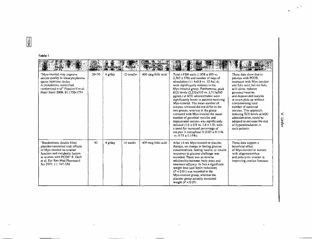
Table I.
"Myo·inosito\may improve 30+30 4 g/day 12 month~ 400 mcg folic acid Total r-FSH unit~ (1,958 ± 695 vs. These data show that in oocyte quality in intracytoplasmic 2,383 ± 578) and number of days of patient, with PCOS, sperm injection cycles. stimulation (11.4±0.9 vs. 12.4±1.4) trealment with Myo-ino~itol A prospective, controlled, were signilicantly reduced in the and folic acid, but not folic randomized trial" Papaleo E et al. Myo-inositol group. Furthcrmore, peak acid alone, reduces Fertil Steril 2009: 91: 1750-1754 E(2) levels (2.232±5IO vs. 2,713±595 gcrminal veside~
pglmL) at hCG administration were and degenerated oocytes significantly lower in patients receiving at ovum pick-up without Myo-inositol. The mean number of compromising total oocytes retrieved did not differ in the number of retrieved two groups. whereas in the group oocytes. This approach. cotreated with Myo-inositolthe mean reducing E(2) levels at hGC :< number of germinal vesicles and administration, could be C degenerated oocyte, was signilic,mtly adopted to decrease the risk ::J reduced (1.0 ± 0.9 vs. 1.6 ± 1.0). with of hyperstimulation in rD' a trend for i nereased percentage of such patients.
~
oocytes in metaphase 11 (0.82 ± 0.11 % vs. 0.75 ± 0.15%).
"Randomized, double blind 92 4 g/day 14 weeks 400 meg folic acid After 14-wk Myo-inositol or placebo Thesc data support a placcbo-controlled trial: effects therapy, no change in fasting glucose benclicial erfect of Myo-inositol on ovarian concentrations. fasting insulin, or insulin of Myo-ino~itol in women function and metabolic factors responses to glucose challenge was with oligomenorrhea in women with PCOS" S. Gedi recorded. There was an inverse and polycystic ovaries in et aI, Eur Rev Med Pharmacol relationship between hody mass and improving ovarian runction. Sci 2007: II: 347-354 treatment efficacy. In fact a significant
weight loss (and leptin reduction) (P < 0.01) was recorded in the Myo-inositol group, whereas the placebo group actually increased weight (P < 0.05).

• Polycystic ovary syndrome: a vitamin deficiency? Floating a new pathogenesis hypothesis
5) BURGHEN GA, GIVENS JR, KITABCHI AE. Correlation of hyperandrogenism with hyperinsulinism in polycystic ovarian disease. J Clin Endocrinol Metab 1980; 50: 113-116.
6) LARNER J. Multiple pathways in insulin signalling-fitting the covalent and allosteric puzzle pieces togheter. EndocrJ 1994; 2: 167-171.
7) BAILLARGEON JP, DIAMANTI-KANDARAKIS E, OSTLUND RE JR, APRIDONIDZE T, IUORNO MJ, NESTLER JE. Altered 0-chiro-inositol urinary clearance in women with polycystic ovary syndrome. Diabetes Care 2006; 29: 300-305.
8) luoRNo MJ, JAKUBOWICZ OJ, BAILLARGEON JP, DILLON P, GUNN RD, ALLAN G, NESTLER JE. Effects of d-chiro-inositol in lean women with the polycystic ovary syndrome. Endocr Pract 2002; 8(6): 417-423.
9) HASEGAWA I, MURAKAWA H, SUZUKI M. YAMAMOTO Y, KURABAYASHI T. TANAKA K. Effect of troglitazone on endocrine and ovulatory performance in women with insulin resistance-related polycystic ovary syndrome. Fertil Steril 1999; 71 (2): 323-327.
10) NESTLER JE, JAKUBOWICZ OJ, REAMER P, GUNN RD, ALLAN G. Ovulatory and metabolic effects of D-chiro-inositol in the polycystic ovary syndrome. N Engl J Med 1999; 340: 1314-1320.
11) GERLI S, MIGNOSA M, 01 RENZO GC. Effects of inositol on ovarian function and metabolic factors in women with PCOS: a randomized double blind placebo-controlled trial. Eur Rev Med Pharmacol Sci 2003; 7: 151-159.
12) NG EH, WAT NM, Ho PC. Effects of metformin on ovulation rate, hormonal and metabolic profiles in women with clomiphene-resistant polycystic ovaries: a randomized, double-blinded placebo-controlled trial. Hum Reprod 2001; 16(8): 1625-1631.
13) YOSHIDA K, YAMAGUCHI M, MORINAGA T, IKEUCHI M, KINEHARA M, ASHIDA H. Genetic modification of Bacillus subtilis for production of D-chiro-inositol, an investigation drug candidate for treatment of type 2 diabetes and polycystic ovary syndrome. Appl Environ Microbiol2006; 1310-1315.
v. Unfer AGUNCO Obstetrics & Gynecology Center,
Via G. Cassiani 15, Rome (Italy)