Megumi Hada1,2 and Alexandros G. Georgakilas3*
25
Review 1 2 3 4 5 6 7 8 9 10 11 Formation of clustered DNA Damage after High-LET Irradiation: A review Megumi Hada 1,2 and Alexandros G. Georgakilas 3* 1 NASA Johnson Space Center, Houston, Texas 77058, USA 2 Universities space Research Association, Houston, Texas 77058, USA 3 Department of Biology, Thomas Harriot College of Arts and Sciences, East Carolina University, Greenville NC 27858, USA * Corresponding author : Alexandros G. Georgakilas 12 13 14 Address: Biology Department, Howell Science Complex, East Carolina University, Greenville, NC. 15 Tel: 252-328-5446, Fax: 252-328-4178 16 17 18 19 20 21 e-mail: [email protected] 1
Transcript of Megumi Hada1,2 and Alexandros G. Georgakilas3*

Review 1
2
3
4
5
6
7
8
9
10
11
Formation of clustered DNA Damage after High-LET Irradiation: A review
Megumi Hada1,2 and Alexandros G. Georgakilas3*
1NASA Johnson Space Center, Houston, Texas 77058, USA
2Universities space Research Association, Houston, Texas 77058, USA
3Department of Biology, Thomas Harriot College of Arts and Sciences, East Carolina
University, Greenville NC 27858, USA
*Corresponding author: Alexandros G. Georgakilas 12
13
14
Address: Biology Department, Howell Science Complex, East Carolina University, Greenville,
NC.
15 Tel: 252-328-5446, Fax: 252-328-4178
16
17
18
19
20
21
e-mail: [email protected]
1
Formation of clustered DNA Damage after High-LET Irradiation: A review 1
2
3
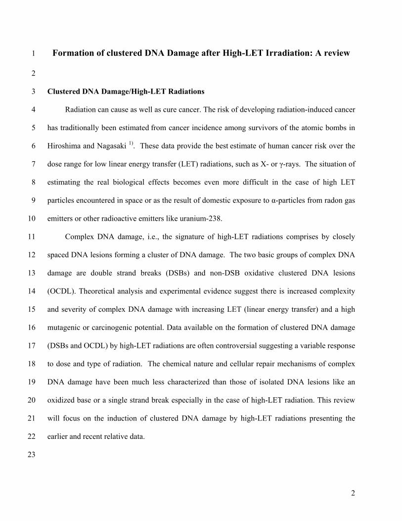
Clustered DNA Damage/High-LET Radiations
Radiation can cause as well as cure cancer. The risk of developing radiation-induced cancer
has traditionally been estimated
4
5 from cancer incidence among survivors of the atomic bombs in
Hiroshima and Nagasaki 1). These data provide the best estimate of human cancer risk over the
dose range for low linear energy transfer (LET) radiations, such as X- or γ-rays. The situation of
estimating the real biological effects becomes even more difficult in the case of high LET
particles encountered in space or as the result of domestic exposure to α-particles from radon gas
emitters or other radioactive emitters like uranium-238.
6
7
8
9
10
11
12
13
14
15
16
17
18
19
20
21
22
23
Complex DNA damage, i.e., the signature of high-LET radiations comprises by closely
spaced DNA lesions forming a cluster of DNA damage. The two basic groups of complex DNA
damage are double strand breaks (DSBs) and non-DSB oxidative clustered DNA lesions
(OCDL). Theoretical analysis and experimental evidence suggest there is increased complexity
and severity of complex DNA damage with increasing LET (linear energy transfer) and a high
mutagenic or carcinogenic potential. Data available on the formation of clustered DNA damage
(DSBs and OCDL) by high-LET radiations are often controversial suggesting a variable response
to dose and type of radiation. The chemical nature and cellular repair mechanisms of complex
DNA damage have been much less characterized than those of isolated DNA lesions like an
oxidized base or a single strand break especially in the case of high-LET radiation. This review
will focus on the induction of clustered DNA damage by high-LET radiations presenting the
earlier and recent relative data.
2
INTRODUCTION TO THE IDEA OF CLUSTERED DNA DAMAGE 1
2
3
4
5
6
7
8
9
10
11
12
13
14
15
16
17
18
19
20
21
22
23
The ‘idea’ of clustered DNA damage was first introduced by Ward as locally multiple damaged
sites (LMDS), i.e., several closely spaced damages within a short DNA segment that could be
produced by ionizing radiation 2,3). Ward introduced the idea of clustered DNA lesions to
account for the increased lethality induced by ionizing radiation, which cannot be fully explained
by the amount of double stand breaks formed, although the specific lesions, if unrepaired or
misrepaired, can lead to a lethal event.
Random energy deposition by ionizing radiation induces a wide variety of DNA lesions
[single (SSBs) and double strand breaks (DSBs), oxidized bases and apurinic-apyrimidinic
(abasic, AP) sites] 4). Ionizing radiation induces damage in DNA by direct ionization and through
generation of hydroxyl radicals that attack DNA and induce some or all of the above lesions
(indirectly). In addition to the prompt breaks induced by radiation, some post-irradiation ones
can be also formed as the result of the attempted repair of some sugar and base residues induced
directly which can later be converted to SSBs or DSBs 5). Two or more DNA lesions of the same
or different nature may be produced in close proximity to each other on opposite DNA strands
(bistranded lesions), generally within one-two helical turns of the DNA molecule. Theoretical 6)
and experimental studies 7) support the induction of these clustered DNA lesions in the cellular
environment or high scavenger conditions by a single radiation track and not by multiple track
events. These various closely spaced bistranded types of DNA damage are called clustered DNA
damages and can include SSBs of varying complexity, oxidized base lesions (oxypurines and
oxypyrimidines: oxybases) and regular as well as oxidized AP sites (Fig. 1). In general clustered
DNA damage can be separated to two major groups: DSBs and non-DSB oxidative clustered
DNA lesions, OCDL 8). Even at doses as low as ~1 Gy (100 rad) ionizing radiation is capable of
3
inducing all of the above types of DNA damage in the form of isolated lesions as well as in the
form of clustered ones (1-10 bp apart)
1
2
3
4
5
6
7
8
9
10
11
12
13
14
15
16
17
18
19
20
21
22
23
9,10). Except ionizing radiation, other radiomimetic drugs
like bleomycin or neocarzinostatin have been shown to induce DNA damage in a form of a
cluster explaining in part the high toxicity of these drugs 11,12). It has been shown that clustered
lesions constitute 50-80% of the total complex DNA damage 13). Simultaneous processing of the
lesions located on opposite strands may generate additional DSBs in addition to the ones directly
induced by ionizing radiation and enhance genomic instability 14,15). In fact, E. coli and rodent
cells do generate de novo DSBs that could result from processing of bistranded clustered DNA
lesions 1619). There are very limited data on the possible accumulation of OCDLs in human cells
or tissues and in mammalian/human tissues exposed to ionizing radiation 20). Gollapalle et al. 8)
showed for the first time that endogenous OCDL can be detected in non-irradiated mice tissues
at a steady state of ~0.3-0.8 clusters/Mbp and that exposure of these tissues to ionizing radiation
(X-rays) leads to an accumulation of DNA clusters detected 20 weeks after irradiation.
Measurement of endogenous OCDL in the human breast cancer line MCF-7 revealed elevated
levels compared with the non-malignant MCF-10A. These cluster frequencies are higher than
the ones detected for DNA isolated from human skin primary cell cultures (20-40 clusters/Gbp or
0.02-0.04 clusters/Mbp) 21). These yields discrepancies may be due to different cell types or
different isolation and enzyme-treatment methods for DNA analysis.
Cytotoxic effects of ionizing radiation are thought to result principally from incompletely or
incorrectly repaired DNA lesions 22). While isolated damages are generally repaired efficiently,
clustered DNA lesions have been suggested to be more difficult to repair, and in general are
considered as DNA damages that are repair-resistant or non-repairable with a high mutagenic
potential and, therefore, considered as highly significant biological endpoints 2,9). A significant
4
number of studies suggest that the mutation frequency increases the closer the spacing of the
clustered DNA lesions
1
2
3
4
5
6
7
8
9
10
11
12
13
14
15
16
17
18
19
20
21
22
23
2325). DNA clusters could be resistant to processing by glycosylases or
endonucleases, as shown for synthetic oligonucleotides containing clusters of specific
composition and configuration 2632). The majority of references on repair of clusters reflect
retarded enzymatic activity. Such repair-resistant clusters could persist for a substantial time
after irradiation 33,34).
PRINCIPLES OF MEASUREMENT OF CLUSTERED DNA LESIONS
Here we aim to give the principles of the approaches used from different laboratories for
the detection of bistranded clustered DNA lesions as well as the general description of the way
that they were evolved and fine-tuned for an efficient detection of closely spaced opposed
oxidative DNA lesions in DNA isolated from human or mammalian cells, genomic DNA like T7
or λ-DNA or supercoiled DNA 35,36).
A unique approach for quantifying these types of bistranded damage in human cells using
DNA repair enzymes isolated from E. coli was initially developed by Sutherland et al. 10). The
idea relies on the fact that repair enzymes participating in base excision repair (BER) like DNA
glycosylases and AP endonucleases will function also in vitro, i.e., on isolated DNA carrying
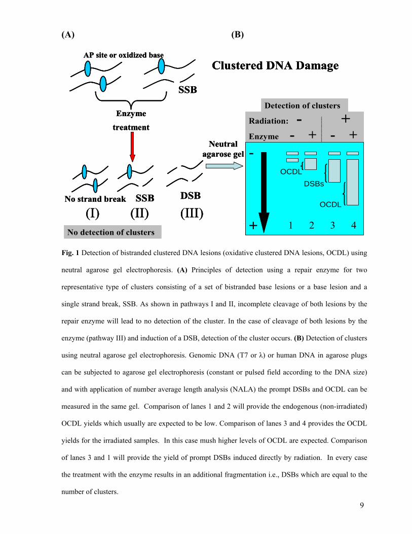
clustered lesions. Once they detect the lesion (Fig. 1 and Table 1) in each cluster, they will excise
it and cleave the DNA strand by their intrinsic lyase activity (DNA glycosylases) or cleave
directly in the case of an AP endonuclease and create a SSB in each strand, i.e., a DSB in the
case of a cluster (Fig. 1). As shown in Table 1, there is an overlapping specificity of Fpg,
EndoIII and EndoIV towards abasic sites as well as between Fpg and EndoIII towards some
types of oxidized bases. The additional DSBs induced by the repair enzymes can be measured
5
using constant field or pulsed field gel electrophoresis (PFGE) following number average length
analysis (NALA) according to the size of extracted DNA and are considered equal to the clusters
detected by the enzyme
1
2
3
4
5
6
7
8
9
10
11
12
13
14
15
16
17
18
19
20
21
22
23
35,37). Gels are stained with ethidium bromide, destained appropriately,
and an electronic image is obtained using usually a CCD camera. Electronic images can be then
processed using a densitometric software and a densitogram is obtained for each gel lane. A
DNA dispersion curve relating DNA length to electrophoretic mobility, based on all length
standards is determined from an analytical mobility function. From the profiles of irradiated and
unirradiated (or enzyme and untreated DNA populations), the number average length, ⎯L, of
each DNA distribution is calculated and the DSBs or clusters will be calculated as described in
(Fig. 1) 35).
In addition to the above approach using repair enzymes as damage probes, Georgakilas et
al. 30,33) have used polyamines (putrescine) for the detection of very closely spaced abasic (AP)
sites (1-5 bp apart), which are poorly detected by Nfo AP endonuclease. Based on the same
principle of enzyme-detection of clusters as enzyme sensitive sites, other groups have also used
neutral agarose gel electrophoresis and fraction of activity released (FAR) assay, hybridization
assay or plasmid nicking assay 38,39).
Finally, in an attempt of a second independent method different groups have developed a
modified version of the neutral single cell gel electrophoresis (Comet assay) using again repair
enzymes as damage probes 19,40). Since its early introduction in 1984 41) and later in its alkaline
version in 1988 42), the single-cell gel electrophoresis (SCGE) or Comet assay a modified version
of the microgel electrophoresis, has been widely used for the detection of low levels of various
types of DNA lesions including SSBs, DSBs and oxidized bases as reviewed in 43,44). The Comet
or microgel assay under neutral running conditions has been used for the detection of DSBs in a
6

variety of cells including lymphocytes 4548). To our knowledge there are very limited data on the
use of the neutral Comet
1
2
3
4
5
6
7
8
9
10
11
40,47) or microgel assay 15,19) for the detection of non-DSB clustered
DNA lesions using different repair enzymes as enzymatic probes. In all cases, incomplete
cleavage of lesions by the repair enzymes can lead to a detection of only a fraction of the clusters
(Fig. 1). This issue becomes very important especially in the case of high-LET radiations where
the density of the lesions is expected to be very high 49). The clusters described in Fig. 1 are an
idealized form a simple cluster. In reality and particularly in the case of high-LET, one would
expect a complex DNA lesion, for example a DSB with 5-10 additional surrounding lesions 20).
Therefore each cluster detected by the enzyme is only a fraction of the lesions participating in the
cluster.
7
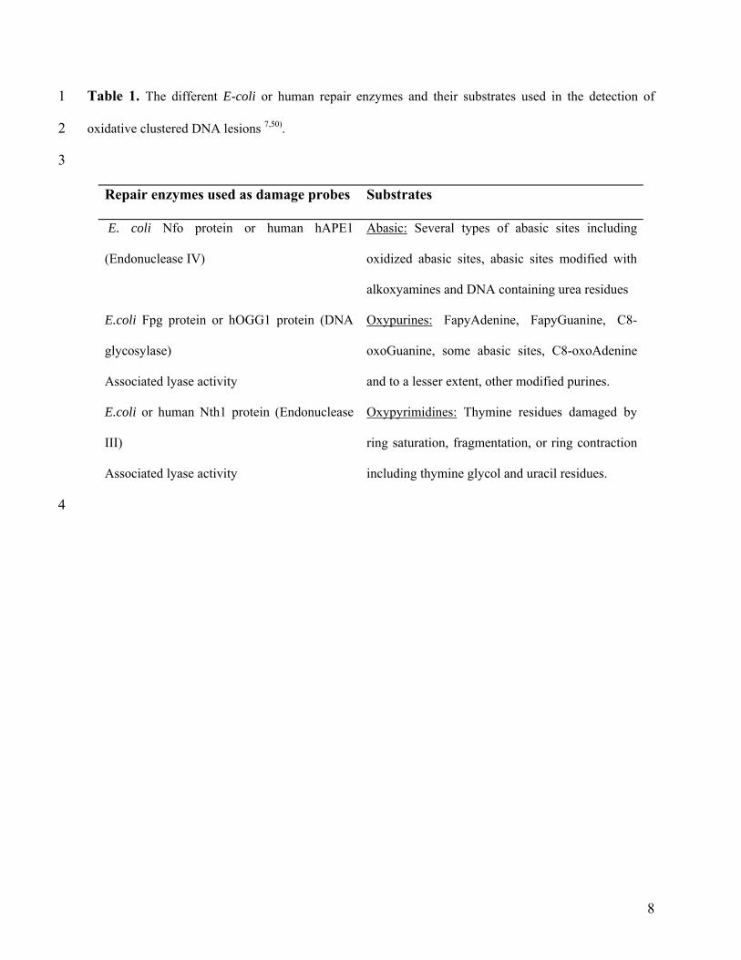
Table 1. The different E-coli or human repair enzymes and their substrates used in the detection of
oxidative clustered DNA lesions
1
2
3
7,50).
Repair enzymes used as damage probes Substrates
E. coli Nfo protein or human hAPE1
(Endonuclease IV)
Abasic: Several types of abasic sites including
oxidized abasic sites, abasic sites modified with
alkoxyamines and DNA containing urea residues
E.coli Fpg protein or hOGG1 protein (DNA
glycosylase)
Associated lyase activity
Oxypurines: FapyAdenine, FapyGuanine, C8-
oxoGuanine, some abasic sites, C8-oxoAdenine
and to a lesser extent, other modified purines.
E.coli or human Nth1 protein (Endonuclease
III)
Associated lyase activity
Oxypyrimidines: Thymine residues damaged by
ring saturation, fragmentation, or ring contraction
including thymine glycol and uracil residues.
4
8
(A) (B)
Radiation: - +Enzyme - + - +
DSBs
OCDL
OCDL
AP site or oxidized base
SSBNo strand break
Neutral agarose gel
DSB
No detection of clusters
(I) (II)
Enzyme
treatment
Detection of clusters
Clustered DNA Damage
1 2 3 4
-
+
SSB
(III)
Radiation: - +Enzyme - + - +
DSBs
OCDL
OCDL
AP site or oxidized base
SSBNo strand break
Neutral agarose gel
DSB
No detection of clusters
(I) (II)
Enzyme
treatment
Detection of clusters
Clustered DNA Damage
1 2 3 4
-
+
SSB
(III)
Radiation: - +Enzyme - + - +
DSBs
OCDL
OCDL
AP site or oxidized base
SSBNo strand break
Neutral agarose gel
DSB
No detection of clusters
(I) (II)
Enzyme
treatment
Detection of clusters
Clustered DNA Damage
1 2 3 4
-
+
SSB
(III)
Fig. 1 Detection of bistranded clustered DNA lesions (oxidative clustered DNA lesions, OCDL) using
neutral agarose gel electrophoresis. (A) Principles of detection using a repair enzyme for two
representative type of clusters consisting of a set of bistranded base lesions or a base lesion and a
single strand break, SSB. As shown in pathways I and II, incomplete cleavage of both lesions by the
repair enzyme will lead to no detection of the cluster. In the case of cleavage of both lesions by the
enzyme (pathway III) and induction of a DSB, detection of the cluster occurs. (B) Detection of clusters
using neutral agarose gel electrophoresis. Genomic DNA (T7 or λ) or human DNA in agarose plugs
can be subjected to agarose gel electrophoresis (constant or pulsed field according to the DNA size)
and with application of number average length analysis (NALA) the prompt DSBs and OCDL can be
measured in the same gel. Comparison of lanes 1 and 2 will provide the endogenous (non-irradiated)
OCDL yields which usually are expected to be low. Comparison of lanes 3 and 4 provides the OCDL
yields for the irradiated samples. In this case mush higher levels of OCDL are expected. Comparison
of lanes 3 and 1 will provide the yield of prompt DSBs induced directly by radiation. In every case
the treatment with the enzyme results in an additional fragmentation i.e., DSBs which are equal to the
number of clusters.
9

INDUCTION OF CLUSTERED DNA LESIONS BY HIGH-LET RADIATIONS 1
2
3
4
5
6
7
8
9
10
11
12
13
14
15
16
17
18
19
20
21
22
23
Theoretical considerations suggest that, in addition to isolated lesions, low-LET radiation can
create clusters with as many as 10 lesions 51). High-LET radiation is capable of producing
damage of even greater complexity, i.e., up to 25 lesions per cluster 51). Space travel
encompasses exposure to a broad spectrum of radiation ranging from the infrared to galactic
cosmic rays. The major component of galactic cosmic rays is the highly charged, energetic
(HZE) particles ranging from energetic protons to iron nuclei with energies ranging upwards to 1
GeV/nucleon; although of lower fluence than protons, large contributions to dose arise from Fe
and other very high LET nuclei, such as N, Si, etc. 52). Estimates of space radiation health risk
and the development of efficient countermeasures are key issues for manned space exploration.
Accurate risk calculations require a detailed investigation of both the physical aspects (patterns
of energy deposition at the molecular/cellular level) and the biological response to high LET
particles. The uncertainties in radiation risk assessment for deep-space missions are between 400
and 1,500% 53). These uncertainties are largely due to the lack of information on the biologic
response to HZE particles relative to the more extensively studied biological response to low
LET radiation. Late effects of high-LET radiation are arguably the health risk not only for the
human space exploration but also for increasing number of cancer patients treated by heavy-ion
therapy including young adults and children.
Measurement of DSBs for high LET radiations has been controversial 54). In addition, very
limited data exists for the efficacy of high-LET particles to induce OCDL in human cells
10,34,55,56). Many early studies on DSB induction by radiation of different qualities have shown an
increased yield of DSBs with increasing LET in non-cellular systems 57) while other later studies
have shown a decline of DSBs with increasing LET for SV40 DNA under high radio-quenching
10
conditions 58) or plasmid DNA 39) irradiated in radio-quenching conditions mimicking the cellular
chemical environment. For mammalian cells though, relative biological effectiveness (RBE)
values for DSB induction have been usually found close or a little bit higher to unity
1
2
3
4
5
6
7
8
9
10
11
12
13
14
15
16
17
18
19
20
21
22
23
59,60). The
exclusion of smaller DNA fragments (<10 kbp) during PFGE analysis of DSBs can have a
significant impact on the measurement of DSBs and OCDL for high-LET radiations 61).
Friedland et al. 61) using the PARTRAC Monte Carlo simulation code have shown that the DSB
yield can drop significantly i.e., RBE values smaller than unity if during DSB measurements
DNA fragments smaller than 10 kbp are excluded. Experimental studies have shown the
importance of small DNA fragments (<200 kbp) especially in the case of high-LET charged
particles 6265) due to the non-random distribution of breaks 6365). The advantage of using NALA,
especially in the case of high-LET radiations, is that this method does not require any specific
knowledge of the distribution of DNA fragments (e.g., random or Poisson-distributed cleavage)
37). Another significant parameter related to the detection of clustered DNA lesions are the heat-
labeled sites within locally multiply damaged sites which are produced by radiation and are
subsequently transformed into DSBs during the lysis procedure. These artifactual DSBs can
consist 30-50% of the real DSBs induced by radiation depending on the lysis conditions (37oC or
50oC) 14,66). .
In the case of non-DSB clusters the situations is clearer. A decrease has been detected for
OCDL yields and for high-LET radiations compared to low LET and RBE values much lower
than unity have been found by different groups using different DNA systems and methods
34,56,67,68). Theoretical analysis and specifically Monte Carlo simulations predict that the initial
yield of clusters other than the DSB tends to decrease with increasing particle LET, which is
consistent with experimental observations 69). In addition, several in vitro studies using different
11

DNAs in radio-quenching solutions indicate a similar decrease of clusters with increasing LET
summarized also in Table 2
1
2
3
4
5
6
7
8
9
10
11
12
13
14
15
16
17
18
34,68,70,71). This decrease could be the result of an actual decline in
the induced oxybase lesions 72) and SSBs 39,73) for high LET charged particles associated with
decreased formation of radicals in the solution by ions of higher LET 74), or an inability of the
methods to detect these highly dense clusters. By the last comment, we refer to the already
established compromised ability of repair enzymes to detect and cleave very closely spaced
DNA lesions 29,33,75). Earlier studies using enzymatic (Fpg and/or Nth) or cell extract treatment
of γ- or α-irradiated plasmid DNA under cell mimetic conditions suggest an increase of the
contribution of base lesions to clustered DNA damage with LET 76). Finally and related to all the
gel electrophoresis based methods for detection of clusters, an important issue that should be
mentioned here is that even if the enzymes cleave the resulting DNA fragments (<1 kbp) will
remain undetected under the most current electrophoresis separation regimens applied as
discussed above. Two general trends can be mentioned: i) For DSBs, a small dependence to
LET with a tendency to increase at least for LET values 1-150 keV/µm, for non-DSB clusters a
decline with LET and a linear dependence on dose for both DSBs and OCDL and ii) In most
cases, non-DSB cluster yields (Fpg-, EndoIII- and EndoIV-clusters) tend to be lower than the
corresponding yields of prompt DSBs for high-LET radiations. For low-LET radiations, the
non-DSB clusters tend to be higher compared to prompt DSBs (Table 2).
12

Table 2. Data available on the dependence of non-DSB clustered DNA damage to LET. The yields of
DSBs and non-DSB clusters have been included. The different types of clusters are presented according
to the type of repair enzyme used for the detection i.e., Fpg-, EndoIII and EndoIV-clusters. Yields are
presented as lesions/Gbp/Gy.
1
2
3
4
5
LET
(keV/μm)
Radiation DSBs
/Gbp/Gy
Fpg-
/Gbp/Gy
EndoIII-
/Gbp/Gy
EndoIV-
/Gbp/Gy
Model System
1
110
60Co γ-rays 238Pu α-particles
15
25
48
15
Plasmid pMSG-CAT
10 mM Tris
Ref. 71)
1
110
60Co γ-rays 238Pu α-particles
7.1
15
29.9
5
Plasmid pMSG-CAT
200 mM Tris
Ref. 71)
0.3
97
145
1440
137Cs γ-rays 244Cm α-particles
56Fe 197Au
0.81 x 10³
071 x 103
0.31 x 10³
0.25 x 10³
2.35 x 10³
0.35 x 10³
Plasmid pEC
10 mM phosphate buffer
Ref. 77)
0.225
0.3
2
12.97
50.32
107.7
150.4
1H 137Cs γ-rays
X-ray 12C 28Si 48Ti 56Fe
9.56 x 10³
3.78 x 10³
5.62 x 10³
3.72 x 10³
3.57 x 10³
2.81 x 10³
1.95 x 10³
6.67 x 10³
10.05 x 10³
4.37 x 10³
2.85 x 10³
2.42 x 10³
2.35 x 10³
4.09 x 10³
5.34 x 10³
2.04 x 10³
1.27 x 10³
1.30 x 10³
1.22 x 10³
T7 DNA in PBS
Ref. 68)
0.3
148
137Cs γ-rays 56Fe
11.9
10.9
10.68
7.12
11.87
8.54
9.5
5.5
Human monocytes
Ref. 34)
6 7
13
Hada and Sutherland 68) and Tsao et al. 34) have also shown a prevalence of Fpg-clusters to
Endo IV (Nfo)-or EndoIII-clusters for T7 or human DNA irradiated with
1
2
3
4
5
6
7
8
9
10
11
12
13
14
15
16
17
18
19
20
21
22
23
56Fe ions under radio-
quenching conditions (Table 2). As mentioned earlier, another effect that has to be taken into
consideration is the conversion of heat labile sites to strand breaks 78). It is a possible source of
overestimation of DSBs and non-DSB clusters and, especially in the case of γ rays, the
artifactual conversion of heat labile sites into DSB can result in the underestimation of RBE
values for Fe ions 59). In general, and as also stated by Boucher et al. 79), the role of artifactual
DNA oxidation is very important but based on the above different group data, its role in the case
of high-LET radiations is expected to be minimum. Finally as shown in Table 1, the overlapping
specificity of the different repair enzymes is another potential source of overestimation in the
measurement of total number of non-DSB clusters that can reach in some cases even a factor of
two 40).
CONCLUDING COMMENTS
Although a significant amount of data has been produced on the efficiency of high-LET
radiations to induce clustered DNA damage, still many questions remain unanswered.
Significant deviations appear between theoretical and experimental data. Therefore, we believe
that the primary objectives of the future studies should be to i) provide analytical, quantitative,
and theoretical information on the induction and processing of different patterns of complex
DNA damage (DSB and non-DSB clustered DNA lesions) produced directly and indirectly
(bystander effects) by high-LET radiations in vivo and in vitro and ii) measure the biological
effectiveness of high-LET radiations (like space HZE particles or α-particles) for the induction of
chromosome damage and apoptosis in vivo and in vitro and correlate these endpoints with the
14
1
2
3
4
5
6
7
8
9
10
11
12
levels of complex DNA damage detected in each case, and finally iii) improve the experimental
detection methods for including the very small DNA fragments expected to be induced in the
case of high-LET radiations. A better knowledge of the induction and repair of complex DNA
damage is essential for the calculations of the risk factors of high LET radiations to induce
carcinogenesis (as in the case of radon-gas emitted α-particles, heavy-ion therapy or space
radiation).
ACKNOWLEDGEMENTS
This work has been supported by funds provided to Dr. Georgakilas by the Biology Department
of East Carolina University, a Research/Creative Activity Grant and a College Research Award
to A. Georgakilas (East Carolina University).
15
REFERENCES 1
2
3
4
5
6
7
8
9
10
11
12
13
14
15
16
17
18
19
20
21
22
1. Preston, D. L. and Piece, D. A. (2003) Studies of mortality of atomic bomb survivors.
Report 13: Solid cancer and noncancer disease mortality: 1950-1997. Radiat. Res. 160:
381-407.
2. Ward, J. F. (1981) Some biochemical consequences of the spatial distribution of ionizing
radiation produced free radicals. Radiat. Res. 86: 185-195.
3. Ward, J. F. (1985) Biochemistry of DNA lesions. Radiat. Res. 104: S103-S111.
4. Ward, J. F. (1994) The complexity of DNA damage: relevance to biological
consequences. Int. J. Radiat. Biol. 66: 427-432.
5. Wallace, S. S. (1998) Enzymatic Processing of Radiation-Induced Free Radical Damage
in DNA. Radiat. Res. 150 (suppl.): S60-S79.
6. Goodhead, D. T., Thacker, J. and Cox, R. (1993) Effects of radiations of different
qualities on cells: Molecular mechanism of damage and repair. Int. J. Radiat. Biol.
63(543-556).
7. Sutherland, B. M., Bennett, P. V., Sidorkina, O. and Laval, J. (2000) Clustered Damages
and Total Lesions Induced in DNA by Ionizing Radiation: Oxidized Bases and Strand
Breaks. Biochemistry 39: 8026-8031.
8. Gollapalle, E., Wong, R., Adetolu, R., Tsao, D., Francisco, D., Sigounas, G. and
Georgakilas, A. G. (2007) Detection of oxidative clustered DNA lesions in X-irradiated
mouse skin tissues and human MCF-7 breast cancer cells. Radiat. Res. 167: 207-216.
9. Goodhead, D. T. (1994) Initial events in the cellular effects of ionizing radiations:
clustered damage in DNA. Int. J. Radiat. Biol. 65: 7-17.
16
1
2
3
4
5
6
7
8
9
10
11
12
13
14
15
16
17
18
19
20
21
22
10. Sutherland, B., Bennett, P. V., Sidorkina, O. and Laval, J. (2000) DNA Damage Clusters
Induced by Ionizing Radiation in Isolated DNA and in Human Cells. Proc. Natl. Acad.
Sci. USA 97: 103-108.
11. Povirk, L. F. and Houlgrave, C. W. (1988) Effect of apurinic/apyrimidinic endonucleases
and polyamines on DNA treated with bleomycin and neocarzinostatin: Specific formation
and cleavage at closely opposed lesions in complementary strands. Biochemistry 27:
3850-3857.
12. Regulus, P., Duroux, B., Bayle, P. A., Favier, A., Cadet, J. and Ravanat, J. L. (2007)
Oxidation of the sugar moiety of DNA by ionizing radiation or bleomycin could induce
the formation of a cluster DNA lesion. Proc. Natl. Acad. Sci. USA 104: 14032-14037.
13. Sutherland, B. M., Bennett, P. V., Sutherland, J. C. and Laval, J. (2002) Clustered DNA
damages induced by X-rays in human cells. Radiat. Res. 157: 611-616.
14. Gulston, M., C., d. L., Jenner, T., Davis, E. and O'Neill, P. (2004) Processing of clustered
DNA damage generates additional double-strand breaks in mammialian cells post-
irradiation. Nucleic Acids Res. 32: 1602-1609.
15. Yang, N., Chaudhry, M. A. and Wallace, S. S. (2006) Base excision repair by hNTH1
and hOGG1: a two edged sword in the processing of DNA damage in gamma-irradiated
human cells. DNA Repair 5: 43-51.
16. Dugle, D., Gillespie, C. and Chapman, J. D. (1976) DNA strand breaks, repair and
survival in X-irradiated mammalian cells. Proc. Natl. Acad. Sci. U. S. A. 73(809-812).
17. Ahnstrom, G. and Bryant, P. E. (1982) DNA double-strand breaks generated by the repair
of X-ray damage in Chinese hamster cells. Int. J. Radiat. Biol. 41: 671-676.
17

1
2
3
4
5
6
7
8
9
10
11
12
13
14
15
16
17
18
19
20
21
22
18. Bonura, T. and Smith, K. C. (1975) Enzymatic production of deoxyribonucleic acid
double-strand breaks after ultraviolet irradiation of Escherichia coli K12. J. Bacteriol.
121: 511-517.
19. Blaisdell, J. O. and Wallace, S. (2001) Abortive base-excision repair of radiation-induced
clustered DNA lesions in Escherichia coli. Proc. Natl. Acad. Sci. USA 98: 7426-30.
20. Georgakilas, A. G. (2008) Processing of DNA damage clusters in human cells: Current
status of knowledge Mol. Biosyst. 4: 30-35.
21. Bennett, P. V., Cuomo, N. L., Paul, S., Tafrov, S. T. and Sutherland, B. M. (2005)
Endogenous DNA damage clusters in human skin, 3-D model and cultured skin cells.
Free Radic. Biol. Med. 39: 832-839.
22. Lindahl, T. and Wood, R. D. (1999) Quality control of DNA repair. Science 286: 1897-
1905.
23. Pearson, C. G., Shikazono, N., Thacker, J. and O'Neill, P. (2004) Enhanced mutagenic
potential of 8-oxo-7,8-dihydroguanine when present within a clustered DNA damage site.
Nucleic Acids Res. 32: 263-270.
24. Malyarchuk, S., Brame, K. L., Youngblood, R., Shi, R. and Harrison, L. (2004) Two
clustered 8-oxo-7,8-dihydroguanine (8-oxodG) lesions increase the point mutation
frequency of 8-oxodG, but do not result in double strand breaks or deletions in
Escherichia coli. Nucleic Acids Res. 32: 5721-5731.
25. Cunniffe, S. M., Lomax, M. E. and O'Neill, P. (2007) An AP site can protect against the
mutagenic potential of 8-oxoG when present within a tandem clustered site in E. coli.
DNA Repair doi:10.1016/j.dnarep.2007.07.003.
18

1
2
3
4
5
6
7
8
9
10
11
12
13
14
15
16
17
18
19
20
21
22
23
26. Chaudhry, M. A. and Weinfeld, M. (1997) Reactivity of human apurinic/apyrimidinic
endonuclease and Escherichia coli exonuclease III with bistranded abasic sites in DNA.
J. Biol. Chem. 272(25): 15650-15655.
27. Harrison, L., Hatahet, Z. and Wallace, S. (1999) In Vitro Repair of Synthetic Ionizing
Radiation-induced Multiply Damaged DNA Sites. J. Mol. Biol. 290: 667-684.
28. McKenzie, A. A. and Strauss, P. R. (2001) Oligonucleotides with bistranded abasic sites
interfere with substrate binding and catalysis by human apurinic/apyrimidinic
endonuclease. Biochemistry 40: 13254-13261.
29. David-Cordonnier, M. H., Cunniffe, S. M. T., Hickson, I. D. and O'Neill, P. (2002)
Efficiency of incision of an AP site within clustered DNA damage by the major human
AP endonuclease. Biochemistry 41: 634-642.
30. Georgakilas, A. G., Bennett, P. V. and Sutherland, B. M. (2002) High efficiency
detection of bistranded abasic clusters in g-irradiated DNA by putrescine. Nucleic Acids
Res. 30: 2800-2808.
31. Lomax, M. E., Cunniffe, S. and O'Neill, P. (2004) 8-OxoG retards the activity of the
ligase III/XRCC1 complex during the repair of a single-strand break, when present within
a clustered DNA damage site. DNA Repair 3: 289-299.
32. Eot-Houllier, G., Eon-Marchais, S., Gasparutto, D. and Sage, E. (2005) Processing of a
complex multiply damaged DNA site by human cell extracts and purified repair proteins.
Nucleic Acids Res. 33: 260-271.
33. Georgakilas, A. G., Bennett, P. V., Wilson III, D. M. and Sutherland, B. M. (2004)
Processing of bistranded abasic DNA clusters in gamma-irradiated human hematopoietic
cells. Nucleic Acids Res. 32: 5609-5620.
19
34. Tsao, D., Kalogerinis, P., Tabrizi, I., Dingfelder, M., Stewart, R. D. and Georgakilas, A.
G. (2007) Induction and Processing of clustered DNA lesions in human monocytes
exposed to low doses of HZE
1
2
3
4
5
6
7
8
9
10
11
12
13
14
15
16
17
18
19
20
21
22
56Fe particles. Radiat. Res. 168: 87-97.
35. Sutherland, B. M., Georgakilas, A. G., Bennett, P. V., Laval, J. and Sutherland, J. C.
(2003) Quantifying clustered DNA damage induction and repair by gel electrophoresis,
electronic imaging and number average length analysis. Mutat. Res. (Review) 531: 93-
107.
36. Terato, H. and Ide, H. (2004) Clustered DNA damage induced by heavy ion particles.
Biol. Sci. Space 18: 206-215.
37. Sutherland, B. M., Bennett, P. V., Georgakilas, A. G. and Sutherland, J. C. (2003)
Evaluation of Number Average Length Analysis In Quantifying Double Strand Breaks in
Genomic DNAs. Biochemistry 42: 3375-3384.
38. Gulston, M., Fulford, J., Jenner, T., de Lara, C. and O'Neill, P. (2002) Clustered DNA
damage induced by g radiation in human fibroblasts (HF 19), hamster (V79-4) cells and
plasmid DNA is revealed as Fpg and Nth sensitive sites. Nucleic Acids Res. 30: 3464-
3472.
39. Leloup, C., Garty, G., Assaf, G., Cristovao, A., Breskin, A., Chechik, R., Shchemelinin,
S., Paz-Elizur, T., Livneh, Z., Schulte, R. W., Bashkirov, V., Milligan, J. R. and
Grosswendt, B. (2005) Evaluation of lesion clustering in irradiated plasmid DNA. Int. J.
Radiat. Biol. 81: 41-54.
40. Holt, S. M. and Georgakilas, A. G. (2007) Detection of complex DNA damage in γ-
irradiated acute lymphoblastic leukemia pre-B NALM-6 cells. Radiat. Res. 168: 527-534.
20

1
2
3
4
5
6
7
8
9
10
11
12
13
14
15
16
17
18
19
20
21
22
41. Ostling, O. and Johanson, K. J. (1984) Microelectrophoretic study of radiation-induced
DNA damages in individual mammalian cells. Biochem. Biophys. Res. Commun. 123:
291-298.
42. Singh, N. P., McCoy, M. T., Tice, R. R. and Schneider, E. L. (1998) A simple technique
for quantitation of low levels of DNA damage in individual cells. Exp. Cell Res. 175:
184-191.
43. Olive, P. L. and Banath, J. P. (2006) The comet assay: a method to measure DNA
damage in individual cells. Nat. Protoc. 1: 23-29.
44. Collins, A. R. (2004) The Comet assay for DNA damage and repair: Principles,
applications and limitations (Review). Molecular Biotechnology 26: 249-261.
45. Trenz, K., Schutz, P. and Speit, G. (2005) Radiosensitivity of lymphoblastoid cell lines
with a heterozygous BRCA1 mutation is not detected by the comet assay and pulsed field
gel electrophoresis. Mutagenesis 20: 131-137.
46. Yang, N., Galick, H. and Wallace, S. S. (2004) Attempted base excision repair of
ionizing radiation damage in human lymphoblastoid cells produces lethal and mutagenic
double strand breaks. DNA Repair 3: 1323-1334.
47. Angelis, K. J., Dusinska, M. and Collins, A. R. (1999) Single cell gel electrophoresis:
detection of DNA damage at different levels of sensitivity. Electrophoresis 20: 2133-
2138.
48. Olive, P. L. and Banath, J. P. (1993) Detection of DNA double-strand breaks through the
cell cycle after exposure to X-rays, bleomycin, etoposide and 125IdUrd. Int. J. Rad. Biol.
64: 349-358.
21
1
2
3
4
5
6
7
8
9
10
11
12
13
14
15
16
17
18
19
20
21
22
49. Nikjoo, H., O' Neill, P., Wilson, E. W. and Goodhead, D. (2001) Computational approach
for determing the spectrum of DNA damage induced by ionizing radiation. Radiat. Res.
156: 577-583.
50. Wilson , D. M., Sofinowski, T. M. and McNeill, D. R. (2003) Repair mechanisms for
oxidative DNA damage. Frontiers in Bioscience 8: 963-981.
51. Semenenko, V. A. and Stewart, R. D. (2004) A fast Monte Carlo algorithm to simulate
the spectrum of DNA damages formed by ionizing radiation. Radiat. Res. 161: 451-457.
52. Ponomarev, A. L., Cucinotta, F. A., Sachs, R. K. and Brenner, D. J. (2001) Monte Carlo
predictions of DNA fragment-size distributions for large sizes after HZE particle
irradiation. Physica Medica XVII: 153-156.
53. NASA (1998). Strategic program plan for space radiation health research.
Washington,DC, NASA.
54. Prise, K. M., Pinto, M., Newman, H. C. and Michael, B. D. (2001) A review of studies of
ionizing radiation-induced double-strand break clustering. Radiat Res 156: 572-576.
55. Terato, H., Tanaka, R., Nakaarai, Y., Furusawa, Y. and Ide, H. (2004) Analysis of DNA
damage generated by high-energy particles. Nucleic Acids Symp. Ser. (Oxf). 48: 145-
146.
56. Terato, H., Tanaka, R., Nakaarai, Y., Hirayama, R., Furusawa, Y. and Ide, H. (2007)
Analysis of complex DNA lesions generated by heavy ion beams. Nucleic Acids Symp.
Ser. (Oxf) 51(1): 221-222.
57. Neary, G. J., Horgan, V. J., Bance, D. A. and Stretch, A. (1972) Further data on DNA
strand breakage by various radiation qualities. Int. J. Radiat. Biol. 22: 525-537.
22

1
2
3
4
5
6
7
8
9
10
11
12
13
14
15
16
17
18
19
20
21
22
23
58. Taucher-Scholz, G. and Kraft, G. (1999) Influence of radiation quality on the yield of
DNA strand breaks in SV40 DNA irradiated in solution. Radiat. Res. 151: 595-604.
59. Belli, M., Campa, A., Dini, V., Esposito, G., Furusawa, Y., Simone, G., Sorrentino, E.
and Tabocchini, M. A. (2006) DNA fragmentation induced in human fibroblasts by
accelerated 56Fe ions of differing energies. Radiat Res 165: 713-720.
60. Prise, K. M., Ahnstrom, G., Belli, M., Carlsson, J., Frankenberg, D., Kiefer, J., Lobrich,
M., Michael, B. D., Nygren, J., Simone, G. and Stenerlow, B. (1998) A review of dsb
induction data for varying quality radiation. Int. J. Radiat. Biol. 74: 173-184.
61. Friedland, W., Dingfelder, M., Jacob, P. and Paretzke, H. G. (2005) Calculated DNA
double-strand break and fragmentation yields after irradiation with He ions. Radiat. Phys.
Chem. 72: 279-286.
62. Ponomarev, A. L. and Cucinotta, F. A. (2006) Chromatin loops are responsible for higher
counts of small DNA fragments induced by high-LET radiation, while chromosomal
domains do not affect the fragment sizes. Int. J. Radiat. Biol. 82: 293-305.
63. Friedland, W., Jacob, P., Paretzke, H. G. and Stork, T. (1998) Monte Carlo simulation of
the production of short DNA fragments by low-linear energy transfer radiation using
higher-order DNA models. Radiat. Res. 150: 170-182.
64. Lobrich, M., Cooper, P. K. and Rydberg, B. (1996) Non-random distribution of DNA
double-strand breaks induced by particle irradiation. Int. J. Radiat. Biol. 70: 493-503.
65. Rydberg, B. (1996) Clusters of DNA Damage Induced by Ionizing Radiation: Formation
of Short DNA Fragments. II. Experimental Detection. Radiation Research 145: 200-209.
66. Rydberg, B., ouml and rn (2000) Radiation-Induced Heat-Labile Sites that Convert into
DNA Double-Strand Breaks. Radiation Research 153(6): 805-812.
23
1
2
3
4
5
6
7
8
9
10
11
12
13
14
15
16
17
18
19
20
21
67. Sutherland, B. M., Bennett, P., Georgakilas, A. G. and Hada, M. (2003). Bistranded DNA
Damage Clusters Induced by Low LET Radiation and Heavy Charged Particles:
Formation and Repair. 14th Annual NASA Space Radiation Investigator’s Workshop,
April 27-30, Houston, Texas, DOE/NASA.
68. Hada, M. and Sutherland, B. M. (2006) Spectrum of complex DNA damages depends on
the incident radiation. Radiat. Res. 165: 223-230.
69. Semenenko, V. A. and Stewart, R. D. (2006) Fast Monte Carlo simulation of DNA
damage formed by electrons and light ions. Phys. Med. Biol. 51: 1693-1706.
70. Milligan, J. R., Aguilera, J. A., Nguyen, T. T., Paglinawan, R. A. and Ward, J. F. (2000)
DNA strand-break yields after post-irradiation incubation with base excision repair
endonucleases implicate hydroxyl radical pairs in double-strand break formation. Int. J.
Radiat. Biol. 76: 1475-83.
71. Prise, K. M., Pullar, C. H. and Michael, B. D. (1999) A study of endonuclease III-
sensitive sites in irradiated DNA: detection of alpha-particle-induced oxidative damage.
Carcinogenesis 20: 905-909.
72. Pouget, J.-P., Ravanat, J.-L., Douki, T., Richard, M.-J. and Cadet, J. (1999) Measurement
of DNA base damage in cells exposed to low doses of gamma-radiation: comparison
between the HPLC-EC and comet assays. Int. J. Radiat. Biol. 75: 51-58.
73. Georgakilas, A. G., Haveles, K. S., Sophianopoulou, V., Sakelliou, L., Zarris, G. and
Sideris, E. G. (2000) alpha-Particle-induced changes on the stability and size of DNA.
Radiat. Res. 153: 258-262.
24
1
2
3
4
5
6
7
8
9
10
11
12
13
14
15
16
17
18
19
20
74. Moritake, T., Tsuboi, K., Anzai, K., Ozawa, T., Ando, K. and Nose, T. (2003) ESR spin
trapping of hydroxyl radicals in aqueous solution irradiated with high-LET carbon-ion
beams Radiat. Res. 159: 670-675.
75. David-Cordonnier, M. H., Boiteux, S., and and O'Neill, P. (2001) Efficiency of excision
of 8-oxo-guanine within DNA clustered damage by XRS5 nuclear extracts and purified
human OGG1 protein. Biochemistry 40: 11811-11818.
76. Jenner, T. J., Fulford, J. and Neill, P. (2001) Contribution of Base Lesions to Radiation-
Induced Clustered DNA Damage: Implication for Models of Radiation Response. Radiat.
Res. 156(5): 590-593.
77. Milligan, J. R., Aguilera, J. A., Paglinawan, R. A., Ward, J. F. and Limoli, C. L. (2001)
DNA strand break yields after post-high LET irradiation incubation with endonuclease-
III and evidence for hydroxyl radical clustering. Int. J. Radiat. Biol. 77: 155-164.
78. Stenerlow, B., Karlsson, K. H., Cooper, B. and Rydberg, B. (2003) Measurement of
prompt DNA double-strand breaks in mammalian cells without including heat-labile
sites: results for cells deficient in nonhomologous end joining. Radiat. Res. 159: 502-510.
79. Boucher, D., Testard, I. and Averbeck, D. (2006) Low levels of clustered oxidative DNA
damage induced at low and high LET irradiation in mammalian cells. Radiat. Environ.
Biophys. 45: 267-276.
25


![Tzakia Alexandros brochure 2016 - pages 16]j](https://static.fdocuments.us/doc/165x107/58ec3e3b1a28ab40458b45b7/tzakia-alexandros-brochure-2016-pages-16j.jpg)