Megalocornea - webeye.ophth.uiowa.edu · 16.09.2010 · bilateral enlargement of the corneal...
6
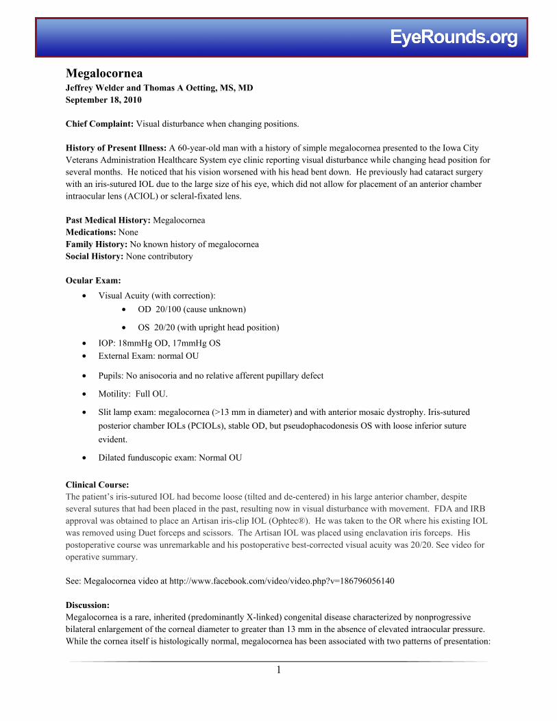
Megalocornea Jeffrey Welder and Thomas A Oetting, MS, MD September 18, 2010 Chief Complaint: Visual disturbance when changing positions. History of Present Illness: A 60-year-old man with a history of simple megalocornea presented to the Iowa City Veterans Administration Healthcare System eye clinic reporting visual disturbance while changing head position for several months. He noticed that his vision worsened with his head bent down. He previously had cataract surgery with an iris-sutured IOL due to the large size of his eye, which did not allow for placement of an anterior chamber intraocular lens (ACIOL) or scleral-fixated lens. Past Medical History: Megalocornea Medications: None Family History: No known history of megalocornea Social History: None contributory Ocular Exam: • Visual Acuity (with correction): • OD 20/100 (cause unknown) • OS 20/20 (with upright head position) • IOP: 18mmHg OD, 17mmHg OS • External Exam: normal OU • Pupils: No anisocoria and no relative afferent pupillary defect • Motility: Full OU. • Slit lamp exam: megalocornea (>13 mm in diameter) and with anterior mosaic dystrophy. Iris-sutured posterior chamber IOLs (PCIOLs), stable OD, but pseudophacodonesis OS with loose inferior suture evident. • Dilated funduscopic exam: Normal OU Clinical Course: The patient’s iris-sutured IOL had become loose (tilted and de-centered) in his large anterior chamber, despite several sutures that had been placed in the past, resulting now in visual disturbance with movement. FDA and IRB approval was obtained to place an Artisan iris-clip IOL (Ophtec®). He was taken to the OR where his existing IOL was removed using Duet forceps and scissors. The Artisan IOL was placed using enclavation iris forceps. His postoperative course was unremarkable and his postoperative best-corrected visual acuity was 20/20. See video for operative summary. See: Megalocornea video at http://www.facebook.com/video/video.php?v=186796056140 Discussion: Megalocornea is a rare, inherited (predominantly X-linked) congenital disease characterized by nonprogressive bilateral enlargement of the corneal diameter to greater than 13 mm in the absence of elevated intraocular pressure. While the cornea itself is histologically normal, megalocornea has been associated with two patterns of presentation: 1
Transcript of Megalocornea - webeye.ophth.uiowa.edu · 16.09.2010 · bilateral enlargement of the corneal...
Megalocornea Jeffrey Welder and Thomas A Oetting MS MD September 18 2010
Chief Complaint Visual disturbance when changing positions
History of Present Illness A 60-year-old man with a history of simple megalocornea presented to the Iowa City Veterans Administration Healthcare System eye clinic reporting visual disturbance while changing head position for several months He noticed that his vision worsened with his head bent down He previously had cataract surgery with an iris-sutured IOL due to the large size of his eye which did not allow for placement of an anterior chamber intraocular lens (ACIOL) or scleral-fixated lens
Past Medical History Megalocornea Medications None Family History No known history of megalocornea Social History None contributory
Ocular Exam
bull Visual Acuity (with correction)
bull OD 20100 (cause unknown)
bull OS 2020 (with upright head position)
bull IOP 18mmHg OD 17mmHg OS
bull External Exam normal OU
bull Pupils No anisocoria and no relative afferent pupillary defect
bull Motility Full OU
bull Slit lamp exam megalocornea (gt13 mm in diameter) and with anterior mosaic dystrophy Iris-sutured
posterior chamber IOLs (PCIOLs) stable OD but pseudophacodonesis OS with loose inferior suture
evident
bull Dilated funduscopic exam Normal OU
Clinical Course The patientrsquos iris-sutured IOL had become loose (tilted and de-centered) in his large anterior chamber despite several sutures that had been placed in the past resulting now in visual disturbance with movement FDA and IRB approval was obtained to place an Artisan iris-clip IOL (Ophtecreg) He was taken to the OR where his existing IOL was removed using Duet forceps and scissors The Artisan IOL was placed using enclavation iris forceps His postoperative course was unremarkable and his postoperative best-corrected visual acuity was 2020 See video for operative summary
See Megalocornea video at httpwwwfacebookcomvideovideophpv=186796056140
Discussion Megalocornea is a rare inherited (predominantly X-linked) congenital disease characterized by nonprogressive bilateral enlargement of the corneal diameter to greater than 13 mm in the absence of elevated intraocular pressure While the cornea itself is histologically normal megalocornea has been associated with two patterns of presentation
1
1) simple isolated megalocornea with no additional ocular or systemic manifestations and 2) megalocornea with other associated ocular and systemic abnormalities including megalophthalmos anterior andor dysgenesis of the iris lens or ciliary body A large cornea can also been seen in buphthalmos as in congenital glaucoma (diffuse enlargement of the eye) but is considered a separate disease and should not be confused with megalocornea
The definitive cause of megalocornea is currently unknown A number of mechanisms have been proposed including failure of anterior cup fusion during embryogenesis It has been hypothesized that this allows more room for corneal growth during development (Mann 1957) The findings of normal endothelial cell density and posterior placement of the iris-lens diaphragm seem to support the ldquocup fusionrdquo theory Embryologically the cornea forms by growing anteriorly from its base near the ciliary ring If the diameter of the ciliary ring is enlarged the cornea will also be enlarged In megalocornea hyperplasia of the cornea is observed which implies synchronous growth of all corneal layers and actively proliferating endothelial cells Skuta and colleagues point out that this type of primary overgrowth in megalocornea results in normal endothelial cell density as opposed to decreased endothelial cell density which would be seen in a cornea that was enlarged from secondary distension related to congenital glaucoma (Skuta 1983)
Megalocornea is known to be a genetic disorder with over 90 of cases being X-linked recessive with a gene locus indentified in band Xq12-q26 (most likely q21-q23) (Meire 1991 OMIM 2000) As such this is primarily a disease affecting males
Physical findings of simple megalocornea include bilateral corneal enlargement most often between 13-165 mm a dome-shaped cornea with normal thickness and occasional central mosaic dystrophy increased anterior chamber depth posterior positioning of the lens-iris diaphragm shortened vitreous length and normal intraocular pressure (Meire and Delleman 1994)
Other key conditions in the differential diagnosis of congenital corneal enlargement include megalophthalmos anterior and primary congenital glaucoma (Table 1) One feature that distinguishes megalophthalmos anterior from megalocornea is widening of the ciliary body band such that it appears wider than the trabecular meshwork and scleral spur on gonioscopy (Kuchenbecker 2002 Meire and Delleman 1994) Other ocular abnormalities associated with megalophthalmos anterior include iridodonesis iris stromal hypoplasia phacodonesis ectopia lentis cataracts and pigmentary glaucoma with Krukenberg spindles (Vail 1931 Neumann 1984 Javadi 2000 Chien-Kuang 2005 Vaz 2007)
Findings that differentiate congenital glaucoma from megalocornea include elevated intraocular pressure optic disc cupping corneal edema Haabrsquos striae (horizontal tears in Descemetrsquos membrane) increased axial eye length decreased endothelial cell density flattened cornea and decreased lens thickness (Ho 2004) It has also been noted that while megalocornea is extremely symmetric in presentation congenital glaucoma can display asymmetry (Harley 1983) Congenital glaucoma usually develops late in the fetus and corneal findings are secondary to high intraocular pressure In contrast megalocornea is thought to be pre-determined at a much earlier embryologic period and corneal findings are due to primary corneal overgrowth (Meire 1994)
Management of megalocornea depends on the degree of abnormality and the severity of associated ocular and systemic abnormalities Simple megalocornea can be managed with routine correction of refractive error and consistent follow-up to monitor for cataracts and glaucoma Megalocornea poses several challenges in the surgical management of cataracts Enlargement of the ciliary ring and capsular bag in addition to weakened zonules makes standard placement of a routine PCIOL problematic In such cases there is a high likelihood of lens displacement within the capsule or posteriorly into the vitreous
2
A number of surgical techniques have been developed to perform satisfactory intraocular lens (IOL) implantation in patients with megalocornea Some innovations include posterior iris-sutured IOLs that include bites through the anterior capsule posterior iris-clip IOLs the iris-supported Binkhorst IOL and anterior chamber IOLs with or without iris suturing (Neumann 1984 Dua 1999 Javadi 2000 Basti 2005 Vaz 2007) Oetting and Newsom (2006) at the University of Iowa described the use of the Artisan IOL (Ophtec BV) which attaches to the iris via clips located on both sides of the optic Such anchoring of the optic to the iris confers stability to the IOL position without having to rely on disease-altered anatomy
Patients with megalocornea should be followed regularly for glaucoma and cataracts and screened for iris lens or ciliary body dysgenesis Systemic conditions associated with megalocornea are numerous and include albinism Alport syndrome craniosynostosis Down syndrome Marfan syndrome megalocornea-mental retardation syndrome osteogenesis imperfecta and polycystic kidney syndrome among others (Roche 2002) Diagnosis of megalocornea should trigger a developmental evaluation by a pediatrician Overall the prognosis of simple megalocornea is excellent
Table 1 Differential diagnosis of enlarged cornea Simple Megalocornea Anterior Megalophthalmos Primary Infantile Glaucoma
Inheritance X-linked recessive X-linked recessive Sporadic Age of presentation Congenital Congenital First year Natural History Non-progressive Non-progressive Progressive Symptoms None Variable based on clinical
presentation Photophobia epiphora
Physical Findings IOP Normal Normal Elevated Visual acuity Variable Variable Myopia (Refractive from high
axial length) Corneal Exam Cornea globosa mosaic
dystrophy (frequently) Cornea globosa mosaic dystrophy (frequently) Krukenberg spindle
Flattened cornea buphthalmos Haab striae corneal edema decreased endothelial cells
Corneal diameter gt13 mm symmetric gt13 mm symmetric Variable asymmetric Axial length Normal Normal Increased axial length AC Increased depth Increased depth Increased depth Lens Normal but increased
propensity for cataract formation
Phacodonesis ectopia lentis increased propensity for cataract formation
Normal or decreased thickness
Lens-iris diaphragm Posterior placement Posterior placement enlarged Normal Iris Normal Iridodonesis iris stromal
hypoplasia Normal with high insertion
Vitreous Decreased length Decreased length Increased length Optic disc Normal with increased
propensity for glaucoma Normal with increased propensity for glaucoma
Cupping
3
Figure 1 Gross visualization shows cornea white to white measuring gt15 mm
Figure 2 Slit-lamp examination revealing central mosaic dystrophy in a patient with megalocornea
Figure 3 Artisan anterior chamber IOL in a patient with megalocornea
4
EPIDEMIOLOGY bull Incidence Unknown bull Gender 90 male predominance bull Genetics X-linked recessive Band Xq12-q26
(most likely q21-q23)
bull Small number via autosomal dominant or recessive
SIGNS bull IOP Normal bull Cornea Diameter gt13 mm
Thickness Normal Slitlamp exam Normal vs mosaic
bull Anterior Chamber Increased depth bull Iris-lens diaphragm Displaced posteriorly bull Other ocular signs Variable iris lens and
ciliary body abnormalities
SYMPTOMS bull Ocular Ranges from asymptomatic to visual
disturbances associated with cataract (blurred vision glare) glaucoma (asymptomatic vs altitudinal vision loss) ectopia lentis (monocular diplopia) and other potential abnormalities
bull Systemic Numerous documented physical and cognitive associations
TREATMENT bull Simple megalocornea Correction of refractive
error and close follow-up for development of secondary complications
bull Cataract Extraction with IOL placement (many techniques)
bull Glaucoma Medical and surgical management bull NOTE Children should be referred for an
extensive developmental workup
Differential Diagnosis bull Simple megalocornea
bull Megalophthalmos anterior
bull Congenital glaucoma
bull Keratoglobus
bull High myopia
References
Basti S and Koch DD Secondary peripheral iris suture fixation of an acrylic IOL in megalocornea J Cataract Refract Surg 2005 31(1)7
Chien-Kuang et Al Anterior Megalophthalmos Chang Gung Med J 2005 28191-195
Dua HS et Al Cataract extraction and intraocular lens implantation in anterior megalophthalmos J Cataract Refract Surg 1999 25716-719
Harley R Abnormalities of corneal size and shape Megalocornea and anterior megalophthalmos In Pediatric Ophthalmology Philadelphia WB Saunders 1983 468-471
Ho CL and Walton DS Primary Megalocornea clinical features for differentiation from infantile glaucoma J Pediatr Ophthalmol Strabismus 2004 41(1)11-17
Javadi MA et Al Cataract surgery and intraocular lens implantation in anterior megalophthalmos J Cataract Refract Surg 2000 261687-1690
Kraft SP et Al Megalocornea a clinical and echographic study of an autosomal dominant pedigree J Pediatr Ophthalmol Strabismus 1984 21190-193
5
Kuchenbecker J and Behrens-Baumann W Ciliary body dysplasia in megalohthalmos anterior diagnosed using ultrasound biomicroscopy Nature 2002 16(5)638-9
Mann I Developmental abnormalities of the eye 2nd ed Philadelphia JBLippincott Co 1957 pp 251-253352
Meire FM Megalocornea Clinical and genetic aspects Doc Ophthalmol 1994 8746-52
Meire FM and Delleman JW Autosomal dominant congenital meiosis with megalocornea Ophthalmic Paediatr Genet 1992 13123-129
Meire FM and Delleman JW Biometry in X linked Megalocornea pathognomonic findings British Journal of Ophthalmology 1994 78781-785
Meire FM et Al X-linked Megalocornea Ocular finding and linkage analysis Ophthalmic Paediatr Genet 1991 12(3)153-157
Neumann AC Anterior megalophthalmos and intraocular lens implantation J Am Intraocul Implant Soc 1984 10(2)220-222
Oetting TA and Hendrix MA Megalocornea eMedicine Ophthalmology Last updated May 2010 lthttpemedicinemedscapecomarticle1196299-overviewgt
Oetting TA and Newsom TH Bilateral Artisan lens for aphakia and megalocornea Long-term follow-up J Cataract Refract Surg 2006 32(3)526-528
OMIM Online Mendelian Inheritance in Man OMIM(TM) McKusick-Nathans Institute for Genetic Medicine Johns Hopkins University (Baltimore MD) and National Center for Biotechnology Information National Library of Medicine (Bethesda MD) 2000 Available at httpwwwncbinlmgovOmim
Roche et Al Congenital Megalocornea J Fr Ophthalmol 2002 25(3)312-318
Skuta GL et Al Corneal endothelial cell measurements in megalocornea Arch Ophthalmol 1983 101(1)51-3
Vail DT Adult hereditary anterior megalophthalmos sine glaucoma a definite disease entity with special reference to extraction of the cataract Arch Ophthalmol 1931 6 39
Vaz FM and Osher RH Cataract surgery and anterior megalophthalmos Custom intraocular lens and special considerations J Cataract Refract Surg 2007 33(12)2147-2150
Suggested citation format Welder J Oetting TA Megalocornea EyeRoundsorg Sept 16 2010 Available from httpwwwEyeRoundsorgcases121-megalocorneahtm
6