MEDIPIX - Наука сближает народы · 2 Introducing Medipix The high-energy physics...
12
MEDIPIX
-
Upload
nguyenliem -
Category
Documents
-
view
220 -
download
0
Transcript of MEDIPIX - Наука сближает народы · 2 Introducing Medipix The high-energy physics...

MEDIPIX

2
Introducing Medipix
The high-energy physics community in general, and CERN in particular, is constantly looking for ways to do things faster, better and more accurately, as well as for opportunities to implement its innovations in society at large. Medipix, a family of successive generations of hybrid pixel detectors, hasmade the journey from experiments at the Large Hadron Collider (LHC) to applications in places such as the International Space Station, classrooms, and medical devices.
In essence, Medipix is an integrated circuit composed of thousandsof identical channels each of which is connected to a sensitive elementto form a small 2-dimensional particle detector. Its ability to count singlephotons enables it to produce X-ray images that are high resolution andnoise-free, making it excellent for use in medical imaging and a broad rangeof applications involving radiation detection.
Hybrid pixel technology enables the detection of charged particles with high resolution, high contrast and almost no noise, all reasons that drove most of the LHC experiments to adopt the technology for their vertex detectors. The detectors are so sensitive that they can detect individual X-ray photons.Researchers from CERN and some external collaborating groups saw the opportunity to transfer the technology used in the LHC experiments to applications outside the fi eld of high-energy physics, thus the Medipix projectwas born.
This picture was taken in 1995 and demon-strated the potential of hybrid silicon-pixel detectors for tracking applications in high-energy physics. It shows 153 high-energy particle tracks fl ying through a telescope of half a million pixels.
The strong link between high-energy physics and medicine has always been
one that inspires innovation. Medipix is a spin-off from the electronics developed for
the detectors used at the Large Hadron Collider.

3
Timepix3 chip, a multipurpose hybrid pixel detector developed within the Medipix3 Collaboration.
The History of Medipix
The very fi rst Medipix chip was produced in the 1990’s when an informal collaboration of four institutes demonstrated the potential of the newtechnology to provide noise-free single-photon counting. The result was the Medipix1 chip. Since then, the Medipix family has expanded to include the Medipix2 chip with improved spatial resolution and Timepix which is a modifi ed version of Medipix2 with the additional functionality of time or amplitudemeasurements. Last but not least are Medipix3 and Timepix3 which, as wellas counting photons, can determine the energy level of each individual photon detected.
The Medipix2 Collaboration was formed in 1999. It was supposed to end after four years, but still meets on a regular basis more than 15 years later. This is in large part a result of the number of applications to which the technologyhas been applied. In 2005 the EUDet Collaboration approached the Medipix design team to request that each pixel be adapted to measure particle arrival time instead of the number of impinging photons: the Timepix chip was launched. A direct evolution from the Medipix2 chip, it allows measurementof arrival time, “time-over-threshold” (ToT) or event counting independently in each pixel. The Timepix chip was launched in 2006 and its immediate success led to another wave of new applications.
The Medipix3 Collaboration was formed in 2005 to develop the Medipix3 chip, which makes it possible to mitigate the eff ects of charge-sharingby allowing pixels to communicate with each other on an event-by-eventbasis. Moreover, by using a more advanced CMOS technology, it becamepossible to integrate two counters on a single small pixel, permitting oneimage to be taken while the previous one is being read out. The Timepix3chip was developed by the same collaboration with the aim of producinga general-purpose integrated circuit suitable for readout of both semicon-ductor detectors and gas-fi lled detectors. It has been used in test beams in experiments at CERN with excellent results.
4
Technical Introduction
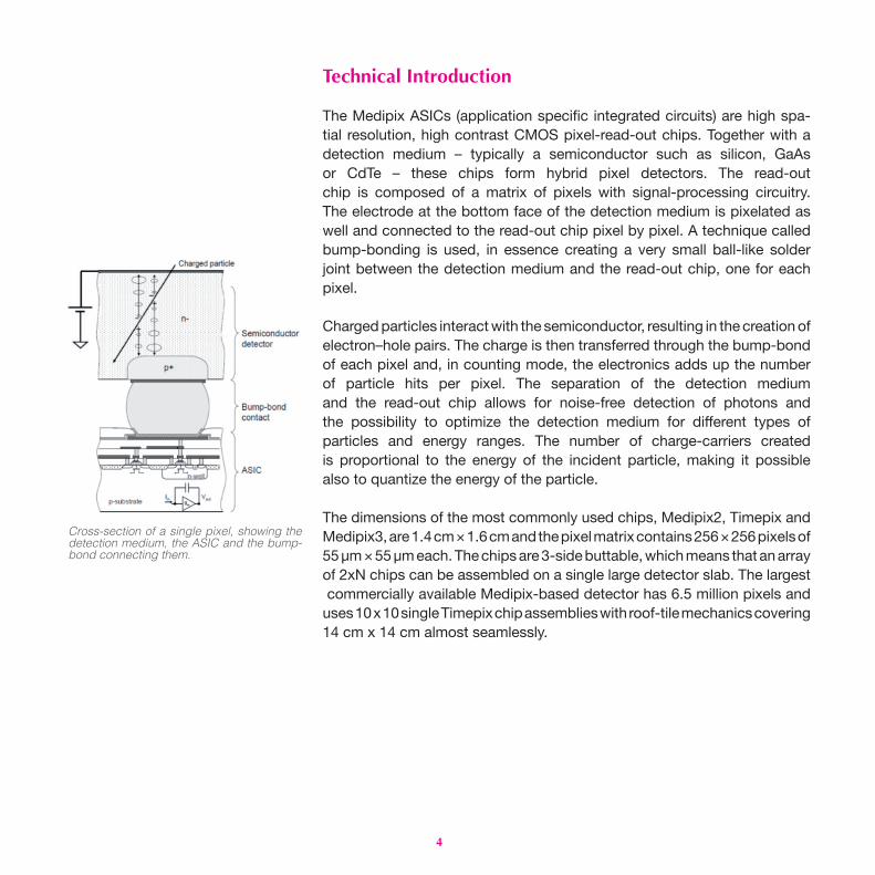
The Medipix ASICs (application specifi c integrated circuits) are high spa-tial resolution, high contrast CMOS pixel-read-out chips. Together with adetection medium – typically a semiconductor such as silicon, GaAsor CdTe – these chips form hybrid pixel detectors. The read-outchip is composed of a matrix of pixels with signal-processing circuitry. The electrode at the bottom face of the detection medium is pixelated as well and connected to the read-out chip pixel by pixel. A technique called bump-bonding is used, in essence creating a very small ball-like solder joint between the detection medium and the read-out chip, one for each pixel.
Charged particles interact with the semiconductor, resulting in the creation of electron–hole pairs. The charge is then transferred through the bump-bond of each pixel and, in counting mode, the electronics adds up the number of particle hits per pixel. The separation of the detection mediumand the read-out chip allows for noise-free detection of photons andthe possibility to optimize the detection medium for diff erent types ofparticles and energy ranges. The number of charge-carriers createdis proportional to the energy of the incident particle, making it possiblealso to quantize the energy of the particle. The dimensions of the most commonly used chips, Medipix2, Timepix and Medipix3, are 1.4 cm × 1.6 cm and the pixel matrix contains 256 × 256 pixels of 55 µm × 55 µm each. The chips are 3-side buttable, which means that an array of 2xN chips can be assembled on a single large detector slab. The largest commercially available Medipix-based detector has 6.5 million pixels and uses 10 x 10 single Timepix chip assemblies with roof-tile mechanics covering 14 cm x 14 cm almost seamlessly.
Cross-section of a single pixel, showing the detection medium, the ASIC and the bump-bond connecting them.

5
Application Areas and CommercializationThe Medipix technology is one of CERN’s most successful technology-transfer cases. Each collaboration has triggered a significant number of commercial activities in widely different application areas. Industrial X-ray imaging, medical imaging and material analysis are just some of the arenas in which Medipix chips are operating today. The industrial partners and licence holders commercializing the Medipix technology range from estab-lished enterprises to young start-up companies. Thus, the technology is contributing to value creation, not only in its application areas, but also through business creation.
Application Area: Medical ImagingRadiography and computed tomography (CT) use X-ray photons to study the human body. The Medipix chips that implement on-pixel single photon counting provide many advantages for use in these fields. The technol-ogy has been applied in X-ray CT, in prototype systems for digital mam-mography, in CT imagers for mammography and for beta- and gamma-autoradiography of biological samples. Moreover, with the Medipix3 chip, the images are no longer black and white – they have colours to indicate different energy levels of the photons. The colour X-ray imaging technique produces clearer and more accurate pictures that should help doctors give their patients more accurate diagnoses.
More than 32 organizations, institutions and companies have been involved with the development of the family of Medipix chips.
In July 2015, CERN had ten active Medipix licences with commercial actors: six licences for Medipix2/Timepix and four licences for Medipix3. Seven of these are held by young start-up companies, most of them having spun out from the collaborating institutes.
A tiled X-ray image of a mouse skull

6
Application Area: EducationIn 2013, a kit especially designed for classroom-use based on Timepix was put on the market. The kit consists of a user-friendly particle detector that can detect ionizing radiation in real time. The students can then observe the particles arriving on the detector on a computer screen. The ability to visually distinguish the signals of alpha, beta and gamma particles and the video-styleframe-rate makes it a compelling demonstration tool for educators teachingabout radiation. Students also learn about computer-aided data analysis by using the USB-based read-out system.
A pilot project, called CERN@School, has been launched by the Simon Langton School in Canterbury, England, with the aim of delivering kits, together with the associated teaching material, to a large number of schools. High-school students are encouraged to initiate their own research projects using the kits.
Langton student Katherine Evans presenting at the CERN@School symposium.
Students from the Thomas Hardye School, Dorchester, England visiting CERN.

7
Application Area: Space DosimetryIn 2012 the Timepix chip arrived at the International Space Station. This was the culmination of eff orts by collaboration members at the Institute ofExperimental and Applied Physics in Prague and the University of Houston, Texas, to use the chip for monitoring the radiation environment of the station. Five chips were plugged into the USB ports of the station laptops andimmediately started delivering data to the Earth-based team in Houston.This team is now carefully analysing the data and comparing it withdata from the more conventional but rather bulky area-monitors.
In December 2014, two Timepix chips were incorporated into a battery-operated system that fl ew on the NASA Orion rocket. The development, which was driven by the University of Houston, provides a very compact and low-power solution to measure the radiation fi eld to which astronauts are exposed. The approach has been adopted as the baseline for radiation measurements for the Orion project.
The astronaut Chris Cassidy working near the Timepix USB on the International Space Station.
There are two Timepix chips inside the BIRD (battery-operated independent radiation detector) that was used on the NASA Orion Rocket.
8
Application Area: Material AnalysisThe Medipix2 and the Medipix3 chips are both being used for commercial X-ray of materials analysis. The Dutch company PANalytical use Medipix for their cutting-edge X-ray diffractometers. These instruments are being used for a myriad of purposes such as the characterization of pharmaceuticals, evaluation and synthesis of new materials, and the detection of counterfeit drugs. This is possible because of the extended dynamic range provided by the single-photon counting approach.
The single-photon counting technique provides outstanding images at very low rates enabling high-resolution phase-contrast imaging with micro-focus X-ray sources. In low rate environments it is possible to perform X-ray polarimetry measurements with the Timepix chip using either silicon as the sensing material or gas. Back to High-Energy PhysicsThe Medipix family’s roots extend deep into the tunnels of CERN’s LHC and high-energy physics. Originally developed for particle physics, they found their way to medical imaging and then came back for the second run of the LHC. As an example, the chips are now being used in the ATLAS experiment to provide independent, real-time information about the radiation environment in the experimental cavern – in principle the same task that Timepix does at the International Space Station. A direct ‘spin back’ to high-energy physics is VELOpix, based on Timepix3. Benefiting from developments made in the framework of the Medipix3 consortium which were not originally intended for high-energy physics, VELOpix will serve as the read-out chip in the new vertex detector of the LHCb experiment, which is planned for installation in 2018.
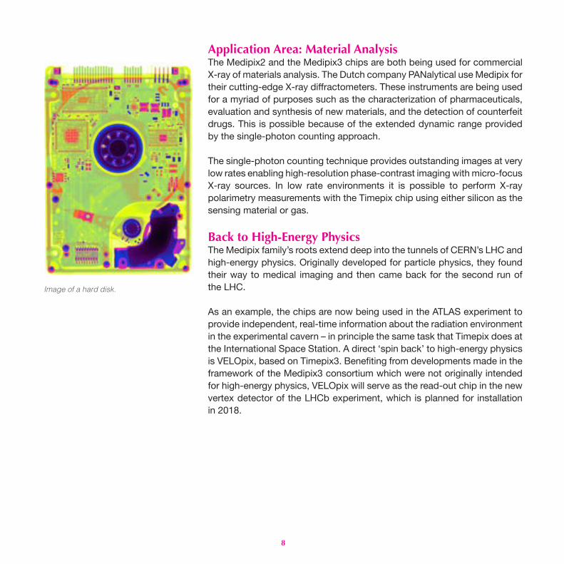
Image of a hard disk.

9
Other Application AreasThe Medipix and Timepix chips are also being used in other application areas such as:
• X-ray detection and imaging at synchrotron light sources
• Gamma cameras
• Low-energy electron microscopy
• Focal planes for electron microscopes
• Single visible photon imaging with microchannel plates
• Axion search TPC readout using GridPlix
• Neutron imaging (combined with B-doped microchannel plates)
• Time-of-flight mass spectrometry
X-ray image of a small flower, acquired with Medipix3. The capability of Medipix detectors to use low-energy X-ray photons (< 20 keV) for radiography enables high contrast even for objects with low X-ray attenuation. The blossom and the tiny hairs on top of the calyx (closed flower) are visible as well as the inner parts, e.g. the stigmas.The colours represent different intensities.
10
The Next Step: Medipix4Up until now it has been possible to make pixel detector read-out chips that can be abutted on three sides only. The fourth side is used for on-chipperipheral logic and wire-bond pads that permit electronic read-out.Because of this structure, large detector areas can be covered,but only with gaps in the coverage,or by using complicated roof-tilemechanics. Through-silicon-via (TSV) technology provides the possibilityof reading the chips through copper-fi lled holes that bring the signalsfrom the front side of the chip to its rear. Recent developments basedon the Medipix3 chips have demonstrated that this has now becomea viable option for pixel detector read-out. However, the TSV processing of Medipix3 only allows a reduction in the peripheral area by avoiding the use of wire bonds. The peripheral logic is still present.
A new collaboration, called Medipix4, is in formation with the aim ofdesigning pixel read-out chips that for the fi rst time are fully prepared for TSV processing and may be tiled on all 4 sides. In other words, all of the communication with the pixel matrix will now go through the rear of the chip – the peripheral logic and control elements will be integrated insidethe pixel matrix. This will not only enable large areas to be covered seam-lessly, but will also permit the development of new read-out architecturesby avoiding the need to send all of the data to one side of the chip for read-out. Two new chips are foreseen: Medipix4, which will target spectroscopicX-ray imaging at rates compatible with medical CT scans, and Timepix4,which will provide particle identifi cation and tracking with higher spatial and timing precision.
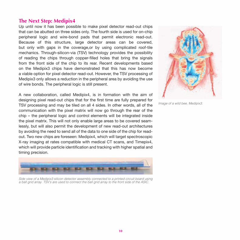
Image of a wild bee, Medipix3.
Side view of a Medipix3 silicon detector assembly connected to a printed circuit board using a ball grid array. TSV’s are used to connect the ball grid array to the front side of the ASIC.
11
Contact Information
CERN Knowledge Transfer Groupcern.ch/[email protected]
Picture credits
Front page: Simon Procz, University of Freiburg, Medipix flower IPage 2: CERN, 1995Page 3: CERN, Timepix3 chip; CERN, IllustrationPage 4: CERN, Cross sectionPage 5: Simon Procz, University of Freiburg, Mouse skullPage 6: Becky Parker, Katherine Evans; Judith Wardlaw, Thomas Hardye SchoolPage 7: NASA, Orion; NASA, ISSPage 8: Simon Procz, University of Freiburg and Elias Hamann, KIT, Hard diskPage 9: Simon Procz, University of Freiburg, Medipix flower IIPage 10: Simon Procz, University of Freiburg, Wild bee; CERN, Medipix3
Collaboration Partners
Medipix1CERN; University of Freiburg; University of Glasgow; Istituto Nazionale Di Fisica Nucleare of Pisa and Napoli.
Medipix2 CERN; Institut de Física d’Altes Energie, Barcelona; University of Cagliari; Space Sciences Laboratory, University of California, Berkeley; Commissariat à l’Energie Atomique; Czech Academy of Sciences; IEAP, Czech Technical University in Prague; ESRF; Universität Erlangen-Nurnberg; Friedrich- Alexander-Universität Freiburg-i.B; University of Glasgow; University of Houston; Laboratory of Molecular Biology, Medical Research Council, Cambridge; Mid-Sweden University; Università di Napoli Federico Il; NIKHEF; Università di Pisa.
Medipix3CERN; Brazilian Synchrotron Light Laboratory; University of Canterbury; Commissariat à l’Energie Atomique; Deutsches Elektronen-Synchrotron; Diamond Light Source; Albert-Ludwigs-Universität Freiburg; University of Glasgow; IPS, Karlsruhe Institute of Technology; Leiden University; NIKHEF; Mid-Sweden University; IEAP, Czech Technical University; ESRF; Universität Erlangen-Nurnberg; Space Sciences Laboratory, University of California, Berkeley; VTT Technical Research Centre of Finland; University of Houston; Universidad de los Andes; University of Bonn; FOM Institute for Atomic and Molecular Physics; Technical University of Munich.
CERN-Brochure-2015-007-Eng September 2015

cern.ch