
Medicine Image DICOM Display Update: Color Presentation ... · •ICC - International ... 3D)...
45
The Medicine Behind the Image DICOM Display Update: DICOM Display Update: Color Presentation States Color Presentation States Hanging Protocols Hanging Protocols Dr. David A. Clunie, MB.,BS., FRACR Chief Technology Officer Princeton Radiology Pharmaceutical Research
Transcript of Medicine Image DICOM Display Update: Color Presentation ... · •ICC - International ... 3D)...
The
MedicineBehind the
Image DICOM Display Update:DICOM Display Update:Color Presentation StatesColor Presentation States
Hanging ProtocolsHanging Protocols
Dr. David A. Clunie, MB.,BS., FRACRChief Technology Officer
Princeton Radiology Pharmaceutical Research
OverviewOverview
• Review of Grayscale Presentation State• Color Presentation States
– Color Consistency– Presentation States applied to Color Images– Color Blending - CT-PET fusion
• Hanging Protocols
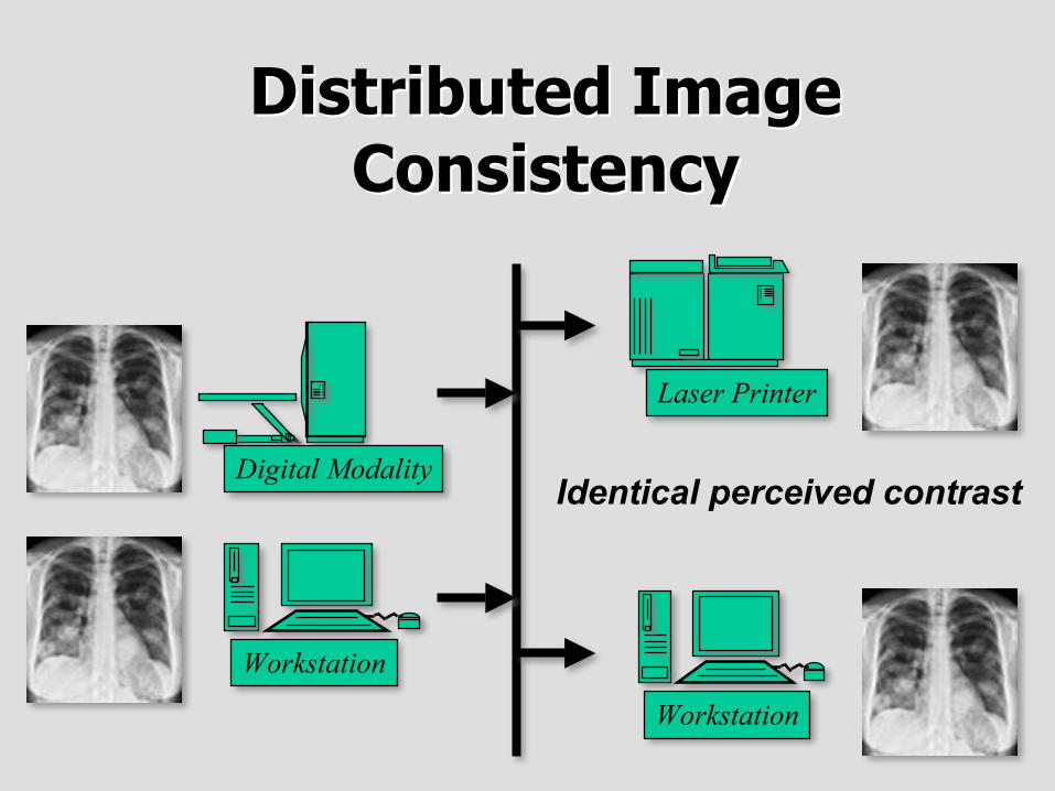
Distributed ImageDistributed ImageConsistencyConsistency
Digital Modality
Workstation
Laser Printer
Workstation
Identical perceived contrast
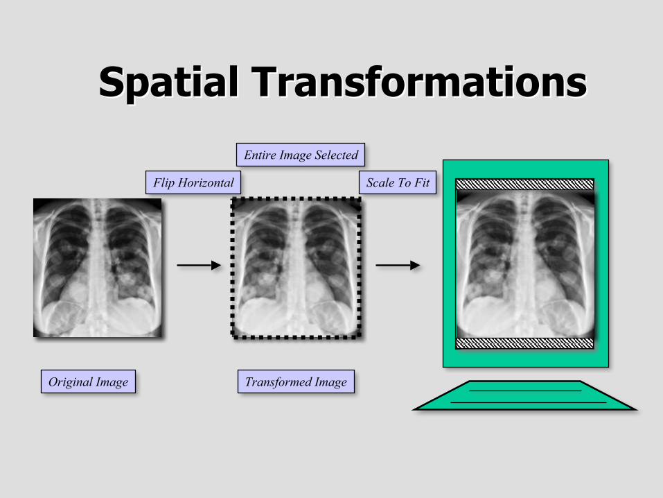
Spatial TransformationsSpatial Transformations
Original Image
Entire Image Selected
Transformed Image
Scale To FitFlip Horizontal
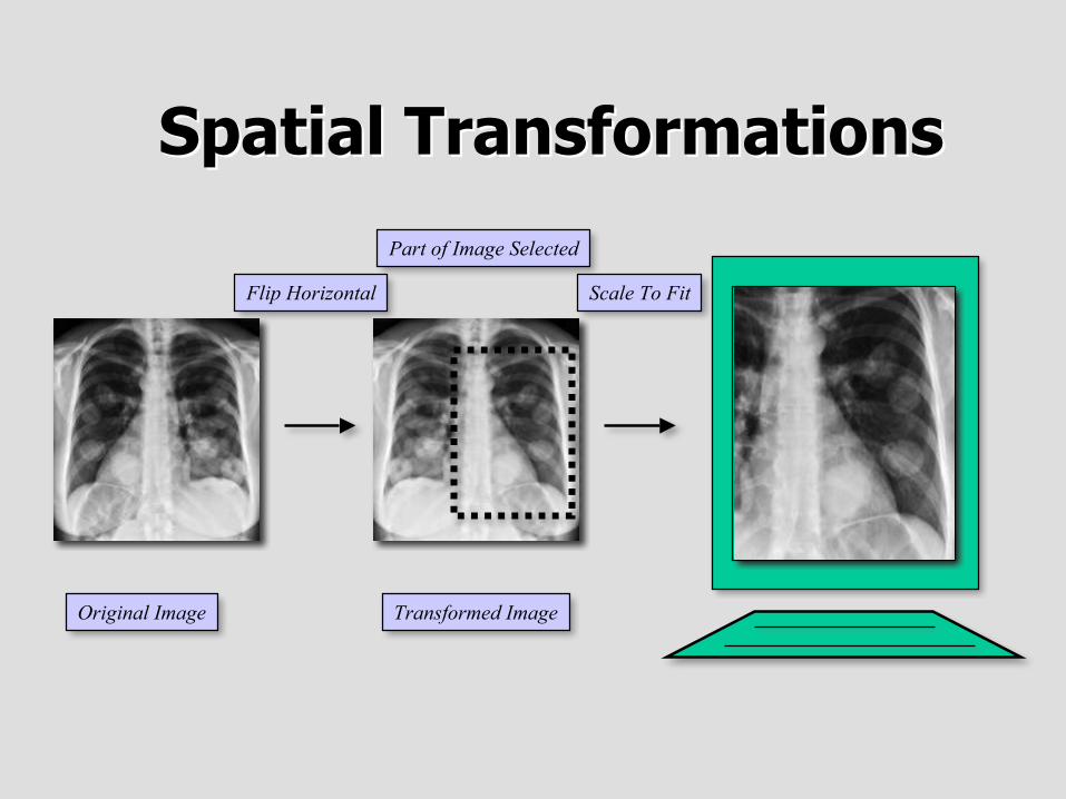
Spatial TransformationsSpatial Transformations
Original Image
Part of Image Selected
Transformed Image
Scale To FitFlip Horizontal
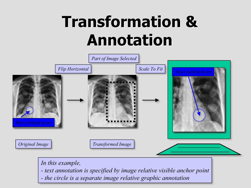
Transformation &Transformation &AnnotationAnnotation
Original Image
Part of Image Selected
Transformed Image
Scale To FitFlip Horizontal
Mass behind heart
Mass behind heart
In this example,- text annotation is specified by image relative visible anchor point- the circle is a separate image relative graphic annotation
Limitations of GrayscaleLimitations of GrayscalePresentation StatesPresentation States
• Apply to grayscale images– no means to specify spatial transformations or graphic
annotations for color images
• Only grayscale consistency– standard display function defined only for luminance
• No pseudo-color capability• No blending or fusion capability
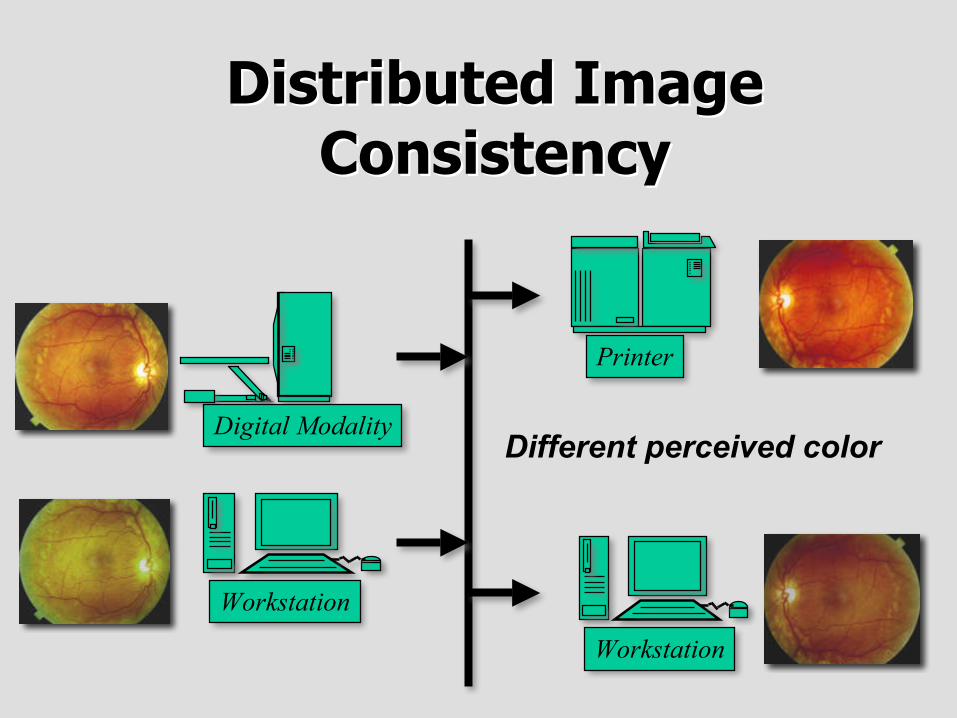
Distributed ImageDistributed ImageConsistencyConsistency
Digital Modality
Workstation
Printer
Workstation
Different perceived color
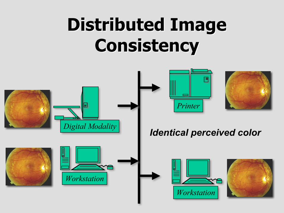
Distributed ImageDistributed ImageConsistencyConsistency
Digital Modality
Workstation
Printer
Workstation
Identical perceived color
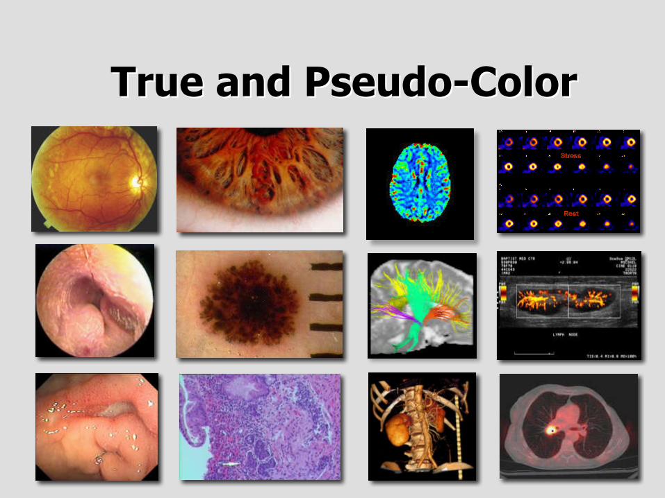
TrueTrue and Pseudo-Colorand Pseudo-Color
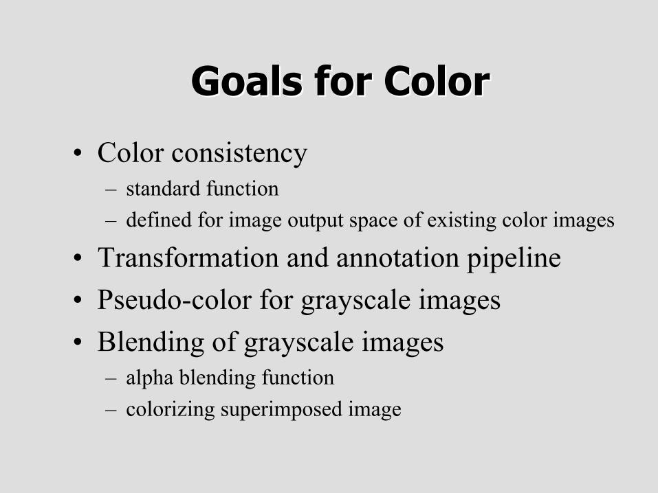
Goals for ColorGoals for Color
• Color consistency– standard function– defined for image output space of existing color images
• Transformation and annotation pipeline• Pseudo-color for grayscale images• Blending of grayscale images
– alpha blending function– colorizing superimposed image
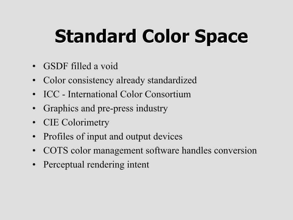
StandardStandard Color SpaceColor Space• GSDF filled a void• Color consistency already standardized• ICC - International Color Consortium• Graphics and pre-press industry• CIE Colorimetry• Profiles of input and output devices• COTS color management software handles conversion• Perceptual rendering intent
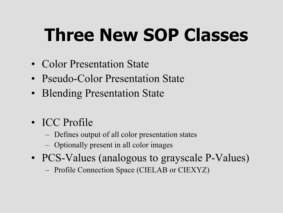
Three New SOP ClassesThree New SOP Classes
• Color Presentation State• Pseudo-Color Presentation State• Blending Presentation State
• ICC Profile– Defines output of all color presentation states– Optionally present in all color images
• PCS-Values (analogous to grayscale P-Values)– Profile Connection Space (CIELAB or CIEXYZ)
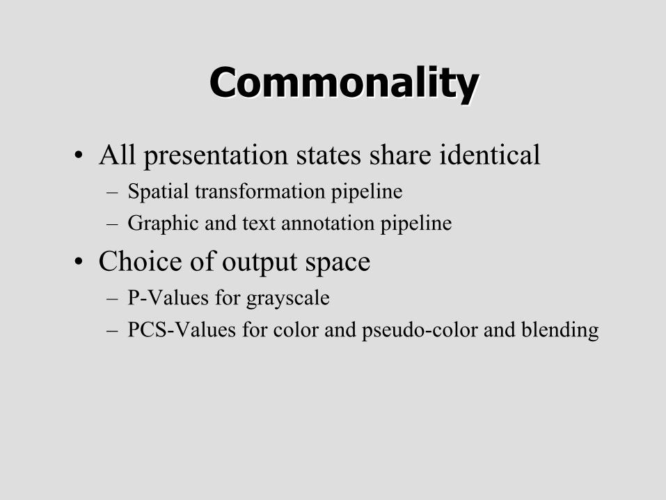
CommonalityCommonality
• All presentation states share identical– Spatial transformation pipeline– Graphic and text annotation pipeline
• Choice of output space– P-Values for grayscale– PCS-Values for color and pseudo-color and blending
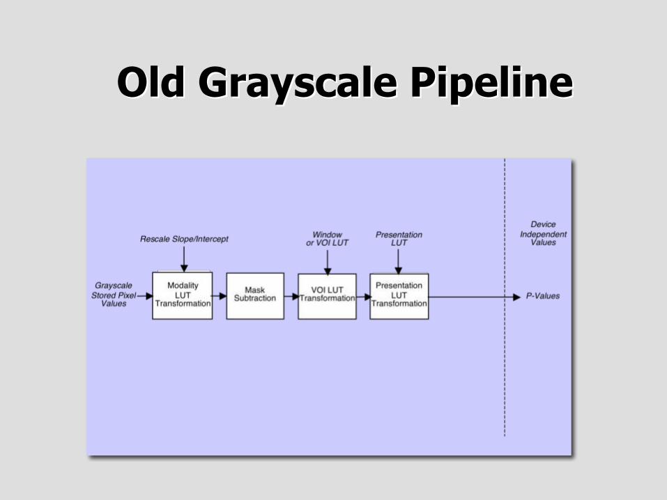
Old Grayscale PipelineOld Grayscale Pipeline
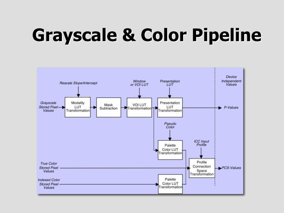
Grayscale & Color PipelineGrayscale & Color Pipeline
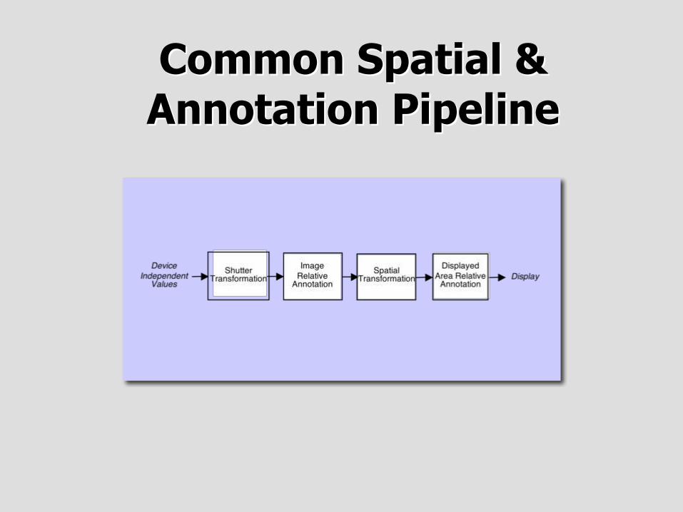
Common Spatial &Common Spatial &Annotation PipelineAnnotation Pipeline
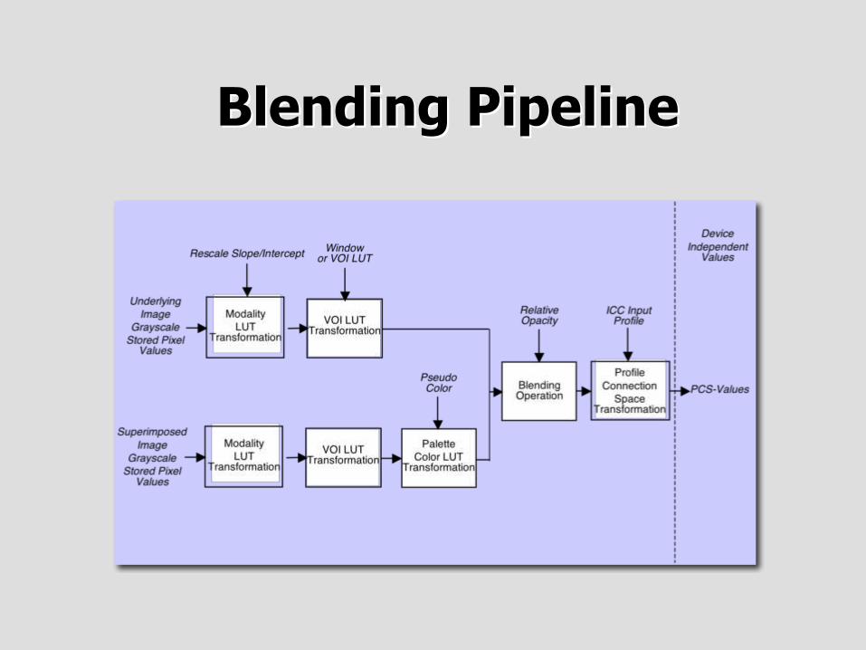
Blending PipelineBlending Pipeline
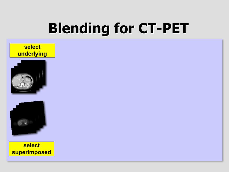
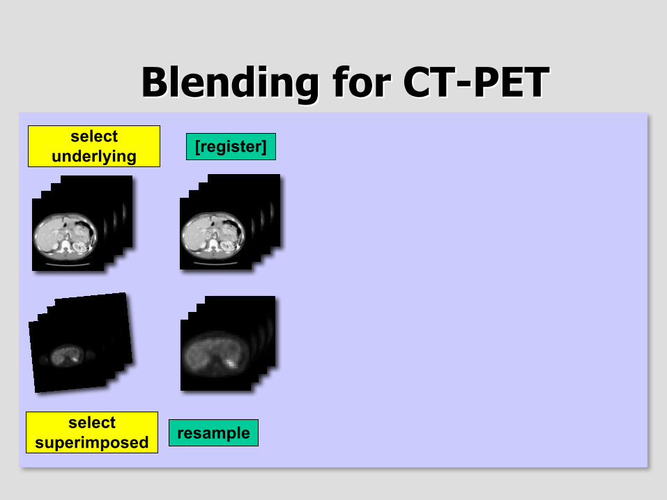
Blending for CT-PETBlending for CT-PET
selectsuperimposed
selectunderlying
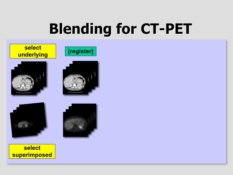
Blending for CT-PETBlending for CT-PET
selectsuperimposed
[register]selectunderlying
Blending for CT-PETBlending for CT-PET
selectsuperimposed
[register]
resample
selectunderlying
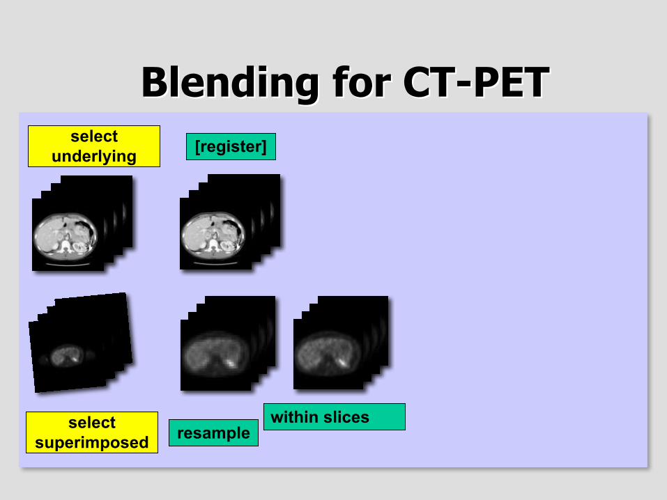
Blending for CT-PETBlending for CT-PET
selectsuperimposed
[register]
resamplewithin slices
selectunderlying
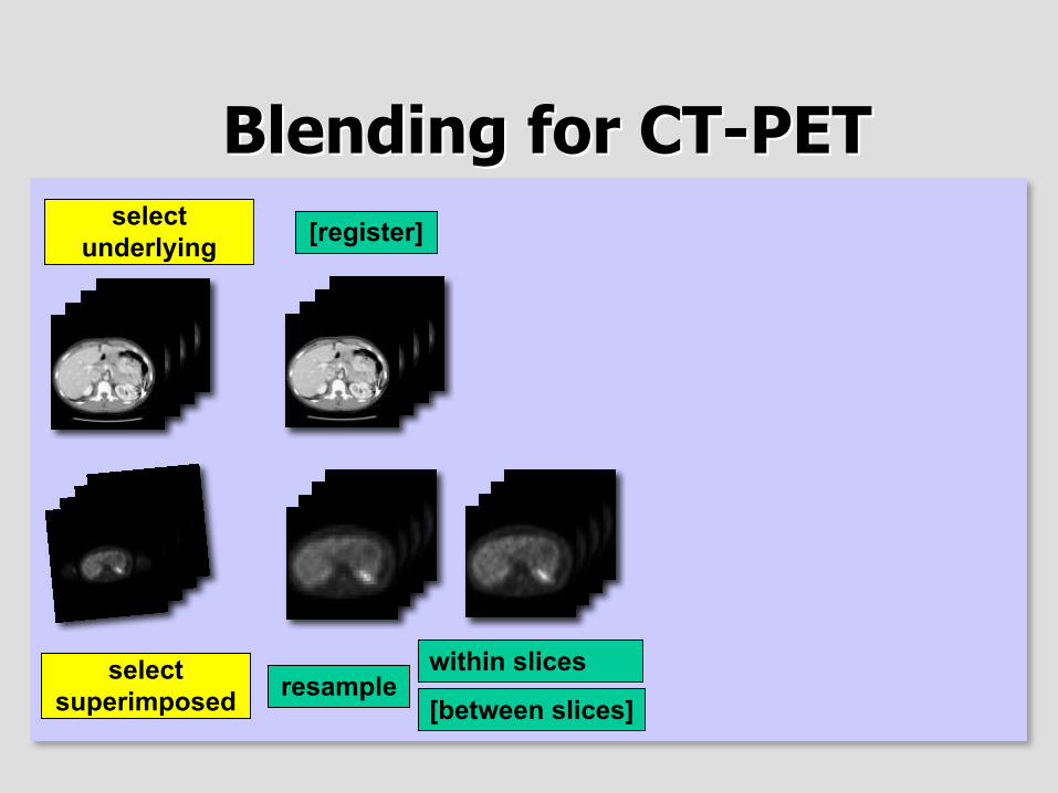
Blending for CT-PETBlending for CT-PET
selectsuperimposed
[register]
resamplewithin slices
[between slices]
selectunderlying
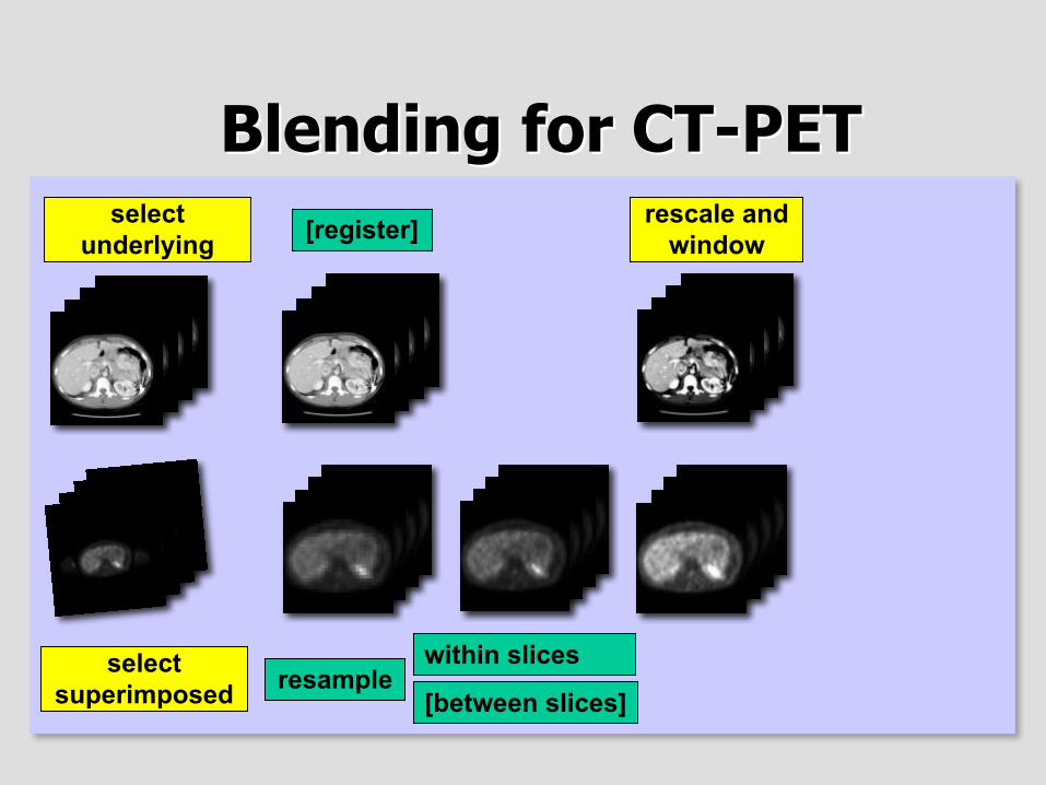
Blending for CT-PETBlending for CT-PET
selectsuperimposed
[register]
resamplewithin slices
[between slices]
selectunderlying
rescale andwindow
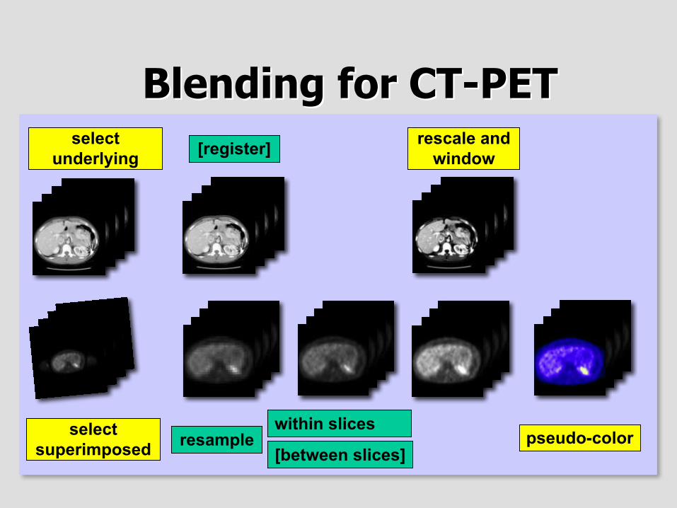
Blending for CT-PETBlending for CT-PET
selectsuperimposed
[register]
resamplewithin slices
[between slices]
selectunderlying
rescale andwindow
pseudo-color
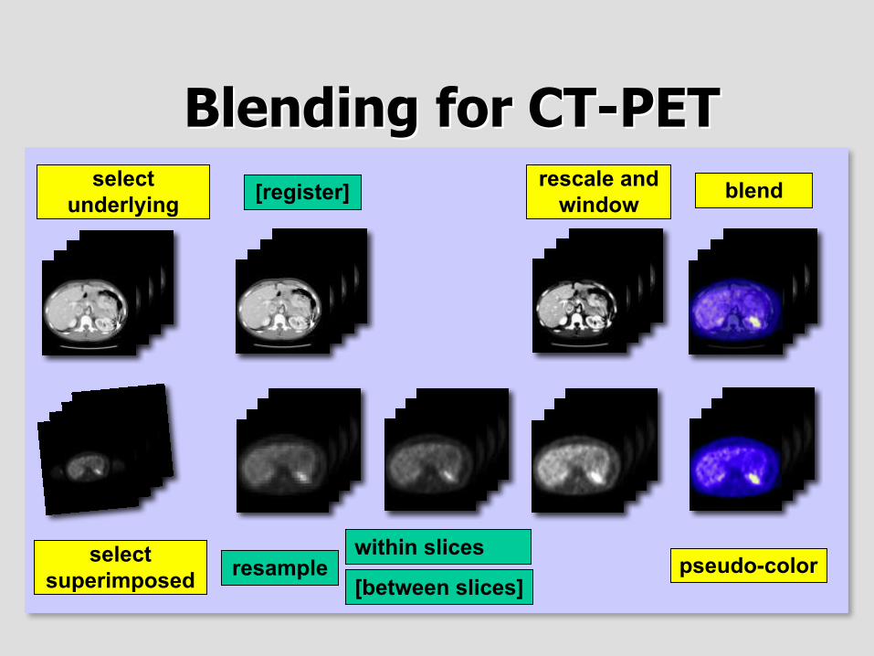
Blending for CT-PETBlending for CT-PET
selectsuperimposed
[register]
resamplewithin slices
[between slices]
selectunderlying
rescale andwindow blend
pseudo-color
Color - ConclusionColor - Conclusion
• Color consistency using industry standard• Transformation/annotation for color images• Exchange of pseudo-color information• Support for specifying sets of images to be
blended, and how to blend (but not registeror resample) them
OverviewOverview
• Review of Grayscale Presentation State• Color Presentation States
– Color Consistency– Presentation States applied to Color Images– Color Blending - CT-PET fusion
• Hanging Protocols
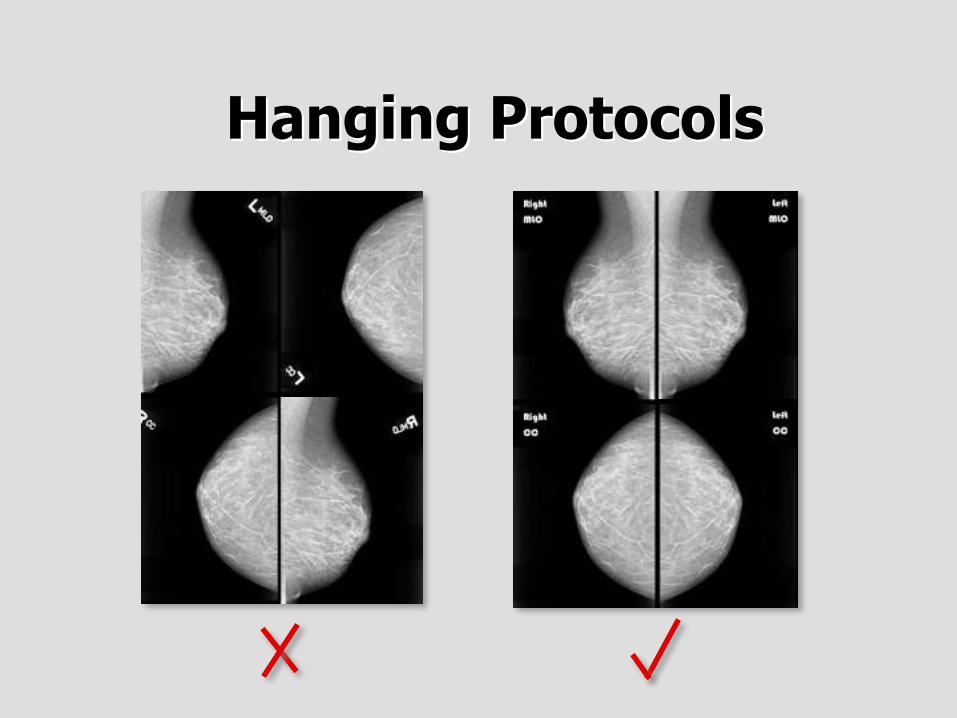
Hanging ProtocolsHanging Protocols
• “Default display protocols”• A set of instructions• How to layout a class of images for display• Order, orientation, windowing, processing
• Not specific to a particular patient’s images• Hence a protocol, not a presentation state
Hanging ProtocolsHanging Protocols
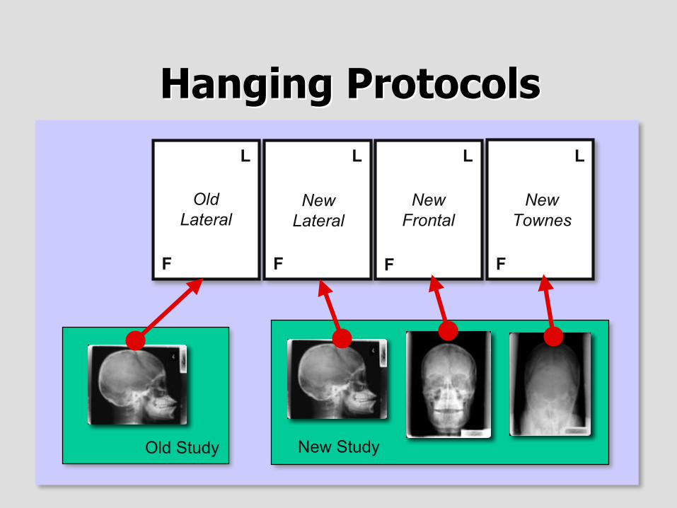
Hanging ProtocolsHanging Protocols
New Study
OldLateral
NewLateral
NewFrontal
NewTownes
L L L L
FFFF
Old Study
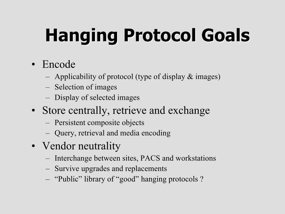
Hanging Protocol GoalsHanging Protocol Goals
• Encode– Applicability of protocol (type of display & images)– Selection of images– Display of selected images
• Store centrally, retrieve and exchange– Persistent composite objects– Query, retrieval and media encoding
• Vendor neutrality– Interchange between sites, PACS and workstations– Survive upgrades and replacements– “Public” library of “good” hanging protocols ?

New Information ModelNew Information Model
• Required for storage and query/retrieval• No Patient/Study/Series hierarchy
• New Storage Service Class• New Query Model• Still C-STORE, C-FIND, C-MOVE
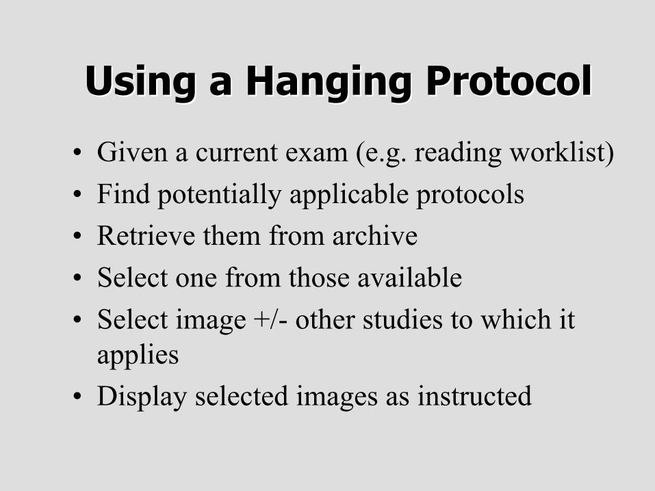
Using a Hanging ProtocolUsing a Hanging Protocol
• Given a current exam (e.g. reading worklist)• Find potentially applicable protocols• Retrieve them from archive• Select one from those available• Select image +/- other studies to which it
applies• Display selected images as instructed
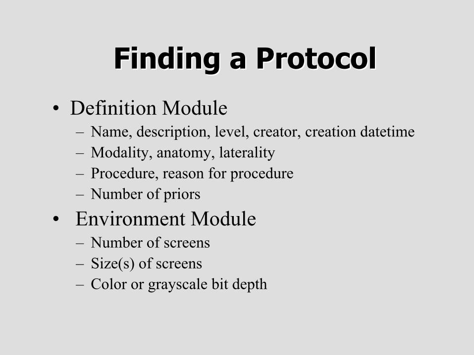
Finding a ProtocolFinding a Protocol
• Definition Module– Name, description, level, creator, creation datetime– Modality, anatomy, laterality– Procedure, reason for procedure– Number of priors
• Environment Module– Number of screens– Size(s) of screens– Color or grayscale bit depth
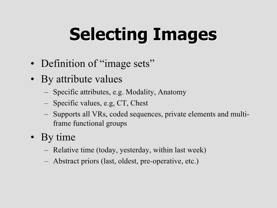
Selecting ImagesSelecting Images
• Definition of “image sets”• By attribute values
– Specific attributes, e.g. Modality, Anatomy– Specific values, e.g, CT, Chest– Supports all VRs, coded sequences, private elements and multi-
frame functional groups
• By time– Relative time (today, yesterday, within last week)– Abstract priors (last, oldest, pre-operative, etc.)
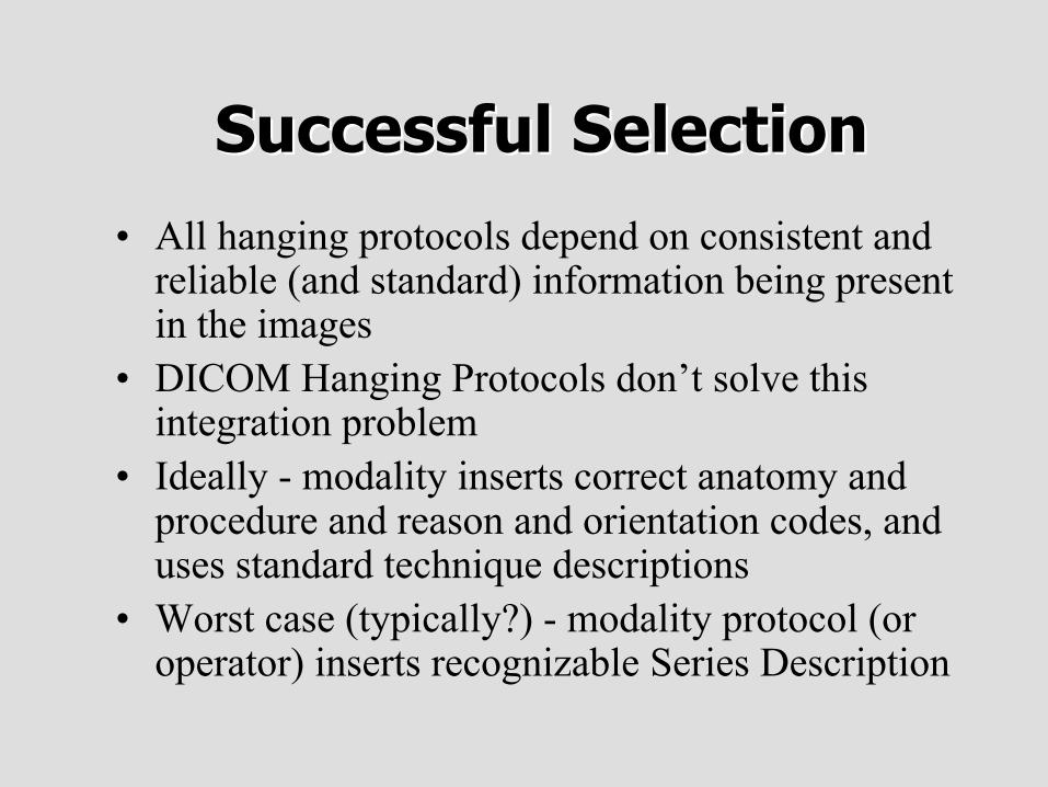
Successful SelectionSuccessful Selection
• All hanging protocols depend on consistent andreliable (and standard) information being presentin the images
• DICOM Hanging Protocols don’t solve thisintegration problem
• Ideally - modality inserts correct anatomy andprocedure and reason and orientation codes, anduses standard technique descriptions
• Worst case (typically?) - modality protocol (oroperator) inserts recognizable Series Description
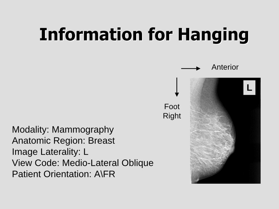
Information for HangingInformation for Hanging
Modality: MammographyAnatomic Region: BreastImage Laterality: LView Code: Medio-Lateral ObliquePatient Orientation: A\FR
Anterior
FootRight
L
PriorsPriors
• Concept of the “current” study required• Protocol chooses priors based on
– Relative time– Abstract temporal ranges (previous, last, etc.)– Abstract coded descriptions (“pre-operative”)
• Does NOT specify how to find them or get them• May have been pushed, may need a query• May be hard to find by abstract descriptions• Creative use of queries or out-of-band information
Mapping to Image BoxesMapping to Image Boxes
• Image Sets are mapped to Image Boxes• Image Box types
– Tiled (e.g. 3x4)– Stack (single image paged manually)– Cine (time-based play back)– Processed (e.g. MPR, 3D)– Single (e.g. a place for a report or waveform)
• Specify– Scrolling mode– Playback rate
Mapping to Image BoxesMapping to Image Boxes
• Filtering– By attribute, or abstract, e.g. “category” of “image plane” “axial”
• Sorting– By attribute, or abstract, e.g. “along axis” “increasing”
• Orientation– E.g. rotate/flip until row left column posterior (L\P)
• Annotation– Patient demographics, technique and graphics on or off
Processing & PresentationProcessing & Presentation• Reformatting, e.g., MPR, 3D, slab• Thickness, interval• View direction, e.g., axial, sagittal, coronal• Type, e.g., MIP, surface, volume• VOI Type (windowing), e.g., brain, bone• Pseudo-color type, e.g., hot iron• Invert grayscale• True size• Synchronized scrolling (by Display Set number)• Navigation and localization
Display of Image BoxesDisplay of Image Boxes
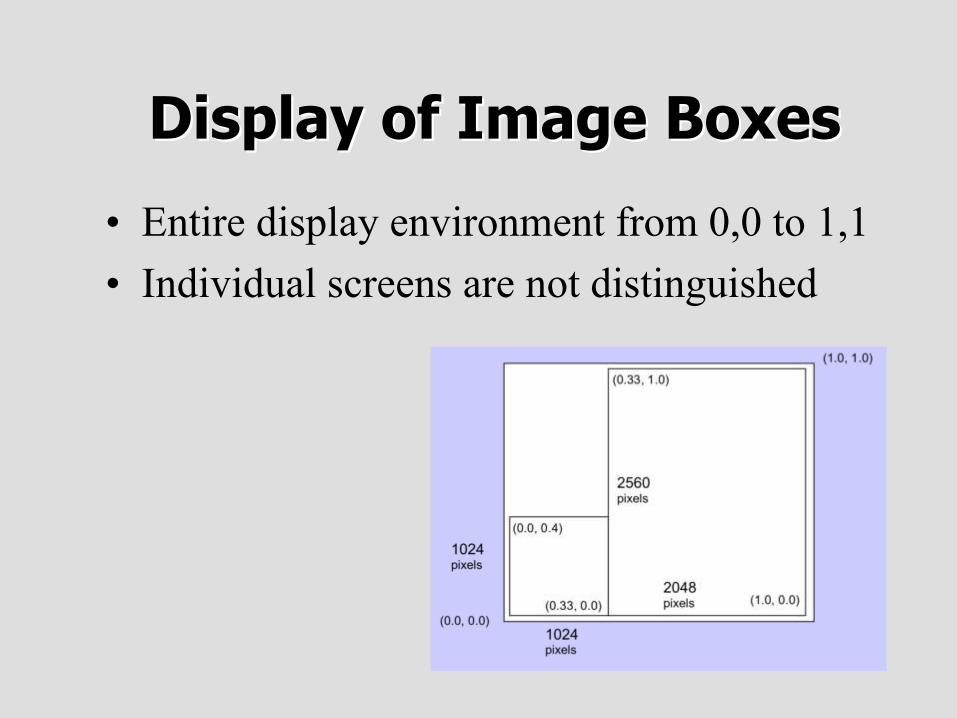
• Entire display environment from 0,0 to 1,1• Individual screens are not distinguished
Display of Image BoxesDisplay of Image Boxes
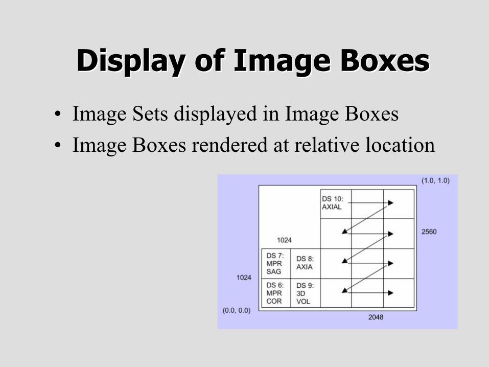
• Image Sets displayed in Image Boxes• Image Boxes rendered at relative location
Hanging Protocols - ConclusionHanging Protocols - Conclusion
• Interchangeable• Vendor neutral• Multi-modality• Support selection of priors• Full richness of current display modes• Flexible• Extensible
• Non-trivial to implement and retrofit• Dependent on reliable image attributes