Medical Image Segmentation using 3D Convolutional Neural ...
34
Medical Image Segmentation using 3D Convolutional Neural Networks: A Review S Niyas a,* , S J Pawan a , M Anand Kumar b , Jeny Rajan a a Department of Computer Science and Engineering, National Institute of Technology Karnataka, Surathkal, Mangalore – 575025, Karnataka, India. b Department of Information Technology, National Institute of Technology Karnataka, Surathkal, Mangalore – 575025, Karnataka, India. Abstract Computer-aided medical image analysis plays a significant role in assisting medical practitioners for expert clinical diagnosis and deciding the optimal treatment plan. At present, convolutional neural networks (CNNs) are the preferred choice for medical image analysis. In addition, with the rapid advancements in three-dimensional (3D) imaging systems and the availability of excellent hardware and software support to process large volumes of data, 3D deep learning methods are gaining popularity in medical image analysis. Here, we present an extensive review of the recently evolved 3D deep learning methods in medical image segmentation. Furthermore, the research gaps and future directions in 3D medical image segmentation are discussed. Keywords: 3D deep learning, Convolutional Neural Networks, Medical image analysis 1. Introduction Artificial intelligence (AI) techniques have been extensively used in analyzing medical images in the mod- ern healthcare system. Computer-aided diagnosis (CADx) enables analyzing the medical data and extracting valuable information using specialized software to assist physicists in making rapid and informative clinical decisions. These diagnostic systems reduce the subjectivity in decision-making and the overall cost involved. In recent years, popular medical imaging modalities, such as X-ray, computed tomography (CT), ultrasonog- raphy (USG), and magnetic resonance imaging (MRI), have advanced in terms of acquisition time, image quality, and cost-effectiveness. [1]. However, these techniques exhibit inherent limitations such as noise, motion artifacts, non-uniform contrasts, and registration errors, leading to an unreliable clinical diagnosis. Medical image processing entails problem-specific strategies based on image processing algorithms. Some of its typical applications include image registration, denoising, enhancement, compression, classification, and * Corresponding author. Email addresses: [email protected] (S Niyas ), [email protected] (S J Pawan), [email protected] (M Anand Kumar), [email protected] (Jeny Rajan) Preprint submitted to Neurocomputing February 8, 2022 arXiv:2108.08467v2 [eess.IV] 6 Feb 2022
Transcript of Medical Image Segmentation using 3D Convolutional Neural ...

Medical Image Segmentation using 3D Convolutional Neural Networks: AReview
S Niyasa,∗, S J Pawana, M Anand Kumarb, Jeny Rajana
aDepartment of Computer Science and Engineering, National Institute of Technology Karnataka, Surathkal, Mangalore –575025, Karnataka, India.
bDepartment of Information Technology, National Institute of Technology Karnataka, Surathkal, Mangalore – 575025,Karnataka, India.
Abstract
Computer-aided medical image analysis plays a significant role in assisting medical practitioners for expert
clinical diagnosis and deciding the optimal treatment plan. At present, convolutional neural networks
(CNNs) are the preferred choice for medical image analysis. In addition, with the rapid advancements in
three-dimensional (3D) imaging systems and the availability of excellent hardware and software support to
process large volumes of data, 3D deep learning methods are gaining popularity in medical image analysis.
Here, we present an extensive review of the recently evolved 3D deep learning methods in medical image
segmentation. Furthermore, the research gaps and future directions in 3D medical image segmentation are
discussed.
Keywords: 3D deep learning, Convolutional Neural Networks, Medical image analysis
1. Introduction
Artificial intelligence (AI) techniques have been extensively used in analyzing medical images in the mod-
ern healthcare system. Computer-aided diagnosis (CADx) enables analyzing the medical data and extracting
valuable information using specialized software to assist physicists in making rapid and informative clinical
decisions. These diagnostic systems reduce the subjectivity in decision-making and the overall cost involved.
In recent years, popular medical imaging modalities, such as X-ray, computed tomography (CT), ultrasonog-
raphy (USG), and magnetic resonance imaging (MRI), have advanced in terms of acquisition time, image
quality, and cost-effectiveness. [1]. However, these techniques exhibit inherent limitations such as noise,
motion artifacts, non-uniform contrasts, and registration errors, leading to an unreliable clinical diagnosis.
Medical image processing entails problem-specific strategies based on image processing algorithms. Some of
its typical applications include image registration, denoising, enhancement, compression, classification, and
∗Corresponding author.Email addresses: [email protected] (S Niyas ), [email protected] (S J Pawan), [email protected]
(M Anand Kumar), [email protected] (Jeny Rajan)
Preprint submitted to Neurocomputing February 8, 2022
arX
iv:2
108.
0846
7v2
[ee
ss.I
V]
6 F
eb 2
022

segmentation, which predominantly used traditional machine learning models to analyze the characteristics
such as contrast variation, orientation, shape, and texture patterns. However, machine learning models
exhibit various limitations [2, 3], such as 1) high dependency on handcrafted features extracted by a domain
expert, 2) inevitable manual intervention, and 3) arbitrary parameters. These limitations of traditional
machine learning algorithms have resulted in automated feature extraction methods such as convolutional
neural networks (CNNs) [4].
Studies on neural-network-based decision-making are being conducted since the 1950s. Several challenges
were faced initially, mainly due to the lack of ample data and adequate computational capabilities. However,
factors such as digitalization that lead to data availability, advancements in parallel distributed computing,
and the surge in the semiconductor industry accelerated advanced research on neural networks. With the
increasing prominence of image data in the digital data space, research on developing neural network models
for image analysis increased tremendously, as evident from the introduction of convolutional neural networks
(CNN). The first popular CNN method was published by LeCun et al. [5] for handwritten character recogni-
tion. Subsequently, several successful deep CNN models have been developed to solve various classification
[6] and segmentation problems [7].
Medical image processing has also benefited from these developments periodically. As a result, several
studies related to medical image analysis using deep learning techniques have been reported in the literature.
Despite the significant growth in the research on deep learning-based medical image analysis during the last
couple of years, statistics on classification outweigh that on segmentation approaches. The statistics of
CNN-based medical image processing papers retrieved from PubMed are shown in Fig. 1a.
The literature reports numerous dedicated reviews on CNN-based medical image analysis. For instance,
the review article by Shen et al. [3] provides a comprehensive overview of medical image processing using
advanced machine learning and deep-learning techniques. Their study highlights the advantages of various
CNN models over traditional machine learning methods in different medical applications. Similarly, Litjens
et al. [8] thoroughly discussed the applications of deep learning in various fields of medical image analysis.
In addition, several review papers [9, 10, 11, 12, 13, 14] provide a basic understanding of CNN models and
their applications in medical image analysis. However, most of these reviews have focused on analyzing deep
learning approaches in an application-specific manner.
Since CNN models are designed to operate using image data, the dimensionality of the images plays a
significant role in selecting the appropriate model. Generally, the majority of the medical image data are
constructed using either two-dimensional (2D) or 3D imaging techniques. For example, digital X-rays, retinal
fundus images, microscopic images in pathology, mammograms, etc., belong to the 2D image categories in
the biomedical domain. Similarly, MRI, CT, and ultrasound (US) are widely used 3D medical images.
However, most of the published studies on 3D medical image analysis have used 2D CNN models. Fig. 1b
substantiates our statement showing the breakdown of studies that have used 2D and 3D deep learning
2

(a) Breakdown of papers based on the application.
(b) Breakdown of papers based on the deep learning approach (2D or 3D CNN).
Figure 1: Statistics of papers retrieved from PubMed on medical image analysis using deep learning techniques (from 2011-
2021). The data for the year 2021 has been extrapolated from the papers listed till June 2021.
techniques in medical image analysis.
Segmentation of organs or anatomical structures is a crucial step in medical image processing. It is
primarily used to detect abnormalities and estimate the true extent of the organ or lesion. In [15], Singh
3

et al. presented an overview of various building blocks in 3D CNN architectures and several deep-learning
approaches in volumetric medical image analysis. To the best of our knowledge, there are no comprehen-
sive reviews in the literature focusing on medical image segmentation using 3D deep learning techniques.
This motivated us to conduct an in-depth review of current deep learning trends in 3D medical image
segmentation. We have also discussed the future perspectives in 3D medical image segmentation.
This review covers several articles that mainly focused on the 3D deep-learning techniques for volumetric
medical image segmentation. Papers reporting overlapping techniques have been excluded from this review
to minimize redundancy. The primary contributions of this review article are to provide
1. an overview of 3D deep learning for medical image processing.
2. an in-depth review on various state-of-the-art 3D deep learning-based medical image segmentation.
3. a discussion on research gaps and future directions in 3D medical image segmentation.
The rest of this review is organized as follows. Section 2 presents a brief overview of 2D and 3D CNN
models and a detailed survey on existing 3D deep learning models along with their contributions to medical
image segmentation. Section 3 provides a critical discussion and an outlook for future research.
2. 3D CNN in Medical Image Segmentation
2.1. Convolutional Neural Networks: An overview
CNN is an advanced version of ANN and is primarily designed to learn spatial features from images
without using any handcrafted features. In CNN models, a set of kernels (or filters) are utilized to learn
various characteristics, and these learnable kernel values are updated based on the value of the cost function.
The convoluted output from each kernel is considered as the feature map and is passed to the subsequent
layers. While using a set of K kernels, the feature map generated in the nth layer can be represented as:
Xkn = σ(wk
n ∗Xn−1 + bkn), k = {1, 2, ...,K} (1)
where Wn = {w1n, w2
n, ..., wKn} is the set of weights and Bn = {b1n, b2n, ..., bKn} is the set of biases in the
nth layer. Xkn represents the feature map created in the nth layer after convolving the feature map from
(n− 1)th layer with kth kernel.
CNNs do not require separate filters to detect similar objects in multiple locations in the image and hence
make the number of weight parameters independent of the image size. This helps to reduce the number of
trainable parameters and makes the operation translational invariant across the image. CNN architecture
(for classification) generally consists of three building blocks: convolution layers, pooling layers, and fully
connected dense layers [13]. A convolution layer uses small multi-dimensional kernels to extract spatial
4

features from image locations. The pooling layer is designed to pass relevant features to the subsequent
layers by down-sampling the feature space. In addition, pooling reduces the number of trainable parameters
in the subsequent dense layers, expands the receptive field for multi-scale feature extraction, and provides
translation invariance to small shift errors.
A typical CNN model uses multiple convolution operations followed by activation and pooling layers
for extracting multi-scale features; the complexity of image features advances as the model depth increases.
The fully connected dense layer is designed to transform the feature map from the previous layer to one-
dimensional feature vectors. The final fully connected layer operates as a classification layer and provides
the probabilities of the target classification task. Furthermore, a CNN model uses several additional modules
such as batch normalization [16], regularization, and dropout [17], to improve the learning process [15]. A
high level representation of a CNN classification model is shown in Figure 2.
Figure 2: A high level representation of a CNN classification model.
2.2. 2D CNN vs 3D CNN in Image Segmentation
In image segmentation, every pixels needs to be classified simultaneously. CNN model for segmentation
uses convolution and pooling layers similar to those in a classification network. However, the fully connected
layers are usually excluded in the segmentation model to retain the spatial relationship of pixels throughout
the network. In addition, adequate upsampling layers are added to the final layers of the network to
compensate for the pooling operation. Hence, the probability of each pixel is estimated and finally, the
segmentation mask is created.
Long et al. [18] proposed the first fully convolutional neural network (FCNN), which consists of a
sequence of convolution and pooling layers to extract features from different scales of image. To compensate
5
the reduction in feature space due to pooling, the latent space is upsampled directly to the actual image
dimension using deconvolution and skip connections. Followed by this FCNN approach, several segmentation
methods have been proposed. Liu et al. [19] improved the FCNN model by introducing global pooling, and
Noh et al. [20] improved by introducing a symmetric encoder-decoder network that uses VGG-16 Net layers
in the encoder side. However, these early deep learning based segmentation approaches lack precision in
the segmentation outcome and often miss fine details while reconstructing the segmentation mask from the
latent feature space.
Ronneberger et al. [21] proposed an idea that can efficiently reconstruct the low-resolution latent vec-
tor to full-size images with the help of already learned features from the encoder layers. The network
uses deconvolution in symmetry with the convolution layers to form an encoder-decoder architecture with
skip connections to reduce the information loss and improve the saliency in semantic segmentation. Several
improved versions of the semantic segmentation came later by designing the architecture with advanced con-
volution modules and design improvements. Recurrent neural networks (RNN) [22, 23], DeepLab networks
[24, 25, 26, 27], Attention-based models [28, 29], and Generative Adversarial Network (GAN) based models
[30, 31] are some of the popular semantic segmentation methods. Although several CNN-based segmentation
architectures have been proposed in the literature, the encoder-decoder based FCNN architectures and their
derivatives [21, 25, 26] are considered the best performing models for several segmentation problems. A
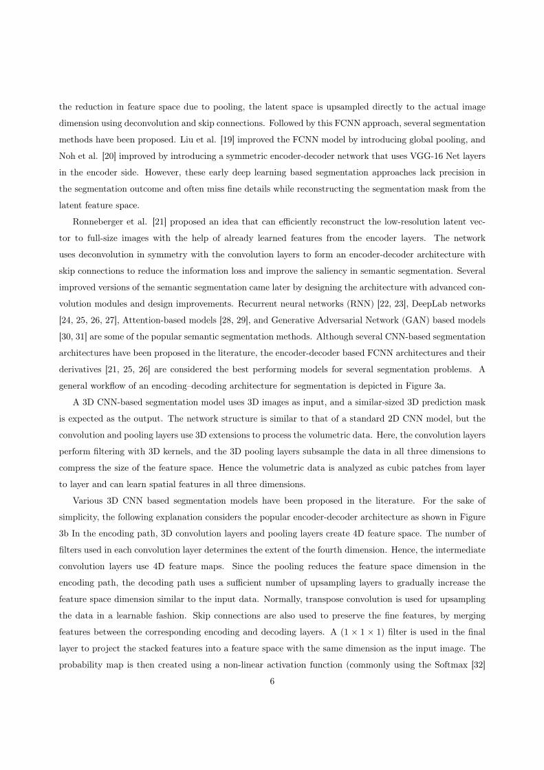
general workflow of an encoding–decoding architecture for segmentation is depicted in Figure 3a.
A 3D CNN-based segmentation model uses 3D images as input, and a similar-sized 3D prediction mask
is expected as the output. The network structure is similar to that of a standard 2D CNN model, but the
convolution and pooling layers use 3D extensions to process the volumetric data. Here, the convolution layers
perform filtering with 3D kernels, and the 3D pooling layers subsample the data in all three dimensions to
compress the size of the feature space. Hence the volumetric data is analyzed as cubic patches from layer
to layer and can learn spatial features in all three dimensions.
Various 3D CNN based segmentation models have been proposed in the literature. For the sake of
simplicity, the following explanation considers the popular encoder-decoder architecture as shown in Figure
3b In the encoding path, 3D convolution layers and pooling layers create 4D feature space. The number of
filters used in each convolution layer determines the extent of the fourth dimension. Hence, the intermediate
convolution layers use 4D feature maps. Since the pooling reduces the feature space dimension in the
encoding path, the decoding path uses a sufficient number of upsampling layers to gradually increase the
feature space dimension similar to the input data. Normally, transpose convolution is used for upsampling
the data in a learnable fashion. Skip connections are also used to preserve the fine features, by merging
features between the corresponding encoding and decoding layers. A (1 × 1 × 1) filter is used in the final
layer to project the stacked features into a feature space with the same dimension as the input image. The
probability map is then created using a non-linear activation function (commonly using the Softmax [32]
6

(a) A 2D CNN architecture for segmentation.
(b) A 3D CNN architecture for segmentation.
Figure 3: 2D and 3D CNN architectures for segmentation.
7

activation function to limit the probability values between 0 and 1), followed by thresholding which creates
the final prediction mask. The key differences between the 2D and 3D deep learning methods for image
segmentation are listed below.
1. In 2D CNN, the convolution kernel moves in two (x and y) directions, while in 3D convolution, it
moves in three (x, y, and z) directions.
2. 2D convolution can learn only 2D spatial features while 3D convolution can also extract inter-slice
information from adjacent frames.
3. In 2D CNN, the model accepts 2D images (or matrices) as the input data and provides corresponding
2D segmentation maps. In 3D CNN, both the input and output are 3D in nature.
4. The input and output feature space of 2D convolution layers (except in the input and final layers) are
3D in nature. Similarly, the input and output feature space of 3D convolution layers are 4D.
5. Since the convolution operations in a 3D CNN are significantly higher, and it needs more memory
space to save the parameters and feature space than 2D CNN.
2.3. An Overview on 3D Medical Data
Medical images are acquired with different imaging principles. The characteristics of these images differ,
in terms of spatial resolution, image intensity range, size of the image, and noise. In addition, they vary in
terms of dimensionality and data representation. 2D data mostly use relatively simple Euclidean represen-
tation, which describes each data point using pixel intensity values. In medical images, these pixel values
represent the state of the anatomical structure under consideration. For instance, the pixel intensity of X-
ray images varies with the radiation absorption, whereas it depends on the acoustic pressure in ultrasound
images or the radio frequency (RF) signal amplitude in MRI.
The representation of 3D data is more complex. Nevertheless, several standard representations are
reported in the literature, such as projections, volumetric representations, point clouds, and graphs [33].
The majority of the 3D medical image data belong to the volumetric representation, where volumetric
elements or voxels (analogous to pixels in a 2D image) are used to model 3D data by describing how the
3D object is distributed through the three perpendicular axes. While acquiring 3D medical data, the device
scans the body parts in any of the three planes: axial (front to back), coronal (top to bottom), and sagittal
(side to side). Normally, multiple 2D slices are acquired across the area under consideration and are stacked
to produce 3D image volumes using appropriate image registration techniques [34, 35, 36].
The 3D image visualization has provided a great opportunity for clinicians to evaluate the cross-section
of anatomic structures. This has increased the understanding of the complex patterns and structural mor-
phology, mostly in radiology. Currently, 3D imaging is commonly used in several modalities such as CT,
8

MRI, USG, and PET. Although 3D imaging techniques have numerous advantages over 2D images, they
have certain limitations as well. For example, compared to 2D imaging methods, they require significantly
large storage space and are often expensive. However, over time visualization and interpretation of 3D med-
ical data have become simple, thanks to modern deep learning algorithms and the availability of powerful
graphics processing units (GPUs) [37].
2.4. Why 3D CNN is Important in 3D Medical Image Segmentation?
Several medical image segmentation approaches proposed in the literature use 2D deep learning methods
[38, 39]. Though 2D CNN can learn the spatial relationship between the pixels in a 2D plane, it cannot
learn the inter-slice relationship between the frames. In medical images such as X-ray or Fundus images,
information is restricted into a 2D plane, and hence 2D convolutions can effectively detect relevant features.
However, in 3D volumetric data such as MRI, CT, or USG, the region of interest may spread across multiple
frames, and hence the inter-slice information became significant. In such cases, individual slice analysis with
2D convolutions cannot retrieve all useful information and may affect the segmentation performance. On
the other side, 3D CNN uses convolution kernels in three directions, and hence the inter-slice information
can be learned to provide better segmentation results. For instance, Table 1 shows a comparison of 2D
and 3D CNN in terms of computation complexity and memory requirements while segmenting brain tumor
regions from MR images.[40].
The 2D and 3D UNet models used in this analysis are identical except in the convolution and pooling
operation. The study uses a 3D dataset containing 224 samples, each sample having a size of 256×256×32.
The same dataset is used in 2D CNN after reshaping, consisting of 7168 2D samples, each having a size of
256 × 256. This ensures that the total number of frames used for training is identical for both 2D CNN
and 3D CNN models. The number of filters in each convolution layer is designed to make the trainable
parameters comparable for both models. The training time required to complete one epoch in the 3D CNN
model is almost half the time required in the 2D CNN model. This advantage in computation time in
3D CNN is due to the optimization in vector multiplication while using more parameters in a single 3D
convolution kernel. However, in practice, 3D CNN may require a large number of samples to achieve high
segmentation accuracy, which increases the computation complexity. Since 3D CNNs have to be processed
on 4D feature maps, the graphical memory requirement is also higher than a 2D network, and can be
preferred over 2D CNN models when the inter-slice information is crucial and hardware resources with high
performance computing are available.
2.5. 3D CNN in Medical Image Segmentation
3D CNNs have been successfully used in several medical image segmentation tasks and classified into
various subgroups based on different learning approaches. In this study, we considered 3D CNN papers for
9

Table 1: Performance comparison of 2D and 3D UNet, in terms of number of trainable parameters (TN ), training time per
epoch TT and GPU memory required TM .
CNN ModelsNo. of samples
used for training
Sample
size
TN
(in million)
TT
(in seconds)
TM
(in GB)
3D UNet 224 256× 256× 32 1.656 52 2.22
2D UNet 7168 256× 256 1.692 108 0.176
medical image segmentation published between 2015 and 2021 and are classified into one of the following
categories:
1. Fully supervised 3D CNN models
(a) Direct 3D CNN models
(b) 3D Patch-wise segmentation models
(c) Multi-task learning models
2. 3D CNN with Semi-supervised learning
3. 3D CNN with Weakly-supervised learning
4. Cost-effective approximations of 3D CNN
Here the main categorization is based on whether or not the model is learning from fully annotated 3D
data. The fully supervised 3D CNN uses image volumes with corresponding labeled masks for the training
process. Many works in the literature belong to this category as the approach is straightforward with less
complexity in the training process [8, 38]. Based on the implementation of the 3D CNN architecture and the
nature of the learning paradigm, they are further classified into three main sub-classes. More explanation
of these models are given in section 2.5.1. Since the labeling of large medical image datasets is challenging,
research also expanded to deep learning models that can learn from limited labeled samples. In recent years,
a few 3D CNN papers were also proposed in these semi-supervised and weakly-supervised medical image
segmentation tasks and are included in the current literature review.
3D CNN models demand substantially more memory, and various studies have been published that can
simulate 3D CNN in order to extract inter-slice features using cost-effective approximations [41, 42, 43].
This survey also discusses a few of these efforts in order to get a better understanding of 3D medical image
segmentation. The subsequent sections performs a detailed review and provides in-depth comparison of
state-of-the-art 3D medical image segmentation methods within each category.
10

2.5.1. Fully supervised 3D CNN models
1. Direct 3D CNN models
A straightforward implementation of a 3D CNN model is possible by replacing the 2D convolution
and pooling operations in a conventional CNN model with 3D convolution and pooling. A basic
representation of such a 3D architecture is shown in Figure 3b. Such CNN models that use 3D
equivalents of existing 2D segmentation architectures are reviewed under this category.
Milletari et al. [44] presented an advanced 3D U-Net architecture (V-Net) with residual blocks (instead
of cascaded convolution blocks) and strided convolution (instead of max-pooling). The methodology
use a Dice score based loss function to reduce the class imbalance between the voxel classes. This
model was evaluated on the PROMISE2012 dataset [45], and the results were very close to the state-
of-the-art 2D segmentation models. Bui et al. [46] proposed another 3D CNN architecture inspired by
the 2D FCNN model proposed by Long et al. [18] for brain segmentation in infant brain MRI volumes
[47]. The encoder path uses multiple coarse and fine dense 3D convolution layers to extract multi-scale
features. Downsampling makes use of strided convolution to reduce the feature size and to increase the
receptive field. Multiple convolution layers are used to extract four different scales of feature space,
then upsampled and concatenated to generate the final probability map. However, direct merging of
the upsampled features in the decoder path creates semantic gaps in the feature space, and may create
undesired outcomes in the segmentation results.
In [48], Kayalibay et al. proposed another 3D encoder-decoder architecture with deep supervision [49].
In this model, the feature space created at different decoder levels in the network are merged using an
element-wise summation of the features. Hence, the learning is based on the error between the ground
truth and a combination of feature maps from different decoding levels. In the approach, the learning
process is directly dependent on the coarse features from the different decoder levels, and helps to
speed-up the convergence. However, the element-wise summation may limit the learning process when
the semantic gap between the different decoder levels is significant. Dou et al. [50] proposed a similar
approach that can work on relatively small 3D datasets. The model uses an encoder-decoder FCNN
with 3D convolutions. The outputs from each decoder layer are upsampled using 3D deconvolution and
merged to get the final segmentation mask. The integration of deep supervision and the post-processing
using CRF [51] helped to improve the overall segmentation performance.
In [52], Li et al. presented a 3D CNN model using dilated convolution [53] instead of max-pooling,
and residual connections. For utilizing multi-scale features, the dilation factor of the 3D convolution
kernel is steadily increased in the subsequent layers. Hence, the spatial resolution of the feature space
is kept constant throughout the network. The residual blocks merge the features from different layers
to reduce vanishing gradients problem and feature degradation. However, the absence of max-pooling
11

reduces the translation invariance in the feature space and accelerates the computation complexity.
Chen et al. [54] proposed a residual deep 3D CNN architecture for segmenting the brain region from
3D MRI volumes. The proposed architecture employs a voxel-wise residual network (VoxResNet). The
architecture uses a 3D extension of 2D deep residual networks that extracts features from multiple
scales using a series of convolution operations, and the features at multiple scales are fused to generate
the segmentation mask. For training such a deep network with small training data, multi-modal
contextual information is integrated into the network to take advantage of additional information
from various MRI sequences. Experiments were conducted using three MRI sequences: T1, T1-IR,
and T2-FLAIR from the brain structure segmentation task [55].
In [56], Niyas et al. presented a 3D Residual U-Net architecture for segmenting focal cortical dysplasia
(FCD) from 3D brain MRI. The method aims to retain the advantages of both 2D and 3D CNN
methods by an effective design of input data slices and CNN architecture. The model proposes a
shallow sliced stacking approach to generate large number of 3D samples from small datasets. The
customized 3D U-Net architecture with residual connections in the encoder path helps in extracting
multi-scale features with fewer trainable parameters.
Schlemper et al. [57] proposed an attention gate (AG) based 3D U-Net architecture for medical
image segmentation which automatically learns to concentrate on various target structures. The
features from the encoding path to the decoding path are propagated through the skip connections
with self-attention gating modules [58]. The AG module uses grid-based gating that allows attention
coefficients to focus more on local neighborhoods, that helps to locate the relevant regions in the
image. Performance improvements of the proposed network over U-Net are experimentally observed
to be consistent across different imaging datasets [59], [60]. However, in an FCNN with encoder-decoder
architecture, features in the lower depth are minimal and naive, while the features at advanced depths
are of higher granularity. Hence, the attention modules at shallow depths may not help passing the
salient features to the upsampling layers [61].
Wang et al. [62] proposed another 3D FCNN method that integrates recursive residual blocks and
pyramid pooling to extract more complex features. The recursive residual blocks contain multiple
residual connections that can minimize feature degradation problems. Pyramid pooling [63] generates
fused feature maps at different decoding levels for obtaining both local and global information. It
helps to eliminate the fixed size constraints of CNN without losing spatial information. The proposed
architecture gave better multi-class segmentation performance while detecting white matter (WM),
gray matter (GM), and cerebrospinal fluid (CSF) from 3D MR images from CANDI, IBSR18, and
IBSR20 datasets [64, 65].
A 3D version of multi-scale U-Net segmentation was presented in [66] by Peng et al. The model uses
12

multiple U-Net modules to extract long-distance spatial information at different scales. The U-Net
blocks use Xception [67] modules instead of normal convolution to extract more complex features.
Feature maps at different resolutions are upsampled and fused to generate the segmentation mask.
The authors claimed that the upsampled feature maps at different scales could extract and utilize
appropriate features with faster learning. Further, the 3D convolutions are replaced with depth-wise
separable convolutions to reduce the computation complexity. However, the complex structure of the
overall network and the deep convolution layers demand high memory and computation requirements.
The model cascade (MC) strategy is one of the popular strategy in CNN-based image segmentation
that can alleviate the class imbalance problem by using a set of individual networks for coarse-to-fine
segmentation. Despite its notable performance, it leads to undesired system complexity and overlook
the correlation among the models. Zhou et al. [68] proposed a lightweight deep 3D CNN architecture:
one-pass multi-task network (OM-Net) that can handle the native flaws in the MC approach. OM-Net
combines discrete segmentation tasks into a one-pass deep model, to learn joint features and solve
the class imbalance problem better than MC. The prediction results between tasks are correlated
using a cross-task guided attention (CGA) block that can adaptively re-calibrate channel-wise feature
responses. The model shows significant performance improvements in multiple public datasets [40, 69].
In the above-mentioned medical image segmentation approaches, the 3D CNN models mainly use a
straight-forward extension of various 2D CNN building blocks. However, the volumetric data analysis
brings a significant increase in the memory requirement and computation costs while training deep
3D CNN models compared to corresponding 2D versions. Several studies were also conducted in
developing 3D segmentation models that works with limited training data and hardware resources.
Such approaches are discussed in the following sections.
2. 3D Patch-wise segmentation models
The resolution of input 3D data is the primary reason behind the need for large memory and higher
computation complexity in training a 3D CNN segmentation model. However, this could be circum-
vented with a patch-based approach. In patch-wise approach, the input 3D images are converted to
small 3D sub-samples and analyzed individually. Finally, the patch-wise prediction outcomes will be
merged to create the final 3D prediction map. A high-level block diagram of 3D Patch-wise segmen-
tation is shown in Figure 4. The patch-wise analysis is an interesting are of research in 3D medical
image analysis, and such patch-wise 3D deep learning based techniques are discussed in this section.
Yu et al.[70] proposed a deep supervised 3D fractal network for whole heart and great vessel segmen-
tation in MRI volumes. This method is an expansion of the FractalNet [71] and uses deep supervision
using multiple auxiliary classifiers deployed at expanding layers to reduce the vanishing gradient prob-
13

Figure 4: A high-level block diagram of 3D Patch-wise segmentation model.
lem. The methodology uses cropped 3D patches of size (64 × 64 × 64) and reported good results in
the HVSMR 2016 challenge dataset [72].
A deep dual pathway 3D CNN architecture was proposed by Kamnitsas et al. [73] for brain lesion
segmentation. They employed a two-way network that simultaneously learns from multiple image scales
to analyze features from different receptive fields. The approach uses 3D patches at two different scales
and classifies the center voxels as any target classes using fully connected dense layers. A 3D fully
connected conditional random field (CRF) is also utilized in the post-processing stage to reduce false
alarms in the final segmentation mask. This method shows improved segmentation results over BRATS
2015 [40] and ISLES [74] datasets, compared with various 2D CNN methods. An advanced version
of this model was also proposed in [75], which used multiple residual connections in the encoding
path. The model reported better segmentation performance and was bench-marked at BRATS 2016
challenge [76].
In [77], Kamnitsas et al. presented an ensembles of multiple models and architectures (EMMA) for
robust performance by aggregating predictions from multiple models. This method use an ensemble of
a DeepMedic model [75], three 3D FCNN models [18], and two 3D U-Net models [21]. An ensembler
computes the confidence maps, and finds the average class confidence of the individual models. Finally,
each voxel in the segmentation map was labeled with the class label with the highest confidence score.
The EMMA approach won the first place in the BRATS 2017 challenge [40, 69, 78]. Nevertheless, the
ensemble with seven 3D architectures requires higher computation complexity and training time.
In, [79], Chen et al. proposed a hierarchical 3D CNN architecture to segment Glioma from multi-modal
brain MRI volumes. The model uses two distinct scales of 3D image patches to examine multi-scale
features. The 3D CNN architecture uses a series of hierarchical dense convolutions without pooling
layers. The model uses a patch-wise analysis that reduces the class imbalance and the computational
14

cost involved in training large 3D datasets. This method was validated on the BRATS 2017 [69] dataset
and reported better segmentation performance compared to similar patch-wise 3D approaches.
The patch-based segmentation helps to reduce the hardware resources for training and generates a
large number of data samples that favor adequate learning. However, the patch-wise analysis often
fails to extract global features from the actual image volumes. This may limit the learning performance
when the abnormality is region-specific.
3. Multi-task learning models
Multi-task learning (MTL) is a machine learning approach that can assess multiple related tasks with a
single network [80]. MTL is advantageous in medical images because many tasks such as classification
and segmentation may be required concurrently throughout the diagnostic process [81]. Graphical
representation of a 3D CNN based MTL is shown in Figure 5. This section discusses a few such
approaches that use 3D CNN for multi-task learning.
Figure 5: A high-level block diagram of 3D CNN based Multi-Task learning model.
Zhou et al. [82] proposed multi-task learning of classification and segmentation using a 3D CNN
model for classifying tumors in breast ultrasound images. The authors used a modified version of 3D
V-Net [44] as the backbone of this model. To conduct a multi-scale analysis, feature maps from three
higher-level encoder and decoder layers of the segmentation network are fused together to perform the
classification task. Sharing higher-level feature maps and a multi-task loss function encourages the
network to learn common generic features for classification and segmentation.
Another multi-tasking approach has been reported by Gordaliza et al. [83] to infer tuberculosis from
15

CT images. The model uses higher-order encoder features and processes two individual feed-forward
neural networks to understand the tuberculosis condition. The approach also used several optimiza-
tions such as self normalization, the use of scaled exponential linear unit (SELU) activation function,
and uncertainty weighted multi-task loss to improve the performance both for detection and counting
the number of nodules.
Ge et al. [84] proposed a multi-task learning approach for segmenting and quantifying left ventricle
(LV) from paired apical views (A4C and A2C) of echo sequences. The method offers an overall cardiac
analysis using a multi-task network: K-Net, an end-to-end network that can simultaneously segment
LV and quantify its extent over the 3D plane. The methodology uses 2D convolution in different stages
to segment and quantifies the 3D structure of LV using complex echo data. The reported results over a
sufficiently large echo dataset also prove the proposed model’s superiority in the heterogeneous learning
approach.
2.5.2. Semi-supervised Learning
The 3D CNN models discussed in Section 2.5.1 mostly use fully supervised deep learning algorithms
that require thousands of annotated 3D volumes. However, accurate marking of ground-truth images is a
labor-intensive and tedious process. Hence, the supervised learning algorithms are more expensive in terms
of both time and cost. Consequently, research also commenced on alternatives to process sparsely annotated
training data. Semi-supervised learning (SSL) methods [85, 86, 87] are one of those types that require a few
labeled image samples with a large number of unlabeled samples, and such SSL based 3D medical image
segmentation works are discussed in this section. A basic 3D SSL framework is shown in Figure 6.
In [88], Çiçek et al. presented one of the first promising 3D CNN network for volumetric segmentation
that learns from sparsely labeled 3D images. The proposed model extends the classical U-Net architecture
[21] by substituting all 2D operations with their 3D equivalents. The study in [88] outlined two cases: a semi-
automated model and a fully automated model. In both cases, the network learns from sparse annotated
data and this helps to reduce the human effort in ground truth labeling without considerable degradation in
the segmentation performance. The experiments were conducted on the Xenopus kidney dataset [89], and
both the semi-automated and fully automated models showed considerable performance improvements over
state-of-the-art 2D CNN architectures.
In [90], Mondal et al. proposed a 3D multi-modal medical image segmentation method based on gener-
ative adversarial networks (GANs) [91]. The method uses a semi-supervised training with a mix of labeled
and unlabeled images. The proposed architecture uses several design considerations to modify the standard
adversarial learning approaches to generate 3D volumes of multiple modalities. The 3D GANs generate fake
samples and are used along with labeled and unlabeled 3D volumes, and a discriminator defines separate
loss functions for these labeled, unlabeled and synthetic (fake) training samples. However, generating useful
16

Figure 6: A high-level representation of a 3D CNN based Mean teacher Semi-supervised learning model.
synthetic samples may not represent the actual data distribution and thus becomes challenging working
with 3D medical image data.
Zhou et al. [92] proposed a straight-forward semi-supervised segmentation approach named deep multi-
planar co-training (DMPCT), which uses parallel training to extract information from multiple planes. The
multiplanar fusion generates reliable pseudo labeling to train deep segmentation networks. The DMPCT
framework consists of a teacher network that uses fully labeled images for training. The trained model then
creates the pseudo labels from the unlabelled training data with a multi-planar fusion module. Subsequently,
the student model uses both labeled and pseudo labeled data for the final training process.
Yang et al. [93] proposed a similar semi-supervised segmentation technique to detect catheter from vol-
umetric ultrasound images. The segmentation model uses a deep Q network (DQN) for localizing the target
region. After the catheter localization, the method uses a twin-UNet model for the semantic segmentation
of the catheter volume around the localized region by a patch-based strategy. This model uses a typical
teacher network followed by a student network to train both labeled and unlabeled 3D patches based on a
set of hybrid constraints.
In [94], Li et al. presented a shape-aware 3D segmentation for medical images to use extensive unlabeled
17

data so as to enforce a geometric shape analysis on the segmentation output. The model uses a deep CNN
architecture that predicts semantic segmentation and signed distance map (SDM) of object surfaces. The
network uses an adversarial loss between the predicted SDMs of labeled and unlabeled data during training
to leverage shape-aware features. The integration of adversarial loss, which uses a generative discrimination
function, helps supervise learning with unlabelled data and extract generalized features.
In [95], Wang et al. proposed a tailored modern semi-supervised learning (SSL) method named as
FocalMix for the detection of lesions from 3D medical images. The model is built on MixMatch [96] SSL
framework, which uses a prediction for unlabeled images and MixUp augmentation. The proposed 3D CNN
model uses a Soft-target Focal Loss along with an anchor level target prediction to improve lesion detection.
The study also uses two adaptive augmentation methods: image-level MixUp and object-level MixUp, to
generate the final training data.
Zhang et al. [97] proposed a 3D medical image segmentation model using semi-supervised 3D CNN. This
dual-task mutual learning model uses two side-by-side frameworks. One network works on the region-based
shape constraint, while the learning in the other network focuses on boundary-based surface mismatch. The
main contribution of this model is the use of a signed distance map (SDM) and the conventional ground truth
maps to get a better intuition of region features and shape features together. Both networks use an identical
3D V-Net architecture to learn the voxel characteristics with a shared loss function. During the training with
labeled image volumes, the loss function concentrates more towards the difference in segmentation map and
actual ground truth. On the other hand, while training with unlabeled images, the model uses a consistency
loss function based on distance maps to ensure prediction consistency while working with similar images.
Hence, the dual network tries to reduce both losses and aids a better segmentation accuracy.
Another semi-supervised method is presented by Li et al. [98] which uses a 3D CNN-based mean teacher
framework with hierarchical consistency regularization for 3D left atrium MR images. The model felicitates
the prediction consistency between the teacher and student network at multiple scales. During training, the
student network uses multi-scale deep supervision while hierarchically regularizing the network’s prediction
consistency. Hence, the model learns from both the labeled and unlabeled volumes by minimizing the
supervised loss and consistency loss concurrently.
In semi-supervised approaches, the training demands a subset of data with accurately marked ground
truths. Usually, multiple networks are used in semi-supervised models to process the labeled and unlabeled
data using a shared feedback mechanism. The learning is often synchronized by evaluating the segmentation
losses and consistency in predicting unlabeled data. The performance of a semi-supervised model highly
depends on this feedback and the accuracy of the annotated images. Since SSL requires precise annotation
(for a subset of data), it is challenging while dealing with large 3D image datasets. Hence, several algorithms
that can learn from weakly labeled data have also been published in the medical image segmentation domain.
Such methods will be discussed in Section 2.5.3.
18

2.5.3. Weakly-supervised Learning
Weakly supervised is a learning paradigm that uses noisy or low-quality annotations in the learning
process. In weakly-supervised learning, the data labeling need not be as highly accurate as in the case of fully
supervised learning. In medical image segmentation, weakly-supervised learning is highly significant as the
abstract level annotation is relatively easier and may be accomplished by non-experts with minimal support
from radiologists. The labels for weakly-supervised learning can also be made from batch clustering or
from noisy predictions from comparable pre-trained models. Hence, the data preparation becomes relatively
cheaper and practical, but at the cost of more noise or false labeling in the training data. A block level
representation of a weakly-supervised 3D CNN is shown in Figure 7. Several 3D CNN models with weak
supervision have been reported recently, and those works are discussed here.
Figure 7: Block diagram of a Weakly-supervised 3D CNN model.
Yang et al. [99] proposed a weakly-supervised method for segmenting catheters from 3D frustum ultra-
sound images. The methodology use data annotated with 3D bounding boxes over the catheter regions. A
pseudo label generator module is introduced here to reduce the impact of inaccurate ground truth marking.
This model uses a sequential network with a localization module to detect frustum volumes, followed by
the segmentation stage to extract catheter voxels. The localization network used 3D ResNet-10 encoder
architecture, and the feature maps are then converted to the final segmentation map using a decoder with
multiple transpose convolutions. This approach makes the frustum segmentation faster and cheaper without
any significant degradation in the segmentation performance.
Wu et al. [100] proposed a weakly-supervised brain lesion segmentation using attentional representation
learning from 3D image volumes with image-level annotation. The approach used an attention mechanism
that is dimensionally independent on the class activation map (CAM) and can estimate the lesion labels with
minimum demands of the trainable parameters and learn the representation model from the dimensional
independent CAM to extract the foreground voxels.
Another similar weakly-supervised 3D CNN-based segmentation is proposed by Zhu et al. [101] that
19

requires only image-level class labels. The proposed model: CIVA-Net, uses weakly annotated labels for
volumetric image segmentation on 3D cryo-ET datasets. The input to the network is image-level class
labels, and a pre-processing seed generation stage is used initially for generating pseudo labels for each
voxel. Using the cross-image consensus stage, the similar voxel groups are merged using co-occurrence
learning to generate the pseudo localization map. An inter-voxel affinity learning is also proposed to analyze
the inter-pixel relationship from the pseudo localization map to forge the affinity voxel pairs. The final
segmentation stage uses VoxResNet [102] to predict the segmentation map using the pseudo localization
map and the affinity pairs generated from previous steps.
Weakly-supervised learning is highly recommended for 3D medical images where the voxel shares a
similar pattern in the region of interest. However, the pseudo labeling of the region of the region of interest
just from the image class label is highly susceptible to errors and can reduce the segmentation performance
significantly.
2.5.4. Cost-effective approximations of 3D CNN models
Since the straight-forward 3D CNN models are highly susceptible to large computation costs and require
large datasets, several alternative techniques have been proposed that can use simulated 3D architectures
with cost-effective approximations. For instance, 2D frames from a 3D image can be processed in three
orthogonal directions and then merged to create the final 3D prediction mask, and such a model is shown in
Figure 8. Numerous prominent approximations for segmenting 3D medical images have been reported and
discussed in this subsection.
Figure 8: A cost-effective approximation of 3D CNN model.
Extraction of inter-slice features from a 3D volume is possible by analyzing three orthogonal planes
(sagittal, coronal, and transverse planes) and by classifying the voxel at the intersection of three planes.
20

This method was successfully applied by Ciompi et al. [41] for classifying lung nodules. This model learn
from 2D patches in three perpendicular planes centered at a given voxel, at three different scales. Fully
connected layers combine the streams and perform the voxel classification. However, CNN-based models
with fully-connected layers are computationally less efficient for segmentation than FCNN architectures. A
similar 3D segmentation approach by combining information from three orthogonal planes was discussed
by Kitrungrotsakul et al. [103]. The proposed method (VesselNet) uses 2D DenseNet [104] network for
classifying voxels in three different 2D planes. The features from the individual networks are then fused
for getting the probability to predict the segmentation map. However, this approach is not an end-to-end
segmentation model and hence misses most contextual and location-based information.
Mlynarski et al. [42] proposed a similar CNN architecture that combines the benefits of the short-range
3D context and the long-range 2D context. This architecture uses modality-specific sub-networks to focus
on missing MR sequences. During training, three individual 2D U-Net models were used to create features
from axial, coronal, and sagittal slices. The final 3D network was then trained using these 2D features along
with 3D patches from the input volumes. The study also suggests considering the outputs from multiple 3D
models to minimize the constraints of specific choices of neural network models. However, the use of multiple
2D and 3D models demands more hardware resources, and patch-wise analysis fails to learn region-specific
features.
Another 3D CNN model was introduced by Heinrich et al. [105], which uses trainable 3D convolution
kernels that learn both filter coefficients and spatial filter offsets in a continuous space, based on the principle
of differentiable image interpolation. The proposed One Binary Extremely Large and Inflecting Sparse Kernel
(OBELISK) works with fewer parameters and reduced memory consumption. The deformable convolutions
in the OBELISK filter replace the continuously sampled spatial filter with a sparse sampling approach. This
helps to extract information from wider spatial contexts and replace multiple small filter kernels at different
scales. However, the computation complexity may be higher in OBELISK due to the unoptimized filter
sampling.
Roth et al. [106] proposed an automated segmentation system for 3D CT pancreas volumes based on a
dual-stage cascaded method. It included a localization method followed by a segmentation. 2D holistically
nested convolutional networks (HNN) [107] on the three orthogonal directions, were used in the localization
stage. The 2D HNN pixel probability maps are then merged to get a 3D bounding box of the foreground
regions. In the second stage, the model focuses on semantic segmentation over the voxels in the bounding
box. Two different HNN realizations are integrated in the segmentation stage that extract mid-level cues
of deeply-learned boundary maps of the target organ. The authors also presented an advanced multi-class
segmentation model [108] for segmenting the liver, spleen, and pancreas from CT volumes. The cascaded
network helps to provide boundary preserving segmentation and reduces false detection in the 3D volumes.
Chen et al. [109] presented a separable 3D U-Net architecture that targets to extract spatial information
21

from the image volumes with limited memory and computation cost. The model uses a U-Net architecture
for the end-to-end training with separable 3D (S3D) convolution blocks. The S3D block is an arrange-
ment of parallel 2D convolutions and exploits the advantages of the residual inception architecture. The
model is evaluated on BRATS 2018 [110] data, and the results justify the improvement in the segmentation
performance compared to a standard 2D or 3D U-Net architecture.
Another 3D CNN architecture proposed by Rickmann et al. [43] incorporates a compress-process-
recalibrate (CPR) pipeline using 3D recalibration methods. The method uses a project & excite (PE)
module that compress the intermediate high dimensional 4D feature maps into three 2D projection vectors.
The convolution layers in the processor module learns from this 2D projections and the final recalibration
stage generate the 4D tensors for the subsequent layers. This approach reduces the computation cost, with-
out degrading the segmentation performance. The paper reported improved overall accuracy over different
multi-class segmentation datasets [111, 112, 113, 114].
The volumetric medical segmentation using the above-discussed approaches is highly useful in reducing
the computation complexity and the need for large datasets. The results in the above discussed works prove
its supremacy over other conventional 2D and 3D CNN approaches. However, the 3D approximations are
highly dependent on the characteristics of the input data and the type of segmentation task. This may limit
the generic use of models across different medical image modalities, and the designing of such a universal
model remains an open research challenge.
Some pre-trained 3D medical segmentation models have also been discussed in the literature. Chen et al.
[115] propose a heterogeneous 3D network called Med3D by co-training multi-domain 3D datasets to develop
multiple pre-trained 3D medical image segmentation models. The authors use datasets from various medical
challenges to create a 3D segmentation data set (3DSeg-8) with different modalities, target organs and
pathologies. The pre-trained models were experimented on several 3D medical datasets [116] using transfer
learning to achieve performance gain and faster convergence. The model can work well on similar image
modalities with less training time and can be considered as a suitable option for small 3D image datasets.
Zhou et al. [117] proposed a similar set of pre-trained 3D CNN networks for classification and segmentation
tasks in CT and MRI. The models are collectively known as Generic Autodidactic Models (Models Genesis),
which uses learning by self-supervision. However, transfer learning may not be an appropriate solution
in various scenarios due to the difference in image features across different image modalities and targeted
abnormalities.
We summarize various medical image segmentation approaches using the 3D deep learning techniques
in Table 2. The CNN architecture used, datasets, and remarks are also included in this table. A detailed
discussion regarding the currently facing challenges in 3D medical image segmentation, possible solutions,
and future directions are discussed in Section 3.
22

Reference Dataset Remarks
Direct 3D CNN models
Milletari et al. [44] PROMISE2012 MRI dataset
[45]
3D V-Net with residual blocks and strided convo-
lution.
Bui et al. [46] Infant brain MRI (iSeg [47]) D FCNN with dense blocks and strided convolu-
tion.
Kayalibay et al.
[48]
Hand MRI [118], BRATS
2013 [119], and BRATS 2015
[40]
3D U-Net with deep supervision.
Dou et al. [50] CT Liver (SLiver07 [120]) &
Heart MRI (HVSMR [72])
3D U-Net with deep supervision and CRF [51]
based post-processing.
Li et al. [52] Brain MRI (ADNI [121]) 3D Res-Net with dilated convolution.
Chen et al. [54] Multi-modal Brain MRI
(MRBrainS [55])
VoxResNet: a multi-modal dataset to learn from
small datasets.
Niyas et al. [56] Brain MRI (Private dataset) 3D Residual U-Net with shallow sliced stacking
for generating 3D samples.
Schlemper et al.
[57]
Abdominal CT datasets [59,
60]
Attention U-Net that can learn region specific fea-
tures.
Wang et al. [62] Brain MRI (CANDI [64], and
IBSR [65])
3D U-Net with pyramid pooling [63] for fusing
feature maps from different decoding levels.
Peng et al. [66] Brain MRI (BRATS 2015
[40])
Multiple U-Net architectures with Xception [67]
blocks.
Zhou et al. [68] Brain MRI (BRATS 2015
[40], BRATS 2017[69, 78],
and BRATS 2018 [110])
OM-Net integrated with a cross-task guided at-
tention (CGA) module.
3D Patch-wise Segmentation
Yu et al.[70] Heart MRI (HVSMR [72]) 3D Fractal network with deep supervision.
Kamnitsas et al.
[75]
Brain MRI (BRATS 2015 [40]
and BRATS 2016 [76])
DeepMedic: Multi-scale 3D CNN with residual
connections with 3D fully connected CRF.
Kamnitsas et al.
[77]
Brain MRI (BRATS 2017 [69,
78])
An ensemble of two DeepMedic models, three 3D
FCNN models, and two 3D U-Net models.
Chen et al. [79] Brain MRI (BRATS 2017 [69,
78])
Multi-modal 3D CNN using 3D patches at two
different scales.
Continued on next page
23

Table 2 – Continued from previous page
Reference Dataset Remarks
Multi-task learning models
Zhou et al. [82] 3D Breast Ultrasound (Pri-
vate dataset)
Multi-task network for both classification and
segmentation.
Gordaliza et al. [83] Chest CT (Private dataset) Multi-tasking using self normalized neural net-
works.
Ge et al. [84] Echo images (A4C and A2C)
(Private dataset)
Segmentation and quantification of multi-view
echo sequence using K-Net.
Semi-supervised Learning
Çiçek et al.[88] Xenopus kidney dataset [89] 3D U-Net with sparsely annotated volumes.
Mondal et al. [90] Infant brain MRI (iSeg [47] &
Multi-modal brain MRI (MR-
BrainS [55])
The 3D GAN [91] that generates synthetic sam-
ples to train with labeled and unlabeled 3D vol-
umes
Zhou et al. [92] Abdomen CT (Private
dataset)
Deep multi-planar co-training (DMPCT).
Yang et al. [93] 3D heart ultrasound (Private
dataset)
A localization (using deep Q network (DQN)),
followed by semantic segmentation (using a dual
U-Net).
Li et al. [94] 3D heart MRI [122] 3D V-Net with signed distance map (SDM).
Wang et al. [95] Thoracic CT scans [123, 124] FocalMix: A model built on MixMatch [96] SSL
framework.
Zhang et al. [97] Atrial Segmentation Chal-
lenge MR dataset [122]
SSL framework that uses labeled images and
signed distance map for accurate segmentation.
Li et al. [98] Atrial Segmentation Chal-
lenge MR dataset [122]
Mean teacher framework with hierarchical consis-
tency regularization and multi-scale deep super-
vision.
Weakly-supervised learning
Yang et al. [99] ex-vivo dataset (Private
Dataset)
Use coarsely marked 3d data. The framework
contains ResNet-10 based classification followed
by segmentation.
Wu et al. [100] Brain MRI (BRATS 2017 [69,
78] & ISLES 2015 [125])
Weakly-supervised learning using image-level
class labeling.
Continued on next page
24

Table 2 – Continued from previous page
Reference Dataset Remarks
Zhu et al. [101] 3D cryo-ET Dataset [126] CIVA-Net: an end-to-end model that segment
foreground voxels from image-level class labeling.
Cost-effective approximations of 3D CNN models
Ciompi et al. [41] CT Lung (MILD [127]
dataset)
ConvNet: Multi-stream voxel classification in
three orthogonal planes at three different scales.
Kitrungrotsakul et
al. [103]
CT Liver (Private dataset) VesselNet: Voxel classification in three orthogo-
nal planes.
Mlynarski et al.
[42]
Brain MRI (BRATS 2017 [69,
78])
Used features from three orthogonal planes with
2D CNNs, and 3D image volumes.
Heinrich et al. [105] CT datasets [112, 128] OBELISK-Net that requires fewer trainable pa-
rameters.
Roth et al. [106] Abdomen CT dataset [128] A localization (using Holistically-nested convo-
lutional networks (HNN) on three orthogonal
planes) followed by a semantic segmentation
stage.
Chen et al. [109] Brain MRI (BRATS 2018
[110])
Separable 3D (S3D) U-Net: A parallel 2D convo-
lutions with a residual inception architecture.
Rickmann et al.[43] Multiple 3D medical datasets
[111, 112, 113, 114]
3D ConvNet with project & excite (PE) modules.
Table 2: Overview of articles using 3D deep learning techniques for medical image segmentation.
3. Discussion & Conclusion
3.1. Summary
In this study, we reviewed nearly 40 articles on 3D medical image segmentation using deep learning
techniques. The articles were classified into different categories according to the different 3D deep learning
approaches used to solve the segmentation problem. We believe that deep learning significantly contributes
to medical image segmentation with 3D medical images. Although 3D deep learning methods are consider-
ably less in use than 2D approaches, the exponential increase in the publications related to 3D deep learning
techniques in the past couple of years in medical image analysis is remarkable. Nevertheless, most of the
25

3D CNN-based segmentation approaches rely on traditional CNN architectures by updating the convolu-
tion kernel and pooling from 2D to 3D. This bypass is advantageous for easy implementation; however, it
introduces several optimization issues and restricts the analysis of 3D data to its full potential.
Numerous studies have focused on solving the inherent limitations of 3D deep learning, such as the
computation cost and huge memory requirements while processing large number of 3D samples. The need for
large volumetric datasets is another major challenge in 3D deep learning-based medical image segmentation.
The key aspects of such successful alternative approaches in 3D deep learning-based segmentation were also
reviewed in this study. The following subsections provide a detailed overview of the unique challenges faced
in 3D medical image segmentation and the future trends.
3.2. Challenges in 3D Medical Image Segmentation
Deep learning methods are associated with various challenges in medical image analysis, and the use of
3D CNN for volumetric medical image segmentation further increases the complexity. The primary challenge
faced by the researchers in implementing effective 3D CNN models is the need for large training data. The
use of picture archiving and communication system (PACS)-based tools significantly improved the medical
image management and storage standardization. However, huge amounts of archived medical data often
become worthless due to the lack of proper annotation and the inconsistency in data attributed to the
heterogeneity in imaging systems.
Most of the traditional 2D and 3D CNN models require task-specific labeled data, generally obtained
from domain experts. Working with 3D medical data involves a significant time investment, particularly
during slice by slice annotation. Similar to other supervised machine learning approaches, subjectivity in
labeling is another challenge faced during 3D segmentation. Multiple domain experts might be required to
decode the information corresponding to large 3D datasets. This may also introduce labeling noise in the
training data and reduce the efficiency of volumetric segmentation.
In medical image analysis, generalized feature representation is often limited by the intra-class variance
and inter-class similarity in large 3D datasets. Variations in image acquisition systems, contrast variations,
and noise are the major reasons for overlapping features among different classes that critically influence the
decision-making in unseen test data.
Another limiting factor in 3D medical image segmentation is class imbalance. The class imbalance may
result in over-segmentation of classes with a high voxel share. Generally, 3D medical image segmentation is
required for detecting abnormal or lesion regions from a scanned volumetric image. Hence, the ratio of voxels
belonging to the abnormal and normal classes is extremely low and seriously affects the training process.
The class imbalance problem may be more critical in multi-class segmentation cases. Training a 3D CNN
model on a slarge volumetric dataset is also challenging as it demands large memory and high computation
capability.
26

3.3. Future Directions
Most of the above-discussed challenges are inherent in the medical data and can not be refrained from
the source. Moreover, practically it is impossible to work with clean data. Hence, many studies are being
conducted to design deep learning models to circumvent such issues. Since 3D medical image segmentation is
primarily affected by the unavailability of labeled datasets, several recent studies have proposed techniques
that can learn from partially or coarsely annotated data. Semi-supervised and weakly-supervised learning are
some popular among those methods. Furthermore, such partially-supervised models can reduce the impact
of label error and subjectivity to a certain extent. However, such approaches are still in the early stage.
Hence, developing sophisticated semi-supervised 3D CNN models for volumetric medical image segmentation
is still an open challenge for the research community.
Another possibility for improving the 3D segmentation performance is by reducing the huge class imbal-
ance. Several attempts [129, 130, 131] have been made to reduce class imbalance by using asymmetric loss
functions to provide custom weights for different voxel classes. Although several alternative approaches, such
as selective sampling and adaptive augmentation, are available to reduce the class imbalance problem, such
strategies fail for highly imbalanced datasets. Hence, there is high scope for methods that address the class
imbalance in 3D medical data. 3D CNN can learn multi-dimensional features and contribute significantly to
multitask learning due to its comprehensive learning pattern. GAN-based data synthesis is another solution
to resolve several of the current 3D (or 2D) medical image segmentation challenges. GAN-based analysis
might be extremely relevant if it can generate real-like medical image volumes. Hence, such models for
generating synthetic images will be an interesting research topic in the future. We believe that this analysis
will help the research community to get better insights into current trends, future scopes, challenges and
thus formulate new ideas bridging the research gaps in 3D medical image segmentation.
References
[1] S. Chakraborty, S. Chatterjee, A. S. Ashour, K. Mali, N. Dey, Intelligent computing in medical imaging: A study, in:
Advancements in applied metaheuristic computing, IGI global, 2018, pp. 143–163.
[2] I. Goodfellow, Y. Bengio, A. Courville, Deep Learning, MIT Press, 2016, http://www.deeplearningbook.org.
[3] D. Shen, G. Wu, H.-I. Suk, Deep learning in medical image analysis, Annual review of biomedical engineering 19 (2017)
221–248.
[4] O. I. Abiodun, A. Jantan, A. E. Omolara, K. V. Dada, A. M. Umar, O. U. Linus, H. Arshad, A. A. Kazaure, U. Gana,
M. U. Kiru, Comprehensive review of artificial neural network applications to pattern recognition, IEEE Access 7 (2019)
158820–158846.
[5] Y. LeCun, B. Boser, J. S. Denker, D. Henderson, R. E. Howard, W. Hubbard, L. D. Jackel, Backpropagation applied to
handwritten zip code recognition, Neural computation 1 (4) (1989) 541–551.
[6] A. Dhillon, G. K. Verma, Convolutional neural network: a review of models, methodologies and applications to object
detection, Progress in Artificial Intelligence 9 (2) (2020) 85–112.
27

[7] S. Minaee, Y. Y. Boykov, F. Porikli, A. J. Plaza, N. Kehtarnavaz, D. Terzopoulos, Image segmentation using deep
learning: A survey, IEEE Transactions on Pattern Analysis and Machine Intelligence.
[8] G. Litjens, T. Kooi, B. E. Bejnordi, A. A. A. Setio, F. Ciompi, M. Ghafoorian, J. A. Van Der Laak, B. Van Ginneken,
C. I. Sánchez, A survey on deep learning in medical image analysis, Medical image analysis 42 (2017) 60–88.
[9] K. Yasaka, H. Akai, A. Kunimatsu, S. Kiryu, O. Abe, Deep learning with convolutional neural network in radiology,
Japanese journal of radiology 36 (4) (2018) 257–272.
[10] B. Sahiner, A. Pezeshk, L. M. Hadjiiski, X. Wang, K. Drukker, K. H. Cha, R. M. Summers, M. L. Giger, Deep learning
in medical imaging and radiation therapy, Medical physics 46 (1) (2019) e1–e36.
[11] G. Currie, K. E. Hawk, E. Rohren, A. Vial, R. Klein, Machine learning and deep learning in medical imaging: intelligent
imaging, Journal of medical imaging and radiation sciences 50 (4) (2019) 477–487.
[12] D. Ravì, C. Wong, F. Deligianni, M. Berthelot, J. Andreu-Perez, B. Lo, G.-Z. Yang, Deep learning for health informatics,
IEEE journal of biomedical and health informatics 21 (1) (2016) 4–21.
[13] R. Yamashita, M. Nishio, R. K. G. Do, K. Togashi, Convolutional neural networks: an overview and application in
radiology, Insights into imaging 9 (4) (2018) 611–629.
[14] J. Ker, L. Wang, J. Rao, T. Lim, Deep learning applications in medical image analysis, Ieee Access 6 (2017) 9375–9389.
[15] S. P. Singh, L. Wang, S. Gupta, H. Goli, P. Padmanabhan, B. Gulyás, 3d deep learning on medical images: a review,
Sensors 20 (18) (2020) 5097.
[16] S. Ioffe, C. Szegedy, Batch normalization: Accelerating deep network training by reducing internal covariate shift, in:
International conference on machine learning, PMLR, 2015, pp. 448–456.
[17] N. Srivastava, G. Hinton, A. Krizhevsky, I. Sutskever, R. Salakhutdinov, Dropout: a simple way to prevent neural
networks from overfitting, The journal of machine learning research 15 (1) (2014) 1929–1958.
[18] J. Long, E. Shelhamer, T. Darrell, Fully convolutional networks for semantic segmentation, in: Proceedings of the IEEE
conference on computer vision and pattern recognition, 2015, pp. 3431–3440.
[19] W. Liu, A. Rabinovich, A. C. Berg, Parsenet: Looking wider to see better, arXiv preprint arXiv:1506.04579.
[20] H. Noh, S. Hong, B. Han, Learning deconvolution network for semantic segmentation, in: Proceedings of the IEEE
international conference on computer vision, 2015, pp. 1520–1528.
[21] O. Ronneberger, P. Fischer, T. Brox, U-net: Convolutional networks for biomedical image segmentation, in: International
Conference on Medical image computing and computer-assisted intervention, Springer, 2015, pp. 234–241.
[22] J. Chen, L. Yang, Y. Zhang, M. Alber, D. Z. Chen, Combining fully convolutional and recurrent neural networks for 3d
biomedical image segmentation, in: Advances in neural information processing systems, 2016, pp. 3036–3044.
[23] A. Chakravarty, J. Sivaswamy, Race-net: a recurrent neural network for biomedical image segmentation, IEEE journal
of biomedical and health informatics 23 (3) (2018) 1151–1162.
[24] L.-C. Chen, G. Papandreou, I. Kokkinos, K. Murphy, A. L. Yuille, Semantic image segmentation with deep convolutional
nets and fully connected crfs, arXiv preprint arXiv:1412.7062.
[25] L.-C. Chen, G. Papandreou, I. Kokkinos, K. Murphy, A. L. Yuille, Deeplab: Semantic image segmentation with deep
convolutional nets, atrous convolution, and fully connected crfs, IEEE transactions on pattern analysis and machine
intelligence 40 (4) (2017) 834–848.
[26] L.-C. Chen, Y. Zhu, G. Papandreou, F. Schroff, H. Adam, Encoder-decoder with atrous separable convolution for semantic
image segmentation, in: Proceedings of the European conference on computer vision (ECCV), 2018, pp. 801–818.
[27] L.-C. Chen, G. Papandreou, F. Schroff, H. Adam, Rethinking atrous convolution for semantic image segmentation, arXiv
preprint arXiv:1706.05587.
[28] D. Nie, Y. Gao, L. Wang, D. Shen, Asdnet: Attention based semi-supervised deep networks for medical image segmen-
tation, in: International conference on medical image computing and computer-assisted intervention, Springer, 2018, pp.
28

370–378.
[29] O. Oktay, J. Schlemper, L. L. Folgoc, M. Lee, M. Heinrich, K. Misawa, K. Mori, S. McDonagh, N. Y. Hammerla, B. Kainz,
et al., Attention u-net: Learning where to look for the pancreas, arXiv preprint arXiv:1804.03999.
[30] X. Yi, E. Walia, P. Babyn, Generative adversarial network in medical imaging: A review, Medical image analysis 58
(2019) 101552.
[31] A. Aggarwal, M. Mittal, G. Battineni, Generative adversarial network: An overview of theory and applications, Interna-
tional Journal of Information Management Data Insights (2021) 100004.
[32] C. Nwankpa, W. Ijomah, A. Gachagan, S. Marshall, Activation functions: Comparison of trends in practice and research
for deep learning, 2nd International Conference on Computational Sciences and Technology, (INCCST) (2021) 124–133.
[33] E. Ahmed, A. Saint, A. E. R. Shabayek, K. Cherenkova, R. Das, G. Gusev, D. Aouada, B. Ottersten, A survey on deep
learning advances on different 3d data representations, arXiv preprint arXiv:1808.01462.
[34] R. W. Cox, A. Jesmanowicz, Real-time 3d image registration for functional mri, Magnetic Resonance in Medicine: An
Official Journal of the International Society for Magnetic Resonance in Medicine 42 (6) (1999) 1014–1018.
[35] J. V. Hajnal, D. L. Hill, Medical image registration, CRC press, 2001.
[36] G. Sakas, Trends in medical imaging: from 2d to 3d, Computers & Graphics 26 (4) (2002) 577–587.
[37] S. K. Zhou, H. Greenspan, C. Davatzikos, J. S. Duncan, B. van Ginneken, A. Madabhushi, J. L. Prince, D. Rueckert,
R. M. Summers, A review of deep learning in medical imaging: Image traits, technology trends, case studies with progress
highlights, and future promises, Proceedings of the Institute of Radio Engineers 109 (2021) 820–838.
[38] X. Liu, L. Song, S. Liu, Y. Zhang, A review of deep-learning-based medical image segmentation methods, Sustainability
13 (3) (2021) 1224.
[39] H. R. Roth, C. Shen, H. Oda, M. Oda, Y. Hayashi, K. Misawa, K. Mori, Deep learning and its application to medical
image segmentation, Medical Imaging Technology 36 (2) (2018) 63–71.
[40] B. H. Menze, A. Jakab, S. Bauer, J. Kalpathy-Cramer, K. Farahani, J. Kirby, Y. Burren, N. Porz, J. Slotboom, R. Wiest,
et al., The multimodal brain tumor image segmentation benchmark (brats), IEEE transactions on medical imaging 34 (10)
(2014) 1993–2024.
[41] F. Ciompi, K. Chung, S. J. Riel, A. A. A. Setio, P. Gerke, C. Jacobs, E. T. Scholten, C. Schaefer-Prokop, M. Wille,
A. Marchianó, U. Pastorino, M. Prokop, B. Ginneken, Towards automatic pulmonary nodule management in lung cancer
screening with deep learning, Scientific Reports 7.
[42] P. Mlynarski, H. Delingette, A. Criminisi, N. Ayache, 3d convolutional neural networks for tumor segmentation using
long-range 2d context, Computerized medical imaging and graphics : the official journal of the Computerized Medical
Imaging Society 73 (2019) 60–72.
[43] A.-M. Rickmann, A. G. Roy, I. Sarasua, C. Wachinger, Recalibrating 3d convnets with project & excite, IEEE transactions
on medical imaging 39 (7) (2020) 2461–2471.
[44] F. Milletari, N. Navab, S.-A. Ahmadi, V-net: Fully convolutional neural networks for volumetric medical image segmen-
tation, in: 2016 fourth international conference on 3D vision (3DV), IEEE, 2016, pp. 565–571.
[45] G. Litjens, R. Toth, W. van de Ven, C. Hoeks, S. Kerkstra, B. van Ginneken, G. Vincent, G. Guillard, N. Birbeck,
J. Zhang, et al., Evaluation of prostate segmentation algorithms for mri: the promise12 challenge, Medical image analysis
18 (2) (2014) 359–373.
[46] T. D. Bui, J. Shin, T. Moon, 3d densely convolutional networks for volumetric segmentation, arXiv preprint
arXiv:1709.03199.
[47] L. Wang, D. Nie, G. Li, É. Puybareau, J. Dolz, Q. Zhang, F. Wang, J. Xia, Z. Wu, J.-W. Chen, et al., Benchmark on
automatic six-month-old infant brain segmentation algorithms: the iseg-2017 challenge, IEEE transactions on medical
imaging 38 (9) (2019) 2219–2230.
29

[48] B. Kayalibay, G. Jensen, P. van der Smagt, Cnn-based segmentation of medical imaging data, arXiv preprint
arXiv:1701.03056.
[49] L. Wang, C.-Y. Lee, Z. Tu, S. Lazebnik, Training deeper convolutional networks with deep supervision, arXiv preprint
arXiv:1505.02496.
[50] Q. Dou, L. Yu, H. Chen, Y. Jin, X. Yang, J. Qin, P.-A. Heng, 3d deeply supervised network for automated segmentation
of volumetric medical images, Medical image analysis 41 (2017) 40–54.
[51] S. Zheng, S. Jayasumana, B. Romera-Paredes, V. Vineet, Z. Su, D. Du, C. Huang, P. H. Torr, Conditional random
fields as recurrent neural networks, in: Proceedings of the IEEE international conference on computer vision, 2015, pp.
1529–1537.
[52] W. Li, G. Wang, L. Fidon, S. Ourselin, M. J. Cardoso, T. Vercauteren, On the compactness, efficiency, and representation
of 3d convolutional networks: brain parcellation as a pretext task, in: International conference on information processing
in medical imaging, Springer, 2017, pp. 348–360.
[53] Y. Wei, H. Xiao, H. Shi, Z. Jie, J. Feng, T. S. Huang, Revisiting dilated convolution: A simple approach for weakly-
and semi-supervised semantic segmentation, in: Proceedings of the IEEE Conference on Computer Vision and Pattern
Recognition, 2018, pp. 7268–7277.
[54] H. Chen, Q. Dou, L. Yu, J. Qin, P.-A. Heng, Voxresnet: Deep voxelwise residual networks for brain segmentation from
3d mr images, NeuroImage 170 (2018) 446–455.
[55] A. M. Mendrik, K. L. Vincken, H. J. Kuijf, M. Breeuwer, W. H. Bouvy, J. De Bresser, A. Alansary, M. De Bruijne,
A. Carass, A. El-Baz, et al., Mrbrains challenge: online evaluation framework for brain image segmentation in 3t mri
scans, Computational intelligence and neuroscience 2015.
[56] S. Niyas, V. S. Chethana, I. Show, T. G. Chandrika, S. Vinayagamani, C. Kesavadas, J. Rajan, Segmentation of focal
cortical dysplasia lesions from magnetic resonance images using 3d convolutional neural networks, Biomedical Signal
Processing and Control in press.
[57] J. Schlemper, O. Oktay, M. Schaap, M. Heinrich, B. Kainz, B. Glocker, D. Rueckert, Attention gated networks: Learning
to leverage salient regions in medical images, Medical image analysis 53 (2019) 197–207.
[58] J. Hu, L. Shen, G. Sun, Squeeze-and-excitation networks, in: Proceedings of the IEEE Conference on Computer Vision
and Pattern Recognition (CVPR), 2018.
[59] Roth, R. Holger, A. Farag, E. Turkbey, L. Lu, J. Liu, R. M. Summers, Data from pancreas-ct. the cancer imaging archive,
IEEE Transactions on Image Processing.
[60] H. Roth, H. Oda, Y. Hayashi, M. Oda, N. Shimizu, M. Fujiwara, K. Misawa, K. Mori, Hierarchical 3d fully convolutional
networks for multi-organ segmentation, ArXiv abs/1704.06382.
[61] E. Thomas, S. Pawan, S. Kumar, A. Horo, S. Niyas, S. Vinayagamani, C. Kesavadas, J. Rajan, Multi-res-attention unet
: A cnn model for the segmentation of focal cortical dysplasia lesions from magnetic resonance images., IEEE journal of
biomedical and health informatics PP.
[62] L. Wang, C. Xie, N. Zeng, Rp-net: A 3d convolutional neural network for brain segmentation from magnetic resonance
imaging, IEEE Access 7 (2019) 39670–39679.
[63] K. He, X. Zhang, S. Ren, J. Sun, Spatial pyramid pooling in deep convolutional networks for visual recognition, IEEE
Transactions on Pattern Analysis and Machine Intelligence 37 (2015) 1904–1916.
[64] D. Kennedy, C. Haselgrove, S. Hodge, P. Rane, N. Makris, J. Frazier, Candishare: A resource for pediatric neuroimaging
data, Neuroinformatics 10 (2011) 319–322.
[65] T. Rohlfing, R. Brandt, R. Menzel, C. Maurer, Evaluation of atlas selection strategies for atlas-based image segmentation
with application to confocal microscopy images of bee brains, NeuroImage 21 (2004) 1428–1442.
[66] S. Peng, W. Chen, J. Sun, B. Liu, Multi-scale 3d u-nets: An approach to automatic segmentation of brain tumor,
30
International Journal of Imaging Systems and Technology 30 (2020) 17 – 5.
[67] F. Chollet, Xception: Deep learning with depthwise separable convolutions, 2017 IEEE Conference on Computer Vision
and Pattern Recognition (CVPR) (2017) 1800–1807.
[68] C. Zhou, C. Ding, X. Wang, Z. Lu, D. Tao, One-pass multi-task networks with cross-task guided attention for brain
tumor segmentation, IEEE Transactions on Image Processing 29 (2020) 4516–4529.
[69] S. Bakas, H. Akbari, A. Sotiras, M. Bilello, M. Rozycki, J. Kirby, J. Freymann, K. Farahani, C. Davatzikos, Segmentation
labels and radiomic features for the pre-operative scans of the tcga-lgg collection, The cancer imaging archive 286.
[70] L. Yu, X. Yang, J. Qin, P.-A. Heng, 3d fractalnet: dense volumetric segmentation for cardiovascular mri volumes, in:
Reconstruction, segmentation, and analysis of medical images, Springer, 2016, pp. 103–110.
[71] G. Larsson, M. Maire, G. Shakhnarovich, Fractalnet: Ultra-deep neural networks without residuals, arXiv preprint
arXiv:1605.07648.
[72] D. F. Pace, A. V. Dalca, T. Geva, A. J. Powell, M. H. Moghari, P. Golland, Interactive whole-heart segmentation in
congenital heart disease, in: International Conference on Medical Image Computing and Computer-Assisted Intervention,
Springer, 2015, pp. 80–88.
[73] K. Kamnitsas, C. Ledig, V. F. Newcombe, J. P. Simpson, A. D. Kane, D. K. Menon, D. Rueckert, B. Glocker, Efficient
multi-scale 3d cnn with fully connected crf for accurate brain lesion segmentation, Medical image analysis 36 (2017)
61–78.
[74] H.-L. Halme, A. Korvenoja, E. Salli, Isles (siss) challenge 2015: segmentation of stroke lesions using spatial normalization,
random forest classification and contextual clustering, in: BrainLes 2015, Springer, 2015, pp. 211–221.
[75] K. Kamnitsas, E. Ferrante, S. Parisot, C. Ledig, A. V. Nori, A. Criminisi, D. Rueckert, B. Glocker, Deepmedic for brain
tumor segmentation, in: International workshop on Brainlesion: Glioma, multiple sclerosis, stroke and traumatic brain
injuries, Springer, 2016, pp. 138–149.
[76] S. Bakas, K. Zeng, A. Sotiras, S. Rathore, H. Akbari, B. Gaonkar, M. Rozycki, S. Pati, C. Davatzikos, Glistrboost:
combining multimodal mri segmentation, registration, and biophysical tumor growth modeling with gradient boosting
machines for glioma segmentation, in: BrainLes 2015, Springer, 2015, pp. 144–155.
[77] K. Kamnitsas, W. Bai, E. Ferrante, S. McDonagh, M. Sinclair, N. Pawlowski, M. Rajchl, M. Lee, B. Kainz, D. Rueckert,
et al., Ensembles of multiple models and architectures for robust brain tumour segmentation, in: International MICCAI
Brainlesion Workshop, Springer, 2017, pp. 450–462.
[78] S. Bakas, H. Akbari, A. Sotiras, M. Bilello, M. Rozycki, J. S. Kirby, J. B. Freymann, K. Farahani, C. Davatzikos,
Advancing the cancer genome atlas glioma mri collections with expert segmentation labels and radiomic features, Scientific
data 4 (2017) 170117.
[79] L. Chen, Y. Wu, A. M. DSouza, A. Z. Abidin, A. Wismüller, C. Xu, Mri tumor segmentation with densely connected 3d
cnn, in: Medical Imaging 2018: Image Processing, Vol. 10574, International Society for Optics and Photonics, 2018, p.
105741F.
[80] P. Vafaeikia, K. Namdar, F. Khalvati, A brief review of deep multi-task learning and auxiliary task learning, arXiv
preprint arXiv:2007.01126.
[81] Z. Wu, R. Ge, M. Wen, G. Liu, Y. Chen, P. Zhang, X. He, J. Hua, L. Luo, S. Li, Elnet: Automatic classification and
segmentation for esophageal lesions using convolutional neural network, Medical Image Analysis 67 (2021) 101838.
[82] Y. Zhou, H. Chen, Y. Li, Q. Liu, X. Xu, S. Wang, P.-T. Yap, D. Shen, Multi-task learning for segmentation and
classification of tumors in 3d automated breast ultrasound images, Medical Image Analysis 70 (2021) 101918.
[83] P. M. Gordaliza, J. J. Vaquero, S. Sharpe, F. Gleeson, A. Munoz-Barrutia, A multi-task self-normalizing 3d-cnn to infer
tuberculosis radiological manifestations, arXiv preprint arXiv:1907.12331.
[84] R. Ge, G. Yang, Y. Chen, L. Luo, C. Feng, H. Ma, J. Ren, S. Li, K-net: Integrate left ventricle segmentation and direct
31

quantification of paired echo sequence, IEEE transactions on medical imaging 39 (5) (2019) 1690–1702.
[85] X. Zhu, A. B. Goldberg, Introduction to semi-supervised learning, Synthesis lectures on artificial intelligence and machine
learning 3 (1) (2009) 1–130.
[86] A. Rasmus, H. Valpola, M. Honkala, M. Berglund, T. Raiko, Semi-supervised learning with ladder networks, Proceedings
of the 28th International Conference on Neural Information Processing Systems - Volume 2 (2015) 3546–3554.
[87] J. Snell, K. Swersky, R. S. Zemel, Prototypical networks for few-shot learning, Proceedings of the 31st International
Conference on Neural Information Processing Systems (2017) 4080–4090.
[88] Ö. Çiçek, A. Abdulkadir, S. S. Lienkamp, T. Brox, O. Ronneberger, 3d u-net: learning dense volumetric segmentation
from sparse annotation, in: International conference on medical image computing and computer-assisted intervention,
Springer, 2016, pp. 424–432.
[89] P. Nieuwkoop, J. Faber, Normal table of xenopus laevis (daudin) garland, New York.
[90] A. Mondal, J. Dolz, C. Desrosiers, Few-shot 3d multi-modal medical image segmentation using generative adversarial
learning, ArXiv abs/1810.12241.
[91] I. J. Goodfellow, J. Pouget-Abadie, M. Mirza, B. Xu, D. Warde-Farley, S. Ozair, A. C. Courville, Y. Bengio, Generative
adversarial nets, in: NIPS, 2014.
[92] Y. Zhou, Y. Wang, P. Tang, S. Bai, W. Shen, E. Fishman, A. Yuille, Semi-supervised 3d abdominal multi-organ segmen-
tation via deep multi-planar co-training, in: 2019 IEEE Winter Conference on Applications of Computer Vision (WACV),
IEEE, 2019, pp. 121–140.
[93] H. Yang, C. Shan, A. F. Kolen, et al., Deep q-network-driven catheter segmentation in 3d us by hybrid constrained semi-
supervised learning and dual-unet, in: International Conference on Medical Image Computing and Computer-Assisted
Intervention, Springer, 2020, pp. 646–655.
[94] S. Li, C. Zhang, X. He, Shape-aware semi-supervised 3d semantic segmentation for medical images, in: International
Conference on Medical Image Computing and Computer-Assisted Intervention, Springer, 2020, pp. 552–561.
[95] D. Wang, Y. Zhang, K. Zhang, L. Wang, Focalmix: Semi-supervised learning for 3d medical image detection, in: Pro-
ceedings of the IEEE/CVF Conference on Computer Vision and Pattern Recognition, 2020, pp. 3951–3960.
[96] D. Berthelot, N. Carlini, I. Goodfellow, N. Papernot, A. Oliver, C. Raffel, Mixmatch: A holistic approach to semi-
supervised learning, Advances in Neural Information Processing Systems 32.
[97] Y. Zhang, J. Zhang, Dual-task mutual learning for semi-supervised medical image segmentation, arXiv preprint
arXiv:2103.04708.
[98] S. Li, Z. Zhao, K. Xu, Z. Zeng, C. Guan, Hierarchical consistency regularized mean teacher for semi-supervised 3d left
atrium segmentation, arXiv preprint arXiv:2105.10369.
[99] H. Yang, C. Shan, A. F. Kolen, P. H. de With, Weakly-supervised learning for catheter segmentation in 3d frustum
ultrasound, arXiv preprint arXiv:2010.09525.
[100] K. Wu, B. Du, M. Luo, H. Wen, Y. Shen, J. Feng, Weakly supervised brain lesion segmentation via attentional rep-
resentation learning, in: International Conference on Medical Image Computing and Computer-Assisted Intervention,
Springer, 2019, pp. 211–219.
[101] X. Zhu, J. Chen, X. Zeng, J. Liang, C. Li, S. Liu, S. Behpour, M. Xu, Weakly supervised 3d semantic segmentation using
cross-image consensus and inter-voxel affinity relations, in: Proceedings of the IEEE/CVF International Conference on
Computer Vision, 2021, pp. 2834–2844.
[102] H. Chen, Q. Dou, L. Yu, P.-A. Heng, Voxresnet: Deep voxelwise residual networks for volumetric brain segmentation,
arXiv preprint arXiv:1608.05895.
[103] T. Kitrungrotsakul, X.-H. Han, Y. Iwamoto, L. Lin, A. H. Foruzan, W. Xiong, Y.-W. Chen, Vesselnet: A deep convo-
lutional neural network with multi pathways for robust hepatic vessel segmentation, Computerized medical imaging and
32
graphics : the official journal of the Computerized Medical Imaging Society 75 (2019) 74–83.
[104] G. Huang, Z. Liu, K. Q. Weinberger, Densely connected convolutional networks, 2017 IEEE Conference on Computer
Vision and Pattern Recognition (CVPR) (2017) 2261–2269.
[105] M. Heinrich, O. Oktay, N. Bouteldja, Obelisk-net: Fewer layers to solve 3d multi-organ segmentation with sparse de-
formable convolutions, Medical Image Analysis 54 (2019) 1–9.
[106] H. R. Roth, L. Lu, N. Lay, A. P. Harrison, A. Farag, A. Sohn, R. M. Summers, Spatial aggregation of holistically-nested
convolutional neural networks for automated pancreas localization and segmentation, Medical image analysis 45 (2018)
94–107.
[107] S. Xie, Z. Tu, Holistically-nested edge detection, in: Proceedings of the IEEE international conference on computer
vision, 2015, pp. 1395–1403.
[108] H. R. Roth, H. Oda, X. Zhou, N. Shimizu, Y. Yang, Y. Hayashi, M. Oda, M. Fujiwara, K. Misawa, K. Mori, An
application of cascaded 3d fully convolutional networks for medical image segmentation, Computerized Medical Imaging
and Graphics 66 (2018) 90–99.
[109] W. Chen, B. Liu, S. Peng, J. Sun, X. Qiao, S3d-unet: Separable 3d u-net for brain tumor segmentation, in: Brain-
Les@MICCAI, 2018.
[110] S. Bakas, M. Reyes, A. Jakab, S. Bauer, M. Rempfler, A. Crimi, R. T. Shinohara, C. Berger, S. M. Ha, M. Rozycki,
et al., Identifying the best machine learning algorithms for brain tumor segmentation, progression assessment, and overall
survival prediction in the brats challenge, arXiv preprint arXiv:1811.02629.
[111] A. G. Roy, N. Navab, C. Wachinger, Recalibrating fully convolutional networks with spatial and channel “squeeze and
excitation” blocks, IEEE transactions on medical imaging 38 (2) (2018) 540–549.
[112] O. Jimenez-del Toro, H. Müller, M. Krenn, K. Gruenberg, A. A. Taha, M. Winterstein, I. Eggel, A. Foncubierta-
Rodríguez, O. Goksel, A. Jakab, et al., Cloud-based evaluation of anatomical structure segmentation and landmark
detection algorithms: Visceral anatomy benchmarks, IEEE transactions on medical imaging 35 (11) (2016) 2459–2475.
[113] C. R. Jack Jr, M. A. Bernstein, N. C. Fox, P. Thompson, G. Alexander, D. Harvey, B. Borowski, P. J. Britson,
J. L. Whitwell, C. Ward, et al., The alzheimer’s disease neuroimaging initiative (adni): Mri methods, Journal of Magnetic
Resonance Imaging: An Official Journal of the International Society for Magnetic Resonance in Medicine 27 (4) (2008)
685–691.
[114] D. N. Kennedy, C. Haselgrove, S. M. Hodge, P. S. Rane, N. Makris, J. A. Frazier, Candishare: a resource for pediatric
neuroimaging data (2012).
[115] S. Chen, K. Ma, Y. Zheng, Med3d: Transfer learning for 3d medical image analysis, ArXiv abs/1904.00625.
[116] S. Armato, G. McLennan, L. Bidaut, M. McNitt-Gray, C. Meyer, A. Reeves, B. Zhao, D. Aberle, C. Henschke, E. Hoffman,
E. Kazerooni, H. MacMahon, E. J. R. V. Beeke, D. Yankelevitz, A. Biancardi, P. Bland, M. Brown, R. M. Engelmann,
G. Laderach, D. Max, R. C. Pais, D. Qing, R. Roberts, A. R. Smith, A. Starkey, P. Batrah, P. Caligiuri, A. O. Farooqi,
G. Gladish, C. Jude, R. Munden, I. Petkovska, L. Quint, L. Schwartz, B. Sundaram, L. Dodd, C. Fenimore, D. Gur,
N. Petrick, J. Freymann, J. Kirby, B. Hughes, A. V. Casteele, S. Gupte, M. Sallamm, M. D. Heath, M. Kuhn, E. Dharaiya,
R. Burns, D. Fryd, M. Salganicoff, V. Anand, U. Shreter, S. Vastagh, B. Y. Croft, The lung image database consortium
(lidc) and image database resource initiative (idri): a completed reference database of lung nodules on ct scans., Medical
physics 38 2 (2011) 915–31.
[117] Z. Zhou, V. Sodha, M. M. R. Siddiquee, R. Feng, N. Tajbakhsh, M. B. Gotway, J. Liang, Models genesis: Generic
autodidactic models for 3d medical image analysis, in: International Conference on Medical Image Computing and
Computer-Assisted Intervention, Springer, 2019, pp. 384–393.
[118] A. Gustus, G. Stillfried, J. Visser, H. Jörntell, P. van der Smagt, Human hand modelling: kinematics, dynamics, appli-
cations, Biological cybernetics 106 (11) (2012) 741–755.
33

[119] M. Kistler, S. Bonaretti, M. Pfahrer, R. Niklaus, P. Büchler, The virtual skeleton database: An open access repository
for biomedical research and collaboration, J Med Internet Res 15 (11) (2013) e245.
[120] T. Heimann, B. Van Ginneken, M. A. Styner, Y. Arzhaeva, V. Aurich, C. Bauer, A. Beck, C. Becker, R. Beichel,
G. Bekes, et al., Comparison and evaluation of methods for liver segmentation from ct datasets, IEEE transactions on
medical imaging 28 (8) (2009) 1251–1265.
[121] B. T. Wyman, D. J. Harvey, K. Crawford, M. A. Bernstein, O. Carmichael, P. E. Cole, P. K. Crane, C. DeCarli, N. C. Fox,
J. L. Gunter, et al., Standardization of analysis sets for reporting results from adni mri data, Alzheimer’s & Dementia
9 (3) (2013) 332–337.
[122] Z. Xiong, Q. Xia, Z. Hu, N. Huang, C. Bian, Y. Zheng, S. Vesal, N. Ravikumar, A. Maier, X. Yang, et al., A global
benchmark of algorithms for segmenting the left atrium from late gadolinium-enhanced cardiac magnetic resonance
imaging, Medical Image Analysis 67 (2021) 101832.
[123] A. A. A. Setio, A. Traverso, T. De Bel, M. S. Berens, C. van den Bogaard, P. Cerello, H. Chen, Q. Dou, M. E. Fantacci,
B. Geurts, et al., Validation, comparison, and combination of algorithms for automatic detection of pulmonary nodules
in computed tomography images: the luna16 challenge, Medical image analysis 42 (2017) 1–13.
[124] N. L. S. T. R. Team, Reduced lung-cancer mortality with low-dose computed tomographic screening, New England
Journal of Medicine 365 (5) (2011) 395–409.
[125] O. Maier, B. H. Menze, J. von der Gablentz, L. Häni, M. P. Heinrich, M. Liebrand, S. Winzeck, A. Basit, P. Bentley,
L. Chen, et al., Isles 2015-a public evaluation benchmark for ischemic stroke lesion segmentation from multispectral mri,
Medical image analysis 35 (2017) 250–269.
[126] Q. Guo, C. Lehmer, A. Martínez-Sánchez, T. Rudack, F. Beck, H. Hartmann, M. Pérez-Berlanga, F. Frottin, M. S. Hipp,
F. U. Hartl, et al., In situ structure of neuronal c9orf72 poly-ga aggregates reveals proteasome recruitment, Cell 172 (4)
(2018) 696–705.
[127] J. H. Pedersen, H. Ashraf, A. Dirksen, K. Bach, H. Hansen, P. Toennesen, H. Thorsen, J. Brodersen, B. G. Skov,
M. Døssing, et al., The danish randomized lung cancer ct screening trial—overall design and results of the prevalence
round, Journal of Thoracic Oncology 4 (5) (2009) 608–614.
[128] H. R. Roth, L. Lu, A. Farag, H.-C. Shin, J. Liu, E. B. Turkbey, R. M. Summers, Deeporgan: Multi-level deep convolutional
networks for automated pancreas segmentation, in: International conference on medical image computing and computer-
assisted intervention, Springer, 2015, pp. 556–564.
[129] S. S. M. Salehi, D. Erdogmus, A. Gholipour, Tversky loss function for image segmentation using 3d fully convolutional
deep networks, in: International workshop on machine learning in medical imaging, Springer, 2017, pp. 379–387.
[130] X. Xia, Q. Lu, X. Gu, Exploring an easy way for imbalanced data sets in semantic image segmentation, in: Journal of
Physics: Conference Series, Vol. 1213, IOP Publishing, 2019, p. 022003.
[131] S. R. Hashemi, S. S. M. Salehi, D. Erdogmus, S. P. Prabhu, S. K. Warfield, A. Gholipour, Asymmetric loss functions and
deep densely-connected networks for highly-imbalanced medical image segmentation: Application to multiple sclerosis
lesion detection, IEEE Access 7 (2018) 1721–1735.
34


![Constrained Convolutional Neural Networks for …vgg/rg/slides/ccnn1.pdf · Constrained Convolutional Neural Networks for Weakly Supervised Segmentation ... [CCNN] Convolutional Neural](https://static.fdocuments.us/doc/165x107/5baa6a3809d3f2c9618bd4b3/constrained-convolutional-neural-networks-for-vggrgslidesccnn1pdf-constrained.jpg)